EP0224590A1 - Verfahren zur bestimmung menschlicher lungenoberflächenaktiver substanz und reagenziensatz dazu - Google Patents
Verfahren zur bestimmung menschlicher lungenoberflächenaktiver substanz und reagenziensatz dazuInfo
- Publication number
- EP0224590A1 EP0224590A1 EP86903560A EP86903560A EP0224590A1 EP 0224590 A1 EP0224590 A1 EP 0224590A1 EP 86903560 A EP86903560 A EP 86903560A EP 86903560 A EP86903560 A EP 86903560A EP 0224590 A1 EP0224590 A1 EP 0224590A1
- Authority
- EP
- European Patent Office
- Prior art keywords
- pulmonary surfactant
- human pulmonary
- monoclonal antibodies
- apoproteins
- antibody
- Prior art date
- Legal status (The legal status is an assumption and is not a legal conclusion. Google has not performed a legal analysis and makes no representation as to the accuracy of the status listed.)
- Granted
Links
Images


Classifications
-
- G—PHYSICS
- G01—MEASURING; TESTING
- G01N—INVESTIGATING OR ANALYSING MATERIALS BY DETERMINING THEIR CHEMICAL OR PHYSICAL PROPERTIES
- G01N33/00—Investigating or analysing materials by specific methods not covered by groups G01N1/00 - G01N31/00
- G01N33/48—Biological material, e.g. blood, urine; Haemocytometers
- G01N33/50—Chemical analysis of biological material, e.g. blood, urine; Testing involving biospecific ligand binding methods; Immunological testing
- G01N33/68—Chemical analysis of biological material, e.g. blood, urine; Testing involving biospecific ligand binding methods; Immunological testing involving proteins, peptides or amino acids
- G01N33/6884—Chemical analysis of biological material, e.g. blood, urine; Testing involving biospecific ligand binding methods; Immunological testing involving proteins, peptides or amino acids from lung
-
- C—CHEMISTRY; METALLURGY
- C07—ORGANIC CHEMISTRY
- C07K—PEPTIDES
- C07K16/00—Immunoglobulins [IG], e.g. monoclonal or polyclonal antibodies
- C07K16/18—Immunoglobulins [IG], e.g. monoclonal or polyclonal antibodies against material from animals or humans
-
- Y—GENERAL TAGGING OF NEW TECHNOLOGICAL DEVELOPMENTS; GENERAL TAGGING OF CROSS-SECTIONAL TECHNOLOGIES SPANNING OVER SEVERAL SECTIONS OF THE IPC; TECHNICAL SUBJECTS COVERED BY FORMER USPC CROSS-REFERENCE ART COLLECTIONS [XRACs] AND DIGESTS
- Y10—TECHNICAL SUBJECTS COVERED BY FORMER USPC
- Y10S—TECHNICAL SUBJECTS COVERED BY FORMER USPC CROSS-REFERENCE ART COLLECTIONS [XRACs] AND DIGESTS
- Y10S435/00—Chemistry: molecular biology and microbiology
- Y10S435/962—Prevention or removal of interfering materials or reactants or other treatment to enhance results, e.g. determining or preventing nonspecific binding
-
- Y—GENERAL TAGGING OF NEW TECHNOLOGICAL DEVELOPMENTS; GENERAL TAGGING OF CROSS-SECTIONAL TECHNOLOGIES SPANNING OVER SEVERAL SECTIONS OF THE IPC; TECHNICAL SUBJECTS COVERED BY FORMER USPC CROSS-REFERENCE ART COLLECTIONS [XRACs] AND DIGESTS
- Y10—TECHNICAL SUBJECTS COVERED BY FORMER USPC
- Y10S—TECHNICAL SUBJECTS COVERED BY FORMER USPC CROSS-REFERENCE ART COLLECTIONS [XRACs] AND DIGESTS
- Y10S435/00—Chemistry: molecular biology and microbiology
- Y10S435/975—Kit
Definitions
- the present invention relates to the quantitation of the human pulmonary surfactant conducted with the use of monoclonal antibodies against the human pulmonary surfactant apoproteins and a reagent kit to be used for such purposes.
- the alveoli of the lung of the animal are lined with a physiologically active substance which mainly comprises phospholipid and is called a pulmonary surfactant. While covering the inner walls of alveoli, this substance displays the activity to protect the alveolar epithelium and has an important physiological function for the animal to maintain its respiratory function. More particularly, it is said that the pulmonary surfactant exerts a specific surface action to cause changes in the surface tension of the inner surfaces of alveoli in response to expiration and inspiration, thus contributing to the maintenance of stability among the alveoli and displaying the anti-atelectasis activity as well.
- the insufficiency of such a pulmonary surfactant invites the collapse of the alveoli making them impossible to keep the stabilized ventilation and thus causing the idiopathic respiratory distress syndrome (IRDS) which is sometimes seen with newborns.
- IRDS idiopathic respiratory distress syndrome
- Means can be adapted to prevent a newborn from being born with such a syndrome: when the result of the determination of the pulmonary surfactant content in the amniotic fluid, which is corelated to the growth of the fetal lungs, shows that the fetus is going to be born with immature lungs, it is possible to.exercise such intrauterine therapy for the fetus as augmentation of secretion of the pulmonary surfactant by the administration of steroid.
- lipids such as phospholipid and neutral lipid and about 10% is protein. They exist as a complex of lipid and protein, i.e. as the lipoprotein.
- the removal of lipids from the pulmonary surfactant gives water insoluble protein, which is called apoprotein mainly composed of protein with molecular weight of about 36,000 (36K).
- apoprotein mainly composed of protein with molecular weight of about 36,000 (36K). Since protein excels phopholipid in specificity and can be detected at higher sensitivity, studies have been made as to the use of protein as the marker of the pulmonary surfactant and immunological quantitation by use of polyclonal antibodies has also been attempted. However, problems are also found with the method, in which polyclonal antibodies are used, in that the determination procedure takes a long time and that the sensitivity is not enough.
- the present inventors have conducted investigations into the immunoassay with the use of two kinds of monoclonal antibodies which are specific to the apoprotein obtained by separating and refining the pulmonary surfactant apoproteins from the lung and bronchus lavage fluids of patients with alveolar proteinosis which induces the rich accumulation of pulmonary surfactant.
- the result is the finding that the two kinds of monoclonal antibodies proposed by the present invention are very useful as the reagent to be used for the quantitation of the pulmonary surfactant, thus achieving the present invention.
- the present invention relates to a method of quantitation of human pulmonary surfactant characterized by determining the quantity of human pulmonary surfactant in the test substance according to the immunological method by use of the primary monoclonal antibodies which recognize the human pulmonary surfactant apoproteins and the secondary monoclonal antibodies which recognize said apoproteins but bind to a part of the antigen which is different from the one to which the primary monoclonal antibodies bind.
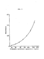
- Fig. 1 and Fig. 2 show calibration curves of the two-site simultaneous immunoassay conducted by use of the monoclonal antibodies of this invention.
- Fig. 3 shows the measured values (aspiration rate) of the pulmonary surfactant lipoprotein) in the amniotic fluid versus temperature of the immunoreaction.
- Fig. 4 shows the immunoreaction time versus lipoprotein concentration, i.e. measured values of the pulmonary surfactant (lipoprotein) in the amniotic fluid.
- the immunoassay was conducted in the presence and absence of 0.25 M Mg
- the especially desirable ones from the viewpoint of reactivity and stability are those antibodies which recognize apoproteins with molecular weight of about 62,000 and/or about 34,000 to 37,000, being mouse antibodies of IgG type.
- the monoclonal antibodies of this invention can be obtained by fusing antibody-producing cells of animals immunized preferably with the human pulmonary surfactant apoproteins and myeloma cells to give hybridomas productive of monoclonal antibodies which recognize said apoproteins; culturing said hybridomas and/or cell line arising therefrom; and collecting monoclonal antibodies, which recognize the human pulmonary surfactant apoproteins, from the culture.
- the pulmonary surfactant is isolated and collected from human lung and/or branchus lavage fluid, preferably from bronchus and lung lavage fluid of patients with alveolar proteinosis.
- the pulmonayr surfactant is a complex (lipoprotein) composed of about 90% lipids and about 10% protein.
- the pulmonayr furfactant is obtained by the method described by Frosolono (see J. Lipid Res. ll, 439m457 (1970)) and the removal of lipids therefrom gives apoprotein proposed in this invention.
- Apoprotein is mainly composed of proteins with a molecular weight of about 62,000 and about 36,000 respectively.
- Proteins with a molecular weight of 36,000 are separated as a wide band when subjected to sodium dodecyl sulfate-polyacrylamide gel electrophoresis (SDS-PAGE) and it is deemed that proteins with a molecular weight of about 34,000 are contained therein.
- SDS-PAGE sodium dodecyl sulfate-polyacrylamide gel electrophoresis
- the apoproteins of this invention accordingly include these proteins are all other apoproteins and their fragments. The molecular weights of the proteins were determined with SDS-PAGE.
- Hybridomas productive of monoclonal antibodies which recognize the apoproteins can be produced according to the cell fusion method whose procedures are generally known per se. Firstly, such animals as monkeys, horses, bovines, goats, sheep, rabbits, rats, mice, etc. are immunized with apoproteins, then antibody-producing cells (lymphocytes) are collected from the spleens and lymph nodes of these immunized animals, followed by cell fusion of these cells with human or animal's myeloma cells. As the myeloma cells, mouse myeloma cells may be used most conveniently.
- mouse myeloma cells include P3-X63-Ag8, P-X63-Ag8-Ul, P3-NS1/1-Ag4-1, P3-X63-Ag8 6.5.3, SP2/0-Agl4, FO, and MPC 11-45.6TG1.7.
- the conditions of cell fusion are as follows. For instance, antibody-producing cells and myeloma cells are mixed at a ratio of 10:1 to 1:10, preferably at a ratio of 1:1 to 1:3.
- An appropriate cell fusion mixture such as RPMI 1640 containing about 35% polyethylene glycol (molecular weight approximately 1,000 to 6,000) and about 7.5% dimethyl sulfoxide is added to the cell mixture and are stirred for one to several minutes at room temperature to 37°C.
- the mixture is diluted little by little with RPMI 1640 containing 10% FCS (fetal calf serum), washed, and ajusted to have cell concentration of 1 to 5 X 10 5 cells/ml with a selective liquid culture medium HAT (hypoxanthine-aminopterin-thymidine).
- FCS fetal calf serum
- HAT hyperxanthine-aminopterin-thymidine
- hybridomas In the HAT liquid culture medium, only hybridomas can survive and 8-azaguanine resistant myeloma cells and fused cells between myeloma cells can not survive (unfused antibody-producing cells perish in the course of several days). Then from the colonies of hybridomas, only those hybridomas that secrete monoclonal antibodies reactive against apoproteins are selected. This process of selection (or screening) can be carried out by subjecting the monoclonal antibodies produced by the respective hybridomas to the enzyme-linked-immunosorbent assay to examine if they undergo the antigen-antibody reaction with the desired apoproteins.
- the hybridomas which secrete monoclonal antibodies this invention aims at, must then be subjected to the cloning to obtain clonal cells.
- This cloning can be effected, for instance, by limiting dilution to monoclonality.
- the colonies grown in a 96-well plate are collected to have their antibody activity against apoproteins examined again by the enzyme-linked-immunosorbent assay and the selected hybridomas are cultured to give monoclonal antibodies specific to the apoproteins.
- Another method of obtaining monoclonal antibodies is to infect the antibody-producing cells with Epstein-barr virus (hereinafter abbreviated to E-B virus) to prepare transformed cells.
- E-B virus Epstein-barr virus
- the transformed cells and/or cell line arising therefrom are cultured and monoclonal antibodies which have a nature to bind to apoproteins are collected from the culture.
- E-B virus is a virus, which belongs to the herpes virus, regarded to be a virus causative of Burkitt's lymphoma and rhinopharynx cancer.
- the antibody-producing cells are infected with E-B virus and cultured for about 2 to 3 weeks in the 5% C0 2 incubator to make them establish transformed cells forming many heterogeneous colonies. Then selections are made from these transformed cells according to the same method as mentioned above to obtain only those which secrete monoclonal antibodies specific to apoproteins and then cloning follows to obtain cloned transformed cells according to the cloning method described above.
- the selected hybridomas or transformed cells are cultured to establish desired specific monoclonal antibodies.
- the hybridomas or transformed cells selected by cloning and productive of antibodies which recognize the pulmonary surfactant apoproteins, can be freezestored and also can be mass-cultured according to a proper method.
- monoclonal antibodies, which bind specifically to apoproteins can be obtained from the culture supernatants.
- the desired antibodies can be obtained from their ascites and serum.
- the purification of the monoclonal antibodies of this invention can be carried out by affinity chromatography by use of protein A.
- the time requred for determination was shortened according to this two-site simultanous immunoassay, in which the first monoclonal antibodies (primary antibodies) fixed to a microplate, antigens (apoproteins), and biotinylated second monoclonal antibodies (secondary antibodies) were made to react with each other simultaneously. Also, the use of skim milk in the blocking agent lowered the background values remarkably.
- the primary antibody fixed to a carrier and the method of fixation may be chosen from the publicly known ones.
- the carrier those which are solid-phase ones including balls, beads, gears, and microplate made of polystyrene, polyethylene, polyacrylate, Teflon, or polyacetal may be used preferably.
- any procedure of detecting any procedure of detecting. Any known methods and procedures such as the determination by the secondary reaction with anti-immunoglobulin antibody or Staphylococcus protein A may be adopted.
- the labeling agents such enzymes as horseradish peroxidase, ⁇ -D-galactosidase, and alkaline phosphatase are used in the enzyme immunoassay (EIA), 125 I and 3 H in the radioimmunoassay (RIA), and fluorescein- isothiocyanate in the fluorescence immunoassay (FIA) in general; however, other labeling agents may also be used so far as their activity is assayable.
- EIA enzyme immunoassay
- RIA radioimmunoassay
- FFA fluorescein- isothiocyanate in the fluorescence immunoassay
- a substrate is used for assaying its activity.
- 2,2'-azinodi-[3-ethylbenzthiazoline sulfonic acidlammonium acid (ABTS)-H 2 0 2 , 5-amino salicylic acid-H 2 0 2 , 0-phenylenediamine-H 2 0 2 , and 4-aminoantipyrine-H 2 0 2 may be used as the substrate for horseradish peroxidase, and fluoresein-di-( ⁇ -D-galactopyranoside) and O-nitrophenol- ⁇ -D-galactopyranoside for ⁇ -D-galactosidase.
- such publicly known reagents as solubilizer, detergent, and reaction terminator are used besides the above-mentioned reagents.
- What is desirably used in this invention is a combination of a biotinylated antibody and an enzyme-labeled avidin.
- Avidin is a basic glycoprotein with a molecular weight of about 68,000 existing in the albumen and is understood to have a very high affinity (affinity constant 10M -1 ) for biotin which is known as vitamin H. It is known that avidin is composed of 4 subunits and what is called avidin in the present invention includes these subunits.
- This invention also includes the aforementioned antibodies and a kit of reagents in its scope.
- a desirable example includes: (1) the first monoclonal antibody (primary antibody) fixed to a carrier and recognizes a human pulmonary surfactant apoprotein; (2) labeled second monoclonal antibody (secondary antibody) which, though recognizes said apoprotein, binds to a part of the antigen which is different from the one to which the primary monoclonal antibody binds; and (3) reagents, which mainly comprises reagents for detecting said labeled antibody, to be used for determining the plumonary surfactant existing in the human amniotic bluid and human lung or bronchus lavage fluid, if necessary.
- the immunoreaction it is desirable to conduct the immunoreaction at a temperature ranging from 40°C to 50°C exclusive. It is also desirable to make magnesium ions (Mg ++ ) coexist in the immunoreaction system.
- difference of reactivity between the standard substance of apoprotein and the pulmonary surfactant in the test substance such as amniotic fluid in the immunoassay. It may be a safe assumption that such difference is atrributable to the steric hindrance caused by the fact that the pulmonary surfactant in the test substance is composed of apoproteins and phospholipid which is ten times as much as the apoproteins.
- the present inventors have found that the apoproteins and the lipoproteins contained in the pulmonary surfactant of the test substance come to display the same behavior in a short time upon immunological observation by adjusting the temperature and making Mg ++ coexisting in performing the assay, thus making it possible to achieve the solid-phase enzyme immunoreaction in a short time.
- This sedimental fraction was suspended in 80 mi of 10 mM Tris buffer (pH 7.4) containing 145 mM sodium chloride and 1 mM disodium ethylenediaminetetraacetic acid. The suspension was layered on a discontinuous gradient prepared with 0.25M and 0.65M sucrose solutions and centrifuged at 40,000 x g for 60 minutes.
- the interface fraction (IB fraction) containing the pulmonary surfactant separated between 0.25M and 0.65M sucrose solutions was collected and resuspended in 400 ml of the same buffer. This suspension was centrifuged at 48,000 x g for 30 minutes to obtain the sediment (pulmonary surfactant).
- Tris buffer pH 7.8 containing 1% Triton X-100 (polyoxyethylenealkylphenyl ether), 3mM EDTA, 1mM phenylmethylsulfonyl fluoride, and 0.5mM dithiothreitol (hereinafter referred to as Triton buffer).
- Triton buffer 1% Triton X-100 (polyoxyethylenealkylphenyl ether), 3mM EDTA, 1mM phenylmethylsulfonyl fluoride, and 0.5mM dithiothreitol
- This precipitate was redispersed in 4 ml of the abovementioned Triton buffer and 4 mt of a butanol-methanol mixture (6:1 v/v)was added thereto. The mixture was shaken vigorously and left standing at 0°C for 10 minutes. After the removal of lipid by extraction, the dispersion was centrifuged at 2,000 r.p.m. for 15 minutes to obtain a precipitate (apoprotein). This procedure was repeated three times to give a final precipitate of apoprotein (LS apoprotein).
- This precipitate was dispersed in 20 ml of Triton buffer and dialyzed against the same Triton buffer with the use of a cellophane membrane (to have butanol-ethanol solution removed) to obtain in dialysate.
- This dialysate was centrifuged at 15,000 X g for 60 minutes to obtain a supernatant.
- the sediment was solubilized by addition of 20 m£ of Triton buffer and centrifuged at 150,000 X g for 60 minutes to give a supernatant. This procedure was repeated six times to collect supernatants. Thus obtained supernatants were pooled to make a total of 150 mi.
- the pooled supernatant was applied onto a Blue Sepharose 4B column (1.8 cm diameter X 3.5 cm) and eluted by use of the same buffer and the void fractions were collected to remove albumin thoroughly.
- the collected void fractions were applied onto a DEAE-Toyopearl column (diameter 1.4 cm X 19 cm) equilibrated with the Triton buffer, eluted with 200 m£ of the same buffer at a flow rate of 15 ml/hr at first, then the elution was continued while linearly increasing the concentration of sodium chloride contained in the same buffer from 0 to 0.5M continuatively and fractions eluted while the sodium chloride concentration was in the range of 0.30M and 0.35M were collected.
- the proteins contained in these fractions proved to have a monocular weight of about 62,000 and about 36,000 respectively.
- the L S apoproteins were emulsified in Freund's complete adjuvant and injected into the peritoneal cavities of BALB/C mice. The mice were boosted with the same LS apoproteins 30 days later.
- myeloma cell line P3-X63-Ag8-Ul had been maintained for incubation in RPMI 1640 (Gibco) supplemented with 15% fetal calf serum.
- the spleen cells obtained from the mice were fused with P3-X63-Ag8-Ul by use of polyethylene glycol 4000 according to the method proposed by Oi et al. (see Selective Methods in Cellular Immunology 1980, pp.
- the medium was replaced with RPMI medium supplemented with 100 ⁇ M hypoxanthine, 0.4 ⁇ M aminopterin, and 16 ⁇ M thymidine (HAT medium).
- HAT medium 16 ⁇ M thymidine
- the LS apoproteins were attached to the ELISA plates and were subjected to the blocking by use of 3% (W/V) BSA (bobine serum albumin) in 10mM phosphate-buffered saline (pH 7.4). After blocking, 50 ⁇ l, of the hybridoma culture medium was added to the abovementioned plates and incubated at room temperature for 2 hours or at 4°C overnight. Thereafter, a secondary antibody of 50 ⁇ l horse biotinylated anti-mouse IgG immunoglobulin (2 pg/mk) was added and incubated at room temperature for 1 hour.
- W/V bobine serum albumin
- Hybridomas productive of antibodies against LS apoproteins were selected and cloned in limiting dilution to monoclonality, finally giving two kinds of monoclonal hybridomas.
- These two kinds of hybridomas were respectively amplified in pristane-treated BALB/C mice obdominal cavities to obtain ascites containing the monoclonal antibodies.
- ascites were made to have their antibodies precipitated with the use of 50% saturated ammonium sulfate, and the precipitate was dissolved in 0.1 M phosphate-buffered saline (pH 8.0).
- the solution was put to a Protein A-Sepharose CL 4B column (Parmaoia Pine Chemicals) and the antibodies was eluted with 0.2 M glycine-hydrochloride buffer (pH 3.0) to be purified.
- the monoclonal antibodies obtained from the two kinds of hybridomas were named PC 6 and PE 10 respectively.
- PC 6 and PE 10 recognized two types of apoproteins 36 K and 62 K obtained from the IB fraction of bronchoalveolar lavage fluids of patients with alveolar proteinosis.
- the same two antibodies also recognized the apoproteins 37 K, 34 K, and 62 K in the human amniotic fluid and normal human bronchoalveolar lavage fluids.
- the 36 K proteins were separated with SDS-PAGE as a wide band and as 37 K and 34 K proteins are contained among them, the 35 K proteins are the same proteins as the 37 K and 34 K proteins.
- PC 6 and PE 10 recognized the epitopes (antigen determinants) neighboring and yet differing from each other from the result of the cross reaction conducted by the dot-immunobinding method. These antibodies were specific to human pulmonary surfactant apoproteins and did not reacted with pulmonary surfactant apoproteins of animals such as rats, swines, rabbits and human serum proteins.
- the antigens specific for monoclonal antibodies were identified using the western blotting technique by the method of Towbin et al. (Pro. N. A. S., vol. 76, pp. 4350 ⁇ 4354).
- the antigens containing pulmonary surfactant apoproteins were subjected to SDS-PAGE.
- the proteins were then transferred from the slab gel to a nitrocellulose sheet after SDS-PAGE with the electrode buffer (pH 8.3) containing 25 mM Tris-hydrochloride, 192 mM glycine, and 20% (v/v) methanol and a voltage gradient of 7 V/cm applied for 2 hours.
- the electrode buffer pH 8.3
- 25 mM Tris-hydrochloride 25 mM Tris-hydrochloride
- 192 mM glycine 192 mM glycine
- 20% (v/v) methanol 20% (v/v) methanol
- Each lane of the nitrocellulose sheet was cut off.
- One lane was used for staining the proteins by Amido black, and the others were subjected to an enzyme immunoassay mentioned below.
- HRP horseradish peroxidase
- a substrate solution comprising 0.1 M citrate buffer (pH 4.6) containing 0.1% o-phenylenediamine and 0.015% H 2 0 2 was added to each well and the reaction was allowed to continue at room temperature for 30 minutes and then stopped by the addition of 100 ⁇ l of 2 M sulfuric acid. After the reaction was made to stop, the absorbance of each well was measured using an automatic microplate reader at two wave length absorbance; A500-A610. Absorbance change observed with different concentrations of the standard substance were plotted to prepare a calibration curve as shown in Fig. 1. Using this calibration curve, the concentrations of the respective test substances were obtained.
- the concentrations of apoproteins in the 59 test substances of amniotic fluids from gestational 23 to 41-week women were determined with the result shown in Table 1 in which it was made clear that the result shown in Table 1 in which it was made clear that the concentrations of apoproteins increased as the gestational weeks increased.
- the concentration of apoproteins was very low for the women of 30 weeks gestation or less. While the apoprotein concentration of the women of 34m36 weeks gestation was 6.5 times higher, and the concentrations for the women of 37 weeks gestation or more was as high as 15.5 times.
- the maleimidic monoclonal antibodies and the thiolic HRP obtained in the above were mixed and concentrated to 4 mg of protein concentration in a collodion bag on the ice bath.
- the concentrate was left standing at 4°C overnight and filtrated on a column of ultrogel Ac A44 to obtain HRP labeled monoclonal antibodies.
- test tubes were washed with a saline solution. Then a mixture of 1% 3,3',5,5'-tetramethylbenzene- containing methanol solution/0.015 H 2 O 2 - containing 0.1 M phosphate-citrate buffer (pH 4.4) mixed at a ratio of 3/7 (V/V) was added to the respective test tubes in a portion of 0.4 ml. After 15-mimute incubation at room temperature, 2 mi of 1.5 NH 2 S0 4 aqueous solution was added to the respective test tubes as the reaction terminator to have the lnzyme reaction stopped.
- the test substances and HRP-labeled monoclonal antibody PE 10 were diluted respectively with buffers, (a) a TBS buffer containing 0.25 M MgC£ 2 , 0.1% skim milk, and 1% Triton X-100 and (b) a TBS buffer containing 0.1% skim milk and 1% Triton X-100.
- the enzyme reactions were carried out respectively for a period of 30 minutes, 45 minutes, 1 hour, 1.5 hours, 2 hours, and 3 hours at 45°C.
- the concentrations of pulmonary surfactants in the amniotic fluid was read from the respective times of enzyme reaction by use of the calibration curve and were plotted in Fig. 4. It is apparent from Fig. 4 that the addition of Mg ++ is effective since the enzyme reaction is much accelerated by the presence of Mg ++ in the buffer.
- the method proposed by the present invention makes it possible to quantitate the pulmonary surfactant apoproteins within the range of 10 to 640 ⁇ g/ml, with the variation coefficient kept below 6%. Human amniotic fluid can be subjected to quantitation if its quantity is 0.2 mk at the least.
- the determination of the pulmonary surfactant apoproteins in the test substance can be performed simply and conveniently in a short time, thus providing a useful method of determination and a kit to be used for such determination in case of an urgent disease such as IRDS, etc.
Landscapes
- Health & Medical Sciences (AREA)
- Life Sciences & Earth Sciences (AREA)
- Chemical & Material Sciences (AREA)
- Immunology (AREA)
- Engineering & Computer Science (AREA)
- Molecular Biology (AREA)
- General Health & Medical Sciences (AREA)
- Hematology (AREA)
- Medicinal Chemistry (AREA)
- Urology & Nephrology (AREA)
- Proteomics, Peptides & Aminoacids (AREA)
- Organic Chemistry (AREA)
- Biochemistry (AREA)
- Biomedical Technology (AREA)
- Cell Biology (AREA)
- Genetics & Genomics (AREA)
- Biotechnology (AREA)
- Microbiology (AREA)
- Biophysics (AREA)
- Food Science & Technology (AREA)
- Physics & Mathematics (AREA)
- Analytical Chemistry (AREA)
- General Physics & Mathematics (AREA)
- Pathology (AREA)
- Preparation Of Compounds By Using Micro-Organisms (AREA)
- Peptides Or Proteins (AREA)
Applications Claiming Priority (5)
| Application Number | Priority Date | Filing Date | Title |
|---|---|---|---|
| JP116738/85 | 1985-05-31 | ||
| JP60116738A JPH0672894B2 (ja) | 1985-05-31 | 1985-05-31 | ヒトの肺表面活性物質の測定方法及びそれに用いる試薬キツト |
| JP5256386A JPS62211554A (ja) | 1986-03-12 | 1986-03-12 | ヒトの肺表面活性物質の測定方法 |
| JP52563/86 | 1986-03-12 | ||
| PCT/JP1986/000258 WO1986007154A1 (fr) | 1985-05-31 | 1986-05-20 | Procede d'analyse d'une substance active se trouvant a la surface des poumons de l'homme et kit reactif destine a etre utilise avec ledit procede |
Publications (3)
| Publication Number | Publication Date |
|---|---|
| EP0224590A1 true EP0224590A1 (de) | 1987-06-10 |
| EP0224590A4 EP0224590A4 (de) | 1987-12-08 |
| EP0224590B1 EP0224590B1 (de) | 1994-11-23 |
Family
ID=26393188
Family Applications (1)
| Application Number | Title | Priority Date | Filing Date |
|---|---|---|---|
| EP86903560A Expired - Lifetime EP0224590B1 (de) | 1985-05-31 | 1986-05-20 | Verfahren zur bestimmung menschlicher lungenoberflächenaktiver substanz und reagenziensatz dazu |
Country Status (4)
| Country | Link |
|---|---|
| US (1) | US5156950A (de) |
| EP (1) | EP0224590B1 (de) |
| DE (1) | DE3650147T2 (de) |
| WO (1) | WO1986007154A1 (de) |
Cited By (5)
| Publication number | Priority date | Publication date | Assignee | Title |
|---|---|---|---|---|
| EP0329794A4 (en) * | 1987-09-01 | 1991-04-24 | Teijin Limited | Method for assaying human lung surface active substance |
| EP0328679B1 (de) * | 1987-08-12 | 1994-07-13 | Teijin Limited | Immuntestverfahren und reagenzsatz dazu |
| US5366861A (en) * | 1987-08-12 | 1994-11-22 | Teijin Limited | Immunoassay and reagent kit used therefor |
| WO2000005585A1 (en) * | 1998-07-24 | 2000-02-03 | Byk Gulden Lomberg Chemische Fabrik Gmbh | Determination of the hydrophobic pulmonary surfactant protein sp-c |
| US8097420B1 (en) | 1997-09-05 | 2012-01-17 | Southern Medical Diagnostics Pty Ltd | Method of diagnosis |
Families Citing this family (10)
| Publication number | Priority date | Publication date | Assignee | Title |
|---|---|---|---|---|
| US5670328A (en) * | 1992-06-09 | 1997-09-23 | Yamasa Corporation | Monoclonal antibodies to human pulmonary surfactant apoprotein D and use thereof |
| US5856196A (en) * | 1993-10-25 | 1999-01-05 | Beth Israel Hospital | Processes for quantitating phosphoglycerides in a lipid mixture and diagnostic uses therefor |
| US5443989A (en) * | 1993-10-25 | 1995-08-22 | Beth Israel Hospital Association | Method for assessing fetal lung maturity using amniotic fluid samples |
| US20060166276A1 (en) * | 1997-09-05 | 2006-07-27 | Lung Health Diagnostics Pty Ltd | Method of diagnosis and agents useful for same |
| WO2004077056A1 (en) * | 2003-02-27 | 2004-09-10 | Lung Health Diagnostics Pty. Ltd. | A method of diagnosis and agents useful for same |
| US7582442B2 (en) * | 2004-03-16 | 2009-09-01 | The Regents Of The University Of Michigan | Methods and compositions for using aleveolar macrophage phospholipase A2 |
| US7319015B2 (en) * | 2004-03-16 | 2008-01-15 | The Regents Of The University Of Michigan | Methods and compositions for using alveolar macrophage phospholipase A2 |
| US20060134694A1 (en) * | 2004-12-22 | 2006-06-22 | Intel Corporation | Methods of protein profiling by thiolation |
| WO2012161288A1 (en) * | 2011-05-20 | 2012-11-29 | Abbott Japan Co. Ltd. | Immunoassay methods and reagents for decreasing nonspecific binding |
| CN112088204B (zh) | 2018-06-01 | 2023-06-06 | 埃克森美孚科技工程公司 | 利用膜级联对烃料流进行无沸腾分流 |
Family Cites Families (4)
| Publication number | Priority date | Publication date | Assignee | Title |
|---|---|---|---|---|
| JPS57136165A (en) * | 1981-02-18 | 1982-08-23 | Mochida Pharmaceut Co Ltd | Immunological measuring reagent |
| US4535057A (en) * | 1982-07-26 | 1985-08-13 | Amf Incorporated | Immunoassay employing monoclonal herpes simplex antibody and biotin-avidin detection system |
| US4474892A (en) * | 1983-02-16 | 1984-10-02 | Board Of Trustees Of The Leland Stanford Junior University | Two-site immunoassays using monoclonal antibodies of different classes or subclasses and test kits for performing same |
| US4562003A (en) * | 1984-10-26 | 1985-12-31 | California Biotechnology, Inc. | Monoclonal antibodies against alveolar surfactant protein |
-
1986
- 1986-05-20 DE DE3650147T patent/DE3650147T2/de not_active Expired - Fee Related
- 1986-05-20 EP EP86903560A patent/EP0224590B1/de not_active Expired - Lifetime
- 1986-05-20 WO PCT/JP1986/000258 patent/WO1986007154A1/ja not_active Ceased
-
1991
- 1991-01-07 US US07/638,026 patent/US5156950A/en not_active Expired - Fee Related
Cited By (6)
| Publication number | Priority date | Publication date | Assignee | Title |
|---|---|---|---|---|
| EP0328679B1 (de) * | 1987-08-12 | 1994-07-13 | Teijin Limited | Immuntestverfahren und reagenzsatz dazu |
| US5366861A (en) * | 1987-08-12 | 1994-11-22 | Teijin Limited | Immunoassay and reagent kit used therefor |
| EP0329794A4 (en) * | 1987-09-01 | 1991-04-24 | Teijin Limited | Method for assaying human lung surface active substance |
| US8097420B1 (en) | 1997-09-05 | 2012-01-17 | Southern Medical Diagnostics Pty Ltd | Method of diagnosis |
| WO2000005585A1 (en) * | 1998-07-24 | 2000-02-03 | Byk Gulden Lomberg Chemische Fabrik Gmbh | Determination of the hydrophobic pulmonary surfactant protein sp-c |
| US6737243B1 (en) | 1998-07-24 | 2004-05-18 | Altana Pharma Ag | Determination of the hydrophobic pulmonary surfactant protein SP-C |
Also Published As
| Publication number | Publication date |
|---|---|
| WO1986007154A1 (fr) | 1986-12-04 |
| EP0224590A4 (de) | 1987-12-08 |
| DE3650147T2 (de) | 1995-04-06 |
| US5156950A (en) | 1992-10-20 |
| EP0224590B1 (de) | 1994-11-23 |
| DE3650147D1 (de) | 1995-01-05 |
Similar Documents
| Publication | Publication Date | Title |
|---|---|---|
| EP0224590B1 (de) | Verfahren zur bestimmung menschlicher lungenoberflächenaktiver substanz und reagenziensatz dazu | |
| Allard et al. | Monoclonal antibodies to the glucose transporter from human erythrocytes. Identification of the transporter as a Mr= 55,000 protein. | |
| EP0203093B1 (de) | Monoklonale antikörper gegen alveolares oberflächenaktives protein | |
| Herr et al. | Characterization of a monoclonal antibody to a conserved epitope on human seminal vesicle-specific peptides: a novel probe/marker system for semen identification | |
| Kuroki et al. | Two-site “simultaneous” immunoassay with monoclonal antibodies for the determination of surfactant apoproteins in human amniotic fluid | |
| GB2095831A (en) | Monoclonal antibody reagent and method for immunological assay | |
| EP0161638B1 (de) | Monoklonaler Antikörper und Verfahren zur quantitativen Bestimmung von Immunoglobulinen mittels dieses Antikörpers | |
| US5670328A (en) | Monoclonal antibodies to human pulmonary surfactant apoprotein D and use thereof | |
| US5316914A (en) | Method for determining human collagen peptides by way of enzyme immunoassay | |
| Kaetzel et al. | Immunochemical characterization with monoclonal antibodies of three major caseins and alpha-lactalbumin from rat milk | |
| JP2867325B2 (ja) | 抗pivka−ii抗体産生ハイブリドーマ及び免疫学的測定方法 | |
| KR19990067153A (ko) | 프로스타글란딘 디 신타아제에 특이적인 단일클론항체 | |
| EP0410004B1 (de) | Immunobestimmung von menschlichem osteocalcin, reagenz und satz dafür | |
| EP0245520B1 (de) | Monoklonaler antikörper gegen glutathion s-transferase und dessen verwendung zur diagnose von krebs | |
| JPH0672894B2 (ja) | ヒトの肺表面活性物質の測定方法及びそれに用いる試薬キツト | |
| EP0401370A1 (de) | Enzymimmuntest für menschliches typiv-collagen gemäss dem sandwichverfahren | |
| CA2281262C (en) | Anti-human medullasin monoclonal antibody, process for producing the same and immunoassay using the same | |
| CA1313828C (en) | Monoclonal antibodies to superficial papillary bladder tumor cells | |
| CA1287801C (en) | Method for determining human collagen peptides by way of enzyme immunoassay | |
| JPH0614041B2 (ja) | 肺表面活性物質の検出方法及びそれに用いる試薬キツト | |
| JPH0636754B2 (ja) | 肺表面活性物質に対するモノクロ−ナル抗体及びその製造法 | |
| Shuster et al. | Enzyme immunoassay of bovine lactoferrin and serum albumin in acid precipitated and ultracentrifugal wheys | |
| EP0602248B1 (de) | Lungenkrankheiten screening durch messung der HUMANES OBERFLÄCHENAKTIVES APOPROTEIN D DER LUNGE | |
| EP0242727B1 (de) | Verfahren zur Bestimmung von Adenocarcinoma-Antigenen | |
| JPH02242158A (ja) | ヒトアポリポ蛋白c―3の測定法 |
Legal Events
| Date | Code | Title | Description |
|---|---|---|---|
| PUAI | Public reference made under article 153(3) epc to a published international application that has entered the european phase |
Free format text: ORIGINAL CODE: 0009012 |
|
| 17P | Request for examination filed |
Effective date: 19870131 |
|
| AK | Designated contracting states |
Kind code of ref document: A1 Designated state(s): CH DE FR GB IT LI NL SE |
|
| A4 | Supplementary search report drawn up and despatched |
Effective date: 19871208 |
|
| 17Q | First examination report despatched |
Effective date: 19890817 |
|
| RAP1 | Party data changed (applicant data changed or rights of an application transferred) |
Owner name: TEIJIN LIMITED |
|
| GRAA | (expected) grant |
Free format text: ORIGINAL CODE: 0009210 |
|
| AK | Designated contracting states |
Kind code of ref document: B1 Designated state(s): CH DE FR GB IT LI NL SE |
|
| REF | Corresponds to: |
Ref document number: 3650147 Country of ref document: DE Date of ref document: 19950105 |
|
| ET | Fr: translation filed | ||
| ITF | It: translation for a ep patent filed | ||
| PLBE | No opposition filed within time limit |
Free format text: ORIGINAL CODE: 0009261 |
|
| STAA | Information on the status of an ep patent application or granted ep patent |
Free format text: STATUS: NO OPPOSITION FILED WITHIN TIME LIMIT |
|
| 26N | No opposition filed | ||
| PGFP | Annual fee paid to national office [announced via postgrant information from national office to epo] |
Ref country code: SE Payment date: 20000327 Year of fee payment: 15 Ref country code: CH Payment date: 20000327 Year of fee payment: 15 |
|
| PGFP | Annual fee paid to national office [announced via postgrant information from national office to epo] |
Ref country code: NL Payment date: 20000425 Year of fee payment: 15 Ref country code: FR Payment date: 20000425 Year of fee payment: 15 |
|
| PGFP | Annual fee paid to national office [announced via postgrant information from national office to epo] |
Ref country code: GB Payment date: 20000502 Year of fee payment: 15 |
|
| PGFP | Annual fee paid to national office [announced via postgrant information from national office to epo] |
Ref country code: DE Payment date: 20000630 Year of fee payment: 15 |
|
| PG25 | Lapsed in a contracting state [announced via postgrant information from national office to epo] |
Ref country code: GB Free format text: LAPSE BECAUSE OF NON-PAYMENT OF DUE FEES Effective date: 20010520 |
|
| PG25 | Lapsed in a contracting state [announced via postgrant information from national office to epo] |
Ref country code: SE Free format text: LAPSE BECAUSE OF NON-PAYMENT OF DUE FEES Effective date: 20010521 |
|
| PG25 | Lapsed in a contracting state [announced via postgrant information from national office to epo] |
Ref country code: LI Free format text: LAPSE BECAUSE OF NON-PAYMENT OF DUE FEES Effective date: 20010619 Ref country code: CH Free format text: LAPSE BECAUSE OF NON-PAYMENT OF DUE FEES Effective date: 20010619 |
|
| PG25 | Lapsed in a contracting state [announced via postgrant information from national office to epo] |
Ref country code: NL Free format text: LAPSE BECAUSE OF NON-PAYMENT OF DUE FEES Effective date: 20011201 |
|
| GBPC | Gb: european patent ceased through non-payment of renewal fee |
Effective date: 20010520 |
|
| REG | Reference to a national code |
Ref country code: CH Ref legal event code: PL |
|
| PG25 | Lapsed in a contracting state [announced via postgrant information from national office to epo] |
Ref country code: FR Free format text: LAPSE BECAUSE OF NON-PAYMENT OF DUE FEES Effective date: 20020131 |
|
| NLV4 | Nl: lapsed or anulled due to non-payment of the annual fee |
Effective date: 20011201 |
|
| PG25 | Lapsed in a contracting state [announced via postgrant information from national office to epo] |
Ref country code: DE Free format text: LAPSE BECAUSE OF NON-PAYMENT OF DUE FEES Effective date: 20020301 |
|
| PG25 | Lapsed in a contracting state [announced via postgrant information from national office to epo] |
Ref country code: IT Free format text: LAPSE BECAUSE OF NON-PAYMENT OF DUE FEES;WARNING: LAPSES OF ITALIAN PATENTS WITH EFFECTIVE DATE BEFORE 2007 MAY HAVE OCCURRED AT ANY TIME BEFORE 2007. THE CORRECT EFFECTIVE DATE MAY BE DIFFERENT FROM THE ONE RECORDED. Effective date: 20050520 |
|
| APAH | Appeal reference modified |
Free format text: ORIGINAL CODE: EPIDOSCREFNO |