EP0249357A2 - Methods of immunoassay - Google Patents
Methods of immunoassay Download PDFInfo
- Publication number
- EP0249357A2 EP0249357A2 EP87304644A EP87304644A EP0249357A2 EP 0249357 A2 EP0249357 A2 EP 0249357A2 EP 87304644 A EP87304644 A EP 87304644A EP 87304644 A EP87304644 A EP 87304644A EP 0249357 A2 EP0249357 A2 EP 0249357A2
- Authority
- EP
- European Patent Office
- Prior art keywords
- absorbance
- phenolphthalein
- enzyme
- wavelength
- hcg
- Prior art date
- Legal status (The legal status is an assumption and is not a legal conclusion. Google has not performed a legal analysis and makes no representation as to the accuracy of the status listed.)
- Granted
Links
Images



Classifications
-
- G—PHYSICS
- G01—MEASURING; TESTING
- G01N—INVESTIGATING OR ANALYSING MATERIALS BY DETERMINING THEIR CHEMICAL OR PHYSICAL PROPERTIES
- G01N33/00—Investigating or analysing materials by specific methods not covered by groups G01N1/00 - G01N31/00
- G01N33/48—Biological material, e.g. blood, urine; Haemocytometers
- G01N33/50—Chemical analysis of biological material, e.g. blood, urine; Testing involving biospecific ligand binding methods; Immunological testing
- G01N33/58—Chemical analysis of biological material, e.g. blood, urine; Testing involving biospecific ligand binding methods; Immunological testing involving labelled substances
- G01N33/581—Chemical analysis of biological material, e.g. blood, urine; Testing involving biospecific ligand binding methods; Immunological testing involving labelled substances with enzyme label (including co-enzymes, co-factors, enzyme inhibitors or substrates)
-
- G—PHYSICS
- G01—MEASURING; TESTING
- G01N—INVESTIGATING OR ANALYSING MATERIALS BY DETERMINING THEIR CHEMICAL OR PHYSICAL PROPERTIES
- G01N33/00—Investigating or analysing materials by specific methods not covered by groups G01N1/00 - G01N31/00
- G01N33/48—Biological material, e.g. blood, urine; Haemocytometers
- G01N33/50—Chemical analysis of biological material, e.g. blood, urine; Testing involving biospecific ligand binding methods; Immunological testing
- G01N33/53—Immunoassay; Biospecific binding assay; Materials therefor
- G01N33/531—Production of immunochemical test materials
- G01N33/532—Production of labelled immunochemicals
- G01N33/535—Production of labelled immunochemicals with enzyme label or co-enzymes, co-factors, enzyme inhibitors or enzyme substrates
-
- Y—GENERAL TAGGING OF NEW TECHNOLOGICAL DEVELOPMENTS; GENERAL TAGGING OF CROSS-SECTIONAL TECHNOLOGIES SPANNING OVER SEVERAL SECTIONS OF THE IPC; TECHNICAL SUBJECTS COVERED BY FORMER USPC CROSS-REFERENCE ART COLLECTIONS [XRACs] AND DIGESTS
- Y10—TECHNICAL SUBJECTS COVERED BY FORMER USPC
- Y10S—TECHNICAL SUBJECTS COVERED BY FORMER USPC CROSS-REFERENCE ART COLLECTIONS [XRACs] AND DIGESTS
- Y10S435/00—Chemistry: molecular biology and microbiology
- Y10S435/962—Prevention or removal of interfering materials or reactants or other treatment to enhance results, e.g. determining or preventing nonspecific binding
-
- Y—GENERAL TAGGING OF NEW TECHNOLOGICAL DEVELOPMENTS; GENERAL TAGGING OF CROSS-SECTIONAL TECHNOLOGIES SPANNING OVER SEVERAL SECTIONS OF THE IPC; TECHNICAL SUBJECTS COVERED BY FORMER USPC CROSS-REFERENCE ART COLLECTIONS [XRACs] AND DIGESTS
- Y10—TECHNICAL SUBJECTS COVERED BY FORMER USPC
- Y10S—TECHNICAL SUBJECTS COVERED BY FORMER USPC CROSS-REFERENCE ART COLLECTIONS [XRACs] AND DIGESTS
- Y10S435/00—Chemistry: molecular biology and microbiology
- Y10S435/973—Simultaneous determination of more than one analyte
-
- Y—GENERAL TAGGING OF NEW TECHNOLOGICAL DEVELOPMENTS; GENERAL TAGGING OF CROSS-SECTIONAL TECHNOLOGIES SPANNING OVER SEVERAL SECTIONS OF THE IPC; TECHNICAL SUBJECTS COVERED BY FORMER USPC CROSS-REFERENCE ART COLLECTIONS [XRACs] AND DIGESTS
- Y10—TECHNICAL SUBJECTS COVERED BY FORMER USPC
- Y10S—TECHNICAL SUBJECTS COVERED BY FORMER USPC CROSS-REFERENCE ART COLLECTIONS [XRACs] AND DIGESTS
- Y10S436/00—Chemistry: analytical and immunological testing
- Y10S436/815—Test for named compound or class of compounds
- Y10S436/817—Steroids or hormones
- Y10S436/818—Human chorionic gonadotropin
Definitions
- the present invention relates to methods of immunoassay of antigens and to kits for carrying out such methods.
- it relates to improvements in immunoassays which employ enzyme-labelled antibodies to quantify the antigen under assay (hereinafter referred to as enzyme immunoassays).
- an enzyme immunoassay employing one or more enzyme label-substrate pairs which give rise to coloured products and in which absorption by the product of substrate conversion by at least one enzyme label at a first wavelength, at or close to the optimum wavelength for absorbance, exceeds the linear range of the detector, which immunoassay includes the steps of measuring the said substrate conversion at a second wavelength at which absorbance by the relevant product is significantly lower than at the said first wavelength and calculating the true absorbance at the said first wavelength by utilizing the result of linear regression analysis of absorbance measurements obtained with product standards at the said first and second wavelengths within the linear range of the detector.
- the technique of the present invention is applicable to any of the known types of enzyme immunoassays wherein enzyme-substrate pairs giving rise to coloured products are employed, e.g. 2-site enzyme immunoassays, including 2-site enzyme immunoassays of the indirect-link type analogous to the radioim- munometric assays described in our co-pending European published application No. 105714 (hereinafter referred to as 2-site IEMAs); 1-site enzyme immunoassays, including 1-site enzyme immunoassays of the indirect-link type described in our co-pending European published application No.
- 1-site IEMAs 177191
- 2-site IEMAs dual analyte enzyme immunoassays wherein two sets of antibodies labelled with different enzymes are employed to enable measurement of two antigens in a single sample.
- 1-site IEMA and 2-site IEMA are illustrated schematically in Figures 1 and 2 respectively of the accompanying Figures.
- the antigen under assay (hereinafter referred to as the analyte) which must have two or more epitopes, is insolubilised by reaction with an unlabelled antibody conjugated to a solid phase and reacted with an enzyme-labelled antibody directed to a different (preferably roomly-spaced) epitope of the analyte.
- the quantity of labelled antibody which becomes immobilised due to the complexing reaction is directly proportional to the amount of analyte present in the sample.
- 2-site enzyme immunoassays of the indirect-link type employ two soluble antibody reagents directed to different epitopes of the analyte, one soluble antibody reagent comprising enzyme-labelled antibody molecules.
- the solid phase employed is conjugated to a further reagent which is capable of specifically non-covalently binding the non-labelled antibodies.
- These antibodies may, for example, conveniently be conjugated to a reagent X as in the assay illustrated in Figure 2.
- the separation step is then achieved by using a solid phase conjugated to a specific binding partner for reagent X.
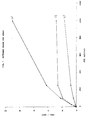
- Fig. 3 models this by showing how phenolphthalein concentration (here expressed as volume of phenolphthalein solution in a fixed total volume) is related to absorbance at 554 nm (the peak visible absorbance for phenolphthalein) when this is measured using a Hewlett-Packard Spectro photometer. It can be seen that measured absorbance is not linear with concentration above A 554 values of about 1.6 and that above A 554 values of 2.5 changes in phenolphthalein concentration produce very little change in A 554 . However if the absorbances at 490 nm (a suitable off peak wavelength) of the same solutions are measured the entire concentration range shown in Fig. 3 is within the linear range of the instrument (Fig. 4).
- This data can be used to calculate A 554 values above the linear range of the instrument by carrying out a linear regression analysis of Ae4 on A 490 for those solutions whose A 554 values fall within the linear range (Figs. 5 and 6).
- the slope of this regression is the factor by which A 490 values are to be multiplied to obtain A 554 values if these are above the linear range of the instrument.
- the enzyme substrate pair alkaline phosphatase/phenolphthalein monophosphate may also be used in a dual analyte enzyme immunoassay according to the present invention together with, for example, ⁇ -galactosidase and p-and/or o-nitrophenyl- ⁇ -D-galactoside, o-or p-nitrophenol being independently detectable in the presence of phenolphthalein by absorbance measurement.
- o-or p-nitrophenol may be measured at 404 nm and a second wavelength at which absorbance is lower, and phenolphthalein may be measured at 554 nm or about 490 nm.
- conditions for the enzyme reaction step will be chosen so that the two enzyme reactions proceed simultaneously.
- a suitable buffer for such a "combo" immunoassay will, for example, be within the pH range 8.5-8.7 and initially comprise 0.25M to 1 M diethanolamine, 3-10 mM phenolphthalein monophosphate and about 50 mM p-nitro-phenyl- ⁇ -D-galactoside.
- Alkaline phosphatase solutions of known concentrations ranging from 0.005 U/mi to 5 U/ml were made up, as were 5 alkaline phosphatase solutions of "unknown" concentrations in this range.
- To 10 ⁇ l of each of these solutions were added 300 ⁇ l of 3 mM phenolphthalein phosphate in substrate buffer (1 M diethanolamine, 0.15M NaCl, 1mM MgC1 2 , pH 8.6). After 15 min. at 37°C, 1 ml of stop solution (200 mM Na 2 C0 3 , 200 mM EDTA, 20 mM Na 3 P0 4 adjusted to pH 12 and a further 100 mM NaOH added) was added and A 490 and A 554 measured. When A 554 was greater than 1.5 the solutions were diluted 10 fold with a mixture of substrate buffer and stop solution in the ratio 0.3:1 and remeasured.
- Monoclonal antibodies against HCG ⁇ -subunit and anti-HCG monoclonal antibodies directed against a non-,8 subunit epitope were labelled with alkaline phosphatase using the following method:-0.16 ml N-succinimidyl 4-(N-maleimidomethyl) cyclohexane-1-carboxylate (SMCC) (60 mM in dimethylformamide (DMF) was added to 1.6 ml of alkaline phosphatase (2 mg/ml in 50 mM sodium borate, 1 mM magnesium chloride and 0.1 mM zinc chloride, pH 7.6) and incubated for 1 hour at 30°C.
- SMCC N-succinimidyl 4-(N-maleimidomethyl) cyclohexane-1-carboxylate
- DMF dimethylformamide
- the enzyme was separated by passage through the Sephadex G-25 medium column (1 x 35 cm) equilibrated in 0.1 M Tris, 1 mM magnesium chloride and 0.1 mM zinc chloride, pH 7.0.
- the purified enzyme was stored at +4 ° C until required.
- N-succinimidyl 3-(2-pyridyidithio) propionate SPDP
- 1 ml of monoclonal antibody 3 mg/ml in 200 mM sodium propionate, pH 6.0
- the antibody was separated by passage through a disposable Sephadex G-25 column (PD-10) equilibrated in 200 mM sodium acetate buffer, pH 4.5.
- Dithiothreitol (1 M) was added to the antibody (1/20 of antibody volume added) and left for 10 minutes at room temperature.
- the antibody was de-salted using a Sephadex G-25 medium column (1 x 35 cm) equilibrated in 200 mM sodium propionate, pH 6.0.
- Antibody and alkaline phosphatase prepared as above were mixed in an equimolar ratio and left to conjugate for 24 hours at 4°C.
- the resulting conjugate was purified by high performance liquid chromatography (HPLC) on a TSK 3000 SW column equilibrated in 200 mM sodium propionate, 1 mM magnesium chloride and 0.1 mM zinc chloride at pH 6.0.
- FITC fluorescein isothiocyanate
- Anti-FITC was a conventional polyclonal antiserum obtained by immunising sheep with FITC conjugated to keyhole limpet haemocyanin.
- the magnetisable cellulose particles were a composite of cellulose containing approximately 50% black ferric(ous) oxide (Fe 3 O 4 ), with mean particle diameter of 3 microns (see Forrest and Rattle, "Magnetic Particle Radioimmunoassay” in lmmunoassays for Clinical Chemistry, p. 147-162, Ed Hunter and Corrie, Churchill Livingstone, Edinburgh (1983)).
- Anti-FITC antiserum was covalently coupled to the magnetisable cellulose following cyanogen bromide activation of the cellulose, according to the procedure of Axen et al ., Nature 214, 1302-1304 (1967). The antiserum was coupled at a ratio of 2 ml antiserum to 1 gram of magnetisable solid phase.
- the solid phase was diluted to 5 mg/ml in 50 mM Tris/HCI buffer, pH 8.0, containing 0.1% sodium azide, 0.5% bovine serum albumin (BSA), fraction V, 0.25% Tween 20 and 0.5% methocell.
- BSA bovine serum albumin
- the substrate buffer consisted of a 1M solution of diethanolamine containing 0.9% (w/v) NaCI, 1 mM MgC1 2 , 1.543 mg/ml phenolphthalein monophosphate disodium salt or 2.02 mg/ml phenolphthalein monophosphate di(cyclohexylammonium) salt at pH 8.6.
- the wash buffer consisted of 0.9% sodium chloride in 10 mM Tris/HCI, pH 8.6.
- the stop solution was prepared by adjusting a solution containing 200 mM EDTA, 200 mM Na 2 C0 3 and 20 mM Na 3 P0 4 to pH 12 and then adding 100 mM NaOH.
- the assay buffer consisted of 0.5% bovine serum albumin (fraction v), 0.2% sheep serum, 1 mM MgC1 2 0.1 mM ZnC1 2 , 0.1 M sodium chloride and 0.2% sodium azide in 0.1 M Tris/HCI, pH 8.0.
- Standard HCG solutions were prepared by adding approximately the required concentration of purified, HCG (obtained as a freeze dried preparation from Biodata SpA, Italy) in normal male human serum and determining the exact concentration by assaying using a HCG MAIAclone kit (Serono Diagnostics Limited) against working reference preparations of HCG which themselves had been calibrated against the First International Reference Preparation (75/537).
- Table 1 shows the results obtained when high ( 1.5) OD samples from an HCG IEMA were treated in two different ways. Results in the column labelled 1/10 dilution were obtained by diluting all tubes (standards and unknowns) where A 554 > 1.5 to 1/10 in a mixture of substrate/stop solution in the ratio 0.3:1. Undiluted A 554 values were calculated from these.
- Results from the column labelled off peak were obtained by measuring at 490 nm all tubes whose A 554 > 1.5 and calculating A 554 fromn A 490 by multiplying by the slope of the linear regression analysis as in Fig. 4. These results are in good agreement.
- Table 2 shows a linear regression analysis of results from HCG IEMA (off peak calculation for high OD) with HCG MAIAclone and a linear regression of results from HCG IEMA (dilution of high O.D. samples) with HCG MAIAclone. In both cases the results are in good agreement.
- Fig. 7 shows the results from an HCG IEMA.
- Curve (b) gives A 554 values as measured using a Hewlett-Packard 8451A spectrophotometer. The detection limit for this assay calculated from A 554 values is 0.36 mlU/ml, but the response is very flat above 250 mlU/ml.
- a 1-site indirect-link enzyme immunoassay for thyroxine as illustrated schematically in Figure 1 was combined with a 2-site indirect-link enzyme immunoassay (i.e. an indirect link sandwich immunoassay) for thyroid-stimulating hormone (TSH) as illustrated schematically in Figure 2.
- TSH thyroid-stimulating hormone
- Monoclonal antibodies to T 4 and TSH were obtained from mouse ascites fluid by the process reported by Kohler and Milstein in Nature 256 (1975) 495-497. Antibodies from individual hybridoma cell lines were screened to identify those producing antibody to discrete antigenic determinants. Those antibodies with the highest affinities for the antigens in question were selected for use in the assay.
- the conjugate FITC-T 4 was prepared and purified by the method of Smith in FEBS Letters 77 25 (1977).
- the substrate buffer consisted of a 1 M solution of diethanolamine containing 150 mM NaCI, 1mM MgCI 2 , 3mM phenophthalein monophosphate and 50 mM p-nitrophenyl- ⁇ -D-galactoside at pH 8.6.
- the stop solution was prepared by adjusting a solution containing 200 mM Na 2 C0 3 , 20 mM Na 3 P0 4 and 300 mM EDTA to pH12 and then adding NaOH to 150 mM.
- a cocktail of reagents was prepared consisting of FITC-T 4 (10.5pM), anti-TSH antibody conjugated to FITC (5 ⁇ g/ml), anti-TSH antibody conjugated to alkaline phosphatase (1 ⁇ g/ml) and 8-anilino-1-naphthalene sulphonic acid (1.5 mg/ul) in assay buffer (100 mM Tris/HCI) buffer containing 0.5% bovine serum albumin (fraction V), 0.2% sheep serum, 0.2% sodium azide, 100 mM sodium chloride, 1 mM magnesium chloride and 0.1 mM zinc chloride at pH 8.0).
- the second reagent (Reagent B) consisted of anti-T4 antibody conjugated to ⁇ -galactosidase (7.61 ⁇ g/ml) in assay buffer.
- the tubes were incubated at 37°C for 20 minutes and then 1 ml of stop solution added. After the solid phase had been sedimented magnetically for at least 15 minutes, the concentration of T 4 was calculated from A 404 of the supernatant. The concentration of TSH was calculated from the absorbance of the supernatant at 554 nm or 490 nm.
- Figure 8 shows a comparison of results obtained for TSH in samples using the above protocol and using a 2-site radiometric immunoassay of the indirect-link type as described in published European application no. 105714 (IRMA assay).
Landscapes
- Health & Medical Sciences (AREA)
- Life Sciences & Earth Sciences (AREA)
- Immunology (AREA)
- Engineering & Computer Science (AREA)
- Molecular Biology (AREA)
- Biomedical Technology (AREA)
- Chemical & Material Sciences (AREA)
- Hematology (AREA)
- Urology & Nephrology (AREA)
- Cell Biology (AREA)
- Biochemistry (AREA)
- Biotechnology (AREA)
- Food Science & Technology (AREA)
- Medicinal Chemistry (AREA)
- Physics & Mathematics (AREA)
- Analytical Chemistry (AREA)
- Microbiology (AREA)
- General Health & Medical Sciences (AREA)
- General Physics & Mathematics (AREA)
- Pathology (AREA)
- Measuring Or Testing Involving Enzymes Or Micro-Organisms (AREA)
- Investigating Or Analysing Materials By The Use Of Chemical Reactions (AREA)
- Apparatus Associated With Microorganisms And Enzymes (AREA)
Abstract
Description
- The present invention relates to methods of immunoassay of antigens and to kits for carrying out such methods. In particular, it relates to improvements in immunoassays which employ enzyme-labelled antibodies to quantify the antigen under assay (hereinafter referred to as enzyme immunoassays).
- Conventional enzyme immunoassays suffer from the problem that it is difficult to have high sensitivity together with a wide assay range. The problem is caused by the limited dynamic range of the signal detection system. Considering for example a 2-site enzyme immunoassay, for high sensitivity a marked increase in signal is required for a unit increase in analyte concentration, but at high analyte concentrations the signal can be greater than the dynamic range of the measurement system. If the signal intensity at high analyte concentrations is reduced to be within the dynamic range of the detection instrument, then the signal change for a unit increase at low analyte concentrations will be reduced and assay sensitivity decreased. The problem is particularly acute for enzyme immunoassays utilising spectrophotometric endpoint detection.
- We have now devised a means of carrying out enzyme immunoassays employing enzyme labels capable of converting a substrate to a coloured product whereby this problem is overcome.
- According to one aspect of the present invention, we provide an enzyme immunoassay employing one or more enzyme label-substrate pairs which give rise to coloured products and in which absorption by the product of substrate conversion by at least one enzyme label at a first wavelength, at or close to the optimum wavelength for absorbance, exceeds the linear range of the detector, which immunoassay includes the steps of measuring the said substrate conversion at a second wavelength at which absorbance by the relevant product is significantly lower than at the said first wavelength and calculating the true absorbance at the said first wavelength by utilizing the result of linear regression analysis of absorbance measurements obtained with product standards at the said first and second wavelengths within the linear range of the detector.
- Selection between the two chosen wavelengths for a particular enzyme-substrate pair thereby enabling the requirement for high sensitivity or wide assay range to be met may conveniently be achieved automatically with appropriate instrumentation
- The technique of the present invention is applicable to any of the known types of enzyme immunoassays wherein enzyme-substrate pairs giving rise to coloured products are employed, e.g. 2-site enzyme immunoassays, including 2-site enzyme immunoassays of the indirect-link type analogous to the radioim- munometric assays described in our co-pending European published application No. 105714 (hereinafter referred to as 2-site IEMAs); 1-site enzyme immunoassays, including 1-site enzyme immunoassays of the indirect-link type described in our co-pending European published application No. 177191 (hereinafter referred to as 1-site IEMAs); and dual analyte enzyme immunoassays wherein two sets of antibodies labelled with different enzymes are employed to enable measurement of two antigens in a single sample. Examples of a 1-site IEMA and a 2-site IEMA are illustrated schematically in Figures 1 and 2 respectively of the accompanying Figures.
- For a better understanding of the present invention, more detailed consideration will now be given to use of the enzyme-substrate pair alkaline phosphatase/phenolphthalein monophosphate in a 2-site enzyme immunoassay (commonly referred to as a sandwich immunoassay).
- In a conventional 2-site enzyme immunoassay, the antigen under assay (hereinafter referred to as the analyte) which must have two or more epitopes, is insolubilised by reaction with an unlabelled antibody conjugated to a solid phase and reacted with an enzyme-labelled antibody directed to a different (preferably roomly-spaced) epitope of the analyte. The quantity of labelled antibody which becomes immobilised due to the complexing reaction is directly proportional to the amount of analyte present in the sample.
- 2-site enzyme immunoassays of the indirect-link type (2-site IEMAs) employ two soluble antibody reagents directed to different epitopes of the analyte, one soluble antibody reagent comprising enzyme-labelled antibody molecules. The solid phase employed is conjugated to a further reagent which is capable of specifically non-covalently binding the non-labelled antibodies. These antibodies may, for example, conveniently be conjugated to a reagent X as in the assay illustrated in Figure 2. The separation step is then achieved by using a solid phase conjugated to a specific binding partner for reagent X.
- When using such an assay system, increasing analyte concentration will result in increasing phenolphthalein concentration.
- The data in Fig. 3 models this by showing how phenolphthalein concentration (here expressed as volume of phenolphthalein solution in a fixed total volume) is related to absorbance at 554 nm (the peak visible absorbance for phenolphthalein) when this is measured using a Hewlett-Packard Spectro photometer. It can be seen that measured absorbance is not linear with concentration above A554 values of about 1.6 and that above A554 values of 2.5 changes in phenolphthalein concentration produce very little change in A554. However if the absorbances at 490 nm (a suitable off peak wavelength) of the same solutions are measured the entire concentration range shown in Fig. 3 is within the linear range of the instrument (Fig. 4). This data can be used to calculate A554 values above the linear range of the instrument by carrying out a linear regression analysis of Ae4 on A490 for those solutions whose A554 values fall within the linear range (Figs. 5 and 6). The slope of this regression is the factor by which A490 values are to be multiplied to obtain A554 values if these are above the linear range of the instrument.
- The enzyme substrate pair alkaline phosphatase/phenolphthalein monophosphate may also be used in a dual analyte enzyme immunoassay according to the present invention together with, for example, β-galactosidase and p-and/or o-nitrophenyl-β-D-galactoside, o-or p-nitrophenol being independently detectable in the presence of phenolphthalein by absorbance measurement. Thus, for example, o-or p-nitrophenol may be measured at 404 nm and a second wavelength at which absorbance is lower, and phenolphthalein may be measured at 554 nm or about 490 nm. Preferably, conditions for the enzyme reaction step will be chosen so that the two enzyme reactions proceed simultaneously. A suitable buffer for such a "combo" immunoassay will, for example, be within the pH range 8.5-8.7 and initially comprise 0.25M to 1 M diethanolamine, 3-10 mM phenolphthalein monophosphate and about 50 mM p-nitro-phenyl-β-D-galactoside.
- Assays of this type are described inter alia in the accompanying Examples which are intended to illustrate the present invention further.
- Alkaline phosphatase solutions of known concentrations ranging from 0.005 U/mi to 5 U/ml were made up, as were 5 alkaline phosphatase solutions of "unknown" concentrations in this range. To 10 µl of each of these solutions, were added 300 µl of 3 mM phenolphthalein phosphate in substrate buffer (1 M diethanolamine, 0.15M NaCl, 1mM MgC12, pH 8.6). After 15 min. at 37°C, 1 ml of stop solution (200 mM Na2C03, 200 mM EDTA, 20 mM Na3P04 adjusted to pH 12 and a further 100 mM NaOH added) was added and A490 and A554 measured. When A554 was greater than 1.5 the solutions were diluted 10 fold with a mixture of substrate buffer and stop solution in the ratio 0.3:1 and remeasured.
- Plots of enzyme concentration against A554 and A490 were used to obtain estimates of the enzyme concentration in the unknown samples. Where A554 values were above 1.5 the value used was calculated from that of the 10 fold dilution.

- Regression of the enzyme concentrations calculated from A554 on enzyme concentrations calculated from A490 values gave:-y = -0.011 + 1.051x; r = 0.999
- Monoclonal antibodies against HCG β-subunit and anti-HCG monoclonal antibodies directed against a non-,8 subunit epitope (anti-whole HCG) were labelled with alkaline phosphatase using the following method:-0.16 ml N-succinimidyl 4-(N-maleimidomethyl) cyclohexane-1-carboxylate (SMCC) (60 mM in dimethylformamide (DMF) was added to 1.6 ml of alkaline phosphatase (2 mg/ml in 50 mM sodium borate, 1 mM magnesium chloride and 0.1 mM zinc chloride, pH 7.6) and incubated for 1 hour at 30°C. The enzyme was separated by passage through the Sephadex G-25 medium column (1 x 35 cm) equilibrated in 0.1 M Tris, 1 mM magnesium chloride and 0.1 mM zinc chloride, pH 7.0. The purified enzyme was stored at +4°C until required.
- 16.3 µl of N-succinimidyl 3-(2-pyridyidithio) propionate (SPDP) (25 mM in ethanol) were added to 1 ml of monoclonal antibody (3 mg/ml in 200 mM sodium propionate, pH 6.0) and incubated for 30 minutes at room temperature. The antibody was separated by passage through a disposable Sephadex G-25 column (PD-10) equilibrated in 200 mM sodium acetate buffer, pH 4.5. Dithiothreitol (1 M) was added to the antibody (1/20 of antibody volume added) and left for 10 minutes at room temperature. The antibody was de-salted using a Sephadex G-25 medium column (1 x 35 cm) equilibrated in 200 mM sodium propionate, pH 6.0.
- Antibody and alkaline phosphatase prepared as above were mixed in an equimolar ratio and left to conjugate for 24 hours at 4°C. The resulting conjugate was purified by high performance liquid chromatography (HPLC) on a TSK 3000 SW column equilibrated in 200 mM sodium propionate, 1 mM magnesium chloride and 0.1 mM zinc chloride at pH 6.0.
- A population of monoclonal antibodies directed against the S-subunit of HCG was labelled with fluorescein isothiocyanate (FITC) using the following method:-200 µg of FITC (Sigma London Chemical Co., England) was reacted with 5 mg antibody in 1.4 ml sodium bicarbonate buffer, 0.2M, pH 9.0 for 18 hours at room temperature. The reaction mixture was purified by gel filtration on Sephadex G-50 superfine, giving a product incorporating an average of 6 molecules FITC per antibody molecule.
- Anti-FITC was a conventional polyclonal antiserum obtained by immunising sheep with FITC conjugated to keyhole limpet haemocyanin. The magnetisable cellulose particles were a composite of cellulose containing approximately 50% black ferric(ous) oxide (Fe3O4), with mean particle diameter of 3 microns (see Forrest and Rattle, "Magnetic Particle Radioimmunoassay" in lmmunoassays for Clinical Chemistry, p. 147-162, Ed Hunter and Corrie, Churchill Livingstone, Edinburgh (1983)). Anti-FITC antiserum was covalently coupled to the magnetisable cellulose following cyanogen bromide activation of the cellulose, according to the procedure of Axen et al ., Nature 214, 1302-1304 (1967). The antiserum was coupled at a ratio of 2 ml antiserum to 1 gram of magnetisable solid phase.
- The solid phase was diluted to 5 mg/ml in 50 mM Tris/HCI buffer, pH 8.0, containing 0.1% sodium azide, 0.5% bovine serum albumin (BSA), fraction V, 0.25% Tween 20 and 0.5% methocell.
- The substrate buffer consisted of a 1M solution of diethanolamine containing 0.9% (w/v) NaCI, 1 mM MgC12 , 1.543 mg/ml phenolphthalein monophosphate disodium salt or 2.02 mg/ml phenolphthalein monophosphate di(cyclohexylammonium) salt at pH 8.6.
- The wash buffer consisted of 0.9% sodium chloride in 10 mM Tris/HCI, pH 8.6.
- The stop solution was prepared by adjusting a solution containing 200 mM EDTA, 200 mM Na2C03 and 20 mM Na3P04 to pH 12 and then adding 100 mM NaOH.
- The assay buffer consisted of 0.5% bovine serum albumin (fraction v), 0.2% sheep serum, 1 mM MgC12 0.1 mM ZnC12, 0.1 M sodium chloride and 0.2% sodium azide in 0.1 M Tris/HCI, pH 8.0.
- Standard HCG solutions were prepared by adding approximately the required concentration of purified, HCG (obtained as a freeze dried preparation from Biodata SpA, Italy) in normal male human serum and determining the exact concentration by assaying using a HCG MAIAclone kit (Serono Diagnostics Limited) against working reference preparations of HCG which themselves had been calibrated against the First International Reference Preparation (75/537).
- 100 µl of sample or standard and 250 µl 'tracer' (50 ng anti-8-subunit HCG-alkaline phosphatase, 100 ng anti-whole HCG-alkaline phosphatase, 800 ng anti-β-subunit HCG-FITC and 300 ng anti-LH (luteinising hormone)) were mixed and incubated for 15 mins at 37°C. (The monoclonal anti-HCG antibodies employed in this instance were found to cross-react with LH and thus anti-LH antibodies were included in the 'tracer' to prevent interference from any LH present in the samples.) 200 µl of magnetisable anti-FITC solid phase was added to each tube, followed by mixing and incubation at 37°C for a further 5 mins. The solid phase was separated magnetically, the supernatant decanted and 500 µl of wash buffer added to each tube. After mixing, the solid phase was again separated magnetically. This washing procedure was repeated twice more and, after the final wash, the tubes were inverted and allowed to drain for 5 mins.
- 300 µl of substrate buffer was added to each tube, mixed, and the tubes incubated at 37°C for 15 mins. 1 ml of stop solution was then added to each tube. The magnetisable particles were sedimented magnetically for 10 minutes and the absorbances of the supernatants read at 490 nm or 554 nm, directly or after dilution.
- The results obtained were compared with those obtained using a HCG MAIAclone kit.
- Table 1 shows the results obtained when high ( 1.5) OD samples from an HCG IEMA were treated in two different ways. Results in the column labelled 1/10 dilution were obtained by diluting all tubes (standards and unknowns) where A554 > 1.5 to 1/10 in a mixture of substrate/stop solution in the ratio 0.3:1. Undiluted A554 values were calculated from these.
- Results from the column labelled off peak were obtained by measuring at 490 nm all tubes whose A554 > 1.5 and calculating A554 fromn A490 by multiplying by the slope of the linear regression analysis as in Fig. 4. These results are in good agreement.
- Table 2 shows a linear regression analysis of results from HCG IEMA (off peak calculation for high OD) with HCG MAIAclone and a linear regression of results from HCG IEMA (dilution of high O.D. samples) with HCG MAIAclone. In both cases the results are in good agreement.
- Fig. 7 shows the results from an HCG IEMA. Curve (b) gives A554 values as measured using a Hewlett-Packard 8451A spectrophotometer. The detection limit for this assay calculated from A554 values is 0.36 mlU/ml, but the response is very flat above 250 mlU/ml.
- Curve (c) shows A490 values. These give a response useable up to 1000 mIU/ml, but a detection limit of 0.9 mIU/ml. Curve (a) shows measured A554 values below A554 = 1.5 and values calculated from A490 for those above. This curve combines the advantages of low detection limit and extended range.

-
- a) correlation with MAIAclone, pregnant samples using off peak
- n = 108
- r = 0.9938
- slope = 1.019
- i = -015 mIU/ml
- b) correlation with MAIAclone diluted IEMA
- n = 20
- r = 0.999
- slope = 1.048
- i = -1.208 mIU/ml
- In this Example, a 1-site indirect-link enzyme immunoassay for thyroxine as illustrated schematically in Figure 1 was combined with a 2-site indirect-link enzyme immunoassay (i.e. an indirect link sandwich immunoassay) for thyroid-stimulating hormone (TSH) as illustrated schematically in Figure 2.
- Monoclonal antibodies to T4 and TSH were obtained from mouse ascites fluid by the process reported by Kohler and Milstein in Nature 256 (1975) 495-497. Antibodies from individual hybridoma cell lines were screened to identify those producing antibody to discrete antigenic determinants. Those antibodies with the highest affinities for the antigens in question were selected for use in the assay.
- Two populations of monoclonal antibodies directed to different epitopes of TSH were labelled with alkaline phosphatase using the same method as used in Example 2 to prepare the anti-HCG-alkaline phosphatase conjugate. Binding of one alkaline phosphatase-labelled monoclonal antibody to TSH did not interfere with the binding of the other enzyme-labelled monoclonal antibody.
- 150 µl of SPDP (25 mM in ethanol) was added to 9.4 ml of anti-T4 antibody at 100 µg/ml in 0.2M sodium propionate buffer at pH 6.0 and incubated at room temperature for 30 minutes. The resulting antibody was then purified by passage down an HPLC TSK 3000 SW column equilibrated in sodium propionate buffer (0.02 M, pH 6.0). The antibody thus obtained was then mixed with an equimolar concentration of β-galactosidase and incubated overnight at 4°C before purification on a TSK 4000 column equilibrated in sodium propionate buffer (0.2 M, pH 6.0).
- A further population of anti-TSH monoclonal antibodies, directed to a third epitope of TSH, was conjugated with FITC using the same method as in Example 2(i) (b).
- The conjugate FITC-T4 was prepared and purified by the method of Smith in FEBS Letters 77 25 (1977).
- As in Example 2(i) (c)
- The substrate buffer consisted of a 1 M solution of diethanolamine containing 150 mM NaCI, 1mM MgCI2, 3mM phenophthalein monophosphate and 50 mM p-nitrophenyl-β-D-galactoside at pH 8.6.
- The stop solution was prepared by adjusting a solution containing 200 mM Na2C03, 20 mM Na3P04 and 300 mM EDTA to pH12 and then adding NaOH to 150 mM.
- A cocktail of reagents (Reagent A) was prepared consisting of FITC-T4 (10.5pM), anti-TSH antibody conjugated to FITC (5 µg/ml), anti-TSH antibody conjugated to alkaline phosphatase (1 µg/ml) and 8-anilino-1-naphthalene sulphonic acid (1.5 mg/ul) in assay buffer (100 mM Tris/HCI) buffer containing 0.5% bovine serum albumin (fraction V), 0.2% sheep serum, 0.2% sodium azide, 100 mM sodium chloride, 1 mM magnesium chloride and 0.1 mM zinc chloride at pH 8.0).
- The second reagent (Reagent B) consisted of anti-T4 antibody conjugated to β-galactosidase (7.61 µg/ml) in assay buffer.
- To 100 µl of sample, 200 µl of reagent A and 100 µl of reagent B were added. After vortexing, the assay was incubated at 37°C for 20 minutes, followed by the addition of 200 ul anti-FITC solid phase (5 mg/ml). After a 5 minute incubation at 37°C, the assay was separated magnetically and the supernatant removed by decantation. The magnetic particle solid phase was washed three times by adding 500 µl of wash buffer (10 mM Tris/HCI containing 0.9% sodium chloride at pH 8.6), vortexing and separating magnetically, followed by decanting, after which the solid phase was drained for two minutes. Upon addition of 300 µl substrate solution, the tubes were incubated at 37°C for 20 minutes and then 1 ml of stop solution added. After the solid phase had been sedimented magnetically for at least 15 minutes, the concentration of T4 was calculated from A404 of the supernatant. The concentration of TSH was calculated from the absorbance of the supernatant at 554 nm or 490 nm.
- Figure 8 shows a comparison of results obtained for TSH in samples using the above protocol and using a 2-site radiometric immunoassay of the indirect-link type as described in published European application no. 105714 (IRMA assay).
Claims (8)
Priority Applications (1)
| Application Number | Priority Date | Filing Date | Title |
|---|---|---|---|
| AT87304644T ATE93321T1 (en) | 1986-06-10 | 1987-05-26 | IMMUNOTEST PROCEDURES. |
Applications Claiming Priority (2)
| Application Number | Priority Date | Filing Date | Title |
|---|---|---|---|
| GB8614084 | 1986-06-10 | ||
| GB868614084A GB8614084D0 (en) | 1986-06-10 | 1986-06-10 | Immunoassay |
Publications (3)
| Publication Number | Publication Date |
|---|---|
| EP0249357A2 true EP0249357A2 (en) | 1987-12-16 |
| EP0249357A3 EP0249357A3 (en) | 1989-10-04 |
| EP0249357B1 EP0249357B1 (en) | 1993-08-18 |
Family
ID=10599225
Family Applications (1)
| Application Number | Title | Priority Date | Filing Date |
|---|---|---|---|
| EP87304644A Expired - Lifetime EP0249357B1 (en) | 1986-06-10 | 1987-05-26 | Methods of immunoassay |
Country Status (12)
| Country | Link |
|---|---|
| US (1) | US4914023A (en) |
| EP (1) | EP0249357B1 (en) |
| JP (1) | JPH0718877B2 (en) |
| AR (1) | AR245534A1 (en) |
| AT (1) | ATE93321T1 (en) |
| AU (1) | AU601434B2 (en) |
| CA (1) | CA1284104C (en) |
| DE (1) | DE3787051T2 (en) |
| ES (1) | ES2046203T3 (en) |
| GB (1) | GB8614084D0 (en) |
| IL (1) | IL82790A (en) |
| ZA (1) | ZA874173B (en) |
Cited By (3)
| Publication number | Priority date | Publication date | Assignee | Title |
|---|---|---|---|---|
| WO1989006802A1 (en) * | 1988-01-20 | 1989-07-27 | Novo Biolabs Limited | Enzyme labelled biochemical assay for two analytes |
| EP0292169A3 (en) * | 1987-05-21 | 1991-02-27 | Bayer Corporation | Substrates for b-galactosidase |
| FR2654836A1 (en) * | 1989-11-17 | 1991-05-24 | Biotrol Sa Lab | APPARATUS FOR AUTOMATICALLY EXECUTING IMMUNODOSING IN SEVERAL SUCCESSIVE STEPS OF AT LEAST ONE BIOLOGICAL SUBSTANCE IN A PLURALITY OF BIOLOGICAL SAMPLES, PROCESS AND REAGENT IMPLEMENTING SAID APPARATUS. |
Families Citing this family (11)
| Publication number | Priority date | Publication date | Assignee | Title |
|---|---|---|---|---|
| US4954435A (en) * | 1987-01-12 | 1990-09-04 | Becton, Dickinson And Company | Indirect colorimetric detection of an analyte in a sample using ratio of light signals |
| DE69228817T2 (en) * | 1991-07-26 | 1999-09-23 | Dade Chemistry Systems Inc., Deerfield | SIGNAL DETECTION CHECK IN THE PRESENCE OF A SUSPENDED SOLID CARRIER |
| GB9913560D0 (en) * | 1999-06-10 | 1999-08-11 | Cortecs Diagnostics Limited | Mixing apparatus and method of mixing |
| DE10223960A1 (en) * | 2002-05-29 | 2003-12-11 | Kist Europe Forschungsges Mbh | Enzyme Immunoassay |
| US7482116B2 (en) | 2002-06-07 | 2009-01-27 | Dna Genotek Inc. | Compositions and methods for obtaining nucleic acids from sputum |
| ES2208121B1 (en) * | 2002-11-29 | 2005-08-16 | Consejo Sup. Investig. Cientificas | ANTIBODIES AND IMMOBILIZED ANTIGENS ON SILICON MAGNETIC PARTICLES AS BIOSENSORS. |
| WO2012177656A2 (en) | 2011-06-19 | 2012-12-27 | Abogen, Inc. | Devices, solutions and methods for sample collection |
| KR102392407B1 (en) * | 2014-03-07 | 2022-04-29 | 디엔에이 제노텍 인코퍼레이티드 | Composition and method for stabilizing nucleic acids in biological samples |
| WO2017023863A1 (en) | 2015-07-31 | 2017-02-09 | Research Institute At Nationwide Children's Hospital | Peptides and antibodies for the removal of biofilms |
| EP3365027B1 (en) * | 2015-10-14 | 2022-03-30 | Research Institute at Nationwide Children's Hospital | Hu specific antibodies and their use in inhibiting biofilm |
| CN114813603B (en) * | 2022-03-09 | 2023-06-20 | 南京林业大学 | A method for quickly characterizing the conversion rate of halogenation reaction of phenolic hydroxyl compounds without standard substance |
Family Cites Families (5)
| Publication number | Priority date | Publication date | Assignee | Title |
|---|---|---|---|---|
| EP0049898B2 (en) * | 1980-10-15 | 1991-08-14 | Takeda Chemical Industries, Ltd. | Method for immunochemical assay and kit therefor |
| EP0105714B1 (en) * | 1982-09-29 | 1988-07-27 | Serono Diagnostics Limited | Immunoassay of antigens |
| US4690890A (en) * | 1984-04-04 | 1987-09-01 | Cetus Corporation | Process for simultaneously detecting multiple antigens using dual sandwich immunometric assay |
| US4672028A (en) * | 1984-05-23 | 1987-06-09 | Icn Micromedic Systems, Inc. | Compositions and method for simultaneous multiple array of analytes using radioisotope chelate labels |
| US4954435A (en) * | 1987-01-12 | 1990-09-04 | Becton, Dickinson And Company | Indirect colorimetric detection of an analyte in a sample using ratio of light signals |
-
1986
- 1986-06-10 GB GB868614084A patent/GB8614084D0/en active Pending
-
1987
- 1987-05-26 ES ES198787304644T patent/ES2046203T3/en not_active Expired - Lifetime
- 1987-05-26 AT AT87304644T patent/ATE93321T1/en not_active IP Right Cessation
- 1987-05-26 EP EP87304644A patent/EP0249357B1/en not_active Expired - Lifetime
- 1987-05-26 DE DE87304644T patent/DE3787051T2/en not_active Expired - Lifetime
- 1987-06-05 US US07/058,483 patent/US4914023A/en not_active Expired - Lifetime
- 1987-06-07 IL IL82790A patent/IL82790A/en not_active IP Right Cessation
- 1987-06-08 AR AR87307799A patent/AR245534A1/en active
- 1987-06-09 CA CA000539226A patent/CA1284104C/en not_active Expired - Lifetime
- 1987-06-09 AU AU74059/87A patent/AU601434B2/en not_active Ceased
- 1987-06-10 ZA ZA874173A patent/ZA874173B/en unknown
- 1987-06-10 JP JP62143429A patent/JPH0718877B2/en not_active Expired - Lifetime
Cited By (5)
| Publication number | Priority date | Publication date | Assignee | Title |
|---|---|---|---|---|
| EP0292169A3 (en) * | 1987-05-21 | 1991-02-27 | Bayer Corporation | Substrates for b-galactosidase |
| WO1989006802A1 (en) * | 1988-01-20 | 1989-07-27 | Novo Biolabs Limited | Enzyme labelled biochemical assay for two analytes |
| FR2654836A1 (en) * | 1989-11-17 | 1991-05-24 | Biotrol Sa Lab | APPARATUS FOR AUTOMATICALLY EXECUTING IMMUNODOSING IN SEVERAL SUCCESSIVE STEPS OF AT LEAST ONE BIOLOGICAL SUBSTANCE IN A PLURALITY OF BIOLOGICAL SAMPLES, PROCESS AND REAGENT IMPLEMENTING SAID APPARATUS. |
| WO1991007662A1 (en) * | 1989-11-17 | 1991-05-30 | Laboratoires Biotrol | Apparatus for automatically carrying out a multi-stage immunoassay of at least one biological substance in a plurality of biological samples; method and reagent using said apparatus |
| US5559002A (en) * | 1989-11-17 | 1996-09-24 | Laboratoires Biotrol | Apparatus for automatically carrying out a multi-stage immunoassay of at least one biological substance in a plurality of biological samples: method using said apparatus |
Also Published As
| Publication number | Publication date |
|---|---|
| EP0249357A3 (en) | 1989-10-04 |
| DE3787051T2 (en) | 1994-03-03 |
| US4914023A (en) | 1990-04-03 |
| GB8614084D0 (en) | 1986-07-16 |
| ZA874173B (en) | 1987-12-11 |
| IL82790A0 (en) | 1987-12-20 |
| DE3787051D1 (en) | 1993-09-23 |
| CA1284104C (en) | 1991-05-14 |
| EP0249357B1 (en) | 1993-08-18 |
| AU7405987A (en) | 1987-12-17 |
| IL82790A (en) | 1991-06-30 |
| ATE93321T1 (en) | 1993-09-15 |
| AU601434B2 (en) | 1990-09-13 |
| AR245534A1 (en) | 1994-01-31 |
| ES2046203T3 (en) | 1994-02-01 |
| JPS6366463A (en) | 1988-03-25 |
| JPH0718877B2 (en) | 1995-03-06 |
Similar Documents
| Publication | Publication Date | Title |
|---|---|---|
| EP0238353B1 (en) | Methods of immunoassay | |
| US4016043A (en) | Enzymatic immunological method for the determination of antigens and antibodies | |
| US4722889A (en) | Immunoassays using multiple monoclonal antibodies and scavenger antibodies | |
| EP0324323B1 (en) | Homogeneous biospecific assay method using lanthanide chelates as labels | |
| US4503143A (en) | Enzyme immunoassay with two-part solution of tetramethylbenzidine as chromogen | |
| EP0249357B1 (en) | Methods of immunoassay | |
| CA1256025A (en) | Immuno-chemical measurement process for haptens and proteins | |
| Toivonen et al. | Two-site time-resolved immunofluorometric assay of human insulin. | |
| Mathis et al. | Homogeneous immunoassays using rare earth cryptates and time resolved fluorescence: principles and specific advantages for tumor markers | |
| EP0190006B1 (en) | Methods of immunoassay | |
| KR920000057B1 (en) | Process and reagent for the determination of a specifically bindable substance | |
| EP0062892B1 (en) | Single incubation immunochemical assay for creatin phosphokinase mb | |
| CN110007073A (en) | A kind of combination assay chemical luminescence reagent kit and its preparation method and application | |
| US4966839A (en) | Process for the determination of an immunologically bindable analyte | |
| USRE32696E (en) | Enzymatic immunological method for determination of antigens and antibodies | |
| EP0389301A2 (en) | Reagent complex for immunoassay | |
| US5783455A (en) | Regenerable solid phase for carrying out specific binding reactions | |
| NO176197B (en) | Measurement method and test set | |
| JPS61186855A (en) | Method of measuring combining capacity of globulin forming thyroxine | |
| Kang et al. | A highly sensitive immunoenzymometric assay involving" common-capture" particles and membrane filtration. | |
| EP0248038A1 (en) | Idiotypic-antigenic conjunction binding assay | |
| Mould et al. | The use of solid-phase chemistry in therapeutic drug monitoring | |
| Papanastasiou-Diamandi et al. | Immunoassay of triiodothyronine in serum by time-resolved fluorometric measurement of europium-chelate complexes in solution | |
| EP0181762B1 (en) | Method for detecting enzymatic activity using particle agglutination | |
| Shimizu et al. | Monoclonal antibodies in immunoenzymetric assays |
Legal Events
| Date | Code | Title | Description |
|---|---|---|---|
| PUAI | Public reference made under article 153(3) epc to a published international application that has entered the european phase |
Free format text: ORIGINAL CODE: 0009012 |
|
| AK | Designated contracting states |
Kind code of ref document: A2 Designated state(s): AT BE CH DE ES FR GB GR IT LI LU NL SE |
|
| RAP3 | Party data changed (applicant data changed or rights of an application transferred) |
Owner name: SERONO DIAGNOSTICS PARTNERS (A MASSACHUSETTS LIMIT |
|
| PUAL | Search report despatched |
Free format text: ORIGINAL CODE: 0009013 |
|
| AK | Designated contracting states |
Kind code of ref document: A3 Designated state(s): AT BE CH DE ES FR GB GR IT LI LU NL SE |
|
| 17P | Request for examination filed |
Effective date: 19900320 |
|
| 17Q | First examination report despatched |
Effective date: 19910912 |
|
| RAP1 | Party data changed (applicant data changed or rights of an application transferred) |
Owner name: APPLIED RESEARCH SYSTEMS ARS HOLDING N.V. |
|
| GRAA | (expected) grant |
Free format text: ORIGINAL CODE: 0009210 |
|
| AK | Designated contracting states |
Kind code of ref document: B1 Designated state(s): AT BE CH DE ES FR GB GR IT LI LU NL SE |
|
| REF | Corresponds to: |
Ref document number: 93321 Country of ref document: AT Date of ref document: 19930915 Kind code of ref document: T |
|
| RAP2 | Party data changed (patent owner data changed or rights of a patent transferred) |
Owner name: ARS HOLDING 89 N.V |
|
| REF | Corresponds to: |
Ref document number: 3787051 Country of ref document: DE Date of ref document: 19930923 |
|
| ET | Fr: translation filed | ||
| ITF | It: translation for a ep patent filed | ||
| REG | Reference to a national code |
Ref country code: GR Ref legal event code: FG4A Free format text: 3009829 |
|
| REG | Reference to a national code |
Ref country code: ES Ref legal event code: FG2A Ref document number: 2046203 Country of ref document: ES Kind code of ref document: T3 |
|
| PLBE | No opposition filed within time limit |
Free format text: ORIGINAL CODE: 0009261 |
|
| STAA | Information on the status of an ep patent application or granted ep patent |
Free format text: STATUS: NO OPPOSITION FILED WITHIN TIME LIMIT |
|
| EPTA | Lu: last paid annual fee | ||
| 26N | No opposition filed | ||
| EAL | Se: european patent in force in sweden |
Ref document number: 87304644.5 |
|
| REG | Reference to a national code |
Ref country code: CH Ref legal event code: PUE Owner name: ARS HOLDING 89 N.V. TRANSFER- BIOCHEM IMMUNOSYSTEM |
|
| BECA | Be: change of holder's address |
Free format text: 971104 *BIOCHEM IMMUNOSYSTEMS L.L.C.:BUSCH HILL BAY STREET, BRIDGETONWN BARBADOS |
|
| REG | Reference to a national code |
Ref country code: FR Ref legal event code: TP Free format text: PUBLIE A TORT DANS LE BOPI 98/03 SUIVANT LA NOTIFICATION DANS LE BOPI 98/07 |
|
| REG | Reference to a national code |
Ref country code: FR Ref legal event code: TP |
|
| NLS | Nl: assignments of ep-patents |
Owner name: BIOCHEM IMMUNOSYSTEMS LLC |
|
| REG | Reference to a national code |
Ref country code: ES Ref legal event code: PC2A |
|
| PGFP | Annual fee paid to national office [announced via postgrant information from national office to epo] |
Ref country code: SE Payment date: 19990510 Year of fee payment: 13 |
|
| PGFP | Annual fee paid to national office [announced via postgrant information from national office to epo] |
Ref country code: GR Payment date: 19990513 Year of fee payment: 13 |
|
| PGFP | Annual fee paid to national office [announced via postgrant information from national office to epo] |
Ref country code: LU Payment date: 19990519 Year of fee payment: 13 |
|
| PGFP | Annual fee paid to national office [announced via postgrant information from national office to epo] |
Ref country code: GB Payment date: 19990520 Year of fee payment: 13 |
|
| PGFP | Annual fee paid to national office [announced via postgrant information from national office to epo] |
Ref country code: CH Payment date: 19990521 Year of fee payment: 13 |
|
| PGFP | Annual fee paid to national office [announced via postgrant information from national office to epo] |
Ref country code: ES Payment date: 19990527 Year of fee payment: 13 |
|
| PGFP | Annual fee paid to national office [announced via postgrant information from national office to epo] |
Ref country code: FR Payment date: 19990528 Year of fee payment: 13 Ref country code: AT Payment date: 19990528 Year of fee payment: 13 |
|
| PGFP | Annual fee paid to national office [announced via postgrant information from national office to epo] |
Ref country code: NL Payment date: 19990531 Year of fee payment: 13 |
|
| PGFP | Annual fee paid to national office [announced via postgrant information from national office to epo] |
Ref country code: BE Payment date: 19990603 Year of fee payment: 13 |
|
| PG25 | Lapsed in a contracting state [announced via postgrant information from national office to epo] |
Ref country code: LU Free format text: LAPSE BECAUSE OF NON-PAYMENT OF DUE FEES Effective date: 20000526 Ref country code: GB Free format text: LAPSE BECAUSE OF NON-PAYMENT OF DUE FEES Effective date: 20000526 Ref country code: AT Free format text: LAPSE BECAUSE OF NON-PAYMENT OF DUE FEES Effective date: 20000526 |
|
| PG25 | Lapsed in a contracting state [announced via postgrant information from national office to epo] |
Ref country code: SE Free format text: LAPSE BECAUSE OF NON-PAYMENT OF DUE FEES Effective date: 20000527 Ref country code: ES Free format text: THE PATENT HAS BEEN ANNULLED BY A DECISION OF A NATIONAL AUTHORITY Effective date: 20000527 |
|
| PG25 | Lapsed in a contracting state [announced via postgrant information from national office to epo] |
Ref country code: LI Free format text: LAPSE BECAUSE OF NON-PAYMENT OF DUE FEES Effective date: 20000531 Ref country code: GR Free format text: LAPSE BECAUSE OF NON-PAYMENT OF DUE FEES Effective date: 20000531 Ref country code: CH Free format text: LAPSE BECAUSE OF NON-PAYMENT OF DUE FEES Effective date: 20000531 Ref country code: BE Free format text: LAPSE BECAUSE OF NON-PAYMENT OF DUE FEES Effective date: 20000531 |
|
| BERE | Be: lapsed |
Owner name: BIOCHEM IMMUNOSYSTEMS L.L.C. Effective date: 20000531 |
|
| PG25 | Lapsed in a contracting state [announced via postgrant information from national office to epo] |
Ref country code: NL Free format text: LAPSE BECAUSE OF NON-PAYMENT OF DUE FEES Effective date: 20001201 |
|
| REG | Reference to a national code |
Ref country code: CH Ref legal event code: PL |
|
| GBPC | Gb: european patent ceased through non-payment of renewal fee |
Effective date: 20000526 |
|
| EUG | Se: european patent has lapsed |
Ref document number: 87304644.5 |
|
| PG25 | Lapsed in a contracting state [announced via postgrant information from national office to epo] |
Ref country code: FR Free format text: LAPSE BECAUSE OF NON-PAYMENT OF DUE FEES Effective date: 20010131 |
|
| NLV4 | Nl: lapsed or anulled due to non-payment of the annual fee |
Effective date: 20001201 |
|
| REG | Reference to a national code |
Ref country code: FR Ref legal event code: ST |
|
| REG | Reference to a national code |
Ref country code: ES Ref legal event code: FD2A Effective date: 20020204 |
|
| PG25 | Lapsed in a contracting state [announced via postgrant information from national office to epo] |
Ref country code: IT Free format text: LAPSE BECAUSE OF NON-PAYMENT OF DUE FEES Effective date: 20050526 |
|
| PGFP | Annual fee paid to national office [announced via postgrant information from national office to epo] |
Ref country code: DE Payment date: 20060529 Year of fee payment: 20 |