EP0276142B1 - Testverfahren - Google Patents
Testverfahren Download PDFInfo
- Publication number
- EP0276142B1 EP0276142B1 EP88300458A EP88300458A EP0276142B1 EP 0276142 B1 EP0276142 B1 EP 0276142B1 EP 88300458 A EP88300458 A EP 88300458A EP 88300458 A EP88300458 A EP 88300458A EP 0276142 B1 EP0276142 B1 EP 0276142B1
- Authority
- EP
- European Patent Office
- Prior art keywords
- reagent
- ligand
- assay
- specific binding
- binding partner
- Prior art date
- Legal status (The legal status is an assumption and is not a legal conclusion. Google has not performed a legal analysis and makes no representation as to the accuracy of the status listed.)
- Expired - Lifetime
Links
- 238000003556 assay Methods 0.000 title claims description 43
- 238000000034 method Methods 0.000 title claims description 36
- 239000003153 chemical reaction reagent Substances 0.000 claims description 105
- 239000003446 ligand Substances 0.000 claims description 73
- 230000003287 optical effect Effects 0.000 claims description 66
- 230000009870 specific binding Effects 0.000 claims description 36
- 238000002198 surface plasmon resonance spectroscopy Methods 0.000 claims description 36
- 230000000694 effects Effects 0.000 claims description 19
- 230000002708 enhancing effect Effects 0.000 claims description 10
- 238000012544 monitoring process Methods 0.000 claims description 6
- 230000008859 change Effects 0.000 claims description 5
- 230000001747 exhibiting effect Effects 0.000 claims description 3
- 238000011534 incubation Methods 0.000 claims description 3
- 239000000427 antigen Substances 0.000 description 55
- 108091007433 antigens Proteins 0.000 description 55
- 102000036639 antigens Human genes 0.000 description 55
- 239000002245 particle Substances 0.000 description 23
- LOKCTEFSRHRXRJ-UHFFFAOYSA-I dipotassium trisodium dihydrogen phosphate hydrogen phosphate dichloride Chemical compound P(=O)(O)(O)[O-].[K+].P(=O)(O)([O-])[O-].[Na+].[Na+].[Cl-].[K+].[Cl-].[Na+] LOKCTEFSRHRXRJ-UHFFFAOYSA-I 0.000 description 15
- 239000002953 phosphate buffered saline Substances 0.000 description 15
- 230000027455 binding Effects 0.000 description 14
- 238000002360 preparation method Methods 0.000 description 12
- 241000700605 Viruses Species 0.000 description 11
- 241000712431 Influenza A virus Species 0.000 description 10
- 230000009918 complex formation Effects 0.000 description 9
- 238000005259 measurement Methods 0.000 description 9
- 230000035945 sensitivity Effects 0.000 description 9
- 238000008416 Ferritin Methods 0.000 description 8
- 239000012634 fragment Substances 0.000 description 8
- 238000012360 testing method Methods 0.000 description 8
- 239000002023 wood Substances 0.000 description 8
- 108091003079 Bovine Serum Albumin Proteins 0.000 description 7
- 230000015572 biosynthetic process Effects 0.000 description 7
- 229940098773 bovine serum albumin Drugs 0.000 description 7
- YBJHBAHKTGYVGT-ZKWXMUAHSA-N (+)-Biotin Chemical compound N1C(=O)N[C@@H]2[C@H](CCCCC(=O)O)SC[C@@H]21 YBJHBAHKTGYVGT-ZKWXMUAHSA-N 0.000 description 6
- 108090001008 Avidin Proteins 0.000 description 6
- 230000001965 increasing effect Effects 0.000 description 6
- 239000000463 material Substances 0.000 description 6
- 239000000243 solution Substances 0.000 description 6
- 102000008857 Ferritin Human genes 0.000 description 5
- 108050000784 Ferritin Proteins 0.000 description 5
- 238000002820 assay format Methods 0.000 description 5
- 239000011521 glass Substances 0.000 description 5
- PCHJSUWPFVWCPO-UHFFFAOYSA-N gold Chemical compound [Au] PCHJSUWPFVWCPO-UHFFFAOYSA-N 0.000 description 5
- 239000010931 gold Substances 0.000 description 5
- 229910052737 gold Inorganic materials 0.000 description 5
- 229920000642 polymer Polymers 0.000 description 5
- 241000894007 species Species 0.000 description 5
- 241000712461 unidentified influenza virus Species 0.000 description 5
- 238000013019 agitation Methods 0.000 description 4
- 230000000890 antigenic effect Effects 0.000 description 4
- 239000011545 carbonate/bicarbonate buffer Substances 0.000 description 4
- 239000003085 diluting agent Substances 0.000 description 4
- 229940079593 drug Drugs 0.000 description 4
- 239000003814 drug Substances 0.000 description 4
- 102000004169 proteins and genes Human genes 0.000 description 4
- 108090000623 proteins and genes Proteins 0.000 description 4
- 230000005855 radiation Effects 0.000 description 4
- 230000004520 agglutination Effects 0.000 description 3
- 229960002685 biotin Drugs 0.000 description 3
- 235000020958 biotin Nutrition 0.000 description 3
- 239000011616 biotin Substances 0.000 description 3
- 210000004027 cell Anatomy 0.000 description 3
- 238000010790 dilution Methods 0.000 description 3
- 239000012895 dilution Substances 0.000 description 3
- 239000012153 distilled water Substances 0.000 description 3
- 238000002474 experimental method Methods 0.000 description 3
- 229940088597 hormone Drugs 0.000 description 3
- 239000005556 hormone Substances 0.000 description 3
- 238000003018 immunoassay Methods 0.000 description 3
- 206010022000 influenza Diseases 0.000 description 3
- 239000004816 latex Substances 0.000 description 3
- 229920000126 latex Polymers 0.000 description 3
- 244000005700 microbiome Species 0.000 description 3
- 150000003384 small molecules Chemical class 0.000 description 3
- 239000007858 starting material Substances 0.000 description 3
- 239000000126 substance Substances 0.000 description 3
- 238000005406 washing Methods 0.000 description 3
- XLYOFNOQVPJJNP-UHFFFAOYSA-N water Chemical compound O XLYOFNOQVPJJNP-UHFFFAOYSA-N 0.000 description 3
- KHURTEAYLZEQHD-UHFFFAOYSA-N 3-n-propylpropane-1,1,3-triamine Chemical compound CCCNCCC(N)N KHURTEAYLZEQHD-UHFFFAOYSA-N 0.000 description 2
- FHVDTGUDJYJELY-UHFFFAOYSA-N 6-{[2-carboxy-4,5-dihydroxy-6-(phosphanyloxy)oxan-3-yl]oxy}-4,5-dihydroxy-3-phosphanyloxane-2-carboxylic acid Chemical compound O1C(C(O)=O)C(P)C(O)C(O)C1OC1C(C(O)=O)OC(OP)C(O)C1O FHVDTGUDJYJELY-UHFFFAOYSA-N 0.000 description 2
- 102100023635 Alpha-fetoprotein Human genes 0.000 description 2
- 108010022366 Carcinoembryonic Antigen Proteins 0.000 description 2
- 102100025475 Carcinoembryonic antigen-related cell adhesion molecule 5 Human genes 0.000 description 2
- XUIIKFGFIJCVMT-GFCCVEGCSA-N D-thyroxine Chemical compound IC1=CC(C[C@@H](N)C(O)=O)=CC(I)=C1OC1=CC(I)=C(O)C(I)=C1 XUIIKFGFIJCVMT-GFCCVEGCSA-N 0.000 description 2
- 102000012673 Follicle Stimulating Hormone Human genes 0.000 description 2
- 108010079345 Follicle Stimulating Hormone Proteins 0.000 description 2
- 239000002202 Polyethylene glycol Substances 0.000 description 2
- RJKFOVLPORLFTN-LEKSSAKUSA-N Progesterone Chemical compound C1CC2=CC(=O)CC[C@]2(C)[C@@H]2[C@@H]1[C@@H]1CC[C@H](C(=O)C)[C@@]1(C)CC2 RJKFOVLPORLFTN-LEKSSAKUSA-N 0.000 description 2
- MUMGGOZAMZWBJJ-DYKIIFRCSA-N Testostosterone Chemical compound O=C1CC[C@]2(C)[C@H]3CC[C@](C)([C@H](CC4)O)[C@@H]4[C@@H]3CCC2=C1 MUMGGOZAMZWBJJ-DYKIIFRCSA-N 0.000 description 2
- 102000011923 Thyrotropin Human genes 0.000 description 2
- 108010061174 Thyrotropin Proteins 0.000 description 2
- 238000007818 agglutination assay Methods 0.000 description 2
- 238000007605 air drying Methods 0.000 description 2
- 229940072056 alginate Drugs 0.000 description 2
- 229920000615 alginic acid Polymers 0.000 description 2
- 235000010443 alginic acid Nutrition 0.000 description 2
- 108010026331 alpha-Fetoproteins Proteins 0.000 description 2
- 239000011324 bead Substances 0.000 description 2
- 230000001419 dependent effect Effects 0.000 description 2
- 230000005670 electromagnetic radiation Effects 0.000 description 2
- 229940028334 follicle stimulating hormone Drugs 0.000 description 2
- JYGXADMDTFJGBT-VWUMJDOOSA-N hydrocortisone Chemical compound O=C1CC[C@]2(C)[C@H]3[C@@H](O)C[C@](C)([C@@](CC4)(O)C(=O)CO)[C@@H]4[C@@H]3CCC2=C1 JYGXADMDTFJGBT-VWUMJDOOSA-N 0.000 description 2
- 230000005764 inhibitory process Effects 0.000 description 2
- NOESYZHRGYRDHS-UHFFFAOYSA-N insulin Chemical compound N1C(=O)C(NC(=O)C(CCC(N)=O)NC(=O)C(CCC(O)=O)NC(=O)C(C(C)C)NC(=O)C(NC(=O)CN)C(C)CC)CSSCC(C(NC(CO)C(=O)NC(CC(C)C)C(=O)NC(CC=2C=CC(O)=CC=2)C(=O)NC(CCC(N)=O)C(=O)NC(CC(C)C)C(=O)NC(CCC(O)=O)C(=O)NC(CC(N)=O)C(=O)NC(CC=2C=CC(O)=CC=2)C(=O)NC(CSSCC(NC(=O)C(C(C)C)NC(=O)C(CC(C)C)NC(=O)C(CC=2C=CC(O)=CC=2)NC(=O)C(CC(C)C)NC(=O)C(C)NC(=O)C(CCC(O)=O)NC(=O)C(C(C)C)NC(=O)C(CC(C)C)NC(=O)C(CC=2NC=NC=2)NC(=O)C(CO)NC(=O)CNC2=O)C(=O)NCC(=O)NC(CCC(O)=O)C(=O)NC(CCCNC(N)=N)C(=O)NCC(=O)NC(CC=3C=CC=CC=3)C(=O)NC(CC=3C=CC=CC=3)C(=O)NC(CC=3C=CC(O)=CC=3)C(=O)NC(C(C)O)C(=O)N3C(CCC3)C(=O)NC(CCCCN)C(=O)NC(C)C(O)=O)C(=O)NC(CC(N)=O)C(O)=O)=O)NC(=O)C(C(C)CC)NC(=O)C(CO)NC(=O)C(C(C)O)NC(=O)C1CSSCC2NC(=O)C(CC(C)C)NC(=O)C(NC(=O)C(CCC(N)=O)NC(=O)C(CC(N)=O)NC(=O)C(NC(=O)C(N)CC=1C=CC=CC=1)C(C)C)CC1=CN=CN1 NOESYZHRGYRDHS-UHFFFAOYSA-N 0.000 description 2
- GQYHUHYESMUTHG-UHFFFAOYSA-N lithium niobate Chemical compound [Li+].[O-][Nb](=O)=O GQYHUHYESMUTHG-UHFFFAOYSA-N 0.000 description 2
- 229910052751 metal Inorganic materials 0.000 description 2
- 239000002184 metal Substances 0.000 description 2
- 239000000813 peptide hormone Substances 0.000 description 2
- 239000008363 phosphate buffer Substances 0.000 description 2
- 229920001223 polyethylene glycol Polymers 0.000 description 2
- 238000001556 precipitation Methods 0.000 description 2
- 210000002966 serum Anatomy 0.000 description 2
- 230000003612 virological effect Effects 0.000 description 2
- VOXZDWNPVJITMN-ZBRFXRBCSA-N 17β-estradiol Chemical compound OC1=CC=C2[C@H]3CC[C@](C)([C@H](CC4)O)[C@@H]4[C@@H]3CCC2=C1 VOXZDWNPVJITMN-ZBRFXRBCSA-N 0.000 description 1
- 208000030507 AIDS Diseases 0.000 description 1
- 108010032595 Antibody Binding Sites Proteins 0.000 description 1
- 206010003445 Ascites Diseases 0.000 description 1
- 241000894006 Bacteria Species 0.000 description 1
- 108010004032 Bromelains Proteins 0.000 description 1
- 241000283707 Capra Species 0.000 description 1
- 229920001661 Chitosan Polymers 0.000 description 1
- 208000007190 Chlamydia Infections Diseases 0.000 description 1
- VYZAMTAEIAYCRO-UHFFFAOYSA-N Chromium Chemical compound [Cr] VYZAMTAEIAYCRO-UHFFFAOYSA-N 0.000 description 1
- LTMHDMANZUZIPE-AMTYYWEZSA-N Digoxin Natural products O([C@H]1[C@H](C)O[C@H](O[C@@H]2C[C@@H]3[C@@](C)([C@@H]4[C@H]([C@]5(O)[C@](C)([C@H](O)C4)[C@H](C4=CC(=O)OC4)CC5)CC3)CC2)C[C@@H]1O)[C@H]1O[C@H](C)[C@@H](O[C@H]2O[C@@H](C)[C@H](O)[C@@H](O)C2)[C@@H](O)C1 LTMHDMANZUZIPE-AMTYYWEZSA-N 0.000 description 1
- 108090000790 Enzymes Proteins 0.000 description 1
- 102000004190 Enzymes Human genes 0.000 description 1
- 108060003951 Immunoglobulin Proteins 0.000 description 1
- 241000913536 Influenza A virus (A/X-31(H3N2)) Species 0.000 description 1
- 102000004877 Insulin Human genes 0.000 description 1
- 108090001061 Insulin Proteins 0.000 description 1
- 241001465754 Metazoa Species 0.000 description 1
- 241000699666 Mus <mouse, genus> Species 0.000 description 1
- 241000699670 Mus sp. Species 0.000 description 1
- 241000283973 Oryctolagus cuniculus Species 0.000 description 1
- 208000002606 Paramyxoviridae Infections Diseases 0.000 description 1
- 102000003982 Parathyroid hormone Human genes 0.000 description 1
- 108090000445 Parathyroid hormone Proteins 0.000 description 1
- 241001494479 Pecora Species 0.000 description 1
- 241000276498 Pollachius virens Species 0.000 description 1
- 239000004793 Polystyrene Substances 0.000 description 1
- 102000003946 Prolactin Human genes 0.000 description 1
- 108010057464 Prolactin Proteins 0.000 description 1
- 239000004365 Protease Substances 0.000 description 1
- 241000606651 Rickettsiales Species 0.000 description 1
- BQCADISMDOOEFD-UHFFFAOYSA-N Silver Chemical compound [Ag] BQCADISMDOOEFD-UHFFFAOYSA-N 0.000 description 1
- 229920002472 Starch Polymers 0.000 description 1
- 229930006000 Sucrose Natural products 0.000 description 1
- CZMRCDWAGMRECN-UGDNZRGBSA-N Sucrose Chemical compound O[C@H]1[C@H](O)[C@@H](CO)O[C@@]1(CO)O[C@@H]1[C@H](O)[C@@H](O)[C@H](O)[C@@H](CO)O1 CZMRCDWAGMRECN-UGDNZRGBSA-N 0.000 description 1
- AUYYCJSJGJYCDS-LBPRGKRZSA-N Thyrolar Chemical compound IC1=CC(C[C@H](N)C(O)=O)=CC(I)=C1OC1=CC=C(O)C(I)=C1 AUYYCJSJGJYCDS-LBPRGKRZSA-N 0.000 description 1
- 230000002776 aggregation Effects 0.000 description 1
- 238000004220 aggregation Methods 0.000 description 1
- 230000004075 alteration Effects 0.000 description 1
- 230000001580 bacterial effect Effects 0.000 description 1
- 235000019835 bromelain Nutrition 0.000 description 1
- 238000006243 chemical reaction Methods 0.000 description 1
- 208000028512 chlamydia infectious disease Diseases 0.000 description 1
- 229960004407 chorionic gonadotrophin Drugs 0.000 description 1
- 229910052804 chromium Inorganic materials 0.000 description 1
- 239000011651 chromium Substances 0.000 description 1
- 238000012875 competitive assay Methods 0.000 description 1
- 230000000536 complexating effect Effects 0.000 description 1
- 239000002131 composite material Substances 0.000 description 1
- 238000004132 cross linking Methods 0.000 description 1
- 238000000432 density-gradient centrifugation Methods 0.000 description 1
- 238000001514 detection method Methods 0.000 description 1
- 238000002405 diagnostic procedure Methods 0.000 description 1
- LTMHDMANZUZIPE-PUGKRICDSA-N digoxin Chemical compound C1[C@H](O)[C@H](O)[C@@H](C)O[C@H]1O[C@@H]1[C@@H](C)O[C@@H](O[C@@H]2[C@H](O[C@@H](O[C@@H]3C[C@@H]4[C@]([C@@H]5[C@H]([C@]6(CC[C@@H]([C@@]6(C)[C@H](O)C5)C=5COC(=O)C=5)O)CC4)(C)CC3)C[C@@H]2O)C)C[C@@H]1O LTMHDMANZUZIPE-PUGKRICDSA-N 0.000 description 1
- 229960005156 digoxin Drugs 0.000 description 1
- LTMHDMANZUZIPE-UHFFFAOYSA-N digoxine Natural products C1C(O)C(O)C(C)OC1OC1C(C)OC(OC2C(OC(OC3CC4C(C5C(C6(CCC(C6(C)C(O)C5)C=5COC(=O)C=5)O)CC4)(C)CC3)CC2O)C)CC1O LTMHDMANZUZIPE-UHFFFAOYSA-N 0.000 description 1
- 238000006073 displacement reaction Methods 0.000 description 1
- 235000013601 eggs Nutrition 0.000 description 1
- 229960005309 estradiol Drugs 0.000 description 1
- 229930182833 estradiol Natural products 0.000 description 1
- 239000012530 fluid Substances 0.000 description 1
- 239000008187 granular material Substances 0.000 description 1
- 230000035931 haemagglutination Effects 0.000 description 1
- 208000006454 hepatitis Diseases 0.000 description 1
- 231100000283 hepatitis Toxicity 0.000 description 1
- HBTBNXFVJYRYGI-UHFFFAOYSA-M hexadecane-1-sulfinate Chemical compound CCCCCCCCCCCCCCCCS([O-])=O HBTBNXFVJYRYGI-UHFFFAOYSA-M 0.000 description 1
- 210000004408 hybridoma Anatomy 0.000 description 1
- 229960000890 hydrocortisone Drugs 0.000 description 1
- 102000018358 immunoglobulin Human genes 0.000 description 1
- 238000001114 immunoprecipitation Methods 0.000 description 1
- 230000006872 improvement Effects 0.000 description 1
- 238000001746 injection moulding Methods 0.000 description 1
- 229940125396 insulin Drugs 0.000 description 1
- 150000002500 ions Chemical class 0.000 description 1
- 230000001678 irradiating effect Effects 0.000 description 1
- 230000001592 luteinising effect Effects 0.000 description 1
- 229910044991 metal oxide Inorganic materials 0.000 description 1
- 150000004706 metal oxides Chemical class 0.000 description 1
- 230000009871 nonspecific binding Effects 0.000 description 1
- 239000000199 parathyroid hormone Substances 0.000 description 1
- 229960001319 parathyroid hormone Drugs 0.000 description 1
- 102000013415 peroxidase activity proteins Human genes 0.000 description 1
- 108040007629 peroxidase activity proteins Proteins 0.000 description 1
- 239000004417 polycarbonate Substances 0.000 description 1
- 229920000515 polycarbonate Polymers 0.000 description 1
- 229920001282 polysaccharide Polymers 0.000 description 1
- 229920002223 polystyrene Polymers 0.000 description 1
- 230000008569 process Effects 0.000 description 1
- 238000012545 processing Methods 0.000 description 1
- 239000000186 progesterone Substances 0.000 description 1
- 229960003387 progesterone Drugs 0.000 description 1
- 229940097325 prolactin Drugs 0.000 description 1
- 230000005180 public health Effects 0.000 description 1
- 238000011002 quantification Methods 0.000 description 1
- 238000006894 reductive elimination reaction Methods 0.000 description 1
- 238000011160 research Methods 0.000 description 1
- 230000000241 respiratory effect Effects 0.000 description 1
- 125000006850 spacer group Chemical group 0.000 description 1
- 238000010561 standard procedure Methods 0.000 description 1
- 235000019698 starch Nutrition 0.000 description 1
- 239000008107 starch Substances 0.000 description 1
- 239000003270 steroid hormone Substances 0.000 description 1
- 150000003431 steroids Chemical class 0.000 description 1
- 239000005720 sucrose Substances 0.000 description 1
- 235000000346 sugar Nutrition 0.000 description 1
- 150000008163 sugars Chemical class 0.000 description 1
- 229960003604 testosterone Drugs 0.000 description 1
- 238000002207 thermal evaporation Methods 0.000 description 1
- 239000005495 thyroid hormone Substances 0.000 description 1
- 229940036555 thyroid hormone Drugs 0.000 description 1
- 229940034208 thyroxine Drugs 0.000 description 1
- XUIIKFGFIJCVMT-UHFFFAOYSA-N thyroxine-binding globulin Natural products IC1=CC(CC([NH3+])C([O-])=O)=CC(I)=C1OC1=CC(I)=C(O)C(I)=C1 XUIIKFGFIJCVMT-UHFFFAOYSA-N 0.000 description 1
- 239000003053 toxin Substances 0.000 description 1
- 231100000765 toxin Toxicity 0.000 description 1
- 108700012359 toxins Proteins 0.000 description 1
- 230000007704 transition Effects 0.000 description 1
- 229940035722 triiodothyronine Drugs 0.000 description 1
- 239000011782 vitamin Substances 0.000 description 1
- 229940088594 vitamin Drugs 0.000 description 1
- 229930003231 vitamin Natural products 0.000 description 1
- 235000013343 vitamin Nutrition 0.000 description 1
Images


Classifications
-
- G—PHYSICS
- G01—MEASURING; TESTING
- G01N—INVESTIGATING OR ANALYSING MATERIALS BY DETERMINING THEIR CHEMICAL OR PHYSICAL PROPERTIES
- G01N33/00—Investigating or analysing materials by specific methods not covered by groups G01N1/00 - G01N31/00
- G01N33/48—Biological material, e.g. blood, urine; Haemocytometers
- G01N33/50—Chemical analysis of biological material, e.g. blood, urine; Testing involving biospecific ligand binding methods; Immunological testing
- G01N33/53—Immunoassay; Biospecific binding assay; Materials therefor
- G01N33/543—Immunoassay; Biospecific binding assay; Materials therefor with an insoluble carrier for immobilising immunochemicals
- G01N33/54366—Apparatus specially adapted for solid-phase testing
- G01N33/54373—Apparatus specially adapted for solid-phase testing involving physiochemical end-point determination, e.g. wave-guides, FETS, gratings
-
- G—PHYSICS
- G01—MEASURING; TESTING
- G01N—INVESTIGATING OR ANALYSING MATERIALS BY DETERMINING THEIR CHEMICAL OR PHYSICAL PROPERTIES
- G01N33/00—Investigating or analysing materials by specific methods not covered by groups G01N1/00 - G01N31/00
- G01N33/48—Biological material, e.g. blood, urine; Haemocytometers
- G01N33/50—Chemical analysis of biological material, e.g. blood, urine; Testing involving biospecific ligand binding methods; Immunological testing
- G01N33/53—Immunoassay; Biospecific binding assay; Materials therefor
- G01N33/543—Immunoassay; Biospecific binding assay; Materials therefor with an insoluble carrier for immobilising immunochemicals
- G01N33/54366—Apparatus specially adapted for solid-phase testing
-
- G—PHYSICS
- G01—MEASURING; TESTING
- G01N—INVESTIGATING OR ANALYSING MATERIALS BY DETERMINING THEIR CHEMICAL OR PHYSICAL PROPERTIES
- G01N33/00—Investigating or analysing materials by specific methods not covered by groups G01N1/00 - G01N31/00
- G01N33/48—Biological material, e.g. blood, urine; Haemocytometers
- G01N33/50—Chemical analysis of biological material, e.g. blood, urine; Testing involving biospecific ligand binding methods; Immunological testing
- G01N33/58—Chemical analysis of biological material, e.g. blood, urine; Testing involving biospecific ligand binding methods; Immunological testing involving labelled substances
- G01N33/585—Chemical analysis of biological material, e.g. blood, urine; Testing involving biospecific ligand binding methods; Immunological testing involving labelled substances with a particulate label, e.g. coloured latex
Definitions
- This invention relates to assay techniques and to means for putting such techniques into effect.
- it relates to an improved assay technique providing enhanced sensitivity and specificity.
- EP-0112721 describes the preparation, and use as biosensors, of optical structures having a preformed relief profile coated with a material (hereinafter called “specific binding partner") capable of binding with the species to be assayed (hereinafter called "ligand”) to form a complex.
- a material hereinafter called "specific binding partner”
- ligand capable of binding with the species to be assayed
- the optical properties of such structures are altered as a result of complex formation between the ligand and the specific binding partner and this phenomenon can consequently form the basis of an assay system.
- the present invention provides a method of assaying for a ligand in a sample which method includes the steps of incubating the sample with a reagent ("reagent Y") immobilised directly or indirectly on the surface of an optical structure capable of exhibiting surface plasmon resonance and monitoring the surface plasmon resonance effect exhibited by said optical structure during the assay characterised in that: (i) an additional reagent ("reagent X") is employed in the assay (incubation of the sample, reagent X and reagent Y being simultaneous or in any desired sequence) where one of reagents X and Y comprises a specific binding partner to said ligand and the other of reagents X and Y comprises either a ligand analogue or a specific binding partner to said ligand, reagent X being such that a direct or indirect complex between reagents X and Y results in said surface having an appreciably enhanced optical thickness as compared to the optical thickness of said surface absent said complex; and (ii) the results of
- reagents X and Y comprises a ligand analogue and the other of reagents X and Y comprises a specific binding partner to the ligand
- complex formation between reagents X and Y may occur directly.
- both reagents X and Y comprise specific binding partners to the ligand
- complex formation between reagents X and Y indirectly via the ligand, if present in the sample may occur.
- reagent Y may be immobilised on the surface of the optical structure either directly or indirectly.
- reagent Y is an antibody
- indirect immobilisation may be effected by means of an anti-species antibody to reagent Y which is itself bound to the surface of the optical structure.
- optical structure capable of exhibiting surface plasmon resonance defines any structure which is optically active with respect to radiation over a predetermined band of wavelengths and which exhibits surface plasmon resonance effects which are critically dependent on the surface characteristics of the structure.
- the structure has a pre-formed surface relief profile and the use of metallised diffraction gratings is particularly preferred.
- other optical structures which exhibit surface plasmon resonance effects e.g. metal coated prisms are also of use in the method according to the invention.
- the surface plasmon resonance (SPR) properties of such optical structures may be observed by exposing the structure to electromagnetic radiation, preferably monochromatic radiation, more particularly polarised light, and monitoring the reflected or transmitted radiation.
- electromagnetic radiation preferably monochromatic radiation, more particularly polarised light
- the use of a diffraction grating and incident radiation in the form of light polarised in a plane transverse to the lines of the grating is particularly preferred.
- ligand analogue refers to a species capable of complexing with the same binding site of the same specific binding partner as the ligand under assay, and includes inter alia within its scope a known quantity of the ligand under assay.
- optical thickness defines a composite optical property of a material which is a function of both its physical thickness and its refractive index. It has been found that the SPR effects of an optical structure, for example a diffraction grating, to which a ligand-specific binding partner complex is bound are critically dependent on the optical thickness of the bound complex. Thus, the change in the SPR effects of an optical structure induced by the formation of a ligand-specific binding partner complex may be enhanced by increasing the optical thickness of the bound layer without increasing the ligand concentration.
- optical surface is not necessarily used herein to denote simply the surface of the optical structure itself but where appropriate in the context includes any material (for example immobilised complex) present on the surface of the optical structure.
- reagent X a reagent which appreciably increases either the physical size of the bound complex or the refractive index of the bound complex.
- the increase in complex size will be of the order of 100 nm or greater or the refractive index will be increased to 1.5-2.0.
- reagent X comprises either a ligand analogue or a specific binding partner to the ligand under assay.
- reagent X must be such that any complex formation (direct or indirect) between reagents X and Y (leading to bound complex on the surface of the optical structure) results in an optical surface of appreciably enhanced optical thickness as described above.
- the reagent X will be a ligand analogue or specific binding partner "labelled" by linkage to an optical thickness enhancing entity, e.g. polystyrene latex, a virus, a microorganism, ferritin, or a protein such as a second specific binding partner.
- an optical thickness enhancing entity e.g. polystyrene latex, a virus, a microorganism, ferritin, or a protein such as a second specific binding partner.
- the sensitivity of the assay method according to the invention may be still further enhanced by the use of a reagent X which incorporates a suitable particle of high refractive index ( ⁇ ) e.g. glass beads ⁇ >2.0, in particular lithium niobate glass with ⁇ >2.2.
- a suitable particle of high refractive index ( ⁇ ) e.g. glass beads ⁇ >2.0, in particular lithium niobate glass with ⁇ >2.2.
- Such reagents increase both the size and refractive index of the bound complex and the concomitant increase in the optical thickness of the optical surface allows a still further improvement in the sensitivity of the assay method according to the invention.
- Methods of assay in accordance with the invention include methods wherein
- reagent X may comprise any suitably large ligand analogue or specific binding partner to the ligand.
- the ligand is an antigen reagent X may be a relatively large antibody thereto, optionally bound to one or more further entities which further increase the optical thickness enhancement properties of reagent X.
- reagent X may be an entity having, at the surface thereof, at least one antigenic determinant analogous to the sample antigen (ligand) such that competition for specific binding sites provided by the immobilised reagent Y can take place between the sample antigen (ligand) and reagent X.
- reagent X of a second specific binding partner to the ligand, optionally linked to one or more other optical thickness enhancing entities or particles as described above, will not only improve the sensitivity of the method according to the invention but will also improve its specificity in the manner described above.
- the ligand to be assayed is an antigen or an antibody
- the use of an antibody or antigen specific thereto, optionally linked to one or more other entities e.g. glass beads, as the optical thickness enhancing reagent X is particularly preferred.
- the method according to the invention extends the range of ligands which may be assayed using SPR effect perturbation techniques from viruses with a molecular weight of 2 ⁇ 108 daltons or higher down to, and including, ligands having a low molecular weight e.g. 200-500 daltons such as steroids, drugs etc.
- the assay techniques of this type are no longer limited by the physical size of the ligand itself.
- the modified procedure according to the invention may be applied to a "competition-type" assay.
- the optical thickness of the optical surface may be increased using as reagent X a ligand analogue linked to a comparatively large "label" particle, such as latex.
- reagent X together with the sample containing the ligand under assay to the surface of an optical structure bearing a specific binding partner to the ligand allows reagent X and the ligand to compete for the binding sites provided by reagent Y.
- Complex between reagents X and Y will be formed in a quantity inversely proportional to the concentration of ligand in the sample.
- reagent X can consist of a large "label" particle coated with a specific binding partner to the ligand to be assayed for and this reagent X can be incubated with the sample and the optical structure coated with immobilised ligand analogue (reagent Y) such that the amount of complex formed between reagents X and Y is again inversely proportional to the concentration of ligand in the sample.
- large "label” entities may be used in a so-called sandwich assay.
- the ligand is capable of binding both with a specific binding partner immobilised on the surface of an optical structure (reagent Y) and with a second specific binding partner, which may be the same or different, carried on the surface of an optical thickness enhancing entity (reagent X). If present, the sample ligand indirectly links reagent X and reagent Y resulting in a complex between all three.
- the enhanced optical thickness of the bound layer is achieved by allowing large aggregates to form following complex formation between reagents X and Y.
- kits for carrying out a method of assay as described herein which comprises a reagent X as defined above and an optical structure on the surface of which is immobilised a reagent Y as defined above.
- the enhanced optical thickness of the optical structure due to complex formation between reagents X and Y is detected by monitoring the SPR effect.
- this involves directing light, polarised in a plane transverse to the lines of the grating, onto the grating surface and detecting the change in surface plasmon resonance effects caused by complex formation.
- the angle of incidence at which there is a sudden dip in the magnitude of reflected or diffracted light may be measured. This sudden dip is caused by light energy being coupled into the surface of the grating and causing surface plasmon resonance to occur at a particular angle which depends on the optical characteristics of the grating surface, and in particular on the optical thickness of the optical surface.
- the angle of incidence may be held constant and the wavelength of the irradiating light varied to detect the altered SPR characteristics of the surface.
- the invention will be particularly described hereinafter (except for Example 3) with reference to an antibody or an antigen as the ligand.
- the invention is not to be taken as being limited to assays of antibodies or antigens. Examples of ligands which may be assayed by the method of the invention are given in Table 1 below, together with an indication of a suitable specific binding partner in each instance.
- the method of the invention has very broad applicability but in particular may be used to assay: hormones, including peptide hormones (e.g. thyroid stimulating hormone (TSH), luteinising hormone (LH), human chorionic gonadotrophin (hCG), follicle stimulating hormone (FSH), insulin and prolactin) or non-peptide hormones (.e.g steroid hormones such as cortisol, estradiol, progesterone and testosterone, or thyroid hormones such as thyroxine (T4) and triiodothyronine), proteins (e.g. carcinoembryonic antigen (CEA) and alphafetoprotein (AFP)), drugs (e.g. digoxin), sugars, toxins, vitamins, viruses such as influenza, parainfluenza, adeno-, hepatitis, respiratory and AIDS viruses, or microorganisms.
- hormones including peptide hormones (e.g. thyroid stimulating hormone (TSH), luteinising hormone (
- antigen as used herein will be understood to include both permanently antigenic species (for example, proteins, bacteria, bacterial fragments, cells, cell fragments and viruses) and haptens which may be rendered antigenic under suitable conditions.
- Antibodies are typically divalent and large antigens such as proteins are often multivalent. It is therefore possible for a single antibody to bind to two antigens, which may be the same or different, simultaneously or for several antibodies to be bound to a single antigen. Thus, when antigen and antibody are mixed in solution the direct antigen-antibody reaction initially leads to the formation of small complexes. When the number of antibody binding sites is close to the number of antigen binding sites extensive cross linking between antibody and antigen can occur which causes large aggregates to develop.
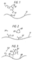
- Figure 1 shows diagrammatically the surface of a diffraction grating 10 on which is immobilised an antibody 12 (reagent Y) specific for the species to be assayed, an antigen 14.
- the sample containing the antigen 14, reagent X in the form of a specific antibody 16 which may bind to the antigen 14 (and which may or may not be the same as antibody 12) and the grating 10 are then brought into contact, simultaneously or in any desired sequence, such that the large antibody-antigen aggregate formed is bound to the surface of the grating via antibody 12.
- the number of antibody/antigen complexes formed on the grating can be detected by the altered SPR properties of the surface.
- reagent X 16 specific antibody
- reagent Y immobilised antibody 12 specific for sample antigen 14 on its surface
- aggregates can be formed directly at the surface of the test diffraction grating and the alteration of the SPR effects of the surface can be monitored with time as aggregate formation proceeds.
- a still further variation on this type of assay system is based on conventional immunoassay systems wherein aggregate formation is inhibited, for example, by small molecules such as drugs.
- reagent X comprising a ligand analogue or a specific binding partner to the ligand linked to one or more particles of defined size, chemical and optical properties.
- the particle itself is the dominant entity measured directly by the SPR effect rather than the sample ligand analogue or specific binding partner to the ligand.
- Suitable particulate labels comprise materials suitable for the formation of small particles e.g. 100 nm diameter, are relatively transparent to allow the incident light to pass through the bound particle, and preferably have a high refractive index, more preferably higher than 2.0, such that surface plasmon resonance effects are enhanced.
- the surface of the particle should be of suitable material to permit immobilisation of the required ligand analogue or specific binding partner e.g. protein antigen or antibody, thereto.
- Synthetic particles may comprise for example polymeric material such as latex, glass, in particular lithium niobate glass, colloidal silver or gold, metal oxide or ferritin and naturally-occurring particles include cells, for example microorganisms, viruses, starch granules and pollen grains.
- Small molecules such as drugs and hormones may be measured in a number of different ways using a competitive assay format.
- reagent X comprises a label particle 18 bearing antigen 20 (which may or may not be the same as sample antigen 24) on its surface.
- Reagent Y comprises a specific antibody 22 immobilised onto the surface of a diffraction grating 10.
- competition for binding to the antibody 22 takes place between sample antigen 24 and antigen 20 bound to particle 18.
- the amount of particle 18 bound to the surface of the test grating 10 will be in inverse proportion to the concentration of the sample antigen 24, thus allowing measurement of the sample antigen 24.
- Figure 3 shows an alternative competition assay.
- reagent Y comprises a diffraction grating 10 bearing an immobilised antigen 28 (which may or may not be the same as sample antigen 34) and reagent X comprises a label particle 30 carrying antibody 32 specific to the sample antigen 34 and the immobilised antigen 28.
- the sample antigen 34 and the immobilised antigen 28 compete for binding sites on the antibody 32, so that the amount of reagent X bound to the surface of the test grating 10 will be in inverse proportion to the concentration of the sample antigen 34, thus allowing measurement of the sample antigen 34.
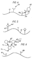
- This format is applicable to antigens which are sufficiently large to bear on their surface more than one site which can be recognised and bound by an antibody. Such antigens are able to bind to at least two antibodies simultaneously and therefore are able to bridge between the antibodies.
- a bridge is formed between the antibody 36 bound to the surface of the test grating 10 (reagent Y) and another antibody 38 (which may or may not be the same as antibody 36) bound to the surface of a label particle 40 which forms reagent X.
- the amount of bound reagent X is then determined thus allowing measurement of the sample antigen 34.
- Increasing the sample antigen concentration causes an increase in the amount of bound reagent X.
- Figure 5 shows an alternative type of sandwich assay where a multivalent sample antigen is to be determined.
- the multivalent sample antigen 60 forms a bridge between the antibody 62 bound to the surface of the test grating 10 (reagent Y) and at least one reagent X comprising an antibody 64 (which may or may not be the same as antibody 62).
- the effective size of the bound sample antigen 60 is thus increased, allowing more sensitive measurement of the sample antigen 60.
- a previously described embodiment of the invention illustrates how antibody-antigen-antibody complex formation in a sandwich-type immunoassay can enhance the sensitivity and specificity of the assay.
- Particles may be used to enhance further the sensitivity of such assays.
- Assays may employ direct agglutination or agglutination inhibition formats. The latter are used for measuring small molecules in a competition assay format as previously described.
- reagent Y a specific binding partner
- reagent X in the form of labelled particles would be added to the sample and ligand, if present in the sample, will cause agglutination to occur. Addition of the particle-enhanced sample to the test grating will allow capture of the aggregates onto the surface where they may be measured by SPR effects.
- the contacting of reagent X, sample and reagent Y attached to a diffraction grating may be simultaneous rather than sequential and the rate of formation of the aggregate on the surface of the grating could be measured to determine the ligand under assay.
- Figure 6 shows diagrammatically an agglutination assay format.
- An inert support particle 38 carries antibodies 40 which bind with the sample antigen 42.
- the resulting large aggregate 44 is bound to the surface of the diffraction grating 10 by antibody 46 (which may or may not be the same as antibody 40) immobilised on the surface of the grating 10.
- the antibody 46 binds to the antigen 42 of the large aggregate and the amount of sample antigen 42 can then be measured.
- Carbohydrate polymers such as alginate or chitosan can exist in different forms, as soluble polymers or as insoluble particles. The transition from one form to the other can be effected by the addition of certain ions e.g. Ca++ causes soluble alginate to precipitate. It is proposed that this effect could be used to enhance the SPR assay described herein.
- reagent X may comprise a long chain polymer labelled with a ligand. This reagent can compete with sample ligand for binding to the specific antibody on the grating surface (reagent Y). Once bound, a developing solution to precipitate the bound polymer is added before measurement.
- a long chain polymer 50 is labelled with antigen 52 (which may or may not be the same as sample antigen 54).
- This labelled antigen competes with "free" sample antigen 54 for binding sites on antibody 56 immobilised on the grating surface.
- a developing solution is then added to precipitate the bound labelled antigen (58, Figure 7b).
- the amount of labelled antigen 58 bound and precipitated onto the surface of the grating 10 is inversely proportional to the concentration of sample antigen 54.
- sandwich format assays could be developed using such precipitatable polymers.
- Monoclonal antibodies were obtained from mouse ascites fluid by the process reported by Milstein and Kohler in Nature 256, 495-497 (1975). Antibodies from individual hybridoma cell lines were screened to identify those producing antibody to discrete antigenic determinants of Influenza A virus. Those antibodies having the highest affinities for Influenza A virus, strain X-31, were selected for use in the assay.
- the capture antibody (antibody A) was used at 50 ⁇ g/ml in phosphate buffered saline (PBS) containing 0.1% bovine serum albumin (BSA) (Sigma Chemical Co. Ltd., England) and the label antibody (antibody B) at 50 ⁇ g/ml in PBS.
- PBS phosphate buffered saline
- BSA bovine serum albumin
- Diffraction gratings (633 nm pitch, 35 nm depth) were fabricated in polycarbonate by injection moulding. The gratings were then coated with a 10 nm thick layer of chromium followed by a 100 nm thick layer of gold. Both metal layers were deposited by thermal evaporation under vacuum.
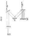
- the instrument consists of a collimated beam of plane polarised white light which illuminates the antibody coated diffraction grating. The light is then directed via a mirror from the antibody coated diffraction grating onto a blazed diffraction grating where the light is broken into its component wavelengths and thence focussed onto a 256 pixel photodiode array which measures the light intensity at specific wavelengths.
- Figure 9 shows the displacement in the Wood's anomaly caused by the influenza virus binding to the immunologically active diffraction grating and the enhanced signal obtained when the second antibody was bound to the influenza virus particle.
- Influenza A virus (strain X-31) was grown and purified as described in Example 1. Haemagglutinin was extracted from the virus using the bromelain method as described by Brand and Skehel in Nature New Biology Volume 238 (1972), p145-147.
- Influenza A virus was prepared as described in Example 1.
- Metallised diffraction gratings were prepared as described in Example 1.
- Figure 10 shows the shifts in the position of the Wood's anomaly, measured in nanometers, which were obtained by binding different dilutions of positive and negative sera to the diffraction gratings in the presence and absence of the virus particle label.
- Biotin conjugated to the enzyme peroxidase via a spacer molecule, diaminodipropylamine (DAPA), avidin and a conjugate of ferritin labelled avidin were obtained from Sigma Chemical Company, London.
- biotin-peroxidase conjugate was made up to 0.068 mg/ml in 0.05 molar carbonate/bicarbonate buffer, pH 9.6, avidin-ferritin and avidin were dissolved in 0.05 molar phosphate buffer pH 7.4.
- the metallised diffraction gratings were made as described in Example 1.
- Figure 11 shows the shifts in the positions of the Wood's anomaly, measured in nanometers, upon the binding of avidin and avidin-ferritin to the biotin coated diffraction gratings. The increase in signal due to the presence of the ferritin is clearly observed.
Landscapes
- Health & Medical Sciences (AREA)
- Life Sciences & Earth Sciences (AREA)
- Immunology (AREA)
- Engineering & Computer Science (AREA)
- Molecular Biology (AREA)
- Biomedical Technology (AREA)
- Chemical & Material Sciences (AREA)
- Hematology (AREA)
- Urology & Nephrology (AREA)
- Biotechnology (AREA)
- Microbiology (AREA)
- Cell Biology (AREA)
- Food Science & Technology (AREA)
- Medicinal Chemistry (AREA)
- Physics & Mathematics (AREA)
- Analytical Chemistry (AREA)
- Biochemistry (AREA)
- General Health & Medical Sciences (AREA)
- General Physics & Mathematics (AREA)
- Pathology (AREA)
- Investigating Or Analysing Materials By Optical Means (AREA)
Claims (8)
- Testverfahren für einen Liganden in einer Probe, wobei dieses Verfahren die Stufen des Bebrütens der Probe mit einem Reagens ("Reagens Y") umfaßt, das direkt oder indirekt auf der Oberfläche einer optischen Struktur immobilisiert ist, die in der Lage ist, Oberflächenplasmonresonanz zu zeigen, und den Oberflächenplasmon-Resonanzeffekt, der durch diese optische Struktur aufgezeigt wird, während des Versuchs zu überwachen, dadurch gekennzeichnet, daß:(i) ein zusätzliches Reagens ("Reagens X") in dem Versuch verwendet wird (Inkubation der Probe, Reagens X und Reagens Y sind gleichzeitig oder in irgendeiner gewünschten Reihenfolge), wobei einer der Reagentien X und Y einen spezifischen Bindungspartner für diesen Liganden umfaßt, und das andere der Reagentien X und Y entweder ein Ligandenanaloges oder einen spezifischen Bindungspartner für diesen Liganden umfaßt, wobei das Reagens X derart ist, daß ein direkter oder indirekter Komplex zwischen den Reagentien X und Y sich in dieser Oberfläche ergibt, die eine merklich vergrößerte optische Dicke hat, verglichen mit der optischen Dicke dieser Oberfläche in Abwesenheit dieses Komplexes; und(ii) die Ergebnisse dieser Überwachung des Oberflächenplasmonresonanzeffekts dazu verwendet werden, irgendwelche Veränderung(en) in der optischen Dicke dieser Oberfläche, die während des Versuchs auftreten, festzustellen.
- Verfahren gemäß Anspruch 1, wobei das Reagens Y ein Ligandenanaloges und das Reagens X ein spezifischer Bindungspartner für den Liganden ist, gegebenenfalls verknüpft an eine die optische Dicke vergrößernde Einheit.
- Verfahren gemäß Anspruch 1, wobei das Reagens Y ein spezifischer Bindungspartner zu dem Liganden und das Reagens X ein Ligandenanaloges ist, verknüpft mit einer die optische Dicke erhöhenden Einheit.
- Verfahren gemäß Anspruch 1, worin das Reagens Y ein spezifischer Bindungspartner zu dem Liganden ist und das Reagens X ein spezifischer Bindungspartner (identisch mit oder verschieden von Reagens Y) zu dem Liganden ist, gegebenenfalls geknüpft an eine die optische Dicke vergrößernde Einheit.
- Verfahren gemäß Anspruch 1, worin das Reagens X die Größe irgendeines gebildeten Komplexes um mindestens 100 nm vergrößert.
- Verfahren gemäß Anspruch 1 oder Anspruch 5, worin das Reagens X einen Brechungsindex von mindestens 2,0 hat.
- Verfahren gemäß einem der vorhergehenden Ansprüche, worin die optische Struktur ein Beugungsgitter ist.
- Kit zur Durchführung des Testverfahrens, wie es in einem der vorhergehenden Ansprüche beansprucht ist, umfassend ein Reagens X, wie in Anspruch 1 definiert, und eine optische Struktur, auf deren Oberfläche ein Reagens Y, wie in Anspruch 1 definiert, immobilisiert ist.
Priority Applications (1)
| Application Number | Priority Date | Filing Date | Title |
|---|---|---|---|
| AT88300458T ATE88278T1 (de) | 1987-01-21 | 1988-01-20 | Testverfahren. |
Applications Claiming Priority (4)
| Application Number | Priority Date | Filing Date | Title |
|---|---|---|---|
| GB878701293A GB8701293D0 (en) | 1987-01-21 | 1987-01-21 | Assay technique |
| GB8701293 | 1987-01-21 | ||
| GB878725797A GB8725797D0 (en) | 1987-11-04 | 1987-11-04 | Assay technique |
| GB8725797 | 1987-11-04 |
Publications (2)
| Publication Number | Publication Date |
|---|---|
| EP0276142A1 EP0276142A1 (de) | 1988-07-27 |
| EP0276142B1 true EP0276142B1 (de) | 1993-04-14 |
Family
ID=26291819
Family Applications (1)
| Application Number | Title | Priority Date | Filing Date |
|---|---|---|---|
| EP88300458A Expired - Lifetime EP0276142B1 (de) | 1987-01-21 | 1988-01-20 | Testverfahren |
Country Status (8)
| Country | Link |
|---|---|
| US (1) | US6093536A (de) |
| EP (1) | EP0276142B1 (de) |
| JP (1) | JPH07111435B2 (de) |
| AU (1) | AU621038B2 (de) |
| CA (1) | CA1309017C (de) |
| DE (1) | DE3880162T2 (de) |
| ES (1) | ES2054793T3 (de) |
| IL (1) | IL85137A (de) |
Families Citing this family (43)
| Publication number | Priority date | Publication date | Assignee | Title |
|---|---|---|---|---|
| US4962023A (en) * | 1987-06-22 | 1990-10-09 | Louisiana State University, Agricultural And Mechanical College | Single incubation immuno sorbent assay method using particle labels to detect test antigen specific antibodies in presence of other antibodies |
| US5501986A (en) * | 1988-04-06 | 1996-03-26 | E. I. Du Pont De Nemours And Company | Piezoelectric specific binding assay with mass amplified reagents |
| JPH03503681A (ja) * | 1988-04-06 | 1991-08-15 | イー・アイ・デユポン・デ・ニモアス・アンド・カンパニー | 質量増幅した因子を使用する圧電特異的結合アツセイ |
| SE8804074D0 (sv) * | 1988-11-10 | 1988-11-10 | Pharmacia Ab | Sensorenhet och dess anvaendning i biosensorsystem |
| SE8902043L (sv) * | 1988-11-10 | 1990-05-11 | Pharmacia Ab | Foerfarande foer karakterisering av makromolekyler |
| SE462454B (sv) * | 1988-11-10 | 1990-06-25 | Pharmacia Ab | Maetyta foer anvaendning i biosensorer |
| GB8906776D0 (en) * | 1989-03-23 | 1989-05-10 | Amersham Int Plc | Assay method using surface plasmon resonance spectrometry |
| GB8906781D0 (en) * | 1989-03-23 | 1989-05-10 | Amersham Int Plc | Assay method using surface plasmon resonance spectrometry |
| GB8916764D0 (en) * | 1989-07-21 | 1989-09-06 | Sambles John R | Surface plasmon optical sensor |
| US5135852A (en) * | 1989-07-25 | 1992-08-04 | E. I. Du Pont De Nemours And Company | Piezoelectric cell growth biosensing method using polymer-metabolic product complex interactions |
| GB8919411D0 (en) * | 1989-08-25 | 1989-10-11 | Amersham Int Plc | Assay method |
| CA2066643A1 (en) * | 1989-10-04 | 1991-04-05 | Richard Calvin Ebersole | Assay method for biological target complexes on the surface of a biosensor |
| DE4024476C1 (de) * | 1990-08-02 | 1992-02-27 | Boehringer Mannheim Gmbh, 6800 Mannheim, De | |
| GB2248497B (en) * | 1990-09-26 | 1994-05-25 | Marconi Gec Ltd | An optical sensor |
| GB9022304D0 (en) * | 1990-10-15 | 1990-11-28 | Ares Serono Res & Dev Ltd | Assay technique |
| EP0525178B1 (de) * | 1991-02-11 | 1998-11-25 | Biostar, Inc. | Testeinheiten und verfahren, die polymerischen oberflächen-verstärkungsagenzien verwenden |
| JP2609953B2 (ja) * | 1991-03-08 | 1997-05-14 | 理化学研究所 | 表面プラズモン顕微鏡 |
| SE9200917D0 (sv) * | 1991-08-20 | 1992-03-25 | Pharmacia Biosensor Ab | Assay method |
| SE9201984D0 (sv) * | 1992-06-29 | 1992-06-29 | Pharmacia Biosensor Ab | Improvement in optical assays |
| SE504507C2 (sv) * | 1993-05-24 | 1997-02-24 | Pharmacia Biosensor Ab | Sätt att bestämma bindningsegenskaper hos ligander med låg molekylvikt |
| DE4433980C2 (de) * | 1994-09-23 | 1996-08-22 | Boehringer Ingelheim Int | Verfahren und Biosensorhit zur Untersuchung der Wechselwirkung von Biomolekülen mittels Oberflächen-Plasma-Resonanz |
| JPH10221249A (ja) | 1996-12-05 | 1998-08-21 | Norio Miura | 薬物の測定装置とセンサ及び該センサに用いる検出素子 |
| US5776785A (en) * | 1996-12-30 | 1998-07-07 | Diagnostic Products Corporation | Method and apparatus for immunoassay using fluorescent induced surface plasma emission |
| WO2002001230A2 (en) * | 2000-06-23 | 2002-01-03 | Minerva Biotechnologies Corporation | Rapid and sensitive detection of protein aggregation |
| US6771376B2 (en) * | 1999-07-05 | 2004-08-03 | Novartis Ag | Sensor platform, apparatus incorporating the platform, and process using the platform |
| KR100883079B1 (ko) | 1999-07-05 | 2009-02-10 | 노파르티스 아게 | 플랫폼, 플랫폼을 가지는 장치, 및 플랫폼을 사용하는 방법 |
| US6579726B1 (en) | 1999-07-30 | 2003-06-17 | Surromed, Inc. | Instruments, methods and reagents for surface plasmon resonance |
| US7167615B1 (en) | 1999-11-05 | 2007-01-23 | Board Of Regents, The University Of Texas System | Resonant waveguide-grating filters and sensors and methods for making and using same |
| AU7014001A (en) * | 2000-06-23 | 2002-01-08 | Minerva Biotechnologies Corp | Interaction of colloid-immobilized species with species on non-colloidal structures |
| DE60127999T2 (de) * | 2000-11-30 | 2008-01-17 | Chugai Seiyaku K.K. | Messverfahren für die bindungsaktivität eines liganden bindenden proteins mit geringer chemischer stabilität für den ersten liganden |
| DE10141691A1 (de) * | 2001-08-25 | 2003-03-13 | Friz Biochem Gmbh | Verdrängungsassay zur Detektion von Ligat-Ligand-Assoziationsereignissen |
| US6877375B2 (en) * | 2002-05-06 | 2005-04-12 | Battelle Memorial Institute | System and technique for characterizing fluids using ultrasonic diffraction grating spectroscopy |
| US7395711B2 (en) * | 2002-05-06 | 2008-07-08 | Battelle Memorial Institute | System and technique for characterizing fluids using ultrasonic diffraction grating spectroscopy |
| US20060240573A1 (en) * | 2003-07-29 | 2006-10-26 | Lamdagen, Llc | Optical system including nanostructures for biological or chemical sensing |
| US20070231796A1 (en) * | 2003-09-17 | 2007-10-04 | The Regents Of The University Of California | Sensor and method for detection of a target substance |
| JP4371954B2 (ja) * | 2004-08-31 | 2009-11-25 | 富士フイルム株式会社 | 表面プラズモン共鳴分析による被験物質の解析方法 |
| PT103606B (pt) * | 2006-11-15 | 2009-03-16 | Biosurfit Sa | Dispositivo de detecção dinâmico baseado no efeito de ressonância de plasmão de superfície |
| US7652768B2 (en) * | 2006-12-01 | 2010-01-26 | Canon Kabushiki Kaisha | Chemical sensing apparatus and chemical sensing method |
| CN102046814B (zh) | 2008-06-02 | 2015-05-20 | 通用电气健康护理生物科学股份公司 | 浓度测定 |
| JP5683606B2 (ja) * | 2009-11-30 | 2015-03-11 | ジーイー・ヘルスケア・バイオサイエンス・アクチボラグ | 相互作用の分析のための方法及びシステム |
| JP6481371B2 (ja) * | 2015-01-06 | 2019-03-13 | コニカミノルタ株式会社 | 検出方法および検出キット |
| EP3918308A4 (de) * | 2019-01-31 | 2023-09-27 | Precision Bioservices, Inc. | Verfahren zur detektion und charakterisierung von antiviralen vektorantikörpern |
| CN113433094B (zh) * | 2021-05-11 | 2023-01-17 | 中山大学 | 实现fp-wa耦合模式的生物传感器及其制备方法和应用 |
Citations (1)
| Publication number | Priority date | Publication date | Assignee | Title |
|---|---|---|---|---|
| EP0112721A2 (de) * | 1982-12-21 | 1984-07-04 | Ares-Serono N.V. | Testverfahren |
Family Cites Families (10)
| Publication number | Priority date | Publication date | Assignee | Title |
|---|---|---|---|---|
| DE3135196A1 (de) * | 1981-09-05 | 1983-03-17 | Merck Patent Gmbh, 6100 Darmstadt | Verfahren, mittel und vorrichtung zur bestimmung biologischer komponenten |
| US4454233A (en) * | 1981-10-21 | 1984-06-12 | Wang Associates | Method of tagged immunoassay |
| US4621063A (en) * | 1982-10-12 | 1986-11-04 | The Center For Immunological Studies | Methods for the detection and quantitation of immunological substances |
| US4626513A (en) * | 1983-11-10 | 1986-12-02 | Massachusetts General Hospital | Method and apparatus for ligand detection |
| US4978503A (en) * | 1984-06-13 | 1990-12-18 | Ares-Serono Research & Development Limited Partnership | Devices for use in chemical test procedures |
| US4647544A (en) * | 1984-06-25 | 1987-03-03 | Nicoli David F | Immunoassay using optical interference detection |
| GB8423204D0 (en) * | 1984-09-14 | 1984-10-17 | Comtech Res Unit | Assay technique and equipment |
| US4703018A (en) * | 1985-02-20 | 1987-10-27 | E. I. Du Pont De Nemours And Company | High refractive index haloalkyl-functional shell-core polymers and their use in light scattering immunoassays |
| GB8509492D0 (en) * | 1985-04-12 | 1985-05-15 | Plessey Co Plc | Optical assay |
| GB8705649D0 (en) * | 1987-03-10 | 1987-04-15 | Pa Consulting Services | Assay sensor |
-
1988
- 1988-01-19 IL IL85137A patent/IL85137A/xx not_active IP Right Cessation
- 1988-01-20 JP JP63008569A patent/JPH07111435B2/ja not_active Expired - Lifetime
- 1988-01-20 DE DE8888300458T patent/DE3880162T2/de not_active Expired - Lifetime
- 1988-01-20 CA CA000556939A patent/CA1309017C/en not_active Expired - Lifetime
- 1988-01-20 EP EP88300458A patent/EP0276142B1/de not_active Expired - Lifetime
- 1988-01-20 ES ES88300458T patent/ES2054793T3/es not_active Expired - Lifetime
- 1988-01-20 AU AU10631/88A patent/AU621038B2/en not_active Expired
- 1988-01-20 US US07/146,246 patent/US6093536A/en not_active Expired - Fee Related
Patent Citations (1)
| Publication number | Priority date | Publication date | Assignee | Title |
|---|---|---|---|---|
| EP0112721A2 (de) * | 1982-12-21 | 1984-07-04 | Ares-Serono N.V. | Testverfahren |
Non-Patent Citations (1)
| Title |
|---|
| I.TURNER et al., "Biosensors: fundamentals and applications", 1987, Oxford University Press, Oxford (GB); pp. 661-663,666< * |
Also Published As
| Publication number | Publication date |
|---|---|
| AU621038B2 (en) | 1992-03-05 |
| JPH07111435B2 (ja) | 1995-11-29 |
| JPS63271162A (ja) | 1988-11-09 |
| CA1309017C (en) | 1992-10-20 |
| DE3880162D1 (de) | 1993-05-19 |
| AU1063188A (en) | 1988-07-28 |
| IL85137A (en) | 1992-02-16 |
| EP0276142A1 (de) | 1988-07-27 |
| DE3880162T2 (de) | 1993-08-05 |
| ES2054793T3 (es) | 1994-08-16 |
| US6093536A (en) | 2000-07-25 |
Similar Documents
| Publication | Publication Date | Title |
|---|---|---|
| EP0276142B1 (de) | Testverfahren | |
| US4925788A (en) | Immunoassay system and procedure based on precipitin-like interaction between immune complex and Clq or other non-immunospecific factor | |
| Wisdom | Enzyme-immunoassay. | |
| Voiler | Heterogeneous enzyme-immunoassays and their applications | |
| JP2636331B2 (ja) | 抗原特異的な抗体の一段階測定法およびそれに適する試薬 | |
| KR920000056B1 (ko) | 특이적으로 결합가능한 물질의 측정방법 | |
| WO1986004683A1 (en) | Determination of clinical parameters by enzyme immunoprocess | |
| WO2008056165A1 (en) | Saturation assay | |
| KR920000057B1 (ko) | 특이적으로 결합가능한 물질의 측정 방법 및 시약 | |
| US4138213A (en) | Agglutination immunoassay of immune complex with RF or Clq | |
| US5422283A (en) | Solid-phase interferometric immunoassay system | |
| US10845362B2 (en) | Competition assay | |
| JP3833358B2 (ja) | 被検体の亜集団の測定のための均一検出方法 | |
| JP2572829B2 (ja) | 導波管センサー | |
| Geddes et al. | Monitoring immunoreactions with SPR | |
| WO1987002779A1 (en) | Idiotypic-antigenic conjunction binding assay | |
| Wang et al. | A simplified solid-phase immunofluorescence assay for measurement of serum immunoglobulins | |
| JPH1048212A (ja) | 免疫クロマトグラフィー試験片を用いて分析対象物を測定する方法 | |
| EP0546222B1 (de) | Hochempfindlicher optischer Immunoassay durch Gebrauch von Enzymmarkierten Reagenz | |
| JPH10319017A (ja) | 蛍光エネルギー転移を利用した物質の測定方法およびそのための試薬 | |
| Guesdon | Amplification systems for enzyme immunoassay | |
| Ikariyama et al. | Luminescence catalyst immunoassay of β2-microglobulin with haemin as a chemically amplifiable label | |
| JP2616805B2 (ja) | 乾式免疫分析要素 | |
| Grenner | Research Laboratories, Behringwerke AG D-3550 Marburg | |
| HK1000771B (en) | Highly sensitive optical immunoassay using enzyme-labeled reagents |
Legal Events
| Date | Code | Title | Description |
|---|---|---|---|
| PUAI | Public reference made under article 153(3) epc to a published international application that has entered the european phase |
Free format text: ORIGINAL CODE: 0009012 |
|
| AK | Designated contracting states |
Kind code of ref document: A1 Designated state(s): AT BE CH DE ES FR GB GR IT LI LU NL SE |
|
| 17P | Request for examination filed |
Effective date: 19890111 |
|
| 17Q | First examination report despatched |
Effective date: 19900802 |
|
| RAP1 | Party data changed (applicant data changed or rights of an application transferred) |
Owner name: APPLIED RESEARCH SYSTEMS ARS HOLDING N.V. |
|
| GRAA | (expected) grant |
Free format text: ORIGINAL CODE: 0009210 |
|
| AK | Designated contracting states |
Kind code of ref document: B1 Designated state(s): AT BE CH DE ES FR GB GR IT LI LU NL SE |
|
| REF | Corresponds to: |
Ref document number: 88278 Country of ref document: AT Date of ref document: 19930415 Kind code of ref document: T |
|
| ITF | It: translation for a ep patent filed | ||
| REF | Corresponds to: |
Ref document number: 3880162 Country of ref document: DE Date of ref document: 19930519 |
|
| ET | Fr: translation filed | ||
| REG | Reference to a national code |
Ref country code: GR Ref legal event code: FG4A Free format text: 3008066 |
|
| RAP2 | Party data changed (patent owner data changed or rights of a patent transferred) |
Owner name: ARS HOLDING 89 N.V |
|
| NLT2 | Nl: modifications (of names), taken from the european patent patent bulletin |
Owner name: ARS HOLDING 89 N.V. TE WILLEMSTAD, NEDERLANDSE ANT |
|
| REG | Reference to a national code |
Ref country code: CH Ref legal event code: PFA Free format text: ARS HOLDING 89 N.V. |
|
| PLBE | No opposition filed within time limit |
Free format text: ORIGINAL CODE: 0009261 |
|
| STAA | Information on the status of an ep patent application or granted ep patent |
Free format text: STATUS: NO OPPOSITION FILED WITHIN TIME LIMIT |
|
| EPTA | Lu: last paid annual fee | ||
| 26N | No opposition filed | ||
| REG | Reference to a national code |
Ref country code: ES Ref legal event code: FG2A Ref document number: 2054793 Country of ref document: ES Kind code of ref document: T3 |
|
| EAL | Se: european patent in force in sweden |
Ref document number: 88300458.2 |
|
| REG | Reference to a national code |
Ref country code: CH Ref legal event code: PUE Owner name: ARS HOLDING 89 N.V. TRANSFER- APPLIED RESEARCH SYS |
|
| REG | Reference to a national code |
Ref country code: FR Ref legal event code: TP |
|
| REG | Reference to a national code |
Ref country code: ES Ref legal event code: PC2A Owner name: APPLIED RESEARCH SYSTEMS ARS HOLDING N.V. |
|
| REG | Reference to a national code |
Ref country code: ES Ref legal event code: PC2A |
|
| REG | Reference to a national code |
Ref country code: GB Ref legal event code: IF02 |
|
| PGFP | Annual fee paid to national office [announced via postgrant information from national office to epo] |
Ref country code: GR Payment date: 20061218 Year of fee payment: 20 |
|
| PGFP | Annual fee paid to national office [announced via postgrant information from national office to epo] |
Ref country code: NL Payment date: 20070103 Year of fee payment: 20 |
|
| PGFP | Annual fee paid to national office [announced via postgrant information from national office to epo] |
Ref country code: SE Payment date: 20070104 Year of fee payment: 20 |
|
| PGFP | Annual fee paid to national office [announced via postgrant information from national office to epo] |
Ref country code: AT Payment date: 20070111 Year of fee payment: 20 |
|
| PGFP | Annual fee paid to national office [announced via postgrant information from national office to epo] |
Ref country code: CH Payment date: 20070115 Year of fee payment: 20 |
|
| PGFP | Annual fee paid to national office [announced via postgrant information from national office to epo] |
Ref country code: GB Payment date: 20070117 Year of fee payment: 20 |
|
| PGFP | Annual fee paid to national office [announced via postgrant information from national office to epo] |
Ref country code: DE Payment date: 20070118 Year of fee payment: 20 |
|
| PGFP | Annual fee paid to national office [announced via postgrant information from national office to epo] |
Ref country code: LU Payment date: 20070125 Year of fee payment: 20 |
|
| PGFP | Annual fee paid to national office [announced via postgrant information from national office to epo] |
Ref country code: ES Payment date: 20070220 Year of fee payment: 20 |
|
| PGFP | Annual fee paid to national office [announced via postgrant information from national office to epo] |
Ref country code: BE Payment date: 20070320 Year of fee payment: 20 |
|
| PGFP | Annual fee paid to national office [announced via postgrant information from national office to epo] |
Ref country code: IT Payment date: 20070625 Year of fee payment: 20 |
|
| BE20 | Be: patent expired |
Owner name: *APPLIED RESEARCH SYSTEMS ARS HOLDING N.V. Effective date: 20080120 |
|
| REG | Reference to a national code |
Ref country code: GB Ref legal event code: PE20 |
|
| REG | Reference to a national code |
Ref country code: CH Ref legal event code: PL |
|
| NLV7 | Nl: ceased due to reaching the maximum lifetime of a patent |
Effective date: 20080120 |
|
| EUG | Se: european patent has lapsed | ||
| REG | Reference to a national code |
Ref country code: ES Ref legal event code: FD2A Effective date: 20080121 |
|
| PG25 | Lapsed in a contracting state [announced via postgrant information from national office to epo] |
Ref country code: NL Free format text: LAPSE BECAUSE OF EXPIRATION OF PROTECTION Effective date: 20080120 |
|
| PGFP | Annual fee paid to national office [announced via postgrant information from national office to epo] |
Ref country code: FR Payment date: 20070109 Year of fee payment: 20 |
|
| PG25 | Lapsed in a contracting state [announced via postgrant information from national office to epo] |
Ref country code: GB Free format text: LAPSE BECAUSE OF EXPIRATION OF PROTECTION Effective date: 20080119 |
|
| PG25 | Lapsed in a contracting state [announced via postgrant information from national office to epo] |
Ref country code: ES Free format text: LAPSE BECAUSE OF EXPIRATION OF PROTECTION Effective date: 20080121 |