EP0398748A2 - NANBV-Diagnostika: Polynukleotide, geeignet für Reihenuntersuchungen auf Hepatitis C-Virus - Google Patents
NANBV-Diagnostika: Polynukleotide, geeignet für Reihenuntersuchungen auf Hepatitis C-Virus Download PDFInfo
- Publication number
- EP0398748A2 EP0398748A2 EP90305421A EP90305421A EP0398748A2 EP 0398748 A2 EP0398748 A2 EP 0398748A2 EP 90305421 A EP90305421 A EP 90305421A EP 90305421 A EP90305421 A EP 90305421A EP 0398748 A2 EP0398748 A2 EP 0398748A2
- Authority
- EP
- European Patent Office
- Prior art keywords
- hcv
- sequence
- sequences
- oligomer
- cdna
- Prior art date
- Legal status (The legal status is an assumption and is not a legal conclusion. Google has not performed a legal analysis and makes no representation as to the accuracy of the status listed.)
- Granted
Links
Images


Classifications
-
- C—CHEMISTRY; METALLURGY
- C07—ORGANIC CHEMISTRY
- C07K—PEPTIDES
- C07K14/00—Peptides having more than 20 amino acids; Gastrins; Somatostatins; Melanotropins; Derivatives thereof
- C07K14/005—Peptides having more than 20 amino acids; Gastrins; Somatostatins; Melanotropins; Derivatives thereof from viruses
-
- C—CHEMISTRY; METALLURGY
- C12—BIOCHEMISTRY; BEER; SPIRITS; WINE; VINEGAR; MICROBIOLOGY; ENZYMOLOGY; MUTATION OR GENETIC ENGINEERING
- C12Q—MEASURING OR TESTING PROCESSES INVOLVING ENZYMES, NUCLEIC ACIDS OR MICROORGANISMS; COMPOSITIONS OR TEST PAPERS THEREFOR; PROCESSES OF PREPARING SUCH COMPOSITIONS; CONDITION-RESPONSIVE CONTROL IN MICROBIOLOGICAL OR ENZYMOLOGICAL PROCESSES
- C12Q1/00—Measuring or testing processes involving enzymes, nucleic acids or microorganisms; Compositions therefor; Processes of preparing such compositions
- C12Q1/68—Measuring or testing processes involving enzymes, nucleic acids or microorganisms; Compositions therefor; Processes of preparing such compositions involving nucleic acids
- C12Q1/6844—Nucleic acid amplification reactions
- C12Q1/6853—Nucleic acid amplification reactions using modified primers or templates
-
- C—CHEMISTRY; METALLURGY
- C12—BIOCHEMISTRY; BEER; SPIRITS; WINE; VINEGAR; MICROBIOLOGY; ENZYMOLOGY; MUTATION OR GENETIC ENGINEERING
- C12Q—MEASURING OR TESTING PROCESSES INVOLVING ENZYMES, NUCLEIC ACIDS OR MICROORGANISMS; COMPOSITIONS OR TEST PAPERS THEREFOR; PROCESSES OF PREPARING SUCH COMPOSITIONS; CONDITION-RESPONSIVE CONTROL IN MICROBIOLOGICAL OR ENZYMOLOGICAL PROCESSES
- C12Q1/00—Measuring or testing processes involving enzymes, nucleic acids or microorganisms; Compositions therefor; Processes of preparing such compositions
- C12Q1/68—Measuring or testing processes involving enzymes, nucleic acids or microorganisms; Compositions therefor; Processes of preparing such compositions involving nucleic acids
- C12Q1/6844—Nucleic acid amplification reactions
- C12Q1/6858—Allele-specific amplification
-
- C—CHEMISTRY; METALLURGY
- C12—BIOCHEMISTRY; BEER; SPIRITS; WINE; VINEGAR; MICROBIOLOGY; ENZYMOLOGY; MUTATION OR GENETIC ENGINEERING
- C12Q—MEASURING OR TESTING PROCESSES INVOLVING ENZYMES, NUCLEIC ACIDS OR MICROORGANISMS; COMPOSITIONS OR TEST PAPERS THEREFOR; PROCESSES OF PREPARING SUCH COMPOSITIONS; CONDITION-RESPONSIVE CONTROL IN MICROBIOLOGICAL OR ENZYMOLOGICAL PROCESSES
- C12Q1/00—Measuring or testing processes involving enzymes, nucleic acids or microorganisms; Compositions therefor; Processes of preparing such compositions
- C12Q1/70—Measuring or testing processes involving enzymes, nucleic acids or microorganisms; Compositions therefor; Processes of preparing such compositions involving virus or bacteriophage
- C12Q1/701—Specific hybridization probes
- C12Q1/706—Specific hybridization probes for hepatitis
- C12Q1/707—Specific hybridization probes for hepatitis non-A, non-B Hepatitis, excluding hepatitis D
-
- A—HUMAN NECESSITIES
- A61—MEDICAL OR VETERINARY SCIENCE; HYGIENE
- A61K—PREPARATIONS FOR MEDICAL, DENTAL OR TOILETRY PURPOSES
- A61K39/00—Medicinal preparations containing antigens or antibodies
-
- C—CHEMISTRY; METALLURGY
- C12—BIOCHEMISTRY; BEER; SPIRITS; WINE; VINEGAR; MICROBIOLOGY; ENZYMOLOGY; MUTATION OR GENETIC ENGINEERING
- C12N—MICROORGANISMS OR ENZYMES; COMPOSITIONS THEREOF; PROPAGATING, PRESERVING, OR MAINTAINING MICROORGANISMS; MUTATION OR GENETIC ENGINEERING; CULTURE MEDIA
- C12N2770/00—MICROORGANISMS OR ENZYMES; COMPOSITIONS THEREOF; PROPAGATING, PRESERVING, OR MAINTAINING MICROORGANISMS; MUTATION OR GENETIC ENGINEERING; CULTURE MEDIA ssRNA viruses positive-sense
- C12N2770/00011—Details
- C12N2770/24011—Flaviviridae
- C12N2770/24211—Hepacivirus, e.g. hepatitis C virus, hepatitis G virus
- C12N2770/24222—New viral proteins or individual genes, new structural or functional aspects of known viral proteins or genes
Definitions
- the invention relates to materials and methodologies for managing the spread of non-A, non-B hepatitis virus (NANBV) infection. More specifically, it relates to an etiologic agent of non-A, non-B hepatitis (NANBH), hepatitis C virus (HCV), and to polynucleotides and analogs thereof, which are useful in assays for the detection of HCV in biological samples.
- NANBV non-A, non-B hepatitis virus
- HCV hepatitis C virus
- Non-A, Non-B hepatitis is a transmissible disease or family of diseases that are believed to be viral-induced. and that are distinguishable from other forms of viral-associated liver diseases, including that caused by the known hepatitis viruses, i.e., hepatitis A virus (HAV), hepatitis B virus (HBV), and delta hepatitis virus (HDV), as well as the hepatitis induced by cytomegalovirus (CMV) or Epstein-Barr virus (EBV).
- HAV hepatitis A virus
- HBV hepatitis B virus
- HDV delta hepatitis virus
- CMV cytomegalovirus
- EBV Epstein-Barr virus
- NANBH Newcastle disease virus
- PTH Post-transfusion hepatitis
- HCV Hepatitis C virus
- BB-NANBH blood-borne NANBH
- one aspect of the invention is an oligomer capable of hybridizing to an HCV sequence in an analyte polynucleotide strand, wherein the oligomer is comprised of an HCV targeting sequence complementary to at least 4 contiguous nucleotides of HCV cDNA shown in Fig. 18.
- Another aspect of the invention is a process for detecting an HCV sequence in an analyte strand suspected of containing an HCV polynucleotide, wherein the HCV polynucleotide comprises a selected target region, said process comprising:
- Yet another aspect of the invention is a method for preparing blood free of HCV comprising:
- HCV hepatitis C virus
- NANBH blood-borne NANB hepatitis
- HCV is a viral species of which pathogenic strains cause BB-NANBH. There may also be attenuated strains or defective interfering particles derived therefrom. As shown infra, the HCV genome is comprised of RNA. It is known that RNA containing viruses have relatively high rates of spontaneous mutation, i.e., reportedly on the order of 10- 3 to 10- 4 per incorporated nucleotide (Fields & Knipe (1986)). Therefore, since heterogeneity and fluidity of genotype are inherent in RNA viruses, there are multiple strains/isolates, which may be virulent or avirulent, within the HCV species. The compositions and methods described herein, enable the propagation, identification, detection, and isolation of the various HCV strains or isolates.
- HCV1 also called HCV1
- Information from one strain or isolate is sufficient to allow those skilled in the art using standard techniques to isolate new strains, isolates and to identify whether such new strains/isolates are HCV.
- strains ⁇ isolates are described infra. These strains, which were obtained from a number of human sera (and from different geographical areas), were isolated utilizing the information from the genomic sequence of HCV1.
- the genomic structure and the nucleotide sequence of HCV1 genomic RNA has been deduced.
- the genome appears to be single-stranded RNA containing -10,000 nucleotides.
- the genome is positive-stranded, and possesses a continuous, translational open reading frame (ORF) that encodes a polyprotein of about 3,000 amino acids.
- ORF translational open reading frame
- the structural protein(s) appear to be encoded in approximately the first quarter of the N-terminus region, with the majority of the polyprotein responsible for non-structural proteins.
- small but significant co-linear homologies are observed with the non-structural proteins of the flavivirus family, and with the pestiviruses (which are now also considered to be part of the Flavirus family).
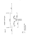
- FIG. 20 A schematic alignment of possible regions of a flaviviral polyprotein (using Yellow Fever Virus as an example), and of a putative polyprotein encoded in the major ORF of the HCV genome, is shown in Fig. 20. In the figure the possible domains of the HCV polyprotein are indicated.
- the flavivirus polyprotein contains. from the amino terminus to the carboxy terminus, the nucleocapsid protein (C), the matrix protein (M), the envelope protein (E), and the non-structural proteins (NS) 1, 2 (a + b), 3, 4 (a + b), and 5.
- HCV nucleocapsid protein
- C nucleocapsid protein
- NS2-5 non-structural proteins 2,3,4, and 5 (NS2-5) of HCV and of yellow fever virus (YFV) appear to have counter parts of similar size and hydropathicity, although there is divergence of the amino acid sequences.
- YFV yellow fever virus
- the region of HCV which would correspond to the regions of YFV polyprotein which contains the M, E, and NS1 protein not only differs in sequence, but also appears to be quite different both in size and hydropathicity.
- HCV Different strains, isolates or subtypes of HCV are expected to contain variations at the amino acid and nucleic acids compared with HCV1. Many isolates are expected to show much (i.e., more than about 40%) homology in the total amino acid sequence compared with HCV1. However, it may also be found that there are other less homologous HCV isolates. These would be defined as HCV according to various criteria such as, for example, an ORF of approximately 9,000 nucleotides to approximately 12,000 nucleotides, encoding a polyprotein similar in size to that of HCV1. an encoded polyprotein of similar hydrophobic andior antigenic character to that of HCV1, and the presence of co-linear peptide sequences that are conserved with HCV1. In addition, it is believed that the genome would be a positive-stranded RNA.
- All HCV isolates encode at least one epitope which is immunologically identifiable (i.e., immunologically cross-reactive) with an epitope encoded in the HCV cDNAs described herein.
- the epitope is contained in an amino acid sequence described herein and is unique to HCV when compared to previously known pathogens. The uniqueness of the epitope may be determined by its immunological reactivity with anti-HCV antibodies and lack of immunological reactivity with antibodies to known pathogens.
- HCV strains and isolates are evolutionary related. Therefore, it is expected that the overall homology of the genomes at the nucleotide level may be about 40% or greater, probably will be about 50% or greater, probably about 60% or greater, and even more probably about 80% or greater; and in addition that there will be corresponding contiguous sequences of at least about 13 nucleotides. It should be noted, as shown infra. that there are variable and hypervariable regions within the HCV genome; therefore. the homology in these regions is expected to be significantly less than that in the overall genome.
- the correspondence between the putative HCV strain genomic sequence and, for example, the CDCiHCV1 cDNA sequence can be determined by techniques known in the art.
- they can be determined by a direct comparison of the sequence information of the polynucleotide from the putative HCV, and the HCV cDNA sequence(s) described herein. They also can be determined by hybridization of the polynucleotides under conditions which form stable duplexes between homologous regions (for example, those which would be used prior to S. digestion), followed by digestion with single stranded specific nuclease(s), followed by size determination of the digested fragments.
- HCV strains or isolates are identifiable by their homology at the polypeptide level.
- HCV strains or isolates are expected to be at least 40% homologous, more than about 50% homologous, probably more than about 70% homologous, and even more probably more than about 80% homologous, and some may even be more than about 90% homologous at the polypeptide level.
- the techniques for determining amino acid sequence homology are known in the art. For example, the amino acid sequence may be determined directly and compared to the sequences provided herein. Alternatively the nucleotide sequence of the genomic material of the putative HCV may be determined (usually via a cDNA intermediate), the putative amino acid sequence encoded therein can be determined, and the corresponding regions compared.
- a polynucleotide "derived from” a designated sequence refers to a polynucleotide sequence which is comprised of a sequence of approximately at least about 6 nucleotides, preferably at least about 8 nucleotides, more preferably at least about 10-12 nucleotides, and even more preferably at least about 15-20 nucleotides corresponding to a region of the designated nucleotide sequence.
- "Corresponding" means homologous to or complementary to the designated sequence.
- the sequence of the region from which the polynucleotide is derived is homologous to or complementary to a sequence which is unique to an HCV genome.
- the derived sequence is homologous or complementary to a sequence that is unique to all or to a majority of HCV isolates. Whether or not a sequence is unique to the HCV genome can be determined by techniques known to those of skill in the art. For example, the sequence can be compared to sequences in databanks, e.g.. Genebank, to determine whether it is present in the uninfected host or other organisms. The sequence can also be compared to the known sequences of other viral agents. including those which are known to induce hepatitis, e.g., HAV, HBV, and HDV, and to members of the Fiavivindae.
- the correspondence or non-correspondence of the derived sequence to :ther sequences can also be determined by hybridization under the appropriate stringency conditions.
- Hybridization techniques for determining the complementarity of nucleic acid sequences are known in the art, and are discussed infra. See also, for example, Maniatis et al. (1982).
- mismatches of duplex polynucleotides formed by hybridization can be determined by known techniques, including for example, digestion with a nuclease such as S1 that specifically digests single-stranded areas in duplex polynucleotides.
- Regions from which typical DNA sequences may be "derived" include but are not limited to, for example, regions encoding specific epitopes, as well as non-transcribed and/or non-translated regions.
- the derived polynucleotide is not necessarily physically derived from the nucleotide sequence shown, but may be generated in any manner, including for example, chemical synthesis or DNA replication or reverse transcription or transcription. In addition, combinations of regions corresponding to that of the designated sequence may be modified in ways known in the art to be consistent with an intended use.
- polynucleotide intends a polynucleotide of genomic, cDNA, semisynthetic, or synthetic origin which, by virtue of its origin or manipulation: (1) is not associated with all or a portion of a polynucleotide with which it is associated in nature, (2) is linked to a polynucleotide other than that to which it is linked in nature, or (3) does not occur in nature.
- polynucleotide refers to a polymeric form of nucleotides of any length, either ribonucleotides or deoxyribonucleotides. This term refers only to the primary structure of the molecule. Thus, this term includes double- and single-stranded DNA and RNA.
- modifications for example, labels which are known in the art, methylation, "caps", substitution of one or more of the naturally occurring nucleotides with an analog, internucleotide modifications such as, for example, those with uncharged linkages (e.g., methyl phosphonates, phosphotriesters, phosphoamidates, carbamates, etc.) and with charged linkages (e.g., phosphorothioates, phosphorodithioates, etc.), those containing pendant moieties, such as, for example proteins (including for e.g., nucleases, toxins, antibodies, signal peptides, poly-L-lysine, etc.), those with intercalators (e.g., acridine, psoralen, etc.), those containing chelators (e.g., metals, radioactive metals, boron, oxidative metals, etc.), those containing alkylators, those with modified linkages (e.g., alkylators, those with
- the "sense strand" of a nucleic acid contains the sequence that has sequence homology to that of mRNA.
- the "anti-sense strand” contains a sequence which is complementary to that of the “sense strand”.
- a "positive stranded genome" of a virus is one in which the genome, whether RNA or DNA, is single-stranded and which encodes a viral polypeptide(s).
- positive stranded RNA viruses include Togaviridae, Coronaviridae, Retroviridae, Picornaviridae, and Caliciviridae. Included also, are the Flaviviridae, which were formerly classified as Togaviradae. See Fields & Knipe (1986).
- primer refers to an oligomer which is capable of acting as a point of initiation of synthesis of a polynucleotide strand when placed under appropriate conditions.
- the primer will be completely or substantially complementary to a region of the polynucleotide strand to be copied. Thus, under conditions conducive to hybridization, the primer will anneal to the complementary region of the analyte strand.
- suitable reactants e.g., a polymerase, nucleotide triphosphates, and the like
- the primer is extended by the polymerizing agent to form a copy of the analyte strand.
- the primer may be single-stranded, or alternatively may be partially or fully double-stranded.
- analyte polynucleotide and “analyte strand” refer to a single- or double-stranded nucleic acid molecule which is suspected of containing a target sequence, and which may be present in a biological sample.
- oligomer refers to primers and to probes.
- the term oligomer does not connote the size of the molecule.
- typically oligomers are no greater than 1000 nucleotides, more typically are no greater than 500 nucleotides, even more typically are no greater than 250 nucleotides; they may be no greater than 100 nucleotides, and may be no greater than 75 nucleotides, and also may be no greater than 50 nucleotides in length.
- probe refers to a structure comprised of a polynucleotide which forms a hybrid structure with a target sequence, due to complementarity of at least one sequence in the probe with a sequence in the target region.
- the polynucleotide regions of probes may be composed of DNA, and. or RNA. and: or synthetic nucleotide analogs. Included within probes are “capture probes” and “label probes”. Preferably the probe does not contain a sequence complementary to sequence(s) used to prime the polymerase chain reaction (PCR).
- target region refers to a region of the nucleic acid which is to be amplified and,or detected.
- target sequence refers to a sequence with which a probe or primer will form a stable hybrid under desired conditions.
- capture probe refers to a polynucleotide comprised of a single-stranded polynucleotide coupled to a binding partner.
- the single-stranded polynucleotide is comprised of a targeting polynucleotide sequence, which is complementary to a target sequence in a target region to be detected in the analyte polynucleotide.
- This complementary region is of sufficient length and complementarity to the target sequence to afford a duplex of stability which is sufficient to immobilize the analyte polynucleotide to a solid surface (via the binding partners).
- the binding partner is specific for a second binding partner; the second binding partner can be bound to the surface of a solid support, or may be linked indirectly via other structures or binding partners to a solid support.
- targeting polynucleotide sequence refers to a polynucleotide sequence which is comprised of nucleotides which are complementary to a target nucleotide sequence; the sequence is of sufficient length and complementarity with the target sequence to form a duplex which has sufficient stability for the purpose intended.
- binding partner refers to a molecule capable of binding a ligand molecule with high specificity, as for example an antigen and an antibody specific therefor.
- the specific binding partners must bind with sufficient affinity to immobilize the analyte copy:complementary strand duplex (in the case of capture probes) under the isolation conditions.
- Specific binding partners are known in the art, and include, for example, biotin and avidin or streptavidin, IgG and protein A, the numerous known receptor-ligand couples, and complementary polynucleotide strands.
- the partners are normally at least about 15 bases in length, and may be at least 40 bases in length; in addition, they have a content of Gs and Cs of at least about 40% and as much as about 60%.
- the polynucleotides may be composed of DNA, RNA, or synthetic nucleotide analogs.
- Coupled refers to attachment by covalent bonds or by strong non-covalent interactions (e.g., hydrophobic interactions, hydrogen bonds, etc.). Covalent bonds may be, for example, ester, ether, phosphoester, amide. peptide, imide, carbon-sulfur bonds, carbon-phosphorus bonds, and the like.
- support refers to any solid or semi-solid surface to which a desired binding partner may be anchored. Suitable supports include glass, plastic, metal. polymer gels, and the like, and may take the form of beads, wells, dipstics. membranes, and the like.
- label refers to any atom or moiety which can be used to provide a detectable (preferably quantifiable) signal, and which can be attached to a polynucleotide or polypeptide.
- label probe refers to an oligomer which is comprised of targeting polynucleotide sequence, which is complementary to a target sequence to be detected in the analyte polynucleotide. This complementary region is of sufficient length and complementarity to the target sequence to afford a duplex comprised of the "label probe” and the "target sequence” to be detected by the label.
- the oligomer is coupled to a label either directly, or indirectly via a set of ligand molecules with high specificity for each other. Sets of ligand molecules with high specificity are described supra., and also includes multimers.
- multimer refers to linear or branched polymers of the same repeating single-stranded polynucleotide unit or different single-stranded polynucleotide units. At least one of the units has a sequence, length. and composition that permits it to hybridize specifically to a first single-stranded nucleotide sequence of interest, typically an analyte or an oligomer (e.g., a label probe) bound to an analyte.
- a first single-stranded nucleotide sequence of interest typically an analyte or an oligomer (e.g., a label probe) bound to an analyte.
- this unit will normally be at least about 15 nucleotides in length, typically no more than about 50 nucleotides in length, and preferably about 30 nucleotides in length; moreover, the content of Gs and Cs will normally be at least about 40%. and at most about 60%.
- the multimer includes a multiplicity of units that are capable of hybridizing specifically and stably to a second single-stranded nucleotide of interest, typically a labeled polynucleotide or another multimer. These units are generally about the same size and composition as the multimers discussed above.
- the first and second oligonucleotide units are heterogeneous (different), and do not hybridize with each other under the conditions of the selected assay.
- multimers may be label probes, or may be ligands which couple the label to the probe.
- viral RNA refers to RNA from the viral genome, fragments thereof, transcripts thereof, and mutant sequences derived therefrom.
- a biological sample refers to a sample of tissue or fluid isolated from an individual. including but not limited to, for example, plasma, serum, spinal fluid, lymph fluid, the external sections of the skin, respiratory. intestinal, and genitourinary tracts, tears, saliva, milk. blood cells, tumors. organs, and also samples of in vitro cell culture constituents (including but not limited to conditioned medium resulting from the growth of cells in cell culture medium, putatively virally infected cells, recombinant cells, and cell components).
- HCV as the etiologic agent of BB-NANBV
- cDNA libraries which contain HCV cDNA sequences.
- These cDNA libraries were derived from nucleic acid sequences present in the plasma of an HCV-infected chimpanzee.
- the construction of one of these libraries, the "c" library (ATCC No. 40394), is described in E.P.O. Publication No. 318,216.
- oligomers can be constructed which are useful as reagents for detecting viral polynucleotides in biological samples. For example, from the sequences it is possible to synthesize DNA oligomers of about 8-10 nucleotides, or larger, which are useful as hybridization probes to detect the presence of HCV RNA in, for example, donated blood, blood fractions, sera of subjects suspected of harboring the virus, or cell culture systems in which the virus is replicating.
- the novel oligomers described herein enable further characterization of the HCV genome.
- Polynucleotide probes and primers derived from these sequences may be used to amplify sequences present in cDNA libraries, and;or to screen cDNA libraries for additional overlapping cDNA sequences, which, in turn, may be used to obtain more overlapping sequences.
- the genome of HCV appears to be RNA comprised primarily of a large open reading frame (ORF) which encodes a large polyprotein.
- the information provided infra allows the identification of additional HCV strains or isolates.
- the isolation and characterization of the additional HCV strains or isolates may be accomplished utilizing techniques known to those of skill in the art, for example, by isolating the nucleic acids from body components which contain viral particles and/or viral RNA, creating cDNA libraries using the oligomers described infra., for screening the libraries for clones containing HCV cDNA sequences described infra., and comparing the HCV cDNAs from the new isolates with the cDNAs described in E.P.O. Publication No. 318,216 and infra. Strains or isolates which fit within the parameters of HCV, as described in the Definitions section, supra., are readily identifiable. Other methods for identifying HCV strains will be obvious to those of skill in the art, based upon the information provided herein.
- the oligomers of the invention contain regions which form hybrid duplex structures with targeted sequences in HCV polynucleotides.
- the HCV polynucleotide hybridizing regions of the oligomers may be ascertained from the HCV cDNA sequence(s) provided herein, and described in E.P.O. Publication No. 318,216.
- a composite of HCV cDNA from HCV1, a prototypic HCV, is shown in Fig. 18.
- the composite sequence is based upon sequence information derived from a number of HCV cDNA clones, which were isolated from a number of HCV cDNA libraries, including the "c" library present in lambda gt11 (ATCC No. 40394), and from human serum.
- the HCV cDNA clones were isolated by methods described in E.P.O. Publication No. 318.216. Briefly, the majority of clones which were isolated contained sequences from the HCV cDNA "c" library which was constructed using pooled serum from a chimpanzee with chronic HCV infection and containing a high titer of the virus, i.e., at least 10 6 chimp infectious doses,ml (CID-ml). The pooled serum was used to isolate viral particles; nucleic acids isolated from these particles was used as the template in the construction of cDNA libranes to the viral genome. The initial clone, 5-1-1, was obtained by screening the "c" library with serum from infected individuals.
- the remainder of the sequence was obtained by screening with synthetic polynucleotide probes, the sequences of which were derived from the 5 -region and the 3 -region of the known HCV cDNA sequence(s).
- oligomers of approximately 8 nucleotides or more can be prepared which hybridize with the positive strand(s) of HCV RNA or its complement, as well as to HCV cDNAs. These oligomers can serve as probes for the detection (including isolation and / or labeling) of polynucleotides which contain HCV nucleotide sequences, and/or as primers for the transcription and/or replication of targeted HCV sequences.
- the oligomers contain a targeting polynucleotide sequence, which is comprised of nucleotides which are complementary to a target HCV nucleotide sequence; the sequence is of sufficient length and complementarity with the HCV sequence to form a duplex which has sufficient stability for the purpose intended.
- a targeting polynucleotide sequence which is comprised of nucleotides which are complementary to a target HCV nucleotide sequence; the sequence is of sufficient length and complementarity with the HCV sequence to form a duplex which has sufficient stability for the purpose intended.
- the purpose is the isolation, via immobilization, of an analyte containing a target HCV sequence.
- the oligomers would contain a polynucleotide region which is of sufficient length and complementarity to the targeted HCV sequence to afford sufficient duplex stability to immobilize the analyte on a solid surface, via its binding to the oligomers, under the isolation conditions.
- the oligomers are to serve as primers for the transcription and or replication of target HCV sequences in an analyte polynucleotide
- the oligomers would contain a polynucleotide region of sufficient length and complementarity to the targeted HCV sequence to allow the polymerizing agent to continue replication from the primers which are in stable duplex form with the target sequence, under the polymerizing conditions.
- the targeting polynucleotide region would be of sufficient length and complementarity to form stable hybrid duplex structures with the label probes and,or multimers to allow detection of the duplex.
- the oligomers may contain a minimum of about 4 contiguous nucleotides which are complementary to targeted HCV sequence; usually the oligomers will contain a minimum of about 8 continguous nucleotides which are complementary to the targeted HCV sequence, and preferably will contain a minimum of about 14 contiguous nucleotides which are complementary to the targeted HCV sequence.
- Suitable HCV nucleotide targeting sequences may be comprised of nucleotides which are complementary nucleotides selected from the following HCV cDNA nucleotides, which are shown in Fig. 18, (nn x - nny denotes from about nucleotide number x to about nucleotide number y)):
- the oligomer need not consist only of the sequence which is complementary to the targeted HCV sequence. It may contain in addition, nucleotide sequences or other moieties which are suitable for the purposes for which the oligomers are used. For example, if the oligomers are used as primers for the amplification of HCV sequences via PCR, they may contain sequences which, when in duplex, form restriction enzyme sites which facilitate the cloning of the amplified sequences.
- the oligomers are to be used as "capture probes" in hybridization assays (described infra), they would contain in addition a binding partner which is coupled to the oligomer containing the nucleotide sequence which is complementary to the targeted HCV sequence.
- a binding partner which is coupled to the oligomer containing the nucleotide sequence which is complementary to the targeted HCV sequence.
- moieities or sequences which are useful of which the oligomers may be comprised or coupled to are those which are known in the art to be suitable for a variety of purposes, including the labeling of nucleotide probes.
- the preparation of the oligomers is by means known in the art, including, for example, by methods which include excision, transcription, or chemical synthesis.
- the target sequences andior regions of the genome which are selected to which the targeting polynucleotides of the oligomers are complementary depend upon the purpose. For example, if the goal is to screen for the presence of HCV in biological samples (e.g. blood), the preferred oligomers would be used as probes and/or primers, and would hybridize to conserved regions of the HCV genome. Some of the conserved regions of the HCV genome to which the oligomers may bind are described herein, for example, the regions which include nucleotide numbers from about the 5-terminus to about 200.
- probes for HCV polynucleotides are a length which allows the detection of unique viral sequences by hybridization. While 6-8 nucleotides may be a workable length, sequences of 10-12 nucleotides are preferred, and about 20 nucleotides or more appears optimal. Preferably, these sequences will derive from regions which lack heterogeneity.
- These probes can be prepared using routine methods, including automated oligonucleotide synthetic methods.
- probes for example, are those derived from the newly isolated clones disclosed herein, as well as the various oligomers useful in probing cDNA libraries, set forth below.
- a complement to any unique portion of the HCV genome will be satisfactory.
- complete complementarity is desirable, though it may be unnecessary as the length of the fragment is increased.
- the biological sample to be analyzed such as blood or serum
- the biological sample to be analyzed may be treated, if desired, to extract the nucleic acids contained therein.
- the resulting nucleic acid from the sample may be subjected to gel electrophoresis or other size separation techniques; alternatively, the nucleic acid sample may be dot blotted without size separation.
- the targeted region of the analyte nucleic acid must be in single stranded form. Where the sequence is naturally present in single stranded form, denaturation will not be required.
- the sequence will be denatured. Denaturation can be carried out by various techniques known in the art. Subsequent to denaturation, the analyte nucleic acid and probe are incubated under conditions which promote stable hybrid formation of the target sequence in the probe with the putative targeted sequence in the analyte, and the resulting duplexes containing the probe(s) are detected.
- Detection of the resulting duplex is usually accomplished by the use of labeled probes; alternatively, the probe may be unlabeled, but may be detectable by specific binding with a ligand which is labeled, either directly or indirectly.
- Suitable labels. and methods for labeling probes and ligands are known in the art, and include, for example, radioactive labels which may be incorporated by known methods (e.g.. nick translation or kinasing), biotin, fluorescent groups. chemiluminescent groups (e.g., dioxetanes. particularly triggered d l oxetanes), enzymes. antibodies, and the like.
- the region of the probes which are used to bind to the analyte can be made completely complementary to the HCV genome. Therefore, usually high stringency conditions are desirable in order to prevent false positives. However, conditions of high stringency should only be used if the probes are complementary to regions of the viral genome which lack heterogeneity.
- the stringency of hybridization is determined by a number of factors during hybridization and during the washing procedure, including temperature, ionic strength, length of time. and concentration of formamide. These factors are outlined in. for example, Maniatis, T. (1982).
- Vanations of this basic scheme which are known in the art, including those which facilitate separation of the duplexes to be detected from extraneous materials and. or which amplify the signal from the labeled moiety, may also be used.
- a number of these variations are reviewed in, for example: Matthews and Kricka (1988), Analytical Biochemistry 169:1; Landegren et al. (1988), Science 242:229: and Mittlin (1989), Clinical chem. 35:1819.
- These and the following publications describing assay formats are hereby incorporated by reference herein.
- Probes suitable for detecting HCV in these assays are comprised of sequences which hybridize with target HCV polynucleotide sequences to form duplexes with the analyte strand, wherein the duplexes are of sufficient stability for detection in the specified assay system.
- a suitable variation is, for example, one which is described in U.S. Patent No. 4,868,105, issued Sept. 9, 1989, and in E.P.O. Publication No. 225,807 (published June 16, 1987).
- These publications describe a solution phase nucleic acid hybridization assay in which the analyte nucleic acid is hybridized to a labeling probe set and to a capturing probe set.
- the probe-analyte complex is coupled by hybridization with a solid- supported capture probe that is complementary to the capture probe set. This permits the analyte nucleic acid to be removed from solution as a solid phase complex. Having the analyte in the form of a solid phase complex facilitates subsequent separation steps in the assay.
- the labeling probe set is complementary to a labeled probe that is bound through hybridization to the solid phaseanaIyte complex.
- HCV genome sequences will be present in serum of infected individuals at relatively low levels, i.e., at approximately 10 2- 10 3 chimp infectious doses (CID) per ml.
- This level may require that amplification techniques be used in hybridization assays.
- amplification techniques are known in the art.
- the Enzo Biochemical Corporation "Bio-Bridge" system uses terminal deoxynucleotide transferase to add unmodified 3'-poly-dT-tails to a DNA probe. The poly dT-tailed probe is hybridized to the target nucleotide sequence, and then to a biotin-modified poly-A.
- EPA 204510 describes a DNA hybridization assay in which analyte DNA is contacted with a probe that has a tail, such as a poly-dT tail, an amplifier strand that has a sequence that hybridizes to the tail of the probe, such as a poly-A sequence. and which is capable of binding a plurality of labeled strands.
- a type of hybridization assay which is described in E.P.O.
- a particularly desirable technique may involve amplification of the target HCV sequences in sera approximately 10,000 fold (i.e., to approximately 10 6 sequencesilml), as part of the hybridization system.
- the amplification may be accomplished, for example, by the polymerase chain reactions (PCR) technique described by Saiki et al. (1986), by Mullis, U.S. Patent No. 4,683,195, and by Mullis et al.
- PCR polymerase chain reactions
- Amplification may be prior to, or preferably subsequent to purification of the HCV target sequence.
- amplification may be utilized in conjunction with the assay methods described in U.S. Patent No. 4,868,105, or if even further amplification is desired, in conjunction with the hybridization system described in E.P.O. Publication No. 317,077.
- Preferred methods for detecting HCV sequences in an analyte polynucleotide strand are based upon the hybridization detection methods described in U.S. Patent No. 4,868,105 and in E.P.O. Publication No. 317,077. These methods are solution-phase sandwich hybridization assays which utilize both capture and label probes which hybridize to target sequences in an analyte nucleic acid. In the use of these assays to screen biological samples for HCV, the probes used would bind to conserved regions of the HCV genome. The capture and label probes may be interspersed in their binding to the target sequence.
- the capture and label probes are in sets, and the probes of one set do not intersperse with the probes of another set.
- the set(s) of multiple capture probes hybridize to the most conserved regions of the genome, while the set(s) of multiple label probes may hybridize to regions which exhibit small amounts of divergence.
- probes could be used which hybridize to sequences in the region of nucleotides from about -318 to about 174, and or nucleotides in the region of about 4378 to about 4902, and/or nucleotides in the region of from about 4056 to about 4448.
- the preferred probes would hybridize to sequences in the 5 -region of the HCV genome, since, as shown infra., this region appears to be highly conserved. Thus, preferred probes may hybridize to, for example, nucleotides from about -318 to about 174 as shown in Fig. 18. Probes could be used which hybridize to either the positive strand in conserved regions, and,or its comple ment, depending upon the purpose, for example, to detect viral genomic sequences, or to detect HCV cDNA sequences resulting from PCR amplification, or to detect replicative intermediates to the positive HCV RNA strand.
- HCV.cPCR polymerase chain reaction technique
- PCR polymerase chain reaction technique
- the HCVI C PCR method utilizes primers and probes derived from the information provided herein concerning the nature of the HCV genome.
- oligonucleotide primers are prepared which match opposite ends of a desired sequence.
- the sequence between the primers need not be known.
- a sample of polynucleotide is extracted and denatured, preferably by heat, and hybridized with oligonucleotide primers which are present in molar excess. Polymerization is catalyzed by a template-and primer-dependent polymerase in the presence of deoxynucleotide triphosphates or nucleotide analogs (dNTPs). This results in two "long products" which contain the respective primers at their 5 -termini, covalently linked to the newly synthesized complements of the original strands.
- dNTPs deoxynucleotide triphosphates or nucleotide analogs
- the replicated DNA is again denatured, hybridized with oligonucleotide primers, returned to polymerizing conditions, and a second cycle of replication is initiated.
- the second cycle provides the two original strands, the two long products from cycle 1, and two "short products" replicated from the long products.
- the short products contain sequences (sense or antisense) derived from the target sequence, flanked at the 5 - and 3 -termini with primer sequences.
- the number of short products is replicated exponentially. Thus, this process causes the amplification of a specific target sequence.
- a sample is provided which is suspected of containing HCV RNA, or a fragment thereof.
- the sample is usually taken from an individual suspected of having NANBH: however, other sources of the sample are included. e.g., conditioned medium or cells from in vitro systems in which the virus has been replicated.
- the sample must contain the target nucleic acid sequence(s).
- HCV RNA The sample is then subjected to conditions which allow reverse transcription of HCV RNA into HCV cDNA.
- Conditions for reverse transcribing RNA are known to those of skill in the art, and are described in, for example, Maniatis et al. (1982), and in Methods in Enzymology.
- a preferred method of reverse transcription utilizes reverse transcnptase from a variety of sources, including recombinant molecules, and isolated from, for example, a retrovirus, preferably from avian myeloblastosis virus (AMV), and suitable conditions for the transcription.
- the HCV cDNA product of reverse transcription is in a RNA:DNA hybrid, which results from the first round of reverse transcription; subsequently, DNA:DNA hybrids result from two or more rounds of transcription.
- the HCV cDNA resulting from reverse transcription is then subjected to PCR to amplify the target sequence.
- the HCV cDNA is denatured, and the separated strands are hybridized with primers which flank the target sequence.
- Strand separation may be accomplished by any suitable denaturing method, including physical, chemical, or enzymatic means, which are known to those of skill in the art.
- a preferred method, which is physical, involves heating the nucleic acid until it is completely (>99%) denatured.
- Typical heat denaturation involves temperatures ranging from about 80 C to about 105°C, for times ranging from about 1 to 10 minutes.
- the target HCV sequences are replicated by a polymerizing means which utilizes a primer oligonucleotide to initiate the synthesis of the replicate chain.
- the primers are selected so that they are complementary to sequences of the HCV genome. Oligomeric primers which are complementary to regions of the sense and antisense strands of HCV cDNA can be designed from the HCV cDNA sequences from the composite cDNA sequence provided in Fig. 18.
- the primers are selected so that their relative positions along a duplex sequence are such that an extension product synthesized from one primer, when it is separated from its template (complement), serves as a template for the extension of the other primer to yield a replicate chain of defined length.
- the primer is preferably single stranded for maximum efficiency in amplification, but may alternatively be double stranded. If double stranded. the primer is first treated to separate its strands before being used to prepare extension products.
- the primer is an oligodeoxyribonucleotide.
- the primer must be sufficiently long to prime the synthesis of extension products in the presence of the agent for polymerization. The exact lengths of the primers will depend on many factors, including temperature and source of the primer and use of the method. For example. depending on the complexity of the target sequence, the oligonucleotide primer typically contains about 15-45 nucleotides, although it may contain more or fewer nucleotides. Short primer molecules generally require cooler temperatures to form sufficiently stable hybrid complexes with the template.
- the primers used herein are selected to be "substantially" complementary to the different strands of each specific sequence to be amplified. Therefore, the primers need not reflect the exact sequence of the template, but must be sufficiently complementary to selectively hybridize with their respective strands.
- a non-complementary nucleotide fragment may be attached to the 5 -end of the primer, with the remainder of the primer sequence being complementary to the strand.
- non-complementary bases or longer sequences can be interspersed into the primer, provided that the primer has sufficient complementarity with the sequence of one of the strands to be amplified to hybridize therewith, and to thereby form a duplex structure which can be extended by the polymerizing means.
- the non-complementary nucleotide sequences of the primers may include restriction enzyme sites. Appending a restriction enzyme site to the end(s) of the target sequence would be particularly helpful for cloning of the target sequence.
- primer may refer to more than one primer, particularly in the case where there is some ambiguity in the information regarding the terminal sequence(s) of the target region to be amplified.
- a “primer” includes a collection of primer oligonucleotides containing sequences representing the possible variations in the sequence or includes nucleotides which allow a typical basepairing.
- One of the primer oligonucleotides in this collection will be homologous with the end of the target sequence.
- a specific case is shown in the Examples, where oligomer sets of 44-mers and 45- mers were utilized to prime the amplification of a potentially variant region of the HCV genome.
- primers which hybridize to conserved regions of the HCV genome.
- the conserved regions may be determined by comparing the nucleotide or amino acid sequences of several HCV strains / isolates. There appear to be at least three regions of conserved amino acid in the HCV genome, described supra., from which primers may be derived. These regions are believed to be.
- the primers described infra., in the Examples are derived from what are believed to be conserved regions of HCV, based upon sequence homology to that of the Flaviviruses.
- the oligonucleotide primers may be prepared by any stable method. Methods for preparing oligonucleotides of specific sequence are known in the art, and include, for example, cloning and restriction of appropriate sequences, and direct chemical synthesis. Chemical synthesis methods may include, for example, the phosphotriester method described by Narang et al. (1979), the phosphodiester method disclosed by Brown et al. (1979), the diethylphosphoramidate method disclosed in Beaucage et al. (1981), and the solid support method in U.S. Patent No. 4,458,066.
- the primers may be labeled, if desired, by incorporating means detectable by spectroscopic, photochemical, biochemical, immunochemical, or chemical means.
- Template-dependent extension of the oligonucleotide primer(s) is catalyzed by a polymerizing agent in the presence of adequate amounts of the four deoxyribonucleotide triphosphates (dATP. dGTP, dCTP and dTTP) or analogs, in a reaction medium which is comprised of the appropriate salts, metal cations, and pH buffering system.
- Suitable polymerizing agents are enzymes known to catalyze primer- and template-dependent DNA synthesis.
- Known DNA polymerases include, for example, E. coii DNA polymerase I or its Klenow fragment, T4 DNA polymerase, and Taq DNA polymerase. The reaction conditions for catalyzing DNA synthesis with these DNA polymerases are known in the art.
- the products of the synthesis are duplex molecules consisting of the template strands and the primer extension strands, which include the target sequence. These products, in turn, serve as template for another round of replication.
- the primer extension strand of the first cycle is annealed with its complementary primer; synthesis yields a "short" product which is bounded on both the 5 -and the 3 -ends by primer sequences or their complements.
- Repeated cycles of denaturation, primer annealing, and extension result in the exponential accumulation of the target region defined by the primers.
- Sufficient cycles are run to achieve the desired amount of polynucleotide containing the target region of nucleic acid. The desired amount may vary, and is determined by the function which the product polynucleotide is to serve.
- the PCR method can be performed in a number of temporal sequences. For example, it can be performed step-wise, where after each step new reagents are added, or in a fashion where all of the reagents are added simultaneously, or in a partial step-wise fashion, where fresh reagents are added after a given number of steps.
- the PCR reaction is carried out as an automated process which utilizes a thermostable enzyme.
- the reaction mixture is cycled through a denaturing region, a primer annealing region, and a reaction region.
- a machine may be employed which is specifically adapted for use with a thermostable enzyme, which utilizes temperature cycling without a liquid handling system, since the enzyme need not be added at every cycle. This type of machine is commercially available from Perkin Elmer Cetus Corp.
- the target polynucleotides are detected by hybridization with a probe polynucleotide which forms a stable hybrid with that of the target sequence under stringent to moderately stringent hybridization and wash conditions. If it is expected that the probes will be completely complementary (i.e., about 99% or greater) to the target sequence, stringent conditions will be used. If some mismatching is expected, for example if variant strains are expected with the result that the probe will not be completely complementary, the stringency of hybridization may be lessened. However, conditions are chosen which rule out nonspecific adventitious binding. Conditions which affect hybridization, and which select against nonspecific binding are known in the art, and are described in, for example, Maniatis et al. (1982).
- stringent conditions are incubation in solutions which contain approximately 0.1 X SSC, 0.1% SDS. at about 65° C incubation / wash temperature, and moderately stringent conditions are incubation in solutions which contain approximately 1-2 X SSC, 0.1% SDS and about 50'-65 C incubation, wash temperature.
- Low stringency conditions are 2 X SSC and about 30 -50° C.
- Probes for HCV target sequences may be derived from the HCV cDNA sequence shown in Fig. 18, or from new HCV isolates.
- the HCV probes may be of any suitable length which span the target region, but which exclude the primers, and which allow specific hybridization to the target region. If there is to be complete complementarity, i.e., if the strain contains a sequence identical to that of the probe, since the duplex will be relatively stable under even stringent conditions, the probes may be short, i.e., in the range of about 10-30 base pairs.
- the probe may be of greater length, since length seems to counterbalance some of the effect of the mismatch(es).
- An example of this is found in the Examples, where the probe was designed to bind to potential variants of HCV1.
- the primers were designed to bind to HCV cDNA derived from a hypothetical conserved region of the HCV genome, and the target region was one which potentially contained variations (based upon the Flavivirus model).
- the probe used to detect the HCV target sequences contained approximately 268 base pairs.
- the probe nucleic acid having a sequence complementary to the target sequence may be synthesized using similar techniques described supra. for the synthesis of primer sequences. If desired, the probe may be labeled. Appropriate labels are described supra.
- the length of the PCR product detected by the probe may be desirable to determine the length of the PCR product detected by the probe. This may be particularly true if it is suspected that variant HCV strains may contain deletions within the target region, or if one wishes to confirm the length of the PCR product. In such cases it is preferable to subject the products to size analysis as well as hybridization with the probe. Methods for determining the size of nucleic acids are known in the art, and include, for example, gel electrophoresis, sedimentation in gradients, and gel exclusion chromatography.
- the presence of the target sequence in a biological sample is detected by determining whether a hybnd has been formed between the HCV polynucleotide probe and the nucleic acid subjected to the PCR amplification technique.
- Methods to detect hybrids formed between a probe and a nucleic acid sequence are known in the art.
- an unlabeled sample may be transferred to a solid matrix to which it binds, and the bound sample subjected to conditions which allow specific hybridization with a labeled probe; the solid matrix is than examined for the presence of the labeled probe.
- the unlabeled probe is bound to the matrix, and after the exposure to the appropriate hybridization conditions, the matrix is examined for the presence of label.
- Other suitable hybridization assays are described supra.
- variant HCV In order to identify variant HCV strains. and thereby to design probes for those variants, the above described HCV cPCR method is utilized to amplify variant regions of the HCV genome, so that the nucleotide sequences of these variant target regions can be determined.
- variant types of HCV might be expected to occur in different geographic locations than that in which the HCV1 strain is predominant. for example. Japan, Africa, etc.; or in different vertebrate species which are also infected with the virus.
- Variant HCV may also arise during passage in tissue culture systems, or be the result of spontaneous or induced mutations.
- primers are designed to flank the suspect region, and preferably are complementary to conserved regions. Primers to two regions of HCV which are probably conserved, based upon the Flavivirus model, are described in the Examples. These primers and probes may be designed utilizing the sequence information for the HCV1 strain provided in Fig. 18.
- Analysis of the nucleotide sequence of the target region(s) may be by direct analysis of the PCR amplified products.
- a process for direct sequence analysis of PCR amplified products is described in Saiki et al. (1988).
- the amplified target sequence(s) may be cloned prior to sequence analysis.
- a method for the direct cloning and sequence analysis of enzymatically amplified genomic segments has been described by Scharf (1986).
- the primers used in the PCR technique are modified near their 5 -ends to produce convenient restriction sites for cloning directly into, for example, an M13 sequencing vector.
- the PCR products are cleaved with the appropriate restriction enzymes.
- the restriction fragments are ligated into the M13 vector, and transformed into, for example, a JM 103 host, plated out, and the resulting plaques are screened by hybridization with a labeled oligonucleotide probe.
- Other methods for cloning and sequence analysis are known in the art.
- HCV is a Flavi-like virus.
- studies are described in E.P.O. publication No. 318,216 owned by the herein assignee, and which is incorporated herein in its entirety.
- a comparison of the HCV cDNA sequence derived from the HCV cDNA clones with known sequences of a number of Flaviviruses show that HCV contains sequences which are homologous to conserved sequences in the Flaviviruses. These conserved sequences may allow the creation of primers which may be universal in their application for amplification of target regions of Flaviviruses, and for HCV.
- sequences are the 16-mer or smaller sequences from the 3 -termini of the primers described in the Examples. Identification of the species is then accomplished utilizing a probe specific for the species.
- the genomes of a number of Flaviviruses are known in the art, and include, for example, Japanese Encephalitis Virus (Sumiyoshi et al. (1987)), Yellow Fever Virus (Rice et al. (1985)), Dengue Type 2 Virus (Hahn et al. (1988)), Dengue Type 4 Virus (Mackow (1987)), and West Nile Virus (Castle et al. (1986)).
- Identification of HCV RNA is accomplished utilizing a probe specific for HCV, the sequence of which can be determined the HCV cDNA sequences provided herein.
- Synthetic oligonucleotides may be prepared using an automated oligonucleotide synthesizer as described by Warner (1984). If desired the synthetic strands may be labeled with 32 P by treatment with polynucleotide kinase in the presence of 32 P-ATP, using standard conditions for the reaction.
- DNA sequences may be modified by known techniques, including, for example site directed mutagenesis, as described by Zoller (1982). Briefly, the DNA to be modified is packaged into phage as a single stranded sequence, and converted to a double stranded DNA with DNA polymerase using, as a primer, a synthetic oligonucleotide complementary to the portion of the DNA to be modified, and having the desired modification included in its own sequence. The resulting double stranded DNA is transformed into a phage supporting host bacterium. Cultures of the transformed bacteria, which contain replications of each strand of the phage, are plated in agar to obtain plaques.
- 50°a of the new plaques contain phage having the mutated sequence, and the remaining 500fo have the original sequence.
- Replicates of the plaques are hybridized to labeled synthetic probe at temperatures and conditions which permit hybridization with the correct strand, but not with the unmodified sequence. The sequences which have been identified by hybridization are recovered and cloned.
- kits for screening for HCV sequences include the oligomeric probe DNAs.
- Kits for amplification of HCV sequences may include the oligomeric primers used in the amplification.
- the kits usually contain the probes or primers in a premeasured or predetermined amount, as well as other suitably packaged reagents and materials, in separate suitable containers, needed for the particular hybridization and / or amplification protocol(s).
- the kit may contain standards, buffers, supports, enzymes, substrates, label probes, binding partners, and/or instructions for conducting the test.
- the clones 13i, 26j, CA59a, CA84a, CA156e and CA167b were isolated from the lambda-gt11 library which contains HCV cDNA (ATCC No. 40394), the preparation of which is described in E.P.O. Publication No. 318,216 (published 31 May 1989), and WO 89/04669 (published 1 June 1989). Screening of the library was with the probes described infra.. using the method described in Huynh (1985). The frequencies with which positive clones appeared with the respective probes was about 1 in 50,000.
- the isolation of clone 13i was accomplished using a synthetic probe derived from the sequence of clone 12f.
- the sequence of the probe was:
- the isolation of clone 26j was accomplished using a probe derived from the 5 -region of clone K9-1.
- the sequence of the probe was:
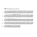
- clone 12f and for clone k9-1 (also called K9-1) are described in E.P.O. Publication No. 318,216, and their sequences are shown in Figs. 1 and 2, respectively.
- the HCV cDNA sequences of clones 13i and 26j are shown in Figs. 4 and 5, respectively. Also shown are the amino acids encoded therein, as well as the overlap of clone 13i with clone 12f, and the overlap of clone 26j with clone 13i.
- the sequences for these clones confirmed the sequence of clone K9-1.
- Clone K9-1 had been isolated from a different HCV cDNA library (See E.P.O. Publication No. 218,316).
- Clone CA59a was isolated utilizing a probe based upon the sequence of the 5 -region of clone 26j. The sequence of this probe was:
- a probe derived from the sequence of clone CA59a was used to isolate clone CA84a.
- the sequence of the probe used for this isolation was:
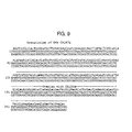
- Clone CA156e was isolated using a probe derived from the sequence of clone CA84a. The sequence of the probe was:
- Clone CA167b was isolated using a probe derived from the sequence of clone CA 156e. The sequence of the probe was:
- nucleotide sequences of the HCV cDNAs in clones CA59a. CA84a, CA156e. and CA167b, are shown Figs. 6. 7, 8, and 9, respectively.
- the amino acids encoded therein, as well as the overlap with the sequences of relevant clones. are also shown in the figures.
- a library of HCV cDNA was constructed from the same batch of infectious chimpanzee plasma used to construct the iambda-gtl HCV cDNA library (ATCC No. 40394) described in E.P.O. Publication No. 318.216, and utilizing essentially the same techniques.
- construction of the pi library utilized a pnmer-extension method, in which the primer for reverse transcriptase was based on the sequence of clone CA59a. The sequence of the primer was: Isolation and Sequence of Clone pil4a
- clone pi14a Screening of the "pi" HCV cDNA library described supra., with the probe used to isolate clone CA167b (See supra.) yielded clone pi14a.
- the clone contains about 800 base pairs of cDNA which overlaps clones CA167b, CA156e, CA84a and CA59a, which were isolated from the lambda gt-11 HCV cDNA library (ATCC No. 40394).
- pi14a also contains about 250 base pairs of DNA which are upstream of the HCV cDNA in clone CA167b.
- Another probe was made based on the sequence of clone CA216a having the following sequence: Screening the lambda-gtl library (ATCC No. 40394) with this probe yielded clone CA290a, the HCV sequences therein being shown in Fig. 11.
- a primer-extension cDNA library was made using nucleic acid extracted from the same infectious plasma used in the original lambda-gt11 cDNA library described above.
- the primer used was based on the sequence of clones CA216a and CA290a:
- the cDNA library was made using methods similar to those described previously for libraries used in the isolation of clones pil4a and k9-1.
- the probe used to screen this library was based on the sequence of clone CA290a:
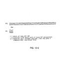
- Clone ag30a was isolated from the new library with the above probe, and contained about 670 basepairs of HCV sequence. See Fig. 12. Part of this sequence overlaps the HCV sequence of clones CA216a and CA290a. About 300 base-pairs of the ag30a sequence, however, is upstream of the sequence from clone CA290a.
- the non-overlapping sequence shows a start codon (') and stop codons that may indicate the start of the HCV ORF.
- putative small encoded peptides (#) which may play a role in regulating translation, as well as the putative first amino acid of the putative polypeptide (), and downstream amino acids encoded therein.
- Clone CA205a was isolated from the original lambda gt-11 library (ATCC No. 40394), using a synthetic probe derived from the HCV sequence in clone CA290a (Fig. 11). The sequence of the probe was:
- the sequence of the HCV cDNA in CA205a shown in Fig. 13. overlaps with the cDNA sequences in both clones ag30a and CA290a.
- the overlap of the sequence with that of CA290a is shown by the dotted line above the sequence (the figure also shows the putative amino acids encoded in this fragment).
- the putative HCV polyprotein appears to begin at the ATG start codon; the HCV sequences in both clones contain an in-frame, contiguous double stop codon (TGATAG) forty two nucleotides upstream from this ATG.
- the HCV ORF appears to begin after these stop codons, and to extend for at least 8907 nucleotides (See the composite HCV cDNA shown in Fig. 18).
- the cDNA in clone 18g overlaps that in clones ag30a and CA205a, described supra.
- the sequence of C18g also contains the double stop codon region observed in clone ag30a.
- the polynucleotide region upstream of these stop codons presumably represents part of the 5 -region of the HCV genome. which may contain short ORFs, and which can be confirmed by direct sequencing of the purified HCV genome.
- These putative small encoded peptides may play a regulatory role in translation.
- the region of the HCV genome upstream of that represented by C18g can be isolated for sequence analysis using essentially the technique described in E.P.O. Publication No.
- 318.216 for isolating cDNA sequences upstream of the HCV cDNA sequence in clone 12f.
- small synthetic oligonucleotide primers of reverse transcriptase which are based upon the sequence of C18g, are synthesized and used to bind to the corresponding sequence in HCV genomic RNA.
- the primer sequences are proximal to the known 5 -terminal of C18g, but sufficiently downstream to allow the design of probe sequences upstream of the primer sequences.
- Known standard methods of priming and cloning ar eused.
- the resulting cDNA libraries are screened with sequences upstream of the priming sites (as deduced from the elucidated sequence of C18g).
- the HCV genomic RNA is obtained from either plasma or liver samples from individuals with NANBH. Since HCV appears to be a Flavi-like virus, the 5 -terminus of the genome may be modified with a "cap” structure. It is known that Flavivirus genomes contain 5 -terminal "cap” structures. (Yellow Fever virus. Rice et al. (1988); Dengue virus. Hahn et al (1988); Japanese Encephalitis Virus (1987)).
- Clones containing cDNA representative of the 3 -terminal region of the HCV genome were isolated from a cDNA library constructed from the original infectious chimpanzee plasma pool which was used for the creation of the HCV cDNA lambda-gt11 library (ATCC No. 40394), described in E.P.O. Publication No. 318,216.
- RNA extracted from the plasma was "tailed" with poly rA using poly (rA) polymerase, and cDNA was synthesized using oligo(dT) 12 . 18 as a primer for reverse transcriptase.
- the resulting RNA:cDNA hybrid was digested with RNAase H, and converted to double stranded HCV cDNA.
- the resulting HCV cDNA was cloned into lambda-gt10, using essentially the technique described in Huynh (1985), yielding the beta (or b) HCV cDNA library.
- the procedures used were as follows.
- nucleic acids in the aqueous phase were precipitated overnight at -20 C, with 2.5 volumes of cold absolute ethanol.
- the precipitates were collected by centrifugation at 10,000 RPM for 40 min.. washed with 70% ethanol containing 20 mM NaCI, and with 100% cold ethanol, dried for 5 min. in a dessicator. and dissolved in water.
- the isolated nucleic acids from the infectious chimpanzee plasma pool were tailed with poly rA utilizing poly-A polymerase in the presence of human placenta ribonuclease inhibitor (HPRI) (purchased from Amersham Corp.), utilizing MS2 RNA as carrier.
- HPRI human placenta ribonuclease inhibitor
- Isolated nucleic acids equivalent to that in 2 ml of plasma were incubated in a solution containing TMN (50 mM Tris HCI, pH 7.9. 10 mM MgCl 2 . 250 mM NaCl, 2.5 mM MnCl 2 . 2 mM dithiothreitol (DTT)).
- the beta HCV cDNA library was screened by hybridization using a synthetic probe, which had a sequence based upon the HCV cDNA sequence in clone 15e.
- the isolation of clone 15e is described in E.P.O. Publication No. 318,216, and its sequence is shown in Fig. 3.
- the sequence of the synthetic probe was:
- clone beta-5a (b5a), which contains an HCV cDNA region of approximately 1000 base pairs.
- the 5'-region of this cDNA overlaps clones 35f, 19g, 26g, and 15e (these clones are described supra).
- the region between the 3 -terminal poly-A sequence and the 3 -sequence which overlaps clone 15e contains approximately 200 base pairs. This clone allows the identification of a region of the 3 - terminal sequence the HCV genome.
- the sequence of b5a is contained within the sequence of the HCV cDNA in clone 16jh (described infra). Moreover, the sequence is also present in CC34a, isolated from the original lambda-gt11 library (ATCC No. 40394). (The original lambda-gt11 library is referred to herein as the "C" library).
- cDNA clones which contain nucleotide sequences derived from the 3 - region of the HCV genome. This was accomplished by amplifying a targeted region of the genome by a polymerase chain reaction technique described in Saiki et al. (1986), and in Saiki et al. (1988), which was modified as described below.
- the HCV RNA which was amplified was obtained from the original infectious chimpanzee plasma pool which was used for the creation of the HCV cDNA lambda-gtl library (ATCC No. 40394) described in E.P.O. Publication No. 318,216. Isolation of the HCV RNA was as described supra. The isolated RNA was tailed at the 3 -end with ATP by E.
- the resultant cDNA was subjected to amplification by PCR using two primers:
- the JH32 primer contained 20 nucleotide sequences hybridizable to the 5 -end of the target region in the cDNA, with an estimated T m of 66 C.
- the JH11 was derived from a portion of the oligo dT-pnmer adapter: thus, it is specific to the 3'-end of the cDNA with a T m of 64 C. Both primers were designed to have a recognition site for the restriction enzyme, Notl, at the 5'-end, for use in subsequent cloning of the amplified HCV cDNA.
- the PCR reaction was carried out by suspending the cDNA and the primers in 100 microliters of reaction mixture containing the four deoxynucleoside triphosphates, buffer salts and metal ions, and a thermostable DNA polymerase isolated from Thermus aquaticus (Taq polymerase), which are in a Perkin Elmer Cetus PCR kit (N801-0043 or N801-0055).
- the PCR reaction was performed for 35 cycles in a Perkin Elmer Cetus DNA thermal cycler. Each cycle consisted of a 1.5 min denaturation step at 94° C, an annealing step at 60 C for 2 min, and a primer extension step at 72 C for 3 min.
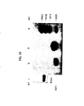
- the PCR products were subjected to Southern blot analysis using a 30 nucleotide probe, JH34, the sequence of which was based upon that of the 3 -terminal region of clone 15e.
- the sequence of JH34 is:
- the PCR products detected by the HCV cDNA probe ranged in size from about 50 to about 400 base pairs.
- the PCR products were cleaved with Notl and size selected by polyacrylamide gel electrophoresis. DNA larger than 300 base pairs was cloned into the Notl site of pUC18S
- the vector pUC18S is constructed by including a Notl polylinker cloned between the EcoRl and Sall sites of pUC18.
- the clones were screened for HCV cDNA using the JH34 probe. A number of positive clones were obtained and sequenced.
- the nucleotide sequence of the HCV cDNA insert in one of these clones, 16jh, and the amino acids encoded therein, are shown in Fig. 15. A nucleotide heterogeneity, detected in the sequence of the HCV cDNA in clone 16jh as compared to another clone of this region, is indicated in the figure.
- the cDNA was then amplified by the PCR reaction using the primers: and
- PCR products were precipitated with spermine, digested with Notl, and extracted with phenol.
- the purified products were cloned into the Notl site of pUC18S, and HCV positive clones were selected using the oligonucleotide:
- the HCV cDNA in one clone, designated p131jh. is shown in Fig. 17. This clone contains an in-frame stop codon for the large ORF contained in the HCV genome.
- a target region of the genome was amplified by the PCR technique described in Saiki et al. (1986), and in Saiki et al (1988).
- the HCV RNA which was amplified was obtained by extracting human serum (U.S.
- Conversion of single- to double-stranded HCV cDNA was accomplished by tailing the DNA with approximately 20 to 50 dA residues using terminal deoxynucleotidyl transferase (Sambrook et al. (1989), MOLECULAR CLONING), and replicating the tailed molecule using the following oligo-dT primer-adapter, which contains a Notl site, and an sp6 promoter:
- the resultant cDNA was subjected to amplification by PCR using two primers, JH94 (described supra.) and JH11, which has the following sequence.
- the PCR reaction was carried out by suspending the cDNA and the primers in 100 microliters of reaction mixture containing the four deoxynucleoside triphosphates. buffer salts and metal ions. and a thermostable DNA polymerase isolated from Thermus aquaticus (Taq polymerase), which are in a Perkin Elmer Cetus PCR kit (N801-0043 or N801-0055).
- the PCR reaction was performed for 35 cycles in a Perkin Elmer Cetus DNA thermal cycler. Each cycle consisted of a 1.5 min denaturation step at 94 C, an annealing step at 60 C for 2 min, and a primer extension step at 72 C for 3 min.
- PCR products were digested with Notl, and cloned into pUC18S.
- Clones containing HCV nucleotide sequences were obtained by screening with a probe, Alex90, which is derived from nucleotides -312 to -283 of the HCV1 genome, and which has the sequence:
- the HCV cDNAs in the isolated clones were sequenced by the dideoxy chain termination method (Sanger et al. (1977)).
- a hairpin structure may serve as a recognition signal for a transcriptase and or it may contnbute to the stability of the RNA at the 5 -terminus.
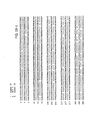
- HCV cDNA sequence has been compiled from a series of overlapping clones derived from various HCV cDNA libraries described herein, and in E.P.O. Publication No. 318,216.
- the clones from which Fig. 18 has been derived are clone 5'-32, b114a, 18g, ag30a, CA205a, CA290a, CA216a, pi14a, CA167b, CA156e, CA84a, CA59a, K9-1 (also called k9-1), 26j, 13i, 12f, 14i, 11 b, 7f, 7e, 8h, 33c, 40b, 37b, 35, 36, 81, 32, 33b, 25c, 14c, 8f, 33f, 33g, 39c, 35f, 19g, 26g, 15e, b5a, 16jh, C6k and p131jh.
- Clone b114a overlaps with clones 18g, ag30a, and CA205a, except that clone b114a contains an extra two nucleotides upstream of the sequence in clone 18g (i.e., 5 -CA). These extra two nucleotides have been included in the HCV genomic sequence shown in Fig. 18.
- the putative sequence of the major HCV polyprotein encoded in the composite of HCV1 cDNA is also shown.
- the first amino acid in the sequence is the putative initiator methionine of the large ORF.
- the variant amino acids, due to the clonal heterogeneities, are indicated above the sequence. Since the lambda gt11 library was created from serum obtained from one individual (see E.P.O. Publication No. 318,216), the results suggest that variant viral sequences (both nucleotide and amino acid) are present in that individual.
- the reading frame, position, and size of the ORFs downstream of the sequence encoding the putative initiator MET of the polyprotein are shown in the Table below.
- the major polyprotein is that translated from reading frame 2.
- an examination of the sequence which is complementary to the genomic strand of HCV RNA also contains several small ORFs.
- One of these ORFs which is complementary to nucleotides -341 to +837 in the HCV RNA sequence, encodes a polypeptide of 385 amino acids.
- HCV18, HCV27 Nucleotide sequences from the 5'- regions of HCV isolates from the U.S.A. (HCV18, HCV27), from Italy (HCVI1, HCVI24), and from Korea (HCVK1) were compared.
- HCV cDNA sequences Isolation of the HCV cDNA sequences was essentially as described supra., for the isolation of 5 - clone32, except for the following.
- the extracted RNA was reverse-transcribed into cDNA using as primers either JH51 or r16, which are complementary to HCV nucleotides -90 to -73 and 366 to 383, respectively.
- the sequences of these primers are as follows.
- Amplification of the HCV dsDNA was by the PCR method using JH93 and JH52 as 5'- and 3'- primers. respectively.
- the HCV sequence in JH93 is derived from HCV nucleotides -317 to -296, that in JH52 is from HCV nucleotides -93 to -117; the nucleotide numbers are indicated in parentheses below the sequences. In JH52 the underlined dinucleotide has been mutated to create the Notl site.
- the sequences of these primers are the following.
- HCV cDNAs were sequenced either by direct sequencing after amplification by PCR, or alternatively, the cloned HCV cDNAs were sequenced by the primer extension and the dideoxy method. Primer extension and the dideoxy method of sequencing were performed as described supra., for the sequence of 5'-clone32.
- Alex90 (see supra. for the sequence) as the 5 -primer, and r25 as the 3-primer.
- Alex90 is derived from HCV nucleotides -312 to -283, and r25 is derived from nucleotides 365 to 342 (See Fig. 18).
- the sequence of r25 is:
- the high levels of sequence conservation in this region may reflect the role of this region in viral replication, and/or transcription, and/or translation.
- HCV HCV which contain sequences which deviate from CDC.HCV1 were identified in human individuals, some of whom were serologically positive for anti-C100-3 antibodies (EC10 was antibody negative). Identification of these new isolates was accomplished by cloning and sequencing segments of the HCV genome which had been amplified by the PCR technique using CDC/HC1 sequences. Amplification was accomplished essentially based on an HCV/cPCR method. The method utilizes primers and probes based upon the HCV cDNA sequences described herein. The first step in the method is the synthesis of a cDNA to either the HCV genome, or its replicative intermediate, using reverse transcriptase. After synthesis of the HCV cDNA.
- RNA in the sample is degraded by techniques known in the art.
- a designated segment of the HCV cDNA is then amplified by the use of the appropriate primers.
- the amplified sequences are cloned, and clones containing the amplified sequences are detected by a probe which is complementary to a sequence lying between the primers, but which does not overlap the primers.
- HCV Isolates Isolated from Humans in the U.S.
- HCV virions Blood samples which were used as a source of HCV virions were obtained from the American Red Cross in Charlotte, North Carolina, and from the Community Blood Center of Kansas, Kansas City, Missouri. The samples were screened for antibodies to the HCV C100-3 antigen using an ELISA assay as described in E.P.O. Publication No. 318,216, and subjected to supplemental Western blot analysis using a polyclonal goat anti-human HRP to measure anti-HCV antibodies. Two samples. #23 and #27, from the American Red Cross and from the Community Blood Center of Kansas, respectively, were determined to be HCV positive by these assays.
- RNA particles present in the serum of these samples were isolated by ultracentrifugation under the conditions described by Bradley et. al. (1985). RNA was extracted from the particles by digestion with proteinase K and SDS at final concentrations of 10 microgramsrml proteinase K. and 0.1% SDS: digestion was for 1 hour at 37 C. Viral RNA was further purified by extraction with chloroform-phenol. as described in E.P.O. Publication No. 318,216.
- HCV RNA in the preparation of RNA was reverse transcribed into cDNA essentially as described in E.P.O. Publication No. 318,216, except that the oligonucleotide JHC 7, which corresponds to the cDNA sequence 1958-1939. and which has the following sequence, was used as primer for the reverse transcriptase reaction.
- the resulting cDNA was then amplified by the PCR method essentially as described supra. for the isolation of clones generated by PCR amplification, except that the oligonucleotide primers used, i.e., JHC 6 and ALX 80, were designed to amplify a 1080 nucleotide segment of the HCV genome from CDC;HCV1 nucleotides 673 to 1751.
- the primers in addition, are designed to incorporate a NOT I restriction site at the 3 -end of the PCR product, and a blunt end at the 5 - terminus.
- the sequences of the primers is: and
- ALX 80 corresponds to nucleotides 673-702 of the CDC/HCV1 sequence; JHC 6 corresponds to nucleotides 1752-1738 of the HCV1 (in addition there are 12 extra nucleotides which encode a Notl site).
- the blunt end terminus was converted into a NOT I site as follows.
- a homopolymer tail of 15 dGs was attached to the PCR product using terminal deoxynucleotide transferase. and the products were again subjected to amplification by PCR using as primers JHC 6 and JHC 13.
- the latter primer, JHC 13, the sequence of which follows. is designed to contain a NOT 1 site in addition to an SP6 phage promoter. (The SP6 promoter is described in GENETIC ENGINEERING. J. Setiow Ed. (1988).
- the PCR products were cleaved with Notl, precipitated with spermine to remove free oligonucleotides (Hoopes et al. (1981)), and cloned into the Notl site of pUC18S (see Section IVA.34.).
- the HCV cDNAs in three clones derived from each HCV isolate, were subjected to sequence analysis. Analysis was essentially by the method described in Chen and Seeburg (1985).
- Consensus sequences of the clones derived from HCV in samples 23 and 27 are shown in Fig. 46 and Fig. 47, respectively.
- the variable sequences are also shown in these figures, as are the amino acids encoded in the consensus sequences.
- Fig. 39 and Fig. 40 show comparisons of the aligned positive strand nucleotide sequences (Fig. 39) and putative amino acid sequences (Fig. 40) of samples 23, 27, and HCV1.
- the amino acid sequence of HCV1 in Fig. 39 represents amino acid numbers 129-467 of the HCV polyprotein encoded by the large ORF in the HCV genomic RNA.
- An examination of Fig. 46 and Fig. 47 show that there are variations in the sequences of the three isolated clones.
- the sequence variations at the nucleotide level and the amino acid level are summarized in the table immediately below.
- the polypeptides designated S and NS1 represent amino acid numbers 130 to - 380, and 380 to - 470, respectively.
- the numbering is from the putative initiator methionine.
- S and NS1 is based upon the positioning of the sequences encoding the polypeptides using the Flavivirus model. As discussed above, however, recent evidence suggests that there is not total correlation between HCV and the Flaviviruses with regard to viral polypeptide domains, particularly in the putative E NS1 domains. Indeed, HCV polypeptides and their coding domains may exhibit substantial deviation from the Flavivirus model.
- HCV23 and HCV27 each contain 1019 nucleotides, indicating a lack of deletion and addition mutants in this region in the selected clones.
- the sequences in Figs. 39 and 40 also show that the isolated sequences are not rearranged in this region.
- sequence variations in two of the putative domains is not uniform.
- the sequence in a putative S region appears to be relatively constant, and randomly scattered throughout the region.
- a putative NS1 region has a higher degree of variability than the overall sequence, and the variation appears to be in a hypervariable pocket of about 28 amino acids which is located about 70 amino acids downstream from the putative N-terminus of the putative polyprotein.
- Variations introduced by the Taq polymerase during PCR amplification would be expected to occur randomly; however, the results show that the variant sequences are clustered in at least one specific region. Moreover, a consensus sequence was derived by sequencing multiple different clones derived from the PCR amplified products.
- HCV HCV Isolates from Humans in Italy and in the U.S.
- Segments of HCV RNA present in different isolates were amplified by the HCV cPCR method. These segments span a region of - 0.6kb to - 1.6Kb downstream from the methionine encoding start codon of the putative HCV polyprotein.
- the isolates are from biological specimens obtained from HCV infected individuals. More specifically, isolate HCT m18 is from human plasma from an individual in the U.S.A., EC1 and EC10 are from a liver biopsy of an Italian patient, and Th is from a peripheral blood mononucleocyte fraction of an American patient. Comparable segments of HCV RNA have been isolated from a chimpanzee.
- TE 50 mM Tris-HCI, pH 8.0.
- RNA extracted from the liver biopsy was provided by Dr. F. Bonino, Ospedale Maggiore di S. Giovanni Battista, Torino, Italy.
- the mononucleocyte fraction was obtained by sedimentation of the individual's aliquot of blood through Ficoll-Paque® (Pharmacia Corp), using the manufacturer's directions. Total RNA was extracted from the fraction using the guanidinium thiocyanate procedure described in E.P.O. Publication No. 318.216 (See also Choo et al (1989).
- RNA or nucleic acid fraction was dried, and resuspended in DEPC treated distilled water. Secondary structures in the nucleic acids were disrupted by heating at 65 C for 10 minutes. and the samples were immediately cooled on ice.
- cDNA was synthesized using 1 to 3 micrograms of total RNA from liver. or from nucleic acids (or RNA) extracted from 10 to 100 microliters of plasma. The synthesis utilized reverse transcriptase, and was in a 25 microliter reaction, using the protocol specified by the manufacturer. BRL. All reaction mixtures for cDNA synthesis contained 23 units of the RNAase inhibitor, RNASINTM Fisher/Promega). Following cDNA synthesis, the reaction mixtures were diluted with water. boiled for 10 minutes, and quickly chilled on ice.
- the primers for the reactions were selected to amplify regions designated "EnvL” and EnvR".
- the "EnvL” region encompasses nucleotides 669-1243. and putative amino acids 117 to 308; the “EnvR” region encompasses nucleotides 1215-1629, and encodes putative amino acids 300-408 (the putative amino acids are numbered starting from the putative methionine initiation codon).
- the relationship of these regions relative to the putative polyprotein encoded in the HCV cDNA. and to the polypeptides encoded in the Flavirus model is shown in Fig. 48.
- the primers for the first round of PCR reactions were derived from the HCV cDNA sequences in either clone ag30a. clone 156e, or clone k9-1.
- the primers used for the amplification of the EnvL region were 156e16B (shown supra), and ag30a16A for the sense strand; the amplification of the EnvR region utilized the primer K91 16B (shown supra), and 156e16a for the sense strand.
- the sequences of the sense strand primers are the following. and
- the PCR reactions were performed essentially according to the manufacturer's directions (Cetus-Perkin-Elmer), except for the addition of 1 microgram of RNase A. The reactions were carned out in a final volume of 100 microliters. The PCR was performed for 30 cycles, utilizing a regimen of 94° C (1 min), 37° C (2 min), and 72° C (3 min), with a 7 minute extension at 72° C for the last cycle. The samples were then extracted with phenol:CHCl 3 , ethanol precipitated two times, resuspended in 10 mM Tris HCI, pH 8.0, and concentrated using Centricon-30 (Amicon) filtration. This procedure efficiently removes oligonucleotides less than 30 nucleotides in size; thus, the primers from the first round of PCR amplification are removed.
- the Centricon-30 concentrated samples were then subjected to a second round of PCR amplification using probes designed from clones 202a and 156e for the EnvL region, and from 156e and 59a for the EnvR region.
- the primers for amplification of the EnvL region have the following sequences.
- the primers for amplification of the EnvR region in RNAs derived from humans have the following sequences. and
- Amplification by PCR was for 35 cycles utilizing a regimen of 94 C (1 min), 60' C (1 min), and 72 C (2 min), with a 7 minute extension at 72 °C for the last cycle.
- the samples were then extracted with phenoI:CHCl 3 , precipitated two times, and digested with EcoRl.
- the PCR reaction products were analyzed by separation of the products by electrophoresis on 6% polyacrylamide gels. DNA of approximately the estimated size of the expected PCR product was electroeluted from the gels, and subcloned into either a pGEM-4 plasmid vector or into lambda gt11.
- the expected product sizes for the EnvL and EnvR after the first round of amplification are 615 bp and 683 bp, respectively; after the second round of amplification the expected product sizes for EnvL and EnvR are 414 bp and 575 bp, respectively.
- the plasmids containing the amplified products were used to transform host cells; the pGEM-4 plasmid was used to transform DH5- alpha, and lambda gt11 was used to transform C600 delta-HFL. Clones of the transformed cells which either hybridized to the appropriate HCV probes (described below), or those which had inserts of the correct size were selected. The inserts were then cloned in M13 and sequenced.
- the probes for all of the HCV/cPCR products consisted of 32p labeled sections of HCV cDNA which had been prepared by PCR amplification of a region of clone 216 (using CA216a16A and 216a16B as primers), and of clone 84 (using CA84a16A and CA84a16B or CA84a16C as primers); 32 P was introduced into the PCR products by nick translation.
- the probes for the first and second round of EnvL amplification were from clone 216.
- Sequence information on variants in the EnvR region was obtained from two clones of EC10, and from the HCV1 clones described in E.P.O. Publication No. 318,216 and supra..
- the two EC10 clones differed by only one nucleotide.
- a comparison of the nucleotide sequences of EC10(clone 2) and a composite of the HCV1 sequences is shown in Fig. 50; each sequence is shown 5 to 3 for the sense strand of the EnvR region, and the sequences have been aligned.
- the double dots between the sequences indicate sequence homology.
- Fig. 51 and Fig. 52 A comparison of the amino acid sequences encoded in the EnvL (amino acids #117-308) and EnvR region (amino acids #300-438) for each of the isolates is shown in Fig. 51 and Fig. 52, respectively. Included in the Figures are sequences for the isolates JH23 and JH27, described supra. Also indicated are sequences from a Japanese isolate; these sequences were provided by Dr. T. Miyamura, Japan. In the figures, the amino acid sequence for the region is given in its entirety for HCV1, and the non-homologous amino acids in the various isolates are indicated.
- Fig. 51 In the EnvL region there is overall about a 93% homology between HCV1 and the other isolates. HCT18. Th, and EC1 have about a 97% homology with HCV1; JH23 and JH27 have about 96% and about 95% homology, respectively, with HCV1.
- Fig. 52 shows that the homologies in the EnvR region are significantly less than in the EnvL region: moreover, one subregion appears to be hypervariable (i.e., from amino acid 383-405). This data is summarized in the Table immediately below.
- the RNA in HCV27 isolated from serum, was analyzed for the presence of positive and negative strands using the PCR method.
- the PCR method was performed essentially as described above, except for the following.
- the extracted HCV27 RNA was reverse transcribed into single-stranded cDNA using as a primer either Alex90 or JH52 (see supra. for the sequences).
- the sequence of Alex90 matches that in nucleotides -312 to -283 of the positive strand of HCV RNA, whereas JH52 matches that of nucleotides -117 to -93 of the negative strand.
- the resulting single-stranded HCV cDNAs were each separately amplified by PCR using Alex90 and JH52. Detection of the amplified products was accomplished by Southern blotting, using Alex89 as the probe. Alex89 matches nucleotide numbers -203 to -175 of HCV RNA.
- the sequence of Alex89 is:
- This example exemplifies the sets of label and capture probes useful to detect HCV RNA in biological samples. using essentially the assay described in U.S. Patent No. 4.868.105.
- the method is a solution-phase sandwich hybridization assay which utilizes both capture and label probes which hybridize to target sequences in an analyte nucleic acid.
- the probes used bind to conserved regions of the HCV genome, and the HCV binding regions are selected for their uniqueness to the HCV genome.
- each capture probe contains, in addition to the sequences complementary to the HCV sequences, the following sequence downstream of the HCV sequence (i.e., at the 3 -end):
- each capture probe is complementary to a sequence in the binding partner(s), so that after hybridization, the duplex can be captured via affixation to the solid phase.
- each label probe contains, in addition to the sequences complementary to the HCV sequences, the following sequence downstream of the HCV sequence: If the method described in E.P.O. Publication No. 317,077 is used, the sequence common to each label probe is complementary to a sequence in a multimer, to allow hybrid duplex formation with that multimer.
- RNA strand is a virion or mRNA strand, which is a "sense" strand.
- replicative intermediate forms may also be detected which would be "anti-sense”; in this case the primer would be "sense”.
- An RNA sense strand containing the target region is hybridized with an anti-sense primer which primes the synthesis of the replicate strand containing the target.
- cDNA to the RNA template is synthesized with a primer- and template-dependent reverse transcriptase. The cDNA in the resulting RNA:cDNA hybrid is released by denaturation and treatment with RNAse.
- Primers are annealed to the cDNA. and extended with a pnmer- and template-dependent DNA polymerase. The products are denatured, re-annealed to primers, and a second round of synthesis is conducted. A number of cycles are run until the amplified product containing the target region is in a desired amount, which is at least a detectable level.
- HCV nuc!eic acids present in liver and plasma of chimpanzees with NANBH. and not in control chimpanzees. were amplified using essentially the polymerase chain re action (PCR) technique described by Saiki et al. (1986).
- the primer oligonucleotides were derived from the HCV cDNA sequences in clone 81 (Fig. 22), or clones 36 (Fig. 23) and 37b (Fig. 24).
- the amplified sequences were detected by gel electrophoresis and a modified Southern blotting method, using as probes the appropriate cDNA oligomer or nick-translated cDNA sequence with a sequence from the region between, but not including, the two primers.
- RNA containing HCV sequences to be examined by the amplification system were isolated from liver biopsies of three chimpanzees with NANBH, and from two control chimpanzees.
- the isolation of the poly A RNA fraction was by the guanidinium thiocyanate procedure described in Maniatis et al. (1982).
- RNA which were to be examined by the amplification system were also isolated from the plasmas of two chimpanzees with NANBH, and from one control chimpanzee, as well as from a pool of plasmas from control chimpanzees.
- One infected chimpanzee had a titer equal to or greater than 10 6 CID/ml, and the other infected chimpanzee had a titer equal to or greater than 10 5 CID / ml.
- the nucleic acids were extracted from the plasma as follows. Either 0.1 ml or 0.01 ml of plasma was diluted to a final volume of 1.0 ml, with a TENBiproteinase K / SDS solution (0.05 M Tris-HCL, pH 8.0, 0.001 M EDTA, 0.1 M NaCl, 1 mgiml Proteinase K, and 0.5% SDS) containing 10 microgramsiml polyadenylic acid. and incubated at 37° C for 60 minutes. After this proteinase K digestion, the resultant plasma fractions were deproteinized by extraction with TE (10.0 mM Tris-HCI, pH 8.0, 1 mM EDTA) saturated phenol.
- the phenol phase was separated by centrifugation, and was reextracted with TENB containing 0.1% SDS.
- the resulting aqueous phases from each extraction were pooled, and extracted twice with an- equal volume of phenol/chloroform/isoamy) alcohol [1:1(99:1)], and then twice with an equal volume of a 99:1 mixture of chloroformlisoamyl alcohol.
- the aqueous phase was brought to a final concentration of 0.2 M Na Acetate, and the nucleic acids were precipitated by the addition of two volumes of ethanol.
- the precipitated nucleic acids were recovered by ultracentrifugation in a SW 41 rotor at 38 K, for 60 minutes at 4 C.
- nucleic acids extracted from plasma by the proteinase KSDSphenol method were further purified by binding to and elution from S and S Elutip-R Columns. The procedure followed was according to the manufacturer's directions.
- the cDNA used as a template for the PCR reaction was derived from the nucleic acids (either total nucleic acids or RNA) prepared as described above. Following ethanol precipitation, the precipitated nucleic acids were dried, and resuspended in DEPC treated distilled water. Secondary structures in the nucleic acids were disrupted by heating at 65 C for 10 minutes, and the samples were immediately cooled on ice.
- cDNA was synthesized using 1 to 3 micrograms of total chimpanzee RNA from liver, or from nucleic acids (or RNA) extracted from 10 to 100 microliters of plasma. The synthesis utilized reverse transcriptase, and was in a 25 microliter reaction, using the protocol specified by the manufacturer, BRL.
- the primers for cDNA synthesis were those also utilized in the PCR reaction, described below. All reaction mixtures for cDNA synthesis contained 23 units of the RNAase inhibitor, RNASINTM (Fisher/Promega). Following cDNA synthesis, the reaction mixtures were diluted with water, boiled for 10 minutes, and quickly chilled on ice.
- PCR reactions were performed essentially according to the manufacturer's directions (Cetus-Perkin-Elmer), except for the addition of 1 microgram of RNase A. The reactions were carried out in a final volume of 100 microliters. The PCR was performed for 35 cycles, utilizing a regimen of 37° C (2 min), 72 ⁇ C (3 min), and 94° C (1 min).
- the primers for cDNA synthesis and for the PCR reactions were derived from the HCV cDNA sequences in either clone 81, clone 36, or clone 37b.
- the HCV cDNA sequences of clones 81, 36, and 37b are shown in Figs. 22, 23, and 24, respectively.
- the sequences of the two 16-mer primers derived from clone 81 were: and
- the sequence of the primer from clone 36 was:
- the sequence of the primer from clone 37b was:
- the primer pairs consisted of either the two 16-mers derived from clone 81, or the 16-mer from clone 36 and the 16-mer from clone 37b.
- the PCR reaction products were analyzed by separation of the products by alkaline gel electrophoresis, followed by Southern blotting, and detec tion of the amplified HCV-cDNA sequences with a 32 P-labeled internal oligonucleotide probe derived from a region of the HCV cDNA which does not overlap the primers.
- the PCR reaction mixtures were extracted with phenol/chloroform, and the nucleic acids precipitated from the aqueous phase with salt and ethanol. The precipitated nucleic acids were collected by centrifugation, and dissolved in distilled water. Aliquots of the samples were subjected to electrophoresis on 1.8% alkaline agarose gels.
- Single stranded DNA of 60, 108, and 161 nucleotide lengths were co-electrophoresed on the gels as molecular weight markers. After electrophoresis, the DNAs in the gel were transferred onto Biorad Zeta ProbeTM paper. Prehybridization and hybridization, and wash conditions were those specified by the manufacturer (Biorad).
- the probes used for the hybridization-detection of amplified HCV cDNA sequences were the following.
- the probe was an 108-mer with a sequence corresponding to that which is located in the region between the sequences of the two primers.
- the probe was the nick-translated HCV cDNA insert derived from clone 35, the nucleotide sequence of which is shown in Fig. 34.
- the primers are derived from nucleotides 155-170 of the clone 37b insert, and 206-268 of the clone 36 insert.
- the 3-end of the HCV cDNA insert in clone 35 overlaps nucleotides 1-186 of the insert in clone 36; and the 5 -end of clone 35 insert overlaps nucleotides 207-269 of the insert in clone 37b. (Compare Figs. 23, 34 and 24.)
- the cDNA insert in clone 35 spans part of the region between the sequences of the clone 36 and 37b derived primers, and is useful as a probe for the amplified sequences which include these primers.
- RNA from the liver specimens was according to the above procedure utilizing both sets of primers and probes.
- the RNA from the liver of the three chimpanzees with NANBH yielded positive hybridization results for amplification sequences of the expected size (161 and 586 nucleotides for 81 and 36 and 37b, respectively). while the control chimpanzees yielded negative hybridization results. The same results were achieved when the experiment was repeated three times.
- RNA from plasma was also according to the above procedure utilizing the primers and probe from clone 81.
- the plasmas were from two chimpanzees with NANBH, from a control chimpanzee, and pooled plasmas from control chimpanzees. Both of the NANBH plasmas contained nucleic acfds.RNA which yielded positive results in the PCR amplified assay, while both of the control plasmas yielded negative results. These results have been repeatedly obtained several times.
- Defective viruses have been known to occur in RNA viruses.
- PCR technology it is possible to design primers to amplify sequences of the HCV genome.
- RNA in captured particles was obtained as described below.
- the analysis for sequences which hybridize to the HCV cDNA derived from clone 81 was carried out utilizing the PCR amplification procedure, as descnbed supra., except that the hybridization probe was a kinased oligonucleotide derived from the clone 81 cDNA sequence. The results showed that the amplified sequences hybridized with the HCV cDNA probe.
- Particles were captured from HCV infected chimpanzee plasma using polystyrene beads coated with an immunopurified antibody directed against the polypeptide encoded in clone 5-1-1.
- the procedure for producing the immunopurified antibody preparation is described in E.P.O. Publication No. 318,216, which is commonly owned by the herein assignee. and which is incorporated herein by reference.
- the HCV polypeptide encoded within clone 5-1-1 was expressed as a fusion polypeptide with superoxide dismutase (SOD). This was accomplished by subcloning the clone 5-1-1 cDNA insert into the expression vector pSODcf1 (Steimer et al. (1986)). DNA isolated from pSODcf1 was treated with BamHl and EcoRl, and the following linker was ligated into the linear DNA created by the restriction enzymes:
- the plasmid containing the insert was isolated. Plasmid containing the insert was restricted with EcoRl. The HCV cDNA insert in clone 5-1-1 was excised with EcoRl, and ligated into this EcoRl linearized plasmid DNA. The DNA mixture was used to transform E. coli strain D1210 (Sadler et al. (1980)). Recombinants with the 5-1-1 cDNA in the correct orientation for expression of the ORF were identified by restriction mapping and nucleotide sequencing. Recombinant bacteria from one clone were induced to express the SOD-NANB 5 - 1 - 1 polypeptide by growing the bacteria in the presence of IPTG.
- the fusion polypeptide was purified from the recombinant E. coli by differential extraction of the cell extracts with urea, followed by chromatography on anion and cation exchange columns.
- the purified SOD-NANB 5 - 1 - 1 polypeptide was attached to a nitrocellulose membrane.
- Antibody in samples of HCV infected serum was absorbed to the matrix-bound polypeptide. After washing to remove non-specifically bound materials and unbound materials, the bound antibody was released from the bound polypeptide.
- a modified form of the PCR assay i.e., a cPCR assay
- putative viral RNA in the sample is reverse transcribed into cDNA with reverse transcriptase; a segment of the resulting cDNA is then amplified utilizing a modified version of the PCR technique described by Saiki et al. (1986).
- the primers for the cPCR technique are derived from HCV RNA, which can be identified by the family of HCV cDNAs provided herein. Amplified product corresponding to the HCV-RNA is detected utilizing a probe derived from the family of HCV cDNAs provided herein.
- cPCR/HCV assay used in these studies were performed utilizing the following methods for the prepara tion of RNA, the reverse transcription of the RNA into cDNA, the amplification of specific segments of the cDNA by PCR, and the analysis of the PCR products.
- RNA was extracted from liver utilizing the guanidium isothiocyanate method for preparing total RNA described in Maniatis et al. (1982).
- RNA from plasma was diluted five- to ten-fold with TENB (0.1 M NaCI, 50 mM Tris-HCI, pH 8.0, 1 mM EDTA) and incubated in a Proteinase KSDS solution (0.5% SDS, 1 mgiml Proteinase K, 20 micrograms/ml Poly A carrier) for 60 to 90 minutes at 37 C.
- the samples were extracted once with phenol (pH 6.5), the resulting organic phase was re-extracted once with TENB containing 0.1% SDS, and the aqueous phases of both extractions were pooled and extracted twice with an equal volume of phenol/CHCl 3 /isoamyl alcohol [1:1(99:1)].
- the resulting aqueous phases were extracted with an equal volume of ChCl 3 /isoamyl alcohol (99:1) twice, and ethanol precipitated using 0.2 M sodium acetate, pH 6.5, and 2.5 volumes of 100% ethanol: precipitation was overnight at -20 C.
- RNA sample containing either 2 micrograms of heat denatured total chimpanzee liver RNA, RNA from 2 microliters of plasma, or 10% of the RNA extracted from 10mm X 4 mm cylindrical human liver biopsies
- dNTP deoxyribonucleotide triphosphate
- DTT millimolar dithiothreitol
- KCI RNase inhibitor
- Amplification of a segment of the HCV cDNA was performed utilizing two synthetic oligomer 16-mer primers whose sequences were derived from HCV cDNA clones 36 (anti-sense) and 37b (sense).
- the sequence of the primer from clone 36 was:
- the sequence of the primer from clone 37b was:
- the primers were used at a final concentration of 1 micromolar each.
- the cDNA samples were incubated with 0.1 microgram of RNAse A and the PCR reactants of the Perkin Elmer Cetus PCR kit (N801-0043 or N801-0055) according to the manufacturer's instructions.
- the PCR reaction was performed for either 30 cycles or 60 cycles in a Perkin Elmer Cetus DNA thermal cycler. Each cycle consisted of a 1 minute denaturation step at 94 C, an annealing step of 2 minutes at 37 C, and an extension step of 3 minutes at 72 C. However. the extension step in the final cycle (30 or 60) was 7 minutes rather than 3 minutes.
- the samples were extracted with an equal volume of phenol: chloroform (1:1), followed by extraction with an equal volume of chloroform, and then the samples were precipitated with ethanol containing 0.2 M sodium acetate.
- the cPCR products were analyzed as follows. The products were subjected to electrophoresis on 1.8% alkaline agarose gels according to Murakawa et al. (1988), and transferred onto ZetaTM Probe paper (BioRad Corp.) by blotting gels overnight in 0.4 M NaOH. The blots were neutralized in 2 X SSC (1 X SSC contains 0.15 M NaCI, 0.015 M sodium citrate), prehybridized in 0.3 M NaCI, 15 mM sodium phosphage buffer, pH 6.8, 15 mM EDTA, 1.0% SDS, 0.5% nonfat milk (Carnation Co.), and 0.5 mg/ml sonicated denatured salmon sperm DNA.
- the blots to be analyzed for HCV cDNA fragments were hybridized to a 32p- labeled probe generated by nick translation of the HCV cDNA insert sequence in clone 35, described in E.P.O. Publication No. 318,216. After hybridization, the blots were washed in 0.1 X SSC (1 X SSC contains 0.15M NaCl, 0.01M Na citrate) at 65 C, dried, and autoradiographed.
- the expected product size is 586 nucleotides in length: products which hybridized with the probe and migrated in the gels in this size range were scored as positive for viral RNA.
- cPCR primers designed to amplify alpha-1 anti-trypsin mRNA was performed to verify the presence of RNA in each sample analyzed.
- the coding region of the alpha-1 anti-trypsin gene is described in Rosenberg et al. (1984).
- Synthetic oligomer 16-mer primers designed to amplify a 365 nucleotide fragment of the coding region of the alpha-1 antitrypsin gene were derived from nucleotides 22-37 (sense) and nucleotides 372-387 (antisense).
- the PCR products were detected using a 32 P nick-translated probe which lies between, and not including, the cDNA/PCR primer sequences.
- the cPCR assay was performed on total RNA isolated from livers of three chimpanzees experimentally infected with a NANBH agent, and from liver biopsies of Italian patients diagnosed as having chronic NANBH.
- Fig. 25A shows the results of the cPCR assay using 1 microgram of each preparation of total liver RNA.
- the RNA was isolated from liver samples of a chimpanzee in the chronic phase of NANBH (910)(lane 1), two chimpanzees in the acute phase of infection (1028 and 508)(lanes 2 and 3, respectively).
- PCR was performed on the samples in lanes 1-3 for 30 cycles and the autoradiogram of the blot containing those lanes was exposed for 5 hours.
- Fig. 25B shows the results of the cPCR assay using 10% of the RNA extracted from 10mm X 4mm liver biopsy cylinders from 15 chronic NANB patients (lanes 1-15), one patient with cryptogenic liver disease (lane 16) and one control sample from a patient with chronic Hepatitis B (lane 17). Amplification by PCR was for 30 cycles and the autoradiogram for the blots were exposed for 4 days, except that lane 1 was exposed for 15 hours. As seen from the results, 915 (60%) of the human samples were positive for HCV RNA (lanes 1.2.4.6.7.10-13). One patient diagnosed with cryptogenic liver disease (lane 16) and one patient with a chronic HBV infection (lane 17) were repeatedly negative in the cPCR assay.
- SOD HCV C100-3 polypepticle (aiso called C100) is a recombinant fusion polypeptide which contains 363 viral amino acids.
- the polypeptide is useful for detecting antibodies to HCV (See Kuo et al. (1989)).
- the method for preparing C100 is described in E.P.O. Publication No. 318,216.
- Radioimmune assay using C100 was performed on the sera collected from the same 17 human patients whose liver samples were subjected to HCV cPCR assay as described supra. The sera was collected on the same day as the liver biopsies. The assay was performed essentially as described in E.P.O. Publication No. 318,216, which is commonly owned and incorporated herein by reference. Briefly, Microtiter plates (Immulon 2, Removeawell strips) were coated with 0.1 microgram of purified C100. The coated plates were incubated for 1 hour at 37° C with the serum samples (100 microliters of a 1:100 dilution) or appropriate controls.
- liver damage was determined by alanine amino transferase (ALT) levels; the presence of HCV RNA was determined by the HCV cPCR assay described above; anti-HCV antibodies were detected utilizing the C100 RIA.
- ALT alanine amino transferase
- RNA extracted from 1 microliter of chimpanzee plasma Serum was taken from chimpanzee 771 on days 25, 32, 70 and 88 post-infection; cPCR was performed for 30 cycles and the autoradiogram was exposed for 18 days. Serum was taken from chimpanzee 910 on days 11, 28, and 67 post-infection; cPCR was performed for 60 cycles and the autoradiogram was exposed for 5 days.
- Fig. 26A for chimpanzee 771
- Fig. 26B for chimpanzee 910. From a comparison of Figs. 26A and 26B, it appears that an early, well defined peak of ALT values dunng acute hepatitis correlates with the presence of viral RNA in the infected individual.
- HCV RNA which is indicative of a state of viremia, precedes the presence of anti-HCV antibodies.
- Chimpanzee 771 (Fig. 26A) exhibited a clearly defined acute episode of post-transfusion NANBH at 28 days, as characterized by an initial peak of ALT levels.
- HCV RNA was detected in the serum collected at day 25, and at day 32. However, during this acute phase, anti-HCV antibodies were absent.
- HCV RNA was below the experimental level of detection, and anti-HCV antibodies were rising.
- HCV RNA remained undetectable, while anti-HCV antibodies were significantly increased over that of day 70.
- ALT values in plasma from individuals with chronic NANBH do not necessarily correlate with weak virus production.
- a pool of 17 different plasma samples taken from chimpanzee 910 over a period of two to three and one-half years post inoculation was monitored for ALT levels and for HCV RNA.
- the ALT values of the samples did not exceed 45 mU/ml; nevertheless, titration studies indicated high titers of HCV (3 x 10 6 CID;ml).
- cPCR was carried out for 30 cycles, and the autoradiogram was exposed for 15 hours; the cPCR analysis clearly showed the presence of viral RNA (data not shown).
- the sensitivity of the HCV cPCR assay was determined by analyzing ten-fold serial dilutions of a plasma pool of known titer.
- the chimpanzee plasma had a titer of ⁇ 3 x 10 5 CID l ml, and RNA was extracted from ten-fold dilutions of 1 microliter of the plasma.
- cPCR was performed for 30 cycles, and after hybridization and washing, the autoradiogram was exposed for 15 hours.
- the cPCR products resulting from amplification of - 300, - 30, and ⁇ 3 CID of HCV genomes are shown in lanes 1-3, respectively of Fig. 29.
- the samples in lanes 1 and 2 were detectable on autoradiograms exposed for 2 hours.
- Primers consisting of a set of oligomer 44-mers and a set of oligomer 45-mers, were designed to amplify strains of HCV which are similar or identical to the HCV isolate from which the cDNA sequence in Fig. 18 is derived.
- the premise underlying the design of these primers is our discovery that HCV is a Flavi-like virus.
- Members of the Flaviviridae family when compared to HCV, have two major conserved sets of amino acid sequences, TATPPG and QRRGR, in the putative NS3 region of these viruses. Several other smaller sets may be seen, for example, GDD in the putative NS5 region. Other sets are determinable by comparison of the known amino acid sequences with that of HCV.
- the 44-mer and 45-mer oligomer primers were designed so that the sequences encoding these amino acids were incorporated within the primer. Moreover, they contain degeneracies at the 3 -end of each primer, and are derived from two different regions of the HCV genome which are present in clone 40b (See Fig. 28), and which are derived from the region encoding putative NS3 of HCV.
- the formulae for the oligonucleotide primers in the sets are:
- the HCV cPCR assay was carried out utilizing these primers to amplify HCV RNA in chimpanzee 910 plasma.
- the assay method was essentially as described in Section supra., except that the 44-mer and 45-mer sets of oligomer primers were substituted for the primers derived from clone 36 and clone 37b.
- detection of amplified HCV cDNA was by hybridization with a probe derived from clone 40a, the sequence of which is shown in Fig. 32.
- the probe was prepared by amplifying a segment of clone 40a utilizing the PCR method described supra., and 18-mer primers containinq the following sequences: and
- the probe preparation was labeled with 32 P by nick translation.
- Fig. 33 shows an autoradiograph of the Southern blots probed with the sequence derived from Clone 40a.
- 32 P labeled Mspl digested pBR322 DNA fragments served as markers (lane 1).
- the predicted size of the PCR product resulting from amplification using these primers is 490 nucleotides (nt).
- Duplicate reactions are shown in lanes 2 and 3.
- the 5'-region HCV cDNA which is flanked by the regions represented in clones ag30a and k9-1 encodes a segment of putative envelope and or matrix protein(s) (EM). Serum obtained from the chimpanzee from which the HCV cDNA "c" library, was constructed was analyzed by HCV/cPCR to determine whether variants within this target region were present.
- the HCV cPCR assay was performed essentially as described supra.. for the isolation of clone 5'-32, except for the primers and probes used.
- Fig. 37 shows the relationship of the primers and probes (and the clones from which they were derived) to that of the target region of HCV cDNA.
- One set of PCR primers, ag30a16A and K91Env16B, were derived from clones ag30a and k9-1, which are upstream and downstream, respectively, of the target sequence.
- the expected size of the cPCR product primed by ag30a16A and K91Env16B is 1.145 kb based upon the confirmed sequence of HCV cDNA.
- PCR primers covering the region amplified using ag30a16A and K9lEnv16B, and overlapping each other were also used for PCR amplification of HCV RNA in the serum.
- the PCR reactions were run using as one set of primers ag30a16A and CA156e16B, and as the second set of primers CA156e16A and k91Env16B.
- the expected PCR product sizes for these pairs were 615 nucleotides (NT) and 683 NT. respectively.
- the table immediately following lists the pnmer, the clone from which it was derived, and the primer sequence.
- the probes for all of the HCV/cPCR products consisted of 32 P labeled sections of HCV cDNA which had been prepared by PCR amplification of a region of clone 216 (using CA216a16A and 216a16B as primers), and of clone 84 (using CA84a16A and CA84a168 as primers); 32 P was introduced into the PCR products by nick translation. These probes did not overlap the primers used in the HCV cPCR reactions.
- Fig. 38 shows an autoradiograph of a Southern blot in which the HCV cPCR products were hybridized with the 32 P-labeled probes.
- the HCV/cPCR product extended from primers ag30a16A and K9lEnv16B (lane 1) was approximately 1.1Kb; no other PCR products were observed in a 15 hour exposure.
- the HCV products extended from the primer sets ag30a15ACA156e16B (lane 2) and CA156e16A K91Env16B (lane 3) were approximately 625NT and approximately 700 NT, respectively.
- the size of the PCR products were determined by comparison with the relative migrations of fragments resulting from the digestion of pBR322 with Mspl and of PhiX 174 digested with Haelll (lane 5).
- HCV is a flavi-like virus, allows a strategy for cloning uncharactenzed HCV cDNA sequences utilizing the PCR technique, and primers derived from the regions encoding conserved amino acid sequences in flaviviruses.
- one of the primers is derived from a defined HCV genomic sequence
- the other primer which flanks a region of unsequenced HCV polynucleotide is derived from a conserved region of the flavivirus genome.
- the flavivirus genomes are known to contain conserved sequences within the NS1, and E polypeptides, which are encoded in the 5 -region of the flavivirus genome.
- upstream primers are designed which are derived from the conserved sequences within these flavivirus polypeptides.
- the downstream primers are derived from an upstream end of the known portion of the HCV cDNA.
- the Lee procedure utilizes mixed oligonucleotide primers complementary to the reverse translation products of an amino acid sequence; the sequences in the mixed primers takes into account every codon degeneracy for the conserved amino acid sequence.
- primer mixes Three sets of primer mixes are generated, based on the amino acid homologies found in several flaviviruses, including Dengue-2,4 (D-2,4), Japanese Encephalitis Virus (JEV), Yellow Fever (YF), and West Nile Virus (WN).
- the primer mixture derived from the most upstream conserved sequence (5'-1), is based upon the amino acid sequence gly-trp-gly, which is part of the conserved sequence asp-arg-gly-trp-gly- aspN found in the E protein of D-2, JEV, YF, and WN.
- the next primer mixture (5 -2) is based upon a downstream conserved sequence in E protein, phe-asp-gly-asp-ser-tyr-ileu-phe-gly-asp-ser-tyr-ileu, and is derived from phe-gly-asp; the conserved sequence is present in D-2, JEV, YF, and WN.
- the third primer mixture (5 -3) is based on the amino acid sequence arg-ser-cys, which is part of the conserved sequence cys-cys-arg-ser-cys in the NS1 protein of D-2, D-4, JEV, YF, and WN.
- each primer in each mixture also contains a constant region at the 5 -end which contains a sequence encoding sites for restriction enzymes, Hindlll, Mbol, and EcoRl.
- the downstream primer, ssc5h20A is derived from a nucleotide sequence in clone 5h, which contains HCV cDNA with sequences with overlap those in clones 14i and 11 b.
- the sequence of ssc5h20A is An alternative primer, ssc5h34A, may also be used. This primer is derived from a sequence in clone 5h, and in addition contains nucleotides at the 5 -end which create a restriction enzyme site, thus facilitating cloninq.
- the sequence of ssc5h34A is
- the PCR reaction which was initially described by Saiki et al. (1986), is carried out essentially as described in Lee et al. (1988), except that the template for the cDNA is RNA isolated from HCV infected chimpanzee liver, or from viral particles isolated from HCV infected chimpanzee serum.
- the annealing conditions are less stringent in the first round of amplification (0.6M NaCI, and 25 C), since the part of the primer which will anneal to the HCV sequence is only 9 nucleotides, and there could be mismatches.
- ssc5h34A is used, the additional sequences not derived from the HCV genome tend to destabilize the primer-template hybrid.
- the annealing conditions can be more stringent (0.066M NaCI, and 32' C-37° C), since the amplified sequences now contain regions which are complementary to, or duplicates of the primers.
- the first 10 cycles of amplification are run with Klenow enzyme I, under appropriate PCR conditions for that enzyme. After the completion of these cycles, the samples are extracted, and run with Taq polymerase, according to kit directions, as furnished by Cetus. Perkin-Elmer.
- the amplified HCV cDNA sequences are detected by hybridization using a probe derived from clone 5h.
- This probe is derived from sequences upstream of those used to derive the primer, and does not overlap the sequences of the clone 5h derived primers.
- the sequence of the probe is
- both probes and primers derived from HCV cDNA. and kits containing them, are useful for the accurate, relatively simple, and economic determination of the presence of HCV in biological samples, more particularly in blood which may be used for transfusions, and in individuals suspected of having HCV an infection.
- these methods and oligomers may be useful for detecting an earlier stage of HCV infection than are immunological assays based upon the use of a recombinant HCV polypeptides.
- an amplified polynucleotide hybridization assay detects HCV RNA in occasional samples which are anti-HCV antibody negative.
- the probes and primers described herein may be used amplified hybridization assays, in conjunction with an immunoassays based on HCV polypeptides to more completely identify infections due to HCV, and HCV-infected biological specimens. including blood.
- the information provided herein allows the design of primers and/or probes which are derived from conserved regions of the HCV genome.
- the provision of these primers and probes makes available a general method which will detect variant HCV strains, and which will be of use in the screening of blood and blood products.
- the method should aid in the detection and/or identification of variant strains of HCV. This, in turn, should lead to the development of additional immunological reagents for the detection and diagnosis of HCV, as well as the development of additional polynucleotide reagents for detection and or treatment of HCV.
- sets of primers and probes designed from the conserved amino acid sequences of Flaviviruses and HCV allow for a universal detection method for these infectious agents.
- strain D1210 The following derivatives of strain D1210 were deposited on 3 May 1989.
Landscapes
- Chemical & Material Sciences (AREA)
- Life Sciences & Earth Sciences (AREA)
- Organic Chemistry (AREA)
- Health & Medical Sciences (AREA)
- Proteomics, Peptides & Aminoacids (AREA)
- Wood Science & Technology (AREA)
- Zoology (AREA)
- Engineering & Computer Science (AREA)
- Immunology (AREA)
- Genetics & Genomics (AREA)
- General Health & Medical Sciences (AREA)
- Biophysics (AREA)
- Molecular Biology (AREA)
- Biochemistry (AREA)
- General Engineering & Computer Science (AREA)
- Physics & Mathematics (AREA)
- Bioinformatics & Cheminformatics (AREA)
- Microbiology (AREA)
- Biotechnology (AREA)
- Analytical Chemistry (AREA)
- Virology (AREA)
- Communicable Diseases (AREA)
- Chemical Kinetics & Catalysis (AREA)
- Gastroenterology & Hepatology (AREA)
- Medicinal Chemistry (AREA)
- Measuring Or Testing Involving Enzymes Or Micro-Organisms (AREA)
- Peptides Or Proteins (AREA)
- Saccharide Compounds (AREA)
- Micro-Organisms Or Cultivation Processes Thereof (AREA)
- Pharmaceuticals Containing Other Organic And Inorganic Compounds (AREA)
Applications Claiming Priority (6)
| Application Number | Priority Date | Filing Date | Title |
|---|---|---|---|
| US35596189A | 1989-05-18 | 1989-05-18 | |
| US355961 | 1989-05-18 | ||
| US45663789A | 1989-12-21 | 1989-12-21 | |
| US456637 | 1989-12-21 | ||
| US50543590A | 1990-04-04 | 1990-04-04 | |
| US505435 | 1990-04-04 |
Publications (3)
| Publication Number | Publication Date |
|---|---|
| EP0398748A2 true EP0398748A2 (de) | 1990-11-22 |
| EP0398748A3 EP0398748A3 (de) | 1991-10-23 |
| EP0398748B1 EP0398748B1 (de) | 2002-01-09 |
Family
ID=27408242
Family Applications (1)
| Application Number | Title | Priority Date | Filing Date |
|---|---|---|---|
| EP90305421A Expired - Lifetime EP0398748B1 (de) | 1989-05-18 | 1990-05-18 | NANBV-Diagnostika: Polynukleotide, geeignet für Reihenuntersuchungen auf Hepatitis C-Virus |
Country Status (17)
| Country | Link |
|---|---|
| EP (1) | EP0398748B1 (de) |
| JP (3) | JP2656996B2 (de) |
| AT (1) | ATE211772T1 (de) |
| AU (1) | AU638719B2 (de) |
| BG (1) | BG60348B2 (de) |
| CA (1) | CA2017157C (de) |
| DE (1) | DE69033891T2 (de) |
| DK (1) | DK0398748T3 (de) |
| ES (1) | ES2166749T3 (de) |
| FI (1) | FI105279B (de) |
| GE (1) | GEP20002164B (de) |
| HU (1) | HUT59964A (de) |
| LV (1) | LV10729B (de) |
| MC (1) | MC2188A1 (de) |
| NO (1) | NO303503B1 (de) |
| RO (1) | RO113059B1 (de) |
| WO (1) | WO1990014436A1 (de) |
Cited By (22)
| Publication number | Priority date | Publication date | Assignee | Title |
|---|---|---|---|---|
| FR2655990A1 (fr) * | 1989-12-18 | 1991-06-21 | Wellcome Found | Agent viral responsable de l'hepatite non-a non-b et polypeptides derives de celui-ci utiles pour le diagnostic et la vaccination. |
| EP0461863A1 (de) * | 1990-06-12 | 1991-12-18 | Immuno Japan Inc. | Oligonukleotid-Primer und ihre Anwendung zum Nachweis von non-A, non-B-Hepatitisvirus mit grosser Genauigkeit |
| WO1992000328A1 (en) * | 1990-06-30 | 1992-01-09 | Akzo N.V. | Non-a non-b sequences |
| EP0469348A3 (de) * | 1990-07-11 | 1992-02-12 | SHIONOGI SEIYAKU KABUSHIKI KAISHA trading under the name of SHIONOGI & CO. LTD. | cDNS-Sequenz und Detektion des Hepatitis C-Virus |
| EP0510952A1 (de) * | 1991-04-26 | 1992-10-28 | Immuno Japan Inc. | Oligonukleotid-Primer und ihre Anwendung zum Nachweis von Non-A, Non-B-Hepatitis mit höchster Genauigkeit |
| WO1992021759A1 (fr) * | 1991-06-06 | 1992-12-10 | Institut Pasteur | Sequences nucleotidiques et peptidiques d'un isolat de virus de l'hepatite c, applications diagnostiques et therapeutiques |
| WO1993002103A1 (en) * | 1991-07-19 | 1993-02-04 | Sorin Biomedica S.P.A. | Epitopes of the env protein of the hepatitis c virus |
| EP0531974A1 (de) * | 1991-09-12 | 1993-03-17 | Cedars-Sinai Medical Center | Direkter Nachweis der RNS des Hepatitis-C-Virus |
| EP0543924A4 (en) * | 1990-08-10 | 1993-07-21 | Chiron Corporation | Nanbv diagnostics: polynucleotides useful for screening for hepatitis c virus |
| WO1993010239A3 (en) * | 1991-11-21 | 1993-07-22 | Common Services Agency | Hepatitis-c virus testing |
| US5527669A (en) * | 1991-08-27 | 1996-06-18 | Hoffmann-La Roche Inc. | Methods, primers and probes for detection of hepatitis C and novel variants |
| EP0776981A2 (de) | 1995-11-29 | 1997-06-04 | F. Hoffmann-La Roche Ag | Oligonukleotid-Primer für Amplifizierung von HCV-Nukleinsäure |
| US5747240A (en) * | 1991-11-07 | 1998-05-05 | Kink; John A. | Epitope mapping of the c33 region of HCV |
| US5866139A (en) * | 1992-06-04 | 1999-02-02 | Institut Pasteur | Nucleotide and peptide sequences of a hepatitis C virus isolate, diagnostic and therapeutic applications |
| US5998130A (en) * | 1990-06-25 | 1999-12-07 | The Research Foundation For Microbial Diseases Of Osaka University | Non-A, non-B hepatitis virus genomic cDNA and antigen polypeptide |
| DE19832050A1 (de) * | 1998-07-16 | 2000-01-27 | Biotest Pharma Gmbh | Verfahren zum Nachweis von Hepatitis B- bzw. Hepatitis C-Virusgenomen in Plasmaproben und spezifische Primer |
| US6680059B2 (en) | 2000-08-29 | 2004-01-20 | Tripep Ab | Vaccines containing ribavirin and methods of use thereof |
| US6858590B2 (en) | 2000-08-17 | 2005-02-22 | Tripep Ab | Vaccines containing ribavirin and methods of use thereof |
| US6960569B2 (en) | 2000-08-17 | 2005-11-01 | Tripep Ab | Hepatitis C virus non-structural NS3/4A fusion gene |
| US7022830B2 (en) | 2000-08-17 | 2006-04-04 | Tripep Ab | Hepatitis C virus codon optimized non-structural NS3/4A fusion gene |
| US7166287B1 (en) | 1989-12-18 | 2007-01-23 | Glaxo Wellcome Inc. | Viral agent |
| EP2527474A1 (de) * | 2006-10-20 | 2012-11-28 | Innogenetics N.V. | Verfahren zur Analyse von Sequenzvariationen innerhalb des HCV-NS3/4A-Genombereichs |
Families Citing this family (45)
| Publication number | Priority date | Publication date | Assignee | Title |
|---|---|---|---|---|
| JPS6489246A (en) * | 1987-09-30 | 1989-04-03 | Sony Corp | Fixing method for color sorting mechanism of color cathode-ray tube |
| US5698390A (en) * | 1987-11-18 | 1997-12-16 | Chiron Corporation | Hepatitis C immunoassays |
| US5712088A (en) * | 1987-11-18 | 1998-01-27 | Chiron Corporation | Methods for detecting Hepatitis C virus using polynucleotides specific for same |
| US6171782B1 (en) | 1987-11-18 | 2001-01-09 | Chiron Corporation | Antibody compositions to HCV and uses thereof |
| US6861212B1 (en) | 1987-11-18 | 2005-03-01 | Chiron Corporation | NANBV diagnostics and vaccines |
| US5714596A (en) * | 1987-11-18 | 1998-02-03 | Chiron Corporation | NANBV diagnostics: polynucleotides useful for screening for hepatitis C virus |
| US6027729A (en) * | 1989-04-20 | 2000-02-22 | Chiron Corporation | NANBV Diagnostics and vaccines |
| JP3268502B2 (ja) | 1990-06-12 | 2002-03-25 | 徹雄 中村 | 非a非b型肝炎ウイルス関連ポリヌクレオチド、ポリペプ タイド |
| WO1992001714A1 (fr) * | 1990-07-24 | 1992-02-06 | Yamanouchi Pharmaceutical Co., Ltd. | Antigene du virus de l'hepatite non a, non b |
| KR920703640A (ko) * | 1990-11-29 | 1992-12-18 | 테루카츄 아리마 | 비a비b형 간염 바이러스 항원 단백질 |
| WO1992012992A2 (en) * | 1991-01-14 | 1992-08-06 | James N. Gamble Institute Of Medical Research | Basic structural immunogenic polypeptides having epitopes for hcv, antibodies, polynucleotide sequences, vaccines and methods |
| DK0501557T3 (da) * | 1991-03-01 | 1995-04-10 | Akzo Nobel Nv | Peptider, der er immunokemisk reaktive med antistoffer rettet mod hepatitis non-A, non-B-virus |
| US5574132A (en) * | 1991-04-05 | 1996-11-12 | Biochem Immunosystems Inc. | Peptides and mixtures thereof for detecting antibodies to hepatitis C virus (HCV) |
| WO1993006126A1 (en) * | 1991-09-13 | 1993-04-01 | Chiron Corporation | Immunoreactive hepatitis c virus polypeptide compositions |
| GB9204274D0 (en) * | 1992-02-28 | 1992-04-08 | Wellcome Found | Novel peptides |
| US5922857A (en) * | 1992-09-28 | 1999-07-13 | Chiron Corporation | Methods and compositions for controlling translation of HCV proteins |
| JPH06153961A (ja) * | 1992-11-27 | 1994-06-03 | Imuno Japan:Kk | C型肝炎ウイルス部分遺伝子、ならびにc型肝炎ウイルス遺 伝子の高感度検出法 |
| JPH08510240A (ja) | 1993-05-12 | 1996-10-29 | カイロン コーポレイション | C型肝炎ウイルスe2/ns1領域の保存モチーフ |
| EP1510580B1 (de) | 1994-07-29 | 2008-12-10 | Novartis Vaccines and Diagnostics, Inc. | Hepatitis C E2 verkuerzte Polypeptid sowie Verfahen zu seiner Herstellung |
| AU3241095A (en) * | 1994-07-29 | 1996-03-04 | Chiron Corporation | Novel hepatitis c e1 and e2 truncated polypeptides and methods of obtaining the same |
| CA2288374A1 (en) | 1997-05-06 | 1998-11-12 | Chiron Corporation | Intracellular production of hepatitis c e1 and e2 truncated polypeptides |
| CA2366688A1 (en) | 1999-02-26 | 2000-08-31 | Chiron Corporation | Use of bioadhesives and adjuvants for the mucosal delivery of antigens |
| US8206749B1 (en) | 1999-02-26 | 2012-06-26 | Novartis Vaccines And Diagnostics, Inc. | Microemulsions with adsorbed macromolecules and microparticles |
| US7196066B1 (en) | 1999-11-03 | 2007-03-27 | Powderject Vaccines, Inc. | DNA-vaccines based on constructs derived from the genomes of human and animal pathogens |
| DK1354204T4 (da) * | 2000-06-15 | 2010-06-14 | Novartis Vaccines & Diagnostic | HCV-antigen/antistof-kombinationssay |
| US6586584B2 (en) | 2001-01-29 | 2003-07-01 | Becton, Dickinson And Company | Sequences and methods for detection of Hepatitis C virus |
| AU2002303330B2 (en) | 2001-05-31 | 2008-07-24 | Novartis Vaccines And Diagnostics, Inc. | Chimeric alphavirus replicon particles |
| AR045702A1 (es) | 2001-10-03 | 2005-11-09 | Chiron Corp | Composiciones de adyuvantes. |
| EP2572707A3 (de) | 2002-02-20 | 2013-11-06 | Novartis Vaccines and Diagnostics, Inc. | Mikropartikel mit adsorbierten polypeptidhaltigen Molekülen |
| US7598421B2 (en) | 2002-05-08 | 2009-10-06 | Ucl Biomedica Plc | Materials for the delivery of biologically-active material to cells |
| AU2003299994A1 (en) | 2002-12-27 | 2004-07-29 | Chiron Corporation | Immunogenic compositions containing phospholpid |
| US7833775B2 (en) | 2003-02-06 | 2010-11-16 | Aduro Biotech | Modified free-living microbes, vaccine compositions and methods of use thereof |
| KR101192652B1 (ko) | 2003-02-06 | 2012-10-19 | 앤저 테라퓨틱스 인코퍼레이티드 | 비포식 세포로의 침투가 약화된 리스테리아, 리스테리아를 포함하는 백신, 및 그것의 사용방법 |
| US7695725B2 (en) | 2003-02-06 | 2010-04-13 | Aduro Biotech | Modified free-living microbes, vaccine compositions and methods of use thereof |
| US7731967B2 (en) | 2003-04-30 | 2010-06-08 | Novartis Vaccines And Diagnostics, Inc. | Compositions for inducing immune responses |
| US7842289B2 (en) | 2003-12-24 | 2010-11-30 | Aduro Biotech | Recombinant nucleic acid molecules, expression cassettes, and bacteria, and methods of use thereof |
| EP1784211A4 (de) | 2004-07-29 | 2010-06-30 | Novartis Vaccines & Diagnostic | Immunogene zusammensetzungen für grampositive bakterien wie streptococcus agalactiae |
| ES2377978T3 (es) | 2004-08-27 | 2012-04-03 | Novartis Vaccines And Diagnostics, Inc. | Mutantes de proteínas no estructurales de VHC y usos de los mismos |
| CA2618429A1 (en) | 2005-05-25 | 2007-03-22 | Tripep Ab | A hepatitis c virus non-structural ns3/4a fusion gene |
| US20090214593A1 (en) | 2007-08-16 | 2009-08-27 | Tripep Ab | Immunogen platform |
| EP2185195A2 (de) | 2007-08-16 | 2010-05-19 | Tripep Ab | Immunogen-plattform |
| EP3067048B1 (de) | 2007-12-07 | 2018-02-14 | GlaxoSmithKline Biologicals SA | Zusammensetzungen zur induzierung von immunreaktionen |
| CN102076843A (zh) | 2008-05-19 | 2011-05-25 | 艾杜罗生物科技公司 | 包含prfa*突变体李斯特菌的组合物及其使用方法 |
| JP2012519724A (ja) | 2009-03-09 | 2012-08-30 | ヘンルイ ウイルリアム | シグナルペプチドを担体として有する細菌毒素とのハプテン‐担体抱合体及び免疫原性組成物としてのその使用 |
| FI122954B (fi) | 2011-01-31 | 2012-09-14 | Teknoware Oy | LED-putkilamppu ja valaisinjärjestely |
Family Cites Families (7)
| Publication number | Priority date | Publication date | Assignee | Title |
|---|---|---|---|---|
| US4683195A (en) * | 1986-01-30 | 1987-07-28 | Cetus Corporation | Process for amplifying, detecting, and/or-cloning nucleic acid sequences |
| US4683202A (en) * | 1985-03-28 | 1987-07-28 | Cetus Corporation | Process for amplifying nucleic acid sequences |
| NZ227011A (en) * | 1987-11-18 | 1992-03-26 | Chiron Corp | Non-a, non-b-hepatitis (hepatitis c) antigens, proteins, nucleotide sequences, vaccines and kits |
| US5191064A (en) * | 1988-09-30 | 1993-03-02 | The Research Foundation For Microbial Diseases (Osaka University) | Non-a, non-b hepatitis virus antigen peptide |
| KR0185373B1 (ko) * | 1989-03-17 | 1999-05-01 | 로버트 피. 블랙버언 | Hcv 폴리단백질에서 유래되는 hcv 아미노산 서열 부분을 포함하는 폴리펩티드 및 그 사용 |
| CA2032907C (en) * | 1989-12-22 | 2002-05-14 | Richard R. Lesniewski | Hepatitis c assay |
| AU7672991A (en) * | 1990-04-03 | 1991-10-30 | Southwest Foundation For Biomedical Research | Purified hcv and hcv proteins and peptides |
-
1990
- 1990-05-18 DK DK90305421T patent/DK0398748T3/da active
- 1990-05-18 EP EP90305421A patent/EP0398748B1/de not_active Expired - Lifetime
- 1990-05-18 MC MC@@@@@@@@@@@@D patent/MC2188A1/xx unknown
- 1990-05-18 JP JP2508534A patent/JP2656996B2/ja not_active Expired - Lifetime
- 1990-05-18 GE GE1876A patent/GEP20002164B/en unknown
- 1990-05-18 ES ES90305421T patent/ES2166749T3/es not_active Expired - Lifetime
- 1990-05-18 HU HU904754A patent/HUT59964A/hu unknown
- 1990-05-18 AT AT90305421T patent/ATE211772T1/de not_active IP Right Cessation
- 1990-05-18 AU AU58123/90A patent/AU638719B2/en not_active Expired
- 1990-05-18 WO PCT/US1990/002853 patent/WO1990014436A1/en not_active Ceased
- 1990-05-18 CA CA002017157A patent/CA2017157C/en not_active Expired - Lifetime
- 1990-05-18 RO RO149205A patent/RO113059B1/ro unknown
- 1990-05-18 DE DE69033891T patent/DE69033891T2/de not_active Expired - Lifetime
-
1991
- 1991-11-18 NO NO914517A patent/NO303503B1/no not_active IP Right Cessation
- 1991-11-18 BG BG095495A patent/BG60348B2/bg unknown
- 1991-11-18 FI FI915443A patent/FI105279B/fi active
-
1993
- 1993-06-01 LV LVP-93-456A patent/LV10729B/en unknown
-
1996
- 1996-10-30 JP JP30570796A patent/JP3701754B2/ja not_active Expired - Lifetime
-
1997
- 1997-05-19 JP JP14598597A patent/JP3698520B2/ja not_active Expired - Lifetime
Cited By (43)
| Publication number | Priority date | Publication date | Assignee | Title |
|---|---|---|---|---|
| US6210675B1 (en) | 1989-12-18 | 2001-04-03 | Glaxo Wellcome Inc. | PT-NANB hepatitis polypeptides |
| BE1005485A5 (fr) * | 1989-12-18 | 1993-08-10 | Wellcome Found | Agent viral. |
| GR900100868A (en) * | 1989-12-18 | 1992-05-12 | Wellcome Found | Viral agent |
| US7166287B1 (en) | 1989-12-18 | 2007-01-23 | Glaxo Wellcome Inc. | Viral agent |
| FR2655990A1 (fr) * | 1989-12-18 | 1991-06-21 | Wellcome Found | Agent viral responsable de l'hepatite non-a non-b et polypeptides derives de celui-ci utiles pour le diagnostic et la vaccination. |
| EP0461863A1 (de) * | 1990-06-12 | 1991-12-18 | Immuno Japan Inc. | Oligonukleotid-Primer und ihre Anwendung zum Nachweis von non-A, non-B-Hepatitisvirus mit grosser Genauigkeit |
| US5998130A (en) * | 1990-06-25 | 1999-12-07 | The Research Foundation For Microbial Diseases Of Osaka University | Non-A, non-B hepatitis virus genomic cDNA and antigen polypeptide |
| US6217872B1 (en) | 1990-06-25 | 2001-04-17 | The Research Foundation For Microbial Diseases Of Osaka University | Non-A, non-B hepatitis virus genomic cDNA and antigen polypeptide |
| WO1992000328A1 (en) * | 1990-06-30 | 1992-01-09 | Akzo N.V. | Non-a non-b sequences |
| US5620843A (en) * | 1990-06-30 | 1997-04-15 | Akzo Nobel N.V. | Non-A non-B sequences |
| EP0469348A3 (de) * | 1990-07-11 | 1992-02-12 | SHIONOGI SEIYAKU KABUSHIKI KAISHA trading under the name of SHIONOGI & CO. LTD. | cDNS-Sequenz und Detektion des Hepatitis C-Virus |
| AU660940B2 (en) * | 1990-08-10 | 1995-07-13 | Novartis Vaccines And Diagnostics, Inc. | NANBV diagnostics: polynucleotides useful for screening for hepatitis C virus |
| EP0543924A4 (en) * | 1990-08-10 | 1993-07-21 | Chiron Corporation | Nanbv diagnostics: polynucleotides useful for screening for hepatitis c virus |
| EP0510952A1 (de) * | 1991-04-26 | 1992-10-28 | Immuno Japan Inc. | Oligonukleotid-Primer und ihre Anwendung zum Nachweis von Non-A, Non-B-Hepatitis mit höchster Genauigkeit |
| US6210962B1 (en) | 1991-06-06 | 2001-04-03 | Institut Pasteur | Nucleotide and peptide sequences of an isolate of the hepatitis C virus, diagnostic and therapeutic applications thereof |
| WO1992021759A1 (fr) * | 1991-06-06 | 1992-12-10 | Institut Pasteur | Sequences nucleotidiques et peptidiques d'un isolat de virus de l'hepatite c, applications diagnostiques et therapeutiques |
| US5879904A (en) * | 1991-06-06 | 1999-03-09 | Institut Pasteur | Nucleotide and peptide sequences of a hepatitis C virus isolate, diagnostic and therapeutic applications |
| WO1993002103A1 (en) * | 1991-07-19 | 1993-02-04 | Sorin Biomedica S.P.A. | Epitopes of the env protein of the hepatitis c virus |
| US5580718A (en) * | 1991-08-27 | 1996-12-03 | Hoffmann-La Roche Inc. | Primers and probes for detection of hepatitis C and novel variants |
| US5527669A (en) * | 1991-08-27 | 1996-06-18 | Hoffmann-La Roche Inc. | Methods, primers and probes for detection of hepatitis C and novel variants |
| EP0531974A1 (de) * | 1991-09-12 | 1993-03-17 | Cedars-Sinai Medical Center | Direkter Nachweis der RNS des Hepatitis-C-Virus |
| US5747240A (en) * | 1991-11-07 | 1998-05-05 | Kink; John A. | Epitope mapping of the c33 region of HCV |
| US5763159A (en) * | 1991-11-21 | 1998-06-09 | Common Services Agency | Hepatitis-C virus testing |
| US8318421B2 (en) | 1991-11-21 | 2012-11-27 | Common Services Agency | Hepatitis-C virus testing |
| US7976849B2 (en) | 1991-11-21 | 2011-07-12 | Common Services Agency | Hepatitis-C virus testing |
| EP0610436A1 (de) * | 1991-11-21 | 1994-08-17 | Common Services Agency | Detektion des Hepatis-C Virus. |
| WO1993010239A3 (en) * | 1991-11-21 | 1993-07-22 | Common Services Agency | Hepatitis-c virus testing |
| US7709006B2 (en) | 1991-11-21 | 2010-05-04 | Common Services Agency | Hepatitis-C virus testing |
| US7179470B2 (en) | 1991-11-21 | 2007-02-20 | Common Services Agency | Hepatitis-C virus testing |
| US5866139A (en) * | 1992-06-04 | 1999-02-02 | Institut Pasteur | Nucleotide and peptide sequences of a hepatitis C virus isolate, diagnostic and therapeutic applications |
| EP0776981A2 (de) | 1995-11-29 | 1997-06-04 | F. Hoffmann-La Roche Ag | Oligonukleotid-Primer für Amplifizierung von HCV-Nukleinsäure |
| DE19832050A1 (de) * | 1998-07-16 | 2000-01-27 | Biotest Pharma Gmbh | Verfahren zum Nachweis von Hepatitis B- bzw. Hepatitis C-Virusgenomen in Plasmaproben und spezifische Primer |
| DE19832050C2 (de) * | 1998-07-16 | 2002-10-10 | Biotest Pharma Gmbh | Verfahren zum Nachweis von Hepatitis B- bzw. Hepatitis C-Virusgenomen in Plasmaproben und spezifische Primer |
| US7022830B2 (en) | 2000-08-17 | 2006-04-04 | Tripep Ab | Hepatitis C virus codon optimized non-structural NS3/4A fusion gene |
| US6858590B2 (en) | 2000-08-17 | 2005-02-22 | Tripep Ab | Vaccines containing ribavirin and methods of use thereof |
| US6960569B2 (en) | 2000-08-17 | 2005-11-01 | Tripep Ab | Hepatitis C virus non-structural NS3/4A fusion gene |
| US7241440B2 (en) | 2000-08-17 | 2007-07-10 | Tripep Ab | Vaccines containing ribavirin and methods of use thereof |
| US7244715B2 (en) | 2000-08-17 | 2007-07-17 | Tripep Ab | Vaccines containing ribavirin and methods of use thereof |
| US7307066B2 (en) | 2000-08-17 | 2007-12-11 | Tripep Ab | Hepatitis C virus codon optimized non-structural NS3/4A fusion gene |
| US7638499B2 (en) | 2000-08-17 | 2009-12-29 | Tripep Ab | Hepatitis C virus codon optimized non-structural NS3/4A fusion gene |
| US6680059B2 (en) | 2000-08-29 | 2004-01-20 | Tripep Ab | Vaccines containing ribavirin and methods of use thereof |
| US7244422B2 (en) | 2000-08-29 | 2007-07-17 | Tripep Ab | Vaccines containing ribavirin and methods of use thereof |
| EP2527474A1 (de) * | 2006-10-20 | 2012-11-28 | Innogenetics N.V. | Verfahren zur Analyse von Sequenzvariationen innerhalb des HCV-NS3/4A-Genombereichs |
Also Published As
| Publication number | Publication date |
|---|---|
| CA2017157A1 (en) | 1990-11-18 |
| JP3701754B2 (ja) | 2005-10-05 |
| EP0398748B1 (de) | 2002-01-09 |
| HU904754D0 (en) | 1992-01-28 |
| GEP20002164B (en) | 2000-07-10 |
| FI105279B (fi) | 2000-07-14 |
| WO1990014436A1 (en) | 1990-11-29 |
| NO914517L (no) | 1992-01-16 |
| EP0398748A3 (de) | 1991-10-23 |
| JPH10113176A (ja) | 1998-05-06 |
| JP2656996B2 (ja) | 1997-09-24 |
| JP3698520B2 (ja) | 2005-09-21 |
| JPH05500155A (ja) | 1993-01-21 |
| LV10729A (lv) | 1995-06-20 |
| FI915443A0 (fi) | 1991-11-18 |
| CA2017157C (en) | 2003-07-29 |
| NO303503B1 (no) | 1998-07-20 |
| NO914517D0 (no) | 1991-11-18 |
| AU638719B2 (en) | 1993-07-08 |
| ATE211772T1 (de) | 2002-01-15 |
| DE69033891T2 (de) | 2002-08-29 |
| ES2166749T3 (es) | 2002-05-01 |
| MC2188A1 (fr) | 1992-09-16 |
| DE69033891D1 (de) | 2002-02-21 |
| BG60348B2 (bg) | 1994-09-30 |
| RO113059B1 (ro) | 1998-03-30 |
| DK0398748T3 (da) | 2002-03-18 |
| AU5812390A (en) | 1990-12-18 |
| JPH1089A (ja) | 1998-01-06 |
| LV10729B (en) | 1995-12-20 |
| HUT59964A (en) | 1992-07-28 |
Similar Documents
| Publication | Publication Date | Title |
|---|---|---|
| EP0398748B1 (de) | NANBV-Diagnostika: Polynukleotide, geeignet für Reihenuntersuchungen auf Hepatitis C-Virus | |
| US5714596A (en) | NANBV diagnostics: polynucleotides useful for screening for hepatitis C virus | |
| US5863719A (en) | Methods for detecting hepatitis C virus using polynucleotides specific for same | |
| EP0543924B1 (de) | Nanbv diagnostik: für die untersuchung auf hepatitis c verwendbare polynukleotide | |
| AU2008249300B2 (en) | Methods and compositions for identifying and characterizing hepatitis C | |
| JP2005040145A (ja) | Hcv関連オリゴヌクレオチドおよびhcv単離物の特定領域遺伝子の増幅方法 | |
| RU2145635C1 (ru) | Олигомер (варианты), способ обнаружения последовательности hcv (варианты), набор для обнаружения, способ получения крови | |
| US5851759A (en) | Heteroduplex tracking assay (HTA) for genotyping HCV | |
| US20040101820A1 (en) | Polynucleotide probe and primer originating in hepatitis e virus of japanese, chips having the same, kits having the same and method of detecting hepatits e virus using the same | |
| Wei et al. | Genetic variability and characterization of non-structural region 5 of hepatitis C virus genome from Chinese patients | |
| PT94081B (pt) | Diagnosticos para o nambv: polinucleotidos uteis na deteccao do virus da hepatite c |
Legal Events
| Date | Code | Title | Description |
|---|---|---|---|
| PUAI | Public reference made under article 153(3) epc to a published international application that has entered the european phase |
Free format text: ORIGINAL CODE: 0009012 |
|
| AK | Designated contracting states |
Kind code of ref document: A2 Designated state(s): AT BE CH DE DK ES FR GB GR IT LI LU NL SE |
|
| PUAL | Search report despatched |
Free format text: ORIGINAL CODE: 0009013 |
|
| AK | Designated contracting states |
Kind code of ref document: A3 Designated state(s): AT BE CH DE DK ES FR GB GR IT LI LU NL SE |
|
| 17P | Request for examination filed |
Effective date: 19920310 |
|
| 17Q | First examination report despatched |
Effective date: 19941025 |
|
| GRAG | Despatch of communication of intention to grant |
Free format text: ORIGINAL CODE: EPIDOS AGRA |
|
| RIC1 | Information provided on ipc code assigned before grant |
Free format text: 7C 12Q 1/70 A, 7C 12Q 1/68 B, 7C 12N 15/51 - |
|
| RIC1 | Information provided on ipc code assigned before grant |
Free format text: 7C 12Q 1/70 A, 7C 12Q 1/68 B, 7C 12N 15/51 - |
|
| GRAG | Despatch of communication of intention to grant |
Free format text: ORIGINAL CODE: EPIDOS AGRA |
|
| GRAH | Despatch of communication of intention to grant a patent |
Free format text: ORIGINAL CODE: EPIDOS IGRA |
|
| GRAH | Despatch of communication of intention to grant a patent |
Free format text: ORIGINAL CODE: EPIDOS IGRA |
|
| GRAA | (expected) grant |
Free format text: ORIGINAL CODE: 0009210 |
|
| REG | Reference to a national code |
Ref country code: GB Ref legal event code: IF02 |
|
| AK | Designated contracting states |
Kind code of ref document: B1 Designated state(s): AT BE CH DE DK ES FR GB GR IT LI LU NL SE |
|
| REF | Corresponds to: |
Ref document number: 211772 Country of ref document: AT Date of ref document: 20020115 Kind code of ref document: T |
|
| REG | Reference to a national code |
Ref country code: CH Ref legal event code: EP |
|
| REG | Reference to a national code |
Ref country code: CH Ref legal event code: NV Representative=s name: PATENTANWAELTE SCHAAD, BALASS, MENZL & PARTNER AG |
|
| REF | Corresponds to: |
Ref document number: 69033891 Country of ref document: DE Date of ref document: 20020221 |
|
| REG | Reference to a national code |
Ref country code: DK Ref legal event code: T3 |
|
| REG | Reference to a national code |
Ref country code: ES Ref legal event code: FG2A Ref document number: 2166749 Country of ref document: ES Kind code of ref document: T3 |
|
| ET | Fr: translation filed | ||
| NLR4 | Nl: receipt of corrected translation in the netherlands language at the initiative of the proprietor of the patent | ||
| REG | Reference to a national code |
Ref country code: GR Ref legal event code: EP Ref document number: 20020401242 Country of ref document: GR |
|
| PLBE | No opposition filed within time limit |
Free format text: ORIGINAL CODE: 0009261 |
|
| STAA | Information on the status of an ep patent application or granted ep patent |
Free format text: STATUS: NO OPPOSITION FILED WITHIN TIME LIMIT |
|
| 26N | No opposition filed | ||
| REG | Reference to a national code |
Ref country code: CH Ref legal event code: PFA Owner name: NOVARTIS VACCINES & DIAGNOSTICS, INC. Free format text: CHIRON CORPORATION#4560 HORTON STREET#EMERYVILLE, CALIFORNIA 94608 (US) -TRANSFER TO- NOVARTIS VACCINES & DIAGNOSTICS, INC.#4560 HORTON STREET#EMERYVILLE, CA 94608-2916 (US) |
|
| NLT1 | Nl: modifications of names registered in virtue of documents presented to the patent office pursuant to art. 16 a, paragraph 1 |
Owner name: NOVARTIS VACCINES AND DIAGNOSTICS, INC. |
|
| REG | Reference to a national code |
Ref country code: FR Ref legal event code: CD |
|
| PGFP | Annual fee paid to national office [announced via postgrant information from national office to epo] |
Ref country code: ES Payment date: 20090605 Year of fee payment: 20 Ref country code: DK Payment date: 20090514 Year of fee payment: 20 Ref country code: NL Payment date: 20090504 Year of fee payment: 20 |
|
| PGFP | Annual fee paid to national office [announced via postgrant information from national office to epo] |
Ref country code: SE Payment date: 20090512 Year of fee payment: 20 Ref country code: LU Payment date: 20090616 Year of fee payment: 20 Ref country code: IT Payment date: 20090519 Year of fee payment: 20 Ref country code: AT Payment date: 20090514 Year of fee payment: 20 Ref country code: DE Payment date: 20090514 Year of fee payment: 20 Ref country code: FR Payment date: 20090515 Year of fee payment: 20 |
|
| PGFP | Annual fee paid to national office [announced via postgrant information from national office to epo] |
Ref country code: BE Payment date: 20090525 Year of fee payment: 20 |
|
| PGFP | Annual fee paid to national office [announced via postgrant information from national office to epo] |
Ref country code: CH Payment date: 20090513 Year of fee payment: 20 |
|
| PGFP | Annual fee paid to national office [announced via postgrant information from national office to epo] |
Ref country code: GR Payment date: 20090421 Year of fee payment: 20 Ref country code: GB Payment date: 20090513 Year of fee payment: 20 |
|
| REG | Reference to a national code |
Ref country code: NL Ref legal event code: V4 Effective date: 20100518 |
|
| BE20 | Be: patent expired |
Owner name: NOVARTIS VACCINES & DIAGNOSTICS INC. Effective date: 20100518 |
|
| REG | Reference to a national code |
Ref country code: CH Ref legal event code: PL |
|
| REG | Reference to a national code |
Ref country code: DK Ref legal event code: EUP |
|
| EUG | Se: european patent has lapsed | ||
| REG | Reference to a national code |
Ref country code: ES Ref legal event code: FD2A Effective date: 20100519 |
|
| PG25 | Lapsed in a contracting state [announced via postgrant information from national office to epo] |
Ref country code: NL Free format text: LAPSE BECAUSE OF EXPIRATION OF PROTECTION Effective date: 20100518 |
|
| PG25 | Lapsed in a contracting state [announced via postgrant information from national office to epo] |
Ref country code: ES Free format text: LAPSE BECAUSE OF EXPIRATION OF PROTECTION Effective date: 20100519 |
|
| PG25 | Lapsed in a contracting state [announced via postgrant information from national office to epo] |
Ref country code: GB Free format text: LAPSE BECAUSE OF EXPIRATION OF PROTECTION Effective date: 20100517 |
|
| PG25 | Lapsed in a contracting state [announced via postgrant information from national office to epo] |
Ref country code: DE Free format text: LAPSE BECAUSE OF EXPIRATION OF PROTECTION Effective date: 20100518 |