EP0417298A1 - Nachweis von menschlichem gewebefaktoraktivator - Google Patents
Nachweis von menschlichem gewebefaktoraktivator Download PDFInfo
- Publication number
- EP0417298A1 EP0417298A1 EP90902686A EP90902686A EP0417298A1 EP 0417298 A1 EP0417298 A1 EP 0417298A1 EP 90902686 A EP90902686 A EP 90902686A EP 90902686 A EP90902686 A EP 90902686A EP 0417298 A1 EP0417298 A1 EP 0417298A1
- Authority
- EP
- European Patent Office
- Prior art keywords
- active substance
- human
- tissue factor
- human tissue
- factor active
- Prior art date
- Legal status (The legal status is an assumption and is not a legal conclusion. Google has not performed a legal analysis and makes no representation as to the accuracy of the status listed.)
- Withdrawn
Links
Images


Classifications
-
- C—CHEMISTRY; METALLURGY
- C07—ORGANIC CHEMISTRY
- C07K—PEPTIDES
- C07K16/00—Immunoglobulins [IG], e.g. monoclonal or polyclonal antibodies
- C07K16/18—Immunoglobulins [IG], e.g. monoclonal or polyclonal antibodies against material from animals or humans
-
- C—CHEMISTRY; METALLURGY
- C07—ORGANIC CHEMISTRY
- C07K—PEPTIDES
- C07K14/00—Peptides having more than 20 amino acids; Gastrins; Somatostatins; Melanotropins; Derivatives thereof
- C07K14/435—Peptides having more than 20 amino acids; Gastrins; Somatostatins; Melanotropins; Derivatives thereof from animals; from humans
- C07K14/46—Peptides having more than 20 amino acids; Gastrins; Somatostatins; Melanotropins; Derivatives thereof from animals; from humans from vertebrates
- C07K14/47—Peptides having more than 20 amino acids; Gastrins; Somatostatins; Melanotropins; Derivatives thereof from animals; from humans from vertebrates from mammals
- C07K14/4701—Peptides having more than 20 amino acids; Gastrins; Somatostatins; Melanotropins; Derivatives thereof from animals; from humans from vertebrates from mammals not used
- C07K14/4702—Regulators; Modulating activity
- C07K14/4705—Regulators; Modulating activity stimulating, promoting or activating activity
Definitions
- This invention relates to a method of detecting a human tissue factor active substance. More specifically, this invention relates to a method of detecting a human tissue factor active substance in human urine by utilizing an anti-human tissue factor active substance monoclonal antibody. Furthermore, this invention relates to an anti-human tissue factor active substance monoclonal antibody which can be utilized in the above method, hybridoma cells which are produced by the monoclonal antibody, the use of the monoclonal antibody in the purification of a human tissue factor active substance, and to a substantially pure human tissue factor active substance itself.
- Tissue Factor Human tissue factor
- tissue thromboplastin Human tissue factor
- TF tissue thromboplastin
- apoprotein protein moiety
- Apoprotein is a glycoprotein having a molecular weight of about 50,000 and is considered to be a kind of membrane protein. In intact cells, the tissue factor is considered to exist as covered with the surface.
- TNF Tumor Necrosis Factor
- IL Interleukin
- cytokines stimulates certain cells, and on the cell surface, activation of tissue factor takes place [(P.R. Conling, C. S. Greenberg, and J. B. Weinberg: Blood, vol 72, No. 1, 128 - 133 (1988)].
- a human tissue factor apoprotein is water-insoluble, but when it is digested with an enzyme such as trypsin, it is solubilized.
- tissue factor exists in various organs of the entire body, but occurs especially in large amounts in the lungs, brain and placenta, and vasclar endothelial cells also produce tissue factors [Colucci M. et al.: I. Clin. Invest. 71 , 1893 - 1896 (1983)].
- tissue factor (TF) active substance a substance showing the activity like tissue factor binding to the monoclonal antibody.
- TF tissue factor active substance.
- the present inventors found that the amount of this substance differs between healthy persons and patients of kidney diseases (nephritis). Accordingly, to measure the amount of this substance is useful for the diagnosis of kidney diseases (nephritis), and this finding led to the accomplishment of the present invention.
- the present invention provides a method of immunologically detecting a tissue factor active substance in human urine.
- the immunological assay of a human tissue factor active substance in accordance with this invention can also be carried out by an immuno-precipitation method, an immunodiffusion method and an immunoelectrophoresis method using an anti-human tissue factor active substance polyclonal antibody.
- the following methods may utilizing an anti-human tissue factor active substance monoclonal anti-body be preferably used.
- a method of detecting a human tissue factor active substance in human urine which comprises adding a known amount of a labelled anti-human tissue factor active substance monoclonal antibody to human urine to react it with a human tissue factor active substance that may exist in the urine, thereafter adding an insoluble carrier to which a human tissue factor active substance is fixed which may be bound to the antibody, then separating the insoluble carrier, and measuring the amount of the antibody bound to the insoluble carrier.
- the immunological assaying method by the sandwich method is generally a method by which by using an antibody bound to two different sites of an antigen, the presence or absence of the antigen or its amount is determined [see Wide, Radioimmunoassay method, 199 - 206 (1970)].
- the sandwich method may be carried out by performing all reactions in the liquid phase (liquid phase method), or part of the reaction may be carried out in the solid phase (solid phase method). In the method of this invention, it is carried out advantageously by the liquid phase method because of the ease of operation.
- the anti-human TF active substance monoclonal antibodies used in this method may be not particularly limited if they are two types of monoclonal antibodies which recognize different epitopes of human TF active substance.
- Specific examples are anti-human TF active substance monoclonal antibodies which bind specifically to human TF active substance and which do not inhibit the blood coagulating (clotting) activity of the human TF active substance, and which have been found by the present inventors. More specifically, they include hybridoma cells FERM P-10505 (trasferred under the Butapest Treaty relating to the international recognition of the deposition of microorganisms in the Patent procedures (simply "the Butapest Treaty") on January 22, 1990, and assigned No.
- BP-2739 producing monoclonal antibody GX3
- FERM P-10506 transferred to deposition based on the Budapest Treaty, and assigned FERM BP-2740 (producing monoclonal antibody GX4)
- FERM P-10507 producing monoclonal antibody EX6
- anti-human TF active substance monoclonal antibodies having equivalent binding characteristics.
- the human tissue factors active substance include those having the same structure as TF derived from known brain and placenta, and substances which have a different structure from known TF, namely substances which induce blood coagulation in the presence of a phospholipid, and a calcium ion.
- GX3 and GX4 recognize a fragment of human TF apoprotein represented by the following formula (I) but do not recognize the fragment of the human TF active substance apoprotein represented by the following formula (II)
- GX4 also recognizes a higher structure of the apoprotein of formula (I).
- EX6 dies not recognize the fragment of the apoprotein of formula (1), but recognizes the fragment of the apoproerin of formula (II).
- Preferred among these antibodies is an anti-human TF monoclonal antibody which does not bind to a fragment on the carboxyl group terminus side containing a domain which binds to a phospholipid of a TF apoprotein derived from human placenta obtained by treating a TF apoprotein derived from human placenta with CNBr, which binds to a fragment on the amino group terminus side not containing a domain binding to a phospholipid of TF apoprotein derived from human placenta, and which does not inhibit blood clotting activity of human TF, that is to say a monoclonal antibody of anti-human tissue factor which does not bind to a fragment of human TF apoprotein of formula (II), binds to a fragment of human TF apoprotein represented by formula (1) and does not inhibit the blood coagulation (clotting) activity of the human TF active substance.
- a monoclonal antibody of anti-human tissue factor which does not bind to a fragment of human TF
- examples include monoclonal antibodies produced by hybridoma cell FERMK BP-2739 (producing monoclonal antibody GX3) and FERMK BP-2740 (producing monoclonal antibody GX4), and anti-human TF active substance monoclonal antibodies having equivalent binding characteristics.
- antibodies may be used in the form of a complete antibody, but also as antibody fragments which maintain essential binding properties, for example, as univalent Fab, Fab' and (Fab')2.
- antibody is meant to include not only a compete form of antibody, but also its fragments.
- two types of monoclonal antibodies which recognize different epitopes of human TF active substance are properly selected from the anti-human TF active substance monoclonal antibodies, and one of them is fixed to an insoluble carrier and used as a first antibody, and the other as a second antibody.
- GX3 monoclonal antibody
- GX4 monoclonal antibody
- immunologically inert and substantially inert solid in the aqueous media used in the present method may be used without restriction as the insoluble carriers for the primary antibodies.
- examples include polymeric materials as polystyrene, polyethylene, polypropylene, polyesters, polyacrylonitrile, fluorine resins, cellulose or its derivatives, crosslinked dextran, polysaccharides and agarose, inorganic solid substances such as silica, glass and metals; and combinations of these materials.
- the shape of the insoluble carrier may be various such as a tray, a sphere, a fiber, a rod, a disc, a container, a cell or test tube.
- Fixation of the primary antibody to the insoluble carrier may be performed by any known methods. For example, there may be used a physical method in which an antibody and an insoluble carrier are contacted for a fixed period of time in a buffer prepared to have a suitable pH and a salt concentration to perform adsortion. There may be also a chemical method by which an antibody having introduced thereinto a SH crosslinking agent (such as SPDP, or maleimide) is reacted with an insoluble carrier having its surface treated with SH to bind the antibody through a disulfide bond (S-S).
- a SH crosslinking agent such as SPDP, or maleimide
- the anti-human TF active substance monoclonal antibody as a secondary antibody is usually labelled. This is however not essential. Another monoclonal antibody or polyclonal antibody which binds to such a secondary antibody may be used as a tertiary antibody, and it may be labelled.
- a final detecting means is not an essential problem, and it is essential to use two kinds of anti-human tissue factor monoclonal antibodies which recognize different epitopes as two antibodies of one pair.
- the detecting means which are labelled and fixed by such a combination may be any means.
- Examples of the labelling substances used to label secondary and tertiary antibodies include enzymes, radioactive substances, fluorescent substances and other binding substances such as gold colloid, and magnetic powders. They may be bound to antibodies by known methods [Ishikawa, E. et al. Ann. New York Acad. Sci., 420, 74 - 89 (1983)].
- enzymes which are actually used as labels are alkalline phosphatase, peroxidase, beta-KD-galactosidase; radioactive substances such as 125 I, 131 I, 14 C, 3 H and fluorescent substances such as fluorescein isothiocyanate and tetramethyl Rhodamine isothiocyanate. These are merely illustrative, and any labeling substances heretofore used for immunological assaying methods may be used.
- the secondary antibodies are generally used as solutions in solvents.
- they may be used in the form of aqueous solutions containing buffers showing a pH of nearly neutrality in aqueous solution such as phosphate, Tris and Hepes and aqueous solutions containing nonionic surface active agents such as Triton and Tween.
- An anti-human TF active substance monoclonal antibody (primary antibody) is fixed to a suitable insoluble carrier (such as a plastic container) to be referred to as a "fixed antibody"). Then to avoid a non-specific binding between an insoluble carrier and a reagent or an assay sample to be measured, the surface of the insoluble carrier is coated with a suitable substances (such as bovine serum albumin; BSA).
- a suitable insoluble carrier such as a plastic container
- the so obtained insoluble carrier to which the primary antibody is fixed is contacted with an assay sample (human urine or its dilution) for a fixed period of time at a fixed temperature to allow them to react.
- an assay sample human urine or its dilution
- the human TF active substance in the assay sample binds to the fixed antibody (primary antibody).
- the fixed antibody is washed with a suitable washing solution, then a solution (such as an aqueous solution) of an anti-human TF active substance monoclonal antibody (secondary antibody) labelled with a suitable labelling substance as contacted at a fixed temperature for a fixed time with a human TF active substance binding to the fixed carrier in the insoluble carrier to react it with the secondary antibody.
- the reaction product was washed with a suitable washing solution.
- the amount of the labelling substance labelled to the secondary antibody on the insoluble carrier was determined by a customary method according to the labelling substance.
- the amount of a human TF active substance in the assay sample may be calculated.
- the assay reagent in the sandwich method described above is composed mainly of two types anti-human TF monoclonal antibodies which recognize different epitopes, one antibody being used as a primary antibody bound to an insoluble carrier and the other as a secondary antibody.
- auxiliary agents in addition to antibodies may be included to form a kit.
- auxiliary agents include dissolving agents for dissolving solid reagents), washing agents to be used to wash the insoluble carriers, a substrate for measuring enzyme activity, and a reaction stopper therefor, which are normally used in the kit of an immunological assaying reagent.
- Assaying of the TF active substance in urine by the competitive method may be carried out by using a labelled human TF active substance and an anti-human TF active substance monoclonal antibody fixed to an insoluble carrier.
- the human TF active substance used in the competitive method binds to an anti-human TF active substasnce monoclonal antibody fixed to an insoluble carrier and preferably has an equivalent bindability and binding strength. It may be a TF isolated from human placenta or TF isolated from human brain each having a known structure, or TF extracted from the tissues. A preferred example may be a human TF active substance purified and isolated from human urine by a method to be described.
- Labelling of the human TF active substance may be carried out by the same method as described in method A.
- An example of the anti-human TF active substance monoclonal antibody fixed to an insoluble carrier may be the fixed antibody described with regard to the method A.
- a labelled human TF active substance monoclonal antibody and an anti-human TF active substance monoclonal antibody fixed (fixed antibody) is added to an assay sample (human urine or its dilution), the human TF active substance (a) in the assay sample, and the labelled human TF active substasnce (b) reacts competitively with the fixed antibody, and the human TF active substances (a) and (b) bind to the human TF active substances (a) and (b) according to their existing proportions with respect to the fixed antibodies.
- the fixed antibody is separated from the reaction system, and the amount of the labelling substasnce bound to the fixed antibody is measured. By interpolating the amount with respect to the standard curve, the amount of the human TF active substance can be determined .
- a known amount (in an amount in excess of the reaction equivalent to human TF active substance which is anticipated to exist in an assay sample) of a labelled anti-human TF active substance monoclonal antibody (to be referred to as the labelled antibody) is added to the assay sample (human urine or its diluted solution) and reacted with a human TF active substance that can exist in the assay sample at a predetermined temperature for a fixed period of time.
- the labelled anti-human TF active substance monoclonal antibody (labelled antibody) used here may be those exemplified as the secondary antibodies mentioned in method A.
- an insoluble carrier to which a human TF active substance capable of binding to the labelled antibody (to be referred to as the fixed antigen) is added, and reacted at a predetermined temperature for a predetermined period of time to bind the unreacted labelled antibody to the fixed antigen.
- the fixed antigen is separated from the reaction system, and the amount of the labelling substance bound to the fixed antigen is measured in accordance with a customary method.
- the amount of the labelled antibody bound to the fixed antigen be determined. From this amount and the amount of the labelled antibody added first, the amount of the human TF active substance in the assay sample, the amount of the human TF active substance can be determined.
- the fixed antigen used in the above method binds to the labelled antibody, and has equivalent antigenicity to the human TF active substance, but preferably may be a human TF active substance purified and isolated from human urine by the method to be described.
- the method of fixing the antigen may be the same physical or chemical method as described in regard to method A. According to the methods of this invention, a human TF active substance in human urine can be determined directly within a short period of time.
- the kidney tisssues inceimpulsly perform metabolism and excretion, By some disease, damage is imparted to the tissue, and the human TF active substance separates from the surface of the cells, and is considered to be afloat in the urine. Accordingly, the human TF active substance in human urine can be a marker for diagnosis of kidney diseases such as renal failure, nephrosis syndrome and nephritis.
- kidney diseases such as renal failure, nephrosis syndrome and nephritis.
- the present inventiors measured the amount of the human TF active substance in the urine of a hepatic patient by the method of this invention, its amount is much larger than in the amount in a healthy person (see Example 7).
- the method of this invention is effective for the diagnosis of kidney diseases, particularly, renal failure, nephrosis syndrome, and nephritis.
- the method of detecting the TF active substance in the human urine by the sandwich method will be specifically described as follows:
- An insoluble carrier such as a plate or a bead having adsorbed an anti-human TF active substance monoclonal antibody (primary antibody) adsorbed thereto is blocked with a blocking reagent.
- a blocking reagent examples include bovineserum albumin (BSA), albumins of other animals, gelatin and skim milk.
- human tissue factor apoprotein was diluted in various concentrations, and added. Further, an assay sample (human urine or its diluted solution) was diluted to suitable concentrations and added. After an immunological reaction is performed at room temperature for a certain period of time, and the insoluble carrier is washed with a solution containing Tween 20. Now, another peroxidase-labelled anti-hman TF active substance monoclonal antibody (secondary antibody) was added, and the reaction was carried out at room temperature for a predetermined pediod of time, followed by washing the reaction system. The reaction mixture was addded to the substrate solution to induce color formation. Each assay sample was determined for its absorbance at 415 nm.
- the content of the human TF active substance in the human urine is determined.
- the amount of the human TF active substance in the human urine is expressed as the amount (micrograms/ml of the urine) or the amount in the human urine collected per 24 hours (mg/day). If the amount of the human TF active substance is at least 5 micrograms/ml of urine, preferably at least 10 micrograms/ml, the subject is diagnozed as being suffering from a kidney disease (for example, nephritis). If the amount in the human urine collected for 24 hours (1 day) is at least 5 mg/day, preferably at least 10 mg/day, the subject is diagnozed as suffering from a kidney disease (for example, nephritis).
- the method of its production is not particularly limited, and it may be produced by a general method.
- a specific method is to immunize mouse spleen cells with a human tissue factor as an antigen, and fuse the these mouse spleen cells with mouse myeloman cells, and produce the monoclonal antibody from the resulting hybridoma cells [Köhler & Milstein: Nature, 256, 496 - 497 (1975)]
- the antigen examples include a human placenta-derived tissue factor, human placenta-derived tissue facor apoprotein, a fragment on the N-terminus side not containing a domain binding to a phospholipid which fragment is obtained by decomposing a human placenta-derived TF apoprotein with CNBr, and the human TF apoprotein fragment represented by formula (1).
- a human placenta-derived tissue factor human placenta-derived tissue facor apoprotein
- a fragment on the N-terminus side not containing a domain binding to a phospholipid which fragment is obtained by decomposing a human placenta-derived TF apoprotein with CNBr and the human TF apoprotein fragment represented by formula (1).
- the fragment on N-terminus side not containing a domain binding to a phospholipid which is obtained by decompositng a human placenta-derived TF apoprotein with CNBr and the fragment of the human TF apoprotein which is represented by formula (1).
- the human placenta-derived TF apoprotein used as the antigen is isolated from human placenta and purfified by the method of Gonmori et al. (Gonmori H. et al: Thromb. Haemostas. 36 , 90-103 (1976). Its amino acid sequence is represented by the following formula (III).
- mice Female BALB/C mice may be used, but mice of other strains may also be used.
- the immunization plan and the concentration of the antigen should be selected such that lymphocytes which have been stimulated in sufficient amounts of concentration may be formed.
- the mouse is immunized three times peritoneally witth 50 micrograms at intervals of two weeks, and then 30 micrograms of the antigen was administered to the vein. Several days after the final immunization, the spleen cells were taken out for fusion.
- mice spleen immunized as above was taken out aseptically, and a single cell suspension was prepared from it.
- These spleen cells were subjected to cell fusion from mouse myeloma single cells from a suitable line in the presence of a suitable fusion promotor.
- the preferred ratio of the spleen cells to the myeloma cells is about 20:1 to about 2:1.
- the use of 0.5 to 1.5 ml of a fusion medium per about 108 spleen cells is suitable.
- mice myeloma cells used for cell fusion are well known, and P3-X63-Ag8-U1 cells (P3-U1) [Yelton D.F. et al.; Current Topics in Microbiology and Immunology, 81, 1 (1978)] are preferred.
- a preferred fusion promotor for example, polyethylene glycol having an average molecular weight of 1000 to 4000 may advantageously be used.
- Other fusion promotors known in this field may also be used.
- a mixture of Unsufed spleen cells, unfused mouse myeloma cells and hybridoma cells were diluted with a selective medium which does not support unfused mouse myeloma cells, and cultivated for a time period sufficient to cause the unfused cells to die away (for about 1 week).
- the medium for example, HAT medium which does not support the unfused mouse myeloma cells is used.
- HAT medium which does not support the unfused mouse myeloma cells is used.
- the selective medium the unfused myeloma cells die away. Since the unfused spleen cells are non-tumorous cells, they die away after a certain period of time (after 1 week).
- the fused cells have both the tumorous nature of the myeloma cells and the nature of the spleen cells they can survive in the selective medium.
- the supernant liquid was collected.
- Antibodies to human tissue factors were screened by the enzyme linked immuno-sorbent assay (ELISA).
- the antibodies are produced by two different methods.
- a suitable method for example, the limiting dilution method
- the monoclonal antibodies produced by the hybridoma cells can be obtained from the supernatant liquid.
- the hybridoma cells can be injected intraperitoneally to syngeneic or semi-syngeneic mice. after a certain period of time, the monoclonal antibodies produced by the hybridoma cells can be obtained from the blood and the ascites of a host animal.
- the monoclonal antibody EX6 may be produced by the hybridoma cells obtained from spleen cells obtained by immunizing human placenta-derived TF apoproteins of formula (III).
- the aforesaid monoclonal antibodies GX3 and GX4 cannot be obtained by immunization with the TF apoprotein of formula (III), but can be produced by hybridoma cells from the spleen cells obtained by treating TF apoprotein of formula (III) with CNBr.
- the human tissue factor active substance can be separated and purified from a liquid containing human tissue factor active substances such as human urine.
- a method of purifying a human tissue factor active substance which comprises bringing an anti-human tissue factor active substance monoclonal antibody bound to an insoluble carrier into contact with a liquid containing a human tissue factor substance, separating the insoluble carrier to which the antibody is fixed from the said carrier, and subjecting the human tissue factor active substacne to an elution treatment.
- the insoluble carrier used in this method to bind the anti-human TF active substance monoclonal antibody is not particularly limited if it is a substantially insoluble solid used for an eluent for elution treatment, and various materials may be used. Examples include Sepharose, agarose, a polyacrylamide resin, cellulose and its derivatives, dextran, and maleic acid polymer. These materials are usually in the form of beads, fibers, powders or gels.
- Examples of the method of binding an antibody to these materials may include the method of forming an amide linkage between the antibody and the the carboxyl groups or amino groups that can exist in these materials; and the method of introducing functional groups capable of reacting with these antibodies, for example epoxy groups thiopropyl groups and CNBr groups into these materials, and binding the antibodies to these groups (see, for example, Axen, R et al. Nature, 214, 1302-1304 (1967)].
- the insoluble carrier to which the anti-human TF active substance monoclonal antibody is bound may be, for example, filled in a column.
- a liquid containing a human TF active substance By flowing a liquid containing a human TF active substance into the column, the liquid is brought into contact with the insoluble carrier.
- an eluting agent is poured into the column, fractions showing human TF activity are gathered from the eluent.
- a substantially pure human TF active substance can be obtained.
- Examples of the eluent that can be used in the eluting treatment include, for example, Glycine-HCl (pH 2.5), 3M NaSCN (pH 7.4), 2.5M NaI (pH 7.5), 6M guanidine-HCl (pH 3.1), 8M urea (pH 7.0) and 50%, v/v, ethylene glycol (pH 11.5).
- the human tissue factor active substance which can be separated from human urine by the above purifying method is a novel substance which amino acid sequence is partly differs from the tissue factors reported in the previous publications.
- the human TF active substance purified from human urine develops blood coagulation activity by activating factor VII in the presence of a phospholipid and a calcium ion, has an optimum pH of 7.5 to 8.5 and can develop activity in a pH range of 5.5 to 12. It has a molecular weight of 54,000 to 60,000, and is further a TF-like protein which is decomposed with cyanogen bromide into two fragments having a molecular weight of 36,000 to 40,000 and 18,000 to 20,000.
- this human urine-derived TF-like protein can be likewise recognized by monoclonal antibody GX3 which can recognize TF derived from human placenta.
- the TF-like protein when the TF-like protein is stirred at room temperature for 10 minutes with respect to physiological saline and maintained stationary for 1 hour, this protein showed a solubility of about 50 mg/100 ml of water.
- the physicochemical properties of the TF active substance derived from human urine showing these properties are as follows:-
- the amino acid composition was analyzed by hydrolyzing the urine TF active substance at 110 °C for 24 hours in a vapor of 6N-HCl containing 0.1 % (v/v) of phenol, and by using an amino acid automatic analyzing device (PICO-TAG; made by Waters Company.
- PICO-TAG amino acid automatic analyzing device
- the human urine-derived TF active substance having such characteristics is considered to have utility as a hemostatic for trauna and after surgical operation, as a therapeutic agent for hemophilia patients having a neutral antibody to factor VIII, and as an initiator for blood coagulation.
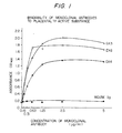
- Figerie 1 shows the strength of binding of the monoclonal antibody of this invention to a human placenta TF active substance
- Figure 2 shows the differences in the antigen recognizing sites (epitopes) of three types of the monoclonal antibodies of this invention
- FIG. 3 shows effects of the monoclonal antibodies of this invention on the activities of tissue factor
- Figure 4 is a calibration curve for assaying human TF active substances
- Figure 5 is a summary of the relation of various kidney deseases to contents of human TF active substances in human urine;
- Figure 6 shows the relation between the activities of urine TF active substance and the pH
- Figure 7 shows the outside of the overall composition of the protein of this invention.
- Example 1 Obtaining hybridoma (fused cells) producing a monoclonal antibody binding to a human TF active substance
- a human TF apoprotein purified from a human placenta extract (to be referred to as antigen 1) and a fragment on the N-terminus side not containing a domain binding to a phospholipid (to be abbreviated as antigen 2) obtained by decomposing human placenta-derived TF apoprotein with CNBr were each immunized with (four weeks of age) of female Balb/C mice (total three mice) four times at intervals of 14 days.
- 50 micrograms of the antigen was dissolved in physiological saline and the solution was mixed with an equal amount of Freund complete adjuvant, and the emulsion was administered peritoneally.
- mice In the 2nd and third immunization, 50 micrograms of the antigen was mixed with Freund incomplete adjuvant, and administered intraperitoneally. In the final immunization (fourth immunization), 30 micrograms of the antigen was additionally administered through the mice. Three days after the final immunization, the spleen cells of the immunized mice were used for cell fusion.
- the extract spine cells of the mice and the myeloma cells (P3U1) of the same strain of mouse were mixed at a rate of about 5:1, and subjected to cell fusion by using 50% polyethylene glycol 1540 by the method of Köhler and Milstein.
- the cells after fusion were suspended in RPMI 1640 containing 10% of fetal serum so that the cell concentration became 1 x 10⁵ cells/ml, and poured onto a 96-well microplate at a rate of 100 microliters per well.
- the hybridomas (fused cells ) were cultivated in a CO2 incubator (5 % CO2 37 °C).
- the culture medium was exchanged by using HAT medium containing hypoxanthine, aminoputerine and thymidine (HAT medium), and the cells were proliferated in the HAT medium. Hydridomas composed of the spleen cells and the mouse myeloma cells were screened.
- the class of IgG of each clone purified from mouise ascites was determined by the Ouchterlony method.
- the purified monoclonal antibodies (GX3, GX4 and EX6) were used each in lmmunobeads (made by Bio-Rad Co.) and labelled with 125 I to prepare antibody solutions.
- 125 I-labelled monoclonal antibody was added to the wells of the plate so as to have various concentrations in the range of 0.1 to 5 micrograms/ml, and reacted at 37°C for 2 hours. Then the wells were washed three times by adding 10 mM phosphate buffer (pH 7.2) containing 0.05 % Tween 20 at a rate of 100 microliters/well.
- the wells were cut out from the plate, put into a plastic test tube, and 125 I radioactivity was measured by a gamma counter (radioactivity bound to the solid antigen (bound (cpm)). At the same time, the radioactivity of the monoclonal antibody solution for addition to the wells was measured at the same time (the total radioactivity of the added antibody: total (cpm)).
- Human TF apoprotein in a concentration of 5 micrograms/ml extracted and purified from human placenta was caused to be adsorbed to a microtiter plate and after blocking with 1% BSA, was reacted with a monoclonal antibody solution 0.16 - 5.0 micrograms/ml) diluted to a suitable concentration. Then, an anti-mouse antibody labelled with alkaline phosphatase was added
- Human placenta-derived TF apoprotein was adsorbed to a microtiter plate in a concentration of 5 micrograms/ml, and after blocking with 1% BSA, each of various monoclonal antibody solutions (0.16 to 5.0 micrograms/ml) and a EX6 antibody labelled with peroxidase were simultaneously reacted with the antigen.
- HCOOH was added to 40 micrograms of human TF apoprotein to form a 70% HCOOH solution.
- CNBr powder was added to this solution and dissolved, and the reaction was performed at room temperature for 18 hours. Thereafter, HCOOH was dried up.
- the protein was dissolved by adding 40 microliters of H2O, and then an amount equivalent to 2 micrograms was reduced in the presence of 2-mercaptoethanol.
- SDS-polyacrylamide electrophoresis was carried out (fragments having a molecular weigth of 31,000 and 27,000).
- a TF apoprotein (M.W. 58,000) not treated with CNBr was similarly reduced, and electrophoresis was carried out.
- the protein in the gel was electrically transferred to a nitrocellulose membrane by using a blotting device.
- the nitrocellulose membrane was blocked with TBS containing 3 % of gelatin (20 mM Tris solution-0.15M NaCl, pH 7.4), and a 1% gelatin-TBS solution containing each of various monoclonal antibodies (GX3, GX4 and EX6) in a concentration of 2 micrograms/ml was reacted overnight at room temperature with the nitrocellulose membrane.
- the nitrocellulose membrane was washed with 0.05 % of Tween 20-TBS three times, and the reaction was carried out with a 1% gelatin-TBS solution of a peroxidase-labelled anti-mouse Ig antibody at room temperature for 4 hours at room temperature. After washing, a 4-chloro-1-naphthal substrate solution was added to color the enzyme-labelled antibody bound to the nitrocellulose membrane The protein bound to the monoclonal antibody could be detected as a deep blue band.
- the three monoclonal antibodies obtained had the binding properties given in Table 4.
- the monoclonal antibodies did not affect the activity of inducing coagulation of human placenta-derived TF.
- Monoclonal antibody GX3 was diluted with PBS (10 mM phosphate buffer - 0.15M NaCl pH7.4) so as to provide a concentration of 20 micrograms/ml, and 100 microliters of the resulting solution was added to a well of a microtiter plate. It was allowed to stand overnight to induce adsorption of the antibody to the solid phase. PBS containing 1% BSA was added at a rate of 150 microliters/well. Successively, the plate was allowed to stand at room temperature and allowed to stand for 2 hours. The plate was washed with PBS (washing buffer (containing 0.05% Tween 20 and 0.1% BSA.
- human placenta-derived TF apoprotein was diluted with the washing buffer in a concentration of 25 ng/ml, 50 ng/ml and 100 ng/ml, and human urine was diluted to 80-fold and 40-fold and the diluted solutions were added in an amount of 100 microliters/well and reacted at 37 °C for 1 hour.
- the plate was washed with the washing buffer three times.
- the peroxidase-labelled monoclonal antibody GX4 was diluted to a concentration of 300 ng/ml, and added in an amount of 100 microliters/well, and reacted at 37 °C for 1 hour. After washing with the washing buffer three times, and the substrate solution (ABTS) in an amount of 100 microliters/well, and the absorbance at a wavelength of 415 nm was measured.
- ABTS substrate solution
- Figure 4 shows a calibration curve prepared by using human placenta-derived tissue factor apoprotein.
- concentration of the human TF (the axis of abscissas) and the absorbance (the axis of ordinates) are in a linear relation, and by using this callibration curve, a human TF active substance in a solution (human urine) can be determined.
- Figure 5 summarizes the results of measurements.
- the human urine in a healthy person contained human TF in a concentration of 2 to 3 micrograms/ml.
- TF was defected in a much higher concentration than in normal healthy persons as 10 to 25 micrograms/ml.
- the column was washed with 200 ml of a washing buffer A (containing 20 mM Tris-HCl, 0.5M NaCl pH 7.6, 0.05% Tween 20 and 20 units/ml of transyrol), and then with 150 ml of a washing buffer (20 mM Tris-HCl, 0.10 M NaCl, pH 7.6, and 20 units/ml of transyrol. It was confirmed that the washing fraction had an absorbance (A280)of 0 at a wavelength of 280 nm. Subsequently, 3M NaSCN solution (pH 7.0) was passed through the column at a flow rate of 20 ml/hr, and the TF active substance in the urine which bound to the antibody column was eluted.
- a washing buffer A containing 20 mM Tris-HCl, 0.5M NaCl pH 7.6, 0.05% Tween 20 and 20 units/ml of transyrol. It was confirmed that the washing fraction had an absorbance (A280)of 0 at a wavelength
- the NaSCN salt was removed by dialysis, and by using a protein assay reagent(dye reagent made by Bio-Rad Co., Ltd.), the amount of protein determined by colorimetry. It was 591 micrograms.
- the column was washed by 6M guanidine-HCl (pH 3.1), and preserved by passing 200 ml of the washing buffer B.
- the above purification operations excepting the measurement of the concentration of the protein were all performed at 4 °C.
- Human fresh plasma 500 microliters was added 0.1M citrate buffer (pH 3.0), or 0.05M Tris buffer (pH 9.0) or 0.1M soium hydrogen carbonate-NaOH buffer (pH 12.5) to adjust the pH to various values.
- 0.1M citrate buffer pH 3.0
- 0.05M Tris buffer pH 9.0
- 0.1M soium hydrogen carbonate-NaOH buffer pH 12.5
- the use of this invention permits direct assaying of a human TF active substance. Even a solution of a human TF active substance (for example, in urine) can be determined within s short period of time quantitativelyu without undergoing influences by other foreign materials. Furthermore, by assaying a human TF active substance in human urine, a kidney disease can be diagnozed, and the degree of damage to the kidney tissue can be grasped.
- a human TF active substance for example, in urine
- a kidney disease can be diagnozed, and the degree of damage to the kidney tissue can be grasped.
Landscapes
- Chemical & Material Sciences (AREA)
- Health & Medical Sciences (AREA)
- Organic Chemistry (AREA)
- Life Sciences & Earth Sciences (AREA)
- Medicinal Chemistry (AREA)
- Biophysics (AREA)
- General Health & Medical Sciences (AREA)
- Genetics & Genomics (AREA)
- Biochemistry (AREA)
- Molecular Biology (AREA)
- Proteomics, Peptides & Aminoacids (AREA)
- Immunology (AREA)
- Toxicology (AREA)
- Zoology (AREA)
- Gastroenterology & Hepatology (AREA)
- Preparation Of Compounds By Using Micro-Organisms (AREA)
- Micro-Organisms Or Cultivation Processes Thereof (AREA)
Applications Claiming Priority (8)
| Application Number | Priority Date | Filing Date | Title |
|---|---|---|---|
| JP1022634A JP2779193B2 (ja) | 1989-02-02 | 1989-02-02 | 抗ヒト組織因子モノクローナル抗体 |
| JP22634/89 | 1989-02-02 | ||
| JP3622889A JPH02216054A (ja) | 1989-02-17 | 1989-02-17 | ヒト組織因子の測定方法,試薬およびキット |
| JP36228/89 | 1989-02-17 | ||
| JP9645689A JPH02275359A (ja) | 1989-04-18 | 1989-04-18 | 腎疾患の診断方法、診断試薬およびキット |
| JP96456/89 | 1989-04-18 | ||
| JP31460289 | 1989-12-04 | ||
| JP314602/89 | 1989-12-04 |
Publications (2)
| Publication Number | Publication Date |
|---|---|
| EP0417298A1 true EP0417298A1 (de) | 1991-03-20 |
| EP0417298A4 EP0417298A4 (en) | 1993-03-10 |
Family
ID=27457805
Family Applications (1)
| Application Number | Title | Priority Date | Filing Date |
|---|---|---|---|
| EP19900902686 Withdrawn EP0417298A4 (en) | 1989-02-02 | 1990-02-02 | Detection of human tissue factor activator |
Country Status (4)
| Country | Link |
|---|---|
| EP (1) | EP0417298A4 (de) |
| AU (1) | AU631603B2 (de) |
| CA (1) | CA2026666A1 (de) |
| WO (1) | WO1990008956A1 (de) |
Cited By (3)
| Publication number | Priority date | Publication date | Assignee | Title |
|---|---|---|---|---|
| WO2004064870A3 (en) * | 2003-01-22 | 2005-04-28 | Novo Nordisk As | Radiolabelled tissue factor binding agent and the use thereof |
| GB2484897A (en) * | 2010-10-19 | 2012-05-02 | Term Diagnostics Ltd C | Detection of urinary tissue factor (uTF) |
| CN109883932A (zh) * | 2019-02-18 | 2019-06-14 | 武汉伊莱瑞特生物科技股份有限公司 | 流式抗体及其制备方法和滴定方法 |
Family Cites Families (2)
| Publication number | Priority date | Publication date | Assignee | Title |
|---|---|---|---|---|
| ATE120645T1 (de) * | 1986-11-04 | 1995-04-15 | Genentech Inc | Verfahren und therapeutische zubereitungen für die behandlung von gerinnungsstörungen. |
| IE81149B1 (en) * | 1987-02-12 | 2000-05-03 | Genentech Inc | Methods and deoxyribonucleic acid for the preparation of tissue factor protein |
-
1990
- 1990-02-02 EP EP19900902686 patent/EP0417298A4/en not_active Withdrawn
- 1990-02-02 WO PCT/JP1990/000127 patent/WO1990008956A1/ja not_active Ceased
- 1990-02-02 AU AU50347/90A patent/AU631603B2/en not_active Ceased
- 1990-02-02 CA CA 2026666 patent/CA2026666A1/en not_active Abandoned
Non-Patent Citations (4)
| Title |
|---|
| CLINICAL BIOCHEMISTRY vol. 21, no. 5, 1988, NEW YORK NY USA pages 311 - 314 W. BÜRGI ET AL. 'One-step sandwich enzyme immunoassay for insulin using monoclonal antibodies.' * |
| LABORATORY RESEARCH vol. 53, no. 2, 1985, pages 156 - 165 R.C. WIGGENS ET AL. 'Procoagulant activity in glomeruli and urine of rabbits with nephrotoxic nephritis.' * |
| See also references of WO9008956A1 * |
| THROMBOSIS RESEARCH vol. 13, no. 3, 1978, WASHINTON DC USA pages 311 - 324 M. MATSUDA ET AL. 'Purification and characterization of procoagulant in human urine.' * |
Cited By (4)
| Publication number | Priority date | Publication date | Assignee | Title |
|---|---|---|---|---|
| WO2004064870A3 (en) * | 2003-01-22 | 2005-04-28 | Novo Nordisk As | Radiolabelled tissue factor binding agent and the use thereof |
| GB2484897A (en) * | 2010-10-19 | 2012-05-02 | Term Diagnostics Ltd C | Detection of urinary tissue factor (uTF) |
| CN109883932A (zh) * | 2019-02-18 | 2019-06-14 | 武汉伊莱瑞特生物科技股份有限公司 | 流式抗体及其制备方法和滴定方法 |
| CN109883932B (zh) * | 2019-02-18 | 2021-08-10 | 武汉伊莱瑞特生物科技股份有限公司 | 流式抗体及其制备方法和滴定方法 |
Also Published As
| Publication number | Publication date |
|---|---|
| CA2026666A1 (en) | 1990-08-03 |
| WO1990008956A1 (en) | 1990-08-09 |
| AU631603B2 (en) | 1992-12-03 |
| AU5034790A (en) | 1990-08-24 |
| EP0417298A4 (en) | 1993-03-10 |
Similar Documents
| Publication | Publication Date | Title |
|---|---|---|
| EP0683234B2 (de) | Antikörper gegen beta-amyloid oder derivative davon und seine verwendung | |
| US5955317A (en) | Antibodies to β-amyloids or their derivatives and use thereof | |
| EP0203587A2 (de) | Ras-Oncogene-Peptide und Antikörper | |
| AU707232B2 (en) | Antibody to aminoterminal propeptide of type 1 procollagen, and assay method using it | |
| EP0972781B1 (de) | Protein S polypeptide und deren Verwendungen | |
| CA2155793C (en) | Monoclonal antibiodies for selective immunological determination of high molecular weight, intact laminin forms in body fluids | |
| KR960013460B1 (ko) | 안티-pci 모노클로날 항체 | |
| US6008325A (en) | Antibody to aminoterminal propeptide of type 1 procollagen | |
| Bützow et al. | Monoclonal antibodies reacting with placental protein 5: use in radioimmunoassay, Western blot analysis, and immunohistochemistry | |
| EP0331100A1 (de) | Antiendothelin-Antikörper und ihre Verwendung | |
| KR960002740B1 (ko) | 안티-트롬빈-결합물질 모노클로날 항체, 이를 생산하는 하이브리도마 및 모노클로날 항체를 이용한 트롬빈-결합물질의 정제법 및 측정법 | |
| EP0417298A1 (de) | Nachweis von menschlichem gewebefaktoraktivator | |
| Scott et al. | The preparation of plasma fibronectin antigen and antiserum | |
| US5679583A (en) | Monoclonal antibodies for the selective immunological determination of intact procollagen peptide (type III) and procollagen (type III) in body fluids | |
| Zhang et al. | Purification, characterization, and cellular localization of the 100-kDa human placental GTPase-activating protein. | |
| US5430129A (en) | Purified, native dystrophin | |
| EP0345811B1 (de) | Gegen menschliches Fibrinopepid A spezifische monoklonale Antikörper | |
| EP0401006B1 (de) | Antikörper gegen humanes ANP und Verfahren zur immunologischen Bestimmung von humanem ANP | |
| AU2323092A (en) | Proteins s polypeptides and uses thereof | |
| JPH03163095A (ja) | ヒト神経成長因子の部分ペプチド、抗体およびその用途 | |
| EP0328939B1 (de) | Verfahren, Reagens, Satz und monoklonale Antikörper zur Diagnose von Krebs in menschlichen Verdauungsorganen durch Bestimmung von saurer Glutathion-S-Transferase | |
| IE911285A1 (en) | Monoclonal antibodies against PP4, processes for the¹preparation thereof and the use thereof | |
| IE881290L (en) | Monoclonal antibodies for the selective immunological¹determination of intact procollagen peptide (Type III) and¹procollagen (Type III) in body fluids | |
| JP3754611B2 (ja) | ヒト老化マーカー及びストレスマーカーの検定方法 | |
| EP0312645B1 (de) | Antigene Peptide des Komplementfaktors C3a, Verwendung der Peptide, Zell-Linien, Antikörper gegen die Peptide und Verwendung der Antikörper |
Legal Events
| Date | Code | Title | Description |
|---|---|---|---|
| PUAI | Public reference made under article 153(3) epc to a published international application that has entered the european phase |
Free format text: ORIGINAL CODE: 0009012 |
|
| 17P | Request for examination filed |
Effective date: 19901002 |
|
| AK | Designated contracting states |
Kind code of ref document: A1 Designated state(s): BE CH DE DK FR GB LI SE |
|
| A4 | Supplementary search report drawn up and despatched |
Effective date: 19930115 |
|
| AK | Designated contracting states |
Kind code of ref document: A4 Designated state(s): BE CH DE DK FR GB LI SE |
|
| STAA | Information on the status of an ep patent application or granted ep patent |
Free format text: STATUS: THE APPLICATION HAS BEEN WITHDRAWN |
|
| 18W | Application withdrawn |
Withdrawal date: 19940422 |