EP0728009B1 - Verwendung von einem mittel, das tyrosinphosphorylierung moduliert, zur modulation der durchlässigkeit einer physiologischen sperrschicht - Google Patents
Verwendung von einem mittel, das tyrosinphosphorylierung moduliert, zur modulation der durchlässigkeit einer physiologischen sperrschicht Download PDFInfo
- Publication number
- EP0728009B1 EP0728009B1 EP95901509A EP95901509A EP0728009B1 EP 0728009 B1 EP0728009 B1 EP 0728009B1 EP 95901509 A EP95901509 A EP 95901509A EP 95901509 A EP95901509 A EP 95901509A EP 0728009 B1 EP0728009 B1 EP 0728009B1
- Authority
- EP
- European Patent Office
- Prior art keywords
- cells
- catenin
- tyrosine
- agent
- brain
- Prior art date
- Legal status (The legal status is an assumption and is not a legal conclusion. Google has not performed a legal analysis and makes no representation as to the accuracy of the status listed.)
- Expired - Lifetime
Links
- OUYCCCASQSFEME-QMMMGPOBSA-N L-tyrosine Chemical compound OC(=O)[C@@H](N)CC1=CC=C(O)C=C1 OUYCCCASQSFEME-QMMMGPOBSA-N 0.000 title claims abstract description 127
- OUYCCCASQSFEME-UHFFFAOYSA-N tyrosine Natural products OC(=O)C(N)CC1=CC=C(O)C=C1 OUYCCCASQSFEME-UHFFFAOYSA-N 0.000 title claims abstract description 127
- 230000026731 phosphorylation Effects 0.000 title claims abstract description 91
- 238000006366 phosphorylation reaction Methods 0.000 title claims abstract description 91
- 230000035699 permeability Effects 0.000 title claims abstract description 56
- 230000004888 barrier function Effects 0.000 title claims abstract description 32
- 230000008499 blood brain barrier function Effects 0.000 claims abstract description 40
- 210000001218 blood-brain barrier Anatomy 0.000 claims abstract description 38
- 239000003814 drug Substances 0.000 claims abstract description 35
- 239000003795 chemical substances by application Substances 0.000 claims abstract description 29
- 230000000694 effects Effects 0.000 claims abstract description 27
- 230000030609 dephosphorylation Effects 0.000 claims abstract description 19
- 229940079593 drug Drugs 0.000 claims abstract description 18
- 230000003247 decreasing effect Effects 0.000 claims abstract description 7
- 102000000905 Cadherin Human genes 0.000 claims description 84
- 108050007957 Cadherin Proteins 0.000 claims description 84
- BQVCCPGCDUSGOE-UHFFFAOYSA-N phenylarsine oxide Chemical group O=[As]C1=CC=CC=C1 BQVCCPGCDUSGOE-UHFFFAOYSA-N 0.000 claims description 84
- 210000001578 tight junction Anatomy 0.000 claims description 59
- 102000015735 Beta-catenin Human genes 0.000 claims description 57
- 108060000903 Beta-catenin Proteins 0.000 claims description 57
- PZYFJWVGRGEWGO-UHFFFAOYSA-N trisodium;hydrogen peroxide;trioxido(oxo)vanadium Chemical compound [Na+].[Na+].[Na+].OO.OO.OO.[O-][V]([O-])([O-])=O PZYFJWVGRGEWGO-UHFFFAOYSA-N 0.000 claims description 55
- 102000016362 Catenins Human genes 0.000 claims description 50
- 108010067316 Catenins Proteins 0.000 claims description 50
- 210000004556 brain Anatomy 0.000 claims description 48
- 102000004022 Protein-Tyrosine Kinases Human genes 0.000 claims description 20
- 108090000412 Protein-Tyrosine Kinases Proteins 0.000 claims description 20
- 230000009822 protein phosphorylation Effects 0.000 claims description 15
- 238000002360 preparation method Methods 0.000 claims description 13
- 206010028980 Neoplasm Diseases 0.000 claims description 12
- 230000002441 reversible effect Effects 0.000 claims description 7
- 238000006209 dephosphorylation reaction Methods 0.000 claims description 6
- 239000000203 mixture Substances 0.000 claims description 6
- 101710098414 Tyrosine-protein phosphatase Proteins 0.000 claims description 4
- 208000003174 Brain Neoplasms Diseases 0.000 claims description 3
- 206010048962 Brain oedema Diseases 0.000 claims description 3
- 108050000637 N-cadherin Proteins 0.000 claims description 3
- 230000028993 immune response Effects 0.000 claims description 3
- 210000000265 leukocyte Anatomy 0.000 claims description 3
- 201000006417 multiple sclerosis Diseases 0.000 claims description 3
- 206010030124 Oedema peripheral Diseases 0.000 claims description 2
- 206010037423 Pulmonary oedema Diseases 0.000 claims description 2
- 230000000903 blocking effect Effects 0.000 claims description 2
- 201000011510 cancer Diseases 0.000 claims description 2
- 230000001988 toxicity Effects 0.000 claims description 2
- 231100000419 toxicity Toxicity 0.000 claims description 2
- -1 ZO-1 Proteins 0.000 claims 2
- 206010027476 Metastases Diseases 0.000 claims 1
- 230000009401 metastasis Effects 0.000 claims 1
- 150000003839 salts Chemical class 0.000 claims 1
- 229910052720 vanadium Inorganic materials 0.000 claims 1
- LEONUFNNVUYDNQ-UHFFFAOYSA-N vanadium atom Chemical group [V] LEONUFNNVUYDNQ-UHFFFAOYSA-N 0.000 claims 1
- 108090000623 proteins and genes Proteins 0.000 abstract description 113
- 102000004169 proteins and genes Human genes 0.000 abstract description 108
- 150000001875 compounds Chemical class 0.000 abstract description 6
- 210000003169 central nervous system Anatomy 0.000 abstract 2
- 210000004027 cell Anatomy 0.000 description 215
- 235000018102 proteins Nutrition 0.000 description 107
- 102100031426 Ras GTPase-activating protein 1 Human genes 0.000 description 54
- 101800001622 120 kDa surface-exposed protein Proteins 0.000 description 52
- 239000012133 immunoprecipitate Substances 0.000 description 48
- 101000617805 Homo sapiens Staphylococcal nuclease domain-containing protein 1 Proteins 0.000 description 46
- 101000830894 Homo sapiens Targeting protein for Xklp2 Proteins 0.000 description 46
- 101000942603 Schizosaccharomyces pombe (strain 972 / ATCC 24843) Condensin complex subunit 3 Proteins 0.000 description 46
- 102100021996 Staphylococcal nuclease domain-containing protein 1 Human genes 0.000 description 46
- 101000963191 Xenopus laevis Maternal DNA replication licensing factor mcm3 Proteins 0.000 description 46
- 210000002889 endothelial cell Anatomy 0.000 description 46
- 108090000020 Alpha-catenin Proteins 0.000 description 31
- 102000003730 Alpha-catenin Human genes 0.000 description 31
- 230000007423 decrease Effects 0.000 description 31
- 102000000591 Tight Junction Proteins Human genes 0.000 description 28
- 108010002321 Tight Junction Proteins Proteins 0.000 description 28
- LOKCTEFSRHRXRJ-UHFFFAOYSA-I dipotassium trisodium dihydrogen phosphate hydrogen phosphate dichloride Chemical compound P(=O)(O)(O)[O-].[K+].P(=O)(O)([O-])[O-].[Na+].[Na+].[Cl-].[K+].[Cl-].[Na+] LOKCTEFSRHRXRJ-UHFFFAOYSA-I 0.000 description 27
- 239000002953 phosphate buffered saline Substances 0.000 description 27
- 239000000872 buffer Substances 0.000 description 26
- 238000003119 immunoblot Methods 0.000 description 21
- DCWXELXMIBXGTH-UHFFFAOYSA-N phosphotyrosine Chemical compound OC(=O)C(N)CC1=CC=C(OP(O)(O)=O)C=C1 DCWXELXMIBXGTH-UHFFFAOYSA-N 0.000 description 21
- LSGOVYNHVSXFFJ-UHFFFAOYSA-N vanadate(3-) Chemical compound [O-][V]([O-])([O-])=O LSGOVYNHVSXFFJ-UHFFFAOYSA-N 0.000 description 19
- 102000004160 Phosphoric Monoester Hydrolases Human genes 0.000 description 18
- 108090000608 Phosphoric Monoester Hydrolases Proteins 0.000 description 18
- 208000006011 Stroke Diseases 0.000 description 18
- 238000001114 immunoprecipitation Methods 0.000 description 18
- 210000001519 tissue Anatomy 0.000 description 18
- 239000000463 material Substances 0.000 description 17
- 102000054078 gamma Catenin Human genes 0.000 description 16
- 108010084448 gamma Catenin Proteins 0.000 description 16
- 210000002966 serum Anatomy 0.000 description 16
- 238000002415 sodium dodecyl sulfate polyacrylamide gel electrophoresis Methods 0.000 description 16
- MCAHMSDENAOJFZ-UHFFFAOYSA-N Herbimycin A Natural products N1C(=O)C(C)=CC=CC(OC)C(OC(N)=O)C(C)=CC(C)C(OC)C(OC)CC(C)C(OC)C2=CC(=O)C=C1C2=O MCAHMSDENAOJFZ-UHFFFAOYSA-N 0.000 description 15
- 210000002867 adherens junction Anatomy 0.000 description 15
- 230000000875 corresponding effect Effects 0.000 description 15
- 210000002919 epithelial cell Anatomy 0.000 description 15
- MCAHMSDENAOJFZ-BVXDHVRPSA-N herbimycin Chemical compound N1C(=O)\C(C)=C\C=C/[C@H](OC)[C@@H](OC(N)=O)\C(C)=C\[C@H](C)[C@@H](OC)[C@@H](OC)C[C@H](C)[C@@H](OC)C2=CC(=O)C=C1C2=O MCAHMSDENAOJFZ-BVXDHVRPSA-N 0.000 description 15
- 238000002474 experimental method Methods 0.000 description 14
- 210000003657 middle cerebral artery Anatomy 0.000 description 14
- OKKJLVBELUTLKV-UHFFFAOYSA-N Methanol Chemical compound OC OKKJLVBELUTLKV-UHFFFAOYSA-N 0.000 description 12
- 241000700159 Rattus Species 0.000 description 12
- 238000000034 method Methods 0.000 description 12
- DBMJMQXJHONAFJ-UHFFFAOYSA-M Sodium laurylsulphate Chemical compound [Na+].CCCCCCCCCCCCOS([O-])(=O)=O DBMJMQXJHONAFJ-UHFFFAOYSA-M 0.000 description 11
- 238000007792 addition Methods 0.000 description 11
- 238000004458 analytical method Methods 0.000 description 11
- 244000309466 calf Species 0.000 description 11
- 108010014223 Armadillo Domain Proteins Proteins 0.000 description 10
- CURLTUGMZLYLDI-UHFFFAOYSA-N Carbon dioxide Chemical compound O=C=O CURLTUGMZLYLDI-UHFFFAOYSA-N 0.000 description 10
- 210000004204 blood vessel Anatomy 0.000 description 10
- 238000002372 labelling Methods 0.000 description 10
- 108010088751 Albumins Proteins 0.000 description 9
- 102000009027 Albumins Human genes 0.000 description 9
- 241000283690 Bos taurus Species 0.000 description 9
- 206010030113 Oedema Diseases 0.000 description 9
- 229920004890 Triton X-100 Polymers 0.000 description 9
- ATNOAWAQFYGAOY-GPTZEZBUSA-J [Na+].[Na+].[Na+].[Na+].Cc1cc(ccc1\N=N\c1ccc2c(cc(c(N)c2c1O)S([O-])(=O)=O)S([O-])(=O)=O)-c1ccc(\N=N\c2ccc3c(cc(c(N)c3c2O)S([O-])(=O)=O)S([O-])(=O)=O)c(C)c1 Chemical compound [Na+].[Na+].[Na+].[Na+].Cc1cc(ccc1\N=N\c1ccc2c(cc(c(N)c2c1O)S([O-])(=O)=O)S([O-])(=O)=O)-c1ccc(\N=N\c2ccc3c(cc(c(N)c3c2O)S([O-])(=O)=O)S([O-])(=O)=O)c(C)c1 ATNOAWAQFYGAOY-GPTZEZBUSA-J 0.000 description 9
- 229960003699 evans blue Drugs 0.000 description 9
- 239000003112 inhibitor Substances 0.000 description 9
- 210000004088 microvessel Anatomy 0.000 description 9
- 239000000758 substrate Substances 0.000 description 9
- 238000005406 washing Methods 0.000 description 9
- 108010058765 Oncogene Protein pp60(v-src) Proteins 0.000 description 8
- 239000013504 Triton X-100 Substances 0.000 description 8
- 230000009471 action Effects 0.000 description 8
- 150000001413 amino acids Chemical group 0.000 description 8
- 230000001413 cellular effect Effects 0.000 description 8
- WQABCVAJNWAXTE-UHFFFAOYSA-N dimercaprol Chemical compound OCC(S)CS WQABCVAJNWAXTE-UHFFFAOYSA-N 0.000 description 8
- 239000006166 lysate Substances 0.000 description 8
- 108090000765 processed proteins & peptides Proteins 0.000 description 8
- 230000009257 reactivity Effects 0.000 description 8
- 230000004044 response Effects 0.000 description 8
- 238000010186 staining Methods 0.000 description 8
- IVOMOUWHDPKRLL-KQYNXXCUSA-N Cyclic adenosine monophosphate Chemical compound C([C@H]1O2)OP(O)(=O)O[C@H]1[C@@H](O)[C@@H]2N1C(N=CN=C2N)=C2N=C1 IVOMOUWHDPKRLL-KQYNXXCUSA-N 0.000 description 7
- FFEARJCKVFRZRR-BYPYZUCNSA-N L-methionine Chemical compound CSCC[C@H](N)C(O)=O FFEARJCKVFRZRR-BYPYZUCNSA-N 0.000 description 7
- 229920002684 Sepharose Polymers 0.000 description 7
- IVOMOUWHDPKRLL-UHFFFAOYSA-N UNPD107823 Natural products O1C2COP(O)(=O)OC2C(O)C1N1C(N=CN=C2N)=C2N=C1 IVOMOUWHDPKRLL-UHFFFAOYSA-N 0.000 description 7
- 229940095074 cyclic amp Drugs 0.000 description 7
- 230000001419 dependent effect Effects 0.000 description 7
- MHMNJMPURVTYEJ-UHFFFAOYSA-N fluorescein-5-isothiocyanate Chemical compound O1C(=O)C2=CC(N=C=S)=CC=C2C21C1=CC=C(O)C=C1OC1=CC(O)=CC=C21 MHMNJMPURVTYEJ-UHFFFAOYSA-N 0.000 description 7
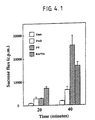
- 230000004907 flux Effects 0.000 description 7
- 238000011534 incubation Methods 0.000 description 7
- 230000005764 inhibitory process Effects 0.000 description 7
- 230000003993 interaction Effects 0.000 description 7
- 210000004692 intercellular junction Anatomy 0.000 description 7
- 239000002609 medium Substances 0.000 description 7
- 239000000047 product Substances 0.000 description 7
- 102000016904 Armadillo Domain Proteins Human genes 0.000 description 6
- LFQSCWFLJHTTHZ-UHFFFAOYSA-N Ethanol Chemical compound CCO LFQSCWFLJHTTHZ-UHFFFAOYSA-N 0.000 description 6
- 230000001086 cytosolic effect Effects 0.000 description 6
- 238000001378 electrochemiluminescence detection Methods 0.000 description 6
- 238000010438 heat treatment Methods 0.000 description 6
- JKMHFZQWWAIEOD-UHFFFAOYSA-N 2-[4-(2-hydroxyethyl)piperazin-1-yl]ethanesulfonic acid Chemical compound OCC[NH+]1CCN(CCS([O-])(=O)=O)CC1 JKMHFZQWWAIEOD-UHFFFAOYSA-N 0.000 description 5
- 241000282465 Canis Species 0.000 description 5
- 241000289632 Dasypodidae Species 0.000 description 5
- 241000255581 Drosophila <fruit fly, genus> Species 0.000 description 5
- KCXVZYZYPLLWCC-UHFFFAOYSA-N EDTA Chemical compound OC(=O)CN(CC(O)=O)CCN(CC(O)=O)CC(O)=O KCXVZYZYPLLWCC-UHFFFAOYSA-N 0.000 description 5
- 241000283973 Oryctolagus cuniculus Species 0.000 description 5
- 239000012722 SDS sample buffer Substances 0.000 description 5
- 101150001535 SRC gene Proteins 0.000 description 5
- 229930006000 Sucrose Natural products 0.000 description 5
- CZMRCDWAGMRECN-UGDNZRGBSA-N Sucrose Chemical compound O[C@H]1[C@H](O)[C@@H](CO)O[C@@]1(CO)O[C@@H]1[C@H](O)[C@@H](O)[C@H](O)[C@@H](CO)O1 CZMRCDWAGMRECN-UGDNZRGBSA-N 0.000 description 5
- 230000033228 biological regulation Effects 0.000 description 5
- 210000004292 cytoskeleton Anatomy 0.000 description 5
- 238000001514 detection method Methods 0.000 description 5
- 210000002950 fibroblast Anatomy 0.000 description 5
- 230000006870 function Effects 0.000 description 5
- 239000000499 gel Substances 0.000 description 5
- 239000001963 growth medium Substances 0.000 description 5
- 238000005259 measurement Methods 0.000 description 5
- 230000001404 mediated effect Effects 0.000 description 5
- 230000005012 migration Effects 0.000 description 5
- 238000013508 migration Methods 0.000 description 5
- 230000002093 peripheral effect Effects 0.000 description 5
- 239000005720 sucrose Substances 0.000 description 5
- 229940121358 tyrosine kinase inhibitor Drugs 0.000 description 5
- 108700001666 APC Genes Proteins 0.000 description 4
- 102000007469 Actins Human genes 0.000 description 4
- 108010085238 Actins Proteins 0.000 description 4
- 108091003079 Bovine Serum Albumin Proteins 0.000 description 4
- 102000004266 Collagen Type IV Human genes 0.000 description 4
- 108010042086 Collagen Type IV Proteins 0.000 description 4
- DHMQDGOQFOQNFH-UHFFFAOYSA-N Glycine Chemical compound NCC(O)=O DHMQDGOQFOQNFH-UHFFFAOYSA-N 0.000 description 4
- 108010001336 Horseradish Peroxidase Proteins 0.000 description 4
- TWRXJAOTZQYOKJ-UHFFFAOYSA-L Magnesium chloride Chemical compound [Mg+2].[Cl-].[Cl-] TWRXJAOTZQYOKJ-UHFFFAOYSA-L 0.000 description 4
- CSNNHWWHGAXBCP-UHFFFAOYSA-L Magnesium sulfate Chemical compound [Mg+2].[O-][S+2]([O-])([O-])[O-] CSNNHWWHGAXBCP-UHFFFAOYSA-L 0.000 description 4
- 102000007474 Multiprotein Complexes Human genes 0.000 description 4
- 108010085220 Multiprotein Complexes Proteins 0.000 description 4
- 108091000080 Phosphotransferase Proteins 0.000 description 4
- FAPWRFPIFSIZLT-UHFFFAOYSA-M Sodium chloride Chemical compound [Na+].[Cl-] FAPWRFPIFSIZLT-UHFFFAOYSA-M 0.000 description 4
- 235000001014 amino acid Nutrition 0.000 description 4
- 210000004369 blood Anatomy 0.000 description 4
- 239000008280 blood Substances 0.000 description 4
- 229940098773 bovine serum albumin Drugs 0.000 description 4
- 210000004781 brain capillary Anatomy 0.000 description 4
- 238000012512 characterization method Methods 0.000 description 4
- 239000003153 chemical reaction reagent Substances 0.000 description 4
- 230000006378 damage Effects 0.000 description 4
- KXGVEGMKQFWNSR-LLQZFEROSA-N deoxycholic acid Chemical compound C([C@H]1CC2)[C@H](O)CC[C@]1(C)[C@@H]1[C@@H]2[C@@H]2CC[C@H]([C@@H](CCC(O)=O)C)[C@@]2(C)[C@@H](O)C1 KXGVEGMKQFWNSR-LLQZFEROSA-N 0.000 description 4
- 229940042399 direct acting antivirals protease inhibitors Drugs 0.000 description 4
- 238000003365 immunocytochemistry Methods 0.000 description 4
- 210000003734 kidney Anatomy 0.000 description 4
- 239000003446 ligand Substances 0.000 description 4
- 230000004807 localization Effects 0.000 description 4
- 239000012139 lysis buffer Substances 0.000 description 4
- 210000003205 muscle Anatomy 0.000 description 4
- 230000037361 pathway Effects 0.000 description 4
- 239000000137 peptide hydrolase inhibitor Substances 0.000 description 4
- YBYRMVIVWMBXKQ-UHFFFAOYSA-N phenylmethanesulfonyl fluoride Chemical compound FS(=O)(=O)CC1=CC=CC=C1 YBYRMVIVWMBXKQ-UHFFFAOYSA-N 0.000 description 4
- 102000020233 phosphotransferase Human genes 0.000 description 4
- 230000001737 promoting effect Effects 0.000 description 4
- 239000005483 tyrosine kinase inhibitor Substances 0.000 description 4
- 108091005990 tyrosine-phosphorylated proteins Proteins 0.000 description 4
- 210000003606 umbilical vein Anatomy 0.000 description 4
- QTBSBXVTEAMEQO-UHFFFAOYSA-N Acetic acid Chemical compound CC(O)=O QTBSBXVTEAMEQO-UHFFFAOYSA-N 0.000 description 3
- CSCPPACGZOOCGX-UHFFFAOYSA-N Acetone Chemical compound CC(C)=O CSCPPACGZOOCGX-UHFFFAOYSA-N 0.000 description 3
- UXVMQQNJUSDDNG-UHFFFAOYSA-L Calcium chloride Chemical compound [Cl-].[Cl-].[Ca+2] UXVMQQNJUSDDNG-UHFFFAOYSA-L 0.000 description 3
- 241000283707 Capra Species 0.000 description 3
- 201000009030 Carcinoma Diseases 0.000 description 3
- IAZDPXIOMUYVGZ-UHFFFAOYSA-N Dimethylsulphoxide Chemical compound CS(C)=O IAZDPXIOMUYVGZ-UHFFFAOYSA-N 0.000 description 3
- 241000287828 Gallus gallus Species 0.000 description 3
- 239000012741 Laemmli sample buffer Substances 0.000 description 3
- 239000004472 Lysine Substances 0.000 description 3
- KDXKERNSBIXSRK-UHFFFAOYSA-N Lysine Natural products NCCCCC(N)C(O)=O KDXKERNSBIXSRK-UHFFFAOYSA-N 0.000 description 3
- 101100326322 Mus musculus Brd8 gene Proteins 0.000 description 3
- 239000000020 Nitrocellulose Substances 0.000 description 3
- 229930040373 Paraformaldehyde Natural products 0.000 description 3
- 108010089430 Phosphoproteins Proteins 0.000 description 3
- 102000007982 Phosphoproteins Human genes 0.000 description 3
- 240000004808 Saccharomyces cerevisiae Species 0.000 description 3
- 235000014680 Saccharomyces cerevisiae Nutrition 0.000 description 3
- HEMHJVSKTPXQMS-UHFFFAOYSA-M Sodium hydroxide Chemical compound [OH-].[Na+] HEMHJVSKTPXQMS-UHFFFAOYSA-M 0.000 description 3
- 229940076850 Tyrosine phosphatase inhibitor Drugs 0.000 description 3
- 239000001110 calcium chloride Substances 0.000 description 3
- 229910001628 calcium chloride Inorganic materials 0.000 description 3
- 230000017455 cell-cell adhesion Effects 0.000 description 3
- 239000003638 chemical reducing agent Substances 0.000 description 3
- 230000009089 cytolysis Effects 0.000 description 3
- 229960003964 deoxycholic acid Drugs 0.000 description 3
- 230000014509 gene expression Effects 0.000 description 3
- RAXXELZNTBOGNW-UHFFFAOYSA-N imidazole Natural products C1=CNC=N1 RAXXELZNTBOGNW-UHFFFAOYSA-N 0.000 description 3
- 208000028867 ischemia Diseases 0.000 description 3
- 230000007246 mechanism Effects 0.000 description 3
- 229920001220 nitrocellulos Polymers 0.000 description 3
- 229920002866 paraformaldehyde Polymers 0.000 description 3
- 239000004417 polycarbonate Substances 0.000 description 3
- 229920000515 polycarbonate Polymers 0.000 description 3
- 102000004196 processed proteins & peptides Human genes 0.000 description 3
- 102000005962 receptors Human genes 0.000 description 3
- 108020003175 receptors Proteins 0.000 description 3
- 238000011084 recovery Methods 0.000 description 3
- 238000011160 research Methods 0.000 description 3
- 210000002027 skeletal muscle Anatomy 0.000 description 3
- 239000006228 supernatant Substances 0.000 description 3
- MPLHNVLQVRSVEE-UHFFFAOYSA-N texas red Chemical compound [O-]S(=O)(=O)C1=CC(S(Cl)(=O)=O)=CC=C1C(C1=CC=2CCCN3CCCC(C=23)=C1O1)=C2C1=C(CCC1)C3=[N+]1CCCC3=C2 MPLHNVLQVRSVEE-UHFFFAOYSA-N 0.000 description 3
- 238000010361 transduction Methods 0.000 description 3
- 230000026683 transduction Effects 0.000 description 3
- 230000009466 transformation Effects 0.000 description 3
- 150000004917 tyrosine kinase inhibitor derivatives Chemical class 0.000 description 3
- 239000012130 whole-cell lysate Substances 0.000 description 3
- DGVVWUTYPXICAM-UHFFFAOYSA-N β‐Mercaptoethanol Chemical compound OCCS DGVVWUTYPXICAM-UHFFFAOYSA-N 0.000 description 3
- ULTTYPMRMMDONC-UHFFFAOYSA-N 5-[(2,5-dihydroxyphenyl)methyl-[(2-hydroxyphenyl)methyl]amino]-2-hydroxybenzoic acid Chemical compound C1=C(O)C(C(=O)O)=CC(N(CC=2C(=CC=CC=2)O)CC=2C(=CC=C(O)C=2)O)=C1 ULTTYPMRMMDONC-UHFFFAOYSA-N 0.000 description 2
- IJGRMHOSHXDMSA-UHFFFAOYSA-N Atomic nitrogen Chemical compound N#N IJGRMHOSHXDMSA-UHFFFAOYSA-N 0.000 description 2
- 101001125937 Bos taurus Plakophilin-1 Proteins 0.000 description 2
- 101100123613 Caenorhabditis elegans hecd-1 gene Proteins 0.000 description 2
- 102000016289 Cell Adhesion Molecules Human genes 0.000 description 2
- 108010067225 Cell Adhesion Molecules Proteins 0.000 description 2
- 206010008089 Cerebral artery occlusion Diseases 0.000 description 2
- AOJJSUZBOXZQNB-TZSSRYMLSA-N Doxorubicin Chemical compound O([C@H]1C[C@@](O)(CC=2C(O)=C3C(=O)C=4C=CC=C(C=4C(=O)C3=C(O)C=21)OC)C(=O)CO)[C@H]1C[C@H](N)[C@H](O)[C@H](C)O1 AOJJSUZBOXZQNB-TZSSRYMLSA-N 0.000 description 2
- 239000004471 Glycine Substances 0.000 description 2
- 244000068988 Glycine max Species 0.000 description 2
- 235000010469 Glycine max Nutrition 0.000 description 2
- 101001125939 Homo sapiens Plakophilin-1 Proteins 0.000 description 2
- 206010061216 Infarction Diseases 0.000 description 2
- GDBQQVLCIARPGH-UHFFFAOYSA-N Leupeptin Natural products CC(C)CC(NC(C)=O)C(=O)NC(CC(C)C)C(=O)NC(C=O)CCCN=C(N)N GDBQQVLCIARPGH-UHFFFAOYSA-N 0.000 description 2
- 108010046938 Macrophage Colony-Stimulating Factor Proteins 0.000 description 2
- 102000007651 Macrophage Colony-Stimulating Factor Human genes 0.000 description 2
- 241001465754 Metazoa Species 0.000 description 2
- 108700005084 Multigene Family Proteins 0.000 description 2
- MWUXSHHQAYIFBG-UHFFFAOYSA-N Nitric oxide Chemical compound O=[N] MWUXSHHQAYIFBG-UHFFFAOYSA-N 0.000 description 2
- 108700020796 Oncogene Proteins 0.000 description 2
- 102100029331 Plakophilin-1 Human genes 0.000 description 2
- 229920001213 Polysorbate 20 Polymers 0.000 description 2
- 102100033237 Pro-epidermal growth factor Human genes 0.000 description 2
- 102000001708 Protein Isoforms Human genes 0.000 description 2
- 108010029485 Protein Isoforms Proteins 0.000 description 2
- 102000001253 Protein Kinase Human genes 0.000 description 2
- 102000017143 RNA Polymerase I Human genes 0.000 description 2
- 108010013845 RNA Polymerase I Proteins 0.000 description 2
- PXIPVTKHYLBLMZ-UHFFFAOYSA-N Sodium azide Chemical compound [Na+].[N-]=[N+]=[N-] PXIPVTKHYLBLMZ-UHFFFAOYSA-N 0.000 description 2
- 239000007983 Tris buffer Substances 0.000 description 2
- 229940122618 Trypsin inhibitor Drugs 0.000 description 2
- 101710162629 Trypsin inhibitor Proteins 0.000 description 2
- 241000251539 Vertebrata <Metazoa> Species 0.000 description 2
- 230000004913 activation Effects 0.000 description 2
- 230000004075 alteration Effects 0.000 description 2
- 230000003367 anti-collagen effect Effects 0.000 description 2
- 239000000427 antigen Substances 0.000 description 2
- 102000036639 antigens Human genes 0.000 description 2
- 108091007433 antigens Proteins 0.000 description 2
- 230000002238 attenuated effect Effects 0.000 description 2
- 238000000376 autoradiography Methods 0.000 description 2
- 230000027455 binding Effects 0.000 description 2
- 230000008436 biogenesis Effects 0.000 description 2
- 239000001045 blue dye Substances 0.000 description 2
- 210000005013 brain tissue Anatomy 0.000 description 2
- 210000004899 c-terminal region Anatomy 0.000 description 2
- 238000004113 cell culture Methods 0.000 description 2
- 230000010261 cell growth Effects 0.000 description 2
- 230000009134 cell regulation Effects 0.000 description 2
- 239000002738 chelating agent Substances 0.000 description 2
- 210000002987 choroid plexus Anatomy 0.000 description 2
- 239000000470 constituent Substances 0.000 description 2
- 239000013578 denaturing buffer Substances 0.000 description 2
- 238000011161 development Methods 0.000 description 2
- 230000018109 developmental process Effects 0.000 description 2
- 238000010790 dilution Methods 0.000 description 2
- 239000012895 dilution Substances 0.000 description 2
- 238000001493 electron microscopy Methods 0.000 description 2
- 210000000981 epithelium Anatomy 0.000 description 2
- 235000013861 fat-free Nutrition 0.000 description 2
- 238000000799 fluorescence microscopy Methods 0.000 description 2
- 238000001502 gel electrophoresis Methods 0.000 description 2
- BRZYSWJRSDMWLG-CAXSIQPQSA-N geneticin Chemical compound O1C[C@@](O)(C)[C@H](NC)[C@@H](O)[C@H]1O[C@@H]1[C@@H](O)[C@H](O[C@@H]2[C@@H]([C@@H](O)[C@H](O)[C@@H](C(C)O)O2)N)[C@@H](N)C[C@H]1N BRZYSWJRSDMWLG-CAXSIQPQSA-N 0.000 description 2
- 230000001900 immune effect Effects 0.000 description 2
- 238000012744 immunostaining Methods 0.000 description 2
- 238000001727 in vivo Methods 0.000 description 2
- 230000007574 infarction Effects 0.000 description 2
- 230000002401 inhibitory effect Effects 0.000 description 2
- NOESYZHRGYRDHS-UHFFFAOYSA-N insulin Chemical compound N1C(=O)C(NC(=O)C(CCC(N)=O)NC(=O)C(CCC(O)=O)NC(=O)C(C(C)C)NC(=O)C(NC(=O)CN)C(C)CC)CSSCC(C(NC(CO)C(=O)NC(CC(C)C)C(=O)NC(CC=2C=CC(O)=CC=2)C(=O)NC(CCC(N)=O)C(=O)NC(CC(C)C)C(=O)NC(CCC(O)=O)C(=O)NC(CC(N)=O)C(=O)NC(CC=2C=CC(O)=CC=2)C(=O)NC(CSSCC(NC(=O)C(C(C)C)NC(=O)C(CC(C)C)NC(=O)C(CC=2C=CC(O)=CC=2)NC(=O)C(CC(C)C)NC(=O)C(C)NC(=O)C(CCC(O)=O)NC(=O)C(C(C)C)NC(=O)C(CC(C)C)NC(=O)C(CC=2NC=NC=2)NC(=O)C(CO)NC(=O)CNC2=O)C(=O)NCC(=O)NC(CCC(O)=O)C(=O)NC(CCCNC(N)=N)C(=O)NCC(=O)NC(CC=3C=CC=CC=3)C(=O)NC(CC=3C=CC=CC=3)C(=O)NC(CC=3C=CC(O)=CC=3)C(=O)NC(C(C)O)C(=O)N3C(CCC3)C(=O)NC(CCCCN)C(=O)NC(C)C(O)=O)C(=O)NC(CC(N)=O)C(O)=O)=O)NC(=O)C(C(C)CC)NC(=O)C(CO)NC(=O)C(C(C)O)NC(=O)C1CSSCC2NC(=O)C(CC(C)C)NC(=O)C(NC(=O)C(CCC(N)=O)NC(=O)C(CC(N)=O)NC(=O)C(NC(=O)C(N)CC=1C=CC=CC=1)C(C)C)CC1=CN=CN1 NOESYZHRGYRDHS-UHFFFAOYSA-N 0.000 description 2
- 230000003834 intracellular effect Effects 0.000 description 2
- 230000000302 ischemic effect Effects 0.000 description 2
- GDBQQVLCIARPGH-ULQDDVLXSA-N leupeptin Chemical compound CC(C)C[C@H](NC(C)=O)C(=O)N[C@@H](CC(C)C)C(=O)N[C@H](C=O)CCCN=C(N)N GDBQQVLCIARPGH-ULQDDVLXSA-N 0.000 description 2
- 108010052968 leupeptin Proteins 0.000 description 2
- 229910001629 magnesium chloride Inorganic materials 0.000 description 2
- 229910052943 magnesium sulfate Inorganic materials 0.000 description 2
- 238000012423 maintenance Methods 0.000 description 2
- 210000001161 mammalian embryo Anatomy 0.000 description 2
- 208000037819 metastatic cancer Diseases 0.000 description 2
- 208000011575 metastatic malignant neoplasm Diseases 0.000 description 2
- 201000007309 middle cerebral artery infarction Diseases 0.000 description 2
- 229940126619 mouse monoclonal antibody Drugs 0.000 description 2
- 230000035772 mutation Effects 0.000 description 2
- 210000002569 neuron Anatomy 0.000 description 2
- 230000002018 overexpression Effects 0.000 description 2
- 230000035479 physiological effects, processes and functions Effects 0.000 description 2
- 238000007747 plating Methods 0.000 description 2
- 235000010486 polyoxyethylene sorbitan monolaurate Nutrition 0.000 description 2
- 239000000256 polyoxyethylene sorbitan monolaurate Substances 0.000 description 2
- 235000008476 powdered milk Nutrition 0.000 description 2
- 108060006633 protein kinase Proteins 0.000 description 2
- 230000001105 regulatory effect Effects 0.000 description 2
- 238000000926 separation method Methods 0.000 description 2
- 239000011780 sodium chloride Substances 0.000 description 2
- 239000011550 stock solution Substances 0.000 description 2
- UCSJYZPVAKXKNQ-HZYVHMACSA-N streptomycin Chemical compound CN[C@H]1[C@H](O)[C@@H](O)[C@H](CO)O[C@H]1O[C@@H]1[C@](C=O)(O)[C@H](C)O[C@H]1O[C@@H]1[C@@H](NC(N)=N)[C@H](O)[C@@H](NC(N)=N)[C@H](O)[C@H]1O UCSJYZPVAKXKNQ-HZYVHMACSA-N 0.000 description 2
- 125000003396 thiol group Chemical group [H]S* 0.000 description 2
- 238000001890 transfection Methods 0.000 description 2
- 102000035160 transmembrane proteins Human genes 0.000 description 2
- 108091005703 transmembrane proteins Proteins 0.000 description 2
- 230000032258 transport Effects 0.000 description 2
- LENZDBCJOHFCAS-UHFFFAOYSA-N tris Chemical compound OCC(N)(CO)CO LENZDBCJOHFCAS-UHFFFAOYSA-N 0.000 description 2
- 239000002753 trypsin inhibitor Substances 0.000 description 2
- 210000004881 tumor cell Anatomy 0.000 description 2
- 230000002861 ventricular Effects 0.000 description 2
- BQCNSTFWSKOWMA-GORDUTHDSA-N 2,5-dihydroxycinnamic acid methyl ester Chemical compound COC(=O)\C=C\C1=CC(O)=CC=C1O BQCNSTFWSKOWMA-GORDUTHDSA-N 0.000 description 1
- FBFJAXUYHGSVFN-UHFFFAOYSA-N 25(R)-pennogenin-3-O-alpha-L-rhamnopyranosyl-(14)-alpha-L-rhamnopyranosyl-(14)-[alpha-L-rhamnopyranosyl-(12)]-beta-D-glucopyranoside Natural products O1C2CC3C4CC=C5CC(OC6C(C(O)C(OC7C(C(O)C(OC8C(C(O)C(O)C(C)O8)O)C(C)O7)O)C(CO)O6)OC6C(C(O)C(O)C(C)O6)O)CCC5(C)C4CCC3(C)C2(O)C(C)C21CCC(C)CO2 FBFJAXUYHGSVFN-UHFFFAOYSA-N 0.000 description 1
- QFVHZQCOUORWEI-UHFFFAOYSA-N 4-[(4-anilino-5-sulfonaphthalen-1-yl)diazenyl]-5-hydroxynaphthalene-2,7-disulfonic acid Chemical compound C=12C(O)=CC(S(O)(=O)=O)=CC2=CC(S(O)(=O)=O)=CC=1N=NC(C1=CC=CC(=C11)S(O)(=O)=O)=CC=C1NC1=CC=CC=C1 QFVHZQCOUORWEI-UHFFFAOYSA-N 0.000 description 1
- FBFJAXUYHGSVFN-IYUYFXHASA-N 68124-04-9 Chemical compound C([C@@]12[C@H]([C@@]3([C@@]4(C)CC[C@@H]5[C@@]6(C)CC[C@@H](CC6=CC[C@H]5[C@@H]4C[C@@H]3O2)O[C@H]2[C@@H]([C@@H](O)[C@H](O[C@H]3[C@@H]([C@H](O)[C@@H](O[C@H]4[C@@H]([C@H](O)[C@@H](O)[C@H](C)O4)O)[C@H](C)O3)O)[C@@H](CO)O2)O[C@H]2[C@@H]([C@H](O)[C@@H](O)[C@H](C)O2)O)O)C)C[C@@H](C)CO1 FBFJAXUYHGSVFN-IYUYFXHASA-N 0.000 description 1
- AAZMHPMNAVEBRE-SDBHATRESA-N 8-(4-chlorophenylthio)-cAMP Chemical compound N=1C=2C(N)=NC=NC=2N([C@H]2[C@@H]([C@@H]3OP(O)(=O)OC[C@H]3O2)O)C=1SC1=CC=C(Cl)C=C1 AAZMHPMNAVEBRE-SDBHATRESA-N 0.000 description 1
- HRPVXLWXLXDGHG-UHFFFAOYSA-N Acrylamide Chemical compound NC(=O)C=C HRPVXLWXLXDGHG-UHFFFAOYSA-N 0.000 description 1
- 229920000936 Agarose Polymers 0.000 description 1
- 102100022014 Angiopoietin-1 receptor Human genes 0.000 description 1
- 101710131689 Angiopoietin-1 receptor Proteins 0.000 description 1
- 241000271566 Aves Species 0.000 description 1
- 108010017384 Blood Proteins Proteins 0.000 description 1
- 102000004506 Blood Proteins Human genes 0.000 description 1
- 108090000715 Brain-derived neurotrophic factor Proteins 0.000 description 1
- 102000004219 Brain-derived neurotrophic factor Human genes 0.000 description 1
- 102000014914 Carrier Proteins Human genes 0.000 description 1
- 102000016938 Catalase Human genes 0.000 description 1
- 108010053835 Catalase Proteins 0.000 description 1
- 206010008120 Cerebral ischaemia Diseases 0.000 description 1
- 108010005939 Ciliary Neurotrophic Factor Proteins 0.000 description 1
- 102100031614 Ciliary neurotrophic factor Human genes 0.000 description 1
- 102100033473 Cingulin Human genes 0.000 description 1
- 101710122611 Cingulin Proteins 0.000 description 1
- 102000008186 Collagen Human genes 0.000 description 1
- 108010035532 Collagen Proteins 0.000 description 1
- 108020004414 DNA Proteins 0.000 description 1
- 239000006144 Dulbecco’s modified Eagle's medium Substances 0.000 description 1
- 108050009340 Endothelin Proteins 0.000 description 1
- 102000002045 Endothelin Human genes 0.000 description 1
- 102000004190 Enzymes Human genes 0.000 description 1
- 108090000790 Enzymes Proteins 0.000 description 1
- 101800003838 Epidermal growth factor Proteins 0.000 description 1
- 241000283074 Equus asinus Species 0.000 description 1
- 206010015866 Extravasation Diseases 0.000 description 1
- 102100024785 Fibroblast growth factor 2 Human genes 0.000 description 1
- 108090000379 Fibroblast growth factor 2 Proteins 0.000 description 1
- 101000859069 Gallus gallus Catenin alpha-2 Proteins 0.000 description 1
- 108010010803 Gelatin Proteins 0.000 description 1
- SXRSQZLOMIGNAQ-UHFFFAOYSA-N Glutaraldehyde Chemical compound O=CCCCC=O SXRSQZLOMIGNAQ-UHFFFAOYSA-N 0.000 description 1
- 208000016988 Hemorrhagic Stroke Diseases 0.000 description 1
- 241000282412 Homo Species 0.000 description 1
- 101001001462 Homo sapiens Importin subunit alpha-5 Proteins 0.000 description 1
- 101000691574 Homo sapiens Junction plakoglobin Proteins 0.000 description 1
- 102100035692 Importin subunit alpha-1 Human genes 0.000 description 1
- 102000004877 Insulin Human genes 0.000 description 1
- 108090001061 Insulin Proteins 0.000 description 1
- 206010022773 Intracranial pressure increased Diseases 0.000 description 1
- FBOZXECLQNJBKD-ZDUSSCGKSA-N L-methotrexate Chemical compound C=1N=C2N=C(N)N=C(N)C2=NC=1CN(C)C1=CC=C(C(=O)N[C@@H](CCC(O)=O)C(O)=O)C=C1 FBOZXECLQNJBKD-ZDUSSCGKSA-N 0.000 description 1
- 102000018697 Membrane Proteins Human genes 0.000 description 1
- 108010052285 Membrane Proteins Proteins 0.000 description 1
- 101000859065 Mus musculus Catenin alpha-1 Proteins 0.000 description 1
- 101000916174 Mus musculus Catenin beta-1 Proteins 0.000 description 1
- 102000004868 N-Methyl-D-Aspartate Receptors Human genes 0.000 description 1
- 108090001041 N-Methyl-D-Aspartate Receptors Proteins 0.000 description 1
- 108010025020 Nerve Growth Factor Proteins 0.000 description 1
- 102000015336 Nerve Growth Factor Human genes 0.000 description 1
- 229940084576 Neurotransmitter agonist Drugs 0.000 description 1
- 229940123247 Neurotransmitter antagonist Drugs 0.000 description 1
- 238000000636 Northern blotting Methods 0.000 description 1
- 102000007999 Nuclear Proteins Human genes 0.000 description 1
- 108010089610 Nuclear Proteins Proteins 0.000 description 1
- 102000003940 Occludin Human genes 0.000 description 1
- 108090000304 Occludin Proteins 0.000 description 1
- 241001494479 Pecora Species 0.000 description 1
- 229930182555 Penicillin Natural products 0.000 description 1
- JGSARLDLIJGVTE-MBNYWOFBSA-N Penicillin G Chemical compound N([C@H]1[C@H]2SC([C@@H](N2C1=O)C(O)=O)(C)C)C(=O)CC1=CC=CC=C1 JGSARLDLIJGVTE-MBNYWOFBSA-N 0.000 description 1
- 102000035195 Peptidases Human genes 0.000 description 1
- 108091005804 Peptidases Proteins 0.000 description 1
- 229940099471 Phosphodiesterase inhibitor Drugs 0.000 description 1
- 108010038512 Platelet-Derived Growth Factor Proteins 0.000 description 1
- 102000010780 Platelet-Derived Growth Factor Human genes 0.000 description 1
- 241001505332 Polyomavirus sp. Species 0.000 description 1
- 239000004365 Protease Substances 0.000 description 1
- 102000003923 Protein Kinase C Human genes 0.000 description 1
- 108090000315 Protein Kinase C Proteins 0.000 description 1
- 102000002727 Protein Tyrosine Phosphatase Human genes 0.000 description 1
- 101000930457 Rattus norvegicus Albumin Proteins 0.000 description 1
- 102000004278 Receptor Protein-Tyrosine Kinases Human genes 0.000 description 1
- 108090000873 Receptor Protein-Tyrosine Kinases Proteins 0.000 description 1
- 108010073443 Ribi adjuvant Proteins 0.000 description 1
- 108060006706 SRC Proteins 0.000 description 1
- 229940124639 Selective inhibitor Drugs 0.000 description 1
- 238000012300 Sequence Analysis Methods 0.000 description 1
- 208000007107 Stomach Ulcer Diseases 0.000 description 1
- 210000001744 T-lymphocyte Anatomy 0.000 description 1
- 208000007536 Thrombosis Diseases 0.000 description 1
- 102000011154 Tight junction protein ZO-1 Human genes 0.000 description 1
- 108050001370 Tight junction protein ZO-1 Proteins 0.000 description 1
- 102000005789 Vascular Endothelial Growth Factors Human genes 0.000 description 1
- 108010019530 Vascular Endothelial Growth Factors Proteins 0.000 description 1
- 102000003970 Vinculin Human genes 0.000 description 1
- 108090000384 Vinculin Proteins 0.000 description 1
- 235000018936 Vitellaria paradoxa Nutrition 0.000 description 1
- 238000009825 accumulation Methods 0.000 description 1
- 239000012190 activator Substances 0.000 description 1
- 239000013543 active substance Substances 0.000 description 1
- 239000000853 adhesive Substances 0.000 description 1
- 230000001070 adhesive effect Effects 0.000 description 1
- 229940009456 adriamycin Drugs 0.000 description 1
- 238000001042 affinity chromatography Methods 0.000 description 1
- 239000000556 agonist Substances 0.000 description 1
- 210000003484 anatomy Anatomy 0.000 description 1
- 210000004102 animal cell Anatomy 0.000 description 1
- 239000005557 antagonist Substances 0.000 description 1
- 230000003466 anti-cipated effect Effects 0.000 description 1
- 230000000259 anti-tumor effect Effects 0.000 description 1
- 108010065901 antigen 7H6 Proteins 0.000 description 1
- 239000002246 antineoplastic agent Substances 0.000 description 1
- 238000013459 approach Methods 0.000 description 1
- 239000007864 aqueous solution Substances 0.000 description 1
- 210000001367 artery Anatomy 0.000 description 1
- 239000012298 atmosphere Substances 0.000 description 1
- 210000002469 basement membrane Anatomy 0.000 description 1
- 239000011324 bead Substances 0.000 description 1
- 230000008901 benefit Effects 0.000 description 1
- 108091008324 binding proteins Proteins 0.000 description 1
- 230000000975 bioactive effect Effects 0.000 description 1
- 230000031018 biological processes and functions Effects 0.000 description 1
- 229960000074 biopharmaceutical Drugs 0.000 description 1
- 230000015572 biosynthetic process Effects 0.000 description 1
- 230000036770 blood supply Effects 0.000 description 1
- 230000037396 body weight Effects 0.000 description 1
- MAEVSPLUELJOMM-UHFFFAOYSA-N caffeic acid methyl ester Natural products COC(=O)C=CC1=CC=C(O)C=C1O MAEVSPLUELJOMM-UHFFFAOYSA-N 0.000 description 1
- 210000001043 capillary endothelial cell Anatomy 0.000 description 1
- 235000011089 carbon dioxide Nutrition 0.000 description 1
- 230000015556 catabolic process Effects 0.000 description 1
- 230000003915 cell function Effects 0.000 description 1
- 230000004709 cell invasion Effects 0.000 description 1
- 230000012292 cell migration Effects 0.000 description 1
- 102000008373 cell-cell adhesion mediator activity proteins Human genes 0.000 description 1
- 108040002566 cell-cell adhesion mediator activity proteins Proteins 0.000 description 1
- 238000005119 centrifugation Methods 0.000 description 1
- 210000000349 chromosome Anatomy 0.000 description 1
- DQLATGHUWYMOKM-UHFFFAOYSA-L cisplatin Chemical compound N[Pt](N)(Cl)Cl DQLATGHUWYMOKM-UHFFFAOYSA-L 0.000 description 1
- 229960004316 cisplatin Drugs 0.000 description 1
- 238000010367 cloning Methods 0.000 description 1
- 229920001436 collagen Polymers 0.000 description 1
- 230000000112 colonic effect Effects 0.000 description 1
- 230000001010 compromised effect Effects 0.000 description 1
- 239000003636 conditioned culture medium Substances 0.000 description 1
- 230000002596 correlated effect Effects 0.000 description 1
- 230000001054 cortical effect Effects 0.000 description 1
- 230000008878 coupling Effects 0.000 description 1
- 238000010168 coupling process Methods 0.000 description 1
- 238000005859 coupling reaction Methods 0.000 description 1
- 238000007428 craniotomy Methods 0.000 description 1
- 230000009260 cross reactivity Effects 0.000 description 1
- 210000004748 cultured cell Anatomy 0.000 description 1
- 125000004122 cyclic group Chemical group 0.000 description 1
- 231100000433 cytotoxic Toxicity 0.000 description 1
- 229940127089 cytotoxic agent Drugs 0.000 description 1
- 230000001472 cytotoxic effect Effects 0.000 description 1
- 239000007857 degradation product Substances 0.000 description 1
- 229940009976 deoxycholate Drugs 0.000 description 1
- 210000001047 desmosome Anatomy 0.000 description 1
- 239000003599 detergent Substances 0.000 description 1
- 150000001982 diacylglycerols Chemical class 0.000 description 1
- 238000010494 dissociation reaction Methods 0.000 description 1
- 230000005593 dissociations Effects 0.000 description 1
- 150000004662 dithiols Chemical class 0.000 description 1
- 231100000673 dose–response relationship Toxicity 0.000 description 1
- 239000000975 dye Substances 0.000 description 1
- 238000001962 electrophoresis Methods 0.000 description 1
- 230000003511 endothelial effect Effects 0.000 description 1
- ZUBDGKVDJUIMQQ-UBFCDGJISA-N endothelin-1 Chemical compound C([C@@H](C(=O)N[C@@H](CC(C)C)C(=O)N[C@@H](CC(O)=O)C(=O)N[C@@H]([C@@H](C)CC)C(=O)N[C@@H]([C@@H](C)CC)C(=O)N[C@@H](CC=1C2=CC=CC=C2NC=1)C(O)=O)NC(=O)[C@H]1NC(=O)[C@H](CC=2C=CC=CC=2)NC(=O)[C@@H](CC=2C=CC(O)=CC=2)NC(=O)[C@H](C(C)C)NC(=O)[C@H]2CSSC[C@@H](C(N[C@H](CO)C(=O)N[C@@H](CO)C(=O)N[C@H](CC(C)C)C(=O)N[C@@H](CCSC)C(=O)N[C@H](CC(O)=O)C(=O)N[C@@H](CCCCN)C(=O)N[C@@H](CCC(O)=O)C(=O)N2)=O)NC(=O)[C@@H](CO)NC(=O)[C@H](N)CSSC1)C1=CNC=N1 ZUBDGKVDJUIMQQ-UBFCDGJISA-N 0.000 description 1
- 210000003038 endothelium Anatomy 0.000 description 1
- HKSZLNNOFSGOKW-UHFFFAOYSA-N ent-staurosporine Natural products C12=C3N4C5=CC=CC=C5C3=C3CNC(=O)C3=C2C2=CC=CC=C2N1C1CC(NC)C(OC)C4(C)O1 HKSZLNNOFSGOKW-UHFFFAOYSA-N 0.000 description 1
- 229940116977 epidermal growth factor Drugs 0.000 description 1
- 235000020776 essential amino acid Nutrition 0.000 description 1
- 239000003797 essential amino acid Substances 0.000 description 1
- DEFVIWRASFVYLL-UHFFFAOYSA-N ethylene glycol bis(2-aminoethyl)tetraacetic acid Chemical compound OC(=O)CN(CC(O)=O)CCOCCOCCN(CC(O)=O)CC(O)=O DEFVIWRASFVYLL-UHFFFAOYSA-N 0.000 description 1
- 230000005284 excitation Effects 0.000 description 1
- 239000000284 extract Substances 0.000 description 1
- 238000000605 extraction Methods 0.000 description 1
- 230000036251 extravasation Effects 0.000 description 1
- 239000012530 fluid Substances 0.000 description 1
- GNBHRKFJIUUOQI-UHFFFAOYSA-N fluorescein Chemical compound O1C(=O)C2=CC=CC=C2C21C1=CC=C(O)C=C1OC1=CC(O)=CC=C21 GNBHRKFJIUUOQI-UHFFFAOYSA-N 0.000 description 1
- 230000002496 gastric effect Effects 0.000 description 1
- 201000005917 gastric ulcer Diseases 0.000 description 1
- 239000008273 gelatin Substances 0.000 description 1
- 229920000159 gelatin Polymers 0.000 description 1
- 235000019322 gelatine Nutrition 0.000 description 1
- 235000011852 gelatine desserts Nutrition 0.000 description 1
- 238000010353 genetic engineering Methods 0.000 description 1
- 229940045109 genistein Drugs 0.000 description 1
- TZBJGXHYKVUXJN-UHFFFAOYSA-N genistein Natural products C1=CC(O)=CC=C1C1=COC2=CC(O)=CC(O)=C2C1=O TZBJGXHYKVUXJN-UHFFFAOYSA-N 0.000 description 1
- 235000006539 genistein Nutrition 0.000 description 1
- ZCOLJUOHXJRHDI-CMWLGVBASA-N genistein 7-O-beta-D-glucoside Chemical compound O[C@@H]1[C@@H](O)[C@H](O)[C@@H](CO)O[C@H]1OC1=CC(O)=C2C(=O)C(C=3C=CC(O)=CC=3)=COC2=C1 ZCOLJUOHXJRHDI-CMWLGVBASA-N 0.000 description 1
- 239000011521 glass Substances 0.000 description 1
- 230000002518 glial effect Effects 0.000 description 1
- 239000003102 growth factor Substances 0.000 description 1
- 238000004128 high performance liquid chromatography Methods 0.000 description 1
- 102000056116 human JUP Human genes 0.000 description 1
- 210000004408 hybridoma Anatomy 0.000 description 1
- 239000012216 imaging agent Substances 0.000 description 1
- 238000002649 immunization Methods 0.000 description 1
- 238000005470 impregnation Methods 0.000 description 1
- 238000001802 infusion Methods 0.000 description 1
- 238000013383 initial experiment Methods 0.000 description 1
- 238000002347 injection Methods 0.000 description 1
- 239000007924 injection Substances 0.000 description 1
- 229940125396 insulin Drugs 0.000 description 1
- 210000002490 intestinal epithelial cell Anatomy 0.000 description 1
- 230000006525 intracellular process Effects 0.000 description 1
- 230000004068 intracellular signaling Effects 0.000 description 1
- 230000009545 invasion Effects 0.000 description 1
- 108010045069 keyhole-limpet hemocyanin Proteins 0.000 description 1
- 230000003902 lesion Effects 0.000 description 1
- 239000007788 liquid Substances 0.000 description 1
- 230000033001 locomotion Effects 0.000 description 1
- 125000003588 lysine group Chemical group [H]N([H])C([H])([H])C([H])([H])C([H])([H])C([H])([H])C([H])(N([H])[H])C(*)=O 0.000 description 1
- 230000002934 lysing effect Effects 0.000 description 1
- 210000004379 membrane Anatomy 0.000 description 1
- 239000012528 membrane Substances 0.000 description 1
- 235000006109 methionine Nutrition 0.000 description 1
- 150000002742 methionines Chemical class 0.000 description 1
- 229960000485 methotrexate Drugs 0.000 description 1
- BQCNSTFWSKOWMA-UHFFFAOYSA-N methyl grevillate Natural products COC(=O)C=CC1=CC(O)=CC=C1O BQCNSTFWSKOWMA-UHFFFAOYSA-N 0.000 description 1
- 238000000386 microscopy Methods 0.000 description 1
- 230000004048 modification Effects 0.000 description 1
- 238000012986 modification Methods 0.000 description 1
- 238000007479 molecular analysis Methods 0.000 description 1
- 238000010369 molecular cloning Methods 0.000 description 1
- 239000003068 molecular probe Substances 0.000 description 1
- 230000004660 morphological change Effects 0.000 description 1
- 230000000869 mutational effect Effects 0.000 description 1
- 230000007498 myristoylation Effects 0.000 description 1
- 230000004770 neurodegeneration Effects 0.000 description 1
- 208000015122 neurodegenerative disease Diseases 0.000 description 1
- 210000004498 neuroglial cell Anatomy 0.000 description 1
- 229910052757 nitrogen Inorganic materials 0.000 description 1
- 230000008689 nuclear function Effects 0.000 description 1
- 231100000590 oncogenic Toxicity 0.000 description 1
- 230000002246 oncogenic effect Effects 0.000 description 1
- 230000008520 organization Effects 0.000 description 1
- 238000007911 parenteral administration Methods 0.000 description 1
- 230000036961 partial effect Effects 0.000 description 1
- 230000007170 pathology Effects 0.000 description 1
- 230000000149 penetrating effect Effects 0.000 description 1
- 229940049954 penicillin Drugs 0.000 description 1
- 230000000144 pharmacologic effect Effects 0.000 description 1
- 239000002571 phosphodiesterase inhibitor Substances 0.000 description 1
- 229920003023 plastic Polymers 0.000 description 1
- 239000004033 plastic Substances 0.000 description 1
- 239000011148 porous material Substances 0.000 description 1
- 230000016833 positive regulation of signal transduction Effects 0.000 description 1
- 230000004481 post-translational protein modification Effects 0.000 description 1
- 230000003389 potentiating effect Effects 0.000 description 1
- 238000011533 pre-incubation Methods 0.000 description 1
- 239000002244 precipitate Substances 0.000 description 1
- 230000002265 prevention Effects 0.000 description 1
- 230000008569 process Effects 0.000 description 1
- 230000002035 prolonged effect Effects 0.000 description 1
- 230000006916 protein interaction Effects 0.000 description 1
- 108020000494 protein-tyrosine phosphatase Proteins 0.000 description 1
- 230000017854 proteolysis Effects 0.000 description 1
- 230000002685 pulmonary effect Effects 0.000 description 1
- 238000000746 purification Methods 0.000 description 1
- 230000009467 reduction Effects 0.000 description 1
- 230000002829 reductive effect Effects 0.000 description 1
- 238000012552 review Methods 0.000 description 1
- PYWVYCXTNDRMGF-UHFFFAOYSA-N rhodamine B Chemical compound [Cl-].C=12C=CC(=[N+](CC)CC)C=C2OC2=CC(N(CC)CC)=CC=C2C=1C1=CC=CC=C1C(O)=O PYWVYCXTNDRMGF-UHFFFAOYSA-N 0.000 description 1
- 108700038288 rhodamine-phalloidin Proteins 0.000 description 1
- 239000000523 sample Substances 0.000 description 1
- 238000003345 scintillation counting Methods 0.000 description 1
- 230000028327 secretion Effects 0.000 description 1
- 230000019491 signal transduction Effects 0.000 description 1
- 230000011664 signaling Effects 0.000 description 1
- 239000002356 single layer Substances 0.000 description 1
- 102000030938 small GTPase Human genes 0.000 description 1
- 108060007624 small GTPase Proteins 0.000 description 1
- 210000000329 smooth muscle myocyte Anatomy 0.000 description 1
- CMZUMMUJMWNLFH-UHFFFAOYSA-N sodium metavanadate Chemical compound [Na+].[O-][V](=O)=O CMZUMMUJMWNLFH-UHFFFAOYSA-N 0.000 description 1
- 159000000000 sodium salts Chemical class 0.000 description 1
- 239000002904 solvent Substances 0.000 description 1
- 241000894007 species Species 0.000 description 1
- 230000009870 specific binding Effects 0.000 description 1
- 238000013222 sprague-dawley male rat Methods 0.000 description 1
- 238000012453 sprague-dawley rat model Methods 0.000 description 1
- HKSZLNNOFSGOKW-FYTWVXJKSA-N staurosporine Chemical compound C12=C3N4C5=CC=CC=C5C3=C3CNC(=O)C3=C2C2=CC=CC=C2N1[C@H]1C[C@@H](NC)[C@@H](OC)[C@]4(C)O1 HKSZLNNOFSGOKW-FYTWVXJKSA-N 0.000 description 1
- CGPUWJWCVCFERF-UHFFFAOYSA-N staurosporine Natural products C12=C3N4C5=CC=CC=C5C3=C3CNC(=O)C3=C2C2=CC=CC=C2N1C1CC(NC)C(OC)C4(OC)O1 CGPUWJWCVCFERF-UHFFFAOYSA-N 0.000 description 1
- 230000000638 stimulation Effects 0.000 description 1
- 229960005322 streptomycin Drugs 0.000 description 1
- 208000023516 stroke disease Diseases 0.000 description 1
- 230000004960 subcellular localization Effects 0.000 description 1
- 238000007920 subcutaneous administration Methods 0.000 description 1
- 239000000126 substance Substances 0.000 description 1
- 230000008961 swelling Effects 0.000 description 1
- 238000012360 testing method Methods 0.000 description 1
- 238000010257 thawing Methods 0.000 description 1
- 230000001225 therapeutic effect Effects 0.000 description 1
- 238000002560 therapeutic procedure Methods 0.000 description 1
- 230000001052 transient effect Effects 0.000 description 1
- 125000001493 tyrosinyl group Chemical group [H]OC1=C([H])C([H])=C(C([H])=C1[H])C([H])([H])C([H])(N([H])[H])C(*)=O 0.000 description 1
- VBEQCZHXXJYVRD-GACYYNSASA-N uroanthelone Chemical compound C([C@@H](C(=O)N[C@H](C(=O)N[C@@H](CS)C(=O)N[C@@H](CC(N)=O)C(=O)N[C@@H](CS)C(=O)N[C@H](C(=O)N[C@@H]([C@@H](C)CC)C(=O)NCC(=O)N[C@@H](CC=1C=CC(O)=CC=1)C(=O)N[C@@H](CO)C(=O)NCC(=O)N[C@@H](CC(O)=O)C(=O)N[C@@H](CCCNC(N)=N)C(=O)N[C@@H](CS)C(=O)N[C@@H](CCC(N)=O)C(=O)N[C@@H]([C@@H](C)O)C(=O)N[C@@H](CCCNC(N)=N)C(=O)N[C@@H](CC(O)=O)C(=O)N[C@@H](CC(C)C)C(=O)N[C@@H](CCCNC(N)=N)C(=O)N[C@@H](CC=1C2=CC=CC=C2NC=1)C(=O)N[C@@H](CC=1C2=CC=CC=C2NC=1)C(=O)N[C@@H](CCC(O)=O)C(=O)N[C@@H](CC(C)C)C(=O)N[C@@H](CCCNC(N)=N)C(O)=O)C(C)C)[C@@H](C)O)NC(=O)[C@H](CO)NC(=O)[C@H](CC(O)=O)NC(=O)[C@H](CC(C)C)NC(=O)[C@H](CO)NC(=O)[C@H](CCC(O)=O)NC(=O)[C@@H](NC(=O)[C@H](CC=1NC=NC=1)NC(=O)[C@H](CCSC)NC(=O)[C@H](CS)NC(=O)[C@@H](NC(=O)CNC(=O)CNC(=O)[C@H](CC(N)=O)NC(=O)[C@H](CC(C)C)NC(=O)[C@H](CS)NC(=O)[C@H](CC=1C=CC(O)=CC=1)NC(=O)CNC(=O)[C@H](CC(O)=O)NC(=O)[C@H](CC=1C=CC(O)=CC=1)NC(=O)[C@H](CO)NC(=O)[C@H](CO)NC(=O)[C@H]1N(CCC1)C(=O)[C@H](CS)NC(=O)CNC(=O)[C@H]1N(CCC1)C(=O)[C@H](CC=1C=CC(O)=CC=1)NC(=O)[C@H](CO)NC(=O)[C@@H](N)CC(N)=O)C(C)C)[C@@H](C)CC)C1=CC=C(O)C=C1 VBEQCZHXXJYVRD-GACYYNSASA-N 0.000 description 1
- 230000000982 vasogenic effect Effects 0.000 description 1
- 229910000166 zirconium phosphate Inorganic materials 0.000 description 1
Images


Classifications
-
- C—CHEMISTRY; METALLURGY
- C12—BIOCHEMISTRY; BEER; SPIRITS; WINE; VINEGAR; MICROBIOLOGY; ENZYMOLOGY; MUTATION OR GENETIC ENGINEERING
- C12N—MICROORGANISMS OR ENZYMES; COMPOSITIONS THEREOF; PROPAGATING, PRESERVING, OR MAINTAINING MICROORGANISMS; MUTATION OR GENETIC ENGINEERING; CULTURE MEDIA
- C12N9/00—Enzymes; Proenzymes; Compositions thereof; Processes for preparing, activating, inhibiting, separating or purifying enzymes
-
- A—HUMAN NECESSITIES
- A61—MEDICAL OR VETERINARY SCIENCE; HYGIENE
- A61K—PREPARATIONS FOR MEDICAL, DENTAL OR TOILETRY PURPOSES
- A61K31/00—Medicinal preparations containing organic active ingredients
-
- A—HUMAN NECESSITIES
- A61—MEDICAL OR VETERINARY SCIENCE; HYGIENE
- A61K—PREPARATIONS FOR MEDICAL, DENTAL OR TOILETRY PURPOSES
- A61K31/00—Medicinal preparations containing organic active ingredients
- A61K31/28—Compounds containing heavy metals
- A61K31/285—Arsenic compounds
-
- A—HUMAN NECESSITIES
- A61—MEDICAL OR VETERINARY SCIENCE; HYGIENE
- A61K—PREPARATIONS FOR MEDICAL, DENTAL OR TOILETRY PURPOSES
- A61K33/00—Medicinal preparations containing inorganic active ingredients
- A61K33/24—Heavy metals; Compounds thereof
-
- A—HUMAN NECESSITIES
- A61—MEDICAL OR VETERINARY SCIENCE; HYGIENE
- A61K—PREPARATIONS FOR MEDICAL, DENTAL OR TOILETRY PURPOSES
- A61K45/00—Medicinal preparations containing active ingredients not provided for in groups A61K31/00 - A61K41/00
- A61K45/06—Mixtures of active ingredients without chemical characterisation, e.g. antiphlogistics and cardiaca
-
- A—HUMAN NECESSITIES
- A61—MEDICAL OR VETERINARY SCIENCE; HYGIENE
- A61P—SPECIFIC THERAPEUTIC ACTIVITY OF CHEMICAL COMPOUNDS OR MEDICINAL PREPARATIONS
- A61P25/00—Drugs for disorders of the nervous system
-
- A—HUMAN NECESSITIES
- A61—MEDICAL OR VETERINARY SCIENCE; HYGIENE
- A61P—SPECIFIC THERAPEUTIC ACTIVITY OF CHEMICAL COMPOUNDS OR MEDICINAL PREPARATIONS
- A61P35/00—Antineoplastic agents
-
- A—HUMAN NECESSITIES
- A61—MEDICAL OR VETERINARY SCIENCE; HYGIENE
- A61P—SPECIFIC THERAPEUTIC ACTIVITY OF CHEMICAL COMPOUNDS OR MEDICINAL PREPARATIONS
- A61P37/00—Drugs for immunological or allergic disorders
-
- A—HUMAN NECESSITIES
- A61—MEDICAL OR VETERINARY SCIENCE; HYGIENE
- A61P—SPECIFIC THERAPEUTIC ACTIVITY OF CHEMICAL COMPOUNDS OR MEDICINAL PREPARATIONS
- A61P43/00—Drugs for specific purposes, not provided for in groups A61P1/00-A61P41/00
-
- A—HUMAN NECESSITIES
- A61—MEDICAL OR VETERINARY SCIENCE; HYGIENE
- A61P—SPECIFIC THERAPEUTIC ACTIVITY OF CHEMICAL COMPOUNDS OR MEDICINAL PREPARATIONS
- A61P9/00—Drugs for disorders of the cardiovascular system
-
- C—CHEMISTRY; METALLURGY
- C12—BIOCHEMISTRY; BEER; SPIRITS; WINE; VINEGAR; MICROBIOLOGY; ENZYMOLOGY; MUTATION OR GENETIC ENGINEERING
- C12N—MICROORGANISMS OR ENZYMES; COMPOSITIONS THEREOF; PROPAGATING, PRESERVING, OR MAINTAINING MICROORGANISMS; MUTATION OR GENETIC ENGINEERING; CULTURE MEDIA
- C12N9/00—Enzymes; Proenzymes; Compositions thereof; Processes for preparing, activating, inhibiting, separating or purifying enzymes
- C12N9/99—Enzyme inactivation by chemical treatment
Definitions
- This invention relates to the control of permeability of the blood-brain barrier and other physiological barriers.
- the blood-brain barrier serves to separate the molecular, ionic and cellular environment of the blood from that of the brain. To a major degree, this separation is achieved by inter-endothelial tight junctions of high electrical resistance which greatly diminish paracellular flux. It is clear that the permeability of the tight junctions of the blood-brain barrier is not immutable. Rather, permeability appears to undergo dynamic regulation, especially by second messenger pathways.
- Cyclic AMP elevation decreases permeability in brain endothelial cells in culture, a model system for the study of the blood-brain barrier (Rubin et al., 1991). Cyclic AMP also decreases tight junction permeability in peripheral endothelial cells (Stelzner et al., 1989; Langeler et al., 1991).
- the permeability properties of the tight junction also depend upon the integrity of the adherens junction. Disruption of the adherens junction by removal of extracellular Ca 2+ leads to an opening of tight junctions in MDCK cells (see Martinez-Palomo et al., 1980; Gumbiner and Simons, 1986) and in endothelial cells (Rutten et al., 1987). Protein kinases appear to be involved in this indirect modulation of tight junctional integrity in MDCK cells (Citi, 1992).
- the Ca 2+ -sensitive components of the adherens junction complex are the cadherins (reviewed by Geiger and Ayalon, 1992).
- transmembrane proteins mediate intercellular adhesiveness in a Ca 2+ -dependent, homophilic manner via their extracellular domains.
- the cytoplasmic domain of the cadherins associates with three further proteins termed ⁇ -, ⁇ - and ⁇ -catenin (Ozawa et al., 1989), which link the cadherins to the actin cytoskeleton and are required for cadherin adhesiveness (Hirano et al., 1987; Nagafuchi and Takeichi, 1988; Ozawa et al., 1990; Kintner, 1992; see Stappert and Kemler, 1993).
- BBECs bovine brain microvessel endothelial cells
- MDCK Madin-Darby canine kidney
- TER transcellular electrical resistance
- the present invention is based, in contrast, on the surprising discovery that tyrosine protein phosphorylation is crucial to the control of the permeability of tight junctions in both epithelial and endothelial cells; tyrosine protein phosphorylation may therefore be manipulated to control the permeability of the blood-brain and other physiological barriers. Decreasing the degree of tyrosine protein phosphorylation reduces permeability of the blood-brain or other barrier, whereas increasing the degree of tyrosine protein phosphorylation increases permeability.
- an agent which promotes tyrosine protein dephosphorylation in the preparation of a medicament for reducing permeability of a physiological barrier such as the blood-brain barrier.
- the invention therefore has use in a method of reducing permeability of a physiological barrier such as the blood-brain barrier, the method comprising administering to a subject an effective amount of an agent which promotes tyrosine protein dephosphorylation.
- an agent which promotes tyrosine protein phosphorylation in the preparation of a medicament for increasing permeability of a physiological barrier such as the blood-brain barrier.
- the invention therefore also has use in a method of increasing permeability of a physiological barrier such as the blood-brain barrier, the method comprising administering to a subject an effective amount of an agent which promotes tyrosine protein phosphorylation.
- tyrosine phosphatases cause dephosphorylation of appropriate proteins at tyrosine residues and tyrosine kinases cause corresponding phosphorylation.
- Compounds which directly or indirectly activate tyrosine protein phosphatase and/or which directly or indirectly inhibit tyrosine kinase are therefore useful as promoters of tyrosine protein dephosphorylation and consequently as agents for reducing permeability of physiological barriers such as the blood-brain barrier.
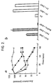
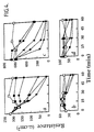
- Vanadate is relatively membrane-impermeant, but H 2 O 2 oxidises vanadate to membrane-permeant pervanadate which then enters the cells to inhibit tyrosine phosphatases (see eg Fantus et al , Biochemistry 28 8864-8871 (1989); Volberg et al, Cell Regulation 2 105-120 (1991); O'Shea et al, Proc. Natl. Acad. Sci. USA . 89 10306-10310 (1992)). Indeed, preformed pervanadate elicited a decrease in TER (Figure 1b). Over a longer time period, vanadate alone caused a decrease in TER ( Figure 1c), presumably because of its eventual entry into the cells.
- PAO phenylarsine oxide
- the degree of tyrosine phosphorylation of proteins reflects the balance of the activities of tyrosine kinases and tyrosine phosphatases.
- Pervanadate and PAO by inhibition of tyrosine phosphatases, must cause increased protein tyrosine phosphorylation through active kinases.
- the subcellular localisation of tyrosine phosphorylated proteins was first determined. This was achieved by immunostaining using an anti-phosphotyrosine antibody (PY20) (ICN Biomedicals, Ltd). In this procedure, the cells are fixed in paraformaldehyde to preserve the organisation and covalent modifications of proteins as found in the intact cell. After membrane-permeabilisation, the fixed cells are incubated with antibody and sites of bound antibody are detected using a flurochrome-conjugated secondary antibody followed by fluorescence microscopy.
- the cytoplasmic domain of the cadherins associates with three further proteins termed ⁇ -, ⁇ - and ⁇ -catenins, which link the cadherins to the cytoskeleton (Stappert and Kemler, Curr. Opinion in Neurobiol. 3 60-66 (1993)). Recently, it was reported that pervanadate treatment or pp60 v-src over-expression resulted in the tyrosine phosphorylation of components of the cadherin/catenin complex (Matsuyoshi et al, J. Cell Biol. 118 703-714 (1992); Behrens et al, J. Cell Biol.
- pervanadate and PAO increase the permeability of tight junctions by causing the tyrosine phosphorylation of components of the cadherin/catenin complex. It was therefore decided to examine more closely the effect of pervanadate and PAO on the tyrosine phosphorylation of this complex in MDCK strain I cells.
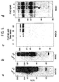
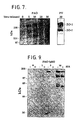
- the complex was immunoprecipitated from cells solubilised in a way that preserves pre-existing protein interactions using rr1, a mouse monoclonal antibody that recognises E-cadherin (Gumbiner et al, J. Cell Biol. 102 457-468 (1986)). Immunoprecipitated proteins were separated by SDS-PAGE, and tyrosine phosphorylation was detected by immunoblotting using an anti-phosphotyrosine antibody and enhanced chemiluminescence.
- Pervanadate caused an increase in the tyrosine phosphorylation of several proteins in anti-E-cadherin immunoprecipitates with molecular masses expected of E-cadherin (120 kDa) and associated catenins (molecular mass range, ⁇ 80 ⁇ 102 kDa: Fig. 6A).
- PAO predominantly stimulated the tyrosine phosphorylation of only one of the catenins (Fig. 6A), which on the basis of apparent molecular mass could be ⁇ - or ⁇ -catenin.
- peptide-directed antibodies were raised that specifically recognize ⁇ - or ⁇ -catenin.
- PAD-treated cells were lysed in SDS, followed by heating to dissociate protein complexes.
- SDS SDS
- tyrosine phosphorylated proteins not proteins associates with tyrosine phosphoproteins, are immunoprecipitated using anti-phosphotyrosine antibody.
- ⁇ -catenin is rapidly increased in response to treatment of the cells with either PAO or, as expected, pervanadate (Fig. 6B).
- ⁇ -catenin was not detectable in the phosphotyrosine immunoprecipitates (Fig. 6C).
- Tight junction-associated proteins are tyrosine phosphorylated
- Herbimycin A attenuated the ability of PAO to increase protein tyrosine phosphorylation (Fig. 9), correlating with its effects on tight junction permeability (Fig. 8). This inhibitory effect of herbimycin A is consistent with PAO action via inhibition of tyrosine phosphatases and suggests that a tyrosine kinase inhibitor could be useful in modulation of tight junction permeability.
- Stroke is the result of reduced blood supply causing (usually) focal ischaemia in the brain. This can be caused by either the rupture of a blood vessel (haemorrhagic stroke) or due to blood vessel blockage by a blood clot (occlusive stroke).
- Recovery from stroke is compromised by development of oedema that begins soon after the initial insult, and continues for several days. This oedema, if widespread, causes increased intracranial pressure and can harm the brain, reducing the extent of recovery.
- the oedema seen in the first hours after stroke is likely to be cytotoxic, caused by swelling of cellular elements in the region of damage. However, oedema is still present for the next few days and this maintained oedema is vasogenic, i.e.
- MCA middle cerebral artery
- focal cerebral ischaemia was induced by permanent occlusion, by cauterization, of the left MCA of adult Sprague Dawley rats. After 24 hours, rats were either sacrificed with no further operation or were intravenously injected with Evan's blue dye (see below) 30 minutes before being sacrificed. The brain was removed immediately and frozen. The area of brain served by the MCA was visibly swollen 24 hours after occlusion.
- Evan's blue The blood protein albumin is restricted to the lumen of vessels that have an intact blood-brain barrier, but when blood-brain barrier function is lost albumin leaves the vessels and enters the brain parenchyma.
- Evan's blue dye binds to albumin in the blood, and when albumin leaks into the tissues albumin-associated dye can be detected by fluorescence microscopy. Frozen sections of unfixed tissue were examined for Evan's blue extravasation into the brain parenchyma. MCA occlusion caused Evan's blue to leak into the brain parenchyma in part of the left cortex, this could be detected within 24 hours. Gross morphological changes in the labelled area indicated that the brain was severely damaged following the operation. Tissue away from the damaged area (e.g. in the right cortex) did not label with Evan's blue.
- Albumin Blood-brain barrier breakdown was also detected by incubating sections with antibody to rat albumin to demonstrate albumin leakage. Sections of brain labelled in this way also showed increased labeling of the parenchyma (outside the vessels), near the site of damage, whilst undamaged areas were again unlabelled, indicating a local loss of blood-brain barrier function at the sites affected by occlusion.
- control of the level of protein tyrosine phosphorylation appears to be essential for the maintenance of functional tight junctions.
- Many (bioactive) factors act through receptors that can alter levels of tyrosine phosphorylation, either through changing the activity of tyrosine kinases or tyrosine phosphatases. If factors released during ischaemia increase endothelial cells permeability by a mechanism that depends on alteration of the level of protein tyrosine phosphorylation, then it is possible that their action could be controlled through drugs that prevent elevation of protein tyrosine phosphorylation.
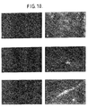
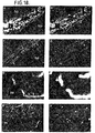
- brain sections were labelled with an antibody to phosphotyrosine (monoclonal antibody 4G10) and labelled with a polyclonal antibody to collagen type IV to outline blood vessels.
- 4G10 label was restricted to a few large, peripherally located, vessels with a pattern of staining consistent with tyrosine phosphorylation of proteins at the borders of cells lining the vessels.
- MCA occlusion an increase in 4G10 label was seen associated with microvessels located at the edge of the severely damaged tissue (see Fig. 10).
- the increase in 4G10 label was restricted to vessels in the area of the lesion, but not every vessel was labelled.
- 4G10 distribution in the microvessels is similar to that seen when brain tissue from normal adult rat is exposed to pervanadate, a drug that elevates levels of phosphotyrosine by inhibiting the enzymes responsible for tyrosine dephosphorylation.
- pervanadate induces increased phosphotyrosine in both small and large blood vessels, whilst after 24 hours of MCA occlusion, only microvessels showed increase phosphotyrosine label.
- cluster of bands corresponding to p120 and the similar cluster to p100 could also represent splice variants of, respectively, p100 and p120. It is also possible that p100 could simply represent a degradation product of p120, although samples were prepared in denaturing buffer and immunoprecipitations were performed in the presence of inhibitors of a broad spectrum of proteases.
- p120 immunoreactive protein was not unequivocally identified in the MDCK cells because of the poor reactivity of the 2B12 antibody with canine protein.
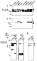
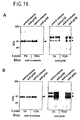
- This apparent association between p120/p100 and the cadherin/catenin complex was not restricted to epithelial cells as similar results were obtained using endothelial cells, both by [ 35 S]methionine-labelling (Fig.15) and immunoblot analysis (Fig.16).
- Fig.15 [ 35 S]methionine-labelling
- Fig.16 immunoblot analysis
- anti- ⁇ -catenin immunoprecipitates from MDCK cells lysed in TDS buffer failed to react with ⁇ -catenin (results not shown).
- ⁇ -catenin immunoprecipitates from TDS-lysed endothelial cells although clearly containing ⁇ -catenin, and presumably a cadherin, did not contain any anti-p120 reactivity.
- Cross-reactivity with ⁇ -catenin a protein of approximately 85 kDa, is unlikely as the immunoblots shown in Fig11 fail to show reactivity with protein below 100 kDa.
- This inhibitor increased the tyrosine phosphorylation of the anti-p120 immunoreactive material (a major p100 band, a minor p120 band) in these cells, as analyzed by anti-p120 immunoblotting of anti-p120 immunoprecipitates from SDS lysates (results not shown; see Staddon et al., J. Cell Sci. in press).
- the p120/p100 proteins could also be involved in the interaction between cadherins and the actin-based cytoskeleton.
- p120/p100 may also be part of a signalling cascade, communicating information about the state of cell-cell adhesiveness to the interior of the cell.
- ⁇ -catenin is an arm protein (McCrea et al., 1991) and can associate with cadherins and the APC gene product (Rubinfeld et al., 1993; Su et al., 1993), also an arm protein (see Peifer et al., 1994).
- p120 is an arm protein (Reynolds et al., 1992; Peifer et al., 1994), and, as we describe here, p100 is an immunologically related protein. These proteins can interact with ⁇ -catenin. The exact nature of the interaction between p120/p100 and the catenins remains to be established. These proteins may interact directly, or associate with different regions of the cytoplasmic domain of cadherins. Other linking or intermediary binding proteins could also be involved. Clearly, there appears to be diverse interactions among arm proteins, suggesting the importance of the arm motif in intracellular signalling.
- pp60 v-src activity in MDCK cells resulted in a rapid (10-30 min) tyrosine phosphorylation of E-cadherin and ⁇ -catenin (Behrens et al, J. Cell Biol. 120 757-766 (1993)). Behrens et al (1993) also reported that pp60 v-src resulted in a decrease in TER of MDCK cells. However, the decrease in TER was observed several hours after tyrosine phosphorylation of E-cadherin/ ⁇ -catenin. Furthermore, pp60 v-src resulted in a major alteration in cell morphology, from epithelial to fibroblast-like. The results of Behrens et al (1993) differ from the present results in that a very rapid decrease in TER is shown here in the absence of any gross changes in cell morphology.
- cadherins The conserved cytoplasmic domain of cadherins is known to associate with three proteins, termed ⁇ -, ⁇ - and ⁇ -catenin (Ozawa et al., 1989), which serve to link cadherins to the actin-based cortical cytoskeleton (Hirano et al., 1987).
- the association of cadherins with catenins is essential for intercellular Ca 2+ -dependent adhesiveness (Nagafuchi and Takeichi, 1988; Ozawa et al., 1990; Kintner, 1992).
- ⁇ -catenin is homologous to vinculin (Herrenknecht et al., 1991; Nagafuchi et al., 1991), making it a good candidate for interaction with the actin-based cytoskeleton (see Ozawa et al., 1990; Hirano et al., 1992).
- ⁇ -catenin is homologous to the Drosophila segment polarity gene armadillo, suggesting a role in developmental signalling in vertebrates (McCrea et al., 1991).
- ⁇ -catenin is probably identical to plakoglobin (Knudsen and Wheelock, 1992; but see Piepenhagen and Nelson, 1993), which again is homologous to armadillo (see Franke et al., 1989; Peifer and Wieschaus, 1990). Indeed, ⁇ -catenin and plakoglobin appear to form a multigene family (Peifer et al., 1992).
- tumour suppressor protein (Kinzler et al., 1991), p120, a pp60 src substrate (Reynolds et al., 1992), smgGDS, an exchange factor for ras-related G proteins (Kikuchi et al., 1992), a suppressor of RNA polymerase I mutations in yeast (Yano et al., 1992; 1994) and band 6 protein, a major desmosomal constituent (Hatzfeld et al., 1994).
- the function of the repeats in these arm proteins is unknown.
- the APC gene product associates with ⁇ -catenin (Rubinfeld et al., 1993; Su et al., 1993), supporting an important role for catenins in intracellular processes that regulate cell growth. Furthermore, this illustrates that cadherins are not exclusive cellular partners of catenins, raising the possibility of other interactions among catenins, cadherins and arm proteins, important in a variety of biological processes.
- p120 was initially identified as one of several substrates of the tyrosine kinase pp60 src (Reynolds et al., 1989; Kanner et al., 1990). It is membrane-associated and can be myristoylated, but does not appear to be glycosylated (Kanner et al., 1991). Mutational analysis suggested that tyrosine phosphorylation of p120 is necessary for that of pp60 src -mediated cellular transformation (Linder and Burr, 1988; Reynolds et al., 1989).
- Tyrosine phosphorylation of p120 was also observed in response to epidermal growth factor, platelet-derived growth factor, colony-stimulating factor 1 and in polyoma virus middle T antigen-transformed cells (Downing and Reynolds, 1991; Kanner et al., 1991), but the exact role of p120 in cellular physiology and pathology remains to be established.
- endothelial cell tyrosine kinase or activation of tyrosine phosphatases
- would lessen say, the amount of oedema that results from increased blood-brain barrier leakiness following stroke and lessen or prevent oedema associated with brain tumours.
- inhibition of endothelial tyrosine kinase should prevent cell migration.
- Available tyrosine kinase inhibitors include: genistein, herbimycin A, lavendustin A; methyl 2,5-dihydroxycinnamate; staurosporine and tyrphostins.
- Tyrosine kinase activators include various ligands, such as FGF, PDGF and VEGF, which activate receptors that couple to appropriate tyrosine kinases. Also, it may be that, during ischaemia, a ligand is released from glial or neuronal cells which activates an endothelial cell tyrosine kinase, thereby causing opening of the blood-brain barrier; a blocker of this ligand would be particularly useful in stroke therapy.
- Tyrosine phosphatase inhibitors include vanadate and phenylarsine oxide, of which the latter is preferred because of its more specific activity.
- Agents which promote tyrosine protein dephosphorylation, and therefore promote closure of a physiological barrier such as the blood-brain barrier are useful in a number of ways.
- a medicament may be useful in decreasing brain oedema following stroke or associated with brain tumours and/or in blocking the entry into the brain of both leukocytes that mediate an immune response, such as occurs in multiple sclerosis, and metastatic cancer cells that may form tumours.
- Such a medicament may also be useful in promoting tyrosine protein dephosphorylation in peripheral endothelial cells to prevent or mitigate peripheral oedema such as high altitude pulmonary oedema.
- a further use for such a medicament would be in promoting tyrosine protein dephosphorylation in gastric epithelial cells to treat gastric ulcer, which may be exacerbated by loose tight junctions (Ohkusa et al . Gut 34 86-89 (1993)).
- Such a medicament may be useful in promoting peripheral endothelial cell tyrosine protein phosphorylation to increase delivery of chemotherapeutic agents to peripheral tumours. (Although peripheral tumours do not have a barrier as tight as the blood-brain barrier, the centre of the tumour is often less well vascularised.) Such a medicament may also be useful in promoting pulmonary epithelial cell tyrosine protein phosphorylation, preferably when the medicament is administered using an inhaler, so that tight junctions become leaky to fluid, which can then act to dilute the accumulation of mucous in the airways.
- a third aspect of the invention there is provided the use of an agent which promotes tyrosine protein phosphorylation and the use of a blood-brain barrier- or other physiological barrier-impermeant drug in the preparation of a medicament for delivering the drug to the brain or other part of the body across the barrier.
- the drug may be any pharmaceutically, veterinarily or diagnostically useful compound or composition of compounds which is normally impermeant to the blood-brain or other physiological barrier or at least insufficiently permeant.
- the pharmacological nature of the drug is otherwise unimportant.
- the invention is therefore useful in the delivery of a wide range of drugs across physiological barriers such as the blood-brain barrier.
- anti-tumour compounds such as methotrexate, adriamycin and cisplatin
- growth factors such as NGF, BDNF and CNTF, which are used to treat neurodegenerative disease
- imaging agents especially those that are antibody based
- neurotransmitter antagonists or agonists which do not penetrate the blood-brain barrier such as certain NMDA receptor blockers.
- composition comprising an agent which promotes tyrosine protein phosphorylation and a drug to be delivered across a physiological barrier such as the blood-brain barrier.
- the composition may also contain a pharmaceutically or veterinarily acceptable carrier.
- the invention may also useful in preventing certain unwanted drugs crossing the barrier, if their transit across the barrier is by way of tight junctions.
- compositions of, and agents useful in, the invention may be adapted for administration by any suitable means, enteral or parenteral.
- Compositions for parenteral administration, such as injection or infusion, will generally be sterile.
- the dosage of any particular agent will depend on a number of factors and is likely to be optimised in experimental or clinical trials. In any event, the appropriate dosage for a particular patient or subject will be determined by the responsible physician or clinician in each case.
- Tissue culture-treated, polycarbonate Transwell filters (0.4 ⁇ M) were from Costar. All tissue culture materials were from Gibco except for plasma-derived horse serum which was from Lampire Biological Laboratories (Pipersville, PA). Vanadate was from Sigma and phenylarsine oxide (PAO) was from Kodak. Gel electrophoresis reagents were from Bio-Rad. Other reagents used were of the highest grade commercially available.
- Antibodies against ⁇ - and ⁇ -catenin were raised in rabbits using synthetic peptides coupled to keyhole limpet haemocyanin (Pierce) by glutaraldehyde.
- the peptide for ⁇ -catenin corresponded to amino acids 890-901 of mouse ⁇ E-catenin (Herrenknecht et al., 1991), the peptide for ⁇ -catenin to amino acids 751-781 of mouse ⁇ -catenin (Butz et al., 1992).
- the ⁇ -catenin peptide contained an additional C-terminal lysine for coupling, which is not in the protein.
- the rat monoclonal anti-ZO-1 antibody (R40.76) was from Dr. Bruce Stevenson (Department of Anatomy and Cell Biology, University of Alberta, Edmonton, Canada).
- the mouse monoclonal anti-E-cadherin antibody rr1 was from Dr. Barry Gumbiner (Memorial Sloan-Kettering Cancer Center, New York, NY).
- Rat monoclonal anti- ⁇ -catenin ( ⁇ -18) was from Prof. Shoichiro Tsukita and Dr. Akira Nagafuchi (Department of Information Physiology, National Institute for Physiological Sciences, Okazaki, Japan).
- Monodonal anti-phosphotyrosine antibody PY20 was from ICN Biomedicals.
- Horseradish peroxidase-conjugated anti-phosphotyrosine antibody RC20H was from Transduction Laboratories.
- Phosphotyrosine antibody (4G10) conjugated to agarose was from Upstate Biotechnology Inc.
- FITC-conjugated goat anti-rat and anti-mouse IgG were from Jackson Immunoresearch Laboratories Ltd.
- MDCK cells strain I
- FCS 10 % FCS
- 10 5 cells in 0.2 ml of medium were plated on 6.5 mm Transwell filters.
- the basolateral chamber contained 0.75 ml of medium. The cells were used between 4 and 7 days after plating.
- bovine brain endothelial cells Primary cultures of bovine brain endothelial cells were prepared essentially as described by Rubin et al, (1991). The cells were continuously grown in 50 % astrocyte-conditioned medium. After plating 2.5 x 10 4 cells on 6.5 mm, collagen-coated Transwell filters, the cells were maintained for 2-3 days in the absence or presence of 250 ⁇ M 8-(4-chlorophenylthio)cAMP (CPT-cAMP) plus 17.5 ⁇ M of the phosphodiesterase inhibitor Ro20-1724 (Calbiochem), as described by Rubin et al. (1991).
- CPT-cAMP 8-(4-chlorophenylthio)cAMP
- Ro20-1724 Calbiochem
- Cells were fixed in 3 % paraformaldehyde made up in PBS containing 0.5 mM CaCl 2 and 0.5 mM MgSO 4 . After 15 minutes at room temperature, the fixed cells were washed and then permeabilized by incubation with 0.5 % Triton X-100 in PBS for 10 minutes. After washing, the cells were incubated for 30 minutes in PBS containing 10 % calf serum plus 0.1 M lysine, pH 7.4. Incubation with primary antibody was in PBS containing 10 % calf serum for either 1 h at room temperature or overnight at 4°C.
- the cells were then incubated for 30-60 minutes at room temperature with a 1:100 dilution of FITC-conjugated goat anti-mouse or anti-rat IgG, as appropriate, in PBS containing 10 % calf serum.
- rhodamine phalloidin (2 U/ml; Molecular Probes, Inc.) was included with the secondary antibody.
- the filters were mounted with Citifluor (Citifluor Products, Canterbury, UK) and examined using a Nikon Microphot-FXA fluorescence microscope fitted with 40 x and 60 x objectives. Photographs were taken using Kodak T-MAX film (400 ASA).
- protease inhibitors used were: leupeptin, 10 ⁇ g/ml; ⁇ 2 -macroglobulin, 0.1 U/ml; soybean trypsin inhibitor, 10 ⁇ g/ml; PMSF, 1 mM.
- the cadherin/catenin complex was isolated at 4°C using buffer comprising: 20 mM imidazole, pH 6.8; 100 mM KCl; 10 mM EGTA; 2 mM MgCl 2 ; 300 mM sucrose; 0.2 % Triton X-100; 1 mM vanadate; 1 mM NaF; 25 ⁇ M PAO; 0.02 % NaN 3 ; protease inhibitors.
- Denatured proteins were isolated by immunoprecipitation after lysing the cells in: 1 % SDS; Hepes, pH 7.4; 25 mM NaF; 1mM vanadate; 25 ⁇ M PAO; 2 mM EDTA.
- the lysates were heated for 5 minutes at 100°C.
- DNA was sheared using a 23 G needle and the sample was then diluted into 2 ml of ice-cold buffer comprising: 3 % Triton X-100; 0.1 M NaCI; Hepes, pH 7.4; 25 mM NaF; 1mM vanadate; 25 ⁇ M PAO; 2 mM EDTA; protease inhibitors.
- ZO-1 and associated proteins were isolated at 4°C essentially as described by Gumbiner et al. (1991) using buffer comprising: 1 % Triton X-100; 0.5 % sodium deoxycholate; 0.2 % SDS; 0.15 M NaCl; 10 mM Hepes, pH 7.4; 25 mM NaF; 25 ⁇ M PAO; 2 mM EDTA; protease inhibitors.
- the filters were probed with horseradish peroxidase-conjugated anti-phosphotyrosine antibody (RC20H) or other unconjugated antibodies as described. After washing, unconjugated antibody was reacted with the appropriate horseradish peroxidase-conjugated secondary antibody. The filters were then extensively washed and immunoreactive bands were detected by enhanced chemiluminescence (Amersham) following the manufacturer's instructions.
- R20H horseradish peroxidase-conjugated anti-phosphotyrosine antibody
- unconjugated antibody was reacted with the appropriate horseradish peroxidase-conjugated secondary antibody.
- the filters were then extensively washed and immunoreactive bands were detected by enhanced chemiluminescence (Amersham) following the manufacturer's instructions.
- the anti-canine E-cadherin antibody rr1 developed by Gumbiner and Simons (1986) was provided by Barry Gumbiner (Memorial Sloan-Kettering Cancer Center, NY) or obtained from the Developmental Studies Hybridoma Bank maintained by the Department of Pharmacology and Molecular Sciences, Johns Hopkins University School of Medicine, Baltimore, MD 21205, and the Department of Biological Sciences. University of Iowa, Iowa City, IA 52242, under contract N01-HD-2-3144 from the NICHD.
- the anti-human E-cadherin antibody HECD-1 (Shimoyama et al., 1989) was from Takara Biomedicals (Shiga, Japan).
- Anti-p120 and anti-focal adhesion kinase (FAK) antibodies were from Transduction Laboratories (Lexington, KY).
- the anti-p120 antibody 2B12 (Kanner et al., 1990) was a gift from J. T. Parsons (University of Virginia, Charlottesville, VA).
- the peptide-directed antibodies against ⁇ - and ⁇ -catenin (Staddon et al., J. Cell Sci . in press) were kindly provided by Kurtheimknecht (Eisai Research Laboratories Ltd., University College London, London UK). All secondary antibodies used for immunoprecipitation and immunocytochemistry were from Jackson Laboratories Inc. (West Grove, PA).
- HRP-conjugated secondary antibodies used for immunoblotting were from Amersham (Buckinghamshire, UK).
- Caco-2 epidermal growth factor-derived cells derived from a human colonic tumour: 5 % CO 2 , MEM, 10 % FCS, 1 % non-essential amino-acids, 1 ⁇ g/ml insulin
- ECV304 a cell line derived from human umbilical vein endothelial cells: 5 % CO 2 , M199, 10 % FCS
- LLC-PK1 epidermal cells derived from porcine kidney: 5 % CO 2 , M199,10 % FCS
- MDBK epipithelial cells derived from bovine kidney: 5 % CO 2 , MEM, 10 % FCS
- Strain I MDCK cells epidermal brain endothelial cells (see Durieu-Trau)
- Caco-2, ECV304, LLC-PK1 and MDBK cells were obtained from the European Collection of Animal Cell Cultures (Salisbury, UK). MDCK cells were provided by Barry Gumbiner. RBE4 cells were from Pierre Couraud (liable Paris VII, Paris, France) and Swiss 3T3 fibroblasts were from Enrique Rozengurt (Imperial Cancer Research Fund, London, UK). Human umbilical vein endothelial cells were from Clonetics (Palo Alto, CA) and cultured according to the manufacturer's instructions. Primary cultures of bovine or porcine brain endothelial cells were grown as described by Rubin et al. (1991).
- tissue culture-treated, polycarbonate Transwell filters polycarbonate, 0.4 ⁇ m; Costar, Cambridge, MA. Other cells were grown on tissue culture plastic.
- the filters were blocked in 5 % (w/v) non-fat dried milk in PBS at 4°C for 16-18 hours. Filters were then incubated with primary antibody in PBS containing 0.05 % Tween-20 and 1 % BSA, followed by detection with appropriate HRP-conjugated secondary antibody and chemiluminescence (ECL, Amersham).
- TX buffer (1 % (v/v) Triton X-100, 25 mM Hepes, 2 mM EDTA, 0.1 M NaCl, 25 mM NaF, 1 mM vanadate, 25 ⁇ M phenylarsine oxide, pH 7.6 (adjusted with NaOH), 1 mM PMSF, 10 ⁇ g/ml soybean trypsin inhibitor, 0.1 U/ml ⁇ 2 -macroglobulin, 10 ⁇ g/ml leupeptin) or TDS buffer, which was identical to the TX buffer except that it was supplemented with 0.5 % (w/v) sodium deoxycholate and 0.2 % (w/v) SDS.
- the cells were incubated with lysis buffer for 10-15 minutes and then scraped.
- the lysates were collected and centrifuged at 14,000 x g for 20 minutes.
- the supernatant was precleared with Protein A Sepharose (Pharmacia, UK) for 1-2 hours and then incubated with primary antibody for 1 hour followed by a further 1 hour with Protein A Sepharose alone, in the case of rabbit antibodies, or together with rabbit anti-mouse antibodies for the mouse monoclonal antibodies.
- immune complexes were dissociated by addition of Laemmli sample buffer followed by heating at 100°C for 5 minutes. Protein analysis was by SDS-PAGE and immunoblotting as described above.
- the cells were then incubated for 30-60 minutes with a 1:100 dilution of fluorophore-conjugated anti-mouse or anti-rabbit IgG, as appropriate, in PBS containing 10 % calf serum.
- the filters were mounted with Citifluor (Citifluor Products, Canterbury, UK) and examined using a Nikon Microphot-FXA fluorescence microscope fitted with 40 x and 60 x objectives. Photographs were taken using Kodak T-MAX film (400 ASA).
- cryosections brain and skeletal muscle from CO 2 -asphyxiated rats were removed and rapidly frozen in liquid nitrogen. Tissue blocks were mounted in Tissue Tek (R. Lamb, London, UK) and sections of 5-10 ⁇ m thickness were cut on a Bright cryostat, air-dried and stored for up to 4 weeks at -20°C. After thawing, the sections were fixed and permeabilized as described above. The sections were then washed, blocked with PBS containing 10% calf serum for 15 minutes and incubated with primary antibody diluted in PBS containing 10% calf serum for 2 hours.
- Pervanadate decreases the transcellular electrical resistance of MDCKI cells.
- Strain I MDCK cells were grown on 6.5mm, 0.4 ⁇ m pore, TRANSWELLTM filters (Costar Catalogue No. 3413). The cells were plated at 10 5 per filter and incubated for 4-7 days at 37°C in a humidified 5% CO 2 incubator. Under these conditions the strain I MDCK cells gave transcellular electrical resistance values of 2000-5000 ⁇ -cm 2 .
- vanadate was added (as the sodium salt pH7), to a final concentration of 0.01mM ( ⁇ ) 0.1mM ( ⁇ ) or 1.0mM ( ⁇ ).
- Transcellular electrical resistance was measured, with respect to control cells ( ⁇ ), using the MILLICELL-ERSTM system from Millipore (UK) Limited at the times indicated. After 3 hours, H 2 O 2 was added to a final concentration of 2mM ( arrow ).
- vanadate and H 2 O 2 were preincubated prior to addition to the cells and excess H 2 O 2 was removed by catalase. The pervanadate was then added to the cells to give a final concentration of 0.01mM ( ⁇ ), 0.03mM ( ⁇ ) or 0.1mM ( ⁇ ), and TER was determined with respect to control cells ( ⁇ ) and cells to which the Ca 2+ chelator BAPTA was added ( ⁇ , 2.5mM final concentration).
- Phenylarsine oxide decreases the transcellular electrical resistance of MDCK I cells. Stain I MDCK cells were grown on TRANSWELL filters under the conditions described in Example 1. In the experiment whose results are shown graphically in Figure 2a, phenylarsine oxide was added to a final concentration of 1 ⁇ M ( ⁇ ), 3 ⁇ M ( ⁇ ), 10 ⁇ M ( ⁇ ), or 30 ⁇ M ( ⁇ ) and the TER was expressed relative to the control cells ( ⁇ ).
- phenylarsine oxide (PAO) or BAPTA was added to a final concentration of 10 ⁇ M and 3mM, respectively, to cells that had been preincubated for 10 minutes with 200 ⁇ M ⁇ -mercaptoethanol ( ⁇ ME) or 100 ⁇ M 2,3-dimercaptopropranol (DMP).
- TER was determined 30 minutes later.
- the values shown are the mean ⁇ S.D. of values derived from triplicate TRANSWELL filters, except for the BAPTA measurements which were the average of two filters.
- Pervanadate (PV) was added to a final concentration of 0.1mM to cells that had been preincubated for 10 min in the absence or presence of 500 ⁇ M ⁇ -mercaptoethanol or 250 ⁇ M 2,3-dimercaptopropanol. TER was determined 30 minutes later. The values shown are the means of determinations from two TRAMSWELL filters.
- Dimercaptopropanol (100 ⁇ M) was added 15 min ( ⁇ ), 30 min ( ⁇ ) or 45 min ( ⁇ ) after phenylarsine oxide.
- TER was determined at the times indicated. The values shown (means ⁇ S.D. of triplicate TRANSWELL filters) have been expressed relative to the initial TER.
- Pervanadate and phenylarsine oxide decrease the transcellular electrical resistance of brain capillary endothelial cells.
- Bovine brain capillary endothelial cells were grown on TRANSWELL filters and treated for two days in the absence ( Figures 4b and 4d) or presence ( Figures 4a and 4c) of CPT-cAMP/Ro20-1724, as described by Rubin et al, J. Cell Biol. 115 1725-1735 (1991).
- the data in Figures 4a/4b and Figures 4c/4d were derived from two independent preparations of cells, giving different initial resistance values.
- PAO and pervanadate increase the paracellular flux of sucrose in MDCK cells.
- Pervanadate increases the tyrosine phosphorylation of many proteins in MDCK strain I cells and BBECs: comparison with the discrete effects of phenylarsine oxide.
- MDCK strain I cells Figure 5a
- cyclic AMP treated BBECs Figure 5b
- PV pervanadate
- Figure 5c MDCK strain in cells were incubated with 10 ⁇ M phenylarsine oxide for the indicated times.
- cyclic AMP treated BBECs were treated with the indicated concentrations of phenylarsine oxide for 60 min. In all cases, the cells were lysed with SDS-containing buffer.
- Solubilised proteins were separated by SDS-PAGE and transferred to nitrocellulose. Tyrosine phosphorylation was detected using an anti-phosphotyrosine antibody conjugated to horseradish peroxidase followed by enhanced chemiluminescence. It should be noted that the exposure times (seconds) to obtain the results shown in Figures 5a and 5b were considerably shorter than those (minutes) to obtain the results in Figures 5c and 5d. With respect to the BBECs, similar results were obtained if the cells were treated with or without cyclic AMP.
- PAO increases the tyrosine phosphorylation of proteins at intercellular junctions in MDCK cells: comparison with the effect of pervanadate.
- MDCK cells were treated in the absence (A, F) or presence of either 100 ⁇ M pervanadate for 15 (B, G) or 30 minutes (C, H) or with 10 ⁇ M PAO for 15 (D, I) or 30 minutes (E, J).
- the cells were stained with an anti-phosphotyrosine antibody (A-F) or an anti-E-cadherin antibody (G-J), as described under Materials and Methods. Exposure times were constant for panels A-F. Bar, 20 ⁇ m (see Fig 5.1).
- PAO increases the tyrosine phosphorylation of proteins at intercellular junctions in brain endothelial cells: comparison with the effect of pervanadate.
- Filter-grown brain endothelial cells were treated with CPT-cAMP/Ro20-1724 for 2 days as described by Rubin et al. (1991). The cells were then incubated in the absence (A, E) or presence of either 10 ⁇ M PAO for 5 (B), 10 (C) or 30 minutes (D) or 100 ⁇ M pervanadate for 5 (F), 10 (G) or 30 minutes (H). The cells were then fixed and stained with anti-phosphotyrosine antibody as described under Materials and Methods. Exposure times were constant for panels A-D and constant, but less, for panels E-H. Bar, 20 ⁇ m (see Fig 5.2).
- PAO stimulates the tyrosine phosphorylation of components of the E-cadherin/catenin complex in MDCK cells.
- Fig 6 A filter-grown cells were untreated (Cont.) or incubated for 30 minutes with 10 ⁇ M PAO or for 15 minutes with 100 ⁇ M pervanadate (PV). The cells were lysed in buffer containing Triton X-100 and the E-cadherin/catenin complex was immunoprecipitated using anti-E-cadherin antibody. Proteins were resolved by SDS-PAGE and tyrosine phosphorylation was detected by immunoblotting with anti-phosphotyrosine antibody.
- PAO stimulates the tyrosine phosphorylation of a catenin of approximate molecular mass 100 kDa.
- Pervanadate stimulates the tyrosine phosphorylation of several catenins as well as E-cadherin.
- Figs 6 B, C, D, E MDCK cells were incubated with either 10 ⁇ M PAO or 100 ⁇ M pervanadate (PV) for the indicated times. The cells were lysed into denaturing buffer (containing SDS) to dissociated protein complexes and individual tyrosine phosphoproteins were immunoprecipitated using phosphotyrosine antibody.
- the immunoprecipitates were analyzed for either ⁇ -catenin (arrow, Fig 6 B) or ⁇ -catenin (arrow, Fig 6 C) content by immunoblotting.
- Identical lysates were immunoprecipitated using peptide-directed ⁇ -catenin antibody and blotted with the same antibody (Fig 6 D), to confirm the success of the immunoprecipitation, or phosphotyrosine antibody (Fig 6 E).
- Arrows indicate the migration of ⁇ -catenin.
- the exposure times to obtain the negative results shown in Figs 6 C and 6 E were similar to those used to obtain the result in Fig 6 A.
- Fig 6 B the ⁇ -catenin content of the phosphotyrosine immunoprecipitates from PAO or pervanadate treated cells is increased (Fig 6 B), in contrast, the ⁇ -catenin content is not (Fig 6 C).
- tyrosine phosphorylation (Fig 6 E) of ⁇ -catenin immunoprecipitates (Fig 6 D) could not be detected.
- Figs 6 F and 6 G MDCK cells were untreated (Cont.), incubated with 10 ⁇ M PAO for either 15 minutes and then 100 ⁇ M 2,3-dimercaptopropanol for a further 45 minutes (PAO 15'/DMP 45').
- the E-cadherin/catenin immunoprecipitate was analysed by immunoblotting for phosphotyrosine (Fig 6 F) or ⁇ -catenin content (Fig 6 G).
- the arrow in Fig 6 F indicates the migration of the tyrosine phosphorylated band seen in Fig 6 A.
- the arrow in Fig 6 G indicates the migration of ⁇ -catenin.
- the tyrosine phosphorylation of the catenin is reversed by the subsequent addition of 2,3-dimercaptopropnal (Fig 6 F).
- the constant content of ⁇ -catenin in the immunoprecipitates (Fig 6 G) indicates quantitatively successful immunoprecipitation in all cases.
- PAO stimulates the tyrosine phosphorylation of tight junction associated proteins in MDCK cells.
- MDCK cells were treated with 10 ⁇ M PAO or 100 ⁇ M pervanadate for the indicated times. The cells were lysed in buffer containing Triton X-100, SDS and sodium deoxycholate. ZO-1 and associated proteins were immunoprecipiated using ZO-1 antibody and analyzed for phosphotyrosine content by immunoblotting.
- PAO treatment resulted in the tyrosine phosphorylation of ZO-1 (220 kDa).
- Pervanadate also stimulated tyrosine phosphorylation of ZO-1, as well as ZO-2 (160 kDa) (see Fig 7).
- Herbimycin A attenuates the PAO-induced decrease in transcellular electrical resistance in MDCK cells.
- cultures were incubated in the absence or presence of the tyrosine kinase inhibitor herbimycin A ( ⁇ , control; ⁇ , 2.5 ⁇ M herbimycin A: ⁇ , control; ⁇ , 10 ⁇ M herbimycin A) for 16-18 hours after which a resistance measurement was taken.
- the addition of herbimycin A alone did not affect resistance.
- PAO was then added to give the final concentrations indicated and after a further 30 minutes incubation the effects on resistance were determined. The resistance values have been expressed relative to the initial values (see Fig 8).
- Herbimycin A attenuates the PAO-induced increase in protein tyrosine phosphorylation in MDCK cells.
- the cultures that had been used to generate the data testing the effects of 10 ⁇ M herbimycin A in Example 8 were lysed in SDS sample buffer and separated by SDS-PAGE. Protein tyrosine phosphorylation was detected by immunoblotting using anti-phosphotyrosine antibody.
- HA herbimycin A (see Fig 9).
- Middle cerebral artery occlusion (a stroke model) elevates the levels of tyrosine phosphorylation in cells associated with small blood vessels near the area damaged by the occlusion.
- the left middle cerebral artery of adult male Sprague-Dawley rats was exposed by craniotomy and occluded by cauterization. This gives a useful stroke model. Twenty four hours after the operation. Evan's blue (4% in PBS, 6 ⁇ l per gram body weight) was injected intravenously. After thirty minutes, the animal was anaesthetised, the blood flushed out with PBS, the brain removed and snap frozen in powdered dry ice. Sections of unfixed tissue were made using a Bright OTF cryostat.
- Sections (24 ⁇ m) were air-dried on gelatin-coated glass slides, fixed for 10 minutes in methanol at -20°C, washed in PBS (phosphate-buffered saline) and mounted in Citifluor.
- PBS phosphate-buffered saline
- Sections were air-dried and permeablised with 95% ethanol at -95°C for 30 minutes followed by acetone at room temperature for 1 minute. All of the antibody incubations and washes used PBS containing 5x10 -4 M sodium vanadate. Sections were washed in PBS, blocked with 1% BSA (bovine serum albumin) for 15 minutes and incubated overnight at 4°C with either 10 ⁇ g/ml anti-phosphotyrosine antibody (4G10) or control antibody (1:100) together with anti-collagen type IV (1:40,000).
- BSA bovine serum albumin
- Sections were then blocked for 15 minutes in PBS/vanadate containing 1% BSA and 0.1% Triton-X100, incubated with FITC-conjugated anti-mouse IgG2b (1:100) and Texas Red-conjugated anti-rabbit Ig (1:100) for 1 hour at room temperature and mounted in Cititluor.
- anti-rat albumin antibody (1:200) replaced the 4G10 antibody, and in this case FITC-conjugated anti-sheep IgG (1:100) was used instead of the anti-mouse IgG2b.
- Anti-phosphotyrosine antibody 4G10 was from Upstate Biotechnology Inc. (TCS Biologicals, Buckingha, UK), sheep anti-rat albumin from Biogenesis (Bournemouth, UK), control mouse IgG2b from Zymed (Cambridge Bioscience, Cambridge, UK), collagen type-IV from Biogenesis, FITC-conjugated anti-mouse IgG2b from Nordic Immunological Laboratories Ltd. (Maidenhead, UK) and Texas Red-conjugated anti-rabbit Ig and FITC-conjugated anti-sheep IgG were from Jackson Immunoresearch Laboratories Inc. (Stratech, Luton, UK).
- Sections were viewed on a Nikon Microphot FXA microscope using phase or Nomarski optics to determine morphology, and with epifluoresence to visualise the fluorophores. Excitation of Texas Red and Evan's blue was at 510-560 nm and FITC at 495 nm.
- Figs 10 A, C and E Collagen type IV. Collagen type IV is in the basement membrane around endothelial and smooth muscle cells and the antibody outlines blood vessels.
- Figs 10 B, D and F Phosphotyrosine label of the same sections shown in Figs 10 A, C and E. Phosphotyrosine is not detectable in the microvessels in the normal tissue of the right cortex of an operated rat (Fig 10 B), but is increased in vessels near the damaged area in the cortex on the left side of the same brain (Figs 10 D). Artificial elevation of the levels of phosphotyrosine using pervanadate induces detectable levels of phosphotyrosine in both large and small vessels (Fig 10 F (note that Fig 10 F is inverted)).
- p120 protein is recognized by the anti-p120 antibody and cross-reacts with the 2B12 antibody, whereas p100 is recognized only by anti-p120.
- MDBK cells were lysed in TDS buffer. Immunoprecipitations were performed using the anti-p120 antibody or 2B12 followed by cross-blotting (Panel A). MDBK cells were chosen because p120 and p100 are well separated by SDS-PAGE and react with both antibodies. In Panel B, MDBK cells were labelled with [ 35 S]methionine, lysed in TDS buffer and then immunoprecipitated using anti-p120 or 2B12. Proteins were separated by SDS-PAGE and detected by autoradiography.
- Panel A MDCK cells were labelled with [ 35 S]methionine and then lysed in either TX buffer or TDS buffer. Immunoprecipitations were performed using anti-p120 or anti-E-cadherin (rr1). Proteins were separated by SDS-PAGE followed by fluorography. Bands corresponding to E-cadherin (E), ⁇ -( ⁇ ) and ⁇ -catenin ( ⁇ ) which are seen strongly in the rr1 immunoprecipitates have been indicated. The major anti-p120 reactive band migrates at approximately the position of that of p100(see Fig 11A).
- Panel B Caco-2 cells were labelled with [ 35 S]methionine and then lysed in TX buffer. Immunoprecipitations were performed using anti-E-cadherin (HECD-1), anti- ⁇ -catenin or anti-p120 antibodies. Proteins were separated by SDS-PAGE followed by fluorography. In all cases, four major comigrating bands were immunoprecipitated, corresponding in order of increasing mobility to E-cadherin, ⁇ -, ⁇ - and ⁇ -catenin. (see Fig 13).
- Detection of anti-p120 reactive material in anti-E-cadherin immunoprecipitates and E-cadherin in the anti-p120 immunoprecipitates MDCK cells were lysed in TX buffer. Immunopredpitations were performed with either antibody to E-cadherin (rr1), the anti-p120 antibody or anti-focal adhesion kinase (anti-FAK). A sham immunoprecipitation was performed in the absence of primary antibody (-).
- rr1 (arrowhead, E-cadherin) ; anti- ⁇ -catenin (arrowhead, ⁇ -catenin); anti-p120 (arrowhead, anti-p120 reactivity); anti-FAK (arrowhead, FAK).
- anti-p120 antibody immunoprecipitated E-cadherin and ⁇ -catenin.
- rr1 could also immunoprecipitate anti-p120 immunoreactive material, but to a lesser extent than that immunoprecipitated by the anti-p120 antibody.
- Anti-FAK could only immunoprecipitate FAK, and the sham immunoprecipitation did not result in any detectable E-cadherin, ⁇ -catenin or anti-p120 reactive material (see Fig 14).
- Comparison or anti- ⁇ -catenin, anti-p120 and 2B12 immunoprecipitates from primary cultures of bovine brain endothelial cells.
- Cells were labelled with [ 35 S]methionine and then lysed in TX buffer or TDS buffer. Immunoprecipitations were performed using anti- ⁇ -catenin, anti-p120 and 2B12. Proteins were separated by SDS-PAGE followed by fluorography.
- ⁇ -catenin immunoprecipitation from TX lysed cells bands corresponding to a cadherin (C), ⁇ -catenin ( ⁇ ) and ⁇ -catenin ( ⁇ ) have been indicated.
- ⁇ -catenin immunoprecipitates and ⁇ -catenin in anti-p120 immunoprecipitates from human umbilical vein endothelial cells (Panel A) and ECV304 cells (Panel B). Cells were lysed in either TX or TDS buffer. Lysates were immunoprecipitated using anti- ⁇ -catenin or the anti-p120 antibody, followed by analysis by SDS-PAGE and immunoblotting. The migration of ⁇ -catenin (arrowheads), p100 ( ⁇ ) and p120 ( ⁇ ) have been indicated. Note the absence of anti-p120 reactive material in the ⁇ -catenin immunoprecipitates obtained using TDS buffer. Even though ⁇ -catenin and p100, as defined by its reactivity with anti-p120 antibody, migrate very closely, they are clearly distinct proteins (see Fig 16).
- MDCK cells Panel a,b
- porcine brain endothelial cells porcine brain endothelial cells
- Secondary antibodies were fluorescein-conjugated anti-mouse and rhodamine-conjugated anti-rabbit. In this instance, it was verified that each secondary antibody was absolutely specific for its designated species of primary antibody. Bar: 20 ⁇ m (see Fig 17).
- Anti-p120 immunoreactivity and ⁇ -catenin co-localise at intercellular junctions of choroid plexus epithelium (arrows, a and b) and ventricular ependymal cells (arrowheads, a and b). Both antigens also co-localise at interendothelial junctions of blood vessels of macrovascular origin (arrows in c and d).
- Microvascular profiles in brain sections were identified by labelling with anti-collagen IV antibody (f); co-labelling with the anti-p120 antibody (e), revealed the presence of antigen at interendothelial junctions (e, arrows).
- ⁇ -catenin co-localised with anti-p120 immunoreactivity (not shown).
- anti-p120 immunoreactivity is limited to areas between muscle fibres where blood vessels are located (g, arrows). Higher magnification reveals a punctate staining pattern (h, arrows) which is likely to reflect anti-p120 immunoreactivity at interendothelial junctions.
- Panels a,c and e depicts the brain parenchyma; in Panel a, v the ventricular lumen and cp the choroid plexus; in Panel g, m refers to muscle tissue. Bars: ( a, g ), 100 ⁇ m; ( c, e, h ), 25 ⁇ m (see Fig 18).
- the immune complex was washed five times and then dissociated by the addition of Laemmli sample buffer followed by heating at 100°C for 5 minutes. Proteins were precipitated by addition of four volumes of ethanol and incubation at -20°C for 16 hours. The precipitate was resolved by SDS-PAGE (6 % acrylamide) and proteins were visualized by Coomassie Blue. Protein corresponding to p100 was excised from the gel and digested with LysC. Peptides were separated by HPLC and sequenced. Mouse p120 sequence was described by Reynolds et al., 1992. Clearly, human p100 is closely related to mouse p120.
Landscapes
- Health & Medical Sciences (AREA)
- Chemical & Material Sciences (AREA)
- Life Sciences & Earth Sciences (AREA)
- General Health & Medical Sciences (AREA)
- Medicinal Chemistry (AREA)
- Veterinary Medicine (AREA)
- Public Health (AREA)
- Animal Behavior & Ethology (AREA)
- Pharmacology & Pharmacy (AREA)
- Organic Chemistry (AREA)
- Engineering & Computer Science (AREA)
- Bioinformatics & Cheminformatics (AREA)
- Epidemiology (AREA)
- Wood Science & Technology (AREA)
- Genetics & Genomics (AREA)
- Zoology (AREA)
- General Chemical & Material Sciences (AREA)
- Chemical Kinetics & Catalysis (AREA)
- Biomedical Technology (AREA)
- Nuclear Medicine, Radiotherapy & Molecular Imaging (AREA)
- General Engineering & Computer Science (AREA)
- Biotechnology (AREA)
- Molecular Biology (AREA)
- Biochemistry (AREA)
- Microbiology (AREA)
- Inorganic Chemistry (AREA)
- Neurosurgery (AREA)
- Neurology (AREA)
- Immunology (AREA)
- Cardiology (AREA)
- Heart & Thoracic Surgery (AREA)
- Medicines That Contain Protein Lipid Enzymes And Other Medicines (AREA)
- Steroid Compounds (AREA)
- Pharmaceuticals Containing Other Organic And Inorganic Compounds (AREA)
- Peptides Or Proteins (AREA)
- Acyclic And Carbocyclic Compounds In Medicinal Compositions (AREA)
Claims (18)
- Verwendung eines Mittels, das Tyrosinproteindephosphorylierung fördert, bei der Herstellung eines Medikaments zur Verringerung der Durchlässigkeit einer physiologischen Schranke, wobei die Schranke eine dichte inter-Endothelial Zellenverbindung umfaßt.
- Verwendung nach Anspruch 1, bei der das Mittel direkt oder indirekt Tyrosinproteinphosphatase aktiviert und/oder direkt oder indirekt Tyrosinkinase inhibiert.
- Verwendung eines Mittels, das die Tyrosinproteinphosphorylierung fördert, bei der Herstellung eines Medikaments zur Erhöhung der Durchlässigkeit einer physiologischen Schranke wie der Blut/Hirn-Schranke, wobei die Schranke eine dichte inter-Endothelialzellenverbindung umfaßt.
- Verwendung nach Anspruch 3, bei der das Mittel direkt oder indirekt Tyrosinproteinphosphatase inhibiert und/oder direkt oder indirekt Tyrosinkinase aktiviert.
- Verwendung nach einem der Ansprüche 1 bis 4, bei der die Phosphorylierungs- oder Dephosphorylierungsförderwirkung des Mittels reversibel oder in ausreichendem Maße reversibel ist, um ungünstige Toxizitätsprobleme zu vermeiden.
- Verwendung nach Anspruch 4 oder 5, bei der das Mittel ein vanadiumhaltiges Salz ist.
- Verwendung nach Anspruch 6, bei der das Mittel ein Pervanadat ist.
- Verwendung nach Anspruch 4, bei der das Mittel Phenylarsinoxid ist.
- Verwendung nach einem der Ansprüche 1 bis 8, bei der an der Tyrosinphosphorylierung oder -dephosphorylierung eine oder mehrere Komponenten des Cadherin/Catenin-Komplexes beteiligt sind.
- Verwendung nach einem der Ansprüche 1 bis 8, bei der an der Tyrosinphosphorylierung oder -dephosphorylierung ein Cadherin (z. B. E-Cadherin, N-Cadherin, P-Cadherin), β-Catenin, ZO-1, ZO-2, p100 oder p120 beteiligt ist.
- Verwendung nach Anspruch 9, bei der an der Tyrosinphosphorylierung oder -dephosphorylierung β-Catenin, ZO-1, ZO-2, p100 oder p120 beteiligt ist.
- Verwendung eines Mittels, das die Tyrosinproteindephosphorylierung fördert, bei der Herstellung eines Medikaments zur Verringerung von Hirnödemen, wie in Folge eines Schlaganfalls oder in Verbindung mit Hirntumoren.
- Verwendung eines Mittels, das die Tyrosinproteindephosphorylierung fördert, bei der Herstellung eines Medikaments zur Behandlung oder Verhinderung peripherer Ödeme, wie von Lungenödemen in Hochlagen mit großer Höhe.
- Verwendung eines Mittels, das die Tyrosinproteindephosphorylierung fördert, bei der Herstellung eines Medikaments zur Blockierung des Eintretens von Leukozyten, welche eine Immunreaktion vermitteln, in das Gehirn.
- Verwendung eines Mittels, das die Tyrosinproteindephosphorylierung fördert, bei der Herstellung eines Medikaments zur Behandlung von multipler Sklerose.
- Verwendung eines Mittels, das die Tyrosinproteindephosphorylierung fördert, bei der Herstellung eines Medikaments zur Verhinderung von Krebsmetastasen.
- Verwendung eines Mittels, das Tyrosinproteinphosphorylierung fördert, und die Verwendung eines die Blut/Hirn-Schranke oder eine andere physiologische Schranke nicht durchdringenden Wirkstoffs bei der Herstellung eines Medikaments zur Abgabe des Wirkstoffs an das Gehirn oder einen anderen Teil des Körpers (wie einen Tumor) auf der anderen Seite der Schranke, wobei die Schranke eine dichte inter-Endothelialzellenverbindung umfaßt.
- Zusammensetzung, die ein Mittel, das die Tyrosinproteinphosphorylierung fördert, und einen Wirkstoff umfasst, der durch eine physiologische Schranke hindurch abgegeben werden soll, wobei die Schranke eine dichte inter-Endothelialzellenverbindung umfaßt, z. B. die Blut/Hirn-Schranke.
Applications Claiming Priority (3)
| Application Number | Priority Date | Filing Date | Title |
|---|---|---|---|
| GB939323884A GB9323884D0 (en) | 1993-11-19 | 1993-11-19 | Physiological modulation |
| GB9323884 | 1993-11-19 | ||
| PCT/GB1994/002543 WO1995013820A1 (en) | 1993-11-19 | 1994-11-18 | Use of an agent which modulates tyrosine phosphorylation for modulating the permeability of a psychological barrier |
Publications (2)
| Publication Number | Publication Date |
|---|---|
| EP0728009A1 EP0728009A1 (de) | 1996-08-28 |
| EP0728009B1 true EP0728009B1 (de) | 2003-01-22 |
Family
ID=10745427
Family Applications (1)
| Application Number | Title | Priority Date | Filing Date |
|---|---|---|---|
| EP95901509A Expired - Lifetime EP0728009B1 (de) | 1993-11-19 | 1994-11-18 | Verwendung von einem mittel, das tyrosinphosphorylierung moduliert, zur modulation der durchlässigkeit einer physiologischen sperrschicht |
Country Status (7)
| Country | Link |
|---|---|
| US (2) | US6312686B1 (de) |
| EP (1) | EP0728009B1 (de) |
| JP (1) | JPH09505076A (de) |
| AT (1) | ATE231396T1 (de) |
| DE (1) | DE69432055T2 (de) |
| GB (1) | GB9323884D0 (de) |
| WO (1) | WO1995013820A1 (de) |
Cited By (1)
| Publication number | Priority date | Publication date | Assignee | Title |
|---|---|---|---|---|
| CN109432410A (zh) * | 2018-12-19 | 2019-03-08 | 上海康孕企业管理合伙企业(有限合伙) | 血红素在预防和/或改善高原反应的药物、食品及保健食品中的应用 |
Families Citing this family (23)
| Publication number | Priority date | Publication date | Assignee | Title |
|---|---|---|---|---|
| GB9423372D0 (en) | 1994-11-18 | 1995-01-11 | Eisai London Res Lab Ltd | Proteins and their uses |
| GB9525703D0 (en) * | 1995-12-15 | 1996-02-14 | Ciba Geigy Ag | Screening method |
| GB9620390D0 (en) | 1996-09-30 | 1996-11-13 | Eisai London Res Lab Ltd | Substances and their uses |
| AU3378599A (en) * | 1998-04-03 | 1999-10-25 | University Of Pittsburgh | Benzoquinoid ansamycins for the treatment of cardiac arrest and stroke |
| US6574501B2 (en) * | 1998-07-13 | 2003-06-03 | Childrens Hospital Los Angeles | Assessing blood brain barrier dynamics or identifying or measuring selected substances or toxins in a subject by analyzing Raman spectrum signals of selected regions in the eye |
| US7398119B2 (en) * | 1998-07-13 | 2008-07-08 | Childrens Hospital Los Angeles | Assessing blood brain barrier dynamics or identifying or measuring selected substances, including ethanol or toxins, in a subject by analyzing Raman spectrum signals |
| DE69927845D1 (de) * | 1998-12-11 | 2005-11-24 | Eisai Co Ltd | Verwendung des vaskularen endothelialen wachstumsfaktors (vegf) |
| US6174875B1 (en) | 1999-04-01 | 2001-01-16 | University Of Pittsburgh | Benzoquinoid ansamycins for the treatment of cardiac arrest and stroke |
| US6841351B2 (en) * | 1999-07-30 | 2005-01-11 | Agy Therapeutics, Inc. | High-throughput transcriptome and functional validation analysis |
| US6924109B2 (en) * | 1999-07-30 | 2005-08-02 | Agy Therapeutics, Inc. | High-throughput transcriptome and functional validation analysis |
| US6961599B2 (en) * | 2001-01-09 | 2005-11-01 | Childrens Hospital Los Angeles | Identifying or measuring selected substances or toxins in a subject using resonant raman signals |
| US20030118585A1 (en) * | 2001-10-17 | 2003-06-26 | Agy Therapeutics | Use of protein biomolecular targets in the treatment and visualization of brain tumors |
| EP1383543A4 (de) * | 2001-03-23 | 2006-06-21 | Agy Therapeutics Inc | Verwendung von biomolekularen targets bei der behandlung und visualisierung von hirntumoren |
| AU2002303804A1 (en) * | 2001-05-18 | 2002-12-03 | The Scripps Research Institute | Retroviral vectors and methods of using same |
| GB2396106B (en) * | 2002-12-12 | 2006-11-15 | Johnson & Johnson Medical Ltd | Wound dressings comprising enzyme inhibitors |
| US7485706B2 (en) * | 2003-07-30 | 2009-02-03 | The Board Of Trustees Of The Leland Stanford Junior University | Neurodegenerative protein aggregation inhibition methods and compounds |
| US20050074506A1 (en) * | 2003-10-02 | 2005-04-07 | Brainsgate Ltd. | Targeted release of nitric oxide in the CNS circulation for modulating the BBB and treating disorders |
| EP1768687A2 (de) * | 2004-06-29 | 2007-04-04 | Massachusetts Institute Of Technology | Verfahren und zusammensetzungen in zusammenhang mit der modulation intrazellulärer verbindungen |
| CA2763671A1 (en) * | 2005-04-26 | 2006-11-02 | Pfizer Inc. | P-cadherin antibodies |
| CN102014959B (zh) * | 2008-03-10 | 2016-01-20 | 康奈尔大学 | 血脑屏障通透性的调节 |
| US20130079682A1 (en) * | 2011-09-25 | 2013-03-28 | David J. Mischelevich | Ultrasound-neuromodulation techniques for control of permeability of the blood-brain barrier |
| US20130224110A1 (en) * | 2010-09-16 | 2013-08-29 | Cornell University | Use of adenosine receptor signaling to modulate permeability of blood-brain barrier |
| WO2016075670A1 (en) | 2014-11-14 | 2016-05-19 | Novartis Ag | Antibody drug conjugates |
Family Cites Families (3)
| Publication number | Priority date | Publication date | Assignee | Title |
|---|---|---|---|---|
| JPH05500504A (ja) * | 1989-09-27 | 1993-02-04 | アテナ・ニュウロサイエンスィズ・インコーポレイテッド | 細胞付着阻止のための組成物及び使用方法 |
| JPH0413631A (ja) | 1990-04-27 | 1992-01-17 | Tonen Corp | アナカルジウム・オクシデンタレ由来の物質 |
| US5155031A (en) | 1990-06-07 | 1992-10-13 | Posner Barry I | Use of pervanadate as an inhibitor of phosphotyrosine phosphatase |
-
1993
- 1993-11-19 GB GB939323884A patent/GB9323884D0/en active Pending
-
1994
- 1994-11-18 DE DE69432055T patent/DE69432055T2/de not_active Expired - Fee Related
- 1994-11-18 JP JP7514314A patent/JPH09505076A/ja not_active Ceased
- 1994-11-18 AT AT95901509T patent/ATE231396T1/de not_active IP Right Cessation
- 1994-11-18 US US08/648,182 patent/US6312686B1/en not_active Expired - Fee Related
- 1994-11-18 EP EP95901509A patent/EP0728009B1/de not_active Expired - Lifetime
- 1994-11-18 WO PCT/GB1994/002543 patent/WO1995013820A1/en not_active Ceased
-
2001
- 2001-05-04 US US09/848,353 patent/US20020032151A1/en not_active Abandoned
Cited By (1)
| Publication number | Priority date | Publication date | Assignee | Title |
|---|---|---|---|---|
| CN109432410A (zh) * | 2018-12-19 | 2019-03-08 | 上海康孕企业管理合伙企业(有限合伙) | 血红素在预防和/或改善高原反应的药物、食品及保健食品中的应用 |
Also Published As
| Publication number | Publication date |
|---|---|
| ATE231396T1 (de) | 2003-02-15 |
| DE69432055T2 (de) | 2003-11-06 |
| JPH09505076A (ja) | 1997-05-20 |
| US6312686B1 (en) | 2001-11-06 |
| US20020032151A1 (en) | 2002-03-14 |
| DE69432055D1 (de) | 2003-02-27 |
| GB9323884D0 (en) | 1994-01-05 |
| WO1995013820A1 (en) | 1995-05-26 |
| EP0728009A1 (de) | 1996-08-28 |
Similar Documents
| Publication | Publication Date | Title |
|---|---|---|
| EP0728009B1 (de) | Verwendung von einem mittel, das tyrosinphosphorylierung moduliert, zur modulation der durchlässigkeit einer physiologischen sperrschicht | |
| JP2806634B2 (ja) | 前立腺癌,胃癌および乳癌に関連する腫瘍を抑制するための医薬製剤 | |
| Pasqualon et al. | A transmembrane C-terminal fragment of syndecan-1 is generated by the metalloproteinase ADAM17 and promotes lung epithelial tumor cell migration and lung metastasis formation | |
| JP2016520056A (ja) | 癌の治療方法および組成物 | |
| Reisner et al. | In vitro enzyme activation with calbindin‐D28k, the vitamin D‐dependent 28 kDa calcium binding protein | |
| Zhang et al. | Anti-cancer activity of Annexin V in murine melanoma model by suppressing tumor angiogenesis | |
| US20170000851A1 (en) | Ptd-smad7 therapeutics | |
| CN116407620A (zh) | 重组iii型人源化胶原蛋白在乳腺癌治疗中的用途 | |
| EP3126378B1 (de) | Herzstärkende steroidantagonisten und zugehörige verfahren | |
| US20190111117A1 (en) | METHOD FOR PROPHYLAXIS AND/OR TREATMENT OF ErbB2 POSITIVE CANCERS | |
| KR102042015B1 (ko) | 폐 손상, 천식, 과민증, 혈관 부종, 전신 혈관 투과성 증후군 및 비충혈을 치료하는 펩티드 조성물과 이들로 이러한 질환을 치료하는 방법 | |
| EP2281575B1 (de) | Antikörper zur behandlung von krebs | |
| US20090155905A1 (en) | Composition and Method for Increasing Apoptosis in Cancer Cells | |
| US10093699B2 (en) | Peptides with antimicrobial, anticancer and/or wound-healing promoting activities, pharmaceutical compositions containing the same, and use of the peptides with antimicrobial, anticancer and/or wound-healing promoting activities | |
| US11712466B2 (en) | Vaccine composition | |
| CN107446024A (zh) | 一种可拮抗ddx3蛋白rna结合活性的多肽dip‑13及其应用 | |
| US8324170B2 (en) | Interferon gamma receptor beta chain compositions and methods of inhibiting apoptosis | |
| EP1751183A2 (de) | Fas-assoziierter faktor 1 | |
| US20110105382A1 (en) | Calmodulin-binding peptides that reduce cell proliferation in cancer and smooth muscle proliferation diseases | |
| CN113621026A (zh) | 一种多肽及其在制备治疗血小板增多症或抗肿瘤转移药物中的应用 | |
| WO2012112869A2 (en) | Targeted cancer therapy using ets transcriptional inhibitors | |
| Bitler | Determining the Role of Muc1 and Beta-catenin on the Epidermal Growth Factor Receptor Signaling and Localization in Breast Cancer |
Legal Events
| Date | Code | Title | Description |
|---|---|---|---|
| PUAI | Public reference made under article 153(3) epc to a published international application that has entered the european phase |
Free format text: ORIGINAL CODE: 0009012 |
|
| 17P | Request for examination filed |
Effective date: 19960415 |
|
| AK | Designated contracting states |
Kind code of ref document: A1 Designated state(s): AT BE CH DE DK ES FR GB GR IE IT LI LU MC NL PT SE |
|
| RIN1 | Information on inventor provided before grant (corrected) |
Inventor name: MORGAN, MARY, LOUISE Inventor name: HERRENKNECHT, KURT Inventor name: RUBIN, LEE, LAWRENCE Inventor name: STADDON, JAMES MARTIN |
|
| 17Q | First examination report despatched |
Effective date: 20010207 |
|
| GRAH | Despatch of communication of intention to grant a patent |
Free format text: ORIGINAL CODE: EPIDOS IGRA |
|
| GRAH | Despatch of communication of intention to grant a patent |
Free format text: ORIGINAL CODE: EPIDOS IGRA |
|
| GRAA | (expected) grant |
Free format text: ORIGINAL CODE: 0009210 |
|
| AK | Designated contracting states |
Kind code of ref document: B1 Designated state(s): AT BE CH DE DK ES FR GB GR IE IT LI LU MC NL PT SE |
|
| PG25 | Lapsed in a contracting state [announced via postgrant information from national office to epo] |
Ref country code: NL Free format text: LAPSE BECAUSE OF FAILURE TO SUBMIT A TRANSLATION OF THE DESCRIPTION OR TO PAY THE FEE WITHIN THE PRESCRIBED TIME-LIMIT Effective date: 20030122 Ref country code: LI Free format text: LAPSE BECAUSE OF FAILURE TO SUBMIT A TRANSLATION OF THE DESCRIPTION OR TO PAY THE FEE WITHIN THE PRESCRIBED TIME-LIMIT Effective date: 20030122 Ref country code: GR Free format text: LAPSE BECAUSE OF FAILURE TO SUBMIT A TRANSLATION OF THE DESCRIPTION OR TO PAY THE FEE WITHIN THE PRESCRIBED TIME-LIMIT Effective date: 20030122 Ref country code: CH Free format text: LAPSE BECAUSE OF FAILURE TO SUBMIT A TRANSLATION OF THE DESCRIPTION OR TO PAY THE FEE WITHIN THE PRESCRIBED TIME-LIMIT Effective date: 20030122 Ref country code: BE Free format text: LAPSE BECAUSE OF FAILURE TO SUBMIT A TRANSLATION OF THE DESCRIPTION OR TO PAY THE FEE WITHIN THE PRESCRIBED TIME-LIMIT Effective date: 20030122 Ref country code: AT Free format text: LAPSE BECAUSE OF FAILURE TO SUBMIT A TRANSLATION OF THE DESCRIPTION OR TO PAY THE FEE WITHIN THE PRESCRIBED TIME-LIMIT Effective date: 20030122 |
|
| REG | Reference to a national code |
Ref country code: GB Ref legal event code: FG4D |
|
| REG | Reference to a national code |
Ref country code: CH Ref legal event code: EP |
|
| REG | Reference to a national code |
Ref country code: IE Ref legal event code: FG4D |
|
| REF | Corresponds to: |
Ref document number: 69432055 Country of ref document: DE Date of ref document: 20030227 Kind code of ref document: P |
|
| PG25 | Lapsed in a contracting state [announced via postgrant information from national office to epo] |
Ref country code: SE Free format text: LAPSE BECAUSE OF FAILURE TO SUBMIT A TRANSLATION OF THE DESCRIPTION OR TO PAY THE FEE WITHIN THE PRESCRIBED TIME-LIMIT Effective date: 20030422 Ref country code: PT Free format text: LAPSE BECAUSE OF FAILURE TO SUBMIT A TRANSLATION OF THE DESCRIPTION OR TO PAY THE FEE WITHIN THE PRESCRIBED TIME-LIMIT Effective date: 20030422 Ref country code: DK Free format text: LAPSE BECAUSE OF FAILURE TO SUBMIT A TRANSLATION OF THE DESCRIPTION OR TO PAY THE FEE WITHIN THE PRESCRIBED TIME-LIMIT Effective date: 20030422 |
|
| NLV1 | Nl: lapsed or annulled due to failure to fulfill the requirements of art. 29p and 29m of the patents act | ||
| PG25 | Lapsed in a contracting state [announced via postgrant information from national office to epo] |
Ref country code: ES Free format text: LAPSE BECAUSE OF FAILURE TO SUBMIT A TRANSLATION OF THE DESCRIPTION OR TO PAY THE FEE WITHIN THE PRESCRIBED TIME-LIMIT Effective date: 20030730 |
|
| REG | Reference to a national code |
Ref country code: CH Ref legal event code: PL |
|
| ET | Fr: translation filed | ||
| PG25 | Lapsed in a contracting state [announced via postgrant information from national office to epo] |
Ref country code: LU Free format text: LAPSE BECAUSE OF NON-PAYMENT OF DUE FEES Effective date: 20031118 Ref country code: IE Free format text: LAPSE BECAUSE OF NON-PAYMENT OF DUE FEES Effective date: 20031118 |
|
| PLBE | No opposition filed within time limit |
Free format text: ORIGINAL CODE: 0009261 |
|
| STAA | Information on the status of an ep patent application or granted ep patent |
Free format text: STATUS: NO OPPOSITION FILED WITHIN TIME LIMIT |
|
| PG25 | Lapsed in a contracting state [announced via postgrant information from national office to epo] |
Ref country code: MC Free format text: LAPSE BECAUSE OF NON-PAYMENT OF DUE FEES Effective date: 20031130 |
|
| 26N | No opposition filed |
Effective date: 20031023 |
|
| REG | Reference to a national code |
Ref country code: IE Ref legal event code: MM4A |
|
| REG | Reference to a national code |
Ref country code: FR Ref legal event code: TP |
|
| REG | Reference to a national code |
Ref country code: GB Ref legal event code: 732E |
|
| PGFP | Annual fee paid to national office [announced via postgrant information from national office to epo] |
Ref country code: DE Payment date: 20081114 Year of fee payment: 15 |
|
| PGFP | Annual fee paid to national office [announced via postgrant information from national office to epo] |
Ref country code: IT Payment date: 20081126 Year of fee payment: 15 |
|
| PGFP | Annual fee paid to national office [announced via postgrant information from national office to epo] |
Ref country code: FR Payment date: 20081112 Year of fee payment: 15 |
|
| PGFP | Annual fee paid to national office [announced via postgrant information from national office to epo] |
Ref country code: GB Payment date: 20081112 Year of fee payment: 15 |
|
| GBPC | Gb: european patent ceased through non-payment of renewal fee |
Effective date: 20091118 |
|
| REG | Reference to a national code |
Ref country code: FR Ref legal event code: ST Effective date: 20100730 |
|
| PG25 | Lapsed in a contracting state [announced via postgrant information from national office to epo] |
Ref country code: FR Free format text: LAPSE BECAUSE OF NON-PAYMENT OF DUE FEES Effective date: 20091130 |
|
| PG25 | Lapsed in a contracting state [announced via postgrant information from national office to epo] |
Ref country code: DE Free format text: LAPSE BECAUSE OF NON-PAYMENT OF DUE FEES Effective date: 20100601 |
|
| PG25 | Lapsed in a contracting state [announced via postgrant information from national office to epo] |
Ref country code: GB Free format text: LAPSE BECAUSE OF NON-PAYMENT OF DUE FEES Effective date: 20091118 |
|
| PG25 | Lapsed in a contracting state [announced via postgrant information from national office to epo] |
Ref country code: IT Free format text: LAPSE BECAUSE OF NON-PAYMENT OF DUE FEES Effective date: 20091118 |