EP0842475B1 - Procedes et appareil de diagnostic de maladies assiste par ordinateur - Google Patents
Procedes et appareil de diagnostic de maladies assiste par ordinateur Download PDFInfo
- Publication number
- EP0842475B1 EP0842475B1 EP96927247A EP96927247A EP0842475B1 EP 0842475 B1 EP0842475 B1 EP 0842475B1 EP 96927247 A EP96927247 A EP 96927247A EP 96927247 A EP96927247 A EP 96927247A EP 0842475 B1 EP0842475 B1 EP 0842475B1
- Authority
- EP
- European Patent Office
- Prior art keywords
- neural network
- disease
- patient
- biomarkers
- values
- Prior art date
- Legal status (The legal status is an assumption and is not a legal conclusion. Google has not performed a legal analysis and makes no representation as to the accuracy of the status listed.)
- Expired - Lifetime
Links
- 208000037265 diseases, disorders, signs and symptoms Diseases 0.000 title claims abstract description 182
- 201000010099 disease Diseases 0.000 title claims abstract description 180
- 238000000034 method Methods 0.000 title claims abstract description 131
- 238000013528 artificial neural network Methods 0.000 claims abstract description 231
- 238000012549 training Methods 0.000 claims abstract description 74
- 239000000090 biomarker Substances 0.000 claims description 128
- 238000003745 diagnosis Methods 0.000 claims description 72
- 238000012360 testing method Methods 0.000 claims description 53
- 208000001132 Osteoporosis Diseases 0.000 claims description 49
- 206010060862 Prostate cancer Diseases 0.000 claims description 46
- 208000000236 Prostatic Neoplasms Diseases 0.000 claims description 45
- 230000008569 process Effects 0.000 claims description 29
- 208000029725 Metabolic bone disease Diseases 0.000 claims description 22
- 206010049088 Osteopenia Diseases 0.000 claims description 22
- 238000007781 pre-processing Methods 0.000 claims description 22
- 108010044467 Isoenzymes Proteins 0.000 claims description 13
- 206010006187 Breast cancer Diseases 0.000 claims description 12
- 208000026310 Breast neoplasm Diseases 0.000 claims description 12
- 206010033128 Ovarian cancer Diseases 0.000 claims description 12
- 206010061535 Ovarian neoplasm Diseases 0.000 claims description 12
- RJKFOVLPORLFTN-LEKSSAKUSA-N Progesterone Chemical compound C1CC2=CC(=O)CC[C@]2(C)[C@@H]2[C@@H]1[C@@H]1CC[C@H](C(=O)C)[C@@]1(C)CC2 RJKFOVLPORLFTN-LEKSSAKUSA-N 0.000 claims description 12
- 230000003044 adaptive effect Effects 0.000 claims description 9
- 239000013060 biological fluid Substances 0.000 claims description 9
- 238000001514 detection method Methods 0.000 claims description 9
- OYPRJOBELJOOCE-UHFFFAOYSA-N Calcium Chemical compound [Ca] OYPRJOBELJOOCE-UHFFFAOYSA-N 0.000 claims description 8
- 239000011575 calcium Substances 0.000 claims description 8
- 229910052791 calcium Inorganic materials 0.000 claims description 8
- 210000002966 serum Anatomy 0.000 claims description 8
- 206010009944 Colon cancer Diseases 0.000 claims description 7
- 101000623901 Homo sapiens Mucin-16 Proteins 0.000 claims description 7
- 102100023123 Mucin-16 Human genes 0.000 claims description 7
- 208000024313 Testicular Neoplasms Diseases 0.000 claims description 7
- 206010057644 Testis cancer Diseases 0.000 claims description 7
- 208000029742 colonic neoplasm Diseases 0.000 claims description 7
- 229960005309 estradiol Drugs 0.000 claims description 7
- 201000003120 testicular cancer Diseases 0.000 claims description 7
- VOXZDWNPVJITMN-ZBRFXRBCSA-N 17β-estradiol Chemical compound OC1=CC=C2[C@H]3CC[C@](C)([C@H](CC4)O)[C@@H]4[C@@H]3CCC2=C1 VOXZDWNPVJITMN-ZBRFXRBCSA-N 0.000 claims description 6
- 101001024605 Homo sapiens Next to BRCA1 gene 1 protein Proteins 0.000 claims description 6
- 229910019142 PO4 Inorganic materials 0.000 claims description 6
- 229930182833 estradiol Natural products 0.000 claims description 6
- 239000010452 phosphate Substances 0.000 claims description 6
- NBIIXXVUZAFLBC-UHFFFAOYSA-K phosphate Chemical compound [O-]P([O-])([O-])=O NBIIXXVUZAFLBC-UHFFFAOYSA-K 0.000 claims description 6
- 238000012545 processing Methods 0.000 claims description 6
- 239000000186 progesterone Substances 0.000 claims description 6
- 229960003387 progesterone Drugs 0.000 claims description 6
- INZOTETZQBPBCE-NYLDSJSYSA-N 3-sialyl lewis Chemical compound O[C@H]1[C@H](O)[C@H](O)[C@H](C)O[C@H]1O[C@H]([C@H](O)CO)[C@@H]([C@@H](NC(C)=O)C=O)O[C@H]1[C@H](O)[C@@H](O[C@]2(O[C@H]([C@H](NC(C)=O)[C@@H](O)C2)[C@H](O)[C@H](O)CO)C(O)=O)[C@@H](O)[C@@H](CO)O1 INZOTETZQBPBCE-NYLDSJSYSA-N 0.000 claims description 5
- 230000005856 abnormality Effects 0.000 claims description 5
- 102000015872 Human beta Subunit Chorionic Gonadotropin Human genes 0.000 claims description 4
- 108010010590 Human beta Subunit Chorionic Gonadotropin Proteins 0.000 claims description 4
- 238000003384 imaging method Methods 0.000 claims description 4
- 238000004088 simulation Methods 0.000 claims description 4
- 101150029707 ERBB2 gene Proteins 0.000 claims description 3
- 102100028123 Macrophage colony-stimulating factor 1 Human genes 0.000 claims description 2
- 101710127797 Macrophage colony-stimulating factor 1 Proteins 0.000 claims description 2
- 230000002962 histologic effect Effects 0.000 claims description 2
- 238000012886 linear function Methods 0.000 claims description 2
- 238000010946 mechanistic model Methods 0.000 claims description 2
- 238000009795 derivation Methods 0.000 claims 1
- 238000005516 engineering process Methods 0.000 abstract description 8
- 238000004458 analytical method Methods 0.000 description 33
- 210000002569 neuron Anatomy 0.000 description 33
- 230000006870 function Effects 0.000 description 24
- 206010028980 Neoplasm Diseases 0.000 description 22
- 201000011510 cancer Diseases 0.000 description 21
- 238000012216 screening Methods 0.000 description 21
- 210000000988 bone and bone Anatomy 0.000 description 19
- 238000004393 prognosis Methods 0.000 description 19
- 102000007066 Prostate-Specific Antigen Human genes 0.000 description 18
- 108010072866 Prostate-Specific Antigen Proteins 0.000 description 18
- 238000004422 calculation algorithm Methods 0.000 description 17
- 238000002405 diagnostic procedure Methods 0.000 description 15
- 102000002260 Alkaline Phosphatase Human genes 0.000 description 14
- 108020004774 Alkaline Phosphatase Proteins 0.000 description 14
- 206010004446 Benign prostatic hyperplasia Diseases 0.000 description 14
- 208000004403 Prostatic Hyperplasia Diseases 0.000 description 14
- 238000013459 approach Methods 0.000 description 14
- 210000004205 output neuron Anatomy 0.000 description 14
- 238000010276 construction Methods 0.000 description 13
- 230000035945 sensitivity Effects 0.000 description 13
- 210000004369 blood Anatomy 0.000 description 11
- 239000008280 blood Substances 0.000 description 11
- 238000005259 measurement Methods 0.000 description 11
- 241000282412 Homo Species 0.000 description 9
- 102100035703 Prostatic acid phosphatase Human genes 0.000 description 9
- 230000037182 bone density Effects 0.000 description 9
- 230000002068 genetic effect Effects 0.000 description 9
- 108010043671 prostatic acid phosphatase Proteins 0.000 description 9
- 230000009466 transformation Effects 0.000 description 9
- 241001465754 Metazoa Species 0.000 description 8
- 238000011156 evaluation Methods 0.000 description 8
- 230000008901 benefit Effects 0.000 description 7
- 239000000470 constituent Substances 0.000 description 7
- 239000000126 substance Substances 0.000 description 7
- 230000007423 decrease Effects 0.000 description 6
- 238000009826 distribution Methods 0.000 description 6
- 230000036541 health Effects 0.000 description 6
- 230000003211 malignant effect Effects 0.000 description 6
- 239000000523 sample Substances 0.000 description 6
- 102100025475 Carcinoembryonic antigen-related cell adhesion molecule 5 Human genes 0.000 description 5
- 210000000481 breast Anatomy 0.000 description 5
- 210000004027 cell Anatomy 0.000 description 5
- 238000002591 computed tomography Methods 0.000 description 5
- 230000007613 environmental effect Effects 0.000 description 5
- 229910052500 inorganic mineral Inorganic materials 0.000 description 5
- 210000002364 input neuron Anatomy 0.000 description 5
- 239000011707 mineral Substances 0.000 description 5
- 239000013598 vector Substances 0.000 description 5
- 241000282414 Homo sapiens Species 0.000 description 4
- 102100024319 Intestinal-type alkaline phosphatase Human genes 0.000 description 4
- 101710184243 Intestinal-type alkaline phosphatase Proteins 0.000 description 4
- 230000002159 abnormal effect Effects 0.000 description 4
- 238000004891 communication Methods 0.000 description 4
- 230000003247 decreasing effect Effects 0.000 description 4
- 239000003814 drug Substances 0.000 description 4
- 210000004185 liver Anatomy 0.000 description 4
- 238000002559 palpation Methods 0.000 description 4
- 238000003909 pattern recognition Methods 0.000 description 4
- 230000002250 progressing effect Effects 0.000 description 4
- 238000011160 research Methods 0.000 description 4
- 208000024891 symptom Diseases 0.000 description 4
- 210000001519 tissue Anatomy 0.000 description 4
- 238000000844 transformation Methods 0.000 description 4
- 102100023635 Alpha-fetoprotein Human genes 0.000 description 3
- LFQSCWFLJHTTHZ-UHFFFAOYSA-N Ethanol Chemical compound CCO LFQSCWFLJHTTHZ-UHFFFAOYSA-N 0.000 description 3
- 210000000577 adipose tissue Anatomy 0.000 description 3
- 230000003542 behavioural effect Effects 0.000 description 3
- 230000005540 biological transmission Effects 0.000 description 3
- 230000008859 change Effects 0.000 description 3
- 238000002790 cross-validation Methods 0.000 description 3
- 229940079593 drug Drugs 0.000 description 3
- 230000002526 effect on cardiovascular system Effects 0.000 description 3
- 239000012530 fluid Substances 0.000 description 3
- 208000014674 injury Diseases 0.000 description 3
- 238000009533 lab test Methods 0.000 description 3
- 230000036210 malignancy Effects 0.000 description 3
- 238000009607 mammography Methods 0.000 description 3
- 239000000203 mixture Substances 0.000 description 3
- 230000001537 neural effect Effects 0.000 description 3
- 210000000056 organ Anatomy 0.000 description 3
- 230000002611 ovarian Effects 0.000 description 3
- 230000001575 pathological effect Effects 0.000 description 3
- 210000004197 pelvis Anatomy 0.000 description 3
- 210000002307 prostate Anatomy 0.000 description 3
- 230000005855 radiation Effects 0.000 description 3
- 230000001850 reproductive effect Effects 0.000 description 3
- 238000010561 standard procedure Methods 0.000 description 3
- 238000007619 statistical method Methods 0.000 description 3
- 230000008733 trauma Effects 0.000 description 3
- 206010003805 Autism Diseases 0.000 description 2
- 208000020706 Autistic disease Diseases 0.000 description 2
- 206010065687 Bone loss Diseases 0.000 description 2
- 102000004190 Enzymes Human genes 0.000 description 2
- 108090000790 Enzymes Proteins 0.000 description 2
- 208000016604 Lyme disease Diseases 0.000 description 2
- 102000007651 Macrophage Colony-Stimulating Factor Human genes 0.000 description 2
- 108010046938 Macrophage Colony-Stimulating Factor Proteins 0.000 description 2
- OAICVXFJPJFONN-UHFFFAOYSA-N Phosphorus Chemical compound [P] OAICVXFJPJFONN-UHFFFAOYSA-N 0.000 description 2
- 208000032023 Signs and Symptoms Diseases 0.000 description 2
- 210000001744 T-lymphocyte Anatomy 0.000 description 2
- MUMGGOZAMZWBJJ-DYKIIFRCSA-N Testostosterone Chemical compound O=C1CC[C@]2(C)[C@H]3CC[C@](C)([C@H](CC4)O)[C@@H]4[C@@H]3CCC2=C1 MUMGGOZAMZWBJJ-DYKIIFRCSA-N 0.000 description 2
- 230000004913 activation Effects 0.000 description 2
- 238000001994 activation Methods 0.000 description 2
- 230000004075 alteration Effects 0.000 description 2
- 230000000747 cardiac effect Effects 0.000 description 2
- 238000012512 characterization method Methods 0.000 description 2
- 210000000038 chest Anatomy 0.000 description 2
- HVYWMOMLDIMFJA-DPAQBDIFSA-N cholesterol Chemical compound C1C=C2C[C@@H](O)CC[C@]2(C)[C@@H]2[C@@H]1[C@@H]1CC[C@H]([C@H](C)CCCC(C)C)[C@@]1(C)CC2 HVYWMOMLDIMFJA-DPAQBDIFSA-N 0.000 description 2
- 235000019504 cigarettes Nutrition 0.000 description 2
- 238000003759 clinical diagnosis Methods 0.000 description 2
- 238000000205 computational method Methods 0.000 description 2
- 238000007796 conventional method Methods 0.000 description 2
- 238000013079 data visualisation Methods 0.000 description 2
- 230000034994 death Effects 0.000 description 2
- 238000003066 decision tree Methods 0.000 description 2
- 230000006866 deterioration Effects 0.000 description 2
- 230000018109 developmental process Effects 0.000 description 2
- 238000012631 diagnostic technique Methods 0.000 description 2
- 208000035475 disorder Diseases 0.000 description 2
- 230000009977 dual effect Effects 0.000 description 2
- 229940011871 estrogen Drugs 0.000 description 2
- 239000000262 estrogen Substances 0.000 description 2
- 230000003325 follicular Effects 0.000 description 2
- 238000009472 formulation Methods 0.000 description 2
- 230000012010 growth Effects 0.000 description 2
- 230000006872 improvement Effects 0.000 description 2
- 238000007901 in situ hybridization Methods 0.000 description 2
- 230000000938 luteal effect Effects 0.000 description 2
- 238000002595 magnetic resonance imaging Methods 0.000 description 2
- 230000007246 mechanism Effects 0.000 description 2
- 238000012986 modification Methods 0.000 description 2
- 230000004048 modification Effects 0.000 description 2
- 238000003058 natural language processing Methods 0.000 description 2
- 230000007830 nerve conduction Effects 0.000 description 2
- 230000000144 pharmacologic effect Effects 0.000 description 2
- 239000011574 phosphorus Substances 0.000 description 2
- 229910052698 phosphorus Inorganic materials 0.000 description 2
- 235000017924 poor diet Nutrition 0.000 description 2
- 108090000623 proteins and genes Proteins 0.000 description 2
- 230000002294 pubertal effect Effects 0.000 description 2
- 108020003175 receptors Proteins 0.000 description 2
- 102000005962 receptors Human genes 0.000 description 2
- 210000000614 rib Anatomy 0.000 description 2
- 229920002477 rna polymer Polymers 0.000 description 2
- 230000002381 testicular Effects 0.000 description 2
- 238000012546 transfer Methods 0.000 description 2
- 238000013519 translation Methods 0.000 description 2
- 210000002700 urine Anatomy 0.000 description 2
- 238000010200 validation analysis Methods 0.000 description 2
- AOFUBOWZWQFQJU-SNOJBQEQSA-N (2r,3s,4s,5r)-2,5-bis(hydroxymethyl)oxolane-2,3,4-triol;(2s,3r,4s,5s,6r)-6-(hydroxymethyl)oxane-2,3,4,5-tetrol Chemical compound OC[C@H]1O[C@](O)(CO)[C@@H](O)[C@@H]1O.OC[C@H]1O[C@H](O)[C@H](O)[C@@H](O)[C@@H]1O AOFUBOWZWQFQJU-SNOJBQEQSA-N 0.000 description 1
- 108010077173 BB Form Creatine Kinase Proteins 0.000 description 1
- 208000008035 Back Pain Diseases 0.000 description 1
- 208000006386 Bone Resorption Diseases 0.000 description 1
- 101100314454 Caenorhabditis elegans tra-1 gene Proteins 0.000 description 1
- 108010022366 Carcinoembryonic Antigen Proteins 0.000 description 1
- 206010008342 Cervix carcinoma Diseases 0.000 description 1
- 241001456553 Chanodichthys dabryi Species 0.000 description 1
- 208000035473 Communicable disease Diseases 0.000 description 1
- 102000004420 Creatine Kinase Human genes 0.000 description 1
- 108010042126 Creatine kinase Proteins 0.000 description 1
- 102100022785 Creatine kinase B-type Human genes 0.000 description 1
- 102100022786 Creatine kinase M-type Human genes 0.000 description 1
- 102000004127 Cytokines Human genes 0.000 description 1
- 108090000695 Cytokines Proteins 0.000 description 1
- 102000053602 DNA Human genes 0.000 description 1
- 108020004414 DNA Proteins 0.000 description 1
- 238000002965 ELISA Methods 0.000 description 1
- 101000914324 Homo sapiens Carcinoembryonic antigen-related cell adhesion molecule 5 Proteins 0.000 description 1
- 101000914321 Homo sapiens Carcinoembryonic antigen-related cell adhesion molecule 7 Proteins 0.000 description 1
- 101001047117 Homo sapiens Creatine kinase B-type Proteins 0.000 description 1
- 101001047110 Homo sapiens Creatine kinase M-type Proteins 0.000 description 1
- 101000617725 Homo sapiens Pregnancy-specific beta-1-glycoprotein 2 Proteins 0.000 description 1
- 108010052919 Hydroxyethylthiazole kinase Proteins 0.000 description 1
- 108010027436 Hydroxymethylpyrimidine kinase Proteins 0.000 description 1
- PMMYEEVYMWASQN-DMTCNVIQSA-N Hydroxyproline Chemical compound O[C@H]1CN[C@H](C(O)=O)C1 PMMYEEVYMWASQN-DMTCNVIQSA-N 0.000 description 1
- 206010023509 Kyphosis Diseases 0.000 description 1
- 108010051884 MB Form Creatine Kinase Proteins 0.000 description 1
- 108010059343 MM Form Creatine Kinase Proteins 0.000 description 1
- 206010027476 Metastases Diseases 0.000 description 1
- 102000013967 Monokines Human genes 0.000 description 1
- 108010050619 Monokines Proteins 0.000 description 1
- 238000012614 Monte-Carlo sampling Methods 0.000 description 1
- 241000208125 Nicotiana Species 0.000 description 1
- 235000002637 Nicotiana tabacum Nutrition 0.000 description 1
- 241000906034 Orthops Species 0.000 description 1
- 208000002193 Pain Diseases 0.000 description 1
- 208000037273 Pathologic Processes Diseases 0.000 description 1
- 238000001358 Pearson's chi-squared test Methods 0.000 description 1
- 208000024799 Thyroid disease Diseases 0.000 description 1
- 102000040945 Transcription factor Human genes 0.000 description 1
- 108091023040 Transcription factor Proteins 0.000 description 1
- 208000006105 Uterine Cervical Neoplasms Diseases 0.000 description 1
- 230000009471 action Effects 0.000 description 1
- 230000000172 allergic effect Effects 0.000 description 1
- 108010026331 alpha-Fetoproteins Proteins 0.000 description 1
- 239000012491 analyte Substances 0.000 description 1
- 238000002399 angioplasty Methods 0.000 description 1
- 230000000578 anorexic effect Effects 0.000 description 1
- 239000000427 antigen Substances 0.000 description 1
- 108091007433 antigens Proteins 0.000 description 1
- 102000036639 antigens Human genes 0.000 description 1
- 238000003556 assay Methods 0.000 description 1
- 208000010668 atopic eczema Diseases 0.000 description 1
- 238000002555 auscultation Methods 0.000 description 1
- 230000006399 behavior Effects 0.000 description 1
- SQVRNKJHWKZAKO-UHFFFAOYSA-N beta-N-Acetyl-D-neuraminic acid Natural products CC(=O)NC1C(O)CC(O)(C(O)=O)OC1C(O)C(O)CO SQVRNKJHWKZAKO-UHFFFAOYSA-N 0.000 description 1
- 239000000091 biomarker candidate Substances 0.000 description 1
- 238000001574 biopsy Methods 0.000 description 1
- 230000036772 blood pressure Effects 0.000 description 1
- 238000009530 blood pressure measurement Methods 0.000 description 1
- 230000024279 bone resorption Effects 0.000 description 1
- 238000004364 calculation method Methods 0.000 description 1
- 239000002775 capsule Substances 0.000 description 1
- 150000001720 carbohydrates Chemical class 0.000 description 1
- 235000014633 carbohydrates Nutrition 0.000 description 1
- 210000001175 cerebrospinal fluid Anatomy 0.000 description 1
- 201000010881 cervical cancer Diseases 0.000 description 1
- 239000007795 chemical reaction product Substances 0.000 description 1
- 235000012000 cholesterol Nutrition 0.000 description 1
- 238000004587 chromatography analysis Methods 0.000 description 1
- 210000001072 colon Anatomy 0.000 description 1
- 238000005094 computer simulation Methods 0.000 description 1
- 230000001054 cortical effect Effects 0.000 description 1
- 230000001086 cytosolic effect Effects 0.000 description 1
- 235000013365 dairy product Nutrition 0.000 description 1
- 230000006378 damage Effects 0.000 description 1
- 206010061428 decreased appetite Diseases 0.000 description 1
- 230000006735 deficit Effects 0.000 description 1
- 230000003111 delayed effect Effects 0.000 description 1
- 238000001739 density measurement Methods 0.000 description 1
- 230000037123 dental health Effects 0.000 description 1
- 238000013461 design Methods 0.000 description 1
- 238000011161 development Methods 0.000 description 1
- 239000000104 diagnostic biomarker Substances 0.000 description 1
- 235000014113 dietary fatty acids Nutrition 0.000 description 1
- 230000001079 digestive effect Effects 0.000 description 1
- 238000004090 dissolution Methods 0.000 description 1
- PMMYEEVYMWASQN-UHFFFAOYSA-N dl-hydroxyproline Natural products OC1C[NH2+]C(C([O-])=O)C1 PMMYEEVYMWASQN-UHFFFAOYSA-N 0.000 description 1
- 238000013399 early diagnosis Methods 0.000 description 1
- 230000000694 effects Effects 0.000 description 1
- 239000003792 electrolyte Substances 0.000 description 1
- 238000001493 electron microscopy Methods 0.000 description 1
- 230000002124 endocrine Effects 0.000 description 1
- 238000001839 endoscopy Methods 0.000 description 1
- 231100000317 environmental toxin Toxicity 0.000 description 1
- 235000019441 ethanol Nutrition 0.000 description 1
- 230000029142 excretion Effects 0.000 description 1
- 210000003722 extracellular fluid Anatomy 0.000 description 1
- 238000000605 extraction Methods 0.000 description 1
- 229930195729 fatty acid Natural products 0.000 description 1
- 239000000194 fatty acid Substances 0.000 description 1
- 150000004665 fatty acids Chemical class 0.000 description 1
- 230000037406 food intake Effects 0.000 description 1
- 235000012631 food intake Nutrition 0.000 description 1
- 230000002496 gastric effect Effects 0.000 description 1
- 210000004907 gland Anatomy 0.000 description 1
- 208000035474 group of disease Diseases 0.000 description 1
- 239000003102 growth factor Substances 0.000 description 1
- 239000001963 growth medium Substances 0.000 description 1
- 210000001624 hip Anatomy 0.000 description 1
- 230000001744 histochemical effect Effects 0.000 description 1
- 238000007489 histopathology method Methods 0.000 description 1
- 229940088597 hormone Drugs 0.000 description 1
- 239000005556 hormone Substances 0.000 description 1
- 229960002591 hydroxyproline Drugs 0.000 description 1
- 238000003365 immunocytochemistry Methods 0.000 description 1
- 238000000338 in vitro Methods 0.000 description 1
- 208000021267 infertility disease Diseases 0.000 description 1
- 230000000977 initiatory effect Effects 0.000 description 1
- 238000007689 inspection Methods 0.000 description 1
- 230000003993 interaction Effects 0.000 description 1
- 230000003834 intracellular effect Effects 0.000 description 1
- 210000002977 intracellular fluid Anatomy 0.000 description 1
- 230000002427 irreversible effect Effects 0.000 description 1
- 238000012804 iterative process Methods 0.000 description 1
- 238000011005 laboratory method Methods 0.000 description 1
- 230000001926 lymphatic effect Effects 0.000 description 1
- 210000004698 lymphocyte Anatomy 0.000 description 1
- 239000003550 marker Substances 0.000 description 1
- 239000000463 material Substances 0.000 description 1
- 239000011159 matrix material Substances 0.000 description 1
- 230000009245 menopause Effects 0.000 description 1
- 230000002503 metabolic effect Effects 0.000 description 1
- 238000012544 monitoring process Methods 0.000 description 1
- 239000002858 neurotransmitter agent Substances 0.000 description 1
- 238000010606 normalization Methods 0.000 description 1
- 108091008819 oncoproteins Proteins 0.000 description 1
- 102000027450 oncoproteins Human genes 0.000 description 1
- 238000005457 optimization Methods 0.000 description 1
- 229940127234 oral contraceptive Drugs 0.000 description 1
- 239000003539 oral contraceptive agent Substances 0.000 description 1
- 210000002997 osteoclast Anatomy 0.000 description 1
- 230000001009 osteoporotic effect Effects 0.000 description 1
- 210000001672 ovary Anatomy 0.000 description 1
- 210000003101 oviduct Anatomy 0.000 description 1
- 238000007427 paired t-test Methods 0.000 description 1
- 231100000915 pathological change Toxicity 0.000 description 1
- 230000036285 pathological change Effects 0.000 description 1
- 230000009054 pathological process Effects 0.000 description 1
- 230000007170 pathology Effects 0.000 description 1
- 239000013610 patient sample Substances 0.000 description 1
- 238000002600 positron emission tomography Methods 0.000 description 1
- 208000001685 postmenopausal osteoporosis Diseases 0.000 description 1
- 238000002360 preparation method Methods 0.000 description 1
- 230000002265 prevention Effects 0.000 description 1
- 238000007639 printing Methods 0.000 description 1
- 239000000047 product Substances 0.000 description 1
- 239000000092 prognostic biomarker Substances 0.000 description 1
- 208000037821 progressive disease Diseases 0.000 description 1
- 102000004169 proteins and genes Human genes 0.000 description 1
- 208000020016 psychiatric disease Diseases 0.000 description 1
- 238000010926 purge Methods 0.000 description 1
- 238000003127 radioimmunoassay Methods 0.000 description 1
- 230000009467 reduction Effects 0.000 description 1
- 230000011514 reflex Effects 0.000 description 1
- 230000000241 respiratory effect Effects 0.000 description 1
- 230000004044 response Effects 0.000 description 1
- 238000012552 review Methods 0.000 description 1
- 210000003296 saliva Anatomy 0.000 description 1
- 206010039722 scoliosis Diseases 0.000 description 1
- 238000010845 search algorithm Methods 0.000 description 1
- 238000010206 sensitivity analysis Methods 0.000 description 1
- 238000000926 separation method Methods 0.000 description 1
- SQVRNKJHWKZAKO-OQPLDHBCSA-N sialic acid Chemical compound CC(=O)N[C@@H]1[C@@H](O)C[C@@](O)(C(O)=O)OC1[C@H](O)[C@H](O)CO SQVRNKJHWKZAKO-OQPLDHBCSA-N 0.000 description 1
- 241000894007 species Species 0.000 description 1
- 150000003431 steroids Chemical class 0.000 description 1
- 210000002784 stomach Anatomy 0.000 description 1
- 238000013517 stratification Methods 0.000 description 1
- 238000012916 structural analysis Methods 0.000 description 1
- 238000001356 surgical procedure Methods 0.000 description 1
- 229960003604 testosterone Drugs 0.000 description 1
- BKVIYDNLLOSFOA-UHFFFAOYSA-N thallium Chemical compound [Tl] BKVIYDNLLOSFOA-UHFFFAOYSA-N 0.000 description 1
- 229910052716 thallium Inorganic materials 0.000 description 1
- 229940126585 therapeutic drug Drugs 0.000 description 1
- 230000001225 therapeutic effect Effects 0.000 description 1
- 210000001685 thyroid gland Anatomy 0.000 description 1
- 208000021510 thyroid gland disease Diseases 0.000 description 1
- 238000003325 tomography Methods 0.000 description 1
- 231100000765 toxin Toxicity 0.000 description 1
- 239000003053 toxin Substances 0.000 description 1
- 108700012359 toxins Proteins 0.000 description 1
- FGMPLJWBKKVCDB-UHFFFAOYSA-N trans-L-hydroxy-proline Natural products ON1CCCC1C(O)=O FGMPLJWBKKVCDB-UHFFFAOYSA-N 0.000 description 1
- 238000013518 transcription Methods 0.000 description 1
- 230000035897 transcription Effects 0.000 description 1
- 108010020589 trehalose-6-phosphate synthase Proteins 0.000 description 1
- 150000003626 triacylglycerols Chemical class 0.000 description 1
- 210000000689 upper leg Anatomy 0.000 description 1
- 230000002485 urinary effect Effects 0.000 description 1
- 238000002255 vaccination Methods 0.000 description 1
- 238000012800 visualization Methods 0.000 description 1
- 210000000707 wrist Anatomy 0.000 description 1
Images


Classifications
-
- G—PHYSICS
- G16—INFORMATION AND COMMUNICATION TECHNOLOGY [ICT] SPECIALLY ADAPTED FOR SPECIFIC APPLICATION FIELDS
- G16H—HEALTHCARE INFORMATICS, i.e. INFORMATION AND COMMUNICATION TECHNOLOGY [ICT] SPECIALLY ADAPTED FOR THE HANDLING OR PROCESSING OF MEDICAL OR HEALTHCARE DATA
- G16H15/00—ICT specially adapted for medical reports, e.g. generation or transmission thereof
-
- G—PHYSICS
- G16—INFORMATION AND COMMUNICATION TECHNOLOGY [ICT] SPECIALLY ADAPTED FOR SPECIFIC APPLICATION FIELDS
- G16H—HEALTHCARE INFORMATICS, i.e. INFORMATION AND COMMUNICATION TECHNOLOGY [ICT] SPECIALLY ADAPTED FOR THE HANDLING OR PROCESSING OF MEDICAL OR HEALTHCARE DATA
- G16H10/00—ICT specially adapted for the handling or processing of patient-related medical or healthcare data
- G16H10/60—ICT specially adapted for the handling or processing of patient-related medical or healthcare data for patient-specific data, e.g. for electronic patient records
-
- G—PHYSICS
- G16—INFORMATION AND COMMUNICATION TECHNOLOGY [ICT] SPECIALLY ADAPTED FOR SPECIFIC APPLICATION FIELDS
- G16H—HEALTHCARE INFORMATICS, i.e. INFORMATION AND COMMUNICATION TECHNOLOGY [ICT] SPECIALLY ADAPTED FOR THE HANDLING OR PROCESSING OF MEDICAL OR HEALTHCARE DATA
- G16H50/00—ICT specially adapted for medical diagnosis, medical simulation or medical data mining; ICT specially adapted for detecting, monitoring or modelling epidemics or pandemics
- G16H50/50—ICT specially adapted for medical diagnosis, medical simulation or medical data mining; ICT specially adapted for detecting, monitoring or modelling epidemics or pandemics for simulation or modelling of medical disorders
-
- Y—GENERAL TAGGING OF NEW TECHNOLOGICAL DEVELOPMENTS; GENERAL TAGGING OF CROSS-SECTIONAL TECHNOLOGIES SPANNING OVER SEVERAL SECTIONS OF THE IPC; TECHNICAL SUBJECTS COVERED BY FORMER USPC CROSS-REFERENCE ART COLLECTIONS [XRACs] AND DIGESTS
- Y02—TECHNOLOGIES OR APPLICATIONS FOR MITIGATION OR ADAPTATION AGAINST CLIMATE CHANGE
- Y02A—TECHNOLOGIES FOR ADAPTATION TO CLIMATE CHANGE
- Y02A90/00—Technologies having an indirect contribution to adaptation to climate change
- Y02A90/10—Information and communication technologies [ICT] supporting adaptation to climate change, e.g. for weather forecasting or climate simulation
Definitions
- the present invention relates to methods of diagnosing, screening or prognosing diseases. More particularly, the present invention relates to a method of diagnosing, screening or prognosing diseases in humans or animals, and for determining the severity and cause of the disease.
- the present invention further relates to a computer assisted method of diagnosing, screening or prognosing diseases, utilizing one or multiple neural networks to obtain a diagnostic index.
- the method is used to diagnose, and prognose diseases such as osteoporosis and cancers, including but not limited to ovarian, breast, testicular, colon and prostate cancer.
- the invention includes a system to receive patient data transmitted from data transmitting stations, to process these data through the trained neural networks to produce a diagnostic value or prognostic value, and to transmit these values to a remote data receiving means.
- the term "disease” is defined as a deviation from the normal structure or function of any part, organ or system of the body (or any combination thereof).
- a specific disease is manifested by characteristic symptoms and signs, including both chemical and physical changes.
- a disease is often associated with a variety of other factors including but not limited to demographic, environmental, employment, genetic and medically historical factors.
- Certain characteristic signs, symptoms, and related factors can be quantitated through a variety of methods to yield important diagnostic information.
- biomarkers for the quantifiable signs, symptoms and/or analytes in biological fluids characteristic of a particular disease. Current diagnostic and prognostic methods depend on the identification and evaluation of these biomarkers, both individually and as they relate to one another.
- diagnosis of a particular disease involves the subjective analysis by a clinician, such as a physician, veterinarian, or other health care provider, of the data obtained from the measurement of the factors mentioned above in conjunction with a consideration of many of the traditionally less quantitative factors such as employment history.
- a clinician such as a physician, veterinarian, or other health care provider
- this subjective process of diagnosing or prognosing a disease usually cannot accommodate all the potentially relevant factors and provide an accurate weighting of their contribution to a correct diagnosis or prognosis.
- the pathological process involves gradual changes that become apparent only when overt change has occurred.
- pathological changes involve subtle alterations in multiple biomarkers. It is uncommon that a single biomarker will be indicative of the presence or absence of a disease. It is the pattern of those biomarkers relative to one another and relative to a normal reference range, that is indicative of the presence of a disease. Additional factors including but not limited to demographic, environmental, employment, genetic and medically historical factors may contribute significantly to the diagnosis or prognosis of a disease, especially when considered in conjunction with patterns of biomarkers. Unfortunately, the subjective diagnostic process of considering the multiple factors associated with the cause or presence of a disease is somewhat imprecise and many factors that may contribute significantly are not afforded sufficient weight or considered at all.
- Prostate cancer affects numerous individuals each year and many of them are killed by the disease.
- the early and accurate diagnosis of prostate cancer has been very difficult to achieve with reliability and accuracy.
- early diagnosis of prostate cancer is essential to maximizing the possibility of successfully treating the disease.
- Current screening techniques include digital rectal examination (DRE), transurethral prostatic biopsy, and measurement of prostate specific antigen (PSA) in the blood. Reliance on serum PSA levels, especially low PSA levels, as a sole diagnostic measure of prostate cancer often provides unacceptable levels of inaccurate diagnosis.
- DRE digital rectal examination
- PSA prostate specific antigen
- Reliance on serum PSA levels, especially low PSA levels, as a sole diagnostic measure of prostate cancer often provides unacceptable levels of inaccurate diagnosis.
- These screening techniques miss many cases of early stage prostate cancer resulting in growth of the cancer within the prostate gland and also outside the capsule of the gland. It is essential to diagnose this disease in the early stages, well before metastases have occurred.
- diagnostic methods should be capable of distinguishing between benign prostatic hyperplasia (BPH) and prostate cancer and to distinguish between cases of cancer and non-cancer.
- BPH benign prostatic hyperplasia
- What is also needed is a valid, reliable, sensitive and accurate technique that can diagnose or prognose prostate cancer at an early stage and also distinguish the various stages of prostate cancer which can be characterized as T1b, T2, T3 and TNxM1.
- Osteoporosis and osteopenia provide another example of disease with multiple biomarkers, the following biomarkers collectively show characteristic changes in the presence of osteoporosis: calcium, phosphate, estradiol (follicular, mid-cycle, luteal, or post-menopausal), progesterone (follicular, mid-cycle, luteal, mid-luteal, oral contraceptive, or over 60 years), alkaline phosphatase, percent liver-ALP, and total intestinal-ALP.
- a diagnosing clinician would next compare the measurements to a normal reference range. While some of the biomarkers may fall outside the normal reference range, others may fall clearly within the normal reference range.
- all of the biomarker values may fall within a normal reference range.
- a clinician may suspect that a patient has undergone some bone loss, but will be unable to reach a conclusive and meaningful diagnosis as to the presence of the disease osteoporosis.
- osteoopenia means any decrease in bone mass below the normal.
- osteoporosis as used herein means a specific form of generalized osteopenia characterized by a decrease in bone density, low bone mass, and microarchitectural deterioration of bone tissue.
- Osteopenia encompasses a group of diseases with diverse etiologies typified by reduction in bone mass per unit volume to a level below that which is necessary for adequate mechanical support. Osteoporosis is the result of the gradual depletion of the inorganic portion of the skeleton and can be caused by any number of factors.
- Primary osteoporosis is an age related disorder that is particularly common in women and is characterized by decreased bone mass in the absence of other recognizable causes. However, osteoporosis occurs in both men and women. In women it is recognized usually at the 5 th or 6 th decade, following menopause. In men osteoporosis is often recognized around their 6 th or 7 th decade of life.
- risk factors In addition to being female, the three most significant risk factors are poor diet, lack of exercise, and being postmenopausal. Other risk factors which are associated with osteoporosis include racial factors such as Caucasian or Oriental ancestry, a fair complexion, and a family history of osteoporosis.
- osteoporosis may be insidious or sudden, following trauma.
- the most common complaint associated with osteoporosis is back pain.
- the pain may spread to the pelvis, the thorax, and the shoulders.
- the vertebrae can compress, and the back can take on a "bent" appearance.
- Conditions such as kyphosis (humpback) or scoliosis may occur.
- the spine becomes deformed, other body parts can be affected as well. For example, the ribs can be pushed against the pelvis, or the stomach can be pushed into the pelvis.
- osteoporosis can also lead to fractures of the hip, wrist, and ribs.
- Calcium and phosphorus are the main components of the inorganic portion of the skeleton. Chemical analysis of blood may reveal calcium, phosphorus, and alkaline phosphatase within the normal range. However, an isoenzyme of alkaline phosphatase may be significantly increased. Increased bone resorption seen in osteoporotic patients, which occurs as a result of the action of osteoclasts, usually involves the dissolution of both minerals and organic matrix eventually leading to increased excretion of urinary hydroxyproline. Serum estradiol which is secreted almost entirely by the ovary is significantly decreased in these patients.
- An early decrease in bone mass can be measured by non-invasive assessment of the skeleton by four widely available methods that are known to those skilled in the art, including single photon absorptometry, dual photon absorptometry (DPA), dual-energy x-ray absorptometry (DXA), and quantitative computed tomography (CAT scan).
- DPA dual photon absorptometry
- DXA dual-energy x-ray absorptometry
- CAT scan quantitative computed tomography
- Magnetic resonance imaging (MRI) and positron emission tomographic (PET) techniques may also reveal information useful in the diagnosis of various diseases including osteopenia and osteoporosis by providing information concerning bone density and vitality.
- Radiographic absorptometry is a method for non-invasive measurement of bone mineral x-rays of the hand. Radiographs, taken with a standard x-ray machine, are sent to a central laboratory for computer-controlled analysis.
- One of the problems with the current methods for diagnosing osteoporosis is that the procedures do not give any information about the underlying cause of the osteoporosis, making it difficult to prescribe an appropriate course of treatment for the patient.
- a common cause of postmenopausal osteoporosis is an estrogen deficit, which x-ray techniques cannot measure.
- Another problem inherent in the current diagnostic methods for osteopenia is that all of the current methods require expensive, sophisticated medical instrumentation to perform the bone density measurements. Additionally, patients must be exposed to x-rays. This makes a general screening of high risk populations impractical due to the expense and unavailability of the necessary instrumentation to the average clinic.
- neural networks have been gaining popularity as a means for recognizing and analyzing subtle diagnostic patterns in multivariate laboratory data.
- Neural networks possess the ability to discern patterns and trends too subtle or too complex for humans and conventional computational methods to identify. While humans can not easily assimilate more than two or three variables at once, neural networks can perceive correlations among hundreds of variables. Examples of areas in which neural networks have been explored for their value in clinical diagnosis and/or prognosis include:
- Neural networks are capable of pattern recognition particularly suited to making diagnoses. Unlike current methods for arriving at a diagnosis from a logical set of rules, neural networks do not require explicit encoding of process knowledge in a set of rules. Neural networks learn from examples. Neural networks learn more efficiently when the data to be input into the neural network is preprocessed.
- the first approach applies known knowledge and facts (physiological, anatomical, molecular biological, etc.) of a given disease process and attempts to establish links between observed or measured data and one of several possible classification classes.
- knowledge and facts are often expressed as rules (e.g. clinical expert systems), certain forms of numerical functions (e.g. statistical distributions in parametric statistical inferences), or even complex models that can only be described with systems of equations (e.g. pharmacokinetic models).
- the second approach uses numerical procedures to adaptively construct and modify a numerical classification system based on available training data which are essentially sets of input values paired with known classification results.
- the human expert knowledge is not or can not be expressed in an explicit form. Instead, the knowledge is implicitly provided in the training data with confirmed classifications.
- the extraction of such knowledge through supervised learning (learning from examples) and the adaptive construction of the classification system are left entirely to the learning algorithm.
- Classification systems with this second approach include various forms of neural network classifiers such as Multilayer Feedforward Perceptrons.
- the first approach uses explicit knowledge in the subject area to associate observed unknown data with a known class.
- knowledge is incomplete, or a portion of it cannot be expressed in explicit and precise terms, so that it can be directly coded into the classification system.
- the pure numerical pattern classification approach places the burden of constructing the classification system entirely to the adaptive learning process. The performance of the obtained system is limited to the amount and extent of information contained in the training data and the effectiveness of the learning algorithm in extracting such information, despite the fact that there may exist a tremendous amount of prior knowledge about the subject area.
- the training of a neural network may be extremely difficult if not impossible since the number of input variables may be too large and the relationship of these variables to a specific disease may be too weak to achieve the desired predictive accuracy.
- an approach to diagnosing and prognosing disease that incorporates an apparatus and a system capable of accommodating a large number of factors, such as biomarker and demographic factors.
- This system should be capable of processing a large number of patients and patient variables such as biomarker and demographic factors.
- This approach to diagnosis and prognosis of disease should select factors with high predictive values, preprocess these factors, and input the data into a computer-based neural network or multiple neural networks in order to train the neural network(s) to predict or diagnose disease.
- These neural network(s) should produce a diagnostic index comprised of one or several output values indicative of the presence (diagnosis) or future occurrence (prognosis) of a disease.
- the system should possess the capacity to input patient data into the trained neural network and produce an output value to indicate if the patient has or will have the disease.
- Such a system could be used for diagnosis and prognosis of any disease or condition for which a neural network may be specifically trained.
- the present invention relates to a method for diagnosing or prognosing a disease in a patient to a method for training a computer based neural network and to a system as set out in claims 1, 12. More particularly, the present invention relates to a computer-based method employing trained neural networks, and a process for diagnosing, screening or prognosing diseases in patients such as humans or animals, and for determining the severity and cause of the disease.
- This objective is accomplished by performing the following steps: collecting data about patients, such types of data optionally including biological, physical, demographic, racial, environmental, and medical historical data; selecting those data that are associated with the diagnosis of a disease; digitizing the data; scaling these digitized values; performing tests to analyze the discriminating power of these data; grouping individual data values; preprocessing these data to make preprocessed values; inputting selected data into a computer-based neural network in order to train the neural network; analyzing the contributions of individual data inputs to the neural network; selecting the optimally trained neural network based on performance, accuracy and cost, the neural network being trained to produce a diagnostic index; and inputting other patient data into the trained neural network to produce an output value which indicates whether the patient may have or be susceptible to the disease.
- An embodiment of the present invention also includes an apparatus and process for rapidly diagnosing, screening or prognosing diseases in large numbers of patients, wherein the patient data is transmitted to a central facility from a remote location.
- patient data is received and introduced into a computer system which performs the following functions: analysis of the patient data to evaluate correctness of the data format; scaling the data to provide values for different types of in similar ranges; introduction of scaled patient data into a trained neural network for computation of an output value; comparison of the output value to a diagnostic index produced by the trained neural network; formulation of a diagnosis or prognosis based on this comparison; transmission of the diagnosis or prognosis to a remote location, optionally the location which sent the original patient data set or the office of a health care provider.
- This embodiment of the present invention permits the rapid evaluation of large data sets comprised of patient data including biomarker data and demographic data, formulation of a diagnosis or prognosis for a particular disease or for several diseases, and rapid transmission of the results to the health care provider or facility responsible for the patient.
- This system not only provides improved diagnostic capability resulting in enhanced health to the patient, but also reduces cost due to wasted time, delayed treatment and incorrect diagnosis.
- This system provides the capability to screen numerous patient samples for diagnosis and prognosis of disease and enables health care providers to access sophisticated computer-based neural networks specifically trained to diagnose disease with high levels of precision and accuracy.
- the present invention may be used to rapidly and accurately diagnose and prognose prostate cancer, even at very early stages.
- large numbers of patient data sets comprised of biomarkers and optionally demographic data may be screened rapidly and economically to diagnose and predict prostate cancer with high precision and accuracy.
- this invention facilitates determination of the stage of prostate cancer and distinguishes between benign prostatic hyperplasia and prostate cancer.
- the present invention may be used to rapidly and accurately diagnose and prognose osteoporosis and osteopenia, even at very early stages.
- large numbers of patient data sets comprised of biomarkers and optionally demographic data may be screened rapidly and economically to diagnose and predict osteoporosis and osteopenia with high precision and accuracy.
- this invention facilitates determination of the extent of osteoporosis and osteopenia and provides information about the causative variables.
- adequate data such as biomarker and demographic data
- Large numbers of multivariable patient data sets may be screened for the presence of a disease or to prognose a disease using this system.
- Still another object of the present invention is to provide a system comprised of a method and apparatus comprising a computer-based trained neural network system that will diagnose, screen or prognose and determine the severity of a disease by receiving patient data from another location through a data receiving means, transmitting the data into a computer or through several computers containing a computer-based trained neural network, processing the patient data through the trained neural network, or optionally multiple trained neural networks, to produce an output value, which is a diagnostic value, transmitting these diagnostic values to another location, optionally to another computer for transmission to a remote location, optionally comprising a computer, or other data receiving means.
- This system may contain one or several computers and one or several trained neural networks.
- It is another object of the present invention is to provide an apparatus for diagnosing, screening or prognosing and determining the severity of a disease.
- Still a further embodiment of the present invention provides a method for screening, prognosing and diagnosing prostate cancer.
- Another embodiment of the present invention provides a method for screening, prognosing and diagnosing osteoporosis and osteopenia.
- Still another embodiment of the present invention provides a method for screening, prognosing and diagnosing breast cancer.
- Yet another embodiment of the present invention provides a method for screening and diagnosing ovarian cancer.
- Another embodiment of the present invention provides a method for screening, prognosing and diagnosing colon cancer.
- An additional embodiment of the present invention provides method for screening, prognosing and diagnosing testicular cancer.
- An advantage of the present invention is that it provides a method for diagnosing disease which will provide a better understanding of the probable cause of the disease.
- Another advantage of the present invention is that it provides a method for diagnosing cancer which will provide a better understanding of the probable cause of the cancer.
- Another advantage of the present invention is that it provides a diagnostic test for cancer which can be used to rapidly and economically screen data sets from large numbers of patients.
- Still another advantage of the present invention is that it provides a test for osteoporosis which will also give information as to the underlying cause of the osteopenic condition.
- Another advantage of the present invention is that it provides a diagnostic test for osteoporosis which can be used to screen large numbers of individuals.
- An advantage of the present invention is to provide a method for diagnosing osteoporosis and determining the underlying cause of the osteopenia without having to subject the patient to radiation.
- Fig. 1 illustrates a feed forward neural network having multiple outputs.
- Fig. 2 illustrates a feed forward neural network having a single output.
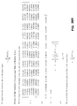
- Fig. 3 is an equation illustrating the mathematical relationship between the input and output of a typical neuron.
- Fig. 4 is a schematic illustration of the second preferred embodiment of the present invention.
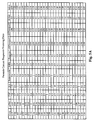
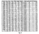
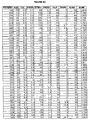
- Fig. 5 shows the training data used to construct the prostate cancer neural network prognostic system.
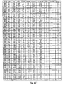
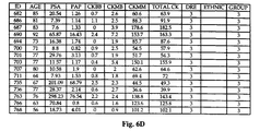
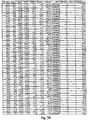
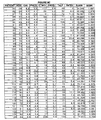
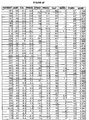
- Fig. 6 shows the training data used to construct the neural network ProstAsureTM system for prostate cancer detection.
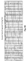
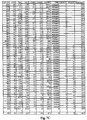
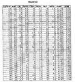
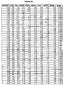
- Fig. 7 shows the test data used to construct the neural network ProstAsureTM system for prostate cancer detection.
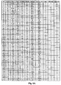
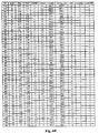
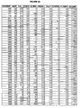
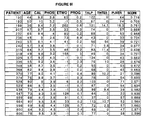
- Fig. 8 shows the training data used to construct the QuiOsTM osteoporosis neural network diagnostic system.
- Fig. 9 shows the testing data used to test the QuiOsTMosteoporosis neural network diagnostic system.
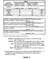
- Fig. 10 demonstrates the sensitivity and specificity of the QuiOsTM system in diagnosing osteopenia.
- Fig. 11 is a scatterplot of 726 test samples showing that QuiOsTM values correlate with bone mineral density (BMD) measurements at L2-L4 and Ward's triangle in the form of T-scores.
- BMD bone mineral density
- Fig. 12 is a schematic representation of the simultaneous multi access reasoning technology.
- Fig. 13 provides an schematic representation of an approach for the construction and training of a computer-based neural network based classifier for the diagnosis and prognosis of disease
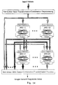
- Fig. 14 shows a configuration of a neural network based diagnostic system
- Fig. 15 is a schematic overview of the ProstAsureTM computer-based neural network system architecture for receiving patient data, analyzing the patient data with a trained neural network and transmitting results.
- Fig. 16 is the system architecture for analyzing patient data input and computation of ProstAsureTM diagnostic values.
- Fig. 17 provides ProstAsureTM reference ranges for normal, BPH and prostate cancer in different age groups.
- Fig. 18 provides diagnostic guidelines for samples in the ProstAsureTM test data set.
- Fig. 19 shows statistically significant ProstAsureTM results in the diagnosis normal, BPH and cancer patients.
- Fig. 20 demonstrates ProstAsureTM results in 193 test cancer cases.
- Fig. 21 is a scatterplot of ProstAsureTM values vs. PSA values in 416 test samples.
- ProstAsureTM effectively separates normal, BPH and cancer patients better than using a single biomarker (PSA).
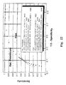
- Fig. 22 shows receiver-operating characteristic (ROC) curves comparing the diagnostic power of ProstAsureTM and PSA alone.
- the area under the curve is a measure of the usefulness of the test.
- the Rel. ProstAsureTM refers to normalization with age-specific reference ranges. ProstAsureTM significantly outperforms PSA with statistical significance in separating cancer from normal and BPH.
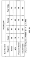
- Fig. 23 demonstrates ProstAsureTM sensitivities and specificities computed with training and test data.
- Fig. 24 shows the sensitivity and specificity of ProstAsureTM in detecting and discriminating prostate cancer and identifying normal and BPH patients.
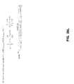
- Fig. 25 is a mathmetical description of the ProstAsureTM algorithm.
- Fig. 26 is a mathematical description of the QuiOsTM algorithm.
- disease is defined as a deviation from the normal structure or function of any part, organ or system of the body (or any combination thereof).
- a specific disease is manifested by characteristic symptoms and signs, including biological, chemical and physical changes and is often associated with a variety of other factors including but not limited to demographic, environmental, employment, genetic and medically historical factors. Certain characteristic signs, symptoms, and related factors can be quantitated through a variety of methods to yield important diagnostic information.
- patient refers to any human or animal.
- biomarkers for the disease.
- Current diagnostic and prognostic methods depend on the identification and evaluation of these biomarkers, both individually and as they relate to one another.
- biomarkers includes all types of biological data from a patient.
- the patient data may include a variety of types of data which have some association with the disease.
- the information may be biological.
- Such data may be derived from measurement of any biological parameter.
- substances include, but are not limited to, endocrine substances such as hormones, exocrine substances such as enzymes, and neurotransmitters, electrolytes, proteins, carbohydrates, growth factors, cytokines, monokines, fatty acids, triglycerides, and cholesterol.
- Other types of biological data may be derived from histological analysis of organs, tissues or cells removed from patients, including histological analyses performed at the light microscopic and electron microscopic levels utilizing any number of techniques including, but not limited to, structural analysis, histochemical, immunocytochemical, in situ hybridization, and autoradiographic techniques.
- Biological data may be derived from analysis of cells removed from patients and grown in culture. Various characteristics of these cells may be examined histologically and biochemically. For example, cells removed from a patient and placed in culture may be examined for the presence of specific markers associated with the presence of a disease. Cells may be examined for their metabolic activity or for the products made and released into the culture medium.
- Biological data about a patient includes results from genetic and molecular biological analysis of the nuclear and cytoplasmic molecules associated with transcription and translation such as various forms of ribonucleic acid, deoxyribonucleic acid and other transcription factors, and the end product molecules resulting from the translation of such ribonucleic acid molecules.
- radiographs are also included in the category of biological data.
- mammograms include X-ray, magnetic resonance imaging, computerized assisted tomography, visualization of radiopaque materials introduced into the body, positron emission tomography, endoscopy, sonograms, echocardiograms, and improvements thereof.
- Biological data also includes data concerning the age, height, growth rate, dental health, cardiovascular status, reproductive status (pre-pubertal, pubertal, post-pubertal, pre-menopausal, menopausal, post-menopausal, fertile, infertile), body fat percentage, and body fat distribution.
- Biological data also includes the results of physical examinations, including but not limited to manual palpation, digital rectal examination, prostate palpation, testicular palpation, weight, body fat amount and distribution, auscultation, testing of reflexes, blood pressure measurements, heart and related cardiovascular sounds, vaginal and other gynecologic examinations, including cervical, uterine and ovarian palpation, evaluation of the uterine tubes, breast examinations, and radiograpic and infrared examination of the breasts.
- physical examinations including but not limited to manual palpation, digital rectal examination, prostate palpation, testicular palpation, weight, body fat amount and distribution, auscultation, testing of reflexes, blood pressure measurements, heart and related cardiovascular sounds, vaginal and other gynecologic examinations, including cervical, uterine and ovarian palpation, evaluation of the uterine tubes, breast examinations, and radiograpic and infrared examination of the breasts.
- Additional biological data can be obtained in the form of a medical history of the patient.
- Such data includes, but is not limited to the following: medical history of ancestors including grandparents and parents, siblings, and descendants, their medical problems, genetic histories, psychological profiles, psychiatric disease, age at death and cause of death; prior diseases and conditions; prior surgeries; prior angioplasties, vaccinations; habits such as exercise schedules, alcohol consumption, cigarette consumption and drug consumption; cardiac information including but not limited to blood pressure, pulse, electrocardiogram, echocardiogram, coronary arteriogram, treadmill stress tests, thallium stress tests and other cardiovascular imaging techniques. All of the aforementioned types of biological data are considered as "biomarkers" for the purposes of the present application.
- biological fluid includes, but is not limited to, blood, serum, cerebrospinal, peritoneal, salivary, lacrimal, peritoneal, reproductive, intraocular, digestive, respiratory, pleural, pericardial, lymphatic, urine, intracellular and extracellular fluids, and neural fluids.
- demographic data includes information concerning the patient's race, species, sex, ethnicity, environment, exposure to environmental toxins and radiation, stress level, behavioral patterns, previous occupations and current occupation. Demographic data may also be used to provide patient information that is useful in the diagnosis and prognosis of disease.
- the present invention provides a method for diagnosing, screening or prognosing a disease in a patient comprising the steps of measuring the concentrations of a predetermined set of biomarkers known to be associated with the disease; converting these concentrations to digitized values; preprocessing the digitized values to make preprocessed values; and sending the preprocessed values to a computer-based neural network in order to train the neural network to diagnose or prognose the disease, whereby the diagnostic index from the neural network indicates when the patient has the disease or may develop the disease.
- the present invention also comprises an apparatus for diagnosing, screening or prognosing a disease in a patient comprising a means for digitizing the concentrations of a predetermined set of biomarkers known to be associated with the disease from the patient; a means for preprocessing the digitized values; and a computer-based trained neural network coupled to the digitizing and scaling means for generating network output values; means for comparing the output values from the neural network to the diagnostic index to produce a diagnostic value which indicates when the patient has the disease or may develop the disease.
- a trained neural network is utilized to determine a diagnostic index corresponding to the presence and severity of a disease by analyzing a set of predetermined biomarkers or demographic data for that disease.
- concentrations of certain biomarkers or demographic data related to the incidence of a particular disease are determined for a patient. These data are converted to digitized values. These digitized values are then preprocessed (scaling, truncation, linear/nonlinear combination, etc.) and the preprocessed values, optionally together with one or several secondary values computed from the original values are then sent to a trained neural network to yield a diagnostic index.
- Preprocessing of the data occurs at this stage and serves to decrease the burden on the neural network and enhance the accuracy and sensitivity of the neural network for diagnosis and prognosis of disease.
- a neural network is trained by introducing a population of patients in which a disease state is known, along with the biomarker values or demographic data for those patients and "teaching" the neural network to recognize the patterns in the biomarkers. After the neural network is trained, biomarker values from patients with unknown disease states are introduced to the trained neural network. The neural network then processes the information to produce an output value whereby the output values from the neural network are diagnostic values which indicate whether the patient has the disease or may develop the disease
- the artificial neural network may, through their weight connections, correspond to data patterns that are important for categorizing diseases. Additionally, the neural network can identify unique patterns of data associated with a variety of disorders that may help to classify borderline cases that do not appear to fit into either a malignant or benign pattern.
- the present invention also comprises a method for diagnosing, screening or prognosing a disease in a patient comprising the steps of measuring the concentrations of a predetermined set of biomarkers known to be associated with the disease from the patient, digitizing the concentrations, preprocessing the digitized values to make preprocessed values, scaling the digitized values of the analytes, and introducing the preprocessed values to a first trained neural network, and sending the output value from the first neural network and a second set of predetermined biomarkers, which could include one or more of the biomarkers in the first set of predetermined biomarkers, to a second trained neural network, whereby the output values from the second neural network are compared to the diagnostic index to produce a diagnostic value which indicates when the patient has the disease or may develop the disease
- a second embodiment of the present invention involves a two step analysis of the biomarkers by neural network. This avoids the bias created by a dominant predictive variable when training a network.
- the dominant biomarker or predictive variable is excluded from the first analysis by neural network and is then included in a second analysis by neural network. For example, if age is thought to be the dominant predictive variable in the diagnosis of osteoporosis, that variable is not included in the training of the first neural network, and the training data set is limited to the other selected biomarkers.
- a second neural network is trained using the diagnostic index and the entire set of input variables, including age, to yield another diagnostic index.
- the final diagnostic index is a composition of an artificial neural network generated index and results from heuristic analysis using other non-numerical patient information.
- the present invention provides a system, including the ProstAsureTM system, comprising an apparatus and method for diagnosing, screening or prognosing prostate cancer in patients.
- data obtained from analysis of biomarkers and optionally from demographic information is preprocessed (e.g. scaled) and input into a trained neural network.
- Prostate specific antigen (PSA), prostatic acid phosphatase (PAP), and three forms of creatine kinase (BB, MB, and MM) are used as the biomarkers in this invention.
- PSA Prostate specific antigen
- PAP prostatic acid phosphatase
- BB creatine kinase
- MB MB
- MM creatine kinase
- other biomarkers and demographic data may be used in this invention.
- the results of a digital rectal examination in which the prostate is palpated may optionally be combined with other biomarkers or demographic data.
- the trained neural network provides an output value which indicates whether the patient has prostate cancer.
- the trained neural network is capable of providing highly accurate diagnoses and prognoses at early stages in the progression of prostate cancer, thereby displaying a high degree of sensitivity and specificity.
- the stage of prostate cancer is determined, even at very early stages in the disease.
- this invention distinguishes benign prostatic hyperplasia from prostate cancer, and distinguishes prostate cancer from non-cancerous conditions.
- Another specific embodiment of the present invention includes a system comprising a method and apparatus for diagnosing and determining the severity and underlying cause of osteopenia and osteoporosis in a patient using a computer-based trained neural network.
- the method comprises determining the serum level of the following biomarkers: calcium, phosphate, total alkaline phosphatase, an alkaline phosphatase isoenzyme, estradiol, and progesterone.
- the alkaline phosphatase isoenzyme is preferably t-lymphocyte derived alkaline phosphatase or blood, liver or intestinal alkaline phosphatase isoenzyme.
- the age of the patient or demographic data may be included in the trained neural network.
- the bone density coefficient that is calculated by the algorithm correlates to a very high degree to bone density as measured by standard methods, such as radiographic absorptometry, quantitative computed tomography, dual photon absorptometry and direct measurement of bone density.
- standard methods such as radiographic absorptometry, quantitative computed tomography, dual photon absorptometry and direct measurement of bone density.
- the bone density coefficient that is measured is then compared to an osteopenic severity scale.
- Another embodiment of the present invention is directed to a computer assisted method for screening, prognosing and diagnosing diseases utilizing a neural network to obtain a conclusive diagnosis.
- the present invention can be adapted to existing diagnostic devices that have a collection means, a sample detecting means capable of detecting the quantity of an analyte in a biological fluid and a means of either printing or displaying the results of the tests on video display means.
- biomarkers collectively alter in response to a disease process, and collectively constitute a new diagnostic biomarker with better disease predictability than the individual biomarkers.
- biomarkers are processed and analyzed as a group in a computer-based trained neural network to yield a single diagnostic index, the sensitivity and specificity of the diagnosis is increased, making it possible for a physician to detect the presence of a disease earlier and with greater precision, or estimate a prognosis with greater precision, than by analysis of the individual biomarkers.
- a biological fluid or several biological fluids are first collected from a patient.
- Biomarkers associated with a specific disease are measured in the biological fluids using standard laboratory techniques, to determine their concentrations, or in some cases their presence or absence. It is to be understood that this process can be carried out automatically in conventional diagnostic machines. For purposes of illustration, descriptions of the methods for obtaining the values for the biomarkers for osteopenia and also for prostate cancer are provided elsewhere in this section.
- the biomarkers relied upon to diagnose a disease by the method of the present invention must be predictive of the suspected disease and must be statistically significant for analysis by a neural network.
- the selection of biomarkers that offers statistically significant discriminating power in the diagnosis of disease involves several steps. First an inventory of biomarkers that have shown certain relevancy in the diagnosis of the disease of interest must be conducted. In general, only the biomarkers that reflect different aspects of the disease process or other diagnostic information need to be included. Second, the selected biomarkers need to have a reasonable diagnostic value in terms of sensitivity, specificity, and positive and negative predictive powers. The design and implementation of experimental protocol from which the biomarkers are developed and evaluated should also be considered. Third, if the number of candidate biomarkers is large, a formal discriminating power analysis may be conducted.
- biomarker values and demographic data values are scaled to provide relatively similar ranges of values between different biomarkers or demographic variables. In this manner, the variances due to the different numerical ranges inherent in the measurement of different variables are decreased.
- Preprocessing of the input variables comprised of biomarkers and other demographic data is an important step in the training of the neural network. If the number of candidates are not too large, they may be all included in the initial attempt of neural network training. If one or several of the input biomarkers to the network are irrelevant to the classification decision making process, it will be reflected in the network connection weights of the trained neural networks. These values may then be removed from the biomarker set for a particular disease.
- Other methods for evaluating the statistical significance of a biomarker selected for analysis by neural network and selecting biomarkers for training a neural network are well known in the art.
- AFP Alpha-Fetoprotein CA125: Cancer Antigen 125 CA 15-3® Breast Antigens 115D8/DF3 CA 19-9: Carbohydrate Antigen 19-9
- CEA Carcinoembryonic Antigen CK-MM Creatine kinase, MM subfraction CK-MB Creatine kinase, MB subfraction CK-BB: Creatine kinase, BB subfraction DM/70K: Ovarian marker NB/70K
- HCG-Beta Human Chorionic Gonadotropin
- LASA-P® Lipid-Associated Sialic Acid in Plasma M-CSF Macrophage colony-stimulating factor
- PAP Prostatic Acid Phosphatase
- PSA Prostate Specific Antigen
- biomarkers and demographic data for the disease must be quantifiable.
- the biomarkers and demographic data must also be predictive of the disease and must be statistically significant relative to one another.
- the method of the present invention is equally suited to the diagnosis of any disease in which biomarkers and demographic data can be identified, including but not limited to infectious diseases, and genetic abnormalities.
- the biomarker values are digitized, preprocessed and analyzed by a computer-based, trained neural network to yield a single diagnostic value.
- the most common neural network architecture for pattern classification problems is the feedforward network, which typically consists of an input layer, one or more hidden layers, and an output layer.
- Figs. 1 and 2 illustrate the arrangement of neurons in two different feedforward networks.
- the elements that make up each layer of a neural network are referred to as neurons or nodes.
- Inputs are fed forward from the input layer to the hidden layers and then to the output layer.
- the number of neurons in each layer is determined before the network is trained.
- the inputs to the neural network are predictor variables. These predictor variables can be quantitative or qualitative. Neural networks make no data distribution assumptions and can simultaneously use both quantitative and qualitative inputs.
- the biomarker values, and the optionally generated secondary values are rescaled during preprocessing to values between 0.0 and 1.0 or between -1.0 and 1.0, constitute the input variables.
- the outputs of the network represent output categories. For example, a malignancy may be represented by maximal output of the malignant output neuron and silence of the benign neuron, whereas a benign process is represented by maximal output of the benign neuron and silence of the malignant neuron.
- a simple arithmetic function combines the outputs of the two neurons to yield a single diagnostic index.
- a single output neuron may be used. An output of greater than 0.5 would indicate a malignancy and an output of less than 0.5 would indicate a benign condition. In this way a diagnostic index is directly obtained.
- a reversed denotation could be used.
- the number of hidden layers and the number of nodes in the hidden layers are configurable parameters that have a significant influence on the performance of the network.
- the optimal number of hidden neurons is determined empirically.
- the means for determining the optimum number of hidden neurons is well known to those skilled in the art and depends on the complexity of the problem being solved.
- one embodiment of the neural network is a multi-layer feedforward perceptron using a backpropogation training algorithm.
- the number of hidden layers and the number of neurons in each hidden layer was determined to adequately match the level of complexity of the diagnostic problem.
- the criteria outlined below are used to determine if a chosen network configuration is appropriate.
- the network achieves a high rate of correctly classifying the training set but fails to accurately classify a large number of samples in the testing set, network structure is probably too complex for the problem being solved, i.e. it has sufficient inherent flexibility to fit the training data set, but not sufficient predictive power to classify the test data set. If this is the case, the number of neurons in the hidden layers should gradually be reduced, or, if there are multiple hidden layers, the hidden layers should be gradually reduced.
- Every input neuron is connected to every neuron in the following hidden layer and neuron in a hidden layer is connected to every neuron in the following adjacent hidden layer or output layer, depending on the number of hidden layers.
- Each of the multiple connections to a particular neuron is weighted.
- each node sums the input activations, multiplied by the respective connection weights plus a bias term.
- the weighted sum then passes through a non-linear output function, typically a sigmoidal function, which gives the network the ability to represent complex non-linear relationships.
- a neuron fires if the sum of the weighted inputs to it are greater than a threshold value. As illustrated in Fig. 3, once a neuron is above a threshold, the magnitude of its output is a sigmoid function of the net input.
- the end result of activity in the neural network is the net output, a complex nonlinear function of the inputs.
- the values of the biomarkers or demographic variables for a specific disease are determined and scaled.
- the biomarkers are fed forward from the input layer to the hidden layer (or layers) and then to the output layer of the neural network.
- the number of neurons in the input layer is determined before the network is trained and corresponds to the number of biomarkers predictive for a specific disease.
- the biomarkers are preselected and biomarker values are preprocessed.
- diagnostic variables may include demographic information.
- the number of neurons in the output layer depends on the type of output desired.
- the number of neurons in the hidden layer is determined empirically during training.
- the neural network used for diagnosing a specific disease must be trained to do so.
- the neural network is trained by back propagation.
- Back propagation refers to the technique of training a neural network to accurately model a set of input and output examples by determining the best connection weights between the values, and is well known in the art.
- Other techniques which may be used to train a neural network for purposes of this invention may include any other non-linear global optimization technique, such as the genetic search algorithm; however, the feed forward, back propagation network is most popular.
- the connection weights in the network are randomized.
- the training data are then presented to the network one datum at a time.
- the training data consists of the biomarker values or demographic values for a group of patients, and the diagnosis for each of those patients.
- the biomarker values, and, optionally, demographic values are digitized and preprocessed and the preprocessed values are the input variables used to train the network.
- the network uses the patient's preprocessed values to estimate a diagnosis, which is then compared to the actual diagnosis. If the network's diagnosis is correct, then the connection strengths and thresholds within the network are not changed, and the next patient is presented to the network.
- connection weights and thresholds in both the hidden layer and the output layer are adjusted to reduce the size of the classification error. After adjustments are made, the next patient is presented. Training proceeds until all patients in the training group are correctly classified or some preset stopping criteria are satisified (e.g. a maximum number of iterations).
- the trainer may set the decision limits regarding the definition of a classification error, i.e. an incorrect diagnosis.
- the relevant parameter is the error tolerance, which specifies how close the estimated output has to be to the actual output to be correct. For example, if two output neurons are used and the training tolerance is set at 5%, the estimate of malignancy is considered correct if the malignant output neuron fires at 95% of maximum and the benign neuron fires at 5% of maximum.
- a correct estimate of a benign diagnosis means that the benign output neuron first at 95% of maximum, while the malignant neuron fires at 5% of maximum.
- the methods for determining a classification error are well known to those skilled in the art.
- a benign normal diagnosis is set at an output of 0.1 and a malignant or abnormal diagnosis is set at an output of 0.9.
- the reversed denotation can be used. Error tolerance is an adjustable parameter and is significant in determining the success of the network at making an accurate diagnosis.
- biomarker values and optionally demographic values from patients with unknown disease conditions and possibly no disease are digitized, preprocessed and introduced to the trained neural network.
- the neural network then processes the information to produce a value corresponding to a diagnosis of the presence or absence of the particular disease. In accordance with the present invention, this is accomplished by using either one single output neuron or multiple output neurons. If more than one output neuron are used, the output from the neurons are combined to generate a single diagnostic index.
- the diagnostic value obtained by analysis of the biomarkers by a trained neural network is further analyzed by a set of heuristic rules in combination with additional patient information.
- the additional patient information includes things such as family medical history and demographic information. This data is then processed to yield a second single diagnostic value.
- the simultaneous, multi-access reasoning technology system of the present invention utilizes both existing knowledge and implicit information that can only be numerically extracted from training data.
- Use of existing knowledge may be in the form of setting normal reference ranges of biomarkers specific to the patient being diagnosed.
- the system has four major functional blocks as described in Figure 12:
- a Neural Shell 2, Release 1.5 (Ward Systems Group, Inc.) neural network development program was used for the training of the neural network on a Pentium 60 mhz computer (Magitronic, Inc.). In other Examples, different computer hardware is used.
- the following example describes the training of a neural network to prognose prostate cancer.
- the training set contained 40 samples (28 stable and 12 progressing) and the generalization testing set contained 12 samples (9 stable and 3 progressing).
- the initial network architecture was selected based on the level of complexity of the classification task.
- a multi-layer feedforward network was used. Selection of the initial architecture involved the selection of the number of hidden layers and the number of neurons in each hidden layer. Several trial iterations were performed to determine an adequate configuration that showed good results on both the training sample set and the generalization test sample set.
- the present network had one hidden layer, having nine neurons, and two output neurons.
- connection weights among the neurons were randomly set.
- the neural network had five input neurons, corresponding to five input variables significant for prostate cancer: TPS, PSA, PAP, CEA, and testosterone.
- the training data are shown in Fig. 5.
- the five input variables for each patient were first linearly scaled into the continuous range between 0.0 and 1.0.
- the resultant five numbers were then presented as an input vector to the input neurons of the artificial neural network.
- the network For each of the input vectors, the network generated an output based on the connection weights among the network neurons.
- the output can be a single value or a vector of numbers, depending on the number of output neurons used.
- the network used had two output neurons.
- Each neuron in the network participated in the output calculation by passing the sum of all inputs to the neuron through a non-linear s-shaped function (often a logistic function) and sending the result to each and every one of the neurons in the following adjacent layer.
- the generated output or each output neuron was compared to the desired "target" output.
- a value of 0.1 corresponded to a diagnosis of stable and an output of 0.9 corresponded to a diagnosis of progressing.
- the difference was used to calculate an error term to guide the training algorithm, i.e., the back propagation algorithm, in the adjustment of network connection weights in an attempt to reduce the differences between network outputs and target values over the training sample set.
- the neural network correctly classified 100% of the samples.
- the trained neural network When presented with the generalization test results, the trained neural network correctly identified 100% of the stable samples and 66% of the samples where the disease was progressing.
- the mathematical description of the ProstAsureTM Algorithm is provided in Figure 25 which is attached hereto.
- the training data and test data for ProstAsureTM are provided in Figures 6 and 7, respectively.
- the training data set shows patient data for age, PSA, PAP, CK-BB, CK-MB, CK-MM, total CK, digital rectal examination, and ethnic group.
- the ProstAsureTM system displayed the following sensitivities and specificities (expressed as a percentage) for cancer detection in the test data and training data: Sensitivity for Detecting Test Training Prostate Cancer (PC) 80.3% 84.4% Stage 2 PC 85.3 85.1 Stages T2, T3 and TNxM1(PC) 87.9 87.9 For detecting BPH as BPH 66.1 68.9 Specificity for Identifying Percent Non-Cancer as Non-Cancer 92.8% 91.8 Presumed Normal as Normal 67.6 69.2
- This example illustrates the construction and training of a neural network for diagnosis of osteoporosis.
- the training and test data are shown in Figs. 8 and 9 respectively.
- the mathematical description of the QuiOsTM algorithm is attached hereto as Figure 26.
- This example illustrates the construction and training of a neural network for diagnosis of osteoporosis.
- Fig. 8 provides the data used to train the neural network to diagnose osteoporosis.
- the biomarkers selected included age, calcium, phosphate, estradiol (ETWO), progesterone, total alkaline phosphatase, total intestinal alkaline phosphatase, and % liver alkaline phosphatase.
- Fig. 8 further includes the diagnostic index obtained by the neural network.
- Fig. 9 provides the data use to test the network trained with the data in Fig. 9, and the neural network diagnostic index obtained.
- the severity of disease in a set of humans or animals with varying severity of disease is measured by a standard method or methods.
- the measurement is then assigned a numerical value corresponding to a severity scale.
- the scale ranges from humans or animals with no disease, to humans or animals with severe disease.
- the scale is preferably a numerical scale. For example, one could assign a value which corresponds to normal or slight disease, another value which corresponds to moderate disease and a third value which corresponds to severe disease.
- the concentration of a predetermined set of blood constituents in the set of humans or animals with varying severity of disease is then determined. According to the present invention, it is preferable to measure the blood constituents in the same set of humans or animals in which the severity of disease was measured by the conventional method or methods.
- An example of practicing one embodiment of the present invention is a method for diagnosing osteopenia in a patient.
- the method preferably utilizes six blood constituents. These constituents are calcium, phosphate, total alkaline phosphatase, an alkaline phosphatase isoenzyme, estradiol, and progesterone.
- the alkaline phosphatase isoenzymes preferred for practicing the present invention include lymphocyte-derived alkaline phosphatase isoenzyme and bone, liver or intestinal alkaline phosphatase isoenzymes.
- the present invention includes calculating a bone density quotient using the aforementioned six blood constituents by entering the values for the tests into an QuiOsTM algorithm which is attached hereto as Figure 26. Age, weight and height are also included in the QuiOsTM algorithm.
- an indication of the underlying cause of the osteopenia can be determined using the present invention. For example, by practicing the present invention as described herein, one can determine whether the osteopenia in a patient is caused by post-menopausal lack of estrogen or is caused by some other condition, such as cancer. This allows the attending physician to be better able to prescribe the appropriate treatment for the osteopenia.
- the test for t-lymphocyte derived alkaline phosphatase is experimental only; however, the test for blood, liver and intestinal alkaline phosphatase isoenzymes are also known.
- the type of test used to determine the six serum constituents is not critical to the present invention as long as the tests give accurate blood concentrations of the constituents being measured. The results are shown in Figures 10 and 11 and show excellent sensitivity and specificity in the diagnosis of osteopenia.
- This is a computer-based system which provides the capability to receive patient data, analyze the data with a trained neural network, produce an output value indicative of the presence or absence of prostate cancer, transmit the value to another computer, and transmit the value to another location.
- the system is schematically illustrated in Figure 15.
- the individual boxes in Fig. 15 are numbered and referred to in the following description.
- This system provides high capacity to receive and analyze a large volume of patient data, to rapidly produce output values to diagnose prostate cancer and to optionally transmit these results to remote locations.
- This system permits the rapid analysis of numerous sets of patient data and provides diagnostic values to the clinical laboratory and to the health care provider.
- Figure 15 represents a preferred embodiment of the present invention and that other system configurations, such as different hardware configurations involving a single computer or multiple computers may be employed in the practice of this invention for the diagnosis of any disease including prostate cancer.
- the data files contain data from patient tests that are required for the computation of the diagnostic index for prostate cancer.
- the data file is a standard ASCII file. Each patient record consists of one line in the file. Lines in the file are delimited with carriage return; line feed (CR/LF) pairs.
- the fields in a record are delimited with an ASCII character ",", and each record contains the following seven fields: 1) identification (ID)- alphanumeric; 2) Age - numeric; 3) prostate specific antigen (PSA)- numeric; 4) PAP - numeric; 5) CKBB - numeric; 6) CKMB - numeric; 7) CKMM - numeric.
- Each alphanumeric field contains a string of characters consisting of letters 'a' through 'z', 'A' through 'Z', digits '0' through '9', and the characters '_', '.', '$'.
- a numeric field contains a string representation of a decimal number. It may contain a single decimal point '.'. The space character ' ' and comma character ',' are not allowed within a number.
- Each patient record occupies a single line in the input data file. Data fields in a record are separated by commas. In the result file to be returned to LabCorp, the input values are repeated and then followed by two additional data fields also separated by commas: the computed ProstAsureTM (HORUS Therapeutics, Rochester, NY) value, and an integer valued error code.
- the computed ProstAsureTM HORUS Therapeutics, Rochester, NY
- a patient record Prior to the actual computation of the ProstAsureTM value, a patient record is checked for errors according to the following error detection criteria in the order as they are listed as shown in Fig. 16. Whenever an error other than code 130 happens, error check stops. No ProstAsureTM Value is computed for the patient.
- the ProstAsureTM field in the output file record is filled with "xxxx" and the first non-130 error code will attach as the last field.
- the ProstAsureTM value is computed and reported in the result file record. The error code 0 or 130 is attached accordingly as the last field.
- the ProstAsureTM system consists of two computer workstations and associated communication devices and links (Fig. 15). The following paragraphs provide an overview of the system.
- ProstAsureTM Station I (14) is primarily a communication control station. It downloads test data from a remote computer system (LabCorp) (1) via a modem (2) and a telephone line and uploads the ProstAsureTM results back to the remote system. Station I (14) is linked to Station II (15) by a direct null modem cable (7) on their corresponding serial ports. Station I (14) sends successfully downloaded data files to Station II (15) and receives ProstAsureTM result files from Station II for uploading. Station I controls the timing of download intervals. It processes error conditions (6) by retrying and/or alarming operators (13) depending on the error conditions. Station I compares the data portions of outgoing result files and incoming data files (5) before uploading (4) and (3) to ensure integrity of the result files.
- LabCorp remote computer system
- Station I (14) is linked to Station II (15) by a direct null modem cable (7) on their corresponding serial ports.
- Station I (14) sends successfully downloaded data files to Station II (15) and receives
- ProstAsureTM Station II (15) is the main computation station for ProstAsureTM algorithm computation (11) and houses the trained neural network (11). Station II (15) receives data files from Station I (14) and sends (8 and 9) results files to Station I (14). Station II (15) reads and verifies the data received from Station I. If invalid data are detected (12 and 10), the ProstAsureTM result field will be marked with "x" characters and a special error code will also reflect the condition. If the data values are detected to be unusual but valid, the ProstAsureTM result will be provided; however, an error code will indicate the condition.
- Station II invokes a dynamic link library (DLL) from the commercial software package NSHELL2 release 2.0 (Ward Systems Group, Inc., Frederick, MD) a leading neural network software, to perform the computations of neural network structures.
- DLL dynamic link library
- Station II contains error handling procedure (10) to process various error conditions. It alarms (9 and 10) the operators (13) under critical error conditions.
- Station II is also responsible for archiving the original data files and result files.
- the ProstAsureTM system uses Kermit software (Columbia University) for communications between two Stations I and II and between Station I and remote computer systems. Kermit is reliable, well tested communication protocol.
- the ProstAsureTM system software runs under Microsoft Windows environment, which provides a consistent, friendly user interface. ProstAsureTM software is designed to execute in full screen mode to simplify the operations.
- the system requires a laser printer that has Microsoft Windows supported printer driver. Also required is null modem 7 and RS-232 cable for connection between Station I and II via serial ports.
- the term "emergent warning procedure” defines an automated procedure to report an abnormal situation in the computer software or hardware system, or in the data file transferring mechanism that requires the immediate attention or intervention of a human operator and/or the Director of Operation 13.
- the affected computer produces a loud siren that can be heard throughout the facility; 2) the affected computer screen blinks and displays the error message and the corresponding error code; and 3) the computer system automatically dials the pager number of the officer on duty.
- Station I 14 downloads patient test data from and uploads computed results back to the host computer (1) (a HP3000 UNIX based workstation) located at the Laboratory Corporation of America (LabCorp) facility in Research Triangle Park, North Carolina.
- Station I serves as a buffer and a firewall between the external data source (LabCorp host computer) and the ProstAsureTM processing workstation (Station II) 15 .
- the following are detailed descriptions of functions provided by Station I. These are also the functions tested during system validation.
- Station II receives data from and sends data to Station I.
- Station II processes data using the ProstAsureTM algorithm. The following functions are provided by Station II. These functions are tested during system validation.
- the present invention includes use of a computer-assisted neural network to diagnose ovarian cancer.
- This new version uses biomarkers listed above under the heading of Ovarian Cancer II and includes CA125, M-CSF, OVX1, LASA, CAA7-24 and CA19-9. When tested with an independent data set of 186 subjects, the test achieves a sensitivity of 89% and a specificity of 89%.
Landscapes
- Health & Medical Sciences (AREA)
- Engineering & Computer Science (AREA)
- Epidemiology (AREA)
- General Health & Medical Sciences (AREA)
- Medical Informatics (AREA)
- Primary Health Care (AREA)
- Public Health (AREA)
- Measuring And Recording Apparatus For Diagnosis (AREA)
- Medical Treatment And Welfare Office Work (AREA)
- Investigating Or Analysing Biological Materials (AREA)
- Eye Examination Apparatus (AREA)
- Other Investigation Or Analysis Of Materials By Electrical Means (AREA)
- Automatic Analysis And Handling Materials Therefor (AREA)
- Management, Administration, Business Operations System, And Electronic Commerce (AREA)
- Apparatus For Radiation Diagnosis (AREA)
Claims (22)
- Procédé pour établir un diagnostic et (ou) un pronostic concernant une maladie chez un patient, comprenant les étapes consistant à:convertir en valeurs numérisées des concentrations de marqueurs biologiques d'un patient, associés à la maladie,prétraiter les valeurs numérisées pour réaliser des valeurs prétraitées en utilisant uniquement les marqueurs biologiques du patient qui présentent un pouvoir discriminatif,entrer les valeurs prétraitées dans un ordinateur qui contient un réseau neuronal entraíné,introduire les valeurs prétraitées dans le réseau neuronal entraíné, le réseau neuronal entraíné étant entraíné de manière spécifique pour diagnostiquer et (ou) pronostiquer la maladie ou pour déterminer la gravité de la maladie, et produire une valeur de sortie (Yi), la valeur de sortie correspondant à la présence ou à l'absence ou à la gravité de la maladie, ettransmettre la valeur de sortie du réseau neuronal entraíné à un récepteur de valeur de sortie connecté à un moyen d'affichage.
- Procédé selon la revendication 1, comprenant en outre les étapes consistant à:convertir en valeurs numérisées des données démographiques du patient,prétraiter les valeurs numérisées pour réaliser des valeurs prétraitées,entrer les valeurs prétraitées dans l'ordinateur, etintroduire les valeurs prétraitées dans le réseau neuronal entraíné.
- Procédé selon la revendication 1, comprenant en outre les étapes consistant à:introduire, dans un deuxième réseau neuronal entraíné d'un ordinateur, la valeur de sortie du réseau neuronal entraíné et un deuxième ensemble de valeurs prétraitées à partir de marqueurs biologiques du patient, ou éventuellement d'un deuxième ensemble de valeurs prétraitées à partir de données démographiques du patient, le deuxième réseau neuronal entraíné ayant appris à diagnostiquer ou pronostiquer la maladie et produisant une deuxième valeur de sortie, la deuxième valeur de sortie correspondant à la présence ou à l'absence ou à la gravité de la maladie, ettransmettre, à un récepteur de valeur de sortie connecté à un moyen d'affichage, la deuxième valeur de sortie du deuxième réseau neuronal entraíné.
- Procédé selon la revendication 1, comprenant les étapes consistant à:inscrire dans un dossier de patient les données concernant les marqueurs biologiques du patient et éventuellement les données démographiques du patient,introduire dans un ordinateur les données figurant dans le dossier du patient, ordinateur dans lequel les données sont analysées pour détecter des erreurs de format de données,convertir en valeurs numérisées les données du patient ne comportant aucune erreur de format,prétraiter les valeurs numérisées pour réaliser des valeurs prétraitées, introduire les valeurs prétraitées dans un réseau neuronal entraíné, le réseau neuronal entraíné ayant appris de manière spécifique à diagnostiquer ou pronostiquer la maladie et à produire une valeur de sortie qui indique la présence ou l'absence ou la gravité de la maladie, etinscrire dans le dossier du patient la valeur de sortie et les données relatives au patient.
- Procédé selon l'une quelconque des revendications 1 à 4, dans lequel la maladie est sélectionnée dans le groupe comprenant l'ostéoporose, l'ostéopénie, le cancer du sein, le cancer de l'ovaire, le cancer du colon, le cancer de la prostate et le cancer du testicule.
- Procédé selon la revendication 5, dans lequel la maladie est le cancer de la prostate et les marqueurs biologiques sont l'âge du patient, LASA-A®, PAP, PSA, CK-MB, CK-MM, CK-BB ou n'importe quelle combinaison des marqueurs biologiques.
- Procédé selon la revendication 5, dans lequel la maladie est l'ostéoporose et les marqueurs biologiques sont l'âge du patient, la calcémie, la phosphatémie, l'estradiol, la progestérone, l'ALP, l'Isoenzyme 1 de l'ALP, l'Isoenzyme 2 de l'ALP, ou n'importe quelle combinaison des marqueurs biologiques.
- Procédé selon la revendication 5, dans lequel la maladie est le cancer de l'ovaire et les marqueurs biologiques sont l'âge du patient, LASA-P®, CA125, CA125II, DM/70K, MCSF, OVX1, CA7-24, CA19-9 ou n'importe quelle combinaison des marqueurs biologiques.
- Procédé selon la revendication 5, dans lequel la maladie est le cancer du sein et les marqueurs biologiques sont l'âge du patient, LASA-P®, CEA, CA15-3®, HER2/neu, ou n'importe quelle combinaison des marqueurs biologiques.
- Procédé selon la revendication 5, dans lequel la maladie est le cancer du testicule et les marqueurs biologiques sont l'âge du patient, LASA-P®, AFP, HCG-Beta, CA15-3® ou n'importe quelle combinaison des marqueurs biologiques.
- Procédé selon la revendication 5, dans lequel la maladie est le cancer du colon et les marqueurs biologiques sont l'âge du patient, LASA-P®, CA19-9, CEA ou n'importe quelle combinaison des marqueurs biologiques.
- Procédé pour entraíner un réseau neuronal assisté par ordinateur, destiné à établir un diagnostic et un pronostic d'une maladie chez un patient, comprenant les étapes consistant à:prétraiter des marqueurs biologiques d'un patient, ledit procédé comprenant les étapes consistant à:sélectionner des marqueurs biologiques du patient, associés à un processus pathologique,tester de manière statistique et/ou informatique la puissance discriminative pour indiquer individuellement, par une combinaison linéaire et/ou non linéaire, la présence ou l'absence de la maladie dans les marqueurs biologiques choisis du patient,appliquer des outils statistiques, mathématiques ou informatiques, et/ou une connaissance intelligente pour l'obtention d'entrées secondaires pour le réseau neuronal, qui soient des combinaisons linéaires ou non linéaires des marqueurs biologiques originels ou transformés,sélectionner uniquement les marqueurs biologiques du patient ou les entrées secondaires obtenues qui présentent un pouvoir discriminatif, etentraíner le réseau neuronal assisté par ordinateur à l'aide des marqueurs biologiques prétraités du patient ou des entrées secondaires obtenues.
- Procédé selon la revendication 1 ou 12, dans lequel les marqueurs biologiques du patient sont des données démographiques, des données diagnostiques électriques, des données diagnostiques d'imagerie, des données diagnostiques histologiques et des concentrations de marqueurs biologiques dans un fluide biologique ou une combinaison de ces marqueurs biologiques.
- Procédé pour établir un diagnostic ou un pronostic de maladie chez un patient, comprenant les étapes consistant à:introduire des marqueurs biologiques d'un patient dans le réseau neuronal entraíné assisté par ordinateur, entraíné suivant le procédé selon la revendication 12,recevoir du réseau neuronal assisté par ordinateur une valeur de sortie correspondant à la présence ou l'absence ou la gravité de la maladie, ettransmettre la valeur de sortie du réseau neuronal assisté par ordinateur à un récepteur de valeur de sortie connecté à un moyen d'affichage.
- Procédé selon la revendication 14, comprenant en outre l'étape consistant à introduire dans un deuxième réseau neuronal entraíné d'un ordinateur la valeur de sortie du réseau neuronal entraíné assisté par ordinateur et un deuxième ensemble de valeurs prétraitées à partir de marqueurs biologiques du patient, le deuxième réseau neuronal entraíné ayant appris à établir un diagnostic ou un pronostic concernant la maladie et produisant une deuxième valeur de sortie, la deuxième valeur de sortie correspondant à la présence ou l'absence ou la gravité de la maladie.
- Procédé selon la revendication 12, comprenant les étapes consistant à:attribuer au hasard des poids de connexion (Wji) dans le réseau neuronal assisté par ordinateur,obtenir des concentrations de marqueurs biologiques d'un patient. associés à une maladie à partir d'un groupe de patients présentant la maladie,convertir en valeurs numérisées les concentrations des marqueurs biologiques,prétraiter les valeurs numérisées pour réaliser des valeurs prétraitées.entraíner le réseau en introduisant en série les valeurs prétraitées obtenues avec chaque patient, en tant que variables (Xi) d'entrée, par l'intermédiaire d'entrées dans le réseau neuronal,obtenir une valeur de sortie du réseau neuronal, la valeur de sortie étant une fonction complexe non linéaire des entrées et indiquant un diagnostic du réseau neuronal et correspondant à la présence ou à l'absence et (ou) la gravité de la maladie,comparer la valeur de sortie du réseau neuronal avec un diagnostic réel,régler les poids de connexion et les seuils pour réduire l'ampleur de l'erreur de classification si le diagnostic du réseau neuronal ne correspond pas au diagnostic réel,introduire les valeurs prétraitées d'un patient dans le groupe de patients en tant que variables d'entrée, par l'intermédiaire d'entrées dans le réseau neuronal,obtenir une deuxième valeur de sortie du réseau neuronal,comparer la deuxième valeur de sortie du réseau neuronal avec le diagnostic réel et régler les poids de connexion si le diagnostic du réseau neuronal ne correspond pas au diagnostic réel,répéter les étapes précédentes pour introduire dans le réseau neuronal des valeurs prétraitées provenant d'autres patients du groupe,obtenir de nouvelles valeurs de sortie, comparer les nouvelles valeurs de sortie avec le diagnostic réel et éventuellement régler les poids de connexion, etétablir éventuellement des limites décisionnelles concernant une erreur de classification.
- Système comprenant:un ordinateur contenant un réseau neuronal entraíné, le réseau neuronal entraíné ayant appris à établir un diagnostic et/ou un pronostic concernant une maladie, ou à déterminer la gravité d'une maladie, le réseau comprenant un bloc de traitement de données d'entrée, un bloc de modélisation et de simulation mécaniste, un bloc pour la détection de différences et d'anomalies et un sous-système de classification adaptative de modèles.ledit système comprenant en outre:un moyen pour convertir en valeurs numérisées des concentrations de marqueurs biologiques d'un patient associées à la maladie,un moyen pour prétraiter les valeurs numériques afin de produire des valeurs prétraitées,un moyen pour entrer les valeurs prétraitées dans le réseau neuronal entraíné, dans lequel le réseau neuronal entraíné produit une valeur de sortie, la valeur de sortie correspondant à la présence ou à l'absence ou à la gravité de la maladie, etun moyen pour communiquer la valeur de sortie.
- Système selon la revendication 17, comprenant en outre:un moyen pour convertir en valeurs numérisées des données démographiques d'un patient,un moyen pour prétraiter les valeurs numérisées afin de réaliser des valeurs prétraitées,un moyen pour entrer les valeurs prétraitées dans le réseau neuronal entraíné, le réseau neuronal entraíné produisant une valeur de sortie, la valeur de sortie correspondant à la présence ou à l'absence ou à la gravité de la maladie, etun moyen pour communiquer la valeur de sortie.
- Système selon la revendication 17 ou 18, comprenant en outre plus d'un réseau neuronal entraíné, les réseaux neuronaux entraínés ayant appris d'une manière spécifique à établir un diagnostic et un pronostic concernant une maladie et produisant chacun une valeur de sortie, la valeur de sortie correspondant à la présence ou l'absence ou la gravité de la maladie.
- Système selon l'une quelconque des revendications 17 à 19, dans lequel la maladie est sélectionnée dans le groupe comprenant l'ostéoporose, l'ostéopénie, le cancer du sein, le cancer de l'ovaire, le cancer du colon, le cancer de la prostate et le cancer du testicule.
- Système selon l'une quelconque des revendications 17 à 20, comprenant en outre un ordinateur distant servant à recevoir la valeur de sortie du moyen servant à communiquer la valeur de sortie.
- Support lisible par un ordinateur, sur lequel sont stockées des instructions exécutables par un ordinateur pour mettre en oeuvre le procédé selon l'une quelconque des revendications 1 à 16.
Applications Claiming Priority (6)
| Application Number | Priority Date | Filing Date | Title |
|---|---|---|---|
| US142595P | 1995-07-25 | 1995-07-25 | |
| US1425P | 1995-07-25 | ||
| US61425 | 1995-07-25 | ||
| US08/642,848 US5769074A (en) | 1994-10-13 | 1996-05-03 | Computer assisted methods for diagnosing diseases |
| US642848 | 1996-05-03 | ||
| PCT/US1996/012177 WO1997005553A1 (fr) | 1995-07-25 | 1996-07-25 | Procedes de diagnostic de maladies assiste par ordinateur |
Publications (2)
| Publication Number | Publication Date |
|---|---|
| EP0842475A1 EP0842475A1 (fr) | 1998-05-20 |
| EP0842475B1 true EP0842475B1 (fr) | 2000-11-08 |
Family
ID=26669012
Family Applications (1)
| Application Number | Title | Priority Date | Filing Date |
|---|---|---|---|
| EP96927247A Expired - Lifetime EP0842475B1 (fr) | 1995-07-25 | 1996-07-25 | Procedes et appareil de diagnostic de maladies assiste par ordinateur |
Country Status (13)
| Country | Link |
|---|---|
| EP (1) | EP0842475B1 (fr) |
| JP (2) | JPH11504739A (fr) |
| CN (1) | CN1194045A (fr) |
| AT (1) | ATE197511T1 (fr) |
| AU (1) | AU717449B2 (fr) |
| CA (1) | CA2227543C (fr) |
| DE (1) | DE69610926T2 (fr) |
| DK (1) | DK0842475T3 (fr) |
| ES (1) | ES2152548T3 (fr) |
| GR (1) | GR3035370T3 (fr) |
| NZ (1) | NZ315428A (fr) |
| PT (1) | PT842475E (fr) |
| WO (1) | WO1997005553A1 (fr) |
Cited By (6)
| Publication number | Priority date | Publication date | Assignee | Title |
|---|---|---|---|---|
| WO2003030705A3 (fr) * | 2001-10-09 | 2003-09-12 | G Schuhfried Ges M B H Dr | Dispositif d'evaluation de donnees brutes psychologiques et biomedicales |
| US7608458B2 (en) | 2004-02-05 | 2009-10-27 | Medtronic, Inc. | Identifying patients at risk for life threatening arrhythmias |
| DE102009009228A1 (de) * | 2009-02-17 | 2010-08-26 | GEMAC-Gesellschaft für Mikroelektronikanwendung Chemnitz mbH | Verfahren und Vorrichtung zur agglutinationsbasierten erkennung von spezifischen Erkankungen über einen Bluttest |
| US8027791B2 (en) | 2004-06-23 | 2011-09-27 | Medtronic, Inc. | Self-improving classification system |
| US8335652B2 (en) | 2004-06-23 | 2012-12-18 | Yougene Corp. | Self-improving identification method |
| WO2020144645A1 (fr) * | 2019-01-10 | 2020-07-16 | 3M Innovative Properties Company | Amélioration de hiérarchisation de document à l'aide de codes générés automatisés |
Families Citing this family (88)
| Publication number | Priority date | Publication date | Assignee | Title |
|---|---|---|---|---|
| US5660176A (en) | 1993-12-29 | 1997-08-26 | First Opinion Corporation | Computerized medical diagnostic and treatment advice system |
| USRE43433E1 (en) | 1993-12-29 | 2012-05-29 | Clinical Decision Support, Llc | Computerized medical diagnostic and treatment advice system |
| US6206829B1 (en) | 1996-07-12 | 2001-03-27 | First Opinion Corporation | Computerized medical diagnostic and treatment advice system including network access |
| US6678669B2 (en) | 1996-02-09 | 2004-01-13 | Adeza Biomedical Corporation | Method for selecting medical and biochemical diagnostic tests using neural network-related applications |
| US6309816B1 (en) | 1997-04-16 | 2001-10-30 | Horus Therapeutics, Inc. | Methods for diagnosing cancer by measuring creatine kinase |
| US6394952B1 (en) | 1998-02-03 | 2002-05-28 | Adeza Biomedical Corporation | Point of care diagnostic systems |
| EP1223852A4 (fr) * | 1999-10-15 | 2007-07-18 | W Jean Dodds | Diagnostic de sante animale |
| US20030040018A1 (en) * | 2000-04-28 | 2003-02-27 | Norman Bitterlich | Method for determining significant losses in bone density |
| US7567870B1 (en) | 2000-07-31 | 2009-07-28 | Institute For Systems Biology | Multiparameter analysis for predictive medicine |
| US7315784B2 (en) | 2001-02-15 | 2008-01-01 | Siemens Aktiengesellschaft | Network for evaluating data obtained in a biochip measurement device |
| US20030013120A1 (en) * | 2001-07-12 | 2003-01-16 | Patz Edward F. | System and method for differential protein expression and a diagnostic biomarker discovery system and method using same |
| US7343247B2 (en) | 2001-07-30 | 2008-03-11 | The Institute For Systems Biology | Methods of classifying drug responsiveness using multiparameter analysis |
| JP3823192B2 (ja) * | 2002-04-19 | 2006-09-20 | 学校法人慶應義塾 | 診療支援装置、診療支援方法、及び診療支援プログラム |
| AU2003265743B2 (en) * | 2002-10-03 | 2008-02-14 | Joachim Diederich | Method and apparatus for assessing psychiatric or physical disorders |
| US20050228236A1 (en) * | 2002-10-03 | 2005-10-13 | The University Of Queensland | Method and apparatus for assessing psychiatric or physical disorders |
| US7367339B2 (en) * | 2002-10-03 | 2008-05-06 | Scott Laboratories, Inc. | Neural networks in sedation and analgesia systems |
| WO2004052191A1 (fr) * | 2002-12-09 | 2004-06-24 | Ajinomoto Co., Inc. | Processeur d'informations sur l'etat de l'organisme, procede de traitement d'informations sur l'etat de l'organisme, systeme de gestion d'informations sur l'etat de l'organisme, programme, et support d'enregistrement |
| DE10317717B4 (de) * | 2003-04-17 | 2005-02-24 | Pe Diagnostik Gmbh | Verfahren zur Diagnose von Erkrankungen unter Verwendung von Indikatorstoffen |
| EP1723432A1 (fr) | 2004-02-05 | 2006-11-22 | Medtronic, Inc. | Methodes et appareil permettant d'identifier des patients presentant un risque de developper des arythmies mettant leur vie en danger |
| US9081879B2 (en) | 2004-10-22 | 2015-07-14 | Clinical Decision Support, Llc | Matrix interface for medical diagnostic and treatment advice system and method |
| CN100349548C (zh) * | 2004-12-23 | 2007-11-21 | 香港理工大学 | 一种早期检测特发性脊柱侧凸的智能增强系统 |
| US20070081700A1 (en) * | 2005-09-29 | 2007-04-12 | General Electric Company | Systems, methods and apparatus for creation of a database of images from categorical indices |
| US20070156382A1 (en) * | 2005-12-29 | 2007-07-05 | Graham James L Ii | Systems and methods for designing experiments |
| US9022930B2 (en) | 2006-12-27 | 2015-05-05 | Cardiac Pacemakers, Inc. | Inter-relation between within-patient decompensation detection algorithm and between-patient stratifier to manage HF patients in a more efficient manner |
| US7629889B2 (en) | 2006-12-27 | 2009-12-08 | Cardiac Pacemakers, Inc. | Within-patient algorithm to predict heart failure decompensation |
| AU2008310577A1 (en) * | 2007-10-12 | 2009-04-16 | Patientslikeme, Inc. | Self-improving method of using online communities to predict health-related outcomes |
| JP5204471B2 (ja) * | 2007-12-12 | 2013-06-05 | シスメックス株式会社 | 動物検査情報提供システム、中央装置、動物用分析装置、動物検査情報提供方法およびコンピュータプログラム |
| JP5203687B2 (ja) * | 2007-12-12 | 2013-06-05 | シスメックス株式会社 | 動物検査情報提供システム、中央装置、動物検査情報提供方法及びコンピュータプログラム |
| EP2255308A4 (fr) * | 2008-02-08 | 2015-07-22 | Phadia Ab | Méthode, progiciel et système conçus pour permettre une aide à la décision clinique |
| JP5280735B2 (ja) * | 2008-05-07 | 2013-09-04 | 紀文 日比 | Peg施行患者の予後の予測装置、及びpeg施行患者の予後の予測プログラム |
| CN102084366A (zh) * | 2008-05-12 | 2011-06-01 | 皇家飞利浦电子股份有限公司 | 医学分析系统 |
| AU2009295315A1 (en) | 2008-09-25 | 2010-04-01 | Cae Healthcare Inc. | Simulation of medical imaging |
| JP2010231308A (ja) * | 2009-03-26 | 2010-10-14 | Olympus Corp | 生活習慣病予防装置および生活習慣病予防プログラム |
| EP2430574A1 (fr) | 2009-04-30 | 2012-03-21 | Patientslikeme, Inc. | Systèmes et procédés d'encouragement pour soumission de données dans des communautés en ligne |
| KR101244252B1 (ko) * | 2009-11-23 | 2013-03-25 | 한국과학기술정보연구원 | 역학 시뮬레이션 시스템 및 방법 |
| CN105149023B (zh) * | 2010-06-30 | 2018-06-12 | 安派科生物医学科技有限公司 | 疾病检测仪 |
| CA3141754A1 (fr) * | 2011-10-24 | 2013-05-02 | President And Fellows Of Harvard College | Amelioration du diagnostic d'un trouble par intelligence artificielle et technologies mobiles de soins sans compromission de la precision |
| KR101993716B1 (ko) * | 2012-09-28 | 2019-06-27 | 삼성전자주식회사 | 카테고리별 진단 모델을 이용한 병변 진단 장치 및 방법 |
| HK1216765A1 (zh) * | 2013-03-14 | 2016-12-02 | 奥特拉西斯公司 | 使用所测分析物改进疾病诊断的方法 |
| JP2016519807A (ja) * | 2013-03-15 | 2016-07-07 | ザ クリーブランド クリニック ファウンデーションThe Cleveland ClinicFoundation | 自己進化型予測モデル |
| US10874355B2 (en) | 2014-04-24 | 2020-12-29 | Cognoa, Inc. | Methods and apparatus to determine developmental progress with artificial intelligence and user input |
| US20160070879A1 (en) * | 2014-09-09 | 2016-03-10 | Lockheed Martin Corporation | Method and apparatus for disease detection |
| CN104523264B (zh) * | 2014-12-31 | 2017-01-18 | 深圳职业技术学院 | 一种心电信号处理方法 |
| CN113421652B (zh) * | 2015-06-02 | 2024-06-28 | 推想医疗科技股份有限公司 | 对医疗数据进行分析的方法、训练模型的方法及分析仪 |
| US11972336B2 (en) | 2015-12-18 | 2024-04-30 | Cognoa, Inc. | Machine learning platform and system for data analysis |
| CN105956378A (zh) * | 2016-04-26 | 2016-09-21 | 成都聚恒康科技有限公司 | 肿瘤诊疗临床决策支持系统 |
| EP3481283B1 (fr) * | 2016-07-06 | 2020-09-09 | Koninklijke Philips N.V. | Surveillance des caractéristiques d'os |
| CN106202968B (zh) * | 2016-07-28 | 2020-02-07 | 北京柏惠维康科技有限公司 | 癌症的数据分析方法及装置 |
| EP3312748A1 (fr) * | 2016-10-19 | 2018-04-25 | Fujitsu Limited | Procédé permettant d'aider un diagnostic, programme et appareil |
| JP6280997B1 (ja) * | 2016-10-31 | 2018-02-14 | 株式会社Preferred Networks | 疾患の罹患判定装置、疾患の罹患判定方法、疾患の特徴抽出装置及び疾患の特徴抽出方法 |
| CN118609834A (zh) | 2016-11-14 | 2024-09-06 | 科格诺亚公司 | 用于评估发育状况并提供覆盖度和可靠性控制的方法和装置 |
| KR101876338B1 (ko) * | 2016-12-28 | 2018-07-09 | 아주대학교산학협력단 | 신경망을 이용한 간 경변 예측 장치 및 방법 |
| CA3053245A1 (fr) | 2017-02-09 | 2018-08-16 | Cognoa, Inc. | Plate-forme et systeme de medecine personnalisee numerique |
| US12417825B2 (en) | 2017-09-15 | 2025-09-16 | Alden Scientific, Inc. | Systems and methods for collecting and analyzing comprehensive medical information |
| EP3460723A1 (fr) | 2017-09-20 | 2019-03-27 | Koninklijke Philips N.V. | Évaluation des données d'entrée faisant appel à un algorithme d'apprentissage profond |
| CN107895168A (zh) * | 2017-10-13 | 2018-04-10 | 平安科技(深圳)有限公司 | 数据处理的方法、数据处理的装置及计算机可读存储介质 |
| WO2019079182A1 (fr) * | 2017-10-16 | 2019-04-25 | Illumina, Inc. | Apprentissage semi-supervisé pour l'apprentissage d'un ensemble de réseaux neuronaux à convolution profonde |
| US11861491B2 (en) | 2017-10-16 | 2024-01-02 | Illumina, Inc. | Deep learning-based pathogenicity classifier for promoter single nucleotide variants (pSNVs) |
| KR102071491B1 (ko) * | 2017-11-10 | 2020-01-30 | 주식회사 디시젠 | 차세대 염기서열분석을 이용한 기계학습 기반 유방암 예후 예측 방법 및 예측 시스템 |
| CN108231189B (zh) * | 2017-12-12 | 2022-12-09 | 华南师范大学 | 基于双深度神经学习网络的数据处理方法和疾病诊断装置 |
| CN108143411A (zh) * | 2017-12-13 | 2018-06-12 | 东南大学 | 一种面向孤独症诊断的静息态脑电分析系统 |
| CN108333126B (zh) * | 2018-01-17 | 2020-12-01 | 南通市第一人民医院 | 一种人工智能实时动态光谱脑脊液监测系统 |
| KR101951727B1 (ko) * | 2018-02-02 | 2019-02-26 | (주)바이오인프라생명과학 | 멀티 암 판별을 위한 컴퓨팅 방법 및 이를 이용한 컴퓨팅 장치 |
| CN108538393B (zh) * | 2018-04-02 | 2021-12-10 | 重庆邮电大学 | 基于大数据的骨质质量评估专家系统及预测模型建立方法 |
| CN108877950B (zh) * | 2018-04-24 | 2021-07-06 | 绍兴市人民医院 | 一种基于计算机的骨质疏松筛查方法和装置 |
| JP7130038B2 (ja) * | 2018-06-12 | 2022-09-02 | 富士フイルム株式会社 | 内視鏡画像処理装置、内視鏡画像処理装置の作動方法、内視鏡画像処理プログラム及び記憶媒体 |
| JP7608171B2 (ja) * | 2018-06-19 | 2025-01-06 | エリプシス・ヘルス・インコーポレイテッド | 精神的健康評価のためのシステム及び方法 |
| US20190385711A1 (en) | 2018-06-19 | 2019-12-19 | Ellipsis Health, Inc. | Systems and methods for mental health assessment |
| CN109009222A (zh) * | 2018-06-19 | 2018-12-18 | 杨成伟 | 面向心脏病类型和严重程度的智能评估诊断方法及系统 |
| CN109285147B (zh) * | 2018-08-30 | 2020-12-29 | 杭州深睿博联科技有限公司 | 用于乳腺钼靶钙化检测的图像处理方法及装置、服务器 |
| CN112654291B (zh) | 2018-09-10 | 2025-04-01 | 京瓷株式会社 | 推定装置、推定系统以及记录推定程序的计算机可读取的非暂时性记录介质 |
| CN109599185B (zh) * | 2018-11-14 | 2021-05-25 | 金色熊猫有限公司 | 疾病数据处理方法、装置、电子设备及计算机可读介质 |
| US11894139B1 (en) | 2018-12-03 | 2024-02-06 | Patientslikeme Llc | Disease spectrum classification |
| WO2020187952A1 (fr) * | 2019-03-20 | 2020-09-24 | Koninklijke Philips N.V. | Environnement de flux de travail de confirmation d'écho activé par ia |
| CA3134521A1 (fr) | 2019-03-22 | 2020-10-01 | Cognoa, Inc. | Procedes et dispositifs de therapie numerique personnalisee |
| CN110164549A (zh) * | 2019-05-20 | 2019-08-23 | 南通奕霖智慧医学科技有限公司 | 一种基于神经网络分类器的儿科分诊方法及系统 |
| JP7589948B2 (ja) * | 2019-06-11 | 2024-11-26 | 学校法人東海大学 | 罹患判定支援装置、罹患判定支援方法、及び罹患判定支援プログラム |
| US12564342B2 (en) | 2019-09-06 | 2026-03-03 | Cognoa, Inc. | Methods, systems, and devices for the diagnosis of behavioral disorders, developmental delays, and neurologic impairments |
| GB201913666D0 (en) * | 2019-09-23 | 2019-11-06 | Tpp | Data conversion/symptom scoring |
| IT201900019556A1 (it) * | 2019-10-22 | 2021-04-22 | Consiglio Nazionale Ricerche | Metodo per selezionare un gruppo di marcatori biologici e di un vettore di parametri utili nella predizione della probabilità di sopravvivenza a lungo termine al tumore del seno nelle pazienti affetti da tumore al seno |
| JP6734457B1 (ja) * | 2019-10-28 | 2020-08-05 | アニコム ホールディングス株式会社 | 疾患予測システム、保険料算出システム及び疾患予測方法 |
| IL292346B2 (en) | 2019-11-05 | 2025-02-01 | Apeel Tech Inc | Predicting contamination in plant products |
| CN110911010A (zh) * | 2019-11-25 | 2020-03-24 | 深圳安泰创新科技股份有限公司 | 诊疗方案的生成方法、电子设备及计算机可读存储介质 |
| JP7157941B2 (ja) * | 2020-08-05 | 2022-10-21 | 憲一 佐藤 | 癌罹患判定方法、装置、およびプログラム |
| CN112017771B (zh) * | 2020-08-31 | 2024-02-27 | 吾征智能技术(北京)有限公司 | 一种基于精液常规检查数据的疾病预测模型的构建方法及系统 |
| DE102021119035A1 (de) | 2021-07-22 | 2023-01-26 | Marco Willems | Computerimplementiertes Verfahren, Diagnoseunterstützungssystem sowie computerlesbares Speichermedium |
| CN113724861A (zh) * | 2021-09-06 | 2021-11-30 | 汤学民 | 基于深度学习的初步诊断生成方法、装置及计算机设备 |
| CN118675765B (zh) * | 2024-08-23 | 2024-12-13 | 天津易康科技有限公司 | 基于大数据的血清psa异常患者病历分析方法及装置 |
-
1996
- 1996-07-25 EP EP96927247A patent/EP0842475B1/fr not_active Expired - Lifetime
- 1996-07-25 PT PT96927247T patent/PT842475E/pt unknown
- 1996-07-25 NZ NZ315428A patent/NZ315428A/xx not_active IP Right Cessation
- 1996-07-25 DK DK96927247T patent/DK0842475T3/da active
- 1996-07-25 AU AU67131/96A patent/AU717449B2/en not_active Ceased
- 1996-07-25 DE DE69610926T patent/DE69610926T2/de not_active Expired - Lifetime
- 1996-07-25 JP JP9507712A patent/JPH11504739A/ja active Pending
- 1996-07-25 WO PCT/US1996/012177 patent/WO1997005553A1/fr not_active Ceased
- 1996-07-25 CA CA2227543A patent/CA2227543C/fr not_active Expired - Fee Related
- 1996-07-25 AT AT96927247T patent/ATE197511T1/de not_active IP Right Cessation
- 1996-07-25 CN CN96196534A patent/CN1194045A/zh active Pending
- 1996-07-25 ES ES96927247T patent/ES2152548T3/es not_active Expired - Lifetime
-
2001
- 2001-02-06 GR GR20010400197T patent/GR3035370T3/el not_active IP Right Cessation
-
2006
- 2006-07-14 JP JP2006194886A patent/JP2007052774A/ja active Pending
Cited By (6)
| Publication number | Priority date | Publication date | Assignee | Title |
|---|---|---|---|---|
| WO2003030705A3 (fr) * | 2001-10-09 | 2003-09-12 | G Schuhfried Ges M B H Dr | Dispositif d'evaluation de donnees brutes psychologiques et biomedicales |
| US7608458B2 (en) | 2004-02-05 | 2009-10-27 | Medtronic, Inc. | Identifying patients at risk for life threatening arrhythmias |
| US8027791B2 (en) | 2004-06-23 | 2011-09-27 | Medtronic, Inc. | Self-improving classification system |
| US8335652B2 (en) | 2004-06-23 | 2012-12-18 | Yougene Corp. | Self-improving identification method |
| DE102009009228A1 (de) * | 2009-02-17 | 2010-08-26 | GEMAC-Gesellschaft für Mikroelektronikanwendung Chemnitz mbH | Verfahren und Vorrichtung zur agglutinationsbasierten erkennung von spezifischen Erkankungen über einen Bluttest |
| WO2020144645A1 (fr) * | 2019-01-10 | 2020-07-16 | 3M Innovative Properties Company | Amélioration de hiérarchisation de document à l'aide de codes générés automatisés |
Also Published As
| Publication number | Publication date |
|---|---|
| DK0842475T3 (da) | 2000-11-27 |
| GR3035370T3 (en) | 2001-05-31 |
| JP2007052774A (ja) | 2007-03-01 |
| JPH11504739A (ja) | 1999-04-27 |
| CN1194045A (zh) | 1998-09-23 |
| DE69610926D1 (de) | 2000-12-14 |
| AU717449B2 (en) | 2000-03-23 |
| EP0842475A1 (fr) | 1998-05-20 |
| WO1997005553A1 (fr) | 1997-02-13 |
| CA2227543A1 (fr) | 1997-02-13 |
| NZ315428A (en) | 2000-02-28 |
| ATE197511T1 (de) | 2000-11-11 |
| CA2227543C (fr) | 2010-10-05 |
| AU6713196A (en) | 1997-02-26 |
| DE69610926T2 (de) | 2001-06-21 |
| ES2152548T3 (es) | 2001-02-01 |
| PT842475E (pt) | 2001-04-30 |
Similar Documents
| Publication | Publication Date | Title |
|---|---|---|
| EP0842475B1 (fr) | Procedes et appareil de diagnostic de maladies assiste par ordinateur | |
| US5769074A (en) | Computer assisted methods for diagnosing diseases | |
| WO1997005553A9 (fr) | Procedes de diagnostic de maladies assiste par ordinateur | |
| Kapoor et al. | Workflow applications of artificial intelligence in radiology and an overview of available tools | |
| US7228295B2 (en) | Methods for selecting, developing and improving diagnostic tests for pregnancy-related conditions | |
| JP4139822B2 (ja) | ニューラルネットワーク関連アプリケーションを使用して医療診断テストおよび生化学診断テストを選択する方法 | |
| Kahn Jr et al. | Construction of a Bayesian network for mammographic diagnosis of breast cancer | |
| Hong et al. | Machine learning applications in endocrinology and metabolism research: an overview | |
| US5790761A (en) | Method and apparatus for the diagnosis of colorectal cancer | |
| US20090156906A1 (en) | Patient-centric data model for research and clinical applications | |
| JP2008532104A (ja) | 複数の医療関連アウトカムの予測を行い、インターベンション計画の評価を行い、更に同時にバイオマーカー因果性検証を行うことのできる、予測モデルを生成して適用する方法、そのシステム、及びそのコンピュータ・プログラム製品 | |
| CN101443780A (zh) | 用于开发和使用用于预测多种医学结果、评价介入策略以及同时验证生物标志物诱因的预测模型的方法、系统和计算机程序产品 | |
| US5983211A (en) | Method and apparatus for the diagnosis of colorectal cancer | |
| EP4002382B1 (fr) | Utilisation de données médicales temporelles non structurées pour la prédiction de la maladie | |
| Arafat et al. | A Deep Learning Framework for Early Breast Cancer Detection Among US Women: Integrating Mammography and Clinical EHR Data | |
| Woodard et al. | Performance assessment for radiologists interpreting screening mammography | |
| CN111128330A (zh) | 电子病例报告表自动录入方法、装置以及相关设备 | |
| Sun et al. | Prognostication for Patients Receiving Palliative Radiation Therapy | |
| Chen et al. | An analysis of medical informatics and application of computer-aided decision support framework | |
| Flynn et al. | Assessing diagnostic technologies | |
| EP4053853A1 (fr) | Appareil pour déterminer un risque temporel de cancer du sein | |
| Ravliya | AI: Different Aspects in Obstetrics and Gynecology | |
| Albuquerque et al. | A Machine Learning based approach to osteoporosis classification: correlational and comparative analysis between Osseus and DXA exams | |
| CN120511030A (zh) | 一种融合ai的辅助淋巴管肌瘤病lam诊断系统及其读取介质 | |
| HK1015474A (en) | Computer assisted methods for diagnosing diseases |
Legal Events
| Date | Code | Title | Description |
|---|---|---|---|
| PUAI | Public reference made under article 153(3) epc to a published international application that has entered the european phase |
Free format text: ORIGINAL CODE: 0009012 |
|
| 17P | Request for examination filed |
Effective date: 19980205 |
|
| AK | Designated contracting states |
Kind code of ref document: A1 Designated state(s): AT BE CH DE DK ES FI FR GB GR IE IT LI LU MC NL PT SE |
|
| 17Q | First examination report despatched |
Effective date: 19980610 |
|
| GRAG | Despatch of communication of intention to grant |
Free format text: ORIGINAL CODE: EPIDOS AGRA |
|
| GRAG | Despatch of communication of intention to grant |
Free format text: ORIGINAL CODE: EPIDOS AGRA |
|
| GRAG | Despatch of communication of intention to grant |
Free format text: ORIGINAL CODE: EPIDOS AGRA |
|
| GRAH | Despatch of communication of intention to grant a patent |
Free format text: ORIGINAL CODE: EPIDOS IGRA |
|
| GRAH | Despatch of communication of intention to grant a patent |
Free format text: ORIGINAL CODE: EPIDOS IGRA |
|
| GRAA | (expected) grant |
Free format text: ORIGINAL CODE: 0009210 |
|
| AK | Designated contracting states |
Kind code of ref document: B1 Designated state(s): AT BE CH DE DK ES FI FR GB GR IE IT LI LU MC NL PT SE |
|
| REF | Corresponds to: |
Ref document number: 197511 Country of ref document: AT Date of ref document: 20001111 Kind code of ref document: T |
|
| REG | Reference to a national code |
Ref country code: CH Ref legal event code: EP |
|
| REG | Reference to a national code |
Ref country code: DK Ref legal event code: T3 |
|
| REG | Reference to a national code |
Ref country code: IE Ref legal event code: FG4D |
|
| REF | Corresponds to: |
Ref document number: 69610926 Country of ref document: DE Date of ref document: 20001214 |
|
| REG | Reference to a national code |
Ref country code: ES Ref legal event code: FG2A Ref document number: 2152548 Country of ref document: ES Kind code of ref document: T3 |
|
| ITF | It: translation for a ep patent filed | ||
| ET | Fr: translation filed | ||
| REG | Reference to a national code |
Ref country code: CH Ref legal event code: NV Representative=s name: KIRKER & CIE SA |
|
| REG | Reference to a national code |
Ref country code: PT Ref legal event code: SC4A Free format text: AVAILABILITY OF NATIONAL TRANSLATION Effective date: 20010206 |
|
| PLBE | No opposition filed within time limit |
Free format text: ORIGINAL CODE: 0009261 |
|
| STAA | Information on the status of an ep patent application or granted ep patent |
Free format text: STATUS: NO OPPOSITION FILED WITHIN TIME LIMIT |
|
| 26N | No opposition filed | ||
| REG | Reference to a national code |
Ref country code: GB Ref legal event code: IF02 |
|
| PGFP | Annual fee paid to national office [announced via postgrant information from national office to epo] |
Ref country code: MC Payment date: 20040628 Year of fee payment: 9 |
|
| PGFP | Annual fee paid to national office [announced via postgrant information from national office to epo] |
Ref country code: SE Payment date: 20040706 Year of fee payment: 9 |
|
| PGFP | Annual fee paid to national office [announced via postgrant information from national office to epo] |
Ref country code: LU Payment date: 20040728 Year of fee payment: 9 |
|
| PGFP | Annual fee paid to national office [announced via postgrant information from national office to epo] |
Ref country code: GR Payment date: 20050613 Year of fee payment: 10 |
|
| PGFP | Annual fee paid to national office [announced via postgrant information from national office to epo] |
Ref country code: AT Payment date: 20050622 Year of fee payment: 10 |
|
| PGFP | Annual fee paid to national office [announced via postgrant information from national office to epo] |
Ref country code: PT Payment date: 20050630 Year of fee payment: 10 |
|
| PG25 | Lapsed in a contracting state [announced via postgrant information from national office to epo] |
Ref country code: LU Free format text: LAPSE BECAUSE OF NON-PAYMENT OF DUE FEES Effective date: 20050725 |
|
| PG25 | Lapsed in a contracting state [announced via postgrant information from national office to epo] |
Ref country code: SE Free format text: LAPSE BECAUSE OF NON-PAYMENT OF DUE FEES Effective date: 20050726 |
|
| PG25 | Lapsed in a contracting state [announced via postgrant information from national office to epo] |
Ref country code: MC Free format text: LAPSE BECAUSE OF NON-PAYMENT OF DUE FEES Effective date: 20050731 |
|
| PGFP | Annual fee paid to national office [announced via postgrant information from national office to epo] |
Ref country code: BE Payment date: 20050915 Year of fee payment: 10 |
|
| EUG | Se: european patent has lapsed | ||
| PG25 | Lapsed in a contracting state [announced via postgrant information from national office to epo] |
Ref country code: AT Free format text: LAPSE BECAUSE OF NON-PAYMENT OF DUE FEES Effective date: 20060725 |
|
| PG25 | Lapsed in a contracting state [announced via postgrant information from national office to epo] |
Ref country code: BE Free format text: LAPSE BECAUSE OF NON-PAYMENT OF DUE FEES Effective date: 20060731 |
|
| PG25 | Lapsed in a contracting state [announced via postgrant information from national office to epo] |
Ref country code: PT Free format text: LAPSE BECAUSE OF NON-PAYMENT OF DUE FEES Effective date: 20070125 |
|
| REG | Reference to a national code |
Ref country code: PT Ref legal event code: MM4A Free format text: LAPSE DUE TO NON-PAYMENT OF FEES Effective date: 20070125 |
|
| BERE | Be: lapsed |
Owner name: *HORUS THERAPEUTICS INC. Effective date: 20060731 |
|
| PG25 | Lapsed in a contracting state [announced via postgrant information from national office to epo] |
Ref country code: GR Free format text: LAPSE BECAUSE OF NON-PAYMENT OF DUE FEES Effective date: 20070202 |
|
| REG | Reference to a national code |
Ref country code: DE Ref legal event code: R082 Ref document number: 69610926 Country of ref document: DE Representative=s name: FLEISCHER, ENGELS & PARTNER MBB, PATENTANWAELT, DE |
|
| PGFP | Annual fee paid to national office [announced via postgrant information from national office to epo] |
Ref country code: ES Payment date: 20140611 Year of fee payment: 19 |
|
| PGFP | Annual fee paid to national office [announced via postgrant information from national office to epo] |
Ref country code: FI Payment date: 20140710 Year of fee payment: 19 Ref country code: IE Payment date: 20140710 Year of fee payment: 19 Ref country code: DK Payment date: 20140710 Year of fee payment: 19 Ref country code: CH Payment date: 20140714 Year of fee payment: 19 Ref country code: DE Payment date: 20140724 Year of fee payment: 19 Ref country code: NL Payment date: 20140710 Year of fee payment: 19 |
|
| PGFP | Annual fee paid to national office [announced via postgrant information from national office to epo] |
Ref country code: GB Payment date: 20140723 Year of fee payment: 19 Ref country code: FR Payment date: 20140708 Year of fee payment: 19 |
|
| PGFP | Annual fee paid to national office [announced via postgrant information from national office to epo] |
Ref country code: IT Payment date: 20140714 Year of fee payment: 19 |
|
| REG | Reference to a national code |
Ref country code: DE Ref legal event code: R119 Ref document number: 69610926 Country of ref document: DE |
|
| REG | Reference to a national code |
Ref country code: DK Ref legal event code: EBP Effective date: 20150731 |
|
| REG | Reference to a national code |
Ref country code: CH Ref legal event code: PL |
|
| GBPC | Gb: european patent ceased through non-payment of renewal fee |
Effective date: 20150725 |
|
| REG | Reference to a national code |
Ref country code: NL Ref legal event code: MM Effective date: 20150801 |
|
| REG | Reference to a national code |
Ref country code: IE Ref legal event code: MM4A |
|
| PG25 | Lapsed in a contracting state [announced via postgrant information from national office to epo] |
Ref country code: GB Free format text: LAPSE BECAUSE OF NON-PAYMENT OF DUE FEES Effective date: 20150725 Ref country code: DE Free format text: LAPSE BECAUSE OF NON-PAYMENT OF DUE FEES Effective date: 20160202 Ref country code: LI Free format text: LAPSE BECAUSE OF NON-PAYMENT OF DUE FEES Effective date: 20150731 Ref country code: CH Free format text: LAPSE BECAUSE OF NON-PAYMENT OF DUE FEES Effective date: 20150731 Ref country code: IT Free format text: LAPSE BECAUSE OF NON-PAYMENT OF DUE FEES Effective date: 20150725 |
|
| REG | Reference to a national code |
Ref country code: FR Ref legal event code: ST Effective date: 20160331 |
|
| PG25 | Lapsed in a contracting state [announced via postgrant information from national office to epo] |
Ref country code: FI Free format text: LAPSE BECAUSE OF NON-PAYMENT OF DUE FEES Effective date: 20150725 Ref country code: NL Free format text: LAPSE BECAUSE OF NON-PAYMENT OF DUE FEES Effective date: 20150801 Ref country code: FR Free format text: LAPSE BECAUSE OF NON-PAYMENT OF DUE FEES Effective date: 20150731 |
|
| PG25 | Lapsed in a contracting state [announced via postgrant information from national office to epo] |
Ref country code: IE Free format text: LAPSE BECAUSE OF NON-PAYMENT OF DUE FEES Effective date: 20150725 |
|
| REG | Reference to a national code |
Ref country code: ES Ref legal event code: FD2A Effective date: 20160826 |
|
| PG25 | Lapsed in a contracting state [announced via postgrant information from national office to epo] |
Ref country code: DK Free format text: LAPSE BECAUSE OF NON-PAYMENT OF DUE FEES Effective date: 20150731 |
|
| PG25 | Lapsed in a contracting state [announced via postgrant information from national office to epo] |
Ref country code: ES Free format text: LAPSE BECAUSE OF NON-PAYMENT OF DUE FEES Effective date: 20150726 |