EP0947582A1 - A polypeptide structure for use as a scaffold - Google Patents
A polypeptide structure for use as a scaffold Download PDFInfo
- Publication number
- EP0947582A1 EP0947582A1 EP98870065A EP98870065A EP0947582A1 EP 0947582 A1 EP0947582 A1 EP 0947582A1 EP 98870065 A EP98870065 A EP 98870065A EP 98870065 A EP98870065 A EP 98870065A EP 0947582 A1 EP0947582 A1 EP 0947582A1
- Authority
- EP
- European Patent Office
- Prior art keywords
- seq
- amino acid
- sequence
- scaffold
- functionally equivalent
- Prior art date
- Legal status (The legal status is an assumption and is not a legal conclusion. Google has not performed a legal analysis and makes no representation as to the accuracy of the status listed.)
- Withdrawn
Links
- 210000004896 polypeptide structure Anatomy 0.000 title 1
- 230000027455 binding Effects 0.000 claims abstract description 51
- 108090000765 processed proteins & peptides Proteins 0.000 claims abstract description 39
- 102000004196 processed proteins & peptides Human genes 0.000 claims abstract description 33
- 239000012634 fragment Substances 0.000 claims abstract description 29
- 229920001184 polypeptide Polymers 0.000 claims abstract description 29
- 125000003275 alpha amino acid group Chemical group 0.000 claims description 35
- 239000000427 antigen Substances 0.000 claims description 20
- 102000036639 antigens Human genes 0.000 claims description 20
- 108091007433 antigens Proteins 0.000 claims description 20
- 235000001014 amino acid Nutrition 0.000 claims description 14
- 150000001413 amino acids Chemical class 0.000 claims description 13
- 125000000151 cysteine group Chemical group N[C@@H](CS)C(=O)* 0.000 claims description 8
- 229910052739 hydrogen Inorganic materials 0.000 claims description 4
- 239000001257 hydrogen Substances 0.000 claims description 4
- 230000003993 interaction Effects 0.000 claims description 3
- 102000005962 receptors Human genes 0.000 abstract description 9
- 108020003175 receptors Proteins 0.000 abstract description 9
- 108090000623 proteins and genes Proteins 0.000 description 39
- 102000004169 proteins and genes Human genes 0.000 description 32
- 235000018102 proteins Nutrition 0.000 description 28
- 210000004027 cell Anatomy 0.000 description 22
- 108060003951 Immunoglobulin Proteins 0.000 description 15
- 102000018358 immunoglobulin Human genes 0.000 description 15
- 101000914484 Homo sapiens T-lymphocyte activation antigen CD80 Proteins 0.000 description 9
- 102100027222 T-lymphocyte activation antigen CD80 Human genes 0.000 description 9
- 239000002609 medium Substances 0.000 description 7
- 241000724791 Filamentous phage Species 0.000 description 6
- 230000004927 fusion Effects 0.000 description 6
- 229940072221 immunoglobulins Drugs 0.000 description 6
- XLYOFNOQVPJJNP-UHFFFAOYSA-N water Substances O XLYOFNOQVPJJNP-UHFFFAOYSA-N 0.000 description 6
- 230000013595 glycosylation Effects 0.000 description 5
- 238000006206 glycosylation reaction Methods 0.000 description 5
- 229940045513 CTLA4 antagonist Drugs 0.000 description 4
- 101710132601 Capsid protein Proteins 0.000 description 4
- 101710094648 Coat protein Proteins 0.000 description 4
- 102100021181 Golgi phosphoprotein 3 Human genes 0.000 description 4
- 101710125418 Major capsid protein Proteins 0.000 description 4
- 206010028980 Neoplasm Diseases 0.000 description 4
- 101710141454 Nucleoprotein Proteins 0.000 description 4
- 101710083689 Probable capsid protein Proteins 0.000 description 4
- 108020005038 Terminator Codon Proteins 0.000 description 4
- 238000010367 cloning Methods 0.000 description 4
- 230000004048 modification Effects 0.000 description 4
- 238000012986 modification Methods 0.000 description 4
- 238000002823 phage display Methods 0.000 description 4
- 238000012216 screening Methods 0.000 description 4
- 230000001225 therapeutic effect Effects 0.000 description 4
- 102000008203 CTLA-4 Antigen Human genes 0.000 description 3
- 108010021064 CTLA-4 Antigen Proteins 0.000 description 3
- WQZGKKKJIJFFOK-GASJEMHNSA-N Glucose Natural products OC[C@H]1OC(O)[C@H](O)[C@@H](O)[C@@H]1O WQZGKKKJIJFFOK-GASJEMHNSA-N 0.000 description 3
- 101000889276 Homo sapiens Cytotoxic T-lymphocyte protein 4 Proteins 0.000 description 3
- 102000017727 Immunoglobulin Variable Region Human genes 0.000 description 3
- 108010067060 Immunoglobulin Variable Region Proteins 0.000 description 3
- 108010067902 Peptide Library Proteins 0.000 description 3
- 239000012980 RPMI-1640 medium Substances 0.000 description 3
- 108010008281 Recombinant Fusion Proteins Proteins 0.000 description 3
- 102000007056 Recombinant Fusion Proteins Human genes 0.000 description 3
- 230000001580 bacterial effect Effects 0.000 description 3
- 238000001514 detection method Methods 0.000 description 3
- 239000008103 glucose Substances 0.000 description 3
- 102000043321 human CTLA4 Human genes 0.000 description 3
- 239000000463 material Substances 0.000 description 3
- 238000000034 method Methods 0.000 description 3
- 210000001322 periplasm Anatomy 0.000 description 3
- 238000002415 sodium dodecyl sulfate polyacrylamide gel electrophoresis Methods 0.000 description 3
- 108091026890 Coding region Proteins 0.000 description 2
- 241001524679 Escherichia virus M13 Species 0.000 description 2
- 101100061678 Homo sapiens CTLA4 gene Proteins 0.000 description 2
- 102000001706 Immunoglobulin Fab Fragments Human genes 0.000 description 2
- 108010054477 Immunoglobulin Fab Fragments Proteins 0.000 description 2
- 108010076504 Protein Sorting Signals Proteins 0.000 description 2
- 239000006146 Roswell Park Memorial Institute medium Substances 0.000 description 2
- PXIPVTKHYLBLMZ-UHFFFAOYSA-N Sodium azide Chemical compound [Na+].[N-]=[N+]=[N-] PXIPVTKHYLBLMZ-UHFFFAOYSA-N 0.000 description 2
- 230000004075 alteration Effects 0.000 description 2
- AVKUERGKIZMTKX-NJBDSQKTSA-N ampicillin Chemical compound C1([C@@H](N)C(=O)N[C@H]2[C@H]3SC([C@@H](N3C2=O)C(O)=O)(C)C)=CC=CC=C1 AVKUERGKIZMTKX-NJBDSQKTSA-N 0.000 description 2
- 210000004899 c-terminal region Anatomy 0.000 description 2
- 201000011510 cancer Diseases 0.000 description 2
- 231100000433 cytotoxic Toxicity 0.000 description 2
- 230000001472 cytotoxic effect Effects 0.000 description 2
- 230000007423 decrease Effects 0.000 description 2
- 238000013461 design Methods 0.000 description 2
- 230000000694 effects Effects 0.000 description 2
- 125000001165 hydrophobic group Chemical group 0.000 description 2
- 230000001939 inductive effect Effects 0.000 description 2
- BPHPUYQFMNQIOC-NXRLNHOXSA-N isopropyl beta-D-thiogalactopyranoside Chemical compound CC(C)S[C@@H]1O[C@H](CO)[C@H](O)[C@H](O)[C@H]1O BPHPUYQFMNQIOC-NXRLNHOXSA-N 0.000 description 2
- 239000003446 ligand Substances 0.000 description 2
- 210000004698 lymphocyte Anatomy 0.000 description 2
- 238000004519 manufacturing process Methods 0.000 description 2
- 239000012528 membrane Substances 0.000 description 2
- 239000000203 mixture Substances 0.000 description 2
- 108091005763 multidomain proteins Proteins 0.000 description 2
- 239000008188 pellet Substances 0.000 description 2
- 230000004481 post-translational protein modification Effects 0.000 description 2
- 125000002924 primary amino group Chemical group [H]N([H])* 0.000 description 2
- 238000000746 purification Methods 0.000 description 2
- 230000000452 restraining effect Effects 0.000 description 2
- 239000000243 solution Substances 0.000 description 2
- 239000002904 solvent Substances 0.000 description 2
- 239000006228 supernatant Substances 0.000 description 2
- 238000013519 translation Methods 0.000 description 2
- 238000005406 washing Methods 0.000 description 2
- YBJHBAHKTGYVGT-ZKWXMUAHSA-N (+)-Biotin Chemical compound N1C(=O)N[C@@H]2[C@H](CCCCC(=O)O)SC[C@@H]21 YBJHBAHKTGYVGT-ZKWXMUAHSA-N 0.000 description 1
- SULKGYKWHKPPKO-RAJPIYRYSA-N (4s)-4-[[(2r)-2-[[(2s,3r)-2-[[(2s)-4-amino-4-oxo-2-[[(2s)-pyrrolidine-2-carbonyl]amino]butanoyl]amino]-3-hydroxybutanoyl]amino]-3-sulfanylpropanoyl]amino]-5-[[(2s,3s)-1-[[(2r)-1-[[(2s)-1-[[(2s)-1-[[(2s)-1-[[(2s)-1-[[(2r)-1-[[(2s,3r)-1-[[2-[[(1r)-1-carboxy Chemical compound N([C@@H](CC(N)=O)C(=O)N[C@H](C(=O)N[C@@H](CS)C(=O)N[C@@H](CCC(O)=O)C(=O)N[C@@H]([C@@H](C)CC)C(=O)N[C@@H](CS)C(=O)N[C@@H](C)C(=O)N[C@@H](CC=1C=CC(O)=CC=1)C(=O)N[C@@H](C)C(=O)N[C@@H](C)C(=O)N[C@@H](CS)C(=O)N[C@@H]([C@@H](C)O)C(=O)NCC(=O)N[C@@H](CS)C(O)=O)[C@@H](C)O)C(=O)[C@@H]1CCCN1 SULKGYKWHKPPKO-RAJPIYRYSA-N 0.000 description 1
- 101100136076 Aspergillus oryzae (strain ATCC 42149 / RIB 40) pel1 gene Proteins 0.000 description 1
- 101710192393 Attachment protein G3P Proteins 0.000 description 1
- 108010045634 B7 Antigens Proteins 0.000 description 1
- 102000005738 B7 Antigens Human genes 0.000 description 1
- 241000894006 Bacteria Species 0.000 description 1
- 102100021935 C-C motif chemokine 26 Human genes 0.000 description 1
- 108090000565 Capsid Proteins Proteins 0.000 description 1
- 235000000638 D-biotin Nutrition 0.000 description 1
- 239000011665 D-biotin Substances 0.000 description 1
- 108020004414 DNA Proteins 0.000 description 1
- 102000052510 DNA-Binding Proteins Human genes 0.000 description 1
- 101710096438 DNA-binding protein Proteins 0.000 description 1
- KCXVZYZYPLLWCC-UHFFFAOYSA-N EDTA Chemical compound OC(=O)CN(CC(O)=O)CCN(CC(O)=O)CC(O)=O KCXVZYZYPLLWCC-UHFFFAOYSA-N 0.000 description 1
- 238000002965 ELISA Methods 0.000 description 1
- 108050009340 Endothelin Proteins 0.000 description 1
- 102000002045 Endothelin Human genes 0.000 description 1
- 241000588724 Escherichia coli Species 0.000 description 1
- 102000003886 Glycoproteins Human genes 0.000 description 1
- 108090000288 Glycoproteins Proteins 0.000 description 1
- 101800004305 Guanylin Proteins 0.000 description 1
- 102400001367 Guanylin Human genes 0.000 description 1
- 101000897493 Homo sapiens C-C motif chemokine 26 Proteins 0.000 description 1
- 102000008394 Immunoglobulin Fragments Human genes 0.000 description 1
- 108010021625 Immunoglobulin Fragments Proteins 0.000 description 1
- 102000004895 Lipoproteins Human genes 0.000 description 1
- 108090001030 Lipoproteins Proteins 0.000 description 1
- FYYHWMGAXLPEAU-UHFFFAOYSA-N Magnesium Chemical compound [Mg] FYYHWMGAXLPEAU-UHFFFAOYSA-N 0.000 description 1
- 101710135898 Myc proto-oncogene protein Proteins 0.000 description 1
- 102100038895 Myc proto-oncogene protein Human genes 0.000 description 1
- 108091006012 Myc-tagged proteins Proteins 0.000 description 1
- JOCBASBOOFNAJA-UHFFFAOYSA-N N-tris(hydroxymethyl)methyl-2-aminoethanesulfonic acid Chemical compound OCC(CO)(CO)NCCS(O)(=O)=O JOCBASBOOFNAJA-UHFFFAOYSA-N 0.000 description 1
- 206010034016 Paronychia Diseases 0.000 description 1
- 108020004511 Recombinant DNA Proteins 0.000 description 1
- 101710184528 Scaffolding protein Proteins 0.000 description 1
- 108010090804 Streptavidin Proteins 0.000 description 1
- 229930006000 Sucrose Natural products 0.000 description 1
- CZMRCDWAGMRECN-UGDNZRGBSA-N Sucrose Chemical compound O[C@H]1[C@H](O)[C@@H](CO)O[C@@]1(CO)O[C@@H]1[C@H](O)[C@@H](O)[C@H](O)[C@@H](CO)O1 CZMRCDWAGMRECN-UGDNZRGBSA-N 0.000 description 1
- 239000007994 TES buffer Substances 0.000 description 1
- 108010006785 Taq Polymerase Proteins 0.000 description 1
- 101710150448 Transcriptional regulator Myc Proteins 0.000 description 1
- 108010007389 Trefoil Factors Proteins 0.000 description 1
- 102000007641 Trefoil Factors Human genes 0.000 description 1
- 239000007983 Tris buffer Substances 0.000 description 1
- 230000021736 acetylation Effects 0.000 description 1
- 238000006640 acetylation reaction Methods 0.000 description 1
- 239000008186 active pharmaceutical agent Substances 0.000 description 1
- 229960000723 ampicillin Drugs 0.000 description 1
- 230000003321 amplification Effects 0.000 description 1
- 238000004458 analytical method Methods 0.000 description 1
- 239000012736 aqueous medium Substances 0.000 description 1
- 125000004429 atom Chemical group 0.000 description 1
- 230000001363 autoimmune Effects 0.000 description 1
- 210000003719 b-lymphocyte Anatomy 0.000 description 1
- 210000004369 blood Anatomy 0.000 description 1
- 239000008280 blood Substances 0.000 description 1
- 239000000872 buffer Substances 0.000 description 1
- 150000001720 carbohydrates Chemical class 0.000 description 1
- 235000014633 carbohydrates Nutrition 0.000 description 1
- 125000002915 carbonyl group Chemical group [*:2]C([*:1])=O 0.000 description 1
- 238000004113 cell culture Methods 0.000 description 1
- 210000000170 cell membrane Anatomy 0.000 description 1
- 230000007248 cellular mechanism Effects 0.000 description 1
- 238000010276 construction Methods 0.000 description 1
- 238000012217 deletion Methods 0.000 description 1
- 230000037430 deletion Effects 0.000 description 1
- 235000014113 dietary fatty acids Nutrition 0.000 description 1
- 239000000539 dimer Substances 0.000 description 1
- 239000012153 distilled water Substances 0.000 description 1
- 231100000673 dose–response relationship Toxicity 0.000 description 1
- ZUBDGKVDJUIMQQ-UBFCDGJISA-N endothelin-1 Chemical compound C([C@@H](C(=O)N[C@@H](CC(C)C)C(=O)N[C@@H](CC(O)=O)C(=O)N[C@@H]([C@@H](C)CC)C(=O)N[C@@H]([C@@H](C)CC)C(=O)N[C@@H](CC=1C2=CC=CC=C2NC=1)C(O)=O)NC(=O)[C@H]1NC(=O)[C@H](CC=2C=CC=CC=2)NC(=O)[C@@H](CC=2C=CC(O)=CC=2)NC(=O)[C@H](C(C)C)NC(=O)[C@H]2CSSC[C@@H](C(N[C@H](CO)C(=O)N[C@@H](CO)C(=O)N[C@H](CC(C)C)C(=O)N[C@@H](CCSC)C(=O)N[C@H](CC(O)=O)C(=O)N[C@@H](CCCCN)C(=O)N[C@@H](CCC(O)=O)C(=O)N2)=O)NC(=O)[C@@H](CO)NC(=O)[C@H](N)CSSC1)C1=CNC=N1 ZUBDGKVDJUIMQQ-UBFCDGJISA-N 0.000 description 1
- 238000005516 engineering process Methods 0.000 description 1
- 229930195729 fatty acid Natural products 0.000 description 1
- 239000000194 fatty acid Substances 0.000 description 1
- 150000004665 fatty acids Chemical class 0.000 description 1
- 238000005194 fractionation Methods 0.000 description 1
- PCHJSUWPFVWCPO-UHFFFAOYSA-N gold Chemical compound [Au] PCHJSUWPFVWCPO-UHFFFAOYSA-N 0.000 description 1
- 239000010931 gold Substances 0.000 description 1
- 229910052737 gold Inorganic materials 0.000 description 1
- 239000000833 heterodimer Substances 0.000 description 1
- 235000014304 histidine Nutrition 0.000 description 1
- 125000000487 histidyl group Chemical class [H]N([H])C(C(=O)O*)C([H])([H])C1=C([H])N([H])C([H])=N1 0.000 description 1
- 125000004435 hydrogen atom Chemical group [H]* 0.000 description 1
- 230000002209 hydrophobic effect Effects 0.000 description 1
- 238000010348 incorporation Methods 0.000 description 1
- 208000015181 infectious disease Diseases 0.000 description 1
- 238000003780 insertion Methods 0.000 description 1
- 230000037431 insertion Effects 0.000 description 1
- 230000003834 intracellular effect Effects 0.000 description 1
- 229930027917 kanamycin Natural products 0.000 description 1
- 229960000318 kanamycin Drugs 0.000 description 1
- SBUJHOSQTJFQJX-NOAMYHISSA-N kanamycin Chemical compound O[C@@H]1[C@@H](O)[C@H](O)[C@@H](CN)O[C@@H]1O[C@H]1[C@H](O)[C@@H](O[C@@H]2[C@@H]([C@@H](N)[C@H](O)[C@@H](CO)O2)O)[C@H](N)C[C@@H]1N SBUJHOSQTJFQJX-NOAMYHISSA-N 0.000 description 1
- 229930182823 kanamycin A Natural products 0.000 description 1
- 150000002605 large molecules Chemical class 0.000 description 1
- 150000002632 lipids Chemical class 0.000 description 1
- 239000007788 liquid Substances 0.000 description 1
- 210000005229 liver cell Anatomy 0.000 description 1
- 229920002521 macromolecule Polymers 0.000 description 1
- 229910052749 magnesium Inorganic materials 0.000 description 1
- 239000011777 magnesium Substances 0.000 description 1
- 230000002503 metabolic effect Effects 0.000 description 1
- 229910052751 metal Inorganic materials 0.000 description 1
- 239000002184 metal Substances 0.000 description 1
- 239000000178 monomer Substances 0.000 description 1
- 230000035772 mutation Effects 0.000 description 1
- 238000003199 nucleic acid amplification method Methods 0.000 description 1
- 108020004707 nucleic acids Proteins 0.000 description 1
- 102000039446 nucleic acids Human genes 0.000 description 1
- 150000007523 nucleic acids Chemical class 0.000 description 1
- 230000003204 osmotic effect Effects 0.000 description 1
- 101150040383 pel2 gene Proteins 0.000 description 1
- 101150050446 pelB gene Proteins 0.000 description 1
- 230000026731 phosphorylation Effects 0.000 description 1
- 238000006366 phosphorylation reaction Methods 0.000 description 1
- 230000004962 physiological condition Effects 0.000 description 1
- 210000002729 polyribosome Anatomy 0.000 description 1
- 238000001556 precipitation Methods 0.000 description 1
- 239000000047 product Substances 0.000 description 1
- 230000004853 protein function Effects 0.000 description 1
- 230000000717 retained effect Effects 0.000 description 1
- 238000007363 ring formation reaction Methods 0.000 description 1
- 229920006395 saturated elastomer Polymers 0.000 description 1
- 238000013207 serial dilution Methods 0.000 description 1
- 230000035939 shock Effects 0.000 description 1
- 238000010532 solid phase synthesis reaction Methods 0.000 description 1
- 125000006850 spacer group Chemical group 0.000 description 1
- 230000009870 specific binding Effects 0.000 description 1
- 238000006467 substitution reaction Methods 0.000 description 1
- 239000005720 sucrose Substances 0.000 description 1
- 238000003786 synthesis reaction Methods 0.000 description 1
- 238000012360 testing method Methods 0.000 description 1
- 231100000331 toxic Toxicity 0.000 description 1
- 230000002588 toxic effect Effects 0.000 description 1
- 239000003053 toxin Substances 0.000 description 1
- 231100000765 toxin Toxicity 0.000 description 1
- LENZDBCJOHFCAS-UHFFFAOYSA-N tris Chemical compound OCC(N)(CO)CO LENZDBCJOHFCAS-UHFFFAOYSA-N 0.000 description 1
- 210000004881 tumor cell Anatomy 0.000 description 1
- 239000007160 ty medium Substances 0.000 description 1
- -1 unnatural aa's) Chemical class 0.000 description 1
- 238000003260 vortexing Methods 0.000 description 1
- 239000002676 xenobiotic agent Substances 0.000 description 1
- 230000002034 xenobiotic effect Effects 0.000 description 1
Images


Classifications
-
- C—CHEMISTRY; METALLURGY
- C40—COMBINATORIAL TECHNOLOGY
- C40B—COMBINATORIAL CHEMISTRY; LIBRARIES, e.g. CHEMICAL LIBRARIES
- C40B40/00—Libraries per se, e.g. arrays, mixtures
- C40B40/02—Libraries contained in or displayed by microorganisms, e.g. bacteria or animal cells; Libraries contained in or displayed by vectors, e.g. plasmids; Libraries containing only microorganisms or vectors
-
- C—CHEMISTRY; METALLURGY
- C07—ORGANIC CHEMISTRY
- C07K—PEPTIDES
- C07K14/00—Peptides having more than 20 amino acids; Gastrins; Somatostatins; Melanotropins; Derivatives thereof
- C07K14/435—Peptides having more than 20 amino acids; Gastrins; Somatostatins; Melanotropins; Derivatives thereof from animals; from humans
- C07K14/705—Receptors; Cell surface antigens; Cell surface determinants
- C07K14/70503—Immunoglobulin superfamily
- C07K14/70521—CD28, CD152
-
- C—CHEMISTRY; METALLURGY
- C12—BIOCHEMISTRY; BEER; SPIRITS; WINE; VINEGAR; MICROBIOLOGY; ENZYMOLOGY; MUTATION OR GENETIC ENGINEERING
- C12N—MICROORGANISMS OR ENZYMES; COMPOSITIONS THEREOF; PROPAGATING, PRESERVING, OR MAINTAINING MICROORGANISMS; MUTATION OR GENETIC ENGINEERING; CULTURE MEDIA
- C12N15/00—Mutation or genetic engineering; DNA or RNA concerning genetic engineering, vectors, e.g. plasmids, or their isolation, preparation or purification; Use of hosts therefor
- C12N15/09—Recombinant DNA-technology
- C12N15/10—Processes for the isolation, preparation or purification of DNA or RNA
- C12N15/1034—Isolating an individual clone by screening libraries
- C12N15/1037—Screening libraries presented on the surface of microorganisms, e.g. phage display, E. coli display
-
- C—CHEMISTRY; METALLURGY
- C07—ORGANIC CHEMISTRY
- C07K—PEPTIDES
- C07K2319/00—Fusion polypeptide
Definitions
- the present invention relates to a polypeptide chain having a ⁇ sandwich architecture which can be used as a scaffold, i.e. a supporting framework, carrying antigen- or receptor-binding fragments. More specifically, the present invention relates to a non-glycosylated CTLA4-like ⁇ sandwich carrying B7-binding fragments.
- residues of the interacting domains that point towards the solvent becomes very important as well as the 3D-structure that they adopt, because binding results from complementarity of the surfaces that come into contact: bumps fit into holes, dipoles align and hydrophobic residues contact other hydrophobic residues.
- Individual water molecules eventually can fill a well determined space in intermolecular interfaces, and then usually form hydrogen bonds to one or more atoms of the protein or other biomolecules or to other bound water.
- the 3D structure can often be predicted on the basis of the primary amino acid sequence.
- a major obstacle towards the correct prediction of the entire protein structure is the modelling of "loops". These are regions of the polypeptide chain that have a lower degree of structural and sequential regularity and where insertions and deletions often occur during evolution. Yet, this diversity does not fundamentally alter the core structure of the remaining polypeptide chain. Often such loops, which are on the outside of the protein structure, will determine the interaction with other biomolecules. The conformation of loop fragments is largely controlled by the attachment of their ends to fragments that constitute the framework of the molecule, for instance secondary structural units. This is also the basic idea when grafting such polypeptide chains onto the core of the protein structure which functions as a scaffold. Scaffolds, which are able to restrain the 3D structure of a polypeptide chain within a loop, can thus be of utmost importance.
- Immunoglobulins are multi domain proteins consisting of a heavy and a light chain.
- the variable domain of immunoglobulins is composed of a antigen binding site formed by six loops clustered in space and carried by two domains that adopt a so called- ⁇ sandwich structure (one ⁇ domain from the heavy chain carrying 3 loops and one from the light chain carrying 3 loops) (Amit et al, 1986).
- the high sequence variability of these loops allows immunoglobulins to recognise a variety of antigens. Though this sequence variation does determine in part the structure of the loops and the antigen specificity, the gross structure of the immunoglobulin variable domain's framework is hereby not altered.
- the latter ⁇ -domains consisting of anti-parallel strands that are connected by the loops, function as a scaffold that strongly determines the three dimensional structure of the loops by constraining their structure and providing a carrier function (Amzel,L.M. and Poljak,RJ., 1979; Williams A.F.,1987).
- the ⁇ -sandwich architecture of both the heavy and light chains have a roughly half-cylindrical structure, with a more or less open side.
- both domains (heavy and light chain) are packed onto eachother thereby forming a barrel-like fold.
- minibody which consists of a fusion of the variable regions of an antibody and the CH3 domain of a immunoglobulin, all in a single polypeptide. Minibodies spontaneously dimerize to form an antibody fragment that is bivalent.
- the minibody scaffold which contains a number of residue modifications with respect to the original immunoglobulin sequence, can be obtained both by solid-phase synthesis and by expression in bacteria (Bianchi et al, 1993; Bianchi et al, 1994). Further modifications of the latter molecule were Successful in improving some important properties, for example its solubility in aqueous media (Bianchi et al ,1994). Other attempts to simplify the existing multi domain immunoglobulin molecule have resulted in recombinant fragments, made of two forms: Fv fragments and Fab's.
- Fv fragments are heterodimers composed of a variable heavy chain (V H ) and a variable light chain (V L ) domain and are the smallest functional fragments of antibodies that maintain the binding and specificity of the whole antibody. In these constructs, the immunoglobulin domains forming the characteristic ⁇ -barrel can still interact with each other. However, Fv fragments are unstable. Stable Fv's have been produced by making recombinant molecules in which the V H and V L domains are connected by a peptide linker so that the antigen-binding site is regenerated in a single molecule. These recombinant molecules are termed single chain Fv's (scFv's; Raag and Whitlow, 1995).
- V H -V L heterodimer can alternatively be stabilized by an interchain disulfide bond and is then termed dsFvs (Reiter et al, 1994).
- Fab fragments are composed of the light chain and the heavy chain Fd fragment (V H and CH1) connected to each other via the interchain disulfide bond between CL and CH1.
- each simplification of the otherwise complex multidomain immunoglobulin molecule still results in a unit wherein two immunoglobulin folds interact with each other thereby reconstituting the antigen binding site which consists of two pairs of three loops each grafted onto a separate ⁇ -sandwich framework.
- Another drawback for using immunoglobulins to restrain randomized polypeptide sequences is the fact that the antigen-binding site of the latter molecules, which is constituted of six clustering loops, may not be able to bind small and/or cryptic sites within molecules. In other words, steric hindrance during the screening or selection for proteins that bind small and/or cryptic sites may occur when using an immunoglobulin framework.
- the present invention relates to an alternative, simpler version of a monomeric polypeptide, compared to antibody-derived and small protein scaffolds, which can, to our surprise, efficiently be used as a scaffold.
- both antibody-derived -and "small protein" scaffolds which are used to generate large peptide libraries using phage-display technology, are limited by the fact that phage display is not able to display active forms of proteins that require eukaryotic-specific posttranslational modifications (such as glycosylation) for activity (Cannon et al, 1996). Also Metzler et al (1997) indicated that glycosylation is important for binding activity, structural integrity and solubility of CTLA-4.
- the present invention relates to a scaffold composed of a single-chain polypeptide having the following structural properties:
- the present invention also relates to a scaffold as defined above wherein:
- the scaffold of the present invention can be prepared by any method known in the art such as classical chemical synthesis, as described by Houbenweyl (1974) and Atherton & Shepard (1989), or by means of recombinant DNA techniques as described by Maniatis et al. (1982).
- the number of amino acid per turn of the helix is 3.61.
- the pitch (repeat distance) of the helix is 0.541 nm.
- beta ( ⁇ ) strand refers to the conformation of a polypeptide segment where the main chain adopts an extended conformation (Phi- and psi-angles around -120° and +120°, respectively) leading to a relatively linear structure.
- a beta strand can be interrupted by one or two non-extended residues (for instance a beta bulge), while still be considered as a single strand, as long as the principal axes of the two consecutive sub-fragments do not form an angle greater than 90°.
- beta ( ⁇ ) sandwich architecture refers to two beta-sheets that pack against eachother and in which the strands of both sheets form a so-called relative 'twist angle' of around 30° (within the rage 20-50°).
- any functionally equivalent derivative of said sequences refer to any variant or fragment of the amino acid (aa) sequences represented by SEQ ID 1 to 9 which does not result in a significant alteration of the global structural and functional properties of the scaffold of the present invention.
- the latter terms include, post-translational modifications of the aa sequences represented by SEQ ID 1 to 9 such as glycosylation, acetylation, Phosphorylation, modifications with fatty acids and the like.
- amino acid sequences containing one or more analogues of an aa include unnatural aa's, amino acid sequences with substituted linkages, mutated versions of the amino acid sequences, peptides containing disulfide bonds between cysteine residues, biotinylated amino acid sequences as well as other modifications known in the art. Included within the definition are also those single amino acid substitutions which can further improve the solubility of the unglycosylated protein. Examples of equivalent derivatives, which are not intended to limit the scope of the present invention but are purely illustrative, are:
- the scaffold of the present invention contains at least six ⁇ -strands (S1 to S6) and may, but does not have to, comprise one (A1 or A2 or A3), preferably two (A1 and A2, or, A1 ad A3, or A2 and A3), and most preferably three (A1 and A2 and A3) additional ⁇ -strands (A1 to A3).
- amino acid loops of variable conformation and length refer to any aa Sequence of any lenght and any conformation which is able to connect the ⁇ -strands of the current invention.
- the latter as loops preferably contain at least one aa fragment which binds to a receptor or antigen (see further).
- receptor/antigen binding fragment s
- one out of the 6 loops my contain an above-indicated fragment; preferably, two out of the 6 loops contain an above-indicated fragment and more preferably, three out of the 6 loops contain an above-indicated fragment.
- four, five and six out of the 6 loops may contain an above-indicated fragment.
- the scaffold of the present invention may contain sets of fragments which are able to bind to more than one, preferably two, different receptors/antigens.
- the loops which connect S5 and S6, S1 and S2, S3 and S4, and, A2 and A3 contain fragments binding to a tumor antigen or B7.1/B7.2 expressing cells and the loops connecting A3 and S3, S2 and A2, S4 and S5, and, A1 and B contain fragments binding to a toxin able to kill the tumor cell expressing said tumor antigen or said B7.1/B7.2 expressing cells in a manner similar as the one described in WO91/07437 to Pfeundschuh and WO96/40260 to De Boer & De Gast, respectively.
- Other specific examples of such amino acid loops which are not intended to limit the scope of the present invention but are purely illustrative, are:
- the present invention further relates to a scaffold as defined above wherein said amino acid loops comprise fragments binding to a receptor or antigen.
- fragment binding to a receptor or antigen refer to any possible aa sequence which is part of the said loops (i.e. said fragments comprise maximally as many aa's as the loop itself and comprise, more frequently, less aa's than the loop wherefrom they are derived) and which binds any receptor or antigen.
- the scaffold of the present invention can carry any randomized aa sequence. More specifically, the present invention concerns a scaffold as defined above wherein said amino acid loops comprise fragments binding B7.1 and/or B7.2 (see further in the Examples section). A detailed description of both B7 molecules and their ligands is given in EP 97870092.0 to Lorré et al.
- the present invention concerns a single-chain polypeptide as defined above for use as a scaffold
- the scaffold of the present invention can be used to generate large peptide libraries which can be screened for peptides with desired binding characteristics as described in US 5223409 to Ladner et al. and US 5571698 to Ladner et al.
- a phage based system is used in which each randomized peptide is fused to the gene III protein of the M13 phage.
- non-phage based systems such as screening peptides on polysomes or on the surface of E.
- the scaffold of the present invention carrying randomized aa sequences can be used for therapeutic, diagnostic, and related purposes.
- the targets or ligands bound by the recombinant proteins of the present invention can be proteins, nucleic acids, lipids and carbohydrates, or combinations thereof It has to be understood that 'combinations thereof' refers to glycoproteins, lipoproteins, and the like but also refers to dimeric (homo- or heterodimeric) biomolecules that are bound to each other by non-covalent means. Therefore, the term 'antigen' as used throughout the specification, has to be interpreted in the broadest sense. Because one can use an artificial screening system, the man skilled in the art will also appreciate that recombinant proteins can also be selected or screened for binding to xenobiotic molecules, that would otherwise be highly toxic for a biological system.
- the invention also relates to those recombinant proteins that are able to bind, as already indicated above, two entirely unrelated biomolecules, by means of two binding surfaces, e.g. one surface consisting of the loops connecting the ⁇ -strands on one side of the ⁇ -sandwich and comprising the CDR loops in the natural protein, and one surface consisting of the loops that connect the ⁇ -strands on the other side of the ⁇ -sandwich.
- both binding surfaces can operate independent from each other and selection or screening for such proteins can be achieved in two separate rounds:
- the present invention also regards, as stated above, a scaffold as defined above wherein said single-chain polypeptide is unglycosylated.
- the scaffold of the present invention can also be glycosylated.
- one of the major advantages of the claimed scaffolds is the fact that they remain functional without glycosylation, allowing expression in bacterial systems, the fact of being unglycosylated is not a prerequisite for falling under the protected matter.
- Example 1 Cloning of a recombinant scaffold, named SCA, for display on the surface of filamentous phage
- SCA sequence coding for SCA, which is based on the extracellular part of human CTLA-4 gene as described by Metzler et al. (1997) but without the C-terminal cysteine residue (Cys 123), is cloned into the phage display vector pCES-1 (figure 4).
- the cysteine residue is not included because it my present some problems with the correct folding and presentation of the SCA- p3 fusion on filamentous phage.
- This cloning creates a translational fusion of SCA to the N-terminus of p3 for display on filamentous phage.
- SCA is cloned as a Apa L1/ Not 1 fragment (neither of these restriction sites are present in human CTLA-4) using specific primers (figure 3).
- Human CTLA-4 is amplified from 0.1ng vector pBSK(+)hCTLA-4 using primers CTLA-4 (Front) and CTLA-4 (Back) at 10pM, 10 ⁇ l PCR buffer plus magnesium at 2.5mM, 5 ⁇ l of dNTP's and 1 ⁇ l of taq polymerase (Boerhinger Manheim EXPANDTM). Amplification is done with a hot start of 10 minutes at 94°C and then for 30 cycles 1 min 94°C, 1 min 50°C and 2 min 72°C. The correct sized PCR product is then purified (402bp) and digested with both Apa L1 and Not 1. Vector pCES1 is also digested with Apa L1 and Not 1.
- Example 2 Display of a recombinant, non-glycosylated and monomeric SCA-protein on the surface of filamentous phage
- Phage displaying non-glycosylated SCA proteins on their surface are prepared as follows. Clones 2, 4 and 5 from figure 4 are grown to an OD of 0.5 in the presence of ampicillin at 100 ⁇ g/ml and 2% glucose. Five mls of culture is infected with helper phage M13KO7 at a multiplicity of infection of 20:1 and incubated at 37°C for 30 minutes. Cells are resuspended in TY medium plus Amplicillin and Kanamycin (25 ⁇ g/ml) and grown overnight at 30°C. Phages are recovered by precipitation by PEG and resuspended in PBS.
- Example 3 Soluble SCA - His6/myc expressed in the non-suppressor strain HB2151
- the gene coding for SCA preceded by the pelB signal sequence, is cloned into the phage display vector pCES-1 (figure 4) under control of the IPTG inducible lac-promoter.
- the coding sequence is then fused C-terminally to a His6 followed with a myc-tag.
- An amber stopcodon is located at the junction between this coding sequence and the sequence coding for the phage coat protein g3p. This allows one to choose whether the protein will be exposed on the surface of the viral coat through fusion with the g3p coat protein, or expressed as a soluble protein, without the need for a supplementary cloning step.
- Non-suppressor cells HB2151 are transformed with the His6/myc- tagged construct and grown in liquid LB medium with ampicilline for selection and 1% glucose for metabolic repression of the lac-promoter at 28 °C until saturation is reached.
- This saturated culture is subsequently diluted 20 times in 20 ml LB+A+G and grown at 28 °C until an OD600 is reached of 0.5 to 0.6, which takes about three hours.
- the glucose containing medium is then removed and replaced by LB+A ad 0. 1 mM IPTG.
- the induced culture is further incubated for about 20 hours. The next day the OD600 is measured and the cells are fractionated.
- a periplasmic fraction is prepared using a modified protocol as according to the osmotic shock procedure described by Neu and Heppel (1965). Sixteen ml of cell culture is centrifuged en resuspended in 1 ml of icecold TES-buffer (200 mM Tris pH8, 20 % sucrose; 50 mM EDTA). The cells are then incubated for 10 min on ice and vortexed regularly. Subsequently, the mixture is centrifuged at 10,000 rpm for 1.5 min and the supernatant is disgarded.
- the pellet is quickly taken up in 1 ml ice cold distilled water. After 10 min on ice and regular vortexing the mixture is centrifuged at 14,000 rpm for 2 min and the resulting supernatant is recuperated as a periplasmic fraction.
- This material was compared to pelleted cells and material leaking into the medium, by means of SDS-page and detection with anti-His or anti-myc. This demonstrates that part of the expressed 16 kD SCA protein is indeed present in the periplasm and that part is leaking out into the medium (results not shown).
- the material present in the medium was dialysed against PBS and analyzed by means of SDS-page and detection with anti-CTLA4 or anti -myc (see figure 7).
- the 16 kD SCA-protein is clearly present.
- the 83 kD band is probably due to a fusion of the SCA protein with the gene 3 coat protein of the phage.
- Example 5 The soluble, bacterially expressed, non-glycosylated SCA-molecule binds B7.1 and B7.2 on the membrane
- the soluble SCA-molecule is tested for binding capacity to the human B7.1 and B7.2 molecules.
- the EBV-transformed B cell line (RPMI 8866, 0.5-1x 10 5 cells/sample), which strongly expresses B7.1 and B7.2, is incubated for 20 min at 4°C with the soluble SCA-molecule. After washing twice in RPMI 1640 supplemented with 10% FCS, the cells are incubated for another 20 min at 4°C with mouse anti-cMyc mAb (1 ⁇ g/cell pellet) conjugated to biotine. After washing twice in RPMI 1640 supplemented with 10% FCS, the cells are incubated for another 20 min at 4°C with Streptavidin conjugated to PE (Phycoerythrine).
- the cells are then washed twice in RPMI 1640 supplemented with 10% FCS and finally suspended in PBS supplemented with 1% BSA and 0.1% NaN 3 and analyzed with a FACScan flow cytometer (Becton Dickinson).
- the specific binding of the SCA-molecule is expressed as the mean fluorescent intensity in arbitrary units.
- the results show that soluble, non-glycosylated SCA binds, in a dose-dependent way, to the RPMI 8866 cells which express B7.1 and B7.2 (results not shown).
Landscapes
- Health & Medical Sciences (AREA)
- Life Sciences & Earth Sciences (AREA)
- Chemical & Material Sciences (AREA)
- Organic Chemistry (AREA)
- Genetics & Genomics (AREA)
- Molecular Biology (AREA)
- Zoology (AREA)
- Biochemistry (AREA)
- Engineering & Computer Science (AREA)
- Biotechnology (AREA)
- Immunology (AREA)
- General Engineering & Computer Science (AREA)
- Bioinformatics & Cheminformatics (AREA)
- Wood Science & Technology (AREA)
- Medicinal Chemistry (AREA)
- Biophysics (AREA)
- General Health & Medical Sciences (AREA)
- Microbiology (AREA)
- Biomedical Technology (AREA)
- Gastroenterology & Hepatology (AREA)
- Crystallography & Structural Chemistry (AREA)
- Physics & Mathematics (AREA)
- Virology (AREA)
- Proteomics, Peptides & Aminoacids (AREA)
- Toxicology (AREA)
- Cell Biology (AREA)
- Plant Pathology (AREA)
- General Chemical & Material Sciences (AREA)
- Chemical Kinetics & Catalysis (AREA)
- Peptides Or Proteins (AREA)
Abstract
The present invention relates to a polypeptide chain having a β sandwich
architecture which can be used as a scaffold, i.e. a supporting framework, carrying
antigen- or receptor-binding fragments. More specifically, the present invention describes
a non-glycosylated CTLA4-like β sandwich carrying B7-binding fragments.
Description
- The present invention relates to a polypeptide chain having a β sandwich architecture which can be used as a scaffold, i.e. a supporting framework, carrying antigen- or receptor-binding fragments. More specifically, the present invention relates to a non-glycosylated CTLA4-like β sandwich carrying B7-binding fragments.
- It is clear from the literature that the primary amino acid sequence of a protein determines its three-dimensional (3D) structure, which in turn determines protein function. In most cases the 3D structure is essentially uneffected by amino acids at loci where the amino acid side chain is directed towards the solvent. Loci where limited variety is allowed have the side group directed toward other parts of the protein. This general rule, however, does not hold for those proteins whose function consists of, or relies upon binding with other biomolecules. Binding of such proteins to other molecules is non-covalently but often very tightly and specifically. In such instance, the identity of residues of the interacting domains that point towards the solvent becomes very important as well as the 3D-structure that they adopt, because binding results from complementarity of the surfaces that come into contact: bumps fit into holes, dipoles align and hydrophobic residues contact other hydrophobic residues. Individual water molecules eventually can fill a well determined space in intermolecular interfaces, and then usually form hydrogen bonds to one or more atoms of the protein or other biomolecules or to other bound water.
- The 3D structure can often be predicted on the basis of the primary amino acid sequence. A major obstacle towards the correct prediction of the entire protein structure is the modelling of "loops". These are regions of the polypeptide chain that have a lower degree of structural and sequential regularity and where insertions and deletions often occur during evolution. Yet, this diversity does not fundamentally alter the core structure of the remaining polypeptide chain. Often such loops, which are on the outside of the protein structure, will determine the interaction with other biomolecules. The conformation of loop fragments is largely controlled by the attachment of their ends to fragments that constitute the framework of the molecule, for instance secondary structural units. This is also the basic idea when grafting such polypeptide chains onto the core of the protein structure which functions as a scaffold. Scaffolds, which are able to restrain the 3D structure of a polypeptide chain within a loop, can thus be of utmost importance.
- Immunoglobulins are multi domain proteins consisting of a heavy and a light chain. The variable domain of immunoglobulins is composed of a antigen binding site formed by six loops clustered in space and carried by two domains that adopt a so called-β sandwich structure (one β domain from the heavy chain carrying 3 loops and one from the light chain carrying 3 loops) (Amit et al, 1986). The high sequence variability of these loops allows immunoglobulins to recognise a variety of antigens. Though this sequence variation does determine in part the structure of the loops and the antigen specificity, the gross structure of the immunoglobulin variable domain's framework is hereby not altered. Reversely, the latter β-domains, consisting of anti-parallel strands that are connected by the loops, function as a scaffold that strongly determines the three dimensional structure of the loops by constraining their structure and providing a carrier function (Amzel,L.M. and Poljak,RJ., 1979; Williams A.F.,1987). Furthermore, the β-sandwich architecture of both the heavy and light chains have a roughly half-cylindrical structure, with a more or less open side. In functional immunoglobulins both domains (heavy and light chain) are packed onto eachother thereby forming a barrel-like fold.
- A serious drawback in using the immunoglobulin scaffold to restrain random polypeptide sequences, however, is that the core β-sandwich is composed of two chains (the heavy and light chains) which interact, thereby bringing the variable loops of both chains (6 in total) in close proximity. This complicates the use of immunoglobulins for restraining the conformation of randomized polypeptide sequences such as in antibody libraries on filamentous phage. Large peptide libraries on phage are described in US 5223409 to Ladner et al. and US 5571698 to Ladner et al. In the latter system, each randomized peptide is fused to the gene III protein of the M13 phage. In this regard, it is clear that a large, multi-domain protein is not ideal in phage-derived selection of peptidic leads. Consequently, efforts have been devoted to design novel, smaller and simpler proteins, based on the structure of the variable domain of an antibody. One of these efforts is the construction of a protein, named minibody, which consists of a fusion of the variable regions of an antibody and the CH3 domain of a immunoglobulin, all in a single polypeptide. Minibodies spontaneously dimerize to form an antibody fragment that is bivalent. The minibody scaffold, which contains a number of residue modifications with respect to the original immunoglobulin sequence, can be obtained both by solid-phase synthesis and by expression in bacteria (Bianchi et al, 1993; Bianchi et al, 1994). Further modifications of the latter molecule were Successful in improving some important properties, for example its solubility in aqueous media (Bianchi et al ,1994). Other attempts to simplify the existing multi domain immunoglobulin molecule have resulted in recombinant fragments, made of two forms: Fv fragments and Fab's. Fv fragments are heterodimers composed of a variable heavy chain (VH) and a variable light chain (VL) domain and are the smallest functional fragments of antibodies that maintain the binding and specificity of the whole antibody. In these constructs, the immunoglobulin domains forming the characteristic β-barrel can still interact with each other. However, Fv fragments are unstable. Stable Fv's have been produced by making recombinant molecules in which the VH and VL domains are connected by a peptide linker so that the antigen-binding site is regenerated in a single molecule. These recombinant molecules are termed single chain Fv's (scFv's; Raag and Whitlow, 1995). The VH-VL heterodimer can alternatively be stabilized by an interchain disulfide bond and is then termed dsFvs (Reiter et al, 1994). Fab fragments, on the other hand, are composed of the light chain and the heavy chain Fd fragment (VH and CH1) connected to each other via the interchain disulfide bond between CL and CH1.
- Taken together, each simplification of the otherwise complex multidomain immunoglobulin molecule still results in a unit wherein two immunoglobulin folds interact with each other thereby reconstituting the antigen binding site which consists of two pairs of three loops each grafted onto a separate β-sandwich framework.
- Another drawback for using immunoglobulins to restrain randomized polypeptide sequences is the fact that the antigen-binding site of the latter molecules, which is constituted of six clustering loops, may not be able to bind small and/or cryptic sites within molecules. In other words, steric hindrance during the screening or selection for proteins that bind small and/or cryptic sites may occur when using an immunoglobulin framework. Solutions to the latter problem have been offered by restraining randomized polypeptide sequences of variable length by means of cyclization upon for instance incorporation of cysteine residues at fixed positions forming a disulfide bond, or, by means of incorporating a metal binding motif or a conserved hydrophobic core into the polypeptide, or, by means of embedding peptide segments in "small proteins" with a defined 3D structure such as endothelin, trefoil proteins, guanylin and the like (Ladner, 1995; Cannon et al. 1996). While these solutions have been very elegant, they result in small peptides which may rather quickly be cleared from the bloodstream, thus posing problems for certain therapeutic purposes.
- The present invention relates to an alternative, simpler version of a monomeric polypeptide, compared to antibody-derived and small protein scaffolds, which can, to our surprise, efficiently be used as a scaffold.
- The more tightly a polypeptide segment is constrained, the more likely this segment will bind to its target with high specificity and affinity (Ladner, 1995). In other words, there is a need to design scaffolds which are able to carry, or are embedded with, polypeptides efficiently binding to an antigen or receptor. Already existing scaffolds, such as antibody-derived scaffolds and "small proteins", are still faced with particular limitations. Indeed, antibody-derived scaffolds containing 6 loops constituting the antigen-binding site, are composed of rather large and complex dimers which, due to steric hindrance, may not be usefull for carrying polypeptides binding small and/or cryptic sites. Furthermore, large molecules containing an antigen binding site composed of six loops have a relatively higher chance to bind in a non-specific manner, or to cross-react, compared to smaller and simpler molecules containing an antigen binding site composed of less than six loops (Maclennan, 1995). Moreover, antibody-derived scaffolds have the tendency to denature more easily during the aggressive purification protocols required for biotherapeutic production (Maclennan, 1995). "Small proteins", on the other hand, may be quickly cleared from the bloodstream which is not desirable for certain therapeutic purposes. Furthermore, both antibody-derived -and "small protein" scaffolds, which are used to generate large peptide libraries using phage-display technology, are limited by the fact that phage display is not able to display active forms of proteins that require eukaryotic-specific posttranslational modifications (such as glycosylation) for activity (Cannon et al, 1996). Also Metzler et al (1997) indicated that glycosylation is important for binding activity, structural integrity and solubility of CTLA-4.
- In order to overcome the above-indicated limitations, it is an aim of the present invention to provide an alternative, simplier and preferably unglycosylated scaffold which is not easily cleared from the blood stream.
- It is also an aim of the present invention to provide a scaffold structure which is sufficiently stable to allow grafting of polypeptide chains as loops onto said scaffold without substantial alteration of the scaffold structure.
- It is further an aim of the present invention to provide a scaffold structure that can restrain randomized polypeptide sequences thereby determining the 3D-structure of said polypeptide sequences.
- It is also an aim of the present invention to provide a scaffold structure wherein at least one, preferably two and more preferably three of the restrained polypeptide loops are constituting a binding domain for other molecules.
- It is also an aim of the present invention to provide a scaffold structure wherein at least two, preferably three, more preferably four, more preferably five and even more preferably six of the restrained polypeptide loops are constituting at least two binding domains for other molecules.
- It is also an in of the present invention to provide a scaffold structure which is soluble under physiological conditions.
- It is also an aim of the present invention to provide an unglycosylated scaffold structure that remains soluble and/or functional in a bacterial background. In this regard, it should be clear, in view of the prior art (i.e. see Cannon et al., 1996, and, Meltzer et al, 1997) which indicates that unglycosylated molecules may aggregate and/or may have a reduced bioactivity, that a person skilled in the art is guided away to use an unglycosylated scaffold.
- It is also an aim of the present invention to provide a scaffold structure that is essentially functional as a monomeric protein.
- All these aims are met by the following embodiments of the present invention.
-
- Figure 1 shows a schematic representation of the scaffold of the present invention. The six basic β-strands are named S1, S2, S3, S4, S5 and S6; the three additional or optional β-strands are named A1, A2 and A3. The loops connecting the strands are indicated by a thin, black, curving line.
- Figure 2 shows the sequence coding for SCA, which is based on the extracellular part of human CTLA-4 gene as described by Metzler et al. (1997) but without the C-terminal cysteine residue (Cys 123). β-sheets are underlined, Cysteine residues and glycosylation sites are shown. Mutations and there effect on CD80/86 binding are also indicated.
- Figure 3 indicates the specific primers used to clone SCA as a ApaL1/Not1 fragment.
- Figure 4 shows a shematic representation of the Phagemid pCES1 construct. Antibody genes: VL-CL, variable (V) and constant (C) region of the light chain; VH-CH1, variable and first constant region of the heavy chain; PlacZ, promoter; rbs, ribosome binding site; S, signal sequence; H6, six histidines stretch for IMAC purification; tag, c-myc-derived tag; amber, amber codon that allows production of soluble Fab fragments in non-suppressor strains, gIII, gene encoding one of the minor coat proteins of filamentous phage. Restriction sites used for cloning are indicated.
- Figure 5 demonstrates the expected patterns of the BstN1 fingerprints of clones with the correct sized insert.
- Figure 6 demonstrates a strong signal showing binding to both B7.1-Ig and B7.2-Ig and not to BSA or plastic. The latter signal also decreases proportionately with the amount of phage present.
- Figure 7 shows an SDS-page analysis with DTT (
lanes 1 and 3) or without DTT (lanes 2 ad 4) followed by detection with anti-CTLA-4 (lanes 1 and 2) or with anti-myc (lanes 3 ad 4). which demonstrates that part of the expressed 16 kD SCA protein is present in the medium. The 83 kD band is probably due to a fusion of the SCA protein with thegene 3 coat protein of the phage. -
- The invention described herein draws on previously published work and pending patent applications. By way of example, such work consists of scientific papers, patents or pending patent applications. All these publications and applications, cited previously or below are hereby incorporated by reference.
- The present invention relates to a scaffold composed of a single-chain polypeptide having the following structural properties:
- it contains at least two cysteine residues which form at least one disulphide bond, and
- it possesses less than 10% of alpha helical conformation, and
- it contains at least six β-strands (S1, S2, S3, S4, S5, S6) which:
- are connected by amino acid loops of variable conformation and lenght according to the following topology: S1-S2-S3-S4-S5-S6, and
- form two β-sheets (one formed by S1/S4/S3 and one formed by S6/S5/S2 wherein the symbol "/" denotes the hydrogen bonding interactions between two spatially adjacent β strands) which are each characterized by an anti-parallel arrangement of said β strands and which are packed onto eachother so that they form a β sandwich architecture. Furthermore, the present invention regards a scaffold as defined above
- The present invention also relates to a scaffold as defined above wherein:
- A1 is the amino acid sequence AQPAVVLA (SEQ ID 1) or any functionally equivalent derivative of said sequence,
- S1 is the amino acid sequence ASFPVEY (SEQ ID 2) or any functionally equivalent derivative of said sequence,
- S2 is the amino acid sequence EVRVTVLRQA (SEQ ID 3) or any functionally equivalent derivative of said sequence,
- A2 is the amino acid sequence QVTEVCAA (SEQ ID 4) or any functionally equivalent derivative of said sequence,
- A3 is the amino acid sequence TYMMGNELTFLDDS (SED ID 5) or any functionally equivalent derivative of said sequence,
- S3 is the amino acid sequence ICTGTSS (SEQ ID 6) or any functionally equivalent derivative of said sequence,
- S4 is the amino acid sequence QVNLTIQ (SEQ ID 7) or any functionally equivalent derivative of said sequence,
- S5 is the amino acid sequence GLYICKVE ( SEQ ID 8) or any functionally equivalent derivative of said sequence,
- S6 is the amino acid sequence GIGNGTQIY (SEQ ID 9) or any functionally equivalent derivative of said sequence.
- It should be clear that the scaffold of the present invention can be prepared by any method known in the art such as classical chemical synthesis, as described by Houbenweyl (1974) and Atherton & Shepard (1989), or by means of recombinant DNA techniques as described by Maniatis et al. (1982).
- The term "alpha helical conformation" refers to the conformation of a polypeptide segment where the main chain adopts a fragment of a right-handed helix (phi- and psi-angles both near 60°), wherein all of the C=O and N-H groups of the peptide linkages lay roughly parallel to the axis of the helix, each carbonyl group being hydrogen bonded to the fourth N-H group on up the chain. The number of amino acid per turn of the helix is 3.61. The pitch (repeat distance) of the helix is 0.541 nm.
- The term "beta (β) strand" refers to the conformation of a polypeptide segment where the main chain adopts an extended conformation (Phi- and psi-angles around -120° and +120°, respectively) leading to a relatively linear structure. A beta strand can be interrupted by one or two non-extended residues (for instance a beta bulge), while still be considered as a single strand, as long as the principal axes of the two consecutive sub-fragments do not form an angle greater than 90°.
- The term "beta (β) sandwich architecture" refers to two beta-sheets that pack against eachother and in which the strands of both sheets form a so-called relative 'twist angle' of around 30° (within the rage 20-50°).
- The terms "any functionally equivalent derivative of said sequences" refer to any variant or fragment of the amino acid (aa) sequences represented by
SEQ ID 1 to 9 which does not result in a significant alteration of the global structural and functional properties of the scaffold of the present invention. The latter terms include, post-translational modifications of the aa sequences represented bySEQ ID 1 to 9 such as glycosylation, acetylation, Phosphorylation, modifications with fatty acids and the like. Included within the definition are, for example, amino acid sequences containing one or more analogues of an aa (including unnatural aa's), amino acid sequences with substituted linkages, mutated versions of the amino acid sequences, peptides containing disulfide bonds between cysteine residues, biotinylated amino acid sequences as well as other modifications known in the art. Included within the definition are also those single amino acid substitutions which can further improve the solubility of the unglycosylated protein. Examples of equivalent derivatives, which are not intended to limit the scope of the present invention but are purely illustrative, are: - for A1, the amino acid sequences TQPSVVLA (SEQ ID 10), TQPPVVLA (SEQ ID 11), SQPAVVLA (SEQ ID 12), KQSPLLVVD (SEQ ID 13), KQSPMLVVN (SEQ ID 14), KQSPMLVAY (SEQ ID 15), KQLPRLVVY (SEQ ID 16), AQRPLLIVA (SEQ ID 17), TQSPAIMSA (SEQ ID 18), QESGPGLV (SEQ ID 19) and the like,
- for S1, the amino acid sequences ASFPCEY (SEQ ID 20), ASFSCEY (SEQ ID 21), VSLSCRY (SEQ ID 22), VNLSCKY (SEQ ID 23), ATLVCNY (SEQ ID 24, VTMTCSA (SEQ ID 25), VTMTCKS (SEQ ID 26), VTITCKA (SEQ ID 27), VTMSCKS (SEQ ID 28), MKLSCVA (SEQ ID 29), LRLSCAT (SEQ ID 30), LKLSCAA (SEQ ID 31), RKLSCAA (SEQ ID 32), LSITCTV (SEQ ID 33), VKLSCTA (SEQ ID 34), VKISCKA (SEQ ID 35), LSITCTV (SEQ ID 36), VKISCKA (SEQ ID 37), VQISCKA (SEQ ID 38) and the like,
- for S2, the amino acid sequences EVRVTVLRQT (SEQ ID 39), EVRVTVLREA (SEQ ID 40), EFRASLYKGV (SEQ ID 41), EFRASLYKGA (SEQ ID 42), EFRASLHKGL (SEQ ID 43), EFRASLHKGT (SEQ ID 44), EVRMYMYQQK (SEQ ID 45), KNYLTWYQQK (SEQ ID 46), EVRVVWYQQK (SEQ ID 47), NFRLAWYQQK (SEQ ID 48), KNFLAWYQQK (SEQ ID 49), EVRMSWVRQS (SEQ ID 50), EVRMEWVRQP (SEQ ID 51), EVRMSWVRHT (SEQ ID 52), EVRMHWVRQA (SEQ ID 53), EVRVNWVRQP (SEQ ID 54), EVRMNWVKQR (SEQ ID 55), EVRIEWVKQR (SEQ ID 56), EVRVHWVRQS (SEQ ID 57), EVRIEWVKER (SEQ ID 58) and the like,
- for A2, the amino acid sequences QVTEVCAT (SEQ ID 59), QMTEVCAT (SEQ ID 60), QVTEVCAG (SEQ ID 61), QMTEVCAM (SEQ ID 62), SDVEVCVG (SEQ ID 63), SAVEVCVV (SEQ ID 64), SAVEVCAV (SEQ ID 65), SAVEVCFI (SEQ ID 66), SPRLLIYD (SEQ ID 67), SPKLLIYW (SEQ ID 68), PPKLLIYG (SEQ ID 69), GLEWVAEI (SEQ ID 70), RLEWIAAS (SEQ ID 71), RLEWVATI (SEQ ID 72), GLEWVAYI (SEQ ID 73), GLEWLGMI (SEQ ID 74), GLEWIGRI (SEQ ID 75), GLEWIGWI (SEQ ID 76), GLEWLGMI (SEQ ID 77), GLEWIGEI (SEQ ID 78), GLEWIGEI (SEQ ID 79) and the like,
- for A3, the amino acid sequences TFTVKNTLGFLDDP (SEQ ID 80), TFTEKNTVGFLDYP (SEQ ID 81), TYMVEDELTFLDDS (SEQ ID 82), TYTVENELTFIDDS (SEQ ID 83), NGNFTYQPQFRSNAEF (SEQ ID 84), NGNFTYQPQFRPNVGF (SEQ ID 85), NGNFSHPHQFHSTTGF (SEQ ID 86), NGNHSHPLQSHTNKEF (SEQ ID 87), YGNYSQQLQVYSKTGF (SEQ ID 88), NGNYSHQPQFYSSTGF (SEQ ID 89), SWNMTHKINSNSNKEF (SEQ ID 90), TSNLASGVPV (SEQ ID 91), ASTRESGVPD (SEQ ID 92), ASTRHIGVPD (SEQ ID 93), RLNSDNFATHYAESVKG (SEQ ID 94), RNKGNKYTTEYSASVKG (SEQ ID 95), SNGGGYTYYQDSVKG (SEQ ID 96), SSGSSTLHYADTVKG (SEQ ID 97), WGDGNTDYNSALKS (SEQ ID 98), DPANGNIQYDPKFRG (SEQ ID 99), YPGSGNTKYNEKFKG (SEQ ID 100), LPGSGSTNYNEKFKG (SEQ ID 101), WGGGSIEYNPALKS (SEQ ID 102), LPGSGRTNYREKFKG (SEQ ID 103) and the like,
- for S3, the amino acid sequences FCSGTFN (SEQ ID 104), TCIGTSR (SEQ ID 105), TCTGISH (SEQ ID 106), NCDGDFD (SEQ ID 107), NCDGNFD (SEQ ID 108), NCDGKLG (SEQ ID 109), NCTVKVG (SEQ ID 110), NCDGKLG (SEQ ID 111), DCDGKLG (SEQ ID 112), NCRGIHD (SEQ ID 113), RFSGSGS (SEQ ID 114), RFTGSGS (SEQ ID 115), RFAGSGS (SEQ ID 116), KFIISRD (SEQ ID 117), RFIVSRD (SEQ ID 118), RFTISRD (SEQ ID 119), RFTISRD (SEQ ID 120), RLSISFD (SEQ ID 121), KATITAD (SEQ ID 122), KATLTVD (SEQ ID 123), RLSISKD (SEQ ID 124), KATFTAD (SEQ ID 125), KATFTAD (SEQ ID 126) and the like,
- for S4, the amino acid sequences RVNLTIQ (SEQ ID 127), KVNLTIQ (SEQ ID 128), TVTFRLW (SEQ ID 129), TVTFYLK (SEQ ID 130), TVTFYLQ (SEQ ID 131), SVTFYLQ (SEQ ID 132), TVTFYLR (SEQ ID 133), KVIFNLW (SEQ ID 134), SYSLTIS (SEQ ID 135), DFTLSIS (SEQ ID 136), DYTLTIS (SEQ ID 137), DFTLTIS (SEQ ID 138), RLYLQMN (SEQ ID 139), ILYLQMN (SEQ ID 140), TLFLEMT (SEQ ID 141), TLFLQMT (SEQ ID 142), QVFLKMN (SEQ ID 143), TAYLQL (SEQ ID 144), TAYMQLS (SEQ ID 145), QVFLKMN (SEQ ID 146), TATMQLS (SEQ ID 147), QIFLKMN (SEQ ID 148) and the like,
- for S5, the amino acid sequences GLYFCKVE (SEQ ID 149), GLYLCKVE (SEQ ID 150); GLYVCKVE (SEQ ID 151), DIYFCKIE (SEQ ID 151), DIYFCLKE (SEQ ID 152), ATYYCQQW (SEQ ID 153), AVYYCQNN (SEQ ID 154), ALYYCQQH (SEQ ID 155), AVYVCQND (SEQ ID 156), GIYYCVLR (SEQ ID 157), AIYYCARN (SEQ ID 158), GLYYCARR (SEQ ID 159), GMYYCARW (SEQ ID 160), ARYYCARE (SEQ ID 161), AVYYCATK (SEQ ID 162), AVYFCARG (SEQ ID 163), AVYYCARH (SEQ ID 164), AXYYCVSY (SEQ ID 165), AVYVCTRG (SEQ ID 166) and the like,
- for S6, the amino acid sequence GMGNGTQIY (SEQ ID 167), ERSNGTIIH (SEQ ID 168), EKSNGTIIH (SEQ ID 169), EKSNGTVIH (SEQ ID 170), TFGVGTKLE (SEQ ID 171), TFGAGTKLE (SEQ ID 172), TFGGGTKLE (SEQ ID 173), YWGQGTSVT (SEQ ID 174), VWGAGTTVT (SEQ ID 175), YWGRGTLVT (SEQ ID 176), YWGRGTLVT (SEQ ID 177), YWGQGTTLT (SEQ ID 178), YWGQGTTLT (SEQ ID 179), YWGQGTLVT (SEQ ID 180) and the like.
- It should be clear that the scaffold of the present invention contains at least six β-strands (S1 to S6) and may, but does not have to, comprise one (A1 or A2 or A3), preferably two (A1 and A2, or, A1 ad A3, or A2 and A3), and most preferably three (A1 and A2 and A3) additional β-strands (A1 to A3).
- The terms "amino acid loops of variable conformation and length" refer to any aa Sequence of any lenght and any conformation which is able to connect the β-strands of the current invention. The latter as loops preferably contain at least one aa fragment which binds to a receptor or antigen (see further). In this regard, it should be clear that not all loops need to contain (a) receptor/antigen binding fragment (s). Indeed, one out of the 6 loops my contain an above-indicated fragment; preferably, two out of the 6 loops contain an above-indicated fragment and more preferably, three out of the 6 loops contain an above-indicated fragment. However, also four, five and six out of the 6 loops may contain an above-indicated fragment. In the latter regard, the scaffold of the present invention may contain sets of fragments which are able to bind to more than one, preferably two, different receptors/antigens. For exmaple, the loops which connect S5 and S6, S1 and S2, S3 and S4, and, A2 and A3 contain fragments binding to a tumor antigen or B7.1/B7.2 expressing cells and the loops connecting A3 and S3, S2 and A2, S4 and S5, and, A1 and B contain fragments binding to a toxin able to kill the tumor cell expressing said tumor antigen or said B7.1/B7.2 expressing cells in a manner similar as the one described in WO91/07437 to Pfeundschuh and WO96/40260 to De Boer & De Gast, respectively. Other specific examples of such amino acid loops, which are not intended to limit the scope of the present invention but are purely illustrative, are:
- loops connecting said β-strands A1 and S1: SSHGV (SEQ ID 181), SSRGI (SEQ ID 182), SSRGV (SEQ ID 183), SSRGV (SEQ ID 184), SNE (SEQ ID 185), NNE (SEQ ID 186), DNE (SEQ ID 187), DNA (SEQ ID 188), SPGEK (SEQ ID 189), QPGGS (SEQ ID 190) and the like,
- loops connecting said β-strands S1 and S2: SPSHNTD (SEQ ID 191), ASSHNTD (SEQ ID 192), ASPGKAT (SEQ ID 193), ESSGKAD (SEQ ID 194), ASHGKAT (SEQ ID 195), SYNLLAK (SEQ ID 196), TYNLFSK (SEQ ID 197), SYNLFSR (SEQ ID 198), TYNGTGK (SEQ ID 199), and the like,
- loops connecting said β-strands S2 and A2: ND (SEQ ID 200), DS (SEQ ID 201), GS (SEQ ID 202), NS (SEQ ID 203), PGS (SEQ ID 204), PGQ (SEQ ID 205), PEK (SEQ ID 206), PGK (SEQ ID 207), PDK (SEQ ID 208), PEK (SEQ ID 209), and the like,
- loops connecting said β-strands S3 and S4: ES (SEQ ID 210), GN (SEQ ID 211), NE (SEQ ID 212), KD (SEQ ID 213), GT (SEQ ID 214), DSKS (SEQ ID 215), TSQS (SEQ ID 216), NAKN (SEQ ID 217), NPKN (SEQ ID 218), and the like,
- loops connecting said β-strands S4 and S5: GLRAVDT (SEQ ID 219), GLRAADT (SEQ ID 220), GLRAMDT (SEQ ID 221), GLSAMDT (SEQ ID 222), NLHVNHT (SEQ ID 223), NLDVNHT (SEQ ID 224), NLYVNQT (SEQ ID 225), DLYVNQT (SEQ ID 226), NLFVNQT (SEQ ID 227), NMSASQT (SEQ ID 228), and the like,
- loops connecting said β-strands S5 and S6: LMYPPPYFV (SEQ ID 229), LMYPPPYYL (SEQ ID 230), LMYPPPYYV (SEQ ID 231), FMYPPPYLDN (SEQ ID 232), VMYPPPYLDN (SEQ ID 233), VLYPPPYIDN (SEQ ID 234), VMYPPPYIGN (SEQ ID 235), AMYPPPYVYN (SEQ ID 236), and the like.
- The present invention further relates to a scaffold as defined above wherein said amino acid loops comprise fragments binding to a receptor or antigen.
- The terms "fragments binding to a receptor or antigen" refer to any possible aa sequence which is part of the said loops (i.e. said fragments comprise maximally as many aa's as the loop itself and comprise, more frequently, less aa's than the loop wherefrom they are derived) and which binds any receptor or antigen. In this regard, it should be clear that the scaffold of the present invention can carry any randomized aa sequence. More specifically, the present invention concerns a scaffold as defined above wherein said amino acid loops comprise fragments binding B7.1 and/or B7.2 (see further in the Examples section). A detailed description of both B7 molecules and their ligands is given in EP 97870092.0 to Lorré et al.
- The present invention concerns a single-chain polypeptide as defined above for use as a scaffold In other words, the scaffold of the present invention can be used to generate large peptide libraries which can be screened for peptides with desired binding characteristics as described in US 5223409 to Ladner et al. and US 5571698 to Ladner et al. In the two latter patents, a phage based system is used in which each randomized peptide is fused to the gene III protein of the M13 phage. However, also non-phage based systems such as screening peptides on polysomes or on the surface of E. coli (Tuerk & Gold, 1990, Science 249:505-510), or, using a system wherein each randomized peptide is fused to a DNA binding protein as described in US 5498530 to Schatz: et al. can be used.
- The scaffold of the present invention carrying randomized aa sequences can be used for therapeutic, diagnostic, and related purposes. The man skilled in the art will appreciate that the targets or ligands bound by the recombinant proteins of the present invention can be proteins, nucleic acids, lipids and carbohydrates, or combinations thereof It has to be understood that 'combinations thereof' refers to glycoproteins, lipoproteins, and the like but also refers to dimeric (homo- or heterodimeric) biomolecules that are bound to each other by non-covalent means. Therefore, the term 'antigen' as used throughout the specification, has to be interpreted in the broadest sense. Because one can use an artificial screening system, the man skilled in the art will also appreciate that recombinant proteins can also be selected or screened for binding to xenobiotic molecules, that would otherwise be highly toxic for a biological system.
- It has to be understood that the invention also relates to those recombinant proteins that are able to bind, as already indicated above, two entirely unrelated biomolecules, by means of two binding surfaces, e.g. one surface consisting of the loops connecting the β-strands on one side of the β-sandwich and comprising the CDR loops in the natural protein, and one surface consisting of the loops that connect the β-strands on the other side of the β-sandwich. As such, both binding surfaces can operate independent from each other and selection or screening for such proteins can be achieved in two separate rounds:
- in the first round, the biomolecule that binds to one partner is retrieved from a library wherein the amino acid sequence of one set of loops has been randomized while the other set of loops is untouched e.g. as in the naturally occurring protein,
- in the second round, the biomolecule that binds to a second partner is retrieved from a library wherein the amino acid sequence of the other set of loops has been randomized. In the second round, the randomized library may already contain those loops that bind for the first partner or, alternatively, remain untouched as in the naturally occurring protein. In the latter case, both sets of binding loops as retrieved which bind separate partners have to be recombined within a single scaffolding protein. One binding partner can be used to target for certain cell types such as cancer cells inducing intracellular uptake, while the other binding partner can be a component of a life sustaining cellular mechanism of which the function is inhibited upon binding. One of the binding sites can also be a cytotoxic molecule, thereby forcing uptake of the cytotoxic molecule into the targeted cells. This strategy can also be used to force stake of endogeneous molecules, such as auto-immune antibodies, toward cell types, such as liver cells. One binding partner can be used to target for certain cell types, while the other binding partner can be used to target for other cell types thereby forcing both cell types to stay in close proximity, such as any lymphocytes with for instance lymphocytes of a different type or with instance cancer cells. One of the binding partners can also be used to function as a spacer in cases wherein immobilisation of the entire protein is desired, such as on membranes or on any carrier for chromatographic purposes.
- The present invention also regards, as stated above, a scaffold as defined above wherein said single-chain polypeptide is unglycosylated. However, it has to be understood that the scaffold of the present invention can also be glycosylated. Though one of the major advantages of the claimed scaffolds is the fact that they remain functional without glycosylation, allowing expression in bacterial systems, the fact of being unglycosylated is not a prerequisite for falling under the protected matter. Once a recombinant scaffold has been selected in a bacterial background for binding a specific molecule and thus as an unglycosylated protein, one can decide to glycosylate the scaffold by expression in a eukaryotic background. This can have distinct advantages over the nonglycosylated scaffold for therapeutic purposes.
- The present invention will now be illustrated by reference to the following examples which set forth particularly advantageous embodiments. However, it should be noted that these embodiments are illustrative and can not be construed as to restrict the invention in any way.
- The sequence coding for SCA, which is based on the extracellular part of human CTLA-4 gene as described by Metzler et al. (1997) but without the C-terminal cysteine residue (Cys 123), is cloned into the phage display vector pCES-1 (figure 4). The cysteine residue is not included because it my present some problems with the correct folding and presentation of the SCA- p3 fusion on filamentous phage. This cloning creates a translational fusion of SCA to the N-terminus of p3 for display on filamentous phage. SCA is cloned as a ApaL1/Not1 fragment (neither of these restriction sites are present in human CTLA-4) using specific primers (figure 3). Human CTLA-4 is amplified from 0.1ng vector pBSK(+)hCTLA-4 using primers CTLA-4 (Front) and CTLA-4 (Back) at 10pM, 10µl PCR buffer plus magnesium at 2.5mM, 5µl of dNTP's and 1µl of taq polymerase (Boerhinger Manheim EXPAND™). Amplification is done with a hot start of 10 minutes at 94°C and then for 30
cycles 1 min 94°C, 1 min 50°C and 2 min 72°C. The correct sized PCR product is then purified (402bp) and digested with both ApaL1 and Not1. Vector pCES1 is also digested with ApaL1 and Not1. Both digested vector and digested SCA- insert are purified. Ligation is performed overnight at 16°C and is transformed into TG1. The BstN1 fingerprints of clones with the correct sized insert, as assessed by PCR, demonstrate the expected patterns (Figure 5). - Phage displaying non-glycosylated SCA proteins on their surface are prepared as follows.
Clones - The gene coding for SCA, preceded by the pelB signal sequence, is cloned into the phage display vector pCES-1 (figure 4) under control of the IPTG inducible lac-promoter. The coding sequence is then fused C-terminally to a His6 followed with a myc-tag. An amber stopcodon is located at the junction between this coding sequence and the sequence coding for the phage coat protein g3p. This allows one to choose whether the protein will be exposed on the surface of the viral coat through fusion with the g3p coat protein, or expressed as a soluble protein, without the need for a supplementary cloning step. When a suppressor type of strain is transformed with this construct, translation will continue through the stopcodon resulting in a fused SCA-g3p protein exposed on the surface of a tip of the phage. In a non-suppressor strain the amber stopcodon is recognized and translation stops. Subsequently, the His6- or myc-tagged protein is transported through the cell membrane and accumulates in the periplasm. Non-suppressor cells HB2151 are transformed with the His6/myc- tagged construct and grown in liquid LB medium with ampicilline for selection and 1% glucose for metabolic repression of the lac-promoter at 28 °C until saturation is reached. This saturated culture is subsequently diluted 20 times in 20 ml LB+A+G and grown at 28 °C until an OD600 is reached of 0.5 to 0.6, which takes about three hours. The glucose containing medium is then removed and replaced by LB+A ad 0. 1 mM IPTG. The induced culture is further incubated for about 20 hours. The next day the OD600 is measured and the cells are fractionated.
- To examine whether the expressed SCA protein is actually secreted and accumulated in the periplasm, and possibly leaking out into the medium, a periplasmic fraction is prepared using a modified protocol as according to the osmotic shock procedure described by Neu and Heppel (1965).
Sixteen ml of cell culture is centrifuged en resuspended in 1 ml of icecold TES-buffer (200 mM Tris pH8, 20 % sucrose; 50 mM EDTA). The cells are then incubated for 10 min on ice and vortexed regularly. Subsequently, the mixture is centrifuged at 10,000 rpm for 1.5 min and the supernatant is disgarded. The pellet is quickly taken up in 1 ml ice cold distilled water. After 10 min on ice and regular vortexing the mixture is centrifuged at 14,000 rpm for 2 min and the resulting supernatant is recuperated as a periplasmic fraction. This material was compared to pelleted cells and material leaking into the medium, by means of SDS-page and detection with anti-His or anti-myc. This demonstrates that part of the expressed 16 kD SCA protein is indeed present in the periplasm and that part is leaking out into the medium (results not shown). The material present in the medium was dialysed against PBS and analyzed by means of SDS-page and detection with anti-CTLA4 or anti -myc (see figure 7). The 16 kD SCA-protein is clearly present. The 83 kD band is probably due to a fusion of the SCA protein with thegene 3 coat protein of the phage. - The soluble SCA-molecule is tested for binding capacity to the human B7.1 and B7.2 molecules. The EBV-transformed B cell line (RPMI 8866, 0.5-
1x 10⁵ cells/sample), which strongly expresses B7.1 and B7.2, is incubated for 20 min at 4°C with the soluble SCA-molecule. After washing twice in RPMI 1640 supplemented with 10% FCS, the cells are incubated for another 20 min at 4°C with mouse anti-cMyc mAb (1µg/cell pellet) conjugated to biotine. After washing twice in RPMI 1640 supplemented with 10% FCS, the cells are incubated for another 20 min at 4°C with Streptavidin conjugated to PE (Phycoerythrine). The cells are then washed twice in RPMI 1640 supplemented with 10% FCS and finally suspended in PBS supplemented with 1% BSA and 0.1% NaN3 and analyzed with a FACScan flow cytometer (Becton Dickinson). The specific binding of the SCA-molecule is expressed as the mean fluorescent intensity in arbitrary units. The results show that soluble, non-glycosylated SCA binds, in a dose-dependent way, to the RPMI 8866 cells which express B7.1 and B7.2 (results not shown). -
- Amit,A.G., Mariuzza,R.A.,Phillips,S.E.V., and Poljak,R.J. (1986) Three dimensional structure of an antigen -antibody coplex at 2.8Å resolution. Science 233: 747
- Amzel,L.M. and Poljak,R.J. (1979) Three dimensional structure of immunoglobulins. Ann.Rev.Biochem 48: 961
- Atherton & Shepard (1989) Solid phase peptide synthesis. IRL Press, Oxford.
- Bianchi,E.,Sollazzo,M.,Tramontano,A.,Pessi,A. (1993) Affinity purification of a difficult sequence protein. Int.J.Peptide Protein Res. 42: 93
- Bianchi,E,Venturini,S.,Pessi,A.,Tramontano,A.,Sollazzo,M. (1994) High level expression and rational mutagenesis of a designed protein, the minibody: from an insoluble to a soluble protein.
- Cannon E.L., Ladner R.C., McCoy D. (1996) Phage-display technology. IVD Technology Nov./Dec., Canon Communications.
- Houbenweyl (1974) Methode der organischen chemie, vol. 15, I & II (ed. Wunch E). Thieme, Stuttgart.
- Kubinyi H. (1995). Strategies and recent technologies in drug discovery. Pharmazia 50:647-662.
- Ladner R. (1995) Constrained peptides as binding entities.TibTech 13:426-430.
- Maclennan J. (1995) Engineering microprotein ligands for large-scale affinity purification. Biotechnology 13: 1181-1183.
- Maniatis T., Fritsch E., Sambrook J. (1982) Molecular cloning: a laboratory manual. Cold Spring Harbor Laboratory Press, Cold Spring Harbor, NY.
- Metzler W.J., Bajorath J., Fenderson W., Shaw S., Constantine K., Naemura J., Leytze G., Peach R., Lavoie T., Mueller L., Linsley P. (1997) Solution structure of human CTLA-4 and delineation of a CD80/CD86 binding site conserved in CD28. Nat. Struct. Biol. 4: 527-531.
- Neu,H.C. and Heppel,L.A. (1965) J.Biol.Chem 240: 3685
- Reiter,Y.,Brinkmann,U.,Webber,K.,Jung,S.-H.,Lee,B.K. and Pastan,I (1994) Engineering interchain disulfide bonds into conserved framework regions of Fv fragments: improved biochemical characteristics of recombinant immunotoxins containing disulfide-stabilized Fv. Protein Eng. 7: 697
- Raag,R., and Whitlow,M. (1995) Single-chain Fvs. FASEB J. 9:73
- Williams A.F. (1987) A year in the life of the immunoglobulin superfamily. Immunol.Today 8: 298


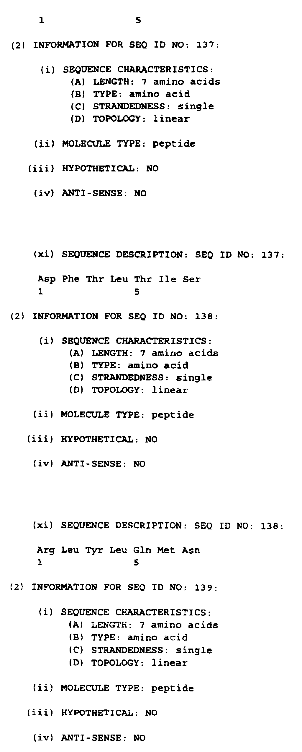


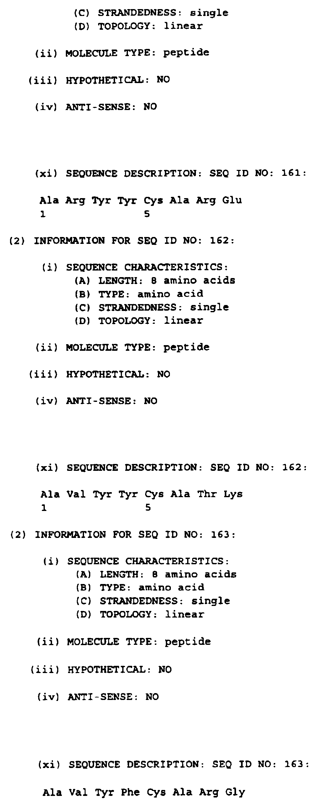


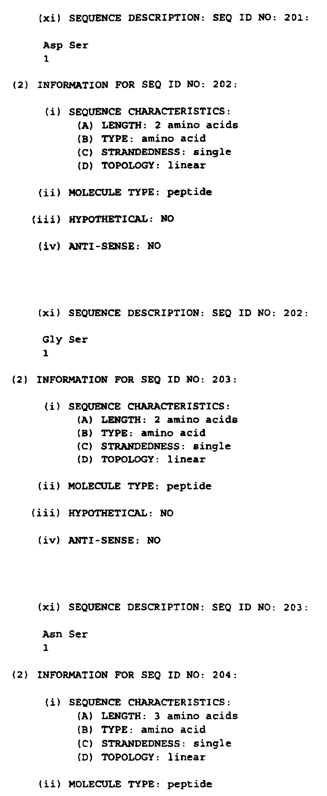
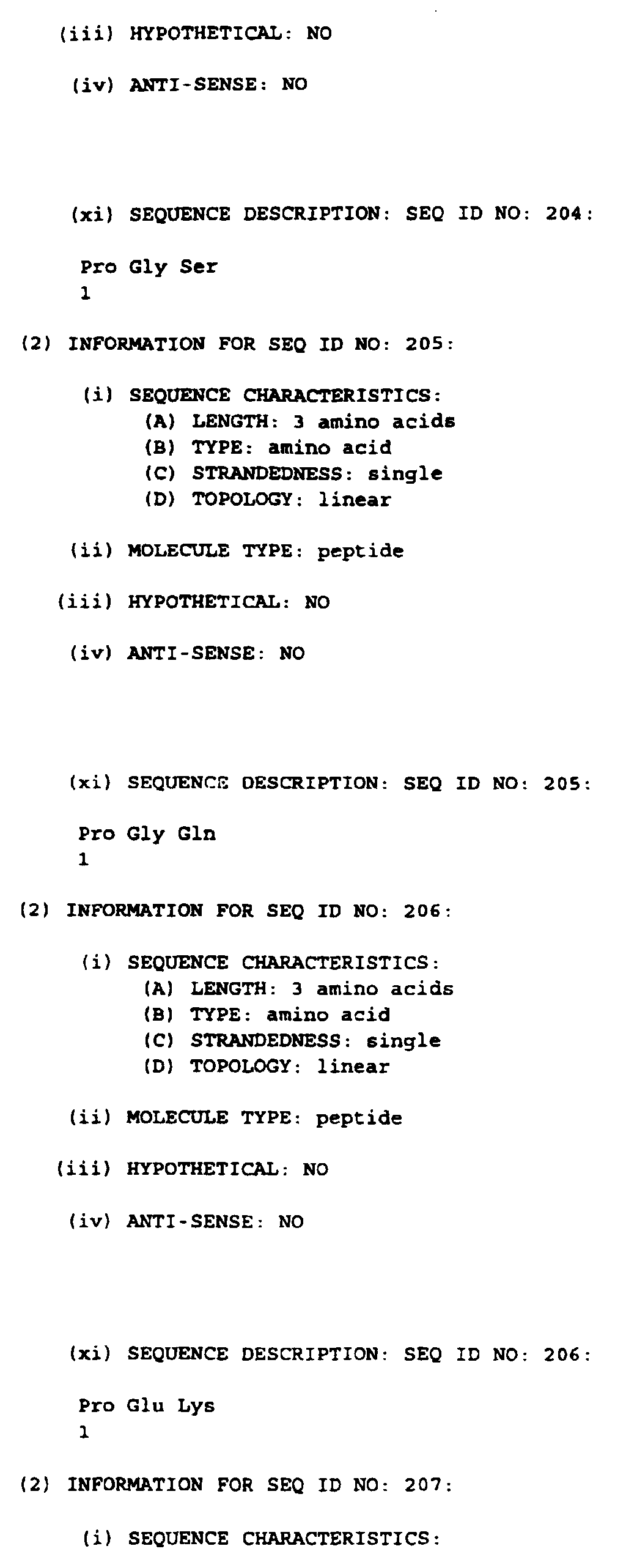


Claims (7)
- A scaffold composed of a single-chain polypeptide having the following structural properties:it contains at least two cysteine residues which form at least one disulphide bond, andit possesses less than 10% of alpha helical conformation, andit contains at least six β-strands (S1, S2, S3, S4, S5, S6) which:are connected by amino acid loops of variable conformation and lenght according to the following topology: S1-S2-S3-S4-S5-S6, andform two β-sheets, one formed by S1/S4/S3 and one formed by S6/S5/S2 wherein the symbol "/" denotes the hydrogen bonding interactions between two spatially adjacent β strands, which are each characterized by an anti-parallel arrangement of said β strands and which are packed onto eachother so that they form a β sandwich architecture.
- A scaffold according to claim 1 which:contains maximum three additional β strands (A1, A2 and A3), andhas the following said topology: A1-S1-S2-A2-A3-S3-S4-S5-S6, andpossesses the following said β-sheets: A1/S1/S4/S3 and S6/S5/S2/A2/A3.
- A scaffold according to claims 1 and 2 wherein:A1 is the amino acid sequence AQPAVVLA (SEQ ID 1) or any functionally equivalent derivative of said sequence,S1 is the amino acid sequence ASFPVEY (SEQ ID 2) or any functionally equivalent derivative of said sequence,S2 is the amino acid sequence EVRVTVLRQA (SEQ ID 3) or any functionally equivalent derivative of said sequence,A2 is the amino acid sequence QVTEVCAA (SEQ ID 4) or any functionally equivalent derivative of said sequence,A3 is the amino acid sequence TYMMGNELTFLDDS (SEP ID 5) or any functionally equivalent derivative of said sequence,S3 is the amino acid sequence ICTGTSS (SEQ ID 6) or any functionally equivalent derivative of said sequence,S4 is the amino acid sequence QVNLTIQ (SEQ ID 7) or any functionally equivalent derivative of said sequence,S5 is the amino acid sequence GLYICKVE( SEQ ID 8) or any functionally equivalent derivative of said sequence,S6 is the amino acid sequence GIGNGTQIY (SEQ ID 9) or any functionally equivalent derivative of said sequence.
- A scaffold according to claims 1 to 3 wherein said single-chain polypeptide is unglycosylated.
- A scaffold according to claims 1 to 4 wherein said amino acid loops comprise fragments binding to a receptor or antigen.
- A scaffold according to claim 4 wherein said amino acid loops comprise fragments binding B7.1 and/or B7.2.
- A single-chain polypeptide as defined in claims 1 to 6 for use as a scaffold.
Priority Applications (1)
| Application Number | Priority Date | Filing Date | Title |
|---|---|---|---|
| EP98870065A EP0947582A1 (en) | 1998-03-31 | 1998-03-31 | A polypeptide structure for use as a scaffold |
Applications Claiming Priority (1)
| Application Number | Priority Date | Filing Date | Title |
|---|---|---|---|
| EP98870065A EP0947582A1 (en) | 1998-03-31 | 1998-03-31 | A polypeptide structure for use as a scaffold |
Publications (1)
| Publication Number | Publication Date |
|---|---|
| EP0947582A1 true EP0947582A1 (en) | 1999-10-06 |
Family
ID=8237017
Family Applications (1)
| Application Number | Title | Priority Date | Filing Date |
|---|---|---|---|
| EP98870065A Withdrawn EP0947582A1 (en) | 1998-03-31 | 1998-03-31 | A polypeptide structure for use as a scaffold |
Country Status (1)
| Country | Link |
|---|---|
| EP (1) | EP0947582A1 (en) |
Cited By (4)
| Publication number | Priority date | Publication date | Assignee | Title |
|---|---|---|---|---|
| WO2000060070A1 (en) * | 1999-04-01 | 2000-10-12 | Innogenetics N.V. | A polypeptide structure for use as a scaffold |
| WO2002048189A3 (en) * | 2000-12-13 | 2003-01-09 | Borean Pharma As | Combinatorial libraries of proteins having the scaffold structure of c-type lectin-like domains |
| WO2003048194A3 (en) * | 2001-12-04 | 2004-02-26 | Tegenero Ag | Peptide or protein containing a c'-d loop of the cd28 receptor family |
| EP2284270A3 (en) * | 2000-12-13 | 2012-07-25 | Anaphore, Inc. | Method for the identification and isolation of binding polypeptides from combinatorial libraries of proteins having the scaffold structure of C-type lectin-like domains |
Citations (7)
| Publication number | Priority date | Publication date | Assignee | Title |
|---|---|---|---|---|
| WO1988006630A1 (en) * | 1987-03-02 | 1988-09-07 | Genex Corporation | Method for the preparation of binding molecules |
| WO1992001047A1 (en) * | 1990-07-10 | 1992-01-23 | Cambridge Antibody Technology Limited | Methods for producing members of specific binding pairs |
| WO1995001994A1 (en) * | 1993-07-09 | 1995-01-19 | Synergen, Inc. | Recombinant ctla4 polypeptides and methods for making the same |
| EP0682039A1 (en) * | 1994-04-15 | 1995-11-15 | Bristol-Myers Squibb Company | CTLA4 molecules and IL4-binding molecules and uses thereof |
| WO1995033770A1 (en) * | 1994-06-03 | 1995-12-14 | Repligen Corporation | Ligands for induction of antigen specific apoptosis in t cells |
| EP0757099A2 (en) * | 1995-07-21 | 1997-02-05 | Bristol-Myers Squibb Company | CTLA4 mutant molecules and uses thereof |
| WO1997037687A1 (en) * | 1996-04-10 | 1997-10-16 | National Jewish Center For Immunology And Respiratory Medicine | Novel product and process for t lymphocyte veto |
-
1998
- 1998-03-31 EP EP98870065A patent/EP0947582A1/en not_active Withdrawn
Patent Citations (7)
| Publication number | Priority date | Publication date | Assignee | Title |
|---|---|---|---|---|
| WO1988006630A1 (en) * | 1987-03-02 | 1988-09-07 | Genex Corporation | Method for the preparation of binding molecules |
| WO1992001047A1 (en) * | 1990-07-10 | 1992-01-23 | Cambridge Antibody Technology Limited | Methods for producing members of specific binding pairs |
| WO1995001994A1 (en) * | 1993-07-09 | 1995-01-19 | Synergen, Inc. | Recombinant ctla4 polypeptides and methods for making the same |
| EP0682039A1 (en) * | 1994-04-15 | 1995-11-15 | Bristol-Myers Squibb Company | CTLA4 molecules and IL4-binding molecules and uses thereof |
| WO1995033770A1 (en) * | 1994-06-03 | 1995-12-14 | Repligen Corporation | Ligands for induction of antigen specific apoptosis in t cells |
| EP0757099A2 (en) * | 1995-07-21 | 1997-02-05 | Bristol-Myers Squibb Company | CTLA4 mutant molecules and uses thereof |
| WO1997037687A1 (en) * | 1996-04-10 | 1997-10-16 | National Jewish Center For Immunology And Respiratory Medicine | Novel product and process for t lymphocyte veto |
Non-Patent Citations (1)
| Title |
|---|
| DARIAVACH P. ET AL.: "HUMAN IG SUPERFAMILY CTLA-4 GENE: CHROMOSOMAL LOCALIZATION AND IDENTITY OF PROTEIN SEQUENCE BETWEEN MURINE AND HUMAN CTLA-4 CYTOPLASMIC DOMAINS", EUROPEAN JOURNAL OF IMMUNOLOGY, vol. 18, December 1988 (1988-12-01), pages 1901 - 1905, XP000613323 * |
Cited By (7)
| Publication number | Priority date | Publication date | Assignee | Title |
|---|---|---|---|---|
| WO2000060070A1 (en) * | 1999-04-01 | 2000-10-12 | Innogenetics N.V. | A polypeptide structure for use as a scaffold |
| WO2002048189A3 (en) * | 2000-12-13 | 2003-01-09 | Borean Pharma As | Combinatorial libraries of proteins having the scaffold structure of c-type lectin-like domains |
| AU2002221568B2 (en) * | 2000-12-13 | 2005-06-02 | Anaphore, Inc. | Combinatorial libraries of proteins having the scaffold structure of C-type lectin-like domains |
| EP2105503A3 (en) * | 2000-12-13 | 2011-03-02 | Anaphore, Inc. | Method for the identification and isolation of binding polypeptides from combinatorial libraries of proteins having the scaffold structure of C-type lectin-like domains |
| US8017559B2 (en) | 2000-12-13 | 2011-09-13 | Anaphore, Inc. | Combinatorial libraries of proteins having the scaffold structure of C-type lectin-like domains |
| EP2284270A3 (en) * | 2000-12-13 | 2012-07-25 | Anaphore, Inc. | Method for the identification and isolation of binding polypeptides from combinatorial libraries of proteins having the scaffold structure of C-type lectin-like domains |
| WO2003048194A3 (en) * | 2001-12-04 | 2004-02-26 | Tegenero Ag | Peptide or protein containing a c'-d loop of the cd28 receptor family |
Similar Documents
| Publication | Publication Date | Title |
|---|---|---|
| CA2367212A1 (en) | A polypeptide structure for use as a scaffold | |
| EP0585287B1 (en) | Methods for producing members of specific binding pairs | |
| US5871907A (en) | Methods for producing members of specific binding pairs | |
| DE69132920T2 (en) | Method for producing filamentous, antibody-presenting bacteriophage particles by using phagemids and the bacteriophage particles produced by them | |
| US5858657A (en) | Methods for producing members of specific binding pairs | |
| US7195866B2 (en) | Production of anti-self antibodies from antibody segment repertoires and displayed on phage | |
| US6225447B1 (en) | Methods for producing members of specific binding pairs | |
| EP0656941B1 (en) | Methods for producing members of specific binding pairs | |
| EP0616640B1 (en) | Production of anti-self antibodies from antibody segment repertoires and displayed on phage | |
| Hufton et al. | Development and application of cytotoxic T lymphocyte-associated antigen 4 as a protein scaffold for the generation of novel binding ligands | |
| US20080305988A1 (en) | V-Like Domain Binding Molecules | |
| CN102770555B (en) | Improved bacterial membrane protein secretion | |
| US20060275811A1 (en) | Method for producing target substance capturing protein and method for selecting component materials thereof | |
| CN102741403B (en) | Display of disulfide-linked dimeric proteins on filamentous phage | |
| EP0947582A1 (en) | A polypeptide structure for use as a scaffold | |
| WO2005049652A2 (en) | Methods of generating antibody diversity in vitro | |
| EP1696031B1 (en) | Production of anti-self antibodies from antibody segment repertoires and displayed on phage | |
| AU689079B2 (en) | Target binding polypeptide | |
| AU2006252050A1 (en) | V-like domain binding molecules | |
| HK1078110A1 (en) | Target binding polypeptide comprising an ig-like vl domain linked to an ig-like vh domain |
Legal Events
| Date | Code | Title | Description |
|---|---|---|---|
| PUAI | Public reference made under article 153(3) epc to a published international application that has entered the european phase |
Free format text: ORIGINAL CODE: 0009012 |
|
| AK | Designated contracting states |
Kind code of ref document: A1 Designated state(s): AT BE CH DE DK ES FI FR GB GR IE IT LI LU MC NL PT SE |
|
| AX | Request for extension of the european patent |
Free format text: AL;LT;LV;MK;RO;SI |
|
| 17P | Request for examination filed |
Effective date: 20000210 |
|
| AKX | Designation fees paid |
Free format text: AT BE CH DE DK ES FI FR GB GR IE IT LI LU MC NL PT SE |
|
| STAA | Information on the status of an ep patent application or granted ep patent |
Free format text: STATUS: THE APPLICATION IS DEEMED TO BE WITHDRAWN |
|
| 18D | Application deemed to be withdrawn |
Effective date: 20031001 |