EP1127894A1 - Method of diagnosis of transmissible spongiform encephalopathy (TSE) - Google Patents
Method of diagnosis of transmissible spongiform encephalopathy (TSE) Download PDFInfo
- Publication number
- EP1127894A1 EP1127894A1 EP01108876A EP01108876A EP1127894A1 EP 1127894 A1 EP1127894 A1 EP 1127894A1 EP 01108876 A EP01108876 A EP 01108876A EP 01108876 A EP01108876 A EP 01108876A EP 1127894 A1 EP1127894 A1 EP 1127894A1
- Authority
- EP
- European Patent Office
- Prior art keywords
- lrp
- laminin receptor
- prp
- tse
- cells
- Prior art date
- Legal status (The legal status is an assumption and is not a legal conclusion. Google has not performed a legal analysis and makes no representation as to the accuracy of the status listed.)
- Withdrawn
Links
Images








Classifications
-
- C—CHEMISTRY; METALLURGY
- C07—ORGANIC CHEMISTRY
- C07K—PEPTIDES
- C07K14/00—Peptides having more than 20 amino acids; Gastrins; Somatostatins; Melanotropins; Derivatives thereof
- C07K14/435—Peptides having more than 20 amino acids; Gastrins; Somatostatins; Melanotropins; Derivatives thereof from animals; from humans
- C07K14/705—Receptors; Cell surface antigens; Cell surface determinants
- C07K14/70546—Integrin superfamily
- C07K14/7055—Integrin beta1-subunit-containing molecules, e.g. CD29, CD49
-
- A—HUMAN NECESSITIES
- A61—MEDICAL OR VETERINARY SCIENCE; HYGIENE
- A61P—SPECIFIC THERAPEUTIC ACTIVITY OF CHEMICAL COMPOUNDS OR MEDICINAL PREPARATIONS
- A61P25/00—Drugs for disorders of the nervous system
- A61P25/28—Drugs for disorders of the nervous system for treating neurodegenerative disorders of the central nervous system, e.g. nootropic agents, cognition enhancers, drugs for treating Alzheimer's disease or other forms of dementia
-
- A—HUMAN NECESSITIES
- A61—MEDICAL OR VETERINARY SCIENCE; HYGIENE
- A61P—SPECIFIC THERAPEUTIC ACTIVITY OF CHEMICAL COMPOUNDS OR MEDICINAL PREPARATIONS
- A61P43/00—Drugs for specific purposes, not provided for in groups A61P1/00-A61P41/00
-
- C—CHEMISTRY; METALLURGY
- C07—ORGANIC CHEMISTRY
- C07K—PEPTIDES
- C07K16/00—Immunoglobulins [IG], e.g. monoclonal or polyclonal antibodies
- C07K16/18—Immunoglobulins [IG], e.g. monoclonal or polyclonal antibodies against material from animals or humans
- C07K16/28—Immunoglobulins [IG], e.g. monoclonal or polyclonal antibodies against material from animals or humans against receptors, cell surface antigens or cell surface determinants
-
- C—CHEMISTRY; METALLURGY
- C12—BIOCHEMISTRY; BEER; SPIRITS; WINE; VINEGAR; MICROBIOLOGY; ENZYMOLOGY; MUTATION OR GENETIC ENGINEERING
- C12N—MICROORGANISMS OR ENZYMES; COMPOSITIONS THEREOF; PROPAGATING, PRESERVING, OR MAINTAINING MICROORGANISMS; MUTATION OR GENETIC ENGINEERING; CULTURE MEDIA
- C12N15/00—Mutation or genetic engineering; DNA or RNA concerning genetic engineering, vectors, e.g. plasmids, or their isolation, preparation or purification; Use of hosts therefor
- C12N15/09—Recombinant DNA-technology
- C12N15/11—DNA or RNA fragments; Modified forms thereof; Non-coding nucleic acids having a biological activity
- C12N15/113—Non-coding nucleic acids modulating the expression of genes, e.g. antisense oligonucleotides; Antisense DNA or RNA; Triplex- forming oligonucleotides; Catalytic nucleic acids, e.g. ribozymes; Nucleic acids used in co-suppression or gene silencing
- C12N15/1138—Non-coding nucleic acids modulating the expression of genes, e.g. antisense oligonucleotides; Antisense DNA or RNA; Triplex- forming oligonucleotides; Catalytic nucleic acids, e.g. ribozymes; Nucleic acids used in co-suppression or gene silencing against receptors or cell surface proteins
-
- A—HUMAN NECESSITIES
- A01—AGRICULTURE; FORESTRY; ANIMAL HUSBANDRY; HUNTING; TRAPPING; FISHING
- A01K—ANIMAL HUSBANDRY; AVICULTURE; APICULTURE; PISCICULTURE; FISHING; REARING OR BREEDING ANIMALS, NOT OTHERWISE PROVIDED FOR; NEW BREEDS OF ANIMALS
- A01K2217/00—Genetically modified animals
- A01K2217/07—Animals genetically altered by homologous recombination
- A01K2217/075—Animals genetically altered by homologous recombination inducing loss of function, i.e. knock out
-
- A—HUMAN NECESSITIES
- A61—MEDICAL OR VETERINARY SCIENCE; HYGIENE
- A61K—PREPARATIONS FOR MEDICAL, DENTAL OR TOILETRY PURPOSES
- A61K38/00—Medicinal preparations containing peptides
-
- C—CHEMISTRY; METALLURGY
- C07—ORGANIC CHEMISTRY
- C07K—PEPTIDES
- C07K2319/00—Fusion polypeptide
-
- C—CHEMISTRY; METALLURGY
- C12—BIOCHEMISTRY; BEER; SPIRITS; WINE; VINEGAR; MICROBIOLOGY; ENZYMOLOGY; MUTATION OR GENETIC ENGINEERING
- C12N—MICROORGANISMS OR ENZYMES; COMPOSITIONS THEREOF; PROPAGATING, PRESERVING, OR MAINTAINING MICROORGANISMS; MUTATION OR GENETIC ENGINEERING; CULTURE MEDIA
- C12N2310/00—Structure or type of the nucleic acid
- C12N2310/10—Type of nucleic acid
- C12N2310/11—Antisense
- C12N2310/111—Antisense spanning the whole gene, or a large part of it
-
- C—CHEMISTRY; METALLURGY
- C12—BIOCHEMISTRY; BEER; SPIRITS; WINE; VINEGAR; MICROBIOLOGY; ENZYMOLOGY; MUTATION OR GENETIC ENGINEERING
- C12N—MICROORGANISMS OR ENZYMES; COMPOSITIONS THEREOF; PROPAGATING, PRESERVING, OR MAINTAINING MICROORGANISMS; MUTATION OR GENETIC ENGINEERING; CULTURE MEDIA
- C12N2799/00—Uses of viruses
- C12N2799/02—Uses of viruses as vector
- C12N2799/021—Uses of viruses as vector for the expression of a heterologous nucleic acid
- C12N2799/026—Uses of viruses as vector for the expression of a heterologous nucleic acid where the vector is derived from a baculovirus
Definitions
- the present invention relates to pharmaceutical compositions comprising a soluble laminin receptor precursor (LRP) or a modification thereof.
- LRP soluble laminin receptor precursor
- the present invention also relates to pharmaceutical compositions comprising a compound which blocks the interaction of LRP and PrP Sc or PrP c . Said compositions allow the treatment of diseases associated with the presence of PrP Sc .
- the present invention provides an expression vector for the recombinant production of LRP or any kind of protein in the baculovirus system.
- Prion proteins are thought to play a central role in the pathogenesis of transmissible spongiform encephalopathies, such as bovine spongiform encephalopathy (BSE), Creutzfeldt-Jakob disease (CJD) and Scrapie (Prusiner, 1991).
- BSE bovine spongiform encephalopathy
- CJD Creutzfeldt-Jakob disease
- Scrapie Scrapie
- PrP Sc infectious protein
- PrP Sc a pathogenic isoform of the normal host-encoded PrP c
- PrP Sc is thought to convert host-PrP c into PrP Sc resulting in pathological modifications in the brain such as spongiform vacuolation, astrocytic proliferation and neuronal loss.
- PrP c and PrP Sc are identical regarding primary structure (Stahl et al., 1993), but CD-and FTIR-spectroscopy reveals that they have different secondary structures (Meyer et al., 1986; Pan et al., 1993). PrP c has a very low content and PrP Sc a high content of ⁇ -sheet structure. Moreover, PrP c can be hydrolyzed completely by proteinase K, whereas PrP Sc is degraded to the proteinase K resistant infectious core PrP27-30 (Caughey et al., 1991; Prusiner et al., 1981; 1983; 1984).
- sPrP27-30 A proteinase K sensitive, soluble and presumably non-infectious version of PrP27-30, designated as sPrP27-30 was identified in human platelets (Perini et al., 1996) and might also act as a precursor for proteinase K resistant PrP27-30.
- PrP c The conversion of PrP c into PrP Sc is thought to involve structural rearrangements of the polypeptide from ⁇ -helix to ⁇ -sheet.
- Two models are currently used to describe this conversion mechanism.
- the heterodimer model Prusiner, 1982
- one exogenous PrP Sc molecule contacts host PrP c resulting in a PrP c -PrP Sc heterodimer which refolds into a PrP Sc -homodimer.
- the nucleation dependent polymerization model proposes that infectivity is a property of a PrP Sc oligomer, which can act as a seed for polymerization.
- PrP c The physiological role of the cellular prion protein PrP c was investigated with a series of knock-out experiments leading to controversial results. Most of the researchers did not recognize any changes in the behaviour or reproduction of PrP-knock-out mice (Büehler et al., 1992). More recent investigations with PrP o/o mice suggested an important role of PrP c in synaptic function including an efficient GABA A receptor-mediated fast inhibition (Collinge et al., 1996) whereas another study did not find any dysfunctions of synapses in the hippocampus (Lledo et al., 1996).
- PrP o/o mice lacking the prn-p gene are, in contrast to wild-type mice, resistant to infections with the mouse PrP Sc (Büehler et al., 1993) demonstrating that PrP Sc is essential for the development of Scrapie and Scrapie-like disease. More recent experiments demonstrated that PrP c is necessary for scrapie induced neurotoxicity (Brandner et al., 1996) and required for scrapie spread within the central nervous system (Brandner et al., 1996b).
- the technical problem underlying the present invention is to provide compounds which can be used for the treatment of diseases which are associated with the presence of PrP Sc .
- LRP laminin receptor precursor
- LRP laminin receptor precursor
- GST::LRP Although the signal sequence of gp67 was present at the N-terminus, GST::LRP was not secreted into the culture medium but synthesized cell-associated most likely due to the presence of a short transmembrane region which was calculated between amino acid 86 and amino acid 101 of LRP (Castronovo et al., 1991a).
- GST::LRP which was specifically recognized by a LRP specific antibody revealed a molecular weight of 67 kDa.
- a polyclonal antibody directed against GST::LRP recognized recombinant LRP but revealed some background reactivity to GST. After thrombin cleavage of the fusion protein SDS gel analysis confirmed molecular weights of 37 kDa for the receptor and 27 kDa for GST.
- the cDNA encoding the laminin receptor precursor was initially isolated in attempts to identify the gene for the 67 kDa laminin receptor precursor (Yow et al., 1988).
- the cDNA encoded for a 37 kDa protein (Yow et al., 1988), whereas the receptor was isolated as a 67 kDa protein from solid tumors (Malinoff and Wicha, 1983; Lesot et al., 1983; Rao et al., 1983).
- Pulse-Chase experiments suggest that the 37 kDa receptor could be the precursor of the 67 kDa receptor (Castronovo et al., 1991b). Homodimerization (Landowski et al., 1995) or the existence of an unknown factor (Castronovo et al., 1991b) could account for the 37/67 kDa polymorphism of the laminin receptor precursor.
- laminin binding proteins include integrins (Albelda and Buck, 1990) and galactoside binding lectins.
- the non-integrin binding 67 kD high affinity laminin receptor precursor (LRP) binds beside laminin (Mecham et al., 1989) also elastin (Hinek et al., 1988).
- LRP laminin receptor precursor
- elastin Hinek et al., 1988.
- binding assays in the presence of laminin were performed.
- Laminin represents a glycoprotein of the extracellular matrix (ECM) mediating cell attachment, differentiation, movement and growth (Hunter et al., 1989).
- EHS laminin was isolated from murine Engelbreth-Holm-Swarm (EHS) tumors and parietal yolk sac (PYS) carcinoma cells (for review see Mecham, 1991).
- EHS laminin was used which consists of three polypeptide chains - A or ⁇ (440 kDa), B1 or ⁇ , and B2 or ⁇ (each 220 kDa) - linked via disulfide bridges resulting in the typical cross-structure (Beck et al., 1990; Burgeson et al., 1994).
- Specific EHS-laminin binding to the receptor was demonstrated in the presence of GST::LRP whereas no binding occurred with GST.
- Laminin/LRP interaction can be competed with FLAG::rPrP27-30 (data not shown) confirming the mapping result in S. cerevisiae that laminin and PrP share the same binding site on LRP (amino acids 161-180).
- the cellular form of the hamster prion protein PrP c (PrP23-231) was synthesized and the soluble version (s) PrP27-30 of proteinase K resistant PrP27-30 (Caughey et al., 1991; Prusiner et al., 1981; 1983; 1984) was fused to the FLAG peptide.
- Both recombinant proteins were secreted into the culture medium, purified to homogeneity by anti-Flag-antibody affinity chromatography and recognized by a PrP specific antibody.
- Molecular weights determined by SDS-PAGE were 27 kDa and 17 kDa for rPrP c and rPrP27-30, respectively, and correspond to rPrP c (Weiss et al., 1995) and rPrP27-30 (Weiss et al., 1996) generated by thrombin cleavage from GST fusion proteins.
- co-infection in Sf9 cells was performed.
- FLAG::rPrP c and authentic LRP were transiently expressed in Cos-7 cells.
- LRP revealed a molecular weight of 37 kDa and was recognized by a polyclonal anti-LRP antibody.
- rPrP c fused to FLAG showed the expected molecular weight of 27 kDa and was also recognized by a polyclonal PrP specific antibody. Interaction between both proteins was proven in Cos-7 cells demonstrating that LRP interacts with PrP in a series of different eukaryotic cells including yeast, insect and Cos-7 cells.
- the laminin receptor precursor family is highly conserved in a wide spectrum of eukaryotic cells (Wewer et al., 1986; Keppel et al., 1991; Garcia-Hernandez et al., 1994), was recently reported to be present in yeast (Demianova et al., 1996) and may be encoded by archaean genomes (Ouzounis et al., 1995).
- the receptor works as a receptor for Sindbis Virus on mammalian cells such as CHO or BHK (Wang et al., 1992) and for Venezuelan Encephalitis Virus from Mosquito Cells (Ludwig et al., 1996).
- laminin itself has a drastic influence on the growth of neuronal cells.
- the LRP works as a receptor for the Sindbis-Virus and the VEE-virus on the surface of mammalian cells.
- the cellular prion protein PrP c is also located on the surface of neuronal cells fixed by the glycosylphosphatidylinositol anchor (GPI; Stahl et al., 1987).
- GPI glycosylphosphatidylinositol anchor
- sPrP27-30 could work as a precursor for proteinase K resistant PrP27-30, a major product in Scrapie infected prion-preparations (Prusiner et al., 1981; 1983; 1984).
- the laminin receptor precursor has also been identified in human platelets (Tandon et al., 1991) which makes a PrP/LRP interaction also conceivable in platelets.
- LRP is required for the proliferation of PrP Sc .
- a model of the function of LRP is discussed in the following section.
- PrP c is synthesized at the rER and secretes via the ER, the golgi and secretory vesicles passing through the plasma membrane to the cell surface. PrP c is located on the surface of neuronal and other cells fixed by a glycosyl-phosphatidyl-inositol (GPI) anchor (Stahl et al., 1987). Treatment of the cell with phospholipase C (PIPLC) removes the diacylglycerol portion of the anchor releasing PrP c from the cell surface (Lehmann and Harris, 1996).
- GPIPLC glycosyl-phosphatidyl-inositol
- PrP c becomes internalized from the cell surface via clathrin coated pits (Shyng et al., 1994, 1995), caveolae or endocytosis (Taraboulos et al., 1995; Vey et al., 1996).
- PrP Sc reaches the brain of TSE infected individuals via nerve cells or via the blood brain barrier. PrP Sc could be transported from the periphery to the nerve cells by cells of the lymphatic system (such as B-lymphocytes which have been proven to be essential for prion propagation (Klein et al., 1997)) including the spleen, the peripheric nervous system (PNS) and the central nervous system (CNS) including the spinal cord. These cells might bind PrP Sc via LRP.
- the lymphatic system such as B-lymphocytes which have been proven to be essential for prion propagation (Klein et al., 1997)
- PNS peripheric nervous system
- CNS central nervous system
- PrP Sc could either bind to PrP c on the cell surface in the absence or presence of an unknown receptor as suggested by Harris (Lehmann and Harris, 1996) such as the laminin receptor precursor. PrP c /PrP Sc binding could be dependent on the presence of the laminin receptor precursor. Membrane trafficking of PrP c and PrP Sc , either separately or complexed, can also be mediated by the laminin receptor precursor. The uptake of both PrP molecules can occur, either complexed or separately, by clathrin coated pits (Shyng et al., 1994, 1995), endocytosis or caveolae (Taraboulos et al., 1995; Vey et al., 1996).
- Lysosomes (Caughey et al., 1992; Taraboulos et al., 1992) are thought to contain predominantly PrP Sc (Caughey et al., 1991). It is thought that PrP c is still GPI anchored in endosomes, endolysosomes and lysosomes.
- the present invention relates to a pharmaceutical composition
- a pharmaceutical composition comprising a soluble LRP, a mutein, fused protein, salt, functional derivative or fragment thereof, that can bind to PrP Sc , PrP c or fragments thereof.
- Said composition might be useful for the treatment of diseases associated with the presence of PrP Sc .
- the soluble part of the LRP corresponds to the extracellular domain of LRP comprising the region from amino acid no. 101 to no. 295 of the amino acid sequence disclosed in Yow et al., 1988 (see Figures 3 and 4 and pages 6396 and 6397). It is, however, apparent for the person skilled in the art that for the preparation of the above pharmaceutical composition not only the soluble part of LRP with the amino acid sequence disclosed in Yow et al., 1988, can be used, but, in addition, a mutein of said soluble LRP, a fused protein, salt, functional derivative or fragment thereof which are still capable of binding PrP Sc or PrP c .
- muteins refers to analogs of the soluble LRP in which one or more of the amino acid residues of the soluble LRP described in Yow et al., 1988, are replaced by different amino acid residues or are deleted, or one or more amino acid residues are added without considerably changing the activity of the resulting product. These muteins are prepared by known synthesis and/or by site-directed mutagenesis techniques, or any other known technique suitable therefor.
- fused protein refers to a polypeptide comprising a soluble LRP according to the invention, or a mutein thereof fused with another protein which has an extended residence time in body fluids.
- the soluble LRP may thus be fused to another protein, polypeptide or the like, e.g., an immunoglobulin or a fragment thereof.
- salts herein refers to both salts of carboxyl groups and to acid addition salts of amino groups of a soluble LRP, muteins and fused proteins thereof.
- Salts of a carboxyl group may be formed by means known in the art and include inorganic salts, for example, sodium, calcium, ammonium, ferric or zinc salts, and the like, and salts with organic bases as those formed, for example, with amines, such as thiethanolamine, arginine or lysine, piperidine, procaine and the like.
- Acid addition salts include, for example, salts with mineral acids such as, for example, hydrochloric acid or sulfuric acid, and salts with organic acids such as, for example, acetic acid or oxalic acid.
- “Functional derivatives” as used herein cover derivatives of a soluble LRP and its fused proteins and muteins, which may be prepared from the functional groups which occur as side chains on the residues or the N- or C-terminal groups, by means known in the art, and are included in the invention as long as they remain pharmaceutically acceptable, i.e. they do not destroy the activity of the protein and do not confer toxic properties on compositions containing it.
- These derivatives may, for example, include polyethylene glycol side-chains which may mask antigenic sites and extend the residence of a soluble LRP in body fluids.
- Other derivatives include aliphatic esters of the carboxyl groups, amides of the carboxyl groups by reaction with ammonia or with primary or secondary amines, N-acyl derivatives of free amino groups of the amino acid residues formed with acyl moieties (e.g. alkanoyl or carbocyclic aroyl groups) or O-acyl derivatives of free hydroxyl groups (for example that of seryl or threonyl residues) formed with acyl moieties.
- acyl moieties e.g. alkanoyl or carbocyclic aroyl groups
- O-acyl derivatives of free hydroxyl groups for example that of seryl or threonyl residues
- fragment of the soluble LRP its fused proteins and its muteins
- the present invention covers any fragment of the polypeptide chain of the protein molecule alone or together with associated molecules or residues linked thereto, e.g. sugar or phosphate residues, or aggregates of the protein molecule or the sugar residues by themselves, provided said fraction has the same biological activity and/or pharmaceutical activity.
- fragment of PrP Sc or PrP c refers to any fragment of the polypeptide chain of the protein which can bind to the LRP.
- compositions may be prepared by any of the well known procedures in which the active ingredient, soluble LRP, is optionally admixed with pharmaceutically acceptable diluents, carriers or excipients. Actual dosages and modes of administration of such pharmaceutical compositions will be determined by the professional practitioners.
- the present invention relates to a pharmaceutical composition
- a pharmaceutical composition comprising a compound that blocks the interaction of LRP and PrP Sc , PrP c or fragments thereof and, optionally, a suitable diluent, carrier and/or excipient.
- Such compounds can be identified, for example, by the screening system described below.
- said compound is an antibody, preferably a monoclonal antibody, or a fragment thereof which binds to the surface epitope of LRP in such a way that binding of PrP Sc , PrP c or fragments thereof is no longer possible.
- the antibody binds to PrP Sc , PrP c or fragments thereof, thus blocking the LRP/PrP interaction.
- Antibodies directed, for example, to the soluble part of LRP may be serum-derived (polyclonal) or monoclonal.
- monoclonal antibodies are prepared using hybridoma technology by fusing antibody producing B cells from immunized animals with myeloma cells and selecting the resulting hybridoma cell line producing the desired antibody.
- synthetic peptides may be prepared using the amino acid sequence described in Yow et al., 1988.
- DNA such as a cDNA or a fragment thereof, may be cloned and expressed and the resulting polypeptide recovered and used as an immunogen.
- These antibodies are useful to inhibit the function of the LRP. Particularly useful are antibodies binding to the PrP binding domain of LRP (amino acids 161 to 180).
- said compounds which block the interaction of LRP and PrP Sc or PrP c comprise:
- the reverse two-hybrid system (Fig. 8) modified from Leanna and Hannink, 1996, represents a straightforward method for the identification of compounds blocking the LRP/PrP interaction.
- the reverse Gal4/LexA two-hybrid system is a genetic selection scheme that selects against the association of PrP fused to the DNA binding domain Gal4/LexA and LRP fused to the activation domain of Gal4 or the acidic activation domain of B42.
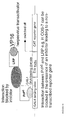
- the mammalian two-hybrid system (supplied by CLONETECH Laboratories Inc., USA) can be used to screen for therapeutics interfering with the LRP/PrP interaction (Fig. 9).
- PrP can be fused to the DNA binding domain of Gal4 (bait plasmid).
- LRP can be fused to VP16, the herpesvirus transactivator in the prey position (prey plasmid).
- prey plasmid the herpesvirus transactivator in the prey position
- the interaction of bait and prey will lead to activation of transcription of the CAT reporter gene (supplied by a reporter plasmid) by RNA polymerase II from the minimal promoter of the adenovirus E1 b gene.
- luciferase can be used as a reporter gene enhancing the sensitivity of the assay.
- Compounds can be screened using, for example, peptide libraries which can be co-transfected into mammalian cells using a selection marker such as neomycin (leading to viable cells in the presence of G 418). Compounds can easily be added to the culture medium.
- mammalian cells are lacking a cell wall so that the uptake/intake of the compounds is much more efficient compared to yeast.
- mammalian cells represent the highest phylogenetic system with the highest degree in glycosylations, phosphorylations and other posttranslational modifications.
- the toxicity of a compound is assayed simultaneously in the mammalian system, whereas the toxicity of a component directed against the LRP/PrP interaction in the yeast two-hybrid system has to be re-assayed in the mammalian system for toxicity.
- yeast and the mammalian systems can be altered with respect to simplifying the screening assay.
- cDNA encoding LRP and PrP c can be cloned into the bait position either in a co-directional or opposite direction leading to the expression of both proteins from a single plasmid.
- the cDNA library encoding for peptides or other presumable PrP/LRP interfering components can be cloned into the prey position so that no additional plasmid has to be transformed or transfected into the yeast or mammalian cells, respectively.
- the present invention also relates to pharmaceutical compositions comprising a compound identified by the method described above.
- the above pharmaceutical compositions might be useful for the treatment of diseases associated with the presence of PrP Sc .
- diseases are the transmissible spongiform encephalopathies, such as scrapie, bovine spongiform encephalopathy, Creutzfeldt-Jakob disease, Fatal Familial Insomnia, chronic wasting disease and feline spongiform encephalopathy (FSE).
- transmissible spongiform encephalopathies such as scrapie, bovine spongiform encephalopathy, Creutzfeldt-Jakob disease, Fatal Familial Insomnia, chronic wasting disease and feline spongiform encephalopathy (FSE).
- diseases associated with the presence of PrP Sc can be treated by blocking the synthesis of LRP, e.g. by applying an antisense-RNA or a ribozyme which can bind/cleave the mRNA encoding LRP, thus inhibiting translation, and expression of LRP.
- RNAs or ribozymes including the transformation of cells and the construction of expression vectors, can be carried out by methods well known to the person skilled in the art, e.g. by the techniques described in Example 9, below.
- the present invention also relates to pharmaceutical compositions comprising an antisense-RNA or ribozyme which blocks the expression of the LRP.
- the present invention provides an expression vector for the recombinant production of LRP or any other kind of protein in the baculovirus system, pFLAG::BAC.
- the protein will be synthesized in the fusion with a FLAG-tag at the aminoterminal end of the protein.
- the FLAG-tag can be cleaved off employing the specific protease enterokinase.
- the recombinant protein will be secreted into the culture medium due to the presence of the signal sequence gp67.
- Proteins of any kind expressed in the fusion with FLAG in the baculovirus system can be phosphorylated and glycosylated, although it is known that the baculovirus system cannot form high-branched sugars.
- FLAG will enhance the solubility and stability of the fusion protein.
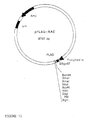
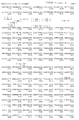
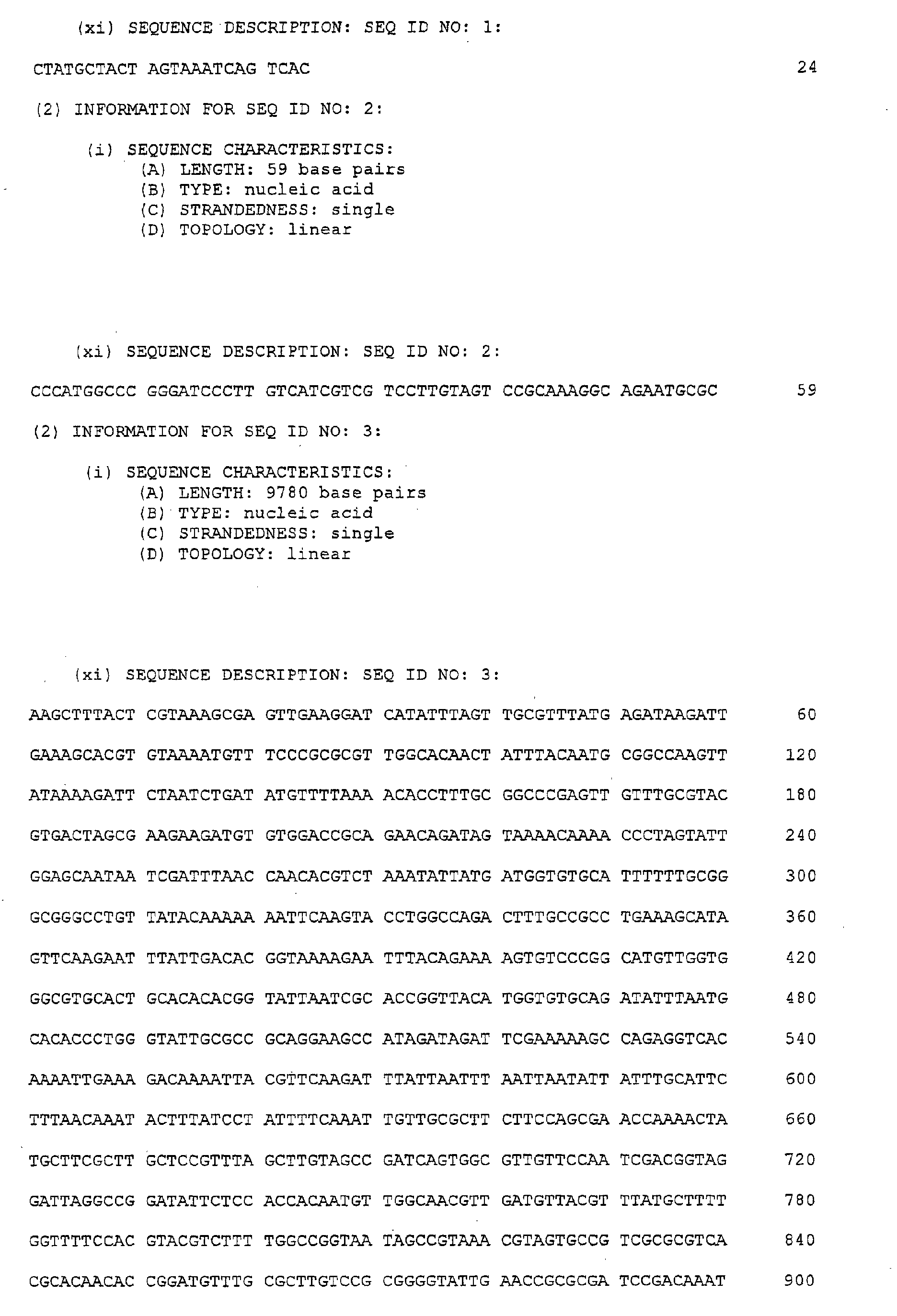
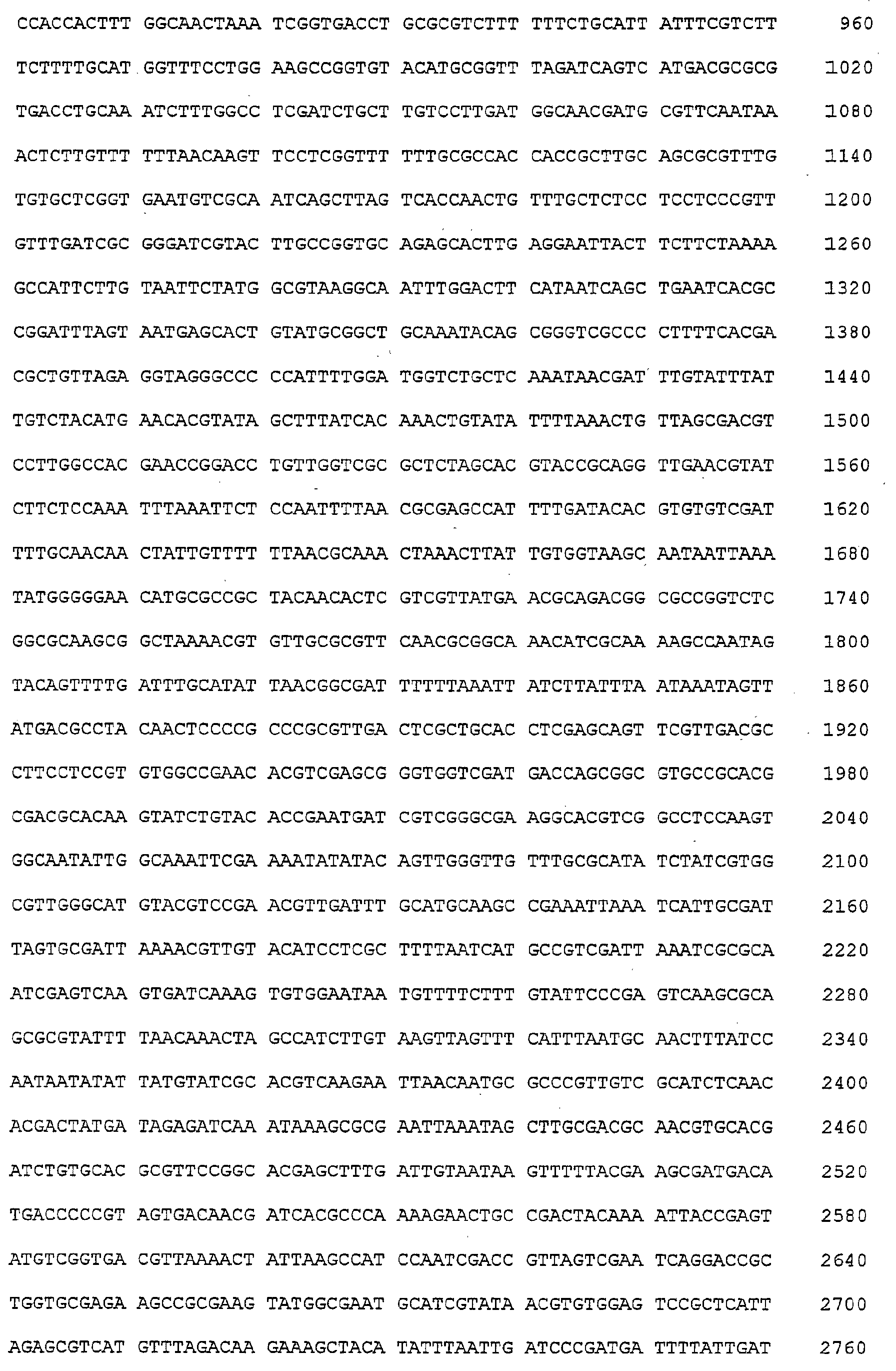
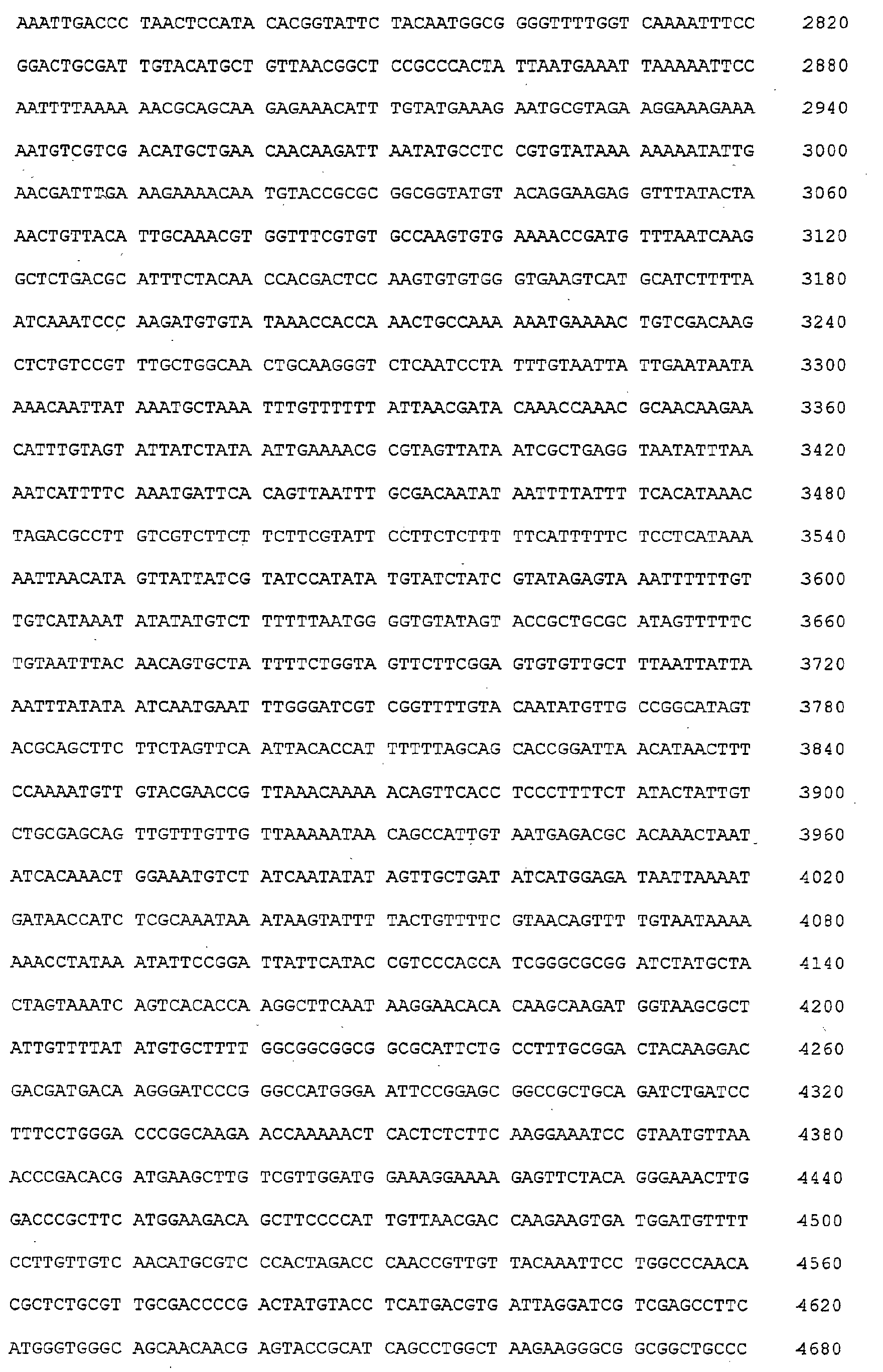
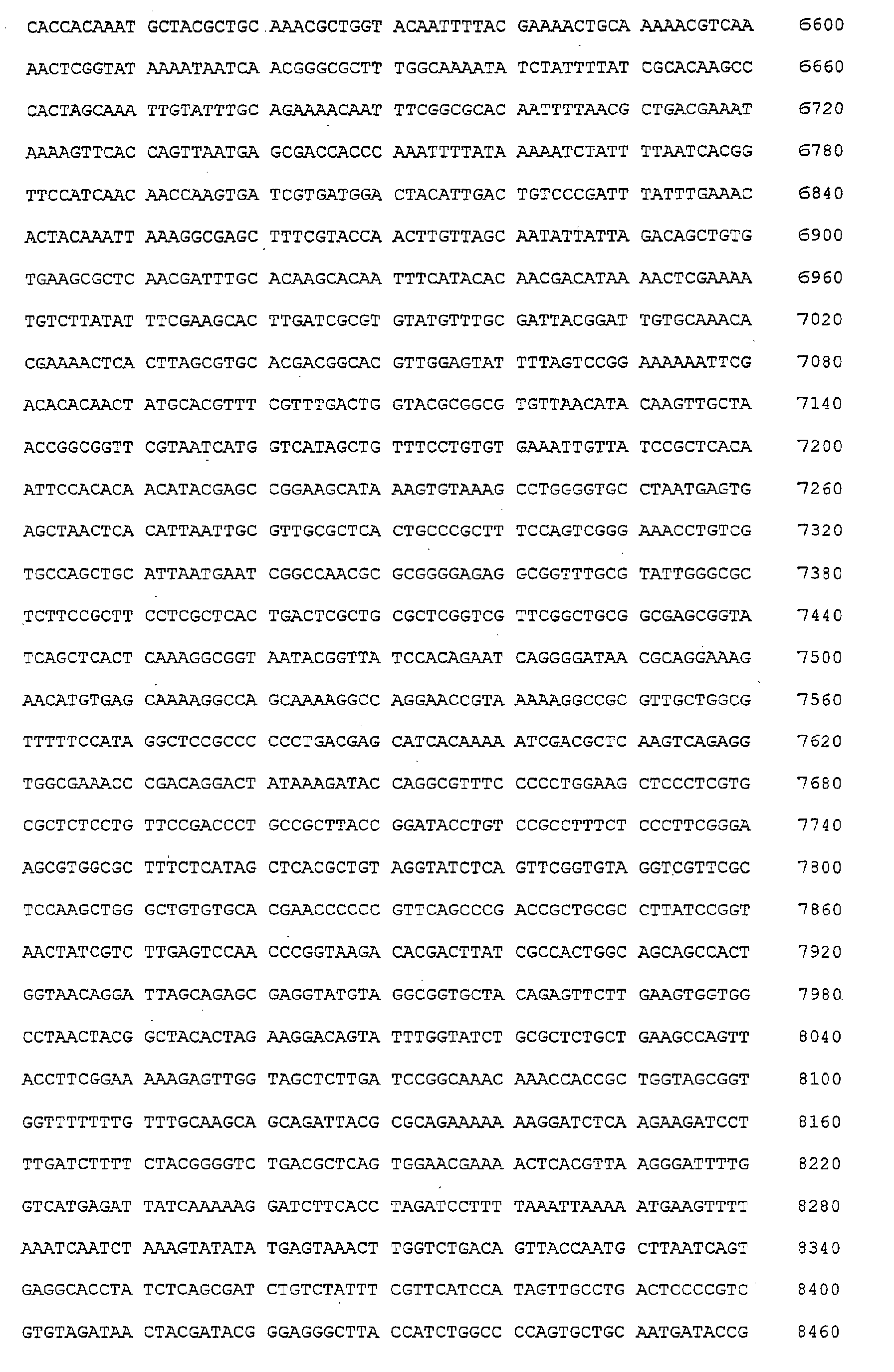
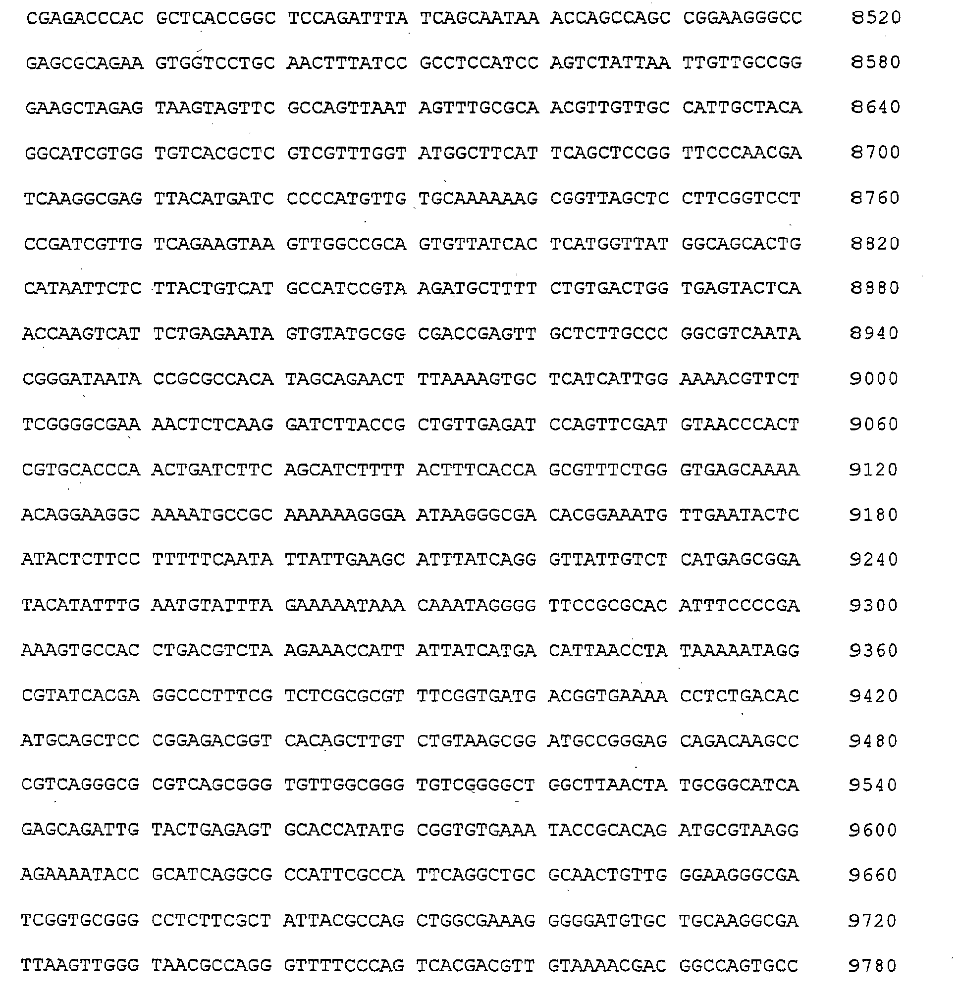
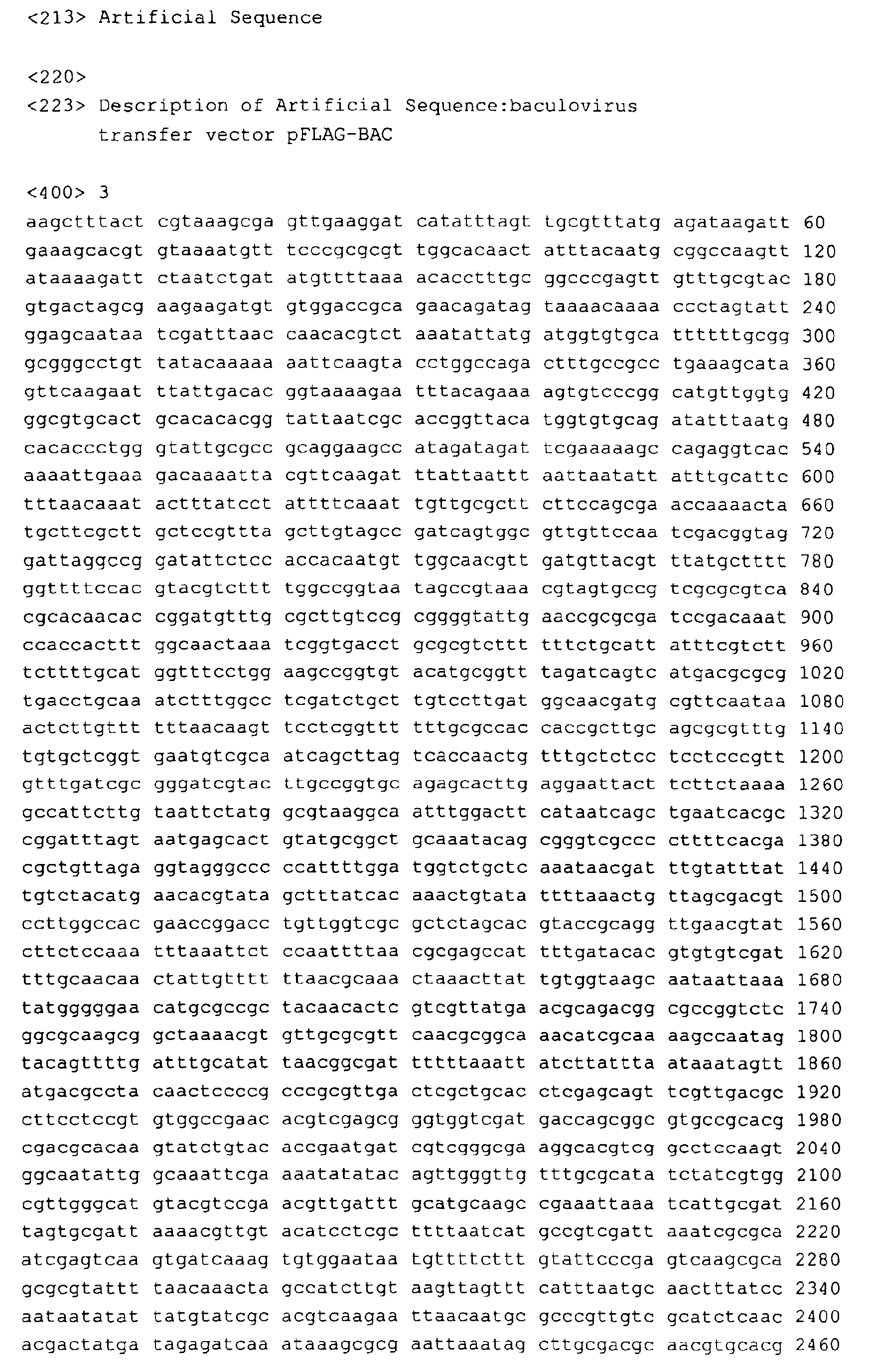
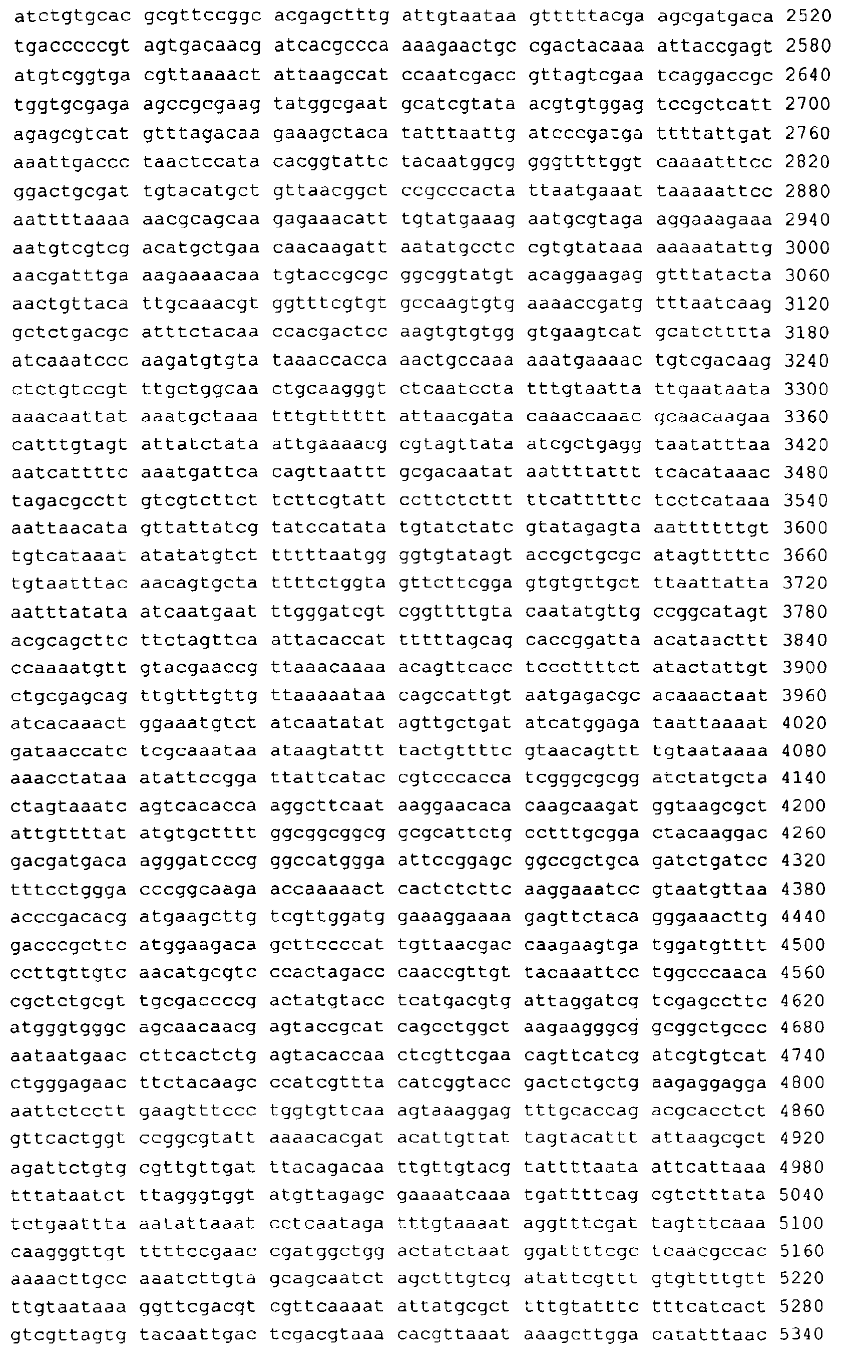
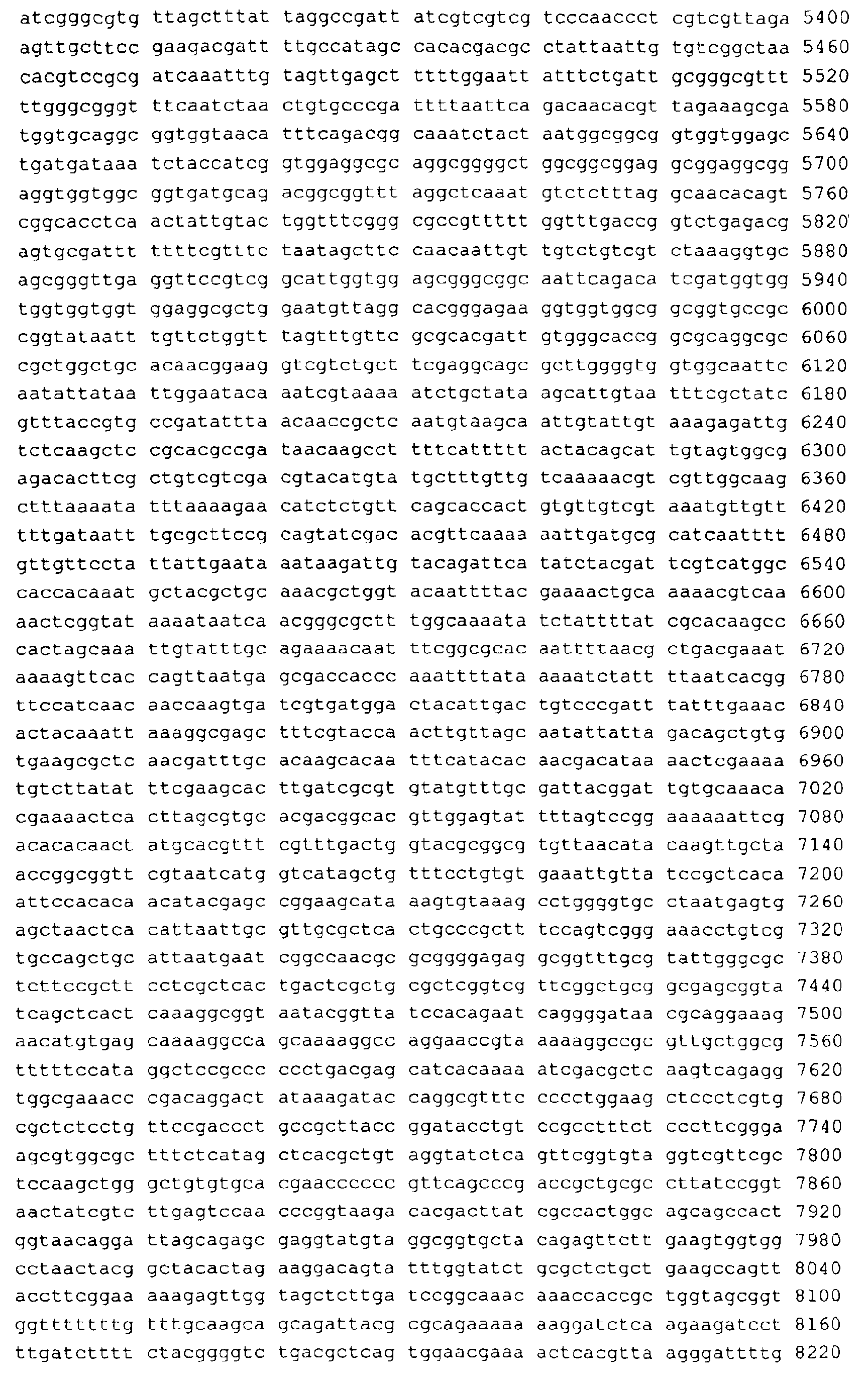
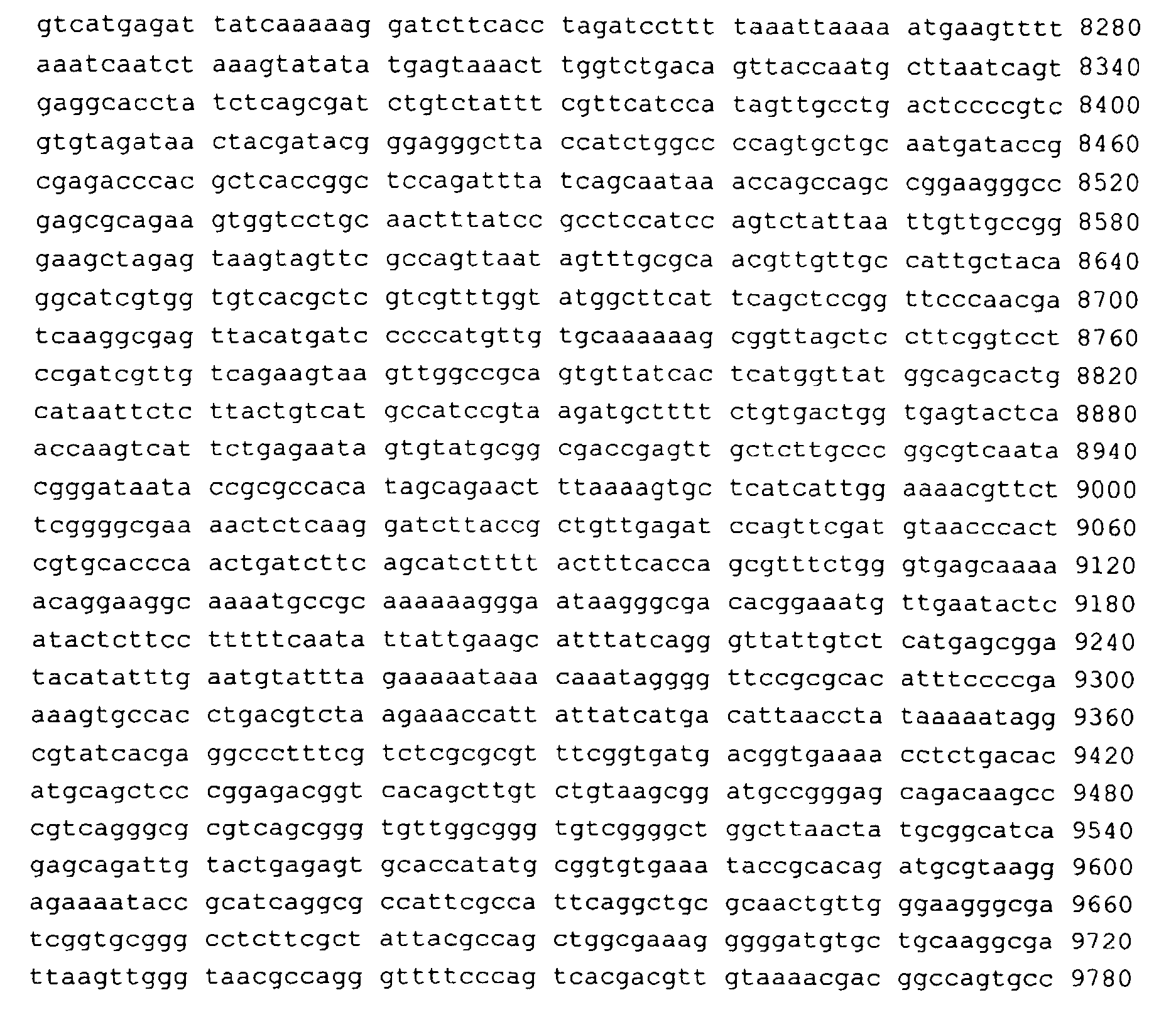
- This vector has the structure shown in Figure 13 and the nucleic acid sequence shown in Figure 14 or substantially said sequence.
- substantially refers to a nucleic acid sequence in which one or more nucleotides of the nucleic acid sequence shown in Figure 14 are replaced by different nucleotides or are deleted, or one or more nucleotides are added without considerably changing the biological properties of the vector as regards the capability (i) of replicating in the baculovirus system, (ii) of expressing the heterologous protein, (iii) of allowing the secretion of the protein in the culture medium and/or (iv) of providing the heterologous protein in a soluble and stable form.
- the nucleic acid sequence which has "substantially" the sequence shown in Figure 14 has at least 90%, preferably at least 95%, more preferably at least 98% and most preferably at least 99% homology to said sequence.
- Such a sequence is prepared by known synthesis and/or by site-directed mutagenesis techniques or any other known technique suitable therefor.
- the biological properties (i) and (iv) of a vector having such a sequence can be assayed by the skilled person using routine methods.
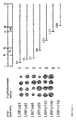
- Fig. 1 Identification of the PrP c 67 kDa LRP interaction in the yeast two-hybrid system in S . cerevisiae.
- Yeast cells containing the reporter plasmid pSH18-34 were cotransformed with prey plasmid pJG4-5 ⁇ 43 encoding LRP (amino acids 44-295) encoding cDNA and the bait plasmids pSH2-1-GST::rPrP c (row 1), pSH2-1-GST (row 2), pSH2-1 (row 3), pSH2-1-CTF-2 (row 4; Wendler et al., 1994) and pEG202-bicoid (row 5; Gyuris et al., 1993).
- LRP amino acids 44-295
- Resuspension of each of the five transformants was carried out in TE and dotted onto galactose plates which were either supplemented with X-gal (a) or leucine deficient (b). Plates were incubated for 3 days at 30°C.
- Fig. 2 Mapping of the PrP/LRP interaction site in the yeast two-hybrid system
- Yeast cells containing the reporter plasmid pSH18-34 were cotransformed with the bait plasmid pSH2-1-GST-PrP c and prey plasmids pJG4-5 ⁇ 43 (row 1), pJG4-5 ⁇ 45 (row 2), pJG4-5 ⁇ 46 (row 3), pJG4-5 ⁇ 89 (row 4), pJG4-5 ⁇ 130 (row 5), pJG4-5 ⁇ 156 (row 6) and pJG4-5 ⁇ 179 (row 7) coding for amino terminal truncated versions of the laminin receptor precursor lacking 43, 45, 46, 89, 130, 156 amino acids (the latter contains 23 amino acids not homologous to the laminin receptor precursor) and 179 amino acids (lacking the laminin binding domain) at the amino terminus.
- Each of five transformants were resuspended in TE, dotted on X-Gal supplemented plates and incubated at 30°C for 3 days.
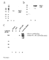
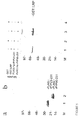
- Fig. 3 Analysis of recombinant LRP fused to GST in baculovirus infected insect cells (Sf9)
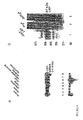
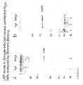
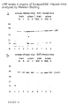
- Fig. 4 (a) Northern Blot Analysis of multiple human tissues. and (b) and (c) Western Blot Analysis of crude lysates of N 2 a/ScN 2 a cells
- Total mRNA isolated from human heart (lane 1), brain (lane 2), placenta (lane 3), lung (lane 4), liver (lane 5), skeletal muscle (lane 6), kidney (lane 7) and pancreas (lane 8) was immobilized on a nylon membrane and probed with a radiolabelled DNA fragment corresponding to 550 bp of the human 67 kDa high affinity laminin receptor precursor cDNA. (bottom) Total mRNA as described above was assayed with a radiolabelled ⁇ -actin probe.
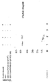
- Fig. 5 Analysis of PrP/LRP interactions by Co-infection assays in baculovirus infected insect cells (Sf9)
- Sf9 cells were co-infected with FLAG-BAC::rPrP23-231 and AcSG2T::LRP (lane 3), FLAG-BAC::rPrP23-231 and AcSG2T (lane 1), FLAG-BAC::rPrP90-231 (rPrP27-30) and AcSG2T (lane 4) and FLAG-BAC::rPrP90-231 (rPrP27-30) and AcSG2T (lane 2).
- GST and GST fusion proteins interacting with FLAG::rPrP23-231 and FLAG::rPrP90-231, respectively, have been isolated and purified by anti-FLAG-antibody affinity chromatography and analyzed by Western Blotting employing a polyclonal GST antibody.
- Fig. 7 Model of the function of the LRP as the receptor for the prion proteins PrP c and/or PrP Sc
- LRP laminin receptor precursor
- Fig. 8 Schematic presentation of the reverse two-hybrid system to screen for components interfering with the LRP/PrP c interaction
- Fig. 9 Schematic presentation of the mammalian two-hybrid system to screen for components interfering with the LRP/PrP c interaction
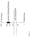
- Fig. 10 Localization of the 67 kDa high affinity laminin receptor precursor in the plasma membrane
- the N-terminus of the receptor is thought to be located in the cytoplasm of the cell (Castronova et al., 1991).
- a short transmembrane region was also suggested between amino acid 88 and amino acid 101 of LRP (Mohane and Argos, 1986).
- the carboxy terminus of LRP is thought to be located extracellularly.
- a short laminin binding domain was identified between amino acid 161 and amino acid 180 of LRP corresponding to the PrP binding domain (Rieger et al., 1997, and Castronovo et al., 1991).
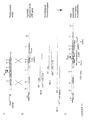
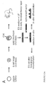
- Fig. 11 Targeted disruption of the LRP gene locus by homologous recombination and introduction of an Anti-LRP gene for antisense RNA production
- the replacement vector A
- the hypothetic genomic structure of one LRP gene locus B
- the mutated locus with the inserted antisense-LRP cassette C
- the antisense-LRP cassette under the control of an inducible promoter (metallothionine promoter, tetracycline-responsive promoter) adjacent to the neomycin resistance cassette (NEO).
- the negative selection marker thymidine kinase (TK) will be added to one site of the homology region.
- Splinkers should be ligated to the linearized vector to prevent vector degradation by endonucleases. Shown are the probes for Southern blot analysis (probe A, Anti-LRP probe, Neoprobe). PCR primers to identify homologous recombined ES cell clones and predicted fragments after restriction enzyme (RE B, RE C, RE D) analysis are indicated.
- P ind inducible promoter.
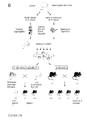
- Fig. 12 Generation of LRP knock-out mice
- Fig. 13 Structure of the vector pFLAG:BAC
- the map shows the position of the polyhedrin promoter, the signal sequence of gp67, the polylinker, the origin of replication and the gene conferring resistance to ampicillin.
- Fig. 14 Nucleic acid sequence of pFLAG:BAC with restriction sites
- Fig. 15 Co-expression of FLAG-tagged Hsp60 and GST-tagged prion proteins from Syrian golden hamster in the baculovirus system
- Fig. 16 Western Blot analysis of brain and spleen homogenates from scrapie (strain C506M3) infected.
- BSE strain 4PB1 infected and uninfected mice.
- Fig. 17 Western Blot analysis of homogenates of brain, spleen, pancreas, liver and skeletal muscle from scrapie (strain 263 K) infected and uninfected hamsters.
- Fig. 19 The Uptake of recombinant Prion Proteins from cattle by murine neuroblastoma cells (N 2 a)
- N 2 a cells were incubated with bovine PrP fused to GST expressed in the baculovirus system.
- Cells were incubated with a PrP specific antibody (3B5) Magnification: 400 fold (A).
- a LRP specific antibody (Rieger et al., 1997) was preincubated before the recombinant bovine PrP was added to the cells followed by addition of a PrP specific antibody (3B5) Magnification 630: fold (B).
- Example 1 Identification of LRP as a compound specifically binding to PrP by using the Yeast Two-Hybrid Screen
- Potential positive EGY48 transformants were selected by growth on -Ura, -His, -Trp, -Leu in the presence of galactose due to the gal-promoter of the DNA-insert in pJG4-5. Positive clones were probed for ⁇ -galactosidase production by dotting them on galactose plates supplemented with 5-bromo-4-chloro-3-indolyl- ⁇ -D-galactopyranoside (X-Gal). The cDNAs of positive clones were recovered and transformed in E . coli KC8 as described (Edenhofer et al.,1996) and sequenced.
- plasmid pSH2-1, pSH2-1-GST, pSH2-1-GST::rPrP c , pSH2-1-CTF-2 (Wendler et al., 1994) and pEG202-bicoid Gyuris et al., 1993) were retransformed in EGY48 and the transformants tested for ⁇ -galactosidase production and their Leu+-phenotype. At least five retransformants were dotted on corresponding plates and incubated for 3 days at 30°C.
- Yeast strain EGY 48 was transformed with the pSH-GST-PrP c construct coding for GST::PrP c fused to LexA and tested for intrinsic activation as described previously (Edenhofer et al., 1996). Correct expression of the bait protein GST::PrP c was confirmed by the repression activity of LexA. The control baits LexA-bicoid and LexA-Gal4 were also positive in the repression assay (data not shown). Expression of IexA-GST::PrP c with a molecular weight of approx. 53 kDa was confirmed by western blotting developed with a polyclonal PrP specific antibody (data not shown).
- PrP specific antibody did not recognize any proteins in the case of pSH-GST and pSH transformed yeast cells (data not shown).
- S. cerevisiae was cotransformed with pSH-GST-PrP c , the reporter plasmid pSH18-34 and the prey plasmid pJG4-5, containing a HeLa-cDNA library controlled by a galactose-inducible promoter.
- a total of approximately 6 x 10 5 transformants were pooled and an aliquot induced by adding galactose supplemented medium. Plating of the induced culture on Leu-deficient galactose plates resulted in approx. 2,000 colonies.
- pJG4-5 ⁇ 179 was constructed by partial digestion of pJG-1.91 with Ava l followed by an Eco RI digestion. The resulting approx. 7 kb DNA fragment lacking the 420 bp Eco RI (5') - Ava l (3') DNA fragment was blunted and religated resulting in pJG4-5 ⁇ 179.
- Example 3 Recombinant LRP synthesized in insect cells binds to Engelbreth-Holm-Swarm (EHS) laminin.
- EHS Engelbreth-Holm-Swarm
- GST::LRP isolated from AcSG2T::LRP infected Sf9 cells migrates with a molecular weight of 67 KDa which was expected from the 27 kDa of GST (Weiss et al., 1995; 1996) plus 37 KDa observed from recombinant LRP expressed in highly aggressive cancer cells (Yow et al., 1988; Rao et al., 1989). No secretion of GST::LRP was observed (Fig.3 A, lane 1), despite the fact that the autographica californica gp67 signal sequence (Whitford et al., 1989) was constructed at the amino terminus of the fusion protein.
- a polyclonal LRP specific antibody specifically recognized the laminin receptor precursor fused to GST (Fig.3 B, lane 2) demonstrating that the recombinant LRP was immunologically active.
- Laminin consists of two polypeptide chains B1 and B2 (each 220 kDa, Fig. 3 C, lane 4) linked via disulfide bonds to form together with polypeptide chain A (440 kDa, Fig. 4, lane 4) the characteristic asymmetric cross-structure.
- binding of laminin can be detected in the presence of an anti-EHS-laminin antibody only with immobilized GST::LRP (Fig. 3 C, lane 3). No binding takes place in the presence of immobilized GST (Fig.3 C lane 2) or unloaded beads (Fig.3 C, lane 1).
- Example 4 LRP mRNA is present in several human organs including the brain
- a human multiple tissue Northern blot has been screened with a DNA probe complementary to a region encoding for the laminin receptor precursor.
- Hybridization of the labeled fragment to the human multiple tissue mRNA blot reveals that mRNA encoding for LRP is ubiquitous distributed in heart (Fig. 4A, lane 1), placenta (lane 3), lung (lane 4), liver (lane 5), skeletal muscle (lane 6), pancreas (lane 8) and the brain (lane 2).
- LRP-mRNA level in kidney was very weak (lane 7).
- LRP expression is tissue-dependent; see Table 1, above, and Figures 16 and 17.
- Fig. 4c(a) demonstrates that the LRP level was twofold increased in Scrapie infected N 2 a cells (lane 2) compared to uninfected N 2 a cells (lane 1).
- the intensities of the protein bands were determined by optical scanning methods.
- the level of ⁇ -actin (42 kDa; control) remained stable in ScN 2 a (Fig. 4c(b), lane 3) compared to N 2 a cells (Fig. 4c(b), lane 2).
- LRP protein levels in brain and spleen of terminal C57BL/6 mice intracerebrally inoculated with either a scrapie (C506M3) or a BSE (4PB1) strain were investigated.
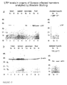
- the LRP level was twofold increased in brain and spleen of scrapie infected mice (Fig. 16a, lanes 2 and 4, respectively) compared to uninfected mice (lanes 3 and 5, respectively), whereas no significant alteration of the LRP amount was observed in either organ of BSE infected mice (Fig. 16a, lanes 6, 7, 8 and 9, respectively).
- ⁇ -actin or actin (42 kDa) levels were unaltered in brain, spleen, pancreas, liver and skeletal muscle of scrapie infected compared to uninfected hamsters (Fig. 17b).
- LRP levels are increased only in those organs that exhibit infectivity and PrP Sc accumulation such as brain, spleen and pancreas (see Table 1, below).
- Example 6 rPrP23-231 (rPrP c ) and rPrP90-231 (rPrP27-30) interact with LRP in insect cells co-infected with recombinant baculoviruses
- FLAG tagged PrP c (Fig. 5A, lane 1) and FLAG::rPrP90-231 (Fig. 5A, lane 2) secreted from Baculovirus infected insect cells revealed molecular weights of 27 kDa and 17 kDa, respectively.
- Co-infection assays demonstrate that GST::LRP can only be detected by co-infecting AcSG2T::LRP with FLAG-BAC::rPrP23-231 (Fig.5 B, lane 3) or FLAG::BAC::rPrP90-231 (Fig.5 B, lane 1).
- Example 7 rPrP23-231 (rPrP c ) interact with authentic LRP in Cos-7 cells
- Example 8 LRP knock-out mice are resistant to PrP Sc infection
- LRP LRP as a receptor for PrP Sc
- LRP knock-out mice Because LRP belongs to a multicopy gene family (Bignon et al., 1991; Douville and Carbonetto, 1992; Fernandez et al., 1991), it is impossible to use the classical knock-out strategy recently described for the construction of a bir 1-knock-out mouse (Förster et al., 1996). Thus, it is necessary to switch off additional loci. This can be done by connecting the classical knock-out approach with ah antisense strategy. This procedure leads to mice with one disrupted LRP gene. The mRNA of further active LRP genes can be inactivated by the induced antisense mRNA.
- the first step in the generation of a LRP knock-out mouse is the screening of one homologue of the LRP gene in a genomic mouse library (129 SV) cloned in a ⁇ -FixllTM vector. Employing an LRP specific DNA probe leads to the identification of the genomic structure of the LRP gene, which is subcloned, mapped and sequenced.
- the second step includes the construction of a replacement vector (Fig. 11). Parts of the LRP gene are disrupted and replaced by the gene encoding neomycin resistance for positive selection (Fig.
- an anti-LRP cassette containing the antisense LRP sequence under the control of an inducible promoter such as the metallothionine promoter or tetracycline-responsive promoter (Gossen and Bujard, 1992).
- the thymidine kinase gene flanking the homology regions on either side of the LRP gene enables negative selection.
- the replacement vector is transferred to murine embryonic stem cells (ES) via electroporation (Fig. 12A).
- ES murine embryonic stem cells
- Fig. 12A The presence of G 418 and gancyclovir leads to clones potentially harboring the homologous recombination event. Screening of positive selected clones via PCR and Southern blot analysis allows the identification of clones mutated in one LRP gene on one allele (Fig. 11, 12A).
- Homologous recombined ES cell clones are transferred into embryonic morulas or blastocytes by morula aggregation and blastocyst injection. Chimeras are identified by coat color and tested for germ line integration by Agouti-coat color (to identify the integration of the homologous recombined embryonic stem cells) and Southern blotting. Heterozygous mice are intercrossed to produce homozygous LRP -/- mice with one disrupted LRP gene. In order to inactivate the additional LRP genes, the metallothionine or tetracycline-responsive promoter (Gossen and Bujard, 1992) is activated by feeding the mice with Zinc or tetracycline, respectively. The antisense LRP mRNA binds additional generated LRP mRNA. This approach can be done during different developmental stages and can be proven by Northern blot analysis, Western blotting and FACS analysis.
- the phenotype of the LRP -/- mice can be characterized including behaviours, histological examination of the brain and other organs and neurological investigations.
- mice are monitored over their whole lifespan for the occurrence of clinical abnormalities including neurological signs.
- their preformance is tested on a rotorod.
- the learning ability of the LRP -/- mice can be assessed and compared with WT mice possessing the same genetic background, by means of appropriate tests (e.g. swimming navigation and Y-maze discrimination test).
- Anatomical investigations are made to detect gross abnormalities of the brain and other organs; histological examinations are peformed using classical techniques and haematoxylin-eosin staining.
- the crucial experiment is the innoculation of the LRP -/- mice with PrP Sc- enriched infectious fractions from mouse and cattle.
- PrP Sc detection procedure (Lasmezas et al., 1997). Mice infected with PrP Sc are sacrificed at the pre-mortem stage by cervical fracture, and brains are immediately removed. One hemisphere (including the cerebellum) is frozen in liquid nitrogen, and stored at -80°C for PrP analysis. The other hemisphere is fixed for pathological examination.
- PrP Sc purification the whole brain hemisphere is homogenized to 10% (w/v) in a 5% glucose solution. Briefly, proteinase K (PK) is used at 10 ⁇ g/ml and digestion is blocked with PMSF.
- PK proteinase K
- samples After addition of sarkosyl to 10% and Tris.HCL pH 7.4 to 10 mM, samples are incubated for 15 minutes at room temperature. They are then centrifuged at 245000 g for 4 hours at 20°C on a 10% sucrose cushion (Beckmann TL100 ultracentrifuge). Pellets are resuspended in Laemmli buffer and analyzed by Western blotting employing a polyclonal anti-mouse PrP antibody. The innoculation of the LRP -/- mice with PrP Sc shows that the mice are resistant towards PrP Sc infection.
- Example 9 Inactivation of the LRP genes in neuroblastoma cells (N 2 a) by Antisense RNA technique
- LRP expression can be downregulated in the presence of an antisense LRP mRNA.
- An alternative ribozyme approach is also conceivable.
- the downregulation of LRP can be meassured in cell culture systems employing scrapie infected versus uninfected neuroblastoma cells (N 2 a). Scrapie infected N 2 a cells should lose their infectivity in the presence of the antisense/ribozyme construct and neuroblastoma cells should not be infectable with PrP Sc any more in the presence of antisense LRP mRNA/ribozymal RNA.
- the same transfection vector can be microinjected into oocytes (fertilized eggs).
- a construct spanning the cDNA from postion -65 to +901 of the mouse 37 kDa LRP ORF was generated by RT-PCR from total RNA isolated from N2a neuroblastoma cells using two oligonucleotide primers.
- the resulting 970 bp DNA fragment was subcloned in antisense orientation into the pCl-neo mammalian expression vector (Promega) using Nhel (5') and Smal (3') restriction sites. The correct insert was verified by restriction analysis and dideoxy sequencing.
- Scrapie-infected (ScN 2 a) and non-infected mouse neuroblastoma cells (N 2 a) were maintained in Dulbecco's modified Eagle's medium (DMEM) (Sigma) supplemented with 10 % FCS, 1 % Glutamine, 100 ⁇ g/ml penicillin and 100 ⁇ g/ml streptomycin.
- DMEM Dulbecco's modified Eagle's medium
- FCS 10 % FCS, 1 % Glutamine, 100 ⁇ g/ml penicillin and 100 ⁇ g/ml streptomycin.
- Cells were grown in a 25 cm 2 culture bottle to 70 % confluency under the above conditions and transfected in serum-free medium for 14 hours with 100 ⁇ g of Lipofectamine (Life Technologies) and 16 ⁇ g of plasmid DNA pCl-neo-asLRP.
- G418 medium containing 400 ⁇ g/ml G418. (Life Technologies). Colonies resistant to G418 were expanded and screened for antisense LRP expression by Northern blot analysis. Furthermore LRP levels were determined by SDS-PAGE and Western-Blotting.
- DNA-sequences Prior to microinjection in oocytes bacterial DNA-sequences had to be removed from the pCl-neo-asLRP plasmid. Therefor the plasmid was cleaved upstream the CMV promotor and downstream the Neomycin gene with Bgl II and Bam HI, respectively.
- the DNA-fragment was purified by TAE-buffered gel electrophoresis and eluted from the gel by standard procedures. After phenol extraction and ethanol precipitation the DNA was resuspended in TE-buffer (5 mM TrisHCl, pH 7.4, 0.2 mM EDTA, pH 7.4), filtered through a 0.2 ⁇ m filter and the DNA concentration determined. The DNA was diluted in TE-buffer to a concentration of 3 to 5 ng/ml and 20 ml aliquots were stored frozen until use.
- the isolated restriction fragment (as described above) was injected into the pronuclei of the fertilized mouse eggs using a micromanipulator. 3-5 ng of DNA were injected into 90-240 fertilized eggs. After microinjection survived eggs were transferred into the oviduct of a pseudopregnant foster mother.
- Offsprings (the presence of the antisense/ribozyme cassette will be analyzed by PCR) should express antisense LRP RNA and should show decreased levels of LRP or should fail to express LRP completely. These animals should be partially or totally resistant towards a Scrapie infection.
- cytoplasmic staining cells were permeabilized with 0.1 % Triton X-100 in PBS for 10 min at 4 °C. After rinsing three times with PBS coverslips were blocked with 10 % FCS in PBS for one hour at room temperature and washed once in PBS. For detection of LRP cells were incubated for one hour at room temperature with a monoclonal LRP-specific antibody, diluted 1:200 in 10 % FCS in PBS. Controls were incubated with pre-immuneserum in 10 % FCS in PBS.
- Example 11 Uptake of recombinant PrP from human and cattle by murine neuroblastoma cells (N 2 a)
- Figure 19 (left panel) demonstrates the uptake of bovine PrP fused to GST by N 2 a cells.
- the small dots represent the bovine PrP on the surface of N 2 a cells detected by a PrP specific antibody (3B5).
- the uptake was prohibited when the cells have been preincubated with a LRP specific antibody (Rieger et al., 1997) ( Figure 19, right panel) demonstrating that LRP is responsible for the uptake of the prion protein by N 2 a cells.
- the LRP specific antibody can be used as a therapeutic tool to prevent the uptake of the prion protein PrP by the LRP.
- the high affinity murine laminin receptor is a member of a multicopy gene family. Biochem. Biophys. Res. Commun. 175, 84-90.
- BLR1 A putative chemokine receptor, BLR1 directs B cell migration to defined lymphoid organs and specific anatomic compartments of the spleen (1996) Cell 87 , 1037-1047.
- the elastin receptor is a galactoside binding protein. Science 239 , 1539-1541.
- PrP27-30 is a normal soluble prion protein fragment released by human platelets. Biochem. Biophys. Res. Commun ., 223, 572-577.
- RNA 3 (1997) 677-87.
- Scrapie agent contains a hydrophobic protein. Proc. Natl . Acad . Sci . U.S.A., 78, 6675-6679.
- High affinity laminin receptor precursor is a receptor for Sindbis Virus in mammalian cells. J. Virol. 66, 4992-5001.
Landscapes
- Health & Medical Sciences (AREA)
- Life Sciences & Earth Sciences (AREA)
- Chemical & Material Sciences (AREA)
- Organic Chemistry (AREA)
- Engineering & Computer Science (AREA)
- Genetics & Genomics (AREA)
- Bioinformatics & Cheminformatics (AREA)
- General Health & Medical Sciences (AREA)
- Biomedical Technology (AREA)
- Medicinal Chemistry (AREA)
- Molecular Biology (AREA)
- Immunology (AREA)
- Zoology (AREA)
- Biophysics (AREA)
- Biochemistry (AREA)
- Proteomics, Peptides & Aminoacids (AREA)
- General Engineering & Computer Science (AREA)
- Public Health (AREA)
- Animal Behavior & Ethology (AREA)
- Pharmacology & Pharmacy (AREA)
- Nuclear Medicine, Radiotherapy & Molecular Imaging (AREA)
- General Chemical & Material Sciences (AREA)
- Veterinary Medicine (AREA)
- Neurology (AREA)
- Neurosurgery (AREA)
- Chemical Kinetics & Catalysis (AREA)
- Wood Science & Technology (AREA)
- Biotechnology (AREA)
- Hospice & Palliative Care (AREA)
- Plant Pathology (AREA)
- Microbiology (AREA)
- Physics & Mathematics (AREA)
- Psychiatry (AREA)
- Cell Biology (AREA)
- Toxicology (AREA)
- Gastroenterology & Hepatology (AREA)
- Peptides Or Proteins (AREA)
- Medicines That Contain Protein Lipid Enzymes And Other Medicines (AREA)
- Analysing Materials By The Use Of Radiation (AREA)
- Investigating Or Analysing Biological Materials (AREA)
Abstract
Description
PrPc and PrPSc are identical regarding primary structure (Stahl et al., 1993), but CD-and FTIR-spectroscopy reveals that they have different secondary structures (Meyer et al., 1986; Pan et al., 1993). PrPc has a very low content and PrPSc a high content of β-sheet structure. Moreover, PrPc can be hydrolyzed completely by proteinase K, whereas PrPSc is degraded to the proteinase K resistant infectious core PrP27-30 (Caughey et al., 1991; Prusiner et al., 1981; 1983; 1984). A proteinase K sensitive, soluble and presumably non-infectious version of PrP27-30, designated as sPrP27-30 was identified in human platelets (Perini et al., 1996) and might also act as a precursor for proteinase K resistant PrP27-30.
The physiological role of the cellular prion protein PrPc was investigated with a series of knock-out experiments leading to controversial results. Most of the researchers did not recognize any changes in the behaviour or reproduction of PrP-knock-out mice (Büehler et al., 1992). More recent investigations with PrPo/o mice suggested an important role of PrPc in synaptic function including an efficient GABAA receptor-mediated fast inhibition (Collinge et al., 1996) whereas another study did not find any dysfunctions of synapses in the hippocampus (Lledo et al., 1996). These discrepancies in the observation of phenotypes in PrPo/o mouse lines may be due to the genetically heterogeneous background (Borchelt and Sisodia, 1996) and the different age of the animals. In summary, the physiological role of PrPc remains unclear. However, PrPo/o mice lacking the prn-p gene are, in contrast to wild-type mice, resistant to infections with the mouse PrPSc (Büehler et al., 1993) demonstrating that PrPSc is essential for the development of Scrapie and Scrapie-like disease. More recent experiments demonstrated that PrPc is necessary for scrapie induced neurotoxicity (Brandner et al., 1996) and required for scrapie spread within the central nervous system (Brandner et al., 1996b).
To map the LRP binding site for PrP, prey plasmids lacking 43 amino acids up to 179 amino acids at the amino terminal end of the laminin receptor precursor were retransformed with lexA-GST::PrPc. Prey LRPΔ157 did interact with PrP whereas LRPΔ180 failed to bind to the prion protein demonstrating that the laminin binding domain (
In order to confirm the PrP/LRP interaction in another eukaryotic cell system, LRP (amino acid 44-amino acid 295) was expressed in baculovirus infected insect cells as a fusion with GST. Although the signal sequence of gp67 was present at the N-terminus, GST::LRP was not secreted into the culture medium but synthesized cell-associated most likely due to the presence of a short transmembrane region which was calculated between
For co-infection assays in insect cells including GST::LRP and prion proteins, the cellular form of the hamster prion protein PrPc (PrP23-231) was synthesized and the soluble version (s) PrP27-30 of proteinase K resistant PrP27-30 (Caughey et al., 1991; Prusiner et al., 1981; 1983; 1984) was fused to the FLAG peptide. Both recombinant proteins were secreted into the culture medium, purified to homogeneity by anti-Flag-antibody affinity chromatography and recognized by a PrP specific antibody. Molecular weights determined by SDS-PAGE were 27 kDa and 17 kDa for rPrPc and rPrP27-30, respectively, and correspond to rPrPc (Weiss et al., 1995) and rPrP27-30 (Weiss et al., 1996) generated by thrombin cleavage from GST fusion proteins. In order to confirm the LRP/PrP interaction observed in S. cerevisiae, co-infection in Sf9 cells was performed. A specific interaction between the laminin receptor precursor and the prion protein PrP in insect cells confirms the PrP/LRP interaction observed in yeast.
To further confirm the PrP/LRP interaction in mammalian cells, FLAG::rPrPc and authentic LRP were transiently expressed in Cos-7 cells. LRP revealed a molecular weight of 37 kDa and was recognized by a polyclonal anti-LRP antibody. rPrPc fused to FLAG showed the expected molecular weight of 27 kDa and was also recognized by a polyclonal PrP specific antibody. Interaction between both proteins was proven in Cos-7 cells demonstrating that LRP interacts with PrP in a series of different eukaryotic cells including yeast, insect and Cos-7 cells.
The laminin receptor precursor family is highly conserved in a wide spectrum of eukaryotic cells (Wewer et al., 1986; Keppel et al., 1991; Garcia-Hernandez et al., 1994), was recently reported to be present in yeast (Demianova et al., 1996) and may be encoded by archaean genomes (Ouzounis et al., 1995). The receptor works as a receptor for Sindbis Virus on mammalian cells such as CHO or BHK (Wang et al., 1992) and for Venezuelan Encephalitis Virus from Mosquito Cells (Ludwig et al., 1996). For a functional involvement of the PrPc/LRP interaction in the pathogenesis of TSEs or the biological function of PrPc one would expect that the laminin receptor precursor is present in the human brain. Therefore, the presence of LRP-specific mRNA in multiple human tissues was tested. This analysis shows that LRP-mRNA is ubiquitously distributed in several tissues including heart and brain. The size of the laminin receptor precursor mRNA of approximately 1.4 kb is identical to that found in human, rat and mouse cells (Rao et al. 1989). In addition, it has been demonstrated that the 67 kDa LRP is present on neuronal cells (Kleinmann et al., 1988; Douville et al., 1988). Moreover, laminin itself has a drastic influence on the growth of neuronal cells. The LRP works as a receptor for the Sindbis-Virus and the VEE-virus on the surface of mammalian cells. The cellular prion protein PrPc is also located on the surface of neuronal cells fixed by the glycosylphosphatidylinositol anchor (GPI; Stahl et al., 1987). Thus, a direct interaction of PrPc and LRP is conceivable on the surface of neuronal cells of the brain. To further prove whether the laminin receptor precursor is overexpressed in Scrapie infected N2a cells (Sc N2a), crude lysates of N2a and Sc N2a cells with a LRP specific antibody were analyzed. LRP is present in crude lysates of N2a cells but levels of LRP in crude lysates of Sc N2a cells are twofold higher compared to N2a cells suggesting that LRP expression is up-regulated in N2a cells after Scrapie exposure. Recently, it has been reported that a soluble form of PrP27-30 was identified in human platelets (Perini et al., 1996). sPrP27-30 could work as a precursor for proteinase K resistant PrP27-30, a major product in Scrapie infected prion-preparations (Prusiner et al., 1981; 1983; 1984). The laminin receptor precursor has also been identified in human platelets (Tandon et al., 1991) which makes a PrP/LRP interaction also conceivable in platelets.
The same approach was used to obtain FLAG::Hsp60. As shown in Figure 15 (lane 1), the protein is homogenous and migrates as a single band (61 kD) on SDS-PAGE.
Purified polyclonal anti-LBP antibody (laminin-binding protein corresponding to LRP) was kindly provided by Dr. J.R. Starkey, Montana. Polyclonal rabbit antibody directed against a peptide corresponding to a region comprising amino acids 95 to 110 of the Syrian Golden hamster prion protein was a generous gift of Dr. M. Groschup, BFAV Tübingen. Polyclonal GST antibody was delivered by Santa Cruz Biotech, USA.
Anti-EHS-laminin antibody was purchased from Sigma.
Furthermore, LRP protein levels in brain and spleen of terminal C57BL/6 mice intracerebrally inoculated with either a scrapie (C506M3) or a BSE (4PB1) strain were investigated. The LRP level was twofold increased in brain and spleen of scrapie infected mice (Fig. 16a,
In order to confirm the direct correlation between LRP levels and PrPSc accumulation, we investigated the amounts of LRP in different organs of syrian hamsters inoculated with the 263K scrapie strain, which is a model with high levels of intracerebral PrPSc accumulation. The LRP level was fivefold increased in the brain (Fig. 17a, lane 2) and fourfold raised in the pancreas (lane 6) of scrapie infected hamsters compared to uninfected control hamsters (
In contrast, LRP expression failed in skeletal muscle (
In summary, LRP levels are increased only in those organs that exhibit infectivity and PrPSc accumulation such as brain, spleen and pancreas (see Table 1, below).
| Increase of LRP levels in Scrapie/BSE infected versus uninfected cells and tissues | |||||
| MOUSE | brain | spleen | ScN2a/ N2a cells | ||
| Scrapie infected | 2 | 2 | 2 fold | ||
| BSE infected | 1.1 fold | unchanged | |||
| HAMSTER | brain | spleen | pancreas | liver | skeletal muscle |
| Scrapie infected | 5 fold | 1.5 | 4 fold | unchanged | not expressed |
The first step in the generation of a LRP knock-out mouse is the screening of one homologue of the LRP gene in a genomic mouse library (129 SV) cloned in a λ-Fixll™ vector. Employing an LRP specific DNA probe leads to the identification of the genomic structure of the LRP gene, which is subcloned, mapped and sequenced. The second step includes the construction of a replacement vector (Fig. 11). Parts of the LRP gene are disrupted and replaced by the gene encoding neomycin resistance for positive selection (Fig. 11) adjacent to an anti-LRP cassette containing the antisense LRP sequence under the control of an inducible promoter such as the metallothionine promoter or tetracycline-responsive promoter (Gossen and Bujard, 1992). The thymidine kinase gene flanking the homology regions on either side of the LRP gene enables negative selection. The replacement vector is transferred to murine embryonic stem cells (ES) via electroporation (Fig. 12A). The presence of G 418 and gancyclovir leads to clones potentially harboring the homologous recombination event. Screening of positive selected clones via PCR and Southern blot analysis allows the identification of clones mutated in one LRP gene on one allele (Fig. 11, 12A).
Anatomical investigations are made to detect gross abnormalities of the brain and other organs; histological examinations are peformed using classical techniques and haematoxylin-eosin staining.
The crucial experiment, however, is the innoculation of the LRP-/- mice with PrPSc- enriched infectious fractions from mouse and cattle.
PrPSc detection procedure (Lasmezas et al., 1997). Mice infected with PrPSc are sacrificed at the pre-mortem stage by cervical fracture, and brains are immediately removed. One hemisphere (including the cerebellum) is frozen in liquid nitrogen, and stored at -80°C for PrP analysis. The other hemisphere is fixed for pathological examination. For PrPSc purification, the whole brain hemisphere is homogenized to 10% (w/v) in a 5% glucose solution. Briefly, proteinase K (PK) is used at 10 µg/ml and digestion is blocked with PMSF. After addition of sarkosyl to 10% and Tris.HCL pH 7.4 to 10 mM, samples are incubated for 15 minutes at room temperature. They are then centrifuged at 245000 g for 4 hours at 20°C on a 10% sucrose cushion (Beckmann TL100 ultracentrifuge). Pellets are resuspended in Laemmli buffer and analyzed by Western blotting employing a polyclonal anti-mouse PrP antibody. The innoculation of the LRP-/- mice with PrPSc shows that the mice are resistant towards PrPSc infection.
First, the downregulation of LRP can be meassured in cell culture systems employing scrapie infected versus uninfected neuroblastoma cells (N2a). Scrapie infected N2a cells should lose their infectivity in the presence of the antisense/ribozyme construct and neuroblastoma cells should not be infectable with PrPSc any more in the presence of antisense LRP mRNA/ribozymal RNA.
Second, the same transfection vector can be microinjected into oocytes (fertilized eggs). After microinjection, survived eggs can be transferred into the oviduct of a pseudopregnant foster mother. The offsprings should show decreased levels of LRP or should fail to express LRP any more resulting in a complete resistance towards a scrapie infection.
Two examples for an antisense and an ribozyme derived approach are given below. Microinjection of in vitro transcribed RNA and antisense oligonucleotides in mouse oocytes and early embryos have been performed to study the gain and loss-of-function of genes (Kola and Sumarsono, 1996). An adenoviral VAI small RNA has been used as a carrier for cytoplasmic delivery of ribozymes(Prislei et al., 1997).
After 24 hours successul mating was verified by taking a plug control. After that female mice were killed and tubes preparated. Fertilized eggs in cumulus cells were washed out and were treated with hyaluronidase for removal of the cumulus cells. Fertilized eggs were kept in culture medium.


Claims (22)
- Method of diagnosis of transmissible spongiform encephalopathy (TSE), said method comprising the following step:
determining the over-expression of a marker protein for TSE in a sample to be tested. - Method of diagnosis of transmissible spongiform encephalopathy (TSE), said method comprising the steps of:(a) determining the expression of a marker protein for TSE in a sample to be tested; and(b) comparing the value obtained with a control value, wherein an increase in the expression level is indicative of the occurence of TSE.
- Method accoding to anyone of claims 1 or 2, wherein the sample is a homogenate from the brain, the spleen, the pancreas, the liver or the muscle.
- Method according to anyone of claims 1 to 3, wherein the marker protein is the laminin receptor (LR) or the laminin receptor precursor (LRP).
- Method according to anyone of claims 1 to 4, wherein the marker protein is the human laminin receptor (LR) or the human laminin receptor precursor (LRP).
- Method according to anyone of claims 1 to 4, wherein the marker protein is the bovine laminin receptor (LR) or the bovine laminin receptor precursor (LRP).
- Method according to anyone of claims 1 to 4, wherein the marker protein is the ovine laminin receptor (LR) or the ovine laminin receptor precursor (LRP).
- Method according to anyone of claims 1 to 4, wherein the marker protein is the feline laminin receptor (LR) or the feline laminin receptor precursor (LRP).
- Method according to anyone of claims 1 to 8, wherein the marker protein is determined by a "standard" immune test.
- Method according to claim 9, wherein said "standard" immune test is a Western Blot test.
- Method according to anyone of claims 1 to 8, wherein the sample is incubated with antibodies which are specific to the laminin receptor (LR) or the laminin receptor precursor (LRP).
- Method according to anyone of claims 1 to 8, wherein the altered expression of the laminin receptor or the laminin receptor precursor as the marker for TSE is determined by standard molecular biology methods.
- Method according to anyone of claims 1 to 12, wherein said sample is derived from an organism selected from the group consisting of a cow, a human, a sheep or a feline animal.
- Diagnostic test kit for detecting a transmissible spongiform encephalopathy (TSE), characterized in that it contains antibodies specific to the laminin receptor precursor (LRP) and/or the laminin receptor (LR) for detecting the altered expression of the laminin receptor (LR) and/or the laminin receptor precursor (LRP) as a marker protein for TSE by a method according to anyone of claims 1 to 13.
- Diagnostic test kit according to claim 14, wherein said antibodies are antibodies against bovine, human, ovine and/or feline LRP/LR.
- Diagnostic test kit according to anyone of claims 14 or 15, characterized in that the antibodies are monoclonal.
- Diagnostic test kit according to anyone of claims 14 or 15, characterized in that the antibodies are polyclonal.
- Diagnostic test kit according to anyone of claims 14 to 17, containing recombinant laminin receptor precursor fused to GST (GST::LRP) synthesized in the baculovirus system as a positive control and recombinant GST synthesized in the baculovirus system as a negative control in the test.
- Diagnostic test kit for detecting a transmissible spongiform encephalopathy (TSE), characterized in that it contains a nucleic acid which is capable of hybridising under stringent conditions with the nucleic acid coding for the laminin receptor precursor (LRP) and/or the laminin receptor (LR), as well as the other necessary elements for performing a method according to claims 14 to 18.
- Use of an antibody capable of binding the laminin receptor (LR) and/or the laminin receptor precursor (LRP) for the preparation of a composition for diagnosing a transmissible spongiform encephalopathy (TSE).
- Use of a nucleic acid capable of hybridizing under stringent conditions with the nucleic acid coding for the laminin receptor precursor (LRP) and/or the laminin receptor (LR) for the preparation of a composition for diagnosing a transmissible spongiform encephalopathy (TSE).
- Use according to anyone of claims 20 or 21, wherein the occurence of TSE is indicated by over-expression of the laminin receptor precursor (LRP) and/or the laminin receptor (LR) .
Priority Applications (1)
| Application Number | Priority Date | Filing Date | Title |
|---|---|---|---|
| EP01108876A EP1127894A1 (en) | 1997-05-30 | 1998-05-29 | Method of diagnosis of transmissible spongiform encephalopathy (TSE) |
Applications Claiming Priority (4)
| Application Number | Priority Date | Filing Date | Title |
|---|---|---|---|
| EP97108712 | 1997-05-30 | ||
| EP97108712 | 1997-05-30 | ||
| EP01108876A EP1127894A1 (en) | 1997-05-30 | 1998-05-29 | Method of diagnosis of transmissible spongiform encephalopathy (TSE) |
| EP98929404A EP0984987B1 (en) | 1997-05-30 | 1998-05-29 | A soluble laminin receptor precursor and methods to ihnibit its interactions |
Related Parent Applications (2)
| Application Number | Title | Priority Date | Filing Date |
|---|---|---|---|
| EP98929404A Division EP0984987B1 (en) | 1997-05-30 | 1998-05-29 | A soluble laminin receptor precursor and methods to ihnibit its interactions |
| EP98929404.6 Division | 1998-05-29 |
Publications (1)
| Publication Number | Publication Date |
|---|---|
| EP1127894A1 true EP1127894A1 (en) | 2001-08-29 |
Family
ID=8226854
Family Applications (2)
| Application Number | Title | Priority Date | Filing Date |
|---|---|---|---|
| EP01108876A Withdrawn EP1127894A1 (en) | 1997-05-30 | 1998-05-29 | Method of diagnosis of transmissible spongiform encephalopathy (TSE) |
| EP98929404A Expired - Lifetime EP0984987B1 (en) | 1997-05-30 | 1998-05-29 | A soluble laminin receptor precursor and methods to ihnibit its interactions |
Family Applications After (1)
| Application Number | Title | Priority Date | Filing Date |
|---|---|---|---|
| EP98929404A Expired - Lifetime EP0984987B1 (en) | 1997-05-30 | 1998-05-29 | A soluble laminin receptor precursor and methods to ihnibit its interactions |
Country Status (7)
| Country | Link |
|---|---|
| EP (2) | EP1127894A1 (en) |
| JP (1) | JP2002501385A (en) |
| AT (1) | ATE359298T1 (en) |
| AU (1) | AU7917598A (en) |
| CA (1) | CA2291720A1 (en) |
| DE (1) | DE69837539T2 (en) |
| WO (1) | WO1998053838A2 (en) |
Cited By (1)
| Publication number | Priority date | Publication date | Assignee | Title |
|---|---|---|---|---|
| WO2005035580A1 (en) * | 2003-10-08 | 2005-04-21 | Weiss, Stefan | Single-chain antibody acting against 37 kda/67 kda laminin receptor as tools for the diagnosis and therapy of prion diseases and cancer, production and use thereof |
Families Citing this family (7)
| Publication number | Priority date | Publication date | Assignee | Title |
|---|---|---|---|---|
| US6891082B2 (en) | 1997-08-01 | 2005-05-10 | The Johns Hopkins University School Of Medicine | Transgenic non-human animals expressing a truncated activintype II receptor |
| DE19918141A1 (en) | 1999-04-21 | 2000-10-26 | Boehringer Ingelheim Vetmed | Diagnosing transmissible spongiform encephalopathy, particularly before appearance of clinical symptoms, by detecting specific markers in blood cells |
| DE19925073C2 (en) | 1999-06-01 | 2001-07-19 | Stefan Weiss | Nucleic acid molecules with specific recognition of native PrP · S ·· c ·, production and use |
| DE10106076A1 (en) * | 2001-02-08 | 2002-08-22 | Susanne Wylezalek | Treatment of bovine spongiform encephalopathy and related diseases, e.g. Creutzfeldt-Jacob disease and scrapie, comprises administration of a medicament derived from Lathyrus sativus L. seeds |
| US20030118585A1 (en) | 2001-10-17 | 2003-06-26 | Agy Therapeutics | Use of protein biomolecular targets in the treatment and visualization of brain tumors |
| GB201519557D0 (en) * | 2015-11-05 | 2015-12-23 | Univ Witwatersrand Jhb | Compounds for use in the treatment of telomere related diseases and/or telomere related medical conditions |
| GB201700529D0 (en) * | 2017-01-12 | 2017-03-01 | Rogers April | Laminin receptor peptide |
Citations (5)
| Publication number | Priority date | Publication date | Assignee | Title |
|---|---|---|---|---|
| WO1989011273A1 (en) * | 1988-05-20 | 1989-11-30 | La Jolla Cancer Research Foundation | Adhesion receptor for laminin and its use |
| US5180809A (en) * | 1988-05-20 | 1993-01-19 | La Jolla Cancer Research Foundation | Adhesion receptor for laminin and its use |
| WO1993011155A1 (en) * | 1991-12-03 | 1993-06-10 | Proteus Molecular Design Limited | Fragments of prion proteins |
| WO1997045746A2 (en) * | 1996-05-29 | 1997-12-04 | Mcgill University | Prion protein binding proteins and uses thereof |
| WO1999030738A2 (en) * | 1997-12-16 | 1999-06-24 | University Of Zürich | Diagnostics and therapeutics for transmissible spongiform encephalopathy and methods for the manufacture of non-infective blood products and tissue derived products |
-
1998
- 1998-05-29 EP EP01108876A patent/EP1127894A1/en not_active Withdrawn
- 1998-05-29 WO PCT/EP1998/003220 patent/WO1998053838A2/en not_active Ceased
- 1998-05-29 AU AU79175/98A patent/AU7917598A/en not_active Abandoned
- 1998-05-29 AT AT98929404T patent/ATE359298T1/en not_active IP Right Cessation
- 1998-05-29 CA CA002291720A patent/CA2291720A1/en not_active Abandoned
- 1998-05-29 JP JP50026699A patent/JP2002501385A/en active Pending
- 1998-05-29 EP EP98929404A patent/EP0984987B1/en not_active Expired - Lifetime
- 1998-05-29 DE DE69837539T patent/DE69837539T2/en not_active Expired - Lifetime
Patent Citations (5)
| Publication number | Priority date | Publication date | Assignee | Title |
|---|---|---|---|---|
| WO1989011273A1 (en) * | 1988-05-20 | 1989-11-30 | La Jolla Cancer Research Foundation | Adhesion receptor for laminin and its use |
| US5180809A (en) * | 1988-05-20 | 1993-01-19 | La Jolla Cancer Research Foundation | Adhesion receptor for laminin and its use |
| WO1993011155A1 (en) * | 1991-12-03 | 1993-06-10 | Proteus Molecular Design Limited | Fragments of prion proteins |
| WO1997045746A2 (en) * | 1996-05-29 | 1997-12-04 | Mcgill University | Prion protein binding proteins and uses thereof |
| WO1999030738A2 (en) * | 1997-12-16 | 1999-06-24 | University Of Zürich | Diagnostics and therapeutics for transmissible spongiform encephalopathy and methods for the manufacture of non-infective blood products and tissue derived products |
Non-Patent Citations (2)
| Title |
|---|
| RIEGER R ETAL: "The human 37-kDa laminin receptor precursor interacts with the prion protein in eukaryotic cells", NATURE MEDICINE,US,NATURE PUBLISHING, CO, vol. 3, no. 12, 1 December 1997 (1997-12-01), pages 1383 - 1388, XP002094757, ISSN: 1078-8956 * |
| SHMAKOV ANDREI N ET AL: "Diverse patterns of expression of the 67-kD laminin receptor in human small intestinal mucosa: Potential binding sites for prion proteins?.", JOURNAL OF PATHOLOGY, vol. 191, no. 3, July 2000 (2000-07-01), pages 318 - 322, XP001012047, ISSN: 0022-3417 * |
Cited By (2)
| Publication number | Priority date | Publication date | Assignee | Title |
|---|---|---|---|---|
| WO2005035580A1 (en) * | 2003-10-08 | 2005-04-21 | Weiss, Stefan | Single-chain antibody acting against 37 kda/67 kda laminin receptor as tools for the diagnosis and therapy of prion diseases and cancer, production and use thereof |
| US7507797B2 (en) | 2003-10-08 | 2009-03-24 | Affimed Therapeutics Ag | Single-chain antibody acting against 37 kDa/67 kDa Iaminin receptor as tools for the diagnosis and therapy of prion diseases and cancer, production and use thereof |
Also Published As
| Publication number | Publication date |
|---|---|
| ATE359298T1 (en) | 2007-05-15 |
| EP0984987B1 (en) | 2007-04-11 |
| JP2002501385A (en) | 2002-01-15 |
| WO1998053838A3 (en) | 1999-05-14 |
| CA2291720A1 (en) | 1998-12-03 |
| EP0984987A2 (en) | 2000-03-15 |
| AU7917598A (en) | 1998-12-30 |
| WO1998053838A2 (en) | 1998-12-03 |
| DE69837539T2 (en) | 2008-01-03 |
| DE69837539D1 (en) | 2007-05-24 |
Similar Documents
| Publication | Publication Date | Title |
|---|---|---|
| US6482929B1 (en) | Antibodies to ARF-p19 | |
| Rieger et al. | The human 37-kDa laminin receptor precursor interacts with the prion protein in eukaryotic cells | |
| JP2024161501A (en) | Compositions and methods for the degradation of misfolded proteins | |
| EP1471926B1 (en) | Compositions and methods for the therapeutic use of an atonal-associated sequence | |
| CA2303405A1 (en) | 53bp2 complexes | |
| JPH10513359A (en) | Inhibitors of cyclin-dependent kinases CDK4 and CDK6, InK4c-p18 and InK4d-p19, and uses thereof | |
| EP0984987B1 (en) | A soluble laminin receptor precursor and methods to ihnibit its interactions | |
| CA2291698A1 (en) | Osf2/cbfa1 compositions and methods of use | |
| EP1066378A2 (en) | Lyst protein complexes and lyst interacting proteins | |
| EP1324652B1 (en) | Transgenic drosophila melanogaster expressing beta amyloid | |
| KR20020026430A (en) | PrP-LIKE GENE | |
| JP2002506875A (en) | Methods and compositions for diagnosis and treatment of chromosome 18p-related disorders | |
| US20030026781A1 (en) | Compositions and methods for regulating endogenous inhibitor of ATP synthase, including treatment for diabetes | |
| KR100811926B1 (en) | Compositions Useful for Modulation of Parkin Gene Activity | |
| US6800750B1 (en) | Mc1-1 gene regulatory elements and a pro-apoptotic mc1-1 variant | |
| WO2001036594A1 (en) | Mcl-1 GENE REGULATORY ELEMENTS AND A PRO-APOPTOTIC Mcl-1 VARIANT | |
| AU7411700A (en) | Gas1 polypeptides | |
| WO2003048196A2 (en) | Means and methods for diagnosing and treating craniofacial malformations |
Legal Events
| Date | Code | Title | Description |
|---|---|---|---|
| PUAI | Public reference made under article 153(3) epc to a published international application that has entered the european phase |
Free format text: ORIGINAL CODE: 0009012 |
|
| 17P | Request for examination filed |
Effective date: 20010409 |
|
| AC | Divisional application: reference to earlier application |
Ref document number: 984987 Country of ref document: EP |
|
| AK | Designated contracting states |
Kind code of ref document: A1 Designated state(s): AT BE CH CY DE DK ES FI FR GB GR IE IT LI LU MC NL PT SE |
|
| AKX | Designation fees paid |
Free format text: AT BE CH CY DE DK ES FI FR GB GR IE IT LI LU MC NL PT SE |
|
| 17Q | First examination report despatched |
Effective date: 20031117 |
|
| STAA | Information on the status of an ep patent application or granted ep patent |
Free format text: STATUS: THE APPLICATION IS DEEMED TO BE WITHDRAWN |
|
| 18D | Application deemed to be withdrawn |
Effective date: 20080701 |