EP1135686B1 - Diagnostic de la phase d'evolution ou de l'agressivite de cancers - Google Patents
Diagnostic de la phase d'evolution ou de l'agressivite de cancers Download PDFInfo
- Publication number
- EP1135686B1 EP1135686B1 EP99960625A EP99960625A EP1135686B1 EP 1135686 B1 EP1135686 B1 EP 1135686B1 EP 99960625 A EP99960625 A EP 99960625A EP 99960625 A EP99960625 A EP 99960625A EP 1135686 B1 EP1135686 B1 EP 1135686B1
- Authority
- EP
- European Patent Office
- Prior art keywords
- fabp
- cancer
- fatty acid
- cells
- levels
- Prior art date
- Legal status (The legal status is an assumption and is not a legal conclusion. Google has not performed a legal analysis and makes no representation as to the accuracy of the status listed.)
- Expired - Lifetime
Links
Images


Classifications
-
- G—PHYSICS
- G01—MEASURING; TESTING
- G01N—INVESTIGATING OR ANALYSING MATERIALS BY DETERMINING THEIR CHEMICAL OR PHYSICAL PROPERTIES
- G01N33/00—Investigating or analysing materials by specific methods not covered by groups G01N1/00 - G01N31/00
- G01N33/48—Biological material, e.g. blood, urine; Haemocytometers
- G01N33/50—Chemical analysis of biological material, e.g. blood, urine; Testing involving biospecific ligand binding methods; Immunological testing
- G01N33/92—Chemical analysis of biological material, e.g. blood, urine; Testing involving biospecific ligand binding methods; Immunological testing involving lipids, e.g. cholesterol, lipoproteins, or their receptors
-
- A—HUMAN NECESSITIES
- A61—MEDICAL OR VETERINARY SCIENCE; HYGIENE
- A61K—PREPARATIONS FOR MEDICAL, DENTAL OR TOILETRY PURPOSES
- A61K33/00—Medicinal preparations containing inorganic active ingredients
- A61K33/24—Heavy metals; Compounds thereof
-
- G—PHYSICS
- G01—MEASURING; TESTING
- G01N—INVESTIGATING OR ANALYSING MATERIALS BY DETERMINING THEIR CHEMICAL OR PHYSICAL PROPERTIES
- G01N33/00—Investigating or analysing materials by specific methods not covered by groups G01N1/00 - G01N31/00
- G01N33/48—Biological material, e.g. blood, urine; Haemocytometers
- G01N33/50—Chemical analysis of biological material, e.g. blood, urine; Testing involving biospecific ligand binding methods; Immunological testing
- G01N33/53—Immunoassay; Biospecific binding assay; Materials therefor
- G01N33/566—Immunoassay; Biospecific binding assay; Materials therefor using specific carrier or receptor proteins as ligand binding reagents where possible specific carrier or receptor proteins are classified with their target compounds
-
- G—PHYSICS
- G01—MEASURING; TESTING
- G01N—INVESTIGATING OR ANALYSING MATERIALS BY DETERMINING THEIR CHEMICAL OR PHYSICAL PROPERTIES
- G01N33/00—Investigating or analysing materials by specific methods not covered by groups G01N1/00 - G01N31/00
- G01N33/48—Biological material, e.g. blood, urine; Haemocytometers
- G01N33/50—Chemical analysis of biological material, e.g. blood, urine; Testing involving biospecific ligand binding methods; Immunological testing
- G01N33/53—Immunoassay; Biospecific binding assay; Materials therefor
- G01N33/575—Immunoassay; Biospecific binding assay; Materials therefor for cancer
- G01N33/57515—Immunoassay; Biospecific binding assay; Materials therefor for cancer of the breast
-
- G—PHYSICS
- G01—MEASURING; TESTING
- G01N—INVESTIGATING OR ANALYSING MATERIALS BY DETERMINING THEIR CHEMICAL OR PHYSICAL PROPERTIES
- G01N33/00—Investigating or analysing materials by specific methods not covered by groups G01N1/00 - G01N31/00
- G01N33/48—Biological material, e.g. blood, urine; Haemocytometers
- G01N33/50—Chemical analysis of biological material, e.g. blood, urine; Testing involving biospecific ligand binding methods; Immunological testing
- G01N33/53—Immunoassay; Biospecific binding assay; Materials therefor
- G01N33/575—Immunoassay; Biospecific binding assay; Materials therefor for cancer
- G01N33/5752—Immunoassay; Biospecific binding assay; Materials therefor for cancer of the lungs
-
- G—PHYSICS
- G01—MEASURING; TESTING
- G01N—INVESTIGATING OR ANALYSING MATERIALS BY DETERMINING THEIR CHEMICAL OR PHYSICAL PROPERTIES
- G01N33/00—Investigating or analysing materials by specific methods not covered by groups G01N1/00 - G01N31/00
- G01N33/48—Biological material, e.g. blood, urine; Haemocytometers
- G01N33/50—Chemical analysis of biological material, e.g. blood, urine; Testing involving biospecific ligand binding methods; Immunological testing
- G01N33/53—Immunoassay; Biospecific binding assay; Materials therefor
- G01N33/575—Immunoassay; Biospecific binding assay; Materials therefor for cancer
- G01N33/57525—Immunoassay; Biospecific binding assay; Materials therefor for cancer of the liver or pancreas
-
- G—PHYSICS
- G01—MEASURING; TESTING
- G01N—INVESTIGATING OR ANALYSING MATERIALS BY DETERMINING THEIR CHEMICAL OR PHYSICAL PROPERTIES
- G01N33/00—Investigating or analysing materials by specific methods not covered by groups G01N1/00 - G01N31/00
- G01N33/48—Biological material, e.g. blood, urine; Haemocytometers
- G01N33/50—Chemical analysis of biological material, e.g. blood, urine; Testing involving biospecific ligand binding methods; Immunological testing
- G01N33/53—Immunoassay; Biospecific binding assay; Materials therefor
- G01N33/575—Immunoassay; Biospecific binding assay; Materials therefor for cancer
- G01N33/57535—Immunoassay; Biospecific binding assay; Materials therefor for cancer of the large intestine, e.g. colon, rectum or anus
-
- G—PHYSICS
- G01—MEASURING; TESTING
- G01N—INVESTIGATING OR ANALYSING MATERIALS BY DETERMINING THEIR CHEMICAL OR PHYSICAL PROPERTIES
- G01N33/00—Investigating or analysing materials by specific methods not covered by groups G01N1/00 - G01N31/00
- G01N33/48—Biological material, e.g. blood, urine; Haemocytometers
- G01N33/50—Chemical analysis of biological material, e.g. blood, urine; Testing involving biospecific ligand binding methods; Immunological testing
- G01N33/53—Immunoassay; Biospecific binding assay; Materials therefor
- G01N33/575—Immunoassay; Biospecific binding assay; Materials therefor for cancer
- G01N33/57545—Immunoassay; Biospecific binding assay; Materials therefor for cancer of the ovaries
-
- G—PHYSICS
- G01—MEASURING; TESTING
- G01N—INVESTIGATING OR ANALYSING MATERIALS BY DETERMINING THEIR CHEMICAL OR PHYSICAL PROPERTIES
- G01N33/00—Investigating or analysing materials by specific methods not covered by groups G01N1/00 - G01N31/00
- G01N33/48—Biological material, e.g. blood, urine; Haemocytometers
- G01N33/50—Chemical analysis of biological material, e.g. blood, urine; Testing involving biospecific ligand binding methods; Immunological testing
- G01N33/53—Immunoassay; Biospecific binding assay; Materials therefor
- G01N33/575—Immunoassay; Biospecific binding assay; Materials therefor for cancer
- G01N33/5755—Immunoassay; Biospecific binding assay; Materials therefor for cancer of the uterine cervix, uterine corpus or endometrium
-
- G—PHYSICS
- G01—MEASURING; TESTING
- G01N—INVESTIGATING OR ANALYSING MATERIALS BY DETERMINING THEIR CHEMICAL OR PHYSICAL PROPERTIES
- G01N33/00—Investigating or analysing materials by specific methods not covered by groups G01N1/00 - G01N31/00
- G01N33/48—Biological material, e.g. blood, urine; Haemocytometers
- G01N33/50—Chemical analysis of biological material, e.g. blood, urine; Testing involving biospecific ligand binding methods; Immunological testing
- G01N33/53—Immunoassay; Biospecific binding assay; Materials therefor
- G01N33/575—Immunoassay; Biospecific binding assay; Materials therefor for cancer
- G01N33/57555—Immunoassay; Biospecific binding assay; Materials therefor for cancer of the prostate
-
- G—PHYSICS
- G01—MEASURING; TESTING
- G01N—INVESTIGATING OR ANALYSING MATERIALS BY DETERMINING THEIR CHEMICAL OR PHYSICAL PROPERTIES
- G01N33/00—Investigating or analysing materials by specific methods not covered by groups G01N1/00 - G01N31/00
- G01N33/48—Biological material, e.g. blood, urine; Haemocytometers
- G01N33/50—Chemical analysis of biological material, e.g. blood, urine; Testing involving biospecific ligand binding methods; Immunological testing
- G01N33/53—Immunoassay; Biospecific binding assay; Materials therefor
- G01N33/575—Immunoassay; Biospecific binding assay; Materials therefor for cancer
- G01N33/57557—Immunoassay; Biospecific binding assay; Materials therefor for cancer of other specific parts of the body, e.g. brain
-
- G—PHYSICS
- G01—MEASURING; TESTING
- G01N—INVESTIGATING OR ANALYSING MATERIALS BY DETERMINING THEIR CHEMICAL OR PHYSICAL PROPERTIES
- G01N33/00—Investigating or analysing materials by specific methods not covered by groups G01N1/00 - G01N31/00
- G01N33/48—Biological material, e.g. blood, urine; Haemocytometers
- G01N33/50—Chemical analysis of biological material, e.g. blood, urine; Testing involving biospecific ligand binding methods; Immunological testing
- G01N33/53—Immunoassay; Biospecific binding assay; Materials therefor
- G01N33/575—Immunoassay; Biospecific binding assay; Materials therefor for cancer
- G01N33/5758—Immunoassay; Biospecific binding assay; Materials therefor for cancer involving compounds serving as markers for tumours, cancers or neoplasias, e.g. cellular determinants, receptors, heat shock/stress proteins, A-protein, oligosaccharides or metabolites
- G01N33/57585—Immunoassay; Biospecific binding assay; Materials therefor for cancer involving compounds serving as markers for tumours, cancers or neoplasias, e.g. cellular determinants, receptors, heat shock/stress proteins, A-protein, oligosaccharides or metabolites involving compounds identifiable in body fluids
-
- G—PHYSICS
- G01—MEASURING; TESTING
- G01N—INVESTIGATING OR ANALYSING MATERIALS BY DETERMINING THEIR CHEMICAL OR PHYSICAL PROPERTIES
- G01N33/00—Investigating or analysing materials by specific methods not covered by groups G01N1/00 - G01N31/00
- G01N33/48—Biological material, e.g. blood, urine; Haemocytometers
- G01N33/50—Chemical analysis of biological material, e.g. blood, urine; Testing involving biospecific ligand binding methods; Immunological testing
- G01N33/53—Immunoassay; Biospecific binding assay; Materials therefor
- G01N33/575—Immunoassay; Biospecific binding assay; Materials therefor for cancer
- G01N33/5758—Immunoassay; Biospecific binding assay; Materials therefor for cancer involving compounds serving as markers for tumours, cancers or neoplasias, e.g. cellular determinants, receptors, heat shock/stress proteins, A-protein, oligosaccharides or metabolites
- G01N33/57595—Immunoassay; Biospecific binding assay; Materials therefor for cancer involving compounds serving as markers for tumours, cancers or neoplasias, e.g. cellular determinants, receptors, heat shock/stress proteins, A-protein, oligosaccharides or metabolites involving intracellular compounds
Definitions
- the present invention relates to a novel method of diagnosing the stage or aggressiveness of cancer and particularly breast and prostate cancer.
- the diagnosis is made based on deviations in measured levels of fatty acid binding proteins in mammalian tissue or body fluids from normal levels of fatty acid binding proteins.
- DRE Digital Rectal Examination
- PSA Prostate Specific Antigen
- LA linoleic acid
- Rimm EB Rimm EB
- Colditz GA Stampfer MJ
- Ascherio A Chute CC
- Willett WC. A prospective study of dietary fat and risk of prostate cancer. J Nat Cancer Inst 85:1571-1579, 1993).
- FABPs bind fatty acids noncovalently with high affinity and translocates them across the cell to the nuclear receptors.
- the existence of various FABP types and the relative abundance of these cytoplasmic proteins in nearly all tissues indicate important functions for these molecules.
- AA and its metabolites are well known to bind to the FABP with high affinity in liver carcinogenesis.
- the expression of one of the FABPs, designated L-FABP, has been shown to be upregulated in liver during carcinogenesis.
- (Custer RP and Sorof S.: Target polypeptide of a carcinogen is associated with normal mitosis and carcinogen-induced hyperplasias in adult hepatocytes. Proc Natl Acad Sci USA 81:7638-6742, 1984.Custer RP and Sorof S.: Mitosis in hepatocytes is generally associated with elevated levels of the target polypeptide of a liver carcinogen. Differentiation 30:176-181, 1985).
- L-FABP L-FABP has been shown to increase in liver carcinogenesis compared to the normal tissue and L-FABP is also known to be secreted in the serum, however nothing is known about the levels of FABPs in different stages of breast cancer, or the effect of hormones, growth factors or bioactive lipids on FABPs.
- FABPs are known to bind many different groups of fatty acids and their derivatives, including eicosanoids and other bioactive lipids, reviewed in (Veerkamp JH, Peeters RA and Matman RGHJ.: Structural and functional features of different types of cytoplasmic fatty acid-binding proteins. Biochim Biophys Acta 1081:1-24, 1991).
- L-FABP exhibits different lipid binding characteristics from that of A-FABP or H-FABP.
- L-FABP transfected into rat hepatoma cells also mediates cell induction by carcinogenic peroxisome proliferators (Khan SH and Sorof S.: Liver fatty acid-binding protein: specific mediator of the mitogenesis induced by two classes of carcinogenic peroxisome proliferators. Proc Natl Acad Sci U S A 91:848-52, 1994.).
- FABP increases the solubility of fatty acids in the cell cytoplasm causing a net diffusion of fatty acids from the plasma membrane to the intracellular membrane compartments.
- Tipping E and Ketterer B. The influence of soluble binding proteins on lipophile transport and metabolism in hepatocytes.
- H-FABP also known as mammary derived growth inhibitor (MDGI). It is present only in normal lactating breast, and completely disappears in mammary cancer cells (Grosse R, Boehmer FD, Langen P, Kurtz A, Lehmann W, Ricoh M and Wallukat G.: Purification, biological assay and immunoassay of mammary-derived growth inhibitor. Methods Enzymol 198:425-440, 1991; Grosse R and Langen P.: Mammary derived growth inhibitor. In M. Spom and A. Roberts, eds Handbook of Experimental Pharmacology. Heidelberg, 1990, pp 249-265).
- MDGI mammary derived growth inhibitor
- the inventors were interested by the differences in the effects on proliferation of MDGI vs L-FABP.
- the inventors performed a similar study (to the eicosanoid generation in hepatic cells) in MCF-7 cells transfected with a clone of the MDGI (H-FABP) gene or vector alone.
- the inventors also have examined these pairs of cells to determine cell cycle pattern changes related to MDGI (You Y., Zhang X., Das R. and Jett M. (1997) Cell cycle effects of mammary derived growth inhibitor in MDGI gene transfected breast cancer cells. In 37th Annual Meeting of American Society for Cell Biology, Abst # 88.).
- H-FABP is intracellular proteins
- H-FABP has been detected in elevated levels in plasma and urine of patients suffering from myocardial infarction, (Sohmiya K, Tanaka T, Tsuji R, Yoshimoto K, Nakayama Y, Hirota Y, Kawamura K, Matsunaga Y, Nishimura S and Miyazaki H.: Plasma and urinary heart-type cytoplasmic fatty acid-binding protein in coronary occlusion and reperfusion induced myocardial injury model.
- E-FABP psoriasis-associated FABP
- A-FABP adipocyte-FABP
- human bladder transitional cell carcinomas Celis JE, Ostergaard M, Basse B, Celis A, Lauridsen JB, Ratz GP, Andersen I, Hein B, Wolf H, Orntoft TF and Rasmussen HH.: Loss of adipocyte-type fatty acid binding protein and other protein biomarkers is associated with progression of human bladder transitional cell carcinomas. Cancer Res 56:4782-90, 1996.). The presence of A-FABP correlated with the grade and stage of the disease.
- A-FABP protein was present in high levels in grade I and II TCCs whereas grade III had 37% reduction and grade IV had no A-FABP expression.
- A-FABP may act as a growth inhibitor similar to the MDGI (H-FABP) protein in breast cancer and loss of A-FABP expression may serve as a prognostic marker for aggressive bladder cancer.
- H-FABP is intracellular proteins
- H-FABP has been detected in elevated levels in plasma and urine of patients suffering from myocardial infarction (Sohmiya K, Tanaka T, Tsuji R, Yoshimoto K, Nakayama Y, Hirota Y, Kawamura K, Matsunaga Y, Nishimura S and Miyazaki H.: Plasma and urinary heart-type cytoplasmic fatty acid-binding protein in coronary occlusion and reperfusion induced myocardial injury model.
- E-FABP psoriasis-associated FABP
- A-FABP adipocyte-FABP
- human bladder transitional cell carcinomas Celis JE, Ostergaard M, Basse B, Celis A, Lauridsen JB, Ratz GP, Andersen I, Hein B, Wolf H, Orntoft TF and Rasmussen HH.: Loss of adipocyte-type fatty acid binding protein and other protein biomarkers is associated with progression of human bladder transitional cell carcinomas. Cancer Res 56:4782-90, 1996). The presence of A-FABP correlated with the grade and stage of the disease.
- A-FABP and E-FABP may act as a tumor suppressors in prostate cells similar to MDGI (H-FABP) in MCF-7 cells (Huynh H, Alpert L and Pollak M.: Silencing of the mammary-derived growth inhibitor (MDGI) gene in breast neoplasms is associated with epigenetic changes. Cancer Res 56:4865-70, 1996.).
- H-FABP mammary-derived growth inhibitor
- the inventors have found altered levels of FABPs; individually, each of these have been shown, in other cell systems, but not in breast or prostate to correlate with the normal or tumor state.
- the inventors are the first to show concomitant decreases in the heart-type FABPs (A-, Hand E-FABPs) and increases in mitosis promoting FABPs (I-, B- and L-FABP). These data support the notion that level(s) of FABP(s) will correlate with stage of prostate cell proliferation and perhaps aggressive tumors.
- the inventors have shown for the first time the presence of multiple FABPs in different tissue types of cancer and have shown that the levels of the different FABPs are indicative of the presence and stage of cancer.
- the FABP message (L- and I- FABP) can be even 22 fold higher in tumor vs normal breast cells, especially in the estrogen receptor positive lines.
- the inventors have also discovered that the A-FABP, E-FABP- class of proteins are downregulated in breast cancer cells.
- the pattern of multiple forms of FABP play a significant role in carcinogenesis or that the presence of cancer can be diagnosed by measuring the level and types of FABPs present in a biopsy sample or serum sample.
- the inventors have provided a basis for a better understanding of the direct role of fatty acids/bioactive lipids and FABPs in development and progression of breast cancer.
- FABPs can also be used as potential marker for breast cancer or, more specifically, for an aggressively growing breast cancer. There are no known detection markers for identification of breast cancer in a patient.
- the present invention can operate as a screening test for breast cancer.
- an object of the present invention to provide a reliable method of diagnosing stage or aggressiveness of cancer, particularly breast and prostate cancer by measuring the presence and amounts of certain types of FABPs.
- the invention relates to methods of diagnosing and treating breast and prostate cancer.
- the invention provides a method of diagnosing brease or prostate cancer comprising the steps of:
- the invention focuses on the role of a family of key proteins involved in metabolism of AA and other lipids.
- the inventors have found that these proteins, called fatty acid binding proteins (FABP), are useful in (a) screening body fluids (the FABPs are known to be secreted) as indicators of cancer, (b) assessing biopsy samples for indicators of stage characteristics and metastatic disease, and (c) as therapeutic targets.
- FABP fatty acid binding proteins
- the inventors suggest that some of the FABP's actually increase the proliferation of cancer cells while others decrease the proliferation of cancer cells, while still others seem to have no apparent affect on the presence of cancer cells.
- AA fatty acid binding proteins
- FABPs are named according to their original site of discovery, although most types have been found to occur in numerous different organs/tissues. Liver-FABP has been associated, in hepatic cells with increased cell proliferation (carcinogenesis or liver regeneration). In contrast, a FABP from the mammary gland, mammary derived growth inhibitor (MDGI) was singularly absent in cancer cells, appearing only during lactation. As such, MDGI has been regarded as being associated with the "normal" lactating breast (a) because of this absence in breast cancer cells/tissues and (b) when it is added exogenously, MDGI blocks the growth of breast cancer cells in vitro.
- MDGI mammary derived growth inhibitor
- the inventors suggest that the role of MDGI, in that specialized state, is to protect the epithelial cells from the incredibly rich mileu of bioactive lipids and other factors that bath the cells during lactation. Therefore, the inventors realized that other FABPs play a role in normal/tumor breast cells and the inventors now have data implicating the presence of several different FABPs in breast cells and an altered balance of several FABPs between normal and tumor breast cells.
- liver (L)-FABP and intestine (I)-FABP have found liver (L)-FABP and intestine (I)-FABP to be significantly elevated (22- and 3-fold, respectively) in breast tumor vs normal cells. Meanwhile, the inventors found the reverse to be true in the case of adipose (A)-FABP and epidermal E-FABP which was 7-fold less in tumor cells than in normal cells. These proteins bind to bioactive lipids, including AA. In liver, prior art suggests that the FABP transports bioactive lipids to the nuclear membrane to receptors which initiate the uncontrolled growth typical of cancer.
- bioactive lipid utilization is critical for a) understanding the progression of the disease, identification of potential markers of aggressive and perhaps, metastasizing, tumors and b) also presents a logical therapeutic target for development of specific drugs.
- the inventors have characterized for the first time the FABPs in tumor vs normal breast cells in terms of the activity and type of FABP(s) elevated in tumor cells.
- the inventors expect that elevated FABPs may be secreted into bodily fluids in sufficient amounts for detection.
- we have demonstrated in prostate cancer cells that L-FABP was secreted into the culture fluid but normal prostate cells did not secrete measurable amounts of this protein.
- previous studies have shown that a) H-FABP was secreted into plasma during severe myocardial infarction and b) E-FABP was decreased in urine in bladder cancer.
- the inventors have shown that the pattern of B-, L-, I-FABPs, taken together, along with decreases in A-, E-, and H-FABPs, are indicative of the relative agressiveness of the cancer.
- the pattern of FABPs provide potential markers to help patients choose a treatment modality. Also, treatment aimed at breast FABPs and bioactive lipid utilization presents a logical target for therapy.
- AA Arachidonic acid
- PPAR peroxisome proliferator-activated receptor
- RXR retinoic X receptor
- RT-PCR primer pairs designed to identify different FABPs the inventors have identified 3 tumor-associated FABPs that were elevated in tumor vs normal prostate cells from 5-100 fold and in breast cells from 3-18 fold. Furthermore, the normal cell-associated FABPs (E-, A-, and H-FABPs) were decreased in tumor cells (3-11 fold). In patient specimens of normal and tumor prostate tissue, these patterns were repeated, in some cases to an even greater degree (Fig. 10).
- the inventors have established a battery of PCR primer pairs by methods well known in the art that are directed toward different types of FABPs and have correlated the levels of FABP with various stages of prostate cancer compared to normal prostate cells. This was initially examined using established human prostate cell lines exemplifying different progressive stages of prostate cancer (adenoma, carcinoma, and metastatic stages). The evidence indicates that the levels of bound eicosanoids to these FABPs are different in prostate cancer cells compared to the normal cells. These findings have been confirmed by screening of human prostate cancer/normal biopsy samples.
- FABPs which are crucial intermediates in transmitting the signals of potent bioactive lipids, are logical targets a) for understanding the progression of tumorigenesis, b) as potential markers and c) for designing therapeutic regimens for prostate cancer.
- Established cancer cell lines were obtained from ATCC, Clonetics Corp., or were gifts from other scientists (Table 3).
- the culture fluid used was that described by the scientist who established the culture. Most of the culture fluids were obtained from Life Technologies, Gaithersburg, MD, or were obtained from the vendor from whom the cell cultures were purchased, if specialized additives were required. Primers were custom synthesized by methods known in the art with a reverse transcriptase kit obtained from Life Technologies. A standard PCR master mix was obtained from Boehringer Mannheim, CT. PCR was performed by methods known in the art and as outlined in Methods in Molecular Biology, Volume 15, PCR protocols, current methods and applications, Edited by Bruce A. White, Humana Press, Totowa, New Jersey, incorporated herein by reference.
- RNA For RT-PCR, equal amounts of RNA were used for the reverse transcriptase reaction to make cDNA. The cDNA was then used for the PCR reaction using specific primers for the different fatty acid binding proteins. Also, PCR was performed for genes such as Actin whose level stays generally the same in all the samples. The PCR products were resolved on agarose gel and the picture under UV was saved in the computer as a TIFF file. Using the NIH Image Program 1.6-1, gel optical units were digitized and obtained for each band observed. The values of the FABP PCR fragments were therefore normalized to the Actin values for each sample. In the figures, the data is represented in Optical Density Units versus the FABP and cell type from Breast and Prostate tissues.
- biotin labeled primers were used and a simple ELISA for quantitation of each sample was used by doing a concentration curve for each sample.
- biotin labeled PCR products were quantitated using streptavidin coated antibodies (Streptavidin has very high affinity for Biotin) which was linked to HRP (Horse Radish Peroxidase) or a fluorescent tag that was used to measure the signals for each sample.
- RNA levels can also be measured in body fluids by measuring sluffed off cells found in the fluids.
- allele-specific oligonucleotide probes for each mRNA are specifically chosen and synthesized in known locations on the arrays, the hybridization patterns and intensities can be interpreted in terms of the identity and the concentrations of various mRNAs simultaneously. Multiple oligonucleotides for each cDNA can be used to better quantify the concentration of mRNA.
- Oligonucleotides specific to the FABP cDNAs can be selected using BLAST search programs. These will be spotted onto a solid surface and then they can be fixed, after which, one can hybridize RNA after labeling them with radioactivity or fluorescent dye and then wash the unbound RNA and then analyze the signals from these chips. A gene blot or a glass chip is developed that will have all the genes for the FABPs spotted on them. For diagnostic protocol, RNA is isolated from patient samples and label them and hybridize them to these blots. We will hybridize RNA samples from a normal patient and compare the differences of the cancer patient on the blots. This technique will allow us to measure the levels of all FABP at once.
- FABP proteins are secreted into the serum.
- the levels of each type of these FABP proteins from the culture media of various cells were measured. From tissue culture, the conditioned media in which these cells are grown was frozen at -80° C. The media was processed several times to concentrate the media using molecular cut off filters. A 500K (Millipore, MA) cut off filters was used to eliminate proteins above that molecular weight, and a 3K cut off filter was used to eliminate proteins below that molecular weight. In this filtering process, the media was concentrated to one-tenth the initial volume. The amount of protein present was quantitated in the concentrate (Bradford method, Bio Rad, CA) and then ELISA was performed to evaluate the levels of the various FABPs by using specific antibodies to each of the different types of FABPs.
- Antibodies such as heart FABP antibody and Liver-FABP antibody obtained from Research Diagnostics, Inc., Pleasant Hill Road, Flanders, NJ 07836 were used in these tests. However, antibodies for the remaining FABPS can also be developed and used. The levels of these proteins were correlated to the stages of cancer in tissue culture cells. Two antisera are available and specific for the protein.
- FABP Fatty acid binding proteins
- the increase of L-FABP is also implicated with liver carcinogenesis.
- the significant increase in the level of L-FABP gene in breast cancer cells that are estrogen receptor negative has never been shown before.
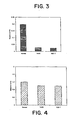
- the level of L-FABP in normal breast cells was undetectable or very low as shown in Fig. 1.
- the I-FABP and the L-FABP are very similar in their genetic sequences and their mode of action. They are both associated with tumor state. In breast cells the normal had low levels of this gene expressed, however only T47D showed significant high levels of expression of the I-FABP as shown in Fig.2.
- Adipose-FABP acts like a tumor suppressor. With the progression of the tumor, a gradual loss of the expression of this gene was observed. In normal breast cells a high level of expression of A-FABP was observed and A-FABP were very low in T47D and MCF-7 cells. Loss of A-FABP was reported with progression of human bladder transitional cell carcinoma. The presence of A-FABP thus correlated with the grade and stage of the disease. Our results also support that A-FABP is expressed in high levels in normal cells and it is significantly downregulated in tumor cells.
- Cellular retinoic acid binding protein is another class of FABP that is known to be regulated by fatty acids. The levels of the gene were measured and no significant difference in normal breast and breast cancer cells was observed, as shown in Fig. 4. This suggests that not all FABPs are altered in normal and tumor state. Some proteins remain unchanged, at least at their message levels.
- the inventors have analyzed the role of FABPs in breast cancer. None was known about the levels of FABPs in different stages of breast cancer, or the effect of hormones, growth factors or bioactive lipids on FABPs.
- the inventors have shown that FABP message (Liver and Intestine type) is 3-22 fold higher in tumor vs normal breast cells, especially in the estrogen receptor positive lines.
- the inventors have also shown that the A-FABP, E-FABP class of proteins are downregulated in breast cancer cells.
- FABPs are also secreted into the surrounding fluid, they can be identified in human fluids such as semen, , saliva, blood and urine samples.
- Normal PrEC cell line from Clonetics was grown in culture and RNA obtained, LNCAP, PC3 and DU145 were also grown in culture to obtain total RNA from them.
- RT-PCR was performed using A-FABP specific primers, the product was resolved on the gel and analyzed by NIH Image program for quantitation. The values were normalized to the actin values.
- a dramatic (10 -14 fold) decrease in the A-FABP message in cancer cells was observed when compared to the normal prostate cells as shown in Fig.9b.
- the expression of A-FABP was undetectable which corresponds to an advanced stage of cancer. Similar results were observed with breast cancer cells.
- E-FABP The expression of E-FABP in prostate normal and cancer cells was studied.
- RT-PCR was performed for the E-FABP gene, the expression pattern was similar to the A-FABP. It decreased 10-14 fold in cancer cells when compared with the normal prostate cells as shown in Fig. 10.
- the PCR product was also sequenced with a cycle sequencing kit (Amersham Corp, Piscataway, NJ) and it matched with human E-FABP sequence.
- the levels of E-FABP went down significantly in prostate cells.
- the level of expression of E-FABP was the most predominant when compared with all the other FABP levels in the prostate cells.
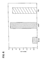
- L-FABP gene expression was elevated significantly in the cancer cell DU 145 when compared to the rest of the prostate cells as shown in Fig. 5.
- DU 145 is a metastatic prostate cell line and thus high levels of L-FABP may corresponds to the aggressiveness of the prostate cancer.
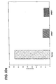
- I-FABP The expression of I-FABP in prostate cells was studied. The expression of the intestine type of FABP was tested in prostate cells. As shown in Fig 6, a dramatic increase in expression of the I-FABP in cancer cells was observed when compared to the normal prostate cells. Since I-and L-FABP are similar in their sequences and also are found to be elevated in cancer cells, these proteins act as biomarkers for prostate cancer.
- Mus-FABP Mus-FABP in prostate cells was studied.
- RT-PCR was performed with Heart/Muscle type of FABP that is in the same family of proteins as the Adipose-FABP, a similar trend in the levels of this FABP was observed. It was high in the normal prostate epithelial cells and was dramatically low in LNCAP, prostate cancer cells as shown in Fig.7. This suggests that the heart type of FABP also has a tumor suppressive role in prostate cancer cells.
- MDGI mammary derived growth inhibitor
- Brain-FABP in prostate cells was studied. The expression of all the different types of FABP in these prostate cells was further tested by performing RT-PCR for brain-FABP. Only the LNCAP cancer cell line was positive for B-FABP. All other cell lines were negative for the expression of the gene.
- FABPs are believed to play a crucial role in promotion of prostate cancer cell growth.
- the members of this broad multigene family currently consist of at least seven types whose amino acid sequences have been obtained from protein purified from tissue or from cDNA nucleotide sequences from tissue RNA.
- each of the FABPs is derived from the human tissue from which it was isolated and includes: 1) adipocyte (A-FABP), 2) heart or muscle (H-FABP), 3) brain (B-FABP), 4) epidermis or psoriasis-associated (E-FABP), 5) liver (L-FABP), 6) intestine (I-FABP), and 7) myelin or P2 (P2-FABP).
- A-FABP, H-FABP, B-FABP, and E-FABP in humans share between 50-65% protein sequence homology and contain a tyrosine near residue 20 that can be phosphorylated.
- Heart-FABP and Muscle-FABP differ only in three amino acids and therefore can be referred to as Muscle/Heart-FABP.
- a test for Heart-FABP will also test for Muscle-FABP.
- the inventors have examined the levels of FABPs in several normal prostate and cancer cell lines in order to establish a correlation between presence and levels of FABP with the stage of cancer represented by each cell line.
- the levels of 5 selected FABPs were analyzed using primers designed for RT-PCR and have found their expression to be altered in prostate cancer vs normal cells.
- Heteropoly and Isopoly Oxometalates metal ion derivatives of polyoxotungstate, hereinafter referred to as HPA.
- HPA metal ion derivatives of polyoxotungstate
- HPA heteropolyanions
- Mice have been given HPA drug subcutaneously at doses up to 28 mg/kg for 3 consecutive weeks or 12 mg/kg for 12 consecutive weeks. Oral and intramuscular administration were also tested. Drugs were dissolved in aqueous solutions for administration.
- Preferred HPA drugs are HPA-Na and HPA-Sm.
- MCF-7 breast cancer cells
- HPA-Na (1uM) increased the levels of A-FABP in LNCAP to a level similar to the normal values as shown in Fig. 18.
- the levels of E-FABP was also altered in a similar manner.
- the level of I-FABP of LNCAP cells was dramatically reduced to the normal level in the presence of this class of drug as shown in Fig. 19. Similar changes in pattern of these FABPs were also observed in breast cancer cells MCF-7, when they were grown for 48 hrs in the presence of this drug.
- HPA drug is able to block the cell cycle of MCF-7 cells in a particular phase (of cell cycle) which ultimately results in terminal differentiation and apoptosis.
- the heteropolyanion drugs can be used alone or in combination with inhibitors of eicosanoid metabolism on the growth of breast cancer cells.
- Inhibitors contemplated are those currently used in clinical situations such as doxorubicin, 5FU, vinca alkaloids, adriamycin as well as lipoxygenase inhibitors.
- the HPA drug can be given simultaneously with or can be given followed by exposing the cancerous cells to one or more applications of an amount of HPA containing metal ions, preferrably HPA-Na, in an amount sufficient to block the growth of the cancerous cells and change the FABP profile to a normal cell FABP profile. Subsequent applications of chemotherapeutic agents or inhibitor can be given in a lower amount than previous applications.
- HPA drugs have efficacy for use as an anticancer agent. It has a great potential, not only for stopping growth of cancer cells but also for changing the balance of the FABPs that play a role in carcinogenesis.
- the detection protocol used to identify prostate cancer-elevated FABPs in human blood, urine or semen samples from patients showing elevated prostate specific antigen (PSA) levels is as follows.
- RNA was isolated using the Trizol method and RT-PCR analysis of the proteins was performed by using primers specific for each FABP. Equal amount of RNA was used for RT-PCR and PCR master mix was used to avoid any handling errors.
- the sequence for FABPs was obtained from the gene bank and the GCG program ( GCG program is well know ) was used to design specific primers.
- the method of quantitation of PCR product was as follows: for internal quantitation control actin primers was used, all the gels were scanned in a gel scanner and digitized using the NIH-Image program for quantitation. Each band for the respective FABP was then normalized with the actin value and then comparison made between prostate normal and cancer cells.
- L-FABP expression was very low in normal breast cells, it is almost absent in normal cells.
- the increase in L-FABP levels in breast cancer cells (T-47D & MCF-7) was 12-15 fold higher compared to the normal cell values.
- the estrogen receptor positive cells had this change in levels of L-FABP, but no change was observed in few estrogen receptor negative cells that were tested as shown in Fig. 1.
- Intestine-FABP The expression of Intestine-FABP was also shown to be upregulated in breast cancer cells compared to the normal breast cells. There was a 4-6 fold increase in the level of I-FABP when compared to the normal cells as shown in Fig. 2.
- Adipose-FABP is same as the MDGI (mammary derived growth inhibitor) and the expression of this FABP is downregulated in cancer cells.
- the normal levels of Adipose-FABP is downregulated by 5-6 fold in cancer cells as shown in Fig. 3.
- CRAB-1 The levels of cellular retinoic acid binding protein expression remains the same in cancer and normal cells. The level of this FABP does not change with the stage of cancer as shown in Fig. 4.
- Liver-FABP In prostate cells only DU-145 cells expressed Liver-FABP, all other cells were negative for L-FABP. There was a 10 fold difference in normal versus DU-145 cells in expression of L-FABP as shown in Fig. 5.
- Intestine-FABP The normal cells expressed very low levels of I-FABP. There was a 4-6 fold increase in expression of I-FABP in LNCAP cells which are prostate cancer cells as shown in Fig. 6.
- Adipose-FABP The expression levels of Adipose-FABP was much higher in normal prostate cells when compared to the LNCAP cells. There was a five-fold decrease in the expression of Adipose-FABP in cancer cells as shown in Fig. 8.
- Muscle-FABP The level of Muscle-FABP was high in normal cells when compared to the cancer cells. The difference in levels was 4-6 fold as shown in Fig. 7.
- CRAB-1 The levels of CRAB-1 expression did not change in normal and cancer prostate cells as shown in Fig. 9a.
- Specific peptides for these FABPs have been developed that will bind to the m-RNA of only one FABP and will not interact with the others.
- the specific peptides are designed to accomplish the inhibition of expression of the specific m-RNA genes.
- We will also design peptides representing unique regions of each specific FABP.
- We will synthesize these peptides, attach them to a carrier molecule and generate specific antibodies for each of FABPs. These antibodies are crucial for use in development of ELISA procedures to detect each of the specific FABPs in body fluids and, perhaps, to also use them for therapeutic interventions.
- Oligonucleotides that will recognize only the respective specific gene. These Oligos will be used for first quantitation of genes by using them as probes. Also we will make antisense RNA for these FABP genes which will be able to block the expression of Liver and Intestine-FABPs which we have shown to be upregulated in cancer cells.
- the invention demonstrates in prostate and breast cancer cells that, there exist a balance of the good type of FABPs (A-FABP, E-FABP and Mus-FABP) and the bad type of FABPs (L-FABP, I-FABP and B-FABP). Change in this balance allows prediction of the stage of the cancer.
- A-FABP good type of FABPs
- E-FABP E-FABP and Mus-FABP
- L-FABP bad type of FABPs
- I-FABP I-FABP
- B-FABP B-FABP
- Tables 1 and 2 show the effective concentration of HPA in breast cancer cells: Tables 1 and 2 show the concentrations required for IC 50 inhibition in cultures of wild type and multi-drug resistant MCF-7 human breast cancer cells, respectively. These tables show polyoxotungstates used on MCF-7 proliferation assay. Table 3: List of normal and tumor cell cultures used in these studies. Table 1 IC 50 of WT MCF-7 breast cancer cells using polyoxotungstates. concentrations shown in ⁇ M a : n ranges from 20 to 30 Name of polyoxotungstate a IC 50 (Exp.1) IC 50 (Exp.1) IC 50 (Exp.1) IC 50 (Exp.1) IC 50 (Exp.1) IC 50 (Exp.1) IC 50 (Exp.1) IC 50 (Exp.1) IC 50 (Exp.1) IC 50 (Exp.1) IC 50 (Exp.1) IC 50 (Exp.1) IC 50 (Exp.1) IC 50 (Exp.1) IC 50 (Exp.1) IC 50 (Exp.1) IC 50
- n ranges from 20 to 30 Name of polyoxotungstate a IC 50 (Exp.1) IC 50 (Exp. #2) K 12.5 Na 1.5 [NaP 5 W 30 O 110 ] ⁇ nH 2 O N/A 2.1 K 13 [CaP 5 W 30 O 110 ] ⁇ nH 2 O 3.8 6.8 K 12 [BiP 5 W 30 O 110 ] ⁇ nH 2 O 3.0 2.5 K 12 [CeP 5 W 30 O 110 ] ⁇ nH 2 O 3.8 3.0 K 12 [SmP 5 W 30 O 110 ] ⁇ nH 2 O 3.8 4.1 K 12 [EuP 5 W 30 O 110 ] ⁇ nH 2 O 0.25 3.8 K 12 [GdP 5 W 30 O 110 ] ⁇ nN 2 O 4.3 4.6 K 12 [TbP 5 W 30 O 110 ] ⁇ nH 2 O 3.5 4.9 K 12 [DyP 5 W 30 O 110 ] ⁇ nH 2 O 3.8 2.3 K 12 [HoP 5 W 30 O 110
- HPV-7 normal immortalized cells obtained from ATCC Cancer: PC-3: adenocarcinoma from bone metastasis from 62 yr old male, obtained from ATCC DU 145: carcinoma of prostate from brain metastasis from 69 yr old male, obtained from ATCC LNCAP: carcinoma of prostate from lymph node metastasis from 50 yr old male, obtained from ATCC.
- RNA obtained from NCI, NIH.
Landscapes
- Health & Medical Sciences (AREA)
- Life Sciences & Earth Sciences (AREA)
- Engineering & Computer Science (AREA)
- Immunology (AREA)
- Chemical & Material Sciences (AREA)
- Molecular Biology (AREA)
- Biomedical Technology (AREA)
- Hematology (AREA)
- Urology & Nephrology (AREA)
- General Health & Medical Sciences (AREA)
- Medicinal Chemistry (AREA)
- Biochemistry (AREA)
- Food Science & Technology (AREA)
- Microbiology (AREA)
- Physics & Mathematics (AREA)
- Analytical Chemistry (AREA)
- Cell Biology (AREA)
- Biotechnology (AREA)
- General Physics & Mathematics (AREA)
- Pathology (AREA)
- Epidemiology (AREA)
- Pharmacology & Pharmacy (AREA)
- Inorganic Chemistry (AREA)
- Animal Behavior & Ethology (AREA)
- Public Health (AREA)
- Veterinary Medicine (AREA)
- Biophysics (AREA)
- Endocrinology (AREA)
- Measuring Or Testing Involving Enzymes Or Micro-Organisms (AREA)
- Investigating Or Analysing Biological Materials (AREA)
- Medicines That Contain Protein Lipid Enzymes And Other Medicines (AREA)
- Pharmaceuticals Containing Other Organic And Inorganic Compounds (AREA)
- Medicines Containing Antibodies Or Antigens For Use As Internal Diagnostic Agents (AREA)
Claims (12)
- Procédé de diagnostic d'un cancer du sein ou de la prostate comprenant les étapes:a) de détection d'expression de deux protéines de liaison aux acides gras (fatty acid binding proteins = FABP) différentes ou plus sélectionnées, dans le cas d'un diagnostic de cancer du sein, à partir de FAPB du foie, FABP de l'intestin, FABP adipocytaire, et FABP épidermique ou, dans le cas d'un diagnostic de cancer de la prostate, à partir de FAPB du foie, FABP de l'intestin, FABP adipocytaire, FABP épidermique, FABP cérébrale et FABP cardiaque/musculaire, présentes dans un échantillon de sérum ou de tissu obtenu chez un sujet;b) d'observation d'un profil de niveaux d'expression des protéines de liaison des acides gras dans ledit échantillon;c) de comparaison des profils de niveaux d'expression des protéines de liaison des acides gras dans l'échantillon à un profil de niveaux d'expression des protéines de liaison des acides gras dans un témoin normal non cancéreux; etd) de diagnostic d'une présence ou absence de cancer du sein ou de la prostate en observant s'il existe une différence entre le profil de l'échantillon et le profil du témoin normal non cancéreux, où si le profil de l'échantillon reflète le profil du contrôle, il n'y a pas ledit cancer et si le profil de l'échantillon est différent du profil du témoin, ledit cancer est présent.
- Procédé selon la revendication 1, dans lequel ledit échantillon est un échantillon de liquide de mammifère sélectionné dans le groupe consistant en du sperme, de la salive, de l'urine et du sang.
- Procédé selon la revendication 1, dans lequel ledit échantillon est un échantillon de tissu de mammifère.
- Procédé selon la revendication 2 ou la revendication 3, dans lequel ladite détection d'expression comprend la mesure de niveaux d'ARN des protéines de liaison des acides gras.
- Procédé selon la revendication 2 ou la revendication 3, dans lequel ladite expression de détection comprend la mesure des quantités de protéines de liaison des acides gras dans l'échantillon avec des anticorps et la détermination de types de protéine de liaison des acides gras dans l'échantillon par ELISA.
- Procédé selon la revendication 5, dans lequel lesdits anticorps sont spécifiques d'au moins deux protéines sélectionnées, dans le cas d'un diagnostic de cancer du sein, à partir de FAPB du foie, FABP de l'intestin, FABP adipocytaire, FABP épidermique ou, dans le cas d'un diagnostic de cancer de la prostate, à partir de la FAPB du foie, FABP de l'intestin, FABP adipocytaire, FABP cérébrale, FABP épidermique et FABP cardiaque/musculaire.
- Procédé selon la revendication 1 dans lequel ladite étape de diagnostic d'une présence ou absence de cancer du sein ou de la prostate comprend le diagnostic de la présence de cancer du sein si, en comparaison avec le témoin, la FABP du foie est élevée, la FABP de l'intestin est élevée, la FABP adipeuse est basse et la FABP épidermique est basse ou de cancer de la prostate si, en comparaison avec le témoin, la FABP du foie est élevée, la FABP de l'intestin est élevée, la FABP adipeuse est basse, la FABP épidermique est basse, la FABP cérébrale est élevée, la FABP cardiaque/musculaire est basse.
- Procédé selon la revendication 1, comprenant en outre les étapes:de soumettre un échantillon témoin normal non cancéreux à un procédé de détection pour mesurer les niveaux d'expression de protéines de liaison des acides gras comprenant la FAPB du foie, FABP de l'intestin, FABP adipocytaire et FABP épidermique pour le cancer du sein et de la prostate et la FABP cérébrale et FABP cardiaque/musculaire pour le cancer de la prostate pour produire le profil de témoin normal non cancéreux des niveaux d'expressions normaux desdites protéines de liaison des acides gras.
- Procédé selon l'une quelconque des revendications 1 à 3 dans lequel ladite détection d'expression comprend la détection des niveaux d'expression de gène de deux ou plus desdites protéines de liaison des acides gras présentes dans ledit échantillon, comprenant en outre le diagnostic du stade ou de l'agressivité dudit cancer en déterminant et en évaluant les différences entre les niveaux témoins attendus et détectés de deux ou plus desdites protéines de liaison des acides gras pour déterminer le stade et l'agressivité dudit cancer.
- Procédé selon la revendication 9 dans lequel les niveaux d'expression de gène de plusieurs protéines de liaison des acides gras présentes dans ledit échantillon sont détectés, et les différences entre les niveaux témoins attendus et détectés d'expression de gène desdites plusieurs protéines de liaison des acides gras sont déterminées et évaluées pour déterminer le stade et l'agressivité dudit cancer.
- Procédé selon la revendication 9 dans lequel ladite détection d'expression comprend la détection de niveaux d'expression de gène pour deux ou plus desdites protéines de liaison des acides gras et pour tous les autres types de protéines de liaison des acides gras présents dans ledit échantillon, en un essai unique utilisant un transfert de gène ou une puce portant des gènes pour des types de protéines de liaison des acides gras.
- Procédé selon l'une quelconque des revendications 1 à 3 dans lequel ladite détection d'expression comprend la détection des quantités de deux ou plus desdites protéines de liaison des acides gras présentes dans ledit échantillon avec des anticorps spécifiques desdites protéines de liaison des acides gras, comprenant en outre un diagnostic du stade ou de l'agressivité dudit cancer en déterminant et en évaluant les différences entre les quantités témoins attendues et détectées de deux ou plus desdites protéines de liaison des acides gras pour déterminer le stade ou l'agressivité dudit cancer.
Applications Claiming Priority (3)
| Application Number | Priority Date | Filing Date | Title |
|---|---|---|---|
| US11048498P | 1998-12-01 | 1998-12-01 | |
| US110484P | 1998-12-01 | ||
| PCT/US1999/028314 WO2000033083A1 (fr) | 1998-12-01 | 1999-11-30 | Diagnostic de la phase d'evolution ou de l'agressivite de cancers |
Publications (2)
| Publication Number | Publication Date |
|---|---|
| EP1135686A1 EP1135686A1 (fr) | 2001-09-26 |
| EP1135686B1 true EP1135686B1 (fr) | 2006-11-22 |
Family
ID=22333265
Family Applications (1)
| Application Number | Title | Priority Date | Filing Date |
|---|---|---|---|
| EP99960625A Expired - Lifetime EP1135686B1 (fr) | 1998-12-01 | 1999-11-30 | Diagnostic de la phase d'evolution ou de l'agressivite de cancers |
Country Status (7)
| Country | Link |
|---|---|
| US (1) | US7175981B2 (fr) |
| EP (1) | EP1135686B1 (fr) |
| AT (1) | ATE346301T1 (fr) |
| AU (1) | AU768734B2 (fr) |
| CA (1) | CA2353546C (fr) |
| DE (1) | DE69934139D1 (fr) |
| WO (1) | WO2000033083A1 (fr) |
Families Citing this family (22)
| Publication number | Priority date | Publication date | Assignee | Title |
|---|---|---|---|---|
| EP1254370A1 (fr) * | 1999-12-28 | 2002-11-06 | Ribonomics, Inc. | Procedes servant a isoler et a caracteriser des complexes endogenes de proteines-arnm (rnpm) |
| US8815517B2 (en) * | 1999-12-28 | 2014-08-26 | Ribonomics, Inc. | Methods for identifying functionally related genes and drug targets |
| WO2005015220A1 (fr) * | 2003-08-04 | 2005-02-17 | Roche Diagnostics Gmbh | Utilisation de la proteine crabp-i en tant que marqueur pour le cancer du sein |
| GB0405349D0 (en) * | 2004-03-10 | 2004-04-21 | Univ Birmingham | Cancer therapy and medicaments therefor |
| DE102005026710A1 (de) * | 2005-06-09 | 2006-12-14 | Basf Ag | Verfahren zum Testen von Substanzen oder Substanzgemischen, dessen Verwendung und entsprechende Analysekits |
| US7799519B2 (en) * | 2005-07-07 | 2010-09-21 | Vanderbilt University | Diagnosing and grading gliomas using a proteomics approach |
| GB0604370D0 (en) * | 2006-03-03 | 2006-04-12 | Univ Dublin | Markers for melanoma progression |
| EP2006682B1 (fr) * | 2006-03-22 | 2014-06-04 | DS Pharma Biomedical Co., Ltd. | Diagnostic de l'enterite aigue par la determination de la proteine intestinale de liaison aux acides gras dans le sang |
| FR2919062B1 (fr) | 2007-07-19 | 2009-10-02 | Biomerieux Sa | Procede de dosage de l'aminoacylase 1 pour le diagnostic in vitro du cancer colorectal. |
| FR2919063B1 (fr) | 2007-07-19 | 2009-10-02 | Biomerieux Sa | Procede de dosage du leucocyte elastase inhibitor pour le diagnostic in vitro du cancer colorectal. |
| FR2919064B1 (fr) | 2007-07-19 | 2009-10-02 | Biomerieux Sa | Procede de dosage de l'apolipoproteine all pour le diagnostic in vitro du cancer colorectal |
| CA2693098C (fr) * | 2007-07-19 | 2019-06-18 | Yasemin Ataman-Onal | Procede de dosage de la proteine de liaison hepatique aux acides gras, de l'antigene carcino-embryonnaire, et de l'antigene carbohydrate 19-9 pour le diagnostic in vitro du cancercolorectal |
| FR2919061B1 (fr) | 2007-07-19 | 2009-10-02 | Biomerieux Sa | Procede de dosage de la plastine-i pour le diagnostic in vitro du cancer colorectal. |
| FR2919060B1 (fr) * | 2007-07-19 | 2012-11-30 | Biomerieux Sa | Procede de dosage de l'ezrine pour le diagnostic in vitro du cancer colorectal. |
| FR2919065B1 (fr) | 2007-07-19 | 2009-10-02 | Biomerieux Sa | Procede de dosage de l'apolipoproteine ai pour le diagnostic in vitro du cancer colorectal |
| WO2009156584A1 (fr) * | 2008-06-24 | 2009-12-30 | Valtion Teknillinen Tutkimuskeskus | Évaluation du risque de métastases et/ou de ddfs chez des patients atteints de néoplasmes, criblage de patients réagissant à une cancérothérapie et ladite thérapie |
| FR2933773B1 (fr) | 2008-07-10 | 2013-02-15 | Biomerieux Sa | Procede de dosage de la proteine disulfide isomerase pour le diagnostic in vitro du cancer colorectal |
| US9581598B2 (en) | 2012-04-25 | 2017-02-28 | Sanford-Burnham Medical Research Institute | Diagnosis and treatment of brain tumor |
| FR3001806B1 (fr) | 2013-02-01 | 2016-05-20 | Biomerieux Sa | Procede de detection d'une lesion colorectale |
| WO2018002362A1 (fr) * | 2016-06-30 | 2018-01-04 | Randox Laboratories Ltd | Mesure de l'antigène fabp pour la pose d'un diagnostic |
| EP3273240A1 (fr) | 2016-07-17 | 2018-01-24 | Mitogro OÜ | Procédé de sélection de patients sensibles aux traitements du cancer |
| JP6581274B1 (ja) * | 2018-09-28 | 2019-09-25 | シミックホールディングス株式会社 | 肝疾患の検査方法、その検査キット及びコンパニオン診断薬 |
Family Cites Families (2)
| Publication number | Priority date | Publication date | Assignee | Title |
|---|---|---|---|---|
| FR2245374B1 (fr) * | 1973-07-27 | 1977-02-25 | Anvar | |
| US4547367A (en) * | 1983-12-20 | 1985-10-15 | The United States Of America As Represented By The Department Of Health And Human Services | Hepatitis B core antigen vaccine |
-
1999
- 1999-11-30 EP EP99960625A patent/EP1135686B1/fr not_active Expired - Lifetime
- 1999-11-30 CA CA002353546A patent/CA2353546C/fr not_active Expired - Fee Related
- 1999-11-30 DE DE69934139T patent/DE69934139D1/de not_active Expired - Fee Related
- 1999-11-30 AT AT99960625T patent/ATE346301T1/de not_active IP Right Cessation
- 1999-11-30 WO PCT/US1999/028314 patent/WO2000033083A1/fr not_active Ceased
- 1999-11-30 AU AU17483/00A patent/AU768734B2/en not_active Ceased
- 1999-11-30 US US09/451,513 patent/US7175981B2/en not_active Expired - Fee Related
Also Published As
| Publication number | Publication date |
|---|---|
| CA2353546A1 (fr) | 2000-06-08 |
| EP1135686A1 (fr) | 2001-09-26 |
| DE69934139D1 (de) | 2007-01-04 |
| ATE346301T1 (de) | 2006-12-15 |
| AU1748300A (en) | 2000-06-19 |
| US20020127619A1 (en) | 2002-09-12 |
| WO2000033083A1 (fr) | 2000-06-08 |
| AU768734B2 (en) | 2004-01-08 |
| CA2353546C (fr) | 2009-05-19 |
| US7175981B2 (en) | 2007-02-13 |
Similar Documents
| Publication | Publication Date | Title |
|---|---|---|
| EP1135686B1 (fr) | Diagnostic de la phase d'evolution ou de l'agressivite de cancers | |
| Järvinen et al. | Predictive value of topoisomerase IIα and other prognostic factors for epirubicin chemotherapy in advanced breast cancer | |
| Spychala et al. | Role of estrogen receptor in the regulation of ecto-5′-nucleotidase and adenosine in breast cancer | |
| JP5068543B2 (ja) | がんに関する診断マーカー | |
| Ricciardelli et al. | Androgen receptor levels in prostate cancer epithelial and peritumoral stromal cells identify non‐organ confined disease | |
| US20120225954A1 (en) | Methods and compositions for the classification of non-small cell lung carcinoma | |
| JP2007325598A (ja) | 診断および治療標的としての前立腺障害において過剰発現される遺伝子 | |
| Leprieur et al. | Clinical and molecular features in patients with advanced non-small-cell lung carcinoma refractory to first-line platinum-based chemotherapy | |
| EP2215482A2 (fr) | Biomarqueur utilisé pour évaluer une réponse à un traitement de fms | |
| JP2010537647A (ja) | 癌患者における化学療法に対する応答を測定するために腫瘍のrna完全性を用いる方法 | |
| US20220218659A1 (en) | PI3K/LYN-ACLY Signaling Inhibition | |
| Deng et al. | Prognostic value of flotillins (flotillin-1 and flotillin-2) in human cancers: a meta-analysis | |
| Liu et al. | Overexpression of the angiogenic tetrapeptide AcSDKP in human malignant tumors | |
| Corte et al. | Cytosolic levels of TFF1/pS2 in breast cancer: their relationship with clinical–pathological parameters and their prognostic significance | |
| JP2022025113A (ja) | 抗pd-1抗体若しくは抗pd-l1抗体療法の奏効性を予測する方法、がんの悪性度を評価する方法、及び抗pd-1抗体若しくは抗pd-l1抗体療法の奏効性を上昇させる方法 | |
| IL227471A (en) | Mrm / srm test for protein-11 bcl-2 protein | |
| KR20240103878A (ko) | 신보강 화학요법에 대한 유방암의 치료 반응 또는 예후 예측을 위한 세포외 소포체 유래 바이오마커 및 이의 용도 | |
| US20140161813A1 (en) | Methods for the diagnosis, treatment and monitoring of cancer | |
| KR20200017452A (ko) | 최적의 암 요법을 위한 slfn11 단백질의 정량화 | |
| Kurehara et al. | A novel gene, RSRC2, inhibits cell proliferation and affects survival in esophageal cancer patients | |
| JP2003504073A (ja) | Bag発現のレベルを測定することによる、癌患者の予後を決定するための方法 | |
| US20210156864A1 (en) | Diagnostic and Therapeutic Biomarkers in Human Cancers and Methods of Use Thereof | |
| CN101027099B (zh) | 用于癌症的诊断标记物 | |
| JP7774310B2 (ja) | 腎がん患者の抗がん剤抵抗性および予後予測方法、抗腎がん物質のスクリーニング方法、ならびに、腎がん治療用医薬組成物 | |
| JP7812576B2 (ja) | 癌診断用キット及びその使用 |
Legal Events
| Date | Code | Title | Description |
|---|---|---|---|
| PUAI | Public reference made under article 153(3) epc to a published international application that has entered the european phase |
Free format text: ORIGINAL CODE: 0009012 |
|
| 17P | Request for examination filed |
Effective date: 20010521 |
|
| AK | Designated contracting states |
Kind code of ref document: A1 Designated state(s): AT BE CH CY DE DK ES FI FR GB GR IE IT LI LU MC NL PT SE |
|
| AX | Request for extension of the european patent |
Free format text: AL;LT;LV;MK;RO;SI |
|
| 17Q | First examination report despatched |
Effective date: 20030620 |
|
| GRAP | Despatch of communication of intention to grant a patent |
Free format text: ORIGINAL CODE: EPIDOSNIGR1 |
|
| GRAS | Grant fee paid |
Free format text: ORIGINAL CODE: EPIDOSNIGR3 |
|
| GRAA | (expected) grant |
Free format text: ORIGINAL CODE: 0009210 |
|
| AK | Designated contracting states |
Kind code of ref document: B1 Designated state(s): AT BE CH CY DE DK ES FI FR GB GR IE IT LI LU MC NL PT SE |
|
| PG25 | Lapsed in a contracting state [announced via postgrant information from national office to epo] |
Ref country code: NL Free format text: LAPSE BECAUSE OF FAILURE TO SUBMIT A TRANSLATION OF THE DESCRIPTION OR TO PAY THE FEE WITHIN THE PRESCRIBED TIME-LIMIT Effective date: 20061122 Ref country code: LI Free format text: LAPSE BECAUSE OF FAILURE TO SUBMIT A TRANSLATION OF THE DESCRIPTION OR TO PAY THE FEE WITHIN THE PRESCRIBED TIME-LIMIT Effective date: 20061122 Ref country code: IT Free format text: LAPSE BECAUSE OF FAILURE TO SUBMIT A TRANSLATION OF THE DESCRIPTION OR TO PAY THE FEE WITHIN THE PRESCRIBED TIME-LIMIT;WARNING: LAPSES OF ITALIAN PATENTS WITH EFFECTIVE DATE BEFORE 2007 MAY HAVE OCCURRED AT ANY TIME BEFORE 2007. THE CORRECT EFFECTIVE DATE MAY BE DIFFERENT FROM THE ONE RECORDED. Effective date: 20061122 Ref country code: FI Free format text: LAPSE BECAUSE OF FAILURE TO SUBMIT A TRANSLATION OF THE DESCRIPTION OR TO PAY THE FEE WITHIN THE PRESCRIBED TIME-LIMIT Effective date: 20061122 Ref country code: CH Free format text: LAPSE BECAUSE OF FAILURE TO SUBMIT A TRANSLATION OF THE DESCRIPTION OR TO PAY THE FEE WITHIN THE PRESCRIBED TIME-LIMIT Effective date: 20061122 Ref country code: BE Free format text: LAPSE BECAUSE OF FAILURE TO SUBMIT A TRANSLATION OF THE DESCRIPTION OR TO PAY THE FEE WITHIN THE PRESCRIBED TIME-LIMIT Effective date: 20061122 Ref country code: AT Free format text: LAPSE BECAUSE OF FAILURE TO SUBMIT A TRANSLATION OF THE DESCRIPTION OR TO PAY THE FEE WITHIN THE PRESCRIBED TIME-LIMIT Effective date: 20061122 |
|
| REG | Reference to a national code |
Ref country code: GB Ref legal event code: FG4D |
|
| PG25 | Lapsed in a contracting state [announced via postgrant information from national office to epo] |
Ref country code: MC Free format text: LAPSE BECAUSE OF NON-PAYMENT OF DUE FEES Effective date: 20061130 Ref country code: IE Free format text: LAPSE BECAUSE OF NON-PAYMENT OF DUE FEES Effective date: 20061130 |
|
| REG | Reference to a national code |
Ref country code: CH Ref legal event code: EP |
|
| REG | Reference to a national code |
Ref country code: IE Ref legal event code: FG4D |
|
| REF | Corresponds to: |
Ref document number: 69934139 Country of ref document: DE Date of ref document: 20070104 Kind code of ref document: P |
|
| PG25 | Lapsed in a contracting state [announced via postgrant information from national office to epo] |
Ref country code: SE Free format text: LAPSE BECAUSE OF FAILURE TO SUBMIT A TRANSLATION OF THE DESCRIPTION OR TO PAY THE FEE WITHIN THE PRESCRIBED TIME-LIMIT Effective date: 20070222 Ref country code: DK Free format text: LAPSE BECAUSE OF FAILURE TO SUBMIT A TRANSLATION OF THE DESCRIPTION OR TO PAY THE FEE WITHIN THE PRESCRIBED TIME-LIMIT Effective date: 20070222 |
|
| PG25 | Lapsed in a contracting state [announced via postgrant information from national office to epo] |
Ref country code: ES Free format text: LAPSE BECAUSE OF FAILURE TO SUBMIT A TRANSLATION OF THE DESCRIPTION OR TO PAY THE FEE WITHIN THE PRESCRIBED TIME-LIMIT Effective date: 20070305 |
|
| PG25 | Lapsed in a contracting state [announced via postgrant information from national office to epo] |
Ref country code: PT Free format text: LAPSE BECAUSE OF FAILURE TO SUBMIT A TRANSLATION OF THE DESCRIPTION OR TO PAY THE FEE WITHIN THE PRESCRIBED TIME-LIMIT Effective date: 20070423 |
|
| NLV1 | Nl: lapsed or annulled due to failure to fulfill the requirements of art. 29p and 29m of the patents act | ||
| PG25 | Lapsed in a contracting state [announced via postgrant information from national office to epo] |
Ref country code: DE Free format text: LAPSE BECAUSE OF NON-PAYMENT OF DUE FEES Effective date: 20070601 |
|
| REG | Reference to a national code |
Ref country code: CH Ref legal event code: PL |
|
| ET | Fr: translation filed | ||
| PLBE | No opposition filed within time limit |
Free format text: ORIGINAL CODE: 0009261 |
|
| STAA | Information on the status of an ep patent application or granted ep patent |
Free format text: STATUS: NO OPPOSITION FILED WITHIN TIME LIMIT |
|
| 26N | No opposition filed |
Effective date: 20070823 |
|
| PG25 | Lapsed in a contracting state [announced via postgrant information from national office to epo] |
Ref country code: GR Free format text: LAPSE BECAUSE OF FAILURE TO SUBMIT A TRANSLATION OF THE DESCRIPTION OR TO PAY THE FEE WITHIN THE PRESCRIBED TIME-LIMIT Effective date: 20070223 |
|
| PG25 | Lapsed in a contracting state [announced via postgrant information from national office to epo] |
Ref country code: LU Free format text: LAPSE BECAUSE OF NON-PAYMENT OF DUE FEES Effective date: 20061130 |
|
| PG25 | Lapsed in a contracting state [announced via postgrant information from national office to epo] |
Ref country code: CY Free format text: LAPSE BECAUSE OF FAILURE TO SUBMIT A TRANSLATION OF THE DESCRIPTION OR TO PAY THE FEE WITHIN THE PRESCRIBED TIME-LIMIT Effective date: 20061122 |
|
| REG | Reference to a national code |
Ref country code: FR Ref legal event code: PLFP Year of fee payment: 17 |
|
| PGFP | Annual fee paid to national office [announced via postgrant information from national office to epo] |
Ref country code: GB Payment date: 20151125 Year of fee payment: 17 |
|
| PGFP | Annual fee paid to national office [announced via postgrant information from national office to epo] |
Ref country code: FR Payment date: 20151127 Year of fee payment: 17 |
|
| GBPC | Gb: european patent ceased through non-payment of renewal fee |
Effective date: 20161130 |
|
| REG | Reference to a national code |
Ref country code: FR Ref legal event code: ST Effective date: 20170731 |
|
| PG25 | Lapsed in a contracting state [announced via postgrant information from national office to epo] |
Ref country code: FR Free format text: LAPSE BECAUSE OF NON-PAYMENT OF DUE FEES Effective date: 20161130 |
|
| PG25 | Lapsed in a contracting state [announced via postgrant information from national office to epo] |
Ref country code: GB Free format text: LAPSE BECAUSE OF NON-PAYMENT OF DUE FEES Effective date: 20161130 |