EP1272848B1 - Vorrichtung und verfahren zur herstellung von zytologischen klingen - Google Patents
Vorrichtung und verfahren zur herstellung von zytologischen klingen Download PDFInfo
- Publication number
- EP1272848B1 EP1272848B1 EP01926624A EP01926624A EP1272848B1 EP 1272848 B1 EP1272848 B1 EP 1272848B1 EP 01926624 A EP01926624 A EP 01926624A EP 01926624 A EP01926624 A EP 01926624A EP 1272848 B1 EP1272848 B1 EP 1272848B1
- Authority
- EP
- European Patent Office
- Prior art keywords
- cover
- slide
- inside surface
- filter
- cytology
- Prior art date
- Legal status (The legal status is an assumption and is not a legal conclusion. Google has not performed a legal analysis and makes no representation as to the accuracy of the status listed.)
- Expired - Lifetime
Links
- 238000000034 method Methods 0.000 title claims abstract description 40
- 238000002360 preparation method Methods 0.000 title claims description 27
- 239000000463 material Substances 0.000 claims abstract description 92
- 230000002745 absorbent Effects 0.000 claims abstract description 75
- 239000002250 absorbent Substances 0.000 claims abstract description 75
- 239000007788 liquid Substances 0.000 claims abstract description 12
- 230000001413 cellular effect Effects 0.000 claims description 18
- 238000012546 transfer Methods 0.000 claims description 11
- 229920000742 Cotton Polymers 0.000 claims description 4
- DHKHKXVYLBGOIT-UHFFFAOYSA-N acetaldehyde Diethyl Acetal Natural products CCOC(C)OCC DHKHKXVYLBGOIT-UHFFFAOYSA-N 0.000 claims description 2
- 125000002777 acetyl group Chemical class [H]C([H])([H])C(*)=O 0.000 claims description 2
- 239000006260 foam Substances 0.000 claims description 2
- 229920002554 vinyl polymer Polymers 0.000 claims description 2
- 229920000515 polycarbonate Polymers 0.000 claims 1
- 239000004417 polycarbonate Substances 0.000 claims 1
- 239000000523 sample Substances 0.000 description 57
- 210000004027 cell Anatomy 0.000 description 39
- 238000009595 pap smear Methods 0.000 description 22
- 239000000853 adhesive Substances 0.000 description 17
- 230000001070 adhesive effect Effects 0.000 description 17
- 238000012216 screening Methods 0.000 description 12
- LFQSCWFLJHTTHZ-UHFFFAOYSA-N Ethanol Chemical compound CCO LFQSCWFLJHTTHZ-UHFFFAOYSA-N 0.000 description 6
- 210000003679 cervix uteri Anatomy 0.000 description 6
- 206010008263 Cervical dysplasia Diseases 0.000 description 5
- 239000000834 fixative Substances 0.000 description 5
- 239000004033 plastic Substances 0.000 description 5
- 229920003023 plastic Polymers 0.000 description 5
- 239000008280 blood Substances 0.000 description 4
- 210000004369 blood Anatomy 0.000 description 4
- 238000009826 distribution Methods 0.000 description 4
- 230000003902 lesion Effects 0.000 description 4
- 239000000123 paper Substances 0.000 description 4
- 238000010186 staining Methods 0.000 description 4
- 210000001519 tissue Anatomy 0.000 description 4
- 239000002023 wood Substances 0.000 description 4
- 238000001574 biopsy Methods 0.000 description 3
- 229920002678 cellulose Polymers 0.000 description 3
- 239000001913 cellulose Substances 0.000 description 3
- 230000002380 cytological effect Effects 0.000 description 3
- 238000003745 diagnosis Methods 0.000 description 3
- 239000011521 glass Substances 0.000 description 3
- 238000002156 mixing Methods 0.000 description 3
- 238000005070 sampling Methods 0.000 description 3
- 208000007879 Atypical Squamous Cells of the Cervix Diseases 0.000 description 2
- 206010028980 Neoplasm Diseases 0.000 description 2
- 208000032124 Squamous Intraepithelial Lesions Diseases 0.000 description 2
- NIXOWILDQLNWCW-UHFFFAOYSA-N acrylic acid group Chemical group C(C=C)(=O)O NIXOWILDQLNWCW-UHFFFAOYSA-N 0.000 description 2
- 229920000800 acrylic rubber Polymers 0.000 description 2
- 238000007605 air drying Methods 0.000 description 2
- 238000004458 analytical method Methods 0.000 description 2
- 230000008901 benefit Effects 0.000 description 2
- 210000001124 body fluid Anatomy 0.000 description 2
- 239000011248 coating agent Substances 0.000 description 2
- 238000000576 coating method Methods 0.000 description 2
- 238000007796 conventional method Methods 0.000 description 2
- 238000005336 cracking Methods 0.000 description 2
- 238000005516 engineering process Methods 0.000 description 2
- 229920002457 flexible plastic Polymers 0.000 description 2
- 239000012530 fluid Substances 0.000 description 2
- 239000012943 hotmelt Substances 0.000 description 2
- 210000004969 inflammatory cell Anatomy 0.000 description 2
- 238000003780 insertion Methods 0.000 description 2
- 230000037431 insertion Effects 0.000 description 2
- 239000002184 metal Substances 0.000 description 2
- 229910052751 metal Inorganic materials 0.000 description 2
- 150000002739 metals Chemical class 0.000 description 2
- 230000000877 morphologic effect Effects 0.000 description 2
- 229920000058 polyacrylate Polymers 0.000 description 2
- 229920000728 polyester Polymers 0.000 description 2
- 239000011148 porous material Substances 0.000 description 2
- 238000007447 staining method Methods 0.000 description 2
- 239000000126 substance Substances 0.000 description 2
- 229920003051 synthetic elastomer Polymers 0.000 description 2
- 239000005061 synthetic rubber Substances 0.000 description 2
- 238000012360 testing method Methods 0.000 description 2
- 210000002700 urine Anatomy 0.000 description 2
- 206010006223 Breast discharge Diseases 0.000 description 1
- 229920003043 Cellulose fiber Polymers 0.000 description 1
- 206010008342 Cervix carcinoma Diseases 0.000 description 1
- 229920001651 Cyanoacrylate Polymers 0.000 description 1
- 229920001353 Dextrin Polymers 0.000 description 1
- 239000004375 Dextrin Substances 0.000 description 1
- MWCLLHOVUTZFKS-UHFFFAOYSA-N Methyl cyanoacrylate Chemical compound COC(=O)C(=C)C#N MWCLLHOVUTZFKS-UHFFFAOYSA-N 0.000 description 1
- 229920001410 Microfiber Polymers 0.000 description 1
- 239000004677 Nylon Substances 0.000 description 1
- 239000002202 Polyethylene glycol Substances 0.000 description 1
- 206010036790 Productive cough Diseases 0.000 description 1
- 244000007853 Sarothamnus scoparius Species 0.000 description 1
- 229920002472 Starch Polymers 0.000 description 1
- 239000004809 Teflon Substances 0.000 description 1
- 229920006362 Teflon® Polymers 0.000 description 1
- 208000006105 Uterine Cervical Neoplasms Diseases 0.000 description 1
- 230000002159 abnormal effect Effects 0.000 description 1
- 230000005856 abnormality Effects 0.000 description 1
- 238000003556 assay Methods 0.000 description 1
- 239000010839 body fluid Substances 0.000 description 1
- 210000000481 breast Anatomy 0.000 description 1
- 230000001680 brushing effect Effects 0.000 description 1
- DQXBYHZEEUGOBF-UHFFFAOYSA-N but-3-enoic acid;ethene Chemical compound C=C.OC(=O)CC=C DQXBYHZEEUGOBF-UHFFFAOYSA-N 0.000 description 1
- 201000011510 cancer Diseases 0.000 description 1
- 239000005018 casein Substances 0.000 description 1
- BECPQYXYKAMYBN-UHFFFAOYSA-N casein, tech. Chemical compound NCCCCC(C(O)=O)N=C(O)C(CC(O)=O)N=C(O)C(CCC(O)=N)N=C(O)C(CC(C)C)N=C(O)C(CCC(O)=O)N=C(O)C(CC(O)=O)N=C(O)C(CCC(O)=O)N=C(O)C(C(C)O)N=C(O)C(CCC(O)=N)N=C(O)C(CCC(O)=N)N=C(O)C(CCC(O)=N)N=C(O)C(CCC(O)=O)N=C(O)C(CCC(O)=O)N=C(O)C(COP(O)(O)=O)N=C(O)C(CCC(O)=N)N=C(O)C(N)CC1=CC=CC=C1 BECPQYXYKAMYBN-UHFFFAOYSA-N 0.000 description 1
- 235000021240 caseins Nutrition 0.000 description 1
- 238000005119 centrifugation Methods 0.000 description 1
- 210000001175 cerebrospinal fluid Anatomy 0.000 description 1
- 201000010881 cervical cancer Diseases 0.000 description 1
- 208000019065 cervical carcinoma Diseases 0.000 description 1
- 230000001010 compromised effect Effects 0.000 description 1
- 239000000356 contaminant Substances 0.000 description 1
- 238000011109 contamination Methods 0.000 description 1
- 230000034994 death Effects 0.000 description 1
- 231100000517 death Toxicity 0.000 description 1
- 230000002950 deficient Effects 0.000 description 1
- 238000013461 design Methods 0.000 description 1
- 238000001514 detection method Methods 0.000 description 1
- 238000011161 development Methods 0.000 description 1
- 235000019425 dextrin Nutrition 0.000 description 1
- VLXBWPOEOIIREY-UHFFFAOYSA-N dimethyl diselenide Natural products C[Se][Se]C VLXBWPOEOIIREY-UHFFFAOYSA-N 0.000 description 1
- WQOXQRCZOLPYPM-UHFFFAOYSA-N dimethyl disulfide Chemical compound CSSC WQOXQRCZOLPYPM-UHFFFAOYSA-N 0.000 description 1
- 238000001035 drying Methods 0.000 description 1
- 239000000428 dust Substances 0.000 description 1
- 239000000839 emulsion Substances 0.000 description 1
- YQGOJNYOYNNSMM-UHFFFAOYSA-N eosin Chemical compound [Na+].OC(=O)C1=CC=CC=C1C1=C2C=C(Br)C(=O)C(Br)=C2OC2=C(Br)C(O)=C(Br)C=C21 YQGOJNYOYNNSMM-UHFFFAOYSA-N 0.000 description 1
- 210000003238 esophagus Anatomy 0.000 description 1
- 239000005038 ethylene vinyl acetate Substances 0.000 description 1
- 238000011156 evaluation Methods 0.000 description 1
- 238000001914 filtration Methods 0.000 description 1
- 230000000984 immunochemical effect Effects 0.000 description 1
- 238000007901 in situ hybridization Methods 0.000 description 1
- 208000015181 infectious disease Diseases 0.000 description 1
- 238000003475 lamination Methods 0.000 description 1
- 239000004816 latex Substances 0.000 description 1
- 229920000126 latex Polymers 0.000 description 1
- 230000007774 longterm Effects 0.000 description 1
- 210000001165 lymph node Anatomy 0.000 description 1
- ADKOXSOCTOWDOP-UHFFFAOYSA-L magnesium;aluminum;dihydroxide;trihydrate Chemical compound O.O.O.[OH-].[OH-].[Mg+2].[Al] ADKOXSOCTOWDOP-UHFFFAOYSA-L 0.000 description 1
- 230000036210 malignancy Effects 0.000 description 1
- 230000002503 metabolic effect Effects 0.000 description 1
- 239000003658 microfiber Substances 0.000 description 1
- 238000000386 microscopy Methods 0.000 description 1
- 210000003097 mucus Anatomy 0.000 description 1
- 229920001778 nylon Polymers 0.000 description 1
- 239000013610 patient sample Substances 0.000 description 1
- 229920001200 poly(ethylene-vinyl acetate) Polymers 0.000 description 1
- 229920006289 polycarbonate film Polymers 0.000 description 1
- 229920001223 polyethylene glycol Polymers 0.000 description 1
- -1 polytetrafluorethylene Polymers 0.000 description 1
- 229920001343 polytetrafluoroethylene Polymers 0.000 description 1
- 229920002689 polyvinyl acetate Polymers 0.000 description 1
- 239000011118 polyvinyl acetate Substances 0.000 description 1
- 239000003755 preservative agent Substances 0.000 description 1
- 230000002335 preservative effect Effects 0.000 description 1
- 238000000746 purification Methods 0.000 description 1
- 238000004445 quantitative analysis Methods 0.000 description 1
- 238000002271 resection Methods 0.000 description 1
- 238000012552 review Methods 0.000 description 1
- 238000007790 scraping Methods 0.000 description 1
- 239000013049 sediment Substances 0.000 description 1
- 231100000444 skin lesion Toxicity 0.000 description 1
- 206010040882 skin lesion Diseases 0.000 description 1
- 239000007921 spray Substances 0.000 description 1
- 210000003802 sputum Anatomy 0.000 description 1
- 208000024794 sputum Diseases 0.000 description 1
- 238000010561 standard procedure Methods 0.000 description 1
- 239000008107 starch Substances 0.000 description 1
- 235000019698 starch Nutrition 0.000 description 1
- 238000003860 storage Methods 0.000 description 1
- 210000001685 thyroid gland Anatomy 0.000 description 1
- 238000003260 vortexing Methods 0.000 description 1
- 238000005406 washing Methods 0.000 description 1
- XLYOFNOQVPJJNP-UHFFFAOYSA-N water Substances O XLYOFNOQVPJJNP-UHFFFAOYSA-N 0.000 description 1
Images


Classifications
-
- G—PHYSICS
- G01—MEASURING; TESTING
- G01N—INVESTIGATING OR ANALYSING MATERIALS BY DETERMINING THEIR CHEMICAL OR PHYSICAL PROPERTIES
- G01N1/00—Sampling; Preparing specimens for investigation
- G01N1/28—Preparing specimens for investigation including physical details of (bio-)chemical methods covered elsewhere, e.g. G01N33/50, C12Q
- G01N1/30—Staining; Impregnating ; Fixation; Dehydration; Multistep processes for preparing samples of tissue, cell or nucleic acid material and the like for analysis
- G01N1/31—Apparatus therefor
- G01N1/312—Apparatus therefor for samples mounted on planar substrates
-
- Y—GENERAL TAGGING OF NEW TECHNOLOGICAL DEVELOPMENTS; GENERAL TAGGING OF CROSS-SECTIONAL TECHNOLOGIES SPANNING OVER SEVERAL SECTIONS OF THE IPC; TECHNICAL SUBJECTS COVERED BY FORMER USPC CROSS-REFERENCE ART COLLECTIONS [XRACs] AND DIGESTS
- Y10—TECHNICAL SUBJECTS COVERED BY FORMER USPC
- Y10S—TECHNICAL SUBJECTS COVERED BY FORMER USPC CROSS-REFERENCE ART COLLECTIONS [XRACs] AND DIGESTS
- Y10S435/00—Chemistry: molecular biology and microbiology
- Y10S435/8215—Microorganisms
- Y10S435/911—Microorganisms using fungi
- Y10S435/921—Candida
- Y10S435/922—Candida albicans
-
- Y—GENERAL TAGGING OF NEW TECHNOLOGICAL DEVELOPMENTS; GENERAL TAGGING OF CROSS-SECTIONAL TECHNOLOGIES SPANNING OVER SEVERAL SECTIONS OF THE IPC; TECHNICAL SUBJECTS COVERED BY FORMER USPC CROSS-REFERENCE ART COLLECTIONS [XRACs] AND DIGESTS
- Y10—TECHNICAL SUBJECTS COVERED BY FORMER USPC
- Y10S—TECHNICAL SUBJECTS COVERED BY FORMER USPC CROSS-REFERENCE ART COLLECTIONS [XRACs] AND DIGESTS
- Y10S435/00—Chemistry: molecular biology and microbiology
- Y10S435/8215—Microorganisms
- Y10S435/911—Microorganisms using fungi
- Y10S435/921—Candida
- Y10S435/923—Candida lipolytica
-
- Y—GENERAL TAGGING OF NEW TECHNOLOGICAL DEVELOPMENTS; GENERAL TAGGING OF CROSS-SECTIONAL TECHNOLOGIES SPANNING OVER SEVERAL SECTIONS OF THE IPC; TECHNICAL SUBJECTS COVERED BY FORMER USPC CROSS-REFERENCE ART COLLECTIONS [XRACs] AND DIGESTS
- Y10—TECHNICAL SUBJECTS COVERED BY FORMER USPC
- Y10S—TECHNICAL SUBJECTS COVERED BY FORMER USPC CROSS-REFERENCE ART COLLECTIONS [XRACs] AND DIGESTS
- Y10S435/00—Chemistry: molecular biology and microbiology
- Y10S435/8215—Microorganisms
- Y10S435/911—Microorganisms using fungi
- Y10S435/921—Candida
- Y10S435/924—Candida tropicalis
-
- Y—GENERAL TAGGING OF NEW TECHNOLOGICAL DEVELOPMENTS; GENERAL TAGGING OF CROSS-SECTIONAL TECHNOLOGIES SPANNING OVER SEVERAL SECTIONS OF THE IPC; TECHNICAL SUBJECTS COVERED BY FORMER USPC CROSS-REFERENCE ART COLLECTIONS [XRACs] AND DIGESTS
- Y10—TECHNICAL SUBJECTS COVERED BY FORMER USPC
- Y10S—TECHNICAL SUBJECTS COVERED BY FORMER USPC CROSS-REFERENCE ART COLLECTIONS [XRACs] AND DIGESTS
- Y10S435/00—Chemistry: molecular biology and microbiology
- Y10S435/962—Prevention or removal of interfering materials or reactants or other treatment to enhance results, e.g. determining or preventing nonspecific binding
-
- Y—GENERAL TAGGING OF NEW TECHNOLOGICAL DEVELOPMENTS; GENERAL TAGGING OF CROSS-SECTIONAL TECHNOLOGIES SPANNING OVER SEVERAL SECTIONS OF THE IPC; TECHNICAL SUBJECTS COVERED BY FORMER USPC CROSS-REFERENCE ART COLLECTIONS [XRACs] AND DIGESTS
- Y10—TECHNICAL SUBJECTS COVERED BY FORMER USPC
- Y10S—TECHNICAL SUBJECTS COVERED BY FORMER USPC CROSS-REFERENCE ART COLLECTIONS [XRACs] AND DIGESTS
- Y10S435/00—Chemistry: molecular biology and microbiology
- Y10S435/97—Test strip or test slide
-
- Y—GENERAL TAGGING OF NEW TECHNOLOGICAL DEVELOPMENTS; GENERAL TAGGING OF CROSS-SECTIONAL TECHNOLOGIES SPANNING OVER SEVERAL SECTIONS OF THE IPC; TECHNICAL SUBJECTS COVERED BY FORMER USPC CROSS-REFERENCE ART COLLECTIONS [XRACs] AND DIGESTS
- Y10—TECHNICAL SUBJECTS COVERED BY FORMER USPC
- Y10S—TECHNICAL SUBJECTS COVERED BY FORMER USPC CROSS-REFERENCE ART COLLECTIONS [XRACs] AND DIGESTS
- Y10S435/00—Chemistry: molecular biology and microbiology
- Y10S435/973—Simultaneous determination of more than one analyte
-
- Y—GENERAL TAGGING OF NEW TECHNOLOGICAL DEVELOPMENTS; GENERAL TAGGING OF CROSS-SECTIONAL TECHNOLOGIES SPANNING OVER SEVERAL SECTIONS OF THE IPC; TECHNICAL SUBJECTS COVERED BY FORMER USPC CROSS-REFERENCE ART COLLECTIONS [XRACs] AND DIGESTS
- Y10—TECHNICAL SUBJECTS COVERED BY FORMER USPC
- Y10S—TECHNICAL SUBJECTS COVERED BY FORMER USPC CROSS-REFERENCE ART COLLECTIONS [XRACs] AND DIGESTS
- Y10S436/00—Chemistry: analytical and immunological testing
- Y10S436/807—Apparatus included in process claim, e.g. physical support structures
- Y10S436/81—Tube, bottle, or dipstick
-
- Y—GENERAL TAGGING OF NEW TECHNOLOGICAL DEVELOPMENTS; GENERAL TAGGING OF CROSS-SECTIONAL TECHNOLOGIES SPANNING OVER SEVERAL SECTIONS OF THE IPC; TECHNICAL SUBJECTS COVERED BY FORMER USPC CROSS-REFERENCE ART COLLECTIONS [XRACs] AND DIGESTS
- Y10—TECHNICAL SUBJECTS COVERED BY FORMER USPC
- Y10T—TECHNICAL SUBJECTS COVERED BY FORMER US CLASSIFICATION
- Y10T436/00—Chemistry: analytical and immunological testing
- Y10T436/25—Chemistry: analytical and immunological testing including sample preparation
- Y10T436/25375—Liberation or purification of sample or separation of material from a sample [e.g., filtering, centrifuging, etc.]
Definitions
- the present invention relates generally to the preparation of cytology slides and, more particularly, to a device and associated method for preparing cytology slides.
- Cytology slides are prepared to screen and diagnose cellular samples taken from, for example, samples from the uterine cervix, urine, sputum, blood, fine needle aspiration biopsy, urethral, bronchial brushings and washings, cerebral spinal fluid, and other body fluids.
- the reliability and efficacy of the screening methods of these slides are measured by their ability to diagnose infections, precancerous lesions or cancerous lesions while at the same time avoiding false positive or negative diagnosis.
- the reliability of these slides is a primary issue. Often, the results are not accurate or are unreadable. Thus, there is a constant effort to improve the reliability and efficacy in the preparation of cytology samples.
- cytology slides One of the most common uses of cytology slides is for screening and diagnosis of a cervical sample.
- Carcinoma of the cervix is one of the most common malignancies in women, causing nearly 5,000 deaths per year in the United States. Approximately 60% of these cases are associated with absent or deficient screening. Approximately 25% of the screening failures are the result of errors in cervical sampling or smear interpretation. 1
- Pap smears Screening for precancerous or cancerous changes of the uterine cervix traditionally involves microscopic assessment of cervical Papanicolaou smears, called Pap smears.
- This traditional method for screening requires scraping a woman's cervix with a sampling device, such as a cotton applicator stick, spatula or brush, and smearing this sample onto a slide for review by a medical lab professional. The specimen is gently spread across a slide to evenly distribute the cell sample. The slide is then fixed, stained, and examined under a light microscope for cellular abnormalities.
- the portion of the cell sample that is smeared onto the slide may contain blood, mucus, inflammatory cells, and clumps of cells.
- Accurate interpretation of up to 40% of conventional Pap smears are compromised by the presence of blood, mucous, obscuring inflammatory cells, scant cellular material and air-drying artifacts. 2
- the presence of these contaminants can obscure many of the cells, causing important precancerous lesions to be missed when the slide is reviewed at the lab or, alternatively, making the entire slide unreadable.
- a false positive Pap test occurs when a patient is told she has abnormal cells when the cells are actually normal. A false positive result may require a woman to undergo unnecessary and costly medical procedures. A false negative Pap test result occurs when a specimen is called normal, but the woman has a lesion. A false negative Pap test may delay the diagnosis and treatment of a precancerous or even a cancerous condition.
- the conventional Pap smear has false negative rates ranging from 10-50%, with up to 90% of those false negatives due to limitations of sampling or slide preparation. 3 To decrease false negative rates associated with interpretation error, rescreening a portion of the negative smear or recalling the patient for another sample is required.
- a gynecologic sample is collected in the same manner as the conventional Pap test using a broom-type device or plastic spatula and endocervical brush combination, but rather than smearing the cytological sample directly onto a microscope slide, this method suspends the sample cells in a fixative solution (i.e. PreservCyt®).
- the ThinPrep® slide preparation system uses an automated apparatus called a Cytyc 2000® that involves filtration using vacuum pressure and positive pressure-transfer steps to prepare cytology slides. 4
- CytoRich® slide preparation system the gynecologic sample is also collected in the same manner as the conventional Pap test. Like the ThinPrep® system, the CytoRich® system also places the sample in a liquid medium for further purification prior to analysis. CytoRich® specimens are processed using two centrifugation steps through a gradient solution to separate the diagnostic cells from the interferring material. The cells are ultimately re-suspended in a final preparation that is applied to the slide using a special pipetting apparatus (Autocyte Prep System®) provided by the manufacturers (Tripath, Inc.). This transfer step can also be performed manually. Thereafter, a sample is placed on a slide and analyzed by cytology.
- Autocyte Prep System® provided by the manufacturers (Tripath, Inc.
- the invention provides a device and a method of using the device that overcomes these problems.
- the device and associated method provide a better quality prepared cytology slide having a more even distribution of cells with less interference from debris than the conventional Pap method, while offering an easier, quicker and more economical procedure as compared to known liquid-based medium methods.
- the device and associated method facilitate the preparation and screening process of cytology slides.
- a device for facilitating the preparation of cytology slides comprises a first cover having an inside surface, a second cover having an inside surface and an interposed spine.
- the spine may be replaced by one or more hinges.
- the first and second cover are pivotably secured to the spine so as to be foldable into a book form capable of an open and closed position.
- An absorbent material is mounted on the inside surface of the first cover and a filter overlays the absorbent material.
- the cytology slide is removeably mounted to the inside surface of the second cover. The slide is positioned on the inside surface of the second cover to contact with the filter when the book-like device is in the closed position.
- the invention is directed to a method of preparing cytology slides.
- the method comprises combining a cellular sample with a liquid-based medium to create a solution, mixing the solution, extracting the sample or an aliquot of the sample from the solution and providing a slide preparation device.
- the device comprises a first cover having an inside surface, a second cover having an inside surface and an interposed spine. The first and second cover are pivotably secured to the spine so as to be foldable into a book form.
- An absorbent material is mounted to the inside surface of the first cover and a filter overlays the absorbent material.
- a slide is removeably mounted to the inside of the second cover so that the filter contacts the slide when the device is in the closed position.
- the method further comprises applying the sample to the filter, closing the book form so that the filter containing the sample contacts the slide, and applying a pressure to the contact surface to effectively transfer the specimen to the slide.
- the range of pressures appropriate for the absorbent matter and the filter in the device is provided by the design of the device. In this way, a cytology slide containing an evenly distributed number of cells is prepared quickly and reliably.
- the invention is directed to a device for facilitating the preparation of a plurality of cytology slides at the same time.
- the device comprises a cover having an inside surface, a base having an inside surface, and an interposed hinge.
- the cover and base pivotably secure to the hinge so as to be foldable into a book form capable of an open and closed position.
- a plurality of absorbent material are mounted on the inside surface of the cover and a filter overlays the absorbent material.
- a plurality of cytology slides corresponding to one of the absorbent material are mounted to the inside surface of the base. Each slide is positioned on the inside surface of the base to contact the corresponding absorbent material and the filter when the book form is in the closed position
- the device and associated method of the invention are applicable for the preparation of cytology slides for any cellular or tissue samples, preferably clinical cellular samples, known in the art.
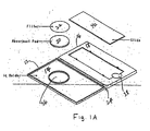
- a kit 100 for facilitating the preparation of cellular samples includes a device 10 comprising a front cover 12, a rear cover 16, and a spine 24 connecting the front and rear covers 12, 16.
- the spine 24 spaces the front cover 12 and the back cover 16 when folded in the closed position and creates a book-like form, wherein the inside 14 of the front cover 12 and the inside 18 of the back cover 16 face and contact against each other.
- the front cover 12 does not need to be the same size as the back cover 16.
- the covers 12, 16 and spine 24 may be made of a single piece of flexible material.
- flexible material can include, for example, cardboard or some flexible plastics to which a controlled pressure may be applied to the outside covers 12, 16 without puncturing or cracking the covers.
- the material should not absorb the sample or solution applied to the filter 22 and absorbent material 20 (described in more detail below).
- the material should also be selected to enable various users, such as, for example, lab technicians and doctors, to apply consistent or controlled amount of pressure to prevent over squeezing the solution, resulting in spillage or distortion of the morphology of the sample by the exertion of too much pressure.
- the device 10 can be made from an inflexible material.
- inflexible material can include, for example, wood, wood-derived materials, metals or inflexible plastics.
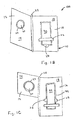
- a hinge 30 connects the front cover 12 and back cover 16.
- the hinge 30 is constructed so that the front cover 12 and back cover 16 face and contact each other when the book form is in the closed position.
- the hinge 30 can be one single, continuous piece 32 (Fig. 2A) or, alternatively, a number of flexible or interlocking pieces 34 (Fig. 2B).
- FIG. 1B the device is shown in the open position.
- Front cover 12 is flipped open, exposing an absorbent material 20 and a filter 22 attached to the inside 14 of the first cover 12.
- a slide 40 is also shown mounted to the inside 18 of the back cover 16 of the device 10.
- the absorbent material 20 is hydrophilic in nature.
- the absorbent material is absorbent to both water and alcohol-based solutions and is preferably uniform in its absorbency.
- the absorbent material is preferably inert and stable so as not to react with the alcohol solution or components of the cellular sample.
- the absorbent material preferably possesses stability properties enabling the finished device to be stored for several years.
- Standard filter papers or blotting papers may be used as an absorbent material.
- one suitable absorbent material is that sold by Bio-Rad® (Hercules, California) called Absorbent Filter Paper (Extra Thick) consisting of 100% cotton fiber.
- the absorbent material can be 100% double-sided, twill-pattern woven cotton (e.g.
- Wipers Natural TX306 made by TexWipe® Company
- polyvinyl acetal foam HydroSponge tm ; TX5390 made by Tex Wipe® Company
- Gelman's® Absorbent Pad Cellulose Fiber Filter Pad, Whatman's® Glass Microfibre filters, Whatman's® cellulose paper, TexWipe® Company (Upper Saddle River, NJ) trademarked Absorbond® (Wipers Synthetic; TX404) consisting of 100% hydro-entangled polyester, Denville® Scientific Hyblot Mater, Millipore® cellulose absorbent pads or any other absorbent material or combination of materials known in the art.
- the absorbent material 20 adheres to the inside 14 of the front cover 12 by, for example, an adhesive or, alternatively, by a heat spot weld.
- the adhesive is selected so as not to interfere with the properties of the absorbent material, as mentioned above.
- One such suitable adhesive is gel cyanoacrylate sold by Loctite under the trademark Loctite 4541®.
- the adhesive can be hot melt or acrylic or synthetic rubber based adhesives or any adhesive known in the art.
- the filter 22 overlays the absorbent material 20.
- Any filter known in the art can be used.
- the filter 22 is preferably a polycarbonate film containing approximately 3 to 8 micrometer pores. The pore sizes can be varied to enable the filter to capture the desired cells on the surface of the filter while allowing the debris to pass through to the absorbent material 20.
- the filter should also be inert, stable, and alcohol resistant.
- the filter can be nylon, cellulose, polyester, teflon, polytetrafluorethylene or any other filter material known in the art.
- An adhesive to attach the filter 22 to the absorbent material 20 is selected so as not to interfere with the sample placed on the filter 20.
- Revertex Chemicals Ltd. sells some suitable adhesives for attaching the filter 22 to the absorbent material 20, such as, for example, starch, dextrin, latex and casein.
- the adhesive can be ethylene vinyl acetate and polyvinyl acetate emulsion adhesive for lamination of the filter 22 and the absorbent material 20.
- the filter 22 can be held in proximity to the absorbent material 20 by mechanical means, such as, for example, a rim of plastic to hold the overlapping filter 22 onto the absorbent material 20 disk.
- the slide 40 is a standard slide, made of either glass or plastic, commonly used in cytology.
- the slide 40 is mounted to the inside 18 of the back cover 16 in such a way that enables the slide 40 to be easily removed.
- the slide 40 is mounted to the inside 18 of the back cover 18 by a piece of adhesive or tape 42.
- any commonly known method known in the art that accomplishes this goal can be used to mount the slide.
- the slide 40 can be press-fit into a cutout portion 44 of the inside 18 of the back cover 16, held by Velcro® strap, or placed in a pocket 46 formed in the inside 18 of the back cover 16 (Fig. 3) to facilitate removal of the slide 40.
- the inside 18 of the back cover 16 includes finger recesses 28 to aid a user in lifting the slide 40 out of the back cover (Fig. 1B and 1C).
- the finger recesses 28 should be positioned away from the preparation area to avoid contamination of the transferred sample.
- the absorbent material 20 with the attached filter 22 are positioned on the inside 14 of the front cover 12 of the book-like device 10 so that when the book-like device 10 is closed, the filter 22 contacts a central location on the slide 40.
- the absorbent material is placed in a recess or shallow well 36 formed in the inside 14 of the front cover 12 so that the upper portion of the absorbent material 20 having the overlaying filter 22 (as will be described in more detail below) extends above the inside surface 14 of the front cover 12.
- This recess or shallow well 36 provides the added benefit of holding any excess solution that escapes from the bottom of the absorbent material 20.
- the absorbent material 20 is held in place on the inside 14 of the front cover 12 by a plurality of supports 50, as illustrated in Fig. 5.
- the inside 14 of the front cover 12 and the inside 16 of the back cover 10 contact each other.
- the upper portion of the absorbent material 20 and filter 22 extend above the inside surface 14 of the front cover 12, there is no need to exert an additional pressure to transfer the sample to the slide 40 from the filter 22, when the back cover 10 comes into contact with front cover 12.
- the slide 40 which can be flush with the inside 18 of the back cover 16 will compress the absorbent material 20 and filter 22, thus transferring the sample to the slide 40.
- the kit 100 may optionally include a mechanical fastening means 60 to lock together and maintain the contact of the inside surfaces 14, 18 of the front and back covers 12, 16, respectively, when the book-form device 10 is closed.
- Any mechanical fastening means known in the art such as Velcro®, tape, clasps, snaps, or the like, can be used.
- the appropriate amount of controlled pressure may be applied by fastening the fastening means 60.
- an audible snap is preferably heard when the fastening means is fastened. This audible snap informs the user that a controlled amount of pressure is being applied to transfer the sample from the filter 22 to the slide 40.
- a cellular sample is any cellular or tissue specimen collected from a subject for the purposes of screening or diagnosing, commonly known in the art, that is capable of being suspended in a liquid-based medium.
- samples are obtained, for example, from the cervix, the mouth or throat, urine sediments, lymph nodes, esophagus, bronchial, breast (i.e. nipple discharge), skin lesions, thyroid, blood and disruptive tissue biopsies.
- the kit 100 is used to prepare a cytology slide with a mono-dispersed cell sample.
- a cytology slide with a mono-dispersed cell sample.
- the cell or tissue sample is obtained by methods known in the art, such as, biopsies, surgical resections, standard bodily fluid collection techniques or the like.
- the sample is taken from the cervix.
- the present invention employs standard techniques for collecting cervical cells; for example, it generally involves the insertion of a cervical brush or broom device and the taking of cells from the surface of the cervix and the endocervix.
- the cellular sample is preferably placed in any liquid-based cytology medium used and known in the art.
- a cytology medium is known as a Universal Collection Medium (UCM) from Digene Corp. (Gaithersburg, MD).
- UCM is a cell collection medium which preserves both cell morphology and cellular biomolecules for quantitative analysis in a cell sample so that multiple assays can be carried out from a single patient sample.
- Other liquid-based cytology media can be used, such as PreservCyt® from Cytyc, Inc. (Boxborough, MA) and CytoRich® Preservative Fluid from TriPath, Inc. (Burlington, N.C.).
- One preferred method places the cellular sample in 1 milliliter of UCM. This solution is then suspended, by mixing. Mixing can be conveniently achieved, for example by vortexing the solution for approximately five to ten seconds.
- a sample is removed carefully, usually with a pipette, and placed on the filter 22. If one milliliter of UCM is used, it may be preferable to use an aliquot of approximately 200 microliters from the solution. This sample is then applied to the surface of the filter 22.
- liquid media may be used and the sample may be suspended in varying amounts of such media.
- the solution and other debris pass through the filter 22 to the absorbent material 20 while the cells are captured on the surface of the filter 22.
- the book-like device 10 is then closed so that the filter 22 contacts the slide 40 for about 15 seconds while a moderate pressure, created when the covers are closed and locked together, is applied to the front and back covers 12, 16, respectively.
- the device or user applies from 200 to 1000 grams of pressure to the slide and specimen for optimum transfer of the cytology specimen to the slide.
- the pressure applied to the covers 12, 16 transfers the cells captured by the filter 22 to the slide 40. After the pressure is applied, the slide 40 can be removed from the book-like device 12 and fixed and stained in the routine manner.
- the cells are preferably wet-fixed before any air drying occurs.
- fixatives Most laboratories use a 95 percent ethanol solution for most routine preparations that require fixations. However, other fixatives known in the art can be used. Most of these fixatives are composed of polyethylene glycol in an alcohol base and are applied either from a dropper bottle or in spray form. Coating fixatives are also widely used for cervical smears. These fixatives cover the sample in a waxy coating that protects it from damage.
- the absorbent material 22 keeps the sample moist as it is transferred to the slide since a liquid-based solution is used and the absorbent material 20 is held in close physical proximity to the specimen.
- the device 10 maintains a "moist” or “humid” environment so that the cells can be preserved in the device 10 before fixing for up to 30 minutes following application on the slide 40. Once on the slide 40, the cell sample should be preserved as mentioned above.
- the cellular sample is generally stained. Staining may be required to retain the transparency of the cells and display variations of cellular maturity and metabolic activity. Any stain or staining method known in the art can be used. 6
- the universal stain for cytological preparations is the Papanicolaou stain.
- any stain known in the art such as Hematonylin and Eosin stain or special chemical stains or immunochemical stains, or in-situ hybridization can be used.
- the slide may be mounted in a suitable medium satisfactory for microscopy.
- mountants that can be used that are well known in the art. These mountants protect the sample from dust and damage and should harden rapidly and not discolor or crystallize during long-term storage.
- a coverslip may then be placed over the sample.
- Coverslipping has traditionally been achieved using a thin glass slip (less than 0.17 millimeters) or continuous film. Various sizes are commercially available but the entire sample preparation should be covered by the mountant and the coverslip.
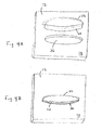
- a kit 200 for facilitating the preparation of a plurality of cellular samples includes a cover 210, a base 230 and at least one hinge 205 for closing cover 210 over base 230 to create a book-like form.
- the cover 210 and base 230 may be made of a single piece of flexible material.
- flexible material can include, for example, cardboard or some flexible plastics to which a controlled pressure may be applied to the outside surface of the cover without puncturing or cracking the cover.
- the material should not absorb the sample or solution applied to the filters and absorbent material, as described in more detail above.
- the material should also be selected to enable various users, such as, for example, lab technicians and doctors, to apply consistent or controlled pressure to prevent over squeezing the solution, resulting in spillage or distortion of the morphology of the sample by the exertion of too much pressure.
- cover 210 and base 230 can be made from an inflexible material.
- inflexible material can include, for example, wood, wood-derived materials, metals or inflexible plastics.
- the weight of the material used for the cover provides enough pressure to effectively transfer the sample from the absorbent material and filter onto the cytology slide.
- the weight of the material used for the cover provides enough pressure to effectively transfer the sample from the absorbent material and filter onto the cytology slide.
- clamps can be used to close cover 210 and base 230 so that the samples may be effectively transferred.
- the cover 210 supports a strip 212 holding a plurality of pieces of absorbent material 20 and a filter strip 22 overlaying the absorbent material 20.
- the absorbent material 20 and filter 22 are made from the same materials discussed above. In alternate embodiments, separate filters corresponding to each piece of absorbent material 20 held by the strip 212 or, alternatively, a filter 22 overlaying a number of pieces of absorbent material 20 can be used.
- the upper portion of the absorbent material 20 and filter 22 extend above the top surface of strip 212 so that when cover 210 is closed over base 230, the samples applied to the absorbent material 20 and filter 22 are effectively transferred to the corresponding slides 40 held by slide carrier 232.
- strip 212 holding absorbent material 20 and filter 22 is supported by cover 210 in a channel 214 formed in the inside surface 216 of the cover 210.
- strip 212 is press fit into the channel 214.
- strip 212 includes feet 220 projecting from the bottom side of strip 212 which are press fit into recesses 222 formed in the bottom surface 213 of channel 214. The feet 220 also serve to stabilize and position strip 212 in relation to cover 210 and keep the strip 212 from sliding out of the channel 210.
- strip 212 can be slid into and resiliently held by channel 214.
- the strip 212 can also include positioning members 226 projecting from the sides of strip 212. Positioning members 226 are received by recesses 227 formed in the sides of channel 214 to ensure that strip 212 is properly positioned in channel 214.
- the absorbent material 20 is adhered to the top surface of strip 212 by, for example, an adhesive or, alternatively, by a heat spot weld.
- the adhesive is selected so as not to interfere with the properties of the absorbent material, as mentioned above.
- the adhesive can be hot melt or acrylic or synthetic rubber based adhesives or any adhesive known in the art.
- the filter overlays the adhesive pad in the same manner as discussed above.
- the base 230 supports a slide carrier 232 capable of holding a plurality of slides 40.
- the number of slides 40 held by slide carrier 232 corresponds to the number of pieces of absorbent material 20 on strip 212 supported by cover 210.
- the slide carrier 232 containing slides 40 is supported by base 230 in a channel 234 formed in the top surface of base 230.
- slide carrier 232 is press fit into channel 234.
- slide carrier 232 includes feet (not shown) projecting from the bottom side of slide carrier 232. The feet are press fit into recesses 238 formed in the bottom surface of channel 234.
- the slide carrier 232 can also include positioning members, similar to positioning members 236 on strip 212, projecting from the sides of slide carrier 232 to ensure that slide carrier 232 is properly positioned in channel 234.
- the slide carrier 232 includes a plurality of slots 240 separated by ridges or dividers 242 for securely holding slides 40 in place when the kit 200 is opened and closed. While slide carrier 232 is held in channel 234 in base 230, the top surface 244 of dividers 242 is flush with the top surface of base 230 to ensure proper closing of kit 200. Finger recesses 250 may be provided to facilitate the insertion and removal of the slides 40 from slide carrier 232.
- the slide carrier 232 can be hingeably attached to base 230.
- the slides 40 are inserted into separate, individual slots formed in the base 230 itself.
- the method of applying or smearing a sample to any of the absorbent materials 20 and filter 22 and transferring the sample to the slide 40 is identical to the method described above for the book-like device 10.
- the kit 200 one lab technician or doctor can prepare multiple slides at the same time.
- the slides may be individually analyzed or stored in slide carrier 232 for future analysis.
- a cervical sample was taken from a female patient and placed in a one milliliter of Universal Collection Medium. 7 An aliquot of 200 microliters is removed from the solution and applied to the surface of the filter 22 of the book-form device 10. The book-form device 10 was closed for approximately 15 seconds while a controlled amount of pressure was applied. The slide 40 was then removed from the book-form device 10 and the sample was fixed, stained and placed under a light microscope.
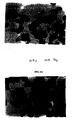
- Fig. 6A shows the morphology of cervical cells prepared by the method of the present invention.
- Fig 6B was taken from archived routine Pap smears to show the morphology, distribution of cells and staining of stored samples fixed to slides for comparison.
- the morphology of the cells prepared by the method of the present invention shows better quality and more evenly distributed cells with less interference from debris than the conventional Pap smear.
- Table 1 shows a comparison of Pap slides from the same patients made with the conventional Pap smear technique and the UCM book-form device Pap slide technique. These results demonstrate complete agreement between the two procedures.
- Table 1. UCM vs. Conventional Pap* Case ID Conventional Pap UCM Manual Pap 1W WNL WNL 2W LSIL HSIL 3W WNL WNL 4W WNL WNL 5W WNL WNL 6W WNL WNL 7W WNL WNL 8W WNL WNL 9W WNL WNL 10W WNL WNL 11W WNL WNL 12W WNL WNL 13W WNL WNL 14W WNL WNL 15W WNL WNL 100% Agreement
Landscapes
- Health & Medical Sciences (AREA)
- Life Sciences & Earth Sciences (AREA)
- Engineering & Computer Science (AREA)
- Biomedical Technology (AREA)
- Molecular Biology (AREA)
- Physics & Mathematics (AREA)
- Chemical & Material Sciences (AREA)
- Analytical Chemistry (AREA)
- Biochemistry (AREA)
- General Health & Medical Sciences (AREA)
- General Physics & Mathematics (AREA)
- Immunology (AREA)
- Pathology (AREA)
- Sampling And Sample Adjustment (AREA)
- Investigating Or Analysing Biological Materials (AREA)
- Preparation Of Compounds By Using Micro-Organisms (AREA)
Claims (20)
- Vorrichtung zum Erleichtern der Aufbereitung von cytologischen Slides, wobei die Vorrichtung umfasst:eine erste Abdeckung mit einer Innenseite;eine zweite Abdeckung mit einer Innenseite;ein dazwischen angebrachtes Gelenk, wobei die erste und die zweite Abdeckung schwenkbar an das Gelenk befestigt sind, so dass diese in eine Buchform gefaltet werden können, die für eine geöffnete und eine geschlossene Position geeignet ist;ein Absorptionsmaterial, das auf der Innenseite der ersten Abdeckung angebracht ist;einen Filter, wobei der Filter über dem Absorptionsmaterial liegt; undein cytologisches Slide, wobei das cytologische Slide an der Innenseite der zweiten Abdeckung angebracht ist, wobei das Slide auf der Innenseite der zweiten Abdeckung positioniert ist, um den Filter zu berühren, wenn sich die Buchform in der geschlossenen Position befindet.
- Vorrichtung nach Anspruch 1, wobei der Filter ein Polycarbonat ist.
- Vorrichtung nach Anspruch 1, wobei das Absorptionsmaterial ein Polyvinylacetalschaum ist.
- Vorrichtung nach Anspruch 1, wobei das Absorptionsmaterial eine Faser aus 100% Baumwolle ist.
- Vorrichtung nach Anspruch 1, wobei das cytologische Slide abnehmbar an der zweiten Abdeckung angebracht ist.
- Vorrichtung zum Erleichtern der Aufbereitung von cytologischen Slides, wobei die Vorrichtung umfasst:eine erste Abdeckung mit einer Innenseite;eine zweite Abdeckung mit einer Innenseite, wobei die zweite Abdeckung gelenkig an die erste Abdeckung angebracht ist, um in eine Buchform gefaltet werden zu können, die für eine geöffnete und eine geschlossene Position geeignet ist;ein Absorptionsmaterial, das an der Innenseite der ersten Abdeckung angebracht ist;einen Filter, wobei der Filter über dem Absorptionsmaterial liegt; undein cytologisches Slide, wobei das cytologische Slide an der Innenseite der zweiten Abdeckung angebracht ist, wobei das Slide auf der Innenseite der zweiten Abdeckung positioniert ist, um den Filter zu berühren, wenn sich die Buchform in der geschlossenen Position befindet.
- Vorrichtung nach Anspruch 6, wobei die erste Abdeckung über ein Gelenk, das ein einzelnes ununterbrochenes Stück umfasst, gelenkig an die zweite Abdeckung angebracht ist.
- Vorrichtung nach Anspruch 6, wobei die erste Abdeckung über ein Gelenk, das eine Vielzahl von miteinander verbundenen Stücken umfasst, gelenkig an die zweite Abdeckung angebracht ist.
- Vorrichtung nach Anspruch 5, wobei das cytologische Slide abnehmbar an der zweiten Abdeckung angebracht ist.
- Verfahren zum Aufbereiten cytologischer Slides, wobei das Verfahren umfasst:Kombinieren einer Zellprobe mit einem auf einer Flüssigkeit basierenden Medium, um eine Lösung zu erzeugen;Entfernen eines Aliquots aus der Lösung;Bereitstellen einer Slide-Aufbereitungsvorrichtung, die eine erste Abdeckung mit einer Innenseite umfasst, eine zweite Abdeckung mit einer Innenseite, wobei die erste und die zweite Abdeckung schwenkbar aneinander befestigt sind, um in eine Buchform gefaltet werden zu können, wobei ein Absorptionsmaterial an die Innenseite der ersten Abdeckung befestigt ist, einen Filter, wobei der Filter über dem Absorptionsmaterial liegt, sowie ein Slide, das an die Innenseite der zweiten Abdeckung angebracht ist;Aufbringen des Aliquots auf den Filter;Verschließen der Buchform, so dass der Filter, der das Aliquot enthält, das Slide berührt; undAnwenden eines Druckes auf die erste und die zweite Abdeckung.
- Verfahren nach Anspruch 10, wobei das auf einer Flüssigkeit basierende Medium ein universelles Sammelmedium (Universal Collection Medium) ist.
- Vorrichtung zum Erleichtern der Aufbereitung einer Vielzahl von cytologischen Slides, wobei die Vorrichtung umfasst:eine Abdeckung mit einer Innenseite;eine Basis mit einer Innenseite;ein dazwischen liegendes Gelenk, wobei die Abdeckung und die Basis schwenkbar an das Gelenk befestigt sind, um in eine Buchform gefaltet werden zu können, die für eine geöffnete und eine geschlossene Position geeignet ist;eine Vielzahl von Absorptionsmaterialien, die an der Innenseite der Abdeckung angebracht sind;einen Filter, wobei der Filter über den Absorptionsmaterialien liegt; undeine Vielzahl von cytologischen Slides, wobei jeder cytologische Slide einem der Absorptionsmaterialien entspricht, die auf der Abdeckung angebracht sind, wobei die cytologischen Slides an der Innenseite der Basis angebracht sind, wobei jedes Slide auf der Innenseite der Basis positioniert ist, um das entsprechende Absorptionsmaterial und den Filter zu berühren, wenn sich die Buchform in der geschlossenen Position befindet.
- Vorrichtung nach Anspruch 12, wobei die Slides entfernbar an der Innenseite der Basis angebracht sind.
- Vorrichtung nach Anspruch 13, wobei die Slides entfernbar an einen Slideträger angebracht sind, wobei der Slideträger entfernbar an der Innenseite der Basis angebracht ist.
- Vorrichtung nach Anspruch 13, wobei die Slides entfernbar an einem Slideträger angebracht sind, wobei der Slideträger gelenkig an der Innenseite der Basis angebracht ist.
- Vorrichtung nach Anspruch 12, wobei das Absorptionsmaterial an einem Streifen angebracht ist, wobei der Streifen entfernbar an der Innenseite der Abdeckung angebracht ist.
- Vorrichtung nach Anspruch 12, wobei der Filter eine Vielzahl von individuellen Filtern enthält, wobei jeder Filter einem der Absorptionsmaterialien entspricht.
- Vorrichtung nach Anspruch 12, wobei die Abdeckung ein Gewicht aufweist, um wirksam eine Probe, die auf das Absorptionsmaterial und den Filter aufgebracht wird, zu dem Slide zu übertragen, wenn sich die Buchform in der geschlossenen Position befindet.
- Vorrichtung nach Anspruch 14, wobei der Slideträger eine Vielzahl von Einschüben für das Halten einer Vielzahl von cytologischen Slides enthält.
- Vorrichtung nach Anspruch 12, wobei die cytologischen Slides entfernbar an der Basis angebracht sind.
Applications Claiming Priority (3)
| Application Number | Priority Date | Filing Date | Title |
|---|---|---|---|
| US542746 | 2000-04-04 | ||
| US09/542,746 US6436662B1 (en) | 2000-04-04 | 2000-04-04 | Device and method for cytology slide preparation |
| PCT/US2001/010968 WO2001075446A1 (en) | 2000-04-04 | 2001-04-04 | Device and method for cytology slide preparation |
Publications (3)
| Publication Number | Publication Date |
|---|---|
| EP1272848A1 EP1272848A1 (de) | 2003-01-08 |
| EP1272848A4 EP1272848A4 (de) | 2006-01-18 |
| EP1272848B1 true EP1272848B1 (de) | 2007-08-08 |
Family
ID=24165118
Family Applications (1)
| Application Number | Title | Priority Date | Filing Date |
|---|---|---|---|
| EP01926624A Expired - Lifetime EP1272848B1 (de) | 2000-04-04 | 2001-04-04 | Vorrichtung und verfahren zur herstellung von zytologischen klingen |
Country Status (10)
| Country | Link |
|---|---|
| US (3) | US6436662B1 (de) |
| EP (1) | EP1272848B1 (de) |
| JP (1) | JP2003529768A (de) |
| AT (1) | ATE369561T1 (de) |
| AU (2) | AU5314601A (de) |
| BR (1) | BR0109786A (de) |
| CA (1) | CA2402859A1 (de) |
| DE (1) | DE60129803T2 (de) |
| MX (1) | MXPA02009812A (de) |
| WO (1) | WO2001075446A1 (de) |
Families Citing this family (51)
| Publication number | Priority date | Publication date | Assignee | Title |
|---|---|---|---|---|
| WO1999031273A2 (en) * | 1997-12-12 | 1999-06-24 | Digene Corporation | Universal collection medium |
| US6436662B1 (en) * | 2000-04-04 | 2002-08-20 | Digene Corporation | Device and method for cytology slide preparation |
| US7601497B2 (en) | 2000-06-15 | 2009-10-13 | Qiagen Gaithersburg, Inc. | Detection of nucleic acids by target-specific hybrid capture method |
| US7439016B1 (en) * | 2000-06-15 | 2008-10-21 | Digene Corporation | Detection of nucleic acids by type-specific hybrid capture method |
| US20020130074A1 (en) * | 2000-10-13 | 2002-09-19 | Domanik Richard A. | Optical analysis of cellular material |
| US7223363B2 (en) * | 2001-03-09 | 2007-05-29 | Biomicro Systems, Inc. | Method and system for microfluidic interfacing to arrays |
| AU2003302333A1 (en) * | 2002-12-20 | 2004-07-22 | Board Of Regents, The University Of Texas System | Methods and apparatus for electrosmear analysis |
| US20040137551A1 (en) * | 2003-01-13 | 2004-07-15 | Markovic Nenad S. | Cervical acid phosphatase - papanicolaou (CAP-PAP) test kit, method and accesories, processes for producing and using the same |
| US7435601B2 (en) * | 2003-02-19 | 2008-10-14 | Fitzco Incorporated | Biological specimen handling method |
| US7588733B2 (en) * | 2003-12-04 | 2009-09-15 | Idexx Laboratories, Inc. | Retaining clip for reagent test slides |
| US20060161076A1 (en) * | 2005-01-06 | 2006-07-20 | Diamics, Inc. | Systems and methods for collection of cell clusters |
| US20060189893A1 (en) * | 2005-01-06 | 2006-08-24 | Diamics, Inc. | Systems and methods for detecting abnormal cells |
| DE102005015005A1 (de) * | 2005-04-01 | 2006-10-05 | Qiagen Gmbh | Verfahren zur Behandlung einer Biomoleküle enthaltenden Probe |
| US7678337B2 (en) * | 2005-04-26 | 2010-03-16 | Abbott Laboratories Inc. | Assembly for carrying and holding slides |
| US20070009389A1 (en) * | 2005-07-08 | 2007-01-11 | Antti Seppo | Slide deposition chamber |
| WO2007022483A2 (en) * | 2005-08-19 | 2007-02-22 | Bioventures, Inc. | Method and device for the collection and isolation of nucleic acid |
| US7357042B2 (en) * | 2005-12-01 | 2008-04-15 | Cytyc Corporation | Filter contamination control device |
| US8241593B2 (en) * | 2006-05-04 | 2012-08-14 | The Bode Technology Group, Inc. | Specimen tray |
| US8962262B2 (en) * | 2006-05-11 | 2015-02-24 | Arbor Vita Corporation | Method of protein extraction from cells |
| US7758816B2 (en) * | 2007-03-01 | 2010-07-20 | Wescor Inc. | Large area cytocentrifuge sample chamber |
| JP2008249565A (ja) * | 2007-03-30 | 2008-10-16 | Mie Univ | 細胞標本調製方法 |
| US7951345B2 (en) * | 2007-06-01 | 2011-05-31 | Lary Research & Development, Llc | Useful specimen transport apparatus with integral capability to allow three dimensional x-ray images |
| US7906317B2 (en) * | 2007-10-21 | 2011-03-15 | King Car Food Industrial Co., Ltd. | Apparatus for thin-layer cell smear preparation and in-situ hybridization |
| US9207240B2 (en) * | 2007-11-14 | 2015-12-08 | Arbor Vita Corporation | Method of efficient extraction of protein from cells |
| JP2011518333A (ja) * | 2008-04-17 | 2011-06-23 | キアジェン ゲイサーズバーグ インコーポレイテッド | 標的核酸の存在を判別するための合成プローブを用いた組成物、方法、およびキット |
| WO2010062546A1 (en) | 2008-10-27 | 2010-06-03 | Qiagen Gaithersburg Inc. | Fast results hybrid capture assay on an automated platform |
| JP6108661B2 (ja) * | 2009-01-28 | 2017-04-05 | キアジェン ゲイサーズバーグ インコーポレイテッド | 配列特異的な大量試料調製方法およびアッセイ法 |
| EP2425019B1 (de) | 2009-05-01 | 2014-03-19 | QIAGEN Gaithersburg, Inc. | Ungezieltes verstärkungsverfahren zur erkennung von rna-spleissformen in einer probe |
| JP5826752B2 (ja) | 2009-09-14 | 2015-12-02 | キアジェン ゲイサーズバーグ インコーポレイテッド | 細胞学用培地に固定された組織サンプルから核酸またはタンパク質を回収するための組成物および方法 |
| EP2519854A2 (de) * | 2009-12-31 | 2012-11-07 | Abbott Point Of Care, Inc. | Verfahren und vorrichtung zur sicherung der ebenen ausrichtung einer analysekammer |
| CA2787924A1 (en) * | 2010-01-29 | 2011-08-04 | Qiagen Gaithersburg, Inc. | Methods and compositions for sequence-specific purification and multiplex analysis of nucleic acids |
| CA2787781A1 (en) | 2010-01-29 | 2011-08-04 | Qiagen Gaithersburg, Inc. | Method of determining and confirming the presence of an hpv in a sample |
| JP2013528049A (ja) | 2010-05-19 | 2013-07-08 | キアゲン ガイサーズバーグ アイエヌシー. | 核酸の配列特異的な精製及び多重分析のための方法及び組成物 |
| JP5771934B2 (ja) * | 2010-10-01 | 2015-09-02 | コニカミノルタ株式会社 | 細胞染色方法及び細胞染色用キット |
| CN102445368B (zh) * | 2010-10-13 | 2013-05-01 | 青岛市市立医院 | 检验载玻片 |
| ES2629461T3 (es) * | 2010-11-15 | 2017-08-09 | Onpharma, Inc. | Aparato y procedimientos para retener fluidos expulsados durante una transferencia de fluido |
| CN103597095B (zh) | 2011-02-24 | 2016-08-17 | 奇亚根盖瑟斯堡股份有限公司 | 用于检测hpv核酸的材料和方法 |
| US20130050692A1 (en) | 2011-08-30 | 2013-02-28 | Molecular Devices, Llc | Presentation system and method for low-volume solution samples |
| US9453996B2 (en) | 2013-10-23 | 2016-09-27 | Tokitae Llc | Devices and methods for staining and microscopy |
| WO2015146682A1 (ja) * | 2014-03-28 | 2015-10-01 | シャープ株式会社 | 濾過用具 |
| WO2017047617A1 (ja) * | 2015-09-14 | 2017-03-23 | 国立大学法人滋賀医科大学 | 観察標本作製用細胞保持基材ホルダー及びそれを含むキット並びに観察標本の作製方法 |
| EP3351921A4 (de) | 2015-09-14 | 2019-08-21 | National University Corporation Shiga University OF Medical Science | Zellhaltesubstrathalter zur vorbereitung einer beobachtungsprobe, kit damit und verfahren für die zubereitung einer beobachtungsprobe |
| JP6738012B2 (ja) * | 2015-09-14 | 2020-08-12 | 国立大学法人滋賀医科大学 | 細胞観察標本作製用細胞保持基材ホルダー及びそれを含むキット |
| EP4265332B1 (de) * | 2017-06-13 | 2025-02-19 | Veterinary Diagnostics Institute, Inc. | System und verfahren zur stabilisierung, lagerung und rückgewinnung von blutproben |
| KR101904207B1 (ko) * | 2017-08-24 | 2018-10-04 | (의)삼성의료재단 | 표시선을 가지는 조직 표본 제작용 슬라이드 글라스 |
| CN112368560B (zh) | 2018-07-04 | 2024-08-06 | 奥林巴斯株式会社 | 细胞检查装置及细胞检查方法 |
| CA3107736C (en) | 2018-07-28 | 2023-10-17 | Leavitt Medical, Inc. | Methods and systems for preparing cytological samples |
| JP7483724B2 (ja) | 2019-01-18 | 2024-05-15 | テックサイト・インコーポレイテッド | 光学顕微鏡の基準焦点面を識別するための印刷カバースリップ及びスライド |
| IT202000003043A1 (it) * | 2020-02-14 | 2021-08-14 | Bioevo S R L | Dispositivo e metodo per il trasporto, la conservazione, la preparazione e l’analisi di campioni biologici. |
| KR102952511B1 (ko) * | 2024-04-08 | 2026-04-14 | 주식회사 예송 | 세포 자동 도말 장치 |
| KR102952510B1 (ko) * | 2024-04-08 | 2026-04-14 | 주식회사 예송 | 세포 자동 도말 장치 |
Family Cites Families (10)
| Publication number | Priority date | Publication date | Assignee | Title |
|---|---|---|---|---|
| US4190314A (en) | 1973-07-13 | 1980-02-26 | Stephen Goldsmith | Microscope and microscope slide for cytological analysis |
| US3933594A (en) * | 1974-08-16 | 1976-01-20 | Polaroid Corporation | Method and device for determining the concentration of a substance in a fluid |
| US5301685A (en) * | 1989-01-10 | 1994-04-12 | Guirguis Raouf A | Method and apparatus for obtaining a cytology monolayer |
| CA2032482C (en) * | 1989-05-09 | 1998-06-16 | Josefina T. Baker | Specimen collection device and method |
| US5240606A (en) | 1990-07-09 | 1993-08-31 | Cytyc Corporation | Apparatus for preparing cells for examination |
| US5494828A (en) | 1994-07-13 | 1996-02-27 | Leopando; Mark E. | Slide dispensing device and method |
| US5470758A (en) * | 1994-12-14 | 1995-11-28 | Shandon, Inc. | Large cytology sample chamber for distributing material onto a microscope slide |
| US6300140B1 (en) * | 1997-10-01 | 2001-10-09 | Leonard Bloom | Rapid test employing an adhesive slide |
| ES2399327T3 (es) * | 1998-10-05 | 2013-03-27 | Biopath Automation, L.L.C. | Método para recolectar y manipular muestras de tejido para análisis de biopsia |
| US6436662B1 (en) * | 2000-04-04 | 2002-08-20 | Digene Corporation | Device and method for cytology slide preparation |
-
2000
- 2000-04-04 US US09/542,746 patent/US6436662B1/en not_active Expired - Lifetime
-
2001
- 2001-04-04 DE DE60129803T patent/DE60129803T2/de not_active Expired - Lifetime
- 2001-04-04 JP JP2001572872A patent/JP2003529768A/ja active Pending
- 2001-04-04 AT AT01926624T patent/ATE369561T1/de not_active IP Right Cessation
- 2001-04-04 AU AU5314601A patent/AU5314601A/xx active Pending
- 2001-04-04 MX MXPA02009812A patent/MXPA02009812A/es active IP Right Grant
- 2001-04-04 BR BR0109786-5A patent/BR0109786A/pt active Search and Examination
- 2001-04-04 AU AU2001253146A patent/AU2001253146B2/en not_active Ceased
- 2001-04-04 EP EP01926624A patent/EP1272848B1/de not_active Expired - Lifetime
- 2001-04-04 US US09/826,327 patent/US7001776B2/en not_active Expired - Lifetime
- 2001-04-04 CA CA002402859A patent/CA2402859A1/en not_active Abandoned
- 2001-04-04 WO PCT/US2001/010968 patent/WO2001075446A1/en not_active Ceased
-
2002
- 2002-05-22 US US10/153,340 patent/US6890729B2/en not_active Expired - Lifetime
Also Published As
| Publication number | Publication date |
|---|---|
| MXPA02009812A (es) | 2004-09-06 |
| EP1272848A4 (de) | 2006-01-18 |
| WO2001075446A1 (en) | 2001-10-11 |
| AU2001253146B2 (en) | 2005-03-17 |
| DE60129803D1 (de) | 2007-09-20 |
| WO2001075446A9 (en) | 2002-12-19 |
| EP1272848A1 (de) | 2003-01-08 |
| US6890729B2 (en) | 2005-05-10 |
| DE60129803T2 (de) | 2008-04-30 |
| CA2402859A1 (en) | 2001-10-11 |
| JP2003529768A (ja) | 2003-10-07 |
| US20020177184A1 (en) | 2002-11-28 |
| AU5314601A (en) | 2001-10-15 |
| BR0109786A (pt) | 2005-01-11 |
| US7001776B2 (en) | 2006-02-21 |
| US20020039796A1 (en) | 2002-04-04 |
| US6436662B1 (en) | 2002-08-20 |
| ATE369561T1 (de) | 2007-08-15 |
Similar Documents
| Publication | Publication Date | Title |
|---|---|---|
| EP1272848B1 (de) | Vorrichtung und verfahren zur herstellung von zytologischen klingen | |
| AU2001253146A1 (en) | Device and method for cytology slide preparation | |
| Linder | Recent advances in thin‐layer cytology. | |
| Cowell et al. | Diagnostic cytology and hematology of the horse | |
| US6337189B1 (en) | Fixative system, method and composition for biological testing | |
| US5301685A (en) | Method and apparatus for obtaining a cytology monolayer | |
| CA2427967A1 (en) | Methods and devices for collecting, handling and processing mammary fluid samples for evaluating breast diseases, including cancer | |
| Stanley et al. | Fine needle aspiration of palpable masses | |
| US5259391A (en) | Method and device for cell sampling | |
| Barr | Cutaneous cytology | |
| Maksem et al. | Manual method for liquid‐based cytology: A demonstration using 1,000 gynecological cytologies collected directly to vial and prepared by a smear‐slide technique | |
| Dimenstein | Grossing biopsies: an introduction to general principles and techniques | |
| Khalbuss et al. | SpinThin, a simple, inexpensive technique for preparation of thin‐layer cervical cytology from liquid‐based specimens: Data on 791 cases | |
| Wilkinson et al. | Techniques and results of aspiration cytology for diagnosis of benign and malignant diseases of the breast | |
| US6106483A (en) | Apparatus for obtaining a cytology monolayer | |
| Frank et al. | The cytodiagnosis of prostatic carcinoma: a follow-up study | |
| Bauer | Cytological collection techniques and sample preparation | |
| Fisher et al. | Acquisition and management of cytologic specimens | |
| Stevens et al. | Evaluation of the CytoRich® technique for cervical smears | |
| Saǧiroǧlu | TMK-101,“Türk,” a new rapid polychrome staining technique for office practice | |
| Shah et al. | Exfoliative cytology and cytocentrifuge preparation of oral premalignant and malignant lesions | |
| Inhorn et al. | Validation of the ThinPrep Papanicolaou test for cervical cancer diagnosis | |
| CN214795400U (zh) | 一种方便使用的检验载玻片 | |
| Popescu et al. | Preliminary study concerning the Cytoscreen system importance (Liquid Based Cytology) in gynecologic cytology | |
| Merritt et al. | A simplified filter technique for cytologic detection of urinary malignancies |
Legal Events
| Date | Code | Title | Description |
|---|---|---|---|
| PUAI | Public reference made under article 153(3) epc to a published international application that has entered the european phase |
Free format text: ORIGINAL CODE: 0009012 |
|
| 17P | Request for examination filed |
Effective date: 20021028 |
|
| AK | Designated contracting states |
Kind code of ref document: A1 Designated state(s): AT BE CH CY DE DK ES FI FR GB GR IE IT LI LU MC NL PT SE TR |
|
| A4 | Supplementary search report drawn up and despatched |
Effective date: 20051205 |
|
| GRAP | Despatch of communication of intention to grant a patent |
Free format text: ORIGINAL CODE: EPIDOSNIGR1 |
|
| GRAS | Grant fee paid |
Free format text: ORIGINAL CODE: EPIDOSNIGR3 |
|
| GRAL | Information related to payment of fee for publishing/printing deleted |
Free format text: ORIGINAL CODE: EPIDOSDIGR3 |
|
| GRAS | Grant fee paid |
Free format text: ORIGINAL CODE: EPIDOSNIGR3 |
|
| GRAA | (expected) grant |
Free format text: ORIGINAL CODE: 0009210 |
|
| AK | Designated contracting states |
Kind code of ref document: B1 Designated state(s): AT BE CH CY DE DK ES FI FR GB GR IE IT LI LU MC NL PT SE TR |
|
| REG | Reference to a national code |
Ref country code: GB Ref legal event code: FG4D |
|
| REG | Reference to a national code |
Ref country code: CH Ref legal event code: EP |
|
| REG | Reference to a national code |
Ref country code: IE Ref legal event code: FG4D |
|
| REF | Corresponds to: |
Ref document number: 60129803 Country of ref document: DE Date of ref document: 20070920 Kind code of ref document: P |
|
| ET | Fr: translation filed | ||
| PG25 | Lapsed in a contracting state [announced via postgrant information from national office to epo] |
Ref country code: NL Free format text: LAPSE BECAUSE OF FAILURE TO SUBMIT A TRANSLATION OF THE DESCRIPTION OR TO PAY THE FEE WITHIN THE PRESCRIBED TIME-LIMIT Effective date: 20070808 Ref country code: ES Free format text: LAPSE BECAUSE OF FAILURE TO SUBMIT A TRANSLATION OF THE DESCRIPTION OR TO PAY THE FEE WITHIN THE PRESCRIBED TIME-LIMIT Effective date: 20071119 Ref country code: FI Free format text: LAPSE BECAUSE OF FAILURE TO SUBMIT A TRANSLATION OF THE DESCRIPTION OR TO PAY THE FEE WITHIN THE PRESCRIBED TIME-LIMIT Effective date: 20070808 |
|
| NLV1 | Nl: lapsed or annulled due to failure to fulfill the requirements of art. 29p and 29m of the patents act | ||
| REG | Reference to a national code |
Ref country code: CH Ref legal event code: PL |
|
| PG25 | Lapsed in a contracting state [announced via postgrant information from national office to epo] |
Ref country code: CH Free format text: LAPSE BECAUSE OF FAILURE TO SUBMIT A TRANSLATION OF THE DESCRIPTION OR TO PAY THE FEE WITHIN THE PRESCRIBED TIME-LIMIT Effective date: 20070808 Ref country code: AT Free format text: LAPSE BECAUSE OF FAILURE TO SUBMIT A TRANSLATION OF THE DESCRIPTION OR TO PAY THE FEE WITHIN THE PRESCRIBED TIME-LIMIT Effective date: 20070808 Ref country code: LI Free format text: LAPSE BECAUSE OF FAILURE TO SUBMIT A TRANSLATION OF THE DESCRIPTION OR TO PAY THE FEE WITHIN THE PRESCRIBED TIME-LIMIT Effective date: 20070808 |
|
| PG25 | Lapsed in a contracting state [announced via postgrant information from national office to epo] |
Ref country code: BE Free format text: LAPSE BECAUSE OF FAILURE TO SUBMIT A TRANSLATION OF THE DESCRIPTION OR TO PAY THE FEE WITHIN THE PRESCRIBED TIME-LIMIT Effective date: 20070808 |
|
| PG25 | Lapsed in a contracting state [announced via postgrant information from national office to epo] |
Ref country code: GR Free format text: LAPSE BECAUSE OF FAILURE TO SUBMIT A TRANSLATION OF THE DESCRIPTION OR TO PAY THE FEE WITHIN THE PRESCRIBED TIME-LIMIT Effective date: 20071109 Ref country code: DK Free format text: LAPSE BECAUSE OF FAILURE TO SUBMIT A TRANSLATION OF THE DESCRIPTION OR TO PAY THE FEE WITHIN THE PRESCRIBED TIME-LIMIT Effective date: 20070808 |
|
| PG25 | Lapsed in a contracting state [announced via postgrant information from national office to epo] |
Ref country code: PT Free format text: LAPSE BECAUSE OF FAILURE TO SUBMIT A TRANSLATION OF THE DESCRIPTION OR TO PAY THE FEE WITHIN THE PRESCRIBED TIME-LIMIT Effective date: 20080108 |
|
| PLBE | No opposition filed within time limit |
Free format text: ORIGINAL CODE: 0009261 |
|
| STAA | Information on the status of an ep patent application or granted ep patent |
Free format text: STATUS: NO OPPOSITION FILED WITHIN TIME LIMIT |
|
| PG25 | Lapsed in a contracting state [announced via postgrant information from national office to epo] |
Ref country code: SE Free format text: LAPSE BECAUSE OF FAILURE TO SUBMIT A TRANSLATION OF THE DESCRIPTION OR TO PAY THE FEE WITHIN THE PRESCRIBED TIME-LIMIT Effective date: 20071108 |
|
| 26N | No opposition filed |
Effective date: 20080509 |
|
| PGFP | Annual fee paid to national office [announced via postgrant information from national office to epo] |
Ref country code: LU Payment date: 20080416 Year of fee payment: 8 |
|
| PGFP | Annual fee paid to national office [announced via postgrant information from national office to epo] |
Ref country code: MC Payment date: 20080411 Year of fee payment: 8 |
|
| PGFP | Annual fee paid to national office [announced via postgrant information from national office to epo] |
Ref country code: IE Payment date: 20080415 Year of fee payment: 8 |
|
| PG25 | Lapsed in a contracting state [announced via postgrant information from national office to epo] |
Ref country code: CY Free format text: LAPSE BECAUSE OF FAILURE TO SUBMIT A TRANSLATION OF THE DESCRIPTION OR TO PAY THE FEE WITHIN THE PRESCRIBED TIME-LIMIT Effective date: 20070808 |
|
| REG | Reference to a national code |
Ref country code: FR Ref legal event code: CD |
|
| PG25 | Lapsed in a contracting state [announced via postgrant information from national office to epo] |
Ref country code: IE Free format text: LAPSE BECAUSE OF NON-PAYMENT OF DUE FEES Effective date: 20090406 Ref country code: MC Free format text: LAPSE BECAUSE OF NON-PAYMENT OF DUE FEES Effective date: 20090430 |
|
| PG25 | Lapsed in a contracting state [announced via postgrant information from national office to epo] |
Ref country code: TR Free format text: LAPSE BECAUSE OF FAILURE TO SUBMIT A TRANSLATION OF THE DESCRIPTION OR TO PAY THE FEE WITHIN THE PRESCRIBED TIME-LIMIT Effective date: 20070808 |
|
| PG25 | Lapsed in a contracting state [announced via postgrant information from national office to epo] |
Ref country code: IT Free format text: LAPSE BECAUSE OF NON-PAYMENT OF DUE FEES Effective date: 20080430 |
|
| PG25 | Lapsed in a contracting state [announced via postgrant information from national office to epo] |
Ref country code: LU Free format text: LAPSE BECAUSE OF NON-PAYMENT OF DUE FEES Effective date: 20090404 |
|
| PGFP | Annual fee paid to national office [announced via postgrant information from national office to epo] |
Ref country code: GB Payment date: 20130425 Year of fee payment: 13 Ref country code: DE Payment date: 20130430 Year of fee payment: 13 |
|
| PGFP | Annual fee paid to national office [announced via postgrant information from national office to epo] |
Ref country code: FR Payment date: 20130531 Year of fee payment: 13 |
|
| REG | Reference to a national code |
Ref country code: DE Ref legal event code: R119 Ref document number: 60129803 Country of ref document: DE |
|
| GBPC | Gb: european patent ceased through non-payment of renewal fee |
Effective date: 20140404 |
|
| REG | Reference to a national code |
Ref country code: DE Ref legal event code: R119 Ref document number: 60129803 Country of ref document: DE Effective date: 20141101 |
|
| REG | Reference to a national code |
Ref country code: FR Ref legal event code: ST Effective date: 20141231 |
|
| PG25 | Lapsed in a contracting state [announced via postgrant information from national office to epo] |
Ref country code: DE Free format text: LAPSE BECAUSE OF NON-PAYMENT OF DUE FEES Effective date: 20141101 Ref country code: GB Free format text: LAPSE BECAUSE OF NON-PAYMENT OF DUE FEES Effective date: 20140404 |
|
| PG25 | Lapsed in a contracting state [announced via postgrant information from national office to epo] |
Ref country code: FR Free format text: LAPSE BECAUSE OF NON-PAYMENT OF DUE FEES Effective date: 20140430 |