EP1329514A2 - Method and apparatus for detecting the presence of microbes and determining their physiological status - Google Patents
Method and apparatus for detecting the presence of microbes and determining their physiological status Download PDFInfo
- Publication number
- EP1329514A2 EP1329514A2 EP02021592A EP02021592A EP1329514A2 EP 1329514 A2 EP1329514 A2 EP 1329514A2 EP 02021592 A EP02021592 A EP 02021592A EP 02021592 A EP02021592 A EP 02021592A EP 1329514 A2 EP1329514 A2 EP 1329514A2
- Authority
- EP
- European Patent Office
- Prior art keywords
- excited
- microbial
- microbes
- fluorophores
- fluorescence
- Prior art date
- Legal status (The legal status is an assumption and is not a legal conclusion. Google has not performed a legal analysis and makes no representation as to the accuracy of the status listed.)
- Ceased
Links
Images



Classifications
-
- G—PHYSICS
- G01—MEASURING; TESTING
- G01N—INVESTIGATING OR ANALYSING MATERIALS BY DETERMINING THEIR CHEMICAL OR PHYSICAL PROPERTIES
- G01N21/00—Investigating or analysing materials by the use of optical means, i.e. using sub-millimetre waves, infrared, visible or ultraviolet light
- G01N21/62—Systems in which the material investigated is excited whereby it emits light or causes a change in wavelength of the incident light
- G01N21/63—Systems in which the material investigated is excited whereby it emits light or causes a change in wavelength of the incident light optically excited
- G01N21/64—Fluorescence; Phosphorescence
- G01N21/6486—Measuring fluorescence of biological material, e.g. DNA, RNA, cells
-
- C—CHEMISTRY; METALLURGY
- C12—BIOCHEMISTRY; BEER; SPIRITS; WINE; VINEGAR; MICROBIOLOGY; ENZYMOLOGY; MUTATION OR GENETIC ENGINEERING
- C12Q—MEASURING OR TESTING PROCESSES INVOLVING ENZYMES, NUCLEIC ACIDS OR MICROORGANISMS; COMPOSITIONS OR TEST PAPERS THEREFOR; PROCESSES OF PREPARING SUCH COMPOSITIONS; CONDITION-RESPONSIVE CONTROL IN MICROBIOLOGICAL OR ENZYMOLOGICAL PROCESSES
- C12Q1/00—Measuring or testing processes involving enzymes, nucleic acids or microorganisms; Compositions therefor; Processes of preparing such compositions
- C12Q1/02—Measuring or testing processes involving enzymes, nucleic acids or microorganisms; Compositions therefor; Processes of preparing such compositions involving viable microorganisms
- C12Q1/04—Determining presence or kind of microorganism; Use of selective media for testing antibiotics or bacteriocides; Compositions containing a chemical indicator therefor
-
- G—PHYSICS
- G01—MEASURING; TESTING
- G01N—INVESTIGATING OR ANALYSING MATERIALS BY DETERMINING THEIR CHEMICAL OR PHYSICAL PROPERTIES
- G01N21/00—Investigating or analysing materials by the use of optical means, i.e. using sub-millimetre waves, infrared, visible or ultraviolet light
- G01N21/62—Systems in which the material investigated is excited whereby it emits light or causes a change in wavelength of the incident light
- G01N21/63—Systems in which the material investigated is excited whereby it emits light or causes a change in wavelength of the incident light optically excited
- G01N21/64—Fluorescence; Phosphorescence
- G01N2021/6417—Spectrofluorimetric devices
-
- G—PHYSICS
- G01—MEASURING; TESTING
- G01N—INVESTIGATING OR ANALYSING MATERIALS BY DETERMINING THEIR CHEMICAL OR PHYSICAL PROPERTIES
- G01N21/00—Investigating or analysing materials by the use of optical means, i.e. using sub-millimetre waves, infrared, visible or ultraviolet light
- G01N21/17—Systems in which incident light is modified in accordance with the properties of the material investigated
- G01N21/25—Colour; Spectral properties, i.e. comparison of effect of material on the light at two or more different wavelengths or wavelength bands
- G01N21/31—Investigating relative effect of material at wavelengths characteristic of specific elements or molecules, e.g. atomic absorption spectrometry
- G01N21/33—Investigating relative effect of material at wavelengths characteristic of specific elements or molecules, e.g. atomic absorption spectrometry using ultraviolet light
Definitions
- This invention relates to a method and apparatus for sensing the presence of microbes (bacteria, fungi, protozoa, rickettsiae and/or other microorganisms) and spores on non-living surfaces, in air and in liquids.
- microbes bacteria, fungi, protozoa, rickettsiae and/or other microorganisms
- Simultaneous excitation of a sample with multiple energies characteristic of the excitation for fluorescent cellular and endospore components with the subsequent collection and detection of emitted and reflected/scattered light energies (both associated with and independent of the fluorophores, respectively) is fundamental for the detection of microbes in a sample or on a non-living surface by the method described herein.
- Analysis of the signals is achieved with a method capable of two things: (1) separating the detected fluorescent signals originating from any microbes present from interferences or background signals and/or scattered excitation signals, and (2) a requirement that the intensities of the signals from microbial metabolites, microbial components and spore components fall within physiological ranges.
- the basis for the detection of microbes in a sample is comprised of the following steps: first, excitation of a sample simultaneously with multiple excitation energies characteristic of cellular metabolites, microbial components, and spore components; second, the subsequent collection of the numerous individual fluorescence signals (associated with the maxima and minima of the emissions of these excited metabolites); and finally, analysis of the collected signals with a method capable of removing background fluorescence and comparing the relative signal magnitudes of metabolites to known physiological ranges.
- Some of the fluorescence-based microbe detection methodologies utilize intrinsic cellular fluorophores.
- One method (U. S. Patent 5,424,959 to Reyes, et al .) simply compares the fluorescence spectra of the sample with a library of spectra.
- the method described in U. S. Patent 5,474,910 to Alfano compares the fluorescence of a sample surface to that of a clean surface.
- a popular intrinsic fluorophore used in microbial detection methods is the reduced pyridine nucleotide NADH.
- NADH fluorescence is detected in a forced airstream containing the sample and compared to a blank.
- the ratio of NADH fluorescence to either the scattered excitation signal or other fluorescence emissions is used in U. S. patents to Powers (5,760,406 and 5,968,766).
- the ratios of the microbial pyridine nucleotide fluorescence emission (resultant from the excitation at the different wavelengths) to the reflected excitation signals are calculated and compared, as the basis for both the detection and quantitation of microbes present on the sample.
- This invention is able to locate and quantitate microbes on non-living surfaces, including meats.
- the present invention utilizes excitation of one or more fluorophores coupled with an algorithm that subtracts the detected signals due to the scattered/reflected excitation energies.
- This difference in design and methodology makes the current invention better able to detect and quantitate microbes on non-living surfaces, in liquids and in air relative to other fluorescence methods.
- the current invention is superior in its detection of microbes as the detection of multiple intrinsic fluorophores reduces the probability of false positive results due to background interferences.
- the detection of microbes with the foregoing method and apparatus will have uses in biowarfare agent detection, cell sorting, medical diagnostics, sterilization verification, water quality testing, food production and preparation safety, and emergency response teams tasked with the detection, decontamination and protection of public infrastructure facilities.
- the method and apparatus can be used to find microbes and microbial contamination inexpensively and rapidly in, for example, health-care facilities, research laboratories, water treatment and testing stations, public buildings and on the battlefield.
- the concepts of the present invention reside in a method and apparatus for the detection of microbes in which samples are exposed to electromagnetic radiation of numerous specific energies capable of exciting fluorescence from various metabolites, cofactors and cellular and spore components.
- the microbial cells and spores to be sampled and more specifically the excited metabolites, cofactors and/or other cellular, viral and/or spore components) contained therein emit fluorescence that can be measured.
- the collected fluorescence signals are analyzed with a method capable of (1) removing any background or reflected/scattered excitation signal, and (2) comparing the relative fluorescent signals of metabolites, cofactors and spore components to known physiological ranges.
- the method and apparatus of the present invention provides an inexpensive and rapid way in which to scan samples to detect and quantitate the presence of microbial contamination without contact with the sample. Being able to evaluate microbial contamination in a sample without contact reduces the risk of introducing contamination.
- the light emitted by the light source is specific to or filtered to pass therethrough electromagnetic radiation of energies specific to excite pyridine nucleotides, flavins, porphoryns, cofactors and/or calcium dipicolinate.
- ultraviolet energy energy between 200 and 300 nm
- the ultraviolet light excites aromatic amino acids and nucleic acids, some of whose emission is self-absorbed by the sample sequentially exciting calcium dipicolinate and pyridine nucleotides, some of whose emission is self-absorbed by the sample in turn exciting cofactors (e.g., flavins), part of whose emission is used to excite porphyrins and other flavins.
- cofactors e.g., flavins
- the fluorescent emissions of the sample are collected and analyzed as described previously.
- the use of ultraviolet light results in a relatively shallow sampling penetration depth of a sample.
- the resulting fluorescent signal emanating from the sample can be measured and the presence of microbes determined by comparing the ratios of the emitted signals from the microbes compared to those reflected/scattered from the sample.
- a sensor is used to detect not only the fluorescence generated by the intrinsic fluorophores but also to detect the reflected or scattered electromagnetic radiation. This serves to normalize the signal and compensate for variations in the signal that might otherwise be caused by the use of varying distances between a probe and the sample being scanned and variations between different samples or surfaces.
- microbe detectors rely on the growth of microbial cultures to obtain sufficiently large samples (outgrowth) for the subsequent application of differential metabolic tests for species (genus) identification.
- techniques requiring bacterial outgrowth may fail to detect viable but nonculturable cells.
- the growth media employed may favor the growth of bacteria with specific phenotypes.
- PCR polymerase chain reaction
- nucleic acid sequencing to detect the presence of a particular bacterial species.
- PCR-based techniques utilize the inference of microbial presence, since these techniques provide only a positive analysis for an intact target nucleic acid sequences, not necessarily microbes.
- detection of specific microorganisms in environmental samples is made difficult by the presence of materials that interfere with the effectual amplification of target DNA in 'dirty' or real-world samples.
- Mass spectral analysis of volatile cell components e.g., fatty acids
- sample lysis or pyrolysis has been used for the detection of bacteria and viruses.
- identification of the analyte is unreliable as the compositions of a microbe's volatile components change depending upon different environmental growth conditions.
- Immunological, mass spectral and PCR-based methods are all unable to ascertain if microbes in the sample were viable. Immunological and PCR-based methods both use relatively expensive reagents that require special handling.
- the microbial detection method and apparatus described herein is able to determine the viability of detected bacteria, fungi, protozoa, and rickettsiae. The method and apparatus requires no reagents, no contact with the sample, inexpensive to perform and delivers 'real-time' results.
- the apparatus consists of a light source, excitation filters, focusing optics, collection optics, emission filters and detectors. Electromagnetic radiation is directed from the light source towards the sample, passing through the excitation filters and focusing optics if necessary, to excite the intrinsic fluorophores in the sample. The scattered and reflected excitation radiation, along with the emitted fluorescence radiation, are collected with the collection optics and directed towards the detectors. Emission filters ensure that only the energies of interest are measured.
- the fundamental components for this microbial detection method permit: the excitation of multiple intrinsic microbial fluorophores, collection and detection of emitted and reflected/scattered light energies, and analysis of the detected signals with a method that is able to correct for background interferences and compare the relative signal strengths to known physiological parameters.
- the configuration and components employed in any apparatus using this method should be matched with the application requirements and expected interferences.
- the kind of light source employed is influenced by its ability to produce electromagnetic radiation of the wavelengths required to excite the intrinsic microbial components of interest. Additionally, it is sometimes desirable to use a pulsed light source allowing measurement of the environmental background during the off cycle.
- the light sources that can be used include lamps with various bulbs (e.g., mercury, tungsten, deuterium, xenon), light emitting diodes (LEDs), and diode lasers specific for the required excitation energies.
- the kind of light source used depends upon the intensity of excitation radiation needed and detection limit required.
- the excitation and emission filters used in the various embodiments of the invention include interference filters, impregnated glass, series of cutoff filters, gelatin filters, monochrometers, gratings and the like.
- the light cutoff characteristics of the emission filters used depend on how much of the scattered and reflected excitation radiation signal can be tolerated by the analysis method or what detection limit is required. If light sources having only the energies of interest are employed, the excitation filters may not be necessary; if the light source is collimated (such as a laser) then the focusing optic may not be required. (The purpose of the focusing optic is to direct the excitation radiation to the sampling area or volume.) It is important to note that with multi-photon excitation it is possible to use light sources with energies less than the excitation energies of the fluorophores of interest.
- the purpose of the collection optics is to deliver the light emitted from the excited microbial fluorophores and that scattered and reflected from the sample to the detectors. If interference filters are utilized to discriminate these emission energies, then the collected light needs to be collimated for these filters to work optimally. Fiberoptic cables can also be used to both deliver the excitation radiation to the sample and to collect the emitted radiation and direct it towards the detectors. It is possible, and sometimes desirable, to utilize polished metal reflective, sapphire, fused silica, quartz, MgF 2 , and/or CaF 2 optical components as many optical components exhibit fluorescence in the ultraviolet and visible range.
- the detectors are used to convert the emitted electromagnetic radiation into an electrical signal that can be measured.
- Numerous detectors, with different sensitivities, can be utilized in the embodiments of the invention: photomultiplier tubes (PMTs), avalanche photodiodes (APDs), pin diodes, CCDs, and the like.
- the detector chosen would depend upon the energy of the radiation to be detected, the strength of the emission signal, and the required detection limit of the apparatus.
- the collected emission energies having been converted to amplified electrical signals, are analyzed with a method capable of removing any background fluorescence and scattered excitation contributions.
- the choice of excitation and emission energies used in a specific embodiment depends upon the target microbes and their expected physiological status. Table I lists the excitation and emission ranges of some of the more abundant intrinsic fluorescent compounds found in various microbes (and proteinaceous toxins) and indicates their likely presence in each. (Proteinaceous microbial toxins can be detected using this method and apparatus in a manner similar to that used for the detection of viruses.) Excitation and Emission Ranges for Microbial Fluorophores.
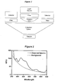
- Figure 2 shows the emission spectra of a bacterial solution ( Bacillus thuringiensis ) in a minimally fluorescing media when excited with light at 345nm.
- the solid line shows the observed emission spectra of the bacteria and the dashed line indicates the contribution of the Rayleigh scattering to this spectra.
- Subtraction of the Rayleigh background from the observed spectra results in the true emission spectra due to the metabolites excited by 345 nm light ( Figure 3 D).
- I is the intensity of the incident light; A is determined by the experimental conditions; the value for the constant C is typically determined by the characteristics of the instrument used to collect the data.
- the combined emission spectrum of the bacterial solution when excited with 325 nm, 345 nm and 570 nm shows minima near 515 nm and 850 nm.
- the measured fluorescence intensities at 515 nm and 850 nm are used to calculate the unknown values of A and C from the aforementioned equation, ultimately allowing for the subtraction of the background signal from the detected signal.
- Ralyleigh scattering background subtraction is particularly suited for liquid and air samples; other sample media exhibit different backgrounds and can be treated with the appropriate methods (e.g., Mie scattering, etc.).
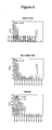
- Figure 3 shows the background-subtracted emission spectra of viable bacteria, non-viable bacteria and spore solutions ( Bacillus thuringiensis ) due to the various intrinsic fluorophores excited at 280 nm (A), 315 nm (B), 325 nm (C), 345 nm (D), 570 nm (E) and 660 nm (F).
- Figure 4 shows the obvious differences of the fluorescence signals (normalized to the emission at 440 nm after background subtraction) between said viable cell (A), non-viable cell (B) and the spore (C) solutions. The analysis method uses these differences between the viable cells, non-viable cells and spore solutions to distinguish between these in samples. The magnitudes of the detected and background-subtracted signals are used to quantitate the number of microbes in the sample.
- excitation filters at 325 nm, 345 nm and 570 nm would allow for the detection of and discrimination between live cells, dead cells and spores. These excitation filters would allow the excitation of reduced pyridine nucleotides, various flavins, calcium dipicolinate, hemoproteins and other components.
- the selection of filters for the emission detection of the excited fluorophores would include those at 405 nm, 440 nm, 480 nm and 650 nm; these filters correspond to maxima in the emission spectra of the excited flurophores. Additionally, other emission filters (545 nm and 850 nm) allow for the determination of the magnitude of the reflected/scattered background.
- a pulsed xenon lamp was used as the light source with interference excitation filters.
- a focusing optic is added to collimate the light before the interference filters.
- the focusing and collection optical pieces were constructed from polished reflective optics to eliminate any background fluorescence.
- the parabolic collection optics, which collected ca . 90% of the emitted signal, were fitted with interference emission filters, collimating optics and PMTs.
- the instrument functions, data collection, integration and analysis were controlled by a microcontroller.
- the detection method required the relative ratios of the detected and background-corrected signals to lie within certain physiological ranges. Analysis of greater than 500 samples from more than twenty different species of bacteria and spores showed that the numerous ratios could be used to ensure a statistically significant identification.
- Figure 5 shows the distribution for just one of these ratios (440 nm/480 nm ratio after background subtraction) of 22 species of bacteria, thus defining the physiological range required for the detection method. The method could also discriminate bacteria-containing solutions from sterile media and other biochemical buffers.
- 650/405, 405/440, 480/440, 650/440, 405/480 and 650/480 e.g., Neyman-Pearson test, fuzzy logic and a trained neural network (utilizing a multilayer perceptron), these gave a 99%, 95.6% and 100% probability of detection, respectively for the presence of bacteria.
- Neyman-Pearson test e.g., Neyman-Pearson test, fuzzy logic and a trained neural network (utilizing a multilayer perceptron)
- these gave a 99%, 95.6% and 100% probability of detection, respectively for the presence of bacteria.
- False alarm probabilities of the 500 data points taken for these detection algorithms were as follows: Neyman-Pearson (0.01 %), fuzzy logic (0 %), and neural net (0 %).
- Figure 6 shows data from one emission (RPN) of the instrument for viable and non-viable Salmonella typhi cells on the surface of a glass slide. The difference between viable and non-viable cells in the signal from this fluorescence is clear.
- Figure 7 shows the response of the RPN emission to Escherichia coli on the surface of turkey. A detection limit well below that observed for other microbial detection methods is observed in real-time, without the need for reagents or touching the meat surface.
- LEDs centered around 570 and 660 nm are used to excite the component(s) found in spores and dead cells shown in Figures 3D and 3F.
- Figure 8 shows the emission spectra of paper and various samples of envelopes when excited with light at 660 nm; the arrow in this figure shows the location of the emission expected from spores and non-viable cells at this excitation. As the paper and envelopes contain this spectroscopic window it is possible to detect bacterial endospores behind paper and inside envelopes.
Landscapes
- Health & Medical Sciences (AREA)
- Chemical & Material Sciences (AREA)
- Life Sciences & Earth Sciences (AREA)
- Engineering & Computer Science (AREA)
- Organic Chemistry (AREA)
- Zoology (AREA)
- Proteomics, Peptides & Aminoacids (AREA)
- Wood Science & Technology (AREA)
- Analytical Chemistry (AREA)
- Biochemistry (AREA)
- General Health & Medical Sciences (AREA)
- Molecular Biology (AREA)
- Immunology (AREA)
- Physics & Mathematics (AREA)
- Microbiology (AREA)
- Biomedical Technology (AREA)
- Toxicology (AREA)
- Biotechnology (AREA)
- Pathology (AREA)
- General Physics & Mathematics (AREA)
- Nuclear Medicine, Radiotherapy & Molecular Imaging (AREA)
- Biophysics (AREA)
- Bioinformatics & Cheminformatics (AREA)
- General Engineering & Computer Science (AREA)
- Genetics & Genomics (AREA)
- Investigating, Analyzing Materials By Fluorescence Or Luminescence (AREA)
- Measuring Or Testing Involving Enzymes Or Micro-Organisms (AREA)
- Investigating Or Analysing Biological Materials (AREA)
- Apparatus Associated With Microorganisms And Enzymes (AREA)
- Investigating Or Analysing Materials By The Use Of Chemical Reactions (AREA)
Abstract
Description
| Excitation and Emission Ranges for Microbial Fluorophores. | |||||||
| Excitation Range (nm) | Emission Range (nm) | Fluorophore | Viable Cells | Non-viable Cells | Spores | Viruses | Toxins |
| 260 - 285 | 340 - 360 | Nucleic Acids | X | X | X | X | |
| 265 - 280 | 340 - 360 | Tryptophan | X | X | X | X | X |
| 265 - 280 | 340 - 360 | Tyrosine | X | X | X | X | X |
| 270 - 280 | 380 - 400 | ATP | X | ||||
| 270 - 290 | 460 - 480 | Ca-Dipic | X | ||||
| 310 - 330 | 400 - 430 | Ca-Dipic | X | ||||
| 320 - 330 | 430 - 450 | RPN | X | ||||
| 340 - 365 | 430 - 450 | RPN | X | ||||
| 340 - 360 | 470 - 490 | RPN | X | ||||
| 430 - 450 | 520 - 535 | Flavins | X | ||||
| 470 - 485 | 560 - 580 | Flavins | X | ||||
| 560 - 585 | 615 - 680 | Porphyrins | X | X | |||
| 560 - 580 | 620 - 700 | Flavins | X | X | |||
| 610 - 650 | 750 - 800 | Unknown | X | X | |||
| 650 - 670 | 730 - 780 | Unknown | X | X | |||
| (In Table I, ATP is adenosine triphosphate and RPN refers to the reduced pyridine nucleotides.) |
Claims (27)
- A method for the detection of microbes comprising:a. exciting at least one intrinsic microbial fluorophore having a specific range of electromagnetic radiation wavelength above 200 nm; whereby said microbe containing intrinsic fluorophores is excited to emit fluorescence, and;b. detecting the fluorescence signals associated with the minima and maxima of the excited microbial fluorophores, and;c. subtracting the reflected/scattered excitation energies and background from the detected signals of the excited microbial fluoresence; whereby the enumeration of microbes is determined by the magnitude of the detected fluorescence.
- The method as set forth in Claim 1, wherein the relative ratios of multiple detected, background-corrected signals are determined; whereby the detection of the physiological state of the microbes depends upon the requirement that the ratios of the background-corrected fluorescence signals lie within specified physiological ranges and that the enumeration of the microbes is determined by the magnitude of said detected signals whose relative ratios lie within said expected ranges.
- A method as set forth in Claim 1, wherein said microbe fluorephores are selected from the group consisting of nucleic acid polymers, tryptophan-containing proteins, tyrosine-containing proteins, adenosine triphosphate, calcium dipicinolate, reduced pyridine nucleotides, flavins, porphyrin-containing proteins, and other components excited in the 610-670 nm region.
- The method of Claim 1 wherein the viable microbes to be detected include bacteria, fungi, protazoa, and rickettsiae; and the intrinsic microbial fluorophores used to detect the microbes include nucleic acid polymers, tyrosine-containing proteins, tryptophan-containing proteins, adenosine triphosphate, reduced pyridine nucleotides, flavins, porphyrin-containing proteins, and others excited in the 610-670 nm region.
- The method of Claim 1 wherein non-viable microbes to be detected include bacteria, fungi, protazoa, and rickettsiae; and the intrinsic microbial fluorophores used to detect the microbes include nucleic acid polymers, tryptophan-containing proteins, tyrosine-containing proteins, reduced pyridine nucleotides, flavins, porphyrin-containing proteins, and others excited in the 610-670 nm region.
- The method of Claim 1 wherein the microbes to be detected are bacterial endospores and the intrinsic fluorophores used to detect the endospores include nucleic acid polymers, tyrosine-containing proteins, tryptophan-containing proteins, calcium dipicolinic acid, and others excited in the 610-670 nm region.
- The method of claim 1 wherein the microbes to be detected include viruses, and the intrinsic fluorophores used to detect the viruses include nucleic acid polymers, tyrosine-containing proteins and tryptophan-containing proteins.
- A method for the detection of microbes comprising:a. excitation of multiple intrinsic microbial fluorophores with ultraviolet electromagnetic radiation having wavelengths between 200 and 300 nm, whereby any microbes present containing intrinsic fluorophores are excited to emit fluorescence, some of whose fluorescence is self-absorbed to excite other microbial fluorophores that in turn emit fluorescence;b. detecting the fluorescence signals associated with the minima and maxima of the excited microbial fluorophores;c. subtracting the fluorescence from the reflected/scattered excitation energies and background from the detected signals; andd. determining that the relative ratios of the detected, background-corrected signals lie within physiological ranges, whereby the enumeration of the microbes is determined by the magnitude of the detected signals whose relative ratios lie within physiological ranges.
- A method as set forth in Claim 8, wherein the intrinsic microbial fluorophores of said microbes include one or more of the group consisting of nucleic acid polymers, tryptophan-containing proteins, adenosine triphosphate and calcium dipicinolate compounds.
- A method as set forth in Claim 8, wherein secondary-excited microbial fluorophores include one or more of the group consisting of calcium dipicinolate, reduced pyridine nucleotides, flavins, porphyrin-containing proteins, cellular components excited in the 610-670 nm region, and the like.
- The method of Claim 8 wherein the viable microbes to be detected include bacteria, fungi, protazoa, and rickettsiae and the intrinsic microbial fluorophores used to detect the microbes are selected from the group consisting of nucleic acid polymers, tyrosine-containing proteins, tryptophan-containing proteins, adenosine triphosphate, reduced pyridine nucleotides, flavins, porphyrin-containing proteins, and cellular components excited in the 610-670 nm region.
- The method of Claim 8 wherein the non-viable microbes to be detected include bacteria, fungi, protazoa, and rickettsiae; and the intrinsic microbial fluorophores used to detect the microbes are selected from the group consisting of nucleic acid polymers, tyrosine containing proteins, tryptophan-containing proteins, reduced pyridine nucleotides, flavins, porphyrin-containing proteins, and cellular components excited in the 610-670 nm region.
- The method of Claim 8 wherein the microbes to be detected are bacterial spores and the intrinsic fluorophores used to detect the spores include nucleic acid polymers, tyrosine-containing proteins, tryptophan-containing proteins, calcium dipicolinic acid, and spore components excited in the 610-670 nm region.
- Apparatus for the detection of microbes on a non-living surface or in air or liquid comprising:a. means for directing electromagnetic radiation towards the sample, said means adapted to emit radiation energies capable of exciting at least one intrinsic microbial fluorophore;b. at least one detector for electromagnetic radiation capable of converting the emitted, or reflected/scattered radiation into electrical signals, said detector adapted to detect electromagnetic radiation at wavelengths above 320 nm to detect the minima and maxima associated with the fluorescence emission of said microbial fluorophores; andc. means for analyzing the electrical signals corresponding to the fluorescence of the intrinsic microbial fluorophores, and the reflected/scattered excitation energies to determine the presence of microbes.
- The method as defined in claim 1 wherein the electromagnetic waves are directed towards the microbes in time-modulated pulses.
- The apparatus defined in Claim 14 wherein the means for directing electromagnetic radiation includes means for time-modulating the electromagnetic radiation.
- A method for the detection of microbial proteinaceous toxins comprising:a. exciting at least one intrinsic microbial fluorophore having a specific range of electromagnetic radiation wavelength above 200 nm; whereby said microbe present containing intrinsic fluorophores is excited to emit fluorescence, and;b. detecting the fluorescence signals associated with the minima and maxima of the excited microbial fluorophores, and;c. subtracting the reflected/scattered excitation energies and background from the detected signals of the excited microbial fluoresence; whereby enumeration of the microbial toxin is determined by the magnitude of said detected background-corrected fluorescence.
- A method as set forth in Claim 17, wherein said microbe fluorephores are selected from the group consisting of tryptophan-containing proteins and tyrosine-containing proteins.
- A method for the detection of non-viable bacteria and spores comprising:a. exciting at least one intrinsic microbial fluorophore having a specific range of electromagnetic radiation having wavelengths between 550 and 700 nm; whereby said microbe present containing intrinsic fluorophores is excited to emit fluorescence, and;b. detecting the fluorescence signals associated with the minima and maxima of the excited microbial fluorophores, and;c. subtracting the reflected/scattered excitation energies and background from the detected signals of the excited microbial fluoresence; whereby the enumeration of non-viable bacteria and spores is determined by the magnitude of the detected fluorescence.
- The method as set forth in Claim 19, wherein the relative ratios of multiple detected, background-corrected signals are determined; whereby the distinction between spores and non-viable bacteria depends upon the requirement that the ratios of the background-corrected fluorescence signals lie within specified physiological ranges and that the enumeration of spores is determined by the magnitude of said detected signals whose relative ratios lie within said expected ranges.
- A method of Claim 19 wherein the intrinsic microbial fluorophores used to detect the non-viable bacteria and bacterial spores are selected from a group including flavins, porphyrin-containing proteins, other components excited in the 610-670 nm region.
- The method of Claim 19 wherein the non-viable bacteria and bacterial spores are detected on surfaces, inside paper envelopes, through paper, in solution and in aerosols.
- A method for the detection of spores and non-viable bacteria comprising:a. excitation of multiple intrinsic microbial fluorophores with electromagnetic radiation having wavelengths between 550 and 640 nm, whereby any spores and non-viable bacteria present containing intrinsic fluorophores are excited to emit fluorescence, some of whose fluorescence is self-absorbed to excite other spore fluorophores that in turn emit fluorescence;b. detecting the fluorescence signals associated with the minima and maxima of the excited microbial fluorophores;c. subtracting the fluorescence from the reflected/scattered excitation energies and background from the detected signals; andd. determining that the relative ratios of the detected, background-corrected signals lie within physiological ranges, whereby the enumeration of the non-viable bacteria and bacterial spores is determined by the magnitude of the detected signals whose relative ratios lie within physiological ranges.
- A method as set forth in Claim 23, wherein the intrinsic microbial fluorophores of said microbes include one or more of the group consisting of flavins, porphyrin-containing proteins, and other components excited in the 610-670 nm region.
- A method as set forth in Claim 23, wherein secondary-excited microbial fluorophores include one or more of the group consisting of intrinsic components excited in the 610-680 nm region, and the like.
- The method of Claim 23 wherein the non-viable bacteria and spores are detected on surfaces, inside paper envelopes, through paper, in solution and in aerosols.
- A method as set forth in Claim 23, wherein the spores and non-viable bacteria are detected inside paper envelopes and the secondary-excited microbial fluorophores are excited by emissions from the excited paper products.
Applications Claiming Priority (2)
| Application Number | Priority Date | Filing Date | Title |
|---|---|---|---|
| US10/054,419 US6750006B2 (en) | 2002-01-22 | 2002-01-22 | Method for detecting the presence of microbes and determining their physiological status |
| US54419 | 2002-01-22 |
Publications (2)
| Publication Number | Publication Date |
|---|---|
| EP1329514A2 true EP1329514A2 (en) | 2003-07-23 |
| EP1329514A3 EP1329514A3 (en) | 2004-02-18 |
Family
ID=21990934
Family Applications (1)
| Application Number | Title | Priority Date | Filing Date |
|---|---|---|---|
| EP02021592A Ceased EP1329514A3 (en) | 2002-01-22 | 2002-09-27 | Method and apparatus for detecting the presence of microbes and determining their physiological status |
Country Status (6)
| Country | Link |
|---|---|
| US (2) | US6750006B2 (en) |
| EP (1) | EP1329514A3 (en) |
| JP (1) | JP4033754B2 (en) |
| CN (1) | CN100489501C (en) |
| CA (1) | CA2402575C (en) |
| SG (1) | SG107620A1 (en) |
Cited By (7)
| Publication number | Priority date | Publication date | Assignee | Title |
|---|---|---|---|---|
| SG137684A1 (en) * | 2004-04-02 | 2007-12-28 | Microbiosystems Ltd Partnershi | Method for detecting the presence of dormant cryptobiotic microorganisms |
| WO2008147227A1 (en) * | 2007-05-31 | 2008-12-04 | Veritide Limited | Method for spore detection |
| WO2010062472A1 (en) * | 2008-11-03 | 2010-06-03 | General Electric Company | Methods and systems for measuring microbiological content in aqueous media |
| WO2014180568A1 (en) | 2013-05-08 | 2014-11-13 | Freshdetect Gmbh | Measuring device for measuring a surface coating on an object to be measured, particularly on a foodstuff |
| CZ308044B6 (en) * | 2019-07-09 | 2019-11-13 | Botanický ústav Akademie věd ČR, v.v.i. | Mobile device for non-destructive fluorescence resolution, imaging and quantification of microorganisms on the surface of materials |
| CN112098384A (en) * | 2020-09-22 | 2020-12-18 | 华东交通大学 | A simple method to quickly predict whether water quality is biologically stable |
| WO2026022094A1 (en) | 2024-07-24 | 2026-01-29 | A.P.F. Aqua System Ag | Method for sanitisation, disinfection or sterilisation |
Families Citing this family (62)
| Publication number | Priority date | Publication date | Assignee | Title |
|---|---|---|---|---|
| US7211377B1 (en) * | 2002-01-22 | 2007-05-01 | Microbiosystems, Limited Partnership | Method for detecting the presence of dormant cryptobiotic microorganisms |
| US7186990B2 (en) * | 2002-01-22 | 2007-03-06 | Microbiosystems, Limited Partnership | Method and apparatus for detecting and imaging the presence of biological materials |
| US7330795B2 (en) * | 2002-05-07 | 2008-02-12 | California Institute Of Technology | Method and apparatus for providing signal analysis of a BioNEMS resonator or transducer |
| DE10303404A1 (en) * | 2003-01-27 | 2004-08-05 | Carl Zeiss Jena Gmbh | Method for the detection of fluorescent light |
| JP2005102644A (en) * | 2003-10-01 | 2005-04-21 | Sysmex Corp | Determination of proliferation stage of microorganism and program for determining proliferation stage of microorganism |
| US7824883B2 (en) * | 2003-12-31 | 2010-11-02 | Powers Linda S | Method and apparatus for detecting the presence of microbes with frequency modulated multi-wavelength intrinsic fluorescence |
| US20070037135A1 (en) * | 2005-08-08 | 2007-02-15 | Barnes Russell H | System and method for the identification and quantification of a biological sample suspended in a liquid |
| US20070111225A1 (en) * | 2005-08-10 | 2007-05-17 | California Institute Of Technology | System and method for monitoring an analyte |
| US7781224B2 (en) * | 2005-08-10 | 2010-08-24 | Lawrence Livermore National Security, Llc | Safe biodegradable fluorescent particles |
| JP4435299B2 (en) * | 2005-09-06 | 2010-03-17 | 株式会社堀場製作所 | Carbon nanotube classification apparatus, computer program, and carbon nanotube classification method |
| US7525102B1 (en) * | 2005-10-03 | 2009-04-28 | Sparta, Inc. | Agent detection in the presence of background clutter |
| US20110059462A1 (en) * | 2006-02-21 | 2011-03-10 | University Of South Florida | Automated particulate concentration system |
| DE102006041347B3 (en) * | 2006-09-01 | 2008-02-28 | Rwo Gmbh | Detecting living phytoplankton cells and/or microorganisms in water, to monitor water quality, by measurement of variable fluorescence in measuring chamber and comparison with reference species |
| RU2362145C2 (en) * | 2007-04-05 | 2009-07-20 | Общество с ограниченной ответственностью "Научно-производственный центр медицинских и промышленных биотехнологий Спектролюкс" | Differential diagnosis method for microbes and amino acids |
| US20080281581A1 (en) * | 2007-05-07 | 2008-11-13 | Sparta, Inc. | Method of identifying documents with similar properties utilizing principal component analysis |
| US8014959B2 (en) * | 2007-05-07 | 2011-09-06 | Sparta, Inc. | Population of background suppression lists from limited data in agent detection systems |
| WO2009036105A2 (en) * | 2007-09-11 | 2009-03-19 | Baxter International Inc | Infusion therapy sensor system |
| US9103796B2 (en) * | 2007-12-14 | 2015-08-11 | Kimberly-Clark Worldwide, Inc. | Multi-layered devices for analyte detection |
| JP2009236831A (en) * | 2008-03-28 | 2009-10-15 | Jfe Engineering Corp | Monitoring method and device for dissolved pollutant |
| JP5459209B2 (en) | 2008-07-11 | 2014-04-02 | 星光Pmc株式会社 | Method for judging physiological state of microbial community and wastewater treatment method |
| MX2011005988A (en) * | 2008-12-16 | 2011-09-01 | Bio Merieux Inc | Methods for the characterization of microorganisms on solid or semi-solid media. |
| WO2010080643A1 (en) | 2008-12-18 | 2010-07-15 | Biovigilant Systems, Inc. | Integrated microbial collector |
| JP5667079B2 (en) * | 2008-12-18 | 2015-02-12 | アズビル株式会社 | Compact detector for simultaneous detection of particle size and fluorescence |
| CN102158552A (en) * | 2011-03-30 | 2011-08-17 | 中国科学院上海微系统与信息技术研究所 | Wireless monitoring network for remains of bacteria, heavy metal, pesticide and veterinary drug in food |
| KR20140016923A (en) | 2011-04-06 | 2014-02-10 | 인스턴트 바이오스캔, 엘엘씨 | Microbial detection apparatus and method |
| FR2986237A1 (en) * | 2012-01-27 | 2013-08-02 | Advencis | DEVICE FOR RAPID DETECTION OF MICROORGANISMS |
| CN102590168A (en) * | 2012-02-16 | 2012-07-18 | 无锡迈通科学仪器有限公司 | Light emitting diode-based fluorescent microorganism detector |
| US9052315B2 (en) | 2012-05-09 | 2015-06-09 | Advanced Animal Diagnostics, Inc. | Rapid detection of analytes in liquid samples |
| US10359614B2 (en) | 2012-07-03 | 2019-07-23 | Advanced Animal Diagnostics, Inc. | Diagnostic apparatus |
| TWI464388B (en) * | 2012-08-17 | 2014-12-11 | Wistron Corp | Light calibration device, biological detection calibration system and operating method thereof |
| FR3000209B1 (en) * | 2012-12-26 | 2017-02-24 | Biomerieux Sa | METHOD AND SYSTEM FOR DETECTING AND MEASURING FLUORESCENCE SIGNALS |
| EP4513175A3 (en) * | 2013-02-22 | 2025-05-07 | Life Technologies Corporation | Optical systems and methods for biological analysis |
| US9797893B2 (en) | 2013-05-09 | 2017-10-24 | Advanced Animal Diagnostics, Inc. | Rapid detection of analytes in liquid samples |
| CN103810826A (en) * | 2014-01-07 | 2014-05-21 | 上海朗赋实业有限公司 | Wireless ATP (Adenosine Triphosphate) detector |
| CN103792210B (en) * | 2014-02-28 | 2014-12-03 | 陕西师范大学 | Device for monitoring liquid physical parameters in pipeline in real time in non-contact mode by utilizing glass vision tube |
| CN103954547A (en) * | 2014-05-07 | 2014-07-30 | 江南大学 | Method for fast detecting survival rate of asexual spores of antrodia camphorata |
| JP6425918B2 (en) * | 2014-05-27 | 2018-11-21 | アズビル株式会社 | Monitoring system of purified water production apparatus and monitoring method of purified water production apparatus |
| US10316347B2 (en) | 2014-06-26 | 2019-06-11 | Ecolab Usa Inc. | Endospore detection using hydrophobic collection material |
| US9880159B2 (en) | 2014-10-01 | 2018-01-30 | Linda S. Powers | Cartridge-based detection system |
| JP6274534B2 (en) * | 2014-12-02 | 2018-02-07 | 国立大学法人信州大学 | Microorganism detection method and detection apparatus |
| US9867250B1 (en) | 2015-04-20 | 2018-01-09 | The Arizona Board Of Regents On Behalf Of The University Of Arizona | System having a configurable pico-second pulsed LED driver circuit and photomultiplier tube biasing and gating circuits for real-time, time-resolved transient recording of fluorescence |
| MX388753B (en) | 2015-05-08 | 2025-03-20 | Spectral Platforms Inc | NON-COVALENT COMPLEXES BASED ON ALBUMIN AND METHODS OF USING THE SAME. |
| WO2017029980A1 (en) * | 2015-08-18 | 2017-02-23 | 荏原実業株式会社 | Fluorescence visualization device, fluorescence visualization method, and computer program |
| WO2017182519A1 (en) * | 2016-04-19 | 2017-10-26 | Gea Food Solutions Germany Gmbh | Determination of a surface covering during food preparation and/or food packaging |
| CN106770079A (en) * | 2016-11-17 | 2017-05-31 | 无锡艾科瑞思产品设计与研究有限公司 | A kind of fluorescent microorganism detector based on light emitting diode |
| WO2019044410A1 (en) * | 2017-08-29 | 2019-03-07 | パナソニックIpマネジメント株式会社 | Light observation device |
| JP7026337B2 (en) * | 2017-08-29 | 2022-02-28 | パナソニックIpマネジメント株式会社 | Optical observation device |
| EP4209581A1 (en) * | 2018-06-06 | 2023-07-12 | AMVAC Hong Kong Limited | Microbial consortia producing dipicolinic acid and methods for selecting microbes for co-formulation with carriers |
| JP7104563B2 (en) * | 2018-06-08 | 2022-07-21 | シャープ株式会社 | Animal identification device, control method and program of animal identification device |
| JP7337351B2 (en) * | 2018-08-10 | 2023-09-04 | 日本たばこ産業株式会社 | Methods, programs, and apparatus for creating criteria for classifying plant-derived materials containing chlorophyll and at least one of polyphenols and terpenoids |
| US11455298B2 (en) | 2019-02-06 | 2022-09-27 | Parsons Corporation | Goal-directed semantic search |
| FR3103900B1 (en) * | 2019-11-29 | 2024-07-19 | Univ Du Mans | Method for rapid identification of microorganisms by analysis of excitation-emission matrices |
| EP4168732A4 (en) * | 2020-06-23 | 2025-02-12 | Spectral Platforms, Inc. | SPECTROSCOPIC METHODS, REAGENTS AND SYSTEMS FOR DETECTION, IDENTIFICATION AND CHARACTERIZATION OF BACTERIA EXHIBITING ANTIMICROBIAL SENSITIVITY |
| WO2022146052A1 (en) * | 2020-12-30 | 2022-07-07 | Seegene, Inc. | Optical spectrometry-based method and device for detecting target analyte in sample |
| CN112725237A (en) * | 2021-01-27 | 2021-04-30 | 川北医学院附属医院 | Intestinal bacteria culture observation method |
| IL291606A (en) * | 2021-03-22 | 2022-10-01 | Kando Env Services Ltd | Non-contact monitoring of fluid characteristics in wastewater transport system |
| CN218685344U (en) * | 2021-05-24 | 2023-03-24 | 广州市鸿利秉一光电科技有限公司 | A sterilization device with visualized sterilization effect |
| US20250186647A1 (en) * | 2022-04-11 | 2025-06-12 | Epirus, Inc. | Systems and methods for neutralizing matter |
| CN115261213A (en) * | 2022-08-17 | 2022-11-01 | 山东师范大学 | Portable rapid monitoring device for airborne microorganisms based on the principle of automatic transmission and feedback |
| CN117030664A (en) * | 2023-02-10 | 2023-11-10 | 上海凌泽信息科技有限公司 | An 850nm laser microbial virus detection method and system device |
| CN115927766B (en) * | 2023-02-23 | 2023-07-18 | 中国人民解放军国防科技大学 | A culture condition control method and system for improving the optical attenuation performance of microorganisms |
| CN120468031B (en) * | 2025-05-29 | 2026-01-06 | 武汉格林环源净化工程有限公司 | A device and method for detecting the concentration of microbial communities in sewage sludge |
Family Cites Families (19)
| Publication number | Priority date | Publication date | Assignee | Title |
|---|---|---|---|---|
| US3566114A (en) * | 1968-04-25 | 1971-02-23 | Aubrey K Brewer | Method and means for detection of microorganisms in the atmosphere |
| US3850525A (en) | 1973-07-09 | 1974-11-26 | Beckman Instruments Inc | Simultaneous multiple measurements in laser photometers |
| US4745285A (en) | 1986-08-21 | 1988-05-17 | Becton Dickinson And Company | Multi-color fluorescence analysis with single wavelength excitation |
| US4900934A (en) | 1987-07-15 | 1990-02-13 | University Of Utah | Apparatus for simultaneous visualization and measurement of fluorescence from fluorescent dye-treated cell preparations and solutions |
| US5208651A (en) | 1991-07-16 | 1993-05-04 | The Regents Of The University Of California | Apparatus and method for measuring fluorescence intensities at a plurality of wavelengths and lifetimes |
| GB9301118D0 (en) * | 1993-01-21 | 1993-03-10 | Secr Defence | Enzyme linked assays |
| US5418371A (en) | 1993-02-01 | 1995-05-23 | Aslund; Nils R. D. | Apparatus for quantitative imaging of multiple fluorophores using dual detectors |
| US5294799A (en) | 1993-02-01 | 1994-03-15 | Aslund Nils R D | Apparatus for quantitative imaging of multiple fluorophores |
| US5424959A (en) | 1993-07-19 | 1995-06-13 | Texaco Inc. | Interpretation of fluorescence fingerprints of crude oils and other hydrocarbon mixtures using neural networks |
| US5474910A (en) | 1993-10-15 | 1995-12-12 | Alfano; Robert R. | Method and device for detecting biological molecules and/or microorganisms within a desired area or space |
| US5491343A (en) | 1994-03-25 | 1996-02-13 | Brooker; Gary | High-speed multiple wavelength illumination source, apparatus containing the same, and applications thereof to methods of irradiating luminescent samples and of quantitative luminescence ratio microscopy |
| GB9414096D0 (en) * | 1994-07-13 | 1994-08-31 | Secr Defence | Labelled capture assay |
| US5701012A (en) | 1996-03-19 | 1997-12-23 | Her Majesty The Queen In Right Of Canada, As Represented By The Minister Of National Defence | Fluorescent biological particle detection system |
| US5760406A (en) | 1996-06-03 | 1998-06-02 | Powers; Linda | Method and apparatus for sensing the presence of microbes |
| US5866430A (en) | 1996-06-13 | 1999-02-02 | Grow; Ann E. | Raman optrode processes and devices for detection of chemicals and microorganisms |
| AU7164998A (en) * | 1997-04-28 | 1998-11-24 | B-E Safe, Inc. | Taxonomic identification of microorganisms, proteins and peptides involved in vertebrate disease states |
| AU8148898A (en) | 1997-06-23 | 1999-01-04 | Luminex Corporation | Interlaced lasers for multiple fluorescence measurement |
| US6124597A (en) | 1997-07-07 | 2000-09-26 | Cedars-Sinai Medical Center | Method and devices for laser induced fluorescence attenuation spectroscopy |
| US5968766A (en) | 1998-03-31 | 1999-10-19 | B.E. Safe | Method and apparatus for sensing the presence of microbes |
-
2002
- 2002-01-22 US US10/054,419 patent/US6750006B2/en not_active Expired - Fee Related
- 2002-09-10 CA CA2402575A patent/CA2402575C/en not_active Expired - Fee Related
- 2002-09-27 EP EP02021592A patent/EP1329514A3/en not_active Ceased
- 2002-10-25 JP JP2002310523A patent/JP4033754B2/en not_active Expired - Fee Related
- 2002-10-31 CN CNB021479763A patent/CN100489501C/en not_active Expired - Fee Related
- 2002-11-01 SG SG200206612A patent/SG107620A1/en unknown
-
2003
- 2003-12-31 US US10/749,329 patent/US20040171137A1/en not_active Abandoned
Cited By (14)
| Publication number | Priority date | Publication date | Assignee | Title |
|---|---|---|---|---|
| SG137684A1 (en) * | 2004-04-02 | 2007-12-28 | Microbiosystems Ltd Partnershi | Method for detecting the presence of dormant cryptobiotic microorganisms |
| WO2008147227A1 (en) * | 2007-05-31 | 2008-12-04 | Veritide Limited | Method for spore detection |
| US8711354B2 (en) * | 2007-05-31 | 2014-04-29 | Veritide Limited | Method for spore detection |
| WO2010062472A1 (en) * | 2008-11-03 | 2010-06-03 | General Electric Company | Methods and systems for measuring microbiological content in aqueous media |
| US8481302B2 (en) | 2008-11-03 | 2013-07-09 | General Electric Company | Total bacteria monitoring system |
| DE102013008003A1 (en) * | 2013-05-08 | 2014-11-13 | Freshdetect Gmbh | Measuring device for measuring a surface covering on a measuring object, in particular on a foodstuff |
| WO2014180568A1 (en) | 2013-05-08 | 2014-11-13 | Freshdetect Gmbh | Measuring device for measuring a surface coating on an object to be measured, particularly on a foodstuff |
| DE102013008003B4 (en) * | 2013-05-08 | 2015-03-19 | Freshdetect Gmbh | Measuring device for measuring a surface covering on a measuring object, in particular on a food, and its use |
| DE202014010777U1 (en) | 2013-05-08 | 2016-08-12 | Freshdetect Gmbh | Production line for processing food |
| EP3229014A2 (en) | 2013-05-08 | 2017-10-11 | FreshDetect GmbH | Measuring device for measuring a surface lining on a measurement object, specifically on a foodstuff |
| CZ308044B6 (en) * | 2019-07-09 | 2019-11-13 | Botanický ústav Akademie věd ČR, v.v.i. | Mobile device for non-destructive fluorescence resolution, imaging and quantification of microorganisms on the surface of materials |
| CN112098384A (en) * | 2020-09-22 | 2020-12-18 | 华东交通大学 | A simple method to quickly predict whether water quality is biologically stable |
| CN112098384B (en) * | 2020-09-22 | 2023-09-01 | 华东交通大学 | A Simple Method to Rapidly Predict Whether Water Quality Is Biostable |
| WO2026022094A1 (en) | 2024-07-24 | 2026-01-29 | A.P.F. Aqua System Ag | Method for sanitisation, disinfection or sterilisation |
Also Published As
| Publication number | Publication date |
|---|---|
| HK1056912A1 (en) | 2004-03-05 |
| US6750006B2 (en) | 2004-06-15 |
| CN1434285A (en) | 2003-08-06 |
| SG107620A1 (en) | 2004-12-29 |
| CA2402575A1 (en) | 2003-07-22 |
| CN100489501C (en) | 2009-05-20 |
| JP4033754B2 (en) | 2008-01-16 |
| US20030138875A1 (en) | 2003-07-24 |
| JP2003210193A (en) | 2003-07-29 |
| EP1329514A3 (en) | 2004-02-18 |
| CA2402575C (en) | 2011-11-08 |
| US20040171137A1 (en) | 2004-09-02 |
Similar Documents
| Publication | Publication Date | Title |
|---|---|---|
| US6750006B2 (en) | Method for detecting the presence of microbes and determining their physiological status | |
| US7186990B2 (en) | Method and apparatus for detecting and imaging the presence of biological materials | |
| Estes et al. | Reagentless detection of microorganisms by intrinsic fluorescence | |
| JP5588977B2 (en) | Methods and systems for detection and / or characterization of biological particles in a sample | |
| US7582415B2 (en) | Rapid detection of replicating cells | |
| US7824883B2 (en) | Method and apparatus for detecting the presence of microbes with frequency modulated multi-wavelength intrinsic fluorescence | |
| CN101464409B (en) | Apparatus and method for fast quantitative bacteria detection | |
| US10184144B2 (en) | Identification and/or characterization of a microbial agent using taxonomic hierarchical classification | |
| US7211377B1 (en) | Method for detecting the presence of dormant cryptobiotic microorganisms | |
| Simões et al. | An ultrasensitive fluorimetric sensor for pre-screening of water microbial contamination risk | |
| JP5799086B2 (en) | Identification and / or characterization of microbial factors using taxonomic hierarchy classification | |
| JP3740019B2 (en) | Microorganism weighing device | |
| JP4487985B2 (en) | Microorganism weighing device | |
| JP4967280B2 (en) | Microbe counting device | |
| JP2735901B2 (en) | Method for measuring the number of living cells, dead cells, and particles other than microorganism cells of microorganisms | |
| HK1056912B (en) | Method and apparatus for detecting the presence of microbes and determining their physiological status | |
| EP1582859A1 (en) | Method for detecting the presence of dormant cryptobiotic microorganisms | |
| Lloyd | Powers et al. | |
| Sikora et al. | Mobile device for detection of biological threat | |
| CN121049212A (en) | Methods for selecting detection conditions for bacterial detection devices | |
| CN113008767A (en) | Static cell analysis device and method | |
| TH106506A (en) | Methods for detecting analyzes in microscopic sequences of samples and devices for performing such methods. |
Legal Events
| Date | Code | Title | Description |
|---|---|---|---|
| PUAI | Public reference made under article 153(3) epc to a published international application that has entered the european phase |
Free format text: ORIGINAL CODE: 0009012 |
|
| AK | Designated contracting states |
Designated state(s): AT BE BG CH CY CZ DE DK EE ES FI FR GB GR IE IT LI LU MC NL PT SE SK TR |
|
| AX | Request for extension of the european patent |
Extension state: AL LT LV MK RO SI |
|
| PUAL | Search report despatched |
Free format text: ORIGINAL CODE: 0009013 |
|
| AK | Designated contracting states |
Kind code of ref document: A3 Designated state(s): AT BE BG CH CY CZ DE DK EE ES FI FR GB GR IE IT LI LU MC NL PT SE SK TR |
|
| AX | Request for extension of the european patent |
Extension state: AL LT LV MK RO SI |
|
| RIC1 | Information provided on ipc code assigned before grant |
Ipc: 7G 01N 33/483 B Ipc: 7C 12M 1/34 B Ipc: 7G 01N 21/64 A |
|
| 17P | Request for examination filed |
Effective date: 20040405 |
|
| 17Q | First examination report despatched |
Effective date: 20040528 |
|
| AKX | Designation fees paid |
Designated state(s): AT BE BG CH CY CZ DE DK EE ES FI FR GB GR IE IT LI LU MC NL PT SE SK TR |
|
| 17Q | First examination report despatched |
Effective date: 20040528 |
|
| STAA | Information on the status of an ep patent application or granted ep patent |
Free format text: STATUS: THE APPLICATION HAS BEEN REFUSED |
|
| 18R | Application refused |
Effective date: 20130325 |