EP1481052B1 - Hyaluronansynthase-gene und expression davon - Google Patents
Hyaluronansynthase-gene und expression davon Download PDFInfo
- Publication number
- EP1481052B1 EP1481052B1 EP02804804A EP02804804A EP1481052B1 EP 1481052 B1 EP1481052 B1 EP 1481052B1 EP 02804804 A EP02804804 A EP 02804804A EP 02804804 A EP02804804 A EP 02804804A EP 1481052 B1 EP1481052 B1 EP 1481052B1
- Authority
- EP
- European Patent Office
- Prior art keywords
- host cell
- recombinant host
- udp
- recombinant
- nucleic acid
- Prior art date
- Legal status (The legal status is an assumption and is not a legal conclusion. Google has not performed a legal analysis and makes no representation as to the accuracy of the status listed.)
- Expired - Lifetime
Links
Images


Classifications
-
- C—CHEMISTRY; METALLURGY
- C12—BIOCHEMISTRY; BEER; SPIRITS; WINE; VINEGAR; MICROBIOLOGY; ENZYMOLOGY; MUTATION OR GENETIC ENGINEERING
- C12N—MICROORGANISMS OR ENZYMES; COMPOSITIONS THEREOF; PROPAGATING, PRESERVING, OR MAINTAINING MICROORGANISMS; MUTATION OR GENETIC ENGINEERING; CULTURE MEDIA
- C12N9/00—Enzymes; Proenzymes; Compositions thereof; Processes for preparing, activating, inhibiting, separating or purifying enzymes
- C12N9/10—Transferases (2.)
- C12N9/1048—Glycosyltransferases (2.4)
- C12N9/1051—Hexosyltransferases (2.4.1)
Definitions
- the present invention relates to a nucleic acid segment having a coding region encoding enzymatically active hyaluronate synthase (HAS), and to the use of this nucleic acid segment in the preparation of recombinant cells which produce hyaluronate synthase and its hyaluronic acid product.
- HAS enzymatically active hyaluronate synthase
- Hyaluronate is also known as hyaluronic acid or hyaluronan.
- streptococcal infections is a major health and economic problem worldwide, particularly in developing countries.
- One reason for this is due to the ability of Streptococcal bacteria to grow undetected by the body's phagocytic cells, i.e., macrophages and polymorphonuclear cells (PMNs). These cells are responsible for recognizing and engulfing foreign microorganisms.
- phagocytic cells i.e., macrophages and polymorphonuclear cells (PMNs).
- PMNs polymorphonuclear cells
- HA hyaluronic acid
- the structure of HA is identical in both prokaryotes and eukaryotes.
- HA Since HA is generally nonimmunogenic, the encapsulated bacteria do not elicit an immune response and are therefore not targeted for destruction. Moreover, the capsule exerts an antiphagocytic effect on PMNs in vitro and prevents attachment of Streptococcus to macrophages. Precisely because of this, in Group A and Group C Streptococci, the HA capsules are major virulence factors in natural and experimental infections. Group A Streptococcus are responsible for numerous human diseases including pharyngitis, impetigo, deep tissue infections, rheumatic fever and a toxic shock-like syndrome. The Group C Streptococcus equisimilis is responsible for osteomyelitis, pharyngitis, brain abscesses, and pneumonia.
- HA is a high molecular weight linear polysaccharide of repeating disaccharide units consisting of N-acetylglucosamine (GlcNAc) and glucuronic acid (GlcUA).
- the number of repeating disaccharides in an HA molecule can exceed 30,000, a M r >10 7 .
- HA is the only glycosaminogylcan synthesized by both mammalian and bacterial cells, particularly Groups A and C Streptococci and Type A Pasteurella multocida. These strains make HA which is secreted into the medium as well as HA capsules.
- HA is synthesized by mammalian and bacterial cells by the enzyme hyaluronate synthase which has been localized to the plasma membrane. It is believed that the synthesis of HA in these organisms is a multi-step process. Initiation involves binding of an initial precursor, UDP-GlcNAc or UDP-GlcUA. This is followed by elongation which involves alternate addition of the two sugars to the growing oligosaccharide chain. The growing polymer is extruded across the plasma membrane region of the cell and into the extracellular space.
- the HA biosynthetic system was one of the first membrane heteropolysaccharide synthetic pathways studied, the mechanism of HA synthesis is still not well understood. This may be because in vitro systems developed to date are inadequate in that de novo biosynthesis of HA has not been accomplished.
- HA has been identified in virtually every tissue in vertebrates and has achieved widespread use in various clinical applications, most notably and appropriately as an intra-articular matrix supplement and in eye surgery.
- the scientific literature has also shown a transition from the original perception that HA is primarily a passive structural component in the matrix of a few connective tissues and in the capsule of certain strains of bacteria to a recognition that this ubiquitous macromolecule is dynamically involved in many biological processes: from modulating cell migration and differentiation during embryogenesis to regulation of extracellular matrix organization and metabolism to important roles in the complex processes of metastasis, wound healing, and inflammation.
- HA is highly metabolically active and that cells focus much attention on the processes of its synthesis and catabolism.
- the half-life of HA in tissues ranges from 1 to 3 weeks in cartilage to ⁇ 1 day in epidermis.
- HA is also used in numerous technical applications (e.g., lubricating compounds), cosmetics and neutraceuticals.
- HAS HA synthase
- Markovitz et al. successfully characterized the HAS activity from Streptococcus pyogenes and discovered the enzymes's membrane localization and its requirements for sugar nucleotide precursors and Mg 2+ .
- Prehm found that elongating HA, made by B6 cells, was digested by hyaluronidase added to the medium and proposed that HAS resides at the plasma membrane.
- Philipson and Schwartz also showed that HAS activity cofractionated with plasma membrane markers in mouse oligodendroglioma cells.
- HAS assembles high M r HA that is simultaneously extruded through the membrane into the extracellular space (or to make the cell capsule in the case of bacteria) as glycosaminoglycan synthesis proceeds.
- This mode of biosynthesis is unique among macromolecules since nucleic acids, proteins, and lipids are synthesized in the nucleus, endoplasmic reticulum/Golgi, cytoplasm, or mitochondria.
- the extrusion of the growing chain into the extracellular space also allows for unconstrained polymer growth, thereby achieving the exceptionally large size of HA, whereas confinement of synthesis within a Golgi or post-Golgi compartment limits the overall amount or length of the polymers formed.
- High concentrations of HA within a confined lumen may also create a high viscosity environment that might be deleterious for other organelle functions.
- HasB is a UDP-glucose dehydrogenase, which is required to convert UDP-glucose to UDP-GlcUA, one of the substrates for HA synthesis.
- HasC is a UDP-glucose pyrophosphorylase, which is required to convert glucose 1-phosphate and UTP to UDP-glucose.
- the elusive HA synthase gene was finally cloned by a transposon mutagenesis approach, in which an acapsular mutant Group A strain was created containing a transposon interruption of the HA synthesis operon.
- Known sequences of the transposon allowed the region of the junction with streptococcal DNA to be identified and then cloned from wild-type cells.
- the encoded spHAS was 5-10% identical to a family of yeast chitin synthases and 30% identical to the Xenopus laevis protein DG42 (developmentally expressed during gastrulation), whose function was unknown at the time.
- spHAS was a dual action single enzyme and (ii) the areas of sequence homology between the spHAS, chitin synthase, and DG42, the almost simultaneous identification of eukaryotic HAS cDNAs in 1996 by four laboratories, further strengthened the inventor's protein hypothesis that HAS is a multigene family encoding distinct isozymes. Two genes ( HAS1 and HAS2 ) were quickly discovered in mammals, and a third gene HAS3 was later discovered. A second streptococcal seHAS or Streptococcus equisimilis hyaluronate synthase, was identified and is disclosed in detail in U.S. Serial No.
- the seHAS protein has a high level of identity (approximately 70 percent) to the spHAS enzyme. This identity, however, is interesting because the seHAS gene does not cross-hybridize to the spHAS gene.
- mmHAS1 is 30% identical to spHAS and 55% identical to DG42.
- HAS1 cDNA isolated from a fetal brain library.
- the hsHAS1 cDNAs reported by the two groups, however, differ in length; they encode a 578 or a 543 amino acid protein. HAS activity has only been demonstrated for the longer form.
- Spicer et al. used a degenerate RT-PCR approach to clone a mouse embryo cDNA encoding a second distinct enzyme, which is designated mmHAS2. Transfection of mmHAS2 cDNA into COS cells directed de novo production of an HA cell coat detected by a particle exclusion assay, thereby providing strong evidence that the HAS2 protein can synthesize HA.
- Watanabe and Yamaguchi screened a human fetal brain cDNA library to identify hsHAS2.
- Fulop et al independently used a similar strategy to identify mmHAS2 in RNA isolated from ovarian cumulus cells actively synthesizing HA, a critical process for normal cumulus oophorus expansion in the pre-ovulatory follicle. Cumulus cell-oocyte complexes were isolated from mice immediately after initiating an ovulatory cycle, before HA synthesis begins, and at later times when HA synthesis is just beginning (3 h) or already apparent (4 h).
- RT-PCR showed that HAS2 mRNA was absent initially but expressed at high levels 3-4 h later suggesting that transcription of HAS2 regulates HA synthesis in this process.
- Both hsHAS2 are 552 amino acids in length and are 98% identical.
- mmHAS1 is 583 amino acids long and 95% identical to hsHAS1, which is 578 amino acids long.
- Spicer et al used a PCR approach to identify a third HAS gene in mammals.
- the mmHAS3 protein is 554 amino acids long and 71, 56, and 28% identical, respectively, to mmHAS1, mmHAS2, DG42, and spHAS.
- Spicer et al. have also localized the three human and mouse genes to three different chromosomes (HAS1 to hsChr 19/mmChr 17; HAS2 to hsChr 8/mmChr 15; HAS3 to hsChr 16/mmChr 8).
- HAS proteins There are common predicted structural features shared by all the HAS proteins, including a large central domain and clusters of 2-3 transmembrane or membrane-associated domains at both the amino and carboxyl ends of the protein.
- the central domain which comprises up to ⁇ 88% of the predicted intracellular HAS protein sequences, probably contains the catalytic regions of the enzyme.
- This predicted central domain is 264 amino acids long in spHAS (63% of the total protein) and 307-328 residues long in the eukaryotic HAS members (54-56% of the total protein).
- the exact number and orientation of membrane domains and the topological organization of extracellular and intracellular loops have not yet been experimentally determined for any HAS.
- spHAS is a HAS family member that has been purified and partially characterized. Initial studies using spHAS/alkaline phosphatase fusion proteins indicate that the N terminus, C terminus, and the large central domain of spHAS are, in fact, inside the cell. spHAS has 6 cysteines, whereas HAS 1, HAS2, and HATS3 have 13, 14 and 14 Cys residues, respectively. Two of the 6 Cys residues in spHAS are conserved and identical in HAS1 and HAS2. Only one conserved Cys residue is found at the same position (Cys-225 in spHAS) in all the HAS family members.
- the possible presence of disulfide bonds or the identification of critical Cys residues needed for any of the multiple HAS functions noted below has not yet been elucidated for any members of the HAS family.
- the HAS enzyme family is highly unusual in the large number of functions required for the overall polymerization of HA. At least six discrete activities are present within the HAS enzyme: binding sites for each of the two different sugar nucleotide precursors (UDP-GlcNAc and UDP-GlcUA), two different glycosyltransferase activities, one or more binding sites that anchor the growing HA polymer to the enzyme (perhaps related to a B-X 7 -B motif), and a ratchet-like transfer mechanism that moves the growing polymer one or two sugars at a time. This later activity is likely coincident with the stepwise advance of the polymer through the membrane. All of these functions, and perhaps others as yet unknown, are present in a relatively small protein ranging in size from 419 (spHAS) to 588 (xHAS) amino acids.
- HAS genes from Pasteurella multocida, Paramecium bursaria Chlorella virus (PBCV-1), Streptococcus pyogenes, Streptococcus uberis, Sulfolobus solfataricus, Bacillus anthracis plasmid pXO1, and Ectocarpus siliculosus virus.
- PBCV-1 Paramecium bursaria Chlorella virus
- Streptococcus pyogenes Streptococcus uberis
- Sulfolobus solfataricus Sulfolobus solfataricus
- Bacillus anthracis plasmid pXO1 Bacillus anthracis plasmid pXO1
- Ectocarpus siliculosus virus for example, disclosed hereinafter are the sequences of HAS genes from Pasteurella multocida, Paramecium bursaria Chlorella virus (PBCV-1), Streptococcus pyogene
- hyaluronan synthase in these systems and the purification and use of the hyaluronan synthase from these different systems indicates an ability to purify and isolate nucleic acid sequences encoding enzymatically active hyaluronan synthase in many different prokaryotic and viral sources, indeed, from microbial sources in general.
- Group C Streptococcus equisimilis strain D181 synthesizes and secretes hyaluronic acid (HA).
- Investigators have used this strain and Group A Streptococcus pyogenes strains, such as S43 and A111, to study the biosynthesis of HA and to characterize the HA-synthesizing activity in terms of its divalent cation requirement, precursor (UDP-GlcNAc and UDP-GlcUA) utilization, and optimum pH.
- HA has been prepared commercially by isolation from either rooster combs or extracellular media from Streptococcal cultures.
- One method which has been developed for preparing HA is through the use of cultures of HA-producing Streptococcal bacteria.
- U.S. Patent No. 4,517,295 describes such a procedure wherein HA-producing Streptococci are fermented under anaerobic conditions in a CO 2 -enriched growth medium. Under these conditions, HA is produced and can be extracted from the broth. It is generally felt that isolation of HA from rooster combs is laborious and difficult, since one starts with HA in a less pure state.
- the advantage of isolation from rooster combs is that the HA produced is of higher molecular weight.
- preparation of HA by bacterial fermentation is easier, since the HA is of higher purity to start with.
- the molecular weight of HA produced in this way is smaller than that from rooster combs.
- HA prepared by Streptococcal fermentation oftentimes elicits immune responses as does HA obtained from rooster combs. Therefore, a technique that allows for the production of high molecular weight HA by bacterial fermentation is a distinct improvement over existing procedures.
- high molecular weight HA has a wide variety of useful applications -- ranging from cosmetics to eye surgery. Due to its potential for high viscosity and its high biocompatibility, HA finds particular application in eye surgery as a replacement for vitreous fluid. HA has also been used to treat racehorses for traumatic arthritis by intra-articular injections of HA, in shaving cream as a lubricant, and in a variety of cosmetic products due to its physiochemical properties of high viscosity and its ability to retain moisture for long periods of time. In fact, in August of 1997 the U.S. Food and Drug Agency approved the use of high molecular weight HA in the treatment of severe arthritis through the injection of such high molecular weight HA directly into the affected joints.
- Bacillus is an effective secretor of proteins, and therefore substitution of Bacillus for E. coli or yeast in processes for the production of genetically-engineered proteins yields an enhanced secretion of the protein in question.
- Bacillus strains are also "Generally Recognized As Safe” or "GRAS" micro-organisms, as opposed to other commonly used bacterial strains, such as E. coli.
- Bacillus has long been used in the food and drink industry and in the production of antibiotics.
- One advantage of Bacillus is that it does not contain pyrogenic substances or produce toxins. There is extensive industrial experience in using Bacillus in fermentations, such as in the production of detergent proteases and alpha-amylase.
- the present invention addresses one or more shortcomings in the art. Using recombinant DNA technology, methods of producing enzymatically active HAS in a Bacillus cell into which a purified nucleic acid segment having a coding region encoding enzymatically active HAS has been introduced is disclosed in conjunction with the preparation of recombinant Bacillus cells which produce HAS and its hyaluronic acid product.
- the present disclosure involves the application of recombinant DNA technology to solving one or more problems in the art of hyaluronic acid (HA) preparation.
- HA hyaluronic acid
- These problems are addressed through the isolation and use of a nucleic acid segment having a coding region encoding an enzymatically active hyaluronate synthase (HAS) gene, a gene responsible for HA chain biosynthesis, such as a HAS gene from Group A or C Streptococcus, Pasteurella multocida, Sulfolobus solfataricus, and Ectocarpus siliculosus virus.
- HAS enzymatically active hyaluronate synthase
- the HAS genes disclosed herein were cloned from DNA of an appropriate microbial source and engineered into useful recombinant constructs which were introduced into a Bacillus cell for the preparation of HA and for the preparation of large quantities of the HAS enzyme itself.
- the present invention provides a recombinant Bacillus host cell and a method for producing hyaluronic acid using the host cell as defined in claims 1 and 18, respectively.
- hyaluronic acid synthase hyaluronate synthase
- hyaluronan synthase hyaluronan synthase
- HA synthase a glycosaminoglycan polysaccharide chain composed of alternating glucuronic acid and N-acetylglucosamine sugars, ⁇ 1,3 and ⁇ 1,4 linked.
- SeHAS describes the HAS enzyme derived from Streptococcus equisimilis, wherein expression of the gene encoding the seHAS enzyme correlates with virulence of Streptococcal Group A and Group C strains by providing a means of escaping phagocytosis and immune surveillance.
- the present disclosure concerns the isolation and characterization of hyaluronate or hyaluronic acid synthase genes, cDNAs, and gene products (HAS), as may be used for the polymerization of glucuronic acid and N-acetylglucosamine into the glycosaminoglycan hyaluronic acid.
- HAS gene products
- the present document identifies the HAS locus and discloses the nucleic acid sequences which encode for enzymatically active HAS genes from Streptococcus equisimilis, Streptococcus pyogenes, Streptococcus uberis, Pasteurella multocida, Sulfolobus solfactaricus, Bacillus anthracis pXO1, and Ectocarpus siliculosus virus.
- the HAS genes also provide new probes to assess the potential of bacterial specimens to produce hyaluronic acid.
- the disclosure is directed to the isolation of a purified nucleic acid segment which has a coding region encoding enzymatically active HAS, whether it be from prokaryotic or eukaryotic sources.
- a purified nucleic acid segment which has a coding region encoding enzymatically active HAS, whether it be from prokaryotic or eukaryotic sources.
- the enzyme, and indeed the gene is one found in both eukaryotes and some prokaryotes.
- Eukaryotes are also known to produce HA and thus have HA synthase genes that can be employed.
- HA synthase-encoding nucleic acid segments employed in the present invention are defined as being isolated free of total chromosomal or genomic DNA such that they may be readily manipulated by recombinant DNA techniques. Accordingly, as used herein, the phrase "a purified nucleic acid segment" refers to a DNA segment isolated free of unrelated chromosomal or genomic DNA and retained in a state rendering it useful for the practice of recombinant techniques, such as DNA in the form of a discrete isolated DNA fragment, or a vector (e.g., plasmid, phage or virus) incorporating such a fragment.
- a vector e.g., plasmid, phage or virus
- the present invention concerns a recombinant host cell, wherein the recombinant host cell is a Bacillus cell comprising a recombinant vector comprising a purified nucleic acid segment having a coding region encoding enzymatically active hyaluronan synthase of SEQ ID NO: 12.
- the purified nucleic acid segment may comprise a nucleotide sequence in accordance with SEQ ID NO: 11.
- the recombinant vector may be introduced into the Bacillus cell by at least one of transformation, transfection, transduction, and electroporation.
- the coding region described herein above is under the control of a promoter, such as a Gram-positive compatible promoter or a Bacillus- compatible promoter.
- the recombinant host cell may also include at least one modified RNA polymerase promoter wherein, when the modified RNA polymerase promoter is recognized by an RNA polymerase, the RNA polymerase is capable of expressing RNA in an amount greater than an endogenous RNA polymerase promoter.
- Such modification may be a mutation or the presence of tandem promoter elements, which may be the same or different promoter elements.
- the recombinant host cell may further include at least one additional mRNA stabilizing or destabilizing element than is found in a native Bacillus cell.
- the Bacillus cell may have enhanced production of at least one of UDP-GlcUA and UDP-GlcNAc.
- the recombinant host cell further has at least one purified nucleic acid segment having a coding region encoding an enzymatically active UDP-GlcUA biosynthetic pathway enzyme selected from the group consisting of UDP-glucose dehydrogenase, UDP-glucose pyrophosphorylase, and combinations thereof.
- Such purified nucleic acid segment may be provided on the above-described recombinant vector or may be provided on a different recombinant vector.
- the two coding regions may be under the control of at least one copy of at least one promoter or under the control of different promoters.
- the presence of the at least one nucleic acid segment encoding a UDP-sugar precursor biosynthesis pathway enzyme will provide the recombinant host cell with an activity greater than a native host cell expressing an endogenous UDP-sugar precursor biosynthesis pathway enzyme.
- the recombinant host cell may include at least one mutated UDP-sugar precursor biosynthesis gene, wherein the mutation increases the half-life of a mRNA transcribed therefrom, encodes a mRNA having an increased translational efficiency or occurs in a ribosome binding site in the UDP-sugar precursor biosynthesis gene such that a ribosome has an increased binding affinity for the ribosome binding site.
- the present invention further comprises a method of producing hyaluronic acid, which comprises constructing the recombinant host cell described herein above by introducing the purified nucleic acid segment(s) described herein above and growing the recombinant host cell in a medium to secrete hyaluronic acid.
- the Bacillus host may be grown at a temperature in the range of from about 25°C to about 42°C in chemically defined media, complex media or a medium containing glucose and at least one of N-acetylglucosamine and glucosamine.
- the secreted hyaluronic acid is then recovered, and the recovered hyaluronic acid may further be extracted from the medium and then purified.
- the hyaluronic acid may be separated from cells and debris by at least one of filtration, centrifugation and flocculation, followed by concentrating the hyaluronic acid and then separated the concentrated hyaluronic acid from the medium by at least one method selected from the group consisting of precipitation, ultrafiltration and dialysis.
- This separation may further include the addition of trichloroacetic acid, which facilitates in separating cells and debris from the hyaluronic acid.
- the precipitation agent may be at least one of an alcohol, an organic solvent or compound and an aliphatic positively-charged salt, and may be selected from the group consisting of ethanol, isopropanol, acetone, cetyl triammonium bromide or cetyl pyridinium chloride.
- the present disclosure further comprises hyaluronic acid prepared by the methods described herein above.
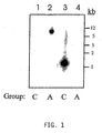
- FIG. 1 depicts that cross hybridization between seHAS and spHAS genes does not occur.
- the Group A probe used in lanes 1 and 2 only hybridizes with Group A DNA (lane 2) while the Group C probe used in lanes 3 and 4 only hybridizes with lane 3.
- FIG. 2 figuratively depicts the relatedness of seHAS to the bacterial, viral and eukaryotic HAS proteins.
- FIG. 3 figuratively depicts possible evolutionary relationships and similarities among some of the known hyaluronan synthases.
- FIG. 4 depicts the HA size distribution produced by various engineered Streptococcal HAS enzymes.
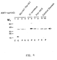
- FIG. 5 figuratively depicts the overexpression of recombinant seHAS and spHAS in E. coli.
- FIG. 6 depicts recombinant HA production in Bacillus subtilis and Bacillus licheniformis.
- FIG. 7 depicts purification of Streptococcal HA synthase.
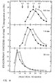
- FIG. 8 depicts a gel filtration analysis of HA synthesized by recombinant streptococcal HAS expressed in yeast membranes.
- FIG. 9 is a Western blot analysis of recombinant seHAS using specific antibodies.
- FIG. 10 is a kinetic analysis of the HA size distributions produced by recombinant seHAS and spHAS.
- FIG. 11 graphically depicts the hydropathy plots for seHAS and predicted membrane associated regions.
- FIG. 12 is a graphical model for the topologic organization of seHAS in the membrane.
- FIG. 13 is a demonstration of the synthesis of authentic HA by the recombinant seHAS.
- FIG. 14 depicts the recognition of nucleic acid sequences encoding seHAS, encoding spHAS, or encoding both seHAS and spHAS using specific oligonucleotides and PCR,
- FIG. 15 depicts oligonucleotides used for specific PCR hybridization.
- FIG. 16A is a plot depicting recombinant HA production in live Bacillus subtilis by comparing HA production by Bacillus subtilis 168 (pSM 143 vector alone) to a Bacillus subtilis 168 (pSM143 containing seHAS).
- FIG. 16B is an enlargement of a section of the plot in FIG. 16A .
- FIG. 17A is a plot depicting nutritional control of recombinant HA size distribution produced by spHAS in live Bacillus subtilis.
- FIG. 17B is a plot depicting recombinant HA production in live Bacillus subtilis 168 compared to Bacillus subtilis that contains vector alone.
- FIG. 18A and 18B are photomicrographs of recombinant E. coli.
- India ink staining 1,000X magnification
- the capsular material could be removed from the E. coli K5(pPmHAS) cells by brief treatment with Streptomyces HA lyase.
- PmHAS directs polymerization of the HA polysaccharide.
- FIG. 19 is a schematic model of GAG biosynthesis in Gram positive and Gram negative bacteria.
- FIG. 20 is an agarose gel demonstrating the PCR amplification of the HAS gene from Streptococcus uberis.
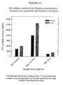
- FIG. 21 depicts HA synthase activity from Streptococcus pyogenes, Streptococcus equisimilis, and Streptococcus uberis,
- nucleic acid segment and “DNA segment” are used interchangeably and refer to a DNA molecule which has been isolated free of total genomic DNA of a particular species. Therefore, a “purified” DNA or nucleic acid segment as used herein, refers to a DNA segment which contains a Hyaluronate Synthase ("HAS") coding sequence yet is isolated away from, or purified free from, unrelated genomic DNA of the source cell. Included within the term “DNA segment”, are DNA segments and smaller fragments of such segments, and also recombinant vectors, including, for example, plasmids, cosmids, phage, viruses, and the like.
- HAS Hyaluronate Synthase
- a DNA segment comprising an isolated or purified HAS gene refers to a DNA segment including HAS coding sequences isolated substantially away from other naturally occurring genes or protein encoding sequences.
- the term "gene” is used for simplicity to refer to a functional protein, polypeptide or peptide encoding unit. As will be understood by those in the art, this functional term includes genomic sequences, cDNA sequences or combinations thereof. "Isolated substantially away from other coding sequences" means that the gene of interest, in this case HAS, forms the significant part of the coding region of the DNA segment, and that the DNA segment does not contain large portions of naturally-occurring coding DNA, such as large chromosomal fragments or other functional genes or DNA coding regions. Of course, this refers to the DNA segment as originally isolated, and does not exclude genes or coding regions later added to, or intentionally left in the segment by the hand of man.
- a Class I or Class II HAS such as a Class I HAS from S. equisimilis or S. pyogenes, or a Class II HAS from P. multocida.
- Streptococcus is subdivided taxonomically into Lancefield Groups based on different cell wall carbohydrate antigens. There are 18 distinct groups, but the most common pathogens are A, B, C and D. Historically, the most common pathogens are also often given specific species names, but the unified Lancefield testing method is recognized as being a clear method of typing and thus a useful classification scheme .
- the Streptococcus species that is utilized as the source of the HAS gene for employment in the present invention is the Group C Streptococcus , S. uberis .
- HAS gene isolating the HAS gene from prokaryotes
- eukaryotic enzymes may require significant post-translational modifications that can only be achieved in a eukaryotic host. This will tend to limit the applicability of any eukaryotic HA synthase gene that is obtained.
- those of ordinary skill in the art will likely realize additional advantages in terms of time and ease of genetic manipulation where a prokaryotic enzyme gene is sought to be employed.
- DNA sequences for use in accordance with the present invention will further include genetic control regions which allow the expression of the sequence in a selected recombinant host.
- control region employed will generally vary depending on the particular use (e.g., cloning host) envisioned.
- the invention concerns the use of isolated DNA segments and recombinant vectors incorporating DNA sequences and which encode a HAS gene, that includes within its amino acid sequence an amino acid sequence in accordance with SEQ ID NO: 12.
- the invention concerns the use of isolated DNA segments and recombinant vectors incorporating DNA sequences which encode a gene that includes within its amino acid sequence the amino acid sequence of a HAS gene or DNA, and in particular to a HAS gene or cDNA, corresponding to Streptococcus uberis HAS.
- preferred sequences are those which are essentially as set forth in SEQ ID NO: 12.
- Nucleic acid segments having HA synthase activity may be isolated by the methods described herein.
- the term "a sequence essentially as set forth in SEQ ID NO:X” means that the sequence substantially corresponds to a portion of SEQ ID NO:X and has relatively few amino acids which are not identical to, or a biologically functional equivalent of, the amino acids of SEQ ID NO:X.
- the term "biologically functional equivalent” is well understood in the art and is further defined in detail herein, as a gene having a sequence essentially as set forth in SEQ ID NO:X, and that is associated with the ability of prokaryotes to produce HA or a hyaluronic acid coat.
- the seHAS and spHAS coding sequences are approximately 70% identical and rich in the bases adenine (A) and thymine (T).
- FIG. 1 The inability of spHAS and seHAS to cross-hybridize is shown in FIG. 1 .
- the very A-T rich sequences will form less stable hybridization complexes than G-C rich sequences.
- Another possible explanation could be that there are several stretches of As or Ts in both sequences that could hybridize in a misaligned and unstable manner. This would put the seHAS and spHAS gene sequences out of frame with respect to each other, thereby decreasing the probability of productive hybridization.
- nucleic acid segment which encodes enzymatically active hyaluronate synthase.
- a nucleic acid segment encoding enzymatically active hyaluronate synthase may contain conserved or semi-conserved substitutions to the sequences set forth in SEQ ID NOS: 11 and 12 and yet still be within the scope of the invention.
- nucleic acid sequence may be highly identical and retain its enzymatic activity with regard to its unadulterated parent, and yet still fail to hybridize thereto.
- the present document discloses nucleic acid segments encoding enzymatically active hyaluronate synthases, such as seHAS, spHAS, suHAS and pmHAS. Although seHAS and spHAS are 70% identical an both encode enzymatically active hyaluronate synthase, they do not cross hybridize. Thus, one of ordinary skill in the art would appreciate that substitutions can be made to the seHAS nucleic acid segment listed in SEQ ID NO: 1 without changing its functionality. Standardized and accepted functionally equivalent amino acid substitutions are presented in Table I.
- the present invention employs a purified nucleic acid segment that encodes a protein in accordance with SEQ ID NO: 12 further defined as a recombinant vector.
- recombinant vector refers to a vector that has been modified to contain a nucleic acid segment that encodes an HAS protein, or fragment thereof.
- the recombinant vector may be further defined as an expression vector comprising a promoter operatively linked to said HAS encoding nucleic acid segment.
- the present invention is a host cell, made recombinant with a recombinant vector comprising a HAS gene.
- engineered or “recombinant” cell is intended to refer to a cell into which a recombinant gene, such as a gene encoding HAS, has been introduced. Therefore, engineered cells are distinguishable from naturally occurring cells which do not contain a recombinantly introduced gene. Engineered cells are thus cells having a gene or genes introduced through the hand of man. Recombinantly introduced genes will either be in the form of a cDNA gene, a copy of a genomic gene, or will include genes positioned adjacent to a promoter not naturally associated with the particular introduced gene.
- a host other than Streptococcus as may be used to produce recombinant HA synthase, it may be advantageous to employ a prokaryotic system such as E. coli, Bacillus strains, Lactococcus sp., or even eukaryotic systems such as yeast or Chinese hamster ovary, African green monkey kidney cells, VERO cells, or the like.
- a prokaryotic system such as E. coli, Bacillus strains, Lactococcus sp., or even eukaryotic systems such as yeast or Chinese hamster ovary, African green monkey kidney cells, VERO cells, or the like.
- yeast or Chinese hamster ovary such as yeast or Chinese hamster ovary, African green monkey kidney cells, VERO cells, or the like.
- the host cell is a Bacillus cell, such as a Bacillus subtilis or Bacillus licheniformis cell, and the vector introduced therein contains a Bacillus -compatible promoter to which the HAS gene is operably linked.
- the host cell is a Bacillus cell, such as Bacillus alkalophilus, Bacillus amyloliquefaciens, Bacillus brevis, Bacillus circulans, Bacillus clausii, Bacillus coagulans, Bacillus firmus, Bacillus lautus, Bacillus lentus, Bacillus licheniformis, Bacillus metaterium, Bacillus pumilus, Bacillus stearothermophilus, Bacillus subtilis and Bacillus thuringienisis,
- Bacillus cell such as Bacillus alkalophilus, Bacillus amyloliquefaciens, Bacillus brevis, Bacillus circulans, Bacillus clausii, Bacillus coagulans, Bacillus firmus, Bacillus lautus, Bacillus lentus, Bacillus licheniformis, Bacillus metaterium, Bacillus pumilus, Bacillus stearothermophilus, Bacillus subtilis and Bacillus
- the HA synthase-encoding DNA segments further include DNA sequences, known in the art functionally as origins of replication or "replicons", which allow replication of contiguous sequences by the particular host.
- origins of replication or "replicons” allow the preparation of extrachromosomally localized and replicating chimeric segments or plasmids, to which HA synthase DNA sequences are ligated.
- the employed origin is one capable of replication in bacterial hosts suitable for biotechnology applications.
- the isolation and use of other replication origins such as the SV40, polyoma or bovine papilloma virus origins, which may be employed for cloning or expression in a number of higher organisms, are well known to those of ordinary skill in the art.
- the present disclosure relates to a recombinant transformation vector which includes the HA synthase coding gene sequence together with an appropriate replication origin and under the control of selected control regions.
- HAS gene or cDNA may be obtained which contain full complements of genes or cDNAs from a number of sources, including other strains of Streptococcus, or from eukaryotic sources, such as cDNA libraries.
- Virtually any molecular cloning approach may be employed for the generation of DNA fragments in accordance with the present disclosure.
- the only limitation generally on the particular method employed for DNA isolation is that the isolated nucleic acids should encode a biologically functional equivalent HA synthase.
- any cloning vector can be employed to realize advantages in accordance with the invention.
- Typical useful vectors include plasmids and phages for use in prokaryotic organisms and even viral vectors for use in eukaryotic organisms. Examples include pKK223-3, pSA3, recombinant lambda, SV40, polyoma, adenovirus, bovine papilloma virus and retroviruses.
- pKK223-3 pSA3
- recombinant lambda SV40
- polyoma polyoma
- adenovirus bovine papilloma virus
- retroviruses retroviruses
- Vectors such as these allow one to perform clonal colony selection in an easily manipulated host such as E. coli, followed by subsequent transfer back into a food grade Lactococcus or Bacillus strain for production of HA.
- These are benign and well studied organisms used in the production of certain foods and biotechnology products. These are advantageous in that one can augment the Lactococcus or Bacillus strain's ability to synthesize HA through gene dosaging (i.e., providing extra copies of the HA synthase gene by amplification) and/or inclusion of additional genes to increase the availability of HA precursors.
- HA synthase gene copy number The inherent ability of a bacterium to synthesize HA can also be augmented through the formation of extra copies, or amplification, of the plasmid that carries the HA synthase gene. This amplification can account for up to a 10-fold increase in plasmid copy number and therefore the HA synthase gene copy number.
- Another procedure that would further augment HA synthase gene copy number is the insertion of multiple copies of the gene into the plasmid.
- Another technique would include integrating the HAS gene into chromosomal DNA. This extra amplification would be especially feasible, since the bacterial HA synthase gene size is small.
- the chromosomal DNA-ligated vector is employed to transfect the host that is selected for clonal screening purposes such as E. coli, through the use of a vector that is capable of expressing the inserted DNA in the chosen host.
- the HA synthase gene is introduced into the host cell chromosome via homologous or heterologous recombination.
- the has gene may be more stable in this configuration, especially without drug selection.
- Various vectors may be employed to introduce the has gene into Bacillus, such as pTLH or pKSV7, or into yeast, such as YIp211, or into animal cells, such as pcDNA/FRT.
- the DNA is first introduced into the host cell by transformation, transduction or electroporation.
- the DNA segment with the has gene is then stably integrated into the host chromosome.
- the spHAS gene was used to repair a mutant Streptococcus chromosome by transduction and integration; this operation resulted in HA production (DeAngelis et al, 1993).
- a eukaryotic source such as dermal or synovial fibroblasts or rooster comb cells
- an enzyme with reverse transcriptase activity and ligation with the selected vector.
- a preferred technique involves reverse transcription.
- a cDNA library is prepared in the selected host by accepted techniques, such as by ligation into the appropriate vector and amplification in the appropriate host.
- phage expression vectors such as ⁇ gt11, ⁇ gt12, ⁇ Gem11, and/or ⁇ ZAP for the cloning and expression screening of cDNA clones.
- the invention concerns the use of isolated DNA segments and recombinant vectors that include within their sequence a nucleic acid sequence essentially as set forth in SEQ ID NO: 11.
- the term "essentially as set forth in SEQ ID NO:11”, for example, is used in the same sense as described above and means that the nucleic acid sequence substantially corresponds to a portion of SEQ ID NO:11, and has relatively few codons which are not identical, or functionally equivalent, to the codons of SEQ ID NO:11.
- the term “functionally equivalent codon” is used herein to refer to codons that encode the same amino acid, such as the six codons for arginine or serine, and also refers to codons that encode biologically equivalent amino acids as set forth in Table I.
- amino acid and nucleic acid sequences may include additional residues, such as additional N- or C-terminal amino acids or additional 5' or 3' nucleic acid sequences, and yet still be essentially as set forth in one of the sequences disclosed herein, so long as the sequence meets the criteria set forth above, including the maintenance of biological protein activity where protein expression and enzyme activity are concerned.
- the addition of terminal sequences particularly applies to nucleic acid sequences which may, for example, include various non-coding sequences flanking either of the 5' or 3' portions of the coding region or may include various internal sequences, which are known to occur within genes.
- the amino acid sequence of the has gene product in eukaryotes appears to be 40% larger than that found in prokaryotes.
- sequences which have between about 40% and about 80%; or more preferably, between about 80% and about 90%; or even more preferably, between about 90% and about 99%; of nucleotides which are identical to the nucleotides of SEQ ID NO: 11 will be sequences which are "essentially as set forth in SEQ ID NO: 11". Sequences which are essentially the same as those set forth in SEQ ID NO: 11 may also be functionally defined as sequences which are capable of hybridizing to a nucleic acid segment containing the complement of SEQ ID NO: 11 under standard or less stringent hybridizing conditions. Suitable standard hybridization conditions will be well known to those of skill in the art and are clearly set forth herein.
- standard hybridization conditions is used to describe those conditions under which substantially complementary nucleic acid segments will form standard Watson-Crick base-pairing.
- a number of factors are known that determine the specificity of binding or hybridization, such as pH, temperature, salt concentration, the presence of agents, such as formamide and dimethyl sulfoxide, the length of the segments that are hybridizing, and the like.
- salt and temperature preferred conditions for hybridization will include 1.2-1.8 x High Phosphate Buffer (HPB) at 40-50°C.
- nucleic acid sequences which are “complementary” are those which are capable of base-pairing according to the standard Watson-Crick complementarity rules.
- complementary sequences means nucleic acid sequences which are substantially complementary, as may be assessed by the same nucleotide comparison set forth above, or as defined as being capable of hybridizing to the nucleic acid segment of SEQ ID NO: 11.
- nucleic acid segments employed in the present invention may be combined with other DNA sequences, such as promoters, polyadenylation signals, additional restriction enzyme sites, multiple cloning sites, epitope tags, poly histidine regions, other coding segments, and the like, such that their overall length may vary considerably. It is therefore contemplated that a nucleic acid fragment of almost any length may be employed, with the total length preferably being limited by the ease of preparation and use in the intended recombinant DNA protocol.
- this invention is not limited to the particular nucleic acid and amino acid sequences of SEQ ID NO: 11 and SEQ ID NO: 12, respectively.
- Recombinant vectors and isolated DNA segments may therefore variously include the HAS coding regions themselves, coding regions bearing selected alterations or modifications in the basic coding region as defined in claim 1, or they may encode larger polypeptides which nevertheless include HAS-coding regions or may encode biologically functional equivalent proteins or peptides which have variant amino acids sequence as defined in claim 1.
- the capsule of Carter Type A P. multocida was long suspected of containing hyaluronic acid (HA). Characterization of the HA synthase of P. multocida led to interesting enzymological differences between it and the seHAS and spHAS proteins.
- HA hyaluronic acid
- P. multocida cells produce a readily visible extracellular HA capsule, and since the two streptococcal HASs are membrane proteins, membrane preparations of the fowl cholera pathogen were tested.
- crude membrane fractions derived from ultrasonication alone possessed very low levels of UDP-GlcNAc-dependent UDP-[ 14 C]GlcUA incorporation into HA[ ⁇ 0.2 pmol of GlcUA transfer ( ⁇ g of proteins) -1 h -1 ] when assayed under conditions similar to those for measuring streptococcal HAS activity.
- the enzyme from E. coli with the recombinant hasA plasmid was also recalcitrant to isolation at first. These results were in contrast to the easily detectable amounts obtained from Streptococcus by similar methods.
- Gel-filtration analysis using a Sephacryl S-200 column indicates that the molecular mass of the majority of the 14 C-labeled product synthesized in vitro is ⁇ 8 x 10 4 Da since the material elutes in the void volumes, such a value corresponds to a HA molecule composed of at least 400 monomers.
- This product is sensitive to Streptomyces hyaluronidase digestion but resistant to protease treatment.
- the parameters of the HAS assay were varied to maximize incorporation of UDP-sugars into polysaccharide by P. multocida membranes. Streptococcal spHAS requires Mg 2+ , and therefore this metal ion was included in the initial assays of P. multocida membranes.
- the P. multocida HAS (pmHAS) was relatively active from pH 6.5 to 8.6 in Tris-type buffers with an optimum at pH 7.
- the HAS activity was linear with respect to the incubation time at neutral pH for at least 1 h.
- the pmHAS was apparently less active at higher ionic strengths because the addition of 100 mM NaCl to the reaction containing 50 mM Tris, pH 7, and 20 mM MgCl 2 reduced sugar incorporation by ⁇ 50%.
- the metal ion specificity of the pmHAS was assessed at pH 7. Under metal-free conditions in the presence of EDTA, no incorporation of radiolabeled precursor into polysaccharide was detectable ( ⁇ 0.5% of maximal signal).
- Mn 2+ gave the highest incorporation rates at the lowest ion concentrations for the tested metals (Mg, Mn, Co, Cu, and Ni). Mg 2+ gave about 50% of the Mn 2+ stimulation but at 10-fold higher concentrations.
- Co 2+ or Ni 2+ at 10mM supported lower levels of activity (20% or 9%, respectively, of 1 mM Mn 2+ assays), but membranes supplied with 10 mM Cu 2+ were inactive. Indeed, mixing 10 mM Cu 2+ and 20 mM Mg 2+ with the membrane preparation resulted in almost no incorporation of label into polysaccharide ( ⁇ 0.8% of Mg only value).
- V max values for both sugars were the same because the slopes, corresponding to 1/ V max , of the Hanes-Woolf plots were equivalent.
- the K M value for UDP-GlcNAc was increased by about 25-50% to ⁇ 105 ⁇ M and the V max increased by a factor of 2-3-fold in the presence of Mn 2+ .
- the HA synthase enzymes from either P. multocida, S. equisimilis, or S. pyogenes utilizes UDP-sugars, but they possess somewhat different kinetic optima with respect to pH and metal ion dependence and K M values.
- the enzymes are most active at pH 7; however, the pmHAS reportedly displays more activity at slightly acidic pH and is relatively inactive above pH 7.4.
- the pmHAS utilizes Mn 2+ more efficiently than Mg 2+ under the in vitro assay conditions, but the identity of the physiological metal cofactor in the bacterial cell is unknown.
- Mg 2+ was much better than Mn 2+ but the albeit smaller effect of Mn 2+ was maximal at ⁇ 10-fold lower concentrations than the optimal Mg 2+ concentration.
- the pmHAS apparently binds the UDP-sugars more tightly than spHAS.
- the measured K M values for the pmHAS in crude membranes are about 2-3-fold lower for each substrate than those obtained from the HAS found in streptococcal membranes: 50 or 39 ⁇ M for UDP-GlcUA and 500 or 150 ⁇ M for UDP-GlcNAc, respectively.
- Chlorella virus PBCV-1 encodes a functional glycosyltransferase that can synthesize hyaluronan. This finding is contrary to the general observation that viruses either: (a) utilize host cell glycosyltransferases to create new carbohydrate structures, or (b) accumulate host cell glycoconjugates during virion maturation. Furthermore, HA has been generally regarded as restricted to animals and a few of their virulent bacterial pathogens. Though many plant carbohydrates have been characterized, neither HA nor a related analog has previously been detected in cells of plants or protists.
- the vertebrate, bacterial and viral HAS enzymes have several regions of sequence similarity. While sequencing the double-stranded DNA genome of virus PBCV-1 [ P aramecium b ursaria C hlorella v irus], an ORF [open reading frame], A98R (Accession #442580), encoding a 567 residue protein with 28 to 33% amino acid identity to the various HASs was discovered. This protein is designated cvHAS (Chlorella virus HA synthase). The gene sequence encoding PBCV-1 and the protein sequence it encodes are shown in SEQ ID NOS:7 and 8, respectively.
- PBCV-1 is the prototype of a family (Phycodnarviridae) of large (175-190 nm diameter) polyhedral, plaque-forming viruses that replicate in certain unicellular, eukaryotic chlorella-like green algae.
- PBCV-1 virions contain at least 50 different proteins and a lipid component located inside the outer glycoprotein capsid.
- the PBCV-1 genome is a linear, nonpermuted 330-kb dsDNA molecule with covalently closed hairpin ends.
- the A98R gene product should be an integral membrane protein.
- recombinant A98R was produced in Escherichia coli and the membrane fraction was assayed for HAS activity.
- UDP-GlcUA and UDP-GlcNAc were incorporated into the polysaccharide by the membrane fraction derived from cells containing the A98R gene on a plasmid, pCVHAS, (average specific activity 2.5 pmoles GlcUA transfer/ ⁇ g protein/min) but not by samples from control cells ( ⁇ 0.001 pmoles GlcUA transfer/ ⁇ g protein/min).
- PBCV-1 infected Chlorella cells were examined for A98R gene expression.
- a ⁇ 1,700-nucleotide A98R transcript appeared ant ⁇ 15 min post-infection and disappeared by 60 min after infection indicating that A98R is an early gene. Consequently, membrane fractions from uninfected and PBCV-1 infected chlorella cells were assayed at 50 and 90 min post-infection for HAS activity. Infected cells, but not uninfected cells, had activity.
- radiolabel incorporation from UDP-[14C]GlcUA into polysaccharide depended on both Mn 2+ and UDP-GlcNAc. This radiolabeled product was also degraded by HA lyase. Disrupted PBCV-1 virions had no HAS activity.
- PBCV-1 infected Chlorella cells were analyzed for HA polysaccharide using a highly specific 125 I-labeled HA-binding protein. Extracts from cells at 50 and 90 min post-infection contained substantial amounts of HA, but not extracts from uninfected algae or disrupted PBCV-1 virions. The labeled HA-binding protein also interacted with intact infected cells at 50 and 90 min post-infection, but not healthy cells. Therefore, a considerable portion of the newly synthesized HA polysaccharide was immobilized at the outer cell surface of the infected algae.
- the extracellular HA does not play any obvious role in the interaction between the virus and its algal host because neither plaque size nor plaque number was altered by including either testicular hyaluronidase (465 units/ml) or free HA polysaccharide (100 ⁇ g/ml) in the top agar of the PBCV-1 plaque assay.
- the PBCV-1 genome also has additional genes that encode for an UDP-glucose dehydrogenase (UDP-Glc DH) and a glutamine:fructose-6-phosphate aminotransferase (GFAT).
- UDP-Glc DH converts UDP-Glc into UDP-GlcUA, a required precursor for HA biosynthesis.
- GFAT converts fructose-6-phosphate into glucosamine-6-phosphate, an intermediate in the UDP-GlLcNAc metabolic pathway.
- Both of these PBCV-1 genes like the A98R HAS, are expressed early in infection and encode enzymatically active proteins. The presence of multiple enzymes in the HA biosynthesis pathway indicates that HA production must serve an important function in the life cycle of the chlorella viruses.
- FIG. 2 is an alignment of the protein sequences of Group C seHAS and suHAS from S. equisimilis and S. uberis, respectively; Group A spHAS from S.
- glycosyltransferases Regions of similarity between HASs and other enzymes that synthesize ⁇ -linked polysaccharides from UDP-sugar precursors are also being discovered as more glycosyltransferases are sequenced. Examples include bacterial cellulose synthase, fungal and bacterial chitin synthases, and the various HASs. The significance of these similar structural motifs will become more apparent as the three-dimensional structures of glycosyltransferases accumulate.
- FIG. 3 depicts the possible evolutionary relationships among the known hyaluronan synthases.
- the phylogenetic tree of FIG. 3 was generated by the Higgins-Sharp algorithm using the DNAsis multiple alignment program. The calculated matching percentages are indicated at each branch of the dendrogram.
- the DNA segments used in the present invention encompass biologically functional equivalent HAS proteins and peptides. Such sequences may arise as a consequence of codon redundancy and functional equivalency which are known to occur naturally within nucleic acid sequences and the proteins thus encoded.
- functionally equivalent proteins or peptides may be created via the application of recombinant DNA technology, in which changes in the protein structure may be engineered, based on considerations of the properties of the amino acids being exchanged. Changes designed by man may be introduced through the application of site-directed mutagenesis techniques, e.g., to introduce improvements to the enzyme activity or to antigenicity of the HAS protein or to test HAS mutants in order to examine HA synthase activity at the molecular level.
- HAS coding sequence can be manipulated in a manner to produce an altered hyaluronate synthase which in turn is capable of producing hyaluronic acid having differing polymer sizes and/or functional capabilities.
- the HAS coding sequence may be altered in such a manner that the hyaluronate synthase has an altered sugar substrate specificity so that the hyaluronate synthase creates a new hyaluronic acid-like polymer incorporating a different structure such as a previously unincorporated sugar or sugar derivative.
- modified structure denotes a hyaluronic acid polymer containing a sugar or derivative not normally found in the naturally occurring HA polysaccharide.
- modified size distribution refer to the synthesis of hyaluronic acid molecules of a size distribution not normally found with the native enzyme; the engineered size could be much smaller or larger than normal.
- hyaluronic acid products of differing size have application in various areas, such as drug delivery.
- Applications in angiogenesis and wound healing are potentially large if hyaluronic acid polymers of from about 4 to about 20 monosaccharides can be made in good quantities.
- Another particular application for small hyaluronic acid oligosaccharides is in the stabilization of recombinant human proteins used for medical purposes.
- a major problem with such proteins is their clearance from the blood and a short biological half life.
- One present solution to this problem is to couple a small molecule shield that prevents the protein from being cleared from the circulation too rapidly.
- Very small molecular weight hyaluronic acid is well suited for this role and would be nonimmunogenic and biocompatible. Larger molecular weight hyaluronic acid attached to a drug or protein may be used to target the reticuloendothelial cell system which has endocytic receptors for hyaluronic acid.
- the size distribution of the hyaluronic acid polymer made by the hyaluronate synthase could be regulated to give different sizes.
- the kinetic control of product size can be altered by decreasing temperature, decreasing time of enzyme action and by decreasing the concentration of one or both sugar nucleotide substrates. Decreasing any or all of these variables will give lower amounts and smaller sizes of hyaluronic acid product.
- the disadvantages of these approaches are that the yield of product will also be decreased and it may be difficult to achieve reproducibility from day to day or batch to batch.
- the size distribution of the HA polymer can be regulated by altering the intrinsic ability of the enzyme to synthesize a large hyaluronic acid product.
- Changes to the protein can be engineered by recombinant DNA technology, including substitution, deletion and addition of specific amino acids (or even the introduction of prosthetic groups through metabolic processing). Such changes that result in an intrinsically slower enzyme then allows more reproducible control of hyaluronic acid size by kinetic means.
- the final hyaluronic acid size distribution is determined by certain characteristics of the enzyme, that rely on particular amino acids in the sequence.
- hyaluronan synthase can be engineered to produce hyaluronic acid polymers of different size, in particular smaller, than the normal wildtype enzyme.
- the figure shows the distribution of HA sizes (in millions of Daltons, a measure of molecular weight) for a series of spHAS enzymes, each of which was engineered by site directed mutagenesis to have a single amino acid change from the native enzyme. Each has a different Cysteine residue replaced with Alanine.
- the cluster of five curves with open symbols represent the following spHAS proteins: wildtype, C124A, C261A, C366A, and C402A.
- the filled circles represent the poorly expressed C225A protein which is only partially active.
- the filled triangles represent the C280A spHAS protein, which is found to synthesize a much smaller range of HA polymers than the normal enzyme or the other variants shown. This reduction to practice shows that it is feasible to engineer the hyaluronate synthase enzyme to synthesize a desired range of HA product sizes.
- Any of the HAS genes encoding hyaluronate synthase disclosed herein can also be manipulated by site directed mutagenesis to produce an enzyme which synthesizes a desired range of HA product sizes.
- Structurally modified hyaluronic acid is no different conceptually than altering the size distribution of the hyaluronic acid product by changing particular amino acids in the desired HAS or the spHAS.
- Derivatives of UDP-GlcNAc in which the N-acetyl group is missing UDP-GlcN or replaced with another chemically useful group, are expected to be particularly useful.
- the strong substrate specificity must rely on a particular subset of amino acids among the 20% that are conserved. Specific changes to one or more of these residues creates a functional synthase that interacts less specifically with one or more of the substrates than the native enzyme.
- This altered enzyme could then utilize alternate natural or special sugar nucleotides to incorporate sugar derivatives designed to allow different chemistries to be employed for the following purposes: (i) covalently coupling specific drugs, proteins, or toxins to the structurally modified hyaluronic acid for general or targeted drug delivery, radiological procedures, etc. (ii) covalently cross linking the hyaluronic acid itself or to other supports to achieve a gel, or other three dimensional biomaterial with stronger physical properties, and (iii) covalently linking hyaluronic acid to a surface to create a biocompatible film or monolayer.
- Bacteria can also be engineered to produce hyaluronic acid.
- Bacteria can also be engineered to produce hyaluronic acid.
- strains of B. subtilis containing a HAS gene as well as the gene for one of the sugar nucleotide precursors.
- These bacteria were intended as first generation prototypes for the generation of a bacterium able to produce hyaluronic acid in larger amounts than presently available using a wild type natural strain.
- strain #3 makes and secretes HA into the medium.
- strain #1 The parent strain or the strain with just the dehydrogenase gene (strain #1) does not make HA.
- strain #2 which contains just the spHAS gene alone makes HA, but only about 10% of what strain #3 makes. Agarose gel electrophoresis showed that the HA secreted into the medium by strain #3 is very high molecular weight.
- B. subtilis 168 can be engineered to produce and secrete HA by the introduction by recombinant DNA techniques of the spHAS gene and the hasB gene. Although HA is made by this modified strain even without inclusion of the latter gene, the level of HA made with it is greatly elevated .
- B, subtilis 168 contains two genes ( tauD and gtaB ) that increase the levels of both sugar nucleotides needed for HA synthesis.
- Table IV demonstrates that B.
- subtilis 168 also makes HA, even in the absence of the hasB gene, when engineered to contain and express (on the plasmid pSM143, ATCC) seHAS as well as specific seHAS variants engineered to produce HA of different size than the wildtype.
- the variants seHAS(C226A) and seHAS(C281A) supported HA synthesis in live B. subtilis 168 cells. The level of HA synthesis in these latter cases was less than observed with cells expressing spHAS and the hasB gene, due to the lower endogenous level of the two precursors needed for HA synthesis.
- FIG. 6 is a digital image of an agarose gel stained for HA.
- HA production can be seen in B. subtilis, B, licheniformis and E. faecalis strains having a plasmid encoding spHAS (pPD41 ⁇ 5) incorporated therein.
- pPD41 ⁇ EcoRV a plasmid containing a nonfunctional hasA gene
- subtilis 168 Containing spHAS and hasB genes Strain Number Cells Medium(*) Strain with genes Cell density (A 600 ( ⁇ g HA per ml of culture) 1 0 0 hasB 4.8 2 4 35 SpHAS 3.9 3 ⁇ 10 ⁇ 250 SpHAS + hasB 3.2 (*) Most HA is in media but some was cell-associated; HA was determined using the HA Test 50 kit from Pharmacia. TABLE IV Hyaluronan (HA) Produced in B.
- the vector used is a Gram positive/ E. coli shuttle vector that has a medium copy number in B . subtilis and a gene for erythromycin resistance (enabling resistence to 8 ⁇ g/ml in B. subtilis or 175 ⁇ g/ml in E. coli).
- the B. subtilis host strain used is 1A1 from BGSC, which has a tryptophan requirement but otherwise is wildtype, and can sporulate.
- Cell growth and HA production was in Spizizens Minimal Media plus tryptophan, glucose, trace elements and erthromycin (8 ⁇ g/ml). Growth was at 32°C with vigorous agitation until the medium was exhausted ( ⁇ 36 hours).
- Tables III and IV demonstrate that these bioengineered cells, which would not normally make hyaluronic acid, became competent to do so when they are transformed with the spHAS or seHAS gene. Any one of the HAS genes described herein would also be capable of being introduced into a non-hyaluronic acid producing bacteria to create a bioengineered bacterial strain capable of producing hyaluronic acid.
- HAS genes described herein whether from genomic DNA, or a cDNA, one may proceed to prepare an expression system for the recombinant preparation of the HAS protein.
- the engineering of DNA segment(s) for expression in a prokaryotic or eukaryotic system may be performed by techniques generally known to those of skill in recombinant expression.
- HAS may be successfully expressed in eukaryotic expression systems, however, the inventors aver that bacterial expression systems can be used for the preparation of HAS for all purposes. It is believed that bacterial expression will ultimately have advantages over eukaryotic expression in terms of ease of use, cost of production, and quantity of material obtained thereby.
- streptococcal hyaluronan synthase is shown in Table V and FIG. 7 .
- Fractions from various stages of the purification scheme were analyzed by SDS-PAGE on a 12.5% gel, which was then stained with Coomassie Brilliant Blue R-250.
- transformation of host cells with DNA segments encoding HAS will provide a convenient means for obtaining a HAS protein. It is also proposed that cDNA, genomic sequences, and combinations thereof, are suitable for eukaryotic expression, as the host cell will, of course, process the genomic transcripts to yield functional mRNA for translation into protein.
- Another aspect of the present invention is a method of preparing a protein composition
- growing a recombinant host cell comprising a vector that encodes a protein which includes an amino acid sequence in accordance with SEQ ID NO: 12.
- the host cell will be grown under conditions permitting nucleic acid expression and protein production followed by recovery of the protein so produced.
- the production of HAS and ultimately HA, including the host cell, conditions permitting nucleic acid expression, protein production and recovery will be known to those of skill in the art in light of the present disclosure and the methods described herein.
- HA Hosts for the expression of hyaluronic acid are commonly prokaryotes, such as S. equisimilis, and other suitable members of the Streptococcus species.
- HA may be synthesized by heterologous host cells expressing recombinant HA synthase, such as species members of the Bacillus, Ehterococcus, or even Escherichia genus.
- the hosts for use in the methods of expression of HA in accordance with the present invention are Bacillus species, because such cells are effective industrial secretors, and several species have been designated as GRAS organisms.
- Bacillus cells that may be utilized in the methods of the present invention include, but are not limited to, Bacillus alkalophi / us, Bacillus amyloliquefaciens, Bacillus brevis, Bacillus circulans, Bacillus clausii, Bacillus coagulans, Bacillus firmus, Bacillus lautus, Bacillus lentus, Bacillus licheniformis, Bacillus metaterium, Bacillus pumilus, Bacillus stearothermophilus, Bacillus subtilis and Bacillus thuringienisis.
- a most preferred host is Bacillus subtilis.
- HAS adenovirus-based, cytomegalovirus-based, yeast-based, and the like.
- baculovirus-based, glutamine synthase-based, dihydrofolate reductase-based systems, SV-40 based, adenovirus-based, cytomegalovirus-based, yeast-based, and the like could be employed.
- SV-40 based adenovirus-based, cytomegalovirus-based, yeast-based, and the like
- FIG. 8 shows that the spHAS or the x1HAS enzyme was produced in recombinant yeast using the pYES2 plasmid.
- UDP-GlcUA and UDP-GlcNAc either enzyme makes high molecular weight HA, as observed in these gel filtration chromatography profiles (the HA peak is from about 13 ml to about 25 ml).
- FIG. 8 shows a gel filtration analysis of hyaluronic acid synthesized by recombinant HASs expressed in yeast membranes.
- a DNA fragment encoding (a) the open reading frame of 419 amino acid residues corresponding to spHAS (with the original Val codon switched to Met) or (b) the x1HAS protein was subcloned by standard methods in the pYES2 yeast expression vector (from Invitrogen) to produce pYES/HA.
- Membranes from cells with this construct were prepared by agitation with glass beads.
- the samples derived from pYES/HA constructs contained substantial HA synthase activity and the "42 kDa" HAS protein was detected by Western analysis using specific antibodies; membranes from cells with vector alone possessed neither activity nor the immunoreactive band (not shown).
- Membranes (315 ⁇ g protein) were first incubated with carrier free UDP-[ 14 C]GlcUA (1 ⁇ Ci 14 C) and 900 uM unlabeled UDP-GlcNAc in 50 mM Tris, pH 7, 20 mM MgCl2, 1mM DTT, and 0.05 M Nacl (450 ⁇ l reaction volume) at 30 degrees Celsius for 1.5 minutes.
- the column was eluted at 0.5 ml/min and radioactivity in the fractions (1 ml) was quantitated by liquid scintillation counting after adding BioSafeII cocktail (4.5 ml, Research Products Intl.). The void volume and the totally included volumes were at elution volumes of 14 ml and 35.5 ml, respectively. The peak of blue dextran (average 2x10 6 Da) eluted at 25-27 ml.
- the recombinant HAS expressed in the eukaryotic yeast cells makes high molecular weight hyaluronic acid in vitro.
- an appropriate polyadenylation site e.g., 5'-AATAAA-3'
- the poly A addition site is placed about 30 to 2000 nucleotides "downstream" of the termination site of the protein at a position prior to transcription termination.
- HAS human immunoglobulin-derived neurotrophic factor
- Examples of cell lines for expressing HAS cDNA include cell lines typically employed for eukaryotic expression such as 293, AtT-20, HepG2, VERO, HeLa, CHO, WI 38, BHK, COS-7, RIN and MDCK cell lines.
- This will generally include the steps of providing a recombinant host bearing the recombinant DNA segment encoding the HAS enzyme and capable of expressing the enzyme; culturing the recombinant host in media under conditions that will allow for transcription of the cloned HAS gene or cDNA and appropriate for the production of the hyaluronic acid; and separating and purifying the HAS enzyme or the secreted hyaluronic acid from the recombinant host.
- the conditions appropriate for expression of the cloned HAS gene or cDNA will depend upon the promoter, the vector, and the host system that is employed. For example, where one employs the lac promoter, one will desire to induce transcription through the inclusion of a material that will stimulate lac transcription, such as isopropylthiogalactoside.
- a material that will stimulate lac transcription such as isopropylthiogalactoside.
- the cloned seHAS and spHAS genes are expressed as HIS 6 containing proteins in E. coli for the purpose of purification of the HAS as shown in FIG. 5 . Where other promoters are employed, different materials may be needed to induce or otherwise up-regulate transcription.
- FIG. 5 depicts the overexpression of recombinant seHAS and spHAS in E. coli.
- Membrane proteins (5 ⁇ g per lane) were fractionated by SDS-PAGE using a 10% (w/v) gel under reducing conditions. The gel was stained with Coomassie blue R-250, photographed, scanned, and quantitated using a molecular dynamics personal densitometer (model PDSI P60). The position of HA synthase is marked by the arrow.
- Lane A is native spHAS (Group A); Lane C is native seHAS; Lane E is recombinant seHAS; Lane P is recombinant spHAS; Lane V is vector alone. Standards used were Bio-rad low Mr and shown in kDa.
- appropriate genes encoding enzymes needed for the biosynthesis of sugar nucleotide precursors or by using growth media containing substrates for the precursor-supplying enzymes, such as N-acetylglucosamine or glucosamine (GlcNAc or GlcNH 2 ) and glucose (Glc).
- a host would be chosen to optimize production of HA.
- a suitable host would be one that produced large quantities of the sugar nucleotide precursors to support the HAS enzyme and allow it to produce large quantities of HA.
- Such a host may be found naturally or may be made by a variety of techniques including mutagenesis or recombinant DNA technology.
- the genes for the sugar nucleotide synthesizing enzymes could also be isolated and incorporated in a vector along with the HAS gene or cDNA.
- a preferred embodiment of the present invention is a host containing these ancillary recombinant genes or cDNAs, thereby allowing for increased production of the sugar nucleotides and thus HA.
- HA will be secreted or otherwise shed by the recombinant organism into the surrounding media, allowing the ready isolation of HA from the media by known techniques.
- HA can be separated from the cells and debris by at least one of filtration, centrifugation, and flocculation, and the addition of trichloroacetic acid may further facilitate in separating cells and debris from the hyaluronic acid.
- the HA is then separated from the media by at least one of precipitation, ultrafiltration and dialysis.
- Precipitation agents include alcohols such as ethanol and isopropanol, organic solvents or compounds such as acetone or quaternary organic ammonium (aliphatic positively-charged) salts such as cetyl pyridinium chloride (CPC) or cetyl triammonium bromide (CTB).
- alcohols such as ethanol and isopropanol
- organic solvents or compounds such as acetone or quaternary organic ammonium (aliphatic positively-charged) salts such as cetyl pyridinium chloride (CPC) or cetyl triammonium bromide (CTB).
- CPC cetyl pyridinium chloride
- CTB cetyl triammonium bromide
- a preferred technique for isolation of HA is described in U.S. Pat. No. 4,517,295 in which the organic carboxylic acid, trichloroacetic acid, is added to the bacterial suspension at the end of the fermentation.
- the trichloroacetic acid causes the bacterial cells to clump and die and facilitates the ease of separating these cells and associated debris from HA, the desired product.
- the clarified supernatant is concentrated and dialyzed to remove low molecular weight contaminants including the organic acid.
- the aforementioned procedure utilizes filtration through filter cassettes, such as those containing 0.22 ⁇ m pore size filters. Diafiltration is continued until the conductivity of the solution decreases to approximately 0.5 mega-ohms.
- the concentrated HA is precipitated by adding an excess of reagent grade ethanol or other organic solvent and the precipitated HA is then dried by washing with ethanol and vacuum dried, lyophilized to remove alcohol.
- the HA can then be redissolved in a borate buffer, pH 8, and precipitated with CPC or certain other organic ammonium salts such as CETAB, a mixed trimethyl ammonium bromide solution at 4 degree(s) Celsius.
- CETAB certain other organic ammonium salts
- the precipitated HA is recovered by coarse filtration, resuspended in 1 M NaCl, diafiltered and concentrated as further described in the above referenced patent.
- the resultant HA is filter sterilized and ready to be converted to an appropriate salt, dry powder or sterile solution, depending on the desired end use.
- transfection is carried out by the calcium phosphate precipitation method, well known to those of skill in the art.
- other methods may also be used for introducing DNA into cells, such as by nuclear injection, cationic lipids, electroporation, protoplast fusion or by the BIOLISTICTM Bioparticle delivery system developed by DuPont (1989).
- the advantage of using the DuPont system is a high transformation efficiency. If prokaryotic cells or cells which contain substantial cell wall constructions are used, the preferred method of transfection is calcium treatment using calcium chloride to induce competence or electroporation.
- Plasmids containing the desired coding and control sequences employ standard ligation techniques. Isolated plasmids or DNA fragments are cleaved, tailored, and religated in the form desired to construct the plasmids required. Cleavage is performed by treating with restriction enzyme(s) in suitable buffer. In general, about 1 ⁇ g plasmid or DNA fragments are used with about 1 unit of enzyme in about 20 ⁇ l of buffer solution. Appropriate buffers and substrate amounts for particular restriction enzymes are specified by the manufacturer. Incubation times of about 1 hour at 37° C are workable.
- nucleic acid is recovered from the aqueous fraction by precipitation with ethanol. If blunt ends are required, the preparation is treated for 15 minutes at 15° C with 10 units of Polymerase I (Klenow), phenolchloroform extracted, and ethanol precipitated.
- polymerase I Polymerase I
- phenolchloroform extracted
- ethanol precipitated for ligation approximately equimolar amounts of the desired components, suitably end tailored to provide correct matching are treated with about 10 units T4 DNA ligase per 0.5 ⁇ g DNA.
- T4 DNA ligase per 0.5 ⁇ g DNA.
- the first step was to amplify the plasmid DNA by cloning into specifically competent E. coli SURE cells (Stratagene) by doing transformation at 30-32°C.
- the recombinant plasmid is used to transform E. coli K5 strain Bi8337-41, which can produce the UDP-GlcUA precursor, and successful transformants selected by antibiotic resistance as appropriate. Plasmids from the library of transformants are then screened for bacterial colonies that exhibit HA production. These colonies are picked, amplified and the plasmids purified and analyzed by restriction mapping.
- the plasmids showing indications of a functional HAS gene are then further characterized by any number of sequence analysis techniques which are known by those of ordinary skill in the art.
- prokaryotes were used for the initial cloning of DNA sequences and construction of the vectors useful in the invention.
- a suitable source may be Gram-positive cells, particularly those derived from the Group C Streptococcal strains. Bacteria with a single membrane, but a thick cell wall such as Staphylococci and Streptococci are Gram-positive. Gram-negative bacteria such as E. coli contain two discrete membranes rather than one surrounding the cell. Gram-negative organisms tend to have thinner cell walls. The single membrane of the Gram-positive organisms is analogous to the inner plasma membrane of Gram-negative bacteria.
- plasmid vectors containing origins of replication and control sequences which are derived from species compatible with the host cell are used in connection with these hosts.
- the vector ordinarily carries an origin of replication, as well as marking sequences which are capable of providing phenotypic selection in transformed cells.
- E. coli is typically transformed using pBR322, a plasmid derived from an E. coli species.
- pBR322 contains genes for ampicillin and tetracycline resistance and thus provides easy means for identifying transformed cells.
- a pBR plasmid or a pUC plasmid, or other microbial plasmid or phage must also contain, or be modified to contain, promoters which can be used by the microbial organism for expression of its own proteins.
- promoters may be heterologous promoters, that is, promoters from another organism, as long as the promoter is compatible with the host cell, i.e., recognized by the RNA polymerase of the host cell such that the RNA polymerase transcribes the gene to which the host-compatible promoter is attached.
- promoters most commonly used in recombinant DNA construction include the lacZ promoter, tac promoter, the T7 bacteriophage promoter, and tryptophan (trp) promoter system. While these are the most commonly used, other microbial promoters have been discovered and utilized, and details concerning their nucleotide sequences have been published, enabling a skilled worker to ligate them functionally with plasmid vectors.
- the promoter may be a modified RNA polymerase promoter having increased promoter activity.
- the modification to the promoter may be a mutation, or the addition of two or more promoter elements in tandem.
- the two or more tandem promoter elements may be the same promoter element or two or more different promoter elements.
- tandem promoter elements as used herein will be understood to mean two or more promoter sequences each of which is operably linked to a coding sequence such that the promoter sequences direct the production of a polypeptide encoded by the coding sequence by mediating the transcription of the coding sequence into mRNA.
- Tandem promoters as well as constructs and methods for use thereof in expression in Bacillus cells, are described in detail in US Patent Nos. 5,955,310 and 6,255,076 , issued to Widner et al on September 21, 1999 and July 3, 2001, respectively.
- a recombinant vector for use in the present invention contains more than one nucleic acid segment wherein each has a coding region encoding a protein, such as for example, a nucleic acid segment having a coding region encoding enzymatically active hyaluronan synthase and a nucleic acid segment having a coding region encoding enzymatically active UDP-glucose dehydrogenase
- each of the nucleic acid segments are operably linked to a promoter.
- the two or more nucleic acid segments may be linked to the same promoter, and this single promoter may drive expression of both genes, or the two or more nucleic acid segments may be linked to different promoters.
- eukaryotic microbes such as yeast cultures may also be used.
- Saccharomyces cerevisiae, or common baker's yeast is the most commonly used among eukaryotic microorganisms, although a number of other strains are commonly available.
- the plasmid YRp7 for example, is commonly used. This plasmid already contains the trp1 gene which provides a selection marker for a mutant strain of yeast lacking the ability to grow without tryptophan, for example, ATCC No. 44076 or PEP4-1.
- Suitable promoting sequences in yeast vectors include the promoters for the galactose utilization genes, the 3-phosphoglycerate kinase or other glycolytic enzymes, such as enolase, glyceraldehyde-3-phosphate dehydrogenase, hexokinase, pyruvate decarboxylase, phosphofructokinase, glucose-6-phosphate isomerase, 3-phosphoglycerate mutase, pyruvate kinase, triosephosphate isomerase, phosphoglucose isomerase, and glucokinase.
- the termination sequences associated with these genes are also ligated into the expression vector 3' of the sequence desired to be expressed to provide polyadenylation of the mRNA and termination.
- Other promoters which have the additional advantage of transcription controlled by growth conditions are the promoter region for alcohol dehydrogenase 2, cytochrome C, acid phosphatase, degradative enzymes associated with nitrogen metabolism, and the aforementioned glyceraldehyde-3-phosphate dehydrogenase, and enzymes responsible for maltose and galactose utilization.
- Any plasmid vector containing a yeast-compatible promoter, origin of replication and termination sequences is suitable.
- cultures of cells derived from multicellular organisms may also be used as hosts.
- any such cell culture is workable, whether from vertebrate or invertebrate culture.
- interest has been greatest in vertebrate cells, and propagation of vertebrate cells in culture has become a routine procedure in recent years.
- useful host cell lines are VERO and HeLa cells, Chinese hamster ovary (CHO) cell lines, and WI38, BHK, COS, and MDCK cell lines.
- control functions on the expression vectors are often provided by viral material.
- promoters are derived from polyoma, Adenovirus 2, bovine papilloma virus and most frequently Simian Virus 40 (SV40).
- the early and late promoters of SV40 virus are particularly useful because both are obtained easily from the virus as a fragment which also contains the SV40 viral origin of replication. Smaller or larger SV40 fragments may also be used, provided there is included the approximately 250 bp sequence extending from the Hind III site toward the Bg1 I site located in the viral origin of replication.
- An origin of replication may be provided either by construction of the vector to include an exogenous origin, such as may be derived from SV40 or other viral (e.g., Polyoma, Adeno, BPV) source, or may be provided by the host cell chromosomal replication mechanism, If the vector is integrated into the host cell chromosome, the latter mechanism is often sufficient.
- seHAS The encoded protein, designated seHAS, is 417 amino acids (calculated molecular weight of 47,778 and pI of 9.1) and is the smallest member of the HAS family identified thus far ( FIG. 2 ). seHAS also migrates anomalously fast in SDS-PAGE (M r ⁇ 42 kDa) ( FIGS. 5 and 9 ).
- FIG. 9 is a graphical representation of a Western Blot analysis of recombinant seHAS using specific antibodies.
- Group C C; lane 1 or Group A (A; lane 4) Streptococcal membranes and E. coli membranes (9 ⁇ g/lane) containing recombinant seHAS (E; lanes 2, 7, and 9) or spHAS (P; lanes 3, 6, 8, and 10) were fractionated by reducing SDS-PAGE and electrotransferred to nitrocellulose.
- the seHAS and spHAS protein-encoding sequences are 72% identical.
- the deduced protein sequence of seHAS was confirmed by reactivity with a synthetic peptide antibody ( FIG. 9 ).
- Recombinant seHAS expressed in E . coli was recovered in membranes as a major protein ( FIG. 5 ) and synthesized very large molecular weight HA in the presence of UDP-GlcNAc and UDP-GlcUA in vitro ( FIG. 10 ).
- FIG. 10 shows a kinetic analysis of the HA size distributions produced by seHAS and spHAS.
- E coli membranes containing equal amounts of seHAS or spHAS protein were incubated at 37°C with 1.35 mM UDP-[ 14 C] GlcUA (1.3 x 10 3 dpm/nmol) and 3.0 mM UDP-GlcNAc as described in the application. These substrate concentrations are greater than 15 times the respective Km valves.
- Samples taken at 0.5, 1.0, and 60 min were treated with SDS and chromatographed over Sephacryl S400 HR.
- the HA profiles in the fractionation range of the column fractions 12-24) are normalized to the percent of total HA in each fraction.
- the values above the arrows in the top panel are the MWs (in millions) of HA determined directly in a separate experiment using a Dawn multiangle laser light scattering instrument (Wyatt Technology Corp.), The size distributions of HA synthesized by seHAS ( ⁇ , ⁇ , ⁇ ,) and spHAS ( ⁇ , ⁇ , ⁇ ) at 0.5 min ( ⁇ , ⁇ ), 1.0 min ( ⁇ , ⁇ ) and 60 min ( ⁇ , ⁇ ) are shown as indicated. Analysis showed that seHAS and spHAS are essentially identical in the size distribution of HA chains they synthesize ( FIG. 10 ), SeHAS is twice as fast as spHAS in its ability to make HA.
- pKK-223 Expression vector was from Pharmacia, PCR 2.1 cloning vector was from Invitrogen, and predigested ⁇ Zap Express TM Bam HI/CIAP Vector was from Stratagene.
- Genomic DNA from Streptococcus equisimilis isolated by the method of Caparon and Scott was partially digested with Sau3A1 to an average size of 2-12 kb.
- the digested DNA was precipitated with ethanol, washed and ligated to the Bam HI/CIAP ⁇ Zap Express vector.
- Ligated DNA was packaged into phage with a Packagene TM extract obtained from Promega.
- the titer of the packaged phage library was checked using XL1-Blue MRF' E. coli as a host.
- oligonucleotides were designed based upon conserved sequences among spHAS ( Streptococcus pyogenes ) , DG42 (Xenopus laevis HAS; 19) and nodC (a Rhizobium meliloti nodulation factor; 20) and were used for PCR amplification with D181 genomic DNA as a template. Amplification conditions were 34 cycles at: 94°C for 1 min, 44°C for 1 min, 72°C for 1.5 min followed by a final extension at 72°C for 10 min.
- Oligonucleotide HADRF1, 5'- GAY MGA YRT YTX ACX AAT TAY GCT ATH GAY TTR GG-3' corresponds to the sequence D 259 RCLTNYAIDL (spHAS).
- Oligonucleotide HACTR1, 5'-ACG WGT WCC CCA NTC XGY ATT TTT NAD XGT RCA-3' (antisense strand) corresponds to the region C 404 TIKNTEWGTR (spHAS).
- the degeneracy of bases at some positions are represented by nomenclature adopted by the IUPAC in its codes for degenerate bases listed in Table VI.
- HAVAF1 5'-GTN GCT GCT GTW RTX CCW WSX TWT AAY GAR GA-3' (corresponding to the region V 66 AAVIPSYNE of spHAS) and HAVDF1, 5'-GTX RWT GAY GGN WSX WSN RAX GAT GAX GC-3' (based on V 100 DDGSSNTD of spHAS).
- Two unique antisense primers were synthesized based on the sequence of the 459 bp PCR product.
- the 459 bp PCR product was radiolabeled using the Prime-It 11 random primer labeling Kit (Stratagene) according to the manufacturers instructions. Oligonucleotides were labeled by Kinace-It Kinasing Kit (Stratagene) using [Y 32 P]ATP. Radiolabeled products were separated from nonlabeled material on NucTrap Push columns (Stratagene). The oligoprobe hybridized specifically with a D181 genomic digest on Southern blots.
- XLBLUE MRF' was used as a host (3000 plaques/plate) on Nitrocellulose membranes containing adsorbed phage, were prehybridized at 60 °C and hybridized with 5'-end labeled oligonucleotide, D181.5, in QuikHyb Hybridization solution (Stratagene) at 80°C according to instructions.
- the membranes were then washed with 2x SSC buffer and 0.1% (w/v) SDS at room temperature for 15 min, at 60°C with 0.1x SSC buffer and 0.1% SDS (w/v) for 30 min, dried and then exposed to Bio-Max MS film overnight at -70°C. Positive plaques were replated and rescreened twice. Pure positive phages were saved in SM buffer with chloroform. PCR on these phages with vector primers revealed 3 different insert sizes.
- PCR with a combination of vector primers and primers from different regions of the cloned 1042 bp sequence revealed that only one of the three different phages had the complete HAS gene.
- the insert size in this phage was 6.5 kb. Attempts to subclone the insert into plasmid form by autoexcision from the selected phage library clone failed. Therefore, a PCR strategy was applied again on the pure positive phage DNA to obtain the 5' and 3' end of the ORF.
- Oligonucleotide primers D181.3 (5'-GCCCTGTGTCGGAACATTCA-3') and T3 (vector primer) amplified a 3kb product and oligonucleotides D181.5 and T7 (vector primer) amplified a 2.5 kb product.
- the 5' and 3'-end sequences of the ORF were obtained by sequencing these two above products. Analysis of all PCR product sequences allowed us to reconstruct the ORF of the 1254 bp seHAS gene.
- Primers were designed at the start and stop codon regions of seHAS to contain an Eco R1 restriction site in the sense oligonucleotide (5'-AGGATCCGAATTCATGAGAACATTAAAAAACCTC-3') and a Pst 1 site in the antisense oligonucleotide (5-AGAATTCTGCAG-fTATAATAATTTTTTACGTGT-3'). These primers amplified a 1.2 kb PCR product from D181 genomic DNA as well as from pure hybridization-positive phage.
- the 1,2 kb product was purified by agarose gel electrophoresis, digested with Pst1 and EcoR1 and cloned directionally into Pst1 -and EcoR1 -digested pKK223 vector.
- the ligated vector was transformed into E . coli SURE cells that were then grown at 30°C. This step was practically important since other host cells or higher temperatures resulted in deletions of the cloned insert.
- Colonies were isolated and their pDNA purified. Out of six colonies (named a,b,c,d,e, and f), five had the correct size insert, while one had no insert.
- HA synthase activity was assayed in membranes prepared from the 5 above clones. Fresh log phase cells were harvested at 3000g, washed at 4°C with PBS and membranes were isolated by a modification of a protoplast method as known by those of ordinary skill in the art. Membrane preparations from Streptococcus pyogenes and Streptococcus equisimilis were also obtained by modification of a different protoplast procedure. Membranes were incubated at 37°C in 50 mM sodium and potassium phosphate, pH 7.0 with 20 mM MgCl 2 , 1 mM DTE, 120 ⁇ M UDP-GlcUA and 300 ⁇ M UDP-GlcNAc.
- HA polysaccharide Authenticity of the HA polysaccharide was assessed by treatment of a separate identical sample with the HA-specific hyaluronate lyase of Streptomyces hyalurolyticus (EC 4.2.2.1) at 37°C for 3 hrs. The digest was then subjected to gel filtration.
- SDS-PAGE was performed according to the Laemmli method. Electrotransfers to nitrocellulose were performed within standard blotting buffer with 20% methanol using a Bio-Rad mini Transblot device. The blots were blocked with 2% BSA in TBS. Protein A/G alkaline phosphatase conjugate (Pierce) and p-nitroblue tetrazolium/5-bromo-4-chloro-3 indolyl phosphate p-toluidine salt were used for detection.
- Plasmids were sequenced on both strands using fluorescent labeled vector primers. Sequencing reactions were performed using a Thermosequenase TM kit for fluorescent labeled primers (with 7-deazaG). Samples were electrophoresed on a Pharmacia ALF Express DNA Sequencer and data were analyzed by the ALF Manager Software v3.02. Internal regions of inserts were sequenced with internal primers using the ABI Prism 377 (Software version 2.1.1). Ambiguous regions were sequenced manually using Sequenase TM 7-deaza - DNA polymerase, 7-deaza GTP master mix (USB) and [ ⁇ - 35 S] dATP (Amersham Life Sciences). The sequences obtained were compiled and analyzed using DNASIS, v2.1 (Hitachi Software Engineering Co., Ltd.). The nucleotide and amino acid sequences were compared with other sequences in the Genbank and other databases.
- the ORF of the reconstructed gene encodes a novel predicted protein of 417 amino acids that was not in the database and it is two amino acids shorter than spHAS.
- the two bacterial proteins are 72% identical and the nucleic acid sequences are 70% identical.
- the predicted molecular weight of the seHAS protein is 47,778 and the predicted isoelectric point is at pH 9.1.
- Recently identified mammalian HASs such as the mouse and human isozymes (mHAS1, 2, 3, and hHAS1, 2, 3, respectively, in FIG. 2 ) are similar to the bacterial proteins.
- the overall identity between the two groups is ⁇ 28-31%, and in addition many amino acids in seHAS are highly conserved with those of the eukaryotic HASs (e.g.
- the PBCY-1 HAS is 28-33 percent identical to the mammalian HASs, and is predicted to have a similar topology in the lipid membrane.
- the same family members are almost completely identical (e.g. muHAS1 and huHAS1 are 95% identical; muHAS2 and huHAS2 are 98% identical).
- the different HAS family members are more divergent (e.g. muHAS1 and muHAS2 are 53% identical; muHAS1 and muHAS3 are 57% identical; muHAS2 and muHAS3 are 71% identical).
- FIG. 11 shows the hydropathy plot for seHAS and predicted membrane topology.
- the hydrophilicity plot for the Streptococcal Group C HAS was generated by the method of Kyte and Doolittle (J. Mol. Biol. 157, 105, 1982 ) using DNASIS.
- the protein is predicted to be an integral membrane protein.
- FIG. 12 shows a model for the topologic organization of seHAS in the membrane.
- the proposed topology for the protein conforms to the charge-in rule and puts the large central domain inside. This domain is likely to contain most of the substrate binding and catalytic functions of the enzyme. Cys 226 in seHAS, which is conserved in all HAS family members, as well as the other three cysteines are shown in the central domain. Cys 281 is a critical residue whose alteration can dramatically alter the size distribution of HA product synthesized by the enzyme.
- the overall membrane topology predicted for seHAS is identical to that for spHAS and the eukaryotic HASs reported thus far.
- the protein has two putative transmembrane domains at the amino terminus and 2-3 membrane-associated or transmembrane domains at the carboxyl end.
- the hydropathy plots for the two Streptococcal enzymes are virtually identical and illustrate the difficulty in predicting the topology of the extremely hydrophobic region of ⁇ 90 residues at K 313 -R 406 in seHAS (K 313 -K 405 in spHAS).
- seHAS was efficiently expressed in E . coli cells. Roughly 10% of the total membrane protein was seHAS as assessed by staining of SDS-PAGE gels ( FIG. 5 ). The prominent seHAS band at 42 kD is quantitatively missing in the vector-only control lane. This unusually high level of expression for a membrane protein is also found for spHAS, using the same vector in SURE cells. About 8% of the membrane protein is spHAS in E . coli SURE cells. In contrast, the amount of seHAS in Group C membranes is not more than 1% of the total membrane protein. The spHAS in Group A membranes is barely detectable. The recombinant seHAS expressed in E.
- coli SURE cells does not synthesize HA in vivo, since these cells lack UDP-GlcUA, one of the required substrates.
- membranes containing the recombinant seHAS protein synthesize HA when provided with the substrates UDP-GlcNAc and UDP-GlcUA ( FIG. 13 ).
- FIG. 13 shows the synthesis of authentic HA by recombinant seHAS.
- E. coli membranes (69 ⁇ g) prepared from cells containing recombinant seHAS or vector alone were incubated at 37°C for 1 hour with 700 ⁇ M UDP-[ 3 H]GlcNAc (2.78 x 10 3 dpm/nmol; ⁇ , ⁇ ) and 300 ⁇ M UDP-[ 14 C]GlcUA (3.83 x 10 3 dpm/nmol; ⁇ , ⁇ ) in a final volume of 200 ⁇ l as described herein. The enzyme reaction was stopped by addition of EDTA to a final concentration of 25 mM.
- V o marks the excluded volume and V l the included volume.
- the ratio of [ 14 C] GlcUA: [ 3 H] GlcNAc incorporated into the total amount of HA fractionated on the column is 1.4, which is identical to the ratio of specific activities of the two substrates. Therefore, the molar ratios of the sugars incorporated into product is 1:1 as predicted for authentic HA. Membranes from cells transformed with vector alone did not synthesize HA.
- HA synthesis was linear with membrane protein (at ⁇ 0.2 ⁇ g) and for at least 1 hour. Also, membranes prepared from nontransformed cells or cells transformed with vector alone have no detectable HAS activity. HA synthesis is negligible if Mg +2 is chelated with EDTA ( ⁇ 5% of control) or if either of the two substrates are omitted ( ⁇ 2% of control). Recombinant seHAS also showed the expected specificity for sugar nucleotide substrates, being unable to copolymerize either UDP-GalA, UDP-Glc or UDP-GalNAc with either of the two normal substrates (Table II).
- the average mass of the HA synthesized by seHAS in isolated membranes is 5-10x10 6 Da.
- the product of the recombinant seHAS is judged to be authentic HA based on the equimolar incorporation of both sugars and its sensitivity to degradation by the specific Streptomyces hyaluronidase ( FIG. 13 ).
- the enzyme produced a broad distribution of HA chain lengths.
- the peak fraction corresponds to an HA mass of 7.5x10 6 Da which is a polymer containing approximately 36,000 monomeric sugars.
- the distribution of HA sizes resolved on this column ranged from 2-20x10 6 Da.
- the deduced protein sequence of seHAS was confirmed by the ability of antibodies to the spHAS protein to cross-react with the Group C protein ( FIG. 9 ). Polyclonal antibodies to the whole spHAS protein or to just the central domain of spHAS also reacted with the seHAS protein. Antipeptide antibody to the C-terminus of spHAS did not cross-react with this somewhat divergent region in the seHAS protein. However, antipeptide antibody directed against the spHAS sequence E 147 -T 161 recognized the same predicted sequence in seHAS.
- the antipeptide antibody also reacts with the native seHAS and spHAS proteins in Streptococcal membranes and confirms that the native and recombinant enzymes from both species are of identical size. Like the spHAS protein, seHAS migrates anomalously fast on SDS-PAGE. Although the calculated mass is 47,778 Da, the M r by SDS-PAGE is consistently ⁇ 42 kDa.
- the peptide-specific antibody against the region E 147 -T 161 can be used to normalize for HAS protein expression in membranes prepared from cells transformed with genes for the two different enzymes. Using this approach, membranes with essentially identical amounts of recombinant spHAS or seHAS were compared with respect to the initial rate of HA synthesis and the distribution of HA product size.
- the synthesis of HA chains by seHAS is processive.
- the enzymes appear to stay associated with a growing HA chain until it is released as a final product. Therefore, it is possible to compare the rates of HA elongation by seHAS and spHAS by monitoring the size distribution of HA chains produced at early times, during the first round of HA chain synthesis. Based on gel filtration analysis of HA product sizes at various times, we estimated that the average rate elongation by seHAS is about 9,000 monosaccharides/minute at 37°C ( FIG. 10 ). In five minutes, the enzymes can polymerize an HA chain of 5-10x10 6 Da.
- each enzyme molecule could potentially initiate, complete and release on the order of 5-8 such large HA molecules.
- ⁇ 1 min reflecting elongation of the first HA chains, the size distribution of HA produced by seHAS was shifted to larger species compared to spHAS. By 60 min the two distributions of HA product sizes are indistinguishable.
- the cloned seHAS represents the authentic Group C HA synthase. Previously reported or disclosed "Group C" proteins are, therefore, not the true Group C HAS.
- the seHAS protein is homologous to nine of the currently known HA synthases from bacteria, vertebrates, and a virus that now comprise this rapidly growing HA synthase family. This homology is shown particularly in FIG. 2 . In mammals three genes, designated HAS1, HAS2 and HAS3, have been identified and mapped to three different chromosomes in both human and mouse.
- a divergent evolution model suggests that a primitive bacterial HAS precursor may have been usurped early during vertebrate development, or the bacterial pathogenic strategy of making an HA capsule was developed when a primitive bacteria captured a primordial HAS. Convergent evolution of the bacterial and eukaryotic HAS enzymes to a common structural solution seems unlikely, but may have occurred.
- HAS proteins are predicted to be membrane proteins with a similar topology.
- HA synthesis occurs at the plasma membrane and the HA is either shed into the medium or remains cell associated to form the bacterial capsule or a eukaryotic pericellular coat.
- the sugar nucleotide substrates in the cytoplasm are utilized to assemble HA chains that are extruded through the membrane to the external space.
- the protein topology in the very hydrophobic carboxyl portion of the HAS protein appears to be critical in understanding how the enzymes extend the growing HA chain as it is simultaneously extruded through the membrane.
- the unprecedented enzymatic activity may require unusual and complex interactions of the protein with the lipid bilayer.
- Preliminary results based on analysis of spHAS-alkaline phosphatase fusion proteins indicate that the amino and carboxyl termini and the large central domains are all intracellular, as shown in FIGS. 11 and 12 .
- the seHAS protein also contains a large central domain ( ⁇ 63% of the total protein) that appears to contain the two substrate binding sites and the two glycosyltransferase activities needed for HA synthesis.
- FIG. 14 The recognition of nucleic acid sequences encoding only seHAS, only spHAS, or both seHAS and spHAS using specific oligonucleotides is shown in FIG. 14 .
- Three pairs of sense-antisense oligonucleotides were designed based on the sequence of SEQ ID NO:1 and the coding sequence for spHAS.
- the seHAS based nucleic acid segments (se1-se2 and sesp1-sesp2, SEQ ID NOS:3-6, respectively) are indicated in FIG. 15 .
- oligonucleotide pairs were hybridized under typical PCR reactions with genomic DNA from either Group C (seHAS) (lanes 2, 4, and 6) or Group A (spHAS) (lanes 3,5, and 7) streptococci.
- Lanes 1 and 8 indicate the positions of MW standards in kb (kilobases).
- the PCR reactions were performed using Taq DNA polymerase (from Promega) for 25 cycles as follows: 94 degrees Celsius for 1 minute to achieve DNA denaturation, 48 degrees Celsius (42 degrees Celsius for the smaller common sesp primers) for 1 minute to allow hybridization, and 72 degrees Celsius for 1.5 minutes for DNA synthesis.
- the PCR reaction mixtures were then separated by electrophoresis on a 1% agarose gel.
- the se1-se2 primer pair was designed to be uniquely specific for the Group C HAS (seHAS).
- the sp1-sp2 primer pair was designed to be uniquely specific for the Group A HAS (spHAS).
- the sesp1-sesp2 primer pair was designed to hybridize to both the Group A and Group C HAS nucleic acid sequences. All three primer pairs behaved as expected, showing the appropriate ability to cross-hybridize and support the generation of PCR products that were specific and/or unique.
- the oligonucleotides used for specific PCR or hybridization are shown in FIG. 15 .
- Corresponding regions of SEQ ID NO:1 are indicated for each of the synthetic oligonucleotides of SEQ ID NOS: 3, 4, 5, and 6.
- Each of the four oligonucleotides will hybridize specifically with the seHAS sequence and the appropriate pairs of sense/antisense primers are suitable for use in the polymerase chain reaction as shown in FIG. 14 .
- FIGS. 16A and 16B demonstrate recombinant HA production from seHAS in a Bacillus subtilis strain, B. subtilis 168, as evidenced by gel filtration chromatography.
- FIG. 16A is a plot that compares production of HA in Bacillus subtilis 168 transformed with pSM143 vector alone to a Bacillus subtilis 168 transformed with pSM143 containing seHAS. The production of HA can be visualized by the peak between about 13.5 minutes to about 16 minutes.
- FIG. 16B is an enlargement of this peak to omit the large peak caused by radiolabeled protein and sugar that was not incorporated into HA, which can be seen between about 16.5 minutes to about 20 minutes in FIG. 16A .
- FIG. 17A demonstrates nutritional control of the size distribution of recombinant HA produced by spHAS in Bacillus subtilis.
- Bacillus subtilis 168 pPD41 ⁇ 5
- encoding the spHAS enzyme was cultured in Luria Bertani broth (LB) and produced HA that eluted out of the gel filtration column at an earlier time point (peaking at 13.48 minutes) than the same strain cultured in Spizzizens media (Sp) (peaking at about 14.43 minutes).
- LB Luria Bertani broth
- Sp Spizzizens media
- These two cultures were grown in parallel, but larger HA is produced by the bacteria grown in LB.
- Radioactivity of the tritiated HA is quantitated by disintegration per second (DPS). This negative control show that normally B. subtilis does not produce HA.
- the HA peak made by B. subtilis transformed with spHAS is sensitive to the specific HA lyase but not protease.
- HA molecular weight of HA produced in a recombinant host cell by varying the media in which the host cell is grown. For example, by growing the recombinant host cell in a complex media, such as LB (Luria-Bertani), Terrific Broth, NZCYM, SOB, SOC or 2xYT media, a larger molecular weight HA molecule will be produced as compared to HA produced by a recombinant host cell grown on a chemically defined media, such as Spizzizens media or M9 minimal media.
- the size of HA can also be varied by the carbon source supplied, such as glucose.
- FIG. 17B shows the resulting difference in peak appearance when utilizing the Bacillus subtilis 168 containing spHAS and the Bacillus subtilis that contains the vector alone.
- HA hyaluronic acid
- HA polymers The size of HA polymers was analyzed by chromatography on a Phenomenex PolySep-GFC-P 5000 or 6000 column (300X7.8 mm) eluted with 0.2 M sodium nitrate at 0.6 ml/min on a Waters 600E system.
- the columns were standardized with various size dextrans (580, 145, 50, and 20 kDa) or MANT-labeled HA (DeAngelis, 2001) with average molecular weights of 600 and 80 kDa by MALLS.
- the HA polymers (100 ⁇ g) were first loaded on two tandem Toso Biosep TSK-GEL columns (6000PWXL followed by 4000PWXL; each 7.8 mum'30 cm; Japan) and eluted in 50 mM sodium phosphate, 150 mM NaCl, pH 7 at 0.5 ml/min.
- the eluant flowed through an Optilab DSP interferometric refractometer and then a Dawn DSF laser photometer (632.8 nm; Wyatt Technology, Santa Barbara, CA) in the multiangle mode.
- the manufacturer's software package was used to determine the absolute average molecular weight using a dn/dC coefficient of 0.153.
- the HA standards were made by sub-stoichiometric labeling (1 MANT/ ⁇ 50 monosaccharides) of hydroxyl groups on the streptococcal HA polysaccharide with N-methylisatoic anhydride.
- the 600 kDa standard was obtained by subfractionation of bulk HA using preparative HPLC. Extended ultrasonication (2 minute intervals for 30 minutes total, 1% acetone in water, on ice) of the bulk HA with a Heat Systems-Ultrasonic W-380 sonicator with a microtip (power setting 4) was used to produce the 80 kDa standard.
- the PmHAS ORF in the pPm7A insert was amplified by 13 cycles of PCR with Taq polymerase (Fisher) and primers corresponding to the sequence near the deduced amino and carboxyl termini (codons in capitals: sense, 5'-gcgaattcaaaggacagaaaATGAAcACATTATCACAAG-3', and antisense, 5'-gggaattctgcagttaTAGAGTTATACTATTAATAATGAAC-3'; start and stop codons, respectively, in bold). Codon 2 (T to C) was altered (italic lowercase) to increase protein production in E. coli.
- the primers also contained EcoRI and PstI restriction sites (underlined) to facilitate cloning into the expression plasmid pKK223-3 (tac promoter; Pharmacia).
- the resulting recombinant construct, pPmHAS was transformed into E. coli SURE cells (Stratagene), and this strain was used as the source of membrane preparations for in vitro HAS assays.
- Log phase cultures (LB broth, 30°C) were induced with 0.5 mM isopropylthiogalactoside for 3 hours before harvest.
- the plasmid was also transformed into E. coli K5; the resulting strain was examined for the presence of capsule by light microscopy and buoyant density centrifugation.
- the K5 bacterial cultures were not induced routinely as IPTG addition did not increase HA levels in LB or defined media significantly,
- the K5 bacteria are useful foreign hosts because they contain polysaccharide transport proteins and machinery that interact with pmHAS during HA synthesis; these proteins facilitate HA transport out of the cell.
- the HAS activity derived from recombinant HAS was similar to the enzyme obtained from wild-type P . multocida membranes as Mn 2+ stimulated at least ten-fold more activity than Mg 2+ .
- E. coli K5 with pPmHAS produced 460 ⁇ g/ml HA per A600.
- K5 cells with pKK223-3 vector alone did not produce HA (* 0.01 ⁇ g/ml HA per A600).
- wild-type P. multocida P-1059 grown in the same media produced 1,100 ⁇ g/ml HA per A600.
- E. coli K5 with pPmHAS produced such high levels of HA that the cells became encapsulated ( FIG. 18A ).
- the radius of the capsule of the recombinant strain was ⁇ 0.2-0,5 ⁇ m (assuming a bacterial cell width of 0.5 ⁇ m).
- This capsule could be removed by treatment with either ovine testicular hyaluronidase or Streptomyces HA lyase ( FIG. 18B ).
- Neither the native K5 host strain nor transformants containing pKK223-3 vector possessed a readily observable capsule as determined by light microscopy.
- K5 cells with pPmHAS were also deemed encapsulated by buoyant density centrifugation. The recombinant cells floated on top of the 58% Percoll cushion, while the vector control cells or hyaluronidase-treated recombinant cells pelleted through the Percoll cushion (not shown).
- Glycosyltransferases catalyze the formation of the repeating GAG backbone, but in certain cases, these same polypeptides may also play roles in transporting the polymer across the cell membrane.
- the Gram-positive Group A and C Streptococcus possess only one lipid membrane and the capsule operon encodes the synthase and two enzymes for UDP-GlcUA production, UDP-glucose dehydrogenase and UDP-glucose pyrophosphorylase ( ⁇ 4 kilobases of DNA; Crater and van de Rijn, 1995).
- the Gram-negative bacteria capable of GAG biosynthesis possess two lipid membranes, and their capsule loci encode many transport-associated proteins in addition to the glycosyltransferases and the UDP-GlcUA forming enzymes ( ⁇ 10-18 kilobases; Roberts, 1996; Townsend et al, 2001).
- transport of the nascent polymer chain requires an apparatus composed of at least 7 distinct polypeptide species (Whitfield and Roberts, 1999; Silver et al, 2001).
- a complex containing KpsC,M,S,T assembles on the inner membrane and interacts with the KfiA,B,C catalytic complex.
- KpsM and T form the ATP-binding cassette (ABC) transporter.
- a periplasmic protein, KpsD, and a dimer of another inner membrane protein, KpsE, help transport the polymer across the periplasmic space (Arrecubieta, 2001).
- a porin complex in the outer membrane is recruited to transport the growing polysaccharide chain out of the cell.
- P. multocida is also thought to have a Group II-like transport system based on the sequence similarities and gene arrangement of its putative transport proteins to the E . coli proteins.
- the carboxyl-terminal tail is likely to contain a docking segment that interacts with the transport mechanism (Jing and DeAngelis, 2000) ( FIG. 19 ).
- FIG. 21 illustrates the PCR amplification of the HAS gene from four separate mucoid colonies of S . uberis. For each sample, a band was apparent at the expected size of about 1.25 kb, corresponding to the complete reading frame of suHAS plus restriction sites that were added by the PCR amplification.
- FIG. 22 illustrates HA synthase activity from S. pyogenes, S. equisimilis, and S . uberis.
- HAS sequences which have been disclosed herein and illustrated in the alignment of FIG. 2
- HAS sequences have been identified that may be utilized in methods of production of HA in a Bacillus species.
- SEQ ID NOS: 15 and 16 disclose the nucleotide and amino acid sequences, respectively, for an HA synthase found in the archaebacteria Sulfolobus solfactaricus. The isolation of this HA synthase from an extremophile provides a HAS having better stability and faster kinetics than the HA synthases previously described herein due to its ability to function in high temperatures, i.e., about 75°C.
- a group of genes similar to the Streptococcal hasABC operon has been identified in the Bacillus anthracis plasmid pXO1, which harbors the anthrax toxin genes.
- the order of the genes in pXO1 are A, C, B.
- the complete sequence for the pXO1 plasmid is under Accession No. AF065404, and the sequence similar to hasA is ORF 93 of this sequence and starts at 111374 and stops at 112474.
- SEQ ID NOS:17 and 18 represent the nucleotide and amino acid sequences, respectively, of the gene similar to hasA identified in B. anthracis pXO1. There are no reports of a polysaccharide capsule in B.
- a third putative HAS has been identified in a virus that infects the brown algae Ectocarpus siliculosus.
- the amino acid and nucleotide sequences can be found in SEQ ID NOS:19 and 20, respectively. This case is probably similar to the cvHAS of PBCV-1 virus.
- One method of demonstrating HA synthase activity (native or recombinant) for any putative HA synthase involves growing the bacteria in liquid culture, extracting the polysaccharide fraction (i,e., cationic detergent precipitation/high salt extraction/alcohol precipitation/ redissolve in water/solvent extraction/alcohol precipitation), and analysis of the monosaccharide composition after acid hydrolysis. Further analysis includes agarose gel electrophoresis of intact polymers and enzyme-treated samples (HA lyase, chondroitinase, etc.). Also, biological assay using specific HA binding proteins in an ELISA or competition format are useful.
- membranes are prepared from cells, various UDP-sugar substrates are provided and then incorporation into polymer is analyzed, followed by chromatography and/or electrophoresis. Heterologous expression is observed by preparing a gene cassette using PCR with primers and genomic DNA that allows for cloning the ORF into an expression vector. Various hosts can be transformed with such vector, and the resulting recombinant cells can be analyzed for polysaccharide and/or enzyme as described herein previously.
Landscapes
- Life Sciences & Earth Sciences (AREA)
- Chemical & Material Sciences (AREA)
- Health & Medical Sciences (AREA)
- Genetics & Genomics (AREA)
- Organic Chemistry (AREA)
- Zoology (AREA)
- Engineering & Computer Science (AREA)
- Bioinformatics & Cheminformatics (AREA)
- Wood Science & Technology (AREA)
- Microbiology (AREA)
- Biotechnology (AREA)
- Biomedical Technology (AREA)
- Molecular Biology (AREA)
- Biochemistry (AREA)
- General Engineering & Computer Science (AREA)
- General Health & Medical Sciences (AREA)
- Medicinal Chemistry (AREA)
- Micro-Organisms Or Cultivation Processes Thereof (AREA)
- Preparation Of Compounds By Using Micro-Organisms (AREA)
- Enzymes And Modification Thereof (AREA)
Claims (22)
- Rekombinante Wirtszelle, wobei die Rekombinante Wirtszelle eine Zelle eines Bacillus ist und aufweist:einen rekombinanten Vektor, der ein gereinigtes Nucleinsäuresegment aufweist, das über eine codierenden Region verfügt, die enzymatisch aktive Hyaluronansynthase codiert, wobei sich die codierenden Region unter der Kontrolle eines Promotors befindet, undwobei das gereinigte Nucleinsäuresegment ausgewählt ist aus der Gruppe, bestehend aus:A) einem gereinigten Nucleinsäuresegment entsprechend SEQ ID No: 11;B) einem gereinigten Nucleinsäuresegment, welches eine codierende Region aufweist, die enzymatisch aktive Hyaluronansynthase der SEQ ID No: 12 codiert;C) einem gereinigten Nucleinsäuresegment, das über eine Identität zwischen 90% und 99% mit SEQ ID No: 11 verfügt und konservative oder semikonservative Änderungen des Aminosäurecodons aufweist, wobei im Vergleich mit SEQ ID No: 11 diese Änderungen eine Substitution von einer oder mehreren Nucleotidcodons sind, die auf eine Aminosäure von einer oder mehreren der folgenden Gruppen durch ein Codon codieren, das auf eine andere Aminosäure der gleichen Gruppe codiert:Gruppe A: Alanin, Valin, Leucin, Isoleucin, Prolin, Methionin, Phenylalanin und Tryptophan;Gruppe B: Glycin, Serin, Threonin, Cystein, Asparagin und Glutamin;Gruppe C: Asparaginsäure, Glutaminsäure;Gruppe D: Lysin, Arginin, Histidin; sowieeinen rekombinanten Vektor, aufweisend ein gereinigtes Nucleinsäuresegment, das über eine codierenden Region verfügt, die enzymatisch aktives Enzym des UDP-GlcUA Biosynthese-Stoffwechselwegs codiert, wobei das enzymatisch aktive Enzym des UDP-GlcUA Biosynthese-Stoffwechselwegs ausgewählt ist aus der Gruppe, bestehend aus: UDP-Glucosedehydrogenase, UDP-Glucosepyrophosphorylase und Kombinationen davon.
- Rekombinante Wirtszelle nach Anspruch 1, wobei die rekombinante Wirtszelle Hyaluronsäure erzeugt.
- Rekombinante Wirtszelle nach Anspruch 1, wobei die rekombinante Wirtszelle eine Zelle des Bacillus subtilis oder des Bacillus licheniformis ist.
- Rekombinante Wirtszelle nach Anspruch 1, wobei sich die codierende Region, welche enzymatisch aktive Hyaluronansynthase des gereinigten Nucleinsäuresegments codiert, unter der Kontrolle eines grampositiven, bakteriell kompatiblen Promotors befindet.
- Rekombinante Wirtszelle nach Anspruch 4, wobei der Promotor ein Bacillus kompatibler Promotor ist.
- Rekombinante Wirtszelle nach einem der Ansprüche 1 bis 5, wobei der rekombinante Vektor, der ein gereinigtes Nucleinsäuresegment mit einer Region aufweist, welche enzymatisch aktive Hyaluronansynthase codiert, ferner ein gereinigtes Nucleinsäuresegment aufweist, das über eine codierende Region verfügt, welche enzymatisch aktive UDP-Glucosedehydrogenase codiert.
- Rekombinante Wirtszelle nach Anspruch 6, wobei sich die codierende Region, die enzymatisch aktive HAS codiert, sowie die codierende Region, die enzymatisch aktive UDP-Glucosedehydrogenase codiert, unter der Kontrolle mindestens einer Kopie mindestens eines der Promotoren befinden.
- Rekombinante Wirtszelle nach Anspruch 6, wobei sich die codierende Region, die enzymatisch aktive HAS codiert, sowie die codierende Region, die enzymatisch aktive UDP-Glucosedehydrogenase codiert, unter der Kontrolle verschiedener Promotoren befinden.
- Rekombinante Wirtszelle nach einem der vorgenannten Ansprüche 1 bis 8, wobei die rekombinante Wirtszelle über eine verstärkte Erzeugung von mindestens einer der UDP-GlcUA und UDP-GlcNAc verfügt.
- Rekombinante Wirtszelle nach Anspruch 9, wobei die rekombinante Wirtszelle ferner einen modifizierten Promotor der RNA-Polymerase einschließt, wobei, wenn der modifizierte Promotor der RNA-Polymerase durch eine RNA-Polymerase erkannt wird, die RNA-Polymerase in der Lage ist, RNA in einer Menge zu exprimieren, die größer ist als die eines endogenen RNA-Polymerase-Promotors, und wobei die Modifikation des RNA-Polymerase-Promotors ausgewählt ist aus der Gruppe, bestehend aus einer Mutation und Tandem-Promotorelementen.
- Rekombinante Wirtszelle nach Anspruch 9, wobei die rekombinante Wirtszelle ferner mindestens eines der folgenden einschließt:(A) mindestens ein Messenger-RNA stabilisierendes Element mehr, als in einer nativen Zelle des Bacillus angetroffen wird und(B) mindestens ein Messenger-RNA stabilisierendes Element weniger, als in einer nativen Zelle des Bacillus angetroffen wird.
- Rekombinante Wirtszelle nach Anspruch 9, wobei die rekombinante Wirtszelle ferner mindestens ein Nucleinsäuresegment einschließt, das über eine codierende Region verfügt, die ein funktionell aktives Enzym in einem Biosyntheseweg eines UDP-Zuckerpräkursors derart codiert, dass die rekombinante Wirtszelle über eine Aktivität verfügt, die größer ist als die einer nativen Wirtszelle, die ein endogenes Enzym eines UDP-Zuckerpräkursor-Biosynthesewegs exprimiert.
- Rekombinante Wirtszelle nach Anspruch 9, wobei die rekombinante Wirtszelle ferner mindestens ein mutiertes Gen der Biosynthese des UDP-Zuckerpräkursors einschließt, das ausgewählt ist aus der Gruppe, bestehend aus einem mutierten Gen des UDP-Zuckerpräkursors, welches die Halbwertszeit einer transkribierten Messenger-RNA erhöht, sowie einem mutierten Gen des UDP-Zuckerpräkursors, welches eine Messenger-RNA codiert, die über eine erhöhte Translationswirksamkeit verfügt.
- Rekombinante Wirtszelle nach Anspruch 13, wobei die Mutation in dem Gen der Biosynthese des UDP-Zuckerpräkursors in einer Ribosom-Bindungsstelle in dem Gen der Biosynthese des UDP-Zuckerpräkursors derart erfolgt, dass das Ribosom über eine erhöhte Affinität der Bindung für die Ribosom-Bindungsstelle verfügt.
- Rekombinante Wirtszelle nach Anspruch 1, wobei das gereinigte Nucleinsäuresegment in die Zelle des Bacillus mindestens mit Hilfe eines der folgenden eingeführt wird: Transformieren, Transfizieren, Transduzieren und Elektroporieren.
- Rekombinante Wirtszelle nach Anspruch 1, wobei das gereinigte Nucleinsäuresegment, das über eine codierende Region verfügt, die enzymatisch aktive Hyaluronansynthase codiert, das gereinigte Nucleinsäuresegment von (A) ist.
- Rekombinante Wirtszelle nach Anspruch 1, wobei das gereinigte Nucleinsäuresegment, das über eine codierende Region verfügt, die enzymatisch aktive Hyaluronansynthase codiert, das gereinigte Nucleinsäuresegment von (B) ist.
- Verfahren zum Herstellen von Hyaluronsäure, umfassend die Schritte:Bereitstellen der rekombinanten Wirtszelle nach einem der Ansprüche 1 bis 17;Vermehren der rekombinanten Wirtszelle in einem Medium, um Hyaluronsäure abzuscheiden; undGewinnen der abgeschiedenen Hyaluronsäure.
- Verfahren nach Anspruch 18, wobei der Schritt des Gewinnens der Hyaluronsäure das Extrahieren der abgeschiedenen Hyaluronsäure aus dem Medium umfasst.
- Verfahren nach Anspruch 19, ferner umfassend den Schritt des Reinigens der extrahierten Hyaluronsäure.
- Verfahren nach Anspruch 18, wobei der Schritt des Vermehrens der rekombinanten Wirtszelle in einem Medium zum Ausscheiden der Hyaluronsäure ferner so festgelegt ist, dass er ausgewählt wird aus der Gruppe, bestehend aus:(A) Vermehren der rekombinanten Wirtszelle in chemisch definierten Medien von etwa 25° bis etwa 42°C, um Hyaluronsäure abzuscheiden;(B) Vermehren der rekombinanten Wirtszelle in komplexen Medien von etwa 25° bis etwa 42°C, um Hyaluronsäure abzuscheiden;(C) Vermehren der rekombinanten Wirtszelle in einem Medium, das Glucose und mindestens eines der folgenden enthält: N-Acetylglucosamin und Glucosamin, um Hyaluronsäure abzuscheiden.
- Verfahren nach Anspruch 18, wobei der Schritt des Gewinnens der abgeschiedenen Hyaluronsäure ferner festgelegt ist durch:- Abtrennen der Hyaluronsäure von Zellen und Restpartikeln mit Hilfe mindestens einer der folgenden: Filtration, Zentrifugation und Ausflockung;- Anreichern der abgetrennten Hyaluronsäure und- Abtrennen der angereicherten Hyaluronsäure aus dem Medium mit Hilfe mindestens einer der folgenden Methoden, ausgewählt aus der Gruppe: Ausfällung, Ultrafiltration und Dialyse.
Applications Claiming Priority (5)
| Application Number | Priority Date | Filing Date | Title |
|---|---|---|---|
| US29778801P | 2001-06-13 | 2001-06-13 | |
| US29774401P | 2001-06-13 | 2001-06-13 | |
| US297744P | 2001-06-13 | ||
| US297788P | 2001-06-13 | ||
| PCT/US2002/018915 WO2003060063A2 (en) | 1999-12-21 | 2002-06-13 | Hyaluronan synthase genes and expression thereof |
Publications (3)
| Publication Number | Publication Date |
|---|---|
| EP1481052A2 EP1481052A2 (de) | 2004-12-01 |
| EP1481052A4 EP1481052A4 (de) | 2005-11-16 |
| EP1481052B1 true EP1481052B1 (de) | 2010-06-09 |
Family
ID=31498148
Family Applications (1)
| Application Number | Title | Priority Date | Filing Date |
|---|---|---|---|
| EP02804804A Expired - Lifetime EP1481052B1 (de) | 2001-06-13 | 2002-06-13 | Hyaluronansynthase-gene und expression davon |
Country Status (10)
| Country | Link |
|---|---|
| EP (1) | EP1481052B1 (de) |
| JP (1) | JP2005514059A (de) |
| CN (2) | CN101709285B (de) |
| AT (1) | ATE470708T1 (de) |
| AU (2) | AU2002365206B2 (de) |
| CA (1) | CA2451443C (de) |
| DE (1) | DE60236688D1 (de) |
| DK (1) | DK1481052T3 (de) |
| ES (1) | ES2346196T3 (de) |
| WO (1) | WO2003060063A2 (de) |
Families Citing this family (16)
| Publication number | Priority date | Publication date | Assignee | Title |
|---|---|---|---|---|
| AU2008200910C1 (en) * | 2001-12-21 | 2012-11-22 | Novozymes A/S | Methods for producing hyaluronan in a recombinant host cell |
| AU2002366711C1 (en) * | 2001-12-21 | 2009-01-22 | Novozymes A/S | Methods for producing hyaluronan in a recombinant host cell |
| JP2007535917A (ja) * | 2004-03-31 | 2007-12-13 | ノボザイムス バイオポリマー アクティーゼルスカブ | 細菌細胞中でのヒアルロン酸の生産方法 |
| EP1696032A1 (de) | 2005-02-23 | 2006-08-30 | Bayer CropScience GmbH | Verfahren und Mittel zur Herstellung von Hyaluronan in Pilzen |
| JP4639904B2 (ja) | 2005-03-30 | 2011-02-23 | チッソ株式会社 | 蛍光活性を有するルシフェラーゼの活性増強方法 |
| JP4609177B2 (ja) | 2005-04-28 | 2011-01-12 | チッソ株式会社 | カルシウム結合型発光蛋白質溶液の発光時間延長方法 |
| US20080038780A1 (en) * | 2006-02-15 | 2008-02-14 | Novozymes Biopolymer A/S | Production of low molecular weight hyaluronic acid |
| GB0610498D0 (en) * | 2006-05-26 | 2006-07-05 | Univ York | Modified molecule |
| WO2009026635A1 (en) * | 2007-08-31 | 2009-03-05 | The University Of Queensland | Production of hyaluronic acid |
| CN102559559A (zh) * | 2012-02-21 | 2012-07-11 | 山东福瑞达生物医药有限公司 | 一种芽孢杆菌及采用该菌种生产透明质酸酶的方法 |
| ES2665556T3 (es) * | 2012-02-21 | 2018-04-26 | Bloomage Freda Biopharm Co., Ltd. | Bacillus, enzima de ácido hialurónico y usos de los mismos |
| CN108587918A (zh) * | 2018-05-30 | 2018-09-28 | 昆明理工大学 | 一种基于兼养微藻油脂积累的方法 |
| CN108841862A (zh) * | 2018-07-19 | 2018-11-20 | 中国农业科学院北京畜牧兽医研究所 | 一种含有ha蛋白融合标签的植物表达质粒载体及其载体的构建方法 |
| CN111518736B (zh) | 2019-10-24 | 2022-04-15 | 华熙生物科技股份有限公司 | 一种高效合成高纯度透明质酸及其寡聚糖的重组谷氨酸棒状杆菌 |
| CN115315242A (zh) * | 2020-04-22 | 2022-11-08 | 株式会社资生堂 | 透明质酸产生促进剂 |
| EP4067499A1 (de) * | 2021-04-01 | 2022-10-05 | Givaudan SA | Hyaluronsäureproduzierende rekombinante zellen |
Family Cites Families (5)
| Publication number | Priority date | Publication date | Assignee | Title |
|---|---|---|---|---|
| US6455304B1 (en) * | 1994-07-01 | 2002-09-24 | The Board Of Regents Of The University Of Oklahoma | Hyaluronate synthase gene and uses thereof |
| US6492150B1 (en) * | 1996-07-03 | 2002-12-10 | Clear Colutions Biotech, Inc. | Gene encoding hyaluronan synthase |
| EP1832662B1 (de) * | 1998-04-02 | 2011-03-09 | The Board Of Regents Of The University Of Oklahoma | Chondroitin-Synthase kodierendes DNA der Pasteurella multocida und Verwendungsverfahren |
| AU2002359581B2 (en) * | 2001-12-03 | 2008-01-10 | The Board Of Regents Of The University Of Oklahoma | Hyaluronan synthases and methods of making and using same |
| AU2002366711C1 (en) * | 2001-12-21 | 2009-01-22 | Novozymes A/S | Methods for producing hyaluronan in a recombinant host cell |
-
2002
- 2002-06-13 ES ES02804804T patent/ES2346196T3/es not_active Expired - Lifetime
- 2002-06-13 EP EP02804804A patent/EP1481052B1/de not_active Expired - Lifetime
- 2002-06-13 AU AU2002365206A patent/AU2002365206B2/en not_active Ceased
- 2002-06-13 CA CA2451443A patent/CA2451443C/en not_active Expired - Fee Related
- 2002-06-13 WO PCT/US2002/018915 patent/WO2003060063A2/en not_active Ceased
- 2002-06-13 DK DK02804804.9T patent/DK1481052T3/da active
- 2002-06-13 JP JP2003560150A patent/JP2005514059A/ja active Pending
- 2002-06-13 DE DE60236688T patent/DE60236688D1/de not_active Expired - Lifetime
- 2002-06-13 AT AT02804804T patent/ATE470708T1/de active
- 2002-06-13 CN CN200910006838.0A patent/CN101709285B/zh not_active Expired - Fee Related
- 2002-06-13 CN CNA028133706A patent/CN1620511A/zh active Pending
-
2008
- 2008-06-13 AU AU2008202651A patent/AU2008202651B2/en not_active Ceased
Also Published As
| Publication number | Publication date |
|---|---|
| CN101709285B (zh) | 2014-03-26 |
| ES2346196T3 (es) | 2010-10-13 |
| WO2003060063A3 (en) | 2004-09-16 |
| DK1481052T3 (da) | 2010-09-20 |
| AU2002365206B2 (en) | 2008-03-20 |
| WO2003060063A2 (en) | 2003-07-24 |
| DE60236688D1 (de) | 2010-07-22 |
| CA2451443A1 (en) | 2003-07-24 |
| EP1481052A4 (de) | 2005-11-16 |
| JP2005514059A (ja) | 2005-05-19 |
| AU2002365206A1 (en) | 2003-07-30 |
| CA2451443C (en) | 2013-05-28 |
| ATE470708T1 (de) | 2010-06-15 |
| EP1481052A2 (de) | 2004-12-01 |
| AU2008202651A1 (en) | 2008-07-10 |
| CN101709285A (zh) | 2010-05-19 |
| CN1620511A (zh) | 2005-05-25 |
| AU2008202651B2 (en) | 2011-06-02 |
Similar Documents
| Publication | Publication Date | Title |
|---|---|---|
| AU2008202651B2 (en) | Hyaluronan synthase genes and expression thereof | |
| US6991921B2 (en) | Hyaluronan synthase gene and uses thereof | |
| US8735102B2 (en) | Hyaluronan synthase genes and expression thereof in Bacillus hosts | |
| US6951743B2 (en) | Hyaluronan synthase genes and expression thereof in bacillus hosts | |
| US7029880B2 (en) | Streptococcus equisimilis hyaluronan synthase gene and expression thereof in Bacillus subtilis |
Legal Events
| Date | Code | Title | Description |
|---|---|---|---|
| PUAI | Public reference made under article 153(3) epc to a published international application that has entered the european phase |
Free format text: ORIGINAL CODE: 0009012 |
|
| 17P | Request for examination filed |
Effective date: 20040113 |
|
| AK | Designated contracting states |
Kind code of ref document: A2 Designated state(s): AT BE CH CY DE DK ES FI FR GB GR IE IT LI LU MC NL PT SE TR |
|
| RIC1 | Information provided on ipc code assigned before grant |
Ipc: 7C 12N 1/20 B Ipc: 7C 07H 21/04 B Ipc: 7C 12N 9/00 B Ipc: 7C 12N 15/00 B Ipc: 7C 07K 1/00 B Ipc: 7C 12P 21/06 A Ipc: 7C 12N 9/10 B |
|
| RIC1 | Information provided on ipc code assigned before grant |
Ipc: 7C 12N 9/10 L Ipc: 7C 12N 9/10 J Ipc: 7C 12R 1:01 K Ipc: 7C 12N 15/54 B Ipc: 7C 12P 19/26 B Ipc: 7C 12P 19/04 B Ipc: 7C 12R 1:46 J Ipc: 7C 12N 9/10 K Ipc: 7C 12N 9/10 A Ipc: 7C 12R 1:92 M Ipc: 7C 12R 1:07 L Ipc: 7C 12N 9/10 M |
|
| A4 | Supplementary search report drawn up and despatched |
Effective date: 20050904 |
|
| 17Q | First examination report despatched |
Effective date: 20060614 |
|
| GRAP | Despatch of communication of intention to grant a patent |
Free format text: ORIGINAL CODE: EPIDOSNIGR1 |
|
| RAP1 | Party data changed (applicant data changed or rights of an application transferred) |
Owner name: THE BOARD OF REGENTS OF THE UNIVERSITY OF OKLAHOMA |
|
| RIC1 | Information provided on ipc code assigned before grant |
Ipc: C12P 19/04 20060101ALI20091209BHEP Ipc: C12P 19/26 20060101ALI20091209BHEP Ipc: C12N 9/10 20060101AFI20091209BHEP Ipc: C12N 15/54 20060101ALI20091209BHEP |
|
| GRAS | Grant fee paid |
Free format text: ORIGINAL CODE: EPIDOSNIGR3 |
|
| GRAA | (expected) grant |
Free format text: ORIGINAL CODE: 0009210 |
|
| AK | Designated contracting states |
Kind code of ref document: B1 Designated state(s): AT BE CH CY DE DK ES FI FR GB GR IE IT LI LU MC NL PT SE TR |
|
| REG | Reference to a national code |
Ref country code: CH Ref legal event code: NV Representative=s name: E. BLUM & CO. AG PATENT- UND MARKENANWAELTE VSP Ref country code: CH Ref legal event code: EP |
|
| REG | Reference to a national code |
Ref country code: IE Ref legal event code: FG4D |
|
| REF | Corresponds to: |
Ref document number: 60236688 Country of ref document: DE Date of ref document: 20100722 Kind code of ref document: P |
|
| REG | Reference to a national code |
Ref country code: SE Ref legal event code: TRGR |
|
| REG | Reference to a national code |
Ref country code: NL Ref legal event code: T3 |
|
| REG | Reference to a national code |
Ref country code: DK Ref legal event code: T3 |
|
| REG | Reference to a national code |
Ref country code: ES Ref legal event code: FG2A Ref document number: 2346196 Country of ref document: ES Kind code of ref document: T3 |
|
| PG25 | Lapsed in a contracting state [announced via postgrant information from national office to epo] |
Ref country code: GR Free format text: LAPSE BECAUSE OF FAILURE TO SUBMIT A TRANSLATION OF THE DESCRIPTION OR TO PAY THE FEE WITHIN THE PRESCRIBED TIME-LIMIT Effective date: 20100910 Ref country code: CY Free format text: LAPSE BECAUSE OF FAILURE TO SUBMIT A TRANSLATION OF THE DESCRIPTION OR TO PAY THE FEE WITHIN THE PRESCRIBED TIME-LIMIT Effective date: 20100609 |
|
| PG25 | Lapsed in a contracting state [announced via postgrant information from national office to epo] |
Ref country code: MC Free format text: LAPSE BECAUSE OF NON-PAYMENT OF DUE FEES Effective date: 20100630 |
|
| PG25 | Lapsed in a contracting state [announced via postgrant information from national office to epo] |
Ref country code: PT Free format text: LAPSE BECAUSE OF FAILURE TO SUBMIT A TRANSLATION OF THE DESCRIPTION OR TO PAY THE FEE WITHIN THE PRESCRIBED TIME-LIMIT Effective date: 20101011 |
|
| PLBE | No opposition filed within time limit |
Free format text: ORIGINAL CODE: 0009261 |
|
| STAA | Information on the status of an ep patent application or granted ep patent |
Free format text: STATUS: NO OPPOSITION FILED WITHIN TIME LIMIT |
|
| 26N | No opposition filed |
Effective date: 20110310 |
|
| REG | Reference to a national code |
Ref country code: DE Ref legal event code: R097 Ref document number: 60236688 Country of ref document: DE Effective date: 20110309 |
|
| PG25 | Lapsed in a contracting state [announced via postgrant information from national office to epo] |
Ref country code: LU Free format text: LAPSE BECAUSE OF NON-PAYMENT OF DUE FEES Effective date: 20100613 |
|
| PG25 | Lapsed in a contracting state [announced via postgrant information from national office to epo] |
Ref country code: TR Free format text: LAPSE BECAUSE OF FAILURE TO SUBMIT A TRANSLATION OF THE DESCRIPTION OR TO PAY THE FEE WITHIN THE PRESCRIBED TIME-LIMIT Effective date: 20100609 |
|
| REG | Reference to a national code |
Ref country code: FR Ref legal event code: PLFP Year of fee payment: 15 |
|
| REG | Reference to a national code |
Ref country code: FR Ref legal event code: PLFP Year of fee payment: 16 |
|
| PGFP | Annual fee paid to national office [announced via postgrant information from national office to epo] |
Ref country code: FR Payment date: 20170511 Year of fee payment: 16 Ref country code: DK Payment date: 20170612 Year of fee payment: 16 Ref country code: CH Payment date: 20170613 Year of fee payment: 16 Ref country code: GB Payment date: 20170607 Year of fee payment: 16 Ref country code: DE Payment date: 20170606 Year of fee payment: 16 Ref country code: IE Payment date: 20170609 Year of fee payment: 16 |
|
| PGFP | Annual fee paid to national office [announced via postgrant information from national office to epo] |
Ref country code: IT Payment date: 20170619 Year of fee payment: 16 Ref country code: BE Payment date: 20170424 Year of fee payment: 16 Ref country code: NL Payment date: 20170614 Year of fee payment: 16 Ref country code: AT Payment date: 20170525 Year of fee payment: 16 Ref country code: SE Payment date: 20170613 Year of fee payment: 16 Ref country code: FI Payment date: 20170609 Year of fee payment: 16 |
|
| PGFP | Annual fee paid to national office [announced via postgrant information from national office to epo] |
Ref country code: ES Payment date: 20170704 Year of fee payment: 16 |
|
| REG | Reference to a national code |
Ref country code: DE Ref legal event code: R119 Ref document number: 60236688 Country of ref document: DE |
|
| REG | Reference to a national code |
Ref country code: DK Ref legal event code: EBP Effective date: 20180630 |
|
| REG | Reference to a national code |
Ref country code: SE Ref legal event code: EUG |
|
| PG25 | Lapsed in a contracting state [announced via postgrant information from national office to epo] |
Ref country code: FI Free format text: LAPSE BECAUSE OF NON-PAYMENT OF DUE FEES Effective date: 20180613 Ref country code: SE Free format text: LAPSE BECAUSE OF NON-PAYMENT OF DUE FEES Effective date: 20180614 |
|
| REG | Reference to a national code |
Ref country code: CH Ref legal event code: PL |
|
| REG | Reference to a national code |
Ref country code: NL Ref legal event code: MM Effective date: 20180701 |
|
| REG | Reference to a national code |
Ref country code: AT Ref legal event code: MM01 Ref document number: 470708 Country of ref document: AT Kind code of ref document: T Effective date: 20180613 |
|
| GBPC | Gb: european patent ceased through non-payment of renewal fee |
Effective date: 20180613 |
|
| REG | Reference to a national code |
Ref country code: BE Ref legal event code: MM Effective date: 20180630 |
|
| REG | Reference to a national code |
Ref country code: IE Ref legal event code: MM4A |
|
| PG25 | Lapsed in a contracting state [announced via postgrant information from national office to epo] |
Ref country code: NL Free format text: LAPSE BECAUSE OF NON-PAYMENT OF DUE FEES Effective date: 20180701 |
|
| PG25 | Lapsed in a contracting state [announced via postgrant information from national office to epo] |
Ref country code: IT Free format text: LAPSE BECAUSE OF NON-PAYMENT OF DUE FEES Effective date: 20180613 Ref country code: FR Free format text: LAPSE BECAUSE OF NON-PAYMENT OF DUE FEES Effective date: 20180630 Ref country code: AT Free format text: LAPSE BECAUSE OF NON-PAYMENT OF DUE FEES Effective date: 20180613 Ref country code: GB Free format text: LAPSE BECAUSE OF NON-PAYMENT OF DUE FEES Effective date: 20180613 Ref country code: IE Free format text: LAPSE BECAUSE OF NON-PAYMENT OF DUE FEES Effective date: 20180613 Ref country code: LI Free format text: LAPSE BECAUSE OF NON-PAYMENT OF DUE FEES Effective date: 20180630 Ref country code: CH Free format text: LAPSE BECAUSE OF NON-PAYMENT OF DUE FEES Effective date: 20180630 Ref country code: DE Free format text: LAPSE BECAUSE OF NON-PAYMENT OF DUE FEES Effective date: 20190101 |
|
| PG25 | Lapsed in a contracting state [announced via postgrant information from national office to epo] |
Ref country code: BE Free format text: LAPSE BECAUSE OF NON-PAYMENT OF DUE FEES Effective date: 20180630 |
|
| PG25 | Lapsed in a contracting state [announced via postgrant information from national office to epo] |
Ref country code: DK Free format text: LAPSE BECAUSE OF NON-PAYMENT OF DUE FEES Effective date: 20180630 |
|
| REG | Reference to a national code |
Ref country code: ES Ref legal event code: FD2A Effective date: 20190916 |
|
| PG25 | Lapsed in a contracting state [announced via postgrant information from national office to epo] |
Ref country code: ES Free format text: LAPSE BECAUSE OF NON-PAYMENT OF DUE FEES Effective date: 20180614 |