EP1868498B1 - Capteur de detection de parametres corporels et procede de detection de parametres corporels - Google Patents
Capteur de detection de parametres corporels et procede de detection de parametres corporels Download PDFInfo
- Publication number
- EP1868498B1 EP1868498B1 EP06748811.4A EP06748811A EP1868498B1 EP 1868498 B1 EP1868498 B1 EP 1868498B1 EP 06748811 A EP06748811 A EP 06748811A EP 1868498 B1 EP1868498 B1 EP 1868498B1
- Authority
- EP
- European Patent Office
- Prior art keywords
- sensors
- sensor
- bone
- implant
- fragmentary
- Prior art date
- Legal status (The legal status is an assumption and is not a legal conclusion. Google has not performed a legal analysis and makes no representation as to the accuracy of the status listed.)
- Expired - Lifetime
Links
Images


Classifications
-
- A—HUMAN NECESSITIES
- A61—MEDICAL OR VETERINARY SCIENCE; HYGIENE
- A61B—DIAGNOSIS; SURGERY; IDENTIFICATION
- A61B5/00—Measuring for diagnostic purposes; Identification of persons
- A61B5/05—Detecting, measuring or recording for diagnosis by means of electric currents or magnetic fields; Measuring using microwaves or radio waves
-
- A—HUMAN NECESSITIES
- A61—MEDICAL OR VETERINARY SCIENCE; HYGIENE
- A61B—DIAGNOSIS; SURGERY; IDENTIFICATION
- A61B5/00—Measuring for diagnostic purposes; Identification of persons
- A61B5/48—Other medical applications
- A61B5/4851—Prosthesis assessment or monitoring
-
- A—HUMAN NECESSITIES
- A61—MEDICAL OR VETERINARY SCIENCE; HYGIENE
- A61B—DIAGNOSIS; SURGERY; IDENTIFICATION
- A61B5/00—Measuring for diagnostic purposes; Identification of persons
- A61B5/02—Detecting, measuring or recording for evaluating the cardiovascular system, e.g. pulse, heart rate, blood pressure or blood flow
-
- A—HUMAN NECESSITIES
- A61—MEDICAL OR VETERINARY SCIENCE; HYGIENE
- A61B—DIAGNOSIS; SURGERY; IDENTIFICATION
- A61B5/00—Measuring for diagnostic purposes; Identification of persons
- A61B5/0002—Remote monitoring of patients using telemetry, e.g. transmission of vital signals via a communication network
- A61B5/0031—Implanted circuitry
-
- A—HUMAN NECESSITIES
- A61—MEDICAL OR VETERINARY SCIENCE; HYGIENE
- A61B—DIAGNOSIS; SURGERY; IDENTIFICATION
- A61B5/00—Measuring for diagnostic purposes; Identification of persons
- A61B5/0059—Measuring for diagnostic purposes; Identification of persons using light, e.g. diagnosis by transillumination, diascopy, fluorescence
- A61B5/0082—Measuring for diagnostic purposes; Identification of persons using light, e.g. diagnosis by transillumination, diascopy, fluorescence adapted for particular medical purposes
- A61B5/0084—Measuring for diagnostic purposes; Identification of persons using light, e.g. diagnosis by transillumination, diascopy, fluorescence adapted for particular medical purposes for introduction into the body, e.g. by catheters
- A61B5/0086—Measuring for diagnostic purposes; Identification of persons using light, e.g. diagnosis by transillumination, diascopy, fluorescence adapted for particular medical purposes for introduction into the body, e.g. by catheters using infrared radiation
-
- A—HUMAN NECESSITIES
- A61—MEDICAL OR VETERINARY SCIENCE; HYGIENE
- A61B—DIAGNOSIS; SURGERY; IDENTIFICATION
- A61B5/00—Measuring for diagnostic purposes; Identification of persons
- A61B5/07—Endoradiosondes
- A61B5/076—Permanent implantation
-
- A—HUMAN NECESSITIES
- A61—MEDICAL OR VETERINARY SCIENCE; HYGIENE
- A61B—DIAGNOSIS; SURGERY; IDENTIFICATION
- A61B5/00—Measuring for diagnostic purposes; Identification of persons
- A61B5/103—Measuring devices for testing the shape, pattern, colour, size or movement of the body or parts thereof, for diagnostic purposes
- A61B5/11—Measuring movement of the entire body or parts thereof, e.g. head or hand tremor or mobility of a limb
-
- A—HUMAN NECESSITIES
- A61—MEDICAL OR VETERINARY SCIENCE; HYGIENE
- A61B—DIAGNOSIS; SURGERY; IDENTIFICATION
- A61B5/00—Measuring for diagnostic purposes; Identification of persons
- A61B5/145—Measuring characteristics of blood in vivo, e.g. gas concentration or pH-value ; Measuring characteristics of body fluids or tissues, e.g. interstitial fluid or cerebral tissue
- A61B5/1455—Measuring characteristics of blood in vivo, e.g. gas concentration or pH-value ; Measuring characteristics of body fluids or tissues, e.g. interstitial fluid or cerebral tissue using optical sensors, e.g. spectral photometrical oximeters
- A61B5/1459—Measuring characteristics of blood in vivo, e.g. gas concentration or pH-value ; Measuring characteristics of body fluids or tissues, e.g. interstitial fluid or cerebral tissue using optical sensors, e.g. spectral photometrical oximeters invasive, e.g. introduced into the body by a catheter
-
- A—HUMAN NECESSITIES
- A61—MEDICAL OR VETERINARY SCIENCE; HYGIENE
- A61B—DIAGNOSIS; SURGERY; IDENTIFICATION
- A61B5/00—Measuring for diagnostic purposes; Identification of persons
- A61B5/44—Detecting, measuring or recording for evaluating the integumentary system, e.g. skin, hair or nails
- A61B5/441—Skin evaluation, e.g. for skin disorder diagnosis
- A61B5/447—Skin evaluation, e.g. for skin disorder diagnosis specially adapted for aiding the prevention of ulcer or pressure sore development, i.e. before the ulcer or sore has developed
-
- A—HUMAN NECESSITIES
- A61—MEDICAL OR VETERINARY SCIENCE; HYGIENE
- A61B—DIAGNOSIS; SURGERY; IDENTIFICATION
- A61B5/00—Measuring for diagnostic purposes; Identification of persons
- A61B5/45—For evaluating or diagnosing the musculoskeletal system or teeth
- A61B5/4504—Bones
-
- A—HUMAN NECESSITIES
- A61—MEDICAL OR VETERINARY SCIENCE; HYGIENE
- A61B—DIAGNOSIS; SURGERY; IDENTIFICATION
- A61B5/00—Measuring for diagnostic purposes; Identification of persons
- A61B5/45—For evaluating or diagnosing the musculoskeletal system or teeth
- A61B5/4504—Bones
- A61B5/4509—Bone density determination
-
- A—HUMAN NECESSITIES
- A61—MEDICAL OR VETERINARY SCIENCE; HYGIENE
- A61B—DIAGNOSIS; SURGERY; IDENTIFICATION
- A61B5/00—Measuring for diagnostic purposes; Identification of persons
- A61B5/45—For evaluating or diagnosing the musculoskeletal system or teeth
- A61B5/4523—Tendons
-
- A—HUMAN NECESSITIES
- A61—MEDICAL OR VETERINARY SCIENCE; HYGIENE
- A61B—DIAGNOSIS; SURGERY; IDENTIFICATION
- A61B5/00—Measuring for diagnostic purposes; Identification of persons
- A61B5/45—For evaluating or diagnosing the musculoskeletal system or teeth
- A61B5/4528—Joints
-
- A—HUMAN NECESSITIES
- A61—MEDICAL OR VETERINARY SCIENCE; HYGIENE
- A61B—DIAGNOSIS; SURGERY; IDENTIFICATION
- A61B5/00—Measuring for diagnostic purposes; Identification of persons
- A61B5/45—For evaluating or diagnosing the musculoskeletal system or teeth
- A61B5/4533—Ligaments
-
- A—HUMAN NECESSITIES
- A61—MEDICAL OR VETERINARY SCIENCE; HYGIENE
- A61B—DIAGNOSIS; SURGERY; IDENTIFICATION
- A61B5/00—Measuring for diagnostic purposes; Identification of persons
- A61B5/68—Arrangements of detecting, measuring or recording means, e.g. sensors, in relation to patient
- A61B5/6846—Arrangements of detecting, measuring or recording means, e.g. sensors, in relation to patient specially adapted to be brought in contact with an internal body part, i.e. invasive
- A61B5/6867—Arrangements of detecting, measuring or recording means, e.g. sensors, in relation to patient specially adapted to be brought in contact with an internal body part, i.e. invasive specially adapted to be attached or implanted in a specific body part
- A61B5/6878—Bone
-
- A—HUMAN NECESSITIES
- A61—MEDICAL OR VETERINARY SCIENCE; HYGIENE
- A61B—DIAGNOSIS; SURGERY; IDENTIFICATION
- A61B8/00—Diagnosis using ultrasonic, sonic or infrasonic waves
- A61B8/08—Clinical applications
- A61B8/0875—Clinical applications for diagnosis of bone
Definitions
- the present invention relates to a system for evaluating bone density.
- An externally applied sensor system can be used to evaluate skin integrity and pathological pressure that can lead to skin ischemia and ultimately skin breakdown (Decubiti). It is important to detect certain parameters that can lead to skin breakdown. Elements such as pressure, time, shear, and vascular flow, for example, are important to detect. The specific anatomic location is needed.
- the sensor system can be embedded in a thin, adhesive, conforming material that is applied to specific areas of concern. Exemplary areas include the heel, hips, sacrum, and other areas of risk. These sensors map out the anatomic area. If threshold parameters are exceeded, the sensors inform a telemetric receiver that, in turn, activates an alarm to the nurse or other health care professional. In one specific application, the information is used to control the bed that the patient is lying upon to relieve the area of concern. In particular, adjustment of aircells in the mattress can be made to unload the affected area of concern.
- the external sensor system can be configured in various ways.
- a sensor is disposed within a thin, conformable adhesive that is applied directly to the patient's body and is powered by a thin lithium battery.
- This sensor(s) document specific parameters such as pressure, time, shear, and vascular flow.
- the sensor telemetrically informs a receiving unit and sets an alarm if certain pre-programmed parameters are exceeded.
- a visual aid such as a computer screen displaying the patient's body outline, the exact area of concern can be highlighted and, thereby, visualized by the health care professional.
- Embedded sensors are needed to detect certain internal parameters that are not directly visible to the human eye. These sensors will be used in specific locations to detect specific parameters.
- One way of embedding a sensor is through an open surgical procedure.
- the sensor is embedded by the surgeon directly into bone or soft tissue or is attached directly to a secured implant (e.g., a prosthesis (hip, knee)).
- the sensor system is used during the surgical procedure to inform the surgeon on the position and/or function of the implant and of soft tissue balance and/or alignment.
- the sensor is directly embedded with a penetrating instrument that releases the sensor at a predetermined depth.
- the sensor is attached to the secured implant with a specific locking system or adhesive. The sensor is activated prior to closure for validating the sensor.
- Another way of embedding a sensor is through a percutaneous procedure.
- the ability to implant sensors in specific locations is important to evaluate internal systems. Sensors of varying diameters can be implanted into bone, soft tissue, and/or implants.
- the procedure is applied under visualization supplied, for example, by fluoroscopy, ultrasound imaging, and CAT scanning. Such a procedure can be performed under local or regional anesthesia.
- the parameters evaluated are as set forth herein.
- the percutaneous system includes a thin instrument with a sharp trocar that penetrates the necessary tissue plains and a deployment arm releases the sensor(s) at predetermined depth(s).
- the instrument could also house the necessary navigation system to determine the specific anatomic location required.
- the parameters to be evaluated and time factors determine the energy source required for the embedded sensor. Short time frames (up to 5 years) allow the use of a battery. Longer duration needs suggest use of external activation or powering systems or the use of the patient's kinetic energy to supply energy to the sensor system. These activation systems can be presently utilized.
- the sensors would also be activated at predetermined times to monitor implant cycles, abnormal motion and implant wear thresholds.
- the sensors are preprogrammed to "activate” and send required information if a specific threshold is exceeded.
- the sensors could also be activated and used to relay information to an external receiver. Further applications allow readjustment of a "smart implant” to release specific medications, biologics, or other substances, or to readjust alignment or modularity of the implant.
- the sensor system is initially activated and read in a doctor's office and further activation can occur in the patient's house, with the patient having ability to send the information through Internet applications, for example, to the physician.
- Software will be programmed to receive the information, process it, and, then, relay it to the healthcare provider.
- the sensor system has many different applications. For example, it can be used to treat osteoporosis.
- Osteoporosis is a pathological condition of bone that is characterized by decreased bone mass and increased risk of fracture. It is well accepted that bone-mineral content and bone-mineral density are associated with bone strength.
- Bone density is an extremely important parameter of the musculoskeletal system to evaluate. Bone density measurements are used to quantify a person's bone strength and ultimately predict the increased risks associated with osteoporosis. Bone loss leads to fractures, spinal compression, and implant loosening. Presently, physicians use external methods such as specialized X-rays.
- Bone density changes are important in the evaluation of osteoporosis, bone healing, and implant loosening from stress shielding. Another important evaluation is in regard to osteolysis. Osteolysis can destroy bone in a silent manner. It is a pathological reaction of the host to bearing wear, such as polyethylene. The polyethylene particles activate an immune granulomatous response that initially affects the bone surrounding the implant. Bone density changes will occur prior to cystic changes that lead to severe bone loss and implant failure.
- Sensors allow evaluation of changes in bone density, enabling health care providers to know real time internal data.
- Application of the sensors can assess osteoporosis and its progression and/or response to treatment.
- the sensors provide early information regarding fracture healing and early changes of osteolysis (bone changes relating to polyethylene wear in implants).
- Dual-energy x-ray absorptiometry can be used to detect small changes in bone-mineral content at multiple anatomical sites.
- a major disadvantage of the technique is that it does not enable the examiner to differentiate between cortical and trabecular bone.
- Quantitative ultrasound in contrast to other bone-densitometry methods that measure only bone-mineral content, can measure additional properties of bone such as mechanical integrity. Propagation of the ultrasound wave through bone is affected by bone mass, bone architecture, and the directionality of loading.
- Quantitative ultrasound measurements as measures for assessing the strength and stiffness of bone are based on the processing of the received ultrasound signals. The speed of sound and the ultrasound wave propagates through the bone and the soft tissue.
- Prosthetic loosening or subsidence, and fracture of the femur/tibia/acetabulum or the prosthesis, are associated with bone loss. Consequently, an accurate assessment of progressive quantifiable changes in periprosthetic bone-mineral content may help the treating surgeon to determine when to intervene in order to preserve bone stock for revision arthroplasty. This information helps in the development of implants for osteoporotic bone, and aids in the evaluation of medical treatment of osteoporoses and the effects of different implant coatings.
- the sensor system can be used to The sensor system can be used to evaluate function of internal implants. Present knowledge of actual implant function is poor. Physicians continue to use external methods, including X-rays, bone scans, and patient evaluation. However, they are typically left only with open surgical exploration for actual investigation of function. Using sensors according to the present invention permits detection of an implant's early malfunction and impending catastrophic failure. As such, early intervention is made possible. This, in turn, decreases a patient's morbidity, decreases future medical care cost, and increases the patient's quality of life.
- the sensors can be attached directly to implant surfaces (pre-operatively and/or intra-operatively) and/or directly to the implant-bone interface. Sensors can be implanted into the bone and soft tissue as well. In such an application, the physician could evaluate important parameters of the implant-host system. Exemplary parameters that could be measured include: implant stability, implant motion, implant wear, implant cycle times, implant identification, implant pressure/load, implant integration, joint fluid analysis, articulating surfaces information, ligament function, and many more.
- the sensor can be configured to release an orthobiologic from an activated implanted module to increase integration.
- the implant system with the sensors can be used to adjust the angle/offset/soft tissue tension to stabilize the implant if needed.
- Sensors can be used to detect whether or not implant bearings are wearing out. Detectable bearing parameters include early wear, increased friction, etc. An early alarm warning from the sensor could enable early bearing exchange prior to catastrophic failure.
- a joint implant sensor can detect an increase in heat, acid, or other physical property. Such knowledge would provide the physician with an early infection warning.
- the sensor can activate a embedded module that releases an antibiotic.
- the sensors can be used to analyze knee surgeries. Such sensors can be placed posteriorly in the knee to evaluate popliteal artery flow, pressure, and/or rhythm.
- a femoral implant sensor is placed anteriorly to monitor femoral artery/venous flow, pressure, and/or rhythm.
- An internal vascular monitor can be part of the implant and include devices to release antihypertensive or anti-arrthymic modules to modify vascular changes when needed.
- the internal orthopedic implant is, itself, the sensor.
- the reduction screw can be both the implant and the sensor.
- Such a screw can detect abnormal motion at the fracture site and confirm increase in density (i.e., healing).
- BMP bone morphogenic protein
- the sensor be used in spinal implants.
- a sensor placed in the spine/vertebrae can detect abnormal motion at a fusion site.
- the sensor evaluates spinal implant integration at the adjacent vertebral segments and/or detects adjacent vertebral segment instability.
- Implanted sensors can activate a transitioning stabilizing system or implant and determine the areas of excessive motion to enable percutaneous stability from hardware or an orthobiologic.
- FIG. 2 is an anterior-posterior view of the spine portion of FIG. 1 in which the bone graft 40 is shown on either side of the disc 30 with opposing transverse processes 70.
- Sensors 1 can detect and transmit information regarding motion and loads of the vertebra 10, 20 and are implanted in various spinal elements.
- the elements can include the spinal pedicles 80, transverse processes 70, facets, etc.
- FIGS. 1 and 2 depict how sensors 1 can be used in non-instrumented fusions of the spine.
- the sensors 1 are activated at variable times in the post-operative period. Abnormal or excessive motion around the fusion "mass" helps detect a non-union, for example.
- FIG. 3 depicts how sensors 1 can be use in instrumented spinal fusions. More particularly, the sensors 1 are incorporated into the "cage" instrumentation 130 in between an inferior vertebral plate 110 and a superior vertebral plate 120. Such a sensor 1 detects motion and load and is activated to transmit information in the post-operative period to help determine if the fusion mass was solid.
- FIG. 4 depicts how sensors 1 can be use in pedicle screws 130. More particularly, sensors 1 are incorporated into the pedicle screw 130 to help detect any abnormal motion between vertebrae in the fusion mass.
- FIG. 5 depicts how sensors 1 can be use in invertebral disc implants (replacements). More particularly, an artificial disc replacement 140 has sensors 1 placed on the metal-bone interface, for example. These sensors 1 detect loads as well as motion to help, intra-operatively, in the placement of the disc 140 and, post-operatively, determine stable integration of the disc-bone interface. Internal sensors 2 detect "normal" motion between the articulating disc internal interfaces to help confirm, post-operatively, that the disc replacement is functioning and optimize levels with variable loads and spinal motion.
- FIG. 6 depicts a sensor deploying instrument 150 is depicted as having a handle 151 and a plunger 152.
- the handle 151 and plunger 152 allow the insertion of the sensor 3 that is part of A trocar 153.
- the trocar 153 can penetrate the cortex and the sensor 3 can be deployed.
- FIG. 7 depicts the insertion of the sensor 3 in the femur and
- FIG. 8 depicts the insertion of the sensor 3 in a vertebra.
- the sensor 3 can, then, be decoupled with a coupling mechanism 154, for example, by an unscrewing or a derotating process.
- These body areas are used as examples because they are the most commonly affected area with regard to osteoporosis and trauma relating to osteoporosis.
- the sensor 3 can vary in size from several millimeters to over a centimeter.
- the sensor 3 can be implanted percutaneously or in an open surgical manner.
- the sensor 3 can be part of hardware used in the hip and/or the spine.
- the sensor 3 can be placed at various depths to allow evaluation of the cortex as well as the travecular bone. With two deployed sensors 3, the distance between the sensors 3 can be determined at the area of concern and the power field that can be generated.
- the energy fields can be standard energy sources such as ultrasound, radiofrequency, and/or electromagnetic fields. The deflection of the energy wave over time, for example, will allow the detection of changes in the desired parameter that is being evaluated.
- An exemplary external monitoring sensor system enables on-contact nightly reads on bone mineral content and density.
- the sensor system can also enables a transfer of energy waves in a vibratory pattern that can mimic load on the bone and lead to improved bone mineral content and density.
- the sensors can also send energy waves through or across an implant to, thus, aid in healing of a fracture.
- FIG. 9 depicts the use of a screw 4 as the internal sensor.
- the fracture 160 is spanned by a compression screw 4 and the sensors 4 are embedded in the screw 4.
- the sensors 4 in the screw 4 can send energy across the fracture site to obtain a baseline density reading and monitor the change in density over time to confirm healing.
- the sensors 4 can also be activated externally to send energy waves to the fracture itself to aid in healing.
- the sensors 4 can also detect the change in motion at the fracture site as well as the motion between the screw and bone. Such information aids in monitoring healing and gives the healthcare provider an ability to adjust weight bearing as indicated.
- the sensors 4 can be activated with a sensor bed system when the patient is asleep, for example.
- the energy source and receiver can be attached to the bed undersurface, for example.
- the received information can be evaluated every night if needed and sent by standard telephonic measures to the doctor.
- the activation of the sensors at night will enable specific interval readings during treatment of osteoporosis by various medications.
- External and internal energy waves sent with sensors according to the invention can be used during the treatment of fractures and spinal fusions.
- Pulsed Ultrasound has shown to activate "integrins," which are receptors on cell surfaces that, when activated, produce an intracellular cascade. Proteins involved in inflammation, angiogenesis, and bone healing are expressed. These proteins include bone morphogenic protein (BMP)-7, alkaline phosphatase, vascular endothelial growth factor and insulin growth factor (IGF)-1.
- BMP bone morphogenic protein
- IGF insulin growth factor
- a sensor system using quantitative ultrasound can be used to evaluate calcaneal bone density externally.
- the system according to the invention is attached to the patients' bed and, by using external ultrasound wave forms as shown in FIGS. 10 and 11 , the bone density can be evaluated.
- the use of energy fields have been shown to stimulate the bone healing process. Stimulation can be effected with external measures, but use of internal sensor systems can change the waveforms and generate a vibratory signal that can effectively "load" the bone. This affect is known, by several orthopedic laws, to strengthen the bone cortex and effectively be use in the treatment of fractures and osteoporoses and is depicted in FIG. 10 .
- the sensors in FIG. 10 are in the cortex or canal.
- the energy wave forms are sent to each other.
- FIG. 11 depicts a vertebral segment in which sensors 4 send energy wave forms to each other and to an external receiver.
- Such a system/treatment can be used to treat fractures and osteoporosis.
- the sensor system according to the present invention depicts mainly the hip and spine, but can be applied to all skeletal segments of the body.
- FIGS. 12 to 18 depict various orientations of sensors according to the invention for treating the knee, hip, and vertebrae.
- FIGS. 19 and 20 depict one exemplary embodiment of a handle 170 that can be releasably connected to an implantable sensor body 5.
- the handle has an exterior thread that screws into an interior correspondingly threaded bore of the body 5.
- Sensors according to the invention are used in multiple orthopedic applications, including intra-operative joint implant alignment.
- Sensors and monitoring devices/systems that can be used include any of those well known in the art, such as those described in the Nexense patents. Computer assisted surgery is also commonplace.
- pins in the femur and tibia allow arrays to be attached to the bones. Such attachment helps in spatial orientation of the knee/hip joint during the operation.
- arrays are recognized by infrared optics or by electromagnetic devices (see FIGS. 21 and 22 ) to replay the information into a recognized software system that allows the surgeon to visualize the joint in a three-dimensional manner while overlaying the implant of choice on the bones. Problems encountered with the application of such pins are many:
- the time associated with inserting the pins, locking the arrays, registering the joint topography contributes to a significantly long procedure duration. There is still a need to individually touch multiple points on the femur and tibia to allow the computer to visualize the topography of the knee. The time for transmission of information from the sensors to the receiver also causes a potential delay. Therefore, it would be desireable to reduce or eliminate each of these problems.
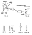
- FIG. 23 illustrates embedded sensors 6 in the femur and the tibia
- FIG. 24 illustrates sensors 6 in the patella.
- the ligaments shown include the medial collateral ligament, the lateral collateral ligament, the anterior cruciate ligament, and the posterior cruciate ligament.
- the sensors 6 are implanted prior to surgery in percutaneously and/or arthroscopically or intra-operatively through open surgery.
- FIG. 25 depicts a ligament or tendon
- FIG. 26 depicts a sensor clamp with a compressive and release handle
- FIG. 27 depicts the deployment of the sensor
- FIG. 28 reveals the deployed sensors in the ligament.
- the sensors can be embedded into the ligaments ( FIG.
- FIG. 25 illustrates an exemplary ligament
- a sensor clamp FIG. 26
- FIG. 27 a sensor clamp
- FIG. 27 a sensor clamp
- An ultrasonic cannula system 180 allows external non radiating visualization of the sensor placement as shown in FIG. 29 .
- the cannula 181 houses the transmitter 182 and the receiver 183.
- the deployment sensor 184 is, then, optimally positioned for insertion.
- the ultrasonic arm could, then, be used to obtain a rapid topography of the joint surface and depth.
- the ultrasonic inserter sends energy waves to the multiple embedded sensors 7 that reflect to one another and back to the ultrasonic transducer as shown in FIG. 17.
- FIG. 17 depicts the ultrasonic sensors 7 using reflection techniques with the sound wave.
- the sound waves reflect off the end of the bone and the embedded sensor 7 back to the receiver in the ultrasonic inserter.
- the receiver detects the reflected sound waves and activates the sensor output to a computer screen for visualization as shown in FIG. 18 .
- the ultrasonic wave also exhibits a thru-beam to the tibia.
- the transmitter beams the ultrasonic wave to a separate receiver 190.
- the femur/tibia deflect the beam triggering the receiver output.
- the added ability of the embedded sensors 7 to continually reflect the ultrasonic beam to the network of sensors 7 allows precise three-dimensional information.
- the sensor 7 is programmed to compensate for irregular surfaces and variable surface temperature.
- the measurement of bone is based on the processing of the received ultrasound signals. Speed of the sound and the ultrasound velocity both provide measurements on the basis of how rapidly the ultrasound wave propagates through the bone and the soft tissue. These measures characteristics permit creation of a rapid three-dimensional geometry, which information can be externally sent to the computer system that will allow integration of the prosthesis as shown in FIG. 18 .
- FIG. 30 The deployment of the sensor can be by a single cannula ( FIG. 30 ) with one or several sensors ( FIG. 31 ), or by a multiple sensor deployment cannula ( FIG. 32 ).
- the sensor would have a calibrated trocar that would penetrate skin, muscle, ligament, tendon, cartilage and bone.
- FIG. 33 depicts the deployment of the sensors in an open knee surgery where the soft tissue has been excluded and the cartilage and bone cuts have been made.
- a handle 190 houses a plunger 191 that controls the depth of sensor deployment. See FIGS. 34 to 37 .
- the minimal depth is determined by the amount of cartilage and bone to be cut for the implantation of the prosthesis or implant. For example, in the femur and tibia, a minimum of 10 to 15 millimeters is cut.
- the sensor is deployed deep with respect to that cut so as not to be dislodged during the procedure and to be able to be used in the post-opeative period.
- the trocar tip would house the elements of the sensor ( FIG. 34 ) and, upon reaching the desired depth of deployment, the sensor 8 is inserted by a release of the locking mechanism ( FIG. 19 ), which can be a screw, or a rotate-to-unlock joint, a break-away, or any other decoupling mechanism.
- the external energy wave that will be used can be ultrasonic, or electromagnetic.
- the use of the optical array method could, therefore, be avoided.
- the deflection of the energy through the various mediums (cartilage and bone) and the time element of the energy wave is received by the sensors 8 and/or reflected back to the external receiver.
- a three-dimensional model is depicted. This enables the surgeon to embed the sensors ( FIG. 33 ), use them during surgery ( FIGS. 18 22 ) and, then, leave them implanted to be utilized after surgery ( FIGS. 12 and 13 ). Accordingly, the speed of information transmission would be greatly increased and processed.
- FIGS. 23 and 24 depict some elements of the knee joint soft tissue.
- the ACL, the PCL, the medial collateral ligament, and the lateral collateral ligament are important for balancing of a knee joint during surgery.
- the sensors are embedded into the ligament of a tendon by a clip mechanism (see FIGS. 25 to 28 ).
- the information is received and processed by a software system that is integrated into the computer-assisted joint surgery device and presents a visual analogue of an intra-opeative joint ( FIG. 22 ). Ligament tension, pressure, shear, etc. is evaluated.
- a soft-tissue balancing grid aids in the surgeons approach regarding soft tissue releases and component rotation.
- FIG. 38 depicts a similar sensor system in the hip.
- the inserter is similar to a single sensor inserter as shown in FIG. 38 , or can be modified as shown in FIG. 38 .
- the inserter is configured to a cannulated acetabular reamer that is used in standard hip surgery.
- the handle 200 stabilizes the construct and the sensors 8 are deployed by depressing a plunger in the handle 200.
- FIG. 40 depicts a cup sensor inserter.
- the cannulated holes allow deployment of the sensor 9.
- the construct can be modified similar to FIG. 29 to include an ultrasonic component to help visualize the anatomy.
- FIGS. 34 to 37 depict the development of "smart" inserters and “smart” instruments.
- the handle 210 of the inserter/instrument houses an array of sensors 8 to aid in the precise cutting of the bone ( FIG. 36 ) as well as the insertion of the prosthesis and sensors ( FIGS. 35 and 37 ).
- These sensors 8 are spatially identified by the ultrasonic/electromagnetic transducer and receiver to allow confirmation that the implant/bone interface was prepared appropriately, and that the implant was inserted to the appropriate depth and angle. The stability of a cemented or press fit component could, then, be tested.
- Sensors implanted onto the prosthesis at the time of surgery or prior to surgery also allow precision insertion and orientation of the prosthesis. Post-operative implant evaluation also is performed.
- FIG. 39 depicts the insertion of the sensors 8 into the femur.
- the sensor 8 can be deployed from the inside-out, from the outside-in, or incorporated into the distal centralizer of the prosthesis and or the canal restrictor.
- FIG. 41 depicts the lateral view of two spinal segments.
- the sensor inserter is shown in a percutaneous manner deploying the sensor into the vertebral body.
- FIG. 42 depicts an axial view of one vertebral level.
- the sensor 9 is implanted through the pedicle that has been prepared for instrumentation.
- FIG. 12 The implanted sensor system following prosthesis insertion is depicted in FIG. 12 , an anterior view of the prosthesis, and shows the knee joint, femoral and tibial prostheses, the polyethylene implant, and the embedded sensors.
- FIG. 13 depicts a lateral view of a knee joint with the prosthesis implanted with sensor system.
- FIG. 14 depicts a total hip prosthesis with the embedded sensor system.
- FIG. 15 depicts a lateral view of the embedded sensors within two segments of the vertebrae and an implant.
- FIG. 16 depicts a sensor system within a vertebral body with a superior (axial) view of a prosthesis/implant.
- the sensor system of the present invention can be used pre-operatively to follow the progression of joint pathology and the different treatment interventions.
- the system can be used intra-operatively to aid in the implantation of the prosthesis/instrumentation/hardware.
- the sensors can, then, be used post-operatively to evaluate changes over time and dynamic changes.
- the sensor are activated intra-operatively and parameter readings are stored. Immediately post-operatively, the sensor is activated and a baseline is known.
- the sensors can be powered by internal batteries or by external measures.
- a patient could be evaluated in bed at night by a non-contact activation system that can use radio frequency or electromagnetic/ultrasonic energy.
- the sensor systems' energy signal can penetrate,the bed, activate the sensors, and transmit to a receiver that also can be attached to the bed.
- the sensors can be "upgraded" over time (e.g., with appropriate software enhancements) to evaluate various parameters.
- the sensors can be modified by an external device, such as a flash drive.
- a set of embedded sensors can monitor the progression of a spinal fusion that is instrumented. Once a given parameter is confirmed, the same sensors can be reprogrammed to monitor the adjacent spinal segments to predict increased stress and, ultimately, subluxation of an adjacent level.
- Another feature of the sensor system is that it can rotate through a series of sensor parameters during an evaluation period.
- An example of such rotation can be evaluation of the bone density as the patient sleeps, and, following this, an evaluation of vascular joint fluid viscosity, and bearing surfaces.
- Such evaluation can occur on a fixed time sequence on specific intervals or randomly as desired.
- the information can telemetrically sent to the health care provider by current telephonic devices.
- the patient can be evaluated in the doctor's office with an external sensor activator. The patient could, then, go through a series of motions that allow the physician to evaluate implant function, including such parameters as load, torque, motion, stability, etc.
- the software system houses the sensor information in a grid that allows interval comparisons. The physician, then, evaluates the data and functions that fall outside the standard deviations are highlighted, with these parameters being further evaluated.
Landscapes
- Health & Medical Sciences (AREA)
- Life Sciences & Earth Sciences (AREA)
- Physics & Mathematics (AREA)
- Engineering & Computer Science (AREA)
- Medical Informatics (AREA)
- Pathology (AREA)
- Biomedical Technology (AREA)
- Heart & Thoracic Surgery (AREA)
- Molecular Biology (AREA)
- Surgery (AREA)
- Animal Behavior & Ethology (AREA)
- General Health & Medical Sciences (AREA)
- Public Health (AREA)
- Veterinary Medicine (AREA)
- Biophysics (AREA)
- Orthopedic Medicine & Surgery (AREA)
- Oral & Maxillofacial Surgery (AREA)
- Dentistry (AREA)
- Rheumatology (AREA)
- Nuclear Medicine, Radiotherapy & Molecular Imaging (AREA)
- Radiology & Medical Imaging (AREA)
- Physiology (AREA)
- Dermatology (AREA)
- Rehabilitation Therapy (AREA)
- Computer Networks & Wireless Communication (AREA)
- Spectroscopy & Molecular Physics (AREA)
- Optics & Photonics (AREA)
- Transplantation (AREA)
- Cardiology (AREA)
- Prostheses (AREA)
- Ultra Sonic Daignosis Equipment (AREA)
- Measuring And Recording Apparatus For Diagnosis (AREA)
- Measurement Of The Respiration, Hearing Ability, Form, And Blood Characteristics Of Living Organisms (AREA)
- Surgical Instruments (AREA)
- Measuring Pulse, Heart Rate, Blood Pressure Or Blood Flow (AREA)
Claims (2)
- Système d'évaluation de la densité osseuse, qui comprend :au moins deux capteurs biométriques (1 à 9) pour une utilisation dans des implants vertébraux sur au moins une vertèbre (10, 20 ; 110, 120) d'une colonne vertébrale, dans lequel lesdits au moins deux capteurs biométriques (1 à 9) sont adaptés pour mesurer au moins un paramètre biométrique dans un environnement adjacent à l'aide d'ondes d'énergie d'ultrasons transmises et reçues entre lesdits au moins deux capteurs biométriques (1 à 9) ; etdans lequel chaque capteur biométrique (1 à 9) est adapté pour envoyer des ondes d'énergie vibratoire qui peuvent imiter une charge sur un os pour le traitement de fractures et de fusions des vertèbres.
- Système selon la revendication 1, qui comprend en outre :un récepteur externe ;dans lequel lesdits au moins deux capteurs biométriques (1 à 9) comprennent des moyens de transmission de données relatives à au moins un paramètre biométrique au récepteur externe ; etdans lequel le récepteur externe comprend des moyens d'analyse des données pour évaluer la densité osseuse calcanéenne d'au moins une vertèbre (10, 20 ; 110, 120).
Priority Applications (4)
| Application Number | Priority Date | Filing Date | Title |
|---|---|---|---|
| EP12005171A EP2510874A3 (fr) | 2005-03-29 | 2006-03-29 | Capteur biométrique |
| EP15164456.4A EP2929836B1 (fr) | 2005-03-29 | 2006-03-29 | Système de détection biométrique |
| EP12005170.1A EP2510873B1 (fr) | 2005-03-29 | 2006-03-29 | Capteur biométrique |
| PL06748811T PL1868498T3 (pl) | 2005-03-29 | 2006-03-29 | Czujnik do wykrywania parametrów ciała oraz sposób wykrywania parametrów ciała |
Applications Claiming Priority (4)
| Application Number | Priority Date | Filing Date | Title |
|---|---|---|---|
| US66579705P | 2005-03-29 | 2005-03-29 | |
| US76386906P | 2006-02-01 | 2006-02-01 | |
| US76376106P | 2006-02-01 | 2006-02-01 | |
| PCT/US2006/011300 WO2006105098A2 (fr) | 2005-03-29 | 2006-03-29 | Capteur de detection de parametres corporels et procede de detection de parametres corporels |
Related Child Applications (5)
| Application Number | Title | Priority Date | Filing Date |
|---|---|---|---|
| EP12005170.1A Division EP2510873B1 (fr) | 2005-03-29 | 2006-03-29 | Capteur biométrique |
| EP15164456.4A Division EP2929836B1 (fr) | 2005-03-29 | 2006-03-29 | Système de détection biométrique |
| EP12005171A Division EP2510874A3 (fr) | 2005-03-29 | 2006-03-29 | Capteur biométrique |
| EP12005170.1 Division-Into | 2012-07-13 | ||
| EP12005171.9 Division-Into | 2012-07-13 |
Publications (3)
| Publication Number | Publication Date |
|---|---|
| EP1868498A2 EP1868498A2 (fr) | 2007-12-26 |
| EP1868498A4 EP1868498A4 (fr) | 2010-04-21 |
| EP1868498B1 true EP1868498B1 (fr) | 2013-05-15 |
Family
ID=37054017
Family Applications (4)
| Application Number | Title | Priority Date | Filing Date |
|---|---|---|---|
| EP06748811.4A Expired - Lifetime EP1868498B1 (fr) | 2005-03-29 | 2006-03-29 | Capteur de detection de parametres corporels et procede de detection de parametres corporels |
| EP12005170.1A Expired - Lifetime EP2510873B1 (fr) | 2005-03-29 | 2006-03-29 | Capteur biométrique |
| EP12005171A Ceased EP2510874A3 (fr) | 2005-03-29 | 2006-03-29 | Capteur biométrique |
| EP15164456.4A Expired - Lifetime EP2929836B1 (fr) | 2005-03-29 | 2006-03-29 | Système de détection biométrique |
Family Applications After (3)
| Application Number | Title | Priority Date | Filing Date |
|---|---|---|---|
| EP12005170.1A Expired - Lifetime EP2510873B1 (fr) | 2005-03-29 | 2006-03-29 | Capteur biométrique |
| EP12005171A Ceased EP2510874A3 (fr) | 2005-03-29 | 2006-03-29 | Capteur biométrique |
| EP15164456.4A Expired - Lifetime EP2929836B1 (fr) | 2005-03-29 | 2006-03-29 | Système de détection biométrique |
Country Status (13)
| Country | Link |
|---|---|
| US (7) | US7918887B2 (fr) |
| EP (4) | EP1868498B1 (fr) |
| JP (2) | JP2008534140A (fr) |
| KR (2) | KR101301862B1 (fr) |
| CN (1) | CN104887235B (fr) |
| AU (1) | AU2006230176B2 (fr) |
| CA (1) | CA2600613C (fr) |
| DK (1) | DK1868498T3 (fr) |
| ES (1) | ES2428639T3 (fr) |
| GB (1) | GB2440059A (fr) |
| PL (1) | PL1868498T3 (fr) |
| RU (4) | RU2011137825A (fr) |
| WO (1) | WO2006105098A2 (fr) |
Families Citing this family (158)
| Publication number | Priority date | Publication date | Assignee | Title |
|---|---|---|---|---|
| US20110213221A1 (en) * | 2005-03-29 | 2011-09-01 | Roche Martin W | Method for Detecting Body Parameters |
| US20100204551A1 (en) * | 2008-10-22 | 2010-08-12 | Martin William Roche | Detection, Prevention and Treatment of Infections in Implantable Devices |
| US20100100011A1 (en) * | 2008-10-22 | 2010-04-22 | Martin Roche | System and Method for Orthopedic Alignment and Measurement |
| US8095198B2 (en) * | 2006-01-31 | 2012-01-10 | Warsaw Orthopedic. Inc. | Methods for detecting osteolytic conditions in the body |
| WO2007090159A1 (fr) * | 2006-01-31 | 2007-08-09 | Medtronic, Inc. | capteur implantable |
| US20070238992A1 (en) * | 2006-02-01 | 2007-10-11 | Sdgi Holdings, Inc. | Implantable sensor |
| US7328131B2 (en) | 2006-02-01 | 2008-02-05 | Medtronic, Inc. | Implantable pedometer |
| US8078282B2 (en) | 2006-02-01 | 2011-12-13 | Warsaw Orthopedic, Inc | Implantable tissue growth stimulator |
| US7993269B2 (en) * | 2006-02-17 | 2011-08-09 | Medtronic, Inc. | Sensor and method for spinal monitoring |
| JP2009529954A (ja) * | 2006-03-14 | 2009-08-27 | マコ サージカル コーポレーション | 補綴装置ならびに補綴装置を埋め込むためのシステムおよび方法 |
| US7918796B2 (en) | 2006-04-11 | 2011-04-05 | Warsaw Orthopedic, Inc. | Volumetric measurement and visual feedback of tissues |
| WO2008052367A1 (fr) * | 2006-10-31 | 2008-05-08 | Ao Technology Ag | Procédé et dispositif servant à mesurer la résistance mécanique locale d'un corps poreux |
| US20080262332A1 (en) * | 2007-04-19 | 2008-10-23 | Medtronic, Inc. | Infection monitoring |
| US7734353B2 (en) | 2007-04-19 | 2010-06-08 | Medtronic Inc. | Controlling temperature during recharge for treatment of infection or other conditions |
| US20080262374A1 (en) * | 2007-04-19 | 2008-10-23 | Medtronic, Inc. | Event triggered infection monitoring |
| US20080262331A1 (en) * | 2007-04-19 | 2008-10-23 | Medtronic, Inc. | Infection monitoring |
| US7766862B2 (en) * | 2007-04-19 | 2010-08-03 | Medtronic, Inc. | Baseline acquisition for infection monitoring |
| US7682355B2 (en) | 2007-04-19 | 2010-03-23 | Medtronic, Inc. | Refined infection monitoring |
| US7604629B2 (en) * | 2007-04-19 | 2009-10-20 | Medtronic Inc. | Multi-parameter infection monitoring |
| US7822465B2 (en) * | 2007-04-25 | 2010-10-26 | Warsaw Orthopedic, Inc. | Device and method for image-based device performance measurement |
| US20090005708A1 (en) * | 2007-06-29 | 2009-01-01 | Johanson Norman A | Orthopaedic Implant Load Sensor And Method Of Interpreting The Same |
| US8100950B2 (en) * | 2007-07-27 | 2012-01-24 | The Cleveland Clinic Foundation | Oblique lumbar interbody fusion |
| US8915866B2 (en) | 2008-01-18 | 2014-12-23 | Warsaw Orthopedic, Inc. | Implantable sensor and associated methods |
| US8197489B2 (en) | 2008-06-27 | 2012-06-12 | Depuy Products, Inc. | Knee ligament balancer |
| JP5185713B2 (ja) * | 2008-07-11 | 2013-04-17 | 日立アロカメディカル株式会社 | 人工関節検索装置 |
| US8078440B2 (en) * | 2008-09-19 | 2011-12-13 | Smith & Nephew, Inc. | Operatively tuning implants for increased performance |
| US8126736B2 (en) | 2009-01-23 | 2012-02-28 | Warsaw Orthopedic, Inc. | Methods and systems for diagnosing, treating, or tracking spinal disorders |
| US8685093B2 (en) | 2009-01-23 | 2014-04-01 | Warsaw Orthopedic, Inc. | Methods and systems for diagnosing, treating, or tracking spinal disorders |
| US20100250284A1 (en) * | 2009-03-26 | 2010-09-30 | Martin Roche | System and method for an orthopedic dynamic data repository and registry for request |
| US8556830B2 (en) | 2009-03-31 | 2013-10-15 | Depuy | Device and method for displaying joint force data |
| US8597210B2 (en) | 2009-03-31 | 2013-12-03 | Depuy (Ireland) | System and method for displaying joint force data |
| US8740817B2 (en) | 2009-03-31 | 2014-06-03 | Depuy (Ireland) | Device and method for determining forces of a patient's joint |
| US8721568B2 (en) | 2009-03-31 | 2014-05-13 | Depuy (Ireland) | Method for performing an orthopaedic surgical procedure |
| US8551023B2 (en) | 2009-03-31 | 2013-10-08 | Depuy (Ireland) | Device and method for determining force of a knee joint |
| JP5771199B2 (ja) * | 2009-06-11 | 2015-08-26 | アーオー テクノロジー アクチエンゲゼルシャフト | 医療用インプラント、診断装置又は生物学的手法をモニタリング及び/又は制御するための測定信号を、処理及び伝送するための装置 |
| US8707782B2 (en) | 2009-06-30 | 2014-04-29 | Orthosensor Inc | Prosthetic component for monitoring synovial fluid and method |
| US8714009B2 (en) | 2010-06-29 | 2014-05-06 | Orthosensor Inc. | Shielded capacitor sensor system for medical applications and method |
| US8679186B2 (en) | 2010-06-29 | 2014-03-25 | Ortho Sensor Inc. | Hermetically sealed prosthetic component and method therefor |
| US20100331679A1 (en) * | 2009-06-30 | 2010-12-30 | Orthosensor | Pulsed echo sensing device and method for an orthopedic joint |
| US9839390B2 (en) | 2009-06-30 | 2017-12-12 | Orthosensor Inc. | Prosthetic component having a compliant surface |
| US9462964B2 (en) | 2011-09-23 | 2016-10-11 | Orthosensor Inc | Small form factor muscular-skeletal parameter measurement system |
| US8720270B2 (en) | 2010-06-29 | 2014-05-13 | Ortho Sensor Inc. | Prosthetic component for monitoring joint health |
| US8826733B2 (en) | 2009-06-30 | 2014-09-09 | Orthosensor Inc | Sensored prosthetic component and method |
| US9259179B2 (en) | 2012-02-27 | 2016-02-16 | Orthosensor Inc. | Prosthetic knee joint measurement system including energy harvesting and method therefor |
| US8701484B2 (en) | 2010-06-29 | 2014-04-22 | Orthosensor Inc. | Small form factor medical sensor structure and method therefor |
| US20100331733A1 (en) * | 2009-06-30 | 2010-12-30 | Orthosensor | Sensing device and method for an orthopedic joint |
| US8827986B2 (en) * | 2009-10-19 | 2014-09-09 | Pharmaco-Kinesis Corporation | Remotely activated piezoelectric pump for delivery of biological agents to the intervertebral disc and spine |
| FR2952518B1 (fr) * | 2009-11-13 | 2011-10-28 | Univ Paris 6 Pierre Et Marie Curie | Dispositif de mesure d'une activite de la moelle epiniere d'un vertebre |
| US8376937B2 (en) * | 2010-01-28 | 2013-02-19 | Warsaw Orhtopedic, Inc. | Tissue monitoring surgical retractor system |
| US9161717B2 (en) | 2011-09-23 | 2015-10-20 | Orthosensor Inc. | Orthopedic insert measuring system having a sealed cavity |
| EP2386493B1 (fr) * | 2010-05-12 | 2017-09-06 | Mondi Halle GmbH | Sac à fond plat tenant debout composé d'une feuille plastique pouvant être scellée à chaud |
| US20110295159A1 (en) * | 2010-05-25 | 2011-12-01 | Pharmaco-Kinesis Corporation | Method and Apparatus for an Implantable Inertial-Based Sensing System for Real-Time, In Vivo Detection of Spinal Pseudarthrosis and Adjacent Segment Motion |
| US8939030B2 (en) * | 2010-06-29 | 2015-01-27 | Orthosensor Inc | Edge-detect receiver for orthopedic parameter sensing |
| US9095284B2 (en) | 2010-10-28 | 2015-08-04 | Medtronic, Inc. | Distance measurement using implantable acoustic transducers |
| KR101219710B1 (ko) * | 2010-11-29 | 2013-01-08 | 광주과학기술원 | 추간판 이상 검사 프로브 및 검사 장치 |
| US20140379090A1 (en) * | 2011-08-08 | 2014-12-25 | Ecole Polytechnique Federale De Lausanne (Epfl) | In-vivo condition monitoring of metallic implants by electrochemical techniques |
| GB201115411D0 (en) | 2011-09-07 | 2011-10-19 | Depuy Ireland | Surgical instrument |
| US9839374B2 (en) | 2011-09-23 | 2017-12-12 | Orthosensor Inc. | System and method for vertebral load and location sensing |
| US9414940B2 (en) | 2011-09-23 | 2016-08-16 | Orthosensor Inc. | Sensored head for a measurement tool for the muscular-skeletal system |
| US8911448B2 (en) | 2011-09-23 | 2014-12-16 | Orthosensor, Inc | Device and method for enabling an orthopedic tool for parameter measurement |
| US8942662B2 (en) * | 2012-02-16 | 2015-01-27 | The United States of America, as represented by the Secretary, Department of Health and Human Services, Center for Disease Control and Prevention | System and method to predict and avoid musculoskeletal injuries |
| US9271675B2 (en) | 2012-02-27 | 2016-03-01 | Orthosensor Inc. | Muscular-skeletal joint stability detection and method therefor |
| US9622701B2 (en) | 2012-02-27 | 2017-04-18 | Orthosensor Inc | Muscular-skeletal joint stability detection and method therefor |
| US9844335B2 (en) | 2012-02-27 | 2017-12-19 | Orthosensor Inc | Measurement device for the muscular-skeletal system having load distribution plates |
| US9381011B2 (en) | 2012-03-29 | 2016-07-05 | Depuy (Ireland) | Orthopedic surgical instrument for knee surgery |
| US10098761B2 (en) | 2012-03-31 | 2018-10-16 | DePuy Synthes Products, Inc. | System and method for validating an orthopaedic surgical plan |
| US9545459B2 (en) | 2012-03-31 | 2017-01-17 | Depuy Ireland Unlimited Company | Container for surgical instruments and system including same |
| US10070973B2 (en) | 2012-03-31 | 2018-09-11 | Depuy Ireland Unlimited Company | Orthopaedic sensor module and system for determining joint forces of a patient's knee joint |
| US10206792B2 (en) | 2012-03-31 | 2019-02-19 | Depuy Ireland Unlimited Company | Orthopaedic surgical system for determining joint forces of a patients knee joint |
| EP2849683A4 (fr) | 2012-05-18 | 2015-11-25 | Orthalign Inc | Dispositifs et méthodes pour arthroplastie du genou |
| US20140135744A1 (en) | 2012-11-09 | 2014-05-15 | Orthosensor Inc | Motion and orientation sensing module or device for positioning of implants |
| US9241742B2 (en) | 2013-03-14 | 2016-01-26 | DePuy Synthes Products, Inc. | Methods and devices for polyaxial screw alignment |
| KR102548797B1 (ko) | 2013-03-15 | 2023-06-28 | 카나리 메디칼 아이엔씨. | 고관절 치환물을 모니터링하기 위한 장치, 시스템 및 방법 |
| US11793424B2 (en) * | 2013-03-18 | 2023-10-24 | Orthosensor, Inc. | Kinetic assessment and alignment of the muscular-skeletal system and method therefor |
| US9408557B2 (en) * | 2013-03-18 | 2016-08-09 | Orthosensor Inc. | System and method to change a contact point of the muscular-skeletal system |
| WO2014169217A2 (fr) | 2013-04-11 | 2014-10-16 | Ohio University | Systèmes et procédé pour établir la rigidité d'un os au moyen d'une analyse des tissus à réponse mécanique |
| WO2014209916A1 (fr) * | 2013-06-23 | 2014-12-31 | Hunter William L | Dispositifs, systèmes et procédés de surveillance de remplacements du genou |
| FR3010628B1 (fr) | 2013-09-18 | 2015-10-16 | Medicrea International | Procede permettant de realiser la courbure ideale d'une tige d'un materiel d'osteosynthese vertebrale destinee a etayer la colonne vertebrale d'un patient |
| FR3012030B1 (fr) | 2013-10-18 | 2015-12-25 | Medicrea International | Procede permettant de realiser la courbure ideale d'une tige d'un materiel d'osteosynthese vertebrale destinee a etayer la colonne vertebrale d'un patient |
| US20150238691A1 (en) * | 2014-02-25 | 2015-08-27 | Elwha Llc | Control systems for release of medication responsive to joint activity |
| EP3160369A4 (fr) | 2014-06-25 | 2018-04-18 | Canary Medical Inc. | Dispositifs, systèmes et procédés d'utilisation et de surveillance d'implants rachidiens |
| US10874496B2 (en) | 2014-06-25 | 2020-12-29 | Canary Medical Inc. | Devices, systems and methods for using and monitoring implants |
| US10524694B2 (en) | 2014-06-25 | 2020-01-07 | Canaray Medical Inc. | Devices, systems and methods for using and monitoring tubes in body passageways |
| CA2990825A1 (fr) | 2014-06-25 | 2015-12-30 | William L. Hunter | Dispositifs, systemes et procedes d'utilisation et de surveillance de materiel orthopedique |
| US9993177B2 (en) | 2014-08-28 | 2018-06-12 | DePuy Synthes Products, Inc. | Systems and methods for intraoperatively measuring anatomical orientation |
| SG10201902350XA (en) | 2014-09-17 | 2019-04-29 | Canary Medical Inc | Devices, systems and methods for using and monitoring medical devices |
| CN105212951B (zh) * | 2015-10-15 | 2017-10-20 | 中国人民解放军第二军医大学 | 脊柱在体生物力学测量装置 |
| TWI547258B (zh) * | 2015-10-23 | 2016-09-01 | 國立交通大學 | 感測式骨頭固定元件 |
| US10456211B2 (en) | 2015-11-04 | 2019-10-29 | Medicrea International | Methods and apparatus for spinal reconstructive surgery and measuring spinal length and intervertebral spacing, tension and rotation |
| PT3386393T (pt) * | 2015-12-08 | 2021-06-01 | Kneevoice Inc | Avaliação do estado das articulações utilizando sensores acústicos |
| US9633538B1 (en) | 2015-12-09 | 2017-04-25 | International Business Machines Corporation | System and method for wearable indication of personal risk within a workplace |
| US9691262B1 (en) | 2015-12-17 | 2017-06-27 | International Business Machines Corporation | Informing first responders based on incident detection, and automatic reporting of individual location and equipment state |
| US9848269B2 (en) | 2015-12-29 | 2017-12-19 | International Business Machines Corporation | Predicting harmful noise events and implementing corrective actions prior to noise induced hearing loss |
| US10643447B2 (en) | 2015-12-29 | 2020-05-05 | International Business Machines Corporation | Predicting harmful chemical exposures and implementing corrective actions prior to overexposure |
| US10335241B2 (en) | 2015-12-30 | 2019-07-02 | DePuy Synthes Products, Inc. | Method and apparatus for intraoperative measurements of anatomical orientation |
| US9554411B1 (en) | 2015-12-30 | 2017-01-24 | DePuy Synthes Products, Inc. | Systems and methods for wirelessly powering or communicating with sterile-packed devices |
| US10762460B2 (en) | 2015-12-30 | 2020-09-01 | International Business Machines Corporation | Predictive alerts for individual risk of injury with ameliorative actions |
| WO2017139556A1 (fr) | 2016-02-12 | 2017-08-17 | Medos International Sarl | Systèmes et procédés pour mesurer l'orientation anatomique pendant une opération |
| CN109310324A (zh) | 2016-03-23 | 2019-02-05 | 卡纳里医疗公司 | 用于警报植入物的植入式报告处理器 |
| US11191479B2 (en) | 2016-03-23 | 2021-12-07 | Canary Medical Inc. | Implantable reporting processor for an alert implant |
| US10912648B2 (en) | 2016-08-30 | 2021-02-09 | Longeviti Neuro Solutions Llc | Method for manufacturing a low-profile intercranial device and the low-profile intercranial device manufactured thereby |
| US10820835B2 (en) | 2016-09-12 | 2020-11-03 | Medos International Sarl | Systems and methods for anatomical alignment |
| WO2018109556A1 (fr) | 2016-12-12 | 2018-06-21 | Medicrea International | Systèmes et procédés pour des implants rachidiens spécifiques au patient |
| US10657338B2 (en) | 2017-01-16 | 2020-05-19 | Neva Electromagnetics, LLC | Microwave antenna array and testbed for osteoporosis detection |
| WO2018169980A1 (fr) | 2017-03-14 | 2018-09-20 | OrthAlign, Inc. | Systèmes et procédés de mesure & d'équilibrage de tissu mou |
| US11089975B2 (en) | 2017-03-31 | 2021-08-17 | DePuy Synthes Products, Inc. | Systems, devices and methods for enhancing operative accuracy using inertial measurement units |
| WO2018193316A2 (fr) | 2017-04-21 | 2018-10-25 | Medicrea International | Système de conception d'un ou de plusieurs implants rachidiens spécifiques du patient |
| EP3406185B1 (fr) | 2017-05-22 | 2021-02-24 | Vestel Elektronik Sanayi ve Ticaret A.S. | Dispositif médical implantable et système et procédés de communication sans fil intra-osseuse |
| US11419541B2 (en) | 2017-05-31 | 2022-08-23 | Ohio University | Systems and methods for patient specific modeling of the mechanical properties of bone |
| WO2019032488A1 (fr) | 2017-08-07 | 2019-02-14 | DePuy Synthes Products, Inc. | Capteurs implantables dans le corps d'un patient, systèmes et procédés d'utilisation de ceux-ci |
| CN111093556B (zh) * | 2017-09-14 | 2022-08-16 | 学校法人庆应义塾 | 植入物设置强度评价装置以及记录介质 |
| AU2018332792B2 (en) | 2017-09-14 | 2024-07-25 | Howmedica Osteonics Corp. | Non-symmetrical insert sensing system and method therefor |
| US20190117285A1 (en) * | 2017-10-24 | 2019-04-25 | Hae Sun Paik | Checking apparatus for bone conglutination |
| JP2021507732A (ja) * | 2017-11-30 | 2021-02-25 | ブルーイン、バイオメトリクス、リミテッド、ライアビリティー、カンパニーBruin Biometrics, Llc | アコースティックエミッションを用いるインプラント評価 |
| US10918422B2 (en) | 2017-12-01 | 2021-02-16 | Medicrea International | Method and apparatus for inhibiting proximal junctional failure |
| KR101884226B1 (ko) | 2017-12-29 | 2018-08-02 | 신종환 | 골이식 대체재를 이용한 생체신호 검출장치 |
| US11589992B2 (en) | 2018-01-09 | 2023-02-28 | Longeviti Neuro Solutions Llc | Universal low-profile intercranial assembly |
| US11101554B2 (en) | 2018-01-16 | 2021-08-24 | Neva Electromagnetics, LLC | Dual antiphase antenna for better signal transmission into human body or signal reception from human body |
| WO2019147608A1 (fr) * | 2018-01-24 | 2019-08-01 | Ohio University | Procédés pour établir la rigidité d'un os à l'aide d'une analyse de tissu à réponse mécanique |
| DE102018204949A1 (de) * | 2018-03-30 | 2019-10-02 | Bernhard Clasbrummel | Implantat und Verfahren zur Diagnose und/oder Behandlung entzündlicher Gewebezustände |
| US11135066B2 (en) | 2018-04-23 | 2021-10-05 | Medos International Sarl | Mechanical fuse for surgical implants and related methods |
| US12514617B2 (en) | 2018-07-19 | 2026-01-06 | Warsaw Orthopedic, Inc. | Modular set screw design for housing microelectronics |
| US11529208B2 (en) | 2018-07-19 | 2022-12-20 | Warsaw Orthopedic, Inc. | Break-off set screw |
| US20210330249A1 (en) * | 2020-04-22 | 2021-10-28 | Warsaw Orthopedic, Inc. | System and method for post-operative assessment of spinal motion and implant based strain correlation |
| US12396679B2 (en) | 2018-07-19 | 2025-08-26 | Warsaw Orthopedic, Inc. | System and method for post-operative assessment of spinal motion and implant-based strain correlation |
| US12508097B2 (en) | 2018-07-19 | 2025-12-30 | Warsaw Orthopedic, Inc. | Set screw with strain gauges |
| US11278238B2 (en) | 2018-09-14 | 2022-03-22 | Warsaw Orthopedic, Inc. | Wearable sensor device and analysis platform for objective outcome assessment in spinal diseases |
| CN109009225A (zh) * | 2018-07-25 | 2018-12-18 | 中国科学院苏州生物医学工程技术研究所 | 骨骼检测方法、装置及骨密度仪 |
| JP7434677B2 (ja) | 2018-10-05 | 2024-02-21 | ホウメディカ・オステオニクス・コーポレイション | 球関節設置を支持するように構成される計測システム及びその方法 |
| US11998456B2 (en) | 2018-10-05 | 2024-06-04 | Exactech, Inc. | Shoulder joint implant selection system |
| US11992424B2 (en) | 2018-10-05 | 2024-05-28 | Howmedica Osteonics Corp. | Measurement system for a selection of at least one prosthetic component of a shoulder joint during surgery |
| US11944385B2 (en) | 2019-04-02 | 2024-04-02 | Medicrea International | Systems and methods for medical image analysis |
| US11925417B2 (en) | 2019-04-02 | 2024-03-12 | Medicrea International | Systems, methods, and devices for developing patient-specific spinal implants, treatments, operations, and/or procedures |
| US12564447B2 (en) | 2019-04-02 | 2026-03-03 | Medicrea International | Systems, methods, and devices for developing patient-specific spinal implants, treatments, operations, and/or procedures |
| WO2020201353A1 (fr) | 2019-04-02 | 2020-10-08 | Medicrea International | Systèmes, procédés et dispositifs pour développer des implants spinaux spécifiques d'un patient, des traitements, des opérations et/ou des procédures |
| US11937896B1 (en) * | 2019-05-10 | 2024-03-26 | Smith & Nephew, Inc. | Orthopedic implants with improved sensors |
| US20210366610A1 (en) | 2019-06-06 | 2021-11-25 | Canary Medical Inc. | Intelligent joint prosthesis |
| CA3176861A1 (fr) | 2019-06-06 | 2020-12-10 | Canary Medical Inc. | Prothese articulaire intelligente |
| US11826164B2 (en) | 2019-06-28 | 2023-11-28 | Orthosensor Inc. | Subdermal medical system for generating measurement data or providing a therapy |
| CN110742614B (zh) * | 2019-09-26 | 2023-05-16 | 南京林业大学 | 一种卧姿脊柱形态测试仪 |
| WO2021059668A1 (fr) * | 2019-09-27 | 2021-04-01 | 富士フイルム株式会社 | Dispositif de traitement d'informations, procédé de traitement d'informations et programme |
| US11812978B2 (en) | 2019-10-15 | 2023-11-14 | Orthosensor Inc. | Knee balancing system using patient specific instruments |
| US11769251B2 (en) | 2019-12-26 | 2023-09-26 | Medicrea International | Systems and methods for medical image analysis |
| US12408963B2 (en) | 2020-02-20 | 2025-09-09 | Canary Medical Switzerland Ag | Medical device for implanting in boney tissue and characterization of bone fractures |
| US20230157756A1 (en) * | 2020-05-04 | 2023-05-25 | Howmedica Osteonics Corp. | Surgical system for revision orthopedic surgical procedures |
| DE202020103194U1 (de) * | 2020-06-03 | 2020-06-29 | Bornemann Gewindetechnik GmbH & Co. KG | Verbesserte Baugruppe mit Helixform und Anlage umfassend die verbesserte Baugruppe |
| EP4171405B1 (fr) * | 2020-06-26 | 2024-09-11 | Prometheus Regeneration R&D Limited | Micro-capteurs implantables dans un os |
| US12178663B2 (en) | 2020-12-17 | 2024-12-31 | Mazor Robotics Ltd. | Systems, devices, and methods for monitoring a rod reduction process |
| US20220202505A1 (en) * | 2020-12-31 | 2022-06-30 | Martin Roche | Intra-operative and post-operative position, motion, and orientation system |
| EP4059479A1 (fr) * | 2021-03-15 | 2022-09-21 | Skulle Implants OY | Implant orthopédique |
| US12370062B2 (en) | 2021-06-04 | 2025-07-29 | Zimmer, Inc. | Independently implantable sensors for orthopedic implants |
| EP4104895A1 (fr) | 2021-06-14 | 2022-12-21 | Instituto Politécnico De Leiria | Biodispositif biomimétique intelligent et son utilisation |
| US12318144B2 (en) | 2021-06-23 | 2025-06-03 | Medicrea International SA | Systems and methods for planning a patient-specific spinal correction |
| US20230255794A1 (en) * | 2022-02-14 | 2023-08-17 | Orthosensor Inc. | Implant Encoder |
| US12599412B2 (en) | 2022-04-12 | 2026-04-14 | Warsaw Orthopedic, Inc. | Intra-operative options for smart implants |
| US12465408B2 (en) | 2022-04-12 | 2025-11-11 | Warsaw Orthopedic, Inc. | Spinal rod connecting components with active sensing capabilities |
| CN115211900A (zh) * | 2022-07-12 | 2022-10-21 | 北京易迈医疗科技有限公司 | 一种关节置换术的植入物嵌入式监测系统及方法 |
| WO2024233002A1 (fr) * | 2023-05-10 | 2024-11-14 | The Johns Hopkins University | Système et procédé de fixation de capteurs implantables |
Family Cites Families (121)
| Publication number | Priority date | Publication date | Assignee | Title |
|---|---|---|---|---|
| US4146022A (en) * | 1977-11-16 | 1979-03-27 | Ronald A. Johnson | Fracture fixation by cerclage utilizing cortical bone tack and pull-out tension device |
| JPS62117553A (ja) * | 1985-11-16 | 1987-05-29 | 肥後 矢吉 | 機能評価装置 |
| JPH01299537A (ja) * | 1988-05-27 | 1989-12-04 | Agency Of Ind Science & Technol | 音響特性測定装置及び測温装置 |
| US5197488A (en) * | 1991-04-05 | 1993-03-30 | N. K. Biotechnical Engineering Co. | Knee joint load measuring instrument and joint prosthesis |
| US5470354A (en) * | 1991-11-12 | 1995-11-28 | Biomet Inc. | Force sensing apparatus and method for orthopaedic joint reconstruction |
| US5425775A (en) * | 1992-06-23 | 1995-06-20 | N.K. Biotechnical Engineering Company | Method for measuring patellofemoral forces |
| US5445642A (en) * | 1992-09-01 | 1995-08-29 | Depuy Inc. | Method for installing a femoral component |
| US5456724A (en) | 1993-12-15 | 1995-10-10 | Industrial Technology Research Institute | Load sensor for bone graft |
| US5524624A (en) | 1994-05-05 | 1996-06-11 | Amei Technologies Inc. | Apparatus and method for stimulating tissue growth with ultrasound |
| FR2729844B1 (fr) | 1995-01-26 | 1997-11-28 | Sabin Pierre Jean Claude | Ensemble d'appareillage medical a implant permettant des connexions electriques a travers la peau |
| US5676784A (en) * | 1995-03-15 | 1997-10-14 | Abbott Laboratories | Method of fabricating a heater coil for a catheter used to monitor cardiac output |
| US5756145A (en) * | 1995-11-08 | 1998-05-26 | Baylor College Of Medicine | Durable, Resilient and effective antimicrobial coating for medical devices and method of coating therefor |
| US5682886A (en) * | 1995-12-26 | 1997-11-04 | Musculographics Inc | Computer-assisted surgical system |
| ES2112203B1 (es) | 1996-01-24 | 1999-01-01 | Querol Luis Maria Ilzarbe | Dispositivo para medir la temperatura en el proceso de fresado en implantologia quirurgica. |
| US5683396A (en) * | 1996-02-20 | 1997-11-04 | Smith & Nephew, Inc. | Orthopaedic cutting instrumentation with cam locking arrangement |
| US5702429A (en) * | 1996-04-04 | 1997-12-30 | Medtronic, Inc. | Neural stimulation techniques with feedback |
| ATE260601T1 (de) * | 1996-09-27 | 2004-03-15 | Metra Biosystems Inc | Auswertung von ultraschallsignalen zur knochen- analyse mittels der ein messfeld darstellenden werte |
| US6090114A (en) * | 1997-02-10 | 2000-07-18 | Stryker Howmedica Osteonics Corp. | Tibial plateau resection guide |
| US6228089B1 (en) * | 1997-12-19 | 2001-05-08 | Depuy International Limited | Device for positioning and guiding a surgical instrument during orthopaedic interventions |
| US6585649B1 (en) * | 1998-05-02 | 2003-07-01 | John D. Mendlein | Methods and devices for improving ultrasonic measurements using multiple angle interrogation |
| US6493588B1 (en) * | 1998-03-18 | 2002-12-10 | Mmc/Gatx Partnership No. 1 | Electro-nerve stimulator systems and methods |
| US6014588A (en) * | 1998-04-07 | 2000-01-11 | Fitz; William R. | Facet joint pain relief method and apparatus |
| US6582365B1 (en) * | 1998-07-09 | 2003-06-24 | The United States Of America As Represented By The Administrator Of The National Aeronautics And Space Administration | Advanced sensor systems for biotelemetry |
| US6304766B1 (en) * | 1998-08-26 | 2001-10-16 | Sensors For Medicine And Science | Optical-based sensing devices, especially for in-situ sensing in humans |
| US6126693A (en) * | 1998-09-18 | 2000-10-03 | Depuy Orthopaedics, Inc. | Tapped box femoral stem attachment for a modular knee prosthesis |
| EP1117328B1 (fr) * | 1998-09-30 | 2008-09-17 | Sicel Technologies, Inc. | Procedes, systemes et dispositifs implantables associes assurant un phasage dynamique des tumeurs |
| US6063091A (en) * | 1998-10-13 | 2000-05-16 | Stryker Technologies Corporation | Methods and tools for tibial intermedullary revision surgery and associated tibial components |
| US6106477A (en) | 1998-12-28 | 2000-08-22 | Medtronic, Inc. | Chronically implantable blood vessel cuff with sensor |
| WO2000038570A1 (fr) * | 1998-12-31 | 2000-07-06 | Ball Semiconductor, Inc. | Mini-detecteurs orthopediques implantes |
| US6143035A (en) * | 1999-01-28 | 2000-11-07 | Depuy Orthopaedics, Inc. | Implanted bone stimulator and prosthesis system and method of enhancing bone growth |
| CA2363254C (fr) * | 1999-03-07 | 2009-05-05 | Discure Ltd. | Procede et appareil de chirurgie informatisee |
| US6470207B1 (en) * | 1999-03-23 | 2002-10-22 | Surgical Navigation Technologies, Inc. | Navigational guidance via computer-assisted fluoroscopic imaging |
| US7080554B2 (en) * | 1999-04-28 | 2006-07-25 | Nexense Ltd. | High-precision measuring method and apparatus |
| IL129651A (en) | 1999-04-28 | 2004-08-31 | Nexense Ltd | High-precision measuring method and apparatus |
| US6984993B2 (en) | 1999-04-28 | 2006-01-10 | Nexense Ltd. | Method and apparatus for making high-precision measurements |
| US6505075B1 (en) * | 1999-05-29 | 2003-01-07 | Richard L. Weiner | Peripheral nerve stimulation method |
| US6371123B1 (en) * | 1999-06-11 | 2002-04-16 | Izex Technology, Inc. | System for orthopedic treatment protocol and method of use thereof |
| US6347245B1 (en) * | 1999-07-14 | 2002-02-12 | Medtronic, Inc. | Medical device ECG marker for use in compressed data system |
| US7366562B2 (en) * | 2003-10-17 | 2008-04-29 | Medtronic Navigation, Inc. | Method and apparatus for surgical navigation |
| US6573706B2 (en) * | 1999-11-18 | 2003-06-03 | Intellijoint Systems Ltd. | Method and apparatus for distance based detection of wear and the like in joints |
| US6245109B1 (en) * | 1999-11-18 | 2001-06-12 | Intellijoint Systems, Ltd. | Artificial joint system and method utilizing same for monitoring wear and displacement of artificial joint members |
| WO2001037733A2 (fr) * | 1999-11-23 | 2001-05-31 | Noveon Ip Holdings Corp. | Appareil et methode d'evaluation du relachement d'implants et de la cicatrisation de fractures |
| EP2327385B1 (fr) | 1999-12-29 | 2016-03-16 | Hill-Rom Services, Inc. | Support pour patient avec barrière |
| US6701174B1 (en) * | 2000-04-07 | 2004-03-02 | Carnegie Mellon University | Computer-aided bone distraction |
| US6535756B1 (en) * | 2000-04-07 | 2003-03-18 | Surgical Navigation Technologies, Inc. | Trajectory storage apparatus and method for surgical navigation system |
| DE10018654A1 (de) * | 2000-04-14 | 2001-12-13 | Jacek Czernicki | Verfahren und Vorrichtung zur Ermittlung des anatomischen Zustands eines Menschen oder eines Tieres, insbesondere zur Diagnose von Osteoporose |
| US6656135B2 (en) * | 2000-05-01 | 2003-12-02 | Southwest Research Institute | Passive and wireless displacement measuring device |
| US20020010390A1 (en) * | 2000-05-10 | 2002-01-24 | Guice David Lehmann | Method and system for monitoring the health and status of livestock and other animals |
| US7018416B2 (en) * | 2000-07-06 | 2006-03-28 | Zimmer Spine, Inc. | Bone implants and methods |
| EP1311191B1 (fr) | 2000-08-25 | 2012-03-07 | The Cleveland Clinic Foundation | Dispositif implantable pour évaluer la charge exercée sur une paire de vertèbres adjacentes |
| WO2002060380A2 (fr) * | 2000-10-26 | 2002-08-08 | Healthetec, Inc. | Surveillance ultrasonique de la densite osseuse avec retroaction alimentaire |
| US20020107537A1 (en) | 2000-12-12 | 2002-08-08 | Lucia Singh | Metal breath freshening device |
| US6558391B2 (en) * | 2000-12-23 | 2003-05-06 | Stryker Technologies Corporation | Methods and tools for femoral resection in primary knee surgery |
| US20020151770A1 (en) * | 2001-01-04 | 2002-10-17 | Noll Austin F. | Implantable medical device with sensor |
| US7547307B2 (en) * | 2001-02-27 | 2009-06-16 | Smith & Nephew, Inc. | Computer assisted knee arthroplasty instrumentation, systems, and processes |
| US20020161304A1 (en) * | 2001-04-30 | 2002-10-31 | Eide Per Kristian | Monitoring pressure in a body cavity |
| WO2002098296A1 (fr) * | 2001-06-05 | 2002-12-12 | Apex Medical, Inc. | Endogreffe sensible a la pression |
| AU2002309239A1 (en) * | 2001-06-05 | 2002-12-16 | Barnev Ltd. | Probe anchor |
| US6589591B1 (en) | 2001-07-10 | 2003-07-08 | Baylor College Of Medicine | Method for treating medical devices using glycerol and an antimicrobial agent |
| US20050203578A1 (en) * | 2001-08-15 | 2005-09-15 | Weiner Michael L. | Process and apparatus for treating biological organisms |
| EP1443859A4 (fr) | 2001-10-24 | 2006-03-22 | Cutting Edge Surgical Inc | Utilisation d'ultrasons intraosseux lors d'une implantation chirurgicale |
| US6645214B2 (en) * | 2001-11-12 | 2003-11-11 | Depuy Orthopaedics, Inc. | Apparatus and method for bone positioning |
| US6791545B2 (en) * | 2001-12-11 | 2004-09-14 | Lecroy Corporation | Measurement icons for digital oscilloscopes |
| US7206627B2 (en) * | 2002-03-06 | 2007-04-17 | Z-Kat, Inc. | System and method for intra-operative haptic planning of a medical procedure |
| US8010180B2 (en) * | 2002-03-06 | 2011-08-30 | Mako Surgical Corp. | Haptic guidance system and method |
| AU2003228341A1 (en) * | 2002-03-19 | 2003-10-08 | The Board Of Trustees Of The University Of Illinois | System and method for prosthetic fitting and balancing in joints |
| US6758850B2 (en) * | 2002-03-29 | 2004-07-06 | Depuy Orthopaedics, Inc. | Instruments and methods for flexion gap adjustment |
| US6638228B1 (en) * | 2002-04-26 | 2003-10-28 | Koninklijke Philips Electronics N.V. | Contrast-agent enhanced color-flow imaging |
| US8996090B2 (en) | 2002-06-03 | 2015-03-31 | Exostat Medical, Inc. | Noninvasive detection of a physiologic parameter within a body tissue of a patient |
| US7025778B2 (en) * | 2002-06-07 | 2006-04-11 | Endovascular Technologies, Inc. | Endovascular graft with pressure, temperature, flow and voltage sensors |
| US6761741B2 (en) * | 2002-06-10 | 2004-07-13 | Kazuho Iesaka | Prosthetic joint |
| US20060246103A1 (en) * | 2002-07-22 | 2006-11-02 | Ralph James D | Implantable devices for the delivery of therapeutic agents to an orthopaedic surgical site |
| US6821299B2 (en) * | 2002-07-24 | 2004-11-23 | Zimmer Technology, Inc. | Implantable prosthesis for measuring six force components |
| JP3910521B2 (ja) * | 2002-11-05 | 2007-04-25 | セイコーインスツル株式会社 | 血圧測定装置 |
| JP4095919B2 (ja) * | 2002-12-09 | 2008-06-04 | ジンマー株式会社 | 人工膝関節全置換手術用計測装置 |
| DE10259437B3 (de) * | 2002-12-19 | 2004-09-16 | Fresenius Medical Care Deutschland Gmbh | Verfahren und Vorrichtung zur Bestimmung des Blutflusses in einer blutführenden Leitung |
| US7029477B2 (en) * | 2002-12-20 | 2006-04-18 | Zimmer Technology, Inc. | Surgical instrument and positioning method |
| US7022141B2 (en) * | 2002-12-20 | 2006-04-04 | Depuy Products, Inc. | Alignment device for modular implants and method |
| US7542791B2 (en) * | 2003-01-30 | 2009-06-02 | Medtronic Navigation, Inc. | Method and apparatus for preplanning a surgical procedure |
| US7458977B2 (en) * | 2003-02-04 | 2008-12-02 | Zimmer Technology, Inc. | Surgical navigation instrument useful in marking anatomical structures |
| US6925339B2 (en) * | 2003-02-04 | 2005-08-02 | Zimmer Technology, Inc. | Implant registration device for surgical navigation system |
| EP1601979A2 (fr) * | 2003-02-13 | 2005-12-07 | Nexense Ltd. | Appareil pour effectuer des mesures avec une grande sensibilite de divers parametres, et capteurs particulierement utiles dans un tel appareil |
| US7824444B2 (en) | 2003-03-20 | 2010-11-02 | Spineco, Inc. | Expandable spherical spinal implant |
| US7527632B2 (en) * | 2003-03-31 | 2009-05-05 | Cordis Corporation | Modified delivery device for coated medical devices |
| US7972616B2 (en) * | 2003-04-17 | 2011-07-05 | Nanosys, Inc. | Medical device applications of nanostructured surfaces |
| EP2335651A1 (fr) * | 2003-07-11 | 2011-06-22 | DePuy Products, Inc. | Dispositif de mésure in vivo d'éspace articulaire |
| US7190273B2 (en) * | 2003-07-11 | 2007-03-13 | Depuy Products, Inc. | Joint endoprosthesis with ambient condition sensing |
| US20050015002A1 (en) * | 2003-07-18 | 2005-01-20 | Dixon Gary S. | Integrated protocol for diagnosis, treatment, and prevention of bone mass degradation |
| US7455647B2 (en) * | 2003-07-24 | 2008-11-25 | Samih Tarabichi | Dynamic spacer for total knee arthroplasty |
| US7613497B2 (en) * | 2003-07-29 | 2009-11-03 | Biosense Webster, Inc. | Energy transfer amplification for intrabody devices |
| JP4386171B2 (ja) * | 2003-12-04 | 2009-12-16 | 三菱自動車工業株式会社 | 4輪駆動車の動力伝達装置 |
| US7488324B1 (en) * | 2003-12-08 | 2009-02-10 | Biomet Manufacturing Corporation | Femoral guide for implanting a femoral knee prosthesis |
| IL159651A0 (en) * | 2003-12-30 | 2004-06-01 | Nexense Ltd | Method and apparatus for measuring torque |
| US7442196B2 (en) * | 2004-02-06 | 2008-10-28 | Synvasive Technology, Inc. | Dynamic knee balancer |
| US20050176823A1 (en) * | 2004-02-10 | 2005-08-11 | Diaz Robert L. | Intra-operative procedure for post-operative pain control |
| IL160365A0 (en) * | 2004-02-12 | 2005-11-20 | Nexense Ltd | Method and apparatus for detecting panel conditions |
| IL161937A (en) * | 2004-05-11 | 2008-08-07 | Nexense Ltd | Sensor system for high-precision measurements of temperature, composition and/or pressure of a fluid |
| EP1765204B1 (fr) | 2004-06-07 | 2018-12-26 | Synthes GmbH | Implant orthopedique presentant des capteurs |
| US7794499B2 (en) * | 2004-06-08 | 2010-09-14 | Theken Disc, L.L.C. | Prosthetic intervertebral spinal disc with integral microprocessor |
| US20060004431A1 (en) * | 2004-07-01 | 2006-01-05 | Fuller Thomas A | Prophylactic bactericidal implant |
| IL162940A0 (en) | 2004-07-08 | 2005-11-20 | Yechiel Cohen | Clamping device and dental handpiece including same |
| US20060036324A1 (en) * | 2004-08-03 | 2006-02-16 | Dan Sachs | Adjustable spinal implant device and method |
| US20060058798A1 (en) * | 2004-08-24 | 2006-03-16 | Roman Shawn D | Bone distractor with ratchet mechanism |
| US7429936B2 (en) | 2004-08-26 | 2008-09-30 | Massachusetts Institute Of Technology | Parasitic mobility in dynamically distributed sensor networks |
| AU2005302484A1 (en) * | 2004-10-28 | 2006-05-11 | Microchips, Inc. | Orthopedic and dental implant devices providing controlled drug delivery |
| EP1819278A4 (fr) * | 2004-11-15 | 2009-04-08 | Izex Technologies Inc | Implants orthopediques instrumentes et autres implants medicaux |
| IL166292A (en) * | 2005-01-13 | 2009-11-18 | Nexense Ltd | Method and apparatus for high-precision measurement of frequency |
| US20100204551A1 (en) | 2008-10-22 | 2010-08-12 | Martin William Roche | Detection, Prevention and Treatment of Infections in Implantable Devices |
| US7474223B2 (en) * | 2005-04-18 | 2009-01-06 | Warsaw Orthopedic, Inc. | Method and apparatus for implant identification |
| US20070134287A1 (en) * | 2005-12-09 | 2007-06-14 | Biomet Manufacturing Corp | Method for coating biocompatible substrates with antibiotics |
| US7522701B2 (en) * | 2005-12-20 | 2009-04-21 | General Electric Company | System and method for image composition using position sensors |
| US7525309B2 (en) * | 2005-12-30 | 2009-04-28 | Depuy Products, Inc. | Magnetic sensor array |
| US20070225595A1 (en) * | 2006-01-17 | 2007-09-27 | Don Malackowski | Hybrid navigation system for tracking the position of body tissue |
| US20070238992A1 (en) | 2006-02-01 | 2007-10-11 | Sdgi Holdings, Inc. | Implantable sensor |
| US8337508B2 (en) * | 2006-03-20 | 2012-12-25 | Perception Raisonnement Action En Medecine | Distractor system |
| US8635082B2 (en) * | 2006-05-25 | 2014-01-21 | DePuy Synthes Products, LLC | Method and system for managing inventories of orthopaedic implants |
| IL176025A0 (en) * | 2006-05-30 | 2007-08-19 | Nexense Ltd | Force or displacement sensor |
| GB0618612D0 (en) | 2006-09-21 | 2006-11-01 | Smith & Nephew | Medical device |
| US8024026B2 (en) * | 2007-05-31 | 2011-09-20 | General Electric Company | Dynamic reference method and system for use with surgical procedures |
| US8744544B2 (en) * | 2007-10-17 | 2014-06-03 | Integrated Sensing Systems, Inc. | System having wireless implantable sensor |
| US8915866B2 (en) * | 2008-01-18 | 2014-12-23 | Warsaw Orthopedic, Inc. | Implantable sensor and associated methods |
-
2006
- 2006-03-29 EP EP06748811.4A patent/EP1868498B1/fr not_active Expired - Lifetime
- 2006-03-29 EP EP12005170.1A patent/EP2510873B1/fr not_active Expired - Lifetime
- 2006-03-29 EP EP12005171A patent/EP2510874A3/fr not_active Ceased
- 2006-03-29 AU AU2006230176A patent/AU2006230176B2/en not_active Expired
- 2006-03-29 WO PCT/US2006/011300 patent/WO2006105098A2/fr not_active Ceased
- 2006-03-29 EP EP15164456.4A patent/EP2929836B1/fr not_active Expired - Lifetime
- 2006-03-29 KR KR1020077024121A patent/KR101301862B1/ko not_active Expired - Lifetime
- 2006-03-29 PL PL06748811T patent/PL1868498T3/pl unknown
- 2006-03-29 KR KR1020127030805A patent/KR101274641B1/ko not_active Expired - Lifetime
- 2006-03-29 ES ES06748811T patent/ES2428639T3/es not_active Expired - Lifetime
- 2006-03-29 DK DK06748811.4T patent/DK1868498T3/da active
- 2006-03-29 JP JP2008504250A patent/JP2008534140A/ja active Pending
- 2006-03-29 CA CA2600613A patent/CA2600613C/fr not_active Expired - Fee Related
- 2006-03-29 US US11/391,988 patent/US7918887B2/en active Active
- 2006-03-29 CN CN201510178035.9A patent/CN104887235B/zh not_active Expired - Fee Related
-
2007
- 2007-10-03 GB GB0719382A patent/GB2440059A/en not_active Withdrawn
-
2011
- 2011-01-27 US US13/014,773 patent/US8372153B2/en active Active
- 2011-01-27 US US13/014,782 patent/US8444654B2/en active Active
- 2011-01-27 US US13/014,767 patent/US8372147B2/en active Active
- 2011-01-28 US US13/015,685 patent/US8449556B2/en active Active
- 2011-09-14 RU RU2011137825/14A patent/RU2011137825A/ru not_active Application Discontinuation
- 2011-09-14 RU RU2011137826/14A patent/RU2011137826A/ru not_active Application Discontinuation
- 2011-09-14 RU RU2011137824/14A patent/RU2011137824A/ru not_active Application Discontinuation
- 2011-09-14 RU RU2011137823/14A patent/RU2011137823A/ru not_active Application Discontinuation
-
2012
- 2012-10-26 JP JP2012237156A patent/JP2013056173A/ja active Pending
-
2013
- 2013-04-08 US US13/858,556 patent/US8761859B2/en not_active Expired - Lifetime
-
2014
- 2014-05-13 US US14/275,965 patent/US9451919B2/en active Active
Also Published As
Similar Documents
| Publication | Publication Date | Title |
|---|---|---|
| EP1868498B1 (fr) | Capteur de detection de parametres corporels et procede de detection de parametres corporels | |
| US12226184B2 (en) | Method for detecting body parameters | |
| US11457813B2 (en) | Method for detecting body parameters | |
| CN101287408A (zh) | 身体参数检测传感器和用于检测身体参数的方法 | |
| AU2012203891B2 (en) | Body parameter detecting sensor and method for detecting body parameters | |
| AU2012216813B2 (en) | Body parameter detecting sensor and method for detecting body parameters | |
| RU2444287C2 (ru) | Способ и опознавательное устройство для измерения биометрических параметров тела | |
| US12611104B2 (en) | Method for detecting body parameters | |
| HK1125279A (en) | Body parameter detecting sensor and method for detecting body parameters |
Legal Events
| Date | Code | Title | Description |
|---|---|---|---|
| PUAI | Public reference made under article 153(3) epc to a published international application that has entered the european phase |
Free format text: ORIGINAL CODE: 0009012 |
|
| 17P | Request for examination filed |
Effective date: 20071002 |
|
| AK | Designated contracting states |
Kind code of ref document: A2 Designated state(s): AT BE BG CH CY CZ DE DK EE ES FI FR GB GR HU IE IS IT LI LT LU LV MC NL PL PT RO SE SI SK TR |
|
| AX | Request for extension of the european patent |
Extension state: AL BA HR MK YU |
|
| R17D | Deferred search report published (corrected) |
Effective date: 20071122 |
|
| RIC1 | Information provided on ipc code assigned before grant |
Ipc: A61B 5/103 20060101ALI20080115BHEP Ipc: A61B 5/05 20060101AFI20080115BHEP |
|
| DAX | Request for extension of the european patent (deleted) | ||
| A4 | Supplementary search report drawn up and despatched |
Effective date: 20100323 |
|
| 17Q | First examination report despatched |
Effective date: 20100713 |
|
| REG | Reference to a national code |
Ref country code: DE Ref legal event code: R079 Ref document number: 602006036295 Country of ref document: DE Free format text: PREVIOUS MAIN CLASS: A61B0005103000 Ipc: A61B0005070000 |
|
| GRAP | Despatch of communication of intention to grant a patent |
Free format text: ORIGINAL CODE: EPIDOSNIGR1 |
|
| RIC1 | Information provided on ipc code assigned before grant |
Ipc: A61B 5/11 20060101ALI20121029BHEP Ipc: A61B 5/07 20060101AFI20121029BHEP Ipc: A61B 8/08 20060101ALI20121029BHEP Ipc: A61B 5/00 20060101ALI20121029BHEP |
|
| GRAS | Grant fee paid |
Free format text: ORIGINAL CODE: EPIDOSNIGR3 |
|
| GRAA | (expected) grant |
Free format text: ORIGINAL CODE: 0009210 |
|
| AK | Designated contracting states |
Kind code of ref document: B1 Designated state(s): AT BE BG CH CY CZ DE DK EE ES FI FR GB GR HU IE IS IT LI LT LU LV MC NL PL PT RO SE SI SK TR |
|
| REG | Reference to a national code |
Ref country code: GB Ref legal event code: FG4D Ref country code: CH Ref legal event code: EP |
|
| REG | Reference to a national code |
Ref country code: DK Ref legal event code: T3 |
|
| REG | Reference to a national code |
Ref country code: AT Ref legal event code: REF Ref document number: 611702 Country of ref document: AT Kind code of ref document: T Effective date: 20130615 |
|
| REG | Reference to a national code |
Ref country code: IE Ref legal event code: FG4D |
|
| REG | Reference to a national code |
Ref country code: DE Ref legal event code: R096 Ref document number: 602006036295 Country of ref document: DE Effective date: 20130711 |
|
| REG | Reference to a national code |
Ref country code: SE Ref legal event code: TRGR |
|
| REG | Reference to a national code |
Ref country code: NL Ref legal event code: T3 |
|
| REG | Reference to a national code |
Ref country code: CH Ref legal event code: NV Representative=s name: RENTSCH PARTNER AG, CH |
|
| REG | Reference to a national code |
Ref country code: PL Ref legal event code: T3 |
|
| REG | Reference to a national code |
Ref country code: AT Ref legal event code: MK05 Ref document number: 611702 Country of ref document: AT Kind code of ref document: T Effective date: 20130515 |
|
| REG | Reference to a national code |
Ref country code: LT Ref legal event code: MG4D |
|
| PG25 | Lapsed in a contracting state [announced via postgrant information from national office to epo] |
Ref country code: FI Free format text: LAPSE BECAUSE OF FAILURE TO SUBMIT A TRANSLATION OF THE DESCRIPTION OR TO PAY THE FEE WITHIN THE PRESCRIBED TIME-LIMIT Effective date: 20130515 Ref country code: SI Free format text: LAPSE BECAUSE OF FAILURE TO SUBMIT A TRANSLATION OF THE DESCRIPTION OR TO PAY THE FEE WITHIN THE PRESCRIBED TIME-LIMIT Effective date: 20130515 Ref country code: PT Free format text: LAPSE BECAUSE OF FAILURE TO SUBMIT A TRANSLATION OF THE DESCRIPTION OR TO PAY THE FEE WITHIN THE PRESCRIBED TIME-LIMIT Effective date: 20130916 Ref country code: AT Free format text: LAPSE BECAUSE OF FAILURE TO SUBMIT A TRANSLATION OF THE DESCRIPTION OR TO PAY THE FEE WITHIN THE PRESCRIBED TIME-LIMIT Effective date: 20130515 Ref country code: IS Free format text: LAPSE BECAUSE OF FAILURE TO SUBMIT A TRANSLATION OF THE DESCRIPTION OR TO PAY THE FEE WITHIN THE PRESCRIBED TIME-LIMIT Effective date: 20130915 Ref country code: GR Free format text: LAPSE BECAUSE OF FAILURE TO SUBMIT A TRANSLATION OF THE DESCRIPTION OR TO PAY THE FEE WITHIN THE PRESCRIBED TIME-LIMIT Effective date: 20130816 Ref country code: LT Free format text: LAPSE BECAUSE OF FAILURE TO SUBMIT A TRANSLATION OF THE DESCRIPTION OR TO PAY THE FEE WITHIN THE PRESCRIBED TIME-LIMIT Effective date: 20130515 |
|
| REG | Reference to a national code |
Ref country code: ES Ref legal event code: FG2A Ref document number: 2428639 Country of ref document: ES Kind code of ref document: T3 Effective date: 20131108 |
|
| PG25 | Lapsed in a contracting state [announced via postgrant information from national office to epo] |
Ref country code: BG Free format text: LAPSE BECAUSE OF FAILURE TO SUBMIT A TRANSLATION OF THE DESCRIPTION OR TO PAY THE FEE WITHIN THE PRESCRIBED TIME-LIMIT Effective date: 20130815 |
|
| PG25 | Lapsed in a contracting state [announced via postgrant information from national office to epo] |
Ref country code: LV Free format text: LAPSE BECAUSE OF FAILURE TO SUBMIT A TRANSLATION OF THE DESCRIPTION OR TO PAY THE FEE WITHIN THE PRESCRIBED TIME-LIMIT Effective date: 20130515 |
|
| PG25 | Lapsed in a contracting state [announced via postgrant information from national office to epo] |
Ref country code: SK Free format text: LAPSE BECAUSE OF FAILURE TO SUBMIT A TRANSLATION OF THE DESCRIPTION OR TO PAY THE FEE WITHIN THE PRESCRIBED TIME-LIMIT Effective date: 20130515 Ref country code: CZ Free format text: LAPSE BECAUSE OF FAILURE TO SUBMIT A TRANSLATION OF THE DESCRIPTION OR TO PAY THE FEE WITHIN THE PRESCRIBED TIME-LIMIT Effective date: 20130515 Ref country code: EE Free format text: LAPSE BECAUSE OF FAILURE TO SUBMIT A TRANSLATION OF THE DESCRIPTION OR TO PAY THE FEE WITHIN THE PRESCRIBED TIME-LIMIT Effective date: 20130515 |
|
| PG25 | Lapsed in a contracting state [announced via postgrant information from national office to epo] |
Ref country code: RO Free format text: LAPSE BECAUSE OF FAILURE TO SUBMIT A TRANSLATION OF THE DESCRIPTION OR TO PAY THE FEE WITHIN THE PRESCRIBED TIME-LIMIT Effective date: 20130515 |
|
| PLBE | No opposition filed within time limit |
Free format text: ORIGINAL CODE: 0009261 |
|
| STAA | Information on the status of an ep patent application or granted ep patent |
Free format text: STATUS: NO OPPOSITION FILED WITHIN TIME LIMIT |
|
| 26N | No opposition filed |
Effective date: 20140218 |
|
| PGFP | Annual fee paid to national office [announced via postgrant information from national office to epo] |
Ref country code: SE Payment date: 20140311 Year of fee payment: 9 Ref country code: CH Payment date: 20140312 Year of fee payment: 9 Ref country code: DK Payment date: 20140311 Year of fee payment: 9 Ref country code: NL Payment date: 20140308 Year of fee payment: 9 Ref country code: IE Payment date: 20140311 Year of fee payment: 9 Ref country code: LU Payment date: 20140326 Year of fee payment: 9 |
|
| REG | Reference to a national code |
Ref country code: DE Ref legal event code: R097 Ref document number: 602006036295 Country of ref document: DE Effective date: 20140218 |
|
| PGFP | Annual fee paid to national office [announced via postgrant information from national office to epo] |
Ref country code: BE Payment date: 20140312 Year of fee payment: 9 |
|
| PGFP | Annual fee paid to national office [announced via postgrant information from national office to epo] |
Ref country code: PL Payment date: 20140312 Year of fee payment: 9 |
|
| REG | Reference to a national code |
Ref country code: FR Ref legal event code: PLFP Year of fee payment: 10 |
|
| REG | Reference to a national code |
Ref country code: DK Ref legal event code: EBP Effective date: 20150331 |
|
| PG25 | Lapsed in a contracting state [announced via postgrant information from national office to epo] |
Ref country code: LU Free format text: LAPSE BECAUSE OF NON-PAYMENT OF DUE FEES Effective date: 20150329 |
|
| REG | Reference to a national code |
Ref country code: CH Ref legal event code: PL |
|
| PG25 | Lapsed in a contracting state [announced via postgrant information from national office to epo] |
Ref country code: SE Free format text: LAPSE BECAUSE OF NON-PAYMENT OF DUE FEES Effective date: 20150330 |
|
| REG | Reference to a national code |
Ref country code: SE Ref legal event code: EUG |
|
| REG | Reference to a national code |
Ref country code: NL Ref legal event code: MM Effective date: 20150401 |
|
| REG | Reference to a national code |
Ref country code: IE Ref legal event code: MM4A |
|
| PG25 | Lapsed in a contracting state [announced via postgrant information from national office to epo] |
Ref country code: CH Free format text: LAPSE BECAUSE OF NON-PAYMENT OF DUE FEES Effective date: 20150331 Ref country code: IE Free format text: LAPSE BECAUSE OF NON-PAYMENT OF DUE FEES Effective date: 20150329 Ref country code: LI Free format text: LAPSE BECAUSE OF NON-PAYMENT OF DUE FEES Effective date: 20150331 |
|
| REG | Reference to a national code |
Ref country code: FR Ref legal event code: PLFP Year of fee payment: 11 |
|
| PG25 | Lapsed in a contracting state [announced via postgrant information from national office to epo] |
Ref country code: DK Free format text: LAPSE BECAUSE OF NON-PAYMENT OF DUE FEES Effective date: 20150331 |
|
| PG25 | Lapsed in a contracting state [announced via postgrant information from national office to epo] |
Ref country code: PL Free format text: LAPSE BECAUSE OF NON-PAYMENT OF DUE FEES Effective date: 20150329 Ref country code: MC Free format text: LAPSE BECAUSE OF FAILURE TO SUBMIT A TRANSLATION OF THE DESCRIPTION OR TO PAY THE FEE WITHIN THE PRESCRIBED TIME-LIMIT Effective date: 20130515 |
|
| PG25 | Lapsed in a contracting state [announced via postgrant information from national office to epo] |
Ref country code: CY Free format text: LAPSE BECAUSE OF FAILURE TO SUBMIT A TRANSLATION OF THE DESCRIPTION OR TO PAY THE FEE WITHIN THE PRESCRIBED TIME-LIMIT Effective date: 20130515 |
|
| PG25 | Lapsed in a contracting state [announced via postgrant information from national office to epo] |
Ref country code: TR Free format text: LAPSE BECAUSE OF FAILURE TO SUBMIT A TRANSLATION OF THE DESCRIPTION OR TO PAY THE FEE WITHIN THE PRESCRIBED TIME-LIMIT Effective date: 20130515 Ref country code: HU Free format text: LAPSE BECAUSE OF FAILURE TO SUBMIT A TRANSLATION OF THE DESCRIPTION OR TO PAY THE FEE WITHIN THE PRESCRIBED TIME-LIMIT; INVALID AB INITIO Effective date: 20060329 |
|
| REG | Reference to a national code |
Ref country code: FR Ref legal event code: PLFP Year of fee payment: 12 |
|
| PG25 | Lapsed in a contracting state [announced via postgrant information from national office to epo] |
Ref country code: NL Free format text: LAPSE BECAUSE OF NON-PAYMENT OF DUE FEES Effective date: 20150401 |
|
| PG25 | Lapsed in a contracting state [announced via postgrant information from national office to epo] |
Ref country code: BE Free format text: LAPSE BECAUSE OF NON-PAYMENT OF DUE FEES Effective date: 20150331 |
|
| REG | Reference to a national code |
Ref country code: FR Ref legal event code: PLFP Year of fee payment: 13 |
|
| PGFP | Annual fee paid to national office [announced via postgrant information from national office to epo] |
Ref country code: DE Payment date: 20180313 Year of fee payment: 13 |
|
| PGFP | Annual fee paid to national office [announced via postgrant information from national office to epo] |
Ref country code: IT Payment date: 20180321 Year of fee payment: 13 |
|
| PGFP | Annual fee paid to national office [announced via postgrant information from national office to epo] |
Ref country code: ES Payment date: 20180402 Year of fee payment: 13 |
|
| REG | Reference to a national code |
Ref country code: DE Ref legal event code: R119 Ref document number: 602006036295 Country of ref document: DE |
|
| PG25 | Lapsed in a contracting state [announced via postgrant information from national office to epo] |
Ref country code: DE Free format text: LAPSE BECAUSE OF NON-PAYMENT OF DUE FEES Effective date: 20191001 |
|
| PG25 | Lapsed in a contracting state [announced via postgrant information from national office to epo] |
Ref country code: IT Free format text: LAPSE BECAUSE OF NON-PAYMENT OF DUE FEES Effective date: 20190329 |
|
| PGFP | Annual fee paid to national office [announced via postgrant information from national office to epo] |
Ref country code: GB Payment date: 20200318 Year of fee payment: 15 |
|
| PGFP | Annual fee paid to national office [announced via postgrant information from national office to epo] |
Ref country code: FR Payment date: 20200214 Year of fee payment: 15 |
|
| REG | Reference to a national code |
Ref country code: ES Ref legal event code: FD2A Effective date: 20200728 |
|
| PG25 | Lapsed in a contracting state [announced via postgrant information from national office to epo] |
Ref country code: ES Free format text: LAPSE BECAUSE OF NON-PAYMENT OF DUE FEES Effective date: 20190330 |
|
| GBPC | Gb: european patent ceased through non-payment of renewal fee |
Effective date: 20210329 |
|
| PG25 | Lapsed in a contracting state [announced via postgrant information from national office to epo] |
Ref country code: FR Free format text: LAPSE BECAUSE OF NON-PAYMENT OF DUE FEES Effective date: 20210331 Ref country code: GB Free format text: LAPSE BECAUSE OF NON-PAYMENT OF DUE FEES Effective date: 20210329 |