EP2084267B1 - Vaccins comprenant des antigenes de cellules souches cancereuses et procedes - Google Patents
Vaccins comprenant des antigenes de cellules souches cancereuses et procedes Download PDFInfo
- Publication number
- EP2084267B1 EP2084267B1 EP07843269.7A EP07843269A EP2084267B1 EP 2084267 B1 EP2084267 B1 EP 2084267B1 EP 07843269 A EP07843269 A EP 07843269A EP 2084267 B1 EP2084267 B1 EP 2084267B1
- Authority
- EP
- European Patent Office
- Prior art keywords
- cells
- tumor
- cell
- seq
- cancer
- Prior art date
- Legal status (The legal status is an assumption and is not a legal conclusion. Google has not performed a legal analysis and makes no representation as to the accuracy of the status listed.)
- Not-in-force
Links
Images


Classifications
-
- C—CHEMISTRY; METALLURGY
- C12—BIOCHEMISTRY; BEER; SPIRITS; WINE; VINEGAR; MICROBIOLOGY; ENZYMOLOGY; MUTATION OR GENETIC ENGINEERING
- C12N—MICROORGANISMS OR ENZYMES; COMPOSITIONS THEREOF; PROPAGATING, PRESERVING, OR MAINTAINING MICROORGANISMS; MUTATION OR GENETIC ENGINEERING; CULTURE MEDIA
- C12N5/00—Undifferentiated human, animal or plant cells, e.g. cell lines; Tissues; Cultivation or maintenance thereof; Culture media therefor
- C12N5/06—Animal cells or tissues; Human cells or tissues
- C12N5/0602—Vertebrate cells
- C12N5/0693—Tumour cells; Cancer cells
- C12N5/0695—Stem cells; Progenitor cells; Precursor cells
-
- A—HUMAN NECESSITIES
- A61—MEDICAL OR VETERINARY SCIENCE; HYGIENE
- A61K—PREPARATIONS FOR MEDICAL, DENTAL OR TOILETRY PURPOSES
- A61K40/00—Cellular immunotherapy
- A61K40/10—Cellular immunotherapy characterised by the cell type used
- A61K40/19—Dendritic cells
-
- A—HUMAN NECESSITIES
- A61—MEDICAL OR VETERINARY SCIENCE; HYGIENE
- A61K—PREPARATIONS FOR MEDICAL, DENTAL OR TOILETRY PURPOSES
- A61K40/00—Cellular immunotherapy
- A61K40/20—Cellular immunotherapy characterised by the effect or the function of the cells
- A61K40/24—Antigen-presenting cells [APC]
-
- A—HUMAN NECESSITIES
- A61—MEDICAL OR VETERINARY SCIENCE; HYGIENE
- A61K—PREPARATIONS FOR MEDICAL, DENTAL OR TOILETRY PURPOSES
- A61K40/00—Cellular immunotherapy
- A61K40/40—Cellular immunotherapy characterised by antigens that are targeted or presented by cells of the immune system
- A61K40/41—Vertebrate antigens
- A61K40/42—Cancer antigens
- A61K40/4202—Receptors, cell surface antigens or cell surface determinants
- A61K40/4223—CD44 not IgG
-
- A—HUMAN NECESSITIES
- A61—MEDICAL OR VETERINARY SCIENCE; HYGIENE
- A61K—PREPARATIONS FOR MEDICAL, DENTAL OR TOILETRY PURPOSES
- A61K40/00—Cellular immunotherapy
- A61K40/40—Cellular immunotherapy characterised by antigens that are targeted or presented by cells of the immune system
- A61K40/41—Vertebrate antigens
- A61K40/42—Cancer antigens
- A61K40/4202—Receptors, cell surface antigens or cell surface determinants
- A61K40/4224—Molecules with a "CD" designation not provided for elsewhere
-
- A—HUMAN NECESSITIES
- A61—MEDICAL OR VETERINARY SCIENCE; HYGIENE
- A61K—PREPARATIONS FOR MEDICAL, DENTAL OR TOILETRY PURPOSES
- A61K40/00—Cellular immunotherapy
- A61K40/40—Cellular immunotherapy characterised by antigens that are targeted or presented by cells of the immune system
- A61K40/41—Vertebrate antigens
- A61K40/42—Cancer antigens
- A61K40/4238—Regulators of development
- A61K40/424—Apoptosis related proteins, e.g. survivin or livin
-
- A—HUMAN NECESSITIES
- A61—MEDICAL OR VETERINARY SCIENCE; HYGIENE
- A61K—PREPARATIONS FOR MEDICAL, DENTAL OR TOILETRY PURPOSES
- A61K40/00—Cellular immunotherapy
- A61K40/40—Cellular immunotherapy characterised by antigens that are targeted or presented by cells of the immune system
- A61K40/41—Vertebrate antigens
- A61K40/42—Cancer antigens
- A61K40/4244—Enzymes
- A61K40/4251—Kinases, e.g. Raf or Src
-
- A—HUMAN NECESSITIES
- A61—MEDICAL OR VETERINARY SCIENCE; HYGIENE
- A61K—PREPARATIONS FOR MEDICAL, DENTAL OR TOILETRY PURPOSES
- A61K40/00—Cellular immunotherapy
- A61K40/40—Cellular immunotherapy characterised by antigens that are targeted or presented by cells of the immune system
- A61K40/41—Vertebrate antigens
- A61K40/42—Cancer antigens
- A61K40/428—Undefined tumor antigens, e.g. tumor lysate or antigens targeted by cells isolated from tumor
-
- A—HUMAN NECESSITIES
- A61—MEDICAL OR VETERINARY SCIENCE; HYGIENE
- A61P—SPECIFIC THERAPEUTIC ACTIVITY OF CHEMICAL COMPOUNDS OR MEDICINAL PREPARATIONS
- A61P35/00—Antineoplastic agents
-
- A—HUMAN NECESSITIES
- A61—MEDICAL OR VETERINARY SCIENCE; HYGIENE
- A61P—SPECIFIC THERAPEUTIC ACTIVITY OF CHEMICAL COMPOUNDS OR MEDICINAL PREPARATIONS
- A61P37/00—Drugs for immunological or allergic disorders
- A61P37/02—Immunomodulators
- A61P37/04—Immunostimulants
-
- A—HUMAN NECESSITIES
- A61—MEDICAL OR VETERINARY SCIENCE; HYGIENE
- A61K—PREPARATIONS FOR MEDICAL, DENTAL OR TOILETRY PURPOSES
- A61K2239/00—Indexing codes associated with cellular immunotherapy of group A61K40/00
- A61K2239/31—Indexing codes associated with cellular immunotherapy of group A61K40/00 characterized by the route of administration
Definitions
- the 9L gliosarcoma which was generated from inbred Fisher rats, is a widely used syngeneic rat model for brain tumors. Originally produced by N-methyl-nitrosourea mutagenesis in Fisher rats, the tumor was cloned and designated 9L gliosarcoma because of its dual appearance of a glioblastoma and a sarcoma ( Benda et al., J. Neurosurg., 34:310-323, 1971 ; Schmidek et al., J. Neurosurg., 34:335-340, 1971 ).
- the tumor could be proliferated under in vivo and in vitro conditions, making it a useful candidate as a glioma tumor model.
- the 9L gliosarcoma model clinically mimics rapidly growing and fatal intracerebral tumors, making it the most widely used rat brain tumor model.
- Stem cells have been defined as multipotent, self-renewing cells with the potential to differentiate into multiple cell types.
- Systems have been developed to identify the first neural stem cells in a defined media, whereby striatal embryonic progenitors could be harvested and grown in culture as undifferentiated neurospheres (clonally derived aggregates of cells derived from a single stem cell) under the influence of the mitogens EGF and bFGF.
- Many of these cells expressed nestin (an intermediate filament found in neuroepithelial stem cells), but not markers for the more differentiated principal cell types of the CNS-neuronal and glial cells.
- the isolated striatal cells fulfilled the critical features expected from neural stem cells: an unlimited capacity for self-renewal and capacity to differentiate into the principal mature neural cells ( Potten et al., Development, 110:1001-1020, 1990 ; Lee et al., Nat. Neurosci., 8:723-729, 2005 ; Maric et al., J. Neurosci., 23:240-251, 2003 ; Weissman et al., Annu. Rev. Cell. Dev.
- This invention is based, inter alia, on the discovery that vaccines based on cancer stem cell antigens are exceptionally useful for therapy of cancer.
- Immunization of animals with dendritic cells pulsed with antigens from isolated cancer stem cells provided a significant survival benefit as compared to immunization with dendritic cells pulsed with differentiated tumor cells.
- Cancer stem cells were found to express major histocompatibility (MHC), indicating that they can display antigens.
- MHC major histocompatibility
- proteins differentially expressed in cancer stem cells as compared to differentiated tumor cells were identified. These proteins can be useful in providing antigenic compositions for treatment of cancers (e.g., neural cancers such as gliomas).
- the invention is directed to a method of preparing a glioma vaccine, the method comprising:
- “Beneficial results” may include, but are in no way limited to, lessening or alleviating the severity of a disease condition, inhibiting the disease condition from worsening, improving symptoms of a disease condition, and prolonging a patient's life or life expectancy.
- Cancer and “cancerous” refer to or describe the physiological condition in mammals that is typically characterized by unregulated cell growth.
- Examples of cancer include, but are not limited to, breast cancer, colon cancer, lung cancer, prostate cancer, hepatocellular cancer, gastric cancer, pancreatic cancer, cervical cancer, ovarian cancer, liver cancer, bladder cancer, cancer of the urinary tract, thyroid cancer, renal cancer, carcinoma, melanoma, head and neck cancer, and brain cancer; including, but not limited to, gliomas, glioblastomas, glioblastoma multiforme (GBM), oligodendrogliomas, primitive neuroectodermal tumors, low, mid and high grade astrocytomas, ependymomas (e.g., myxopapillary ependymoma papillary ependymoma, subependymoma, anaplastic ependymoma), oligodendrogliomas, medul
- Constants and “disease conditions,” as used herein may include, but are in no way limited to any form of neoplastic cell growth and proliferation, whether malignant or benign, pre-cancerous and cancerous cells and tissues; in particular, gliomas, glioblastomas, glioblastoma multiforme (GBM), oligodendrogliomas, primitive neuroectodermal tumors, low, mid and high grade astrocytomas, ependymomas (e.g., myxopapillary ependymoma papillary ependymoma, subependymoma, anaplastic ependymoma), oligodendrogliomas, medulloblastomas, meningiomas, pituitary adenomas, neuroblastomas, and craniopharyngiomas.
- GBM glioblastomas
- GBM glioblastoma multiforme
- “Mammal” as used herein refers to any member of the class Mammalia , including, without limitation, humans and nonhuman primates such as chimpanzees and other apes and monkey species; farm animals such as cattle, sheep, pigs, goats and horses; domestic mammals such as dogs and cats; laboratory animals including rodents such as mice, rats and guinea pigs, and the like.
- the term does not denote a particular age or sex. Thus, adult and newborn subjects, as well as fetuses, whether male or female, are intended to be included within the scope of this term.
- the terms "patient” and “subject” are used herein interchangeably, and cover mammals, including humans.
- “Pathology” of cancer includes all phenomena that compromise the well-being of the patient. This includes, without limitation, abnormal or uncontrollable cell growth, metastasis, interference with the normal functioning of neighboring cells, release of cytokines or other secretory products at abnormal levels, suppression or aggravation of inflammatory or immunological response, neoplasia, premalignancy, malignancy, invasion of surrounding or distant tissues or organs, such as lymph nodes, etc.
- “Stem-like” or “stem,” as used herein refers to cells that are able to self renew from a single clone, differentiate into terminal cell types, and be serially transplantable in immunodeficient animals.
- Treatment and “treating,” as used herein refer to both therapeutic treatment and prophylactic or preventative measures, wherein the object is to prevent or slow down (lessen) the targeted pathologic condition or disorder even if the treatment is ultimately unsuccessful.
- Those in need of treatment include those already diagnosed as having the disorder as well as those prone to have the disorder or those in whom the disorder is to be prevented.
- a therapeutic agent may directly decrease the pathology of tumor cells, or render the tumor cells more susceptible to treatment by other therapeutic agents or by the subject's own immune system.
- Tumor refers to all neoplastic cell growth and proliferation, whether malignant or benign, and all pre-cancerous and cancerous cells and tissues.
- compositions useful as vaccines for treating cancer that include dendritic cells pulsed with antigens obtained from cancer stem cells (e.g., neurospheres); methods of producing vaccines that include dendritic cells pulsed with antigens obtained from cancer stem cells; methods of treating cancer with vaccines that include dendritic cells pulsed with antigens obtained from cancer stem cells; and kits for treating cancer that include dendritic cells pulsed with antigens obtained from cancer stem cells.
- cancer stem cells e.g., neurospheres
- cancer stem cells obtained from brain tumors were capable of self-renewal and proliferation, and could recapitulate the tumor when injected into rats. Isolated cancer stem cells formed more aggressive tumors as compared to differentiated tumor cells in vitro, and the cancer stem cells showed a higher resistance to chemotherapeutic agents.
- CD133-positive cancer stem cells were obtained from human tumors. These cells were similarly resistant to chemotherapeutic agents and CD133-positive cells were found at a higher level in patients in whom tumors had recurred following resection.
- compositions for use in methods of vaccinating a subject e.g., to treat cancer (e.g., neural cancer, e.g., gliomas) with antigen-presenting cells ("APC"), e.g., dendritic cells (“DC”), that include antigens from cancer stem cells, e.g., presented on the surface of the antigen-presenting cells.
- cancer e.g., neural cancer, e.g., gliomas
- APC antigen-presenting cells
- DC dendritic cells
- Dendritic cells e.g., autologous or allogeneic dendritic cells
- cancer stem cell antigens as a cell lysate, acid elution, cell extract, partially purified antigens, purified antigens, isolated antigens, partially purified peptides, purified peptides, isolated peptides, synthetic peptides, or a combination of two or more of the above.
- the antigen-presenting cells are then administered to a subject in need of cancer vaccination (e.g., a subject diagnosed with or at risk for cancer) to treat the cancer.
- Figure 1 is a schematic diagram of an exemplary process for vaccination of a patient with cancer stem cell antigen-pulsed dendritic cells.
- cancer stem cell The "cancer stem cell” hypothesis proposes that only a small portion of a tumor is represented by the “cancer stem cell,” which allows the tumor to proliferate and self renew, and eventually differentiate into the phenotypically diverse and heterogeneous tumor cell population ( Bjerkvig et al., Nat. Rev. Cancer, 5:899-904, 2005 ).
- Cancer stem cells can be isolated from any type of cancers, e.g., leukemias ( Bonnet and Dick, Nat. Med., 3:730-737, 1997 ), breast cancers ( Al-Hajj et al., Proc. Natl. Acad. Sci.
- Cancer stem cells are characterized by their ability to self-renew and proliferate, and recapitulate through differentiation the tumor from which it is isolated. Additionally, neural cancer stem cells form clonally derived neurospheres in culture.
- Cancer stem cells can be isolated by dissociating tumor cells and culturing them under conditions that promote proliferation of stem cells (e.g., conditions that inhibit differentiation of the stem cells).
- Methods and conditions for isolating stem cells are known in the art. Exemplary methods and conditions can be found in U.S. Patent No. 5,589,376 , U.S. Patent No. 5,643,741 , U.S. Patent No. 5,650,299 , U.S. Patent No. 5,824,489 , U.S. Patent No. 5,849,553 , U.S. Patent No. 5,928,947 , U.S. Patent No. 5,981,708 , U.S. Patent No. 6,337,184 , U.S. Patent No.
- cancer stem cells can be identified or isolated (e.g., isolated from non-stem tumor cells) on the basis of expression (e.g., nucleic acid or protein expression) of molecular markers, e.g., molecular markers described in the U.S. patents referred to in the above paragraph.
- molecular markers include CD133, Bmi-1, Notch, Sonic hedgehog, and Wnt.
- exemplary molecular markers of neural cancer stem cells include CD90, CD44, CXCR4, Nestin, Musashi-1 (Msil), maternal embryonic leucine zipper kinase (MELK), GLI1, PTCH1, Bmi-1, phosphoserine phosphatase (PSP), Snail, OCT4, BCRP1, MGMT, Bcl-2, FLIP, BCL-XL, XIAP, cIAP1, cIAP2, NAIP, and survivin.
- Msil Musashi-1
- MELK maternal embryonic leucine zipper kinase
- GLI1 PTCH1
- Bmi-1 phosphoserine phosphatase
- PSP phosphoserine phosphatase
- Snail OCT4, BCRP1, MGMT, Bcl-2, FLIP, BCL-XL, XIAP, cIAP1, cIAP2, NAIP, and survivin.
- Isolation or identification of cancer stem cells can be performed by standard means, e.g., cell sorting (e.g., fluorescence activated cell sorting (FACS) or magnetic cell sorting (MACS)).
- cell sorting e.g., fluorescence activated cell sorting (FACS) or magnetic cell sorting (MACS)
- FACS fluorescence activated cell sorting
- MCS magnetic cell sorting
- Antigenic peptides useful for loading DCs for vaccination are peptides that stimulate a T cell mediated immune response (e.g., a cytotoxic T cell response) by presentation to T cells on MHC molecules.
- Useful antigenic peptides and proteins include those derived from cancer stem cells (e.g., neural cancer stem cells, CD133 + tumor cells, or neurospheres derived from tumors).
- cancer stem cell antigens can be presented as a lysate of the cancer stem cells.
- the cancer stem cell antigens can be obtained by acid elution of peptides presented on MHC molecules of the cancer stem cells.
- cancer stem cells are washed with an isotonic solution (e.g., Hank's buffered saline solution) to remove media components.
- the cells are then treated with acid (e.g., citrate phosphate buffer, pH 3.2) to dissociate peptides from surface MHCs, and the cells removed from the solution containing the soluble peptides.
- acid e.g., citrate phosphate buffer, pH 3.2
- the acid-eluted cancer stem cell peptide antigens can be further purified (e.g., on a C18 column) and frozen for storage prior to use.
- Specific antigens that can be used in the methods described herein include portions of the amino acid sequences of CD133, CD90, CD44, CXCR4, Nestin, Musashi-1 (Msil), maternal embryonic leucine zipper kinase (MELK), GLI1, PTCH1, Bmi-1, phosphoserine phosphatase (PSP), Snail, OCT4, BCRP1, MGMT, Bcl-2, FLIP, BCL-XL, XIAP, cIAP1, cIAP2, NAIP, and survivin that bind to MHC molecules and are presented to T cells.
- Peptides that bind to MHC class I molecules are generally 8-10 amino acids in length.
- Peptides that bind to MHC class II molecules are generally 13 amino acids or longer (e.g., 13-17 amino acids long).
- CD133 is a 120 kDa, five-transmembrane domain glycoprotein expressed on neural and hematopoietic stem and progenitor cells ( Yin et al., Blood, 90:5002-12, 1997 ).
- Table 1 provides an amino acid sequence ofCD133 (also available in GenBank under accession number NP_006008.1, GI:5174387).
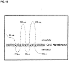
- the structure of CD133 includes an extracellular N-terminus, two short intracellular loops, two large extracellular loops, and an intracellular C-terminus ( FIG. 18 ).
- Exemplary CD133 T cell epitopes include 8-10 or 13-20 contiguous amino acid residues of amino acid residues 325-350 of SEQ ID NO:53.
- An alternately spliced version of CD133 is described in Yu et al., J. Biol. Chem., 277:20711-16, 2002 .
- CD90 is a cell surface glycoprotein found on T cells and neurons.
- Table 1 provides an amino acid sequence of CD90 (also available in GenBank under accession number NP_006279.2, GI:19923362).
- CD44 is a cell surface glycoprotein that may be involved in matrix adhesion.
- Table 1 provides an amino acid sequence of CD44 (also available in GenBank under accession number NP_000601.3, GI:48255935).
- isoforms of CD44 are produced, primarily by alternative splicing (see Marhaba et al., J. Mol. Histol., 35:211-31, 2004 ; Zoeller, Cancer Immunol. Immunother., 53:567-79, 2004 ).
- CXCR4 is a chemokine receptor that has been found to be expressed in breast cancers ( Muller et al., Nature, 410:50-56, 2001 ).
- Table 1 provides an amino acid sequence of CXCR4 (also available in GenBank under accession number NP_001008540.1, GI:56790927).
- An alternative spliced variant is available in GenBank under accession number NP_003458.1, GI:4503175.
- Nestin is an intermediate filament protein expressed in neural progenitor cells ( Dahlstrand et al., J. Cell Sci., 103:589-597, 1992 ).
- Table 1 provides an amino acid sequence of Nestin (also available in GenBank under accession number NP_006608.1, GI:38176300).
- Musashi-1 (Msi1) is an RNA-binding protein expressed in neural progenitor cells ( Good et al., Genomics, 52:382-384, 1998 ; Siddall et al., Proc. Nat. Acad. Sci. USA, 103:84-8407, 2006 ; Okano et al., Exp. Cell Res., 306:349-356, 2005 ).
- Table 1 provides an amino acid sequence of Musashi-1 (also available in GenBank under accession number NP_002433.1, GI:4505255).
- MELK Maternal embryonic leucine zipper kinase
- GLI1 is a zinc-finger transcription factor upregulated in cancers, including gliomas ( Kinzler et al., Science, 236:70-73, 1987 ; Kinzler et al., Nature, 332:371-374, 1988 ; Kasper et al., Eur. J. Cancer, 42:437-445, 2006 ).
- Table 1 provides an amino acid sequence of GLI1 (also available in GenBank under accession number NP_005260.1, GI:4885279).
- PTCH1 is a transmembrane protein that is believed to function as a tumor suppressor ( Katoh et al., Cancer Biol. Ther., 4:1050-54, 2005 ).
- Table 1 provides an amino acid sequence of PTCH1 (also available in GenBank under accession number NP_000255.2, GI: 134254446). Five isoforms of PTCH1 are produced by alternative splicing ( Nagao et al., Genomics, 85:462-71, 2005 ).
- Bmi-1 is a polycomb ring finger protein involved in proliferation of progenitor cells ( Lessard et al., Nature, 423:255-260, 2003 ; Park et al., Nature, 423:302-305, 2003 ; Molofsky et al., Nature, 425:962-967, 2003 ). Bmi-1 can play a role in the malignant transformation of the HOX A9/MEIS-induced murine leukemia model ( Lessard et al., Nature, 423:255-260, 2003 ) as well as in tumors of neural origin ( van Lohuizen et al., Nature, 353:353-355, 1991 ).
- Bmi-1 T cell epitopes include TLQDIVYKL (SEQ ID NO:86), CLPSPSTPV (SEQ ID NO:87), VRYLETSKY (SEQ ID NO:88), KRYLRCPAA (SEQ ID NO:89), YEEEPLKDY (SEQ ID NO:90), and KEEVNDKRY (SEQ ID NO:91) ( Steele et al., Br. J. Cancer 95:1202-11, 2006 ).
- Phosphoserine phosphatase is an enzyme that catalyzes the hydrolysis of O-phosphoserine.
- Table 1 provides an amino acid sequence of PSP (also available in GenBank under accession number NP_004568.2, GI:46249388).
- Snail is a zinc-finger transcription factor and anti-apoptotic protein ( Vega et al., Genes Dev., 18:1131-1143, 2004 ).
- Table 1 provides an amino acid sequence of Snail (also available in GenBank under accession number NP_005976.2, GI:18765741).
- OCT4 is a POU homeodomain-containing transcription factor expressed in pluripotent cells ( Nichols et al., Cell, 95:379-391, 1998 ).
- Table 1 provides an amino acid sequence of OCT4 (also available in GenBank under accession number NP_002692.2, GI:42560248).
- An alternate isoform of OCT4 is available in GenBank under accession number NP_976034.3, GI:116235491.
- BCRP1 is an ATP-binding cassette (ABC) transporter protein involved in multidrug resistance of tumors ( Doyle et al., Proc. Nat. Acad. Sci. USA, 95:1566570, 1998 ).
- Table 1 provides an amino acid sequence of BCRP1 (also available in GenBank under accession number NP_004818.2, GI:62526033).
- MGMT is an O-6-methylguanine-DNA methyltransferase DNA -mismatch repair protein that can provide resistance to some methylating and chloroethylating agents, such as temozolomide ( Rabik et al., Cancer Treat. Rev., 32:261-276, 2006 ; Cai et al., Cancer Res., 65:3319-27, 2005 ).
- Table 1 provides an amino acid sequence of MGMT (also available in GenBank under accession number NP_002403.1, GI:4505177).
- BCL-2 is a mitochondrial anti-apoptotic protein correlated with chemotherapy resistant cancers and decreased overall survival ( Campos et al., Blood, 81:3091-3096, 1993 ).
- Table 1 provides an amino acid sequence of BCL-2 (also available in GenBank under accession number NP_000624.2, GI:72198189).
- An alternatively spliced isoform of BCL-2 is available in GenBank under accession number NP_000648.2, GI:72198346.
- FLIP is an anti-apoptotic protein ( Irmler et al., Nature, 388:190-195, 1997 ).
- Table 1 provides an amino acid sequence of FLIP (also available in GenBank under accession number NP_003870.3, GI:21361769).
- BCL-XL is an anti-apoptotic protein related to BCL-2, which may be involved in chemoresistance ( Boise et al., Cell, 74:597-608, 1993 ; Andreeff et al., Leukemia, 13:1881-92, 1999 ).
- Table 1 provides an amino acid sequence of BCL-XL (also available in GenBank under accession number NP_612815.1, GI:20336335).
- Exemplary T cell epitopes of BCL-XL include Bcl-xL1 18-126 (TAYQSFEQV; SEQ ID NO:92), Bcl-xL173-182 (YLNDHLEPWI; SEQ ID NO:93), and Bcl-xL169-178 (WMATYLNDHL; SEQ ID NO:94) ( Andersen et al., J. Immunol., 175:2709-14, 2005 ).
- XIAP is a member of the inhibitor of apoptosis protein (IAP) family ( Deveraux et al., Nature, 388:300-304, 1997 ).
- Table 1 provides an amino acid sequence of XIAP (also available in GenBank under accession number NP_001158.2, GI:32528299).
- cIAP1 is a member of the IAP family of apoptosis inhibitors ( Rothe et al., Cell, 83: 1243-1252, 1995 ; Liston et al., Nature, 379:349-353, 1996 ).
- Table 1 provides an amino acid sequence of cIAP1 (also available in GenBank under accession number NP_001157.1, GI:4502141).
- cIAP2 is a member of the IAP family of apoptosis inhibitors ( Liston et al., Nature, 379:349-353, 1996 ).
- Table 1 provides an amino acid sequence of cIAP2 (also available in GenBank under accession number NP_031490.1, GI:6680696).
- NAIP is a member of the IAP family of apoptosis inhibitors ( Roy et al., Cell, 80:167-178, 1995 ).
- Table 1 provides an amino acid sequence of NAIP (also available in GenBank under accession number NP_004527.2, GI:119393878).
- An alternatively spliced isoform of NAIP is available in GenBank under accession number NP_075043.1, GI:119393876.
- Survivin is a member of the IAP family of apoptosis inhibitors ( Li et al., Nature, 396:580-584, 1998 ).
- Table 1 provides an amino acid sequence of NAIP (also available in GenBank under accession number NP_001159.2, GI:59859878).
- NAIP also available in GenBank under accession number NP_001159.2, GI:59859878.
- spliced isoforms of survivin have been identified (see Wheatley et al., Int. Rev. Cytol., 247:35-88, 2005 ; Noton et al., J. Biol. Chem., 281:1286-95, 2006 ; and Taubert et al., Oncogene, 24:5258-61, 2005 ).
- T cell epitopes of survivin include Sur20-28 (STFKNWPFL; SEQ ID NO:95), Sur96-104 (LTLGEFLKL; SEQ ID NO:96), Sur133-141 (RAIEQLAAM; SEQ ID NO:97), and Sur126-135 (ETAKKVRRAI; SEQ ID NO:98) ( Bachinsky et al., Cancer Immun., 5:6, 2005 ).
- T cell epitopes of survivin include Sur92-101 (QFEELTLGEF; SEQ ID NO:99), Sur54-62 (LAQCFFCFK; SEQ ID NO: 100), Sur1 12-120 (KIAKETNNK; SEQ ID NO:101), Sur53-62 (DLAQCFFCFK; SEQ ID NO:102), Sur112-121 (KIAKETNNKK; SEQ ID NO:103), Sur18-28 (RISTFKNWPFL; SEQ ID NO:104), Sur86-96 (FLSVKKQFEEL; SEQ ID NO:105), and the modified peptides Sur92T2 (QTEELTLGEF; SEQ ID NO:106), Sur93T2 (FTELTLGEF; SEQ ID NO:107), Sur93S2 (FSELTLGEF; SEQ ID NO:108), Sur38Y9 (MAEAGFIHY; SEQ ID NO:109), Sur47Y10 (PTENEPDLAY; SEQ ID NO: 110), Sur5K9 (TLPPAWQPK; SEQ ID NO:
- T cell epitopes of survivin include ELTLGEFLKL (SEQ ID NO:114) and TLPPAWQPFL (SEQ ID NO:115) ( Schmitz et al., Cancer Res. 60:4845-4849, 2000 ). Additional survivin epitopes are described in Siegel et al., Br. J. Haematol., 122:911-914, 2003 . Table 1.
- T cell epitopes can be identified by a number of different methods.
- Naturally processed MHC epitopes can be identified by mass spectrophotometric analysis of peptides eluted from antigen-loaded APC (e.g., APC that have taken up antigen, or that have been engineered to produce the protein intracellularly). After incubation at 37 °C, cells are lysed in detergent and the MHC protein is purified (e.g., by affinity chromatography). Treatment of the purified MHC with a suitable chemical medium (e.g., under acidic conditions) results in the elution of peptides from the MHC. This pool of peptides is separated and the profile compared with peptides from control APC treated in the same way. The peaks unique to the protein expressing/fed cells are analyzed (for example by mass spectrometry) and the peptide fragments identified. This protocol identifies peptides generated from a particular antigen by antigen processing.
- epitopes can be identified by screening a synthetic library of peptides that overlap and span the length of the antigen in an in vitro assay. For example, peptides that are 9 amino acids in length and which overlap by 5 amino acids may be used. The peptides are tested in an antigen presentation system that includes antigen presenting cells and T cells. T cell activation in the presence of APCs presenting the peptide can be measured (e.g., by measuring T cell proliferation or cytokine production) and compared to controls, to determine whether a particular epitope is recognized by the T cells.
- T cell epitopes can be predicted in silico, e.g., using the methods described in Parker et al., J. Immunol., 152:163, 1994 and Rammensee et al., Immunogenet., 50:213-219, 1999 .
- Antigenic peptides can be obtained by chemical synthesis using a commercially available automated peptide synthesizer. Synthetic peptides can be precipitated and further purified, for example by high performance liquid chromatography (HPLC). Alternatively, isolated peptides can be obtained by purification and/or recombinant methods using host cell and vector expression systems.
- HPLC high performance liquid chromatography
- Antigen presenting cells such as DCs
- suitable for administration to subjects can be isolated or obtained from any tissue in which such cells are found, or may be otherwise cultured and provided using standard techniques. Methods of preparing antigen presenting cells are well-known to those of skill in the art. Mature dendritic cells are typically identified as having the following cell surface marker phenotype: MAC3 - , CD80 + , CD86 + , CD40 low , CD54 + , MHC Class I and MHC Class II, and are capable of FITC-dextran uptake.
- APCs can be found, by way of example, in the bone marrow or PBMCs of a mammal, in the spleen of a mammal or in the skin of a mammal (i.e., Langerhan's cells, which possess certain qualities similar to that of DC, may be found in the skin).
- bone marrow can be harvested from a mammal and cultured in a medium that promotes the growth of DCs.
- GM-CSF, IL-4 and/or other cytokines (e.g., TNF- ⁇ ), growth factors and supplements may be included in this medium.
- clusters of DCs are cultured in the presence of a sufficient number of antigens of interest (e.g., in the presence of cancer stem cell lysate, acid eluted peptides of cancer stem cells, peptides of CD 133, CD90, CD44, CXCR4, Nestin, Musashi-1 (Msil), maternal embryonic leucine zipper kinase (MELK), GLI1, PTCH1, Bmi-1, phosphoserine phosphatase (PSP), Snail, OCT4, BCRP1, MGMT, Bcl-2, FLIP, BCL-XL, XIAP, cIAP1, cIAP2, NAIP, or survivin, or a combination of two or more of the above antigens) and harvested for use in a cancer
- a sufficient number of antigens of interest e.g., in the presence of cancer stem cell lysate, acid eluted peptides of cancer stem cells, peptides of CD 133,
- antigens can be transgenically expressed in DCs, e.g., by transfection of nucleic acids encoding one or more of the antigens or portions of antigens.
- APCs are isolated from a subject (e.g., a human) according to the following exemplary procedure.
- Mononuclear cells are isolated from blood using leukapheresis (e.g., using a COBE Spectra Apheresis System). The mononuclear cells are allowed to become adherent by incubation in tissue culture flasks for 2 hours at 37 °C. Nonadherent cells are removed by washing.
- Adherent cells are cultured in medium supplemented with granulocyte macrophage colony stimulating factor (GM-CSF) and interleukin-4 (IL-4) for five days. On day five, TNF- ⁇ is added to the culture medium for another 3-4 days.
- GM-CSF granulocyte macrophage colony stimulating factor
- IL-4 interleukin-4
- DCs occur in low numbers in all tissues in which they reside, making isolation and enrichment of DCs a requirement. Any of a number of procedures entailing repetitive density gradient separation, fluorescence activated cell sorting techniques, positive selection, negative selection or a combination thereof are routinely used to obtain enriched populations or isolated DCs. Guidance on such methods for isolating DCs can be found in O'Doherty et al., J. Exp. Med., 178:1067-78, 1993 ; Young and Steinman, J. Exp. Med., 171:1315-32, 1990 ; Freudenthal and Steinman, Proc. Nat. Acad. Sci.
- the APC-based cancer vaccine may be delivered to a recipient by any suitable delivery route, which can include injection, infusion, inoculation, direct surgical delivery, or any combination thereof.
- the cancer vaccine can be administered to a human in the deltoid region or axillary region.
- the vaccine can be administered to a subject locally to the site of a tumor, within the tumor, or to an area from which a tumor has been surgically resected.
- an appropriate carrier for administering the cells may be selected by one of skill in the art by routine techniques.
- the pharmaceutical carrier can be a buffered saline solution, e.g., cell culture media.
- the quantity of APC appropriate for administration to a patient as a cancer vaccine to effect the methods disclosed in the application and the most convenient route of such administration may be based upon a variety of factors, as may the formulation of the vaccine itself. Some of these factors include the physical characteristics of the patient (e.g., age, weight, and sex), the physical characteristics of the tumor (e.g., location, size, rate of growth, and accessibility), and the extent to which other therapeutic methodologies (e.g., chemotherapy, and beam radiation therapy) are being implemented in connection with an overall treatment regimen.
- factors include the physical characteristics of the patient (e.g., age, weight, and sex), the physical characteristics of the tumor (e.g., location, size, rate of growth, and accessibility), and the extent to which other therapeutic methodologies (e.g., chemotherapy, and beam radiation therapy) are being implemented in connection with an overall treatment regimen.
- a mammal can be administered with from about 10 5 to about 10 9 APC (e.g., 10 7 APC) in from about 0.05 mL to about 5 mL solution (e.g., saline) in a single administration. Additional administrations can be carried out, depending upon the above-described and other factors, such as the severity of tumor pathology. From about one to about five administrations of about 10 6 APC can be performed at two-week intervals.
- DC vaccination can be accompanied by other treatments.
- a patient receiving DC vaccination may also be receiving chemotherapy, radiation, and/or surgical therapy concurrently.
- Methods of treating cancer using DC vaccination in conjunction with chemotherapy are described in Wheeler et al., U.S. Pat. Pub. No. 2007/0020297 .
- a patient receiving DC vaccination can have already received chemotherapy, radiation, and/or surgical treatment for the cancer.
- a patient receiving DC vaccination can be treated with a COX-2 inhibitor, as described in Yu and Akasaki, WO 2005/037995 .
- the present application provides pharmaceutical compositions including a pharmaceutically acceptable excipient along with a therapeutically effective amount of the inventive vaccine comprising dendritic cells pulsed with cancer stem cell antigens as described herein.
- “Pharmaceutically acceptable excipient” means an excipient that is useful in preparing a pharmaceutical composition that is generally safe, non-toxic, and desirable, and includes excipients that are acceptable for veterinary use as well as for human pharmaceutical use. Such excipients may be solid, liquid, semisolid, or, in the case of an aerosol composition, gaseous.
- the pharmaceutical compositions may be formulated for delivery via any route of administration.
- Route of administration may refer to any administration pathway known in the art, including but not limited to aerosol, nasal, transmucosal, transdermal, or parenteral.
- Parenteral refers to a route of administration that is generally associated with injection, including intraorbital, infusion, intraarterial, intracapsular, intracardiac, intradermal, intramuscular, intraperitoneal, intrapulmonary, intraspinal, intrasternal, intrathecal, intrauterine, intravenous, subarachnoid, subcapsular, subcutaneous, transmucosal, or transtracheal.
- the compositions may be in the form of solutions or suspensions for infusion or for injection, or as lyophilized powders.
- compositions can also contain any pharmaceutically acceptable carrier.
- “Pharmaceutically acceptable carrier” as used herein refers to a pharmaceutically acceptable material, composition, or vehicle that is involved in carrying or transporting a compound of interest from one tissue, organ, or portion of the body to another tissue, organ, or portion of the body.
- the carrier may be a liquid or solid filler, diluent, excipient, solvent, or encapsulating material, or a combination thereof.
- Each component of the carrier must be “pharmaceutically acceptable” in that it must be compatible with the other ingredients of the formulation. It must also be suitable for use in contact with any tissues or organs with which it may come in contact, meaning that it must not carry a risk of toxicity, irritation, allergic response, immunogenicity, or any other complication that excessively outweighs its therapeutic benefits.
- the pharmaceutical compositions may be delivered in a therapeutically effective amount.
- the precise therapeutically effective amount is that amount of the composition that will yield the most effective results in terms of efficacy of treatment in a given subject. This amount will vary depending upon a variety of factors, including but not limited to the characteristics of the therapeutic compound (including activity, pharmacokinetics, pharmacodynamics, and bioavailability), the physiological condition of the subject (including age, sex, disease type and stage, general physical condition, responsiveness to a given dosage, and type of medication), the nature of the pharmaceutically acceptable carrier or carriers in the formulation, and the route of administration.
- Kits to treat cancer are also contemplated.
- the kits are useful for practicing the inventive method of treating cancer with a vaccine comprising dendritic cells pulsed with cancer stem cell antigens as described herein.
- the kit is an assemblage of materials or components, including at least one of the inventive compositions.
- the kit contains a composition including a vaccine comprising dendritic cells pulsed with cancer stem cell antigens as described herein.
- the kit is configured for the purpose of treating a particular cancer.
- the kit is configured for the purpose of treating brain tumors.
- the brain tumor is a glioma.
- the brain tumor is GBM.
- the kit is configured particularly for the purpose of treating mammalian subjects.
- the kit is configured particularly for the purpose of treating human subjects.
- the kit is configured for veterinary applications, treating subjects such as, but not limited to, farm animals, domestic animals, and laboratory animals.
- Instructions for use may be included in the kit.
- Instructions for use typically include a tangible expression describing the technique to be employed in using the components of the kit to effect a desired outcome, such as induction of an immune response against a tumor, to treat a cancer.
- the instructions may comprise instructions to administer a vaccine comprising dendritic cells pulsed with cancer stem cell antigens to the patient.
- the kit also contains other useful components, such as, diluents, buffers, pharmaceutically acceptable carriers, syringes, catheters, applicators, pipetting or measuring tools, or other useful paraphernalia as will be readily recognized by those of skill in the art.
- useful components such as, diluents, buffers, pharmaceutically acceptable carriers, syringes, catheters, applicators, pipetting or measuring tools, or other useful paraphernalia as will be readily recognized by those of skill in the art.
- the materials or components assembled in the kit can be provided to the practitioner stored in any convenient and suitable ways that preserve their operability and utility.
- the components can be in dissolved, dehydrated, or lyophilized form; they can be provided at room, refrigerated or frozen temperatures.
- the components are typically contained in suitable packaging material(s).
- packaging material refers to one or more physical structures used to house the contents of the kit, such as inventive compositions and the like.
- the packaging material is constructed by well known methods, preferably to provide a sterile, contaminant-free environment.
- the packaging materials employed in the kit are those customarily utilized in cancer treatments or in vaccinations.
- a package refers to a suitable solid matrix or material such as glass, plastic, paper, foil, and the like, capable of holding the individual kit components.
- a package can be a glass vial used to contain suitable quantities of an inventive composition containing for example, a vaccine comprising dendritic cells pulsed with cancer stem cell antigens as described herein.
- the packaging material generally has an external label which indicates the contents and/or purpose of the kit and/or its components.
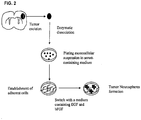
- 9L gliosarcoma To determine if a population of self-renewing stem cells exist within the phenotypically heterogeneous 9L gliosarcoma tumor, the cells were grown as monolayers in the presence of 10% FBS and subsequently grew them in serum free media containing mitogens.
- a schematic diagram of culturing of tumor stem cells is presented in Fig. 2 .
- 9L gliosarcomas were resuspended in Dulbecco's modified Eagle's medium/F-12 medium containing 10% fetal bovine serum (FBS) and plated at a density of 1 ⁇ 10 6 live cells per 75 cm 2 flask. The cells attached and grew as monolayers and were passaged upon confluency.
- FBS fetal bovine serum
- Spheres were derived by placing the 9L gliosarcomas cells grown as monolayers into a defined serum-free NSC medium ( Reynolds et al., J. Neurosci., 12: 4565-74, 1992 ; Reynolds et al., Science, 255:1707-10, 1992 ) consisting of Dulbecco's modified Eagle's medium/F-12 medium supplemented with 20 ng/mL of both epidermal growth factor (EGF; Peprotech, Rocky Hill, NJ) and basic fibroblast growth factor (bFGF; Peprotech, Rocky Hill, NJ). Cells were fed every 2 days by adding fresh NSC media supplemented with growth factors.
- a defined serum-free NSC medium consisting of Dulbecco's modified Eagle's medium/F-12 medium supplemented with 20 ng/mL of both epidermal growth factor (EGF; Peprotech, Rocky Hill, NJ) and basic fibroblast growth factor (bFGF; Peprotech, Rocky Hill, NJ).
- the cells were harvested, dissociated into single cells using trypsin and EDTA (GIBCO BRL) and mechanical pipetting, strained through a cell strainer, and plated at a clonal density of 1,000 cells/mL in neurosphere-conditioned medium to generate clonally derived subspheres ( Geschwind et al., Neuron, 29:325-339, 2001 ; Groszer et al., Science, 294: 2186-89, 2001 ). The cells were fed every 2 days by adding fresh NSC media supplemented with mitogens. Cells and subsequent spheres were observed daily for 18 days, and passaged into fresh media. Subspheres ranging from approximately 15 cells to 40 cells were evident after 18 days and displayed the self-renewing and proliferative capacity of the 9L spheres.
- neurospheres were immunostained for NSC markers nestin and Sox2. Neurospheres were also stained for the lineage markers for astrocytes, GFAP, neurons, beta-tubulin III and MAP2, and oligodendrocytes, myelin/oligodendrocyte. Cells in the outer region of the neurosphere labeled for nestin, while cells throughout the neurosphere were labeled for Sox2. A large number of cells within the tumor spheres were also found to be positive for the lineage marker GFAP, while relatively few cells expressed the neuronal lineage markers ⁇ -tubulin III, MAP2, and myelin/oligodendrocyte.
- spheres were seeded into chamber slides (Lab-TekII, Nalge Nunc International) for differentiation assay.
- the cells were grown for 14 days in medium devoid of growth factors bFGF and EGF but permissive for differentiation, and processed for immunocytochemistry as described below.
- the medium included Dulbecco's modified Eagle's medium/F-12 medium containing 10% fetal bovine serum (FBS).
- the primary antibodies were detected with Cy3 or FITC-conjugated anti-mouse or anti-rabbit IgG antibody (1:200, Jackson Immuno Research).
- the cells were counterstained with 4',6-diamidino-2-phenylindole (DAPI; Vector Laboratories) to identify all nuclei.
- DAPI 4',6-diamidino-2-phenylindole
- the stained sections were examined and photographed using a QED cell scanner program and Nikon Eclipse TE2000-E microscope, and analyzed using Image J (NIH).
- NIH Image J
- CSLCs cancer stem-like cells
- GFAP GFAP
- MAP2 GFAP
- the expression pattern of the differentiated progeny was similar in profile to that of primary cultured tumor cells from which the spheres had originally been isolated and predominantly differentiated into GFAP and MAP2 positive cells that recapitulated the parental tumor phenotype. Additionally, the level of labeling for the NSC markers nestin and Sox2 still remained high, even after 14 days of differentiation.
- the staining pattern for the monolayer population was similar to neurospheres differentiated for 14 days, except for the expression of Sox2, which was greater in neurospheres differentiated for 14 days.
- 9L cells grown as monolayer or neurospheres differ in their ability to grow as a tumor after implantation 5,000 cells from both populations of cells were injected into rats and survival and tumor volume were assayed.
- tumor aggression could be determined.
- Animals in the tumor volume group were sacrificed 18 days after tumor implantation. Tumor volume was assessed by using the formula for an ellipsoid, (length x width x height)/2 ( Advani et al., Cancer Res., 59:2055-58, 1999 ), with the height and the width of the tumor being approximately equal because of the well-defined circumference of the tumors generated by the 9L gliosarcoma (see Sibenaller et al., Neurosurg. Focus, 19:E1, 2005 ). A significantly (P ⁇ 0.02) greater tumor volume was observed in the rats implanted with neurosphere cells as compared to rats implanted with monolayer cells ( FIG. 4A ).
- Six rats in the neurosphere group had large tumors that lead to terminal neurological symptoms, whereas 3 showed evidence of small tumors after 40 days of survival as seen by H&E staining of brain sections.
- only 4 rats had tumors large enough to create terminal neurological symptoms in the monolayer group, whereas 5 showed evidence of small tumor or engraftment after 40 days of survival as determined by H&E staining.
- the rats in the monolayer group which developed terminal neurological signs of tumor did so at a later time (36 days compared to 29 days in the neurosphere group), which was significant when analyzed using the Kaplan-Meier test (P ⁇ 0.02).
- Luciferase substrate D-luciferin Biosynth, International, Inc., Naperville, IL
- luciferase scans were taken 15 minutes later. Luciferase scans generated at 14 days post implantation demonstrated a greater proportion of animals with tumor burden in the neurosphere group compared to the monolayer group, even though a higher expression level of luciferase in vitro was observed in monolayer cells as compared to neurosphere cells.
- the tumor-cell-implanted rat brains were cut with a cryostat into 20 ⁇ m coronal sections and fixed in 4% paraformaldehyde, washed with PBS, and air dried.
- Tumors from the neurosphere cell population were large and well-circumferential, and showed cells positive for the NSC marker nestin, as well as cells positive for the lineage markers GFAP, ⁇ -tubulin III, and myelin/oligodendrocytes. Most of the labeling within the tumor volume was directed against GFAP, and a lesser degree of labeling was observed for ⁇ -tubulin III, myelin/oligodendrocyte, and nestin. A significant portion (> 75%) of the tumor volume stained positive for reticulin, consistent with a sarcomatous component.
- H&E staining revealed a high grade glioma with necrosis consistent with a glioblastoma, displaying the dual nature of the gliosarcoma.
- Neurospheres formed high grade gliomas with necrosis as seen on H&E ( FIG. 5A ).
- the tumors were large and well circumferential as evidenced in non-stained sections ( FIG. 5B ) and stained for the nuclear marker DAPI ( FIG. 5D ).
- a comparison of non-tumor area ( FIG. 5D ) with tumor area ( FIG. 5E ) stained for reticulin revealed high levels of reticulin in tumor engulfed regions, showing the histological sarcomatous component of the gliosarcoma.
- the staining patterns suggest that 9L neurospheres recapitulated the original tumor by differentiating into both neural and glial lineages in vivo.
- 2,000 healthy 9L sphere and monolayer cells were exposed to either Dulbecco's modified Eagle's medium/F-12 medium containing 10% fetal bovine serum (FBS), or 100 ⁇ M stock solution of Temozolamide or Carboplatin dissolved in PBS at concentrations of 1,000 ⁇ M, 500 ⁇ M, 250 ⁇ M, and 125 ⁇ M for 2 days.
- FBS Dulbecco's modified Eagle's medium/F-12 medium containing 10% fetal bovine serum
- Temozolamide or Carboplatin dissolved in PBS at concentrations of 1,000 ⁇ M, 500 ⁇ M, 250 ⁇ M, and 125 ⁇ M for 2 days.
- the viability of the cells was scored by measurement of the absorption of formazan dye (the amount of formazan dye formed directly correlates to the number of metabolically active cells) using the cellular proliferation assay WST-1 (Roche Molecular Biochemicals, Mannheim, Germany).
- Formazan was measured with the use of a microplate reader (Tecan) and spectrophotometer set at a wavelength of 440 nm and a reference wavelength of 890 nm.
- Cellular viability was determined by exposing cells to WST-1 for 4 hours, and calculating the percentage of viable cells. Proliferation was also assessed by using manual cell counting after 7 days in culture, with an initial cellular concentration of 100,000 cells/mL in a 25 mm 2 flask.
- the neurospheres demonstrated significantly (P ⁇ 0.05) greater resistance to temozolamide ( FIG. 6B ) and carboplatin ( FIG. 6A ) when compared to the 9L cells grown as a monolayer under the same conditions.
- FIG. 7 is a bar graph that shows a greater increase in cells in the monolayer group.
- a similar trend in the untreated cells was also observed using the manual cell count method, which showed a greater increase in monolayers by a factor of 1.35.
- This example demonstrates that neurosphere cells have a greater resistance to chemotherapeutic agents.
- Glioblastoma specimens were obtained from patients (with informed consent) via the Brain Tumor Registry and were reviewed and released by a pathologist in the operating room. Independent pathologists classified the tumors by type and grade in accordance with the WHO histological grading of central nervous system tumors. IRB certified technicians processed the glioma tissues under sterile conditions in a laminar flow hood. Tumor cells were cultured in the following complete medium: Ham's F-12/DMEM with high glucose (Irvine scientific, Santa Ana, CA), 10 mM HEPES (Invitrogen, Carlsbad, CA), 0.1 mg/ml Gentamicin (Invitrogen) and 10% heat-inactivated FBS (Irvine Scientific, Santa Ana, CA). The cultured cells were maintained for 3-4 passages. Floating neurosphere-like cells were obtained that were capable of forming new spheres in medium containing FBS for 3-4 passages.
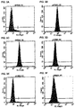
- glioblastoma primary tumor cell lines (Nos. 1049, 377, and 66) were derived by the above method and analyzed by FACS for CD133 expression. Tumor cells were collected and stained with anti-CD133 antibody (mouse monoclonal IgG1; 1: 10; Milteny Biotec) or IgG1 isotype control antibody (BD Pharmingen, San Diego, CA). After PE-anti-mouse IgG1 (BD Pharmingen) staining for 30 minutes, CD133 staining was analyzed by flow cytometry using a FACSCaliburTM fluorescence activated cell sorter (Becton Dickinson, San Jose, CA). CD133 expression was observed in 10.2% (No. 66; FIG. 9C ), 27% (No. 1049, FIG. 9A ) and 69.7% (No. 377; FIG. 9B ) of the total population examined.
- CD133 expression was observed in 10.2% (No. 66; FIG. 9C ), 27% (No
- CD133 positive cancer stem cell was isolated by DAKOcytomationTM (DAKO, Carpinteria, CA) sorting and cultured in a defined serum-free NSC medium ( Kabos et al., Exp. Neurol., 178:288-293, 2002 ) containing 20 ng/ml of basic fibroblast growth factor (bFGF, Peprotech, Rocky Hill, NJ), 20 ng/ml of epidermal growth factor (EGF, Peprotech) and 20 ng/ml leukemia inhibitory factor (LIF, Chemicon, Temecula, CA).
- bFGF basic fibroblast growth factor
- EGF epidermal growth factor
- LIF leukemia inhibitory factor
- CD133 positive cells and CD133 negative cells were obtained by FACS sorting as described in Example 5 and real-time PCR was used to analyze some markers associated with neural precursors in these two populations.
- Total RNA was extracted from the isolated CD133 positive and CD133 negative cells using an RNA4PCRTM kit (Ambion, Austin, TX) according to the manufacturer's protocol.
- RNA4PCRTM kit (Ambion, Austin, TX) according to the manufacturer's protocol.
- cDNA synthesis ⁇ 1 ⁇ g total RNA was reverse-transcribed into cDNA using Oligo dT primer and iScriptTM cDNA synthesis kit reverse transcriptase.
- cDNA was stored at -20 °C.
- Oligonucleotide primers sequences used for SYBR Green real-time PCR Gene Forward Reverse Beta-actin 5'-TTCTACAATGAGCTGCGTGTG-3' (SEQ ID NO:1) 5'-GGGGTGTTGAAGGTCTCAAA-3' (SEQ ID NO:2) CD133 5'-GCATTGGCATCTTCTATGGTT-3' (SEQ ID NO:3) 5'-CGCCTTGTCCTTGGTAGTGT-3' (SEQ ID NO:4)
- MSI1 5'-GAGACTGACGCGCCCCAGCC-3' (SEQ ID NO:5) 5'-CGCCTGGTCCATGAAAGTGACG-3' (SEQ ID NO:6)
- MELK 5'-CTTGGATCAGAGGCAGATGTTTGGAG-3' (SEQ ID NO:7) 5'-GTTGTAATCTTGCATGATCCAGG-3' (SEQ ID NO:8) PSP 5'-GGCGGGGCAGTGCCTTTCAAA-3' (SEQ ID NO:9) 5'-TGTTGGCT
- Quantification of target gene mRNA as compared to an internal control (beta-actin) was performed by following a ⁇ C T method. An amplification plot that had been the plot of fluorescence signal vs. cycle number was drawn. The difference ( ⁇ C T ) between the mean values in the duplicated samples of target gene and those of beta-actin were calculated by Microsoft Excel and the relative quantified value (RQV) was expressed as 2 - ⁇ CT . The relative expression of each gene was compared to autologous CD133 negative cells. The results of the QT-PCT analysis are presented in Table 3. Table 3. Relative Expression of Genes in CD133+ Cancer Stem Cells No. 66 No. 377 No.
- CD90, CD44, CXCR4, Nestin, Musashi-1 (Msil), and maternal embryonic leucine zipper kinase (MELK) mRNA expression on CD133 positive cancer stem cells was upregulated by an average of 15.6, 5.7, 337.8, 2.14, 84, and 1351 fold, respectively, compared to the levels found on autologous CD133 negative tumor cells.
- mRNA levels for GLI1 and PTCH1 were upregulated an average of 46 and 16 times, respectively, in CD133 positive cells, as compared to CD133 negative cells.
- Bmi-1, phosphoserine phosphatase (PSP), SHH, OCT4 and Snail mRNA were expressed in CD133 positive cells derived from the three cell lines; none of the five genes were detectable on CD133 negative cells. Additionally, anti-apoptotic genes were also upregulated (see Example 8).
- CD133 positive cancer stem cells were resistant to chemotherapeutic agents
- the WST-1 Cell Proliferation Assay was used to examine the drug sensitivity of CD133 positive cells and CD133 negative cells (both collected by FACS sorting from the three glioblastoma patients' primary cultured tumor cells as described above).
- CD133 positive and negative cells were exposed to conventional chemotherapeutic agents, temozolomide, carboplatin, VP-16 or taxol at various concentrations, for up to 48 hours in 10% FBS/F-12/DMEM culture medium.
- Temozolomide was supplied by the Schering-Plough Research Institute (Kenilworth, NJ) and was dissolved in DMSO (Sigma Chemical Co., St Louis, MO) at 100 mM stock solution.
- CD133 positive cells isolated from No. 66 showed dramatic drug resistance to the above four agents including temozolomide ( FIG. 10C ), carboplatin ( FIG. 10D ), VP-16 ( FIG. 10A ), and Taxol ( FIG. 10B ) as compared to autologous CD133 negative cells.
- CD133 positive cells isolated from No. 377 showed significant resistance to carboplatin at all concentrations tested ( FIG. 11A ) and to VP-16 at 200 ⁇ M ( FIG. 11B ).
- CD133 positive cells isolated from No. 1049 showed significant resistance to carboplatin at 200 ⁇ M compared to autologous CD133 negative cells ( FIG. 11C ). This example demonstrates increased drug resistance of CD133 positive cancer stem cells as compared to autologous CD133 negative cells.
- BCRP1 has been demonstrated to play an important role in the drug resistance of normal stem cells and tumor stem cells ( Zhou et al., Nat. Med., 7:1028-34, 2001 ; Hirschmann-Jax et al., Proc. Natl. Acad. Sci. USA, 101:14228-33, 2004 ). Higher expression of BCRP1 (6.5 fold) was found in CD133 positive cells as compared to that of autologous CD133 negative cells.
- anti-apoptotic genes such as FLIP, BCL-2, and BCL-XL
- FLIP FLIP
- BCL-2 BCL-2
- BCL-XL anti-apoptotic genes
- IAPs inhibitor of apoptosis protein family members
- XIAP XIAP
- cIAP1 cIAP2
- NAIP apoptosis protein
- survivin were found at higher expression levels on CD133 positive cells 21.9, 39.04, 3.03, 12.1, 6.73, and 1.6 times higher, respectively, than in CD133 negative cells.
- SIRT1 deacetylates the DNA repair factor Ku70, causing it to sequester the pro-apoptotic factor, Bax, away from mitochondria, thereby inhibiting stress-induced apoptotic cell death ( Cohen et al., Science, 305:390-392, 2004 ).
- SIRT1 deacetylase mRNA expression was increased 4.92 times in CD133 positive cells.
- the pro-apoptotic gene BAX was decreased 3 times in CD133 positive cells as compared to autologous CD133 negative cells. Thus, gene expression differences were observed in CD133 positive cells as compared to CD133 negative cells.
- glioma is a highly recurrent tumor even after surgery, chemotherapy, radiation and immunotherapy.
- CD133 positive tumor cells in glioblastoma recurrence, the CD133 expression upon first and second resection of tumor tissue from the same patient were compared.
- RNA extraction and RT-PCR was performed for CD133 as described above.
- CD133 expression was significantly higher in recurrent tumor tissue (relative expression ⁇ 2.5-19.1) than that in autologous primary tumor tissue (relative expression ⁇ 1)( FIG. 12 ). This example implicates a resistant CD133 positive tumor population in tumor recurrence.
- a dendritic cell vaccine was generated using antigens from neurospheres, daughter cells, and monolayer cells. Immature dendritic cells were generated from the bone marrow of 6-12-week-old Fisher F344 rats as previously described ( Talmor et al., Eur. J. Immunol., 28:811-817, 1998 ).
- bone marrow was harvested from the femoral and tibial marrow cavities and cultured in RPMI 1640 media supplemented with 10% fetal bovine serum (Gemini Biotechnologies, Calabasas, CA), 1% Penicillin/Streptomycin (Invitrogen, Carlsbad, CA), 50 ng/ml recombinant rat GM-CSF and 100 ng/ml recombinant rat IL-4 (R & D Systems, Minneapolis, MN). Cultures were fed every 2 days by removing 75% of the media and replacing it with fresh media containing cytokines (this washed away most of the lymphocytes and granulocytes).
- Soluble peptides were generated for dendritic cell pulsing by cell lysis.
- 9L neurospheres, daughter cells, and monolayer (adherent) cells were processed in the laboratory to produce a single cell suspension.
- the cells were then lysed by 4 to 5 freeze cycles (on liquid nitrogen) and thaw cycles (room temperature). Lysis was monitored by light microscopy, and larger particles were removed by centrifugation (10 minutes at 600 ⁇ g).
- the supernatants were passed through a 0.2 ⁇ m filter, and protein concentration was determined by BioRad protein assay and aliquots frozen at -80 °C until use.
- Vaccinations were given subcutaneously in the flanks on days 7, 14, and 21 with 50,000 DCs pulsed with antigens from either 9L neurospheres (NS), daughter cells (DtC), monolayer (adherent) cells (AC), or saline control. The animals were followed for survival and euthanized when terminal neurological signs developed, for example, inability to access food, water, seizure activity, weakness, and paralysis.
- a cytotoxic T lymphocyte (CTL) assay was performed. Spleens were removed on day 28 post-intracranial 9L-luciferase tumor cells implantation from groups of rats treated with either control or 9L peptide-pulsed dendritic cells. Splenocytes were isolated and re-stimulated in vitro, as described ( Wunderlich et al., "Assays for T-cell function," In: Coligan et al., eds., Current Protocols in Immunology, New York, NY, John Wiley & Sons, Inc; 1997:3.11.1-3.11.20 ; Ehtesham et al., J.
- CTL cytotoxic T lymphocyte
- a representative number of brains from DC-DtC and DC-NS vaccinated rats were carefully removed and post-fixed in 4% paraformaldehyde. Coronal sections of 20 ⁇ m were cut on a cryostat and blocked with normal horse serum for 1 hour. Slides were then incubated with anti-CD4 (clone OX-38 monoclonal antibody diluted in 1:200 in PBS) for 2 hours at room temperature, followed by a 20 minute incubation at room temperature with the linking antibody (BioGenex biotinylated anti-mouse immunoglobulin). After washing in PBS, the labeled moiety (BioGenex Horse Radish Peroxidase-conjugated streptavidin) was added for 20 minutes at room temperature. DAB (3,3'-diaminobenzidine) was used as the chromogen. For analysis of CD8 expression, slides were incubated overnight with Anti-CD8 alpha Chain, clone OX-8 antibody (Chemicon).
- FIG. 15B Immunohistochemical assessment of brain sections from rats vaccinated with dendritic cells pulsed with NS vaccinated showed that there was a robust infiltration of CD4 + ( FIG. 15B ) and CD8 + lymphocytes that was not observed in the brain sections obtained from rats vaccinated with dendritic cells pulsed with daughter cell antigens ( FIG. 15A ). This infiltration of T cells correlates with increased survival of rats vaccinated with NS pulsed DCs.
- GBM glioblastoma multiforme
- CSC cancer stem cells
- Tumor cells were resuspended in DMEM/F12 medium containing 10% fetal bovine serum (FBS) as growth medium and plated at a density of 2x10 6 live cells per 75 cm 2 flask. The cells attached and grew as a monolayer in flasks. All the five monolayer growing adult GBM cells were switched into a defined serum-free NSC medium ( Reynolds et al., Science, 255:1707-10, 1992 ) containing 20 ng/ml of basic fibroblast growth factor (bFGF, Peprotech, Rocky Hill, NJ) and 20 ng/ml of epidermal growth factor (EGF, Peprotech).
- FBS fetal bovine serum
- NSC Normal human fetal neural stem cells
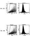
- CSC and NSC cells were stained by FITC-HLA-A,B,C antibody and isotype control antibody (BD Bioscience, San Diego, CA) and analyzed by flow cytometry. Representative CSC and NSC results were shown in the FIGs. 17A-17B and 18A-18B , respectively.
- HLA expression was seen in 5 of 5 cancer stem cells from different patients. CSCs expressed high levels of HLA-A,B,C, however, NSC did not expression MHC class I (HLA-A,B,C) antigens on the surface. This unexpected result indicates that specific cancer stem cell antigens on cancer stem cells can be targeted using vaccines that generate T cells that recognize and kill cancer stem cell antigens in the context of MHC. Therefore cancer stem cells will be targeted, whereas normal stem cells will not.
- a nucleic acid encoding a portion of CD133 extracellular domain 1 is used in cloning and expression on the surface of DC.
- DC are transfected with CD133-1 cDNA construct or empty vector (mock) for 48 hours.
- the successful transfection of DC-CD 133 cells is identified either by anti-CD 133 monoclonal antibody using flow cytometry or by EGFP cloned vector under fluorescent microscopy.
- Antigen presenting cells with CD133 receptor are stimulated by CD133 T cell epitopes.
- Overlapping peptides of 8-10 amino acids of residues 325-350 of SEQ ID NO:53 are produced as MHC class I epitopes.
- overlapping peptides of 13-20 amino acid of residues 325-350 are produced as MHC class II epitopes.
- Stimulated APC using the peptide epitopes lead to enhanced production of CD8 T cells targeted to CD 13 3 molecular on stem/progenitor cells in brain tumors.
Landscapes
- Health & Medical Sciences (AREA)
- Life Sciences & Earth Sciences (AREA)
- General Health & Medical Sciences (AREA)
- Animal Behavior & Ethology (AREA)
- Veterinary Medicine (AREA)
- Public Health (AREA)
- Epidemiology (AREA)
- Engineering & Computer Science (AREA)
- Chemical & Material Sciences (AREA)
- Biomedical Technology (AREA)
- Organic Chemistry (AREA)
- Bioinformatics & Cheminformatics (AREA)
- Wood Science & Technology (AREA)
- Biotechnology (AREA)
- Zoology (AREA)
- Genetics & Genomics (AREA)
- Immunology (AREA)
- Microbiology (AREA)
- Oncology (AREA)
- Developmental Biology & Embryology (AREA)
- Cell Biology (AREA)
- Pharmacology & Pharmacy (AREA)
- Medicinal Chemistry (AREA)
- Biochemistry (AREA)
- General Engineering & Computer Science (AREA)
- Chemical Kinetics & Catalysis (AREA)
- General Chemical & Material Sciences (AREA)
- Nuclear Medicine, Radiotherapy & Molecular Imaging (AREA)
- Medicines Containing Antibodies Or Antigens For Use As Internal Diagnostic Agents (AREA)
- Medicines Containing Material From Animals Or Micro-Organisms (AREA)
- Medicines That Contain Protein Lipid Enzymes And Other Medicines (AREA)
- Mycology (AREA)
Claims (1)
- Procédé de préparation d'un vaccin contre le gliome, le procédé comprenantl'obtention d'une population de cellules dendritiques ; etla mise en contact des cellules dendritiques avec une composition d'antigènes de cellules souches cancéreuses neurales dans des conditions telles que les cellules dendritiques présentent des antigènes de cellules souches cancéreuses neurales, pour ainsi préparer un vaccin contre le gliome, la composition d'antigènes de cellules souches cancéreuses neurales étant un lysat des neurosphères de cellules souches cancéreuses, les neurosphères étant obtenues à partir du gliome.
Applications Claiming Priority (2)
| Application Number | Priority Date | Filing Date | Title |
|---|---|---|---|
| US82695506P | 2006-09-26 | 2006-09-26 | |
| PCT/US2007/079600 WO2008039874A2 (fr) | 2006-09-26 | 2007-09-26 | Vaccins comprenant des antigènes de cellules souches cancéreuses et procédés |
Publications (3)
| Publication Number | Publication Date |
|---|---|
| EP2084267A2 EP2084267A2 (fr) | 2009-08-05 |
| EP2084267A4 EP2084267A4 (fr) | 2011-03-02 |
| EP2084267B1 true EP2084267B1 (fr) | 2018-04-11 |
Family
ID=39230952
Family Applications (1)
| Application Number | Title | Priority Date | Filing Date |
|---|---|---|---|
| EP07843269.7A Not-in-force EP2084267B1 (fr) | 2006-09-26 | 2007-09-26 | Vaccins comprenant des antigenes de cellules souches cancereuses et procedes |
Country Status (4)
| Country | Link |
|---|---|
| US (2) | US8129184B2 (fr) |
| EP (1) | EP2084267B1 (fr) |
| CA (1) | CA2700573C (fr) |
| WO (1) | WO2008039874A2 (fr) |
Families Citing this family (49)
| Publication number | Priority date | Publication date | Assignee | Title |
|---|---|---|---|---|
| WO2005037995A2 (fr) * | 2003-10-06 | 2005-04-28 | Cedars-Sinai Medical Center | Utilisation d'un inhibiteur de cox-2 permettant d'empecher l'anergie des lymphocytes t induite par une therapie cellulaire dendritique |
| WO2005043155A1 (fr) | 2003-10-21 | 2005-05-12 | Cedars-Sinai Medical Center | Systeme et methode pour le traitement du cancer, notamment des cancers du systeme nerveux central |
| WO2008039874A2 (fr) | 2006-09-26 | 2008-04-03 | Cedars-Sinai Medical Center | Vaccins comprenant des antigènes de cellules souches cancéreuses et procédés |
| WO2008039974A2 (fr) | 2006-09-28 | 2008-04-03 | Cedars-Sinai Medical Center | Vaccins contre le cancer et méthodes de vaccination |
| WO2008042407A1 (fr) * | 2006-10-03 | 2008-04-10 | Ludwig Institute For Cancer Research | Cellules de repeuplement de mélanome |
| ITMI20062100A1 (it) * | 2006-10-31 | 2008-05-01 | Fondazione I R C C S Istituto Neurologico | Metodo per la stimolazi0ne di cellule dendritiche e prodotto cellulare cosi'ottenuto per la immunoterapia autologa di tumori solidi umani |
| KR101523698B1 (ko) | 2007-01-22 | 2015-05-29 | 마크로제닉스 웨스트 인코퍼레이티드 | 사람 암 줄기세포 |
| US8877206B2 (en) | 2007-03-22 | 2014-11-04 | Pds Biotechnology Corporation | Stimulation of an immune response by cationic lipids |
| EP2185727A2 (fr) * | 2007-08-02 | 2010-05-19 | GlaxoSmithKline Biologicals SA | Nouveau procédé |
| US20100048524A1 (en) | 2008-03-14 | 2010-02-25 | Angela Brodie | Novel C-17-Heteroaryl Steroidal CYP17 Inhibitors/Antiandrogens;Synthesis In Vitro Biological Activities, Pharmacokinetics and Antitumor Activity |
| CA2721366C (fr) | 2008-04-17 | 2017-06-06 | Elizabeth Ann Vasievich | Stimulation de reponse immunitaire par des enantiomeres de lipides cationiques |
| JP2011529080A (ja) * | 2008-07-24 | 2011-12-01 | ユニバーシティ オブ セントラル フロリダ リサーチ ファウンデーション,インコーポレイテッド | 癌幹細胞を標的とする治療法 |
| PL2328923T3 (pl) * | 2008-09-02 | 2016-06-30 | Cedars Sinai Medical Center | Epitopy CD133 |
| AU2016204708B2 (en) * | 2008-10-01 | 2018-02-22 | Immatics Biotechnologies Gmbh | Novel immunotherapy against several tumors including neuronal and brain tumors |
| PL2172211T3 (pl) * | 2008-10-01 | 2015-05-29 | Immatics Biotechnologies Gmbh | Kompozycja związanych z guzem peptydów i związana z tym szczepionka przeciwrakowa do leczenia glejaka (GBM) i innych rodzajów raka |
| WO2010091306A1 (fr) | 2009-02-05 | 2010-08-12 | Tokai Pharmaceuticals | Nouveaux promédicaments à base d'inhibiteurs cyp17 stéroïdiens/anti-androgènes |
| WO2010124498A1 (fr) * | 2009-04-30 | 2010-11-04 | Beijing Cellonis Biotechnology Co., Ltd | Cellule-souche tumorale dont la résistance a été criblée, sa composition antigénique, le chargement de cellules dendritiques antitumorales avec lesdits antigènes, leurs méthodes de préparation, utilisations et kits ainsi qu'un vaccin à base de cellules dendritiques |
| EP2427485B1 (fr) | 2009-05-07 | 2016-12-07 | ImmunoCellular Therapeutics, Ltd. | Epitopes des cd133 |
| EP2454363B1 (fr) | 2009-07-13 | 2020-07-22 | Biogencell, Ltd. | Procédé d'utilisation de cellules directrices pour l'activation et la différentiation de cellules souches/progénitrices spécifiques |
| JP2011135864A (ja) * | 2009-12-30 | 2011-07-14 | Korea Univ Research & Business Foundation | Oct4及びBmi1、またはその上位調節子を用いて体細胞から胚幹細胞類似細胞への逆分化を誘導する組成物及びこれを用いた胚幹細胞類似細胞の製造方法 |
| CN102692506A (zh) * | 2011-03-21 | 2012-09-26 | 复旦大学 | Cd133在制备肿瘤标志物中的用途及其试剂盒 |
| CA2843200A1 (fr) * | 2011-07-27 | 2013-01-31 | Baylor Research Institute | Vaccinotherapie a cellules dendritiques (cd) pour le cancer du pancreas |
| US8956870B2 (en) | 2012-01-19 | 2015-02-17 | Biogencell, Ltd. | Method for using directing cells for specific stem/progenitor cell activation and differentiation |
| CN105163753B (zh) | 2012-06-15 | 2018-12-21 | Pds生物科技公司 | 阳离子脂质疫苗组合物和使用方法 |
| WO2014165101A1 (fr) * | 2013-03-13 | 2014-10-09 | California Stem Cell, Inc. | Cellules souches de carcinome du côlon individualisées, de pureté élevée, procédés et utilisation associés |
| JP2015526087A (ja) * | 2012-08-15 | 2015-09-10 | ネオステム オンコロジー リミテッド ライビリティ カンパニー | 高純度癌幹細胞及び高純度癌幹細胞集団の迅速な作製方法 |
| WO2014138455A1 (fr) * | 2013-03-07 | 2014-09-12 | California Stem Cell, Inc. | Cellules souches de carcinome hépatocellulaire individualisées et de haute pureté, procédés et utilisation de celles-ci |
| CN105101991A (zh) | 2012-09-21 | 2015-11-25 | Pds生物科技公司 | 改进的疫苗组合物和使用方法 |
| WO2014066615A1 (fr) * | 2012-10-24 | 2014-05-01 | The Regents Of The University Of Michigan | Vaccination et traitement de cellule souche cancéreuse |
| EP2956164A4 (fr) * | 2013-02-14 | 2016-08-31 | Immunocellular Therapeutics Ltd | Vaccins contre le cancer ovarien et méthodes de vaccination |
| US10137182B2 (en) | 2013-02-14 | 2018-11-27 | Immunocellular Therapeutics, Ltd. | Cancer vaccines and vaccination methods |
| US8975290B2 (en) | 2013-03-01 | 2015-03-10 | Colorado State University Research Foundation | Methods and compositions for enhancing an immune response, blocking monocyte migration, amplifying vaccine immunity and inhibiting tumor growth and metastasis |
| WO2014165103A1 (fr) * | 2013-03-12 | 2014-10-09 | California Stem Cell, Inc. | Cellules souches de glioblastome multiforme de haute pureté individualisées et méthodes de stimulation de réponse immunitaire |
| KR20150139529A (ko) * | 2013-03-12 | 2015-12-11 | 네오스템 온콜로지, 엘엘씨 | 활성 자가조직 면역 요법을 위한 고순도 난소암 줄기세포 |
| KR20150127720A (ko) | 2013-03-14 | 2015-11-17 | 유니버시티 오브 매릴랜드, 발티모어 | 안드로겐 수용체 하향 조절제 및 그의 용도 |
| CA2920317A1 (fr) | 2013-08-12 | 2015-02-19 | Tokai Pharmaceuticals, Inc. | Biomarqueurs pour le traitement de troubles neoplasiques a l'aide de therapies ciblant les androgenes |
| WO2015143074A1 (fr) * | 2014-03-20 | 2015-09-24 | University Of Southern California | Immunotherapie du cancer du cerveau |
| WO2016014613A1 (fr) * | 2014-07-22 | 2016-01-28 | The Trustees Of The University Of Pennsylvania | Compositions et méthodes pour l'immunothérapie du cancer |
| TWI563085B (en) * | 2014-08-29 | 2016-12-21 | China Medidal University Hospital | Dendritic cell tumor vaccine and preparation method thereof |
| WO2016161309A1 (fr) * | 2015-04-01 | 2016-10-06 | Colorado State University Research Foundation | Vaccins à base de cellules souches cancéreuses optimisés |
| IL289849B2 (en) | 2015-11-10 | 2024-01-01 | Nat Inst Biotechnology Negev Ltd | Means and methods for reducing tumorigenicity of cancer stem cells |
| WO2017083820A1 (fr) * | 2015-11-13 | 2017-05-18 | Pds Biotechnology Corporation | Lipides en tant que vecteurs synthétiques pour améliorer le traitement et la présentation de l'antigène ex-vivo en thérapie cellulaire dendritique |
| MA47678A (fr) | 2017-03-03 | 2021-05-26 | Treos Bio Ltd | Plateforme personnalisée d'identification de peptide immunogène |
| US10357509B2 (en) * | 2017-03-09 | 2019-07-23 | Thomas Jefferson University | Methods and compositions for treating cancers using antisense |
| CA3084957A1 (fr) | 2017-12-05 | 2019-06-13 | Pds Biotechnology Corporation | Procedes et compositions comprenant des lipides cationiques pour stimuler des genes d'interferon de type 1 |
| KR20210086612A (ko) | 2018-09-04 | 2021-07-08 | 트레오스 바이오 리미티드 | 펩타이드 백신 |
| CN109620952A (zh) * | 2019-01-04 | 2019-04-16 | 北京中台恒基生物技术有限公司 | 一种肿瘤疫苗及其制备方法 |
| US20220313734A1 (en) * | 2019-05-09 | 2022-10-06 | Figene, Llc | Fibroblast generated patient-specific vaccines |
| WO2025056957A1 (fr) * | 2023-09-15 | 2025-03-20 | Jimenez Mendoza, Dimas | Médicaments à base de peptides pour le traitement du cancer |
Family Cites Families (108)
| Publication number | Priority date | Publication date | Assignee | Title |
|---|---|---|---|---|
| US710903A (en) | 1902-06-21 | 1902-10-07 | Louis J Sticklin | Lid-controlling device. |
| US4690915A (en) | 1985-08-08 | 1987-09-01 | The United States Of America As Represented By The Department Of Health And Human Services | Adoptive immunotherapy as a treatment modality in humans |
| US4844893A (en) | 1986-10-07 | 1989-07-04 | Scripps Clinic And Research Foundation | EX vivo effector cell activation for target cell killing |
| WO1989006692A1 (fr) | 1988-01-12 | 1989-07-27 | Genentech, Inc. | Procede de traitement de cellules tumorales par inhibition de la fonction receptrice du facteur de croissance |
| DE69031120T2 (de) * | 1989-05-19 | 1998-01-15 | Genentech, Inc., South San Francisco, Calif. | Her2 extrazellulare domäne |
| US5061620A (en) | 1990-03-30 | 1991-10-29 | Systemix, Inc. | Human hematopoietic stem cell |
| US5925729A (en) | 1991-05-23 | 1999-07-20 | Ludwig Institute For Cancer Research | Tumor rejection antigen precursors, tumor rejection antigens and uses thereof |
| US5342774A (en) | 1991-05-23 | 1994-08-30 | Ludwig Institute For Cancer Research | Nucleotide sequence encoding the tumor rejection antigen precursor, MAGE-1 |
| US5541104A (en) | 1991-05-23 | 1996-07-30 | Ludwig Institute For Cancer Research | Monoclonal antibodies which bind to tumor rejection antigen precursor mage-1 |
| US6037135A (en) * | 1992-08-07 | 2000-03-14 | Epimmune Inc. | Methods for making HLA binding peptides and their uses |
| PT633929E (pt) * | 1992-04-01 | 2004-07-30 | Merix Bioscience Inc | Metodo para proliferacao (in vitro) de precursores de celulas dentifricas e sua utilizacao para produzir imunogenios |
| JPH08500245A (ja) | 1992-07-27 | 1996-01-16 | カリフォルニア インスティテュート オブ テクノロジー | 哺乳動物の多能性神経幹細胞 |
| US5589376A (en) | 1992-07-27 | 1996-12-31 | California Institute Of Technology | Mammalian neural crest stem cells |
| US5928947A (en) | 1992-07-27 | 1999-07-27 | California Institute Of Technology | Mammalian multipotent neural stem cells |
| US5849553A (en) | 1992-07-27 | 1998-12-15 | California Institute Of Technology | Mammalian multipotent neural stem cells |
| US6077519A (en) * | 1993-01-29 | 2000-06-20 | University Of Pittsburgh | Methods for isolation and use of T cell epitopes eluted from viable cells in vaccines for treating cancer patients |
| US5869445A (en) * | 1993-03-17 | 1999-02-09 | University Of Washington | Methods for eliciting or enhancing reactivity to HER-2/neu protein |
| US5801005A (en) * | 1993-03-17 | 1998-09-01 | University Of Washington | Immune reactivity to HER-2/neu protein for diagnosis of malignancies in which the HER-2/neu oncogene is associated |
| US5419900A (en) | 1993-05-19 | 1995-05-30 | The United States Of America As Represented By The Department Of Of Health And Human Services | Immunologic enhancement with intermittent interleukin-2 therapy |
| DE69433967D1 (de) | 1993-10-06 | 2004-09-30 | Univ Florida | Stammzellen-proliferations-faktor |
| US5550214A (en) | 1994-02-10 | 1996-08-27 | Brigham And Women's Hospital | Isolated antigenic oncogene peptide fragments and uses |
| AU706443B2 (en) | 1994-04-22 | 1999-06-17 | Government Of The United States Of America, As Represented By The Secretary Of The Department Of Health And Human Services, The | Melanoma antigens |
| US5874560A (en) * | 1994-04-22 | 1999-02-23 | The United States Of America As Represented By The Department Of Health And Human Services | Melanoma antigens and their use in diagnostic and therapeutic methods |
| US6300090B1 (en) * | 1994-07-29 | 2001-10-09 | The Rockefeller University | Methods of use of viral vectors to deliver antigen to dendritic cells |
| AU712441B2 (en) | 1994-12-14 | 1999-11-04 | Scripps Research Institute, The | In vivo activation of tumor-specific cytotoxic T cells |
| US6010905A (en) * | 1995-01-27 | 2000-01-04 | The United States Of America As Represented By The Department Of Health & Human Services | Method for inducing monocytes to exhibit the phenotype of activated myeloid dendritic cells |
| US5643786A (en) | 1995-01-27 | 1997-07-01 | The United States Of America As Represented By The Department Of Health And Human Services | Method for isolating dendritic cells |
| US5635363A (en) | 1995-02-28 | 1997-06-03 | The Board Of Trustees Of The Leland Stanford Junior University | Compositions and methods for the detection, quantitation and purification of antigen-specific T cells |
| US6514942B1 (en) * | 1995-03-14 | 2003-02-04 | The Board Of Regents, The University Of Texas System | Methods and compositions for stimulating T-lymphocytes |
| US5788963A (en) * | 1995-07-31 | 1998-08-04 | Pacific Northwest Cancer Foundation | Isolation and/or preservation of dendritic cells for prostate cancer immunotherapy |
| US5840839A (en) * | 1996-02-09 | 1998-11-24 | The United States Of America As Represented By The Secretary Of The Department Of Health And Human Services | Alternative open reading frame DNA of a normal gene and a novel human cancer antigen encoded therein |
| US5849589A (en) * | 1996-03-11 | 1998-12-15 | Duke University | Culturing monocytes with IL-4, TNF-α and GM-CSF TO induce differentiation to dendric cells |
| US5861483A (en) | 1996-04-03 | 1999-01-19 | Pro-Neuron, Inc. | Inhibitor of stem cell proliferation and uses thereof |
| US6455678B1 (en) * | 1996-04-26 | 2002-09-24 | Amcell Corporation | Human hematopoietic stem and progenitor cell antigen |
| US5843633A (en) * | 1996-04-26 | 1998-12-01 | Amcell Corporation | Characterization of a human hematopoietic progenitor cell antigen |
| US6458585B1 (en) * | 1996-08-14 | 2002-10-01 | Nexell Therapeutics Inc. | Cytokine-free culture of dendritic cells |
| US7368531B2 (en) | 1997-03-07 | 2008-05-06 | Human Genome Sciences, Inc. | Human secreted proteins |
| WO1998044142A1 (fr) | 1997-04-01 | 1998-10-08 | The General Hospital Corporation | Marqueur moleculaire pour cellules souches musculaires |
| WO1998053048A1 (fr) * | 1997-05-21 | 1998-11-26 | The Government Of The United States Of America As Represented By The Secretary Of The Department Of Health And Human Services | Procedes et compositions permettant d'obtenir des cellules dendritiques a partir de populations amplifiees de monocytes, et d'activer les cellules t |
| EP1040117A4 (fr) | 1997-12-18 | 2005-03-09 | Human Genome Sciences Inc | 110 proteines secretees humaines |
| US20020034819A1 (en) * | 1998-02-23 | 2002-03-21 | Alan K. Smith | Human lineage committed cell composition with enhanced proliferative potential, biological effector function, or both; methods for obtaining same; and their uses |
| US20020192211A1 (en) | 1998-03-17 | 2002-12-19 | Hudziak Robert M. | Method of treating tumor cells by inhibiting growth factor receptor function |
| US6248329B1 (en) | 1998-06-01 | 2001-06-19 | Ramaswamy Chandrashekar | Parasitic helminth cuticlin nucleic acid molecules and uses thereof |
| US6800790B2 (en) | 1998-07-24 | 2004-10-05 | Carnegie Institution Of Washington | Method for maintenance and propagation of germline stem cells using members of the TFG-β family of growth factors |
| US20020115213A1 (en) * | 1998-08-14 | 2002-08-22 | Northeastern Ohio Universities Of Medicine | Engraftable neural progenitor & stem cells for brain tumor therapy |
| US7186409B2 (en) * | 1998-08-14 | 2007-03-06 | The Children's Medical Center Corporation | Neural stem cells and use thereof for brain tumor therapy |
| WO2000015264A1 (fr) * | 1998-09-15 | 2000-03-23 | University Of Pittsburgh Of The Commonwealth System Of Higher Education | Injection in situ de cellules presentant un antigene a expression de la cytokine genetiquement amelioree |
| US7504490B1 (en) | 1998-10-16 | 2009-03-17 | Oscient Pharmaceuticals Corporation | Nucleic acid and amino acid sequences relating to Apergillus fumigatus for diagnostics and therapeutics |
| AU1598500A (en) | 1998-10-26 | 2000-05-15 | Government Of The United States Of America, As Represented By The Secretary Of The Department Of Health And Human Services, The | Hla-a2 and hla-dr specific peptide epitopes from the melanoma antigen trp2 |
| GB9824306D0 (en) | 1998-11-05 | 1998-12-30 | Isis Innovation | Method for producing dendritic dells |
| WO2000027995A1 (fr) | 1998-11-09 | 2000-05-18 | Monash University | Cellules souches embryonnaires |
| BR9916536A (pt) | 1998-12-23 | 2002-01-02 | Searle & Co | Método para o tratamento ou prevenção de um distúrbio de neoplasia em um mamìfero em necessidade deste tratamento ou prevenção, e, combinação |
| JP2003529319A (ja) | 1999-01-08 | 2003-10-07 | キャロル ディ. ウエイス, | HIV−1gp41を標的化する広範に中和する抗体を誘発する方法 |
| ES2290032T3 (es) | 1999-05-03 | 2008-02-16 | Neuro Therapeutics Ab | Procedimiento para la produccion de neuronas dopaminergicas. |
| US6566395B1 (en) * | 1999-05-25 | 2003-05-20 | Biomedicines, Inc. | Methods of treating proliferative disorders |
| US7060284B1 (en) | 1999-08-03 | 2006-06-13 | The Ohio State University | Polypeptides and polynucleotides for enhancing immune reactivity to HER-2 protein |
| US20070098776A1 (en) * | 1999-12-13 | 2007-05-03 | Fikes John D | HLA class I A2 tumor associated antigen peptides and vaccine compositions |
| EP1242049A4 (fr) | 1999-12-13 | 2005-01-05 | Epimmune Inc | Compositions de vaccins et peptides d'antigene associes a une tumeur de hla-a2 de classe i |
| WO2001058479A1 (fr) | 2000-02-08 | 2001-08-16 | The Penn State Research Foundation | Utilisation du recepteur de l'interleukine 13, sous-unite alpha 2, dans l'immunotherapie |
| US20060003323A1 (en) | 2000-03-01 | 2006-01-05 | John Alsobrook | Therapeutic polypeptides, nucleic acids encoding same, and methods of use |
| CA2406664C (fr) | 2000-03-15 | 2011-05-10 | Children's Medical Center Corporation | Administration systemique de cellules souches neurales pour traiter le cancer |
| US20020076707A1 (en) | 2000-06-30 | 2002-06-20 | David Mack | Novel methods of diagnosing cancer, compositions, and methods of screening for cancer modulators |
| US6984522B2 (en) | 2000-08-03 | 2006-01-10 | Regents Of The University Of Michigan | Isolation and use of solid tumor stem cells |
| AU2001288863A1 (en) * | 2000-09-15 | 2002-03-26 | Ortho-Mcneil Pharmaceutical, Inc. | Compositions and methods for inducing specific cytolytic t cell responses |
| AU2002211502A1 (en) | 2000-10-04 | 2002-04-15 | Curagen Corporation | Novel proteins and nucleic acids encoding same and antibodies directed against these proteins |
| US20030202963A1 (en) * | 2000-10-12 | 2003-10-30 | Cornell Research Foundation, Inc. | Method of treating cancer |
| US20040121946A9 (en) | 2000-12-11 | 2004-06-24 | John Fikes | Inducing cellular immune responses to her2/neu using peptide and nucleic acid compositions |
| US6632459B2 (en) | 2000-12-11 | 2003-10-14 | Nutricia N.V. | Chlorogenic acid and an analog thereof for immune system stimulation |
| EP2848255B1 (fr) | 2001-02-20 | 2017-08-23 | Janssen Pharmaceuticals, Inc. | Cellule artificielle, derivée de Drosophila et présentant des antigènes pour la production d'une suspension des cellules CD8 destinée à être utilisée dans le traitement du cancer |
| EP1236740B1 (fr) | 2001-02-28 | 2012-07-18 | Bio Life Science Forschungs- und Entwicklungsges.m.b.H. | Vaccin contre cancers associés à l'oncogène HER-2/neu |
| CN1168740C (zh) * | 2001-04-04 | 2004-09-29 | 上海美恩生物技术有限公司 | 细胞因子基因修饰的抗原提呈细胞/肿瘤细胞偶联物、其制法和用途 |
| DE10132502A1 (de) * | 2001-07-05 | 2003-01-23 | Gsf Forschungszentrum Umwelt | Angriff auf Tumorzellen mit fehlender, niedriger oder anormaler MHC-Expression durch kombinieren von nicht MHC-Restringierten T-Zellen/NK-Zellen und MHC-Restringierten Zellen |
| CA2454734A1 (fr) | 2001-07-25 | 2003-02-06 | I.D.M. Immuno-Designed Molecules | Nouvelles cellules dendritiques isolees, procede de preparation de ces cellules et leur utilisation dans des compositions pharmaceutiques |
| GB0119346D0 (en) | 2001-08-08 | 2001-10-03 | Bioclones Proprietary Ltd | Process for the maturation of dendritic cells |
| JP2004538331A (ja) * | 2001-08-17 | 2004-12-24 | ロジャー ウィリアムス ホスピタル | インサイチュ免疫法 |
| CN1636229B (zh) * | 2001-09-06 | 2012-12-19 | 西北生物治疗药物公司 | 引发单核树突细胞和t细胞th-1应答的组合物和方法 |
| US20040072246A1 (en) * | 2001-09-11 | 2004-04-15 | Roland Martin | System and method for identifying t cell and other epitopes and the like |
| US6645763B2 (en) | 2001-10-12 | 2003-11-11 | Naoya Kobayashi | Immortalized bone marrow mesenchymal stem cell |
| AU2002342151B2 (en) | 2001-10-26 | 2007-07-19 | Immuno-Rx, Inc. | Immunotherapy for reversing immune suppression |
| WO2003066097A2 (fr) | 2002-02-08 | 2003-08-14 | Vlaams Interuniversitair Instituut Voor Biotechnologie Vzw | Nouvelle cible pour inhiber l'angiogenese |
| CA2480008C (fr) | 2002-03-22 | 2015-10-06 | The Penn State Research Foundation | Utilisation de la sous-unite alpha 2 du recepteur il-13 comme vaccin anti-cancer |
| US7413869B2 (en) * | 2002-04-05 | 2008-08-19 | Dendreon Corporation | Method for determining potency of antigenic presenting cell based vaccines |
| US7204982B2 (en) * | 2002-07-02 | 2007-04-17 | The Regents Of The University Of California | Compositions and methods for treatment and detection of multiple cancers |
| US20090110702A1 (en) | 2002-07-12 | 2009-04-30 | The Johns Hopkins University | Mesothelin Vaccines and Model Systems and Control of Tumors |
| US20040057935A1 (en) * | 2002-09-20 | 2004-03-25 | Cedars-Sinai Medical Center | Intratumoral delivery of dendritic cells |
| US20040197903A1 (en) * | 2003-01-31 | 2004-10-07 | Northwest Biotherapeutics, Inc. | Method for induction of proliferation of natural killer cells by dendritic cells cultured with GM-CSF and IL-15 |
| ES2321939T3 (es) * | 2003-02-27 | 2009-06-15 | Northwest Biotherapeutics, Inc. | Generacion de celulas dendriticas a partir de celulas precursoras dendriticas monociticas con gm-csf en ausencia de citoquinas adicionales. |
| WO2005037995A2 (fr) | 2003-10-06 | 2005-04-28 | Cedars-Sinai Medical Center | Utilisation d'un inhibiteur de cox-2 permettant d'empecher l'anergie des lymphocytes t induite par une therapie cellulaire dendritique |
| WO2005043155A1 (fr) | 2003-10-21 | 2005-05-12 | Cedars-Sinai Medical Center | Systeme et methode pour le traitement du cancer, notamment des cancers du systeme nerveux central |
| WO2005079581A1 (fr) | 2004-01-20 | 2005-09-01 | Cedars-Sinai Medical Center | Distribution intratumorale de cellules dendritiques |
| WO2006034334A2 (fr) | 2004-09-21 | 2006-03-30 | University Of Pittsburgh Of The Commonwealth System Of Higher Education | Analogue peptidique capable d'ameliorer la stimulation d'une reponse ctl specifique des gliomes |
| WO2006099448A2 (fr) * | 2005-03-14 | 2006-09-21 | University Of Iowa Research Foundation | Production acceleree des lymphocytes t memoire cd8+ apres vaccination avec les cellules dendritiques |
| US7842466B1 (en) | 2005-09-16 | 2010-11-30 | Celera Corporation | Colon disease targets and uses thereof |
| US20100040637A1 (en) * | 2005-11-23 | 2010-02-18 | Karen Van Orden | Methods and Composition for Treating Diseases Targeting Prominin-1 (CD133) |
| US20080107668A1 (en) * | 2006-08-30 | 2008-05-08 | Immunotope, Inc. | Cytotoxic t-lymphocyte-inducing immunogens for prevention, treatment, and diagnosis of cancer |
| WO2008039874A2 (fr) | 2006-09-26 | 2008-04-03 | Cedars-Sinai Medical Center | Vaccins comprenant des antigènes de cellules souches cancéreuses et procédés |
| WO2008039974A2 (fr) | 2006-09-28 | 2008-04-03 | Cedars-Sinai Medical Center | Vaccins contre le cancer et méthodes de vaccination |
| WO2008054716A2 (fr) | 2006-10-31 | 2008-05-08 | National Stem Cell Holding Inc. | Procédés d'identification, d'isolement et d'utilisation de cellules progénitrices endocrines provenant de pancréas humain adulte |
| ITMI20062100A1 (it) | 2006-10-31 | 2008-05-01 | Fondazione I R C C S Istituto Neurologico | Metodo per la stimolazi0ne di cellule dendritiche e prodotto cellulare cosi'ottenuto per la immunoterapia autologa di tumori solidi umani |
| DE102007001370A1 (de) | 2007-01-09 | 2008-07-10 | Curevac Gmbh | RNA-kodierte Antikörper |
| US8168586B1 (en) | 2007-11-21 | 2012-05-01 | Celera Corporation | Cancer targets and uses thereof |
| PL2328923T3 (pl) | 2008-09-02 | 2016-06-30 | Cedars Sinai Medical Center | Epitopy CD133 |
| EP2427485B1 (fr) * | 2009-05-07 | 2016-12-07 | ImmunoCellular Therapeutics, Ltd. | Epitopes des cd133 |
| EP2608799B1 (fr) | 2010-08-24 | 2018-12-12 | University of Pittsburgh - Of the Commonwealth System of Higher Education | Vaccins contre le cancer du cerveau à base de peptides alpha-2 du récepteur de l'interleukine 13 |
| PH12013501201A1 (en) | 2010-12-09 | 2013-07-29 | Univ Pennsylvania | Use of chimeric antigen receptor-modified t cells to treat cancer |
| US9187534B2 (en) | 2011-03-11 | 2015-11-17 | Universite De Geneve | Multi-epitopic vaccine |
| US10137182B2 (en) | 2013-02-14 | 2018-11-27 | Immunocellular Therapeutics, Ltd. | Cancer vaccines and vaccination methods |
| EP2956164A4 (fr) | 2013-02-14 | 2016-08-31 | Immunocellular Therapeutics Ltd | Vaccins contre le cancer ovarien et méthodes de vaccination |
-
2007
- 2007-09-26 WO PCT/US2007/079600 patent/WO2008039874A2/fr not_active Ceased
- 2007-09-26 US US11/862,135 patent/US8129184B2/en active Active
- 2007-09-26 EP EP07843269.7A patent/EP2084267B1/fr not_active Not-in-force
- 2007-09-26 CA CA2700573A patent/CA2700573C/fr active Active
-
2012
- 2012-02-03 US US13/365,666 patent/US9023338B2/en active Active
Non-Patent Citations (3)
| Title |
|---|
| GALLI ROSSELLA ET AL: "Isolation and characterization of tumorigenic, stem-like neural precursors from human glioblastoma.", CANCER RESEARCH 1 OCT 2004, vol. 64, no. 19, 1 October 2004 (2004-10-01), pages 7011 - 7021, ISSN: 0008-5472 * |
| WANG JIAN ET AL: "CD133 negative glioma cells form tumors in nude rats and give rise to CD133 positive cells.", INTERNATIONAL JOURNAL OF CANCER. JOURNAL INTERNATIONAL DU CANCER 15 FEB 2008, vol. 122, no. 4, 15 February 2008 (2008-02-15), pages 761 - 768, ISSN: 1097-0215 * |
| YU JOHN S ET AL: "Vaccination with tumor lysate-pulsed dendritic cells elicits antigen-specific, cytotoxic T-cells in patients with malignant glioma.", CANCER RESEARCH 15 JUL 2004, vol. 64, no. 14, 15 July 2004 (2004-07-15), pages 4973 - 4979, ISSN: 0008-5472 * |
Also Published As
| Publication number | Publication date |
|---|---|
| US9023338B2 (en) | 2015-05-05 |
| US8129184B2 (en) | 2012-03-06 |
| EP2084267A2 (fr) | 2009-08-05 |
| EP2084267A4 (fr) | 2011-03-02 |
| WO2008039874A3 (fr) | 2008-07-10 |
| US20080206286A1 (en) | 2008-08-28 |
| CA2700573A1 (fr) | 2008-04-03 |
| US20120189664A1 (en) | 2012-07-26 |
| CA2700573C (fr) | 2016-11-22 |
| WO2008039874A2 (fr) | 2008-04-03 |
Similar Documents
| Publication | Publication Date | Title |
|---|---|---|
| EP2084267B1 (fr) | Vaccins comprenant des antigenes de cellules souches cancereuses et procedes | |
| US12128070B2 (en) | Natural killer cell containing exogenous mitochondrium and pharmaceutical composition comprising same | |
| JP2016510756A (ja) | 個別的高純度肝細胞癌幹細胞、その作製方法およびその利用 | |
| US20160030537A1 (en) | Individualized High Purity Glioblastoma Multiforme Stem Cells and Methods for Stimulating Immune Response | |
| CN106581668A (zh) | 一种抗原表位肽组合物及其应用 | |
| CN105339000A (zh) | 用于活性自体免疫疗法的高纯度卵巢癌干细胞 | |
| Baek et al. | Dendritic cell (DC) vaccine in mouse lung cancer minimal residual model; comparison of monocyte-derived DC vs. hematopoietic stem cell derived-DC | |
| EP2968407A1 (fr) | Cellules souches de carcinome du côlon individualisées, de pureté élevée, procédés et utilisation associés | |
| Pellegatta et al. | Dendritic cell vaccines for cancer stem cells | |
| EP2575867B1 (fr) | Nouvelles cellules dentritiques de moelle osseuse productrices d'interférons alpha | |
| A. Chistiakov et al. | Approaches to improve efficiency of dendritic cell-based therapy of high grade gliomas | |
| CN117402218A (zh) | 一种Survivin阳性肿瘤的个体化树突状细胞疫苗及其制备方法 | |
| Vichchatorn et al. | Dendritic cells pulsed with total tumor RNA for activation NK-like T cells against glioblastoma multiforme | |
| CN116999461A (zh) | HLA-DR+CD8+Treg在制备试剂盒或药物中的应用 | |
| RU2395572C1 (ru) | Линия клеток меланомы человека ig, секретирующих рекомбинантный гранулоцитарно-макрофагальный колониестимулирующий фактор | |
| CN102076354A (zh) | 用于引发免疫应答的组合物和方法 | |
| EP1178822B1 (fr) | Vaccin anticancereux a cellules entieres comprenant des cellules malignes et non-malignes co-cultivees | |
| US10022402B2 (en) | Allogenic mesendritic vector for ovarian cancer | |
| EP1957631B1 (fr) | Lignee de cellules de carcinome renal et utilisation de celle-ci | |
| RU2395574C1 (ru) | Линия клеток меланомы человека ilg, секретирующих рекомбинантный гранулоцитарно-макрофагальный колониестимулирующий фактор | |
| CN115927200A (zh) | 一种靶向受体her2的树突状细胞疫苗 | |
| RU2395570C1 (ru) | Линия клеток меланомы человека pg, секретирующих рекомбинантный гранулоцитарно-макрофагальный колониестимулирующий фактор |
Legal Events
| Date | Code | Title | Description |
|---|---|---|---|
| PUAI | Public reference made under article 153(3) epc to a published international application that has entered the european phase |
Free format text: ORIGINAL CODE: 0009012 |
|
| 17P | Request for examination filed |
Effective date: 20090421 |
|
| AK | Designated contracting states |
Kind code of ref document: A2 Designated state(s): AT BE BG CH CY CZ DE DK EE ES FI FR GB GR HU IE IS IT LI LT LU LV MC MT NL PL PT RO SE SI SK TR |
|
| DAX | Request for extension of the european patent (deleted) | ||
| A4 | Supplementary search report drawn up and despatched |
Effective date: 20110202 |
|
| 17Q | First examination report despatched |
Effective date: 20120223 |
|
| REG | Reference to a national code |
Ref country code: DE Ref legal event code: R079 Ref document number: 602007054555 Country of ref document: DE Free format text: PREVIOUS MAIN CLASS: C12N0005060000 Ipc: C12N0005070000 |
|
| RIC1 | Information provided on ipc code assigned before grant |
Ipc: A61K 39/00 20060101ALI20170817BHEP Ipc: C12N 5/095 20100101ALI20170817BHEP Ipc: C12N 5/07 20100101AFI20170817BHEP |
|
| GRAP | Despatch of communication of intention to grant a patent |
Free format text: ORIGINAL CODE: EPIDOSNIGR1 |
|
| INTG | Intention to grant announced |
Effective date: 20171031 |
|
| GRAS | Grant fee paid |
Free format text: ORIGINAL CODE: EPIDOSNIGR3 |
|
| GRAA | (expected) grant |
Free format text: ORIGINAL CODE: 0009210 |
|
| AK | Designated contracting states |
Kind code of ref document: B1 Designated state(s): AT BE BG CH CY CZ DE DK EE ES FI FR GB GR HU IE IS IT LI LT LU LV MC MT NL PL PT RO SE SI SK TR |
|
| REG | Reference to a national code |
Ref country code: GB Ref legal event code: FG4D |
|
| REG | Reference to a national code |
Ref country code: CH Ref legal event code: EP |
|
| REG | Reference to a national code |
Ref country code: AT Ref legal event code: REF Ref document number: 988039 Country of ref document: AT Kind code of ref document: T Effective date: 20180415 |
|
| REG | Reference to a national code |
Ref country code: IE Ref legal event code: FG4D |
|
| REG | Reference to a national code |
Ref country code: DE Ref legal event code: R096 Ref document number: 602007054555 Country of ref document: DE |
|
| REG | Reference to a national code |
Ref country code: NL Ref legal event code: MP Effective date: 20180411 |
|
| REG | Reference to a national code |
Ref country code: LT Ref legal event code: MG4D |
|
| REG | Reference to a national code |
Ref country code: FR Ref legal event code: PLFP Year of fee payment: 12 |
|
| PG25 | Lapsed in a contracting state [announced via postgrant information from national office to epo] |
Ref country code: NL Free format text: LAPSE BECAUSE OF FAILURE TO SUBMIT A TRANSLATION OF THE DESCRIPTION OR TO PAY THE FEE WITHIN THE PRESCRIBED TIME-LIMIT Effective date: 20180411 |
|
| PG25 | Lapsed in a contracting state [announced via postgrant information from national office to epo] |
Ref country code: SE Free format text: LAPSE BECAUSE OF FAILURE TO SUBMIT A TRANSLATION OF THE DESCRIPTION OR TO PAY THE FEE WITHIN THE PRESCRIBED TIME-LIMIT Effective date: 20180411 Ref country code: ES Free format text: LAPSE BECAUSE OF FAILURE TO SUBMIT A TRANSLATION OF THE DESCRIPTION OR TO PAY THE FEE WITHIN THE PRESCRIBED TIME-LIMIT Effective date: 20180411 Ref country code: BG Free format text: LAPSE BECAUSE OF FAILURE TO SUBMIT A TRANSLATION OF THE DESCRIPTION OR TO PAY THE FEE WITHIN THE PRESCRIBED TIME-LIMIT Effective date: 20180711 Ref country code: FI Free format text: LAPSE BECAUSE OF FAILURE TO SUBMIT A TRANSLATION OF THE DESCRIPTION OR TO PAY THE FEE WITHIN THE PRESCRIBED TIME-LIMIT Effective date: 20180411 Ref country code: LT Free format text: LAPSE BECAUSE OF FAILURE TO SUBMIT A TRANSLATION OF THE DESCRIPTION OR TO PAY THE FEE WITHIN THE PRESCRIBED TIME-LIMIT Effective date: 20180411 Ref country code: PL Free format text: LAPSE BECAUSE OF FAILURE TO SUBMIT A TRANSLATION OF THE DESCRIPTION OR TO PAY THE FEE WITHIN THE PRESCRIBED TIME-LIMIT Effective date: 20180411 |
|
| PG25 | Lapsed in a contracting state [announced via postgrant information from national office to epo] |
Ref country code: GR Free format text: LAPSE BECAUSE OF FAILURE TO SUBMIT A TRANSLATION OF THE DESCRIPTION OR TO PAY THE FEE WITHIN THE PRESCRIBED TIME-LIMIT Effective date: 20180712 Ref country code: LV Free format text: LAPSE BECAUSE OF FAILURE TO SUBMIT A TRANSLATION OF THE DESCRIPTION OR TO PAY THE FEE WITHIN THE PRESCRIBED TIME-LIMIT Effective date: 20180411 |
|
| REG | Reference to a national code |
Ref country code: AT Ref legal event code: MK05 Ref document number: 988039 Country of ref document: AT Kind code of ref document: T Effective date: 20180411 |
|
| PG25 | Lapsed in a contracting state [announced via postgrant information from national office to epo] |
Ref country code: PT Free format text: LAPSE BECAUSE OF FAILURE TO SUBMIT A TRANSLATION OF THE DESCRIPTION OR TO PAY THE FEE WITHIN THE PRESCRIBED TIME-LIMIT Effective date: 20180813 |
|
| REG | Reference to a national code |
Ref country code: DE Ref legal event code: R097 Ref document number: 602007054555 Country of ref document: DE |
|
| PG25 | Lapsed in a contracting state [announced via postgrant information from national office to epo] |
Ref country code: RO Free format text: LAPSE BECAUSE OF FAILURE TO SUBMIT A TRANSLATION OF THE DESCRIPTION OR TO PAY THE FEE WITHIN THE PRESCRIBED TIME-LIMIT Effective date: 20180411 Ref country code: CZ Free format text: LAPSE BECAUSE OF FAILURE TO SUBMIT A TRANSLATION OF THE DESCRIPTION OR TO PAY THE FEE WITHIN THE PRESCRIBED TIME-LIMIT Effective date: 20180411 Ref country code: EE Free format text: LAPSE BECAUSE OF FAILURE TO SUBMIT A TRANSLATION OF THE DESCRIPTION OR TO PAY THE FEE WITHIN THE PRESCRIBED TIME-LIMIT Effective date: 20180411 Ref country code: AT Free format text: LAPSE BECAUSE OF FAILURE TO SUBMIT A TRANSLATION OF THE DESCRIPTION OR TO PAY THE FEE WITHIN THE PRESCRIBED TIME-LIMIT Effective date: 20180411 Ref country code: SK Free format text: LAPSE BECAUSE OF FAILURE TO SUBMIT A TRANSLATION OF THE DESCRIPTION OR TO PAY THE FEE WITHIN THE PRESCRIBED TIME-LIMIT Effective date: 20180411 Ref country code: DK Free format text: LAPSE BECAUSE OF FAILURE TO SUBMIT A TRANSLATION OF THE DESCRIPTION OR TO PAY THE FEE WITHIN THE PRESCRIBED TIME-LIMIT Effective date: 20180411 |
|
| PLBE | No opposition filed within time limit |
Free format text: ORIGINAL CODE: 0009261 |
|
| STAA | Information on the status of an ep patent application or granted ep patent |
Free format text: STATUS: NO OPPOSITION FILED WITHIN TIME LIMIT |
|
| PG25 | Lapsed in a contracting state [announced via postgrant information from national office to epo] |
Ref country code: IT Free format text: LAPSE BECAUSE OF FAILURE TO SUBMIT A TRANSLATION OF THE DESCRIPTION OR TO PAY THE FEE WITHIN THE PRESCRIBED TIME-LIMIT Effective date: 20180411 |
|
| 26N | No opposition filed |
Effective date: 20190114 |
|
| PG25 | Lapsed in a contracting state [announced via postgrant information from national office to epo] |
Ref country code: MC Free format text: LAPSE BECAUSE OF FAILURE TO SUBMIT A TRANSLATION OF THE DESCRIPTION OR TO PAY THE FEE WITHIN THE PRESCRIBED TIME-LIMIT Effective date: 20180411 |
|
| PG25 | Lapsed in a contracting state [announced via postgrant information from national office to epo] |
Ref country code: SI Free format text: LAPSE BECAUSE OF FAILURE TO SUBMIT A TRANSLATION OF THE DESCRIPTION OR TO PAY THE FEE WITHIN THE PRESCRIBED TIME-LIMIT Effective date: 20180411 |
|
| REG | Reference to a national code |
Ref country code: BE Ref legal event code: MM Effective date: 20180930 |
|
| REG | Reference to a national code |
Ref country code: IE Ref legal event code: MM4A |
|
| PG25 | Lapsed in a contracting state [announced via postgrant information from national office to epo] |
Ref country code: LU Free format text: LAPSE BECAUSE OF NON-PAYMENT OF DUE FEES Effective date: 20180926 |
|
| PG25 | Lapsed in a contracting state [announced via postgrant information from national office to epo] |
Ref country code: IE Free format text: LAPSE BECAUSE OF NON-PAYMENT OF DUE FEES Effective date: 20180926 |
|
| PG25 | Lapsed in a contracting state [announced via postgrant information from national office to epo] |
Ref country code: BE Free format text: LAPSE BECAUSE OF NON-PAYMENT OF DUE FEES Effective date: 20180930 |
|
| PG25 | Lapsed in a contracting state [announced via postgrant information from national office to epo] |
Ref country code: MT Free format text: LAPSE BECAUSE OF NON-PAYMENT OF DUE FEES Effective date: 20180926 |
|
| PG25 | Lapsed in a contracting state [announced via postgrant information from national office to epo] |
Ref country code: TR Free format text: LAPSE BECAUSE OF FAILURE TO SUBMIT A TRANSLATION OF THE DESCRIPTION OR TO PAY THE FEE WITHIN THE PRESCRIBED TIME-LIMIT Effective date: 20180411 |
|
| PG25 | Lapsed in a contracting state [announced via postgrant information from national office to epo] |
Ref country code: HU Free format text: LAPSE BECAUSE OF FAILURE TO SUBMIT A TRANSLATION OF THE DESCRIPTION OR TO PAY THE FEE WITHIN THE PRESCRIBED TIME-LIMIT; INVALID AB INITIO Effective date: 20070926 |
|
| PG25 | Lapsed in a contracting state [announced via postgrant information from national office to epo] |
Ref country code: CY Free format text: LAPSE BECAUSE OF FAILURE TO SUBMIT A TRANSLATION OF THE DESCRIPTION OR TO PAY THE FEE WITHIN THE PRESCRIBED TIME-LIMIT Effective date: 20180411 |
|
| PG25 | Lapsed in a contracting state [announced via postgrant information from national office to epo] |
Ref country code: IS Free format text: LAPSE BECAUSE OF FAILURE TO SUBMIT A TRANSLATION OF THE DESCRIPTION OR TO PAY THE FEE WITHIN THE PRESCRIBED TIME-LIMIT Effective date: 20180811 |
|
| PGFP | Annual fee paid to national office [announced via postgrant information from national office to epo] |
Ref country code: CH Payment date: 20211004 Year of fee payment: 15 |
|
| REG | Reference to a national code |
Ref country code: CH Ref legal event code: PL |
|
| PG25 | Lapsed in a contracting state [announced via postgrant information from national office to epo] |
Ref country code: LI Free format text: LAPSE BECAUSE OF NON-PAYMENT OF DUE FEES Effective date: 20220930 Ref country code: CH Free format text: LAPSE BECAUSE OF NON-PAYMENT OF DUE FEES Effective date: 20220930 |
|
| PGFP | Annual fee paid to national office [announced via postgrant information from national office to epo] |
Ref country code: GB Payment date: 20231127 Year of fee payment: 17 |
|
| PGFP | Annual fee paid to national office [announced via postgrant information from national office to epo] |
Ref country code: FR Payment date: 20231127 Year of fee payment: 17 Ref country code: DE Payment date: 20231129 Year of fee payment: 17 |
|
| REG | Reference to a national code |
Ref country code: DE Ref legal event code: R119 Ref document number: 602007054555 Country of ref document: DE |
|
| GBPC | Gb: european patent ceased through non-payment of renewal fee |
Effective date: 20240926 |
|
| PG25 | Lapsed in a contracting state [announced via postgrant information from national office to epo] |
Ref country code: DE Free format text: LAPSE BECAUSE OF NON-PAYMENT OF DUE FEES Effective date: 20250401 |
|
| PG25 | Lapsed in a contracting state [announced via postgrant information from national office to epo] |
Ref country code: GB Free format text: LAPSE BECAUSE OF NON-PAYMENT OF DUE FEES Effective date: 20240926 |
|
| PG25 | Lapsed in a contracting state [announced via postgrant information from national office to epo] |
Ref country code: FR Free format text: LAPSE BECAUSE OF NON-PAYMENT OF DUE FEES Effective date: 20240930 |