EP2230519B1 - Biomarker und Verfahren zur Diagnose, Vorhersage und/oder Prognose von Sepsis und Verwendungen davon - Google Patents
Biomarker und Verfahren zur Diagnose, Vorhersage und/oder Prognose von Sepsis und Verwendungen davon Download PDFInfo
- Publication number
- EP2230519B1 EP2230519B1 EP10166054.6A EP10166054A EP2230519B1 EP 2230519 B1 EP2230519 B1 EP 2230519B1 EP 10166054 A EP10166054 A EP 10166054A EP 2230519 B1 EP2230519 B1 EP 2230519B1
- Authority
- EP
- European Patent Office
- Prior art keywords
- sepsis
- histone
- biomarkers
- sirs
- pro
- Prior art date
- Legal status (The legal status is an assumption and is not a legal conclusion. Google has not performed a legal analysis and makes no representation as to the accuracy of the status listed.)
- Not-in-force
Links
- 206010040047 Sepsis Diseases 0.000 title claims description 231
- 239000000090 biomarker Substances 0.000 title claims description 223
- 238000000034 method Methods 0.000 title claims description 96
- 206010051379 Systemic Inflammatory Response Syndrome Diseases 0.000 claims description 141
- 108090000623 proteins and genes Proteins 0.000 claims description 98
- 102000004169 proteins and genes Human genes 0.000 claims description 90
- 102100039869 Histone H2B type F-S Human genes 0.000 claims description 66
- 101001035372 Homo sapiens Histone H2B type F-S Proteins 0.000 claims description 66
- 238000002965 ELISA Methods 0.000 claims description 52
- 108010033040 Histones Proteins 0.000 claims description 44
- 230000027455 binding Effects 0.000 claims description 34
- 238000003745 diagnosis Methods 0.000 claims description 33
- 102000006947 Histones Human genes 0.000 claims description 28
- 210000004369 blood Anatomy 0.000 claims description 28
- 239000008280 blood Substances 0.000 claims description 28
- 108010029485 Protein Isoforms Proteins 0.000 claims description 27
- 102000001708 Protein Isoforms Human genes 0.000 claims description 27
- 238000004393 prognosis Methods 0.000 claims description 23
- 238000011282 treatment Methods 0.000 claims description 23
- 239000000091 biomarker candidate Substances 0.000 claims description 21
- 210000002966 serum Anatomy 0.000 claims description 19
- 238000001514 detection method Methods 0.000 claims description 17
- 241000282414 Homo sapiens Species 0.000 claims description 13
- 238000004458 analytical method Methods 0.000 claims description 12
- 238000005516 engineering process Methods 0.000 claims description 11
- 230000002860 competitive effect Effects 0.000 claims description 10
- 102100027369 Histone H1.4 Human genes 0.000 claims description 9
- 101710192078 Histone H1.4 Proteins 0.000 claims description 9
- 102000017286 Histone H2A Human genes 0.000 claims description 9
- 108050005231 Histone H2A Proteins 0.000 claims description 9
- 101710103773 Histone H2B Proteins 0.000 claims description 9
- 102100021639 Histone H2B type 1-K Human genes 0.000 claims description 9
- 238000002552 multiple reaction monitoring Methods 0.000 claims description 7
- 230000001965 increasing effect Effects 0.000 claims description 6
- 238000003127 radioimmunoassay Methods 0.000 claims description 6
- 238000003118 sandwich ELISA Methods 0.000 claims description 6
- 108091023037 Aptamer Proteins 0.000 claims description 5
- 238000004949 mass spectrometry Methods 0.000 claims description 5
- 239000000126 substance Substances 0.000 claims description 5
- 238000003114 enzyme-linked immunosorbent spot assay Methods 0.000 claims description 4
- 238000003018 immunoassay Methods 0.000 claims description 4
- 150000003384 small molecules Chemical class 0.000 claims description 4
- 238000010606 normalization Methods 0.000 claims description 2
- 238000011084 recovery Methods 0.000 claims description 2
- 102100034523 Histone H4 Human genes 0.000 claims 4
- 230000004043 responsiveness Effects 0.000 claims 1
- 101001082142 Homo sapiens Pentraxin-related protein PTX3 Proteins 0.000 description 95
- 102100027351 Pentraxin-related protein PTX3 Human genes 0.000 description 95
- 239000000523 sample Substances 0.000 description 86
- 108010046938 Macrophage Colony-Stimulating Factor Proteins 0.000 description 85
- 102000007651 Macrophage Colony-Stimulating Factor Human genes 0.000 description 85
- 235000018102 proteins Nutrition 0.000 description 84
- 108010048233 Procalcitonin Proteins 0.000 description 77
- CWCXERYKLSEGEZ-KDKHKZEGSA-N procalcitonin Chemical group C([C@@H](C(=O)N1CCC[C@H]1C(=O)N[C@@H](CCC(N)=O)C(=O)N[C@H](C(=O)N[C@@H](C)C(=O)N[C@@H]([C@@H](C)CC)C(=O)NCC(=O)N[C@@H](C(C)C)C(=O)NCC(=O)N[C@@H](C)C(=O)N1[C@@H](CCC1)C(=O)NCC(O)=O)[C@@H](C)O)NC(=O)[C@@H](NC(=O)[C@H](CC=1NC=NC=1)NC(=O)[C@H](CC=1C=CC=CC=1)NC(=O)[C@H](CCCCN)NC(=O)[C@H](CC(N)=O)NC(=O)[C@H](CC=1C=CC=CC=1)NC(=O)[C@H](CC(O)=O)NC(=O)[C@H](CCC(N)=O)NC(=O)[C@@H](NC(=O)[C@H](CC=1C=CC(O)=CC=1)NC(=O)[C@@H](NC(=O)CNC(=O)[C@H](CC(C)C)NC(=O)[C@H](CCSC)NC(=O)[C@H]1NC(=O)[C@H]([C@@H](C)O)NC(=O)[C@H](CO)NC(=O)[C@H](CC(C)C)NC(=O)[C@H](CC(N)=O)NC(=O)CNC(=O)[C@@H](N)CSSC1)[C@@H](C)O)[C@@H](C)O)[C@@H](C)O)C1=CC=CC=C1 CWCXERYKLSEGEZ-KDKHKZEGSA-N 0.000 description 77
- 108010074051 C-Reactive Protein Proteins 0.000 description 65
- 102100032752 C-reactive protein Human genes 0.000 description 65
- 101001021253 Homo sapiens Hepcidin Proteins 0.000 description 54
- 102100036284 Hepcidin Human genes 0.000 description 53
- 108060003558 hepcidin Proteins 0.000 description 50
- 102000018511 hepcidin Human genes 0.000 description 49
- 101800000407 Brain natriuretic peptide 32 Proteins 0.000 description 45
- 102400000667 Brain natriuretic peptide 32 Human genes 0.000 description 44
- 101800002247 Brain natriuretic peptide 45 Proteins 0.000 description 44
- XEEYBQQBJWHFJM-UHFFFAOYSA-N Iron Chemical compound [Fe] XEEYBQQBJWHFJM-UHFFFAOYSA-N 0.000 description 34
- 238000005259 measurement Methods 0.000 description 30
- 239000000427 antigen Substances 0.000 description 29
- 108091007433 antigens Proteins 0.000 description 29
- 102000036639 antigens Human genes 0.000 description 29
- 208000015181 infectious disease Diseases 0.000 description 27
- 108060008683 Tumor Necrosis Factor Receptor Proteins 0.000 description 25
- 210000004556 brain Anatomy 0.000 description 25
- 102000003298 tumor necrosis factor receptor Human genes 0.000 description 25
- XJOTXKZIRSHZQV-RXHOOSIZSA-N (3S)-3-amino-4-[[(2S,3R)-1-[[(2S)-1-[[(2S)-1-[(2S)-2-[[(2S,3S)-1-[[(1R,6R,12R,17R,20S,23S,26R,31R,34R,39R,42S,45S,48S,51S,59S)-51-(4-aminobutyl)-31-[[(2S)-6-amino-1-[[(1S,2R)-1-carboxy-2-hydroxypropyl]amino]-1-oxohexan-2-yl]carbamoyl]-20-benzyl-23-[(2S)-butan-2-yl]-45-(3-carbamimidamidopropyl)-48-(hydroxymethyl)-42-(1H-imidazol-4-ylmethyl)-59-(2-methylsulfanylethyl)-7,10,19,22,25,33,40,43,46,49,52,54,57,60,63,64-hexadecaoxo-3,4,14,15,28,29,36,37-octathia-8,11,18,21,24,32,41,44,47,50,53,55,58,61,62,65-hexadecazatetracyclo[32.19.8.26,17.212,39]pentahexacontan-26-yl]amino]-3-methyl-1-oxopentan-2-yl]carbamoyl]pyrrolidin-1-yl]-1-oxo-3-phenylpropan-2-yl]amino]-3-(1H-imidazol-4-yl)-1-oxopropan-2-yl]amino]-3-hydroxy-1-oxobutan-2-yl]amino]-4-oxobutanoic acid Chemical compound CC[C@H](C)[C@H](NC(=O)[C@@H]1CCCN1C(=O)[C@H](Cc1ccccc1)NC(=O)[C@H](Cc1cnc[nH]1)NC(=O)[C@@H](NC(=O)[C@@H](N)CC(O)=O)[C@@H](C)O)C(=O)N[C@H]1CSSC[C@H](NC(=O)[C@@H]2CSSC[C@@H]3NC(=O)[C@@H]4CSSC[C@H](NC(=O)[C@H](Cc5ccccc5)NC(=O)[C@@H](NC1=O)[C@@H](C)CC)C(=O)N[C@@H](CSSC[C@H](NC(=O)[C@H](CCCCN)NC(=O)[C@H](CO)NC(=O)[C@H](CCCNC(N)=N)NC(=O)[C@H](Cc1cnc[nH]1)NC3=O)C(=O)NCC(=O)N[C@@H](CCSC)C(=O)N2)C(=O)NCC(=O)N4)C(=O)N[C@@H](CCCCN)C(=O)N[C@@H]([C@@H](C)O)C(O)=O XJOTXKZIRSHZQV-RXHOOSIZSA-N 0.000 description 24
- 239000002934 diuretic Substances 0.000 description 24
- 230000001452 natriuretic effect Effects 0.000 description 24
- 229940066919 hepcidin Drugs 0.000 description 23
- 108090000765 processed proteins & peptides Proteins 0.000 description 23
- 239000003550 marker Substances 0.000 description 22
- 239000012472 biological sample Substances 0.000 description 21
- 230000014509 gene expression Effects 0.000 description 20
- BFHAYPLBUQVNNJ-UHFFFAOYSA-N Pectenotoxin 3 Natural products OC1C(C)CCOC1(O)C1OC2C=CC(C)=CC(C)CC(C)(O3)CCC3C(O3)(O4)CCC3(C=O)CC4C(O3)C(=O)CC3(C)C(O)C(O3)CCC3(O3)CCCC3C(C)C(=O)OC2C1 BFHAYPLBUQVNNJ-UHFFFAOYSA-N 0.000 description 18
- 229910052742 iron Inorganic materials 0.000 description 17
- 206010053879 Sepsis syndrome Diseases 0.000 description 16
- 230000004069 differentiation Effects 0.000 description 16
- 238000003556 assay Methods 0.000 description 15
- 238000012360 testing method Methods 0.000 description 13
- 210000004027 cell Anatomy 0.000 description 12
- 201000010099 disease Diseases 0.000 description 12
- 208000037265 diseases, disorders, signs and symptoms Diseases 0.000 description 12
- 108060008682 Tumor Necrosis Factor Proteins 0.000 description 10
- 102000000852 Tumor Necrosis Factor-alpha Human genes 0.000 description 10
- 239000012634 fragment Substances 0.000 description 9
- 239000011159 matrix material Substances 0.000 description 9
- 102000004196 processed proteins & peptides Human genes 0.000 description 9
- 238000000926 separation method Methods 0.000 description 9
- MZOFCQQQCNRIBI-VMXHOPILSA-N (3s)-4-[[(2s)-1-[[(2s)-1-[[(1s)-1-carboxy-2-hydroxyethyl]amino]-4-methyl-1-oxopentan-2-yl]amino]-5-(diaminomethylideneamino)-1-oxopentan-2-yl]amino]-3-[[2-[[(2s)-2,6-diaminohexanoyl]amino]acetyl]amino]-4-oxobutanoic acid Chemical compound OC[C@@H](C(O)=O)NC(=O)[C@H](CC(C)C)NC(=O)[C@H](CCCN=C(N)N)NC(=O)[C@H](CC(O)=O)NC(=O)CNC(=O)[C@@H](N)CCCCN MZOFCQQQCNRIBI-VMXHOPILSA-N 0.000 description 8
- 230000000694 effects Effects 0.000 description 8
- 210000000440 neutrophil Anatomy 0.000 description 8
- 102000004190 Enzymes Human genes 0.000 description 7
- 108090000790 Enzymes Proteins 0.000 description 7
- 238000013459 approach Methods 0.000 description 7
- 210000002540 macrophage Anatomy 0.000 description 7
- 210000002381 plasma Anatomy 0.000 description 7
- 102000004127 Cytokines Human genes 0.000 description 6
- 108090000695 Cytokines Proteins 0.000 description 6
- 206010021143 Hypoxia Diseases 0.000 description 6
- 206010040070 Septic Shock Diseases 0.000 description 6
- 238000003795 desorption Methods 0.000 description 6
- 238000002474 experimental method Methods 0.000 description 6
- 244000005700 microbiome Species 0.000 description 6
- 230000036303 septic shock Effects 0.000 description 6
- 210000001519 tissue Anatomy 0.000 description 6
- 101800001288 Atrial natriuretic factor Proteins 0.000 description 5
- 102400001282 Atrial natriuretic peptide Human genes 0.000 description 5
- 101800001890 Atrial natriuretic peptide Proteins 0.000 description 5
- 238000010521 absorption reaction Methods 0.000 description 5
- 125000000218 acetic acid group Chemical group C(C)(=O)* 0.000 description 5
- 230000021736 acetylation Effects 0.000 description 5
- 238000006640 acetylation reaction Methods 0.000 description 5
- 230000000845 anti-microbial effect Effects 0.000 description 5
- 239000012131 assay buffer Substances 0.000 description 5
- NSQLIUXCMFBZME-MPVJKSABSA-N carperitide Chemical compound C([C@H]1C(=O)NCC(=O)NCC(=O)N[C@@H](CCCNC(N)=N)C(=O)N[C@@H](CCSC)C(=O)N[C@@H](CC(O)=O)C(=O)N[C@@H](CCCNC(N)=N)C(=O)N[C@H](C(NCC(=O)N[C@@H](C)C(=O)N[C@@H](CCC(N)=O)C(=O)N[C@@H](CO)C(=O)NCC(=O)N[C@@H](CC(C)C)C(=O)NCC(=O)N[C@@H](CSSC[C@@H](C(=O)N1)NC(=O)[C@H](CO)NC(=O)[C@H](CO)NC(=O)[C@H](CCCNC(N)=N)NC(=O)[C@H](CCCNC(N)=N)NC(=O)[C@H](CC(C)C)NC(=O)[C@@H](N)CO)C(=O)N[C@@H](CC(N)=O)C(=O)N[C@@H](CO)C(=O)N[C@@H](CC=1C=CC=CC=1)C(=O)N[C@@H](CCCNC(N)=N)C(=O)N[C@@H](CC=1C=CC(O)=CC=1)C(O)=O)=O)[C@@H](C)CC)C1=CC=CC=C1 NSQLIUXCMFBZME-MPVJKSABSA-N 0.000 description 5
- 238000013461 design Methods 0.000 description 5
- 230000002458 infectious effect Effects 0.000 description 5
- 238000002372 labelling Methods 0.000 description 5
- 210000004185 liver Anatomy 0.000 description 5
- 102000005962 receptors Human genes 0.000 description 5
- 108020003175 receptors Proteins 0.000 description 5
- 230000035945 sensitivity Effects 0.000 description 5
- YBJHBAHKTGYVGT-ZKWXMUAHSA-N (+)-Biotin Chemical compound N1C(=O)N[C@@H]2[C@H](CCCCC(=O)O)SC[C@@H]21 YBJHBAHKTGYVGT-ZKWXMUAHSA-N 0.000 description 4
- 241000894006 Bacteria Species 0.000 description 4
- 241000288113 Gallirallus australis Species 0.000 description 4
- 241000282412 Homo Species 0.000 description 4
- 206010061218 Inflammation Diseases 0.000 description 4
- 235000001014 amino acid Nutrition 0.000 description 4
- 150000001413 amino acids Chemical class 0.000 description 4
- 230000003321 amplification Effects 0.000 description 4
- 210000001124 body fluid Anatomy 0.000 description 4
- 230000002596 correlated effect Effects 0.000 description 4
- 230000001146 hypoxic effect Effects 0.000 description 4
- 230000004054 inflammatory process Effects 0.000 description 4
- 238000003199 nucleic acid amplification method Methods 0.000 description 4
- 108020004707 nucleic acids Proteins 0.000 description 4
- 102000039446 nucleic acids Human genes 0.000 description 4
- 150000007523 nucleic acids Chemical class 0.000 description 4
- 230000002285 radioactive effect Effects 0.000 description 4
- 239000013074 reference sample Substances 0.000 description 4
- 230000004044 response Effects 0.000 description 4
- 239000007790 solid phase Substances 0.000 description 4
- 210000002700 urine Anatomy 0.000 description 4
- 230000007067 DNA methylation Effects 0.000 description 3
- 241001465754 Metazoa Species 0.000 description 3
- 206010057249 Phagocytosis Diseases 0.000 description 3
- 108091006976 SLC40A1 Proteins 0.000 description 3
- 108010045517 Serum Amyloid P-Component Proteins 0.000 description 3
- 102100036202 Serum amyloid P-component Human genes 0.000 description 3
- 230000004913 activation Effects 0.000 description 3
- 239000012491 analyte Substances 0.000 description 3
- 238000000668 atmospheric pressure chemical ionisation mass spectrometry Methods 0.000 description 3
- 238000001854 atmospheric pressure photoionisation mass spectrometry Methods 0.000 description 3
- 230000001580 bacterial effect Effects 0.000 description 3
- 230000009286 beneficial effect Effects 0.000 description 3
- 230000008901 benefit Effects 0.000 description 3
- 230000015572 biosynthetic process Effects 0.000 description 3
- 210000001772 blood platelet Anatomy 0.000 description 3
- 239000003795 chemical substances by application Substances 0.000 description 3
- 238000003066 decision tree Methods 0.000 description 3
- 239000003814 drug Substances 0.000 description 3
- 229940079593 drug Drugs 0.000 description 3
- 238000002330 electrospray ionisation mass spectrometry Methods 0.000 description 3
- 210000001842 enterocyte Anatomy 0.000 description 3
- 230000002440 hepatic effect Effects 0.000 description 3
- 230000006698 induction Effects 0.000 description 3
- 208000014674 injury Diseases 0.000 description 3
- 230000000968 intestinal effect Effects 0.000 description 3
- 230000002147 killing effect Effects 0.000 description 3
- 238000004519 manufacturing process Methods 0.000 description 3
- 230000007246 mechanism Effects 0.000 description 3
- 210000004379 membrane Anatomy 0.000 description 3
- 239000012528 membrane Substances 0.000 description 3
- 238000002493 microarray Methods 0.000 description 3
- 239000000203 mixture Substances 0.000 description 3
- 230000008782 phagocytosis Effects 0.000 description 3
- 229920002981 polyvinylidene fluoride Polymers 0.000 description 3
- 230000008569 process Effects 0.000 description 3
- 238000012545 processing Methods 0.000 description 3
- 239000000047 product Substances 0.000 description 3
- 239000000758 substrate Substances 0.000 description 3
- 238000001269 time-of-flight mass spectrometry Methods 0.000 description 3
- 230000008733 trauma Effects 0.000 description 3
- XLYOFNOQVPJJNP-NJFSPNSNSA-N ((18)O)water Chemical compound [18OH2] XLYOFNOQVPJJNP-NJFSPNSNSA-N 0.000 description 2
- ODHCTXKNWHHXJC-VKHMYHEASA-N 5-oxo-L-proline Chemical compound OC(=O)[C@@H]1CCC(=O)N1 ODHCTXKNWHHXJC-VKHMYHEASA-N 0.000 description 2
- 102000044503 Antimicrobial Peptides Human genes 0.000 description 2
- 108700042778 Antimicrobial Peptides Proteins 0.000 description 2
- 102100039341 Atrial natriuretic peptide receptor 2 Human genes 0.000 description 2
- 102000004506 Blood Proteins Human genes 0.000 description 2
- 108010017384 Blood Proteins Proteins 0.000 description 2
- 102000055006 Calcitonin Human genes 0.000 description 2
- 108060001064 Calcitonin Proteins 0.000 description 2
- 108010077544 Chromatin Proteins 0.000 description 2
- 208000035473 Communicable disease Diseases 0.000 description 2
- 206010061818 Disease progression Diseases 0.000 description 2
- 238000012286 ELISA Assay Methods 0.000 description 2
- 238000008157 ELISA kit Methods 0.000 description 2
- 238000004252 FT/ICR mass spectrometry Methods 0.000 description 2
- 102000000213 Hemojuvelin Human genes 0.000 description 2
- 108050008605 Hemojuvelin Proteins 0.000 description 2
- 208000001953 Hypotension Diseases 0.000 description 2
- 108090001005 Interleukin-6 Proteins 0.000 description 2
- 108090001007 Interleukin-8 Proteins 0.000 description 2
- 108010063738 Interleukins Proteins 0.000 description 2
- 102000015696 Interleukins Human genes 0.000 description 2
- 206010065973 Iron Overload Diseases 0.000 description 2
- 241000699670 Mus sp. Species 0.000 description 2
- 238000005481 NMR spectroscopy Methods 0.000 description 2
- 206010028980 Neoplasm Diseases 0.000 description 2
- 108700025763 PTX3 Proteins 0.000 description 2
- 206010037660 Pyrexia Diseases 0.000 description 2
- 102000004142 Trypsin Human genes 0.000 description 2
- 108090000631 Trypsin Proteins 0.000 description 2
- 230000001154 acute effect Effects 0.000 description 2
- 239000004599 antimicrobial Substances 0.000 description 2
- 239000011230 binding agent Substances 0.000 description 2
- 229960002685 biotin Drugs 0.000 description 2
- 235000020958 biotin Nutrition 0.000 description 2
- 239000011616 biotin Substances 0.000 description 2
- 238000004820 blood count Methods 0.000 description 2
- BBBFJLBPOGFECG-VJVYQDLKSA-N calcitonin Chemical compound N([C@H](C(=O)N[C@@H](CC(C)C)C(=O)NCC(=O)N[C@@H](CCCCN)C(=O)N[C@@H](CC(C)C)C(=O)N[C@@H](CO)C(=O)N[C@@H](CCC(N)=O)C(=O)N[C@@H](CCC(O)=O)C(=O)N[C@@H](CC(C)C)C(=O)N[C@@H](CC=1NC=NC=1)C(=O)N[C@@H](CCCCN)C(=O)N[C@@H](CC(C)C)C(=O)N[C@@H](CCC(N)=O)C(=O)N[C@@H]([C@@H](C)O)C(=O)N[C@@H](CC=1C=CC(O)=CC=1)C(=O)N1[C@@H](CCC1)C(=O)N[C@@H](CCCNC(N)=N)C(=O)N[C@@H]([C@@H](C)O)C(=O)N[C@@H](CC(N)=O)C(=O)N[C@@H]([C@@H](C)O)C(=O)NCC(=O)N[C@@H](CO)C(=O)NCC(=O)N[C@@H]([C@@H](C)O)C(=O)N1[C@@H](CCC1)C(N)=O)C(C)C)C(=O)[C@@H]1CSSC[C@H](N)C(=O)N[C@@H](CO)C(=O)N[C@@H](CC(N)=O)C(=O)N[C@@H](CC(C)C)C(=O)N[C@@H](CO)C(=O)N[C@@H]([C@@H](C)O)C(=O)N1 BBBFJLBPOGFECG-VJVYQDLKSA-N 0.000 description 2
- 229960004015 calcitonin Drugs 0.000 description 2
- 239000013592 cell lysate Substances 0.000 description 2
- 210000000170 cell membrane Anatomy 0.000 description 2
- 230000008859 change Effects 0.000 description 2
- 239000003153 chemical reaction reagent Substances 0.000 description 2
- 210000003483 chromatin Anatomy 0.000 description 2
- 238000004587 chromatography analysis Methods 0.000 description 2
- 239000011248 coating agent Substances 0.000 description 2
- 238000000576 coating method Methods 0.000 description 2
- 230000000295 complement effect Effects 0.000 description 2
- 238000002790 cross-validation Methods 0.000 description 2
- 230000034994 death Effects 0.000 description 2
- 230000007123 defense Effects 0.000 description 2
- 238000011161 development Methods 0.000 description 2
- 235000005911 diet Nutrition 0.000 description 2
- 230000000378 dietary effect Effects 0.000 description 2
- 230000005750 disease progression Effects 0.000 description 2
- 239000012636 effector Substances 0.000 description 2
- 239000002158 endotoxin Substances 0.000 description 2
- 238000011156 evaluation Methods 0.000 description 2
- 230000006870 function Effects 0.000 description 2
- 210000003494 hepatocyte Anatomy 0.000 description 2
- 238000010348 incorporation Methods 0.000 description 2
- 230000001939 inductive effect Effects 0.000 description 2
- 238000002347 injection Methods 0.000 description 2
- 239000007924 injection Substances 0.000 description 2
- 210000005007 innate immune system Anatomy 0.000 description 2
- 210000000265 leukocyte Anatomy 0.000 description 2
- 239000003446 ligand Substances 0.000 description 2
- 229920006008 lipopolysaccharide Polymers 0.000 description 2
- 238000004811 liquid chromatography Methods 0.000 description 2
- 230000014759 maintenance of location Effects 0.000 description 2
- 238000001840 matrix-assisted laser desorption--ionisation time-of-flight mass spectrometry Methods 0.000 description 2
- 230000001404 mediated effect Effects 0.000 description 2
- 238000002483 medication Methods 0.000 description 2
- 108020004999 messenger RNA Proteins 0.000 description 2
- 230000011987 methylation Effects 0.000 description 2
- 238000007069 methylation reaction Methods 0.000 description 2
- 238000002156 mixing Methods 0.000 description 2
- 108091005601 modified peptides Proteins 0.000 description 2
- 238000012544 monitoring process Methods 0.000 description 2
- 210000001616 monocyte Anatomy 0.000 description 2
- 229940126619 mouse monoclonal antibody Drugs 0.000 description 2
- HPNRHPKXQZSDFX-OAQDCNSJSA-N nesiritide Chemical compound C([C@H]1C(=O)NCC(=O)N[C@@H](CCCNC(N)=N)C(=O)N[C@@H](CCCCN)C(=O)N[C@@H](CCSC)C(=O)N[C@@H](CC(O)=O)C(=O)N[C@@H](CCCNC(N)=N)C(=O)N[C@H](C(N[C@@H](CO)C(=O)N[C@@H](CO)C(=O)N[C@@H](CO)C(=O)N[C@@H](CO)C(=O)NCC(=O)N[C@@H](CC(C)C)C(=O)NCC(=O)N[C@@H](CSSC[C@@H](C(=O)N1)NC(=O)CNC(=O)[C@H](CO)NC(=O)CNC(=O)[C@H](CCC(N)=O)NC(=O)[C@@H](NC(=O)[C@H](CCSC)NC(=O)[C@H](CCCCN)NC(=O)[C@H]1N(CCC1)C(=O)[C@@H](N)CO)C(C)C)C(=O)N[C@@H](CCCCN)C(=O)N[C@@H](C(C)C)C(=O)N[C@@H](CC(C)C)C(=O)N[C@@H](CCCNC(N)=N)C(=O)N[C@@H](CCCNC(N)=N)C(=O)N[C@@H](CC=1N=CNC=1)C(O)=O)=O)[C@@H](C)CC)C1=CC=CC=C1 HPNRHPKXQZSDFX-OAQDCNSJSA-N 0.000 description 2
- 239000000101 novel biomarker Substances 0.000 description 2
- 244000052769 pathogen Species 0.000 description 2
- 239000013610 patient sample Substances 0.000 description 2
- 229920001184 polypeptide Polymers 0.000 description 2
- 230000007026 protein scission Effects 0.000 description 2
- 238000011002 quantification Methods 0.000 description 2
- 230000000306 recurrent effect Effects 0.000 description 2
- 230000009467 reduction Effects 0.000 description 2
- 230000002829 reductive effect Effects 0.000 description 2
- 230000001105 regulatory effect Effects 0.000 description 2
- 238000011160 research Methods 0.000 description 2
- 238000012216 screening Methods 0.000 description 2
- 230000028327 secretion Effects 0.000 description 2
- 230000009919 sequestration Effects 0.000 description 2
- 238000001228 spectrum Methods 0.000 description 2
- 230000004083 survival effect Effects 0.000 description 2
- 230000009885 systemic effect Effects 0.000 description 2
- 230000035488 systolic blood pressure Effects 0.000 description 2
- 239000003053 toxin Substances 0.000 description 2
- 231100000765 toxin Toxicity 0.000 description 2
- 108700012359 toxins Proteins 0.000 description 2
- 239000012588 trypsin Substances 0.000 description 2
- 238000001419 two-dimensional polyacrylamide gel electrophoresis Methods 0.000 description 2
- 238000001262 western blot Methods 0.000 description 2
- 108091032973 (ribonucleotides)n+m Proteins 0.000 description 1
- OWZPCEFYPSAJFR-UHFFFAOYSA-N 2-(butan-2-yl)-4,6-dinitrophenol Chemical compound CCC(C)C1=CC([N+]([O-])=O)=CC([N+]([O-])=O)=C1O OWZPCEFYPSAJFR-UHFFFAOYSA-N 0.000 description 1
- ZCYVEMRRCGMTRW-UHFFFAOYSA-N 7553-56-2 Chemical compound [I] ZCYVEMRRCGMTRW-UHFFFAOYSA-N 0.000 description 1
- 206010000234 Abortion spontaneous Diseases 0.000 description 1
- 208000010444 Acidosis Diseases 0.000 description 1
- 241000251468 Actinopterygii Species 0.000 description 1
- 108010062271 Acute-Phase Proteins Proteins 0.000 description 1
- 102000011767 Acute-Phase Proteins Human genes 0.000 description 1
- 208000037259 Amyloid Plaque Diseases 0.000 description 1
- 208000030760 Anaemia of chronic disease Diseases 0.000 description 1
- 108010032595 Antibody Binding Sites Proteins 0.000 description 1
- 102100039339 Atrial natriuretic peptide receptor 1 Human genes 0.000 description 1
- 101710102159 Atrial natriuretic peptide receptor 2 Proteins 0.000 description 1
- 108090001008 Avidin Proteins 0.000 description 1
- 208000031729 Bacteremia Diseases 0.000 description 1
- 210000003771 C cell Anatomy 0.000 description 1
- 241000282465 Canis Species 0.000 description 1
- 241000283707 Capra Species 0.000 description 1
- 241001529572 Chaceon affinis Species 0.000 description 1
- 206010010071 Coma Diseases 0.000 description 1
- 208000028399 Critical Illness Diseases 0.000 description 1
- 206010069802 Device related sepsis Diseases 0.000 description 1
- 208000037487 Endotoxemia Diseases 0.000 description 1
- 241000283073 Equus caballus Species 0.000 description 1
- 241000206602 Eukaryota Species 0.000 description 1
- 241000282324 Felis Species 0.000 description 1
- 102000003886 Glycoproteins Human genes 0.000 description 1
- 108090000288 Glycoproteins Proteins 0.000 description 1
- 101000961044 Homo sapiens Atrial natriuretic peptide receptor 1 Proteins 0.000 description 1
- 101000961040 Homo sapiens Atrial natriuretic peptide receptor 2 Proteins 0.000 description 1
- 108010001336 Horseradish Peroxidase Proteins 0.000 description 1
- 108010002352 Interleukin-1 Proteins 0.000 description 1
- 108010002350 Interleukin-2 Proteins 0.000 description 1
- 102000000588 Interleukin-2 Human genes 0.000 description 1
- 108010002386 Interleukin-3 Proteins 0.000 description 1
- 102100039064 Interleukin-3 Human genes 0.000 description 1
- 102000004889 Interleukin-6 Human genes 0.000 description 1
- 102000004890 Interleukin-8 Human genes 0.000 description 1
- 102400000112 Katacalcin Human genes 0.000 description 1
- 101800003632 Katacalcin Chemical group 0.000 description 1
- OUYCCCASQSFEME-QMMMGPOBSA-N L-tyrosine Chemical compound OC(=O)[C@@H](N)CC1=CC=C(O)C=C1 OUYCCCASQSFEME-QMMMGPOBSA-N 0.000 description 1
- JVTAAEKCZFNVCJ-UHFFFAOYSA-M Lactate Chemical compound CC(O)C([O-])=O JVTAAEKCZFNVCJ-UHFFFAOYSA-M 0.000 description 1
- 206010049694 Left Ventricular Dysfunction Diseases 0.000 description 1
- 108090001030 Lipoproteins Proteins 0.000 description 1
- 102000004895 Lipoproteins Human genes 0.000 description 1
- 241000124008 Mammalia Species 0.000 description 1
- 206010048294 Mental status changes Diseases 0.000 description 1
- 206010027417 Metabolic acidosis Diseases 0.000 description 1
- 241000699666 Mus <mouse, genus> Species 0.000 description 1
- 102400001263 NT-proBNP Human genes 0.000 description 1
- 108020001621 Natriuretic Peptide Proteins 0.000 description 1
- 102000004571 Natriuretic peptide Human genes 0.000 description 1
- 102100036836 Natriuretic peptides B Human genes 0.000 description 1
- 101710187802 Natriuretic peptides B Proteins 0.000 description 1
- 206010030302 Oliguria Diseases 0.000 description 1
- 206010053159 Organ failure Diseases 0.000 description 1
- 206010033645 Pancreatitis Diseases 0.000 description 1
- 102000035195 Peptidases Human genes 0.000 description 1
- 108091005804 Peptidases Proteins 0.000 description 1
- 206010036422 Postpartum sepsis Diseases 0.000 description 1
- 241000288906 Primates Species 0.000 description 1
- 239000004365 Protease Substances 0.000 description 1
- 102000003923 Protein Kinase C Human genes 0.000 description 1
- 108090000315 Protein Kinase C Proteins 0.000 description 1
- 108010076504 Protein Sorting Signals Proteins 0.000 description 1
- 241000283984 Rodentia Species 0.000 description 1
- 240000004808 Saccharomyces cerevisiae Species 0.000 description 1
- 206010039897 Sedation Diseases 0.000 description 1
- XUIMIQQOPSSXEZ-UHFFFAOYSA-N Silicon Chemical compound [Si] XUIMIQQOPSSXEZ-UHFFFAOYSA-N 0.000 description 1
- 208000001871 Tachycardia Diseases 0.000 description 1
- 241000251539 Vertebrata <Metazoa> Species 0.000 description 1
- 206010052428 Wound Diseases 0.000 description 1
- 208000027418 Wounds and injury Diseases 0.000 description 1
- 206010000210 abortion Diseases 0.000 description 1
- 231100000176 abortion Toxicity 0.000 description 1
- 230000009471 action Effects 0.000 description 1
- 239000012190 activator Substances 0.000 description 1
- 210000001789 adipocyte Anatomy 0.000 description 1
- 230000002411 adverse Effects 0.000 description 1
- 230000004075 alteration Effects 0.000 description 1
- 150000001412 amines Chemical class 0.000 description 1
- 210000004381 amniotic fluid Anatomy 0.000 description 1
- 208000022400 anemia due to chronic disease Diseases 0.000 description 1
- 239000003242 anti bacterial agent Substances 0.000 description 1
- 229940088710 antibiotic agent Drugs 0.000 description 1
- 229940121375 antifungal agent Drugs 0.000 description 1
- 239000003429 antifungal agent Substances 0.000 description 1
- 230000000890 antigenic effect Effects 0.000 description 1
- 108010030694 avidin-horseradish peroxidase complex Proteins 0.000 description 1
- 239000011324 bead Substances 0.000 description 1
- 230000000975 bioactive effect Effects 0.000 description 1
- 230000008827 biological function Effects 0.000 description 1
- 230000033228 biological regulation Effects 0.000 description 1
- 238000001574 biopsy Methods 0.000 description 1
- 230000036765 blood level Effects 0.000 description 1
- 230000036772 blood pressure Effects 0.000 description 1
- 208000037815 bloodstream infection Diseases 0.000 description 1
- 239000010839 body fluid Substances 0.000 description 1
- 210000000988 bone and bone Anatomy 0.000 description 1
- 210000004899 c-terminal region Anatomy 0.000 description 1
- 230000004094 calcium homeostasis Effects 0.000 description 1
- 150000001720 carbohydrates Chemical class 0.000 description 1
- 230000000747 cardiac effect Effects 0.000 description 1
- 230000015556 catabolic process Effects 0.000 description 1
- 125000002091 cationic group Chemical group 0.000 description 1
- 230000001413 cellular effect Effects 0.000 description 1
- 210000001175 cerebrospinal fluid Anatomy 0.000 description 1
- 210000003756 cervix mucus Anatomy 0.000 description 1
- 238000006243 chemical reaction Methods 0.000 description 1
- 239000007795 chemical reaction product Substances 0.000 description 1
- 230000035606 childbirth Effects 0.000 description 1
- 238000003776 cleavage reaction Methods 0.000 description 1
- 238000004440 column chromatography Methods 0.000 description 1
- 238000004891 communication Methods 0.000 description 1
- 150000001875 compounds Chemical class 0.000 description 1
- 230000021615 conjugation Effects 0.000 description 1
- 238000010276 construction Methods 0.000 description 1
- 230000000875 corresponding effect Effects 0.000 description 1
- 230000008878 coupling Effects 0.000 description 1
- 238000010168 coupling process Methods 0.000 description 1
- 238000005859 coupling reaction Methods 0.000 description 1
- 230000009260 cross reactivity Effects 0.000 description 1
- 235000018417 cysteine Nutrition 0.000 description 1
- XUJNEKJLAYXESH-UHFFFAOYSA-N cysteine Natural products SCC(N)C(O)=O XUJNEKJLAYXESH-UHFFFAOYSA-N 0.000 description 1
- 230000003247 decreasing effect Effects 0.000 description 1
- 238000006731 degradation reaction Methods 0.000 description 1
- 230000003111 delayed effect Effects 0.000 description 1
- 239000000104 diagnostic biomarker Substances 0.000 description 1
- 230000003292 diminished effect Effects 0.000 description 1
- 231100000676 disease causative agent Toxicity 0.000 description 1
- 231100000673 dose–response relationship Toxicity 0.000 description 1
- 239000000975 dye Substances 0.000 description 1
- 238000013399 early diagnosis Methods 0.000 description 1
- 238000002101 electrospray ionisation tandem mass spectrometry Methods 0.000 description 1
- 206010014665 endocarditis Diseases 0.000 description 1
- 210000002889 endothelial cell Anatomy 0.000 description 1
- 210000003743 erythrocyte Anatomy 0.000 description 1
- 239000000284 extract Substances 0.000 description 1
- 238000000605 extraction Methods 0.000 description 1
- 210000000416 exudates and transudate Anatomy 0.000 description 1
- 210000003608 fece Anatomy 0.000 description 1
- 210000005002 female reproductive tract Anatomy 0.000 description 1
- 239000012530 fluid Substances 0.000 description 1
- 238000002637 fluid replacement therapy Methods 0.000 description 1
- 239000007850 fluorescent dye Substances 0.000 description 1
- 230000037406 food intake Effects 0.000 description 1
- 238000005194 fractionation Methods 0.000 description 1
- 244000053095 fungal pathogen Species 0.000 description 1
- 239000007789 gas Substances 0.000 description 1
- 238000004817 gas chromatography Methods 0.000 description 1
- 230000002068 genetic effect Effects 0.000 description 1
- 239000011521 glass Substances 0.000 description 1
- 150000004676 glycans Chemical class 0.000 description 1
- 230000012010 growth Effects 0.000 description 1
- 230000036541 health Effects 0.000 description 1
- 210000001308 heart ventricle Anatomy 0.000 description 1
- 230000003284 homeostatic effect Effects 0.000 description 1
- 239000005556 hormone Substances 0.000 description 1
- 229940088597 hormone Drugs 0.000 description 1
- 230000002209 hydrophobic effect Effects 0.000 description 1
- 230000002631 hypothermal effect Effects 0.000 description 1
- 208000018875 hypoxemia Diseases 0.000 description 1
- 230000007954 hypoxia Effects 0.000 description 1
- 230000003100 immobilizing effect Effects 0.000 description 1
- 230000000899 immune system response Effects 0.000 description 1
- 238000003318 immunodepletion Methods 0.000 description 1
- 238000001114 immunoprecipitation Methods 0.000 description 1
- 239000003547 immunosorbent Substances 0.000 description 1
- 238000011534 incubation Methods 0.000 description 1
- 208000027866 inflammatory disease Diseases 0.000 description 1
- 230000002757 inflammatory effect Effects 0.000 description 1
- 230000028709 inflammatory response Effects 0.000 description 1
- 238000001990 intravenous administration Methods 0.000 description 1
- 239000011630 iodine Substances 0.000 description 1
- 229910052740 iodine Inorganic materials 0.000 description 1
- 238000004255 ion exchange chromatography Methods 0.000 description 1
- 238000005040 ion trap Methods 0.000 description 1
- 150000002500 ions Chemical class 0.000 description 1
- 230000010438 iron metabolism Effects 0.000 description 1
- 235000020796 iron status Nutrition 0.000 description 1
- 238000001155 isoelectric focusing Methods 0.000 description 1
- BOARIOLZPFSAQJ-NQSKQZERSA-N katacalcin Chemical group C([C@@H](C(=O)N[C@H](C(=O)N[C@@H](OC)C(=O)N[C@@H](CCSC)C(=O)N1[C@@H](CCC1)C(=O)N[C@@H](CCC(N)=O)C(=O)N[C@@H](CC(N)=O)C(=O)N[C@@H](C)C(=O)N[C@@H](CC(N)=O)C(O)=O)C(C)C)NC(=O)[C@H]1N(CCC1)C(=O)[C@H](CCCNC(N)=N)NC(=O)C(CC=1NC=NC=1)NC(=O)C(CC(O)=O)NC(=O)C(CCCNC(N)=N)NC(=O)C(CCC(O)=O)NC(=O)C(CC(C)C)NC(=O)[C@H](CC(O)=O)NC(=O)[C@H](CO)NC(=O)C(CO)NC(=O)C(CCSC)NC(=O)[C@@H](N)CC(O)=O)C1=CN=CN1 BOARIOLZPFSAQJ-NQSKQZERSA-N 0.000 description 1
- 210000003734 kidney Anatomy 0.000 description 1
- 238000011813 knockout mouse model Methods 0.000 description 1
- 208000006443 lactic acidosis Diseases 0.000 description 1
- 230000003902 lesion Effects 0.000 description 1
- 230000000670 limiting effect Effects 0.000 description 1
- 150000002632 lipids Chemical class 0.000 description 1
- 230000004807 localization Effects 0.000 description 1
- 208000012866 low blood pressure Diseases 0.000 description 1
- 210000002751 lymph Anatomy 0.000 description 1
- 239000006166 lysate Substances 0.000 description 1
- 206010025482 malaise Diseases 0.000 description 1
- 238000001819 mass spectrum Methods 0.000 description 1
- 239000000463 material Substances 0.000 description 1
- 238000005399 mechanical ventilation Methods 0.000 description 1
- 230000010534 mechanism of action Effects 0.000 description 1
- 239000012092 media component Substances 0.000 description 1
- 238000013160 medical therapy Methods 0.000 description 1
- 230000003340 mental effect Effects 0.000 description 1
- 230000000813 microbial effect Effects 0.000 description 1
- 208000015994 miscarriage Diseases 0.000 description 1
- 239000003226 mitogen Substances 0.000 description 1
- 230000000921 morphogenic effect Effects 0.000 description 1
- 230000008383 multiple organ dysfunction Effects 0.000 description 1
- 210000004165 myocardium Anatomy 0.000 description 1
- 210000000107 myocyte Anatomy 0.000 description 1
- JSOQIZDOEIKRLY-UHFFFAOYSA-N n-propylnitrous amide Chemical compound CCCNN=O JSOQIZDOEIKRLY-UHFFFAOYSA-N 0.000 description 1
- 210000004898 n-terminal fragment Anatomy 0.000 description 1
- 239000000692 natriuretic peptide Substances 0.000 description 1
- 210000002445 nipple Anatomy 0.000 description 1
- 210000000056 organ Anatomy 0.000 description 1
- 230000004768 organ dysfunction Effects 0.000 description 1
- 230000008520 organization Effects 0.000 description 1
- 230000008506 pathogenesis Effects 0.000 description 1
- 230000007170 pathology Effects 0.000 description 1
- 239000008188 pellet Substances 0.000 description 1
- 102000041715 pentraxin family Human genes 0.000 description 1
- 108091075331 pentraxin family Proteins 0.000 description 1
- 230000010412 perfusion Effects 0.000 description 1
- 210000003819 peripheral blood mononuclear cell Anatomy 0.000 description 1
- 230000002093 peripheral effect Effects 0.000 description 1
- 230000036581 peripheral resistance Effects 0.000 description 1
- 239000012071 phase Substances 0.000 description 1
- 230000026731 phosphorylation Effects 0.000 description 1
- 238000006366 phosphorylation reaction Methods 0.000 description 1
- 230000035790 physiological processes and functions Effects 0.000 description 1
- 210000002826 placenta Anatomy 0.000 description 1
- 238000002264 polyacrylamide gel electrophoresis Methods 0.000 description 1
- 229920000642 polymer Polymers 0.000 description 1
- 239000003910 polypeptide antibiotic agent Substances 0.000 description 1
- 229920001282 polysaccharide Polymers 0.000 description 1
- 239000005017 polysaccharide Substances 0.000 description 1
- 230000012495 positive regulation of renal sodium excretion Effects 0.000 description 1
- 230000003389 potentiating effect Effects 0.000 description 1
- 230000001376 precipitating effect Effects 0.000 description 1
- 239000002243 precursor Substances 0.000 description 1
- 238000007639 printing Methods 0.000 description 1
- 108010008064 pro-brain natriuretic peptide (1-76) Proteins 0.000 description 1
- 230000000770 proinflammatory effect Effects 0.000 description 1
- 230000035755 proliferation Effects 0.000 description 1
- 238000011321 prophylaxis Methods 0.000 description 1
- 238000003498 protein array Methods 0.000 description 1
- 230000006337 proteolytic cleavage Effects 0.000 description 1
- 238000000746 purification Methods 0.000 description 1
- 238000012797 qualification Methods 0.000 description 1
- 238000004445 quantitative analysis Methods 0.000 description 1
- 238000012207 quantitative assay Methods 0.000 description 1
- 230000005855 radiation Effects 0.000 description 1
- 230000002441 reversible effect Effects 0.000 description 1
- 238000012552 review Methods 0.000 description 1
- 230000000630 rising effect Effects 0.000 description 1
- 210000003296 saliva Anatomy 0.000 description 1
- 238000005070 sampling Methods 0.000 description 1
- 230000007017 scission Effects 0.000 description 1
- 210000002374 sebum Anatomy 0.000 description 1
- 238000001004 secondary ion mass spectrometry Methods 0.000 description 1
- 230000018448 secretion by cell Effects 0.000 description 1
- 230000036280 sedation Effects 0.000 description 1
- 210000000582 semen Anatomy 0.000 description 1
- 230000000405 serological effect Effects 0.000 description 1
- 230000011664 signaling Effects 0.000 description 1
- 229910052710 silicon Inorganic materials 0.000 description 1
- 239000010703 silicon Substances 0.000 description 1
- 238000001542 size-exclusion chromatography Methods 0.000 description 1
- 210000000813 small intestine Anatomy 0.000 description 1
- 238000000638 solvent extraction Methods 0.000 description 1
- 230000009870 specific binding Effects 0.000 description 1
- 208000000995 spontaneous abortion Diseases 0.000 description 1
- 238000012706 support-vector machine Methods 0.000 description 1
- 238000001356 surgical procedure Methods 0.000 description 1
- 230000002459 sustained effect Effects 0.000 description 1
- 210000004243 sweat Anatomy 0.000 description 1
- 208000024891 symptom Diseases 0.000 description 1
- 210000001179 synovial fluid Anatomy 0.000 description 1
- 238000003786 synthesis reaction Methods 0.000 description 1
- 230000009897 systematic effect Effects 0.000 description 1
- 230000006794 tachycardia Effects 0.000 description 1
- 208000008203 tachypnea Diseases 0.000 description 1
- 206010043089 tachypnoea Diseases 0.000 description 1
- 210000001138 tear Anatomy 0.000 description 1
- 230000001225 therapeutic effect Effects 0.000 description 1
- 238000004809 thin layer chromatography Methods 0.000 description 1
- 206010043554 thrombocytopenia Diseases 0.000 description 1
- 210000001685 thyroid gland Anatomy 0.000 description 1
- 238000012549 training Methods 0.000 description 1
- 238000011830 transgenic mouse model Methods 0.000 description 1
- 230000032258 transport Effects 0.000 description 1
- 238000010396 two-hybrid screening Methods 0.000 description 1
- OUYCCCASQSFEME-UHFFFAOYSA-N tyrosine Natural products OC(=O)C(N)CC1=CC=C(O)C=C1 OUYCCCASQSFEME-UHFFFAOYSA-N 0.000 description 1
- 238000010200 validation analysis Methods 0.000 description 1
- 230000002227 vasoactive effect Effects 0.000 description 1
- 238000011179 visual inspection Methods 0.000 description 1
Images


Classifications
-
- G—PHYSICS
- G01—MEASURING; TESTING
- G01N—INVESTIGATING OR ANALYSING MATERIALS BY DETERMINING THEIR CHEMICAL OR PHYSICAL PROPERTIES
- G01N33/00—Investigating or analysing materials by specific methods not covered by groups G01N1/00 - G01N31/00
- G01N33/48—Biological material, e.g. blood, urine; Haemocytometers
- G01N33/50—Chemical analysis of biological material, e.g. blood, urine; Testing involving biospecific ligand binding methods; Immunological testing
- G01N33/68—Chemical analysis of biological material, e.g. blood, urine; Testing involving biospecific ligand binding methods; Immunological testing involving proteins, peptides or amino acids
- G01N33/6893—Chemical analysis of biological material, e.g. blood, urine; Testing involving biospecific ligand binding methods; Immunological testing involving proteins, peptides or amino acids related to diseases not provided for elsewhere
-
- A—HUMAN NECESSITIES
- A61—MEDICAL OR VETERINARY SCIENCE; HYGIENE
- A61P—SPECIFIC THERAPEUTIC ACTIVITY OF CHEMICAL COMPOUNDS OR MEDICINAL PREPARATIONS
- A61P5/00—Drugs for disorders of the endocrine system
-
- C—CHEMISTRY; METALLURGY
- C07—ORGANIC CHEMISTRY
- C07K—PEPTIDES
- C07K17/00—Carrier-bound or immobilised peptides; Preparation thereof
- C07K17/02—Peptides being immobilised on, or in, an organic carrier
-
- G—PHYSICS
- G01—MEASURING; TESTING
- G01N—INVESTIGATING OR ANALYSING MATERIALS BY DETERMINING THEIR CHEMICAL OR PHYSICAL PROPERTIES
- G01N2800/00—Detection or diagnosis of diseases
- G01N2800/26—Infectious diseases, e.g. generalised sepsis
-
- G—PHYSICS
- G01—MEASURING; TESTING
- G01N—INVESTIGATING OR ANALYSING MATERIALS BY DETERMINING THEIR CHEMICAL OR PHYSICAL PROPERTIES
- G01N2800/00—Detection or diagnosis of diseases
- G01N2800/50—Determining the risk of developing a disease
-
- G—PHYSICS
- G01—MEASURING; TESTING
- G01N—INVESTIGATING OR ANALYSING MATERIALS BY DETERMINING THEIR CHEMICAL OR PHYSICAL PROPERTIES
- G01N2800/00—Detection or diagnosis of diseases
- G01N2800/52—Predicting or monitoring the response to treatment, e.g. for selection of therapy based on assay results in personalised medicine; Prognosis
Definitions
- the invention relates to protein and/or peptide based biomarkers and molecules binding thereto for diagnosis of disease or determination of a particular condition in a subject, in particular certain peptides or proteins as biomarkers for sepsis and methods for use of the same in diagnosing, prognosing and/or predicting onset of sepsis including methods involving determining increased, decreased or altered expression of said biomarkers in a sample of a subject.
- Sepsis is more commonly called a blood stream infection or blood poisoning. It is the presence of bacteria (bacteremia), infectious organisms, or their toxins in the blood or other tissues of the body. Sepsis often occurs in patients suffering from systemic inflammatory response syndrome (SIRS), as a result of e.g. surgery, trauma, burns, pancreatitis and other non-infectious events that cause inflammation to occur. SIRS combined with an infection is called sepsis and can occur in many different stages of severity. The infection can occur simultaneously with the occurrence of SIRS e.g. due to infection of a wound or trauma or can occur later due to the latent presence of an infectious organism.
- SIRS systemic inflammatory response syndrome
- Sepsis may be associated with clinical symptoms of systemic (bodywide) illness, such as fever, chills, malaise, low blood pressure, and mental status changes. Sepsis can be a serious situation, a life threatening disease calling for urgent and comprehensive care. Treatment depends on the type of infection, but usually begins with antibiotics or similar medications.
- sepsis may be the result of infection by a wide variety of organisms it is a condition which is particularly difficult to predict and diagnose early enough for effective intervention. It is an excessive and uncontrolled inflammatory response in an individual usually resulting from an individual's inappropriate immune system response to a pathogenic organism. Moreover, there may not be significant numbers of organisms at accessible sites or in body fluids of the affected individual, thus increasing the difficulty of diagnosis. There is therefore a need to identify biomarkers indicating the risk, or early onset of sepsis, regardless of the causative agent, to allow early and effective intervention. Differentiating between patients who are at risk of developing sepsis and those who are not, will also assist in managing the disease condition. In particular, the ability to distinguish SIRS from sepsis in a patient is highly desirable.
- Biomarkers are biological indicators that signal a changed physiological state due to a disease or therapeutic intervention. It has been demonstrated that certain substances, including proteins and peptides, are expressed differentially in diseased tissue and bodily fluid samples in certain conditions such as sepsis, when compared to normal tissue and bodily fluid samples. Hence, differentially expressed protein/peptides(s) present in (or absent from) diseased samples from a patient, whilst being absent (or present) in normal tissue, is/are candidate biomarkers for that disease or condition.
- biomarkers may be insufficient for the accurate diagnosis of a disease or condition, especially one as complex as sepsis. As a result there is a continuing need for identification of biomarkers that may be used to identify or profile the condition at various stages in its pathology.
- PCT Procalcitonin
- C-reactive protein is a further widely used marker for diagnosing sepsis, but is unable to distinguish between sepsis and SIRS without infection.
- Roth et al. disclose the measurement of soluble histones in sera of sepsis patients compared to healthy controls using an ELISA assay against DNA associated histone fragments.
- WO 2009/061918 A1 which is an Article 54(3) EPC document disclose that histone proteins are present in higher amounts in the blood of animals suffering from induced sepsis by introduction of lipopolysaccharide (LPS) as compared with control animals.
- LPS lipopolysaccharide
- the inventors have now developed methods that enable rapid quantification, qualification and comparison of protein and peptide profiles derived from different biological samples and as a result have identified novel biomarkers for diagnosis, prognosis and/or prediction of sepsis and its different stages and of sepsis versus SIRS without infection in a subject.
- the present invention provides novel biomarkers that enable the medical doctor or the clinician to differentiate between SIRS and sepsis conditions and methods for accurate, rapid, and sensitive prediction, prognosis and/or diagnosis of sepsis versus SIRS through (1) a measurement of the quantity or quality of one or more of said biomarkers taken from a biological sample from a reference subject, be it a healthy subject, a patient having sepsis or a patient having SIRS without infection, to provide a "reference biomarker profile" for said biomarkers that is indicative of the condition and (2) through comparison of this reference biomarker profile with a "candidate biomarker profile" of said biomarker(s) from a comparable biological sample from a subject that has sepsis, is at risk of developing sepsis, or is at a particular stage in the progression of sepsis.
- a "reference biomarker profile” may be obtained from a population of individuals who (1) do not have and have never had sepsis, (2) who have sepsis or are suffering from the onset of sepsis or a particular stage in the progression of sepsis or (3) who have SIRS without infection. If the biomarker profile from the test subject contains characteristic features of the biomarker profile from the reference population, then the individual can be diagnosed as respectively being (1) healthy, (2) being at risk of developing sepsis, having sepsis or as being at the particular stage in the progression of sepsis or (3) as having SIRS. The reference biomarker profile may also be obtained from various populations of individuals including those who are suffering from SIRS or those who are suffering from an infection but who are not suffering from SIRS.
- the present invention allows the clinician to distinguish between those patients who have SIRS but are not likely to develop severe sepsis, who have sepsis, or who are at risk of developing sepsis.
- the present invention provides subject-matter as set forth in any one and all of the appending claims 1 to 10.
- the specification further provides the use of Histone proteins as defined herein for the prediction, prognosis and/or diagnosis of sepsis or for the differentiation between SIRS and sepsis in a subject.
- the specification also provides a method for the prediction, prognosis and/or diagnosis of sepsis or for the differentiation between SIRS and sepsis in a subject comprising obtaining a candidate biomarker profile from a biological sample taken from said subject wherein said candidate biomarker profile is based on the measurement of the quantity of Histone proteins, in said sample and comparing said candidate biomarker profile with a reference biomarker profile obtained form a healthy subject or a patient having SIRS.
- a method for the prediction, prognosis and/or diagnosis of sepsis or for the differentiation between SIRS and sepsis in a subject comprising: obtaining a candidate biomarker profile from a biological sample taken from said subject wherein said candidate biomarker profile is based on at least one or two biomarkers selected from the group consisting of Histone proteinspro-Hepcidin (pro-HEPC), soluble TNF-receptor 2 (sTNFR2), Pentraxin-3 (PTX-3), Macrophage Colony-Stimulating Factor (MCSF), pro-Brain Natriuretic Protein (pro-BNP), Procalcitonin (PCT) and c-Reactive Protein (CRP) and comparing said candidate profile with a reference biomarker profile obtained form a healthy subject or a patient having SIRS.
- pro-HEPC Histone proteinspro-Hepcidin
- sTNFR2 soluble TNF-receptor 2
- PTX-3 Pentraxin-3
- MCSF Macr
- the specification further provides for a method for the prediction, prognosis and/or diagnosis of sepsis or for the differentiation between SIRS and sepsis in a subject comprising measuring the level of at least one or two biomarkers selected from the group consisting of Histone proteins, pro-Hepcidin (pro-HEPC), soluble TNF-receptor 2 (sTNFR2), Pentraxin-3 (PTX-3), Macrophage Colony-Stimulating Factor (MCSF), pro-Brain Natriuretic Protein (pro-BNP), Procalcitonin (PCT) and c-Reactive Protein (CRP) in a biological sample from said subject, using said obtained measurements to create a profile for said biomarkers and comparing said profile with a reference biomarker profile obtained form a healthy subject or a patient having SIRS.
- biomarkers selected from the group consisting of Histone proteins, pro-Hepcidin (pro-HEPC), soluble TNF-receptor 2 (sTNFR2), Pentraxin-3
- the specification provides for a method for the prediction, prognosis and/or diagnosis of sepsis or for the differentiation between SIRS and sepsis in a subject comprising determining a quantity of at least one or two biomarkers selected from the group consisting of Histone proteins, pro-Hepcidin (pro-HEPC), soluble TNF-receptor 2 (sTNFR2), Pentraxin-3 (PTX-3), Macrophage Colony-Stimulating Factor (MCSF), pro-Brain Natriuretic Protein (pro-BNP), Procalcitonin (PCT) and c-Reactive Protein (CRP) in a sample obtained from a subject; and comparing the quantity of the selected biomarkers in the test subject sample with a range of normal values of the selected biomarkers in control subjects; whereby an increase or decrease in the quantity of the selected biomarker in the sample to a level higher or lower than the range of normal values of the selected biomarkers is indicative of sepsis.
- biomarkers selected from the group
- the specification further provides for a method for the prediction, prognosis and/or diagnosis of sepsis or for the differentiation between SIRS and sepsis in a subject comprising determining a quantity of at least one or two biomarkers selected from the group consisting of Histone proteins, pro-Hepcidin (pro-HEPC), soluble TNF-receptor 2 (sTNFR2), Pentraxin-3 (PTX-3), Macrophage Colony-Stimulating Factor (MCSF), pro-Brain Natriuretic Protein (pro-BNP), Procalcitonin (PCT) and c-Reactive Protein (CRP) and comparing the quantity of the selected biomarkers in the test subject sample with a range of values of the selected biomarkers obtained from subjects with sepsis; whereby a comparable quantity of the selected biomarkers in said sample to the range of values of the selected biomarkers in subjects with sepsis is indicative of sepsis.
- biomarkers selected from the group consisting of Histone proteins, pro-
- the specification provides for a method for the prediction, prognosis and/or diagnosis of sepsis or for the differentiation between SIRS and sepsis in a subject comprising obtaining a candidate antibody profile from a biological sample taken from said individual wherein said candidate antibody profile is based on an antibody to at least one or two biomarkers selected from the group consisting of Histone proteins, pro-Hepcidin (pro-HEPC), soluble TNF-receptor 2 (sTNFR2), Pentraxin-3 (PTX-3), Macrophage Colony-Stimulating Factor (MCSF), pro-Brain Natriuretic Protein (pro-BNP), Procalcitonin (PCT) and c-Reactive Protein (CRP) and comparing said candidate antibody profile with a reference antibody profile.
- biomarkers selected from the group consisting of Histone proteins, pro-Hepcidin (pro-HEPC), soluble TNF-receptor 2 (sTNFR2), Pentraxin-3 (PTX-3), Macrophage Colony
- the specification further provides for a method for determining whether a subject is responsive to treatment for sepsis with a substance, comprising the steps of obtaining a candidate biomarker profile from a biological sample taken from said individual wherein said candidate biomarker profile is based on at least one or two biomarkers selected from the group consisting of Histone proteins, pro-Hepcidin (pro-HEPC), soluble TNF-receptor 2 (sTNFR2), Pentraxin-3 (PTX-3), Macrophage Colony-Stimulating Factor (MCSF), pro-Brain Natriuretic Protein (pro-BNP), Procalcitonin (PCT) and c-Reactive Protein (CRP) and comparing said candidate profile with a reference biomarker profile.
- biomarkers selected from the group consisting of Histone proteins, pro-Hepcidin (pro-HEPC), soluble TNF-receptor 2 (sTNFR2), Pentraxin-3 (PTX-3), Macrophage Colony-Stimulating Fact
- one of the selected biomarkers is a member of the family of Histone proteins.
- said Histone proteins are selected from the group of Histone H1.4, Histone H2A (different isoforms), Histone H2B (different isoforms), Histone H3 (different isoforms), Histone H4 (different isoforms).
- Preferred samples to be analysed in the method as described herein are blood or urine, more preferable the sample is serum or plasma.
- the method as described herein uses immunoassay technology selected from the group of direct ELISA, indirect ELISA, sandwich ELISA, competitive ELISA, multiplex ELISA, radioimmunoassay, or ELISPOT technologies to establish the biomarker profile.
- the biomarker profile is established using mass spectrometry analysis methods of the proteins present in said sample.
- kits for the prediction, prognosis and/or diagnosis of sepsis comprising binding molecules to at least one or two biomarkers selected from the group consisting of Histone proteins, pro-Hepcidin (pro-HEPC), soluble TNF-receptor 2 (sTNFR2), Pentraxin-3 (PTX-3), Macrophage Colony-Stimulating Factor (MCSF), pro-Brain Natriuretic Protein (pro-BNP), Procalcitonin (PCT) and c-Reactive Protein (CRP).
- a kit may further comprise a biomarker reference profile or a reference value of the quantity of one or more biomarkers as defined herein, obtained from a healthy subject or a subject having SIRS for comparison of the results.
- the kit comprises at least binding molecules that are specific for binding a member of the Histone protein family.
- the kit comprises two or more binding molecules that are specific for binding different members of the Histone protein family.
- said Histone proteins are selected from the group of Histone H1.4, Histone H2A (different isoforms), Histone H2B (different isoforms), Histone H3 (different isoforms), Histone H4 (different isoforms).
- Preferred binding molecules as described herein are monoclonal antibodies, polyclonal antibodies, aptamers, photoaptamers, specific interacting proteins, specific interacting small molecules.
- the specification encompasses a protein microarray comprising protein fragments of at least two biomarkers selected form the group of Histone proteins, pro-Hepcidin (pro-HEPC), soluble TNF-receptor 2 (sTNFR2), Pentraxin-3 (PTX-3), Macrophage Colony-Stimulating Factor (MCSF), pro-Brain Natriuretic Protein (pro-BNP), Procalcitonin (PCT) and c-Reactive Protein (CRP) coated on a solid phase.
- biomarkers selected form the group of Histone proteins, pro-Hepcidin (pro-HEPC), soluble TNF-receptor 2 (sTNFR2), Pentraxin-3 (PTX-3), Macrophage Colony-Stimulating Factor (MCSF), pro-Brain Natriuretic Protein (pro-BNP), Procalcitonin (PCT) and c-Reactive Protein (CRP) coated on a solid phase.
- the microarray as described herein comprises protein fragments of at least two members of the Histone proteins family.
- said Histone proteins are selected from the group of Histone H1.4, Histone H2A (different isoforms), Histone H2B (different isoforms), Histone H3 (different isoforms), Histone H4 (different isoforms).
- Methods as taught herein further comprise methods in which measurements of any combination of the biomarkers selected from the group of Histone proteins, pro-Hepcidin (pro-HEPC), soluble TNF-receptor 2 (sTNFR2), Pentraxin-3 (PTX-3), Macrophage Colony-Stimulating Factor (MCSF), pro-Brain Natriuretic Protein (pro-BNP), Procalcitonin (PCT) and c-Reactive Protein (CRP) are included in the creation of the candidate and reference profile. It will be understood that additional biomarkers may also be included such as biomarkers already used for the diagnosis or prognosis of sepsis or SIRS.
- the specification further provides methods as outlined above wherein the profile is created using antibodies to said biomarkers.
- the candidate and reference biomarker profiles will be created based on measurements of antibodies to the biomarkers and are referred to hereinafter as candidate antibody profiles and reference antibody profiles.
- Sepsis may be characterised as an initial systemic inflammatory response syndrome (SIRS), sepsis, severe sepsis (sepsis with acute organ dysfunction), septic shock (sepsis with refractory arterial hypotension), multiple organ dysfunction or failure and death.
- SIRS systemic inflammatory response syndrome
- SIRS systemic inflammatory response syndrome with no signs of infection. It can be characterized by the presence of at least two of the four following clinical criteria: fever or hypothermia (temperature 100,4°F [38°C] or 96,8°F [36°C]), tachycardia (90 beats per minute), tachypnea (20 breaths per minute or PaCO2 4,3 kPa [32 mm Hg] or the need for mechanical ventilation), and an altered white blood cell count of 12,000 cells/ mL, 4000 cells/mL, or the presence of 10% band forms, respectively.
- fever or hypothermia temperature 100,4°F [38°C] or 96,8°F [36°C]
- tachycardia 90 beats per minute
- tachypnea (20 breaths per minute or PaCO2 4,3 kPa [32 mm Hg] or the need for mechanical ventilation
- white blood cell count 12,000 cells/ mL, 4000 cells/mL, or the presence of 10% band forms,
- SIRS can be defined as SIRS with an infection. Infection can be diagnosed by standard textbook criteria or, in case of uncertainty, by an infectious disease specialist.
- “Severe sepsis” can be defined as the presence of sepsis and at least one of the following manifestations of inadequate organ perfusion or function: hypoxemia (PaO2 10 kPa [75 mm Hg]), metabolic acidosis (pH 7,30), oliguria (output 30 mL/hr), lactic acidosis (serum lactate level 2 mmol/L), or an acute alteration in mental status without sedation (i.e., a reduction by at least 3 points from baseline value in the Glasgow Coma Score).
- hypoxemia PaO2 10 kPa [75 mm Hg]
- metabolic acidosis pH 7,30
- oliguria output 30 mL/hr
- lactic acidosis serum lactate level 2 mmol/L
- an acute alteration in mental status without sedation i.e., a reduction by at least 3 points from baseline value in the Glasgow Coma Score.
- Septic shock can be defined as the presence of sepsis accompanied by a sustained decrease in systolic blood pressure (90 mm Hg, or a drop of 40 mm Hg from baseline systolic blood pressure) despite fluid resuscitation, and the need for vasoactive amines to maintain adequate blood pressure.
- sepsis can also arise in many different circumstances and therefore sepsis can be further classified for example: incarcerated sepsis which is an infection that is latent after the primary lesion has apparently healed but may be activated by a slight trauma; catheter sepsis which is sepsis occurring as a complication of intravenous catheterization; oral sepsis which is a disease condition in the mouth or adjacent parts which may affect the general health through the dissemination of toxins; puerperal sepsis which is infection of the female genital tract following childbirth, abortion, or miscarriage; sepsis len ⁇ ta which is a condition produced by infection with a-hemolytic streptococci, characterized by a febrile illness with endocarditis.
- sepsis may be predicted or diagnosed by obtaining a profile of biomarkers from a sample obtained from an individual.
- the methods and kits as taught herein are particularly useful in predicting and diagnosing sepsis in an individual who has an infection, or has sepsis, but who has not yet been diagnosed as having sepsis, who is suspected of having sepsis, or who is at risk of developing sepsis.
- the methods and kits as taught herein may also be used to differentiate between SIRS and sepsis and to detect and diagnose SIRS in an individual or to detect that a person is not at risk of developing sepsis.
- the methods and kits as taught herein also may be used to detect various stages of the sepsis progression such as sepsis, severe sepsis, septic shock, and organ failure.
- Biomarker profiles may be created in a number of ways and may be a ratio of two or more measurable aspects of a biomarker.
- a biomarker profile comprises at least two measurements, where the measurements can correspond to the same or different biomarkers.
- a biomarker profile may also comprise at least three, four, five, 10, 20, 30 or more measurements.
- a biomarker profile may comprise hundreds, or even thousands, of measurements.
- the profile of a biomarkers obtained from an individual namely the candidate biomarker profile is compared to a reference biomarker profile.
- the reference biomarker profile can be generated from one individual or a population of individuals.
- the population for example, may comprise two, ten, or many more, possibly hundreds of individuals.
- the reference biomarker profile and the candidate biomarker profiles that are compared in the methods as taught herein may be generated from the same individual for the purposes on monitoring disease progression. In this instance it would be expected that the candidate and reference profiles are generated from biological samples taken at different time points and compared to one another. Such a comparison may be used, for example, to determine the status of sepsis in the individual by repeated measurements over time.
- the reference biomarker profiles may be chosen from individuals who are sepsispositive and suffering from one of the stages in the progression of sepsis, or from individuals with increased risk of developing sepsis, or from populations of individuals who do not have SIRS, from individuals who do not have SIRS but who are suffering from an infectious process, from individuals who are suffering from SIRS without the presence of sepsis or from individuals who are suffering from the onset of sepsis.
- the reference biomarker profile may be generated from a healthy population.
- the methods as taught herein comprise comparing a candidate biomarker profile with a reference biomarker profile.
- comparison includes any means to determine at least one difference in the candidate and the reference biomarker profiles.
- a comparison may include a visual inspection, an arithmetical or statistical comparison of measurements. Such statistical comparisons include, but are not limited to, applying a rule.
- the comparison to determine a difference in the biomarker profiles may also include measurements of these standards, such that measurements of the biomarker are correlated to measurements of the internal standards.
- the comparison should enable prognosis, diagnosis and/or predication of the presence of sepsis of increased risk of sepsis of, SIRS, or even absence of sepsis or SIRS.
- the comparison can indicate the stage of sepsis at which an individual may be.
- biomarkers of sepsis are based on the identification of six new biomarkers of sepsis. However, these may be used in conjunction with other biomarkers and these may include any biological compound such as a protein or fragment thereof, a peptide, a polypeptide, a proteoglycan, a glycoprotein, a lipoprotein, a carbohydrate, a lipid, a nucleic acid, or other polymer, or any biological molecule that is present in the biological sample and that may be isolated from, or measured in, the biological sample.
- a biomarker can be the entire molecule, or it can be part thereof that may be partially functional or recognized, for example, by an antibody, aptamer or other specific binding molecule.
- a biomarker is useful if it is specific for sepsis and measurable.
- Such a measurable aspect may include, for example, the presence, absence, or concentration of the biomarker in the biological sample from the individual and/or its presence as part of a profile of biomarkers.
- MS data were deisotoped, clustered and features were constructed using in house developed software called euCatLabel.
- the output of this data processing is an expression matrix containing all features from all analyzed samples.
- Each feature is represented by a unique combination of m/z, COFRADIC sorting pool and NanoLC retention time and features can be present in a number of samples ranging from 1 to all. If a feature is present in a sample it will carry a ratio, which represents intensity of the feature in the reference sample (ie the 16 O peak) over the intensity in the respective sample (the 18 O peak).
- Recurrent quantifiable features are features with a reliable ratio reading in at least one sample. For the intermittent matrix analysis of 3 samples each, all features were considered that were either present in 5/6 samples or were present in all 3 samples of the SIRS patient group and in none of the samples of the SEPSIS patient group.
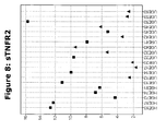
- X1 soluble TNF-receptor 2 (sTNFR2)
- Soluble TNF-receptor 2 (TNF-sr75 or sTNFR2) has been shown to be a putative marker for diagnosing sepsis by Neilson et al., 1996 (Cytokine. 1996 vol.12:938-43 ).
- the levels of TNF-alpha and TNF-soluble receptor were raised in patients who became clinically septic and correlated well with the severity of sepsis (using the APACHE III score).
- TNF-sr55 and TNF-sr75 In septic patients there was no difference in the pattern of changes in the two types of receptor (TNF-sr55 and TNF-sr75). However, in non-septic patients TNF-sr75 was higher in those with endotoxaemia than those without.
- TNF-sr55 This difference was not observed with TNF-sr55 which suggests a different mechanism of release or degree of sensitivity for the two soluble receptors. Regardless of severity of illness, the levels of all three molecules (TNF-alpha and the two receptors) appeared to start rising at about the same time point. The peak TNF-alpha level was reached earlier (2-4 h) than that of the two TNF-sr (4-8 h). The relative rise in TNF-alpha was greater than that of the soluble receptors and this difference was even more marked in those with more severe sepsis. The relationship between peak TNF-alpha and peak TNF-sr was non-linear and the concentration of each TNF-sr appeared to plateau at the higher levels of TNF-alpha. This suggests the exhaustion of a limited pool or saturation of the rate of release. Taken together, these results suggest sepsis develops because of delayed and insufficient secretion of TNF-sr compared with TNF-alpha.
- Pentraxins are highly conserved proteins throughout evolution starting from the horse shoe crab to man.
- the classic pentraxins are of the short form and are known as C-reactive protein (CRP) and serum amyloid P component (SAP). They are made in the liver upon stimulus from interleukin (IL)-6.
- CRP C-reactive protein
- SAP serum amyloid P component
- PTX3 is made in response to primary proinflammatory signals (bacterial products, IL-1, and tumor necrosis factor [TNF] but not IL-6) by diverse cell types, predominantly macrophages and endothelial cells.
- primary proinflammatory signals bacterial products, IL-1, and tumor necrosis factor [TNF] but not IL-6
- PTX3 is elevated in patients with SIRS, sepsis, and septic shock. Maximal elevations of PTX3 were observed in patients with septic shock.
- PTX3 levels were correlated with disease severity as assessed by clinical scores. In particular, a dramatic difference was apparent in PTX3 levels at admission or on day 2 between patients who survived and those who eventually died.
- M-CSF Macrophage Colony Stimulating Factor
- M-CSF is a hematopoietic growth factor that mainly stimulates the growth, differentiation, and proliferation of cells of the monocyte-macrophage lineage.
- ⁇ ren et al., 2001 studied the serum M-CSF levels in healthy, septic, and hypoxic term neonates on the first day of life and examined the relationship of serum M-CSF levels and circulating monocyte and thrombocyte counts in these newborn infants.
- Three groups were defined in this prospective study: group 1, healthy neonates with no risk factors ( n 5 40); group 2, neonates who had severe hypoxia ( n 5 20); and group 3, neonates who fulfilled the criteria for early-onset sepsis ( n 5 18).
- Blood samples were collected for complete blood cell count and serum M-CSF levels by peripheral venipuncture from each infant in the first 24 hours after birth before any medical therapy.
- the gestational ages and birth weights did not differ significantly between the groups.
- Serum MCSF levels of the septic neonates were significantly higher than of both healthy and hypoxic neonates, but did not differ significantly between the healthy and hypoxic neonates.
- pro-Hepcidin pro-HEPC
- Hepcidin (cf. Malyszko and Mysliwiec, Kidney Blood Press Res 2007;30:15-30 for an overview) is a circulating antimicrobial peptide mainly synthesized in the liver, which has been recently proposed as a factor regulating the uptake of dietary iron and its mobilization from macrophages and hepatic stores. Inflammation causes an increase of production of hepcidin, which is a potent mediator of anemia of chronic diseases. Hepcidin (hep atic bacteri cid al prote in ), a recently discovered small, cysteine-rich cationic peptide from plasma, produced by the liver and had antimicrobial properties.
- hepcidin The structure of hepcidin is highly conservative among mammals, suggesting a key role in major biological functions. Closely related hepcidin sequences are found in vertebrates from fish to humans.
- prohepcidin After a cleavage of the 24 amino acid N-terminal signal peptide, prohepcidin is transported through the hepatocyte basolateral membrane into the circulation.
- the major circulating bioactive forms of hepcidin consist only of the carboxy-terminal portion (peptides of 25, 22 and 20 amino acids). The exact location of the final prohormone processing is unknown. Propeptide convertases could be located in the blood or in the cell membrane of capillaries. There is still no reliable information available on normal serum levels of mature hepcidin. Substantial progress has been made to elucidate the mechanism of action of hepcidin. Presently, the main hepcidin function is homeostatic regulation of iron metabolism and mediation of host defense and inflammation.
- hepcidin gene is overexpressed in livers of experimentally iron-loaded mice and hepcidin knockout mice develop iron overload. Injection of hepcidin inhibits intestinal iron absorption in mice independent of their iron status. It was shown that hepcidin injection results in a dose-dependent in serum iron. Conversely, iron ingestion in humans induced hepcidin secretion in urine. These observations suggest that hepcidin plays a role as a negative regulator of intestinal iron absorption and iron release from macrophages. Evidence from transgenic mouse models indicates that hepcidin is the predominant negative regulator of iron absorption in the small intestine, iron transport across the placenta, and iron release from the macrophages.
- Hepcidin controls intestinal iron absorption by regulating ferroportin expression on the basolateral membrane of enterocytes. Hepcidin directly regulates the expression of the ferroportin on cell membranes, acts by binding to the cellular iron exporter ferroportin and inducing its internalization and degradation, thus trapping iron in enterocytes, macrophages and hepatocytes. The net effect of hepcidin is the diminished absorption of dietary iron, sequestration of iron in macrophages and sequestration of iron in hepatic stores. The absence of hepcidin synthesis would naturally lead to a major loss of control over iron release by enterocytes and macrophages followed by circulatory iron overload.
- Brain natriuretic peptide (also known as B-type natriuretic peptide or "GC-B”) is a 32 amino acid polypeptide secreted by the ventricles of the heart in response to excessive stretching of myocytes (heart muscles cells) in the ventricles.
- NT-proBNP co-secreted 76 amino acid N-terminal fragment
- BNP binds to and activates NPRA in a similar fashion to atrial natriuretic peptide (ANP) but with 10-fold lower affinity.
- ANP atrial natriuretic peptide
- Brain natriuretic peptide was originally identified in extracts of porcine brain, but in humans it is produced mainly in the cardiac ventricles.
- Physiologic actions of BNP and ANP include decrease in systemic vascular resistance and central venous pressure as well as an increase in natriuresis.
- the resulting effect of these peptides is a decrease in cardiac output and a decrease in blood volume.
- the backbone of the NETs constitutes out of chromatin released from the suicidal neutrophil and take the form of DNA-based threads and cables. In addition to DNA, some proteins, such as histones appear to form some globular domains in these NETs.
- Microorganisms are killed by neutrophil cultures that have been stimulated to produce NETs by IL-8, bacterial components, or the protein kinase C activator PMA, and in which phagocytosis is prevented, indicating that NETs have antimicrobial activity. Furthermore, this activity can be eliminated if the NETs are disrupted by treating activated neutrophils with DNases. After an infection, neutrophils will first kill bacteria by phagocytosis and only later by the production of NETs.
- NETs might contain specific antifungal agents.
- the specific contribution of the granular antimicrobial peptides, proteases and other enzymes or the histones to microbial killing is not understood. It is possible that different pathogens have different levels of susceptibility to these components. NETs could also make microorganisms susceptible to other antimicrobial effectors.
- Sepsis is caused by an infection of a microorganism, and it has been demonstrated that the innate immune system is activated in patients at the onset of sepsis.
- This response comprises of several events, one of them being the production of NETs in the blood, comprised of chromatin back-bone, coated with intrecallular and intrenuclear proteins such as histones. This might in fact be the explanation of the increase of histone protein in the blood of patients with sepsis. In samples from patients with SIRS, no such histone presence in the blood was detected. This means that histones as identified by the method as described herein may be a very clear indicator of sepsis enabling the differentiation between SIRS and sepsis patients.
- any of the above markers identified herein can be used separately or in combination in the kits, microarrays and methods as described herein. Any combination of two or more of the markers identified herein can be used together. In addition, any combination of one or more of the newly identified biomarkers can be used together with other known sepsis markers.
- One preferred known sepsis marker is the PCT (procalcitonin) marker or the CRP (c-reactive protein) marker. Preferred combinations of the markers are the following:
- the combination always comprises one or more members of the Histone protein family as a biomarker in combination with any one or more other biomarkers of sepsis or SIRS
- PTX-3 + pro-HEPC The most preferred combinations are PTX-3 + pro-HEPC; and PCT + pro-HEPC + sTNFR2.
- Procalcitonin is a precursor of the hormone calcitonin, which is involved with calcium homeostasis, and is produced by the C-cells of the thyroid gland. It is there that procalcitonin is cleaved into calcitonin, katacalcin and a protein residue. It is not released into the blood stream of healthy individuals. With the derangements that a severe infection with an associated systemic response brings, the blood levels of procalcitonin may rise to 100 ng/ml. In blood serum, procalcitonin has a half-life of 25 to 30 hours.
- Measurement of procalcitonin can be used as a marker of severe sepsis and generally grades well with the degree of sepsis, although levels of procalcitonin in the blood are very low.
- PCT has the greatest sensitivity (85%) and specificity (91%) for differentiating patients with SIRS from those with sepsis, when compared with IL-2, IL-6, IL-8, CRP and TNF-alpha.
- the test is not routinely used and has yet to gain widespread acceptance (cf. Meisner et al., 1999, Crit Care vol3(1):45-50 ).
- CRP C-reactive protein
- C-reactive protein is a plasma protein, an acute phase protein produced by the liver and by adipocytes. It is a member of the pentraxin family of proteins and has been widely used as a marker for sepsis, especially in neonates. Its accuracy is however controversial.
- Biomarker profiles may be generated by the use of one or more separation methods.
- suitable separation methods may include a mass spectrometry method, such as electrospray ionization mass spectrometry (ESI-MS), ESI-MS/MS, ESI-MS/ (MS)" (n is an integer greater than zero), matrix-assisted laser desorption ionization time- of-flight mass spectrometry (MALDI-TOF-MS), surface-enhanced laser desorption/ionization time-of-flight mass spectrometry (SELDI-TOF-MS),desorption/ionization on silicon (DIOS), secondary ion mass spectrometry (SIMS), quadrupole time-of-flight (Q-TOF), atmospheric pressure chemical ionization mass spectrometry (APCI-MS), APCI-MS/MS, APCI- (MS)", atmospheric pressure photoionization mass spectrometry (APPI-MS),APPI-MS/MS, and APPI-
- mass spectrometry methods may include, inter alia, quadrupole, fourier transform mass spectrometry (FTMS) and ion trap.
- suitable separation methods may include chemical extraction partitioning, column chromatography, ion exchange chromatography, hydrophobic (reverse phase) liquid chromatography, isoelectric focusing, one-dimensional polyacrylamide gel electrophoresis (PAGE), two-dimensional polyacrylamide gel electrophoresis (2D-PAGE) or other chromatography, such as thin-layer, gas or liquid chromatography, or any combination thereof.
- the biological sample may be fractionated prior to application of the separation method.
- Biomarker profiles may also be generated by methods that do not require physical separation of the biomarkers themselves.
- nuclear magnetic resonance (NMR) spectroscopy may be used to resolve a profile of biomarkers from a complex mixture of molecules.
- NMR nuclear magnetic resonance
- Additional procedures include nucleic acid amplification technologies, which may be used to generate a profile of biomarkers without physical separation of individual biomarkers. (See Stordeur et al., J. Immunol. Methods 259: 55-64 (2002 ) and Tan et al., Proc. Nat. Acad. Sci. USA 99: 11387-11392 (2002 ), for example).
- Laser desorption/ionization time-of-flight mass spectrometry may be used to create a profile of biomarkers where the biomarkers are proteins or protein fragments that have been ionized and vaporized off an immobilizing support by incident laser radiation. A profile is then created by the characteristic time-of-flight for each protein, which depends on its mass-to-charge (“m/z”) ratio.
- m/z mass-to-charge ratio
- Laser desorption/ionization time-of-flight mass spectrometry allows the generation of large amounts of information in a relatively short period of time.
- a biological sample is applied to one of several varieties of a support that binds all of the biomarkers, or a subset thereof, in the sample.
- Cell lysates or samples are directly applied to these surfaces in volumes as small as 0.5RL, with or without prior purification or fractionation.
- the lysates or sample can be concentrated or diluted prior to application onto the support surface.
- Laser desorption/ionization is then used to generate mass spectra of the sample, or samples, in as little as three hours.
- the protein biomarker profile as described herein may be established using immunoassay technologies such as direct ELISA, indirect ELISA, sandwich ELISA, competitive ELISA, multiplex ELISA, radioimmunoassay, ELISPOT technologies, and other similar techniques known in the art.
- immunoassay technologies such as direct ELISA, indirect ELISA, sandwich ELISA, competitive ELISA, multiplex ELISA, radioimmunoassay, ELISPOT technologies, and other similar techniques known in the art.
- the direct ELISA uses the method of directly labeling the antibody itself. Microwell plates are coated with a sample containing the target antigen, and the binding of labeled antibody is quantitated by a colorimetric, chemiluminescent, or fluorescent end-point. Since the secondary antibody step is omitted, the direct ELISA is relatively quick, and avoids potential problems of cross-reactivity of the secondary antibody with components in the antigen sample. However, the direct ELISA requires the labeling of every antibody to be used, which can be a time-consuming and expensive proposition. In addition, certain antibodies may be unsuitable for direct labeling. Direct methods also lack the additional signal amplification that can be achieved with the use of a secondary antibody.
- the indirect, two-step ELISA method uses a labeled secondary antibody for detection. First, a primary antibody is incubated with the antigen. This is followed by incubation with a labeled secondary antibody that recognizes the primary antibody. For ELISA it is important that the antibody enzyme conjugate is of high specific activity. This is achieved when the antibody is affinity purified and the enzyme conjugation chemistry preserves antibody specificity as well as enzyme activity
- the sandwich ELISA measures the amount of antigen between two layers of antibodies.
- the antigens to be measured must contain at least two antigenic sites, capable of binding to the antibody, since at least two antibodies act in the sandwich. For this reason, sandwich assays are restricted to the quantitation of multivalent antigens such as proteins or polysaccharides. Sandwich ELISAs for quantitation of antigens are especially valuable when the concentration of antigens is low and/or they are contained in high concentrations of contaminating protein.
- one antibody (the "capture” antibody) is purified and bound to a solid phase typically attached to the bottom of a plate well. Antigen is then added and allowed to complex with the bound antibody.
- Unbound products are then removed with a wash, and a labeled second antibody (the "detection” antibody) is allowed to bind to the antigen, thus completing the "sandwich".
- the assay is then quantitated by measuring the amount of labeled second antibody bound to the matrix, through the use of a colorimetric substrate.
- Major advantages of this technique are that the antigen does not need to be purified prior to use, and that these assays are very specific.
- one disadvantage is that not all antibodies can be used. Monoclonal antibody combinations must be qualified as "matched pairs", meaning that they can recognize separate epitopes on the antigen so they do not hinder each other's binding.
- the ELISA kits are good enough to reach detection senility at sub-nanogram per ml level and are useful for screening protein targets and quantifying their expression in different conditions.
- monoclonal antibodies can be further introduced into the ELISA kit to pair with polyclonal IgY as either capture or detection antibodies.
- the competitive ELISA When two "matched pair" antibodies are not available for a target, another option is the competitive ELISA.
- the advantage to the competitive ELISA is that non-purified primary antibodies may be used.
- a detection enzyme such as horseradish peroxidase.
- the enzyme may be linked to either the antigen or the primary antibody.
- An example of such a competitor is a labeled antigen.
- this type of ELISA there is an inverse relationship between the signal obtained and the concentration of the analyte in the sample, due to the competition between the free analyte and the ligand-enzyme conjugate for the antibody coating the microplate, i.e.
- an unlabeled purified primary antibody is coated onto the wells of a 96 well microtiter plate. This primary antibody is then incubated with unlabeled standards and unknowns. After this reaction is allowed to go to equilibrium, conjugated antigen is added. This conjugate will bind to the primary antibody wherever its binding sites are not already occupied by unlabeled antigen. Thus, the more unlabeled antigens in the sample or standard, the lower the amount of conjugated antigen bound. The plate is then developed with substrate and color change is measured.
- Multiplex ELISA is a microtiter plate ELISA-based protein array assay that allows simultaneous detection of multiple analytes at multiple array addresses within a single well.
- multiplex ELISA There are different types of multiplex ELISA have been developed and in practice. One of the examples is to measure antigens by coating or printing capture antibodies in an array format within a single well to allow for the construction of "sandwich" ELISA quantification assays.
- multiplex ELISA can also be achieved through antibody array, where different primary antibodies can be attached to a solid phase e.g. a glass plate to capture corresponding antigens in a biological sample.
- the detection method can be direct or indirect, sandwich or competitive, labeling or non-labeling, depending upon antibody array technologies.
- the Enzyme-Linked Immunosorbent Spot (ELISpot) assay employs the sandwich assay approach of the Enzyme-Linked ImmunoSorbent Assay (ELISA), with some variations.
- the capture antibody is coated aseptically onto a polyvinylidene difluoride (PVDF)-backed microwell plate.
- PVDF polyvinylidene difluoride
- the plate is blocked with serum proteins, cells of interest are plated out at varying densities, along with antigen or mitogen, and plates are incubated at 37°C. Cytokine secreted by activated cells is captured locally by the coated antibody on the high surface area PVDF membrane.
- the wells are washed to remove cells, debris, and media components.
- a second antibody (biotinylated) reactive with a distinct epitope of the target cytokine is employed to detect the captured cytokine.
- the detected cytokine is then visualized using avidin-HRP, and a precipitating substrate (e.g. AEC).
- the colored end product (spot) represents an individual cytokine-producing cell.
- the spots can be counted manually (e.g., with a dissecting microscope) or using an automated reader to capture the microwell images and to analyze spot number and size.
- Radioimmunoassay involves mixing known quantities of radioactive antigen (frequently labeled with gamma-radioactive isotopes of iodine attached to tyrosine) with antibody to that antigen, then adding unlabeled or "cold” antigen and measuring the amount of labeled antigen displaced. Initially, the radioactive antigen is bound to the antibodies. When "cold” (unlabeled, quest) antigen is added, the two compete for antibody binding sites - at higher concentrations of "cold” antigen, more of it binds to the antibody, displacing the radioactive variant. The bound antigens are separated from the unbound ones.
- the term "profile” includes any set of data that represents the distinctive features or characteristics associated with a condition of sepsis.
- the term encompasses a "nucleic acid profile” that analyzes one or more genetic markers, a "protein profile” that analyzes one or more biochemical or serological markers, and combinations thereof.
- nucleic acid profiles include, but are not limited to, a genotypic profile, gene copy number profile, gene expression profile, DNA methylation profile, and combinations thereof.
- Non-limiting examples of protein profiles include a protein expression profile, protein activation profile, protein cleavage profile, and combinations thereof.
- a "genotypic profile” includes a set of genotypic data that represents the genotype of one or more genes associated with a condition of sepsis.
- a "gene copy number profile” includes a set of gene copy number data that represents the amplification of one or more genes associated with a condition of sepsis.
- a “gene expression profile” includes a set of gene expression data that represents the mRNA levels of one or more genes associated with a condition of sepsis.
- a "DNA methylation profile” includes a set of methylation data that represents the DNA methylation levels (e.g., methylation status) of one or more genes associated with a condition of sepsis.
- a "protein expression profile” includes a set of protein expression data that represents the levels of one or more proteins associated with a condition of sepsis.
- a "protein activation profile” includes a set of data that represents the activation (e.g., phosphorylation status) of one or more proteins associated with a condition of sepsis.
- a “protein cleavage profile” includes a set of data that represent the proteolytic cleavage of one or more proteins associated with a condition of sepsis.
- subject typically includes humans, but can also include other animals such as, e.g., other primates, rodents, canines, felines, equines, ovines, porcines, and the like.
- sample includes any biological specimen obtained from a subject.
- Samples include, without limitation, whole blood, plasma, serum, red blood cells, white blood cells (e.g., peripheral blood mononuclear cells), saliva, urine, stool (i.e., faeces), tears, sweat, sebum, nipple aspirate, ductal lavage, tumour exudates, synovial fluid, cerebrospinal fluid, lymph, fine needle aspirate, amniotic fluid, any other bodily fluid, cell lysates, cellular secretion products, inflammation fluid, semen and vaginal secretions.
- the sample may be whole blood or a fractional component thereof such as plasma, serum, or a cell pellet.
- the sample is readily obtainable by minimally invasive methods.
- Samples may also include tissue samples and biopsies, tissue homogenates and the like.
- the specification provides for a method for establishing a healthy reference biomarker profile comprising the steps of:
- the specification provides for a method for establishing a SIRS reference biomarker profile comprising the steps of:
- the specification provides for a method for establishing a sepsis reference biomarker profile comprising the steps of:
- kits for predicting, prognosing and/or diagnosing sepsis in a subject comprise at least one biomarker as defined herein or molecules specifically binding thereto.
- biomarkers that are useful in the methods and kits as taught herein are those selected from the group of Histone proteins, soluble TNF-receptor 2 (sTNFR2), Pentraxin-3 (PTX-3), Macrophage Colony-Stimulating Factor (MCSF), pro-Hepcidin (pro-HEPC), pro-Brain Natriuretic Protein (pro-BNP), Procalcitonin (PCT) and c-Reactive Protein (CRP) but a kit may include one or two or three or four or all of the biomarkers listed therein with or without other biomarkers in addition.
- sTNFR2 soluble TNF-receptor 2
- PTX-3 Pentraxin-3
- MCSF Macrophage Colony-Stimulating Factor
- pro-HEPC pro-Hepcidin
- the specification also provides a method and a kit for assessing the occurrence and stage or severity of sepsis in a subject, which can range from the very onset of sepsis, to septic shock and eventually the death of the subject, by measuring the quantity of one or of a combination of one or more of the biomarkers as defined herein selected from the group of histone proteins soluble TNF-receptor 2 (sTNFR2), Pentraxin-3 (PTX-3), Macrophage Colony-Stimulating Factor (MCSF), pro-Hepcidin (pro-HEPC), pro-Brain Natriuretic Protein (pro-BNP), Procalcitonin (PCT) and c-Reactive Protein (CRP) in combination with known biomarkers for sepsis or, in the sample from the subject and comparing the biomarker measurements to that of a sample obtained from a healthy or non-sepsis subject.
- the biomarkers as defined herein selected from the group of histone proteins soluble TNF-recept
- the specification provides a means for a clinician to estimate the degree of sepsis at an initial assessment, and to monitor the change in status of the sepsis (worsening, improving, or remaining the same) based on the detected amount of the one or more biomarkers in the sample of the subject.
- the clinician would establish a protocol of collecting and analyzing a quantity of fresh sample from the patient at selected intervals.
- the sample is obtained intermittently during a prescribed period.
- the period of time between intermittent sampling may be dictated by the condition of the subject, and can range from a sample each 24 hours to a sample taken continuously, more typically from each 4 hours to each 30 minutes.
- both a qualitative level of one or more of the biomarkers present in the sample can be analyzed and estimated, and a quantitative level of one or more of the biomarkers present in the sample can be analyzed and measured.
- the clinician would select the qualitative method, the quantitative method, or both, depending upon the status of the patient.
- the quantity of sample to be collected is less than 10 milliliter, less than 1 milliliter, and more typically less than 10 ⁇ l.
- a typical sample can range from about 1 ⁇ l to about 1 ml.
- the larger quantities of sample (about 10 ml) are used for quantitative assays.
- these small amounts of sample are easily and readily available or obtainable from clinical subjects who are either prone to developing sepsis, or have developed sepsis.
- the clinician can employ the method and kit as taught herein to monitor the progress of the treatment or intervention.
- one or more subsequent post-treatment samples will be taken and analyzed for the presence of one or more of the biomarkers as the treatment of the sepsis condition commences and continues.
- the treatment is continued until the presence of one or more of the biomarkers as defined herein in subsequent post-treatment samples is normalized when compared to a sample obtained from a healthy or non-sepsis subject.
- the expression of one or more of the biomarkers, and its presence in the sample will be altered and normalized when compared to a sample of a healthy or non-sepsis subject.
- the degree of amelioration will be expressed by a correspondingly normalized level of one or more of the biomarkers, detected in a sample.
- the method can be used to detect the complete normalization of one or more of the biomarkers as defined herein, signaling the completion of the course of treatment.
- binding molecules refers to all suitable binding molecules that are specifically binding or interacting with one of the biomarkers as defined herein and that can be used in the methods and kits as taught herein.
- suitable binding agents are antibodies, aptamers, specifically interacting small molecules, specifically interacting proteins, and other molecules that specifically bind to one of the biomarkers.
- Both monoclonal and polyclonal antibodies that bind one of the biomarkers as defined herein are useful in the methods and kits as taught herein.
- the monoclonal and polyclonal antibodies can be prepared by methods known in the art and are often commercially available.
- Aptamers that bind specifically to the biomarkers as defined herein can be obtained using the so called SELEX or Systematic Evolution of Ligands by EXponential enrichment.
- SELEX Systematic Evolution of Ligands by EXponential enrichment.
- multiple rounds of selection and amplification can be used to select for DNA or RNA molecules with high specificity for a target of choice, developed by Larry Gold and coworkers and described in US patent 6,329,145 .
- Recently a more refined method of designing co-called photoaptamers with even higher specificity has been described in US patent 6,458,539 by the group of Larry Gold.
- binding agents such as interacting proteins and small molecules
- binding agents such as interacting proteins and small molecules
- Examples are two-hybrid analysis, immunoprecipitation methods and the like.
- the step of detecting the complex of the capture antibody and one or more of the biomarkers comprises contacting the complex with a second antibody for detecting the biomarker.
- the method for detecting the complex of one or more of the biomarkers and the primary antibody or binding molecule comprises the steps of: separating any unbound material of the sample from the capture antibody-biomarker complex; contacting the capture antibody-biomarker complex with a second antibody for detecting the biomarker, to allow formation of a complex between the biomarker and the second antibody; separating any unbound second antibody from the biomarker-second antibody complex; and detecting the second antibody of the biomarker-second antibody complex.
- a kit for use in the method typically comprises one or more media having affixed thereto one or more capture antibodies or binding molecules, whereby the sample is contacted with the media to expose the capture antibody or binding molecule to the biomarker present in the sample.
- the kit includes an acquiring means that can comprise an implement, such as a spatula or a simple stick, having a surface comprising the media.
- the acquiring means can also comprise a container for accepting the sample, where the container has a sample-contacting surface that comprises the media.
- the assay for detecting the complex of one or more of the biomarkers and the antibody or binding molecule can comprise an ELISA, and can be used to quantitate the amount of one or more the biomarkers in a sample.
- the acquiring means can comprise an implement comprising a cassette containing the media.
- biomarkers as defined herein can provide an indication of the presence of the protein in a sample in a short period of time.
- a method and a kit as taught herein can detect the biomarker in a sample within four hours, more typically within two hours, and most typically within one hour, following the sepsis condition.
- the biomarker can be detected within about 30 minutes following the sepsis condition.
- a rapid one-step method of detecting one or more of the biomarkers as defined herein can reduce the time for detecting the sepsis condition.
- a typical method can comprise the steps of: obtaining a sample suspected of containing one or more of the biomarkers; mixing a portion of the sample with one or more detecting antibodies or binding molecules that each specifically bind to one of the biomarkers, so as to initiate the binding the detecting antibody or binding molecule to the biomarkers in the sample; contacting the mixture of sample and detecting antibody or binding molecule with an immobilized capture antibody or binding molecule which specifically binds to the biomarker, which capture antibody or binding molecule does not cross-react with the detecting antibody or binding molecule, so as to bind the detecting antibody or binding molecule to the biomarker, and the biomarker to the capture antibody or binding molecule, to form a detectable complex; removing unbound detecting antibody or binding molecule and any unbound sample from the complex; and detecting the detecting antibody or binding molecule of the complex.
- the detectable antibody or binding molecule can be labeled with a detectable marker, such as a radioactive label, a fluorescent label, an enzyme label, a biological dye, a magnetic bead, (strept)avidin, or biotin, as is well known in the art.
- a detectable marker such as a radioactive label, a fluorescent label, an enzyme label, a biological dye, a magnetic bead, (strept)avidin, or biotin, as is well known in the art.
- the specification provides a method for the prediction, prognosis and/or diagnosis of sepsis or the differentiation between SIRS and sepsis in a subject comprising obtaining a candidate biomarker profile from a biological sample taken from said subject wherein said candidate biomarker profile is based on the measurement of the quantity of Histone proteins in said sample, and comparing said candidate biomarker profile with a reference biomarker profile obtained form a healthy subject or a patient having SIRS.
- a method for the prediction, prognosis and/or diagnosis of sepsis or for the differentiation between SIRS and sepsis in a subject comprising: obtaining a candidate biomarker profile from a biological sample taken from said subject wherein said candidate biomarker profile is based on at least one or two biomarkers selected from the group consisting of Histone proteins, soluble TNF-receptor 2 (sTNFR2), Pentraxin-3 (PTX-3), Macrophage Colony-Stimulating Factor (MCSF), pro-Hepcidin (pro-HEPC), pro-Brain Natriuretic Protein (pro-BNP), Procalcitonin (PCT) and c-Reactive Protein (CRP) and comparing said candidate profile with a reference biomarker profile obtained form a healthy subject or a patient having SIRS.
- sTNFR2 soluble TNF-receptor 2
- PTX-3 Pentraxin-3
- MCSF Macrophage Colony-Stimulating Factor
- the specification further provides for a method for the prediction, prognosis and/or diagnosis of sepsis or for the differentiation between SIRS and sepsis in a subject comprising measuring the level of at least one or two biomarkers selected from the group consisting of Histone proteins, soluble TNF-receptor 2 (sTNFR2), Pentraxin-3 (PTX-3), Macrophage Colony-Stimulating Factor (MCSF), pro-Hepcidin (pro-HEPC), pro-Brain Natriuretic Protein (pro-BNP), Procalcitonin (PCT) and c-Reactive Protein (CRP) in a biological sample from said subject; using said measurements obtained in step a) to create a profile for said biomarkers; and comparing said profile with a reference biomarker profile obtained form a healthy subject or a patient having SIRS.
- biomarkers selected from the group consisting of Histone proteins, soluble TNF-receptor 2 (sTNFR2), Pentraxin-3 (PTX-3), Mac
- the specification further provides for a method for the prediction, prognosis and/or diagnosis of sepsis or for the differentiation between SIRS and sepsis in a subject comprising determining a quantity of at least one or two biomarkers selected from the group consisting of Histone proteins, soluble TNF-receptor 2 (sTNFR2), Pentraxin-3 (PTX-3), Macrophage Colony-Stimulating Factor (MCSF), pro-Hepcidin (pro-HEPC), pro-Brain Natriuretic Protein (pro-BNP), Procalcitonin (PCT) and c-Reactive Protein (CRP) in a sample obtained from a subject; and comparing the quantity of the selected biomarkers in the test subject sample with a range of normal values of the selected biomarkers in control subjects; whereby an increase or decrease in the quantity of the selected biomarker in the sample to a level higher or lower than the range of normal values of the selected biomarkers is indicative of sepsis.
- biomarkers selected from the
- the specification further provides for a method for the prediction, prognosis and/or diagnosis of sepsis or for the differentiation between SIRS and sepsis in a subject comprising determining a quantity of at least one or two biomarkers selected from the group consisting of Histone proteins, soluble TNF-receptor 2 (sTNFR2), Pentraxin-3 (PTX-3), Macrophage Colony-Stimulating Factor (MCSF), pro-Hepcidin (pro-HEPC), pro-Brain Natriuretic Protein (pro-BNP), Procalcitonin (PCT) and c-Reactive Protein (CRP) and comparing the quantity of the selected biomarkers in the test subject sample with a range of values of the selected biomarkers obtained from subjects with sepsis; whereby a comparable quantity of the selected biomarkers in said sample to the range of values of the selected biomarkers in subjects with sepsis is indicative of sepsis.
- biomarkers selected from the group consisting of Histone proteins, soluble
- the specification further provides for a method for determining whether an individual is responsive to treatment for sepsis with a substance, comprising the steps of obtaining a candidate biomarker profile from a biological sample taken from said individual wherein said candidate biomarker profile is based on at least one or two biomarkers selected from the group consisting of Histone proteins, soluble TNF-receptor 2 (sTNFR2), Pentraxin-3 (PTX-3), Macrophage Colony-Stimulating Factor (MCSF), pro-Hepcidin (pro-HEPC), pro-Brain Natriuretic Protein (pro-BNP), Procalcitonin (PCT) and c-Reactive Protein (CRP) and comparing said candidate profile with a reference biomarker profile.
- sTNFR2 soluble TNF-receptor 2
- PTX-3 Pentraxin-3
- MCSF Macrophage Colony-Stimulating Factor
- pro-HEPC pro-Hepcidin
- pro-BNP pro-Bra
- the selected biomarker as described herein comprises one or more members of the Histone protein family.
- this marker or these markers are combined with any one or more of the other biomarkers provided herein selected from the group of: soluble TNF-receptor 2 (sTNFR2), Pentraxin-3 (PTX-3), Macrophage Colony-Stimulating Factor (MCSF), pro-Hepcidin (pro-HEPC), pro-Brain Natriuretic Protein (pro-BNP), Procalcitonin (PCT) and c-Reactive Protein (CRP)
- sTNFR2 soluble TNF-receptor 2
- PTX-3 Pentraxin-3
- MCSF Macrophage Colony-Stimulating Factor
- pro-HEPC pro-Hepcidin
- pro-BNP pro-Brain Natriuretic Protein
- PCT Procalcitonin
- CCP c-Reactive Protein
- said Histone proteins are selected from the group of Histone H1.4, Histone H2A (different isoforms), Histone H2B (different isoforms), Histone H3 (different isoforms), Histone H4 (different isoforms).
- the selected biomarkers are pro-Hepcidin (pro-HEPC) and Pentraxin-3 (PTX-3).
- Preferred samples to be analysed in the method as described herein are blood or urine, more preferable the sample is serum or plasma.
- the method as described herein uses immunoassay technology selected from the group of direct ELISA, indirect ELISA, sandwich ELISA, competitive ELISA, multiplex ELISA, radioimmunoassay, or ELISPOT technologies to establish the biomarker profile.
- the biomarker profile is established using mass spectrometry analysis methods of the proteins present in said sample.
- the methods indicated herein are particularly useful to distinguish between SIRS and sepsis.
- the identification of the biomarkers as defined herein could be of use in the treatment or amelioration of the sepsis condition of the subject.
- Another possibility can be the reduction of the level or abundance of a certain biomarker as defined herein in case said biomarker has an increased occurrence in the blood of patients having sepsis when compared to the samples of a subject having SIRS or a healthy subject.
- these biomarkers are Histone proteins, pro-BNP, PTX-3, pro-HEPC and M-CFS. Administering agents that reduce the expression or activity of said proteins may be beneficial to the subject.
- each sample is measured against a reference sample, typically a pool of all patient samples. For each feature present in a certain sample a ratio is obtained which represents the fold difference of the feature intensity in the reference versus feature intensity in the sample. Combining all feature data from all samples into an expression matrix allows comparing features intensities between samples and between groups of samples.
- MS data were deisotoped, clustered and features were constructed using in house developed software called euCatLabel.
- the output of this data processing is an expression matrix containing all features from all analyzed samples.
- Each feature is represented by a unique combination of m/z, COFRADIC sorting pool and NanoLC retention time and features can be present in a number of samples ranging from 1 to all. If a feature is present in a sample it will carry a ratio, which represents intensity of the feature in the reference sample (ie the 16 O peak) over the intensity in the respective sample (the 18 O peak).
- Recurrent quantifiable features are features with a reliable ratio reading in at least one sample. For the intermittent matrix analysis of 3 samples each, all features were considered that were either present in 5/6 samples or were present in all 3 samples of the SIRS patient group and in none of the samples of the SEPSIS patient group.
- the six identified biomarkers are:
- Example 2 ELISA measurements of the newly identified biomarkers as defined herein in samples from patients suffering from SIRS or several stages of sepsis.
- the biomarkers obtained from the Cofradic analysis were analysed for their differential expression in blood samples from patients suffering from SIRS or several stages of sepsis, using ELISA with specific antibodies directed to each of the biomarkers.
- Table 2 of the present application we compared the quantity of these five protein biomarkers in the different blood samples with two known SIRS / sepsis biomarkers procalcitonin (PCT) and c-reactive protein (CRP). From the table it becomes clear that markers X1-X5 show a clear difference in protein quantity between samples from healthy subjects and the samples form subjects suffering from SIRS or sepsis. The results are shown in Figures 1-5 for X1-X5 respectively.
- the Human Hepcidin Prohormone was detected using a polyclonal anti human Pro-Hepcidin antibody (DRG) as a capturing antibody and a Pro-Hepcidin fragment conjugated to biotin as a conjugate in a competitive ELISA set-up.
- DRG polyclonal anti human Pro-Hepcidin antibody
- Sepsis and SIRS samples are diluted as follows: 60 ⁇ l sample + 60 ⁇ l assay buffer (load 50 ⁇ l/well). Healthy samples were diluted in a 1 ⁇ 2 ratio.
- a polyclonal anti-BNP fragment (8-29) antibody (Biomedica) was used as a capturing antibody and a synthetic BNP fragment coupled to HRP was used as a conjugate in a competitive ELISA set-up.
- Sepsis and SIRS samples are diluted as follows: 150 ⁇ l sample + 300 ⁇ l assay buffer (load 200 ⁇ l/well). Healthy samples were diluted in a 1 ⁇ 2 ratio.
- M-CSF was detected by using a mouse monoclonal antibody against human M-CSF(R&D systems) as a capturing antibody and a polyclonal antibody against human M-CSF conjugated to HRP as a detection antibody.
- Sepsis and SIRS samples are diluted as follows: 44 ⁇ l sample +176 ⁇ l assay buffer (load 100 ⁇ l/well). Healthy samples were diluted in a 1 ⁇ 2 ratio.
- sTNF-RII was detected using a mouse monoclonal antibody against human sTNF-RII (R&D systems) as a capture antibody and a polyclonal antibody against human sTNF-RII conjugated to HRP as a detection antibody.
- Sepsis and SIRS samples are diluted as follows: 14 ⁇ l sample + 406 ⁇ l assay buffer (load 200 ⁇ l/well). Healthy samples were diluted in a 1 ⁇ 2 ratio.
- the biomarkers as defined herein are all valuable for distinguishing SIRS conditions from sepsis conditions.
- pro-BNP Figure 5
- PTX3 Figure 2
- pro-HEPC Figure 4
- LSVM Linear Support Vector Machine
- the LSVM induces a classifier from the data that is very similar to the black line classifer shown above. But the linear threshold is computed in a 7-dimensional space (the space of the 7 ELISA measurments) instead of a 1-dimensional space. As such, the classifier is not a simple line but a hyperplane in the 7-dimensional space.
- the LSVM classifier achieves a 15% LOO error when using all 7 elisa measurements (this differs from the 10% reported before due to swaps in the MCSF data).
- pro-Hepcidin is most relevant. This indicates that it is complementary to the other proteins.
- PTX3 also shows nice combined discrimination power.
- PTX3 and pro-HEPC in a LSVM classifier achieves 10% LOO error. Adding any of the individual protein increases LOO error.
- pro-HEPC and sTNRFR2 in an LSVM classifier also achieves 10% LOO error.
- MRM Multiple Reaction Monitoring
Landscapes
- Life Sciences & Earth Sciences (AREA)
- Health & Medical Sciences (AREA)
- Chemical & Material Sciences (AREA)
- Engineering & Computer Science (AREA)
- Molecular Biology (AREA)
- Medicinal Chemistry (AREA)
- General Health & Medical Sciences (AREA)
- Organic Chemistry (AREA)
- Immunology (AREA)
- Proteomics, Peptides & Aminoacids (AREA)
- Biomedical Technology (AREA)
- Hematology (AREA)
- Urology & Nephrology (AREA)
- Biochemistry (AREA)
- Cell Biology (AREA)
- Microbiology (AREA)
- Pathology (AREA)
- General Physics & Mathematics (AREA)
- Biophysics (AREA)
- Genetics & Genomics (AREA)
- Analytical Chemistry (AREA)
- Physics & Mathematics (AREA)
- Food Science & Technology (AREA)
- Biotechnology (AREA)
- Pharmacology & Pharmacy (AREA)
- Endocrinology (AREA)
- Diabetes (AREA)
- Chemical Kinetics & Catalysis (AREA)
- Bioinformatics & Cheminformatics (AREA)
- General Chemical & Material Sciences (AREA)
- Nuclear Medicine, Radiotherapy & Molecular Imaging (AREA)
- Public Health (AREA)
- Animal Behavior & Ethology (AREA)
- Veterinary Medicine (AREA)
- Investigating Or Analysing Biological Materials (AREA)
Claims (10)
- Verwendung von Histonproteinen als Biomarker zur Diagnose, Prognose und/oder Vorhersage von Sepsis gegenüber systemischen Entzündungsreaktionssyndromen (SIRS) in einer Blutprobe eines menschlichen Individuums.
- Verfahren zur Unterscheidung zwischen einem Zustand der Sepsis und systemischen Entzündungsreaktionssyndromen (SIRS) in einem menschlichen Individuum, umfassend:(a) Messen des Spiegels der Histonproteine in einer aus dem Individuum entnommenen Blutprobe; und(b) Vergleichen des Spiegels der Histonproteine mit einem Referenzspiegel der Histonproteine, die aus einem Patient mit SIRS erhalten wurden, wobei die Histonproteine vorzugsweise ausgewählt sind aus der Gruppe, bestehend aus Histon H1.4, Histon H2A, Histon H2B, Histon H3, Histon H4, und Isoformen davon, wobei der erhöhte Nachweis von Histonproteinen in der Probe im Vergleich zu dem Referenzspiegel zeigt, dass der Patient gefährdet ist, an Sepsis oder schwerer Sepsis, statt an SIRS zu erkranken.
- Verfahren zum Überwachen des Fortschritts der Behandlung oder Intervention eines menschlichen Individuums, bei dem Sepsis nachgewiesen wurde, oder zum Bestimmen, ob ein menschliches Individuum, bei dem Sepsis nachgewiesen wurde, auf die Sepsisbehandlung anspricht, umfassend das Messen der Menge von einem oder mehreren Histonproteinen in Blutproben des Individuums, die zu verschiedenen Zeitpunkten während der Behandlung entnommen wurden, wobei die Histonproteine vorzugsweise ausgewählt sind aus der Gruppe, bestehend aus: Histon H1.4, Histon H2A, Histon H2B, Histon H3, Histon H4, und Isoformen davon.
- Verfahren nach Anspruch 3, wobei eine Normalisierung des einen oder mehreren Histonproteinspiegels in der Probe eine Erholung von der Sepsis anzeigt.
- Verfahren nach Anspruch 3, wobei ein verändertes Histonproteinprofil in der Probe, die während der Behandlung entnommen wurde, die Ansprechbarkeit des Individuums auf die Behandlung mit der Substanz anzeigt.
- Verwendung eines Kits, umfassend:a) ein oder mehrere bindende Moleküle an Histonproteine, ausgewählt aus der Gruppe, bestehend aus Histon H1.4, Histon H2A, Histon H2B, Histon H3, Histon H4, und Isoformen davon; undb) einen Referenzwert der Menge der selektierten Biomarker in einem Patient mit SIRS zum Vergleich der Ergebnisse, zur Vorhersage, Prognose und/oder Diagnose von Sepsis gegenüber SIRS, wobei das bindende Molekül vorzugsweise ausgewählt ist aus der Gruppe, bestehend aus monoklonalen Antikörpern, polyklonalen Antikörpern, Aptameren, Photoaptameren, spezifischen wechselwirkenden Proteinen, und spezifischen wechselwirkenden Kleinmolekülen.
- Verfahren nach Anspruch 2, umfassend:(a) Gewinnen eines Kandidaten-Biomarkerprofils aus einer Blutprobe, die aus dem Individuum entnommen wurde, wobei das Kandidaten-Biomarkerprofil auf einem oder mehreren Histonproteinen beruht, ausgewählt aus der Gruppe, bestehend aus Histon H1.4, Histon H2A, Histon H2B, Histon H3, Histon H4, und Isoformen davon, und (b) Vergleichen des Kandidatenprofils mit einem Referenz-Biomarkerprofil.
- Verfahren nach Anspruch 7, wobei das Referenz-Biomarkerprofil einen Bereich von Normalwerten selektierter Biomarker in Kontrollindividuen mit SIRS umfasst, wobei ein Anstieg der Menge der selektierten Biomarker in der Probe auf einen höheren Wert als den Bereich der Normalwerte der selektierten Biomarker Sepsis anzeigt, oder wobei das Referenz-Biomarkerprofil einen Wertebereich selektierter Biomarker umfasst, die aus Individuen mit Sepsis erhalten wurden, wobei eine vergleichbare Menge der selektierten Biomarker in der Probe zum Wertebereich der selektierten Biomarker in Individuen mit Sepsis eine Sepsis anzeigt.
- Verfahren nach einem der vorhergehenden Ansprüche, wobei es sich bei der Probe um Serum oder Plasma handelt.
- Verfahren nach einem der vorhergehenden Ansprüche, wobei der Histonproteinspiegel oder das Biomarkerprofil mittels Immunassay-Technologie erbracht wird, ausgewählt aus der Gruppe mit direkten ELISA-, indirekten ELISA-, Sandwich-ELISA-, kompetitiven ELISA-, Multiplex-ELISA-, Radioimmunassay-, oder ELISPOT-Technologien oder unter Verwendung von Massenspektrometrie- (MS) -Analyse-Verfahren der in der Probe vorhandenen Proteine, vorzugsweise Multiple Reaction Monitoring (MRM).
Priority Applications (1)
| Application Number | Priority Date | Filing Date | Title |
|---|---|---|---|
| EP10166054.6A EP2230519B1 (de) | 2007-11-16 | 2008-11-12 | Biomarker und Verfahren zur Diagnose, Vorhersage und/oder Prognose von Sepsis und Verwendungen davon |
Applications Claiming Priority (3)
| Application Number | Priority Date | Filing Date | Title |
|---|---|---|---|
| EP07120929 | 2007-11-16 | ||
| EP08850110.1A EP2223119B1 (de) | 2007-11-16 | 2008-11-12 | Biomarker und verfahren zur diagnose, vorhersage und/oder prognose von sepsis und verwendungen davon |
| EP10166054.6A EP2230519B1 (de) | 2007-11-16 | 2008-11-12 | Biomarker und Verfahren zur Diagnose, Vorhersage und/oder Prognose von Sepsis und Verwendungen davon |
Related Parent Applications (3)
| Application Number | Title | Priority Date | Filing Date |
|---|---|---|---|
| EP08850110.1 Division | 2008-11-12 | ||
| EP08850110.1A Division EP2223119B1 (de) | 2007-11-16 | 2008-11-12 | Biomarker und verfahren zur diagnose, vorhersage und/oder prognose von sepsis und verwendungen davon |
| EP08850110.1A Division-Into EP2223119B1 (de) | 2007-11-16 | 2008-11-12 | Biomarker und verfahren zur diagnose, vorhersage und/oder prognose von sepsis und verwendungen davon |
Publications (2)
| Publication Number | Publication Date |
|---|---|
| EP2230519A1 EP2230519A1 (de) | 2010-09-22 |
| EP2230519B1 true EP2230519B1 (de) | 2014-05-14 |
Family
ID=40399211
Family Applications (2)
| Application Number | Title | Priority Date | Filing Date |
|---|---|---|---|
| EP10166054.6A Not-in-force EP2230519B1 (de) | 2007-11-16 | 2008-11-12 | Biomarker und Verfahren zur Diagnose, Vorhersage und/oder Prognose von Sepsis und Verwendungen davon |
| EP08850110.1A Not-in-force EP2223119B1 (de) | 2007-11-16 | 2008-11-12 | Biomarker und verfahren zur diagnose, vorhersage und/oder prognose von sepsis und verwendungen davon |
Family Applications After (1)
| Application Number | Title | Priority Date | Filing Date |
|---|---|---|---|
| EP08850110.1A Not-in-force EP2223119B1 (de) | 2007-11-16 | 2008-11-12 | Biomarker und verfahren zur diagnose, vorhersage und/oder prognose von sepsis und verwendungen davon |
Country Status (4)
| Country | Link |
|---|---|
| US (1) | US20100292131A1 (de) |
| EP (2) | EP2230519B1 (de) |
| ES (2) | ES2486268T3 (de) |
| WO (1) | WO2009062948A1 (de) |
Families Citing this family (21)
| Publication number | Priority date | Publication date | Assignee | Title |
|---|---|---|---|---|
| US20110053268A1 (en) * | 2006-10-17 | 2011-03-03 | Naohisa Tomosugi | Method for diagnosis of abnormal iron metabolism using active hepcidin as indicator |
| WO2008146903A1 (ja) * | 2007-05-31 | 2008-12-04 | Naohisa Tomosugi | 活性型ヘプシジンを指標とした、急性虚血性疾患の診断方法 |
| DK2683419T3 (en) | 2011-03-11 | 2018-07-02 | Vib Vzw | Molecules and Methods for Inhibition and Detection of Proteins |
| EP2756118A4 (de) * | 2011-09-16 | 2015-09-16 | Cepheid | Verfahren für den nachweis von sepsis |
| GB201309358D0 (en) | 2013-05-24 | 2013-07-10 | King S College London | Aiding assessment of prognosis in inflammatory disease |
| WO2015007326A1 (en) | 2013-07-18 | 2015-01-22 | Institut D'investigació Biomèdica De Bellvitge (Idibell) | Agents comprising a terminal alpha-galactosyl moiety for use in prevention and/or treatment of inflammatory diseases |
| WO2016128383A1 (en) | 2015-02-10 | 2016-08-18 | B.R.A.H.M.S Gmbh | Free histone proteins as biomarkers |
| GB201605110D0 (en) * | 2016-03-24 | 2016-05-11 | Mologic Ltd | Detecting sepsis |
| GB201616557D0 (en) * | 2016-09-29 | 2016-11-16 | Secretary Of State For Health The | Assay for distinguishing between sepsis and systemic inflammatory response syndrome |
| US11740245B2 (en) * | 2016-11-04 | 2023-08-29 | Centro De Investigación Biomédica En Red (Ciber-Isciii) | Mass spectrometry-based methods for the detection of circulating histones H3 and H2B in plasma from sepsis or septic shock (SS) patients |
| WO2018118691A1 (en) | 2016-12-19 | 2018-06-28 | Indiana University Research & Technology Corporation | Dna methylation in inflammatory disease |
| EP3631460B1 (de) | 2017-05-31 | 2023-11-15 | B.R.A.H.M.S GmbH | Mmp-8 als marker für neurodegenerative erkrankungen |
| EP3438668A1 (de) | 2017-08-04 | 2019-02-06 | B.R.A.H.M.S GmbH | Diagnose und risikostratifizierung von pilzinfektionen |
| CN107688096A (zh) * | 2017-08-19 | 2018-02-13 | 杭州飞悦生物技术有限公司 | 检测降钙素原的酶联免疫试剂盒及制备方法及检测方法 |
| WO2019053116A1 (en) | 2017-09-13 | 2019-03-21 | B.R.A.H.M.S Gmbh | FLUID THERAPY GUIDING METHOD BASED ON PROADRENOMEDULLINE |
| EP3971572A1 (de) | 2017-09-13 | 2022-03-23 | B.R.A.H.M.S GmbH | Proadrenomedullin als marker für anormale thrombozytenspiegel |
| EP3502706A1 (de) | 2017-12-20 | 2019-06-26 | B.R.A.H.M.S GmbH | Workflow für risikobewertung und patientenmanagement unter verwendung von procalcitonin und midregional-proadrenomedullin |
| EP3578989A1 (de) | 2018-06-06 | 2019-12-11 | B.R.A.H.M.S GmbH | Pro-adm zur prognose von traumabedingten komplikationen bei polytraumapatienten |
| JP2021043631A (ja) * | 2019-09-10 | 2021-03-18 | 富士ゼロックス株式会社 | 状態推定装置及び状態推定プログラム |
| CN117705713B (zh) * | 2023-12-13 | 2024-06-11 | 上海长征医院 | 一种脓毒症检测系统 |
| CN120275641B (zh) * | 2025-03-13 | 2025-11-28 | 北京医院 | 用于脓毒症的组蛋白修饰标志物组合及其应用和试剂盒 |
Family Cites Families (10)
| Publication number | Priority date | Publication date | Assignee | Title |
|---|---|---|---|---|
| US6458539B1 (en) | 1993-09-17 | 2002-10-01 | Somalogic, Inc. | Photoselection of nucleic acid ligands |
| GB9617021D0 (en) * | 1996-08-13 | 1996-09-25 | Salpep Biotechnology Inc | Novel peptides for treatment of inflammation and shock |
| US6081612A (en) * | 1997-02-28 | 2000-06-27 | Electro Optical Sciences Inc. | Systems and methods for the multispectral imaging and characterization of skin tissue |
| US6329145B1 (en) | 1999-02-09 | 2001-12-11 | Gilead Science, Inc. | Determining non-nucleic acid molecule binding to target by competition with nucleic acid ligand |
| US20040096917A1 (en) * | 2002-11-12 | 2004-05-20 | Becton, Dickinson And Company | Diagnosis of sepsis or SIRS using biomarker profiles |
| DE10349124A1 (de) * | 2003-10-22 | 2005-05-19 | Roche Diagnostics Gmbh | Differenzialdiagnostik mit Hepcidin |
| US20060195266A1 (en) * | 2005-02-25 | 2006-08-31 | Yeatman Timothy J | Methods for predicting cancer outcome and gene signatures for use therein |
| WO2007041623A2 (en) | 2005-10-03 | 2007-04-12 | Biosite Incorporated | Methods and compositions for diagnosis and/or prognosis in systemic inflammatory response syndromes |
| MX2009000685A (es) * | 2006-07-21 | 2009-01-30 | Amgen Inc | Metodo para detectar y/o cuantificar hepcidina en una muestra. |
| EP2209485B1 (de) * | 2007-11-06 | 2016-03-30 | Oklahoma Medical Research Foundation | Extrazelluläre histone als biomarker zur prognose und zielmoleküle zur behandlung |
-
2008
- 2008-11-12 ES ES10166054.6T patent/ES2486268T3/es active Active
- 2008-11-12 EP EP10166054.6A patent/EP2230519B1/de not_active Not-in-force
- 2008-11-12 EP EP08850110.1A patent/EP2223119B1/de not_active Not-in-force
- 2008-11-12 WO PCT/EP2008/065364 patent/WO2009062948A1/en not_active Ceased
- 2008-11-12 US US12/742,647 patent/US20100292131A1/en not_active Abandoned
- 2008-11-12 ES ES08850110.1T patent/ES2524669T3/es active Active
Non-Patent Citations (4)
| Title |
|---|
| FUCHS TOBIAS A ET AL: "Extracellular DNA traps promote thrombosis", PROCEEDINGS OF THE NATIONAL ACADEMY OF SCIENCES OF THE UNITED STATES OF AMERICA, vol. 107, no. 36, September 2010 (2010-09-01), pages 15880 - 15885, ISSN: 0027-8424 * |
| SAFFARZADEH MONA ET AL: "Neutrophil extracellular traps directly induce epithelial and endothelial cell death: a predominant role of histones.", PLOS ONE 2012, vol. 7, no. 2, 2012, pages e32366, ISSN: 1932-6203 * |
| XU JUN ET AL: "Extracellular histones are major mediators of death in sepsis.", NATURE MEDICINE NOV 2009, vol. 15, no. 11, November 2009 (2009-11-01), pages 1318 - 1321, ISSN: 1546-170X * |
| XU JUN ET AL: "Extracellular Histones Are Mediators of Death through TLR2 and TLR4 in Mouse Fatal Liver Injury", JOURNAL OF IMMUNOLOGY, vol. 187, no. 5, September 2011 (2011-09-01), pages 2626 - 2631, ISSN: 0022-1767 * |
Also Published As
| Publication number | Publication date |
|---|---|
| ES2486268T3 (es) | 2014-08-18 |
| EP2223119B1 (de) | 2014-09-10 |
| EP2230519A1 (de) | 2010-09-22 |
| EP2223119A1 (de) | 2010-09-01 |
| ES2524669T3 (es) | 2014-12-10 |
| US20100292131A1 (en) | 2010-11-18 |
| WO2009062948A1 (en) | 2009-05-22 |
Similar Documents
| Publication | Publication Date | Title |
|---|---|---|
| EP2230519B1 (de) | Biomarker und Verfahren zur Diagnose, Vorhersage und/oder Prognose von Sepsis und Verwendungen davon | |
| EP2300829B1 (de) | Neuer biomarker zur diagnose, vorhersage und/oder prognose von sepsis und verwendungen davon | |
| JP5250568B2 (ja) | 敗血症の合併症の検査 | |
| JP5828849B2 (ja) | 無症候性患者における全身性炎症反応症候群およびセプシスの早期診断および予測に基づくil−6の検出 | |
| JP7104689B2 (ja) | 有害事象を示すマーカーとしてのヒストンおよび/またはproADM | |
| CA3052349C (en) | Proadm as marker indicating an adverse event | |
| JP2009510478A (ja) | 全身性炎症反応症候群の診断および/または予後診断のための方法および組成物 | |
| WO2006071583A2 (en) | Methods and compositions for determining treatment regimens in systemic inflammatory response syndromes | |
| JP2006524331A5 (de) | ||
| JP2007518062A (ja) | 敗血症を診断する方法および診断するための組成物 | |
| CN111602057B (zh) | 使用降钙素原和中区肾上腺髓质素原的风险评估和患者管理的工作流程 | |
| RU2764766C2 (ru) | Гистоны и/или proadm в качестве маркеров, свидетельствующих об органной дисфункции | |
| EP2167962A2 (de) | Verfahren und zusammensetzungen zur diagnose und/oder prognose bei systemischen inflammatorischen response-syndromen | |
| JP2008545139A (ja) | 急性冠症候群の診断方法及び診断キット | |
| KR20210049829A (ko) | 심방 세동의 사정에서 및 뇌졸중의 예측을 위한 순환하는 fgfbp-1 (섬유모세포 성장 인자-결합 단백질 1) | |
| JP2008076394A (ja) | ナトリウム利尿ペプチド、および胎盤増殖因子(PlGF)/可溶性VEGF受容体が妊婦における心疾患に関連した心機能不全と胎盤関連心機能不全とを区別する | |
| EP3973294A1 (de) | Igfbp7-verhältnis für hfpef | |
| CN112888948A (zh) | 用于评定Afib相关性脑卒中的CES-2(羧酸酯酶-2) | |
| Arayamparambil | Role of Biomarkers in Critical Care Medicine: A Review | |
| RU2775090C2 (ru) | Проадреномедуллин в качестве маркера, указывающего на неблагоприятное событие | |
| CN1370277A (zh) | 诊断方法 | |
| JP2010539493A (ja) | ペルオキシソーム−増殖因子−活性化因子−レセプター−γアゴニストもしくはチアゾリジンジオンで処置されたか処置される可能性のある患者における欝血性心リスクの評価 | |
| HK1121527A (en) | Use of nt-proanp and nt-probnp for diagnosing cardiac diseases | |
| HK1177963B (en) | Il-6 detection based early diagnosis and prediction of systemic inflammatory response syndrome and sepsis in asymptomatic patients |
Legal Events
| Date | Code | Title | Description |
|---|---|---|---|
| PUAI | Public reference made under article 153(3) epc to a published international application that has entered the european phase |
Free format text: ORIGINAL CODE: 0009012 |
|
| 17P | Request for examination filed |
Effective date: 20100615 |
|
| AC | Divisional application: reference to earlier application |
Ref document number: 2223119 Country of ref document: EP Kind code of ref document: P |
|
| AK | Designated contracting states |
Kind code of ref document: A1 Designated state(s): AT BE BG CH CY CZ DE DK EE ES FI FR GB GR HR HU IE IS IT LI LT LU LV MC MT NL NO PL PT RO SE SI SK TR |
|
| AX | Request for extension of the european patent |
Extension state: AL BA MK RS |
|
| RIN1 | Information on inventor provided before grant (corrected) |
Inventor name: DEGROEVE, SVEN Inventor name: VANPOUCKE, GRIET Inventor name: HUIJBEN, KATHLEEN Inventor name: KAS, KOEN |
|
| 17Q | First examination report despatched |
Effective date: 20110523 |
|
| GRAP | Despatch of communication of intention to grant a patent |
Free format text: ORIGINAL CODE: EPIDOSNIGR1 |
|
| INTG | Intention to grant announced |
Effective date: 20131204 |
|
| GRAS | Grant fee paid |
Free format text: ORIGINAL CODE: EPIDOSNIGR3 |
|
| GRAA | (expected) grant |
Free format text: ORIGINAL CODE: 0009210 |
|
| AC | Divisional application: reference to earlier application |
Ref document number: 2223119 Country of ref document: EP Kind code of ref document: P |
|
| AK | Designated contracting states |
Kind code of ref document: B1 Designated state(s): AT BE BG CH CY CZ DE DK EE ES FI FR GB GR HR HU IE IS IT LI LT LU LV MC MT NL NO PL PT RO SE SI SK TR |
|
| REG | Reference to a national code |
Ref country code: GB Ref legal event code: FG4D |
|
| REG | Reference to a national code |
Ref country code: AT Ref legal event code: REF Ref document number: 668669 Country of ref document: AT Kind code of ref document: T Effective date: 20140615 |
|
| REG | Reference to a national code |
Ref country code: IE Ref legal event code: FG4D |
|
| REG | Reference to a national code |
Ref country code: DE Ref legal event code: R096 Ref document number: 602008032342 Country of ref document: DE Effective date: 20140626 |
|
| RAP2 | Party data changed (patent owner data changed or rights of a patent transferred) |
Owner name: BIOCARTIS NV |
|
| REG | Reference to a national code |
Ref country code: ES Ref legal event code: FG2A Ref document number: 2486268 Country of ref document: ES Kind code of ref document: T3 Effective date: 20140818 |
|
| REG | Reference to a national code |
Ref country code: NL Ref legal event code: T3 |
|
| REG | Reference to a national code |
Ref country code: AT Ref legal event code: MK05 Ref document number: 668669 Country of ref document: AT Kind code of ref document: T Effective date: 20140514 |
|
| REG | Reference to a national code |
Ref country code: LT Ref legal event code: MG4D |
|
| PG25 | Lapsed in a contracting state [announced via postgrant information from national office to epo] |
Ref country code: IS Free format text: LAPSE BECAUSE OF FAILURE TO SUBMIT A TRANSLATION OF THE DESCRIPTION OR TO PAY THE FEE WITHIN THE PRESCRIBED TIME-LIMIT Effective date: 20140914 Ref country code: NO Free format text: LAPSE BECAUSE OF FAILURE TO SUBMIT A TRANSLATION OF THE DESCRIPTION OR TO PAY THE FEE WITHIN THE PRESCRIBED TIME-LIMIT Effective date: 20140814 Ref country code: GR Free format text: LAPSE BECAUSE OF FAILURE TO SUBMIT A TRANSLATION OF THE DESCRIPTION OR TO PAY THE FEE WITHIN THE PRESCRIBED TIME-LIMIT Effective date: 20140815 Ref country code: FI Free format text: LAPSE BECAUSE OF FAILURE TO SUBMIT A TRANSLATION OF THE DESCRIPTION OR TO PAY THE FEE WITHIN THE PRESCRIBED TIME-LIMIT Effective date: 20140514 Ref country code: LT Free format text: LAPSE BECAUSE OF FAILURE TO SUBMIT A TRANSLATION OF THE DESCRIPTION OR TO PAY THE FEE WITHIN THE PRESCRIBED TIME-LIMIT Effective date: 20140514 Ref country code: CY Free format text: LAPSE BECAUSE OF FAILURE TO SUBMIT A TRANSLATION OF THE DESCRIPTION OR TO PAY THE FEE WITHIN THE PRESCRIBED TIME-LIMIT Effective date: 20140514 |
|
| PG25 | Lapsed in a contracting state [announced via postgrant information from national office to epo] |
Ref country code: PL Free format text: LAPSE BECAUSE OF FAILURE TO SUBMIT A TRANSLATION OF THE DESCRIPTION OR TO PAY THE FEE WITHIN THE PRESCRIBED TIME-LIMIT Effective date: 20140514 Ref country code: HR Free format text: LAPSE BECAUSE OF FAILURE TO SUBMIT A TRANSLATION OF THE DESCRIPTION OR TO PAY THE FEE WITHIN THE PRESCRIBED TIME-LIMIT Effective date: 20140514 Ref country code: AT Free format text: LAPSE BECAUSE OF FAILURE TO SUBMIT A TRANSLATION OF THE DESCRIPTION OR TO PAY THE FEE WITHIN THE PRESCRIBED TIME-LIMIT Effective date: 20140514 Ref country code: LV Free format text: LAPSE BECAUSE OF FAILURE TO SUBMIT A TRANSLATION OF THE DESCRIPTION OR TO PAY THE FEE WITHIN THE PRESCRIBED TIME-LIMIT Effective date: 20140514 Ref country code: SE Free format text: LAPSE BECAUSE OF FAILURE TO SUBMIT A TRANSLATION OF THE DESCRIPTION OR TO PAY THE FEE WITHIN THE PRESCRIBED TIME-LIMIT Effective date: 20140514 |
|
| PG25 | Lapsed in a contracting state [announced via postgrant information from national office to epo] |
Ref country code: PT Free format text: LAPSE BECAUSE OF FAILURE TO SUBMIT A TRANSLATION OF THE DESCRIPTION OR TO PAY THE FEE WITHIN THE PRESCRIBED TIME-LIMIT Effective date: 20140915 |
|
| PG25 | Lapsed in a contracting state [announced via postgrant information from national office to epo] |
Ref country code: CZ Free format text: LAPSE BECAUSE OF FAILURE TO SUBMIT A TRANSLATION OF THE DESCRIPTION OR TO PAY THE FEE WITHIN THE PRESCRIBED TIME-LIMIT Effective date: 20140514 Ref country code: SK Free format text: LAPSE BECAUSE OF FAILURE TO SUBMIT A TRANSLATION OF THE DESCRIPTION OR TO PAY THE FEE WITHIN THE PRESCRIBED TIME-LIMIT Effective date: 20140514 Ref country code: DK Free format text: LAPSE BECAUSE OF FAILURE TO SUBMIT A TRANSLATION OF THE DESCRIPTION OR TO PAY THE FEE WITHIN THE PRESCRIBED TIME-LIMIT Effective date: 20140514 Ref country code: RO Free format text: LAPSE BECAUSE OF FAILURE TO SUBMIT A TRANSLATION OF THE DESCRIPTION OR TO PAY THE FEE WITHIN THE PRESCRIBED TIME-LIMIT Effective date: 20140514 Ref country code: EE Free format text: LAPSE BECAUSE OF FAILURE TO SUBMIT A TRANSLATION OF THE DESCRIPTION OR TO PAY THE FEE WITHIN THE PRESCRIBED TIME-LIMIT Effective date: 20140514 |
|
| REG | Reference to a national code |
Ref country code: DE Ref legal event code: R097 Ref document number: 602008032342 Country of ref document: DE |
|
| PLBE | No opposition filed within time limit |
Free format text: ORIGINAL CODE: 0009261 |
|
| STAA | Information on the status of an ep patent application or granted ep patent |
Free format text: STATUS: NO OPPOSITION FILED WITHIN TIME LIMIT |
|
| 26N | No opposition filed |
Effective date: 20150217 |
|
| REG | Reference to a national code |
Ref country code: DE Ref legal event code: R097 Ref document number: 602008032342 Country of ref document: DE Effective date: 20150217 |
|
| REG | Reference to a national code |
Ref country code: NL Ref legal event code: SD Effective date: 20150604 |
|
| PG25 | Lapsed in a contracting state [announced via postgrant information from national office to epo] |
Ref country code: LU Free format text: LAPSE BECAUSE OF FAILURE TO SUBMIT A TRANSLATION OF THE DESCRIPTION OR TO PAY THE FEE WITHIN THE PRESCRIBED TIME-LIMIT Effective date: 20141112 Ref country code: MC Free format text: LAPSE BECAUSE OF FAILURE TO SUBMIT A TRANSLATION OF THE DESCRIPTION OR TO PAY THE FEE WITHIN THE PRESCRIBED TIME-LIMIT Effective date: 20140514 |
|
| PG25 | Lapsed in a contracting state [announced via postgrant information from national office to epo] |
Ref country code: SI Free format text: LAPSE BECAUSE OF FAILURE TO SUBMIT A TRANSLATION OF THE DESCRIPTION OR TO PAY THE FEE WITHIN THE PRESCRIBED TIME-LIMIT Effective date: 20140514 |
|
| REG | Reference to a national code |
Ref country code: IE Ref legal event code: MM4A |
|
| REG | Reference to a national code |
Ref country code: FR Ref legal event code: PLFP Year of fee payment: 8 |
|
| PG25 | Lapsed in a contracting state [announced via postgrant information from national office to epo] |
Ref country code: IE Free format text: LAPSE BECAUSE OF NON-PAYMENT OF DUE FEES Effective date: 20141112 |
|
| PG25 | Lapsed in a contracting state [announced via postgrant information from national office to epo] |
Ref country code: BG Free format text: LAPSE BECAUSE OF FAILURE TO SUBMIT A TRANSLATION OF THE DESCRIPTION OR TO PAY THE FEE WITHIN THE PRESCRIBED TIME-LIMIT Effective date: 20140514 |
|
| PG25 | Lapsed in a contracting state [announced via postgrant information from national office to epo] |
Ref country code: HU Free format text: LAPSE BECAUSE OF FAILURE TO SUBMIT A TRANSLATION OF THE DESCRIPTION OR TO PAY THE FEE WITHIN THE PRESCRIBED TIME-LIMIT; INVALID AB INITIO Effective date: 20081112 Ref country code: MT Free format text: LAPSE BECAUSE OF FAILURE TO SUBMIT A TRANSLATION OF THE DESCRIPTION OR TO PAY THE FEE WITHIN THE PRESCRIBED TIME-LIMIT Effective date: 20140514 |
|
| REG | Reference to a national code |
Ref country code: FR Ref legal event code: PLFP Year of fee payment: 9 |
|
| REG | Reference to a national code |
Ref country code: FR Ref legal event code: PLFP Year of fee payment: 10 |
|
| PGFP | Annual fee paid to national office [announced via postgrant information from national office to epo] |
Ref country code: TR Payment date: 20201106 Year of fee payment: 13 Ref country code: NL Payment date: 20201125 Year of fee payment: 13 |
|
| PGFP | Annual fee paid to national office [announced via postgrant information from national office to epo] |
Ref country code: FR Payment date: 20201127 Year of fee payment: 13 Ref country code: GB Payment date: 20201126 Year of fee payment: 13 Ref country code: IT Payment date: 20201130 Year of fee payment: 13 Ref country code: ES Payment date: 20201214 Year of fee payment: 13 Ref country code: DE Payment date: 20201125 Year of fee payment: 13 Ref country code: CH Payment date: 20201130 Year of fee payment: 13 |
|
| PGFP | Annual fee paid to national office [announced via postgrant information from national office to epo] |
Ref country code: BE Payment date: 20201125 Year of fee payment: 13 |
|
| REG | Reference to a national code |
Ref country code: DE Ref legal event code: R119 Ref document number: 602008032342 Country of ref document: DE |
|
| REG | Reference to a national code |
Ref country code: CH Ref legal event code: PL |
|
| REG | Reference to a national code |
Ref country code: NL Ref legal event code: MM Effective date: 20211201 |
|
| GBPC | Gb: european patent ceased through non-payment of renewal fee |
Effective date: 20211112 |
|
| PG25 | Lapsed in a contracting state [announced via postgrant information from national office to epo] |
Ref country code: BE Free format text: LAPSE BECAUSE OF NON-PAYMENT OF DUE FEES Effective date: 20211130 |
|
| REG | Reference to a national code |
Ref country code: BE Ref legal event code: MM Effective date: 20211130 |
|
| PG25 | Lapsed in a contracting state [announced via postgrant information from national office to epo] |
Ref country code: LI Free format text: LAPSE BECAUSE OF NON-PAYMENT OF DUE FEES Effective date: 20211130 Ref country code: CH Free format text: LAPSE BECAUSE OF NON-PAYMENT OF DUE FEES Effective date: 20211130 |
|
| PG25 | Lapsed in a contracting state [announced via postgrant information from national office to epo] |
Ref country code: NL Free format text: LAPSE BECAUSE OF NON-PAYMENT OF DUE FEES Effective date: 20211201 |
|
| PG25 | Lapsed in a contracting state [announced via postgrant information from national office to epo] |
Ref country code: GB Free format text: LAPSE BECAUSE OF NON-PAYMENT OF DUE FEES Effective date: 20211112 Ref country code: DE Free format text: LAPSE BECAUSE OF NON-PAYMENT OF DUE FEES Effective date: 20220601 |
|
| PG25 | Lapsed in a contracting state [announced via postgrant information from national office to epo] |
Ref country code: FR Free format text: LAPSE BECAUSE OF NON-PAYMENT OF DUE FEES Effective date: 20211130 |
|
| PG25 | Lapsed in a contracting state [announced via postgrant information from national office to epo] |
Ref country code: IT Free format text: LAPSE BECAUSE OF NON-PAYMENT OF DUE FEES Effective date: 20211112 |
|
| REG | Reference to a national code |
Ref country code: ES Ref legal event code: FD2A Effective date: 20230217 |
|
| PG25 | Lapsed in a contracting state [announced via postgrant information from national office to epo] |
Ref country code: ES Free format text: LAPSE BECAUSE OF NON-PAYMENT OF DUE FEES Effective date: 20211113 |
|
| PG25 | Lapsed in a contracting state [announced via postgrant information from national office to epo] |
Ref country code: TR Free format text: LAPSE BECAUSE OF NON-PAYMENT OF DUE FEES Effective date: 20211112 |