EP2245458B1 - A new method to measure and characterize microvesicles in the human body fluids - Google Patents
A new method to measure and characterize microvesicles in the human body fluids Download PDFInfo
- Publication number
- EP2245458B1 EP2245458B1 EP09704302.0A EP09704302A EP2245458B1 EP 2245458 B1 EP2245458 B1 EP 2245458B1 EP 09704302 A EP09704302 A EP 09704302A EP 2245458 B1 EP2245458 B1 EP 2245458B1
- Authority
- EP
- European Patent Office
- Prior art keywords
- exosomes
- plasma
- body fluid
- antibody
- tumor
- Prior art date
- Legal status (The legal status is an assumption and is not a legal conclusion. Google has not performed a legal analysis and makes no representation as to the accuracy of the status listed.)
- Not-in-force
Links
Images



Classifications
-
- G—PHYSICS
- G01—MEASURING; TESTING
- G01N—INVESTIGATING OR ANALYSING MATERIALS BY DETERMINING THEIR CHEMICAL OR PHYSICAL PROPERTIES
- G01N33/00—Investigating or analysing materials by specific methods not covered by groups G01N1/00 - G01N31/00
- G01N33/48—Biological material, e.g. blood, urine; Haemocytometers
- G01N33/50—Chemical analysis of biological material, e.g. blood, urine; Testing involving biospecific ligand binding methods; Immunological testing
- G01N33/53—Immunoassay; Biospecific binding assay; Materials therefor
- G01N33/566—Immunoassay; Biospecific binding assay; Materials therefor using specific carrier or receptor proteins as ligand binding reagents where possible specific carrier or receptor proteins are classified with their target compounds
- G01N33/567—Immunoassay; Biospecific binding assay; Materials therefor using specific carrier or receptor proteins as ligand binding reagents where possible specific carrier or receptor proteins are classified with their target compounds utilising isolate of tissue or organ as binding agent
-
- G—PHYSICS
- G01—MEASURING; TESTING
- G01N—INVESTIGATING OR ANALYSING MATERIALS BY DETERMINING THEIR CHEMICAL OR PHYSICAL PROPERTIES
- G01N33/00—Investigating or analysing materials by specific methods not covered by groups G01N1/00 - G01N31/00
- G01N33/48—Biological material, e.g. blood, urine; Haemocytometers
- G01N33/50—Chemical analysis of biological material, e.g. blood, urine; Testing involving biospecific ligand binding methods; Immunological testing
- G01N33/53—Immunoassay; Biospecific binding assay; Materials therefor
- G01N33/569—Immunoassay; Biospecific binding assay; Materials therefor for microorganisms, e.g. protozoa, bacteria, viruses
- G01N33/56983—Viruses
- G01N33/56988—HIV or HTLV
-
- G—PHYSICS
- G01—MEASURING; TESTING
- G01N—INVESTIGATING OR ANALYSING MATERIALS BY DETERMINING THEIR CHEMICAL OR PHYSICAL PROPERTIES
- G01N33/00—Investigating or analysing materials by specific methods not covered by groups G01N1/00 - G01N31/00
- G01N33/48—Biological material, e.g. blood, urine; Haemocytometers
- G01N33/50—Chemical analysis of biological material, e.g. blood, urine; Testing involving biospecific ligand binding methods; Immunological testing
- G01N33/53—Immunoassay; Biospecific binding assay; Materials therefor
- G01N33/575—Immunoassay; Biospecific binding assay; Materials therefor for cancer
- G01N33/5751—Immunoassay; Biospecific binding assay; Materials therefor for cancer of the skin, e.g. melanoma
-
- G—PHYSICS
- G01—MEASURING; TESTING
- G01N—INVESTIGATING OR ANALYSING MATERIALS BY DETERMINING THEIR CHEMICAL OR PHYSICAL PROPERTIES
- G01N2333/00—Assays involving biological materials from specific organisms or of a specific nature
- G01N2333/005—Assays involving biological materials from specific organisms or of a specific nature from viruses
- G01N2333/08—RNA viruses
- G01N2333/15—Retroviridae, e.g. bovine leukaemia virus, feline leukaemia virus, feline leukaemia virus, human T-cell leukaemia-lymphoma virus
- G01N2333/155—Lentiviridae, e.g. visna-maedi virus, equine infectious virus, FIV, SIV
- G01N2333/16—HIV-1, HIV-2
-
- G—PHYSICS
- G01—MEASURING; TESTING
- G01N—INVESTIGATING OR ANALYSING MATERIALS BY DETERMINING THEIR CHEMICAL OR PHYSICAL PROPERTIES
- G01N2333/00—Assays involving biological materials from specific organisms or of a specific nature
- G01N2333/005—Assays involving biological materials from specific organisms or of a specific nature from viruses
- G01N2333/08—RNA viruses
- G01N2333/18—Togaviridae; Flaviviridae
-
- G—PHYSICS
- G01—MEASURING; TESTING
- G01N—INVESTIGATING OR ANALYSING MATERIALS BY DETERMINING THEIR CHEMICAL OR PHYSICAL PROPERTIES
- G01N2800/00—Detection or diagnosis of diseases
- G01N2800/28—Neurological disorders
- G01N2800/2814—Dementia; Cognitive disorders
- G01N2800/2828—Prion diseases
Definitions
- the present invention relates generally to the field of cancer diagnosis. More specifically, the invention relates to a method to quantify and qualify exosomes in the human body fluids.
- Exosomes are micro vesicles of a size ranging between 30-120 nm, actively secreted through an exocytosis pathway normally used for receptor discharge and intercellular cross-talk. Exosomes may be detected in cell culture supernatants and some body fluids, following multistep ultracentrifugation. In addition to major hisotcompatibility complex proteins (MHCI, MHC II) and proteins involved in antigen presentation, exosomes may carry membrane and cytocolic proteins involved in many cellular functions. Exosomes are secreted under specific physiological conditions from different cell types such as dendritic cells (DC), lymphocytes, mast cells and epithelical cells.
- DC dendritic cells
- lymphocytes lymphocytes
- mast cells epithelical cells.
- MHC class III antigens This process leads to the formation of basket- like cellular reservoirs that contain these multifusion-derived micro vesicles, also called Multivesicular Bodies (MVB).
- MVB Multivesicular Bodies
- This process has been resolved through utrastructure observations, particularly in normal cells.
- immunoelectron microscopy and western blot analysis of exosome preparations it has been shown that these microvesicles co- express markers of different intracellular vacuoles, such as early endosomes (e.g. Rab5), lysosomes (e.g. CD63, CD81, LAMP-1) and late phagosomes (Rab7), but also some other more cell-specific proteins (e.g. MHC class III antigens).
- Tumor derived exosomes are in many aspects comparable to the exosomes of normal cells, except for the expression of some tumor markers, such as CEA for colonic carcinoma, MART-1 or gp-100 for melanoma. Moreover, the origin of tumor exosomes seems to be in many aspects dissimilar to that of normal exosomes. This is probably because malignant cells have more dynamic vesicle traffic from the cytoplasm to the extracellular space and vice versa.
- tumor exosomes The role of tumor exosomes in cancer progression is a recently emerged reserach area. Inital data suggests that these organelles act as carriers of tumor antigenic material for DC-mediated T cell crosspriming. Such results give support to clinical attempts to use tumor exosomes as anti-cancer vaccines. Growing evidence concerning a vast array of supressive effects exerted by these microvesicles on different components of the immune system is clearly supporting the involvement of tumor exosomes in disease progression. In particular, it has been recently shown that exosomes secreted by human tumor cells of various origins are able to induce apoptosis in activated T cells, through the expression of death ligands (e.g.
- FasL FasL, TRAIL
- FasL FasL, TRAIL
- NK functions inhibit NK functions and promote the generation of myeloid-derived suppressor cells from normal monocytes.
- a central problem in obtaining useful in vivo data on exosomes is the low level of efficiency of currently available methods to obtain exosomes in order to quantify and characterize them from human body fluids, particularly from plasma.
- the body fluids may also be ascite, cerebral fluids, bone marrow, urine, faeces or bronco-alveolar washing.
- this disclosure provides a simple a reliable method to detect and quantify exosomes from body fluids, especially from human plasma.
- a first aspect of the invention is a method to detect and quantify exosomes in human cell derived samples or in body fluid, as defined in appended claim 1.
- Another aspect of the invention is a non-invasive method to monitor tumor growth, as defined in appended claim 6.
- a further aspect of the invention is a test kit for quantifying and qualifying exosomes in human cell derived samples or in body fluid, as defined in appended claim 12.
- an ELISA based test allows quantification and characterization of exosomes from human plasma of both healthy donors and tumor patients. Through this test characterization of exosome-like microvesicles from plasma of both SCID mice engrafted with human melanoma or colon carcinoma cells and tumors patients is possible. This disclosure shows that plasmatic levels of exosomes are directly related to the tumor size and caveolin-1 is exclusively detectable from exosomes purified from plasma of tumor patients.
- tumor exosomes in plasma of human patients is useful in diagnosis and follow up of human tumor malignancies.
- the technology disclosed here allows a non-invasive test useful in clinical practice for diagnosis, follow up and screening of tumors.
- the technology according to this disclosure may also be used in clinical research of tumors.
- this invention provides tools to improve existing clinical tests based on proteins that are expressed on exosomes (e.g. tumor markers).
- an object of this invention is to provide a method to quantify and characterize exosomes, particularly in small volumes.
- Another object of this invention is to provide a method to quantify and characterize exosomes in plasma samples.
- Yet another object of this invention is to provide a method to measure exosome levels in less than 2ml of body fluids, together with a mostly complete characterization of the protein composition of exosomes. Moreover, an object of this invention is to provide a method to simultaneously quantify exosomes in several samples.
- test according to this disclosure shows that the quantity of exosomes in plasma is related to the size of the tumor. Accordingly another object of the invention is to provide a method to follow up and provide a prognosis of cancer patients.
- Exosomes are micro vesicles of a size ranging between 30-120 nm, actively secreted in the extracellular environment by normal as well as tumor cells. Given the increasing understanding of the role of exosomes in cancer progression and the fact that there is an increasing need to find new cures, improve diagnostics and follow up of malignancy and growth of tumors, there is accordingly a need for methods and tools to detect and measure exosomes in human fluids. Because of the potential involvement of exosomes in promoting disease progression through a series of detrimental effects on tumor microenvironment, the possibility of quantifying exosomes in human plasma or serum, through a sensitive, specific and feasible assay is becoming a crucial issue.
- TEM and WB are not quantitative or are only poorly quantitative, whereby there is a clear need for a method to detect and measure exosomes quantitatively in human fluids. Therefore the goal of this disclosure is to provide a new tool for clinical oncologists for diagnosing and follow up studies of cancer patients. Furthermore, a goal of this disclosure is to provide a novel method to be used to diagnose other human diseases, such as viral or prion diseases, where particles are transmitted in microvesicles.
- the novel quantitative test that is disclosed here is based on ELISA mediated detection of exosomes and it is the first easy, sensitive and reliable test for quantifying exosomes.
- the proteins that are detected by this method are not exosome specific but are exclusively shared with cytoplasmic organelles such as endosomes and lysosomes, whose membranes are not recycled as for plasma membrane structures. This feature excludes the possibility of detecting these proteins on debris derived from necrotic tumor cells, or in their soluble form.
- the assay of this disclosure preferably includes a universal tumor marker (caveolin1) which allows preferential detection of tumor-secreted exosomes.
- Koga et al. disclose a method to capture exosomes from purified samples with beads coated against Her2/Neu.
- Admyre et al. J Allegry Clim Imuunol 120, 1418-1423,200 ) disclose an Elisa assay whereby the exeosmes are immobilized with an anti-MHUC-II antibody and detected via a horse radish peroxidase conjugate.
- CA 2453198 discloses an ELISA assay based on the capture of exosomes with anti-MHC-I antibody and the detection with an anti-FAsL antibody
- the novel method of this disclosure is based on sandwich ELISA test that we call ExoTest, to capture and quantify plasmatic exosomes based on expression of housekeeping proteins (CD63 and Rab-5) and tumor-associated marker caveolin-1.
- ExoTest uses an anti-Rab5 antibody for capturing exosomes present in purified exosome preparations.
- As detecting antibody ExoTest uses an antibody recognizing either an exosome antigen (CD63) or other antigens expressed by the cellular source of exosomes like caveolin-1, a protein associated to metastatic behavior of tumors.
- CD63 exosome antigen
- caveolin-1 a protein associated to metastatic behavior of tumors.
- exosome levels in the plasma are significantly correlated to the tumor size and increased with time after engraft.
- circulating exosomes, together with the typical proteins presented tumor markers expressed by the human tumors growing in the mice (not shown).
- ExoTest using the CD63 as detecting antigen was able to quantify exosomes in plasma of patients with melanoma and of healthy subjects. Since tumor cells secrete large amount of exosomes, we were actually surprised not to find a difference between the levels of CD63 -positive exosomes in plasma of melanoma patients and healthy control persons.
- ExoTest we could observe a significant increase in the plasma levels of caveolin-1 -positive exosomes in tumor patients as compared to plasma of healthy individuals, suggesting that caveolin-1 may represent a specific tumor marker, and that ExoTest is a successful test to quantify such increase.
- the results show that an exosome-detecting ExoTest is working and useful for detection and quantification of circulating exosomes in humans.
- the test offers a possibility of detecting different proteins in plasma exosome preparations, with a potential application to specific type of tumor patients.
- This disclosure also provides a novel prognostic/diagnostic tool for tumor patients based on quantification and characterization of plasma exosomes. This is particularly relevant for melanoma patients, because sensitive and reliable serum markers are still limited and LDH (lactate dehydrogenase) levels remain the only prognostic serum factor for assessment of disease course and prognosis.
- melanoma and colon carcinoma were used two types of human tumor cell lines, i.e. melanoma and colon carcinoma.
- Mel 501 and Mel BS are two metastatic melanoma cell lines obtained from melanoma lesions of patients, surgically resected (Istituto Nazionale dei Tumori, Milan, Italy).
- MDM Human monocytes-derived macrophages
- Human plasma samples were collected from EDTA-treated whole blood from patients with primary or metastatic melanoma and from age and sex-matched healthy donors. Samples were stored at -70° C until analysis.
- the supernatant was filtered by using a 0,22 ⁇ m filter (StericupTM, Millipore Corp., Bedford, Massachusetts, USA) and then centrifuged at 100,000 g for 1 h in a Beckman ultracentrifuge (Beckman Coulter) in order to pellet exosomes. After one washing in a large volume of phosphate-buffered saline (PBS), exosomes were suspended in a small volume of PBS or in the proper lysis buffer, and stored at -80°C for further experimental analysis.
- PBS phosphate-buffered saline
- exosomes were suspended in a small volume of PBS or in the proper lysis buffer, and stored at -80°C for further experimental analysis.
- heparinized blood from SCID mice engrafted with human tumors or from tumor patients and healthy donors were centrifuged at 400 x g for 20 minutes. Plasma was then collected, aliquoted and stored at -70°C until analysis. Plasma samples were subject
- Exosome preparations (5-10 ⁇ g) were incubated with 5 ⁇ l 4- ⁇ m-diameter aldehyde/sulfate latex beads (Interfacial Dynamics, Portland, OR) and resuspended into 400 ⁇ l PBS containing 2% FCS.
- Exosomes-coated beads (20 ⁇ l) were incubated with the following antibodies: anti- Rab5 (Santa Cruz), anti- CD63-FITC (Pharmigen), anti-CD81-PE (Pharmingen), anti-cavelolin-1 (clone N-20, Santa Cruz) for 30 minutes at 4°C, followed, when needed, by incubation with PE- or FITC-conjugated secondary antibody and analyzed on a FACSCalibur flow cytometer (BD Biosciences).
- Exosome protein concentration was determined by Bradford microassay method (Bio-Rad Laboratories, Hercules, CA).
- the novel ELISA test (ExoTest) according to this disclosure is based on the presence of specific proteins in the exosomes. These proteins are shared with cytoplasmic organelles such as endosomes and lysosomes (Rab-5 and CD63, respectively), whose membranes are not shed or recycled as for plasma membrane structures, thus excluding the possible presence of structures deriving from membrane disruption.
- the appropriate detection antibody anti-CD63 Mab (clone H5C6 Pharmingen) or anti-caveolin-1 Mab (clone 2297, Pharmingen)
- the plate was incubated with 1OQJI of HRP -conjugated anti-mouse-peroxidase secondary antibody (Pierce, Milan, Italy) diluted 1:50,000 in blocking solution for 1 hour at room temperature.
- ExoTest provides quantification of exosomes present in cell culture preparations and has higher sensitivity for exosome protein detection than Western Blot analysis.
- ExoTest was able to clearly detect exosomes starting from a minimum amount of 3 ⁇ g of purified exosome preparations.
- ExoTest needed a significantly smaller amount of exosome preparations to obtain results comparable to those using WB analysis.
- exosomes are known to represent an important and specific route of intercellular communication, we reasoned that tumor-derived exosomes may differ from circulating exosomes in normal physiological conditions. Recently, it has been reported that prostasomes (membrane vesicles secreted by prostate cancer cells) isolated from prostate carcinoma PC-3 cell line contain caveolin-1 protein, the major component of caveolae. It is also known that the serum levels of caveolin-1 are elevated in prostate cancer patients compared with healthy subjects (Tahir, 2003 #21). However, there is no prior art suggesting association of this protein with membrane vesicles in the blood. Therefore, we evaluated the presence of caveolin-1 on exosomes obtained from plasma of SCID mice engrafted with melanoma tumors.
- Cav1 is strongly expressed on exosomes secreted by human melanoma cells in vitro, while undetectable on both cellular extracts and exosomes from normal human cells such as for instance primary monocyte-derived macrophages (MDM).
- MDM primary monocyte-derived macrophages
- Cav 1 was detected in exosome preparations derived from plasma of SCID mice engrafted with melanoma tumors by Western blot ( Fig. 3B ), flow cytometry ( Fig 3C ) and ExoTest ( Fig. 3D ), while Cav1 was undetectable in plasma-derived exosomes from control animals ( Figs. 3B, 3D ).
- other tumor markers such as MelanA/Mart-1 for melanoma and CEA for CRC, could be used for detecting the in vivo release of tumor exosomes in tumor-bearing SCID mice by ExoTest, with results comparable with those obtained with Cavl.
- CD63+ and Cav1+ exo are plasmatic exosomes expressed as OD450 x 1000.
- ExoTest test allowed the detection of exosomal proteins in plasma-purified exosomes from both melanoma patients and healthy donors ( Fig. 4A ), with up to 4 folds exosome levels in plasma of melanoma patients as compared to healthy donors (p ⁇ 0.001).
- cut-off mean +/- 2SD in healthy donors' samples
- stage III-IV Most of the patients included into the analysis (57/62) were affected by advance disease (stage III-IV). A wide distribution of plasmatic exosome levels was detected by ExoTest in all disease stages, which suggest that the variability in the amount of exosomes present in peripheral circulation of different patients may reflect diverse levels of tumor aggressiveness and may therefore become a novel independent prognostic factor for melanoma.
- AJCC American Joint Committee on Cancer
- Whole plasma can be used for exosome quantification.
- ExoTest for clinical purposes prompted us to verify whether ExoTest could be used for exosome detection in unfractionated biological fluids that would allow an easy and reproducible analysis avoiding the steps of ultracentrifugation. Therefore, we compared the detection and quantification of CD63+ exosomes from unfractionated samples (cell culture supernatants from human macrophages and melanoma cells, and human plasma) and exosomes purified from the same samples. In order to increase the sensitivity of the test, for these specific experiments the HRP-conjugated Mab was incubated for 30 minutes instead of 15 minutes. As is shown in Fig.
Landscapes
- Health & Medical Sciences (AREA)
- Life Sciences & Earth Sciences (AREA)
- Engineering & Computer Science (AREA)
- Immunology (AREA)
- Biomedical Technology (AREA)
- Molecular Biology (AREA)
- Chemical & Material Sciences (AREA)
- Hematology (AREA)
- Urology & Nephrology (AREA)
- Food Science & Technology (AREA)
- General Physics & Mathematics (AREA)
- Cell Biology (AREA)
- Biotechnology (AREA)
- Medicinal Chemistry (AREA)
- Physics & Mathematics (AREA)
- Analytical Chemistry (AREA)
- Biochemistry (AREA)
- General Health & Medical Sciences (AREA)
- Microbiology (AREA)
- Pathology (AREA)
- Virology (AREA)
- AIDS & HIV (AREA)
- Tropical Medicine & Parasitology (AREA)
- Measuring Or Testing Involving Enzymes Or Micro-Organisms (AREA)
- Investigating Or Analysing Biological Materials (AREA)
- Peptides Or Proteins (AREA)
Description
- The present invention relates generally to the field of cancer diagnosis. More specifically, the invention relates to a method to quantify and qualify exosomes in the human body fluids.
- Exosomes are micro vesicles of a size ranging between 30-120 nm, actively secreted through an exocytosis pathway normally used for receptor discharge and intercellular cross-talk. Exosomes may be detected in cell culture supernatants and some body fluids, following multistep ultracentrifugation. In addition to major hisotcompatibility complex proteins (MHCI, MHC II) and proteins involved in antigen presentation, exosomes may carry membrane and cytocolic proteins involved in many cellular functions. Exosomes are secreted under specific physiological conditions from different cell types such as dendritic cells (DC), lymphocytes, mast cells and epithelical cells. This process leads to the formation of basket- like cellular reservoirs that contain these multifusion-derived micro vesicles, also called Multivesicular Bodies (MVB). This process has been resolved through utrastructure observations, particularly in normal cells. However, through immunoelectron microscopy and western blot analysis of exosome preparations it has been shown that these microvesicles co- express markers of different intracellular vacuoles, such as early endosomes (e.g. Rab5), lysosomes (e.g. CD63, CD81, LAMP-1) and late phagosomes (Rab7), but also some other more cell-specific proteins (e.g. MHC class III antigens).
- Release of exosomes from tumor cells is dramatically higher than from normal cells, and it is often associated with immunosuppressive effects. Tumor derived exosomes are in many aspects comparable to the exosomes of normal cells, except for the expression of some tumor markers, such as CEA for colonic carcinoma, MART-1 or gp-100 for melanoma. Moreover, the origin of tumor exosomes seems to be in many aspects dissimilar to that of normal exosomes. This is probably because malignant cells have more dynamic vesicle traffic from the cytoplasm to the extracellular space and vice versa.
- The role of tumor exosomes in cancer progression is a recently emerged reserach area. Inital data suggests that these organelles act as carriers of tumor antigenic material for DC-mediated T cell crosspriming. Such results give support to clinical attempts to use tumor exosomes as anti-cancer vaccines. Growing evidence concerning a vast array of supressive effects exerted by these microvesicles on different components of the immune system is clearly supporting the involvement of tumor exosomes in disease progression. In particular, it has been recently shown that exosomes secreted by human tumor cells of various origins are able to induce apoptosis in activated T cells, through the expression of death ligands (e.g. FasL, TRAIL), inhibit NK functions and promote the generation of myeloid-derived suppressor cells from normal monocytes. These data, together with the reproducible evidence that exosomes of likely tumor origin can be abundantly found in plasma and nonplastic effusions of cancer patients, support a role of tumor exosomes in molding host microenvironment to allow tumor cells growth and progressions.
- Given the increasing understanding of the role of exosomes in cancer progression and the fact that there is an increasing need to find new cures, improve diagnostics and follow up of malignancy and growth of tumors, there is accordingly a need for methods and tools to detect and measure exosomes in human fluids. The fact however is, that methods that are currently used to detect exosomes are either not quantitative (TEM) or only poorly quantitative (WB). Although flow cytometry has been used to quantify exosomes, this method does not allow a precise measurement that the researchers need. In fact, while FACS (Fluorescence Activated Cell Sorter) analysis is a suitable method to quantify cells, even of small size, but it is not suitable to quantify the amount of such small vesicles (i.e. 50-100 nm). Moreover, the rough measurement of total mean fluorescence does not allow a precise quantification on how many microvesicles are present in the given sample. Furthermore, FACS analysis does not allow simultaneous analysis of different samples. Therefore, there is a need for a method to detect and quantify exosomes from small amounts of body fluids. Moreover, given the fact that early diagnosis of cancer is essential for disease treatment, there is a need for potential tumor markers and prognostic factors.
- A central problem in obtaining useful in vivo data on exosomes is the low level of efficiency of currently available methods to obtain exosomes in order to quantify and characterize them from human body fluids, particularly from plasma. The body fluids may also be ascite, cerebral fluids, bone marrow, urine, faeces or bronco-alveolar washing. To provide a solution to these currently faced problems, this disclosure provides a simple a reliable method to detect and quantify exosomes from body fluids, especially from human plasma.
- Accordingly, a first aspect of the invention is a method to detect and quantify exosomes in human cell derived samples or in body fluid, as defined in appended
claim 1. Another aspect of the invention is a non-invasive method to monitor tumor growth, as defined in appended claim 6. A further aspect of the invention is a test kit for quantifying and qualifying exosomes in human cell derived samples or in body fluid, as defined in appended claim 12. - According to this disclosure an ELISA based test (called ExoTest) allows quantification and characterization of exosomes from human plasma of both healthy donors and tumor patients. Through this test characterization of exosome-like microvesicles from plasma of both SCID mice engrafted with human melanoma or colon carcinoma cells and tumors patients is possible. This disclosure shows that plasmatic levels of exosomes are directly related to the tumor size and caveolin-1 is exclusively detectable from exosomes purified from plasma of tumor patients.
- According to this disclosure detection of tumor exosomes in plasma of human patients is useful in diagnosis and follow up of human tumor malignancies.
- The technology disclosed here allows a non-invasive test useful in clinical practice for diagnosis, follow up and screening of tumors. The technology according to this disclosure may also be used in clinical research of tumors.. Moreover, this invention provides tools to improve existing clinical tests based on proteins that are expressed on exosomes (e.g. tumor markers).
- The rationale of this invention is based on two key points:
- 1. There is no previous art on a method that allows quantification and characterization of exosomes, particularly in small volumes, often available in human plasma samples.
- 2. There is an unmet need of a non-invasive diagnostic and/or prognostic test in many human disease conditions, particularly in cancers.
- Accordingly, an object of this invention is to provide a method to quantify and characterize exosomes, particularly in small volumes.
- Another object of this invention is to provide a method to quantify and characterize exosomes in plasma samples.
- Yet another object of this invention is to provide a method to measure exosome levels in less than 2ml of body fluids, together with a mostly complete characterization of the protein composition of exosomes. Moreover, an object of this invention is to provide a method to simultaneously quantify exosomes in several samples.
- Furthermore, the test according to this disclosure shows that the quantity of exosomes in plasma is related to the size of the tumor. Accordingly another object of the invention is to provide a method to follow up and provide a prognosis of cancer patients.
-
-
Figure 1 . Detection of exosomes purified from cell culture supernatants of human melanoma.- A. Schematic representation of ExoTest (ELISA) set up for exosome detection and quantification.
- B. Dose-escalation analysis of purified CD63+ exosomes by ExoTest. The initial concentration corresponded to 5ϕg of exosomes and exosomes were added in two-fold dilutions.
- C. Western blot analysis of CD63, Rab5b and Lamp-1 expression in different amounts of exosomes purified from culture supernatants of human melanoma cells (Me501).
- D. FACS analysis of Rab5b and CD63 expression on melanoma-derived exosomes purified from the supernatant Me501 cells and coated to latex beads.
-
Figure 2 . Detection of plasma exosomes of SCID mice engrafted with human melanoma.- A. Dose-escalation analysis of tumor exosomes purified from plasma of SCID mice engrafted with human melanoma cells by ExoTest.
- B. FACS analysis of Rab-5b and CD63 expression in exosomes purified from plasma of SCID mice engrafted with human melanoma cells (Me501). C. Correlation of tumor size and levels of plasma exosomes in Me501 -engrafted SCID mice analysed at different times during tumor growth. Groups of mice were represented by 5 animals for each time point. Exosome levels are expressed as
OD450 x 1000.
-
Figure 3 . Characterization of caveolin-1 (Cav-1) expression on exosomes.- A. Western blot analysis of Cav-1 in cellular extracts and exosomes from human melanoma cells and macrophages.
- B. Western blot analysis of CD64, Rab-5b and Cav-1 in purified exosomes from Me501 cells, plasma of Me501 -engrafted SCID mice and tumor-negative SCID mice.
- C. FACS analysis of Cav-1 expression on exosomes purified from plasma of Me501 - engrafted SCID mice.
- D. Plasma levels of CD63+ and Cav1+ exosomes form melanoma-bearing SCID mice sacrificed 5 weeks after engraftment.
-
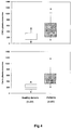
Figure 4 . Quantification of exosomes in plasma from melanoma patients.
Exosomes purified from plasma of healthy donors and melanoma patients were quantified by ExoTest using CD63 (A) or caveolin-1 (B) as detection antigens. Data is expressed as box plot representation: the horizontal and vertical lines in each box represent the median and the 25th- 75th percentiles, respectively. Black dots represent outlier values. Exosome levels are expressed as OD450 x 10000. Differences between groups were evaluated by Mann- Whitney test. -
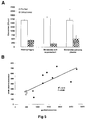
Figure 5 . ExoTest (Elisa) on unfractioned samples.- A. The amount of detectable exosomes was measured in purified exosomes (5 Oµg proteins) as compared to unfractioned culture supernatants (50 ml) from human macrophages, melanoma cells and plasma from melanoma patients. Data is expressed as means +/-SD.
- B. Regression analysis of plasma levels of CD63+ exosomes measured in purified or unfractioned plasma samples from both patients (n=9, black diamonds) and healthy donors (n=4, white diamonds). Exosome levels are expressed as
OD450 x 1000.
-
Figure 6 . Quantification of exosomes in plasma from melanoma patients.
Exosomes purified from plasma of melanoma patients (MEL) and healthy donors (HD) were quantified by Exotest using as detection antigens CD63 or typical melanoma proteins such as gplOO and MART-I. Data are expressed as means ± SD. Exosomes levels are expressed asOD450 x 1000. - Exosomes are micro vesicles of a size ranging between 30-120 nm, actively secreted in the extracellular environment by normal as well as tumor cells. Given the increasing understanding of the role of exosomes in cancer progression and the fact that there is an increasing need to find new cures, improve diagnostics and follow up of malignancy and growth of tumors, there is accordingly a need for methods and tools to detect and measure exosomes in human fluids. Because of the potential involvement of exosomes in promoting disease progression through a series of detrimental effects on tumor microenvironment, the possibility of quantifying exosomes in human plasma or serum, through a sensitive, specific and feasible assay is becoming a crucial issue. If such an assay would be available, it could become a fundamental tool for assessing the potential role of these microvesicles in cancer prognosis and as a novel prognostic factor or a marker for detecting or monitoring neoplastic disease. The currently used methods, e.g. TEM and WB, however are not quantitative or are only poorly quantitative, whereby there is a clear need for a method to detect and measure exosomes quantitatively in human fluids. Therefore the goal of this disclosure is to provide a new tool for clinical oncologists for diagnosing and follow up studies of cancer patients. Furthermore, a goal of this disclosure is to provide a novel method to be used to diagnose other human diseases, such as viral or prion diseases, where particles are transmitted in microvesicles.
- The novel quantitative test that is disclosed here is based on ELISA mediated detection of exosomes and it is the first easy, sensitive and reliable test for quantifying exosomes. The proteins that are detected by this method are not exosome specific but are exclusively shared with cytoplasmic organelles such as endosomes and lysosomes, whose membranes are not recycled as for plasma membrane structures. This feature excludes the possibility of detecting these proteins on debris derived from necrotic tumor cells, or in their soluble form. The assay of this disclosure preferably includes a universal tumor marker (caveolin1) which allows preferential detection of tumor-secreted exosomes. A series of comprehensive studies performed by different comparative methods (Western blotting and flow cytometry) and different experimental conditions described below in the examples, prove the reliability of the novel test of this disclosure.
- Koga et al. (Anticancer Res. 25, 3703-3727, 2005) disclose a method to capture exosomes from purified samples with beads coated against Her2/Neu. Admyre et al. (J ) disclose an Elisa assay whereby the exeosmes are immobilized with an anti-MHUC-II antibody and detected via a horse radish peroxidase conjugate.
CA 2453198 discloses an ELISA assay based on the capture of exosomes with anti-MHC-I antibody and the detection with an anti-FAsL antibody - The novel method of this disclosure is based on sandwich ELISA test that we call ExoTest, to capture and quantify plasmatic exosomes based on expression of housekeeping proteins (CD63 and Rab-5) and tumor-associated marker caveolin-1. ExoTest uses an anti-Rab5 antibody for capturing exosomes present in purified exosome preparations. As detecting antibody, ExoTest uses an antibody recognizing either an exosome antigen (CD63) or other antigens expressed by the cellular source of exosomes like caveolin-1, a protein associated to metastatic behavior of tumors. In a first set of experiments, we used exosome preparation from culture media of human tumor cell lines, comparing the results of ExoTest with those obtained by western blot and FACS. The results showed that typical exosomal antigens were detectable in exosome preparations by using all three methods. However, ExoTest allowed a quantitative analysis of exosomes and a wider panel of proteins in the same preparation with smaller amounts of starting material as compared to FACS or WB analysis. This technical feature is of paramount importance when the analysis is performed on plasma samples. Indeed, the analysis of plasma pools of SCID mice engrafted with human melanoma or colon carcinoma cells was often not reproducible or at least very difficult when WB was used while ExoTest allowed a quantitative analysis of exosome levels in the plasma of human tumor- SCID mice. A novel and important finding is that based on ExoTest results, exosome levels in the plasma are significantly correlated to the tumor size and increased with time after engraft. Moreover, circulating exosomes, together with the typical proteins, presented tumor markers expressed by the human tumors growing in the mice (not shown). Thus, ExoTest using the CD63 as detecting antigen was able to quantify exosomes in plasma of patients with melanoma and of healthy subjects. Since tumor cells secrete large amount of exosomes, we were actually surprised not to find a difference between the levels of CD63 -positive exosomes in plasma of melanoma patients and healthy control persons. With the aim to identify possible tumor-specific exosomes, we performed ExoTest using caveolin-1 as detecting antigen. Recent publications have reported that the plasma levels of circulating caveolin-1 are significantly correlated with metastatic prostate adenocarcinoma. Interestingly, caveolin-1 has been implicated in the pathogenesis of oncogenic cell transformation, tumougenesis, and metastasis. First, we demonstrated by FACS and WB that caveolin-1 is present on human tumor-derived exosomes, purified from plasma of tumor-engrafted SCID mice and tumor patients. Then, by using ExoTest we could observe a significant increase in the plasma levels of caveolin-1 -positive exosomes in tumor patients as compared to plasma of healthy individuals, suggesting that caveolin-1 may represent a specific tumor marker, and that ExoTest is a successful test to quantify such increase.
- Altogether, the results show that an exosome-detecting ExoTest is working and useful for detection and quantification of circulating exosomes in humans. Moreover, the test offers a possibility of detecting different proteins in plasma exosome preparations, with a potential application to specific type of tumor patients. This disclosure also provides a novel prognostic/diagnostic tool for tumor patients based on quantification and characterization of plasma exosomes. This is particularly relevant for melanoma patients, because sensitive and reliable serum markers are still limited and LDH (lactate dehydrogenase) levels remain the only prognostic serum factor for assessment of disease course and prognosis.
- The invention is described in detail below. The examples and experimental details are disclosed to provide an improved understanding and guidance for those skilled in the art. The scope of the invention is determined by the appended claims.
- We used two types of human tumor cell lines, i.e. melanoma and colon carcinoma. Mel 501 and Mel BS are two metastatic melanoma cell lines obtained from melanoma lesions of patients, surgically resected (Istituto Nazionale dei Tumori, Milan, Italy). We also used two colorectal carcinoma cell lines Colo206 (generated from liver metastasis of colorectal cancer patient) and 1869 col (provided by Dr. Maccalli, Istituto Superiore di Sanita'). All cell lines were cultured in RPMI 1640 medium supplemented with 100 IU/ml penicillin, 100 ug/ml streptomycin (Gibco), 2mM glutamine I(Gibco) and 10% fetal calf serum (FCS) (Invitrogen, Milan, Italy). Human monocytes-derived macrophages (MDM) were obtained from buffy coats of healthy blood donors using CD 14 magnetic beads (Milteny Biotec, Germany) and GM-CSF (500U/ml) for 5 days in culture.
- CB.17 SCID/SCID female mice (Harlan, S. Pietro al Natisone, Italy) were used at 4-5 weeks of age and were kept under specific pathogen-free conditions. All animal procedures were carried out according to UKCCR guidelines. SCID mice were housed in micro isolator cages and all food, water and bedding were autoclaved prior to use. Mice were injected subcutaneously into the right flank with 2.5 x 10⁶ cells/mouse of human melanoma or colon carcinoma cells. Tumor growth was measured by a caliper and the tumor weight was estimated with the formula: Tumor weight (mg) = length (mm) x width2 (mm)/2, as previously described (Luciani L et al., J. Natl. Cancer Inst., 2004). The engrafted tumors were allowed to grow up to a weight of 500 mg and 500 µl of plasma from tumor-engrafted mice were collected from different animals sacrificed at different time-points during tumor growth.
- Human plasma samples were collected from EDTA-treated whole blood from patients with primary or metastatic melanoma and from age and sex-matched healthy donors. Samples were stored at -70° C until analysis.
- Supernatants from human melanoma and colon carcinoma cell lines were harvested from 72 h confluent cell cultures in T- 175 flasks and micro vesicles were isolated as previously described (Andreola et al., J. Exp. Med. 2002) with minor modifications. Briefly, after centrifugation at 300 x g for 10 minutes to pellet the cells, the supernatant was centrifuged at 1,200 x g for 20 minutes followed by 10,000 x g for 30 minutes. The supernatant was filtered by using a 0,22 µm filter (Stericup™, Millipore Corp., Bedford, Massachusetts, USA) and then centrifuged at 100,000 g for 1 h in a Beckman ultracentrifuge (Beckman Coulter) in order to pellet exosomes. After one washing in a large volume of phosphate-buffered saline (PBS), exosomes were suspended in a small volume of PBS or in the proper lysis buffer, and stored at -80°C for further experimental analysis. In order to obtain exosomes from plasma samples, heparinized blood from SCID mice engrafted with human tumors or from tumor patients and healthy donors were centrifuged at 400 x g for 20 minutes. Plasma was then collected, aliquoted and stored at -70°C until analysis. Plasma samples were subjected to the same centrifugal procedure described above to isolate exosomes by using a Beckman TL100 for ultracentrifugation of small volumes.
- Determination of antigen expression on exosomes was performed by flow cytometry analysis on purified exosomes bound onto latex beads. Exosome preparations (5-10µg) were incubated with 5µl 4-µm-diameter aldehyde/sulfate latex beads (Interfacial Dynamics, Portland, OR) and resuspended into 400 µl PBS containing 2% FCS. Exosomes-coated beads (20 µl) were incubated with the following antibodies: anti- Rab5 (Santa Cruz), anti- CD63-FITC (Pharmigen), anti-CD81-PE (Pharmingen), anti-cavelolin-1 (clone N-20, Santa Cruz) for 30 minutes at 4°C, followed, when needed, by incubation with PE- or FITC-conjugated secondary antibody and analyzed on a FACSCalibur flow cytometer (BD Biosciences).
- Purified exosomes were lysed in lysis buffer containing 1 % Triton X-100, 0,1 % SDS, 0.1 M Tris HCl (pH 7) and protease inhibitors (10 µg/ml aprotinin, 10 µg/ml leupeptin and 2 mM phenylmethylsulfonyl fluoride) (Sigma). Exosome protein concentration was determined by Bradford microassay method (Bio-Rad Laboratories, Hercules, CA). A total of 50 µg of proteins were resuspended in SDS sample buffer, boiled for 5 min, separated on 10 % SDS-PAGE gel and electroblotted on nitrocellulose (Protran BA85, Schleicher and Schuell). Membranes were blotted with antibodies to CD63 (diluted 1:50) and Rab-5b (diluted 1:50), incubated with appropriate HRP-conjugated secondary antibodies (Amersham Pharmacia) and visualised by enhanced chemiluminescence (ECL, Pierce).
- The novel ELISA test (ExoTest) according to this disclosure is based on the presence of specific proteins in the exosomes. These proteins are shared with cytoplasmic organelles such as endosomes and lysosomes (Rab-5 and CD63, respectively), whose membranes are not shed or recycled as for plasma membrane structures, thus excluding the possible presence of structures deriving from membrane disruption.
- Ninety-six well-plates (Nunc, Milan, Italy) were coated with polyclonal anti-Rab-5b antibody (clone A-20 Santa Cruz) in a volume of 100µl/well of carbonate buffer pH 9.6 at the final concentration of 4 µg/ml and incubated overnight at 4°C. After 3 washes with PBS, 100 µl/well of blocking solution (PBS containing 0.5% BSA) was added and left at room temperature for 1 hour. Following three washes in PBS, 50µg of purified exosomes (in a final volume of 100µl/well) were added and incubated overnight at 37°C. After three washes with PBS, the appropriate detection antibody (anti-CD63 Mab (clone H5C6 Pharmingen) or anti-caveolin-1 Mab (clone 2297, Pharmingen)) was diluted at 4µg/ml in blocking solution and 100 µl/well was incubated for 1 hour at 37°C. After three washes with PBS, the plate was incubated with 1OQJI of HRP -conjugated anti-mouse-peroxidase secondary antibody (Pierce, Milan, Italy) diluted 1:50,000 in blocking solution for 1 hour at room temperature. After the final three washes with PBS, the reaction was developed with POD (Roche Applied Science, Milan, Italy), blocked with 1 N H2SO4 and optical densities were recorded with an ELISA reader by using a 450 nm filter (Biorad).
- ExoTest provides quantification of exosomes present in cell culture preparations and has higher sensitivity for exosome protein detection than Western Blot analysis.
- Culture supernatants of melanoma and colon carcinoma cell lines were processed following the standard procedure to obtain purified exosomes as described above. The sandwich ELISA set up to detect exosomes (ExoTest, see
Fig. 1A ) was performed on exosome preparations obtained from cell culture supernatants by differential centrifugation. ExoTest was able to provide a quantification of the exosomes present in the cell culture supernatants, being CD63+ exosomes detectable in dose-dependent manner (Fig. 1B ). Negative controls,, represented by fractions derived from pellet obtained after the 10 000 g centrifugation, exosomes purified from cell culture medium alone and by only secondary antibody resulted in a barely measurable optical density (OD= 0.07 +/- 0.01). Intra- and inter-test variability were calculated on six replicates of the same preparation run on three different plates and were 30% and 35% , respectively. - Western blot and FACS analysis of the same purified exosome preparations confirmed the qualitative data obtained by ExoTest. In fact, Rab-5b, Lamp-1 and to a lesser extent CD63 proteins were detectable in the various exosome preparations both by WB in exosome lysates (
Fig. 1C ) and both Rab-5b and CD63 were detected by FACS on exosomes bound to latex beads (Fig. 1D ). However, exosome detection and quantification by ExoTest showed a higher sensitivity for the detection of CD63 protein with respect to WB analysis (Fig. 1C ). Indeed, while at least 12.5µg of exosomal proteins were needed to properly detect either CD63 or Rab-5b by WB, ExoTest was able to clearly detect exosomes starting from a minimum amount of 3 µg of purified exosome preparations. - Comparison of the results of qualification and quantification of in vivo exosome preparation of SCID Mice engrafted with human tumors by FACS, WB and ExoTest.
- In order to verify the possibility of detecting human tumor-derived exosomes in vivo by using ExoTest, we first performed experiments in which human melanoma (Me501) and colon carcinoma cell lines were injected subcutaneously in SCID mice. In the first experiments, plasma samples obtained at sacrifice from SCID mice carrying engrafted tumors (100-500 mg) were subjected to the exosome purification procedure. As for the exosomes purified from cell culture supernatants, also exosomes purified from mice plasma were clearly and specifically detectable by ExoTest (
Fig. 2A ) and FACS (Fig. 2B ). Exosome preparations obtained from plasma of control SCID mice (not engrafted with human tumors) resulted in background optical densities comparable to the blank samples (OD = 0.08 ± 0.03), thus suggesting absence of exosomes in the immunocompromised animals or that murine exosomes do not cross react with human CD63 and Rab-5b. Again ExoTest needed a significantly smaller amount of exosome preparations to obtain results comparable to those using WB analysis. - After demonstrating that human exosomes can be recovered and detected by ExoTest, we performed time course experiments with the aim of evaluating whether the amount of plasma exosomes in human tumor-SCID mice correlated with tumor burden In these experiments, melanoma tumors were allowed to grow up to five weeks after the early engraftment, and the exosomes were purified and quantified from plasma of individual animals starting at day 14 after engraftment and during the following three weeks. The results showed that the amount of human exosomes detected by ExoTest increased over time in parallel to the increase of tumor size (
Fig 2C ); suggesting that plasma exosome quantification represents a valuable method to monitor tumor growth. Similar results were obtained with MeBS cells (not shown). - Since exosomes are known to represent an important and specific route of intercellular communication, we reasoned that tumor-derived exosomes may differ from circulating exosomes in normal physiological conditions. Recently, it has been reported that prostasomes (membrane vesicles secreted by prostate cancer cells) isolated from prostate carcinoma PC-3 cell line contain caveolin-1 protein, the major component of caveolae. It is also known that the serum levels of caveolin-1 are elevated in prostate cancer patients compared with healthy subjects (Tahir, 2003 #21). However, there is no prior art suggesting association of this protein with membrane vesicles in the blood. Therefore, we evaluated the presence of caveolin-1 on exosomes obtained from plasma of SCID mice engrafted with melanoma tumors. As is evidenced by results on
Figure 3A Cav1 is strongly expressed on exosomes secreted by human melanoma cells in vitro, while undetectable on both cellular extracts and exosomes from normal human cells such as for instance primary monocyte-derived macrophages (MDM). These results suggest that Ca1 secreted in an exosome-embedded form may be specific feature of melanoma cells, thus representing a potential marker for the ex- vivo analysis of tumor-derived exosomes. We therefore investigated the presence of Cav1 on exosomes obtained from plasma of SCID mice engrafted with melanoma tumors.Cav 1 was detected in exosome preparations derived from plasma of SCID mice engrafted with melanoma tumors by Western blot (Fig. 3B ), flow cytometry (Fig 3C ) and ExoTest (Fig. 3D ), while Cav1 was undetectable in plasma-derived exosomes from control animals (Figs. 3B, 3D ). In agreement with results from melanoma and colo-rectal carcinoma (CRC) patients other tumor markers, such as MelanA/Mart-1 for melanoma and CEA for CRC, could be used for detecting the in vivo release of tumor exosomes in tumor-bearing SCID mice by ExoTest, with results comparable with those obtained with Cavl. However, since melanoma may express heterogeneous or low amount of MelanA/MART-1, especially at metastatic levels, and CEA is present mostly in soluble form in CRC patients' serum, Cav1 is more reliable and reproducible tumor marker. Therefore, ExoTest is developed further with inclusion of anti-Cav1 -specific antibodies. - Quantification of in vivo exosome preparation of plasma of tumor patients by ExoTest: amount of exosomes in plasma as a tool to prognosis.
- The data obtained in the human tumor-SCID mouse model prompted us to investigate whether ExoTest allowed detection and characterization of exosomes purified from human plasma as well. If qualification was possible, we aimed to verify whether tumor patients may have circulating plasma exosomes that differ in quantity and/or quality from those present in plasma of healthy donors. Exosomes were purified from plasma of tumor patients (n=62) and healthy donors (n= 37) and were then subjected to ExoTest and Western Blot analysis for the presence of Rab5 and CD63. Quantification of exosomes based on CD63 and Cav1 expression by ExoTest is shown in table 1.
Table 1. Plasma exosome and LDH levels in the study population. CD63+ and Cav1+ exo are plasmatic exosomes expressed as OD450 x 1000. Plasma LDH values are expressed as IVfL. Data is shown as mean (95% C.I.)CD63+ Cav1+ exo LDH Melanoma patients (n=62) 478 (390 to 566) 524 (458 to 590) 438 (321 to 555) Healthy donors (n= 37) 236 (188 to 285) 213 (180 to 246) 360 (240 to 480) - ExoTest test allowed the detection of exosomal proteins in plasma-purified exosomes from both melanoma patients and healthy donors (
Fig. 4A ), with up to 4 folds exosome levels in plasma of melanoma patients as compared to healthy donors (p<0.001). In order to determine sensitivity and specificity of ExoTest based on the detection of the two exosome protein markers, we calculated the cut-off (mean +/- 2SD in healthy donors' samples) for both CD63 and Cav1 expressing plasma exosomes, which was 526 and 411 (OD450 x 1000), respectively. ExoTest detecting CD63+ exosomes showed a low sensitivity (36%) and good specificity (97.3%) while the ExoTest for Cav1+ exosomes had a higher sensitivity (62%) and similar specificity (97.3%). In line with this observation, the plasma levels of Cav1 + exosomes were significantly higher than the levels of CD63+ exosomes in melanoma patients (P=O.04). These results suggest that: - i) circulating Cav1 may be associated to exosomes in melanoma patients, and ii) quantification of plasma exosomes bearing CaI may be considered a useful tumor marker.
- In addition, we found that plasma LDH did not correlate with either CD63+ or Cav1+ plasma exosomes while a significant correlation was observed between CD63+ and Cav1+ plasma exosomes (Spearman coefficient 0.25, P=0.04) (
Fig. 4C ). Moreover, ExoTest revealed the presence of tumor antigens, such as MART-1 or CEA in the plasma of carcinoma or melanoma patients (Fig.6 ). - Most of the patients included into the analysis (57/62) were affected by advance disease (stage III-IV). A wide distribution of plasmatic exosome levels was detected by ExoTest in all disease stages, which suggest that the variability in the amount of exosomes present in peripheral circulation of different patients may reflect diverse levels of tumor aggressiveness and may therefore become a novel independent prognostic factor for melanoma. Other prognostic factors indicated by the American Joint Committee on Cancer (AJCC), including primary melanoma thickness and ulceration, number of metastatic lymph nodes, and site and number of distant metastases, also may correlate with exosomes serum content, as suggested by the results of this disclosure.
- Whole plasma can be used for exosome quantification.
- The potential application of ExoTest for clinical purposes prompted us to verify whether ExoTest could be used for exosome detection in unfractionated biological fluids that would allow an easy and reproducible analysis avoiding the steps of ultracentrifugation. Therefore, we compared the detection and quantification of CD63+ exosomes from unfractionated samples (cell culture supernatants from human macrophages and melanoma cells, and human plasma) and exosomes purified from the same samples. In order to increase the sensitivity of the test, for these specific experiments the HRP-conjugated Mab was incubated for 30 minutes instead of 15 minutes. As is shown in
Fig. 5A , the presence of exosomes from unfractioned macrophages and melanoma culture supernatants and plasma from nine melanoma patients was detectable by ExoTest. In addition, we performed the same analysis of plasma from 4 healthy donors and regression analysis on the total number of samples analysed (9 patients + 4 healthy donors) showed a significant correlation between the two types of measures (Figure 5B ). These results suggest the potential application of ExoTest in clinical setting using whole plasma and avoiding the complex and time consuming procedures of exosome purification. -
- Thery C, Zitvogel L, Amigorena S. Exosomes: composition, biogenesis and function. Nat Rev Immunol. 2002 Aug; 2(8):569-79.
- Stoorvogel W, Kleijmeer MJ, Geuze HJ, Raposo G. The biogenesis and functions of exosomes. Traffic. 2002 May;3(5):321-30.
- Lamparski HG, Metha-Damani A, Yao JY, Patel S, Hsu DH, Ruegg C, Le Pecq JB. Production and characterization of clinical grade exosomes derived from dendritic cells. J Immunol Methods. 2002 Dec 15;270(2):211-26.
- Wubbolts R, Leckie RS, Veenhuizen PT, Schwarzmann G, Mobius W, Hoernschemeyer J, Slot JW, Geuze HJ, Stoorvogel W. Proteomic and biochemical analyses of human B cell-derived exosomes. Potential implications for their function and multivesicular body formation. J Biol Chem. 2003 Mar 28; 278(13):10963-72.
- Blanchard N, Lankar D, Faure F, Regnault A, Dumont C, Raposo G, Hivroz C. TCR activation of human T cells induces the production of exosomes bearing the TCR/CD3/zeta complex. J Immunol. 2002 .
- Raposo G, Tenza D, Mecheri S, Peronet R, Bonnerot C, Desaymard C. Accumulation of major histocompatibility complex class II molecules in mast cell secretory granules and their release upon degranulation. MoI Biol Cell. 1997 Dec; 8(12): 2631-45. van Niel G, Raposo G, Candalh C, Boussac M, Hershberg R, Cerf-Bensussan N, Heyman M. Intestinal epithelial cells secrete exosome-like vesicles. Gastroenterology. 2001 Aug; 121(2): 337-49. Andreola G, Rivoltini L, Castelli C, Huber V, Perego P, Deho P, et al. Induction of lymphocyte apoptosis by tumor cell secretion of FasL-bearing micro vesicles. J Exp Med. 2002; 195(10): 1303-1316.
- Huber V, Fais S, Iero M, Lugini L, Canese P, Squarcina P, Zaccheddu A, Colone M, Arancia G, Gentile M, Seregni E, Valenti R, Ballabio G, Belli F, Leo E, Parmiani G, Rivoltini L. Human colorectal cancer cells induce T-cell death through release of proapoptotic microvesicles: role in immune escape. Gastroenterology. 2005 Jun; 128(7): 1796-804.
- Chaput N, Schartz NE, Andre F, Zitvogel L. Exosomes for immunotherapy of cancer. Adv Exp Med Biol. 2003; 532:215-21. Taylor DD, Taylor CG, Jiang CG, Black PH. Characterization of plasma membrane shedding from murine melanoma cells. Int J Cancer. 1988 Apr 15; 41(4): 629-35.
- Taylor DD, Lyons KS, Gercel-Taylor C. Shed membrane fragment-associated markers for endometrial and ovarian cancers. Gynecol Oncol. 2002 Mar; 84(3):443-8.
- Ginestra A, La Placa MD, Saladino F, Cassara D, Nagase H, Vittorelli ML. The amount and proteolytic content of vesicles shed by human cancer cell lines correlates with their in vitro invasiveness. Anticancer Res. 1998 Sep-Oct; 18(5A): 3433-7.
- Admyre C, Grunewald J, Thyberg J, Gripenback S, Tornling G, Eklund A, Scheynius A, Gabrielsson S. Exosomes with major histocompatibility complex class II and co-stimulatory molecules are present in human BAL fluid. Eur Respir J. 2003 Oct; 22(4):578-83. Andre F, Schartz NE, Movassagh M, Flament C, Pautier P, Morice P, Pomel C, Lhomme C, Escudier B, Le Chevalier T, Tursz T, Amigorena S, Raposo G, Angevin E, Zitvogel L. Malignant effusions and immunogenic tumour-derived exosomes. Lancet. 2002 Jul 27; 360(9329):295-305.
- Bard MP, Hegmans JP, Hemmes A, Luider TM, Willemsen R, Severijnen LA, van Meerbeeck JP, Burgers SA, Hoogsteden HC, Lambrecht BN. Proteomic analysis of exosomes isolated from human malignant pleural effusions. Am J Respir Cell MoI Biol. 2004 Jul; 31(1):114-21.
- Caby MP, Lankar D, Vincendeau-Scherrer C, Raposo G, Bonnerot C. Exosomal-like vesicles are present in human blood plasma. Int Immunol. 2005 Jul; 17(7):879-87.
- Hegmans JP, Bard MP, Hemmes A, Luider TM, Kleijmeer MJ, Prins JB, Zitvogel L, Burgers SA, Hoogsteden HC, Lambrecht BN. Proteomic analysis of exosomes secreted by human mesothelioma cells. Am J Pathol. 2004 May; 164(5):1807-15.
- Laulagnier K, Motta C, Hamdi S, Roy S, Fauvelle F, Pageaux JF, Kobayashi T, Salles JP, Perret B, Bonnerot C, Record M. Mast cell- and dendritic cell-derived exosomes display a specific lipid composition and an unusual membrane organization. Biochem J. 2004 May 15; 380(Pt 1): 161-71. Clayton A, Turkes A, Dewitt S, Steadman R, Mason MD, Hallett MB. Adhesion and signaling by B cell-derived exosomes: the role of integrins. FASEB J. 2004 Apr de Gassart A, Geminard C, Fevrier B, Raposo G, Vidal M. Lipid raft-associated protein sorting in exosomes. Blood. 2003 Dec 15;102(13):4336-44. Mobius W, van Donselaar E, Ohno-Iwashita Y, Shimada Y, Heijnen HF, Slot JW, Geuze HJ. Recycling compartments and the internal vesicles of multivesicular bodies harbor most of the cholesterol found in the endocytic pathway. Traffic. 2003 Apr; 4(4):222-31.
- Schartz NE, Chaput N, Andre F, Zitvogel L. From the antigen-presenting cell to the antigen- presenting vesicle: the exosomes. Curr Opin MoI Ther. 2002 Aug; 4(4):372-81. Chaput N, Taieb J, Schartz NE, Andre F, Angevin E, Zitvogel L. Exosome-based immunotherapy. Cancer Immunol Immunother. 2004 Mar; 53(3):234-9.
- Nguyen DG, Booth A, Gould SJ, Hildreth JE. Evidence that HIV budding in primary macrophages occurs through the exosome release pathway. J Biol Chem. 2003 Dec 26;278(52):52347-54. Blood. 2003 Dec 15; 102(13):4336-44. Flanagan J, Middeldorp J, Sculley T. Localization of the Epstein-Barr .
- Raposo G, Nijman HW, Stoorvogel W, Liejendekker R, Harding CV, Melief CJ, Geuze HJ.B lymphocytes secrete antigen-presenting vesicles. J Exp Med. 1996 .
- Zitvogel L, Regnault A, Lozier A, Wolfers J, Flament C, Tenza D, Ricciardi-Castagnoli P, Raposo G, Amigorena S. Eradication of established murine tumors using a novel cell-free vaccine: dendritic cell-derived exosomes. Nat Med. 1998 May; 4(5):594-600.
- Smith TJ, Weis JH. Mucosal T cells and mast cells share common adhesion receptors. Immunol Today. 1996 Feb;17(2):60-3. Review.
- Lozupone F, Luciani F, Venditti M, Rivoltini L, Pupa S, Parmiani G, et al. Murine granulocytes control human tumor growth in SCID mice. Int J Cancer. 2000;87:569-573.
- Lozupone F, Pende D, Burgio VL, Castelli C, Spada M, Venditti M, et al. Adoptive transfer of an anti-MART-127-35-specific CD8+ T cell clone leads to immunoselection of human melanoma antigen-loss variants in SCID mice. Eur J. Immunol. 2003; 33:556-566.
- Tahir SA, Yang G, Ebara S, Timme TL, Satoh T, Li 20.L, Goltsov A, Ittmann M, Morrisett JD, Thompson TC. Secreted caveolin-1 stimulates cell suvival/clonal growth and contributes to metastasis in androgen-insensitive prostate cancer. Cancer Res. 2001 May 15;61(10):3882- 5.
- Mouraviev V, Li L, Tahir SA, Yang G, Timme TM, Goltsov A, Ren C, Satoh T, Wheeler TM, Ittmann MM, Miles BJ, Amato RJ, Kadmon D, Thompson TC. The role of caveolin-1 in androgen insensitive prostate cancer. J Urol. 2002 Oct; 168(4 Pt 1):1589-96.
- D D Taylor and C Geijel-Taylor Tumour-derived exosomes and their role in cancer-associated T-cell signalling defects British Journal of Cancer (2005) 92, 305311.
Claims (12)
- A method to quantify and qualify exosomes in human cell derived samples or in body fluid, said method comprising the steps of:a) capturing exosomes of the human cell derived sample or body fluid with a primary anti-body anti-Rab 5b;b) detecting the bound exosomes with a detection antibody, said detection antibody binding to an exosome antigen;c) allowing an enzyme linked secondary antibody react with the detection antibody;d) adding substrate; ande) detecting the reaction.
- The method of claim 1, wherein the detection antibody is anti-CD63 or anti-caveolin-1
- The method of claim 1 or 2 , wherein the method includes a step of purifying an exosome preparation from the human cell derived sample or body fluid, and step a) is performed with purified exosomes.
- The method of claim 1 or 2, wherein the body fluid is human plasma sample, ascites, cerebral fluids, bone marrow, urine, faeces or bronco-alveolar washing.
- The method of claim 4, wherein volume of the body fluid used is less than 2 ml.
- A non-invasive method to monitor tumour growth, said method comprising the steps of:a) periodically analysing a body fluid sample of a patient;b) capturing exosomes of the body fluid sample with a primary antibody, said primary anti-body being anti-Rab-5b;c)detecting the bound exosomes with a detection antibody, said detection antibody being anti-CD63 or anti-caveolin-1;d) allowing an enzyme linked secondary antibody react with the detection antibody;e) adding substrate;f) detecting the reaction; andg) drawing a correlation between quantity of detected exosomes and size of the tumour.
- The method of claim 6, wherein the method includes purification of exosome preparation from the body fluid sample and step c) is performed with the purified exosome preparation.
- The method of claim 6, wherein the body fluid sample is a plasma sample.
- The method of claim 6 , wherein the tumour is melanoma tumour.
- A method to diagnose a tumour, by detecting caveolin-1 bearing exosomes by using the method of claim 1, wherein the detecting antibody is anti-caveolin-1.
- The method of claim 10, wherein the tumour is melanoma tumour.
- A test kit for quantifying and qualifying exosomes in human cell derived samples or in body fluid, said kit comprising:a) a primary antibody preparation for capturing exosomes of the cell derived samples, or the body fluid, said primary antibody being anti-Rab 5b;b) a detection antibody preparation for detecting the bound exosomes, said detection anti-body being anti-CD63 or anti-caveolin-1;c) an enzyme linked secondary antibody preparation for reaction with the detection anti-body; andd) a substrate for the enzyme.
Priority Applications (1)
| Application Number | Priority Date | Filing Date | Title |
|---|---|---|---|
| PL09704302T PL2245458T3 (en) | 2008-01-25 | 2009-01-26 | A NEW METHOD FOR MEASURING AND CHARACTERISTICS OF MICROBLEXERS IN HUMAN BODY FLUIDS |
Applications Claiming Priority (3)
| Application Number | Priority Date | Filing Date | Title |
|---|---|---|---|
| US6252808P | 2008-01-25 | 2008-01-25 | |
| US12/321,412 US8617806B2 (en) | 2008-01-25 | 2009-01-21 | Method to measure and characterize microvesicles in the human body fluids |
| PCT/EE2009/000002 WO2009092386A2 (en) | 2008-01-25 | 2009-01-26 | A new method to measure and characterize microvesicles in the human body fluids |
Publications (2)
| Publication Number | Publication Date |
|---|---|
| EP2245458A2 EP2245458A2 (en) | 2010-11-03 |
| EP2245458B1 true EP2245458B1 (en) | 2016-10-26 |
Family
ID=41064551
Family Applications (1)
| Application Number | Title | Priority Date | Filing Date |
|---|---|---|---|
| EP09704302.0A Not-in-force EP2245458B1 (en) | 2008-01-25 | 2009-01-26 | A new method to measure and characterize microvesicles in the human body fluids |
Country Status (12)
| Country | Link |
|---|---|
| US (1) | US8617806B2 (en) |
| EP (1) | EP2245458B1 (en) |
| JP (1) | JP5583600B2 (en) |
| CN (1) | CN102317778B (en) |
| AU (1) | AU2009207927B2 (en) |
| BR (1) | BRPI0906683B8 (en) |
| CA (1) | CA2713196C (en) |
| ES (1) | ES2613670T3 (en) |
| MX (1) | MX2010008164A (en) |
| PL (1) | PL2245458T3 (en) |
| RU (1) | RU2520741C2 (en) |
| WO (1) | WO2009092386A2 (en) |
Cited By (2)
| Publication number | Priority date | Publication date | Assignee | Title |
|---|---|---|---|---|
| DE102021208893B3 (en) | 2021-08-13 | 2022-11-17 | Hahn-Schickard-Gesellschaft für angewandte Forschung e.V. | Isolating analytes of different analyte classes |
| EP3978925A4 (en) * | 2019-05-24 | 2023-06-28 | Sol Bio Corporation | Affinity separation system and method using switch-like adhesion reaction |
Families Citing this family (57)
| Publication number | Priority date | Publication date | Assignee | Title |
|---|---|---|---|---|
| CA2733672C (en) | 2007-08-16 | 2018-09-11 | The Royal Institution For The Advancement Of Learning/Mcgill University | Tumor cell-derived microvesicles |
| US20100255514A1 (en) | 2007-08-16 | 2010-10-07 | The Royal Institution For The Advancement Of Learning/Mcgill University | Tumor cell-derived microvesicles |
| KR101810799B1 (en) * | 2008-02-01 | 2017-12-19 | 더 제너럴 하스피탈 코포레이션 | Use of microvesicles in diagnosis, prognosis and treatment of medical diseases and conditions |
| US10545149B2 (en) * | 2008-10-06 | 2020-01-28 | Morehouse School Of Medicine | Detection of HIV-related proteins in urine |
| US9487837B2 (en) * | 2008-10-06 | 2016-11-08 | Morehouse School Of Medicine | Exosome-mediated diagnosis of hepatitis virus infections and diseases |
| US7888035B2 (en) | 2008-10-30 | 2011-02-15 | Caris Mpi, Inc. | Methods for assessing RNA patterns |
| WO2010056337A2 (en) | 2008-11-12 | 2010-05-20 | Caris Mpi, Inc. | Methods and systems of using exosomes for determining phenotypes |
| US20130029339A1 (en) | 2009-09-09 | 2013-01-31 | The General Hospital Corporation | Use of microvesicles in analyzing kras mutations |
| EP2475988B1 (en) | 2009-09-09 | 2018-11-14 | The General Hospital Corporation | Use of microvesicles in analyzing nucleic acid profiles |
| CA2782284A1 (en) * | 2009-11-30 | 2011-06-03 | Caris Life Sciences Luxembourg Holdings, S.A.R.L. | Methods and systems for isolating, storing, and analyzing vesicles |
| EP2542696B1 (en) * | 2010-03-01 | 2016-09-28 | Caris Life Sciences Switzerland Holdings GmbH | Biomarkers for theranostics |
| JP2013526852A (en) | 2010-04-06 | 2013-06-27 | カリス ライフ サイエンシズ ルクセンブルク ホールディングス | Circulating biomarkers for disease |
| ES2624284T3 (en) | 2010-07-07 | 2017-07-13 | Aethlon Medical Inc | Methods to quantify exosomes |
| WO2012031008A2 (en) | 2010-08-31 | 2012-03-08 | The General Hospital Corporation | Cancer-related biological materials in microvesicles |
| AU2011316477A1 (en) * | 2010-10-11 | 2013-05-02 | Medsaic Pty Ltd | Assay for disease detection |
| CA2817111C (en) | 2010-11-10 | 2019-03-05 | Exosome Diagnostics, Inc. | Method for isolation of nucleic acid containing particles and extraction of nucleic acids therefrom |
| FR2973029B1 (en) * | 2011-03-23 | 2013-04-26 | Centre Nat Rech Scient | GRAFTED DINUCLEAR METAL COMPLEXES AND THEIR USE AS SENSORS OF CELLULAR MICROPARTICLES |
| WO2012135844A2 (en) * | 2011-04-01 | 2012-10-04 | Cornell University | Circulating exosomes as diagnostic/prognostic indicators and therapeutic targets of melanoma and other cancers |
| WO2012162563A2 (en) * | 2011-05-24 | 2012-11-29 | The Regents Of The University Of California | Method for exosomal biomarker detection by electric field-induced release and measurement |
| KR20140067001A (en) * | 2011-08-08 | 2014-06-03 | 카리스 라이프 사이언스 룩셈부르크 홀딩스, 에스.에이.알.엘. | Biomarker compositions and methods |
| JP6029112B2 (en) * | 2011-12-22 | 2016-11-24 | テオリアサイエンス株式会社 | Exosome analysis method, reagent for exosome analysis, and exosome analyzer |
| EP2889623B1 (en) | 2012-08-24 | 2017-10-04 | The University of Tokyo | Exosome analysis method, exosome analysis apparatus, and use of exosome electrophoresis chip |
| KR101933620B1 (en) * | 2012-09-18 | 2018-12-28 | 삼성전자주식회사 | Compositions and kits for detecting a vesicle, and methods for analyzing the vesicle using the same |
| US9480714B2 (en) | 2012-11-13 | 2016-11-01 | Allan Yang Wu | Methods and systems for processing exosomes |
| CA2904181A1 (en) | 2013-03-13 | 2014-10-09 | Anahit Aghvanyan | Sandwich immunoassay comprising anchoring reagent |
| WO2014159662A1 (en) | 2013-03-13 | 2014-10-02 | University Of Miami | Method for isolation and purification of microvesicles from cell culture supernatants and biological fluids |
| US12558316B2 (en) | 2013-03-13 | 2026-02-24 | University Of Miami | Methods and compositions for the treatment of epidermolysis bullosa |
| US10114015B2 (en) | 2013-03-13 | 2018-10-30 | Meso Scale Technologies, Llc. | Assay methods |
| JP6210004B2 (en) * | 2013-04-09 | 2017-10-11 | 株式会社Jvcケンウッド | Sample analysis device and exosome capture method |
| JPWO2015029979A1 (en) * | 2013-08-30 | 2017-03-02 | 国立大学法人 東京大学 | Exosome analysis method, exosome analysis chip, and exosome analyzer |
| WO2015045666A1 (en) | 2013-09-25 | 2015-04-02 | 国立大学法人東京大学 | Fluid device, exosome analysis method, biomolecule analysis method, and biomolecule detection method |
| JP6255035B2 (en) * | 2013-11-06 | 2017-12-27 | Jsr株式会社 | Separation method, detection method, signal measurement method, disease determination method, drug efficacy evaluation method for disease treatment, kit and liquid composition |
| JP6638917B2 (en) | 2013-12-06 | 2020-02-05 | 国立大学法人 東京大学 | Valve, fluid control structure, fluid device, and valve manufacturing method |
| EP3143401A4 (en) | 2014-05-15 | 2017-10-11 | Meso Scale Technologies, LLC | Improved assay methods |
| WO2015185730A2 (en) * | 2014-06-06 | 2015-12-10 | Hansabiomed Ou | Exosomal biomarkers |
| RU2556825C1 (en) * | 2014-09-15 | 2015-07-20 | Федеральное государственное бюджетное учреждение науки Институт химической биологии и фундаментальной медицины Сибирского отделения Российской академии наук (ИХБФМ СО РАН) | Method of extraction of exosomes from blood |
| US20160320390A1 (en) * | 2015-05-01 | 2016-11-03 | Morehouse School Of Medicine | Compositions and methods for capturing exosomes |
| EP3299816A4 (en) * | 2015-05-20 | 2018-10-03 | JSR Corporation | Separation method, detection method, signal measurement method, disease determination method, drug efficacy assessment method, kit, liquid composition, and specimen diluent |
| CA2988771A1 (en) | 2015-06-09 | 2016-12-15 | The Board Of Regents Of The University Of Texas System | Diagnostic test for early stage cancer |
| JP6606916B2 (en) | 2015-08-21 | 2019-11-20 | 株式会社Jvcケンウッド | Exosomes capture method |
| GB201518466D0 (en) * | 2015-10-19 | 2015-12-02 | Cizzle Biotechnology Ltd | Use |
| CN106093412A (en) * | 2016-06-28 | 2016-11-09 | 叶瑞东 | A kind of peptide molecule is the application in micro-hemorrhage test kit after preparing diagnosing acute cerebral infarction |
| WO2018070479A1 (en) * | 2016-10-13 | 2018-04-19 | 合同会社みらか中央研究所 | Method for recovering extracellular vesicles |
| RU2682721C2 (en) * | 2016-11-17 | 2019-03-21 | Федеральное Государственное Бюджетное Учреждение Науки Институт Молекулярной Биологии Им. В.А. Энгельгардта Российской Академии Наук (Имб Ран) | Biological microchip for detection of tumor exosomes in serum of human for diagnosing colorectal cancer |
| JP6797427B2 (en) * | 2017-01-12 | 2020-12-09 | テオリアサイエンス株式会社 | How to test a subject's likelihood of having pancreatic cancer |
| CN106906294A (en) * | 2017-03-31 | 2017-06-30 | 浙江大学 | A kind of quantitative approach of extracellular vesica/liposome |
| CN107144688B (en) * | 2017-04-07 | 2018-09-11 | 浙江大学 | CD19 positive excretion bodies are as application of the molecular labeling in preparing tumor diagnosis kit and kit |
| EP3647786B1 (en) | 2017-05-29 | 2023-03-08 | National University Corporation Kobe University | Base material for manufacturing sensor for analyzing detection target, sensor for analyzing detection target, method for analyzing detection target |
| DE102017005543A1 (en) * | 2017-06-13 | 2018-12-13 | Forschungszentrum Jülich GmbH | Method for detecting extracellular vesicles in a sample |
| CN107907689A (en) * | 2017-10-10 | 2018-04-13 | 北京大学 | The detection method of excretion body protein CD5L |
| KR101968046B1 (en) * | 2018-07-19 | 2019-04-11 | (주) 바이오인프라생명과학 | Complex biomarkers for early diagnosis of cancer |
| JP6677284B2 (en) * | 2018-10-09 | 2020-04-08 | 凸版印刷株式会社 | Analyte detection method and lateral flow test strip |
| CN120098057A (en) | 2019-03-01 | 2025-06-06 | 中尺度技术有限责任公司 | Electrochemiluminescent labeled probes for use in immunological methods, methods of using such probes, and kits containing such probes |
| CN109991427A (en) * | 2019-04-08 | 2019-07-09 | 王延博 | It is a kind of for detecting the kit of excretion body surface face protein marker in serum |
| CN110095604B (en) * | 2019-04-12 | 2022-03-18 | 南方医科大学南方医院 | Application of Caveolin-1 protein positive exosome as non-small cell lung cancer diagnosis marker |
| CN115176163B (en) * | 2019-11-29 | 2026-03-24 | 富士胶片和光纯药株式会社 | Methods, biomarkers, reagent kits, and devices for assisting in the diagnosis of Alzheimer's disease or mild cognitive impairment. |
| CN111537725A (en) * | 2020-05-29 | 2020-08-14 | 武汉大学 | High-efficiency quantitative detection method, ELISA kit and method of PD-1 level in extracellular vesicles |
Family Cites Families (12)
| Publication number | Priority date | Publication date | Assignee | Title |
|---|---|---|---|---|
| JPH081438B2 (en) * | 1986-05-13 | 1996-01-10 | 三洋化成工業株式会社 | Enzyme immunoassay |
| EP1004664A1 (en) * | 1998-11-24 | 2000-05-31 | Institut National De La Sante Et De La Recherche Medicale (Inserm) | Compositions and methods using lactadherin or variants thereof |
| GB9927320D0 (en) * | 1999-11-18 | 2000-01-12 | Chiron Spa | Exosome separation |
| US6812023B1 (en) * | 2000-04-27 | 2004-11-02 | Anosys, Inc. | Methods of producing membrane vesicles |
| US20040241176A1 (en) * | 2000-04-27 | 2004-12-02 | Ap Cells. Inc. | Method of producing membrane vesicles |
| US7442777B2 (en) * | 2000-11-29 | 2008-10-28 | Arius Research Inc. | Cytotoxicity mediation of cells evidencing surface expression of CD63 |
| US20030190602A1 (en) * | 2001-03-12 | 2003-10-09 | Monogen, Inc. | Cell-based detection and differentiation of disease states |
| US20030224464A1 (en) | 2002-01-31 | 2003-12-04 | Baylor College Of Medicine | Secreted caveolin as a marker for prostate cancer |
| CA2453198A1 (en) * | 2004-01-07 | 2005-07-07 | Wei-Ping Min | Quantification and generation of immune suppressive exosomes |
| JP2005237234A (en) * | 2004-02-25 | 2005-09-08 | Japan Science & Technology Agency | Caveolin antigen and its uses |
| WO2007127464A2 (en) * | 2006-04-28 | 2007-11-08 | University Of Iowa Research Foundation | Methods and compounds to modulate parvovirus transduction of mammalian cells or to alter virus infection, method to identify a viral receptor or co-receptor |
| US20120058492A1 (en) * | 2008-01-25 | 2012-03-08 | Hansabiomed Ou | Method and a Kit To Detect Malignant Tumors and Provide a Prognosis |
-
2009
- 2009-01-21 US US12/321,412 patent/US8617806B2/en not_active Expired - Fee Related
- 2009-01-26 WO PCT/EE2009/000002 patent/WO2009092386A2/en not_active Ceased
- 2009-01-26 BR BRPI0906683A patent/BRPI0906683B8/en not_active IP Right Cessation
- 2009-01-26 PL PL09704302T patent/PL2245458T3/en unknown
- 2009-01-26 CN CN200980111452.4A patent/CN102317778B/en not_active Expired - Fee Related
- 2009-01-26 JP JP2010543379A patent/JP5583600B2/en not_active Expired - Fee Related
- 2009-01-26 ES ES09704302.0T patent/ES2613670T3/en active Active
- 2009-01-26 CA CA2713196A patent/CA2713196C/en not_active Expired - Fee Related
- 2009-01-26 EP EP09704302.0A patent/EP2245458B1/en not_active Not-in-force
- 2009-01-26 AU AU2009207927A patent/AU2009207927B2/en not_active Ceased
- 2009-01-26 MX MX2010008164A patent/MX2010008164A/en active IP Right Grant
- 2009-01-26 RU RU2010135526/15A patent/RU2520741C2/en active
Cited By (3)
| Publication number | Priority date | Publication date | Assignee | Title |
|---|---|---|---|---|
| EP3978925A4 (en) * | 2019-05-24 | 2023-06-28 | Sol Bio Corporation | Affinity separation system and method using switch-like adhesion reaction |
| DE102021208893B3 (en) | 2021-08-13 | 2022-11-17 | Hahn-Schickard-Gesellschaft für angewandte Forschung e.V. | Isolating analytes of different analyte classes |
| WO2023017049A1 (en) | 2021-08-13 | 2023-02-16 | Hahn-Schickard-Gesellschaft für angewandte Forschung e.V. | Isolation of analytes of different analyte classes |
Also Published As
| Publication number | Publication date |
|---|---|
| US8617806B2 (en) | 2013-12-31 |
| PL2245458T3 (en) | 2017-08-31 |
| WO2009092386A2 (en) | 2009-07-30 |
| MX2010008164A (en) | 2010-11-25 |
| US20090220944A1 (en) | 2009-09-03 |
| BRPI0906683A2 (en) | 2017-08-22 |
| CN102317778B (en) | 2015-05-13 |
| JP5583600B2 (en) | 2014-09-03 |
| BRPI0906683B1 (en) | 2021-03-02 |
| AU2009207927A1 (en) | 2009-07-30 |
| CA2713196A1 (en) | 2009-07-30 |
| JP2011510309A (en) | 2011-03-31 |
| RU2010135526A (en) | 2012-02-27 |
| WO2009092386A8 (en) | 2010-10-14 |
| AU2009207927B2 (en) | 2015-04-09 |
| ES2613670T3 (en) | 2017-05-25 |
| CA2713196C (en) | 2014-06-17 |
| EP2245458A2 (en) | 2010-11-03 |
| CN102317778A (en) | 2012-01-11 |
| WO2009092386A3 (en) | 2009-09-17 |
| BRPI0906683B8 (en) | 2021-07-27 |
| RU2520741C2 (en) | 2014-06-27 |
Similar Documents
| Publication | Publication Date | Title |
|---|---|---|
| EP2245458B1 (en) | A new method to measure and characterize microvesicles in the human body fluids | |
| Muhsin-Sharafaldine et al. | Procoagulant and immunogenic properties of melanoma exosomes, microvesicles and apoptotic vesicles | |
| Boukouris et al. | Exosomes in bodily fluids are a highly stable resource of disease biomarkers | |
| Schuler et al. | Human CD4+ CD39+ regulatory T cells produce adenosine upon co-expression of surface CD73 or contact with CD73+ exosomes or CD73+ cells | |
| Chang et al. | Inflammation-associated lysophospholipids as ligands for CD1d-restricted T cells in human cancer | |
| Laurent et al. | Distribution, function, and prognostic value of cytotoxic T lymphocytes in follicular lymphoma: a 3-D tissue-imaging study | |
| Jahrsdörfer et al. | Granzyme B produced by human plasmacytoid dendritic cells suppresses T-cell expansion | |
| Bergmann et al. | Tumor‐derived microvesicles in sera of patients with head and neck cancer and their role in tumor progression | |
| Gonzalez-Begne et al. | Proteomic analysis of human parotid gland exosomes by multidimensional protein identification technology (MudPIT) | |
| Szczepanski et al. | Increased frequency and suppression by regulatory T cells in patients with acute myelogenous leukemia | |
| Stoeck et al. | A role for exosomes in the constitutive and stimulus-induced ectodomain cleavage of L1 and CD44 | |
| Frisullo et al. | Regulatory T cells fail to suppress CD4+ T‐bet+ T cells in relapsing multiple sclerosis patients | |
| Olah et al. | An optimized protocol for the acute isolation of human microglia from autopsy brain samples | |
| US20160370265A1 (en) | Method for isolating exosomes | |
| US20200249234A1 (en) | Method and system for identifying membrane proteins on extracellular vesicles | |
| EP3564386A1 (en) | Method for evaluating immunogenicity of test substance | |
| Tzaridis et al. | A novel serum extracellular vesicle protein signature to monitor glioblastoma tumor progression | |
| Ismail et al. | Pretransplant detection of anti-endothelial cell antibodies could predict renal allograft outcome | |
| NL2033706B1 (en) | Identification and/or isolation of T cell-comprising cell-cell complexes and use thereof. | |
| Teeuwen | Optimizing the therapeutic potential of extracellular vesicles in cancer | |
| Gómez-Herranz et al. | Major Histocompatibility Complex class I heavy chains localize in both cytoplasmic and nuclear compartment | |
| Ham | The role of breast cancer derived-exosomes in the tumour micro-environment | |
| Lau et al. | Fucosylation of HLA-DRB1 regulates CD4+ T cell-mediated anti-melanoma immunity and enhances immunotherapy efficacy | |
| Bergstrom | Identification of altered cell surface glycoproteins and micropattern assay development in the context of human malignancies | |
| Xiao et al. | Cancer cells impede T cell early activation and drive immune evasion by transferring Tmem176b |
Legal Events
| Date | Code | Title | Description |
|---|---|---|---|
| PUAI | Public reference made under article 153(3) epc to a published international application that has entered the european phase |
Free format text: ORIGINAL CODE: 0009012 |
|
| 17P | Request for examination filed |
Effective date: 20100809 |
|
| AK | Designated contracting states |
Kind code of ref document: A2 Designated state(s): AT BE BG CH CY CZ DE DK EE ES FI FR GB GR HR HU IE IS IT LI LT LU LV MC MK MT NL NO PL PT RO SE SI SK TR |
|
| AX | Request for extension of the european patent |
Extension state: AL BA RS |
|
| DAX | Request for extension of the european patent (deleted) | ||
| 17Q | First examination report despatched |
Effective date: 20121026 |
|
| GRAP | Despatch of communication of intention to grant a patent |
Free format text: ORIGINAL CODE: EPIDOSNIGR1 |
|
| RIC1 | Information provided on ipc code assigned before grant |
Ipc: G01N 33/567 20060101ALI20160223BHEP Ipc: G01N 33/569 20060101ALI20160223BHEP Ipc: G01N 33/53 20060101AFI20160223BHEP Ipc: G01N 33/574 20060101ALI20160223BHEP |
|
| INTG | Intention to grant announced |
Effective date: 20160331 |
|
| GRAS | Grant fee paid |
Free format text: ORIGINAL CODE: EPIDOSNIGR3 |
|
| GRAA | (expected) grant |
Free format text: ORIGINAL CODE: 0009210 |
|
| AK | Designated contracting states |
Kind code of ref document: B1 Designated state(s): AT BE BG CH CY CZ DE DK EE ES FI FR GB GR HR HU IE IS IT LI LT LU LV MC MK MT NL NO PL PT RO SE SI SK TR |
|
| REG | Reference to a national code |
Ref country code: GB Ref legal event code: FG4D |
|
| REG | Reference to a national code |
Ref country code: CH Ref legal event code: EP |
|
| REG | Reference to a national code |
Ref country code: AT Ref legal event code: REF Ref document number: 840379 Country of ref document: AT Kind code of ref document: T Effective date: 20161115 |
|
| REG | Reference to a national code |
Ref country code: IE Ref legal event code: FG4D |
|
| REG | Reference to a national code |
Ref country code: DE Ref legal event code: R096 Ref document number: 602009041927 Country of ref document: DE |
|
| REG | Reference to a national code |
Ref country code: RO Ref legal event code: EPE |
|
| RAP2 | Party data changed (patent owner data changed or rights of a patent transferred) |
Owner name: EXOSOMICS SIENA S.P.A. |
|
| REG | Reference to a national code |
Ref country code: DE Ref legal event code: R081 Ref document number: 602009041927 Country of ref document: DE Owner name: EXOSOMICS S.P.A., IT Free format text: FORMER OWNER: HANSABIOMED OUE, HAAPSALU, EE Ref country code: DE Ref legal event code: R081 Ref document number: 602009041927 Country of ref document: DE Owner name: EXOSOMICS SIENA S.P.A., IT Free format text: FORMER OWNER: HANSABIOMED OUE, HAAPSALU, EE |
|
| REG | Reference to a national code |
Ref country code: FR Ref legal event code: PLFP Year of fee payment: 9 |
|
| REG | Reference to a national code |
Ref country code: CH Ref legal event code: NV Representative=s name: JACOBACCI AND PARTNERS SA, CH Ref country code: CH Ref legal event code: PUE Owner name: EXOSOMICS SIENA S.P.A., IT Free format text: FORMER OWNER: HANSABIOMED OUE, EE |
|
| REG | Reference to a national code |
Ref country code: SE Ref legal event code: TRGR |
|
| REG | Reference to a national code |
Ref country code: LT Ref legal event code: MG4D |
|
| PG25 | Lapsed in a contracting state [announced via postgrant information from national office to epo] |
Ref country code: LV Free format text: LAPSE BECAUSE OF FAILURE TO SUBMIT A TRANSLATION OF THE DESCRIPTION OR TO PAY THE FEE WITHIN THE PRESCRIBED TIME-LIMIT Effective date: 20161026 |
|
| REG | Reference to a national code |
Ref country code: NL Ref legal event code: MP Effective date: 20161026 |
|
| REG | Reference to a national code |
Ref country code: AT Ref legal event code: MK05 Ref document number: 840379 Country of ref document: AT Kind code of ref document: T Effective date: 20161026 |
|
| PG25 | Lapsed in a contracting state [announced via postgrant information from national office to epo] |
Ref country code: LT Free format text: LAPSE BECAUSE OF FAILURE TO SUBMIT A TRANSLATION OF THE DESCRIPTION OR TO PAY THE FEE WITHIN THE PRESCRIBED TIME-LIMIT Effective date: 20161026 Ref country code: GR Free format text: LAPSE BECAUSE OF FAILURE TO SUBMIT A TRANSLATION OF THE DESCRIPTION OR TO PAY THE FEE WITHIN THE PRESCRIBED TIME-LIMIT Effective date: 20170127 Ref country code: NO Free format text: LAPSE BECAUSE OF FAILURE TO SUBMIT A TRANSLATION OF THE DESCRIPTION OR TO PAY THE FEE WITHIN THE PRESCRIBED TIME-LIMIT Effective date: 20170126 |
|
| REG | Reference to a national code |
Ref country code: GB Ref legal event code: 732E Free format text: REGISTERED BETWEEN 20170406 AND 20170412 |
|
| REG | Reference to a national code |
Ref country code: ES Ref legal event code: FG2A Ref document number: 2613670 Country of ref document: ES Kind code of ref document: T3 Effective date: 20170525 |
|
| PG25 | Lapsed in a contracting state [announced via postgrant information from national office to epo] |
Ref country code: IS Free format text: LAPSE BECAUSE OF FAILURE TO SUBMIT A TRANSLATION OF THE DESCRIPTION OR TO PAY THE FEE WITHIN THE PRESCRIBED TIME-LIMIT Effective date: 20170226 Ref country code: FI Free format text: LAPSE BECAUSE OF FAILURE TO SUBMIT A TRANSLATION OF THE DESCRIPTION OR TO PAY THE FEE WITHIN THE PRESCRIBED TIME-LIMIT Effective date: 20161026 Ref country code: PT Free format text: LAPSE BECAUSE OF FAILURE TO SUBMIT A TRANSLATION OF THE DESCRIPTION OR TO PAY THE FEE WITHIN THE PRESCRIBED TIME-LIMIT Effective date: 20170227 Ref country code: AT Free format text: LAPSE BECAUSE OF FAILURE TO SUBMIT A TRANSLATION OF THE DESCRIPTION OR TO PAY THE FEE WITHIN THE PRESCRIBED TIME-LIMIT Effective date: 20161026 Ref country code: BE Free format text: LAPSE BECAUSE OF FAILURE TO SUBMIT A TRANSLATION OF THE DESCRIPTION OR TO PAY THE FEE WITHIN THE PRESCRIBED TIME-LIMIT Effective date: 20161026 Ref country code: NL Free format text: LAPSE BECAUSE OF FAILURE TO SUBMIT A TRANSLATION OF THE DESCRIPTION OR TO PAY THE FEE WITHIN THE PRESCRIBED TIME-LIMIT Effective date: 20161026 Ref country code: HR Free format text: LAPSE BECAUSE OF FAILURE TO SUBMIT A TRANSLATION OF THE DESCRIPTION OR TO PAY THE FEE WITHIN THE PRESCRIBED TIME-LIMIT Effective date: 20161026 |
|
| REG | Reference to a national code |
Ref country code: DE Ref legal event code: R097 Ref document number: 602009041927 Country of ref document: DE |
|
| PG25 | Lapsed in a contracting state [announced via postgrant information from national office to epo] |
Ref country code: SK Free format text: LAPSE BECAUSE OF FAILURE TO SUBMIT A TRANSLATION OF THE DESCRIPTION OR TO PAY THE FEE WITHIN THE PRESCRIBED TIME-LIMIT Effective date: 20161026 Ref country code: DK Free format text: LAPSE BECAUSE OF FAILURE TO SUBMIT A TRANSLATION OF THE DESCRIPTION OR TO PAY THE FEE WITHIN THE PRESCRIBED TIME-LIMIT Effective date: 20161026 Ref country code: CZ Free format text: LAPSE BECAUSE OF FAILURE TO SUBMIT A TRANSLATION OF THE DESCRIPTION OR TO PAY THE FEE WITHIN THE PRESCRIBED TIME-LIMIT Effective date: 20161026 Ref country code: EE Free format text: LAPSE BECAUSE OF FAILURE TO SUBMIT A TRANSLATION OF THE DESCRIPTION OR TO PAY THE FEE WITHIN THE PRESCRIBED TIME-LIMIT Effective date: 20161026 |
|
| PG25 | Lapsed in a contracting state [announced via postgrant information from national office to epo] |
Ref country code: BG Free format text: LAPSE BECAUSE OF FAILURE TO SUBMIT A TRANSLATION OF THE DESCRIPTION OR TO PAY THE FEE WITHIN THE PRESCRIBED TIME-LIMIT Effective date: 20170126 |
|
| PLBE | No opposition filed within time limit |
Free format text: ORIGINAL CODE: 0009261 |
|
| STAA | Information on the status of an ep patent application or granted ep patent |
Free format text: STATUS: NO OPPOSITION FILED WITHIN TIME LIMIT |
|
| PG25 | Lapsed in a contracting state [announced via postgrant information from national office to epo] |
Ref country code: MC Free format text: LAPSE BECAUSE OF FAILURE TO SUBMIT A TRANSLATION OF THE DESCRIPTION OR TO PAY THE FEE WITHIN THE PRESCRIBED TIME-LIMIT Effective date: 20161026 |
|
| 26N | No opposition filed |
Effective date: 20170727 |
|
| REG | Reference to a national code |
Ref country code: IE Ref legal event code: MM4A |
|
| PG25 | Lapsed in a contracting state [announced via postgrant information from national office to epo] |
Ref country code: SI Free format text: LAPSE BECAUSE OF FAILURE TO SUBMIT A TRANSLATION OF THE DESCRIPTION OR TO PAY THE FEE WITHIN THE PRESCRIBED TIME-LIMIT Effective date: 20161026 Ref country code: LU Free format text: LAPSE BECAUSE OF NON-PAYMENT OF DUE FEES Effective date: 20170126 |
|
| REG | Reference to a national code |
Ref country code: FR Ref legal event code: PLFP Year of fee payment: 10 |
|
| PG25 | Lapsed in a contracting state [announced via postgrant information from national office to epo] |
Ref country code: IE Free format text: LAPSE BECAUSE OF NON-PAYMENT OF DUE FEES Effective date: 20170126 |
|
| PG25 | Lapsed in a contracting state [announced via postgrant information from national office to epo] |
Ref country code: MT Free format text: LAPSE BECAUSE OF NON-PAYMENT OF DUE FEES Effective date: 20170126 |
|
| PG25 | Lapsed in a contracting state [announced via postgrant information from national office to epo] |
Ref country code: HU Free format text: LAPSE BECAUSE OF FAILURE TO SUBMIT A TRANSLATION OF THE DESCRIPTION OR TO PAY THE FEE WITHIN THE PRESCRIBED TIME-LIMIT; INVALID AB INITIO Effective date: 20090126 |
|
| REG | Reference to a national code |
Ref country code: ES Ref legal event code: PC2A Owner name: EXOSOMICS S.P.A. Effective date: 20191022 |
|
| PG25 | Lapsed in a contracting state [announced via postgrant information from national office to epo] |
Ref country code: CY Free format text: LAPSE BECAUSE OF NON-PAYMENT OF DUE FEES Effective date: 20161026 |
|
| REG | Reference to a national code |
Ref country code: DE Ref legal event code: R082 Ref document number: 602009041927 Country of ref document: DE Representative=s name: KRAMER BARSKE SCHMIDTCHEN PATENTANWAELTE PARTG, DE Ref country code: DE Ref legal event code: R081 Ref document number: 602009041927 Country of ref document: DE Owner name: EXOSOMICS S.P.A., IT Free format text: FORMER OWNER: EXOSOMICS SIENA S.P.A., SIENA, IT |
|
| REG | Reference to a national code |
Ref country code: CH Ref legal event code: PFA Owner name: EXOSOMICS S.P.A., IT Free format text: FORMER OWNER: EXOSOMICS SIENA S.P.A., IT |
|
| PG25 | Lapsed in a contracting state [announced via postgrant information from national office to epo] |
Ref country code: MK Free format text: LAPSE BECAUSE OF FAILURE TO SUBMIT A TRANSLATION OF THE DESCRIPTION OR TO PAY THE FEE WITHIN THE PRESCRIBED TIME-LIMIT Effective date: 20161026 |
|
| PGFP | Annual fee paid to national office [announced via postgrant information from national office to epo] |
Ref country code: RO Payment date: 20201224 Year of fee payment: 13 |
|
| PGFP | Annual fee paid to national office [announced via postgrant information from national office to epo] |
Ref country code: PL Payment date: 20201221 Year of fee payment: 13 |
|
| PGFP | Annual fee paid to national office [announced via postgrant information from national office to epo] |
Ref country code: FR Payment date: 20210128 Year of fee payment: 13 Ref country code: IT Payment date: 20201127 Year of fee payment: 13 Ref country code: CH Payment date: 20210120 Year of fee payment: 13 |
|
| PGFP | Annual fee paid to national office [announced via postgrant information from national office to epo] |
Ref country code: DE Payment date: 20210129 Year of fee payment: 13 Ref country code: GB Payment date: 20210121 Year of fee payment: 13 Ref country code: ES Payment date: 20210201 Year of fee payment: 13 Ref country code: SE Payment date: 20210118 Year of fee payment: 13 Ref country code: TR Payment date: 20210124 Year of fee payment: 13 |
|
| REG | Reference to a national code |
Ref country code: DE Ref legal event code: R119 Ref document number: 602009041927 Country of ref document: DE |
|
| REG | Reference to a national code |
Ref country code: SE Ref legal event code: EUG |
|
| REG | Reference to a national code |
Ref country code: CH Ref legal event code: PL |
|
| GBPC | Gb: european patent ceased through non-payment of renewal fee |
Effective date: 20220126 |
|
| PG25 | Lapsed in a contracting state [announced via postgrant information from national office to epo] |
Ref country code: SE Free format text: LAPSE BECAUSE OF NON-PAYMENT OF DUE FEES Effective date: 20220127 Ref country code: RO Free format text: LAPSE BECAUSE OF NON-PAYMENT OF DUE FEES Effective date: 20220126 Ref country code: GB Free format text: LAPSE BECAUSE OF NON-PAYMENT OF DUE FEES Effective date: 20220126 Ref country code: DE Free format text: LAPSE BECAUSE OF NON-PAYMENT OF DUE FEES Effective date: 20220802 |
|
| PG25 | Lapsed in a contracting state [announced via postgrant information from national office to epo] |
Ref country code: FR Free format text: LAPSE BECAUSE OF NON-PAYMENT OF DUE FEES Effective date: 20220131 |
|
| PG25 | Lapsed in a contracting state [announced via postgrant information from national office to epo] |
Ref country code: LI Free format text: LAPSE BECAUSE OF NON-PAYMENT OF DUE FEES Effective date: 20220131 Ref country code: CH Free format text: LAPSE BECAUSE OF NON-PAYMENT OF DUE FEES Effective date: 20220131 |
|
| PG25 | Lapsed in a contracting state [announced via postgrant information from national office to epo] |
Ref country code: IT Free format text: LAPSE BECAUSE OF NON-PAYMENT OF DUE FEES Effective date: 20220126 |
|
| REG | Reference to a national code |
Ref country code: ES Ref legal event code: FD2A Effective date: 20230329 |
|
| PG25 | Lapsed in a contracting state [announced via postgrant information from national office to epo] |
Ref country code: ES Free format text: LAPSE BECAUSE OF NON-PAYMENT OF DUE FEES Effective date: 20220127 |
|
| PG25 | Lapsed in a contracting state [announced via postgrant information from national office to epo] |
Ref country code: PL Free format text: LAPSE BECAUSE OF NON-PAYMENT OF DUE FEES Effective date: 20220126 |
|
| PG25 | Lapsed in a contracting state [announced via postgrant information from national office to epo] |
Ref country code: TR Free format text: LAPSE BECAUSE OF NON-PAYMENT OF DUE FEES Effective date: 20220126 |