EP2292761A1 - Vascular adhesion molecules and modulation of their function - Google Patents
Vascular adhesion molecules and modulation of their function Download PDFInfo
- Publication number
- EP2292761A1 EP2292761A1 EP10181055A EP10181055A EP2292761A1 EP 2292761 A1 EP2292761 A1 EP 2292761A1 EP 10181055 A EP10181055 A EP 10181055A EP 10181055 A EP10181055 A EP 10181055A EP 2292761 A1 EP2292761 A1 EP 2292761A1
- Authority
- EP
- European Patent Office
- Prior art keywords
- cram
- jam
- cells
- cell
- depicted
- Prior art date
- Legal status (The legal status is an assumption and is not a legal conclusion. Google has not performed a legal analysis and makes no representation as to the accuracy of the status listed.)
- Withdrawn
Links
- 230000002792 vascular Effects 0.000 title description 6
- 108090000765 processed proteins & peptides Proteins 0.000 claims abstract description 33
- 102000004196 processed proteins & peptides Human genes 0.000 claims abstract description 31
- 229920001184 polypeptide Polymers 0.000 claims abstract description 26
- 241001529936 Murinae Species 0.000 claims abstract description 21
- 230000001105 regulatory effect Effects 0.000 claims abstract description 16
- 108060003951 Immunoglobulin Proteins 0.000 claims abstract description 8
- 102000018358 immunoglobulin Human genes 0.000 claims abstract description 8
- 206010061218 Inflammation Diseases 0.000 claims abstract description 7
- 230000004054 inflammatory process Effects 0.000 claims abstract description 7
- 125000003275 alpha amino acid group Chemical group 0.000 claims abstract 6
- 150000001413 amino acids Chemical class 0.000 claims description 27
- 239000002773 nucleotide Substances 0.000 claims description 8
- 125000003729 nucleotide group Chemical group 0.000 claims description 8
- 102000002110 C2 domains Human genes 0.000 claims description 7
- 108050009459 C2 domains Proteins 0.000 claims description 7
- 108091033319 polynucleotide Proteins 0.000 claims description 7
- 238000006243 chemical reaction Methods 0.000 claims description 5
- 239000003814 drug Substances 0.000 claims description 5
- 239000002157 polynucleotide Substances 0.000 claims 5
- 102000040430 polynucleotide Human genes 0.000 claims 5
- 206010028980 Neoplasm Diseases 0.000 abstract description 18
- 210000004027 cell Anatomy 0.000 description 107
- 108010040135 Junctional Adhesion Molecule C Proteins 0.000 description 68
- 102100023429 Junctional adhesion molecule C Human genes 0.000 description 68
- 108010040149 Junctional Adhesion Molecule B Proteins 0.000 description 63
- 210000002889 endothelial cell Anatomy 0.000 description 51
- 230000014509 gene expression Effects 0.000 description 25
- 108090000623 proteins and genes Proteins 0.000 description 25
- 108010040082 Junctional Adhesion Molecule A Proteins 0.000 description 24
- 102100022304 Junctional adhesion molecule A Human genes 0.000 description 24
- 238000003752 polymerase chain reaction Methods 0.000 description 23
- 239000002299 complementary DNA Substances 0.000 description 22
- 210000000265 leukocyte Anatomy 0.000 description 19
- 235000018102 proteins Nutrition 0.000 description 19
- 102000004169 proteins and genes Human genes 0.000 description 19
- 210000004978 chinese hamster ovary cell Anatomy 0.000 description 18
- 238000000034 method Methods 0.000 description 18
- 108010048367 enhanced green fluorescent protein Proteins 0.000 description 17
- 238000010186 staining Methods 0.000 description 17
- 210000001519 tissue Anatomy 0.000 description 17
- 230000003511 endothelial effect Effects 0.000 description 15
- 239000000047 product Substances 0.000 description 15
- 102100024441 Dihydropyrimidinase-related protein 5 Human genes 0.000 description 14
- 101001053479 Homo sapiens Dihydropyrimidinase-related protein 5 Proteins 0.000 description 14
- 230000006870 function Effects 0.000 description 14
- 108091032973 (ribonucleotides)n+m Proteins 0.000 description 13
- 238000004458 analytical method Methods 0.000 description 13
- 230000004807 localization Effects 0.000 description 13
- 230000035699 permeability Effects 0.000 description 12
- 108020004999 messenger RNA Proteins 0.000 description 11
- 239000013598 vector Substances 0.000 description 11
- 108060008682 Tumor Necrosis Factor Proteins 0.000 description 10
- 238000003556 assay Methods 0.000 description 10
- 230000000694 effects Effects 0.000 description 10
- 238000001114 immunoprecipitation Methods 0.000 description 10
- 210000001165 lymph node Anatomy 0.000 description 10
- 230000005012 migration Effects 0.000 description 10
- 238000013508 migration Methods 0.000 description 10
- 102000003390 tumor necrosis factor Human genes 0.000 description 10
- 230000033228 biological regulation Effects 0.000 description 9
- 239000012528 membrane Substances 0.000 description 9
- 238000001890 transfection Methods 0.000 description 9
- 210000004881 tumor cell Anatomy 0.000 description 9
- 210000003038 endothelium Anatomy 0.000 description 8
- 238000002474 experimental method Methods 0.000 description 8
- 230000008685 targeting Effects 0.000 description 8
- 108091060211 Expressed sequence tag Proteins 0.000 description 7
- 238000000636 Northern blotting Methods 0.000 description 7
- 235000001014 amino acid Nutrition 0.000 description 7
- 239000003153 chemical reaction reagent Substances 0.000 description 7
- 238000003501 co-culture Methods 0.000 description 7
- 238000009826 distribution Methods 0.000 description 7
- 238000012423 maintenance Methods 0.000 description 7
- 238000012216 screening Methods 0.000 description 7
- 230000015572 biosynthetic process Effects 0.000 description 6
- 238000010367 cloning Methods 0.000 description 6
- 230000001086 cytosolic effect Effects 0.000 description 6
- 210000003734 kidney Anatomy 0.000 description 6
- 239000010410 layer Substances 0.000 description 6
- 230000007246 mechanism Effects 0.000 description 6
- 239000002609 medium Substances 0.000 description 6
- 201000001441 melanoma Diseases 0.000 description 6
- 238000003757 reverse transcription PCR Methods 0.000 description 6
- 201000009030 Carcinoma Diseases 0.000 description 5
- 108020004414 DNA Proteins 0.000 description 5
- 101150044441 PECAM1 gene Proteins 0.000 description 5
- 102100024616 Platelet endothelial cell adhesion molecule Human genes 0.000 description 5
- 230000033115 angiogenesis Effects 0.000 description 5
- 230000002491 angiogenic effect Effects 0.000 description 5
- 239000013553 cell monolayer Substances 0.000 description 5
- 238000009396 hybridization Methods 0.000 description 5
- 230000003993 interaction Effects 0.000 description 5
- 210000004698 lymphocyte Anatomy 0.000 description 5
- 238000003199 nucleic acid amplification method Methods 0.000 description 5
- 210000001986 peyer's patch Anatomy 0.000 description 5
- 239000002356 single layer Substances 0.000 description 5
- 210000001578 tight junction Anatomy 0.000 description 5
- 108091026890 Coding region Proteins 0.000 description 4
- ZHNUHDYFZUAESO-UHFFFAOYSA-N Formamide Chemical compound NC=O ZHNUHDYFZUAESO-UHFFFAOYSA-N 0.000 description 4
- 230000004988 N-glycosylation Effects 0.000 description 4
- 108091028043 Nucleic acid sequence Proteins 0.000 description 4
- 102000003940 Occludin Human genes 0.000 description 4
- 108090000304 Occludin Proteins 0.000 description 4
- 102000007056 Recombinant Fusion Proteins Human genes 0.000 description 4
- 108010008281 Recombinant Fusion Proteins Proteins 0.000 description 4
- 230000003321 amplification Effects 0.000 description 4
- 239000000872 buffer Substances 0.000 description 4
- 230000001351 cycling effect Effects 0.000 description 4
- 230000013020 embryo development Effects 0.000 description 4
- 210000003989 endothelium vascular Anatomy 0.000 description 4
- 239000012634 fragment Substances 0.000 description 4
- 108020001507 fusion proteins Proteins 0.000 description 4
- 102000037865 fusion proteins Human genes 0.000 description 4
- 239000000203 mixture Substances 0.000 description 4
- 239000013642 negative control Substances 0.000 description 4
- 230000026731 phosphorylation Effects 0.000 description 4
- 238000006366 phosphorylation reaction Methods 0.000 description 4
- 239000000523 sample Substances 0.000 description 4
- 102000029816 Collagenase Human genes 0.000 description 3
- 108060005980 Collagenase Proteins 0.000 description 3
- 229920002307 Dextran Polymers 0.000 description 3
- KCXVZYZYPLLWCC-UHFFFAOYSA-N EDTA Chemical compound OC(=O)CN(CC(O)=O)CCN(CC(O)=O)CC(O)=O KCXVZYZYPLLWCC-UHFFFAOYSA-N 0.000 description 3
- 208000001382 Experimental Melanoma Diseases 0.000 description 3
- 102000003974 Fibroblast growth factor 2 Human genes 0.000 description 3
- 108090000379 Fibroblast growth factor 2 Proteins 0.000 description 3
- 125000002842 L-seryl group Chemical group O=C([*])[C@](N([H])[H])([H])C([H])([H])O[H] 0.000 description 3
- 241001465754 Metazoa Species 0.000 description 3
- OKKJLVBELUTLKV-UHFFFAOYSA-N Methanol Chemical compound OC OKKJLVBELUTLKV-UHFFFAOYSA-N 0.000 description 3
- 108091034117 Oligonucleotide Proteins 0.000 description 3
- 108700026244 Open Reading Frames Proteins 0.000 description 3
- 102100021669 Stromal cell-derived factor 1 Human genes 0.000 description 3
- 101710088580 Stromal cell-derived factor 1 Proteins 0.000 description 3
- JLCPHMBAVCMARE-UHFFFAOYSA-N [3-[[3-[[3-[[3-[[3-[[3-[[3-[[3-[[3-[[3-[[3-[[5-(2-amino-6-oxo-1H-purin-9-yl)-3-[[3-[[3-[[3-[[3-[[3-[[5-(2-amino-6-oxo-1H-purin-9-yl)-3-[[5-(2-amino-6-oxo-1H-purin-9-yl)-3-hydroxyoxolan-2-yl]methoxy-hydroxyphosphoryl]oxyoxolan-2-yl]methoxy-hydroxyphosphoryl]oxy-5-(5-methyl-2,4-dioxopyrimidin-1-yl)oxolan-2-yl]methoxy-hydroxyphosphoryl]oxy-5-(6-aminopurin-9-yl)oxolan-2-yl]methoxy-hydroxyphosphoryl]oxy-5-(6-aminopurin-9-yl)oxolan-2-yl]methoxy-hydroxyphosphoryl]oxy-5-(6-aminopurin-9-yl)oxolan-2-yl]methoxy-hydroxyphosphoryl]oxy-5-(6-aminopurin-9-yl)oxolan-2-yl]methoxy-hydroxyphosphoryl]oxyoxolan-2-yl]methoxy-hydroxyphosphoryl]oxy-5-(5-methyl-2,4-dioxopyrimidin-1-yl)oxolan-2-yl]methoxy-hydroxyphosphoryl]oxy-5-(4-amino-2-oxopyrimidin-1-yl)oxolan-2-yl]methoxy-hydroxyphosphoryl]oxy-5-(5-methyl-2,4-dioxopyrimidin-1-yl)oxolan-2-yl]methoxy-hydroxyphosphoryl]oxy-5-(5-methyl-2,4-dioxopyrimidin-1-yl)oxolan-2-yl]methoxy-hydroxyphosphoryl]oxy-5-(6-aminopurin-9-yl)oxolan-2-yl]methoxy-hydroxyphosphoryl]oxy-5-(6-aminopurin-9-yl)oxolan-2-yl]methoxy-hydroxyphosphoryl]oxy-5-(4-amino-2-oxopyrimidin-1-yl)oxolan-2-yl]methoxy-hydroxyphosphoryl]oxy-5-(4-amino-2-oxopyrimidin-1-yl)oxolan-2-yl]methoxy-hydroxyphosphoryl]oxy-5-(4-amino-2-oxopyrimidin-1-yl)oxolan-2-yl]methoxy-hydroxyphosphoryl]oxy-5-(6-aminopurin-9-yl)oxolan-2-yl]methoxy-hydroxyphosphoryl]oxy-5-(4-amino-2-oxopyrimidin-1-yl)oxolan-2-yl]methyl [5-(6-aminopurin-9-yl)-2-(hydroxymethyl)oxolan-3-yl] hydrogen phosphate Polymers Cc1cn(C2CC(OP(O)(=O)OCC3OC(CC3OP(O)(=O)OCC3OC(CC3O)n3cnc4c3nc(N)[nH]c4=O)n3cnc4c3nc(N)[nH]c4=O)C(COP(O)(=O)OC3CC(OC3COP(O)(=O)OC3CC(OC3COP(O)(=O)OC3CC(OC3COP(O)(=O)OC3CC(OC3COP(O)(=O)OC3CC(OC3COP(O)(=O)OC3CC(OC3COP(O)(=O)OC3CC(OC3COP(O)(=O)OC3CC(OC3COP(O)(=O)OC3CC(OC3COP(O)(=O)OC3CC(OC3COP(O)(=O)OC3CC(OC3COP(O)(=O)OC3CC(OC3COP(O)(=O)OC3CC(OC3COP(O)(=O)OC3CC(OC3COP(O)(=O)OC3CC(OC3COP(O)(=O)OC3CC(OC3COP(O)(=O)OC3CC(OC3CO)n3cnc4c(N)ncnc34)n3ccc(N)nc3=O)n3cnc4c(N)ncnc34)n3ccc(N)nc3=O)n3ccc(N)nc3=O)n3ccc(N)nc3=O)n3cnc4c(N)ncnc34)n3cnc4c(N)ncnc34)n3cc(C)c(=O)[nH]c3=O)n3cc(C)c(=O)[nH]c3=O)n3ccc(N)nc3=O)n3cc(C)c(=O)[nH]c3=O)n3cnc4c3nc(N)[nH]c4=O)n3cnc4c(N)ncnc34)n3cnc4c(N)ncnc34)n3cnc4c(N)ncnc34)n3cnc4c(N)ncnc34)O2)c(=O)[nH]c1=O JLCPHMBAVCMARE-UHFFFAOYSA-N 0.000 description 3
- 108010022164 acetyl-LDL Proteins 0.000 description 3
- 238000013459 approach Methods 0.000 description 3
- 230000004888 barrier function Effects 0.000 description 3
- 239000011324 bead Substances 0.000 description 3
- 229960002424 collagenase Drugs 0.000 description 3
- 125000000151 cysteine group Chemical group N[C@@H](CS)C(=O)* 0.000 description 3
- 230000029087 digestion Effects 0.000 description 3
- 108010007093 dispase Proteins 0.000 description 3
- 230000003828 downregulation Effects 0.000 description 3
- 230000004927 fusion Effects 0.000 description 3
- 238000003364 immunohistochemistry Methods 0.000 description 3
- 238000001727 in vivo Methods 0.000 description 3
- 230000000977 initiatory effect Effects 0.000 description 3
- 210000000056 organ Anatomy 0.000 description 3
- 239000000137 peptide hydrolase inhibitor Substances 0.000 description 3
- 229920002401 polyacrylamide Polymers 0.000 description 3
- 239000004417 polycarbonate Substances 0.000 description 3
- 239000011148 porous material Substances 0.000 description 3
- 230000002285 radioactive effect Effects 0.000 description 3
- 230000002441 reversible effect Effects 0.000 description 3
- 210000004988 splenocyte Anatomy 0.000 description 3
- 230000004960 subcellular localization Effects 0.000 description 3
- 230000008728 vascular permeability Effects 0.000 description 3
- 238000010865 video microscopy Methods 0.000 description 3
- KZMAWJRXKGLWGS-UHFFFAOYSA-N 2-chloro-n-[4-(4-methoxyphenyl)-1,3-thiazol-2-yl]-n-(3-methoxypropyl)acetamide Chemical compound S1C(N(C(=O)CCl)CCCOC)=NC(C=2C=CC(OC)=CC=2)=C1 KZMAWJRXKGLWGS-UHFFFAOYSA-N 0.000 description 2
- 102000007469 Actins Human genes 0.000 description 2
- 108010085238 Actins Proteins 0.000 description 2
- 240000006108 Allium ampeloprasum Species 0.000 description 2
- 235000005254 Allium ampeloprasum Nutrition 0.000 description 2
- 108010012236 Chemokines Proteins 0.000 description 2
- 102000019034 Chemokines Human genes 0.000 description 2
- 108020004635 Complementary DNA Proteins 0.000 description 2
- 241001301450 Crocidium multicaule Species 0.000 description 2
- 108010037362 Extracellular Matrix Proteins Proteins 0.000 description 2
- 102000010834 Extracellular Matrix Proteins Human genes 0.000 description 2
- DHMQDGOQFOQNFH-UHFFFAOYSA-N Glycine Chemical compound NCC(O)=O DHMQDGOQFOQNFH-UHFFFAOYSA-N 0.000 description 2
- 241000282412 Homo Species 0.000 description 2
- 102000018251 Hypoxanthine Phosphoribosyltransferase Human genes 0.000 description 2
- 108010091358 Hypoxanthine Phosphoribosyltransferase Proteins 0.000 description 2
- TWRXJAOTZQYOKJ-UHFFFAOYSA-L Magnesium chloride Chemical compound [Mg+2].[Cl-].[Cl-] TWRXJAOTZQYOKJ-UHFFFAOYSA-L 0.000 description 2
- 229940124158 Protease/peptidase inhibitor Drugs 0.000 description 2
- 108010076504 Protein Sorting Signals Proteins 0.000 description 2
- 108090000184 Selectins Proteins 0.000 description 2
- 102000003800 Selectins Human genes 0.000 description 2
- 238000012300 Sequence Analysis Methods 0.000 description 2
- FAPWRFPIFSIZLT-UHFFFAOYSA-M Sodium chloride Chemical compound [Na+].[Cl-] FAPWRFPIFSIZLT-UHFFFAOYSA-M 0.000 description 2
- 210000001744 T-lymphocyte Anatomy 0.000 description 2
- 108010073929 Vascular Endothelial Growth Factor A Proteins 0.000 description 2
- 102000005789 Vascular Endothelial Growth Factors Human genes 0.000 description 2
- 108010019530 Vascular Endothelial Growth Factors Proteins 0.000 description 2
- 230000006287 biotinylation Effects 0.000 description 2
- 238000007413 biotinylation Methods 0.000 description 2
- 210000004204 blood vessel Anatomy 0.000 description 2
- 230000008568 cell cell communication Effects 0.000 description 2
- 230000022131 cell cycle Effects 0.000 description 2
- 239000003795 chemical substances by application Substances 0.000 description 2
- 230000009260 cross reactivity Effects 0.000 description 2
- 235000018417 cysteine Nutrition 0.000 description 2
- 230000007423 decrease Effects 0.000 description 2
- 238000003745 diagnosis Methods 0.000 description 2
- 238000010494 dissociation reaction Methods 0.000 description 2
- 230000005593 dissociations Effects 0.000 description 2
- 238000005516 engineering process Methods 0.000 description 2
- 239000013604 expression vector Substances 0.000 description 2
- 210000002744 extracellular matrix Anatomy 0.000 description 2
- MHMNJMPURVTYEJ-UHFFFAOYSA-N fluorescein-5-isothiocyanate Chemical compound O1C(=O)C2=CC(N=C=S)=CC=C2C21C1=CC=C(O)C=C1OC1=CC(O)=CC=C21 MHMNJMPURVTYEJ-UHFFFAOYSA-N 0.000 description 2
- 239000000499 gel Substances 0.000 description 2
- 229920000159 gelatin Polymers 0.000 description 2
- 235000019322 gelatine Nutrition 0.000 description 2
- 230000002209 hydrophobic effect Effects 0.000 description 2
- 230000028993 immune response Effects 0.000 description 2
- 238000010185 immunofluorescence analysis Methods 0.000 description 2
- 238000000338 in vitro Methods 0.000 description 2
- 102000006495 integrins Human genes 0.000 description 2
- 108010044426 integrins Proteins 0.000 description 2
- 230000008611 intercellular interaction Effects 0.000 description 2
- 239000003446 ligand Substances 0.000 description 2
- 239000006166 lysate Substances 0.000 description 2
- 238000010232 migration assay Methods 0.000 description 2
- 150000007523 nucleic acids Chemical group 0.000 description 2
- 229920000515 polycarbonate Polymers 0.000 description 2
- 239000013641 positive control Substances 0.000 description 2
- 238000002360 preparation method Methods 0.000 description 2
- 230000008569 process Effects 0.000 description 2
- 238000000746 purification Methods 0.000 description 2
- 238000003753 real-time PCR Methods 0.000 description 2
- 238000007789 sealing Methods 0.000 description 2
- 210000000952 spleen Anatomy 0.000 description 2
- 206010041823 squamous cell carcinoma Diseases 0.000 description 2
- UCSJYZPVAKXKNQ-HZYVHMACSA-N streptomycin Chemical compound CN[C@H]1[C@H](O)[C@@H](O)[C@H](CO)O[C@H]1O[C@@H]1[C@](C=O)(O)[C@H](C)O[C@H]1O[C@@H]1[C@@H](NC(N)=N)[C@H](O)[C@@H](NC(N)=N)[C@H](O)[C@H]1O UCSJYZPVAKXKNQ-HZYVHMACSA-N 0.000 description 2
- 239000006228 supernatant Substances 0.000 description 2
- 210000001550 testis Anatomy 0.000 description 2
- MPLHNVLQVRSVEE-UHFFFAOYSA-N texas red Chemical compound [O-]S(=O)(=O)C1=CC(S(Cl)(=O)=O)=CC=C1C(C1=CC=2CCCN3CCCC(C=23)=C1O1)=C2C1=C(CCC1)C3=[N+]1CCCC3=C2 MPLHNVLQVRSVEE-UHFFFAOYSA-N 0.000 description 2
- 239000003053 toxin Substances 0.000 description 2
- 231100000765 toxin Toxicity 0.000 description 2
- 108700012359 toxins Proteins 0.000 description 2
- 238000013519 translation Methods 0.000 description 2
- 210000005166 vasculature Anatomy 0.000 description 2
- 210000000264 venule Anatomy 0.000 description 2
- 238000005406 washing Methods 0.000 description 2
- 108020005544 Antisense RNA Proteins 0.000 description 1
- 241000283707 Capra Species 0.000 description 1
- 108020004705 Codon Proteins 0.000 description 1
- 108091035707 Consensus sequence Proteins 0.000 description 1
- 238000011537 Coomassie blue staining Methods 0.000 description 1
- 239000006144 Dulbecco’s modified Eagle's medium Substances 0.000 description 1
- 238000002965 ELISA Methods 0.000 description 1
- 102000004190 Enzymes Human genes 0.000 description 1
- 108090000790 Enzymes Proteins 0.000 description 1
- 229920001917 Ficoll Polymers 0.000 description 1
- 238000012413 Fluorescence activated cell sorting analysis Methods 0.000 description 1
- 108010010803 Gelatin Proteins 0.000 description 1
- 239000001828 Gelatine Substances 0.000 description 1
- 239000004471 Glycine Substances 0.000 description 1
- 108010043121 Green Fluorescent Proteins Proteins 0.000 description 1
- 102000004144 Green Fluorescent Proteins Human genes 0.000 description 1
- 101000996823 Homo sapiens Cell surface A33 antigen Proteins 0.000 description 1
- 101150003028 Hprt1 gene Proteins 0.000 description 1
- 102100034343 Integrase Human genes 0.000 description 1
- 108090001007 Interleukin-8 Proteins 0.000 description 1
- FFEARJCKVFRZRR-BYPYZUCNSA-N L-methionine Chemical compound CSCC[C@H](N)C(O)=O FFEARJCKVFRZRR-BYPYZUCNSA-N 0.000 description 1
- 125000000769 L-threonyl group Chemical group [H]N([H])[C@]([H])(C(=O)[*])[C@](O[H])(C([H])([H])[H])[H] 0.000 description 1
- OUYCCCASQSFEME-QMMMGPOBSA-N L-tyrosine Chemical compound OC(=O)[C@@H](N)CC1=CC=C(O)C=C1 OUYCCCASQSFEME-QMMMGPOBSA-N 0.000 description 1
- 108010007622 LDL Lipoproteins Proteins 0.000 description 1
- 101001050319 Mus musculus Junctional adhesion molecule B Proteins 0.000 description 1
- 101001050316 Mus musculus Junctional adhesion molecule C Proteins 0.000 description 1
- 108010069196 Neural Cell Adhesion Molecules Proteins 0.000 description 1
- 102100023616 Neural cell adhesion molecule L1-like protein Human genes 0.000 description 1
- 239000000020 Nitrocellulose Substances 0.000 description 1
- 229930040373 Paraformaldehyde Natural products 0.000 description 1
- 229930182555 Penicillin Natural products 0.000 description 1
- JGSARLDLIJGVTE-MBNYWOFBSA-N Penicillin G Chemical compound N([C@H]1[C@H]2SC([C@@H](N2C1=O)C(O)=O)(C)C)C(=O)CC1=CC=CC=C1 JGSARLDLIJGVTE-MBNYWOFBSA-N 0.000 description 1
- 102000003992 Peroxidases Human genes 0.000 description 1
- 108010002747 Pfu DNA polymerase Proteins 0.000 description 1
- 206010035226 Plasma cell myeloma Diseases 0.000 description 1
- 229920001213 Polysorbate 20 Polymers 0.000 description 1
- 102000003923 Protein Kinase C Human genes 0.000 description 1
- 108090000315 Protein Kinase C Proteins 0.000 description 1
- 102000004022 Protein-Tyrosine Kinases Human genes 0.000 description 1
- 108090000412 Protein-Tyrosine Kinases Proteins 0.000 description 1
- 108010092799 RNA-directed DNA polymerase Proteins 0.000 description 1
- 229920002684 Sepharose Polymers 0.000 description 1
- 108010090804 Streptavidin Proteins 0.000 description 1
- 238000000692 Student's t-test Methods 0.000 description 1
- 108010006785 Taq Polymerase Proteins 0.000 description 1
- 108091036066 Three prime untranslated region Proteins 0.000 description 1
- 108010002321 Tight Junction Proteins Proteins 0.000 description 1
- 102000000591 Tight Junction Proteins Human genes 0.000 description 1
- 101710120037 Toxin CcdB Proteins 0.000 description 1
- 229920004890 Triton X-100 Polymers 0.000 description 1
- GLNADSQYFUSGOU-GPTZEZBUSA-J Trypan blue Chemical compound [Na+].[Na+].[Na+].[Na+].C1=C(S([O-])(=O)=O)C=C2C=C(S([O-])(=O)=O)C(/N=N/C3=CC=C(C=C3C)C=3C=C(C(=CC=3)\N=N\C=3C(=CC4=CC(=CC(N)=C4C=3O)S([O-])(=O)=O)S([O-])(=O)=O)C)=C(O)C2=C1N GLNADSQYFUSGOU-GPTZEZBUSA-J 0.000 description 1
- 102000004142 Trypsin Human genes 0.000 description 1
- 108090000631 Trypsin Proteins 0.000 description 1
- 102000008790 VE-cadherin Human genes 0.000 description 1
- 108010000134 Vascular Cell Adhesion Molecule-1 Proteins 0.000 description 1
- 102100023543 Vascular cell adhesion protein 1 Human genes 0.000 description 1
- 230000002159 abnormal effect Effects 0.000 description 1
- 210000002867 adherens junction Anatomy 0.000 description 1
- 230000001464 adherent effect Effects 0.000 description 1
- 239000011543 agarose gel Substances 0.000 description 1
- 238000003277 amino acid sequence analysis Methods 0.000 description 1
- 230000003698 anagen phase Effects 0.000 description 1
- 239000002870 angiogenesis inducing agent Substances 0.000 description 1
- 239000000427 antigen Substances 0.000 description 1
- 108091007433 antigens Proteins 0.000 description 1
- 102000036639 antigens Human genes 0.000 description 1
- 230000008901 benefit Effects 0.000 description 1
- 108091006004 biotinylated proteins Proteins 0.000 description 1
- 230000000903 blocking effect Effects 0.000 description 1
- 210000004369 blood Anatomy 0.000 description 1
- 239000008280 blood Substances 0.000 description 1
- 238000009835 boiling Methods 0.000 description 1
- 210000004899 c-terminal region Anatomy 0.000 description 1
- 108010018828 cadherin 5 Proteins 0.000 description 1
- 239000006285 cell suspension Substances 0.000 description 1
- 230000001413 cellular effect Effects 0.000 description 1
- 238000005119 centrifugation Methods 0.000 description 1
- 210000003710 cerebral cortex Anatomy 0.000 description 1
- 230000008859 change Effects 0.000 description 1
- 208000037976 chronic inflammation Diseases 0.000 description 1
- 230000006020 chronic inflammation Effects 0.000 description 1
- 238000003776 cleavage reaction Methods 0.000 description 1
- 230000008045 co-localization Effects 0.000 description 1
- 230000015271 coagulation Effects 0.000 description 1
- 238000005345 coagulation Methods 0.000 description 1
- 230000000052 comparative effect Effects 0.000 description 1
- 230000000295 complement effect Effects 0.000 description 1
- 239000003184 complementary RNA Substances 0.000 description 1
- 108091036078 conserved sequence Proteins 0.000 description 1
- 238000010276 construction Methods 0.000 description 1
- 230000001054 cortical effect Effects 0.000 description 1
- XUJNEKJLAYXESH-UHFFFAOYSA-N cysteine Natural products SCC(N)C(O)=O XUJNEKJLAYXESH-UHFFFAOYSA-N 0.000 description 1
- 230000009089 cytolysis Effects 0.000 description 1
- 231100000433 cytotoxic Toxicity 0.000 description 1
- 230000001472 cytotoxic effect Effects 0.000 description 1
- SUYVUBYJARFZHO-RRKCRQDMSA-N dATP Chemical compound C1=NC=2C(N)=NC=NC=2N1[C@H]1C[C@H](O)[C@@H](COP(O)(=O)OP(O)(=O)OP(O)(O)=O)O1 SUYVUBYJARFZHO-RRKCRQDMSA-N 0.000 description 1
- SUYVUBYJARFZHO-UHFFFAOYSA-N dATP Natural products C1=NC=2C(N)=NC=NC=2N1C1CC(O)C(COP(O)(=O)OP(O)(=O)OP(O)(O)=O)O1 SUYVUBYJARFZHO-UHFFFAOYSA-N 0.000 description 1
- 230000003247 decreasing effect Effects 0.000 description 1
- 238000013461 design Methods 0.000 description 1
- 238000001514 detection method Methods 0.000 description 1
- 238000011161 development Methods 0.000 description 1
- 230000018109 developmental process Effects 0.000 description 1
- 238000009792 diffusion process Methods 0.000 description 1
- 229940042399 direct acting antivirals protease inhibitors Drugs 0.000 description 1
- 201000010099 disease Diseases 0.000 description 1
- 208000037265 diseases, disorders, signs and symptoms Diseases 0.000 description 1
- 229940079593 drug Drugs 0.000 description 1
- 230000000212 effect on lymphocytes Effects 0.000 description 1
- 230000010595 endothelial cell migration Effects 0.000 description 1
- 210000004407 endothelial cell of postcapillary venule of lymph node Anatomy 0.000 description 1
- 230000002255 enzymatic effect Effects 0.000 description 1
- 229940088598 enzyme Drugs 0.000 description 1
- 210000002919 epithelial cell Anatomy 0.000 description 1
- 210000003386 epithelial cell of thymus gland Anatomy 0.000 description 1
- 210000000981 epithelium Anatomy 0.000 description 1
- 238000012869 ethanol precipitation Methods 0.000 description 1
- ZMMJGEGLRURXTF-UHFFFAOYSA-N ethidium bromide Chemical compound [Br-].C12=CC(N)=CC=C2C2=CC=C(N)C=C2[N+](CC)=C1C1=CC=CC=C1 ZMMJGEGLRURXTF-UHFFFAOYSA-N 0.000 description 1
- 230000007717 exclusion Effects 0.000 description 1
- 230000020764 fibrinolysis Effects 0.000 description 1
- 238000000684 flow cytometry Methods 0.000 description 1
- 238000001502 gel electrophoresis Methods 0.000 description 1
- 239000008273 gelatin Substances 0.000 description 1
- 235000011852 gelatine desserts Nutrition 0.000 description 1
- ZDXPYRJPNDTMRX-UHFFFAOYSA-N glutamine Natural products OC(=O)C(N)CCC(N)=O ZDXPYRJPNDTMRX-UHFFFAOYSA-N 0.000 description 1
- 239000005090 green fluorescent protein Substances 0.000 description 1
- 230000012010 growth Effects 0.000 description 1
- 230000013632 homeostatic process Effects 0.000 description 1
- 102000054366 human GPA33 Human genes 0.000 description 1
- 210000004408 hybridoma Anatomy 0.000 description 1
- 238000003384 imaging method Methods 0.000 description 1
- 230000008105 immune reaction Effects 0.000 description 1
- 210000000987 immune system Anatomy 0.000 description 1
- 230000036039 immunity Effects 0.000 description 1
- 238000003365 immunocytochemistry Methods 0.000 description 1
- 230000002163 immunogen Effects 0.000 description 1
- 238000002991 immunohistochemical analysis Methods 0.000 description 1
- 239000012133 immunoprecipitate Substances 0.000 description 1
- 230000006872 improvement Effects 0.000 description 1
- 238000000099 in vitro assay Methods 0.000 description 1
- 238000011534 incubation Methods 0.000 description 1
- 230000028709 inflammatory response Effects 0.000 description 1
- 210000004692 intercellular junction Anatomy 0.000 description 1
- 230000003834 intracellular effect Effects 0.000 description 1
- 230000009545 invasion Effects 0.000 description 1
- 238000011835 investigation Methods 0.000 description 1
- 239000002502 liposome Substances 0.000 description 1
- 210000004185 liver Anatomy 0.000 description 1
- 239000012160 loading buffer Substances 0.000 description 1
- 210000004072 lung Anatomy 0.000 description 1
- 210000001365 lymphatic vessel Anatomy 0.000 description 1
- 210000005210 lymphoid organ Anatomy 0.000 description 1
- 229910001629 magnesium chloride Inorganic materials 0.000 description 1
- 238000004519 manufacturing process Methods 0.000 description 1
- 239000003550 marker Substances 0.000 description 1
- 239000000463 material Substances 0.000 description 1
- 229930182817 methionine Natural products 0.000 description 1
- 238000000386 microscopy Methods 0.000 description 1
- 238000010369 molecular cloning Methods 0.000 description 1
- 239000003068 molecular probe Substances 0.000 description 1
- 238000010172 mouse model Methods 0.000 description 1
- 230000035772 mutation Effects 0.000 description 1
- 210000000066 myeloid cell Anatomy 0.000 description 1
- 201000000050 myeloid neoplasm Diseases 0.000 description 1
- 230000014399 negative regulation of angiogenesis Effects 0.000 description 1
- 238000007857 nested PCR Methods 0.000 description 1
- 229920001220 nitrocellulos Polymers 0.000 description 1
- 230000008520 organization Effects 0.000 description 1
- 230000002018 overexpression Effects 0.000 description 1
- 229920002866 paraformaldehyde Polymers 0.000 description 1
- 230000001575 pathological effect Effects 0.000 description 1
- 230000007170 pathology Effects 0.000 description 1
- 229940049954 penicillin Drugs 0.000 description 1
- 108040007629 peroxidase activity proteins Proteins 0.000 description 1
- 230000008288 physiological mechanism Effects 0.000 description 1
- 239000013612 plasmid Substances 0.000 description 1
- 235000010486 polyoxyethylene sorbitan monolaurate Nutrition 0.000 description 1
- 239000000256 polyoxyethylene sorbitan monolaurate Substances 0.000 description 1
- 230000007542 postnatal development Effects 0.000 description 1
- 239000013615 primer Substances 0.000 description 1
- 239000002987 primer (paints) Substances 0.000 description 1
- 230000002062 proliferating effect Effects 0.000 description 1
- 230000035755 proliferation Effects 0.000 description 1
- 238000001273 protein sequence alignment Methods 0.000 description 1
- 239000012264 purified product Substances 0.000 description 1
- 238000010791 quenching Methods 0.000 description 1
- 238000011160 research Methods 0.000 description 1
- 108091008146 restriction endonucleases Proteins 0.000 description 1
- 230000007017 scission Effects 0.000 description 1
- 238000012163 sequencing technique Methods 0.000 description 1
- 230000019491 signal transduction Effects 0.000 description 1
- 239000011780 sodium chloride Substances 0.000 description 1
- 241000894007 species Species 0.000 description 1
- 230000007480 spreading Effects 0.000 description 1
- 238000003892 spreading Methods 0.000 description 1
- 238000003153 stable transfection Methods 0.000 description 1
- 238000010561 standard procedure Methods 0.000 description 1
- 230000004936 stimulating effect Effects 0.000 description 1
- 229960005322 streptomycin Drugs 0.000 description 1
- 239000000126 substance Substances 0.000 description 1
- 238000003786 synthesis reaction Methods 0.000 description 1
- 238000012353 t test Methods 0.000 description 1
- 230000002992 thymic effect Effects 0.000 description 1
- 210000001541 thymus gland Anatomy 0.000 description 1
- 230000030968 tissue homeostasis Effects 0.000 description 1
- 238000012546 transfer Methods 0.000 description 1
- 238000003146 transient transfection Methods 0.000 description 1
- 230000014621 translational initiation Effects 0.000 description 1
- 239000012588 trypsin Substances 0.000 description 1
- 210000004926 tubular epithelial cell Anatomy 0.000 description 1
- 230000005748 tumor development Effects 0.000 description 1
- 230000004614 tumor growth Effects 0.000 description 1
- VBEQCZHXXJYVRD-GACYYNSASA-N uroanthelone Chemical compound C([C@@H](C(=O)N[C@H](C(=O)N[C@@H](CS)C(=O)N[C@@H](CC(N)=O)C(=O)N[C@@H](CS)C(=O)N[C@H](C(=O)N[C@@H]([C@@H](C)CC)C(=O)NCC(=O)N[C@@H](CC=1C=CC(O)=CC=1)C(=O)N[C@@H](CO)C(=O)NCC(=O)N[C@@H](CC(O)=O)C(=O)N[C@@H](CCCNC(N)=N)C(=O)N[C@@H](CS)C(=O)N[C@@H](CCC(N)=O)C(=O)N[C@@H]([C@@H](C)O)C(=O)N[C@@H](CCCNC(N)=N)C(=O)N[C@@H](CC(O)=O)C(=O)N[C@@H](CC(C)C)C(=O)N[C@@H](CCCNC(N)=N)C(=O)N[C@@H](CC=1C2=CC=CC=C2NC=1)C(=O)N[C@@H](CC=1C2=CC=CC=C2NC=1)C(=O)N[C@@H](CCC(O)=O)C(=O)N[C@@H](CC(C)C)C(=O)N[C@@H](CCCNC(N)=N)C(O)=O)C(C)C)[C@@H](C)O)NC(=O)[C@H](CO)NC(=O)[C@H](CC(O)=O)NC(=O)[C@H](CC(C)C)NC(=O)[C@H](CO)NC(=O)[C@H](CCC(O)=O)NC(=O)[C@@H](NC(=O)[C@H](CC=1NC=NC=1)NC(=O)[C@H](CCSC)NC(=O)[C@H](CS)NC(=O)[C@@H](NC(=O)CNC(=O)CNC(=O)[C@H](CC(N)=O)NC(=O)[C@H](CC(C)C)NC(=O)[C@H](CS)NC(=O)[C@H](CC=1C=CC(O)=CC=1)NC(=O)CNC(=O)[C@H](CC(O)=O)NC(=O)[C@H](CC=1C=CC(O)=CC=1)NC(=O)[C@H](CO)NC(=O)[C@H](CO)NC(=O)[C@H]1N(CCC1)C(=O)[C@H](CS)NC(=O)CNC(=O)[C@H]1N(CCC1)C(=O)[C@H](CC=1C=CC(O)=CC=1)NC(=O)[C@H](CO)NC(=O)[C@@H](N)CC(N)=O)C(C)C)[C@@H](C)CC)C1=CC=C(O)C=C1 VBEQCZHXXJYVRD-GACYYNSASA-N 0.000 description 1
- 210000004291 uterus Anatomy 0.000 description 1
- 210000005167 vascular cell Anatomy 0.000 description 1
- 230000037197 vascular physiology Effects 0.000 description 1
- 230000004862 vasculogenesis Effects 0.000 description 1
- 210000003462 vein Anatomy 0.000 description 1
Images


Classifications
-
- C—CHEMISTRY; METALLURGY
- C07—ORGANIC CHEMISTRY
- C07K—PEPTIDES
- C07K14/00—Peptides having more than 20 amino acids; Gastrins; Somatostatins; Melanotropins; Derivatives thereof
- C07K14/435—Peptides having more than 20 amino acids; Gastrins; Somatostatins; Melanotropins; Derivatives thereof from animals; from humans
- C07K14/705—Receptors; Cell surface antigens; Cell surface determinants
- C07K14/70503—Immunoglobulin superfamily
-
- A—HUMAN NECESSITIES
- A61—MEDICAL OR VETERINARY SCIENCE; HYGIENE
- A61P—SPECIFIC THERAPEUTIC ACTIVITY OF CHEMICAL COMPOUNDS OR MEDICINAL PREPARATIONS
- A61P29/00—Non-central analgesic, antipyretic or antiinflammatory agents, e.g. antirheumatic agents; Non-steroidal antiinflammatory drugs [NSAID]
-
- A—HUMAN NECESSITIES
- A61—MEDICAL OR VETERINARY SCIENCE; HYGIENE
- A61P—SPECIFIC THERAPEUTIC ACTIVITY OF CHEMICAL COMPOUNDS OR MEDICINAL PREPARATIONS
- A61P35/00—Antineoplastic agents
-
- A—HUMAN NECESSITIES
- A61—MEDICAL OR VETERINARY SCIENCE; HYGIENE
- A61P—SPECIFIC THERAPEUTIC ACTIVITY OF CHEMICAL COMPOUNDS OR MEDICINAL PREPARATIONS
- A61P37/00—Drugs for immunological or allergic disorders
- A61P37/02—Immunomodulators
- A61P37/06—Immunosuppressants, e.g. drugs for graft rejection
-
- A—HUMAN NECESSITIES
- A61—MEDICAL OR VETERINARY SCIENCE; HYGIENE
- A61P—SPECIFIC THERAPEUTIC ACTIVITY OF CHEMICAL COMPOUNDS OR MEDICINAL PREPARATIONS
- A61P43/00—Drugs for specific purposes, not provided for in groups A61P1/00-A61P41/00
-
- A—HUMAN NECESSITIES
- A61—MEDICAL OR VETERINARY SCIENCE; HYGIENE
- A61P—SPECIFIC THERAPEUTIC ACTIVITY OF CHEMICAL COMPOUNDS OR MEDICINAL PREPARATIONS
- A61P9/00—Drugs for disorders of the cardiovascular system
-
- Y—GENERAL TAGGING OF NEW TECHNOLOGICAL DEVELOPMENTS; GENERAL TAGGING OF CROSS-SECTIONAL TECHNOLOGIES SPANNING OVER SEVERAL SECTIONS OF THE IPC; TECHNICAL SUBJECTS COVERED BY FORMER USPC CROSS-REFERENCE ART COLLECTIONS [XRACs] AND DIGESTS
- Y02—TECHNOLOGIES OR APPLICATIONS FOR MITIGATION OR ADAPTATION AGAINST CLIMATE CHANGE
- Y02P—CLIMATE CHANGE MITIGATION TECHNOLOGIES IN THE PRODUCTION OR PROCESSING OF GOODS
- Y02P20/00—Technologies relating to chemical industry
- Y02P20/50—Improvements relating to the production of bulk chemicals
- Y02P20/582—Recycling of unreacted starting or intermediate materials
Definitions
- the present invention relates to the identification of a new subfamily of vascular adhesion molecules and to the modulation of the function of these molecules for the treatment of various diseases.
- endothelial cells proliferate and differentiate to form new blood vessels via vasculogenesis and angiogenesis.
- the endothelium defines the blood-tissue barrier and consists of non-cycling quiescent cells. These polarized cells are linked to each other by tight junctions and adherens junctions to form a continuous layer of cells.
- the functions of the endothelial layer consist in the maintenance of tissue homeostasis, fibrinolysis, coagulation, vasotonus, and leukocyte transmigration. All these properties are controlled by a fine tuning of the expression and the function of adhesion molecules.
- angiogenic factors which induces a switch from non-cycling quiescent endothelial cells to proliferating endothelium.
- the angiogenic switch is induced by several factors including IL-8, epidermal growth factor (EGF), vascular endothelial growth factor (VEGF), soluble VCAM-1, basic fibroblast growth factor (bFGF), and tumor necrosis factor (TNF).
- EGF extracellular matrix
- bFGF basic fibroblast growth factor
- TNF tumor necrosis factor
- the pattern of endothelial gene expression is modified.
- treatment of endothelial cells with bFGF or TNF results in a fourfold increase in ⁇ V ⁇ 3 integrin expression, an adhesion molecule implicated in endothelial cell migration.
- the angiogenic switch modifies the inflammatory response of endothelium leading to an abnormal migration of leukocytes toward the tumors. Normally, leukocytes extravasate from the blood by adhering to and migrating through endothelium. These mechanisms occur in a multistep process that involves selectins, integrins and Immunoglobulin Superfamily adhesion molecules.
- VCAM tumor associated endothelium
- ICAM insulin receptor CAM
- selectins have been shown to be downregulated.
- the downregulation of these adhesion molecules may represent a mechanism by which tumors avoid invasion by cytotoxic cells of the immune system.
- Ig Sf Immunoglobulin Superfamily
- partially degenerated primers in the present case the level of degeneracy is between 2048 and 4096 different forms of primers within one set
- PCR Polymerase Chain Reaction
- the invention provides a method for the specific identification of differentially expressed DNA-sequences comprising the use of Differential Display Reverse Transcription PCR, in which one set of partially or completely degenerated primers specific for the target gene is used.
- One major limitation of the conventional RNA display strategy is the lack of specificity of the method. In the aim to increase this specificity, the inventors in their search for other adhesion molecules used degenerated primers targeting the sequences encoding molecules with C 2 domains.
- the method allowed the identification of a transcript, downregulated in endothelial cells by confluency in the presence of melanoma or carcinoma cells.
- CRAM-1 Consfluency Regulated Adhesion Molecule
- JAM structurally related molecule
- the comparative tissue distribution of the transcripts encoding JAM, CRAM-1 and CRAM-2 showed a preferential expression of these molecules in endothelial and epithelial compartments suggesting a role in the maintenance of cell-cell contacts. These cell-cell interactions of quiescent endothelial cells regulate the vascular permeability, the cell cycle, and the leukocyte transmigration across endothelial wall.
- the present invention provides for new means of counteracting medical indications like chronic inflammation and tumor development with reagents based on CRAM polypeptides.
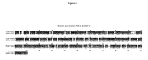
- the present invention relates to a polypeptide in isolated form belonging to a subfamily of the human Immunoglobulin Superfamily, which polypeptide shows at least 70% sequence homology with the amino acid sequence of the murine Confluency Regulated Adhesion Molecules 1 or 2 (CRAM-1 or CRAM-2) as depicted in Fig 3 upper and second (lower) row, respectively.
- Figures 4 and 5 show the alignment on amino acid level between mouse JAM-2 (CRAM-1) and JAM-3 (CRAM-2), respectively.
- the CRAM polypeptides found in the human or animal body are markers for growing cells. CRAM expression is upregulated in cells that are growing.
- the sequence information can be either the amino acid sequence or the nucleotide sequence encoding the amino acid sequence.
- the invention thus relates to a corresponding polypeptide in humans, comprising essentially the amino acid sequence as depicted in Fig 6 or an amino acid sequence that is at least 70% homologous, preferably at least 80%, more preferably at least 90% thereto.
- the two proteins and their corresponding family members can also be used for the preparation of derived molecules, such as antibodies directed against the (poly)peptides of the invention, or recombinant equivalents of the proteins, optionally in soluble form, or peptides comprising at least part of the amino acid sequence of the polypeptides.
- Suitable parts of the amino acid sequence are especially the extracellular domains: VC 2 , and the membrane proximal cytoplasmic sequence: A-[Y,Q]-[R,S]-[R,K]-G-[C,Y]-F.
- the invention also relates to poly- or oligonucleotides having a sequence that encodes a complete polypeptide or part thereof, which polypeptide has an amino acid sequence that is at least 70%, preferably at least 80%, more preferably at least 90%, most preferably essentially 100% homologous to the amino acid sequence of the CRAM-1 or CRAM-2 proteins as disclosed herein. More in particular, the invention relates to nucleotide sequences that are at least 70%, preferably at least 80%, more preferably at least 90%, most preferably essentially 100% homologous to the human DNA CRAM-1 sequence as depicted in Fig. 6 .
- Such poly- or oligonucleotides may for example be RNA or DNA and can be primers, probes, antisense RNA etc.
- All such molecules can be used for modulating the function of the original polypeptides found in the human or animal body or for diagnosis.
- Angiogenesis in for example tumors can be inhibited with antibodies. They can be used as targeting molecules for cells bearing the CRAM polypeptides.
- the antibodies can act on their own or can be coupled to other molecules, such as toxins, radioactive labels, fluorescent labels, enzymatic labels, photo-activatable labels, but also to liposomes, cells, etc.
- the labeled antibodies are particularly suitable for the diagnostic use of the antibodies, i.e. they can be utilized to locate angiogenesis in a growing tumor.
- antibodies coupled to toxins or radioactive molecules can be used to specifically kill the tumor from within by targeting to the (growing) vessels in the tumor.
- CRAM-type molecules were not detected in the normal vasculature except for lymphatics and the high endothelial venules in lymphoid organs such as lymph nodes and Peyer's patches.
- the advantage thereof is that the targeting of for example anti-CRAM antibodies can be highly specific to for example tumor cells thus avoiding undesirable side-effects.
- anti-CRAM antibodies can be used as a targeting molecule for cells bearing the (poly)peptides of the invention.
- the (poly)peptides may also bind the molecule on angiogenic vessels and by that stimulate or inhibit angiogenesis.
- Soluble (poly)peptide having essentially the same amino acid sequence as the CRAM polypeptides can be used in the treatment of inflammation reactions of the vascular endothelium. It was found according to the invention that the transendothelial migration of leukocytes can be inhibited by sCRAM-1-IG2Do or monoclonal antibodies against CRAM-1. This and similar molecules can therefore be used to quench or stimulate an immunological reaction such as found in inflammation.
- Anti-CRAM antibodies can also be used to block cell-cell interactions in growing cells. This leads to disorganization of intercellular contacts which are normally required for the barrier function of blood vessels. This finding may be used to increase the permeability of growing vessels to increase the delivery of drugs to sites, such as growing tumors, post-menstrual uterus, etc. The disorganization of intercellular contacts may therefore be used to block the development of tumor cells bearing the antigen, such as angiomas (tumors originating from vascular endothelium) or some rapidly growing carcinomas.
- angiomas tumors originating from vascular endothelium
- anti-CRAM antibodies can be used in the inhibition of angiogenesis in tumors, in the treatment of inflammation reactions and/or in the modulation, in particular increase, of vascular permeability.
- labeled antibodies for diagnosis use can be made of labeled antibodies but also of labeled oligonucleotides that are complementary to the CRAM DNA or mRNA found in the endothelial cells expressing the CRAM protein(s).
- JAM and JAM-1, CRAM-1 and JAM-2, as well as CRAM-2 and JAM-3 may be used interchangeably.
- the thymic (tEnd.1), and embryonic (eEnd.2) endothelioma cell lines ( Williams et al., 1989, Cell 57:1053-1063 ) were provided by Dr. W. Risau and Dr B. Engelhardt (Max Planck Institute, Bad-Nauheim, Germany).
- the SV40 transformed lymph node endothelial cell line TME was provided by Dr A. Hamann ( Harder et al., 1991, Exp Cell Res. 197:259-267 ).
- the squamous cell carcinoma KLN 205, the CHO, the MDCK, and the myeloma cell line Sp2/0 were obtained from the American Type Tissue Culture Collection (ATCC).
- CHO cells All cells, except CHO, were grown in DMEM (Gibco BRL, Paisley, Scotland), supplemented with 10% FCS (PAA Laboratories, Linz, Austria), 2 mM Glutamine, 100 U/ml Penicillin and 100 U/ml Streptomycin (all Gibco BRL). CHO cells were grown in Nut.Mix.F-12 (HAM) medium supplemented as above. Adherent cells were detached by washing with PBS/ 0.15 mM EDTA followed by 5 min incubation in trypsin/EDTA at 37°C.
- differential PCR was performed with the following degenerated primers: 5' TAYAGNTGYNNNGCYTCYAA 3' , 5' TAYCRGTGYNNNGCYTCYAA 3' , and 5' TAYTAYTGYNNNGCYTCYAA 3' encoding for the most frequent amino acid sequences encountered in C 2 domains: YRCXAS, YQCXAS, and YYCXAS.
- the PCR conditions consisted in using: 2 ⁇ l of diluted cDNA; 2.5 ⁇ l of 10X Goldstar PCR buffer; 2 ⁇ l of MgCl 2 ; 2 ⁇ l of degenerated primers 0,3 mM; 0.5 ⁇ l of dNTP 0.1 mM; 0.1 ⁇ l of ⁇ P 33 dATP 10mCi/ml (Amersham Pharmacia Biotech, D Weg, Switzerland); 15.65 ⁇ l H 2 O 0.25 ⁇ l Goldstar Taq polymerase (Eurogentech, Seraing, Belgium).
- the parameters for the PCR were as follows: 45 sec at 94°C, 90 sec at 50°C, and 45 sec at 72°C repeated 40 times.
- Formamide/EDTA loading buffer was added and samples were denatured for 2 min at 94°C.
- the PCR products were then separated on a 6% polyacrylamide gel, and autoradiographed using Kodak OM-Mat. The band intensities were compared.
- Nucleic acid sequences of two independent clones were determined using the Thermo Sequence Fluorescent Labeled Primer Cycle Sequencing Kit (Amersham Pharmacia Biotech, D Wegdorf, Switzerland) and the LI-COR DNA Analysis System (MWG-Biotech GmbH, Ebersberg, Germany).
- the three primers used were designed based on the EST sequences as follows: 5'-GAGGTACTTGCATGTGCT-3' for synthesis of the first strand, 5'-CGACAGGTGTCAGATAACA-3' and 5'-CACCCTCCTCACTCGT-3' for the two nested PCRs.
- the 5'RACE-PCR product was cloned into pGem-T Vector.
- the cloned 5'RACE-PCR product and the EST were digested with Hpal and Notl restriction enzymes and ligated into pGem-t vector.
- CRAM-2 Cloning of full length CRAM-2 was based on the same strategy of sequence comparison and 5'RACE technique. The full-length cDNA encoding CRAM-2 was finally obtained from ESTs accession numbers: AA690843 and W80145. These two clones differ by the length of the 3' untranslated region.
- Total mRNA from cells or tissues was extracted using Trizol (Life technologies AG, Basel, Switzerland) according to manufacturer's instructions.
- Poly-A + mRNA was extracted from 250 ⁇ g total RNA with the Oligotex mRNA Purification Kit (Qiagen, Zurich, Switzerland).
- Embryonic Poly-A northern blot was purchased from CLONTECH (P.H Stehelin and Cie AG, Basel, Switzerland).
- the riboprobes were prepared from pcDNA3 vector (Invitrogen, Leek, Netherlands), and comprised the sequences encoding for the immunoglobulin domains of JAM-1 and JAM-2, or the full-length coding sequence for ⁇ -actin. Hybridization was performed at 62 °C in buffer containing 50% formamide. The blots were then washed twice (0.5xSSC, 0.1%SDS, 67 °C), and autoradiographed on Kodak X-Omat at -80 °C.
- the sequence encoding EGFP was subcloned from pEGFP-1 vector (CLONTECH, P.H Stehelin and Cie AG, Basel, Switzerland) into pcDNA3 using Hindlll and Notl sites, therefore named pcDNA3/EGFP.
- the inserts encoding JAM-2 or JAM-1 were excised from pGemt or pRc/CMV using SacII/Hpal or HindIII/Scal digestions, respectively.
- the coding sequences were then cloned in pcDNA3/EGFP vector digested with Agel, blunted by fill-in and further digested with HindIII or SacII enzymes. This resulted in fusion sites at amino-acid positions DGV 291 for JAM-2 and QPS 285 for JAM-1.
- the transfection of CHO cells was performed as previously described ( Ballestrem et al., 1998, J Cell Sci. 111:1649-1658 ).
- Stable transfectants used for permeability assays were selected by growing transfected CHO cells for two weeks in medium containing 1 mg/ml of G418. Resistant colonies were isolated and checked for EGFP fluorescence intensity by flow cytometry (FACScalibur apparatus, Becton Dickinson, Mountain View, CA) and fluorescence localization by microscopy (Axiovert, Zeiss, Oberkochen, Germany).
- Time-lapse video microscopy was performed using an Axiovert fluorescence microscope and Openlab software for image acquisition.
- the mammalian expression vector pcDNA 3 (Invitrogen, Leek, Holland) was modified by integrating the Flag-Tag (G. Wiedle, Dep. of Pathology, CMU, Geneva) coding sequences.
- Flag-Tag constructs containing coding sequences for the soluble forms of the JAM, CRAM-1 and CRAM-2 proteins were prepared by PCR. In all cases, the forward primers were designed to fit ATG initiation region. The reverse primers were designed in the sequences encoding the hinge region for the one Ig soluble form or in the sequence encoding the region between the C2 and transmembrane domains for two Ig domains soluble molecules.
- the following monoclonal antibodies were used: anti-PECAM (GC51, rat IgG 2a ; EA-3, rat IgG 1 ) and anti-JAM (H202.106.7.4, rat IgG 1 ) ( Malergue et al., 1998, Mol Immunol. 35:1111-1119 .; Piali et al., 1993, Eur J Immunol. 23:2464-2471 ).
- the panel of CRAM antibodies against JAM-2 was generated in the laboratory using standard techniques, and recombinant soluble molecule as immunogen ( Aurrand-Lions et al., 1996, Immunity. 5: 391-405 ).
- the selected hybridomas were screened by ELISA for the production of antibodies recognizing specifically the recombinant soluble JAM-2 molecule. Positive clones were further tested on CHO cells transfected with JAM-2 cDNA (not shown).
- CRAM antibodies are of the IgG 1 or IgG 2a isotype except CRAM-25F24, which is of the IgG 2b subclass.
- Antibodies were purified on Protein G sepharose columns (Pharmacia Biotech Europe, D Weg, Switzerland) according to the manufacturer instructions.
- CRAM-19H36 mAb was used for immunoprecipitation, whereas CRAM-18F26 was the reagent used for immunohistochemistry. Similar results were obtained with CRAM-4H31 and CRAM-17D33 mAbs.
- Immunofluorescence analysis was performed using secondary reagents coupled to FITC or Texas Red (Jackson Immunoresearch, Milan AG, La Roche, Switzerland) for cytofluorimetry and immunohistochemistry, respectively.
- samples were fixed 5 min with cooled (-20 °C) methanol. Samples were rehydrated in PBS, gelatine 0.2%, Tween 20 0.05%, incubated overnight with the primary antibodies before washing, and revealed with the appropriate secondary reagent coupled to Texas Red.
- dissociation of freshly dissected tissues was performed using collagenase/dispase digestion, according to established procedures ( Kramer et al., 1984, J Cell Biol. 99:692-698 ).
- the dissociated cells were stained for 2 hours at 37 °C with Dil-Acetylated LDL (Molecular Probe Europe BV, Leiden, Netherlands) before staining with anti CRAM-19 and goat anti rat-FITC probe. After three washes, cells were stained with biotinylated anti-CD31 (Pharmingen) and streptavidine Red 670 (Life technologies AG, Basel, Switzerland).
- JAM-1 or JAM-2 expression was analyzed on cells positive for the two endothelial cell markers: Acetylated-LDL (FL-2) and CD31 (FL-3). Negative controls were obtained by omitting primary antibody.
- Permeability was measured using Transwell chambers (6.5 mm diameter, PC filters, 0.4 ⁇ m pore size, Costar Corp). In brief, 1x10 4 transfected or non-transfected CHO cells were cultured to confluency on filters previously coated for 30 min with 0.2% gelatin. After 5 days, the medium was changed for prewarmed Nut/F12 medium without FCS (500 ⁇ l in the lower chamber and 200 ⁇ l in the upper chamber). FITC-dextran (MW 38.900, Sigma Chemical Co) was added in the upper chamber at 1 mg/ml final concentration.
- Transient transfection of 293 T, Bosc 23 or stable transfection of CHO cells with the soluble Ig1Do and Ig2Do Flagtag/pcDNA-3 constructs were carried out using Lipofectamine Reagent according to manufacturer's instructions (Gibco BRL, Paisley, Scotland). Following transfection supernatants were collected every two days during a ten days period. M2 Beads (Kodak, New Haven, USA) covalently linked to anti-Flag antibody were washed twice with PBS containing a Protease Inhibitor Mix (Boehringer Mannheim, Germany). The beads were then incubated at 4°C for 3 hours with supernatant from the transfected cells.
- Leukocyte transmigration across endothelial cells was performed as previously described ( Wheerasinghe et al., 1998, J. Cell Biology, 142: 595-607 ). Briefly, 1x10 5 t-end cells were cultured for two days in transwell units (polycarbonate filters, 5 ⁇ m pore size, costar) in the presence of 1 ⁇ g of Ig soluble recombinant molecules: sJAM 2do or sCRAM-1 2do. After two days, 1x10 6 leukocytes obtained from lymph nodes and Peyer's patches were added to the upper compartment, and the number of transmigrated cells was monitored during the experiment every hour.
- transmigrated cells obtained from five independent wells were pooled and submitted to cytofluorimetric analysis for B-and T-cell markers B220 and CD3. Results were obtained using a Facscalibur machine and the Cell-Quest analysis program (Becton-Dickinson).
- transmigration assay with splenocytes, 3x10 5 endothelial cells were seeded in transwell units (polycarbonate filters, 8 ⁇ m pore size, costar) allowing the cells to form a monolayer over 18 hours.
- Medium in the upper compartment was removed and 1x10 6 leukocytes in 100 ⁇ l, freshly prepared from spleen by Ficoll centrifugation, were added to the upper compartment.
- SDF-1 was added to the medium (final concentration: 40 nM) in the lower compartment to establish a chemokine gradient between lower and upper compartments.
- purified antibodies 18-F26 or 19-H36 were added at the concentration of 10 ⁇ g/ml in the upper compartment with splenocytes. After 4 hours, transmigrated leukocytes (in the lower compartment) were harvested and counted. Results were expressed as % of input.
- the regulation of genes in endothelial cells depends on their environment.
- the present invention was directed to the identification of genes that undergo regulation upon the contact of endothelium with tumor cells.
- an in vitro assay was developed using the co-culture of melanoma tumor cells (B16) with an endothelioma cell line (t-end).
- Total RNA extracted from the mix was used as template to prepare cDNA submitted to a differential PCR screen.
- the cDNA obtained from the endothelial or melanoma cells cultured on their own were used as controls. The three different patterns were compared to identify the transcripts regulated by the co-culture condition.
- CRAM-1 a member of the Immunoglobulin Superfamily
- Fig 8A The resulting 1980 nucleotide long full length coding sequence of the postulated CRAM-1 cDNA is shown in Fig 8A .
- GACATGG strong consensus site for translation initiation 16 bp downstream from the 5' end, followed by a single ORF predicting a protein of 310 amino acid.
- the cleavage site was predicted to be at Ala 31-Val 32.
- the putative structure of the murine CRAM-1 protein is shown in Fig 8B and consists of an extracellular region with a variable heavy chain and a constant type 2 like immunoglobulin domain (Pfam, The Sanger Centre and Blast) with two potential N-linked glycosylation sites (aa 104 and 192).
- the hydrophobicity analysis (Tmpred, ISREC) predicted a transmembrane region between positions 242-260.
- the postulated cytoplasmic domain consisted of 49 amino acids and contained a number of highly conserved Ser/Thr and Tyr phosphorylation sites ( Fig 8A , residues in italic).
- JAM, CRAM-1 and CRAM-2 define a new subfamily
- JAM and CRAM-2 transcripts were strongly downregulated by TNF treatment of endothelial cells whereas the level of CRAM-1 transcript remained unchanged (lanes 2 and 11).
- JAM-2 transcript The tissue distribution of JAM-2 transcript was explored by northern blotting and compared to JAM-1 ( Fig 14 ).
- the JAM-2 transcript was 2 kb long, highly expressed in embryonic tissue, and in Peyer's patches, lymph nodes, kidney, and testis of adult animals. A putative splice variant of 1.8 kb was detected in testis. Expression of JAM-2 transcript was low in lung, liver, spleen, and thymus.
- the relative abundance of JAM-1 and JAM-2 were compared during embryogenesis: the mRNA encoding JAM-2 was detectable as early as day 7.5 post coitum, whereas JAM-1 mRNA was not detected at all during embryogenesis.
- CRAM-1 is widely expressed during embryogenesis and shows a restricted expression to epithelial or endothelial compartments in adult tissues. This is in agreement with the idea that it plays a role in the establishment and the maintenance of the polarized organization of cells.
- JAM-2 a 45kD protein, dependina on homophilic interactions for its localization to cell-cell contacts
- the HEV derived cell line TME expressed the highest level of JAM-2
- this endothelial cell line was used to further study the subcellular localization of JAM-2, and to compare to that of JAM-1.
- the localization of the JAM-2 protein on the surface of the endothelial cells was restricted to cell-cell contacts ( Fig 11A , a).
- the staining for JAM-2 was weaker than that observed for JAM-1 and less prominent in the membrane extensions between cells.
- JAM-2 present at cell-cell contacts interacted homophilically with JAM-2 or whether it interacted heterophilically with another molecule on the neighboring cell.
- JAM-2 protein was fused to green fluorescent protein (JAM-2-EGFP), and the construct transfected in CHO cells.
- JAM-2-EGFP green fluorescent protein
- JAM-2 was only observed in cell-cell contacts where both cells expressed the protein ( Fig 11B ), whereas the contacts between expressing and non-expressing cells were devoid of JAM-2 ( Fig 11B , a, indicated by arrow heads).
- Fig 11B chimeric molecule JAM-1-EGFP
- JAM-2 To characterize JAM-2 biochemically, immunoprecipitations of JAM-2 or EGFP chimeric proteins, expressed by TME cell line or by transfected CHO cells, respectively (Fig11C and D) were performed.
- the apparent molecular weight was identical under reducing or non-reducing conditions (not shown) and corresponded to the predicted molecular weight deduced from the amino-acid sequence of JAM-2, each N-glycosylation site accounting for 5 kDa.
- Immunoprecipitation of JAM-1 using H202-106 anti-JAM specific antibody resulted in a single band of lower molecular weight ( ⁇ 42kD, lane 2) that excluded a possible cross-reactivity between anti-JAM-2 and anti-JAM-1 antibodies.
- leukocyte trans-endothelial migration assays were performed in the presence of recombinant soluble JAM or CRAM-1. Endothelial cells were cultured for two days in the presence of sJAM-Ig2Do or sCRAM-1-Ig2Do. The monolayer integrity was not affected during this period, probably due to the molecular redundancy of the mechanism of cell-cell contact formation. The transmigration assay was performed in the presence of 1 ⁇ g of recombinant soluble molecules.
- the number of transmigrating cells was poorly affected by the presence of sJAM-Ig2Do (open squares) during the first three hours. After four hours, the number of transmigrated cells increased when compared to the control (dashed line). In contrast, the presence of sCRAM-1-Ig2Do (closed circles) strongly reduced the number of transmigrating cells.
- the leukocyte populations were heterogeneous, it was evaluated if sCRAM-1-Ig2Do acted on a specific leukocyte subpopulation or whether transmigration was blocked without specificity.
- the transmigrated cells were labeled for the lymphocyte markers CD3 and B220 ( Fig 20B ).
- sCRAM-1-Ig2Do specifically blocked the transmigration of non-lymphoid leukocytes, i.e. myeloid lineage cells (central panel, dashed columns).
- sJAM-Ig2Do poorly increased the number of transmigrating T cells (left panel, white column) without any effect on other cell subpopulations.
- CRAM-1 Since the transcript that encodes CRAM-1 is not regulated by TNF, but is downregulated when the endothelium was co-cultured with tumor cells, a confluency assay was used to further explore this regulation. Under low confluency, the cells were actively cycling and CRAM-1 interactions did not occur whereas under high confluency the cells divided less and CRAM-1 was engaged. The level of CRAM-1 mRNA expression was determined under various cell densities by semi-quantitative RT-PCR. As shown in Fig 13 , the expression level of CRAM-1 transcripts decreased when confluency was reached (lanes 1, 2, 3 in Fig 13 correspond to 100, 50, and 10% confluency respectively).
- JAM-2 is highly expressed during embryogenesis, and restricted to HEVs and endothelial cells subpopulations in adult tissues
- kidney and mesenteric lymph node sections ( Fig 15 ), which expressed the highest levels of JAM-2 transcript.
- a specific staining of intertubular structures was detected with anti-PECAM (GC51) or anti-JAM-2 (CRAM-18F26) mAbs, whereas anti-ZO-1 or anti-JAM-1 stained predominantly the tubular epithelial cells (not shown).
- vascular structures which dip down into the medulla and correspond to radial veins or vasa recta endothelial structures.
- serial sections were performed and the vascular structures identified with the anti-PECAM staining ( Fig 15d ).
- linear interendothelial stainings were detected with anti-JAM-2, anti-JAM-1 or anti-ZO-1 (Fig 15 a, b and c, respectively).
- HEVs high endothelial venules
- the HEVs were also found to express JAM-1, ZO-1 or PECAM (Fig 15 f, g and h), with subtle differences in the subcellular localization of the stainings (Fig 15 e-h , insets).
- Fig 15 e-h insets.
- Fig 15 i-l the staining with the CRAM-18F26 anti-JAM-2 mAb is restricted to certain endothelial cells or to structures closely associated with vasculature.
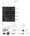
- Endothelial cell lines (tEnd.1, eEnd.2 and TME) expressed low levels of JAM-2 on the cell surface and variable levels of JAM-1 ( Fig 16A ).
- Cytometric analysis of freshly isolated endothelial cells was performed by triple staining of cell suspensions obtained after collagenase/dispase organ dissociation. Endothelial cells were identified by gating cells stained with both PECAM/CD31 and Acetylated-LDL ( Voyta et al., 1984, J. Cell Biol. 99:2034 ). Staining for JAM-2 or JAM-1 on this double positive cell population is shown in Fig 16 B. In kidney and Peyer's patches, all the isolated cells positive for CD31 and Acetylated-LDL were also stained for JAM-2, meaning that, at least in these organs, endothelial cells expressed JAM-2 in vivo . When the staining was performed on cells obtained from lymph node, JAM-2 expression was only found on a cell subpopulation, reflecting a possible heterogeneity of endothelial cell phenotypes within this tissue.
- JAM-2 was specifically localized to cell-cell contacts.
- the CHO cells stably transfected with the fluorescent chimeric molecule, were trypsinized and plated into chamber slides for imaging. After cell spreading, surface expression of JAM-2-EGFP was not uniform, but was rather clustered at cell-cell contacts ( Fig 17A , cells depicted by asterisks).
- Fig 17A cells depicted by asterisks.
- relocalization of JAM-2-EGFP to cell junctions was observed and an intense fluorescence signal was detected at the novel contact point between the cells forming the new cell-cell contact (arrows).
- the chimeric protein was enriched in the membrane protrusions between contacting cells, leading to the "zipper like" pictures seen after 12 or 18 min.
- the localization of JAM-2 at the primary cell-cell contacts was not lost during the formation of the new membrane contact (see upper left corner cell contacts). This finding indicated that JAM-2-EGFP was specifically relocalized to the new cell contact, and that, upon engagement, its localization was stable.
- JAM-2 increases monolaver tightness and participates to tight junctional complexes
- JAM-2 Since a number of molecules participating in cell-cell connection, have been shown to regulate the paracellular permeability of cell monolayers, it was tested whether JAM-2 could also affect this function. Transfection of JAM-2-EGFP reduced the paracellular permeability to FITC dextran and improved sealing of CHO cell monolayers by 42.5%; whereas transfection of the unrelated molecule Tac (IL2R ⁇ ) did not significantly reduce the paracellular permeability of CHO transfected cells ( Fig 18 ). The transfection of JAM-1-EGFP also reduced the paracellular permeability of CHO transfected cells monolayer.
- the EGFP chimeric proteins were transfected in MDCK cells, and their subcellular localization compared with that of the tight junctional marker: occludin.
- Fig 19A when serial pictures every 0.9 ⁇ m were analyzed for EGFP fluorescence and compared to occludin staining, JAM-2-EGFP was specifically enriched in cell-cell contacts at the level of tight junction. At the basal level (left), intracellular dots of EGFP fluorescence were observed.
- a similar analysis ( Fig 19B ) of MDCK cells transfected with JAM-1-EGFP showed a similar co-localization with occludin. Nevertheless, the distribution of JAM-1-EGFP fluorescence was less continuous than that observed for JAM-2-EGFP at the level of tight junctions.
- This example reports the use of a new screening strategy to identify regulated transcripts encoding members of the Ig superfamily of adhesion molecules. Described here is the cloning with this method of the new molecule CRAM-1 as a regulated transcript.
- the regulation observed in endothelial cells grown in the presence of tumors is confirmed by semi-quantitative RT-PCR and is shown to be dependant on the growth phase of the cells. Due to differential expression under changing cell confluency conditions, the name CRAM-1 for "Confluency Regulated Adhesion Molecule-1" was adopted.
- CRAM-2 Also described herein is a closely related sequence to CRAM-1 named CRAM-2.
- CRAM-1 and -2 represent the prototypes of a new subfamily of adhesion molecules which also includes the recently described molecule JAM ( Chretien et al., 1998, Eur. J. Immunol. 28, 4094-4104 ; Malergue et al., 1998, Mol. Immunol. 35, 1111-1119 ).
- CRAM-1 and JAM are preferentially expressed by endothelial and epithelial tissue at the cell-cell contacts and confer special properties to polarized layers.
- the effect of recombinant soluble molecules in a transendothelial migration assay and the regulation of JAM, CRAM-1 and CRAM-2 show that these three molecules play an important role in the maintenance of vascular physiology.
- TDD Targeted Differential Display
- RNA display employs random primers and involves the non-specific amplification of transcripts.
- the aim in this case is to pinpoint any differences in mRNA levels between two biological systems, which are submitted to comparison.
- TDD is an advanced screening method that combines the specificity of RNA fingerprinting with the degeneracy of Differential RNA Display resulting in selectivity. Due to the targeting of related transcripts, this technique significantly reduced the time needed for screening. The identification of new members of specific protein families, therefore, becomes possible. This is a substantial improvement of the reported non-specific screening strategies.
- JAM and CRAM-2 transcripts showed a similar tissue distribution and regulation of expression under the influence of TNF, indicating that they act by similar physiological mechanisms.
- CRAM-1 transcripts are not regulated by TNF but rather by the rate of proliferation or the density of endothelial cells.
- overexpression of CRAM-1 transcripts in cycling cells and its downregulation in quiescent cells indicate that this molecule participates in the establishment of a continuous monolayer. Its function in confluent monolayers of cells is the maintenance of the endothelial cell layer and the related properties. Since different leukocyte populations have to migrate to the site of immune response, it is thought that non-lymphoid cells migrate via a CRAM-1 dependant mechanism, whereas lymphoid cells migrate via JAM or CRAM-2. In this case the immune response can be modulated by using combinations of different soluble recombinant molecules.
Landscapes
- Health & Medical Sciences (AREA)
- Life Sciences & Earth Sciences (AREA)
- Chemical & Material Sciences (AREA)
- Organic Chemistry (AREA)
- Medicinal Chemistry (AREA)
- Immunology (AREA)
- General Health & Medical Sciences (AREA)
- Chemical Kinetics & Catalysis (AREA)
- Pharmacology & Pharmacy (AREA)
- General Chemical & Material Sciences (AREA)
- Nuclear Medicine, Radiotherapy & Molecular Imaging (AREA)
- Veterinary Medicine (AREA)
- Animal Behavior & Ethology (AREA)
- Public Health (AREA)
- Engineering & Computer Science (AREA)
- Bioinformatics & Cheminformatics (AREA)
- Proteomics, Peptides & Aminoacids (AREA)
- Genetics & Genomics (AREA)
- Molecular Biology (AREA)
- Cell Biology (AREA)
- Toxicology (AREA)
- Zoology (AREA)
- Gastroenterology & Hepatology (AREA)
- Biochemistry (AREA)
- Biophysics (AREA)
- Transplantation (AREA)
- Cardiology (AREA)
- Pain & Pain Management (AREA)
- Rheumatology (AREA)
- Heart & Thoracic Surgery (AREA)
- Peptides Or Proteins (AREA)
- Medicines That Contain Protein Lipid Enzymes And Other Medicines (AREA)
- Acyclic And Carbocyclic Compounds In Medicinal Compositions (AREA)
- Medicines Containing Antibodies Or Antigens For Use As Internal Diagnostic Agents (AREA)
- Measuring Or Testing Involving Enzymes Or Micro-Organisms (AREA)
- Pharmaceuticals Containing Other Organic And Inorganic Compounds (AREA)
- Preparation Of Compounds By Using Micro-Organisms (AREA)
- Organic Low-Molecular-Weight Compounds And Preparation Thereof (AREA)
- Materials For Medical Uses (AREA)
Priority Applications (1)
| Application Number | Priority Date | Filing Date | Title |
|---|---|---|---|
| EP10181055A EP2292761A1 (en) | 1999-03-11 | 2000-03-13 | Vascular adhesion molecules and modulation of their function |
Applications Claiming Priority (3)
| Application Number | Priority Date | Filing Date | Title |
|---|---|---|---|
| EP99200746 | 1999-03-11 | ||
| EP00920497A EP1163337B1 (en) | 1999-03-11 | 2000-03-13 | Vascular adhesion molecules and modulation of their function |
| EP10181055A EP2292761A1 (en) | 1999-03-11 | 2000-03-13 | Vascular adhesion molecules and modulation of their function |
Related Parent Applications (1)
| Application Number | Title | Priority Date | Filing Date |
|---|---|---|---|
| EP00920497.5 Division | 2000-03-13 |
Publications (1)
| Publication Number | Publication Date |
|---|---|
| EP2292761A1 true EP2292761A1 (en) | 2011-03-09 |
Family
ID=8239973
Family Applications (2)
| Application Number | Title | Priority Date | Filing Date |
|---|---|---|---|
| EP00920497A Expired - Lifetime EP1163337B1 (en) | 1999-03-11 | 2000-03-13 | Vascular adhesion molecules and modulation of their function |
| EP10181055A Withdrawn EP2292761A1 (en) | 1999-03-11 | 2000-03-13 | Vascular adhesion molecules and modulation of their function |
Family Applications Before (1)
| Application Number | Title | Priority Date | Filing Date |
|---|---|---|---|
| EP00920497A Expired - Lifetime EP1163337B1 (en) | 1999-03-11 | 2000-03-13 | Vascular adhesion molecules and modulation of their function |
Country Status (22)
| Country | Link |
|---|---|
| US (3) | US7393651B2 (is) |
| EP (2) | EP1163337B1 (is) |
| JP (2) | JP4836329B2 (is) |
| CN (2) | CN100469878C (is) |
| AT (1) | ATE500328T1 (is) |
| AU (1) | AU782139B2 (is) |
| BR (1) | BR0008915A (is) |
| CA (1) | CA2362896C (is) |
| CZ (1) | CZ303128B6 (is) |
| DE (1) | DE60045681D1 (is) |
| EE (1) | EE05497B1 (is) |
| ES (1) | ES2361905T3 (is) |
| HU (1) | HU229376B1 (is) |
| IL (1) | IL145310A0 (is) |
| IS (1) | IS2819B (is) |
| MX (1) | MXPA01009110A (is) |
| NO (1) | NO333085B1 (is) |
| NZ (1) | NZ514091A (is) |
| PL (1) | PL207994B1 (is) |
| RU (1) | RU2244748C2 (is) |
| WO (1) | WO2000053749A2 (is) |
| ZA (1) | ZA200107283B (is) |
Families Citing this family (19)
| Publication number | Priority date | Publication date | Assignee | Title |
|---|---|---|---|---|
| US8007798B2 (en) | 1997-11-21 | 2011-08-30 | Genentech, Inc. | Treatment of complement-associated disorders |
| US8088386B2 (en) | 1998-03-20 | 2012-01-03 | Genentech, Inc. | Treatment of complement-associated disorders |
| NZ514091A (en) * | 1999-03-11 | 2004-01-30 | Rmf Dictagene S | Vascular adhesion molecules and modulation of their function |
| GB0100750D0 (en) * | 2001-01-11 | 2001-02-21 | Inpharmatica Ltd | Novel proteins |
| WO2003006673A2 (en) * | 2001-07-11 | 2003-01-23 | Texas Biotechnology Corporation | A nucleic acid encoding a human junctional adhesion protein (jam3) |
| EP1533617A1 (en) * | 2003-11-19 | 2005-05-25 | RMF Dictagene S.A. | Angiogenesis inhibiting molecules, their selection, production and their use in the treatment and diagnosis of cancer |
| US7642341B2 (en) | 2003-12-18 | 2010-01-05 | Merck Serono S.A. | Angiogenesis inhibiting molecules, their selection, production and their use in the treatment of cancer |
| EP3299027A1 (en) | 2005-11-04 | 2018-03-28 | Genentech, Inc. | Use of complement pathway inhibitors to treat ocular diseases |
| RU2464030C2 (ru) * | 2006-05-26 | 2012-10-20 | Регадо Байосайенсиз, Инк. | Введение противосвертывающей системы reg1 |
| AU2007301599B2 (en) | 2006-09-28 | 2013-01-10 | Merck Serono S.A. | Junctional Adhesion Molecule-C (JAM-C) binding compounds and methods of their use |
| CN106188303A (zh) | 2006-11-02 | 2016-12-07 | 健泰科生物技术公司 | 人源化的抗‑d因子抗体 |
| FR2909092B1 (fr) * | 2006-11-24 | 2012-10-19 | Pf Medicament | Nouveaux anticorps anti-proliferation |
| AR066660A1 (es) | 2007-05-23 | 2009-09-02 | Genentech Inc | Prevencion y tratamiento de condiciones del ojo asociadas con su complemento |
| KR101589442B1 (ko) * | 2007-08-06 | 2016-02-01 | 녹손 파르마 아게 | 에스디에프-1 결합형 핵산 및 이의 용도 |
| CR20170001A (es) | 2008-04-28 | 2017-08-10 | Genentech Inc | Anticuerpos anti factor d humanizados |
| AU2014306867B2 (en) | 2013-08-12 | 2017-10-26 | Genentech, Inc. | Compositions and method for treating complement-associated conditions |
| EP3137503A1 (en) | 2014-05-01 | 2017-03-08 | Genentech, Inc. | Anti-factor d antibody variants and uses thereof |
| US10407510B2 (en) | 2015-10-30 | 2019-09-10 | Genentech, Inc. | Anti-factor D antibodies and conjugates |
| WO2017075252A1 (en) | 2015-10-30 | 2017-05-04 | Genentech, Inc. | Anti-factor d antibody variant conjugates and uses thereof |
Citations (2)
| Publication number | Priority date | Publication date | Assignee | Title |
|---|---|---|---|---|
| WO1998040483A2 (en) * | 1997-03-14 | 1998-09-17 | Human Genome Sciences, Inc. | 28 human secreted proteins |
| WO2000053753A2 (en) * | 1999-03-08 | 2000-09-14 | Genentech, Inc. | Promotion or inhibition of angiogenesis and cardiovascularization |
Family Cites Families (10)
| Publication number | Priority date | Publication date | Assignee | Title |
|---|---|---|---|---|
| US20030170864A1 (en) * | 2000-05-30 | 2003-09-11 | Genentech, Inc. | Secreted and transmembrane polypeptides and nucleic acids encoding the same |
| SE9604470D0 (sv) * | 1996-12-04 | 1996-12-04 | Tamas Bartfai Konsulting Ab | Transmembrane component of tight junction |
| EP0970111A2 (en) * | 1997-03-21 | 2000-01-12 | Genetics Institute, Inc. | Secreted proteins and polynucleotides encoding them |
| DE69837761T2 (de) | 1997-08-01 | 2008-01-17 | Serono Genetics Institute S.A. | 5' ests für sekretierte proteine aus gehirngeweben |
| US20030054400A1 (en) * | 1997-09-17 | 2003-03-20 | Genentech, Inc. | Secreted and transmembrane polypeptides and nucleic acids encoding the same |
| EP1002864A1 (en) * | 1998-11-10 | 2000-05-24 | Universita' degli studi di Bologna | HIgR and related V domain for the manufacture of a medicament for preventing or treating HSV-1, HSV-2 and BHV infections |
| KR100468978B1 (ko) * | 1998-12-16 | 2005-02-02 | 제넨테크, 인크. | 분비 및 막횡단 폴리펩티드, 및 이를 코딩하는 핵산 |
| NZ514091A (en) * | 1999-03-11 | 2004-01-30 | Rmf Dictagene S | Vascular adhesion molecules and modulation of their function |
| EP1533617A1 (en) * | 2003-11-19 | 2005-05-25 | RMF Dictagene S.A. | Angiogenesis inhibiting molecules, their selection, production and their use in the treatment and diagnosis of cancer |
| US7642341B2 (en) * | 2003-12-18 | 2010-01-05 | Merck Serono S.A. | Angiogenesis inhibiting molecules, their selection, production and their use in the treatment of cancer |
-
2000
- 2000-03-13 NZ NZ514091A patent/NZ514091A/xx not_active IP Right Cessation
- 2000-03-13 HU HU0200606A patent/HU229376B1/hu not_active IP Right Cessation
- 2000-03-13 CZ CZ20013267A patent/CZ303128B6/cs not_active IP Right Cessation
- 2000-03-13 CN CNB008063206A patent/CN100469878C/zh not_active Expired - Lifetime
- 2000-03-13 BR BR0008915-0A patent/BR0008915A/pt not_active Application Discontinuation
- 2000-03-13 PL PL350417A patent/PL207994B1/pl not_active IP Right Cessation
- 2000-03-13 CN CNA2004100952754A patent/CN1637019A/zh active Pending
- 2000-03-13 MX MXPA01009110A patent/MXPA01009110A/es active IP Right Grant
- 2000-03-13 ES ES00920497T patent/ES2361905T3/es not_active Expired - Lifetime
- 2000-03-13 DE DE60045681T patent/DE60045681D1/de not_active Expired - Lifetime
- 2000-03-13 EP EP00920497A patent/EP1163337B1/en not_active Expired - Lifetime
- 2000-03-13 CA CA2362896A patent/CA2362896C/en not_active Expired - Lifetime
- 2000-03-13 AT AT00920497T patent/ATE500328T1/de not_active IP Right Cessation
- 2000-03-13 AU AU41051/00A patent/AU782139B2/en not_active Expired
- 2000-03-13 RU RU2001127668/13A patent/RU2244748C2/ru not_active IP Right Cessation
- 2000-03-13 IL IL14531000A patent/IL145310A0/xx unknown
- 2000-03-13 WO PCT/EP2000/002219 patent/WO2000053749A2/en not_active Ceased
- 2000-03-13 JP JP2000603370A patent/JP4836329B2/ja not_active Expired - Lifetime
- 2000-03-13 EP EP10181055A patent/EP2292761A1/en not_active Withdrawn
- 2000-03-13 EE EEP200100472A patent/EE05497B1/xx not_active IP Right Cessation
-
2001
- 2001-09-03 ZA ZA200107283A patent/ZA200107283B/xx unknown
- 2001-09-10 IS IS6072A patent/IS2819B/is unknown
- 2001-09-11 NO NO20014417A patent/NO333085B1/no not_active IP Right Cessation
-
2004
- 2004-12-30 US US11/025,834 patent/US7393651B2/en not_active Expired - Fee Related
-
2008
- 2008-06-12 US US12/137,898 patent/US7670826B2/en not_active Expired - Fee Related
-
2010
- 2010-02-10 US US12/703,439 patent/US8143056B2/en not_active Expired - Fee Related
- 2010-05-06 JP JP2010106164A patent/JP2010233572A/ja active Pending
Patent Citations (2)
| Publication number | Priority date | Publication date | Assignee | Title |
|---|---|---|---|---|
| WO1998040483A2 (en) * | 1997-03-14 | 1998-09-17 | Human Genome Sciences, Inc. | 28 human secreted proteins |
| WO2000053753A2 (en) * | 1999-03-08 | 2000-09-14 | Genentech, Inc. | Promotion or inhibition of angiogenesis and cardiovascularization |
Non-Patent Citations (17)
| Title |
|---|
| AURRAND-LIONS ET AL., CELLULAR IMMUNOLOGY, vol. 176, 1997, pages 173 - 179 |
| AURRAND-LIONS ET AL., IMMUNITY, vol. 5, 1996, pages 391 - 405 |
| AURRAND-LIONS M A ET AL: "Cloning of JAM-2 and JAM-3: an emerging junctional adhesion molecular family?", CURRENT TOPICS IN MICROBIOLOGY AND IMMUNOLOGY, SPRINGER, BERLIN, DE, vol. 251, 1 January 2000 (2000-01-01), pages 91 - 98, XP002973229, ISSN: 0070-217X * |
| BALLESTREM ET AL., J CELL SCI., vol. 111, 1998, pages 1649 - 1658 |
| CHRETIEN ET AL., EUR. J. IMMUNOL., vol. 28, 1998, pages 4094 - 4104 |
| HARDER ET AL., EXP CELL RES., vol. 197, 1991, pages 259 - 267 |
| INES MARTIN-PADURA ET AL: "JUNCTIONAL ADHESION MOLECULE, A NOVEL MEMBER OF THE IMMUNOGLOBULIN SUPERFAMILY THAT DISTRIBUTES AT INTERCELLULAR JUNCTIONS AND MODULATES MONOCYTE TRANSMIGRATION", THE JOURNAL OF CELL BIOLOGY, vol. 142, no. 1, 13 July 1998 (1998-07-13), pages 117 - 127, XP002935706, ISSN: 0021-9525, DOI: 10.1083/JCB.142.1.117 * |
| KRAMER ET AL., J CELL BIOL., vol. 99, 1984, pages 692 - 698 |
| LIANG; PARDEE, SCIENCE, vol. 257, 1992, pages 967 - 970 |
| MALERGUE ET AL., MOL IMMUNOL., vol. 35, 1998, pages 1111 - 1119 |
| MALERGUE ET AL., MOL. IMMUNOL., vol. 35, 1998, pages 1111 - 1119 |
| PIALI ET AL., EUR J IMMUNOL., vol. 23, 1993, pages 2464 - 2471 |
| SAMARIDIS; COLONNA, EUR. J. IMMUNOL., vol. 27, 1997, pages 660 - 665 |
| SAMBROOK; FRITSCH; MANIATIS: "Molecular cloning", 1989, COLD SPRING HARBOR LABORATORY PRESS |
| VOYTA ET AL., J. CELL BIOL., vol. 99, 1984, pages 2034 |
| WHEERASINGHE ET AL., J. CELL BIOLOQY, vol. 142, 1998, pages 595 - 607 |
| WILLIAMS ET AL., CELL, vol. 57, 1989, pages 1053 - 1063 |
Also Published As
Similar Documents
| Publication | Publication Date | Title |
|---|---|---|
| US7670826B2 (en) | Confluence regulated adhesion molecules useful in modulating vascular permeability | |
| Aurrand-Lions et al. | JAM-2, a novel immunoglobulin superfamily molecule, expressed by endothelial and lymphatic cells | |
| US6348574B1 (en) | Seven transmembrane receptors | |
| Matsumoto et al. | The distribution of tenascin-X is distinct and often reciprocal to that of tenascin-C. | |
| US5639634A (en) | Cadherin polynucleotides | |
| EP1240326B1 (en) | Triggering receptor involved in natural cytotoxicity mediated by human natural killer cells and antibodies that identify this receptor | |
| AU2005202246B2 (en) | Vascular adhesion molecules and modulation of their function | |
| AU2008201101B2 (en) | Vascular adhesion molecules and modulation of their function | |
| HK1044169B (en) | Vascular adhesion molecules and modulation of their function | |
| WO1998004697A9 (en) | Cdo tumor suppressor gene and protein | |
| WO1998004697A1 (en) | Cdo tumor suppressor gene and protein | |
| HK1075051A (en) | Vascular adhesion molecules and modulation of their function | |
| Fawcett et al. | Molecular tools for the analysis of cell adhesion | |
| HK1011548A (en) | Novel v31 seven transmembrane receptors | |
| HK1036633A (en) | Cadherin materials and methods |
Legal Events
| Date | Code | Title | Description |
|---|---|---|---|
| PUAI | Public reference made under article 153(3) epc to a published international application that has entered the european phase |
Free format text: ORIGINAL CODE: 0009012 |
|
| 17P | Request for examination filed |
Effective date: 20100928 |
|
| AC | Divisional application: reference to earlier application |
Ref document number: 1163337 Country of ref document: EP Kind code of ref document: P |
|
| AK | Designated contracting states |
Kind code of ref document: A1 Designated state(s): AT BE CH CY DE DK ES FI FR GB GR IE IT LI LU MC NL PT SE |
|
| STAA | Information on the status of an ep patent application or granted ep patent |
Free format text: STATUS: THE APPLICATION IS DEEMED TO BE WITHDRAWN |
|
| 18D | Application deemed to be withdrawn |
Effective date: 20110910 |