EP2817626B1 - Verfahren zum Nachweis aktiver Malignitäten - Google Patents
Verfahren zum Nachweis aktiver Malignitäten Download PDFInfo
- Publication number
- EP2817626B1 EP2817626B1 EP13709786.1A EP13709786A EP2817626B1 EP 2817626 B1 EP2817626 B1 EP 2817626B1 EP 13709786 A EP13709786 A EP 13709786A EP 2817626 B1 EP2817626 B1 EP 2817626B1
- Authority
- EP
- European Patent Office
- Prior art keywords
- probe
- aliquot
- solvent
- binding
- mixture
- Prior art date
- Legal status (The legal status is an assumption and is not a legal conclusion. Google has not performed a legal analysis and makes no representation as to the accuracy of the status listed.)
- Revoked
Links
Images


Classifications
-
- G—PHYSICS
- G01—MEASURING; TESTING
- G01N—INVESTIGATING OR ANALYSING MATERIALS BY DETERMINING THEIR CHEMICAL OR PHYSICAL PROPERTIES
- G01N33/00—Investigating or analysing materials by specific methods not covered by groups G01N1/00 - G01N31/00
- G01N33/48—Biological material, e.g. blood, urine; Haemocytometers
- G01N33/50—Chemical analysis of biological material, e.g. blood, urine; Testing involving biospecific ligand binding methods; Immunological testing
- G01N33/53—Immunoassay; Biospecific binding assay; Materials therefor
- G01N33/575—Immunoassay; Biospecific binding assay; Materials therefor for cancer
- G01N33/57555—Immunoassay; Biospecific binding assay; Materials therefor for cancer of the prostate
-
- G—PHYSICS
- G01—MEASURING; TESTING
- G01N—INVESTIGATING OR ANALYSING MATERIALS BY DETERMINING THEIR CHEMICAL OR PHYSICAL PROPERTIES
- G01N33/00—Investigating or analysing materials by specific methods not covered by groups G01N1/00 - G01N31/00
- G01N33/48—Biological material, e.g. blood, urine; Haemocytometers
- G01N33/50—Chemical analysis of biological material, e.g. blood, urine; Testing involving biospecific ligand binding methods; Immunological testing
- G01N33/53—Immunoassay; Biospecific binding assay; Materials therefor
- G01N33/575—Immunoassay; Biospecific binding assay; Materials therefor for cancer
- G01N33/57515—Immunoassay; Biospecific binding assay; Materials therefor for cancer of the breast
-
- G—PHYSICS
- G01—MEASURING; TESTING
- G01N—INVESTIGATING OR ANALYSING MATERIALS BY DETERMINING THEIR CHEMICAL OR PHYSICAL PROPERTIES
- G01N33/00—Investigating or analysing materials by specific methods not covered by groups G01N1/00 - G01N31/00
- G01N33/48—Biological material, e.g. blood, urine; Haemocytometers
- G01N33/50—Chemical analysis of biological material, e.g. blood, urine; Testing involving biospecific ligand binding methods; Immunological testing
- G01N33/53—Immunoassay; Biospecific binding assay; Materials therefor
- G01N33/575—Immunoassay; Biospecific binding assay; Materials therefor for cancer
- G01N33/57535—Immunoassay; Biospecific binding assay; Materials therefor for cancer of the large intestine, e.g. colon, rectum or anus
-
- G—PHYSICS
- G01—MEASURING; TESTING
- G01N—INVESTIGATING OR ANALYSING MATERIALS BY DETERMINING THEIR CHEMICAL OR PHYSICAL PROPERTIES
- G01N33/00—Investigating or analysing materials by specific methods not covered by groups G01N1/00 - G01N31/00
- G01N33/48—Biological material, e.g. blood, urine; Haemocytometers
- G01N33/50—Chemical analysis of biological material, e.g. blood, urine; Testing involving biospecific ligand binding methods; Immunological testing
- G01N33/53—Immunoassay; Biospecific binding assay; Materials therefor
- G01N33/575—Immunoassay; Biospecific binding assay; Materials therefor for cancer
- G01N33/5758—Immunoassay; Biospecific binding assay; Materials therefor for cancer involving compounds serving as markers for tumours, cancers or neoplasias, e.g. cellular determinants, receptors, heat shock/stress proteins, A-protein, oligosaccharides or metabolites
- G01N33/57585—Immunoassay; Biospecific binding assay; Materials therefor for cancer involving compounds serving as markers for tumours, cancers or neoplasias, e.g. cellular determinants, receptors, heat shock/stress proteins, A-protein, oligosaccharides or metabolites involving compounds identifiable in body fluids
-
- G—PHYSICS
- G01—MEASURING; TESTING
- G01N—INVESTIGATING OR ANALYSING MATERIALS BY DETERMINING THEIR CHEMICAL OR PHYSICAL PROPERTIES
- G01N2333/00—Assays involving biological materials from specific organisms or of a specific nature
- G01N2333/435—Assays involving biological materials from specific organisms or of a specific nature from animals; from humans
- G01N2333/76—Assays involving albumins other than in routine use for blocking surfaces or for anchoring haptens during immunisation
- G01N2333/765—Serum albumin, e.g. HSA
Definitions
- the present invention relates generally to analysis of extracellular fluids that contain carrier proteins (e.g., serum albumin), and, more particularly, but not by way of limitation, to methods for detection of active (e.g., aggressive) malignancy in a patient by analyzing serum or plasma samples from the patient.
- carrier proteins e.g., serum albumin
- malignant cells may grow aggressively, forming and growing malignant cells and invading parts of the body near an initial tumor during early stages, and spreading to more distant parts of the body through the blood stream and/or lymphatic system during later stages. These processes are often accompanied by secretion of multiple metabolites to the circulatory system of the patient.
- Analyzing hematologic parameters and/or measuring the concentration of various metabolites in blood samples from a patient are known in the art and may be widely-used methods of diagnosing cancer in a patient, such as, for example, by identification of antigens, hormones, enzymes and other biologically active substances (tumor markers).
- tumor markers are generally each useful for diagnosing a certain type of cancer, such as, for example, alpha-fetoprotein for liver cancer, human chorionic gonadotropin (HCG) for some types of testicular and ovarian cancer, prostate-specific antigen for prostate cancer, CA 15-3 for breast cancer, and several others.
- HCG human chorionic gonadotropin
- prostate-specific antigen for prostate cancer CA 15-3 for breast cancer, and several others.
- tumor markers may not permit recognition or diagnosis of active cancer until later stages when other symptoms are already noticeable and the patient is investigated with methods such as X-rays, CT scans, endoscopy and biopsy. In these later stages, treatment may be less effective and may not be effective enough to prevent the death of the patient.
- mass spectrometry-based profiling of metabolites residing in extracellular fluids (also known as Metabonomics), such as, for example, profiling of low molecular mass serum proteins (Proteomics) and peptides (Peptidome).
- Metabonomics profiling of low molecular mass serum proteins
- peptides peptides
- Serum albumin is generally the most abundant serum protein, and provides a primary transport of hydrophobic metabolites in the circulatory system of the organism.
- the structure of the albumin molecule includes three domains that, in the hydrophilic medium of blood, are generally folded in an albumin globule of a heart-like and/or ellipsoidal shape in which hydrophilic binding sites generally face outward, and the hydrophobic binding sites generally face inward to form what may be referred to as one or more "hydrophobic containers" or "hydrophobic cavities” in the interior of the globule.
- the albumin molecule has multiple specific and non-specific hydrophobic sites which bind various long-chain fatty acids and other hydrophobic metabolites. In the albumin-bound state, hydrophobic metabolites are transferred and/or accumulated in hydrophilic extracellular fluids in the body.
- Known techniques of profiling of serum metabolites generally require dissociation of all albumin-bound metabolites, which may cause a number of shortcomings and/or drawbacks. Additionally, known techniques for profiling of serum metabolites generally are as a research tool for discovery of new tumor biomarkers, rather than as a diagnostic tool for diagnosing cancer.
- WO2010/049011 discloses methods and kits for detecting toxemia in a subject patient.
- the method comprises the steps of: mixing a labeled hydrophobic probe capable of binding to carrier proteins with an aliquot of a subject patient's extracellular fluid containing carrier proteins, the amount of probe such that the molar ratio of the probe to the carrier proteins is between about 0.3 and about 1.5; mixing a solvent with the aliquot, the solvent such that when added to the aliquot and probe the solubility of the probe is increased in the aliquot, and the amount of solvent sufficient to dissociate a portion of the probe from the carrier proteins without causing significant dissociation of toxins from the carrier proteins; analyzing the mixture comprising the aliquot, probe, and solvent to determine the binding efficiency of the carrier proteins; and comparing the subject binding efficiency to at least one control binding efficiency for at least one non-toxemic control patient.
- EP 0 973 043 A1 published January 19, 2000 , describes a method of diagnosing malignant neoplasms by using electron paramagnetic resonance (EPR) spectroscopy (which may also be known in the art as electron spin resonance "ESR" spectroscopy), wherein an aliquot of a subject patient's serum is mixed with the spin probe represented by spin labeled fatty acid.
- ESR electron spin resonance
- This method may produce false-positive and false-negative results in the presence of additional factors such as evaluated blood lipid values, injection of medicaments, elevated blood alcohol level, residing infection diseases of the subject patient, as well effects caused by a solvent of the spin probe.
- additional factors such as evaluated blood lipid values, injection of medicaments, elevated blood alcohol level, residing infection diseases of the subject patient, as well effects caused by a solvent of the spin probe.
- ESR electron spin resonance
- Electron spin resonance (ESR) spectroscopy of spin-labeled fatty acid (FA) molecules binding to human serum albumin is a suitable method for the detection of conformational changes and alterations of transport function of albumin through changes in its FA binding capabilities.
- the aim of the study was to examine whether the FA binding to albumin is detectably and significantly altered in CRC patients when compared with patients having benign colorectal diseases.
- United States Patent No. 7, 166,474 describes a method of detecting changes in transport properties of albumin by using electron paramagnetic resonance (EPR) spectroscopy (which may also be known in the art as electron spin resonance "ESR" spectroscopy) to evaluate a sample that contains albumin (an albumin-containing sample).
- EPR electron paramagnetic resonance
- This method can include evaluating the albumin transport function with respect to long chain fatty acids by using a spin probe represented by a spin-labeled fatty acid.
- the EPR-spectra of the spin probe can be measured in at least three aliquots of the sample, where each aliquot is mixed with significantly different concentrations of the spin probe and a high concentration of ethanol.
- the concentration of ethanol is high enough to induce significant conformational changes of the albumin to enable evaluation of the conformational flexibility of the albumin.
- the conformational state and binding parameters of the albumin are determined from the parameters of the spin probe mobility in two binding sites of serum albumin, and the concentration of unbound spin probe.
- the parameters of albumin transport function are derived from measurements of the conformational changes induced in the albumin artificially by the high ethanol concentration. This method assumes that albumin molecules efficiently release bound substances to target objects at conditions that occur in a healthy patient, and therefore induces a conformational change facilitating dissociation of albumin-bound metabolites.
- This method can include detection of cancer via the evaluation of the conformational changes of albumin in respect of modification caused by metabolites associated with a tumor growth. While this may be useful in the method taught by this patent, the conformational changes of albumin molecules induced by the high ethanol concentration cause dissociation of a significant portion of albumin-bound spin probe in the investigated sample, which largely prevents correct evaluation of the albumin conformation. For example, due to their hydrophobic origin, spin probe molecules dissociated from albumin will aggregate into micelles and associate with or bind to serum lipoproteins.
- these dissociated portions of spin probe can induce EPR signals that interfere with those from the spin probe bound on albumin sites, and thereby prevent precise evaluation of the albumin conformation.
- This method suffers from possible shortcomings that may include, for example, the use of at least three aliquots of each sample, binding on albumin of acute-phase response metabolites in early (e.g., asymptomatic) stages of infection or disease, and excessive dissociation of spin probe from the albumin (and resulting spin probe aggregation into micelles and binding to serum lipoproteins) resulting from the addition of relatively high-concentrations of ethanol.
- These possible shortcomings may, in some instances, contribute to variations in results, including false-positives and false-negatives in detecting the presence of metabolites associated with an active malignant growth in a patient.
- Various embodiments of the present invention can be suitable for detecting variations in serum albumin conformation, functions, characteristics, and/or parameters by mixing various substances with a sample of extracellular fluid (that contains (e.g., serum albumin)) from a subject patient, and analyzing the mixture.
- extracellular fluids include blood serum, blood plasma, lymph fluid, and spinal fluid.
- Some embodiments of the present methods are suitable for detection, or diagnosis of active malignancy in its early or precursor stages.
- Embodiments of the present invention may be suitable, for example, for diagnosis and/or monitoring of cancer patients in clinical settings.
- embodiments of the present invention may be suitable for such diagnosis, monitoring, and/or relapse detection of cancer patients after treatment, or patients (e.g., chemotherapy, radiotherapy, and/or the like).
- the methods of the present invention can be suitable for detection or diagnosis of active malignancy, by detecting or measuring the deviation of parameters of spin probe binding to serum albumin of a subject patient, relative to the parameters of spin probe binding to serum albumin of a control patient.
- the probe binding parameters of the control patient can be established, for example, by testing one or more control patients, by historical test results from patients with known conditions, by statistical aggregation of data from various other patients, or by any other suitable means that permits comparison with the subject patient in such a way as to provide an indication of whether the subject patient has, likely has, is likely to have, does not have, likely does not have, or is not likely to have active malignancy.
- This deviation can, for example, be detected or measured for a sample of serum taken from a subject patient by mixing an aliquot of the serum sample with a labeled hydrophobic probe having a long-hydrocarbon-chain.
- parameters of binding of a hydrophobic probe having a long-hydrocarbon chain to serum albumin can correlate to the amount of cancer-associated metabolites bound to the binding sites of the albumin. This correlation can, for example, be caused by competitive binding on specific albumin sites for substances with long hydrocarbon chains, and indirectly due to allosteric modification of the conformation and affinity of these sites upon ligand binding on other albumin sites.
- cancer-associated metabolites when high levels of cancer-associated metabolites are present in the subject patient's blood, these metabolites may crowd and/or modify the binding sites of the serum albumin, thereby preventing the hydrophobic probe from binding to those binding sites. In this way, metabolites associated with active malignant growth can be detected as their levels rise, and even before other sign or symptoms of the cancer appear.
- the present invention includes various embodiments of methods for detecting active malignancy in a subject patient.
- Some embodiments of the present methods comprise: obtaining a first sample of a subject patient's serum albumin-containing extracellular fluid; mixing a labeled hydrophobic probe capable of binding to serum albumin with an aliquot of the first sample, the amount of probe such that the molar ratio of the probe to the serum albumin is between about 2 and about 7, the probe optionally comprising two or more substances; mixing a solvent with the aliquot of the first sample, the solvent such that when added to the aliquot and probe the solubility of the probe is increased in the aliquot, and the amount of solvent is sufficient to dissociate a portion of the probe from the serum albumin without causing significant dissociation of hydrophobic metabolites from the serum albumin, the solvent optionally comprising two or more substances; analyzing the mixture of aliquot, probe, and solvent to determine for the first sample one or more binding parameters
- Some embodiments further comprise: obtaining a second sample of the subject patient' s serum albumin-containing extracellular fluid taken from the subject patient between 2 and 90 days after the first sample; mixing a labeled hydrophobic probe capable of binding to serum albumin with an aliquot of the second sample, the amount of probe such that the molar ratio of the probe to the serum albumin is between 2 and 7, the probe optionally comprising two or more substances; mixing a solvent with the aliquot of the second sample, the solvent such that when added to the aliquot and probe the solubility of the probe is increased in the aliquot, and the amount of solvent is sufficient to dissociate a portion of the probe from the serum albumin without causing significant dissociation of hydrophobic metabolites from the serum albumin, the solvent optionally comprising two or more substances; analyzing the mixture of aliquot, probe, and solvent to determine for the second sample one or more binding parameters of the probe; and comparing the one or more binding parameters of the second sample to one or more control binding parameters of a probe binding to serum
- Some embodiments of the present methods comprise: obtaining two samples of a subject patient's serum albumin-containing extracellular fluid taken from the subject patient between 2 and 90 days apart; mixing a labeled hydrophobic probe capable of binding to serum albumin with an aliquot of each sample, the amount of probe such that the molar ratio of the probe to the serum albumin is between 2 and 7, the probe optionally comprising two or more substances; mixing a solvent with the aliquot of each sample, the solvent such that when added to the aliquot and probe the solubility of the probe is increased in the aliquot, and the amount of solvent is sufficient to dissociate a portion of the probe from the serum albumin without causing significant dissociation of hydrophobic metabolites from the serum albumin, the solvent optionally comprising two or more substances; analyzing each mixture of aliquot, probe, and solvent to determine for each sample one or more binding parameters of the probe; and comparing the one or more binding parameters of each sample to one
- Some embodiments of the present methods further comprise: diagnosing an active malignancy in the subject patient if: (i) the one or more binding parameters for each of the two samples are closer to the one or more control binding parameters for the at least one control patient with an active malignancy than to the one or more control binding parameters of the cancer-free control patient, and (ii) the subject patient had no indication of acute disease or infection that occurred between the times of the deriving of the samples.
- the labeled hydrophobic probe is capable of binding to specific and non-specific hydrophobic binding sites of serum albumin.
- analyzing each mixture comprises: substantially excluding from the mixture a portion of the probe that is bound on lipoproteins and a portion of the probe that is aggregated into micelles; and determining the one or more probe binding parameters of the sample from the remaining portion of the probe in the mixture.
- the one or more probe binding parameters of each sample are determined from the portion of the probe bound to specific and non-specific hydrophobic binding sites of albumin, and the portion of unbound probe.
- the two samples are taken from the subject patient between 5 and 90 days apart, between 2 and 30 days apart, or between 5 and 30 days apart.
- Some embodiments of the present methods further comprise: verifying that the subject patient had no indication of acute diseases that occurred between the times at which the two samples were taken from the patient.
- analyzing each mixture comprises: measuring the concentrations of the probe bound on specific and non-specific hydrophobic sites of serum albumin, and the unbound probe in the mixture; and deriving the one or more probe binding parameters from at least the concentrations of: the probe bound to specific and non-specific hydrophobic binding sites of albumin, and the unbound probe.
- the probe comprises an organic molecule having between 8 and 28 Carbon atoms, and optionally, where the probe comprises one or more of: a fatty acid, a long-chain fatty acid, 16-DOXYL-stearic acid, or a lysophospholipid.
- the amount of solvent mixed with the aliquot and probe is such that the solvent does not induce significant conformational changes of albumin.
- the amount of solvent mixed with the aliquot and probe is sufficient to increase the concentration of unbound probe in the mixture of the aliquot, the probe, and the solvent to at least 5 times greater than the concentration of unbound probe in a mixture of the aliquot and the probe without the solvent.
- the volume of the solvent mixed with the aliquot is less than about 30% of the volume of the aliquot, and optionally less than about 20% of the volume of the aliquot.
- Some embodiments of the present methods further comprise: normalizing the one or more probe binding parameters of each sample to account for the reduction in albumin concentration caused by the solvent in the mixture; and optionally, where the steps of deriving and normalizing the one or more probe binding parameters are performed simultaneously. Some embodiments comprise: normalizing the one or more probe binding parameters of each sample to account for the reduction in serum albumin concentration caused by medical conditions of the patient. In some embodiments, the one or more control parameters comprise at least one range of control parameters of control patients.
- Some embodiments comprise: repeating the steps of mixing a probe, mixing a solvent, measuring the concentrations, deriving a one or more probe binding parameters for each of two or more aliquots, where a different amount of solvent is used for each of the two or more repetitions; optionally, where the one or more probe binding parameters derived for the two or more repetitions are averaged to derive an average probe binding parameters, and where the averaged one or more probe binding parameters are compared in the step of comparing the one or more probe binding parameters.
- the volume of the aliquot is less than about 100 ⁇ L, and optionally, less than about 50 ⁇ L.
- the probe is labeled with a spin-label.
- the extracellular fluid includes at least one of: blood serum, blood plasma, lymph fluid, and spinal fluid.
- analyzing each mixture comprises verifying that the mixture has reached binding equilibrium, optionally by: measuring two EPR spectra of the mixture at two different times; and comparing the two EPR spectra to determine whether intensities of spectral lines (spectral parameters) in the EPR spectra correspond.
- kits e.g., for detecting active malignancy in a subject patient from a sample of the subject patient's serum albumin-containing extracellular fluid
- kits may comprise: a labeled hydrophobic probe capable of binding to specific and non-specific hydrophobic binding sites of serum albumin, the amount of probe such that when mixed with an aliquot having a predetermined volume of the extracellular fluid, the molar ratio of the probe to the carrier proteins is in the range of about 3 to about 4; and a solvent, the solvent configured such that when mixed with the probe and the aliquot the solubility of the probe is increased in the aliquot, and the amount of solvent sufficient to dissociate a portion of the probe from the albumin without causing significant dissociation of hydrophobic metabolites from serum albumin.
- kits of the related (non-claimed) aspect further comprise instructions for: mixing a labeled hydrophobic probe capable of binding to specific and non-specific hydrophobic binding sites of serum albumin with an aliquot having a predetermined volume of a subject patient's extracellular fluid containing serum albumin, the amount of probe such that the molar ratio of the probe to the carrier proteins is between about 3 and about 4; and mixing an amount of solvent with the mixture of the probe and the aliquot sufficient to dissociate a portion of the probe from the carrier proteins without causing significant dissociation of hydrophobic metabolites from serum albumin.
- Some embodiments comprise instructions for: analyzing the mixture of the aliquot, probe, and solvent to determine one or more binding parameters of the probe; and comparing the one or more binding parameters of each sample to one or more control binding parameters of a probe binding to serum albumin in the extracellular fluid of at least one of: a cancer-free control patient and a control patient with an active malignancy.
- instructions for analyzing the mixture comprise instructions for: substantially excluding from the mixture a portion of the probe that is bound on lipoproteins and a portion of the probe that is aggregated into micelles; and determining the one or more probe binding parameters of the sample from the remaining portion of the probe in the mixture.
- the instructions for analyzing the mixture comprise instructions for: measuring the concentrations of the probe bound on specific and non-specific hydrophobic sites of serum albumin, and the unbound probe in the mixture; and deriving the one or more probe binding parameters from at least the concentrations of: the probe bound to specific and non-specific hydrophobic binding sites of albumin, and the unbound probe.
- any embodiment of any of the present methods can consist of or consist essentially of - rather than comprise/include/contain/have - any of the described steps, elements, and/or features.
- the term "consisting of” or “consisting essentially of” can be substituted for any of the open-ended linking verbs recited above, in order to change the scope of a given claim from what it would otherwise be using the open-ended linking verb.
- Coupled is defined as connected, although not necessarily directly, and not necessarily mechanically; two items that are “coupled” may be integral with each other.
- the terms “a” and “an” are defined as one or more unless this disclosure explicitly requires otherwise.
- the terms “substantially,” “approximately,” and “about” are defined as largely but not necessarily wholly what is specified (and includes what is specified; e.g., substantially 90 degrees includes 90 degrees and substantially parallel includes parallel), as understood by a person of ordinary skill in the art. In any disclosed embodiment, the terms “substantially,” “approximately,” and “about” may be substituted with "within [a percentage] of” what is specified, where the percentage includes .1, 1, 5, and 10 percent.
- a step of a method that "comprises,” “has,” “includes” or “contains” one or more features possesses those one or more features, but is not limited to possessing only those one or more features.
- the blood serum in a method that comprises the step of obtaining a sample of blood serum containing serum albumin, the blood serum includes the specified features but is not limited to having only those features.
- the blood serum could also contain water.
- a device or structure that is configured in a certain way is configured in at least that way, but it can also be configured in other ways than those specifically described.
- Various embodiments of the present methods may include testing, analyzing, and/or evaluating of a sample of extracellular fluid from a subject patient. For clarity and brevity, embodiments are described below for testing blood serum of a subject patient. However, the embodiments, features, steps, and particulars described below can also be applied to other extracellular fluids such as, for example, blood plasma, lymph fluid and spinal fluid.
- Some embodiments of the methods not forming part of the claimed invention include obtaining a serum sample from a subject patient.
- a serum sample can be obtained, for example, by drawing blood from the patient and using centrifugation to substantially isolate the serum from the blood. Other suitable methods may also be used to obtain the serum sample from the subject patient.
- an aliquot can be obtained from this subject sample. Such an aliquot can be separated from the sample or can include the entire sample.
- Some embodiments of the present method include mixing a labeled hydrophobic probe with the aliquot.
- the labeled hydrophobic probe may also be referred to herein as the "labeled probe," the "hydrophobic probe,” or the “probe.”
- the probe is (or comprises) an organic molecule, and/or is selected to be capable of binding with albumin of the serum in an aliquot of a serum sample from a patient.
- the probe can comprise a suitable number of Carbon atoms.
- the probe can comprise between 8 and 28 Carbon atoms, including 8, 9, 10, 11, 12, 13, 14, 15, 16, 17, 18, 19, 20, 21 , 22, 23, 24, 25, 26, 27, or 28 Carbon atoms, or any smaller range between 8 and 28 Carbon atoms, e.g., between 14 and 22, between 16 and 20, or between 18 and 22 Carbon atoms.
- the probe can comprise less than 8 Carbon atoms or more than 28 Carbon atoms.
- the probe can comprise a hydrocarbon chain such as, for example, a hydrocarbon chain having any suitable number of Carbon atoms.
- the probe can comprise a hydrocarbon molecule whose structure is branched and/or whose structure comprises a ring structure.
- the probe can comprise a hydrocarbon molecule comprising elements other than Carbon or Hydrogen, such as, for example, Chlorine, Phosphorous, and/or Nitrogen.
- the probe can comprise a hydrocarbon chain such as, for example, a fatty acid, a long-chain fatty acid, a medium-chain fatty acid, or the like.
- probes suitable for some embodiments of the present method include: 16-DOXYL-stearic acid, free radical; 5-DOXYL-stearic acid, free radical; 16:0-16 PC DOXYL, free radical (1-Palmitoyl-2-Stearoyl-(16-DOXYL)-sn-Glycero-3-Phosphocholine); 12-NBDS or NBD-stearate, fluorescent probe (12-N-Methyl-7-nitrobenzo-2-oxa-1,3-diazolamino stearate); and anthroylstearic acid, fluorescent probe (12-(9-anthroyl)stearic acid).
- Such labeled probes are available from commercial suppliers including, for example, (1) Sigma-Aldrich, Inc., St. Louis, Missouri, U.S.A., www.sigmaaldrich.com; (2) TCI America, Portland, Oregon, U.S.A. and Wellesley Hills, Massachusetts, U.S.A, www.tciamerica.com; (3) Fluorochem, Derbyshire, U.K., www.fluorochem.co.uk; (4) Avanti Polar Lipids Inc., Alabaster, Alabama, U.S.A., www.avantilipids.com; and (5) Santa Cruz Biotechnology, Inc., Santa Cruz, California, U.S.A., http://www.scbt.com .
- the probe may be suspended in a volume of liquid so as to, for example, enable accurate measurement of the amount of the probe.
- This liquid is referred to herein as the "suspension liquid” or “liquid” so as not to be confused with the solvent described herein (even though the suspension liquid may be or comprise a solvent as that term is used in a more general sense).
- the suspension liquid may be (or comprise) the solvent, which is described below.
- the probe may be suspended in the solvent in an amount desired for mixing the aliquot, probe, and solvent without further addition of solvent; or may be suspended in an amount of solvent smaller than is desired, such that additional solvent must be separately added to the mixture.
- the probe may be suspended in 10 ⁇ L of solvent and the suspension added to the aliquot; or (2) the probe may be suspended in 5 ⁇ L of solvent, the suspension added to the aliquot, and 5 ⁇ L of solvent (without probe) also mixed with the aliquot.
- suitable solution liquids for use where the probe solution and solvent are mixed while both are liquid, include the materials described below for the solvent.
- the probe solution may be added to a container first and the liquid portion of the liquid/probe solution permitted to evaporate, leaving substantially-only the probe in the container.
- the aliquot of serum may then be added such that the probe then dissolves into the serum.
- suitable liquids for this suspension/evaporation method include both the liquids described below for the solvent, as well as other liquids (including solvents known in the art as non-polar solvents) that are not miscible with water, such as, for example, methyl ethyl ketone, methyl acetate, diethyl ether, dichloro methane, benzene, pentane, cyclo pentane, etc.
- the amount of probe mixed with the aliquot is sufficient to permit a representative level of binding of the probe to the specific (primary) and non-specific (secondary) hydrophobic sites of serum albumin, i.e., a level of binding that is sufficient to be representative of the probe binding to the specific and non-specific hydrophobic sites, respectively, of serum albumin in the subject patient's body at the time the sample was taken from the subject patient.
- the amount of probe is also small enough to prevent (or to not permit) saturation of the fatty acid binding sites on the serum albumin.
- the amount of probe is such that the molar ratio of probe (that is capable of binding with specific and non-specific sites of serum albumin) to the serum albumin is between about 3 and about 5 so as to, for example, encourage binding of the probe to albumin in a manner that is representative of the circulatory system of the patient.
- the molar ratio of the probe to the serum albumin is between 2 and 7.
- the molar ratio of the probe concentration to the serum albumin concentration can be any suitable ratio that permits the present methods to function as described.
- the concentration of serum albumin in the serum can be approximated from various texts known in the art. In some embodiments, the concentration of serum albumin can be approximated as the expected concentration of serum albumin in a healthy patient.
- results can later be adjusted (as described below) to account for any reduction in albumin concentration expected as a result of the medical conditions of the patient (e.g., toxemia).
- the concentration of serum albumin can be approximated as the expected concentration of serum albumin in a patient with the same medical conditions of the subject patient (e.g., active cancer, tumor remission stage, post-surgery and/or the like), and the amount of probe adjusted accordingly so as to, for example, reduce or eliminate the need to adjust results later to account for a reduction in serum albumin concentration caused by medical conditions of a subject patient.
- the present method include mixing a solvent with the aliquot to, for example, increase the solubility of the hydrophobic probe in the hydrophilic medium of the aliquot.
- the "solvent" mixed with the aliquot is a substance (or combination of substances) capable of increasing the solubility of the probe in the mixture (of at least aliquot, probe, and solvent), and can comprise one or more substances generally known in the art as solvents (e.g., polar solvents) and/or one or more substances not generally considered to be solvents (e.g., solids, water-based or other solutions, and the like).
- the solvent may include a mixture of two or more substances.
- the solvent is selected to be capable of causing some dissociation of the probe from the serum albumin in the aliquot.
- the solvent can be (or comprise) a liquid that is miscible with, or soluble in, water (and/or that may be known in the art as a polar solvent), such as, for example, methanol, ethanol, acetonitrile, dimethyl sulfoxide DMSO), tetrahydrofuran (THF), acetic acid, formamide, and the like.
- the solvent may be added at any suitable stage or in any suitable manner, including, for example, to the aliquot alone, to the probe alone, to a mixture of only the aliquot and probe, or to the complete mixture of aliquot, probe, and solvent.
- Some embodiments of the present methods include mixing water with any component of the mixture or the entire mixture at any suitable stage, including, for example, to the aliquot alone, to the probe alone, to a mixture of only the aliquot and probe, or to the complete mixture of aliquot, probe, and solvent.
- Some embodiments of the present methods include mixing additives with the water, with the solvent described above, with any other component of the mixture, and/or or with the complete mixture, to adjust or modify the isotonic properties of the sample mixture.
- some embodiments of the present method include mixing an amount of NaCl with water to achieve a concentration of about 0.9% NaCl to simulate the isotonic properties of blood and the normal cells of the body, as may be done in other areas of medicine and biological sciences.
- the amount of solvent added to the aliquot is such that a portion of the probe is dissociated from (caused to not bind or stop binding with) the serum albumin without causing significant dissociation of hydrophobic metabolites from the serum albumin.
- "significant dissociation of toxins” is dissociation from serum albumin that substantially affects the representative nature of the binding properties exhibited by the serum albumin in the aliquot (relative to the binding properties the same serum albumin exhibited in the subject patient's body at the time the sample was obtained from the subject patient).
- the amount of solvent added to the aliquot is also such that the solvent does not induce significant conformational changes to the serum albumin.
- "significant conformational changes” are conformational changes to the serum albumin that significantly affect the representative nature of the albumin conformation and binding properties exhibited by the serum albumin in the aliquot (relative to the conformation and binding properties the serum albumin exhibited in the subject patient's body at the time the sample was obtained from the subject patient). For example, a conformational change would be significant if it caused the serum albumin to release bound hydrophobic metabolites so as to bind a measurably greater amount of probe than the serum albumin would have bound without such conformational changes.
- the amount of solvent is such that the concentration of unbound (free) probe in the mixture (of aliquot, probe, and solvent) is at least a multiple greater than (e.g., 5, 6, 7, 8, 9, 10, 20, 50, 100, 500, 1000, 5000, 10000, or 100000 times greater than) the concentration of unbound (free) probe before the addition of the solvent (e.g., in a mixture of only the aliquot and probe).
- the amount of solvent is such that the concentration of unbound (free) probe in the mixture (of aliquot, probe, and solvent) is in a range between about a lower multiple and about a higher multiple greater than (e.g., between about 5 and about 10, between about 10 and about 100, or between about 100 and about 1000 times greater than) the concentration of unbound (free) probe before the addition of the solvent (e.g., in a mixture of only the aliquot and probe).
- the volume of the amount of solvent mixed with the aliquot is a percentage of the volume of the aliquot.
- the volume of solvent added may be between about 1% and about 30%, i.e., between about 0.5 ⁇ L and about 15 ⁇ L, or in any individual percentage or range of percentages within this range, e.g., about 10%, about 20%, between about 5% and about 20%, between about 10% and about 20%, or the like.
- the volume of solvent mixed with the aliquot is greater than about and/or less than about any of: 1%, 2%, 3%, 4%, 5%, 6%, 7%, 8%, 9%, 10%, 11%, 12%, 13%, 14%, 15%, 16%, 17%, 18%, 19%, 20%, 21%, 22%, 23%, 24%, 25%, 26%, 27%, 28%, 29%, and 30%.
- a specific volume of solvent mixed with the aliquot is within this range, such as, for example, for an aliquot of 50 ⁇ L, about 1 ⁇ L, about 5 ⁇ L, about 10 ⁇ L, about 15 ⁇ L, about 20 ⁇ L, or any other suitable volume of solvent can be added.
- solvents in such amounts can reduce variations and/or errors by largely overcoming the influence of stochastic factors such as, for example, influences caused by various trace substances in the aliquot.
- an aliquot of serum may have trace amounts of alcohol that vary significantly relative to trace amounts of alcohol in a different aliquot of serum.
- Such significant relative differences may introduce variations that can affect the repeatability and reliability of the results achieved.
- the solvent in an amount to achieve the effects described above
- the relative significance of such variations can be reduced. This reduction in relative significance can increase the repeatability and reliability of the results achieved by the present methods.
- the probe and the solvent can be mixed together prior to mixing either of the probe or solvent with the aliquot.
- a surfactant may also be added to the mixture to promote, encourage, or assist the binding of probe to the serum albumin in the aliquot of serum.
- the surfactant may be added at any suitable stage or in any suitable manner, including, for example, to the aliquot alone, to the probe alone, to a mixture of only the aliquot and probe, or to the complete mixture of aliquot, probe, and solvent.
- suitable surfactants include nonionic detergents such as Tween 20 and Triton X-100, which may be available from suppliers such as Sigma-Aldrich, Inc., St.
- Some embodiments of the present methods may include incubating the mixture (of probe, solvent, and aliquot of serum) for a period of time including, for example, equal to, less than, or between any of about: 30, 20, 15, 10, 9, 8, 7, 6, 5, 4, 3, 2, or 1 minutes.
- Some embodiments of the present methods may include agitation of the mixture (e.g., by shaking, or by shaking at between, greater than, less than, or between about 5 and 8 Hz for a period of time that can be separate from or at least partially (including wholly) concurrent with the incubation, including, for example, less than about any of 30, 20, 10, 9, 8, 7, 6, 5, 4, 3, 2, or 1 minutes.
- some embodiments may include incubating and/or agitating the mixture at about a predetermined temperature such as, for example, 37° C (approximate human body temperature), room temperature (from 20 to 25° C) or the like.
- a predetermined temperature such as, for example, 37° C (approximate human body temperature), room temperature (from 20 to 25° C) or the like.
- the mixture can be agitated and/or incubated for any suitable period of time, at any suitable frequency, and/or at any suitable temperature.
- some embodiments of the present method include analyzing the mixture of the aliquot, probe, and solvent to determine the parameter of the probe binding to serum albumin including, for example, as a function of concentrations of residual-free and two fractions of the probe bound on specific (primary) and non-specific (secondary) hydrophobic sites of serum albumin in the aliquot.
- the present methods can include analyzing two samples of a patient's albumin-containing extracellular fluid (e.g., serum) obtained at two different times (e.g., 5 to 30 days apart).
- a patient's albumin-containing extracellular fluid e.g., serum
- the presence of active malignancy is indicated if, in the two subject samples (aliquots) obtained at different times with the period between times at least 5 days, the parameter of probe binding to serum albumin is within a control range (or near a representative control value for the range) exhibited by serum albumin in control subjects with active malignancies or outside a control range (or a representative control value for the range) exhibited by serum albumin in healthy control subjects without cancer, and if the subject patient did not have indications of acute diseases occur during the period between the times at which the two samples were obtained.
- the change in probe binding parameter can be normalized to account for the reduction in serum albumin concentration, such that the normalized probe binding parameter can be compared directly to the control range.
- the control subjects may be any one subject or group of subjects suitable for comparison, including, for example, one or more cancer-free patients, chronic disease subjects, healthy subjects, or the like.
- Kb K ⁇ Sc / S ⁇ 1
- S the solvent concentration
- Sc the critical concentration of the solvent in a solution at which the probe is completely soluble (will completely dissociate from the serum albumin and dissolve in the solution)
- K is the constant equal to binding constant Kb in a solution containing the solvent at concentration of Sc /2.
- hydrophobic forces in hydrophilic solutions are functions of the entropy and the enthalpy of the solution, and the introduction of such solvents can modify each of the entropy and the enthalpy of the solution, in some instances, for example, as a linear function of solvent concentration.
- Concentrations of specific site-bound, non-specific site-bound, and unbound (free) fractions of the probe, and other fractions can be measured by any suitable methods or techniques.
- concentrations of the probe bound on specific and non-specific hydrophobic sites of serum albumin and unbound (free) fractions of the labeled probe in the mixture of the aliquot, probe, and solvent can be measured by: fluorescence spectroscopy of dichroism (e.g., where the probe has a fluorescent label), EPR-spectroscopy (e.g., where the probe has a spin label), or the like.
- the concentration of unbound-free probe in the aliquot may be below a minimal concentration measurable with such methods of measurements.
- Some embodiments of the present methods include mixing a solvent with the aliquot, where the amount (volume, concentration, or the like) is such that the solvent does not induce significant conformational changes to albumin in the aliquot and/or significant dissociation of albumin-bound metabolites.
- the amount of solvent is such that the concentration of unbound probe in the mixture is increased to a level sufficient for accurate measurement by the method of measurement used.
- the amount of solvent mixed with the aliquot is the smallest amount necessary to increase the concentration of unbound probe in a mixture of probe, aliquot, and solvent to at least about five times greater than the concentration of unbound probe in a mixture of only the same probe and aliquot.
- Some embodiments of the present method can include one or more intermediate or preparation steps for preparing the mixture after mixing the aliquot, probe, and solvent but before measuring the concentrations of albumin-bound and unbound (free) fractions of the probe.
- the mixture can be incubated in a dialysis tube divided into two volumes by a membrane that is permeable to the probe but not permeable to serum proteins in the mixture so as to separate the mixture into two portions, a first portion containing serum proteins and unbound probe, and a second portion containing unbound probe but substantially no serum proteins.
- a sub-portion containing serum albumin can be isolated.
- the probe concentration can be measured for each of the serum-albumin-containing and serum-protein-free portions of the mixture.
- Measurement of the concentrations of protein-bound probe and/or unbound probe depends of the type of labeled probe used.
- the first separated portion (with serum proteins and unbound probe) and/or isolated sub-portion (with serum albumin) can be analyzed using measurement of fluorescence or circuit dichroism fluorescence. If the measurement method is unsuitable for direct measurement of free probe (e.g., spectroscopy of some fluorescent labels), the second separated portion can be mixed with a portion of albumin and the concentration of the previously-unbound probe analyzed similar to the portion of albumin-bound probe as it is described above.
- the mixture can be analyzed using an EPR-spectrometer to obtain an EPR spectrum of the probe such that the EPR spectrum is simultaneously indicative of different spectral components corresponding to albumin-bound, unbound (free) and other fractions of the probe in the mixture.
- spectral analysis of the EPR-spectrum can permit separation of single spectral components, identification of their relation to different binding sites of serum albumin and unbound fractions of spin probe, and approximation or estimation of their relative concentrations.
- Deep analysis of spectral parameters of single spectral components can permit an approximation or estimation of detailed probe binding parameters characterizing a mobility of a spin-labeled probe bound on the hydrophobic sites of serum albumin, such as, for example, polarity of an albumin site surrounding a bound probe molecule, freedom (degree of ordering) of the probe molecule, and a correlation time of their rotation or oscillation. Taken together, these parameters can permit detailed analysis of conformational changes occurred to albumin molecule under binding of ligands.
- EPR Electron Paramagnetic Resonance
- EasySpin a MATLAB toolbox for simulating and fitting Electron Paramagnetic Resonance
- EPR Spectra Simulation of Anisotropic Spin 1/2 System available from Hanqing Wu, Department of Chemistry, University of Wisconsin-Milwaukee
- ACERT programs available from the Department of Chemistry and Chemical Biology of Cornell University
- EPR Software Suites available from Bruker Biospin
- the EPR Spectral Simulation Program available from The Hoffman Group at Northwestern University
- Public Electron Paramagnetic Resonance Software Tools available from the National Institute of Environmental Health Sciences - National Institute of Health).
- binding efficiency (BE) of a type of albumin site is the concentration ratio of probe bound to the respective type of site (e.g., specific sites) and probe not bound to the respective type of site (e.g., probe bound to non-specific sites, unbound probe, probe aggregated into micelles, and probe bound to lipoproteins).
- binding efficiency is derived, calculated, or otherwise determined once the mixture of aliquot, probe, and solvent has reached binding equilibrium (with respect to the binding of probe to carrier proteins).
- the binding efficiency parameters themselves can permit approximation or estimation of a conformational change or modification to albumin due to change of a content of albumin-bound metabolites occurring, for example, in the circulatory system of patients with active malignancy.
- the mixture of aliquot, probe, and solvent e.g., after incubation but before analyzing
- Testing for binding equilibrium may be useful, for example, in instances in which the aliquot of serum has a relatively higher viscosity that may require a longer period of incubation to reach binding equilibrium (e.g., the state at which substantially all probe that will bind to albumin-whether limited by the amount of the probe or by the available capacity of the albumin-has actually bound to the albumin).
- serum samples from patients with renal impairment, plasmacytosis, macroglobulinemia may have increased viscosity that can decrease the speed of probe diffusion, and thereby increase the time required to reach binding equilibrium.
- whether the mixture has reached binding equilibrium is tested by measuring two EPR spectra (e.g., via a capillary comprising a portion of the mixture) of the mixture at two different times (e.g., separated by, for example, 1, 2, 3, 4, 5, 10, 15, or more minutes), and analyzing the EPR spectra for identity or correspondence of spectral parameters or intensities of spectral lines derived from each spectrum. If the two spectra are substantially the same, then binding equilibrium is likely to have been reached.
- One example of a technique for the comparison of two spectra for this purpose is a curve fitting by the method of least squares (regression), followed by a t-test of the regression parameters. If the t-test indicates that the two EPR spectra are distinct or different, then the mixture may be incubated for an additional period of time (e.g., 5, 10, 15, or more minutes) and may be re-tested to verify binding equilibrium after that additional incubation period. In some instances, if binding equilibrium cannot be verified initially or after additional incubation, the mixture may be excluded as defective. If the t-test indicates that the two EPR spectra have no significant difference, this indicates that the reaction of a probe binding to serum albumin in the mixture reached equilibrium. If binding equilibrium has been reached, the measured EPR spectra used for verifying binding equilibrium may also be used for analyzing one or more parameters of probe binding to sites of serum albumin in the sample.
- the binding efficiencies ( BE1 and BE2 ) of specific (primary) and non-specific (secondary) hydrophobic binding sites of serum albumin can be calculated using the concentrations of the probe bound on the primary site of albumin ( B1 ), on the secondary albumin site ( B2 ) and unbound probe concentration ( F ), and solvent concentration (S) in the mixture.
- N1 is from 4 to 7
- N2 is from 3 to 7.
- equations (2) and (3) can be solved precisely as a set of linear equations, that will give precise estimation of parameters BE1 and N1 from equation (2) and BE2 and N2 from equation (3).
- spin probe or solvent e.g., ethanol
- the present methods were performed for cancer-free control patients and patients with active malignancy to determine control values of probe binding parameters for cancer-free patients, including control values such as mean and a range of normal variation, and to determine control values for cancer patients with active malignancies.
- Table 1 demonstrates the data on difference of probe binding parameters and their derivatives determined in the performed investigation.
- B1 and B2 are relative concentrations of the probe bound on primary and secondary sites of albumin
- F is the relative concentration of unbound probe
- B1/F and B2/F are parameters representing the binding efficiency of the primary and secondary binding sites of albumin
- DP is discriminating parameter designed by square discriminant analysis using a set of probe binding parameters.
- discriminant analysis generally refers to a method of classification or pattern recognition in which a computer sorts or identifies combinations of groups of values for variables, with each group including values that are relatively close to one another and relatively distinct or farther apart from values in other groups.
- the combinations are generally referred to as discriminant functions, which are also referred to as classifiers, and allow recognition of complex patterns, to distinguish between exemplars based on their different patterns, and to make predictions on their classification.
- a discriminant function can be a linear combination of the discriminating variables (linear discriminant function) or quadratic combination (quadratic discriminant function).
- Quadratic discriminant analysis is the most commonly used method for deriving a classifier from empirical data or databases for two or more classes of variables.
- the presented data confirm that the parameters of the spin probe binding to primary and secondary hydrophobic sites of albumin reveal changes to albumin molecules in patients with active malignancies.
- concentration of the probe bound on primary albumin sites, parameter B1 was reduced by 0.08 compared to healthy subjects; and the relative concentration of free or unbound probe molecules, parameter F, was increased by 0.053 compared to healthy subjects.
- parameter B2/F e.g. binding efficiency of the probe on the secondary albumin site in accordance with equation (3) described above.
- the parameter B2/F can be used as the discriminant parameter for detection of the allosteric change of hydrophobic sites of serum albumin conformation and diagnostics of an active malignancy in the subject patient.
- the probe binding parameters can be combined in one probe binding parameter or discriminant parameter DP, in such a way that changes of conformation and binding efficiencies of the both hydrophobic sites of serum albumin become a most distinguishable.
- x ( B1,B2,F ) is vector of three probe binding parameters
- ⁇ 1 is vector of mean values
- M 1 covariation matrix of these parameters for the group of cancer-free persons
- ⁇ 1 0 499 0.373 0.04
- M 1 0.00172 ⁇ 0.00077 ⁇ 0.00024 ⁇ 0.00077 0.00076 ⁇ 0.0001 ⁇ 0.00024 ⁇ 0.0001 0.00018
- ⁇ 2 is vector of mean values and M2 is covariation matrix of these parameters for the group of patients with active malignancies
- ⁇ 2 0.439 0.334 0.077
- M 2 0.00216 0.00016 ⁇ 0.00062
- a discriminant parameter for example B2/F and/or DP
- a discriminant parameter generally depends on precise approximation of parameters of the probe binding on serum albumin sites.
- Investigations performed with the described method revealed that in the mixtures prepared using a serum or plasma aliquots, there can be between 0% to 20% (e.g., 0% to 10%) of the hydrophobic probe (e.g., 16-doxyl stearic acid) aggregated into micelles, and between 0% to 10% (e.g., 0% to 10%) of the probe bound on lipoproteins.
- the hydrophobic probe e.g. 16-doxyl stearic acid
- the step of analyzing the mixture can comprise excluding of a portion of the probe bound on lipoproteins and a portion of the probe aggregated into micelles before or simultaneously with analyzing the portions of albumin-bound and unbound probe.
- EPR spectra for lipoproteins may be similar to EPR spectra for unbound probe
- EPR spectra for micelles may be similar to EPR spectra for albumin-bound probe, which may in some instances undermine the precision of the measured ratio of bound and unbound probe.
- lipoproteins and micelles may be physically removed via one or more filters, membranes, or the like.
- the presence of active malignancy can be indicated if in a first subject sample obtained from a subject patient's extracellular fluid, one or more parameters (e.g., the probe binding parameter) is closer to one or more control parameters (e.g., the binding parameter(s)) of one or more control patients with an active malignancy than to the binding parameters of the cancer-free control patients.
- one or more parameters e.g., the probe binding parameter
- control parameters e.g., the binding parameter(s)
- the presence of active malignancy can be further indicated and/or confirmed if in a second subject sample obtained from a subject patient's extracellular fluid between 2 and 30 days after the first subject sample was obtained, the one or more parameters are closer to the one or more control parameters (e.g., and if the subject patient had no indication of acute diseases occurring during the period between the times at which the respective samples were obtained). Whether a subject had an indication of acute disease occurring during the period between the samples can be determined by observing and/or asking the patient.
- the one or more control parameters may be calculated for a healthy person (e.g., a healthy person that is related to the patient) or multiple healthy persons, may be aggregated or averaged from multiple healthy persons, and/or may include one or more values or ranges expected for healthy persons.
- the use of two samples obtained at different times can reduce false-positive indications of active malignancy (e.g., that may be indicated by the first sample in subject patients with asymptomatic development of acute infection or disease at the time of the first sample), by confirming the lack or absence of symptoms during the period between samples and analyzing the second sample to confirm the results of analyzing the first sample.
- the subject patient can have elevated levels of toxins and acute-phase metabolites in the circulatory system.
- Serum albumin as the main carrier protein in serum, binds such metabolites and delivers them to liver for cleaning in hepatocytes and removal by liver.
- measured parameters of the serum albumin can be changed by the binding of toxins and acute-phase metabolites, and can result in false-positives during analysis of the first sample in the present methods.
- symptoms of the acute infection or disease of a subject patient will generally appear, and what could have been a false-positive indication from the first aliquot can be avoided.
- a subject patient can be re-examined after recovery (e.g., 20 to 30 days after symptoms of the acute infection or disease have ceased).
- Some embodiments of the present methods can comprise testing blood, plasma, plasma products and/or albumin solutions prior to injection or infusion of such plasma products or albumin solutions into a patient.
- Some embodiments of the present methods can comprise evaluating the viability of donor organs (e.g., liver, kidney, or the like) by testing a sample of the donor' s blood serum or other extracellular fluid. Some embodiments of the present methods may comprise evaluating blood (e.g., donated blood) for the presence of cancer-associated metabolites and/or the like.
- donor organs e.g., liver, kidney, or the like
- blood e.g., donated blood
- Some embodiments of the present methods can use EPR spectroscopy to analyze the mixture of aliquot, probe, and solvent (some of which probe is generally bound to albumin in the mixture).
- EPR spectroscopy to analyze the mixture of aliquot, probe, and solvent (some of which probe is generally bound to albumin in the mixture).
- FIG. 4 depicts a suitably labeled stearic acid molecule, 16-doxyl stearic acid.
- the mixture can be placed into the EPR spectrometer and exposed to both a high magnetic field and microwave power, and various properties (such as the properties described herein) of the mixture measured (directly or indirectly).
- Spin-labeled fatty acid probes can be used to study serum albumin by EPR spectroscopy.
- Spin-labeled fatty acid probes may also be referred to herein as "fatty acid probes" (and more generally "labeled probes” or “probes”).
- One exemplary procedure includes mixing an amount of labeled fatty acid probe (e.g. 16-doxyl stearic acid, a fatty acid labeled with a nitroxide radical) with a small (i.e., 50 ⁇ l) amount of serum or plasma.
- the molar ratio of the probe to serum albumin can be in the range between about 3 and about 5 so as to, for example, permit a level of binding that is sufficient to be representative of the binding ability of the specific (e.g., long-chain fatty acids) and non-specific (e.g., metabolites) hydrophobic sites of serum albumin in the subject patient's body at the time the sample was taken from the subject patient while preventing saturation of the fatty acid binding sites on the serum albumin.
- Capacity of the albumin's specific binding site for long chain fatty acid is known to be about 7 (e.g., up to seven long chain fatty acid molecules can be bound at a specific albumin site), and the capacity of the non-specific hydrophobic site of serum albumin is higher (e.g., up to more than seven long chain fatty acid molecules can be bound at a non-specific albumin site).
- the labeled probe can also be mixed with a polar solvent such as, for example, ethanol. The binding affinity of specific and non-specific hydrophobic sites of serum albumin for the labeled probe can be reduced by the ethanol to increase the number of unbound probe molecules in the mixture, as described above.
- the resultant mixture can be incubated with constant agitation (e.g., at 5-8 Hz) for 10 min at 37°C or for 20 min at room temperature (e.g., 20-25 °C).
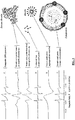
- FIG. 2 This exemplary procedure is schematically depicted in FIG. 2 .
- the embodiment of the present methods depicted in FIG. 2 includes: (1) placing the probe into a container, (2) mixing an aliquot of serum with the probe in the container, (3) mixing solvent with the aliquot and probe in the container, (4) placing the mixture (of aliquot, probe, and solvent) into a pipette (before or after incubation), (5) placing the pipette into, and analyzing the mixture with, the EPR spectrometer to obtain EPR spectra of the mixture, and (6) processing the measurements to obtain the concentrations of protein-bound and unbound probe, and determining the parameters of probe binding to hydrophobic sites of serum albumin in the serum of the aliquot.
- the affinity of albumin for 16-doxyl stearic acid may generally be similar to its affinity for unlabeled stearic acid (which may be relatively high, e.g., a binding constant of about 10 9 mol -1 ).
- affinity for unlabeled stearic acid which may be relatively high, e.g., a binding constant of about 10 9 mol -1 .
- the abundance of albumin relative to other serum proteins and the presence of several high-affinity binding sites for long chain fatty acids may result in 99% or more of the stearic acid probe being bound exclusively to albumin.
- an amount of the mixture can be placed into a glass capillary tube.
- the tube can then be inserted into an EPR spectrometer (e.g., Model No. EPR 01-08 available from MedInnovation GmbH, Wildau, Germany, or Model No. AXM-09 available from Scientific Production Enterprise Albutran, Minsk, Belarus).
- EPR spectrometer e.g., Model No. EPR 01-08 available from MedInnovation GmbH, Wildau, Germany, or Model No. AXM-09 available from Scientific Production Enterprise Albutran, Minsk, Zealand.
- the mixture is exposed to both a high magnetic field and microwave power. This exposure induces resonance of the spin label and absorption of microwave power.
- An EPR spectrum can thereby be generated by scanning measurements of the magnetic field strength and absorption of microwave power.
- EPR spectrometers such as conventional X-Band EPR spectrometers, or other EPR spectrometers operating with a microwave frequency of approximately 9-10 GHz, can be also used for obtaining these measurements.
- the sample can be maintained at 37°C during the measurement process to mimic physiologic conditions.
- the EPR spectrum obtained with the spin probe can be analyzed using a simulation process. Simulation can be performed using least-square fitting of a model spectrum to the measured spectrum. In this way, the EPR spectrum of the spin probe can be calculated using the appropriate model and parameters of the site where the spin probe is situated.
- the EPR spectrum obtained will generally consists of a large set of data points containing some amount of measurement noise or error. If the parameters of the binding site model are accurately established, an ideal experimental spectral curve can generally be derived. This task may be more complex when there are several sites that can bind the spin probe. In this situation, these different binding sites can be considered to improve accuracy when deriving the model spectrum.
- a number of different tools have been developed that enable the derivation of a composite model spectrum for compounds that possess several binding sites for the spin probe. Analysis of the EPR spectrum generated from the stearic acid probe bound to albumin (as described above) reveals five distinct spectral components. The major portion of the spectrum is represented by two components, as represented by lines B and C in FIG. 3 .
- Each of these two spectra components (B and C) is representative of the respective portions of fatty acid probe bound to the specific and non-specific hydrophobic sites of serum albumin (as may correspond to the pictorial representation of serum albumin molecules).
- the three remaining components (D, E, F), respectively, are representative of: unbound probe molecules present singularly in solution, as represented by line D in FIG. 3 (as may correspond to the pictorial representation of individual, unbound probe molecules); probe aggregated into clusters of fatty acid micelles, as represented by line E in FIG. 3 ; and probe associated with or bound to lipoproteins, as represented by line F in FIG. 3 .
- the process of simulation generally determines the values of ideal spectrum parameters representing the equation that provides the best curve fit of the simulated and measured spectra. These parameters can include the intensity of each spectral component as well as specific EPR parameters determining the position, width, and shape of spectrum lines.
- Each EPR spectrum reflects the structural and functional characteristics of the albumin that impact the binding of the probe to the specific (also referred to in some literature as "primary"), and the non-specific (also referred to in some literature as "secondary") sites of serum albumin.
- One technique that is employed for the generation of EPR spectra includes a sample mixture (of serum aliquot, probe, and solvent). The characteristics of the probe binding to albumin can be assessed from the EPR spectra resulting from the respective portions or concentrations of the fatty acid probe bound to specific and non-specific albumin sites, and the portion or concentration of unbound probe.
- Another characteristic that can be generated is an estimation of changes in albumin conformation (significant conformational changes are prevented and/or minimized in the present methods) at the albumin binding sites for fatty acids (certain parameters of the EPR spectrum indicate the mobility of the fatty acid probe at its binding sites on albumin); such mobility can be influenced by several parameters, and those parameters can be used to detect an absence or presence of significant conformational changes of albumin molecule due to albumin binding of toxins, acute-phase metabolites, and/or excessive concentration of a polar solvent in a sample mixture).
- Blood serum was obtained by whole blood centrifugation. An aliquot of 50 ⁇ l of serum from each patient was used for each test. A spin probe of 2-(14-carboxytetradecyl)-2-ethyl-4,4-dimethyl-3-oxazolidinyloxy (purchased from Fluorochem Ltd., Derbyshire, UK) was mixed into the aliquot at a concentration of 1.64 mmol/1. A solvent, 14 ⁇ l of ethanol, was mixed into the aliquot. The molar ratio of the probe to serum albumin in the aliquot was 3.5, based on an albumin concentration in the serum of 4 g/dL. The mixture was then incubated for 10 min at 37° C with continuous agitation in a standard shaker operated at about 5-8 Hz.
- the probe was placed into a glass capillary (e.g., Model No. RM-40, available from KABE LABORTECHNIK GmbH, Numbrecht-Elsenroth, Germany).
- a glass capillary e.g., Model No. RM-40, available from KABE LABORTECHNIK GmbH, Numbrecht-Elsenroth, Germany.
- the EPR spectrum of the mixture was then measured, as described below.
- the above-described capillary was placed into the resonator of an EPR spectrometer for spectroscopic analysis.
- the spectroscopy parameters were as follows: microwave power 15 mW at frequency 9.52 GHz; magnetic field 0.34 T (3400 G) with scan range 12 mT (120 G); modulation amplitude 0.2 mT (2 G); data accumulation by three scans each with 4096 measured points and a sweep time 60 s.
- the capillary temperature was 37° C, and was controlled within +/- 0.2° C.
- the EPR spectrum was analyzed by computer using an EPR-spectrum simulation with nonlinear least-squares fits.
- the fourth and fifth components represented fractions of the probe that were not related to albumin-bound or unbound-free probe, but to the probe molecules aggregated into micelles ( E ) and ones associated with lipoproteins ( F ).
- the considering of the E and F components in the spectrum model can improve accuracy of the analysis of concentrations of albumin-bound and unbound-free probes, but is not necessarily required in embodiments of the present methods.
- Patient A was diagnosed with a tumor of colon.
- the first sample was obtained at the time of Patient A entered the oncology clinic for colonoscopy. Relative concentration of probe micelles and lipoprotein-bound probe in the analyzed sample aliquot, parameter LM, was 13.8%.
- Probe binding parameter B2/F was determined to be reduced down to 5.1 compared the normal range of from 8.5 to 37.5, and parameter DP was increased to 2.1 compared the normal range of from -5.2 to -0.3.
- the second sample was derived after the time period of 12 days.
- the probe binding parameters were determines to be: LM of 13.6%, B2/F of 5.4 and DP of 2.1.
- the presence of the active malignant colon tumor was confirmed by biopsy.
- Patient B was diagnosed with cancer of sigmoid colon.
- relative LM was 15.1%
- parameter B2/F was 4.6
- DP was increased to 5.5.
- the second sample was derived after a period of 14 days.
- the second sample parameters were: LM of 16.7%, B2/F of 3.3, and DP of 12.5.
- the sigmoid colon cancer was confirmed by biopsy.
- Patient C was diagnosed with aggressive prostate cancer.
- LM was 15.8%
- parameter B2/F was 3.1
- DP was increased to 10.0.
- the patient's PSA was increased to 24.0.
- the second sample was derived after the time period of 14 days.
- the second sample parameters were: LM of 16%, B2/F of 5.2, and DP of 11.8.
- the aggressive prostate cancer was confirmed by biopsy.
- Patient D was diagnosed with breast cancer.
- LM was 14.8%
- B2/F was 4.6
- DP was 4.2
- the second sample was taken after 14 days.
- LM was 15.7%
- B2/F was 5.1
- DP was 4.3.
- the presence of breast tumor was confirmed by biopsy.
- Patient G suffered from acute respiratory disease during the period between the times of the obtaining of first and second samples.
- LM was 11.1 %
- B2/F was 5.3 (in the range of from 1.6 to 6.5 observed for patients with active malignancies)
- DP was 0.4 (in range of 0.1 to 66 observed in cancer patients).
- the second sample was derived 28 days later.
- LM was 8.6%
- B2/F was 9.0
- DP was - 1.9.
- the parameters in the second sample were in the normal ranges for cancer-free persons.
- kits including materials for performing the various steps described above.
- the kit includes an amount of labeled probe and an amount of solvent.
- the amounts of probe and solvent are such that when mixed with aliquot of serum having about a predetermined volume (e.g., 50, 60, 70, 80, 90, 100, or more ⁇ l), the mixture will achieve the results described above, and/or have the proportions of ingredients described above.
- the kit can include components for performing the various steps, as described herein. In some embodiments, the kit can further include instructions for performing the various steps or portions of the methods described above.
- the methods can comprise or be limited to any combination of the steps, features, and/or characteristics that have been described, unless the context explicitly or necessarily precludes the combination.
- one embodiment of the present method can include mixing probe with an aliquot and measuring the concentrations of albumin-bound and unbound probe; and another embodiment can include mixing probe and solvent with the aliquot, measuring the concentrations of albumin-bound and unbound probe, and normalizing the concentrations to substantially negate the changes in concentrations caused by the addition of the solvent.
- kits of the related (non-claimed) aspect can include a pipette and an amount of probe; another embodiment can include an amount of probe and an amount of solvent; and another embodiment can include an amount of probe, an amount of liquid solution, and an amount of solvent.
- Embodiments of the present methods can include measuring (e.g., indirectly) the concentrations of albumin-bound and unbound probe in a mixture (aliquot, probe, solvent) with EPR spectroscopy, fluorescent spectroscopy, or any other suitable methods described or otherwise known or developed in the art.
Landscapes
- Health & Medical Sciences (AREA)
- Life Sciences & Earth Sciences (AREA)
- Immunology (AREA)
- Engineering & Computer Science (AREA)
- Molecular Biology (AREA)
- Biomedical Technology (AREA)
- Chemical & Material Sciences (AREA)
- Hematology (AREA)
- Urology & Nephrology (AREA)
- Biotechnology (AREA)
- Microbiology (AREA)
- Cell Biology (AREA)
- Food Science & Technology (AREA)
- Medicinal Chemistry (AREA)
- Physics & Mathematics (AREA)
- Analytical Chemistry (AREA)
- Biochemistry (AREA)
- General Health & Medical Sciences (AREA)
- General Physics & Mathematics (AREA)
- Pathology (AREA)
- Investigating Or Analysing Biological Materials (AREA)
Claims (14)
- Verfahren zum Nachweisen von aktiver Malignität in einem Patientenindividuum, das Folgendes umfasst:Mischen einer markierten hydrophoben Sonde mit der Fähigkeit, an spezifische und unspezifische hydrophobe Bindungsstellen von Serumalbumin zu binden, mit einem aliquoten Teil einer ersten Probe einer serumalbuminhaltigen extrazellulären Flüssigkeit eines Patientenindividuums, wobei die Menge an Sonde derart ist, dass das molare Verhältnis der Sonde zu dem Serumalbumin zwischen 2 und 7 beträgt und wobei die Sonde gegebenenfalls zwei oder mehr Substanzen umfasst;Mischen eines Lösungsmittels mit dem aliquoten Teil der ersten Probe, wobei das Lösungsmittel derart ist, dass, wenn es zu dem aliquoten Teil und der Sonde zugegeben wird, die Löslichkeit der Sonde in dem aliquoten Teil erhöht wird, und wobei die Menge an Lösungsmittel ausreicht, um einen Anteil der Sonde von dem Serumalbumin zu dissoziieren, ohne zu wesentlicher Dissoziation von hydrophoben Metaboliten von dem Serumalbumin zu führen, wobei das Lösungsmittel gegebenenfalls zwei oder mehr Substanzen umfasst;Analysieren der Mischung aus aliquotem Anteil, Sonde und Lösungsmittel, um für die erste Probe einen oder mehrere Bindungsparameter der Sonde zu bestimmen, wobei das Analysieren der Mischung Folgendes umfasst:im Wesentlichen Ausschließen eines Anteils der Sonde, der an
Lipoproteine gebunden ist, und eines Anteils der Sonde, der sich zu Mizellen aggregiert hat, von der Mischung; undBestimmen des einen oder der mehreren Sondenbindungsparameter der Probe aus dem verbleibenden Anteil der Sonde in der Mischung; undVergleichen des einen oder der mehreren Bindungsparameter der ersten Probe mit einem oder mehreren Kontrollbindungsparametern einer Sonde, die an Serumalbumin in der extrazellulären Flüssigkeit von mindestens einem der Folgenden bindet: einem krebsfreien Kontrollpatienten und einem Kontrollpatienten mit aktiver Malignität. - Verfahren nach Anspruch 1, das weiterhin Folgendes umfasst:Mischen einer markierten hydrophoben Sonde mit der Fähigkeit, an spezifische und unspezifische hydrophobe Bindungsstellen von Serumalbumin zu binden, mit einem aliquoten Teil einer zweiten Probe einer serumalbuminhaltigen extrazellulären Flüssigkeit, die zwischen 2 und 90 Tage nach der ersten Probe von dem Patientenindividuum entnommen wurde, eines Patientenindividuums, wobei die Menge an Sonde derart ist, dass das molare Verhältnis der Sonde zu dem Serumalbumin zwischen 2 und 7 beträgt und wobei die Sonde gegebenenfalls zwei oder mehr Substanzen umfasst;Mischen eines Lösungsmittels mit dem aliquoten Teil der zweiten Probe, wobei das Lösungsmittel derart ist, dass, wenn es zu dem aliquoten Teil und der Sonde zugegeben wird, die Löslichkeit der Sonde in dem aliquoten Teil erhöht wird, und wobei die Menge an Lösungsmittel ausreicht, um einen Anteil der Sonde von dem Serumalbumin zu dissoziieren, ohne zu wesentlicher Dissoziation von hydrophoben Metaboliten von dem Serumalbumin zu führen, wobei das Lösungsmittel gegebenenfalls zwei oder mehr Substanzen umfasst;Analysieren der Mischung aus aliquotem Anteil, Sonde und Lösungsmittel, um für die zweite Probe einen oder mehrere Bindungsparameter der Sonde zu bestimmen, wobei das Analysieren der Mischung Folgendes umfasst:im Wesentlichen Ausschließen eines Anteils der Sonde, der an
Lipoproteine gebunden ist, und eines Anteils der Sonde, der sich zu Mizellen aggregiert hat, von der Mischung; undBestimmen des einen oder der mehreren Sondenbindungsparameter der Probe aus dem verbleibenden Anteil der Sonde in der Mischung; undVergleichen des einen oder der mehreren Bindungsparameter der zweiten Probe mit einem oder mehreren Kontrollbindungsparametern einer Sonde, die an Serumalbumin in der extrazellulären Flüssigkeit von mindestens einem der Folgenden bindet: einem krebsfreien Kontrollpatienten und einem Kontrollpatienten mit aktiver Malignität. - Verfahren nach Anspruch 2, das weiterhin Folgendes umfasst:Diagnostizieren einer aktiven Malignität in dem Patientenindividuum, wenn:(i) der eine oder die mehreren Bindungsparameter für jede der zwei Proben näher dem einen oder den mehreren Kontrollbindungsparametern für den mindestens einen Kontrollpatienten mit aktiver Malignität als dem einen oder der mehreren Kontrollbindungsparameter des krebsfreien Kontrollpatienten sind, und(ii) das Patientenindividuum keine Anzeichen von akuter Krankheit oder Infektion, die während den Probenahmezeitpunkten stattgefunden haben, aufweist.
- Verfahren nach Anspruch 3, wobei der eine oder die mehreren Sondenbindungsparameter jeder Probe aus dem Anteil der Sonde, die an spezifische und unspezifische hydrophobe Bindungsstellen von Albumin gebunden sind, und dem Anteil der ungebundenen Sonde bestimmt werden.
- Verfahren nach Anspruch 2, wobei die beiden Proben in einem Zeitabstand von zwischen 5 und 90 Tagen, zwischen 2 und 30 Tagen oder zwischen 5 und 30 Tagen von dem Patientenindividuum entnommen werden.
- Verfahren nach Anspruch 2, das weiterhin Folgendes umfasst:Verifizieren, dass das Patientenindividuum keine Anzeichen von akuten Krankheiten, die während den Zeitpunkten, an denen die zwei Proben von dem Patienten entnommen wurden, stattgefunden haben, aufweist.
- Verfahren nach Anspruch 2, wobei das Analysieren jeder Mischung Folgendes umfasst:Messen der Konzentrationen der Sonde, die an spezifische und unspezifische hydrophobe Stellen von Serumalbumin gebunden ist, und der ungebundenen Sonde in der Mischung; undAbleiten des einen oder der mehreren Sondenbindungsparameter von zumindest den Konzentrationen von: der an spezifische und unspezifische hydrophobe Bindungsstellen von Albumin gebundenen Sonde und der ungebundenen Sonde; und/oderwobei das Analysieren jeder Mischung das Verifizieren, dass die Mischung ihr Bindungsgleichgewicht erreicht hat, umfasst, gegebenenfalls mittels:Messen von zwei EPR-Spektren der Mischung zu zwei unterschiedlichen Zeitpunkten;Vergleichen der beiden EPR-Spektren, um zu bestimmen, ob Intensitäten von Spektrallinien (Spektralparametern) in den EPR-Spektren einander entsprechen.
- Verfahren nach einem der Ansprüche 2 bis 7, wobei die Sonde ein organisches Molekül mit zwischen 8 und 28 Kohlenstoffatomen umfasst und gegebenenfalls wobei die Sonde ein(e) oder mehrere der Folgenden umfasst: eine Fettsäure, eine langkettige Fettsäure, 16-DOXYL-Stearinsäure, oder ein Lysophospholipid, und/oder wobei die Sonde mit einem Spin-Marker markiert ist.
- Verfahren nach einem der Ansprüche 2 bis 8, wobei die Menge an Lösungsmittel, die mit dem aliquoten Teil und der Sonde gemischt wird, derart ist, dass das Lösungsmittel keine signifikanten Konformationsänderungen des Albumins induziert; und/oder wobei die Menge an Lösungsmittel, die mit dem aliquoten Teil und der Sonde gemischt wird, ausreicht, um die Konzentration an ungebundener Sonde in der Mischung aus dem aliquoten Teil, der Sonde und dem Lösungsmittel auf mindestens fünf Mal höher als die Konzentration an ungebundener Sonde in einer Mischung aus dem aliquoten Teil und der Sonde ohne das Lösungsmittel zu erhöhen; und/oder wobei das Volumen des Lösungsmittels, das mit dem aliquoten Teil gemischt wird, weniger als 30% des Volumens des aliquoten Teil und gegebenenfalls weniger als 20% des Volumens des aliquoten Teils beträgt.
- Verfahren nach einem der Ansprüche 2 bis 9, das weiterhin Folgendes umfasst:Normalisieren des einen oder der mehreren Sondenbindungsparameter jeder Probe, um der durch das Lösungsmittel in der Mischung verursachten Reduktion der Albuminkonzentration Rechnung zu tragen;gegebenenfalls wobei die Schritte des Ableitens und Normalisierens des einen oder der mehreren Sondenbindungsparameter gleichzeitig erfolgen; und/oderNormalisieren des einen oder der mehreren Sondenbindungsparameter jeder Probe, um der durch medizinische Zustände des Patienten verursachten Reduktion der Serumalbuminkonzentration Rechnung zu tragen.
- Verfahren nach einem der Ansprüche 2 bis 10, wobei der eine oder die mehreren Kontrollparameter mindestens einen Bereich von Kontrollparametern von Kontrollpatienten umfassen.
- Verfahren nach Anspruch 7, das weiterhin Folgendes umfasst:Wiederholen der Schritte des Mischens einer Sonde, des Mischens eines Lösungsmittels, des Messens der Konzentrationen,
des Ableitens von einem oder mehreren Sonden von einem oder mehreren Sondenbindungsparametern für jeden von zwei oder mehr aliquoten Teilen, wobei für jede der zwei oder mehr Wiederholungen eine unterschiedliche Lösungsmenge verwendet wird; gegebenenfalls wobei für den einen oder die mehreren Sondenbindungsparameter, der/die für die zwei oder mehr Wiederholungen abgeleitet werden, der Durchschnitt berechnet wird, um einen durchschnittlichen Sondenbindungsparameter abzuleiten, und wobei der eine oder die mehreren durchschnittliche(n) Sondenbindungsparameter in dem Schritt des Vergleichens des einen oder der mehreren Sondenbindungsparameter verglichen werden. - Verfahren nach einem der Ansprüche 2 bis 12, wobei das Volumen des aliquoten Teils weniger als 100 µl und gegebenenfalls weniger als 50 µl beträgt.
- Verfahren nach einem der Ansprüche 2 bis 13, wobei die extrazelluläre Flüssigkeit mindestens eine(s) der Folgenden beinhaltet: Blutserum, Blutplasma, Lymphflüssigkeit und Spinalflüssigkeit.
Applications Claiming Priority (2)
| Application Number | Priority Date | Filing Date | Title |
|---|---|---|---|
| US201261600944P | 2012-02-20 | 2012-02-20 | |
| PCT/EP2013/053389 WO2013124325A1 (en) | 2012-02-20 | 2013-02-20 | Methods and kits for detection of active malignancy |
Publications (2)
| Publication Number | Publication Date |
|---|---|
| EP2817626A1 EP2817626A1 (de) | 2014-12-31 |
| EP2817626B1 true EP2817626B1 (de) | 2017-09-27 |
Family
ID=47891603
Family Applications (1)
| Application Number | Title | Priority Date | Filing Date |
|---|---|---|---|
| EP13709786.1A Revoked EP2817626B1 (de) | 2012-02-20 | 2013-02-20 | Verfahren zum Nachweis aktiver Malignitäten |
Country Status (3)
| Country | Link |
|---|---|
| US (1) | US9528992B2 (de) |
| EP (1) | EP2817626B1 (de) |
| WO (1) | WO2013124325A1 (de) |
Families Citing this family (4)
| Publication number | Priority date | Publication date | Assignee | Title |
|---|---|---|---|---|
| US9528992B2 (en) | 2012-02-20 | 2016-12-27 | Uladzimir A. Murauski | Methods and kits for detection of active malignancy |
| CN112930568A (zh) * | 2019-04-23 | 2021-06-08 | 艾斯皮雷科技股份有限公司 | 用于定位或识别恶性肿瘤的装置和方法 |
| JP7356625B2 (ja) * | 2019-07-08 | 2023-10-05 | 国立大学法人大阪大学 | 癌患者の予後予測方法、抗癌療法の有効性の予測方法および癌患者に対する適切な療法の選択方法 |
| EP3786642A1 (de) * | 2019-08-30 | 2021-03-03 | ESPIRE Technologies GmbH | Verfahren für funktionelle eigenschaften von proteinen zur klassifikation von patienten, die einer spezifischen erkrankung zuzuordnen sind |
Citations (3)
| Publication number | Priority date | Publication date | Assignee | Title |
|---|---|---|---|---|
| WO2001065270A2 (de) | 2000-02-28 | 2001-09-07 | E.W. Handels- Und Consulting Gmbh | Verfahren zur esr-spektroskopischen bestimmung von veränderungen der transporteigenschaften des albumins in einer albuminhaltigen probe, esr-spektrometer zur durchführung des verfahrens und einsatz des verfahrens zur diagnose und zur kontrolle albuminhaltiger präparate |
| WO2010049010A1 (en) | 2008-10-28 | 2010-05-06 | Murauski Uladzimir A | Methods and kits for detection of toxemia |
| WO2013124325A1 (en) | 2012-02-20 | 2013-08-29 | Murauski Uladzimir A | Methods and kits for detection of active malignancy |
Family Cites Families (18)
| Publication number | Priority date | Publication date | Assignee | Title |
|---|---|---|---|---|
| SU1459656A1 (ru) | 1985-10-04 | 1989-02-23 | Белорусский Научно-Исследовательский Институт Туберкулеза | Способ диагностики токсемии |
| US5308604A (en) | 1989-04-19 | 1994-05-03 | Deutsches Krebsforschungsinstitut | Conjugates for tumor localization and/or tumor therapy |
| US5183740A (en) | 1990-02-23 | 1993-02-02 | The United States Of America As Represented By The Secretary Of The Navy | Flow immunosensor method and apparatus |
| GB9304372D0 (en) | 1993-03-04 | 1993-04-21 | Birbeck College | Detection of albumin in physiological fluids |
| US6458758B1 (en) | 1993-08-16 | 2002-10-01 | Synzyme Technologies, Inc. | Compositions and methods utilizing nitroxides in combination with biocompatible macromolecules |
| US6589751B2 (en) | 1997-11-04 | 2003-07-08 | Mcw Research Foundation | Immonological detection of the homocystamide adduct and a thiolactone immunoassay for endogeneous homocysteine |
| EP0973043A1 (de) | 1998-07-13 | 2000-01-19 | Newpharm & Trading SA | Methode zur Diagnose von malignen Neoplasmen auf der Grundlage von ESR-Spektroskopie |
| US7070937B1 (en) | 1998-10-02 | 2006-07-04 | Ischemia Technologies, Inc. | Marker useful for detection and measurement of free radical damage and method |
| US6306576B1 (en) | 1999-02-19 | 2001-10-23 | Cleveland Clinic Foundation | Diagnostic methods for asthma |
| PT1384075E (pt) | 2001-03-28 | 2010-03-23 | Heska Corp | Métodos de detecção de doença renal inicial em animais |
| WO2002097038A2 (en) | 2001-05-25 | 2002-12-05 | Human Genome Sciences, Inc. | Chemokine beta-1 fusion proteins |
| CA2448719A1 (en) | 2001-06-09 | 2002-12-19 | Glsynthesis Inc. | Lipid structures and uses thereof |
| EP2261250B1 (de) | 2001-12-21 | 2015-07-01 | Human Genome Sciences, Inc. | GCSF-Albumin Fusionsproteine |
| AU2003244515A1 (en) | 2002-02-07 | 2003-09-02 | Novozymes Delta Limited | Albumin-fused anti-angiogenesis peptides |
| US6989369B2 (en) | 2003-02-07 | 2006-01-24 | Dyax Corp. | Kunitz domain peptides |
| WO2007041596A2 (en) | 2005-10-03 | 2007-04-12 | The General Hospital Corporation | Compositions and methods for the treatment of cancer |
| US7794952B2 (en) | 2005-12-15 | 2010-09-14 | George Mason University | Biomarker isolation and use thereof to characterize physiological state |
| CA2634329A1 (en) | 2005-12-22 | 2007-07-19 | Csl Behring Gmbh | Octanoate-reduced human albumin |
-
2013
- 2013-02-20 US US14/380,032 patent/US9528992B2/en not_active Expired - Fee Related
- 2013-02-20 EP EP13709786.1A patent/EP2817626B1/de not_active Revoked
- 2013-02-20 WO PCT/EP2013/053389 patent/WO2013124325A1/en not_active Ceased
Patent Citations (3)
| Publication number | Priority date | Publication date | Assignee | Title |
|---|---|---|---|---|
| WO2001065270A2 (de) | 2000-02-28 | 2001-09-07 | E.W. Handels- Und Consulting Gmbh | Verfahren zur esr-spektroskopischen bestimmung von veränderungen der transporteigenschaften des albumins in einer albuminhaltigen probe, esr-spektrometer zur durchführung des verfahrens und einsatz des verfahrens zur diagnose und zur kontrolle albuminhaltiger präparate |
| WO2010049010A1 (en) | 2008-10-28 | 2010-05-06 | Murauski Uladzimir A | Methods and kits for detection of toxemia |
| WO2013124325A1 (en) | 2012-02-20 | 2013-08-29 | Murauski Uladzimir A | Methods and kits for detection of active malignancy |
Non-Patent Citations (9)
| Title |
|---|
| A. GURACHEVSKY ET AL.: "Application of spin label electron paramagnetic resonance in the diagnosis and prognosis of cancer and sepsis", CLIN. CHEM. LAB MED, vol. 46, no. 9, 2008, pages 1203 - 1210, XP003030874 |
| D. HORLITZ: "Bindungsparameter verschiedener Medikamente im humanen Serumalbumin", BIBLIOTHEK SIGNATUR 0BB16/06(09, May 2009 (2009-05-01), XP055512424 |
| K. REHBERG: "Automatisierung der MMS-Test-Analyse", BIBLIOTHEK BB 22/06(09, 2009, XP055512434 |
| K. SCHNURR ET AL.: "A new approach in Cancer diagnostics: Analysis of functional properties of albumin", BIOMEDIZINISCHE TECHNIK, vol. 49, no. 2, 2004, pages 434 - 435, XP055512420 |
| K. SCHNURR ET AL.: "Krebsdiagnostik durch funktionelle Analyse von Serumalbumin", BLITZLICHT TUMORDETEKTION, vol. 4, 2009, pages 22 - 24, XP055512279 |
| K. SCHNURR ET AL.: "The analysis of functional characteristics of serum albumin - a novel possibility of early cancer diagnosis", METHODS, POSSIBILITIES AND PERSPECTIVES OF PRE-SYMPTOMATIC TUMOR DIAGNOSIS, vol. 1, 2005, Lengerich, pages 225 - 234, XP055512350 |
| M. GELOS ET AL.: "Analysis of albumin fatty acid binding capacity in patients with benign and malignat colorectal diseases using electron spin resonance (ESR) spectroscopy", INT. J. COLORECTAL DIS., vol. 24, no. 1, 31 July 2009 (2009-07-31), pages 119 - 127, XP019759475 |
| P. SEIDEL ET AL.: "Recognition of malignant processes with neutral nets from ESR spectra of serum albumin", Z. MED. PHYS., vol. 15, no. 4, 2005, pages 265 - 272, XP055512353 |
| S.C. KAZMIERCZAK ET AL.: "Electron spin resonance spectroscopy of serum albumin: A novel new test for cancer diagnosis and monitoring", CLINICAL CHEMISTRY, vol. 52, no. 11, 2006, pages 2129 - 2134, XP055512356 |
Also Published As
| Publication number | Publication date |
|---|---|
| EP2817626A1 (de) | 2014-12-31 |
| US9528992B2 (en) | 2016-12-27 |
| US20150037907A1 (en) | 2015-02-05 |
| WO2013124325A1 (en) | 2013-08-29 |
Similar Documents
| Publication | Publication Date | Title |
|---|---|---|
| Koster et al. | Surface enhanced Raman scattering of extracellular vesicles for cancer diagnostics despite isolation dependent lipoprotein contamination | |
| Wang et al. | Detection of metabolite–protein interactions in complex biological samples by high-resolution Relaxometry: toward Interactomics by NMR | |
| Monleón et al. | Benign and atypical meningioma metabolic signatures by high-resolution magic-angle spinning molecular profiling | |
| Tata et al. | Contrast agent mass spectrometry imaging reveals tumor heterogeneity | |
| EP2817626B1 (de) | Verfahren zum Nachweis aktiver Malignitäten | |
| Gogiashvili et al. | Impact of intratumoral heterogeneity of breast cancer tissue on quantitative metabolomics using high‐resolution magic angle spinning 1H NMR spectroscopy | |
| Zennaro et al. | Metabonomics by proton nuclear magnetic resonance in human pleural effusions: A route to discriminate between benign and malignant pleural effusions and to target small molecules as potential cancer biomarkers | |
| Nagana Gowda et al. | Visualization of bile homeostasis using 1H-NMR spectroscopy as a route for assessing liver cancer | |
| WO2011019864A2 (en) | Methods for mobile zinc measurement | |
| US20220044814A1 (en) | Device and method for localising or identifying malignancies | |
| US20170242017A1 (en) | Methods and devices for determining a disease state | |
| Duarte et al. | Can nuclear magnetic resonance (NMR) spectroscopy reveal different metabolic signatures for lung tumours? | |
| Cox et al. | Current and future applications of in vitro magnetic resonance spectroscopy in hepatobiliary disease | |
| Piotto et al. | 23 Reproducible Sample Preparation and Spectrum Acquisition Techniques for Metabolic Profiling of Human Tissues by Proton High-Resolution Magic Angle Spinning Nuclear Magnetic Resonance | |
| Sharma et al. | Breast cancer metabolomics using NMR | |
| Babourina-Brooks et al. | Glycine: a non-invasive imaging biomarker to aid magnetic resonance spectroscopy in the prediction of survival in paediatric brain tumours | |
| Gelos et al. | Analysis of albumin fatty acid binding capacity in patients with benign and malignant colorectal diseases using electron spin resonance (ESR) spectroscopy | |
| EP2353010B1 (de) | Verfahren zum nachweis einer toxämie | |
| Gurachevsky et al. | Application of spin label electron paramagnetic resonance in the diagnosis and prognosis of cancer and sepsis. | |
| Li et al. | Serum metabolomic analysis of human upper urinary tract urothelial carcinoma | |
| US20090030618A1 (en) | System, method and software arrangement for analyzing and correlating molecular profiles associated with anatomical structures | |
| CN109061179A (zh) | 氨基酸联合因子在构建结直肠癌血液学诊断模型中的应用 | |
| CN105555318A (zh) | 将胸膜液分类的方法 | |
| Schneider et al. | Sample processing considerations for protein stability Studies of low concentration biofluid samples using differential scanning calorimetry | |
| Sharma et al. | MR spectroscopy in breast cancer metabolomics |
Legal Events
| Date | Code | Title | Description |
|---|---|---|---|
| PUAI | Public reference made under article 153(3) epc to a published international application that has entered the european phase |
Free format text: ORIGINAL CODE: 0009012 |
|
| 17P | Request for examination filed |
Effective date: 20140826 |
|
| AK | Designated contracting states |
Kind code of ref document: A1 Designated state(s): AL AT BE BG CH CY CZ DE DK EE ES FI FR GB GR HR HU IE IS IT LI LT LU LV MC MK MT NL NO PL PT RO RS SE SI SK SM TR |
|
| AX | Request for extension of the european patent |
Extension state: BA ME |
|
| DAX | Request for extension of the european patent (deleted) | ||
| 17Q | First examination report despatched |
Effective date: 20150901 |
|
| GRAP | Despatch of communication of intention to grant a patent |
Free format text: ORIGINAL CODE: EPIDOSNIGR1 |
|
| INTG | Intention to grant announced |
Effective date: 20160913 |
|
| STAA | Information on the status of an ep patent application or granted ep patent |
Free format text: STATUS: GRANT OF PATENT IS INTENDED |
|
| GRAS | Grant fee paid |
Free format text: ORIGINAL CODE: EPIDOSNIGR3 |
|
| GRAA | (expected) grant |
Free format text: ORIGINAL CODE: 0009210 |
|
| STAA | Information on the status of an ep patent application or granted ep patent |
Free format text: STATUS: THE PATENT HAS BEEN GRANTED |
|
| AK | Designated contracting states |
Kind code of ref document: B1 Designated state(s): AL AT BE BG CH CY CZ DE DK EE ES FI FR GB GR HR HU IE IS IT LI LT LU LV MC MK MT NL NO PL PT RO RS SE SI SK SM TR |
|
| REG | Reference to a national code |
Ref country code: GB Ref legal event code: FG4D |
|
| REG | Reference to a national code |
Ref country code: CH Ref legal event code: EP |
|
| REG | Reference to a national code |
Ref country code: AT Ref legal event code: REF Ref document number: 932430 Country of ref document: AT Kind code of ref document: T Effective date: 20171015 |
|
| REG | Reference to a national code |
Ref country code: IE Ref legal event code: FG4D |
|
| REG | Reference to a national code |
Ref country code: DE Ref legal event code: R096 Ref document number: 602013027103 Country of ref document: DE |
|
| PG25 | Lapsed in a contracting state [announced via postgrant information from national office to epo] |
Ref country code: HR Free format text: LAPSE BECAUSE OF FAILURE TO SUBMIT A TRANSLATION OF THE DESCRIPTION OR TO PAY THE FEE WITHIN THE PRESCRIBED TIME-LIMIT Effective date: 20170927 Ref country code: NO Free format text: LAPSE BECAUSE OF FAILURE TO SUBMIT A TRANSLATION OF THE DESCRIPTION OR TO PAY THE FEE WITHIN THE PRESCRIBED TIME-LIMIT Effective date: 20171227 Ref country code: SE Free format text: LAPSE BECAUSE OF FAILURE TO SUBMIT A TRANSLATION OF THE DESCRIPTION OR TO PAY THE FEE WITHIN THE PRESCRIBED TIME-LIMIT Effective date: 20170927 Ref country code: LT Free format text: LAPSE BECAUSE OF FAILURE TO SUBMIT A TRANSLATION OF THE DESCRIPTION OR TO PAY THE FEE WITHIN THE PRESCRIBED TIME-LIMIT Effective date: 20170927 Ref country code: FI Free format text: LAPSE BECAUSE OF FAILURE TO SUBMIT A TRANSLATION OF THE DESCRIPTION OR TO PAY THE FEE WITHIN THE PRESCRIBED TIME-LIMIT Effective date: 20170927 |
|
| REG | Reference to a national code |
Ref country code: NL Ref legal event code: MP Effective date: 20170927 |
|
| REG | Reference to a national code |
Ref country code: LT Ref legal event code: MG4D |
|
| REG | Reference to a national code |
Ref country code: AT Ref legal event code: MK05 Ref document number: 932430 Country of ref document: AT Kind code of ref document: T Effective date: 20170927 |
|
| REG | Reference to a national code |
Ref country code: FR Ref legal event code: PLFP Year of fee payment: 6 |
|
| PG25 | Lapsed in a contracting state [announced via postgrant information from national office to epo] |
Ref country code: RS Free format text: LAPSE BECAUSE OF FAILURE TO SUBMIT A TRANSLATION OF THE DESCRIPTION OR TO PAY THE FEE WITHIN THE PRESCRIBED TIME-LIMIT Effective date: 20170927 Ref country code: LV Free format text: LAPSE BECAUSE OF FAILURE TO SUBMIT A TRANSLATION OF THE DESCRIPTION OR TO PAY THE FEE WITHIN THE PRESCRIBED TIME-LIMIT Effective date: 20170927 Ref country code: BG Free format text: LAPSE BECAUSE OF FAILURE TO SUBMIT A TRANSLATION OF THE DESCRIPTION OR TO PAY THE FEE WITHIN THE PRESCRIBED TIME-LIMIT Effective date: 20171227 Ref country code: GR Free format text: LAPSE BECAUSE OF FAILURE TO SUBMIT A TRANSLATION OF THE DESCRIPTION OR TO PAY THE FEE WITHIN THE PRESCRIBED TIME-LIMIT Effective date: 20171228 |
|
| PG25 | Lapsed in a contracting state [announced via postgrant information from national office to epo] |
Ref country code: NL Free format text: LAPSE BECAUSE OF FAILURE TO SUBMIT A TRANSLATION OF THE DESCRIPTION OR TO PAY THE FEE WITHIN THE PRESCRIBED TIME-LIMIT Effective date: 20170927 |
|
| PG25 | Lapsed in a contracting state [announced via postgrant information from national office to epo] |
Ref country code: CZ Free format text: LAPSE BECAUSE OF FAILURE TO SUBMIT A TRANSLATION OF THE DESCRIPTION OR TO PAY THE FEE WITHIN THE PRESCRIBED TIME-LIMIT Effective date: 20170927 Ref country code: RO Free format text: LAPSE BECAUSE OF FAILURE TO SUBMIT A TRANSLATION OF THE DESCRIPTION OR TO PAY THE FEE WITHIN THE PRESCRIBED TIME-LIMIT Effective date: 20170927 Ref country code: ES Free format text: LAPSE BECAUSE OF FAILURE TO SUBMIT A TRANSLATION OF THE DESCRIPTION OR TO PAY THE FEE WITHIN THE PRESCRIBED TIME-LIMIT Effective date: 20170927 |
|
| PG25 | Lapsed in a contracting state [announced via postgrant information from national office to epo] |
Ref country code: EE Free format text: LAPSE BECAUSE OF FAILURE TO SUBMIT A TRANSLATION OF THE DESCRIPTION OR TO PAY THE FEE WITHIN THE PRESCRIBED TIME-LIMIT Effective date: 20170927 Ref country code: SM Free format text: LAPSE BECAUSE OF FAILURE TO SUBMIT A TRANSLATION OF THE DESCRIPTION OR TO PAY THE FEE WITHIN THE PRESCRIBED TIME-LIMIT Effective date: 20170927 Ref country code: SK Free format text: LAPSE BECAUSE OF FAILURE TO SUBMIT A TRANSLATION OF THE DESCRIPTION OR TO PAY THE FEE WITHIN THE PRESCRIBED TIME-LIMIT Effective date: 20170927 Ref country code: IT Free format text: LAPSE BECAUSE OF FAILURE TO SUBMIT A TRANSLATION OF THE DESCRIPTION OR TO PAY THE FEE WITHIN THE PRESCRIBED TIME-LIMIT Effective date: 20170927 Ref country code: IS Free format text: LAPSE BECAUSE OF FAILURE TO SUBMIT A TRANSLATION OF THE DESCRIPTION OR TO PAY THE FEE WITHIN THE PRESCRIBED TIME-LIMIT Effective date: 20180127 Ref country code: AT Free format text: LAPSE BECAUSE OF FAILURE TO SUBMIT A TRANSLATION OF THE DESCRIPTION OR TO PAY THE FEE WITHIN THE PRESCRIBED TIME-LIMIT Effective date: 20170927 |
|
| REG | Reference to a national code |
Ref country code: DE Ref legal event code: R026 Ref document number: 602013027103 Country of ref document: DE |
|
| PLBI | Opposition filed |
Free format text: ORIGINAL CODE: 0009260 |
|
| PLAX | Notice of opposition and request to file observation + time limit sent |
Free format text: ORIGINAL CODE: EPIDOSNOBS2 |
|
| PG25 | Lapsed in a contracting state [announced via postgrant information from national office to epo] |
Ref country code: DK Free format text: LAPSE BECAUSE OF FAILURE TO SUBMIT A TRANSLATION OF THE DESCRIPTION OR TO PAY THE FEE WITHIN THE PRESCRIBED TIME-LIMIT Effective date: 20170927 |
|
| 26 | Opposition filed |
Opponent name: GULDE & PARTNER Effective date: 20180626 |
|
| PG25 | Lapsed in a contracting state [announced via postgrant information from national office to epo] |
Ref country code: PL Free format text: LAPSE BECAUSE OF FAILURE TO SUBMIT A TRANSLATION OF THE DESCRIPTION OR TO PAY THE FEE WITHIN THE PRESCRIBED TIME-LIMIT Effective date: 20170927 |
|
| REG | Reference to a national code |
Ref country code: CH Ref legal event code: PL |
|
| PG25 | Lapsed in a contracting state [announced via postgrant information from national office to epo] |
Ref country code: MC Free format text: LAPSE BECAUSE OF FAILURE TO SUBMIT A TRANSLATION OF THE DESCRIPTION OR TO PAY THE FEE WITHIN THE PRESCRIBED TIME-LIMIT Effective date: 20170927 |
|
| PLBB | Reply of patent proprietor to notice(s) of opposition received |
Free format text: ORIGINAL CODE: EPIDOSNOBS3 |
|
| REG | Reference to a national code |
Ref country code: IE Ref legal event code: MM4A |
|
| REG | Reference to a national code |
Ref country code: BE Ref legal event code: MM Effective date: 20180228 |
|
| PG25 | Lapsed in a contracting state [announced via postgrant information from national office to epo] |
Ref country code: CH Free format text: LAPSE BECAUSE OF NON-PAYMENT OF DUE FEES Effective date: 20180228 Ref country code: LU Free format text: LAPSE BECAUSE OF NON-PAYMENT OF DUE FEES Effective date: 20180220 Ref country code: LI Free format text: LAPSE BECAUSE OF NON-PAYMENT OF DUE FEES Effective date: 20180228 Ref country code: SI Free format text: LAPSE BECAUSE OF FAILURE TO SUBMIT A TRANSLATION OF THE DESCRIPTION OR TO PAY THE FEE WITHIN THE PRESCRIBED TIME-LIMIT Effective date: 20170927 |
|
| PG25 | Lapsed in a contracting state [announced via postgrant information from national office to epo] |
Ref country code: IE Free format text: LAPSE BECAUSE OF NON-PAYMENT OF DUE FEES Effective date: 20180220 |
|
| PG25 | Lapsed in a contracting state [announced via postgrant information from national office to epo] |
Ref country code: BE Free format text: LAPSE BECAUSE OF NON-PAYMENT OF DUE FEES Effective date: 20180228 |
|
| PGFP | Annual fee paid to national office [announced via postgrant information from national office to epo] |
Ref country code: DE Payment date: 20190222 Year of fee payment: 7 Ref country code: GB Payment date: 20190221 Year of fee payment: 7 |
|
| PGFP | Annual fee paid to national office [announced via postgrant information from national office to epo] |
Ref country code: FR Payment date: 20190221 Year of fee payment: 7 |
|
| PG25 | Lapsed in a contracting state [announced via postgrant information from national office to epo] |
Ref country code: MT Free format text: LAPSE BECAUSE OF NON-PAYMENT OF DUE FEES Effective date: 20180220 |
|
| PG25 | Lapsed in a contracting state [announced via postgrant information from national office to epo] |
Ref country code: TR Free format text: LAPSE BECAUSE OF FAILURE TO SUBMIT A TRANSLATION OF THE DESCRIPTION OR TO PAY THE FEE WITHIN THE PRESCRIBED TIME-LIMIT Effective date: 20170927 |
|
| PG25 | Lapsed in a contracting state [announced via postgrant information from national office to epo] |
Ref country code: HU Free format text: LAPSE BECAUSE OF FAILURE TO SUBMIT A TRANSLATION OF THE DESCRIPTION OR TO PAY THE FEE WITHIN THE PRESCRIBED TIME-LIMIT; INVALID AB INITIO Effective date: 20130220 Ref country code: PT Free format text: LAPSE BECAUSE OF FAILURE TO SUBMIT A TRANSLATION OF THE DESCRIPTION OR TO PAY THE FEE WITHIN THE PRESCRIBED TIME-LIMIT Effective date: 20170927 |
|
| PG25 | Lapsed in a contracting state [announced via postgrant information from national office to epo] |
Ref country code: CY Free format text: LAPSE BECAUSE OF FAILURE TO SUBMIT A TRANSLATION OF THE DESCRIPTION OR TO PAY THE FEE WITHIN THE PRESCRIBED TIME-LIMIT Effective date: 20170927 Ref country code: MK Free format text: LAPSE BECAUSE OF NON-PAYMENT OF DUE FEES Effective date: 20170927 |
|
| RDAF | Communication despatched that patent is revoked |
Free format text: ORIGINAL CODE: EPIDOSNREV1 |
|
| PG25 | Lapsed in a contracting state [announced via postgrant information from national office to epo] |
Ref country code: AL Free format text: LAPSE BECAUSE OF FAILURE TO SUBMIT A TRANSLATION OF THE DESCRIPTION OR TO PAY THE FEE WITHIN THE PRESCRIBED TIME-LIMIT Effective date: 20170927 |
|
| REG | Reference to a national code |
Ref country code: DE Ref legal event code: R064 Ref document number: 602013027103 Country of ref document: DE Ref country code: DE Ref legal event code: R103 Ref document number: 602013027103 Country of ref document: DE |
|
| REG | Reference to a national code |
Ref country code: DE Ref legal event code: R119 Ref document number: 602013027103 Country of ref document: DE |
|
| GBPC | Gb: european patent ceased through non-payment of renewal fee |
Effective date: 20200220 |
|
| PUAJ | Public notification under rule 129 epc |
Free format text: ORIGINAL CODE: 0009425 |
|
| 32PN | Public notification |
Free format text: COMMUNICATION IN OPPOSITION PROCEEDINGS (EPO FORMS 2911, 2936, 2331,2916, 2019) DATED 23.09.2020 |
|
| PG25 | Lapsed in a contracting state [announced via postgrant information from national office to epo] |
Ref country code: FR Free format text: LAPSE BECAUSE OF NON-PAYMENT OF DUE FEES Effective date: 20200228 Ref country code: DE Free format text: LAPSE BECAUSE OF NON-PAYMENT OF DUE FEES Effective date: 20200901 Ref country code: GB Free format text: LAPSE BECAUSE OF NON-PAYMENT OF DUE FEES Effective date: 20200220 |
|
| PLAB | Opposition data, opponent's data or that of the opponent's representative modified |
Free format text: ORIGINAL CODE: 0009299OPPO |
|
| RDAG | Patent revoked |
Free format text: ORIGINAL CODE: 0009271 |
|
| STAA | Information on the status of an ep patent application or granted ep patent |
Free format text: STATUS: PATENT REVOKED |
|
| R26 | Opposition filed (corrected) |
Opponent name: GULDE & PARTNER Effective date: 20180626 |
|
| 27W | Patent revoked |
Effective date: 20200806 |
|
| REG | Reference to a national code |
Ref country code: FI Ref legal event code: MGE |