EP3012321B1 - Menschliches zellmodell für gm1-gangliosidose und verwendung davon - Google Patents
Menschliches zellmodell für gm1-gangliosidose und verwendung davon Download PDFInfo
- Publication number
- EP3012321B1 EP3012321B1 EP14880388.5A EP14880388A EP3012321B1 EP 3012321 B1 EP3012321 B1 EP 3012321B1 EP 14880388 A EP14880388 A EP 14880388A EP 3012321 B1 EP3012321 B1 EP 3012321B1
- Authority
- EP
- European Patent Office
- Prior art keywords
- ipscs
- npcs
- cells
- gal
- gene
- Prior art date
- Legal status (The legal status is an assumption and is not a legal conclusion. Google has not performed a legal analysis and makes no representation as to the accuracy of the status listed.)
- Not-in-force
Links
- 201000008892 GM1 Gangliosidosis Diseases 0.000 title claims description 232
- 210000005260 human cell Anatomy 0.000 title description 13
- 210000004027 cell Anatomy 0.000 claims description 162
- 230000014509 gene expression Effects 0.000 claims description 132
- 108010005774 beta-Galactosidase Proteins 0.000 claims description 114
- 108090000623 proteins and genes Proteins 0.000 claims description 111
- 210000005155 neural progenitor cell Anatomy 0.000 claims description 85
- WQZGKKKJIJFFOK-FPRJBGLDSA-N beta-D-galactose Chemical compound OC[C@H]1O[C@@H](O)[C@H](O)[C@@H](O)[C@H]1O WQZGKKKJIJFFOK-FPRJBGLDSA-N 0.000 claims description 78
- 230000035772 mutation Effects 0.000 claims description 63
- 230000000694 effects Effects 0.000 claims description 57
- 102000004169 proteins and genes Human genes 0.000 claims description 46
- 230000001537 neural effect Effects 0.000 claims description 41
- 238000000034 method Methods 0.000 claims description 40
- 101150091511 glb-1 gene Proteins 0.000 claims description 37
- QPJBWNIQKHGLAU-IQZHVAEDSA-N ganglioside GM1 Chemical compound O[C@@H]1[C@@H](O)[C@H](OC[C@H](NC(=O)CCCCCCCCCCCCCCCCC)[C@H](O)\C=C\CCCCCCCCCCCCC)O[C@H](CO)[C@H]1O[C@H]1[C@H](O)[C@@H](O[C@]2(O[C@H]([C@H](NC(C)=O)[C@@H](O)C2)[C@H](O)[C@H](O)CO)C(O)=O)[C@@H](O[C@H]2[C@@H]([C@@H](O[C@H]3[C@@H]([C@@H](O)[C@@H](O)[C@@H](CO)O3)O)[C@@H](O)[C@@H](CO)O2)NC(C)=O)[C@@H](CO)O1 QPJBWNIQKHGLAU-IQZHVAEDSA-N 0.000 claims description 35
- 230000007423 decrease Effects 0.000 claims description 32
- 239000003550 marker Substances 0.000 claims description 32
- 238000009825 accumulation Methods 0.000 claims description 31
- 102100026189 Beta-galactosidase Human genes 0.000 claims description 29
- 210000003712 lysosome Anatomy 0.000 claims description 29
- 230000001868 lysosomic effect Effects 0.000 claims description 29
- 230000003827 upregulation Effects 0.000 claims description 24
- 230000015572 biosynthetic process Effects 0.000 claims description 22
- 102200154287 rs11554273 Human genes 0.000 claims description 18
- 206010061218 Inflammation Diseases 0.000 claims description 17
- 230000004054 inflammatory process Effects 0.000 claims description 17
- 102000008730 Nestin Human genes 0.000 claims description 14
- 108010088225 Nestin Proteins 0.000 claims description 14
- 239000003795 chemical substances by application Substances 0.000 claims description 13
- 210000005055 nestin Anatomy 0.000 claims description 13
- 102100030634 Homeobox protein OTX2 Human genes 0.000 claims description 9
- 101000584400 Homo sapiens Homeobox protein OTX2 Proteins 0.000 claims description 9
- 101000979001 Homo sapiens Methionine aminopeptidase 2 Proteins 0.000 claims description 9
- 101000969087 Homo sapiens Microtubule-associated protein 2 Proteins 0.000 claims description 9
- 108010069196 Neural Cell Adhesion Molecules Proteins 0.000 claims description 9
- 108010032788 PAX6 Transcription Factor Proteins 0.000 claims description 9
- 102100037506 Paired box protein Pax-6 Human genes 0.000 claims description 9
- 239000000203 mixture Substances 0.000 claims description 9
- 238000012216 screening Methods 0.000 claims description 6
- 238000011084 recovery Methods 0.000 claims description 5
- 150000001875 compounds Chemical class 0.000 claims description 4
- 102100021118 Microtubule-associated protein 2 Human genes 0.000 claims 1
- 102000001068 Neural Cell Adhesion Molecules Human genes 0.000 claims 1
- 210000004263 induced pluripotent stem cell Anatomy 0.000 description 87
- 238000010586 diagram Methods 0.000 description 52
- 230000004069 differentiation Effects 0.000 description 47
- 210000002950 fibroblast Anatomy 0.000 description 35
- 108010034143 Inflammasomes Proteins 0.000 description 33
- 230000035508 accumulation Effects 0.000 description 29
- 210000002242 embryoid body Anatomy 0.000 description 25
- 206010043276 Teratoma Diseases 0.000 description 24
- 101000765010 Homo sapiens Beta-galactosidase Proteins 0.000 description 23
- 238000010186 staining Methods 0.000 description 23
- 208000037265 diseases, disorders, signs and symptoms Diseases 0.000 description 21
- 230000001965 increasing effect Effects 0.000 description 21
- 239000013598 vector Substances 0.000 description 20
- 229940122390 Inflammasome inhibitor Drugs 0.000 description 18
- 238000011161 development Methods 0.000 description 18
- 230000018109 developmental process Effects 0.000 description 18
- 201000010099 disease Diseases 0.000 description 18
- 108020004999 messenger RNA Proteins 0.000 description 18
- 241000282414 Homo sapiens Species 0.000 description 17
- 210000002569 neuron Anatomy 0.000 description 16
- 238000000338 in vitro Methods 0.000 description 15
- 206010064571 Gene mutation Diseases 0.000 description 14
- OPIFSICVWOWJMJ-AEOCFKNESA-N 5-bromo-4-chloro-3-indolyl beta-D-galactoside Chemical compound O[C@@H]1[C@@H](O)[C@@H](O)[C@@H](CO)O[C@H]1OC1=CNC2=CC=C(Br)C(Cl)=C12 OPIFSICVWOWJMJ-AEOCFKNESA-N 0.000 description 13
- 102000002260 Alkaline Phosphatase Human genes 0.000 description 13
- 108020004774 Alkaline Phosphatase Proteins 0.000 description 13
- 230000037353 metabolic pathway Effects 0.000 description 13
- 230000002265 prevention Effects 0.000 description 13
- 102100035423 POU domain, class 5, transcription factor 1 Human genes 0.000 description 12
- 239000008194 pharmaceutical composition Substances 0.000 description 12
- 108010021466 Mutant Proteins Proteins 0.000 description 11
- 102000008300 Mutant Proteins Human genes 0.000 description 11
- 150000001413 amino acids Chemical group 0.000 description 11
- 238000004458 analytical method Methods 0.000 description 11
- 230000007246 mechanism Effects 0.000 description 11
- 238000002493 microarray Methods 0.000 description 11
- 230000002829 reductive effect Effects 0.000 description 11
- 208000024891 symptom Diseases 0.000 description 11
- 230000004913 activation Effects 0.000 description 10
- 238000011835 investigation Methods 0.000 description 10
- 210000001519 tissue Anatomy 0.000 description 10
- 230000004906 unfolded protein response Effects 0.000 description 10
- 101001076407 Homo sapiens Interleukin-1 receptor antagonist protein Proteins 0.000 description 9
- 101001076418 Homo sapiens Interleukin-1 receptor type 1 Proteins 0.000 description 9
- 102000051628 Interleukin-1 receptor antagonist Human genes 0.000 description 9
- 101710144554 Interleukin-1 receptor antagonist protein Proteins 0.000 description 9
- 102100026018 Interleukin-1 receptor antagonist protein Human genes 0.000 description 9
- 238000011529 RT qPCR Methods 0.000 description 9
- 239000004480 active ingredient Substances 0.000 description 9
- 230000008859 change Effects 0.000 description 9
- 239000003407 interleukin 1 receptor blocking agent Substances 0.000 description 9
- 108020004414 DNA Proteins 0.000 description 8
- 102100023174 Methionine aminopeptidase 2 Human genes 0.000 description 8
- 102100027347 Neural cell adhesion molecule 1 Human genes 0.000 description 8
- -1 ethylolate Chemical class 0.000 description 8
- 102200123495 rs7637099 Human genes 0.000 description 8
- 239000002299 complementary DNA Substances 0.000 description 7
- 210000001900 endoderm Anatomy 0.000 description 7
- 238000001727 in vivo Methods 0.000 description 7
- 230000001939 inductive effect Effects 0.000 description 7
- 210000003716 mesoderm Anatomy 0.000 description 7
- 230000000877 morphologic effect Effects 0.000 description 7
- 230000037361 pathway Effects 0.000 description 7
- 230000008672 reprogramming Effects 0.000 description 7
- 210000000130 stem cell Anatomy 0.000 description 7
- WSFSSNUMVMOOMR-UHFFFAOYSA-N Formaldehyde Chemical compound O=C WSFSSNUMVMOOMR-UHFFFAOYSA-N 0.000 description 6
- FAPWRFPIFSIZLT-UHFFFAOYSA-M Sodium chloride Chemical compound [Na+].[Cl-] FAPWRFPIFSIZLT-UHFFFAOYSA-M 0.000 description 6
- 238000012790 confirmation Methods 0.000 description 6
- 210000003981 ectoderm Anatomy 0.000 description 6
- 238000010172 mouse model Methods 0.000 description 6
- 210000004498 neuroglial cell Anatomy 0.000 description 6
- 230000001105 regulatory effect Effects 0.000 description 6
- 108091003079 Bovine Serum Albumin Proteins 0.000 description 5
- 101000687905 Homo sapiens Transcription factor SOX-2 Proteins 0.000 description 5
- 108091092878 Microsatellite Proteins 0.000 description 5
- 241000699666 Mus <mouse, genus> Species 0.000 description 5
- 102100024270 Transcription factor SOX-2 Human genes 0.000 description 5
- 238000010276 construction Methods 0.000 description 5
- 230000006378 damage Effects 0.000 description 5
- 230000002757 inflammatory effect Effects 0.000 description 5
- 108010027775 interleukin-1beta-converting enzyme inhibitor Proteins 0.000 description 5
- 230000003834 intracellular effect Effects 0.000 description 5
- 239000002609 medium Substances 0.000 description 5
- 239000000243 solution Substances 0.000 description 5
- 108091032973 (ribonucleotides)n+m Proteins 0.000 description 4
- FWBHETKCLVMNFS-UHFFFAOYSA-N 4',6-Diamino-2-phenylindol Chemical compound C1=CC(C(=N)N)=CC=C1C1=CC2=CC=C(C(N)=N)C=C2N1 FWBHETKCLVMNFS-UHFFFAOYSA-N 0.000 description 4
- 108010076667 Caspases Proteins 0.000 description 4
- 102000011727 Caspases Human genes 0.000 description 4
- KRKNYBCHXYNGOX-UHFFFAOYSA-K Citrate Chemical compound [O-]C(=O)CC(O)(CC([O-])=O)C([O-])=O KRKNYBCHXYNGOX-UHFFFAOYSA-K 0.000 description 4
- 102100036912 Desmin Human genes 0.000 description 4
- 108010044052 Desmin Proteins 0.000 description 4
- 102100029284 Hepatocyte nuclear factor 3-beta Human genes 0.000 description 4
- 101001062347 Homo sapiens Hepatocyte nuclear factor 3-beta Proteins 0.000 description 4
- 101001139134 Homo sapiens Krueppel-like factor 4 Proteins 0.000 description 4
- 101000652324 Homo sapiens Transcription factor SOX-17 Proteins 0.000 description 4
- 102100020677 Krueppel-like factor 4 Human genes 0.000 description 4
- 102100030243 Transcription factor SOX-17 Human genes 0.000 description 4
- 229940098773 bovine serum albumin Drugs 0.000 description 4
- 210000004556 brain Anatomy 0.000 description 4
- 239000000872 buffer Substances 0.000 description 4
- 210000003169 central nervous system Anatomy 0.000 description 4
- 230000002950 deficient Effects 0.000 description 4
- 210000005045 desmin Anatomy 0.000 description 4
- 239000003814 drug Substances 0.000 description 4
- 239000001963 growth medium Substances 0.000 description 4
- 230000005764 inhibitory process Effects 0.000 description 4
- 239000013642 negative control Substances 0.000 description 4
- 239000000047 product Substances 0.000 description 4
- 238000011160 research Methods 0.000 description 4
- 238000012552 review Methods 0.000 description 4
- 239000000523 sample Substances 0.000 description 4
- 238000012163 sequencing technique Methods 0.000 description 4
- 238000012916 structural analysis Methods 0.000 description 4
- 241001430294 unidentified retrovirus Species 0.000 description 4
- 230000003612 virological effect Effects 0.000 description 4
- XLYOFNOQVPJJNP-UHFFFAOYSA-N water Substances O XLYOFNOQVPJJNP-UHFFFAOYSA-N 0.000 description 4
- 208000006096 Attention Deficit Disorder with Hyperactivity Diseases 0.000 description 3
- 102100035904 Caspase-1 Human genes 0.000 description 3
- 108090000426 Caspase-1 Proteins 0.000 description 3
- 102000004127 Cytokines Human genes 0.000 description 3
- 108090000695 Cytokines Proteins 0.000 description 3
- IAZDPXIOMUYVGZ-UHFFFAOYSA-N Dimethylsulphoxide Chemical compound CS(C)=O IAZDPXIOMUYVGZ-UHFFFAOYSA-N 0.000 description 3
- 239000006144 Dulbecco’s modified Eagle's medium Substances 0.000 description 3
- 241000206602 Eukaryota Species 0.000 description 3
- 102100031181 Glyceraldehyde-3-phosphate dehydrogenase Human genes 0.000 description 3
- 101000976622 Homo sapiens Zinc finger protein 42 homolog Proteins 0.000 description 3
- 206010027374 Mental impairment Diseases 0.000 description 3
- 210000000845 cartilage Anatomy 0.000 description 3
- 238000005119 centrifugation Methods 0.000 description 3
- 238000006243 chemical reaction Methods 0.000 description 3
- 208000037976 chronic inflammation Diseases 0.000 description 3
- 230000006020 chronic inflammation Effects 0.000 description 3
- 230000000052 comparative effect Effects 0.000 description 3
- 230000003247 decreasing effect Effects 0.000 description 3
- 230000003828 downregulation Effects 0.000 description 3
- 238000004520 electroporation Methods 0.000 description 3
- 210000001671 embryonic stem cell Anatomy 0.000 description 3
- 239000013604 expression vector Substances 0.000 description 3
- 239000000499 gel Substances 0.000 description 3
- 108020004445 glyceraldehyde-3-phosphate dehydrogenase Proteins 0.000 description 3
- 238000003125 immunofluorescent labeling Methods 0.000 description 3
- 230000002132 lysosomal effect Effects 0.000 description 3
- 108010082117 matrigel Proteins 0.000 description 3
- 230000004060 metabolic process Effects 0.000 description 3
- 238000002156 mixing Methods 0.000 description 3
- 230000009456 molecular mechanism Effects 0.000 description 3
- 210000003205 muscle Anatomy 0.000 description 3
- 230000004770 neurodegeneration Effects 0.000 description 3
- 208000015122 neurodegenerative disease Diseases 0.000 description 3
- 238000007911 parenteral administration Methods 0.000 description 3
- 239000000546 pharmaceutical excipient Substances 0.000 description 3
- 238000002135 phase contrast microscopy Methods 0.000 description 3
- 238000002360 preparation method Methods 0.000 description 3
- 230000008569 process Effects 0.000 description 3
- 239000011780 sodium chloride Substances 0.000 description 3
- 229940124597 therapeutic agent Drugs 0.000 description 3
- 238000001890 transfection Methods 0.000 description 3
- 238000001262 western blot Methods 0.000 description 3
- CSCPPACGZOOCGX-UHFFFAOYSA-N Acetone Chemical compound CC(C)=O CSCPPACGZOOCGX-UHFFFAOYSA-N 0.000 description 2
- 102000007469 Actins Human genes 0.000 description 2
- 108010085238 Actins Proteins 0.000 description 2
- 239000012103 Alexa Fluor 488 Substances 0.000 description 2
- 239000012110 Alexa Fluor 594 Substances 0.000 description 2
- 108700028369 Alleles Proteins 0.000 description 2
- 208000024827 Alzheimer disease Diseases 0.000 description 2
- 241000186063 Arthrobacter Species 0.000 description 2
- CIWBSHSKHKDKBQ-JLAZNSOCSA-N Ascorbic acid Chemical compound OC[C@H](O)[C@H]1OC(=O)C(O)=C1O CIWBSHSKHKDKBQ-JLAZNSOCSA-N 0.000 description 2
- 241000894006 Bacteria Species 0.000 description 2
- 108091008038 CHOP Proteins 0.000 description 2
- 102100029968 Calreticulin Human genes 0.000 description 2
- 102100029145 DNA damage-inducible transcript 3 protein Human genes 0.000 description 2
- 102000004190 Enzymes Human genes 0.000 description 2
- 108090000790 Enzymes Proteins 0.000 description 2
- WZUVPPKBWHMQCE-UHFFFAOYSA-N Haematoxylin Chemical compound C12=CC(O)=C(O)C=C2CC2(O)C1C1=CC=C(O)C(O)=C1OC2 WZUVPPKBWHMQCE-UHFFFAOYSA-N 0.000 description 2
- 101000793651 Homo sapiens Calreticulin Proteins 0.000 description 2
- 101000666295 Homo sapiens X-box-binding protein 1 Proteins 0.000 description 2
- 208000026350 Inborn Genetic disease Diseases 0.000 description 2
- 208000015439 Lysosomal storage disease Diseases 0.000 description 2
- 241000699670 Mus sp. Species 0.000 description 2
- 108020004485 Nonsense Codon Proteins 0.000 description 2
- 239000002033 PVDF binder Substances 0.000 description 2
- 208000018737 Parkinson disease Diseases 0.000 description 2
- 239000012083 RIPA buffer Substances 0.000 description 2
- 238000011530 RNeasy Mini Kit Methods 0.000 description 2
- 108091027981 Response element Proteins 0.000 description 2
- 239000013504 Triton X-100 Substances 0.000 description 2
- 229920004890 Triton X-100 Polymers 0.000 description 2
- 102100038151 X-box-binding protein 1 Human genes 0.000 description 2
- 102100023550 Zinc finger protein 42 homolog Human genes 0.000 description 2
- 230000002159 abnormal effect Effects 0.000 description 2
- 239000005557 antagonist Substances 0.000 description 2
- 235000014121 butter Nutrition 0.000 description 2
- 230000005779 cell damage Effects 0.000 description 2
- 230000032823 cell division Effects 0.000 description 2
- 208000037887 cell injury Diseases 0.000 description 2
- 239000013592 cell lysate Substances 0.000 description 2
- 210000000170 cell membrane Anatomy 0.000 description 2
- 239000013078 crystal Substances 0.000 description 2
- 230000007812 deficiency Effects 0.000 description 2
- 208000035475 disorder Diseases 0.000 description 2
- 230000005014 ectopic expression Effects 0.000 description 2
- 210000002472 endoplasmic reticulum Anatomy 0.000 description 2
- 239000012091 fetal bovine serum Substances 0.000 description 2
- 239000000834 fixative Substances 0.000 description 2
- 230000006870 function Effects 0.000 description 2
- 230000002068 genetic effect Effects 0.000 description 2
- 210000001654 germ layer Anatomy 0.000 description 2
- RWSXRVCMGQZWBV-WDSKDSINSA-N glutathione Chemical compound OC(=O)[C@@H](N)CCC(=O)N[C@@H](CS)C(=O)NCC(O)=O RWSXRVCMGQZWBV-WDSKDSINSA-N 0.000 description 2
- 238000007490 hematoxylin and eosin (H&E) staining Methods 0.000 description 2
- 230000028993 immune response Effects 0.000 description 2
- 230000007154 intracellular accumulation Effects 0.000 description 2
- 239000012528 membrane Substances 0.000 description 2
- WSFSSNUMVMOOMR-NJFSPNSNSA-N methanone Chemical compound O=[14CH2] WSFSSNUMVMOOMR-NJFSPNSNSA-N 0.000 description 2
- MVPQJUFFTWWKBT-UYDLRUHXSA-N methyl (3s)-5-fluoro-3-[[(2s)-2-[[(2s)-2-[[3-(4-hydroxyphenyl)-2-(phenylmethoxycarbonylamino)propanoyl]amino]-3-methylbutanoyl]amino]propanoyl]amino]-4-oxopentanoate Chemical compound C=1C=CC=CC=1COC(=O)NC(C(=O)N[C@H](C(=O)N[C@@H](C)C(=O)N[C@@H](CC(=O)OC)C(=O)CF)C(C)C)CC1=CC=C(O)C=C1 MVPQJUFFTWWKBT-UYDLRUHXSA-N 0.000 description 2
- 230000037434 nonsense mutation Effects 0.000 description 2
- 239000002773 nucleotide Substances 0.000 description 2
- 125000003729 nucleotide group Chemical group 0.000 description 2
- 238000011580 nude mouse model Methods 0.000 description 2
- 230000035699 permeability Effects 0.000 description 2
- 210000001778 pluripotent stem cell Anatomy 0.000 description 2
- 229920002981 polyvinylidene fluoride Polymers 0.000 description 2
- 230000000770 proinflammatory effect Effects 0.000 description 2
- 230000035755 proliferation Effects 0.000 description 2
- 239000011541 reaction mixture Substances 0.000 description 2
- 230000004044 response Effects 0.000 description 2
- 238000003757 reverse transcription PCR Methods 0.000 description 2
- VWDWKYIASSYTQR-UHFFFAOYSA-N sodium nitrate Chemical compound [Na+].[O-][N+]([O-])=O VWDWKYIASSYTQR-UHFFFAOYSA-N 0.000 description 2
- 230000004137 sphingolipid metabolism Effects 0.000 description 2
- 150000003408 sphingolipids Chemical class 0.000 description 2
- 239000000829 suppository Substances 0.000 description 2
- 239000000725 suspension Substances 0.000 description 2
- 238000003786 synthesis reaction Methods 0.000 description 2
- 230000009466 transformation Effects 0.000 description 2
- 230000001131 transforming effect Effects 0.000 description 2
- 238000005406 washing Methods 0.000 description 2
- LUBKKVGXMXTXOZ-QGZVFWFLSA-N (+)-geodin Chemical compound COC(=O)C1=CC(=O)C=C(OC)[C@@]11C(=O)C(C(O)=C(Cl)C(C)=C2Cl)=C2O1 LUBKKVGXMXTXOZ-QGZVFWFLSA-N 0.000 description 1
- 108010022379 (N-acetylneuraminyl)-galactosylglucosylceramide N-acetylgalactosaminyltransferase Proteins 0.000 description 1
- KJCVRFUGPWSIIH-UHFFFAOYSA-N 1-naphthol Chemical compound C1=CC=C2C(O)=CC=CC2=C1 KJCVRFUGPWSIIH-UHFFFAOYSA-N 0.000 description 1
- YUDPTGPSBJVHCN-DZQJYWQESA-N 4-methylumbelliferyl beta-D-galactoside Chemical compound C1=CC=2C(C)=CC(=O)OC=2C=C1O[C@@H]1O[C@H](CO)[C@H](O)[C@H](O)[C@H]1O YUDPTGPSBJVHCN-DZQJYWQESA-N 0.000 description 1
- 102100026882 Alpha-synuclein Human genes 0.000 description 1
- 102000013455 Amyloid beta-Peptides Human genes 0.000 description 1
- 108010090849 Amyloid beta-Peptides Proteins 0.000 description 1
- 241000219194 Arabidopsis Species 0.000 description 1
- 206010003591 Ataxia Diseases 0.000 description 1
- 102100031505 Beta-1,4 N-acetylgalactosaminyltransferase 1 Human genes 0.000 description 1
- 229940123150 Chelating agent Drugs 0.000 description 1
- 206010010904 Convulsion Diseases 0.000 description 1
- 229920002307 Dextran Polymers 0.000 description 1
- 241000255581 Drosophila <fruit fly, genus> Species 0.000 description 1
- 101150059079 EBNA1 gene Proteins 0.000 description 1
- 241000588724 Escherichia coli Species 0.000 description 1
- LYCAIKOWRPUZTN-UHFFFAOYSA-N Ethylene glycol Chemical compound OCCO LYCAIKOWRPUZTN-UHFFFAOYSA-N 0.000 description 1
- 241000282326 Felis catus Species 0.000 description 1
- 102100024785 Fibroblast growth factor 2 Human genes 0.000 description 1
- 108090000379 Fibroblast growth factor 2 Proteins 0.000 description 1
- 239000004606 Fillers/Extenders Substances 0.000 description 1
- 241000233866 Fungi Species 0.000 description 1
- 101150112014 Gapdh gene Proteins 0.000 description 1
- WQZGKKKJIJFFOK-GASJEMHNSA-N Glucose Natural products OC[C@H]1OC(O)[C@H](O)[C@@H](O)[C@@H]1O WQZGKKKJIJFFOK-GASJEMHNSA-N 0.000 description 1
- 108010024636 Glutathione Proteins 0.000 description 1
- 208000028782 Hereditary disease Diseases 0.000 description 1
- 101000729811 Homo sapiens Beta-1,4 N-acetylgalactosaminyltransferase 1 Proteins 0.000 description 1
- 101000984042 Homo sapiens Protein lin-28 homolog A Proteins 0.000 description 1
- 102000015696 Interleukins Human genes 0.000 description 1
- 108010063738 Interleukins Proteins 0.000 description 1
- ZDXPYRJPNDTMRX-VKHMYHEASA-N L-glutamine Chemical compound OC(=O)[C@@H](N)CCC(N)=O ZDXPYRJPNDTMRX-VKHMYHEASA-N 0.000 description 1
- 229930182816 L-glutamine Natural products 0.000 description 1
- 102000004058 Leukemia inhibitory factor Human genes 0.000 description 1
- 108090000581 Leukemia inhibitory factor Proteins 0.000 description 1
- 208000024556 Mendelian disease Diseases 0.000 description 1
- 241001465754 Metazoa Species 0.000 description 1
- 241000699660 Mus musculus Species 0.000 description 1
- 208000012902 Nervous system disease Diseases 0.000 description 1
- 208000014060 Niemann-Pick disease Diseases 0.000 description 1
- 108091034117 Oligonucleotide Proteins 0.000 description 1
- 101710126211 POU domain, class 5, transcription factor 1 Proteins 0.000 description 1
- 239000002202 Polyethylene glycol Substances 0.000 description 1
- 102100025460 Protein lin-28 homolog A Human genes 0.000 description 1
- 238000012181 QIAquick gel extraction kit Methods 0.000 description 1
- 230000010799 Receptor Interactions Effects 0.000 description 1
- CZMRCDWAGMRECN-UGDNZRGBSA-N Sucrose Chemical compound O[C@H]1[C@H](O)[C@@H](CO)O[C@@]1(CO)O[C@@H]1[C@H](O)[C@@H](O)[C@H](O)[C@@H](CO)O1 CZMRCDWAGMRECN-UGDNZRGBSA-N 0.000 description 1
- 229930006000 Sucrose Natural products 0.000 description 1
- 244000299461 Theobroma cacao Species 0.000 description 1
- 235000005764 Theobroma cacao ssp. cacao Nutrition 0.000 description 1
- 235000005767 Theobroma cacao ssp. sphaerocarpum Nutrition 0.000 description 1
- 241000223259 Trichoderma Species 0.000 description 1
- 230000005856 abnormality Effects 0.000 description 1
- 102000035181 adaptor proteins Human genes 0.000 description 1
- 108091005764 adaptor proteins Proteins 0.000 description 1
- 210000001789 adipocyte Anatomy 0.000 description 1
- 239000011543 agarose gel Substances 0.000 description 1
- 239000003513 alkali Substances 0.000 description 1
- 239000012670 alkaline solution Substances 0.000 description 1
- 108090000185 alpha-Synuclein Proteins 0.000 description 1
- XAGFODPZIPBFFR-UHFFFAOYSA-N aluminium Chemical compound [Al] XAGFODPZIPBFFR-UHFFFAOYSA-N 0.000 description 1
- 229910052782 aluminium Inorganic materials 0.000 description 1
- 238000012197 amplification kit Methods 0.000 description 1
- 229930185229 antidesmin Natural products 0.000 description 1
- 239000003963 antioxidant agent Substances 0.000 description 1
- 235000006708 antioxidants Nutrition 0.000 description 1
- 230000001640 apoptogenic effect Effects 0.000 description 1
- 239000007864 aqueous solution Substances 0.000 description 1
- 235000010323 ascorbic acid Nutrition 0.000 description 1
- 229960005070 ascorbic acid Drugs 0.000 description 1
- 239000011668 ascorbic acid Substances 0.000 description 1
- 238000003556 assay Methods 0.000 description 1
- 230000004900 autophagic degradation Effects 0.000 description 1
- WQZGKKKJIJFFOK-VFUOTHLCSA-N beta-D-glucose Chemical compound OC[C@H]1O[C@@H](O)[C@H](O)[C@@H](O)[C@@H]1O WQZGKKKJIJFFOK-VFUOTHLCSA-N 0.000 description 1
- 239000011230 binding agent Substances 0.000 description 1
- 230000003925 brain function Effects 0.000 description 1
- 235000001046 cacaotero Nutrition 0.000 description 1
- 150000001720 carbohydrates Chemical class 0.000 description 1
- 235000014633 carbohydrates Nutrition 0.000 description 1
- 210000005056 cell body Anatomy 0.000 description 1
- 239000006143 cell culture medium Substances 0.000 description 1
- 238000002659 cell therapy Methods 0.000 description 1
- 230000001413 cellular effect Effects 0.000 description 1
- 239000002738 chelating agent Substances 0.000 description 1
- 239000007795 chemical reaction product Substances 0.000 description 1
- 230000002759 chromosomal effect Effects 0.000 description 1
- 230000006720 chronic neuroinflammation Effects 0.000 description 1
- 230000005757 colony formation Effects 0.000 description 1
- 238000004891 communication Methods 0.000 description 1
- 210000004748 cultured cell Anatomy 0.000 description 1
- 230000007547 defect Effects 0.000 description 1
- 230000010454 developmental mechanism Effects 0.000 description 1
- 239000003085 diluting agent Substances 0.000 description 1
- 239000012153 distilled water Substances 0.000 description 1
- 239000003937 drug carrier Substances 0.000 description 1
- 238000001962 electrophoresis Methods 0.000 description 1
- 239000000839 emulsion Substances 0.000 description 1
- 238000005516 engineering process Methods 0.000 description 1
- YQGOJNYOYNNSMM-UHFFFAOYSA-N eosin Chemical compound [Na+].OC(=O)C1=CC=CC=C1C1=C2C=C(Br)C(=O)C(Br)=C2OC2=C(Br)C(O)=C(Br)C=C21 YQGOJNYOYNNSMM-UHFFFAOYSA-N 0.000 description 1
- 206010015037 epilepsy Diseases 0.000 description 1
- 210000000981 epithelium Anatomy 0.000 description 1
- 239000003797 essential amino acid Substances 0.000 description 1
- 235000020776 essential amino acid Nutrition 0.000 description 1
- 150000002148 esters Chemical class 0.000 description 1
- 210000003754 fetus Anatomy 0.000 description 1
- 239000000945 filler Substances 0.000 description 1
- 239000011888 foil Substances 0.000 description 1
- 238000009472 formulation Methods 0.000 description 1
- 230000008571 general function Effects 0.000 description 1
- 208000016361 genetic disease Diseases 0.000 description 1
- 238000003205 genotyping method Methods 0.000 description 1
- 239000008103 glucose Substances 0.000 description 1
- 229960003180 glutathione Drugs 0.000 description 1
- 239000007986 glycine-NaOH buffer Substances 0.000 description 1
- 239000003102 growth factor Substances 0.000 description 1
- 206010019847 hepatosplenomegaly Diseases 0.000 description 1
- 102000049093 human GLB1 Human genes 0.000 description 1
- 230000007062 hydrolysis Effects 0.000 description 1
- 238000006460 hydrolysis reaction Methods 0.000 description 1
- 210000002865 immune cell Anatomy 0.000 description 1
- 238000003365 immunocytochemistry Methods 0.000 description 1
- 208000015181 infectious disease Diseases 0.000 description 1
- 238000001802 infusion Methods 0.000 description 1
- 230000002452 interceptive effect Effects 0.000 description 1
- 238000001361 intraarterial administration Methods 0.000 description 1
- 238000007918 intramuscular administration Methods 0.000 description 1
- 238000007912 intraperitoneal administration Methods 0.000 description 1
- 238000001990 intravenous administration Methods 0.000 description 1
- 230000006799 invasive growth in response to glucose limitation Effects 0.000 description 1
- FZWBNHMXJMCXLU-BLAUPYHCSA-N isomaltotriose Chemical compound O[C@@H]1[C@@H](O)[C@H](O)[C@@H](CO)O[C@@H]1OC[C@@H]1[C@@H](O)[C@H](O)[C@@H](O)[C@@H](OC[C@@H](O)[C@@H](O)[C@H](O)[C@@H](O)C=O)O1 FZWBNHMXJMCXLU-BLAUPYHCSA-N 0.000 description 1
- 238000011813 knockout mouse model Methods 0.000 description 1
- VMPHSYLJUKZBJJ-UHFFFAOYSA-N lauric acid triglyceride Natural products CCCCCCCCCCCC(=O)OCC(OC(=O)CCCCCCCCCCC)COC(=O)CCCCCCCCCCC VMPHSYLJUKZBJJ-UHFFFAOYSA-N 0.000 description 1
- 230000007774 longterm Effects 0.000 description 1
- 239000006166 lysate Substances 0.000 description 1
- 229960003511 macrogol Drugs 0.000 description 1
- 210000002540 macrophage Anatomy 0.000 description 1
- 210000001161 mammalian embryo Anatomy 0.000 description 1
- 238000004519 manufacturing process Methods 0.000 description 1
- 230000035800 maturation Effects 0.000 description 1
- 230000001404 mediated effect Effects 0.000 description 1
- 230000002503 metabolic effect Effects 0.000 description 1
- 210000000274 microglia Anatomy 0.000 description 1
- 230000004065 mitochondrial dysfunction Effects 0.000 description 1
- 230000004660 morphological change Effects 0.000 description 1
- 210000000944 nerve tissue Anatomy 0.000 description 1
- 210000001020 neural plate Anatomy 0.000 description 1
- 230000001722 neurochemical effect Effects 0.000 description 1
- 210000000461 neuroepithelial cell Anatomy 0.000 description 1
- 230000016273 neuron death Effects 0.000 description 1
- 230000004031 neuronal differentiation Effects 0.000 description 1
- 230000002981 neuropathic effect Effects 0.000 description 1
- 235000008390 olive oil Nutrition 0.000 description 1
- 239000004006 olive oil Substances 0.000 description 1
- 210000000056 organ Anatomy 0.000 description 1
- 230000005305 organ development Effects 0.000 description 1
- 239000003960 organic solvent Substances 0.000 description 1
- 239000006174 pH buffer Substances 0.000 description 1
- 239000012188 paraffin wax Substances 0.000 description 1
- 238000003068 pathway analysis Methods 0.000 description 1
- 239000002504 physiological saline solution Substances 0.000 description 1
- 210000004694 pigment cell Anatomy 0.000 description 1
- 229920001223 polyethylene glycol Polymers 0.000 description 1
- 229920000136 polysorbate Polymers 0.000 description 1
- 230000008092 positive effect Effects 0.000 description 1
- 230000000750 progressive effect Effects 0.000 description 1
- 230000007101 progressive neurodegeneration Effects 0.000 description 1
- 230000004853 protein function Effects 0.000 description 1
- 238000000746 purification Methods 0.000 description 1
- 238000003753 real-time PCR Methods 0.000 description 1
- 230000009467 reduction Effects 0.000 description 1
- 230000002441 reversible effect Effects 0.000 description 1
- 230000003248 secreting effect Effects 0.000 description 1
- 210000002966 serum Anatomy 0.000 description 1
- 230000011664 signaling Effects 0.000 description 1
- 238000002741 site-directed mutagenesis Methods 0.000 description 1
- 235000010344 sodium nitrate Nutrition 0.000 description 1
- 239000004317 sodium nitrate Substances 0.000 description 1
- 210000001082 somatic cell Anatomy 0.000 description 1
- 239000003381 stabilizer Substances 0.000 description 1
- 238000010254 subcutaneous injection Methods 0.000 description 1
- 239000007929 subcutaneous injection Substances 0.000 description 1
- 239000000758 substrate Substances 0.000 description 1
- 239000005720 sucrose Substances 0.000 description 1
- 239000004094 surface-active agent Substances 0.000 description 1
- 238000004114 suspension culture Methods 0.000 description 1
- 230000002103 transcriptional effect Effects 0.000 description 1
- 235000015112 vegetable and seed oil Nutrition 0.000 description 1
- 239000008158 vegetable oil Substances 0.000 description 1
- 239000000080 wetting agent Substances 0.000 description 1
- DGVVWUTYPXICAM-UHFFFAOYSA-N β‐Mercaptoethanol Chemical compound OCCS DGVVWUTYPXICAM-UHFFFAOYSA-N 0.000 description 1
Images


Classifications
-
- C—CHEMISTRY; METALLURGY
- C12—BIOCHEMISTRY; BEER; SPIRITS; WINE; VINEGAR; MICROBIOLOGY; ENZYMOLOGY; MUTATION OR GENETIC ENGINEERING
- C12N—MICROORGANISMS OR ENZYMES; COMPOSITIONS THEREOF; PROPAGATING, PRESERVING, OR MAINTAINING MICROORGANISMS; MUTATION OR GENETIC ENGINEERING; CULTURE MEDIA
- C12N5/00—Undifferentiated human, animal or plant cells, e.g. cell lines; Tissues; Cultivation or maintenance thereof; Culture media therefor
- C12N5/06—Animal cells or tissues; Human cells or tissues
- C12N5/0602—Vertebrate cells
- C12N5/0696—Artificially induced pluripotent stem cells, e.g. iPS
-
- C—CHEMISTRY; METALLURGY
- C07—ORGANIC CHEMISTRY
- C07K—PEPTIDES
- C07K14/00—Peptides having more than 20 amino acids; Gastrins; Somatostatins; Melanotropins; Derivatives thereof
- C07K14/435—Peptides having more than 20 amino acids; Gastrins; Somatostatins; Melanotropins; Derivatives thereof from animals; from humans
- C07K14/46—Peptides having more than 20 amino acids; Gastrins; Somatostatins; Melanotropins; Derivatives thereof from animals; from humans from vertebrates
- C07K14/47—Peptides having more than 20 amino acids; Gastrins; Somatostatins; Melanotropins; Derivatives thereof from animals; from humans from vertebrates from mammals
- C07K14/4701—Peptides having more than 20 amino acids; Gastrins; Somatostatins; Melanotropins; Derivatives thereof from animals; from humans from vertebrates from mammals not used
- C07K14/4702—Regulators; Modulating activity
- C07K14/4703—Inhibitors; Suppressors
-
- A—HUMAN NECESSITIES
- A61—MEDICAL OR VETERINARY SCIENCE; HYGIENE
- A61K—PREPARATIONS FOR MEDICAL, DENTAL OR TOILETRY PURPOSES
- A61K38/00—Medicinal preparations containing peptides
- A61K38/16—Peptides having more than 20 amino acids; Gastrins; Somatostatins; Melanotropins; Derivatives thereof
- A61K38/17—Peptides having more than 20 amino acids; Gastrins; Somatostatins; Melanotropins; Derivatives thereof from animals; from humans
- A61K38/19—Cytokines; Lymphokines; Interferons
- A61K38/20—Interleukins [IL]
- A61K38/2006—IL-1
-
- A—HUMAN NECESSITIES
- A61—MEDICAL OR VETERINARY SCIENCE; HYGIENE
- A61K—PREPARATIONS FOR MEDICAL, DENTAL OR TOILETRY PURPOSES
- A61K38/00—Medicinal preparations containing peptides
- A61K38/16—Peptides having more than 20 amino acids; Gastrins; Somatostatins; Melanotropins; Derivatives thereof
- A61K38/55—Protease inhibitors
-
- A—HUMAN NECESSITIES
- A61—MEDICAL OR VETERINARY SCIENCE; HYGIENE
- A61P—SPECIFIC THERAPEUTIC ACTIVITY OF CHEMICAL COMPOUNDS OR MEDICINAL PREPARATIONS
- A61P21/00—Drugs for disorders of the muscular or neuromuscular system
-
- A—HUMAN NECESSITIES
- A61—MEDICAL OR VETERINARY SCIENCE; HYGIENE
- A61P—SPECIFIC THERAPEUTIC ACTIVITY OF CHEMICAL COMPOUNDS OR MEDICINAL PREPARATIONS
- A61P25/00—Drugs for disorders of the nervous system
-
- C—CHEMISTRY; METALLURGY
- C07—ORGANIC CHEMISTRY
- C07K—PEPTIDES
- C07K5/00—Peptides containing up to four amino acids in a fully defined sequence; Derivatives thereof
- C07K5/02—Peptides containing up to four amino acids in a fully defined sequence; Derivatives thereof containing at least one abnormal peptide link
- C07K5/0202—Peptides containing up to four amino acids in a fully defined sequence; Derivatives thereof containing at least one abnormal peptide link containing the structure -NH-X-X-C(=0)-, X being an optionally substituted carbon atom or a heteroatom, e.g. beta-amino acids
-
- C—CHEMISTRY; METALLURGY
- C07—ORGANIC CHEMISTRY
- C07K—PEPTIDES
- C07K5/00—Peptides containing up to four amino acids in a fully defined sequence; Derivatives thereof
- C07K5/04—Peptides containing up to four amino acids in a fully defined sequence; Derivatives thereof containing only normal peptide links
- C07K5/08—Tripeptides
- C07K5/0802—Tripeptides with the first amino acid being neutral
- C07K5/0812—Tripeptides with the first amino acid being neutral and aromatic or cycloaliphatic
-
- C—CHEMISTRY; METALLURGY
- C12—BIOCHEMISTRY; BEER; SPIRITS; WINE; VINEGAR; MICROBIOLOGY; ENZYMOLOGY; MUTATION OR GENETIC ENGINEERING
- C12N—MICROORGANISMS OR ENZYMES; COMPOSITIONS THEREOF; PROPAGATING, PRESERVING, OR MAINTAINING MICROORGANISMS; MUTATION OR GENETIC ENGINEERING; CULTURE MEDIA
- C12N5/00—Undifferentiated human, animal or plant cells, e.g. cell lines; Tissues; Cultivation or maintenance thereof; Culture media therefor
- C12N5/06—Animal cells or tissues; Human cells or tissues
- C12N5/0602—Vertebrate cells
- C12N5/0603—Embryonic cells ; Embryoid bodies
- C12N5/0606—Pluripotent embryonic cells, e.g. embryonic stem cells [ES]
-
- C—CHEMISTRY; METALLURGY
- C12—BIOCHEMISTRY; BEER; SPIRITS; WINE; VINEGAR; MICROBIOLOGY; ENZYMOLOGY; MUTATION OR GENETIC ENGINEERING
- C12N—MICROORGANISMS OR ENZYMES; COMPOSITIONS THEREOF; PROPAGATING, PRESERVING, OR MAINTAINING MICROORGANISMS; MUTATION OR GENETIC ENGINEERING; CULTURE MEDIA
- C12N5/00—Undifferentiated human, animal or plant cells, e.g. cell lines; Tissues; Cultivation or maintenance thereof; Culture media therefor
- C12N5/06—Animal cells or tissues; Human cells or tissues
- C12N5/0602—Vertebrate cells
- C12N5/0618—Cells of the nervous system
- C12N5/0619—Neurons
-
- C—CHEMISTRY; METALLURGY
- C12—BIOCHEMISTRY; BEER; SPIRITS; WINE; VINEGAR; MICROBIOLOGY; ENZYMOLOGY; MUTATION OR GENETIC ENGINEERING
- C12N—MICROORGANISMS OR ENZYMES; COMPOSITIONS THEREOF; PROPAGATING, PRESERVING, OR MAINTAINING MICROORGANISMS; MUTATION OR GENETIC ENGINEERING; CULTURE MEDIA
- C12N5/00—Undifferentiated human, animal or plant cells, e.g. cell lines; Tissues; Cultivation or maintenance thereof; Culture media therefor
- C12N5/06—Animal cells or tissues; Human cells or tissues
- C12N5/0602—Vertebrate cells
- C12N5/0652—Cells of skeletal and connective tissues; Mesenchyme
- C12N5/0656—Adult fibroblasts
-
- C—CHEMISTRY; METALLURGY
- C12—BIOCHEMISTRY; BEER; SPIRITS; WINE; VINEGAR; MICROBIOLOGY; ENZYMOLOGY; MUTATION OR GENETIC ENGINEERING
- C12N—MICROORGANISMS OR ENZYMES; COMPOSITIONS THEREOF; PROPAGATING, PRESERVING, OR MAINTAINING MICROORGANISMS; MUTATION OR GENETIC ENGINEERING; CULTURE MEDIA
- C12N5/00—Undifferentiated human, animal or plant cells, e.g. cell lines; Tissues; Cultivation or maintenance thereof; Culture media therefor
- C12N5/06—Animal cells or tissues; Human cells or tissues
- C12N5/0602—Vertebrate cells
- C12N5/0676—Pancreatic cells
- C12N5/0678—Stem cells; Progenitor cells; Precursor cells
-
- C—CHEMISTRY; METALLURGY
- C12—BIOCHEMISTRY; BEER; SPIRITS; WINE; VINEGAR; MICROBIOLOGY; ENZYMOLOGY; MUTATION OR GENETIC ENGINEERING
- C12N—MICROORGANISMS OR ENZYMES; COMPOSITIONS THEREOF; PROPAGATING, PRESERVING, OR MAINTAINING MICROORGANISMS; MUTATION OR GENETIC ENGINEERING; CULTURE MEDIA
- C12N5/00—Undifferentiated human, animal or plant cells, e.g. cell lines; Tissues; Cultivation or maintenance thereof; Culture media therefor
- C12N5/06—Animal cells or tissues; Human cells or tissues
- C12N5/0602—Vertebrate cells
- C12N5/0693—Tumour cells; Cancer cells
- C12N5/0695—Stem cells; Progenitor cells; Precursor cells
-
- G—PHYSICS
- G01—MEASURING; TESTING
- G01N—INVESTIGATING OR ANALYSING MATERIALS BY DETERMINING THEIR CHEMICAL OR PHYSICAL PROPERTIES
- G01N33/00—Investigating or analysing materials by specific methods not covered by groups G01N1/00 - G01N31/00
- G01N33/48—Biological material, e.g. blood, urine; Haemocytometers
- G01N33/50—Chemical analysis of biological material, e.g. blood, urine; Testing involving biospecific ligand binding methods; Immunological testing
- G01N33/5005—Chemical analysis of biological material, e.g. blood, urine; Testing involving biospecific ligand binding methods; Immunological testing involving human or animal cells
- G01N33/5008—Chemical analysis of biological material, e.g. blood, urine; Testing involving biospecific ligand binding methods; Immunological testing involving human or animal cells for testing or evaluating the effect of chemical or biological compounds, e.g. drugs, cosmetics
- G01N33/502—Chemical analysis of biological material, e.g. blood, urine; Testing involving biospecific ligand binding methods; Immunological testing involving human or animal cells for testing or evaluating the effect of chemical or biological compounds, e.g. drugs, cosmetics for testing non-proliferative effects
- G01N33/5023—Chemical analysis of biological material, e.g. blood, urine; Testing involving biospecific ligand binding methods; Immunological testing involving human or animal cells for testing or evaluating the effect of chemical or biological compounds, e.g. drugs, cosmetics for testing non-proliferative effects on expression patterns
-
- C—CHEMISTRY; METALLURGY
- C12—BIOCHEMISTRY; BEER; SPIRITS; WINE; VINEGAR; MICROBIOLOGY; ENZYMOLOGY; MUTATION OR GENETIC ENGINEERING
- C12N—MICROORGANISMS OR ENZYMES; COMPOSITIONS THEREOF; PROPAGATING, PRESERVING, OR MAINTAINING MICROORGANISMS; MUTATION OR GENETIC ENGINEERING; CULTURE MEDIA
- C12N2501/00—Active agents used in cell culture processes, e.g. differentation
- C12N2501/20—Cytokines; Chemokines
- C12N2501/23—Interleukins [IL]
- C12N2501/2301—Interleukin-1 (IL-1)
-
- C—CHEMISTRY; METALLURGY
- C12—BIOCHEMISTRY; BEER; SPIRITS; WINE; VINEGAR; MICROBIOLOGY; ENZYMOLOGY; MUTATION OR GENETIC ENGINEERING
- C12N—MICROORGANISMS OR ENZYMES; COMPOSITIONS THEREOF; PROPAGATING, PRESERVING, OR MAINTAINING MICROORGANISMS; MUTATION OR GENETIC ENGINEERING; CULTURE MEDIA
- C12N2501/00—Active agents used in cell culture processes, e.g. differentation
- C12N2501/60—Transcription factors
- C12N2501/602—Sox-2
-
- C—CHEMISTRY; METALLURGY
- C12—BIOCHEMISTRY; BEER; SPIRITS; WINE; VINEGAR; MICROBIOLOGY; ENZYMOLOGY; MUTATION OR GENETIC ENGINEERING
- C12N—MICROORGANISMS OR ENZYMES; COMPOSITIONS THEREOF; PROPAGATING, PRESERVING, OR MAINTAINING MICROORGANISMS; MUTATION OR GENETIC ENGINEERING; CULTURE MEDIA
- C12N2501/00—Active agents used in cell culture processes, e.g. differentation
- C12N2501/60—Transcription factors
- C12N2501/603—Oct-3/4
-
- C—CHEMISTRY; METALLURGY
- C12—BIOCHEMISTRY; BEER; SPIRITS; WINE; VINEGAR; MICROBIOLOGY; ENZYMOLOGY; MUTATION OR GENETIC ENGINEERING
- C12N—MICROORGANISMS OR ENZYMES; COMPOSITIONS THEREOF; PROPAGATING, PRESERVING, OR MAINTAINING MICROORGANISMS; MUTATION OR GENETIC ENGINEERING; CULTURE MEDIA
- C12N2501/00—Active agents used in cell culture processes, e.g. differentation
- C12N2501/60—Transcription factors
- C12N2501/604—Klf-4
-
- C—CHEMISTRY; METALLURGY
- C12—BIOCHEMISTRY; BEER; SPIRITS; WINE; VINEGAR; MICROBIOLOGY; ENZYMOLOGY; MUTATION OR GENETIC ENGINEERING
- C12N—MICROORGANISMS OR ENZYMES; COMPOSITIONS THEREOF; PROPAGATING, PRESERVING, OR MAINTAINING MICROORGANISMS; MUTATION OR GENETIC ENGINEERING; CULTURE MEDIA
- C12N2501/00—Active agents used in cell culture processes, e.g. differentation
- C12N2501/60—Transcription factors
- C12N2501/606—Transcription factors c-Myc
-
- C—CHEMISTRY; METALLURGY
- C12—BIOCHEMISTRY; BEER; SPIRITS; WINE; VINEGAR; MICROBIOLOGY; ENZYMOLOGY; MUTATION OR GENETIC ENGINEERING
- C12N—MICROORGANISMS OR ENZYMES; COMPOSITIONS THEREOF; PROPAGATING, PRESERVING, OR MAINTAINING MICROORGANISMS; MUTATION OR GENETIC ENGINEERING; CULTURE MEDIA
- C12N2501/00—Active agents used in cell culture processes, e.g. differentation
- C12N2501/70—Enzymes
-
- C—CHEMISTRY; METALLURGY
- C12—BIOCHEMISTRY; BEER; SPIRITS; WINE; VINEGAR; MICROBIOLOGY; ENZYMOLOGY; MUTATION OR GENETIC ENGINEERING
- C12N—MICROORGANISMS OR ENZYMES; COMPOSITIONS THEREOF; PROPAGATING, PRESERVING, OR MAINTAINING MICROORGANISMS; MUTATION OR GENETIC ENGINEERING; CULTURE MEDIA
- C12N2501/00—Active agents used in cell culture processes, e.g. differentation
- C12N2501/999—Small molecules not provided for elsewhere
-
- C—CHEMISTRY; METALLURGY
- C12—BIOCHEMISTRY; BEER; SPIRITS; WINE; VINEGAR; MICROBIOLOGY; ENZYMOLOGY; MUTATION OR GENETIC ENGINEERING
- C12N—MICROORGANISMS OR ENZYMES; COMPOSITIONS THEREOF; PROPAGATING, PRESERVING, OR MAINTAINING MICROORGANISMS; MUTATION OR GENETIC ENGINEERING; CULTURE MEDIA
- C12N2506/00—Differentiation of animal cells from one lineage to another; Differentiation of pluripotent cells
- C12N2506/13—Differentiation of animal cells from one lineage to another; Differentiation of pluripotent cells from connective tissue cells, from mesenchymal cells
- C12N2506/1307—Differentiation of animal cells from one lineage to another; Differentiation of pluripotent cells from connective tissue cells, from mesenchymal cells from adult fibroblasts
-
- C—CHEMISTRY; METALLURGY
- C12—BIOCHEMISTRY; BEER; SPIRITS; WINE; VINEGAR; MICROBIOLOGY; ENZYMOLOGY; MUTATION OR GENETIC ENGINEERING
- C12N—MICROORGANISMS OR ENZYMES; COMPOSITIONS THEREOF; PROPAGATING, PRESERVING, OR MAINTAINING MICROORGANISMS; MUTATION OR GENETIC ENGINEERING; CULTURE MEDIA
- C12N2506/00—Differentiation of animal cells from one lineage to another; Differentiation of pluripotent cells
- C12N2506/45—Differentiation of animal cells from one lineage to another; Differentiation of pluripotent cells from artificially induced pluripotent stem cells
-
- C—CHEMISTRY; METALLURGY
- C12—BIOCHEMISTRY; BEER; SPIRITS; WINE; VINEGAR; MICROBIOLOGY; ENZYMOLOGY; MUTATION OR GENETIC ENGINEERING
- C12N—MICROORGANISMS OR ENZYMES; COMPOSITIONS THEREOF; PROPAGATING, PRESERVING, OR MAINTAINING MICROORGANISMS; MUTATION OR GENETIC ENGINEERING; CULTURE MEDIA
- C12N2510/00—Genetically modified cells
Definitions
- the present invention relates to a method for the screening of a GM1 gangliosidosis treating agent. Also disclosed herein is a method of preparing a GM1 gangliosidosis human cell model by using the induced pluripotent stem cells (iPSCs) originated from GM1 gangliosidosis patient and by constructing the differentiated tissue-specific cells derived from the iPSCs.
- iPSCs induced pluripotent stem cells
- GM1 gangliosidosis is a very rare hereditary disease caused by deficiency of lysosomal ⁇ -galactosidase ( ⁇ -gal), which is thus classified into lysosomal storage disease (LSD).
- ⁇ -gal is an enzyme encoded by GLB1 gene existing in lysosome, which plays a role in hydrolysis of various biomolecules ( Brunetti-Pierri N and Scaglia F, molecular genetics and metabolism 94, 391-396, 2008 ).
- the most representative substrate of ⁇ -gal is GM1 ganglioside, the lysosomal sphingolipid.
- GM1 ganglioside is rich in the brain and plays an important role in the development and the general functions of nerve cells ( Yu RK et. al, Neurochemical research 37, 1230-1244, 2012 ).
- the deficiency of ⁇ -gal activity leads to the accumulation of GM1 ganglioside in other cellular organs including endoplasmic reticulum (ER), and accordingly causes various symptoms including seizures, ataxia, and hepatosplenomegaly.
- ER endoplasmic reticulum
- the severity of such symptoms and the point of outbreak of disease are presumed to be related to the remaining ⁇ -gal activity.
- the most peculiar clinical symptom of GM1 is the progressive neurodegeneration in CNS.
- GM1 is basically understood as a neurological disorder and seems to share very similar characteristics with other neurodegenerative diseases ( Vitner EB et. al, The Journal of biological chemistry 285, 20423-20427, 2010 ; Sandhoff K and Harzer K. The Journal of neuroscience : the official journal of the Society for Neuroscience 33, 10195-10208, 2013 ).
- inflammasome is a protein complex composed of three kinds of proteins such as sensor protein, adaptor protein, and caspase-1.
- Various stimuli caused by infection, cell damage, and intracellular stress accelerate the formation of inflammasome and promote the activation of caspase-1.
- inflammasome has been studied with immune cells including macrophages and microglias. Also, studies have been going on to prove any function of inflammasome in CNS neurons ( Walsh JG et. al, Nature reviews Neuroscience 15, 84-97, 2014 ). More importantly, the activation of inflammasome has been explained as a molecular mechanism of the development of neurodegenerative disease including Alzheimer's disease and Parkinson's disease ( Heneka MT, et al.Nature493, 674-678, 2013 ).
- Stem cells are the cells in the phase of pre-differentiation before being differentiated into each tissue forming cells, which can be obtained from the tissues of an embryo, a fetus, and an adult.
- Stem cells have self-proliferative activity that makes unlimited proliferation possible from undifferentiated status and have pluripotency, so that they can be differentiated into various tissue cells once a certain stimulus is given. That is, stem cells become to be differentiated by a certain differentiation stimulus (environment), and are self-renewal so as to produce the cells that are same as themselves by cell division, unlike the differentiated cells whose cell division has been finished.
- Stem cells also have proliferation/expansion capacity and plasticity, by which stem cells can be differentiated into different cells when the environment is changed or when a different stimulus is given.
- Human pluripotent stem cells including induced pluripotent stem cells (iPSCs) have excellent differentiation potency, so that they can be differentiated into almost every tissue cells forming human body.
- patient-originated iPSCs can produce tissue-specific differentiated cells showing immunologically and genetically same characteristics as the patient's, in the in vitro differentiation system.
- human pluripotent stem cells are well-known as the effective evaluator not only for the development of patient-customized cell therapy products which are free from worry on immune rejection response but also for understanding complicated disease mechanism in the early stage of organogenesis ( Muotri, A. R. (2009) Epilepsy Behav 14 Suppl 1: 81-85 ; Marchetto, M. C., B. Winner, et al. (2010) Hum Mol Genet 19(R1): R71-76 ).
- WO 2015/083736 discloses a pharmaceutical composition for the treatment of disorders such as Niemann-Pick disease and GM1 gangliosidosis. WO 2015/083736 also discloses a method of screeinging for said pharmaceutical compositions that uses iPS cell strains that phenocopy phenotypes of these disorders.
- the present inventors tried to establish a human cell model for the study of GM1 gangliosidosis (GM1) based on patient-originated iPSCs.
- GM1 GM1 gangliosidosis
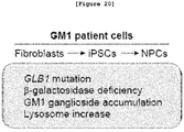
- the inventors first constructed GM1 originated induced pluripotent stem cells (iPSCs) from fibroblasts of GM1 patient and then induced the differentiation of embryoid body (EB) and neural progenitor cells (NPCs) from the same.
- EB embryoid body
- NPCs neural progenitor cells
- the GM1 patient derived iPSCs display both in vitro and in vivo pluripotency and at the same time have GM1 causing gene mutation detected in GM1 patient and accordingly show the reduced ⁇ -gal activity.
- the inventors also induced the differentiation of iPSCs originated from GM1 patient into neural progenitor cells.
- the expression of ⁇ -gal was increased but the activity thereof was reduced in the differentiated neural progenitor cells, suggesting that intracellular GM1 ganglioside and lysosome accumulation was increased.
- the gene expression pattern in the GM1 originated neural progenitor cells was compared with that of the normal cell. As a result, it was confirmed that inflammation related pathway, particularly inflammasome related metabolic pathway, was promoted.
- the neural progenitor cells differentiated from GM1 patient derived iPSCs were treated with an inflammasome inhibitor, not only cell morphology and size but also gene expression pattern were recovered similarly to those of normal cells.
- GM1 outbreak is related to inflammasome activation and therefore the inhibition of inflammasome is functioning to treat GM1.
- the present inventors examined disease-specific phenotype by using GM1 patient originated induced pluripotent stem cells (iPSCs) and iPSCs derived neural progenitor cells.
- iPSCs induced pluripotent stem cells
- inflammasome can be a key molecular target of the study to develop a GM1 treating agent and an inflammasome inhibitor displays positive effect on GM1 treatment.
- the present inventors completed this invention by proposing a novel GM1 gangliosidosis human cell model that can be efficiently used for the study of cause of GM1 and for the development of a therapeutic agent for the disease.
- the present invention provides a method for screening a treatment agent candidate for GM1 gangliosidosis comprising the following steps:
- the present invention further provides a method wherein the neural progenitor cells (NPCs) or neurosphere of step B) have one or more characteristics selected from the following ⁇ ) ⁇ ⁇ ):
- GM1 gangliosidosis GM1 gangliosidosis
- iPSCs induced pluripotent stem cells
- the present disclosure provides a GM1 gangliosidosis iPSCs model characterized by one or more of the following i) ⁇ vii):
- the present disclosure also provides a method for constructing the GM1 ganglioside iPSCs model in vitro comprising the following steps:
- the present disclosure further provides a GM1 gangliosidosis neural progenitor cell model characterized by one or more of the following i) - viii) :
- the present disclosure also provides a method for constructing the GM1 gangliosidosis neural progenitor cell model in vitro comprising the following steps:
- the present disclosure also provides a method for using the induced pluripotent stem cells (iPSCs), neural progenitor cells (NPCs), or neurosphere as the GM1 gangliosidosis model comprising the following steps:
- the present disclosure also provides a method for screening a treatment agent candidate for GM1 gangliosidosis comprising the following steps:
- the present disclosure also provides a pharmaceutical composition for the prevention and treatment of GM1 gangliosidosis comprising Z-YVAD-FMK (methyl (3S)-3-[(2S)-2-[(2S)-2-(2- ⁇ [(benzyloxy)car bonyl]amino ⁇ -3-(4-hydroxyphenyl)propanamido)-3-methylbutanamido]propanamido]-5-fluoro-4-oxopentanoate) as an active ingredient.
- Z-YVAD-FMK methyl (3S)-3-[(2S)-2-[(2S)-2-(2- ⁇ [(benzyloxy)car bonyl]amino ⁇ -3-(4-hydroxyphenyl)propanamido)-3-methylbutanamido]propanamido]-5-fluoro-4-oxopentanoate
- the present disclosure also provides a pharmaceutical composition for the prevention and treatment of GM1 gangliosidosis comprising interleukin-1 receptor antagonist protein as an active ingredient.
- the present disclosure also provides a use of the GM1 gangliosidosis iPSCs model characterized by one or more of the following i) ⁇ vii):
- the present disclosure also provides a use of the GM1 gangliosidosis neural progenitor cell model characterized by one or more of the following i) ⁇ viii) :
- the present disclosure also provides a method for the prevention and treatment of GM1 gangliosidosis containing the step of administering a pharmaceutically effective dose of Z-YVAD-FMK to a subject having GM1 gangliosidosis.
- the present disclosure also provides a method for the prevention and treatment of GM1 gangliosidosis containing the step of administering a pharmaceutically effective dose of interleukin-1 receptor antagonist protein to a subject having GM1 gangliosidosis.
- the present disclosure also provides a use of Z-YVAD-FMK as an active ingredient for the pharmaceutical composition of the present disclosure for the prevention and treatment of GM1 gangliosidosis.
- the present disclosure also provides a use of interleukin-1 receptor antagonist protein as an active ingredient for the pharmaceutical composition of the present disclosure for the prevention and treatment of GM1 gangliosidosis.
- the present disclosure also provides a GM1 gangliosidosis mutant cell model transformed with the ⁇ -gal mutant vector comprising E186A mutation in the amino acid sequence of ⁇ -gal protein.
- the present disclosure also provides a method for constructing a GM1 gangliosidosis cell model in vitro comprising the following steps:
- the present disclosure also provides a method for using the mutant cells transformed with the ⁇ -gal mutant vector comprising E186A mutation in the amino acid sequence of ⁇ -gal protein as the GM1 gangliosidosis cell model.
- the present disclosure provides a use of the mutant cells transformed with the ⁇ -gal mutant vector comprising E186A mutation in the amino acid sequence of ⁇ -gal protein as the GM1 gangliosidosis cell model.
- iPSCs induced pluripotent stem cells originated from the fibroblasts of GM1 gangliosidosis patient can be differentiated into neural progenitor cells (NPCs) and neurosphere cells that can emulate the disease-specific characteristics of GM1 patient, and the GM1 disease symptoms such as intracellular GM1 ganglioside/lysosome accumulation and also gene mutation can be confirmed in the cells. Therefore, the said cells have been successfully established as the GM1 model cells and thus the GM1 human cell model disclosed herein has been proposed as a useful model for the study on the GM1 development mechanism and for the development of a therapeutic agent for the disease.
- NPCs neural progenitor cells
- the molecular symptoms of GM1 patient could be reproduced in the transformed cells having the E186A mutation which is newly identified as the GM1 gangliosidosis causing protein mutation. Therefore, the mutant cells containing the induced E186A mutation can be efficiently used as the GM1 gangliosidosis cell model.
- the present invention provides a method according to claims 1 and 2 for screening a treatment agent candidate for GM1 gangliosidosis.
- the present disclosure also provides a GM1 gangliosidosis iPSCs model characterized by one or more of the following i) ⁇ vii):
- the present disclosure also provides a use of the GM1 gangliosidosis iPSCs model characterized by one or more of the following i) ⁇ vii):
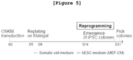
- the present inventors confirmed the GM1 mutant gene in fibroblasts originated from GM1 gangliosidosis patient (see Figure 2 ), and constructed iPSCs (GM1-iPSCs) from the GM1 gangliosidosis patient fibroblasts (see Figures 5 and 6 ). Then, the inventors investigated the characteristics of the GM1-iPSCs. As a result, the GM1-iPSCs had pluripotency in vivo and in vitro and at the same time displayed the decrease of ⁇ -gal activity caused by the GM1 causing gene mutation observed in GM1 patient see Figures 7 ⁇ 13 ).
- the GM1-originated iPSCs model of the present invention has pluripotency with displaying GM1 patient cell like characteristics, the iPSCs model can be efficiently used as a human cell model for the study of GM1 mechanism and for the development of a treating agent of the disease.
- the present disclosure also provides a method for constructing the GM1 ganglioside iPSCs model in vitro comprising the following steps:
- the inducement in step i) is preferably performed by the ectopic expression of pluripotent marker.
- the ectopic expression can be achieved by using retrovirus containing such reprogramming factors as OCT4, SOX2, C-MYC, and KLF4 or by transformation with the episome vector expressing the said reprogramming factors, or any other method to construct iPSCs well known to those in the art.
- the GM1-originated iPSCs model of the present invention has pluripotency with displaying GM1 patient cell like characteristics, the iPSCs model can be efficiently used as a human cell model for the study of GM1 mechanism and for the development of a treating agent of the disease.
- the present disclosure further provides a GM1 gangliosidosis neural progenitor cell model characterized by one or more of the following i) ⁇ viii) :
- the present disclosure also provides a use of the GM1 gangliosidosis neural progenitor cell model characterized by one or more of the following i) ⁇ viii) :
- the present disclosure also provides a method for using the induced pluripotent stem cells (iPSCs), neural progenitor cells (NPCs), or neurosphere as the GM1 gangliosidosis model comprising the following steps:
- the embryoid body differentiation marker is preferably selected from the group consisting of the ectoderm markers NESTIN and TUJ1, the endoderm markers SOX17 and FOXA2, and the mesoderm markers ⁇ -smooth muscle actin (a-SMA) and DESMIN, but not always limited thereto.
- the inflammasome related gene can be one or more genes selected from the group consisting of the inflammatory caspase related genes ( Allan SM et. al, Nature reviews Immunology5, 629-640, 2005 ; McIlwain DR et. al, Cold Spring Harbor perspectives in biology 5, a008656, 2013 ), the genes encoding proinflammatory cytokine including interleukine 1 ⁇ (IL1 ⁇ ) and its downstream molecule ( Walsh JG et. al, Nature reviews Neuroscience15, 84-97, 2014 ; John GR et. al, Glia49, 161-176, 2005 ; Liu L et. al, Journal of neuroinflammation8, 175, 2011 ; Manso Y et. al, Journal of biological inorganic chemistry 16, 1103-1113, 2011 ), and any other genes related to the up-regulation of inflammasome, but not always limited thereto.
- IL1 ⁇ interleukine 1 ⁇
- the present inventors induced the differentiation of GM1 patient originated iPSCs (GM1-iPSCs) into embryoid body (EB) see Figure 14 ). At this time, the expressions of all differentiation markers (ectoderm, endoderm, and mesoderm markers) were confirmed therein, suggesting that the GM1-iPSCs originated EB had differentiation potency (see Figure 15 ).
- the differentiation of GM1-iPSCs into neuronal progenitor cells (GM1-NPCs) was also induced and as a result the mutation of the GM1 causing gene was still observed and the expression of the neural marker gene and protein was also confirmed (see Figures 6 , 16 , and 17 ).
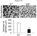
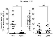
- the GM1 originated neurosphere was smaller than the normal one and displayed cystic neurosphere like phenotype. The yield of neural rosette formation was also decreased (see Figures 18 and 19 ).
- the present inventors also investigated whether or not the GM1 molecular phenotype remained unchanged in GM1-iPSCs and GM1-NPCs. As a result, the expression of GLB1 gene and protein was significantly increased (see Figures 20 and 21 ), while the activity of ⁇ -gal was significantly reduced and accordingly the accumulation of GM1 gangliosidosis and lysosome was increased (see Figures 22 and 23 ).
- the present inventors searched any gene that showed any change in its expression pattern in GM1-NPCs, compared with in normal cells. As a result, the gene expression which was significantly different from normal was confirmed in GM1 originated fibroblasts, iPSCs and NPCs, particularly in GM1-NPCs ( Figures 24 , 25 , and 26 ). Particularly, the inflammation pathway related gene expression was increased, and inflammatory caspase, the inflammasome metabolic pathway gene, and those genes encoding IL1 ⁇ and IL1 ⁇ downstream molecules were also up-regulated (see Figures 27a ⁇ 29 ).
- the GM1 patient originated iPSCs and the neural progenitor cells differentiated from the same, displayed GM1 patient cell like characteristics and also facilitates the investigation of the expression pattern of GM1 specific gene, so that the said iPSCs and neural progenitor cells of the present disclosure can be efficiently used as a human cell model for the study of outbreak mechanism of GM1.
- the present disclosure also provides a method for constructing the GM1 gangliosidosis neural progenitor cell model in vitro comprising the following steps:
- the present invention also provides a method for screening a treatment agent candidate for GM1 gangliosidosis comprising the following steps:
- the characteristics of the neural progenitor cells (NPCs) or neurosphere are one or more preferably selected from the group consisting of the followings, but not always limited thereto:
- the present inventors treated GM1-iPSCs with IL1 ⁇ antagonist, the inflammasome inhibitor, and caspase-1 inhibitor in order to inhibit the increasing expression of inflammasome metabolism related genes which showed significant increase of the expression in GM1, and then induced the differentiation thereof into neurosphere cells.
- the differentiated neurosphere cells were recovered from the cystic cell shape to the normal cell shape, and accordingly the diameter of the cell was also increased (see Figure 30 ).
- the present inventors also confirmed that when GM1-iPSCs was treated with an inflammasome inhibitor, the gene expression pattern was changed and precisely the expression of inflammasome factors, which was increased significantly in GM1-NPCs, was significantly decreased (see Figures 31 ⁇ 34 ).
- the present disclosure also provides a GM1 gangliosidosis mutant cell model transformed with the ⁇ -gal mutant vector comprising E186A mutation in the amino acid sequence of ⁇ -gal protein.
- the present disclosure also provides a method for constructing a GM1 gangliosidosis cell model in vitro comprising the following steps:
- the present disclosure also provides a method for using the mutant cells transformed with the ⁇ -gal mutant vector comprising E186A mutation in the amino acid sequence of ⁇ -gal protein as the GM1 gangliosidosis cell model.
- the present disclosure provides a use of the mutant cells transformed with the ⁇ -gal mutant vector comprising E186A mutation in the amino acid sequence of ⁇ -gal protein as the GM1 gangliosidosis cell model.
- E186A the novel GM1 causing gene mutation
- the inventors measured the ⁇ -gal activity and expression in the cells transformed with the gene expression vector encoding the E186A mutant. As a result, the ⁇ -gal expression and activity were significantly reduced, compared with those in normal cells (see Figures 3d ⁇ 3f ).
- the mutant cells containing E186A mutation can be effectively used as the GM1 gangliosidosis cell model.
- the present disclosure also provides a pharmaceutical composition for the prevention and treatment of GM1 gangliosidosis comprising Z-YVAD-FMK (methyl (3S)-3-[(2S)-2-[(2S)-2-(2- ⁇ [(benzyloxy)car bonyl]amino ⁇ -3-(4-hydroxyphenyl)propanamido)-3-methylbutanamido]propanamido]-5-fluoro-4-oxopentanoate) as an active ingredient.
- Z-YVAD-FMK methyl (3S)-3-[(2S)-2-[(2S)-2-(2- ⁇ [(benzyloxy)car bonyl]amino ⁇ -3-(4-hydroxyphenyl)propanamido)-3-methylbutanamido]propanamido]-5-fluoro-4-oxopentanoate
- the present disclosure also provides a pharmaceutical composition for the prevention and treatment of GM1 gangliosidosis comprising interleukin-1 receptor antagonist protein as an active ingredient.
- the present disclosure also provides a method for the prevention and treatment of GM1 gangliosidosis containing the step of administering a pharmaceutically effective dose of Z-YVAD-FMK to a subject having GM1 gangliosidosis.
- the present disclosure also provides a method for the prevention and treatment of GM1 gangliosidosis containing the step of administering a pharmaceutically effective dose of interleukin-1 receptor antagonist protein to a subject having GM1 gangliosidosis.
- the present disclosure also provides a use of Z-YVAD-FMK as an active ingredient for the pharmaceutical composition of the present disclosure for the prevention and treatment of GM1 gangliosidosis.
- the present disclosure also provides a use of interleukin-1 receptor antagonist protein as an active ingredient for the pharmaceutical composition of the present disclosure for the prevention and treatment of GM1 gangliosidosis.
- the said Z-YVAD-FMK is composed of the amino acid sequence represented by SEQ. ID. NO: 1 and presented as the structure shown in the below [Formula 1], but not always limited thereto.
- the said Z-YVAD-FMK can be the caspase-1 inhibitor including caspase-1 inhibitor VI-calbiochem (product #: cat # 218746; Merck Millipore), which can be any of those commercialized on the market.
- the said interleukin 1-receptor antagonist protein is composed of the amino acid sequence represented by SEQ. ID. NO: 2, but not always limited thereto.
- the said interleukin 1-receptor antagonist protein is the interleukin 1-receptor antagonist protein such as IL1RA (product #: cat # 280-RA-050 (rhIL-1ra); R&D systems), which can be any of those commercialized on the market.
- IL1RA product #: cat # 280-RA-050 (rhIL-1ra); R&D systems
- the Z-YVAD-FMK or IL1RA of the present disclosure demonstrates the effect of recovering the GM1 patient-specific cell morphology and gene expression pattern to the normal ones in GM1-iPSCs and neural progenitor cells differentiated from the same, so that the said Z-YVAD-FMK or IL1RA can be efficiently used as an active ingredient of a pharmaceutical composition for the prevention and treatment of GM1 gangliosidosis.
- the Z-YVAD-FMK or IL1RA of the present disclosure can be administered orally or parenterally and be used in general forms of pharmaceutical formulation.
- the parenteral administration includes intralectal, intravenous, intraperitoneal, intramuscular, intraarterial, transdermal, intranasal, intraocular, or subcutaneous injection, or inhalation.
- the Z-YVAD-FMK or IL1RA of the present disclosure can be prepared for parenteral administration by mixing with generally used diluents or excipients such as fillers, extenders, binders, wetting agents, disintegrating agents and surfactants.
- Formulations for parenteral administration are sterilized aqueous solutions, water-insoluble excipients, suspensions, emulsions, lyophilized preparations and suppositories.
- Water insoluble excipients and suspensions can contain polyethylene glycol, vegetable oil like olive oil, injectable ester like ethylolate, etc.
- Suppositories can contain witepsol, macrogol, tween 61, cacao butter, laurin butter, glycerogelatin, etc.
- the Z-YVAD-FMK or IL1RA of the present disclosure can be mixed with many pharmaceutically acceptable carriers such as physiological saline or organic solvent, and can additionally include carbohydrates such as glucose, sucrose or dextran, antioxidants such as ascorbic acid or glutathione, chelating agents, low molecular proteins or other stabilizers to enhance stability or absorptiveness.
- pharmaceutically acceptable carriers such as physiological saline or organic solvent
- carbohydrates such as glucose, sucrose or dextran
- antioxidants such as ascorbic acid or glutathione
- chelating agents such as ascorbic acid or glutathione
- low molecular proteins or other stabilizers to enhance stability or absorptiveness.
- the effective dose of the Z-YVAD-FMK or IL1RA of the present disclosure is 0.01 ⁇ 100 mg/kg, preferably 0.1 ⁇ 10 mg/kg, and administration times are 1 ⁇ 3 per day.
- the effective dose of the Z-YVAD-FMK or IL1RA of the present disclosure can be administered in the form of bolus by single dose having relatively short period of infusion or by multiple dose of fractionated treatment protocol for a long term.
- the decision of an effective dose depends on the administration pathway, treatment times, age and other conditions of a patient, etc. Therefore, any expert who has knowledge on this field can decide the effective dose of the Z-YVAD-FMK or IL1RA.
- the fibroblasts originated from GM1 gangliosidosis patient were cultured, followed by sequencing of GLB1 that is the causing gene of GM1 gangliosidosis.
- the fibroblast cell lines originated from GM1 gangliosidosis patients (GM00918, GM02439, GM03589, GM05335, GM05652, GM05653, GM10919, and GM12369) were first purchased from Coriell Institute for Medical Research, USA.
- GM00918, GM03589, GM05335, GM05652, GM05653, GM10919, and GM12369 cell lines were originated from those patients diagnosed in early childhood, and GM02439 cell line was originated from the patient diagnosed in adolescence (http://ccr.coriell.org).
- GM1 specific gene mutations such as p.C127Y, p.R148S, p.W161C, p.R201C, and p.Q255H missense mutations and p.R351X nonsense mutation were confirmed in GM1 cell lines.
- GM00918 cell line was cultured in the same conditions as described in Example ⁇ 1-1>.
- the culture medium was treated with LysoTracker Red DND-99 (1:20000; Invitrogen, USA), followed by reaction at 37 °C for 30 minutes. Then, the concentration of intracellular lysosome was measured.
- the cells were treated with 4% formaldehyde, followed by fixing at room temperature for 10 minutes.
- the cells were treated with PBS containing 0.1% triton X-100 for 15 minutes to give permeability to the cell membrane.
- the treated cells were washed with PBS containing 4% bovine serum albumin (BSA), and they were treated with the primary antibody 'anti-GM1 antibody (1:50; ab23943, Abcam, USA)' at 4 °C for overnight, followed by washing.
- the cells were treated with the secondary antibody conjugated with Alexa Fluor 488 or Alexa Fluor 594, and then left at room temperature for 2 hours. Then, the cells were observed under fluorescence microscope to investigate the accumulation of GM1 ganglioside. To compare the expression level, the cells were treated with 4'6-diamidino-2-phenylindole (DAPI) to stain nuclei.
- DAPI 4'6-diamidino-2-phenylindole
- X-gal staining was performed. To do so, the buffer at a low pH (pH 4.5) was prepared with 50 mM citrate and 150 mM NaCl in order to replace PBS buffer above. X-gal staining was performed using ⁇ -gal staining kit (INtRon Biotechnology, Korea) according to the manufacturer's instruction. When blue color was shown, X-gal staining was terminated. The culture time was 4 hours ⁇ overnight according to the cell types. CRL2097 cell line was used as the control to compare the accumulation of lysosome. The fibroblasts originated from minimal brain dysfunction (MBD) patient were used as the control to compare the intracellular accumulation of GM1 ganglioside.
- MBD minimal brain dysfunction
- GLB1 gene was amplified from CRL2097 cell line, the normal fibroblast cell line, which was inserted in pEGFP-N3 vector (BD bioscience Clonetech).
- FLAG tag was inserted in between 3'-end of GLB1 gene and 5'-end of GFP protein, resulting in the construction of a vector containing the wild-type GLB1 gene.
- Site-directed mutagenesis was induced in the site of E186A in GLB1 gene of the constructed vector, resulting in the construction of a vector containing the mutant GLB1 gene.
- the constructed vector containing the mutant GLB1 gene was introduced in 293T cell line for transfection in order to induce the expression of the mutant gene.
- the cells transfected with the E186A GLB1 mutant gene vector were lysed in RIPA buffer, followed by centrifugation at 4°C to eliminate the cell debris. 20 ⁇ 30 ⁇ g of the cell lysate proceeded to electrophoresis on 4 ⁇ 15% gel (Bio-Rad, USA) to separate the protein. The protein was transferred onto PVDF membrane (Bio-Rad, USA), followed by Western blotting using anti-GLB1 antibody (1:500; AF6464, R&D systems, USA). As for the control to correct the color development, GAPDH was stained by treating anti-GAPDH antibody. To confirm the ⁇ -gal activity, X-gal staining was performed by the same manner as described in Example ⁇ 1-2>. As the normal control, the cells transfected with the vector containing the wild-type GLB1 gene were used. As the negative control, the cells transfected with the vector not containing GLB1 gene.
- E186A mutation in GLB1 protein was analyzed by modeling.
- E186A mutation in GLB1 protein was arranged in the GLB1 sequence of human, mouse, Drosophila, Arabidopsis, Trichoderma , and Arthrobacter by using ClustalW2 and BoxShade server.
- the mutant model was made by using the human GLB1 crystal structure (PDB code: 3THC) as a template via PyMOL program.
- the GLB1 related protein sequence of prokaryotes Arthrobacter
- the GLB1 related protein sequence of eukaryotes such as fungi or human
- the 6 kinds of missense mutation sites as shown in Table 2 are important for maintaining the protein functions. Particularly, R148 residue and E186 residue are well preserved, compared with the other four residues, and the sites of these two residues are close to the active E188 residue.
- Human ⁇ -gal protein crystal structure was three-dimensionally analyzed.
- Example 2 Preparation of GM1 originated induced pluripotent stem cells (iPSCs)
- the inventors constructed iPSCs having pluripotency from GM1 patient fibroblasts (GM1-iPSCs) through the reprogramming culture using retrovirus expressing four reprogramming factors such as OCT4, SOX2, C-MYC, and KLF4 ( Son MY et. al, Stem cells 31, 2375-2387; 2013 ) ( Figure 5 ).
- GM02439 the GM1 patient originated fibroblast cell line confirmed with mutant gene in Example ⁇ 1-1> was transfected with retrovirus encoding OCT4, SOX2, C-MYC, and KLF4, followed by culture in somatic cell medium for 5 days. 5 days later, the infected cells were transferred onto the matrigel-coated plate containing human embryonic stem cell culture medium (hESCs-CM), followed by additional culture for 2 ⁇ 3 weeks. Then, iPSC colonies were collected. GLB1 gene cDNA was synthesized from the obtained GM1-iPSCs by the same manner as described in Example ⁇ 1-1>, followed by sequencing. For the control, the wild-type iPSCs (WT-iPSCs) were constructed from CRL-2097 by the same manner as described above.
- WT-iPSCs wild-type iPSCs
- GM1-iPSCs were constructed from GM1 patient originated fibroblasts and they were confirmed to have c.601C>T mutation as equally to the mutation shown in GM1 originated fibroblasts ( Figure 6 ).
- GM1-iPSCs colony type and karyotype of GM1-iPSCs were investigated, followed by short tandem repeat (STR) analysis.
- STR short tandem repeat
- reprogramming of GM1-iPSCs was induced by the same manner as described in Example 2, and then the morphology of GM1-iPSCs colony formation was observed by phase-contrast microscopy.
- GenDix Inc. Kerbet
- HumanPass Inc. Kerbet
- STR genotyping with GM1 originated fibroblast cell line (GM02439) and GM1-iPSCs, and the results were compared with the normal human embryonic stem cell line H9 hESCs.
- GM1-iPSCs having pluripotency were successfully induced from GM1-fibroblast cell line ( Figure 8 ), and the constructed GM1-iPSCs demonstrated that their colony type and karyotype were similar to those of human embryonic stem cells (hESCs) ( Figures 7a and 9 ).
- alkaline phosphatase (pluripotent marker) staining was performed.
- fixative solution was prepared by mixing 1 ml of citrate solution, 2.6 ml of acetone, and 320 ⁇ l of 37% formaldehyde (Sigma Aldrich, USA) using alkaline phosphatase (AP) staining kit (Sigma Aldrich, USA).
- the prepared fixative solution was added to the GM1-iPSCs prepared in Example 2, which stood at room temperature for 15 minutes in the darkness.
- 100 ⁇ l of sodium nitrate solution was mixed with 100 ⁇ l of FRV-alkaline solution, and the mixture stood for 2 minutes.
- the expression of the pluripotent marker protein in the GM1-iPSCs was observed.
- the GM1-iPSCs constructed in Example 2 were treated with 4% formaldehyde, followed by fixing at room temperature for 10 minutes.
- the cells were treated with PBS containing 0.1% triton X-100 for 15 minutes to give permeability to the cell membrane.
- the treated cells were washed with PBS containing 4% bovine serum albumin (BSA), and they were treated with the primary antibody anti-OCT4 antibody (1:100, sc-9081, Cruz Biotechnology, USA), anti-NANOG antibody (1:100, sc-33759, Cruz Biotechnology, USA), anti-TRA-1-81 antibody (1:100, MAB4381, Chemicon, USA), anti-SSEA3 antibody (1:100, MAB1435, R&D Systems, USA), anti-SSEA4 antibody, or anti-TRA-1-60 antibody (1:300, Millipore, USA) at 4°C for overnight, followed by washing.
- BSA bovine serum albumin
- the cells were treated with the secondary antibody (Invitrogen, USA) conjugated with Alexa Fluor 488 or Alexa Fluor 594, and then left at room temperature for 2 hours. Then, the cells were observed under fluorescence microscope to investigate the expressions of OCT4, NANOG, SOX2, SSEA4, Tra-1-80, and Tra-1-61 proteins. To compare the expression level, the cells were treated with 4'6-diamidino-2-phenylindole (DAPI) to stain nuclei.
- DAPI 4'6-diamidino-2-phenylindole
- the pluripotent markers OCT4, NANOG, TRA-1-81, SSEA3, SSEA4, and TRA-1-60 proteins were expressed in GM1-iPSCs at similar level to those of normal cells ( Figure 7c ).
- GM1 originated iPSCs had in vivo pluripotency
- teratoma formation of GM1-iPSCs was induced in the immune deficient nude mouse model.
- GM1-iPSCs constructed by the same manner as described in Example 2 were counted, which were transplanted in three SPF/VAF immune deficient nude mice (OrientBio, Korea) at 6 weeks of age. The animals were raised. 12 weeks later, the mice were sacrificed and teratoma was collected therefrom, to which 4% formaldehyde was added, followed by paraffin embedding. Then, H&E staining was performed by treating harris hematoxylin and eosin to confirm the formation of teratoma in endoderm, ectoderm, and mesoderm. To quantify the teratoma formation, each teratoma region was photographed and the size of each was calculated by using ImageJ software.
- CRL2097 cells the fibroblast originated iPSCs (WT-iPSCs), were injected in 5 mice by the same manner as described above to induce teratoma formation.
- the teratoma obtained from each mouse was sectioned into 2 ⁇ 5 pieces, which were used for the statistic analysis.
- the teratoma originated from GM1-iPSCs was similar in the size to that of WT-iPSCs ( Figure 10 ).
- the teratoma formation was confirmed in ectoderm (nervous tissues and pigment cells), mesoderm (adipocytes, cartilage, and muscle), and endoderm (epithelium), but the differentiation into neurosphere was less observed than in WT-iPSCs ( Figure 11 ).
- the fibroblast cell line GM02439 and the GM1-iPSCs constructed by the same manner as described in Example 2 were suspended in the analysis buffer (pH 4.5) containing 10 mM citrate, 30 mM NaCl, and 1 mM NgCl 2 . Then, the cells were lysed by 10 times repeated freeze/thaw, followed by centrifugation at 14,000 rpm at 4 °C for 5 minutes to eliminate the cell debris. 2 ⁇ g of the lysate was added to the analysis buffer containing 2 mM 4-methylumbelliferyl- ⁇ -D-galactopyranoside (MUG; Sigma-Aldrich, USA), followed by culture at 37 °C for 30 minutes.
- MUG 4-methylumbelliferyl- ⁇ -D-galactopyranoside
- 0.1 M glycine-NaOH buffer pH 10.5
- the concentration of 4-MU was calculated by measuring fluorescence intensity at 445 nm to confirm the ⁇ -gal activity.
- the ⁇ -gal activity of the control was also confirmed by the same manner as described above with the control CRL2097 and WT-iPSCs. Then, the comparative ⁇ -gal activity of GM1 fibroblasts and GM1-iPSCs was calculated and compared.
- GM1-iPSCs were prepared by a different method from the one described in Example 2, which was the non-viral reprogramming method.
- GM02439 fibroblasts were counted and prepared, which were transfected with EBNA1/OriP-based pCET4 episomal vector expressing OCT4, SOX2, KLF4, NANOG, LIN28, L-MYC, and SV40LT by electroporation using NeonTM transfection system (Invitrogen, USA) according to the manufacturer's protocol.
- the conditions for electroporation were as follows: 1650 V pulse voltage, 10 ms pulse width, and 3 pulse. After the transfection by electroporation, the cells were inoculated on the 35 mm matrigel-coated well, followed by culture in iPSCs culture medium.
- the characteristics of the GM1 originated iPSCs induced by non-viral method (Epi-GM1-iPSCs) which were collected from the culture were investigated by observing colony morphology and karyotype, by performing AP staining, by measuring the expressions of the pluripotent markers OCT4, NANOG, TRA-1-81, SSEA3, SSEA4, and TRA-1-60, and by observing in vivo teratoma formation by the same manner as described in Examples ⁇ 3-1>, ⁇ 3-2>, ⁇ 3-3>, and ⁇ 3-4>.
- the Epi-GM1-iPSCs prepared by transfecting the cells with pCET4 episomal vector displayed the characteristics similar to those of the GM1-iPSCs prepared by transfecting the cells with retrovirus. Therefore, it was confirmed that the characteristics of iPSCs did not changed according to the inducement method ( Figure 13 ).
- Example 4 Differentiation of GM1 originated neural progenitor cells (NPCs)
- EB embryoid body
- NPCs neural progenitor cells
- GM1 originated neuronal cells in vitro from GM1-iPSCs To induce the differentiation of GM1 originated neuronal cells in vitro from GM1-iPSCs, the differentiation of embryoid body (EB) and neural progenitor cells (NPCs) was induced from GM1-iPSCs according to the schematic diagram shown in Figure 14 ( Figure 14 ).
- EB embryoid body
- NPCs neural progenitor cells
- the GM1-iPSCs colony induced by reprogramming for 7 days by the same manner as described in Example 2 was cultured in embryoid body differentiation medium (DMED/F12 containing 10% serum replacement (SR)) for 7 days to induce the differentiation of GM1-iPSCs originated embryoid body (GM1-EB).
- the differentiated GM1-EB was further cultured in NPCs medium (DMEM/F12 supplemented with 1 ⁇ N2/B27 (Invitrogen, USA), 20 ng/ml bFGF, 20 ng/ml EGF (Invitrogen, USA), and 10 ng/ml leukemia inhibitory factor (Sigma-Aldrich, USA)) for 2 more weeks.
- the differentiated GM1 originated neural progenitor cells were collected.
- the differentiated cell body was sub-cultured every week by using McClain tissue chopper (Mickle Engineering, Great Britain), and the medium was replaced every other day.
- McClain tissue chopper McClain tissue chopper
- the collected GM1-NPCs were cultured as attached on the matrigel-coated coverslip in NPCs medium not-containing the growth factor for 3 ⁇ 4 weeks.
- the nucleotide sequence mutation in GLB1 which is the causing gene of GM1, was investigated using the differentiated GM1-NPCs by the same manner as described in Example ⁇ 1-1>.
- WT-iPSCs were cultured by the same manner as described above to induce the differentiation of WT-EB and WT-NPCs from WT-iPSCs.
- the GM1 originated NPCs maintained the spherical morphology of neurosphere during the suspension culture, and displayed the same c.601C>T mutation in GLB1 gene as GM1 fibroblast cell line and GM1-iPSCs showed ( Figure 4 ) .
- GM1-iPSCs originated embryoid body (GM1-EB)
- the expressions of the three germ layer markers such as NESTIN and TUJ1 (ectoderm markers), SOX17 and FOXA2 (endoderm markers), and ⁇ -smooth muscle actin (a-SMA) DESMIN (mesoderm markers) were investigated.
- the GM1-EB differentiated by the same manner as described in Example ⁇ 4-1> was immunofluorescent-stained to confirm the expression of NESTIN, TUJ1, SOX17, FOXA2, ⁇ -SMA, or DESMIN.
- anti-NESTIN antibody (1:100; MAB5326, Chemicon, USA)
- anti-TUJ1 antibody (1:500; PRB-435P, Covance, USA
- anti-SOX17 antibody (1:100; MAB1924, R&D Systems, USA
- anti-FOXA2 antibody (1:1000; ab40874, Abcam, USA
- anti- ⁇ -SMA antibody (1:400; A5228, Sigma-Aldrich, USA
- anti-DESMIN antibody (1:50; AB907, Chemicon, USA
- the GM1 originated, differentiated cells expressed all of those three germ layer marker proteins such as ESTIN, TUJ1, SOX17, FOXA2, ⁇ -SMA, and DESMIN, indicating the cells had pluripotency ( Figure 15 ).
- GM1-NPCs To investigate the differentiation of GM1-iPSCs originated NPCs (GM1-NPCs), quantitative real-time PCR (qPCR) was performed, by which the mRNA transcriptional levels of the neuronal marker genes such as NESTIN , NCAM , PAX6 , and OTX2 , and the pluripotent marker gene REX1 were observed in GM1-fibroblasts, GM1-iPSCs, and GM1-NPCs.
- qPCR quantitative real-time PCR
- GM1-iPSCs were constructed by the same manner as described in Example 2 and the differentiation of GM1-NPCs was induced by the same manner as described in Example ⁇ 4-1>. Then, RNA was extracted from GM1-fibroblasts, GM1-iPSCs, or GM1-NPCs by using RNAiso Plus (Takara, Japan) or RNeasy mini kit (Qiagen, USA) according to the manufacturer's protocol. Reverse transcription PCR (PT PCR) was performed using 1 ⁇ 2 ⁇ g of the extracted RNA as a template with Superscript III (Invitrogen, USA) in order to synthesize cDNA of the cell genome.
- RNAiso Plus Takara, Japan
- RNeasy mini kit Qiagen, USA
- qPCR reaction mixture 10 ⁇ l of qPCR reaction mixture was prepared by mixing 0.2 ⁇ l of the reaction product above, 1 ⁇ M oligonucleotide primer, 0.5 ⁇ l of FAST SYBR green master mix (Applied Biosystems, USA), and distilled water, which proceeded to qPCR using 7500 Fast Real-time PCR system (Applied Biosystems, USA).
- the expression of the control GAPDH gene was also confirmed by the same manner as described above to correct the expression level. Based on the expression level of GAPDH mRNA, the fold change of NESTIN, NCAM, PAX6, OTX2, or REX1 mRNA was calculated.
- NPCs neuronal cells, and glial cells were successfully differentiated from GM1-iPSCs
- the expressions of the neuronal cell-specific marker proteins NESTIN, MAP2, TUJ1, and S100 were confirmed in the NPCs, neuronal cells, and glial cells differentiated from GM1-iPSCs.
- Example ⁇ 4-1> the differentiation of GM1-NPCs, and the differentiations of GM1-neuronal cells and GM1-glial cells were induced by the same manner as described in Example ⁇ 4-1>.
- immunofluorescent staining was performed by the same manner as described in Example ⁇ 1-3> to confirm the expressions of NESTIN, MAP2, TUJ1, and S100.
- anti-NESTIN antibody was used for NPCs
- anti-TUJ1 antibody and anti-MAP2 antibody (1:500; AB5622, Chemicon, USA) were used for neuronal cells
- anti-SlOO antibody (1:100; ab52642, Abcam, USA
- the nuclei were stained with 4'6-diamidino-2-phenylindole (DAPI) and compared.
- DAPI 4'6-diamidino-2-phenylindole
- WT-NPCs, and WT-NPCs originated neuronal cells and glial cells were immunofluorescent-stained under the same conditions as described above in order to examine the expression of the neuronal cell-specific marker protein.
- the differentiations of GM1-NPCs and GM1-NPCs originated neurosphere were induced by the same manner as described in Example ⁇ 4-1>. Then, the size and the shape of the neurosphere differentiated from GM1-NPCs were examined by phase-contrast microscopy. As for the control, the size and the shape of the neurosphere differentiated from WT-NPCs were examined and compared with the above.
- teratoma was induced from GM1-iPSCs in an immune-deficient mouse by the same manner as described in Example ⁇ 1-4>. Then, nerve tissue and cartilage tissue were obtained. The mature neuronal cells in GM1-teratoma were observed by H&E staining. The generated teratoma was confirmed by immunocytochemistry using anti-TUJ1 antibody and anti-TH antibody.
- the yield of neural rosettes differentiated from GM1-iPSCs was about 13% by the yield produced in WT-iPSCs, suggesting that GM1-iPSCs had defect of neuronal differentiation potency.
- Example 6 Molecular phenotype of GM1 patient originated iPSCs and neuronal cells differentiated from the same
- GM1-iPSCs were prepared by the same manner as described in Example 2 and the differentiation of GM1-NPCs was induced by the same manner as described in Example ⁇ 4-1>. Then, qPCR was performed with -fibroblasts, GM1-iPSCs, and GM1-NPCs by the same manner as described in Example ⁇ 4-2> to confirm the expression level of GLB1 mRNA.
- the induced GM1-fibrablasts, GM1-iPSCs, or GM1-NPCs were obtained, which were lysed in RIPA buffer, followed by centrifugation at 4 °C to eliminate the cell debris. Then, 20 ⁇ 30 ⁇ g of the cell lysate was electrophoresed on 4 ⁇ 15% gel (Bio-Rad, USA) to separate protein. The obtained protein was transferred onto PVDF membrane (Bio-Rad, USA), followed by Western blotting using anti-GLBl antibody (1:500; AF6464, R&D systems, USA). As for the control to correct the color development, GAPDH was stained by treating anti-GAPDH antibody.
- GM1-iPSCs were constructed by the same manner as described in Example 2, and the differentiation of GM1-NPCs was induced therefrom by the same manner as described in Example ⁇ 4-1>.
- PBS was replaced with a low pH buffer at pH 4.5 containing 50 mM citrate and 150 mM NaCl.
- X-gal staining was performed using ⁇ -gal staining kit (INtRon Biotechnology, Korea) according to the manufacturer's protocol. When blue color was shown, X-gal staining was terminated. The culture time was 4 hours ⁇ overnight according to the cell types.
- GM1 ganglioside was caused by the decrease of ⁇ -gal activity in GM1 patient.
- the accumulation of GM1 ganglioside and lysosome was investigated in GM1-iPSCs and GM1-NPCs.
- GM1-iPSCs were constructed by the same manner as described in Example 2, and the differentiation of GM1-NPCs was induced therefrom by the same manner as described in Example ⁇ 4-1>. Then, GM1 ganglioside in GM1-fibroblasts and GM1-iPSCs was immunofluorescent-stained by the same manner as described in Example ⁇ 3-3>.
- the primary antibody used herein was anti-GM1 antibody (1:50; ab23943, Abcam, USA).
- GM1-fibroblasts and GM1-NPCs were treated with LysoTracker Red DND-99 (1:20000; Invitrogen, USA), followed by reaction at 37 °C for 30 minutes. Then, intracellular lysosome was observed. The accumulation of GM1 ganglioside and lysosome in the control (WT-fibroblasts, WT-iPSCs, and WT-NPCs) was also measured by the same manner as described above.
- Example 7 Investigation of transcripts of GM1-iPSCs and neuronal cells differentiated from the same
- GM1-iPSCs were constructed by the same manner as described in Example 2, and the differentiation of GM1-NPCs was induced therefrom by the same manner as described in Example ⁇ 4-1>. Then, GM1-fibroblasts, GM1-iPSCs, and GM1-NPCs were obtained. Microarray was performed by using Low RNA input linear amplification kit, cRNA cleanup module, and Whole Human Genome Microarray 4X44K (Agilent Technology, USA) according to the manufacturer's protocol. The obtained microarray images were revised as signals corresponding to WT-fibroblasts, WT-iPSCs, and WT-NPCs. The revised value was used to make heatmap by using MeV program and the gene that did not show any change in expression pattern (the change less than double) was discarded.
- GM1-iPSCs were constructed by the same manner as described in Example 2, and the differentiation of GM1-NPCs was induced therefrom by the same manner as described in Example ⁇ 4-1>. Then, GM1-fibroblasts, GM1-iPSCs, and GM1-NPCs were obtained, followed by microarray as described in Example ⁇ 7-1> to examine the gene expression pattern in normal cells and the obtained cells. The gene displayed any change in expression level was selected.
- mRNA expression levels of CHOP, BiP, XBP1, PD1A4, and CALR which were the UPR genes that had been reported earlier to be up-regulated in GM1 model, were compared by qPCR according to the same manner as described in Example ⁇ 5-1> with the relative mRNA expression levels in WT-NPCs and GM1-NPCs.
- GM1-iPSCs were constructed by the same manner as described in Example 2, and the differentiation of GM1-NPCs was induced therefrom by the same manner as described in Example ⁇ 4-1>. Then, GM1-fibroblasts, GM1-iPSCs, and GM1-NPCs were obtained, followed by microarray as described in Example ⁇ 7-1> to examine the gene expression pattern in normal cells and the obtained cells. The gene displayed any change in expression level was selected. The selected gene was analyzed by KEGG using DAVID Bioiformatics Resource ( http://david.abcc.ncifcrf.gov ). Interactive pathway analysis (IPA) was performed by using Ingenuity Systems (EBIOGEN Inc.).
- mRNA expression levels of CHOP, BiP, XBP1, PD1A4, and CALR which were the UPR genes that had been reported earlier to be up-regulated in GM1 model, were compared by qPCR according to the same manner as described in Example ⁇ 5-1> with the relative mRNA expression levels in WT-NPCs and GM1-NPCs.
- GM1-iPSCs were constructed by the same manner as described in Example 2, and the differentiation of GM1-NPCs was induced therefrom by the same manner as described in Example ⁇ 4-1>. Then, GM1-fibroblasts, GM1-iPSCs, and GM1-NPCs were obtained, followed by microarray as described in Example ⁇ 7-1> to investigate the up- or down-regulation of the gene involved in inflammasome. The gene whose expression pattern was significantly changed, compared with that in WT-NPCs was selected, followed by qPCR according to the same manner as described in Example ⁇ 5-1> in order to compare the relative mRNA expression level in WT-NPCs and GM1-NPCs.
- GM1-NPCs were treated with the inflammasome inhibitors IL1 ⁇ antagonist (rhILIRA) and caspase-1 inhibitor (Z-YVAD-FMK), and then the morphology of GM1-NPCs was investigated.
- rhILIRA inflammasome inhibitors IL1 ⁇ antagonist
- Z-YVAD-FMK caspase-1 inhibitor
- the differentiation of GM1-NPCs was induced by the same manner as described in Example ⁇ 4-1>. Then, the induced GM1-NPCs were cultured in the culture medium respectively treated with 1 ⁇ g/ml rhILIRA (R&D Systems, USA) or 10 ⁇ M Z-YVAD-FMK (Merck Millipore, Germany) in order to inhibit inflammasome therein. 2 ⁇ 4 weeks later, the cultured GM1-NPCs originated neurosphere was obtained. The morphology of neurosphere was observed by phase-contrast microscopy.
- GM1-NPCs were differentiated according to the method described in Example ⁇ 4-1>, and then treated with rhILIRA or Z-YVAD-FMK by the same manner as described in Example ⁇ 9-1>.
- Microarray was performed by the same manner as described in Example ⁇ 7-1> in order to investigate the up- or down-regulation of the gene involved in inflammasome.
- the gene whose expression pattern was significantly changed, compared with that in WT-NPCs was selected, followed by qPCR according to the same manner as described in Example ⁇ 5-1> in order to compare the relative mRNA expression level in WT-NPCs and GM1-NPCs.
- WT-NPC was used as the normal control.
- GM1-NPCs were treated with DMSO instead of rhIL1RA or Z-YVAD-FMK.
- the expression pattern (either up- or down regulation) and the mRNA expression level were investigated by the same manner as described above.
Landscapes
- Health & Medical Sciences (AREA)
- Life Sciences & Earth Sciences (AREA)
- Engineering & Computer Science (AREA)
- Chemical & Material Sciences (AREA)
- Biomedical Technology (AREA)
- Organic Chemistry (AREA)
- Bioinformatics & Cheminformatics (AREA)
- Genetics & Genomics (AREA)
- Zoology (AREA)
- General Health & Medical Sciences (AREA)
- Biotechnology (AREA)
- Wood Science & Technology (AREA)
- Biochemistry (AREA)
- Medicinal Chemistry (AREA)
- Microbiology (AREA)
- Cell Biology (AREA)
- General Engineering & Computer Science (AREA)
- Proteomics, Peptides & Aminoacids (AREA)
- Molecular Biology (AREA)
- Immunology (AREA)
- Neurology (AREA)
- Developmental Biology & Embryology (AREA)
- Biophysics (AREA)
- Gastroenterology & Hepatology (AREA)
- Public Health (AREA)
- Pharmacology & Pharmacy (AREA)
- Animal Behavior & Ethology (AREA)
- Veterinary Medicine (AREA)
- Neurosurgery (AREA)
- Epidemiology (AREA)
- Toxicology (AREA)
- Transplantation (AREA)
- Urology & Nephrology (AREA)
- Hematology (AREA)
- Crystallography & Structural Chemistry (AREA)
- Rheumatology (AREA)
- Reproductive Health (AREA)
- Gynecology & Obstetrics (AREA)
- Chemical Kinetics & Catalysis (AREA)
- General Chemical & Material Sciences (AREA)
Claims (2)
- Ein Verfahren zum Screening eines Behandlungsmittelkandidaten für GM1-Gangliosidose, umfassend die folgenden Schritte:A) Behandeln differenzierter neuraler Progenitorzellen (NPCs) oder Neurosphäre von einem GM1-Gangliosidose-iPSCs-Modell mit der Probenverbindung oder -zusammensetzung, wobei das GM1-Gangliosidose-iPSCs-Modell gekennzeichnet ist durch die folgenden Eigenschaften i) ∼ vii):i) c.601C>T-Mutation des GLB1-Gens,ii) R201C-Mutation des β-Galactosidase(β-Gal)-Proteins,iii) normale Zellmorphologie von iPSCs,iv) Expression von Pluripotenz-Markern wie z.B. OCT4, NANOG, TRA-1-81, SSEA3, SSEA4 und TRA-1-60,v) Hochregulierung des GLB1-Gens und β-Gal-Proteins,vi) Verringerung der β-Gal-Aktivität undvii) Anreicherung von GM1-Gangliosid und Lysosom,wobei die NPCs gekennzeichnet sind durch die folgenden Eigenschaften a) ∼ h):a) Expression neuraler Markergene einschließlich NESTIN, NCAM, PAX6 und OTX2,b) Expression neuraler Markerproteine einschließlich MAP2, TUJ1 und S100,c) gespaltene zystische Neurosphäre,d) Verringerung der Bildung neuraler Rosetten,e) Hochregulierung des GLB1-Gens und β-Gal-Proteins,f) Verringerung der β-Gal-Aktivität,g) Anreicherung von GM1-Gangliosid und Lysosom, undh) Hochregulierung von entzündungsbezogenen Genen,B) Analysieren der Eigenschaften der neuralen Progenitorzellen (NPCs) oder Neurosphäre von Schritt A), undC) Vergleichen des in Schritt B) erhaltenen Ergebnisses mit der unbehandelten Kontrollgruppe.
- Das Verfahren gemäß Anspruch 1, wobei die neuralen Progenitorzellen (NPCs) oder Neurosphäre von Schritt B) eine oder mehrere Eigenschaften besitzen/besitzt, ausgewählt aus den folgenden Eigenschaften α) ∼ γ):α) Wiederherstellung von der zystischen Neurosphäre zur normalen Neurosphäre,β) Vergrößerung der Neurosphärenzellen undγ) Verringerung der Expression von entzündungsbezogenem Gen.
Applications Claiming Priority (2)
| Application Number | Priority Date | Filing Date | Title |
|---|---|---|---|
| KR20140118659 | 2014-09-05 | ||
| PCT/KR2014/011207 WO2016035934A1 (ko) | 2014-09-05 | 2014-11-20 | Gm1 강글리오시드증의 인간 세포 모델 및 이의 용도 |
Publications (3)
| Publication Number | Publication Date |
|---|---|
| EP3012321A1 EP3012321A1 (de) | 2016-04-27 |
| EP3012321A4 EP3012321A4 (de) | 2016-09-28 |
| EP3012321B1 true EP3012321B1 (de) | 2019-04-03 |
Family
ID=55532300
Family Applications (1)
| Application Number | Title | Priority Date | Filing Date |
|---|---|---|---|
| EP14880388.5A Not-in-force EP3012321B1 (de) | 2014-09-05 | 2014-11-20 | Menschliches zellmodell für gm1-gangliosidose und verwendung davon |
Country Status (5)
| Country | Link |
|---|---|
| US (2) | US10035830B2 (de) |
| EP (1) | EP3012321B1 (de) |
| JP (1) | JP2016533770A (de) |
| KR (1) | KR101739017B1 (de) |
| WO (1) | WO2016035934A1 (de) |
Families Citing this family (5)
| Publication number | Priority date | Publication date | Assignee | Title |
|---|---|---|---|---|
| US10767164B2 (en) | 2017-03-30 | 2020-09-08 | The Research Foundation For The State University Of New York | Microenvironments for self-assembly of islet organoids from stem cells differentiation |
| WO2019132004A1 (ja) * | 2017-12-28 | 2019-07-04 | 国立大学法人熊本大学 | アルツハイマー病予防剤又は治療剤、そのスクリーニング方法、及びアルツハイマー病予防用又は治療用組成物 |
| WO2019131989A1 (ja) * | 2017-12-28 | 2019-07-04 | 国立大学法人熊本大学 | Gm1ガングリオシドーシス予防剤又は治療剤、及びgm1ガングリオシドーシス予防用又は治療用組成物 |
| WO2020168294A1 (en) | 2019-02-15 | 2020-08-20 | Lysosomal And Rare Disorders Research And Treatment Center, Llc | Methods and compositions for treating lysosomal storage disorders: ambroxol as treatment agent for mucopolysaccharidoses iii (sanfilippo syndrome) |
| WO2022115612A1 (en) * | 2020-11-25 | 2022-06-02 | Lysosomal and Rare Disorders Research and Treatment Center, Inc. | Inhibition of caspase pathway as a treatment for lysosomal storage disorders |
Family Cites Families (9)
| Publication number | Priority date | Publication date | Assignee | Title |
|---|---|---|---|---|
| ATE420101T1 (de) * | 2001-04-20 | 2009-01-15 | Children S Hospital Of Orange | Isolierung von nervenstammzellen unter verwendung von gangliosiden und anderen oberflächenmarkern |
| EP1433776B1 (de) * | 2001-09-07 | 2011-01-19 | Seikagaku Corporation | Carbazuckeraminderivate und mittel gegen störungen des glycolipidmetabolismus, die diese als wirkstoff enthalten |
| AU2003231220A1 (en) * | 2002-04-30 | 2003-11-17 | Saint Louis University | A method for detecting lysosomal storage diseases |
| WO2009049422A1 (en) * | 2007-10-18 | 2009-04-23 | The Hospital For Sick Children | Compositions and methods for enhancing enzyme activity in gaucher, gm1 -gangliosidosis/morquio b disease. and parkinson's disease |
| BR112012004689A2 (pt) * | 2009-09-01 | 2019-09-24 | Lz Therapeutics Inc | métodos para extração e purificação de gangliosídeos. |
| CA2779039A1 (en) | 2009-10-29 | 2011-05-05 | Mcmaster University | Generating induced pluripotent stem cells and progenitor cells from fibroblasts |
| US9393221B2 (en) * | 2011-07-20 | 2016-07-19 | The General Hospital Corporation | Methods and compounds for reducing intracellular lipid storage |
| CA2862157C (en) * | 2012-01-20 | 2016-03-22 | Garnet Biotherapeutics, Inc. | Methods of ganglioside production |
| WO2015083736A1 (ja) * | 2013-12-05 | 2015-06-11 | 国立大学法人熊本大学 | コレステロール蓄積疾患治療薬、およびそのスクリーニング方法 |
-
2014
- 2014-11-20 EP EP14880388.5A patent/EP3012321B1/de not_active Not-in-force
- 2014-11-20 KR KR1020140162438A patent/KR101739017B1/ko active Active
- 2014-11-20 JP JP2016546728A patent/JP2016533770A/ja active Pending
- 2014-11-20 WO PCT/KR2014/011207 patent/WO2016035934A1/ko not_active Ceased
-
2015
- 2015-08-06 US US14/820,348 patent/US10035830B2/en not_active Expired - Fee Related
- 2015-08-06 US US14/820,353 patent/US20160069865A1/en not_active Abandoned
Non-Patent Citations (1)
| Title |
|---|
| None * |
Also Published As
| Publication number | Publication date |
|---|---|
| EP3012321A4 (de) | 2016-09-28 |
| US20160069865A1 (en) | 2016-03-10 |
| JP2016533770A (ja) | 2016-11-04 |
| KR101739017B1 (ko) | 2017-06-08 |
| KR20160029615A (ko) | 2016-03-15 |
| US10035830B2 (en) | 2018-07-31 |
| US20160068580A1 (en) | 2016-03-10 |
| EP3012321A1 (de) | 2016-04-27 |
| WO2016035934A1 (ko) | 2016-03-10 |
Similar Documents
| Publication | Publication Date | Title |
|---|---|---|
| EP3012321B1 (de) | Menschliches zellmodell für gm1-gangliosidose und verwendung davon | |
| Mora et al. | Clinical potentials of human pluripotent stem cells | |
| Dey et al. | Genetically engineered mesenchymal stem cells reduce behavioral deficits in the YAC 128 mouse model of Huntington's disease | |
| EP3286301B1 (de) | Erzeugung von differenzierung von muskellinienzellen aus stammzellen | |
| US20170369848A1 (en) | Engineering mesenchymal stem cells using homologous recombination | |
| US20160215259A1 (en) | Method of Preparing Induced Neural Stem Cells Reprogrammed from Non-Neuronal Cells Using HMGA2 | |
| JP2024042096A (ja) | 神経幹細胞組成物および神経変性障害を処置するための方法 | |
| US20250263752A1 (en) | Gene editing of gba1 in stem cells and method of use of cells differentiated therefrom | |
| US20120141441A1 (en) | Methods and Compositions for Treatment of Muscular Dystrophy | |
| Aigner et al. | Human pluripotent stem cell models of autism spectrum disorder: emerging frontiers, opportunities, and challenges towards neuronal networks in a dish | |
| US20020114788A1 (en) | Cell implantation therapy for neurological diseases or disorders | |
| Pendse et al. | Exon 13-skipped USH2A protein retains functional integrity in mice, suggesting an exo-skipping therapeutic approach to treat USH2A-associated disease | |
| CN114269899B (zh) | 新的重编程方法 | |
| WO2016207300A1 (en) | Enhanced cell reprogramming by mrna | |
| KR101847975B1 (ko) | Gm1 강글리오시드증의 인간 세포 모델 및 이의 용도 | |
| US12133880B2 (en) | Compositions and methods for treatment of GCase related disease states | |
| US20230081881A1 (en) | Transposon-based modulation of gba1 and related compositions and uses thereof | |
| US20120183511A1 (en) | Induction of neuronal differentiation in non-neuronal cells using a nucleic acid molecule | |
| JP7084418B2 (ja) | sRAGEを分泌する幹細胞を含む神経疾患または心血管疾患の予防または治療用薬学組成物 | |
| CN116157511A (zh) | 一种用于诱导体细胞直接转分化为运动神经元的组合物以及其使用方法 | |
| CN112384247B (zh) | 利用细胞融合技术的基因及细胞治疗剂以及其的用途 | |
| KR20260032533A (ko) | 세포 재프로그래밍 | |
| Chear | A patient-specific stem cell model of Batten disease | |
| KR20210086548A (ko) | 면역계 신경교세포로의 분화 유도를 위한 조성물과 방법 및 이의 용도 |
Legal Events
| Date | Code | Title | Description |
|---|---|---|---|
| PUAI | Public reference made under article 153(3) epc to a published international application that has entered the european phase |
Free format text: ORIGINAL CODE: 0009012 |
|
| 17P | Request for examination filed |
Effective date: 20150806 |
|
| AK | Designated contracting states |
Kind code of ref document: A1 Designated state(s): AL AT BE BG CH CY CZ DE DK EE ES FI FR GB GR HR HU IE IS IT LI LT LU LV MC MK MT NL NO PL PT RO RS SE SI SK SM TR |
|
| AX | Request for extension of the european patent |
Extension state: BA ME |
|
| A4 | Supplementary search report drawn up and despatched |
Effective date: 20160829 |
|
| RIC1 | Information provided on ipc code assigned before grant |
Ipc: C12N 5/074 20100101AFI20160823BHEP Ipc: C12N 5/0793 20100101ALI20160823BHEP Ipc: A61K 38/55 20060101ALI20160823BHEP Ipc: C07K 5/02 20060101ALI20160823BHEP Ipc: A61P 21/00 20060101ALI20160823BHEP Ipc: C12N 5/073 20100101ALI20160823BHEP Ipc: C12N 5/077 20100101ALI20160823BHEP Ipc: C12N 5/079 20100101ALI20160823BHEP Ipc: C07K 5/087 20060101ALI20160823BHEP Ipc: C07K 14/47 20060101ALI20160823BHEP |
|
| STAA | Information on the status of an ep patent application or granted ep patent |
Free format text: STATUS: EXAMINATION IS IN PROGRESS |
|
| 17Q | First examination report despatched |
Effective date: 20170612 |
|
| DAX | Request for extension of the european patent (deleted) | ||
| GRAP | Despatch of communication of intention to grant a patent |
Free format text: ORIGINAL CODE: EPIDOSNIGR1 |
|
| STAA | Information on the status of an ep patent application or granted ep patent |
Free format text: STATUS: GRANT OF PATENT IS INTENDED |
|
| INTG | Intention to grant announced |
Effective date: 20181017 |
|
| GRAS | Grant fee paid |
Free format text: ORIGINAL CODE: EPIDOSNIGR3 |
|
| GRAJ | Information related to disapproval of communication of intention to grant by the applicant or resumption of examination proceedings by the epo deleted |
Free format text: ORIGINAL CODE: EPIDOSDIGR1 |
|
| GRAL | Information related to payment of fee for publishing/printing deleted |
Free format text: ORIGINAL CODE: EPIDOSDIGR3 |
|
| STAA | Information on the status of an ep patent application or granted ep patent |
Free format text: STATUS: EXAMINATION IS IN PROGRESS |
|
| GRAP | Despatch of communication of intention to grant a patent |
Free format text: ORIGINAL CODE: EPIDOSNIGR1 |
|
| STAA | Information on the status of an ep patent application or granted ep patent |
Free format text: STATUS: GRANT OF PATENT IS INTENDED |
|
| INTC | Intention to grant announced (deleted) | ||
| GRAA | (expected) grant |
Free format text: ORIGINAL CODE: 0009210 |
|
| STAA | Information on the status of an ep patent application or granted ep patent |
Free format text: STATUS: THE PATENT HAS BEEN GRANTED |
|
| INTG | Intention to grant announced |
Effective date: 20190208 |
|
| AK | Designated contracting states |
Kind code of ref document: B1 Designated state(s): AL AT BE BG CH CY CZ DE DK EE ES FI FR GB GR HR HU IE IS IT LI LT LU LV MC MK MT NL NO PL PT RO RS SE SI SK SM TR |
|
| REG | Reference to a national code |
Ref country code: GB Ref legal event code: FG4D |
|
| REG | Reference to a national code |
Ref country code: CH Ref legal event code: EP Ref country code: AT Ref legal event code: REF Ref document number: 1115749 Country of ref document: AT Kind code of ref document: T Effective date: 20190415 |
|
| REG | Reference to a national code |
Ref country code: DE Ref legal event code: R096 Ref document number: 602014044214 Country of ref document: DE |
|
| REG | Reference to a national code |
Ref country code: IE Ref legal event code: FG4D |
|
| REG | Reference to a national code |
Ref country code: NL Ref legal event code: MP Effective date: 20190403 |
|
| REG | Reference to a national code |
Ref country code: LT Ref legal event code: MG4D |
|
| REG | Reference to a national code |
Ref country code: AT Ref legal event code: MK05 Ref document number: 1115749 Country of ref document: AT Kind code of ref document: T Effective date: 20190403 |
|
| PG25 | Lapsed in a contracting state [announced via postgrant information from national office to epo] |
Ref country code: NL Free format text: LAPSE BECAUSE OF FAILURE TO SUBMIT A TRANSLATION OF THE DESCRIPTION OR TO PAY THE FEE WITHIN THE PRESCRIBED TIME-LIMIT Effective date: 20190403 |
|
| PG25 | Lapsed in a contracting state [announced via postgrant information from national office to epo] |
Ref country code: SE Free format text: LAPSE BECAUSE OF FAILURE TO SUBMIT A TRANSLATION OF THE DESCRIPTION OR TO PAY THE FEE WITHIN THE PRESCRIBED TIME-LIMIT Effective date: 20190403 Ref country code: ES Free format text: LAPSE BECAUSE OF FAILURE TO SUBMIT A TRANSLATION OF THE DESCRIPTION OR TO PAY THE FEE WITHIN THE PRESCRIBED TIME-LIMIT Effective date: 20190403 Ref country code: LT Free format text: LAPSE BECAUSE OF FAILURE TO SUBMIT A TRANSLATION OF THE DESCRIPTION OR TO PAY THE FEE WITHIN THE PRESCRIBED TIME-LIMIT Effective date: 20190403 Ref country code: AL Free format text: LAPSE BECAUSE OF FAILURE TO SUBMIT A TRANSLATION OF THE DESCRIPTION OR TO PAY THE FEE WITHIN THE PRESCRIBED TIME-LIMIT Effective date: 20190403 Ref country code: PT Free format text: LAPSE BECAUSE OF FAILURE TO SUBMIT A TRANSLATION OF THE DESCRIPTION OR TO PAY THE FEE WITHIN THE PRESCRIBED TIME-LIMIT Effective date: 20190803 Ref country code: HR Free format text: LAPSE BECAUSE OF FAILURE TO SUBMIT A TRANSLATION OF THE DESCRIPTION OR TO PAY THE FEE WITHIN THE PRESCRIBED TIME-LIMIT Effective date: 20190403 Ref country code: NO Free format text: LAPSE BECAUSE OF FAILURE TO SUBMIT A TRANSLATION OF THE DESCRIPTION OR TO PAY THE FEE WITHIN THE PRESCRIBED TIME-LIMIT Effective date: 20190703 Ref country code: FI Free format text: LAPSE BECAUSE OF FAILURE TO SUBMIT A TRANSLATION OF THE DESCRIPTION OR TO PAY THE FEE WITHIN THE PRESCRIBED TIME-LIMIT Effective date: 20190403 Ref country code: CZ Free format text: LAPSE BECAUSE OF FAILURE TO SUBMIT A TRANSLATION OF THE DESCRIPTION OR TO PAY THE FEE WITHIN THE PRESCRIBED TIME-LIMIT Effective date: 20190403 |
|
| PG25 | Lapsed in a contracting state [announced via postgrant information from national office to epo] |
Ref country code: GR Free format text: LAPSE BECAUSE OF FAILURE TO SUBMIT A TRANSLATION OF THE DESCRIPTION OR TO PAY THE FEE WITHIN THE PRESCRIBED TIME-LIMIT Effective date: 20190704 Ref country code: BG Free format text: LAPSE BECAUSE OF FAILURE TO SUBMIT A TRANSLATION OF THE DESCRIPTION OR TO PAY THE FEE WITHIN THE PRESCRIBED TIME-LIMIT Effective date: 20190703 Ref country code: RS Free format text: LAPSE BECAUSE OF FAILURE TO SUBMIT A TRANSLATION OF THE DESCRIPTION OR TO PAY THE FEE WITHIN THE PRESCRIBED TIME-LIMIT Effective date: 20190403 Ref country code: LV Free format text: LAPSE BECAUSE OF FAILURE TO SUBMIT A TRANSLATION OF THE DESCRIPTION OR TO PAY THE FEE WITHIN THE PRESCRIBED TIME-LIMIT Effective date: 20190403 Ref country code: PL Free format text: LAPSE BECAUSE OF FAILURE TO SUBMIT A TRANSLATION OF THE DESCRIPTION OR TO PAY THE FEE WITHIN THE PRESCRIBED TIME-LIMIT Effective date: 20190403 |
|
| PG25 | Lapsed in a contracting state [announced via postgrant information from national office to epo] |
Ref country code: IS Free format text: LAPSE BECAUSE OF FAILURE TO SUBMIT A TRANSLATION OF THE DESCRIPTION OR TO PAY THE FEE WITHIN THE PRESCRIBED TIME-LIMIT Effective date: 20190803 Ref country code: AT Free format text: LAPSE BECAUSE OF FAILURE TO SUBMIT A TRANSLATION OF THE DESCRIPTION OR TO PAY THE FEE WITHIN THE PRESCRIBED TIME-LIMIT Effective date: 20190403 |
|
| REG | Reference to a national code |
Ref country code: DE Ref legal event code: R097 Ref document number: 602014044214 Country of ref document: DE |
|
| PG25 | Lapsed in a contracting state [announced via postgrant information from national office to epo] |
Ref country code: DK Free format text: LAPSE BECAUSE OF FAILURE TO SUBMIT A TRANSLATION OF THE DESCRIPTION OR TO PAY THE FEE WITHIN THE PRESCRIBED TIME-LIMIT Effective date: 20190403 Ref country code: EE Free format text: LAPSE BECAUSE OF FAILURE TO SUBMIT A TRANSLATION OF THE DESCRIPTION OR TO PAY THE FEE WITHIN THE PRESCRIBED TIME-LIMIT Effective date: 20190403 Ref country code: SK Free format text: LAPSE BECAUSE OF FAILURE TO SUBMIT A TRANSLATION OF THE DESCRIPTION OR TO PAY THE FEE WITHIN THE PRESCRIBED TIME-LIMIT Effective date: 20190403 Ref country code: RO Free format text: LAPSE BECAUSE OF FAILURE TO SUBMIT A TRANSLATION OF THE DESCRIPTION OR TO PAY THE FEE WITHIN THE PRESCRIBED TIME-LIMIT Effective date: 20190403 |
|
| PLBE | No opposition filed within time limit |
Free format text: ORIGINAL CODE: 0009261 |
|
| STAA | Information on the status of an ep patent application or granted ep patent |
Free format text: STATUS: NO OPPOSITION FILED WITHIN TIME LIMIT |
|
| PG25 | Lapsed in a contracting state [announced via postgrant information from national office to epo] |
Ref country code: IT Free format text: LAPSE BECAUSE OF FAILURE TO SUBMIT A TRANSLATION OF THE DESCRIPTION OR TO PAY THE FEE WITHIN THE PRESCRIBED TIME-LIMIT Effective date: 20190403 Ref country code: SM Free format text: LAPSE BECAUSE OF FAILURE TO SUBMIT A TRANSLATION OF THE DESCRIPTION OR TO PAY THE FEE WITHIN THE PRESCRIBED TIME-LIMIT Effective date: 20190403 |
|
| 26N | No opposition filed |
Effective date: 20200106 |
|
| PG25 | Lapsed in a contracting state [announced via postgrant information from national office to epo] |
Ref country code: TR Free format text: LAPSE BECAUSE OF FAILURE TO SUBMIT A TRANSLATION OF THE DESCRIPTION OR TO PAY THE FEE WITHIN THE PRESCRIBED TIME-LIMIT Effective date: 20190403 |
|
| PG25 | Lapsed in a contracting state [announced via postgrant information from national office to epo] |
Ref country code: SI Free format text: LAPSE BECAUSE OF FAILURE TO SUBMIT A TRANSLATION OF THE DESCRIPTION OR TO PAY THE FEE WITHIN THE PRESCRIBED TIME-LIMIT Effective date: 20190403 |
|
| REG | Reference to a national code |
Ref country code: CH Ref legal event code: PL |
|
| PG25 | Lapsed in a contracting state [announced via postgrant information from national office to epo] |
Ref country code: CH Free format text: LAPSE BECAUSE OF NON-PAYMENT OF DUE FEES Effective date: 20191130 Ref country code: LI Free format text: LAPSE BECAUSE OF NON-PAYMENT OF DUE FEES Effective date: 20191130 Ref country code: LU Free format text: LAPSE BECAUSE OF NON-PAYMENT OF DUE FEES Effective date: 20191120 Ref country code: MC Free format text: LAPSE BECAUSE OF FAILURE TO SUBMIT A TRANSLATION OF THE DESCRIPTION OR TO PAY THE FEE WITHIN THE PRESCRIBED TIME-LIMIT Effective date: 20190403 |
|
| REG | Reference to a national code |
Ref country code: BE Ref legal event code: MM Effective date: 20191130 |
|
| PG25 | Lapsed in a contracting state [announced via postgrant information from national office to epo] |
Ref country code: FR Free format text: LAPSE BECAUSE OF NON-PAYMENT OF DUE FEES Effective date: 20191130 Ref country code: IE Free format text: LAPSE BECAUSE OF NON-PAYMENT OF DUE FEES Effective date: 20191120 |
|
| PG25 | Lapsed in a contracting state [announced via postgrant information from national office to epo] |
Ref country code: BE Free format text: LAPSE BECAUSE OF NON-PAYMENT OF DUE FEES Effective date: 20191130 |
|
| PG25 | Lapsed in a contracting state [announced via postgrant information from national office to epo] |
Ref country code: CY Free format text: LAPSE BECAUSE OF FAILURE TO SUBMIT A TRANSLATION OF THE DESCRIPTION OR TO PAY THE FEE WITHIN THE PRESCRIBED TIME-LIMIT Effective date: 20190403 |
|
| PG25 | Lapsed in a contracting state [announced via postgrant information from national office to epo] |
Ref country code: HU Free format text: LAPSE BECAUSE OF FAILURE TO SUBMIT A TRANSLATION OF THE DESCRIPTION OR TO PAY THE FEE WITHIN THE PRESCRIBED TIME-LIMIT; INVALID AB INITIO Effective date: 20141120 Ref country code: MT Free format text: LAPSE BECAUSE OF FAILURE TO SUBMIT A TRANSLATION OF THE DESCRIPTION OR TO PAY THE FEE WITHIN THE PRESCRIBED TIME-LIMIT Effective date: 20190403 |
|
| PGFP | Annual fee paid to national office [announced via postgrant information from national office to epo] |
Ref country code: GB Payment date: 20210930 Year of fee payment: 8 |
|
| PGFP | Annual fee paid to national office [announced via postgrant information from national office to epo] |
Ref country code: DE Payment date: 20211005 Year of fee payment: 8 |
|
| PG25 | Lapsed in a contracting state [announced via postgrant information from national office to epo] |
Ref country code: MK Free format text: LAPSE BECAUSE OF FAILURE TO SUBMIT A TRANSLATION OF THE DESCRIPTION OR TO PAY THE FEE WITHIN THE PRESCRIBED TIME-LIMIT Effective date: 20190403 |
|
| REG | Reference to a national code |
Ref country code: DE Ref legal event code: R119 Ref document number: 602014044214 Country of ref document: DE |
|
| GBPC | Gb: european patent ceased through non-payment of renewal fee |
Effective date: 20221120 |
|
| PG25 | Lapsed in a contracting state [announced via postgrant information from national office to epo] |
Ref country code: GB Free format text: LAPSE BECAUSE OF NON-PAYMENT OF DUE FEES Effective date: 20221120 Ref country code: DE Free format text: LAPSE BECAUSE OF NON-PAYMENT OF DUE FEES Effective date: 20230601 |