EP3040026A1 - Diagnosevorrichtung und -verfahren für biologisches gewebe - Google Patents
Diagnosevorrichtung und -verfahren für biologisches gewebe Download PDFInfo
- Publication number
- EP3040026A1 EP3040026A1 EP14841148.1A EP14841148A EP3040026A1 EP 3040026 A1 EP3040026 A1 EP 3040026A1 EP 14841148 A EP14841148 A EP 14841148A EP 3040026 A1 EP3040026 A1 EP 3040026A1
- Authority
- EP
- European Patent Office
- Prior art keywords
- interest
- tissue
- time
- value
- biological tissue
- Prior art date
- Legal status (The legal status is an assumption and is not a legal conclusion. Google has not performed a legal analysis and makes no representation as to the accuracy of the status listed.)
- Withdrawn
Links
Images





Classifications
-
- A—HUMAN NECESSITIES
- A61—MEDICAL OR VETERINARY SCIENCE; HYGIENE
- A61B—DIAGNOSIS; SURGERY; IDENTIFICATION
- A61B6/00—Apparatus or devices for radiation diagnosis; Apparatus or devices for radiation diagnosis combined with radiation therapy equipment
-
- A—HUMAN NECESSITIES
- A61—MEDICAL OR VETERINARY SCIENCE; HYGIENE
- A61B—DIAGNOSIS; SURGERY; IDENTIFICATION
- A61B5/00—Measuring for diagnostic purposes; Identification of persons
- A61B5/02—Detecting, measuring or recording for evaluating the cardiovascular system, e.g. pulse, heart rate, blood pressure or blood flow
- A61B5/026—Measuring blood flow
- A61B5/0275—Measuring blood flow using tracers, e.g. dye dilution
-
- A—HUMAN NECESSITIES
- A61—MEDICAL OR VETERINARY SCIENCE; HYGIENE
- A61B—DIAGNOSIS; SURGERY; IDENTIFICATION
- A61B5/00—Measuring for diagnostic purposes; Identification of persons
- A61B5/0033—Features or image-related aspects of imaging apparatus, e.g. for MRI, optical tomography or impedance tomography apparatus; Arrangements of imaging apparatus in a room
- A61B5/0046—Arrangements of imaging apparatus in a room, e.g. room provided with shielding or for improved access to apparatus
-
- A—HUMAN NECESSITIES
- A61—MEDICAL OR VETERINARY SCIENCE; HYGIENE
- A61B—DIAGNOSIS; SURGERY; IDENTIFICATION
- A61B5/00—Measuring for diagnostic purposes; Identification of persons
- A61B5/0059—Measuring for diagnostic purposes; Identification of persons using light, e.g. diagnosis by transillumination, diascopy, fluorescence
- A61B5/0071—Measuring for diagnostic purposes; Identification of persons using light, e.g. diagnosis by transillumination, diascopy, fluorescence by measuring fluorescence emission
-
- A—HUMAN NECESSITIES
- A61—MEDICAL OR VETERINARY SCIENCE; HYGIENE
- A61B—DIAGNOSIS; SURGERY; IDENTIFICATION
- A61B5/00—Measuring for diagnostic purposes; Identification of persons
- A61B5/02—Detecting, measuring or recording for evaluating the cardiovascular system, e.g. pulse, heart rate, blood pressure or blood flow
- A61B5/02007—Evaluating blood vessel condition, e.g. elasticity, compliance
-
- A—HUMAN NECESSITIES
- A61—MEDICAL OR VETERINARY SCIENCE; HYGIENE
- A61B—DIAGNOSIS; SURGERY; IDENTIFICATION
- A61B5/00—Measuring for diagnostic purposes; Identification of persons
- A61B5/02—Detecting, measuring or recording for evaluating the cardiovascular system, e.g. pulse, heart rate, blood pressure or blood flow
- A61B5/026—Measuring blood flow
-
- A—HUMAN NECESSITIES
- A61—MEDICAL OR VETERINARY SCIENCE; HYGIENE
- A61B—DIAGNOSIS; SURGERY; IDENTIFICATION
- A61B5/00—Measuring for diagnostic purposes; Identification of persons
- A61B5/02—Detecting, measuring or recording for evaluating the cardiovascular system, e.g. pulse, heart rate, blood pressure or blood flow
- A61B5/026—Measuring blood flow
- A61B5/0261—Measuring blood flow using optical means, e.g. infrared light
-
- A—HUMAN NECESSITIES
- A61—MEDICAL OR VETERINARY SCIENCE; HYGIENE
- A61B—DIAGNOSIS; SURGERY; IDENTIFICATION
- A61B5/00—Measuring for diagnostic purposes; Identification of persons
- A61B5/02—Detecting, measuring or recording for evaluating the cardiovascular system, e.g. pulse, heart rate, blood pressure or blood flow
- A61B5/026—Measuring blood flow
- A61B5/0295—Measuring blood flow using plethysmography, i.e. measuring the variations in the volume of a body part as modified by the circulation of blood therethrough, e.g. impedance plethysmography
-
- A—HUMAN NECESSITIES
- A61—MEDICAL OR VETERINARY SCIENCE; HYGIENE
- A61B—DIAGNOSIS; SURGERY; IDENTIFICATION
- A61B5/00—Measuring for diagnostic purposes; Identification of persons
- A61B5/06—Devices, other than using radiation, for detecting or locating foreign bodies ; Determining position of diagnostic devices within or on the body of the patient
-
- A—HUMAN NECESSITIES
- A61—MEDICAL OR VETERINARY SCIENCE; HYGIENE
- A61B—DIAGNOSIS; SURGERY; IDENTIFICATION
- A61B5/00—Measuring for diagnostic purposes; Identification of persons
- A61B5/48—Other medical applications
- A61B5/4887—Locating particular structures in or on the body
- A61B5/489—Blood vessels
-
- A—HUMAN NECESSITIES
- A61—MEDICAL OR VETERINARY SCIENCE; HYGIENE
- A61B—DIAGNOSIS; SURGERY; IDENTIFICATION
- A61B5/00—Measuring for diagnostic purposes; Identification of persons
- A61B5/72—Signal processing specially adapted for physiological signals or for diagnostic purposes
- A61B5/7271—Specific aspects of physiological measurement analysis
- A61B5/7282—Event detection, e.g. detecting unique waveforms indicative of a medical condition
-
- A—HUMAN NECESSITIES
- A61—MEDICAL OR VETERINARY SCIENCE; HYGIENE
- A61B—DIAGNOSIS; SURGERY; IDENTIFICATION
- A61B5/00—Measuring for diagnostic purposes; Identification of persons
- A61B5/0059—Measuring for diagnostic purposes; Identification of persons using light, e.g. diagnosis by transillumination, diascopy, fluorescence
- A61B5/0077—Devices for viewing the surface of the body, e.g. camera, magnifying lens
Definitions
- the present invention relates to a biological tissue diagnostic apparatus and method, and more particularly, to a biological tissue diagnostic apparatus and method which analyze image data obtained by detecting energy emitted from tissues of interest in-vivo with a contrast agent administered thereto, thereby accurately diagnosing whether vessels or perfusion of the tissues of interest is abnormal.
- CT-angiography estimates perfusion of a tissue based on information on an anatomical vascular structure obtained through an image and thus cannot provide accurate information on blood flow.
- an ankle-brachial index (ABI) technique is developed to determine abnormality of the leg artery based on the ratio of blood pressures of arms and legs and is applied to a patient suspected to have peripheral arterial disease.
- ABSI ankle-brachial index
- this technique is likely to make a mistake of diagnosing a tissue perfusion rate lower than an actual value ⁇ Kashyap VS, 2008; Luetkemeier MJ, 2001>.
- ICG angiography Typical angiography using indocyanine green (ICG) (ICG angiography) has been proven safe and is clinically used in measurement of angiogenesis of a transplanted skin or a degree of ocular neovascularization of a diabetic patient.
- ICG When irradiated with near-infrared light at 730 nm to 790 nm, ICG emits fluorescent light in a longer wavelength range of 800 nm to 860 nm, which can be measured using a camera or spectrometer. Near-infrared light has penetrability and is less scattered and thus has been actively studied as technology for photographing the human body.
- a biological tissue diagnostic apparatus includes: at least one lighting unit irradiating at least one tissue of interest in-vivo with a contrast agent administered thereto with contrast agent excitation energy; at least one inspection unit detecting energy emitted from the at least one tissue of interest; and a determination unit determining abnormality of vessels or perfusion of the at least one tissue of interest based on image data detected by the at least one inspection unit.
- the determination unit may include: a pattern processor patterning a change of the image data over time; a characteristic value calculator calculating at least one characteristic value based on patterned data; and a diagnostic unit diagnosing abnormality of vessels or perfusion of the at least one tissue of interest based on the at least one characteristic value or combinations thereof.
- the tissue of interest may include a plurality of tissues of interest, and the diagnostic unit may further diagnose an abnormal tissue of interest through comparison of characteristic values of the tissues of interest.
- the determination unit may diagnose a tissue of interest corresponding to a characteristic value departing from a preset normal distribution range of the characteristic values of the tissues of interest as an abnormal tissue of interest.
- a combination value of one or more characteristic values included in the normal groups or the abnormal groups may allow the groups to be classified with a sensitivity and specificity of 80% or higher.
- the at least one characteristic value may be obtained from a change of a specific physical value for a first time period in the tissue of interest obtained from the image data, and the at least one characteristic value obtained for the first time period may include at least one selected from among a period of time from a time point that the physical value is initially detected to a time point that the physical value reaches a maximum value; a slope with which the physical value increases or decreases for a specific period of time; a period of time for which the physical value is maintained at a reference value or more; a period of time from the time point that the physical value is initially detected to a time point before the physical value increases with a slope greater than or equal to a reference slope; and a period of time from a time point that the physical value starts to decrease below the reference value to a time point that the physical value is last detected.
- the contrast agent may be an indocyanine green (ICG) pigment.
- ICG indocyanine green
- the inspection unit may include: a filter member transmitting energy in the near-infrared region among energy emitted from the contrast agent; and a detecting member detecting energy in the near-infrared region having passed through the filter member.
- the inspection unit may include a plurality of inspection units to independently inspect one or more biological tissues, and the detecting member included in each of the inspection units may be controlled in operation time by a controller.
- Operation time of the detecting member may be within a period of time from a time point that detecting is started to a time point that emission of near-infrared rays from the contrast agent is no longer detected, and the detecting member may be controlled to perform detecting at preset time intervals.
- the determination unit determining abnormality of vessels or perfusion of the at least one tissue of interest may represent determination results as to abnormality of the vessels or perfusion by one or more numerical values or a perfusion map.
- the apparatus may further include: a dark chamber blocking external light to increase contrast of an image, wherein the lighting unit may include an illumination member emitting the contrast agent excitation energy, and the dark chamber may accommodate the illumination member and the inspection unit.
- a biological tissue diagnostic method includes: irradiating, by at least one lighting unit, at least one tissue of interest in-vivo with a contrast agent administered thereto with contrast agent excitation energy; detecting, by at least one inspection unit, energy emitted from the at least one tissue of interest; and determining, by a determination unit, abnormality of vessels or perfusion of the at least one tissue of interest based on image data detected by the at least one inspection unit.
- Determining abnormality of vessels or perfusion of the at least one tissue of interest may include: patterning a change of the image data over time; calculating at least one characteristic value based on the patterned data; and diagnosing abnormality of vessels or perfusion of the at least one tissue of interest based on the at least one characteristic value or combinations of the characteristic values.
- the tissue of interest may include a plurality of tissues of interest, and determining abnormality of vessels or perfusion of the at least one tissue of interest may further include: diagnosing an abnormal tissue of interest through comparison of the characteristic values of the tissues of interest.
- the determination unit may diagnose a tissue of interest corresponding to characteristic values departing from a preset normal distribution range as an abnormal tissue of interest.
- the at least one characteristic value may be obtained from a change of a specific physical value for a first time period in the tissue of interest obtained from the image data, and the at least one characteristic value obtained for the first time period may include at least one selected from among a period of time from a time point that the physical value is initially detected to a time point that the physical value reaches a maximum value; a slope with which the physical value increases or decreases for a specific period of time; a period of time for which the physical value is maintained at a reference value or more; a period of time from the time point that the physical value is initially detected to a time point before the physical value increases with a slope greater than or equal to a reference slope; and a period of time from a time point that the physical value starts to decrease below the reference value to a time point that the physical value is last detected.
- tissue diagnostic apparatus and method which can continuously photograph tissues of interest in-vivo for a certain period of time, particularly blood vessels/ lymphatic vessels which are distributed near the skin, such as peripheral tissues and carotid territoriesallowing easy photographing with near-infrared light, , thereby accurately diagnosing whether vessels or perfusion of the tissues of interest is abnormal.
- a biological tissue diagnostic apparatus and method which can simultaneously or selectively photograph one or more tissues in-vivo, specifically both hands and both feet to diagnose abnormality of blood vessels or lymphatic vessels, thereby performing accurate diagnosis through complex diagnosis while reducing diagnostic time.
- Fig. 1 is a perspective view of a biological tissue diagnostic apparatus according to one embodiment of the present invention
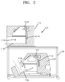
- Fig. 2 is a sectional view of the biological tissue diagnostic apparatus according to the embodiment of the present invention
- Fig. 3 is a block diagram of the biological tissue diagnostic apparatus according to the embodiment of the present invention.
- FIG. 4 is a flowchart illustrating a biological tissue diagnostic method according to one embodiment of the present invention
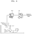
- Fig. 5 and Fig. 6 are schematic views showing inspection of blood flow of legs using the biological tissue diagnostic apparatus according to the embodiment of the present invention.
- Fig. 7 is a graph obtained by patterning a change of fluorescence intensity from a contrast agent over time after administration of the contrast agent to legs of a person with normal blood flow using the biological tissue diagnostic apparatus according to the embodiment of the present invention.
- Fig. 8 is a schematic diagram of a method of defining Onset, T max , Plateau T max , and Slope among characteristic values by which a pattern is characterized for comparison of patterned data.
- Fig. 9 is a graph obtained by patterning blood flow of hands and legs of a person with normal blood flow using the biological tissue diagnostic apparatus according to the embodiment of the present invention
- Fig. 10 is a graph obtained by patterning blood flow of hands and legs of a person with abnormal blood flow using the biological tissue diagnostic apparatus according to the embodiment of the present invention
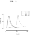
- Fig. 11 is a reference view for illustrating a method for diagnosing an abnormal tissue of interest in-vivo based on graphs obtained by patterning fluorescence intensity of a plurality of tissues of interest.
- a biological tissue diagnostic apparatus diagnoses abnormality of vessels or perfusion including blood flow and blood vessels by administering a contrast agent to a biological tissue.
- the biological tissue diagnostic apparatus includes: at least one lighting unit 110 irradiating at least one tissue of interest in-vivo within a living body with a contrast agent administered thereto with contrast agent excitation energy; at least one inspection unit 120 detecting energy emitted from the contrast agent in the living body; and a determination unit 130 determining abnormality of vessels or perfusion of the at least one tissue of interest based on image data detected by the at least one inspection unit 120.
- the determination unit 130 includes: a pattern processor 132 patterning a change of the image data over time; a characteristic value calculator 134 calculating at least one characteristic value based on the patterned data; and a diagnostic unit 136 diagnosing abnormality of vessels or perfusion of the at least one tissue of interest based on the at least one characteristic value or combinations thereof.
- living body refers to an animal or human and, for convenience, is shown as being a part of the human body.
- tissue of interest may refer to, for example, microvessels of fingertips and toe tips.
- the biological tissue diagnostic apparatus 100 may examine or diagnose microvessels of fingers of both hands at the same time or may examine or diagnose microvessels of fingers of one hand.
- the biological tissue diagnostic apparatus 100 according to this embodiment may examine or diagnose microvessels of toes of both feet at the same time or may examine or diagnose microvessels of toes of one foot.
- the biological tissue diagnostic apparatus 100 may examine or diagnose microvessels of both fingers and toes at the same time or may examine or diagnose microvessels of any one of fingers and toes.
- the biological tissue diagnostic apparatus 100 may simultaneously or separately examine and/or diagnose perfusion (blood flow, lymphatic flow) or vessels (blood vessels, lymphatic vessels) of a plurality of tissues of interest.
- the lighting unit 110 serves to irradiate tissues of interest in-vivo with a contrast agent administered thereto.
- the contrast agent molecular detecting contrast agent
- the contrast agent is a near-infrared fluorescent contrast agent, and is preferably a polymethine pigment, which is a fluorescent pigment, particularly a near-infrared pigment.
- the contrast agent may be an indocyanine green (ICG) pigment.
- the contrast agent is administered to a living body orally or non-orally.
- the lighting unit 110 includes a casing 112 and an illumination member 114.
- the casing 112 is provided with a dark chamber 116 which blocks external light to increase contrast of an image.
- the dark chamber 116 serves to accommodate a living body.
- the casing 112 may be provided with a plurality of dark chambers 116 to accommodate a plurality of living bodies at the same time.
- the plurality of dark chambers 116 may be provided to a single casing 112 or may be provided to the casings 112 in one-to-one correspondence.
- the lighting unit is shown as including two casings 112, and each of the casings 112 is shown as including a pair of dark chambers 116.
- This structure allows the dark chambers 116 to receive both hands and both feet, which are parts of a living body.
- each of the casings 112 may be formed in various shapes and formed of various materials.
- two casings 112 be supported by a frame 119 and integrally connected with each other.
- the frame 119 may be formed in various shapes and formed of various materials.
- the illumination member 114 is provided at a portion of the casing 112 corresponding to the inside of the dark chamber 116 and serves to illuminate biological tissues received by the dark chamber 116.
- the illumination member 114 is configured to emit excitation energy (light) at a specific wavelength to a living body (or living bodies) with a contrast agent, particularly indocyanine green (ICG) injected therein, and serves to activate ICG within a tissue of interest of the living body (or living bodies) with ICG injected therein such that fluorescent signals from the tissue of interest (biological tissue) can be observed.
- a contrast agent particularly indocyanine green (ICG) injected therein
- the excitation energy (light) emitted from the illumination member 114 has a center wavelength of 750 nm to 780 nm, which corresponds to a near-infrared region. Near-infrared rays in this wavelength range are used to observe fluorescent light through ICG administration.
- a light emitting diode or laser emitting energy in the wavelength range set forth above may be used.
- the illumination member 114 may include a white light illumination member which allows monitoring whether a living body is properly supported by a holding member 118 in the dark chamber 116, in addition to the near-infrared illumination member allowing the contrast agent to emit light.
- the illumination member 114 may be provided to the dark chamber 116 in a one-to-one correspondence and may be adjusted in illumination time by a controller 140.
- the controller 140 may control the illumination members 114 provided to the dark chambers 116 to irradiate a tissue of interest (biological tissue) with excitation energy for different periods of time. It should be understood that the controller 140 may control all of the illumination members 114 to be turned on/off at the same time.
- Each of the dark chambers 116 is provided therein with a holding member 118.
- the holding member 118 serves to hold a corresponding part of a living body and may be deformed to allow detecting in a sitting position, a lying position, or a lying on the side position.
- the holding member 118 may be formed in various shapes and formed of various materials to support a corresponding part of a living body, i.e. a hand or foot.
- the inspection unit 120 serves to detect energy (fluorescent signals) in the near-infrared range emitted from a tissue of interest (or tissues of interest) in-vivo (contrast agent) by light irradiated to the tissue of interest in-vivo.
- the inspection unit 120 may include a filter member 122 and a detecting member 124.
- the filter member 122 is disposed in the casing 112 to correspond to each of the dark chambers 116 and serves to mainly transmit energy in the near-infrared range among energy (fluorescent signals) emitted from the tissue of interest in-vivo (contrast agent).
- the range of energy in the near-infrared region allowed to pass through the filter member may vary depending upon specifications of the filter member 122 or system requirements.
- the filter member 122 transmits light in a specific wavelength range among fluorescent signals generated from a corresponding tissue of interest (biological tissue) due to light emitted from the illumination member 114.
- the filter member 122 only transmits light having a near-infrared wavelength of 800 nm to 850 nm among fluorescent signals coming out of a living body by the illumination member 114.
- a band pass filter may be provided to each of the dark chambers 116 and can be adjusted in position and can adjust intensity of energy (fluorescent signals) having a near-infrared wavelength.
- the detecting member 124 is disposed in each of the dark chambers 116 and serves to detect energy (fluorescent signals) in the near-infrared region passing through the filter member 122. Particularly, the detecting member 124 senses light having passed through the filter member 122 and converts the light into a digital signal. Specifically, the detecting member converts an analog image into digital data by changing the image into electrical signals and stores the digital data in a storage medium.
- the detecting member 124 may be a charge-coupled device (CCD) camera among digital cameras. The detecting member 124 detects fluorescent signals that have been input to the inspection unit 120 and then passed through the filter member 122, converts the image into digital data and outputs the digital data.
- CCD charge-coupled device
- the inspection unit 120 and the illumination member 114 may be disposed close to each other and the inspection unit 120 is disposed to photograph a corresponding tissue of interest (biological tissue) within the casing 112 of the lighting unit 110.
- a plurality of filter members 122 and a plurality of detecting members 124 may be provided equal in number to independently examine one or more tissues of interest and are placed within a corresponding dark chamber 116.
- the plurality of filter members 122 and the plurality of detecting members 124 are connected to the controller 140 which controls operation time.
- the controller 140 controls operation time of all of the inspection units 120 disposed within the respective dark chambers 116. In other words, the controller 140 may control the detecting members 124 to operate for the same period of time or to operate for different periods of time.
- Operation time of the detecting member 124 may be set from a time point that detecting is started to a time point that emission of near-infrared rays from the contrast agent is no longer detected, and the detecting member 124 may be controlled to perform detecting at preset time intervals. Particularly, it is desirable that the detecting member 124 be controlled to perform detecting at time intervals of several milliseconds to 10 seconds.
- operation time of the detecting member 124 may vary depending upon the kind or dosage of the contrast agent, external temperature, and the like. The interval in which the detecting member 124 operates may be adjusted within 10 seconds.
- the determination unit 130 serves to determine abnormality of perfusion and/or vessels of a tissue of interest (or tissue of interest) in-vivo based on the image data detected by the inspection unit 120 (resulting data for the near-infrared region).
- the determination unit 130 may be further provided with an input device (not shown) for receiving the digital data from the inspection unit 120, and an output device 130 for outputting data may be connected to the determination unit 130 or the determination unit 130, as shown in Fig. 5 and Fig. 6 .
- the digital data output from the inspection unit 120 are transmitted to the determination unit 130 through wireless or wired communication, for which RSC 232, a parallel port, IEEE 1934, or a USB is preferably used.
- the determination unit 130 includes the pattern processor 132, the characteristic value calculator 134, and the diagnostic unit 136.
- the pattern processor 132 serves to pattern image data obtained by detecting a tissue of interest or a region of interest (ROI) for each inspection area of a living body.
- the characteristic value calculator 134 serves to calculate various characteristic values reflecting perfusion conditions (for example, blood flow conditions) from the resulting patterned data, and the diagnostic unit 136 serves to diagnose abnormality (for example, vascular disease) of vessels or perfusion of the tissue of interest in-vivo based on the calculated characteristic values or combinations thereof.
- perfusion conditions for example, blood flow conditions
- diagnostic unit 136 serves to diagnose abnormality (for example, vascular disease) of vessels or perfusion of the tissue of interest in-vivo based on the calculated characteristic values or combinations thereof.
- the pattern processor 132 processes input signals into fluorescence intensity of a tissue of interest over time in order to pattern the input signals; the characteristic value calculator 134 calculates partial characteristic values from the processed pattern of fluorescence intensity over time; and the diagnostic unit 136 diagnoses abnormality of vessels or perfusion of the tissue of interest or calculates the perfusion rate using each of the characteristic values calculated by the characteristic value calculator 134 or combinations of the characteristic values.
- the determination unit 130 measures partial ICG fluorescence intensity over time of ICG administered to a living body, and performs pattern analysis for each part and diagnosis of abnormality of vessels (perfusion).
- the determination unit 130 may be provided to correspond to the inspection unit 120 provided to each of the dark chambers 116 in one-to-one correspondence. Alternatively, only one determination unit may be provided to be connected to a plurality of the inspection units 120. In addition, the inspection unit 120 is connected to the controller 140.
- the controller 140 controls the lighting unit 110 and inspection unit 120 provided for each of different tissues of interest in-vivo as well as the determination unit 130, and adjusts operation time of each component of the lighting unit 110 and components of the inspection unit 120.
- the characteristic value refers to a factor converted into a real number that can characterize a pattern of resulting data over time in a tissue of interest or a region of interest (ROI), and the determination unit 130 may calculate blood flow in the region of interest through comparison of the characteristic values.
- the characteristic value (values) may be obtained from changes over time of specific physical values (for example, ICG fluorescence intensity) in a tissue of interest, acquired from the data detected by the inspection unit 120.
- a combination value of one or more characteristic values included in the normal groups or the abnormal groups allows the corresponding groups to be classified with a sensitivity and specificity of 80% or higher.
- a plurality of different tissues of interest are simultaneously photographed in the plurality of dark chambers 116 accommodating the illumination member 114, the filter member 122, and the detecting member 124, and the determination unit 130 compares and analyzes characteristic values calculated with respect to the plurality of tissues of interest to determine whether vessels or perfusion (blood vessels, blood flow) is normal.
- Fig. 5 and Fig. 6 are views illustrating inspection of blood flow of a foot, which is one of in-vivo regions.
- unexplained reference numerals are as defined above.
- the determination unit 130 determining abnormality of blood flow and blood vessels of a living body can diagnose development of vascular complication in a diabetic patient or lymphatic disease depending upon results of determination of whether blood flow and blood vessels is abnormal.
- the determination unit 130 can diagnose arteriosclerosis and arteriostenosis, particularly arteriosclerosis and arteriostenosis in the coronary arteries, the carotid, the femoral artery, the anterior tibial artery, and the posterior tibial artery depending upon results of determination of whether blood flow and blood vessels is abnormal.
- the determination unit 130 can diagnose cold hands and feet, Raynaud disease, and Raynaud syndrome depending upon results of determination of whether blood flow and blood vessels is abnormal.
- the determination unit 130 preferably represents results of determination as to whether blood flow and blood vessels are abnormal by one or more numerical values or a perfusion map.
- the determination unit 130 may determine occurrence of vascular complication in a diabetic patient or lymphatic diseases; may determine occurrence of arteriosclerosis and arteriostenosis, particularly, arteriosclerosis and arteriostenosis in the coronary arteries, the carotid, the femoral artery, the anterior tibial artery, and the posterior tibial artery; and may diagnose cold hands and feet, Raynaud disease, and Raynaud syndrome through the pattern processor 132, the characteristic value calculator 134, and the diagnostic unit 136.
- determination or diagnosis results by the determination unit 130 are represented by one or more numerical values or a perfusion map, whereby a user or patient can easily interpret or understand the results.
- characteristic values based on which abnormality of vessels or perfusion (for example, blood vessel, lymphatic vessel, blood flow, lymphatic flow, and the like) is determined, are converted into a numerical formula according to the following procedure.
- a graph is plotted by detecting changes in ICG fluorescence intensity in the top of a foot at 1/5 Hz for 10 minutes after in vivo injection of ICG and patterning the changes with the highest fluorescence intensity assumed as 1 (see Fig. 7 ).
- Fig. 8 is a graph for illustrating changes in fluorescence intensity over time of a person with a normal blood flow and shows T max , Plateau T max , Onset, and Slope, each of which is a characteristic value.
- the biological tissue diagnostic apparatus 100 allows blood flow of a tissue of interest (biological tissue) corresponding to hands or feet in a normal state to be represented by a change in fluorescence intensity over time.
- characteristic values are defined as combinations of at least one of T max , Plateau T max , Onset, and Slope.
- T max refers to a period of time to reach a point where fluorescence intensity reaches a maximum value in a patterned near-infrared emission region
- Plateau T max refers to a section in which fluorescence intensity is greater than a specific value in the patterned near-infrared emission region.
- Onset refers to a region from a time point that pattering is started (initial detection point) to a time point that a sharp inflection occurs in the curve, causing the slope of the curve to become greater than a reference slope; and Slope refers to a pattern in a downward region after Plateau T max in a patterned near-infrared emission region (a period of time from a time point that fluorescence intensity starts to decrease below a reference value to a time point that fluorescence intensity is last detected).
- Plateau section in which fluorescence intensity is greater than or equal to a predetermined reference value (FI c ) or a section with a gradual inclination is observed, it can be said that blood flow is abnormal.
- the period of time (T max ) that it takes to reach the point where ICG fluorescence intensity of ischemic tissue reaches a maximum value in the corresponding section (Plateau T max ) is considered to determine whether blood vessel or blood flow is abnormal.
- T max the period of time that it takes to reach the point where ICG fluorescence intensity of ischemic tissue reaches a maximum value in the corresponding section
- a Plateau T max value ratio of the other tissue of interest to the one tissue of interest is greater than a second preset value, it can be determined that the blood flow is abnormal.
- an Onset value ratio of the other tissue of interest to the one tissue of interest is greater than a third preset value, it can be determined that the blood flow is abnormal.
- the one tissue of interest may be a toe and the other tissue of interest may be a finger.
- Each of the first preset value, the second preset value, and the third preset value is an average value (normal value) for normal persons.
- characteristic values of the other tissue of interest i.e. both hands
- characteristic values of the other tissue of interest may be averaged or separately used.
- one tissue of interest has different ICG patterns, that is, when the ICG patterns of both feet are different from each other, whether one or both legs have arteriostenosis may be diagnosed by finding characteristic values of both feet.
- T max and Onset there is not much difference in T max and Onset between both hands and both feet in normal groups of Fig. 9 , whereas, in the case of a patient with abnormal blood flow of Fig. 10 , T max and Onset of both hands are considerably small as compared with those of both feet and there is significant difference in Onset value between both feet.
- arteriostenosis is diagnosed in both legs and it is diagnosed that the left leg has a relatively severe degree of stenosis.
- Fig. 11 is a reference view illustrating a method for diagnosing an abnormal tissue of interest based on graphs obtained by patterning fluorescence intensity in a plurality of tissues of interest.
- trend graphs obtained by patterning fluorescence intensity in four tissues of interest trend graphs on three tissues of interest (upper left, lower right, lower left) are synchronized in terms of shape in a certain distribution range, whereas the trend graph on the other tissue of interest (upper right), i.e. a tissue of interest of the right hand, shows a different trend therefrom.
- the three synchronized tissues of interest may be considered to exhibit normal blood flow or blood vessel characteristics, whereas blood flow or blood vessel conditions of the other tissue of interest may be considered abnormal.
- the determination unit 130 may diagnose a tissue of interest corresponding to characteristic values departing from a preset normal distribution range as an abnormal tissue of interest.
- the determination unit 130 outputs diagnostic results based on the data detected by the inspection unit 120 through the output device 138.
- the output device 138 a CRT monitor, an LCD, a plasma display, and the like may be used.
- Fig. 4 is a flowchart illustrating a biological tissue diagnostic method according to one embodiment of the present invention. Next, a biological tissue diagnostic method according to one embodiment will be described with reference to Fig. 4 .
- one or more lighting units 110 and one or more inspection units 120 may be provided depending upon the number of tissues of interest.
- an illumination member 114 irradiates at least one tissue of interest in-vivo with a contrast agent administered thereto with contrast agent excitation energy (S410), and an inspection unit 120 detects energy (energy in the near-infrared region) emitted from the tissue of interest in-vivo (contrast agent) (S420).
- a determination unit 130 determines abnormality of vessels or perfusion of the at least one tissue of interest based on image data detected by the inspection unit 120 (S430).
- S430 A more detailed description of step S430 is as follows.
- a pattern processor 132 patterns a change of the image data over time (S431).
- the pattern processor 132 receives electrical signals, which are detected by a detecting member 124 of the inspection unit 120 and then digitized, and process the signals, thereby patterning a trend of a specific physical value (for example, fluorescence intensity) over time.
- a specific physical value for example, fluorescence intensity
- a characteristic value calculator 134 calculates at least one characteristic value based on the patterned data (S432). In other words, the characteristic value calculator 134 calculates one or more characteristic values from the patterned data or patterned graph.
- the characteristic value (values) is obtained from a change of a specific physical value over time in a tissue of interest.
- the at least one characteristic value may include at least one selected from among a period of time from a time point that a physical value (for example, fluorescence intensity) is initially detected to a time point that the physical value reaches a maximum value (T max ); a slope with which the physical value increases or decreases for a specific period of time; a period of time for which the physical value is maintained at a reference value or more (Plateau T max ); a period of time from the time point that the physical value is initially detected to a time point before the physical value increases with a slope greater than or equal to a reference slope (Onset); and a period of time from a time point that the physical value starts to decrease below the reference value to a time point that the physical value is detected last (Slope).
- a physical value for example, fluorescence intensity
- a diagnostic unit 136 diagnoses, based on the at least one characteristic value or combinations of at least two characteristic values, whether vessels or perfusion of the at least one tissue of interest in-vivo is abnormal. For example, in the case that one tissue of interest (biological tissue) and another tissue of interest are simultaneously subjected to blood flow diagnosis, if difference in T max value between the one tissue of interest and the other tissue of interest is greater than a first preset value, it is determined that the blood flow is abnormal, and if a Plateau T max value ratio of the other tissue of interest to the one tissue of interest is greater than a second preset value, it is also determined that the blood flow is abnormal.
- each of the first preset value, the second preset value, and the third preset value is an average value (normal value) for normal persons.
- the tissue of interest may include a plurality of tissues of interest
- the determination unit 130 may diagnose an abnormal tissue of interest by comparing the characteristic values of the tissues of interest.
- the determination unit 130 may diagnose a tissue of interest corresponding to a characteristic value departing from a preset normal distribution range as an abnormal tissue of interest. More specifically, as shown in Fig. 11 , in trend graphs obtained by pattering fluorescence intensity of four tissues of interest, the trend graphs of fluorescence intensity on three tissues of interest are synchronized in shape in a certain distribution range, whereas the trend graph on the other tissue of interest shows a different trend therefrom.
- the three synchronized tissues of interest may be considered to exhibit normal blood flow or blood vessel characteristics, whereas blood flow or blood vessel conditions of the other tissue of interest may be considered abnormal.
- the determination unit 130 may diagnose a tissue of interest corresponding to a characteristic value departing from a preset normal distribution range as an abnormal tissue of interest.
- the biological tissue diagnostic apparatus and method according to the present invention can continuously photograph tissues of interest in-vivo for a certain period of time, particularly blood vessels/lymphatic vessels which are distributed near the skin, such as peripheral tissues and carotid territories, allowing easy photographing with near-infrared light, thereby accurately diagnosing whether vessels or perfusion of the tissues of interest is abnormal.
- the biological tissue diagnostic apparatus and method according to the present invention can simultaneously or selectively photograph one or more tissues in-vivo, specifically both hands and both feet to diagnose abnormality of blood vessels or lymphatic vessels, thereby performing accurate diagnosis throughcomplex diagnosis while reducing diagnostic time.
Landscapes
- Health & Medical Sciences (AREA)
- Life Sciences & Earth Sciences (AREA)
- Engineering & Computer Science (AREA)
- Medical Informatics (AREA)
- Animal Behavior & Ethology (AREA)
- Public Health (AREA)
- Biophysics (AREA)
- Pathology (AREA)
- Biomedical Technology (AREA)
- Heart & Thoracic Surgery (AREA)
- Physics & Mathematics (AREA)
- Molecular Biology (AREA)
- Surgery (AREA)
- Veterinary Medicine (AREA)
- General Health & Medical Sciences (AREA)
- Physiology (AREA)
- Cardiology (AREA)
- Hematology (AREA)
- Nuclear Medicine, Radiotherapy & Molecular Imaging (AREA)
- Vascular Medicine (AREA)
- Radiology & Medical Imaging (AREA)
- Artificial Intelligence (AREA)
- Computer Vision & Pattern Recognition (AREA)
- Psychiatry (AREA)
- Signal Processing (AREA)
- High Energy & Nuclear Physics (AREA)
- Optics & Photonics (AREA)
- Human Computer Interaction (AREA)
- Investigating, Analyzing Materials By Fluorescence Or Luminescence (AREA)
Applications Claiming Priority (3)
| Application Number | Priority Date | Filing Date | Title |
|---|---|---|---|
| KR20130102415 | 2013-08-28 | ||
| KR20140089525A KR20150024766A (ko) | 2013-08-26 | 2014-07-16 | 생체조직 진단장치 및 방법 |
| PCT/KR2014/007771 WO2015030424A1 (ko) | 2013-08-28 | 2014-08-21 | 생체조직 진단장치 및 방법 |
Publications (2)
| Publication Number | Publication Date |
|---|---|
| EP3040026A1 true EP3040026A1 (de) | 2016-07-06 |
| EP3040026A4 EP3040026A4 (de) | 2016-09-21 |
Family
ID=53021822
Family Applications (1)
| Application Number | Title | Priority Date | Filing Date |
|---|---|---|---|
| EP14841148.1A Withdrawn EP3040026A4 (de) | 2013-08-28 | 2014-08-21 | Diagnosevorrichtung und -verfahren für biologisches gewebe |
Country Status (4)
| Country | Link |
|---|---|
| US (1) | US20160206218A1 (de) |
| EP (1) | EP3040026A4 (de) |
| JP (1) | JP6356246B2 (de) |
| KR (1) | KR20150024766A (de) |
Families Citing this family (4)
| Publication number | Priority date | Publication date | Assignee | Title |
|---|---|---|---|---|
| JP2019170469A (ja) * | 2018-03-27 | 2019-10-10 | テルモ株式会社 | リンパ管機能測定装置およびリンパ管機能測定方法 |
| JP7812773B2 (ja) * | 2022-11-04 | 2026-02-10 | 浜松ホトニクス株式会社 | リンパ系検査装置 |
| JPWO2024095519A1 (de) * | 2022-11-04 | 2024-05-10 | ||
| JP7817139B2 (ja) * | 2022-11-04 | 2026-02-18 | 浜松ホトニクス株式会社 | リンパ系検査装置 |
Family Cites Families (5)
| Publication number | Priority date | Publication date | Assignee | Title |
|---|---|---|---|---|
| US7474906B2 (en) * | 2001-05-22 | 2009-01-06 | Alfred E. Mann Institute For Biomedical Engineering At The University Of Southern California | Method for dye injection for the transcutaneous measurement of cardiac output |
| KR100818669B1 (ko) * | 2007-03-09 | 2008-04-02 | 한국과학기술원 | 하지 관류정도 측정장치 |
| KR100973183B1 (ko) * | 2008-10-29 | 2010-07-30 | 한국과학기술원 | 관류 정도 분석 장치 및 방법 |
| EP2459053A2 (de) * | 2009-07-28 | 2012-06-06 | F. Hoffmann-La Roche AG | Nicht invasives optisches in-vivo-bildgebungsverfahren |
| KR101166165B1 (ko) * | 2011-01-19 | 2012-07-18 | 주식회사 뷰웍스 | Icg 형광 동역학 분석을 이용한 뇌혈류 분석 장치 및 방법 |
-
2014
- 2014-07-16 KR KR20140089525A patent/KR20150024766A/ko not_active Ceased
- 2014-08-21 US US14/915,387 patent/US20160206218A1/en not_active Abandoned
- 2014-08-21 EP EP14841148.1A patent/EP3040026A4/de not_active Withdrawn
- 2014-08-21 JP JP2016538844A patent/JP6356246B2/ja not_active Expired - Fee Related
Also Published As
| Publication number | Publication date |
|---|---|
| JP2016531700A (ja) | 2016-10-13 |
| US20160206218A1 (en) | 2016-07-21 |
| JP6356246B2 (ja) | 2018-07-11 |
| EP3040026A4 (de) | 2016-09-21 |
| KR20150024766A (ko) | 2015-03-09 |
Similar Documents
| Publication | Publication Date | Title |
|---|---|---|
| US11033207B2 (en) | Dynamic optical tomographic imaging devices methods and systems | |
| JP5028256B2 (ja) | 光学的にアクセス可能な血管における血液の酸素飽和度を決定するための分光測光法 | |
| KR101599077B1 (ko) | 류마티스 관절염 등의 염증성 질환의 진단 또는 진단 준비 및/또는 치료 모니터링 장치 및 절차 | |
| JP5057783B2 (ja) | 被験者の末梢血液循環を評価するためのシステム又は方法 | |
| JP2007509718A (ja) | 体液および電解質障害をモニタリングするデバイスおよび方法 | |
| US20080004513A1 (en) | VCSEL Tissue Spectrometer | |
| Venermo et al. | Pilot assessment of the repeatability of indocyanine green fluorescence imaging and correlation with traditional foot perfusion assessments | |
| JP5594788B2 (ja) | 関節の状態の光学的検出方法及び光学的検出用装置 | |
| Lapitan et al. | A method and a device prototype for noninvasive measurements of blood perfusion in a tissue | |
| JP5594787B2 (ja) | 光学的検出方法及び関節の状態を光学的に検出する装置 | |
| EP3040026A1 (de) | Diagnosevorrichtung und -verfahren für biologisches gewebe | |
| CN111970963B (zh) | 评估微循环振荡的方法和用于评估微循环振荡的装置 | |
| KR100973183B1 (ko) | 관류 정도 분석 장치 및 방법 | |
| KR101135067B1 (ko) | 레이노 현상 검사 장치 및 방법 | |
| CN112996439A (zh) | 用于确定生物组织的氧合血含量的系统和方法 | |
| IT201800011113A1 (it) | Dispositivo Point-of-Care per diagnosi rapida di intossicazione | |
| US9895064B2 (en) | Device and method for determining a disease activity | |
| WO2015030424A1 (ko) | 생체조직 진단장치 및 방법 | |
| Leyba et al. | Evaluation of hemodynamics in a murine hindlimb ischemia model using spatial frequency domain imaging | |
| KR101032479B1 (ko) | 수학적 모델에 기반한 말초조직 관류 정도 측정장치 및 측정방법 | |
| TWI780949B (zh) | 血液流量評估方法及其系統 | |
| US20250195001A1 (en) | Lower extremity vascular lesion evaluation device and lower extremity vascular lesion evaluation method | |
| Badiola et al. | Monte Carlo simulation-based analysis of unobtrusive PPG monitoring through clothes | |
| Smith et al. | Technique for noninvasive monitoring of blood loss via oxygen saturation measurements in the eye | |
| Chaiken et al. | Analyzing near infrared scattering from human skin to monitor changes in hematocrit |
Legal Events
| Date | Code | Title | Description |
|---|---|---|---|
| PUAI | Public reference made under article 153(3) epc to a published international application that has entered the european phase |
Free format text: ORIGINAL CODE: 0009012 |
|
| 17P | Request for examination filed |
Effective date: 20160321 |
|
| AK | Designated contracting states |
Kind code of ref document: A1 Designated state(s): AL AT BE BG CH CY CZ DE DK EE ES FI FR GB GR HR HU IE IS IT LI LT LU LV MC MK MT NL NO PL PT RO RS SE SI SK SM TR |
|
| AX | Request for extension of the european patent |
Extension state: BA ME |
|
| A4 | Supplementary search report drawn up and despatched |
Effective date: 20160819 |
|
| RIC1 | Information provided on ipc code assigned before grant |
Ipc: A61B 5/0275 20060101AFI20160815BHEP |
|
| DAX | Request for extension of the european patent (deleted) | ||
| STAA | Information on the status of an ep patent application or granted ep patent |
Free format text: STATUS: EXAMINATION IS IN PROGRESS |
|
| 17Q | First examination report despatched |
Effective date: 20180802 |
|
| STAA | Information on the status of an ep patent application or granted ep patent |
Free format text: STATUS: THE APPLICATION IS DEEMED TO BE WITHDRAWN |
|
| 18D | Application deemed to be withdrawn |
Effective date: 20210302 |