EP3289565B1 - Verfahren und vorrichtung zur nichtinvasiven indirekten identifizierung von stellen von veränderungen der blut-retina-schranke - Google Patents
Verfahren und vorrichtung zur nichtinvasiven indirekten identifizierung von stellen von veränderungen der blut-retina-schranke Download PDFInfo
- Publication number
- EP3289565B1 EP3289565B1 EP16727840.7A EP16727840A EP3289565B1 EP 3289565 B1 EP3289565 B1 EP 3289565B1 EP 16727840 A EP16727840 A EP 16727840A EP 3289565 B1 EP3289565 B1 EP 3289565B1
- Authority
- EP
- European Patent Office
- Prior art keywords
- oct
- retinal
- scan
- previous
- sites
- Prior art date
- Legal status (The legal status is an assumption and is not a legal conclusion. Google has not performed a legal analysis and makes no representation as to the accuracy of the status listed.)
- Active
Links
Images




Classifications
-
- A—HUMAN NECESSITIES
- A61—MEDICAL OR VETERINARY SCIENCE; HYGIENE
- A61B—DIAGNOSIS; SURGERY; IDENTIFICATION
- A61B3/00—Apparatus for testing the eyes; Instruments for examining the eyes
- A61B3/10—Objective types, i.e. instruments for examining the eyes independent of the patients' perceptions or reactions
- A61B3/102—Objective types, i.e. instruments for examining the eyes independent of the patients' perceptions or reactions for optical coherence tomography [OCT]
-
- A—HUMAN NECESSITIES
- A61—MEDICAL OR VETERINARY SCIENCE; HYGIENE
- A61B—DIAGNOSIS; SURGERY; IDENTIFICATION
- A61B3/00—Apparatus for testing the eyes; Instruments for examining the eyes
- A61B3/10—Objective types, i.e. instruments for examining the eyes independent of the patients' perceptions or reactions
- A61B3/12—Objective types, i.e. instruments for examining the eyes independent of the patients' perceptions or reactions for looking at the eye fundus, e.g. ophthalmoscopes
- A61B3/1225—Objective types, i.e. instruments for examining the eyes independent of the patients' perceptions or reactions for looking at the eye fundus, e.g. ophthalmoscopes using coherent radiation
-
- G—PHYSICS
- G06—COMPUTING OR CALCULATING; COUNTING
- G06T—IMAGE DATA PROCESSING OR GENERATION, IN GENERAL
- G06T7/00—Image analysis
- G06T7/0002—Inspection of images, e.g. flaw detection
- G06T7/0012—Biomedical image inspection
-
- G—PHYSICS
- G06—COMPUTING OR CALCULATING; COUNTING
- G06T—IMAGE DATA PROCESSING OR GENERATION, IN GENERAL
- G06T2207/00—Indexing scheme for image analysis or image enhancement
- G06T2207/10—Image acquisition modality
- G06T2207/10072—Tomographic images
- G06T2207/10101—Optical tomography; Optical coherence tomography [OCT]
-
- G—PHYSICS
- G06—COMPUTING OR CALCULATING; COUNTING
- G06T—IMAGE DATA PROCESSING OR GENERATION, IN GENERAL
- G06T2207/00—Indexing scheme for image analysis or image enhancement
- G06T2207/30—Subject of image; Context of image processing
- G06T2207/30004—Biomedical image processing
- G06T2207/30041—Eye; Retina; Ophthalmic
Definitions
- the present disclosure relates to a method and device for the non-invasive indirect identification of the sites of alterations of the Blood-Retinal Barrier, in particular it relates to the automated analysis of retinal extracellular space using Optical Coherence Tomography.
- DM Diabetes Mellitus
- DR Diabetic retinopathy
- BRB blood-retinal barrier
- Fluorescein angiography is the imaging technique most frequently used to document the changes occurring in the BRB in DR. It uses sodium fluorescein (NaFI) as a dye and images are acquired after intravenous administration. Because of the intravenous dye administration, minor adverse reactions occur in 5% of the cases. Severe complications, although more rare, may occur, the reason why this imaging technique requires the presence of a MD. Death may still occur in the first 24 to 48 hours for each 220.000 cases [2].
- OCT Optical Coherence Tomography
- the present disclosure relates to a method and device for the non-invasive indirect identification of the sites of alterations of the Blood-Retinal Barrier, herewith optionally mentioned as OCT-Leakage, in particular it relates to the automated analysis of retinal extracellular space using Optical Coherence Tomography.
- the disclosure presents a practical and efficient approach, being a robust rapid method to detect retinal alterations, able to produce images that are easier and simpler to interpret by an OCT operator.
- the disclosure when coupled to a suitable layer segmentation of the retina produces images that are particularly evident of retinal alteration.
- the disclosure an automated method for detecting sites of retinal alterations from optical coherence tomography, OCT, data of an eye, wherein said data comprises OCT A-scan data over an area of the eye and each OCT A-scan data comprises a series of reflectivity values along the depth direction of the OCT A-scan data, said method comprising:
- the threshold is predetermined according to the optical reflectivity of a healthy population.
- a threshold value can be established from the optical reflectivity of A-Scans from a series of healthy control eyes.
- the specific threshold value is established for the OCT device used, as it is dependent on the equipment.
- the predetermined threshold may be 20 when measured using an "OCT Angioplex" or may be 10 when measured using an "OCT Cirrus 5000".
- the method comprises previously obtaining OCT A-scans over an area of the eye, each A-scan comprising a series of reflectivity values along the depth direction of the scan.
- the method further comprising preprocessing the OCT A-scan data, said preprocessing comprising one or more of speckle reduction or normalization operations.
- the method is used for identifying increases of intercellular fluid or increases of extracellular space in the retina or alterations of the blood-retinal barrier.
- the method is used for identifying sites of alterations of the blood-retinal barrier.
- the layer discrimination of the LOR data enhances the information obtained from the OCT, being less sensitive to the image interferences caused by blood vessels - see for example image h).
- NPDR non-proliferative diabetic retinopathy
- EDRS Early Treatment Diabetic Retinopathy Study
- OCT-Microangiography was also performed, in order to evaluate the complementarity of the information obtained by OCT-Microangiography and OCT-Leakage.
- OCT scans the retina locations with a low coherence near-infra red light beam from a superluminescent diode to compute a retina profile by interferometry of the reflected light from the sample and reference arms.
- This axial retinal profile named as A-Scan, is repeated on the next laterally adjacent retina location to produce a B-Scan which provides a high resolution cross-sectional image of the retina.
- OCT microangiograms were acquired with AngioPlex OCT Angiography (Carl Zeiss Meditec, Dublin, CA, USA). This system uses of a light source centered at 840 nm with a 50 nm bandwidth, providing a 5 um axial resolution in tissue. With a acquisition speed of 27 000 A-Scans per second, we were able to scan the 6x6x2 mm2 or 3x3x2 mm2 of retinal volume by 350x350x1024 or 245x245x1024 voxels respectively.
- RNFL Retinal Nerve Fiber Layer
- GCL+IPL Ganglion Cell and Inner Plexiform Layers
- INL Inner Nuclear Layer
- OPL Outer Plexiform Layer
- ONL Outer Nuclear Layer
- IS+OS Inner and Outer Segment
- RPE Retinal Pigment Epithelium
- Root mean square errors (RMSE) between automatic and human grader segmentations for healthy subjects and for diabetic patients are of the same order of magnitude. Larger RMSE were found at OS/RPE interface ranging from 3.97 to 16.61 ⁇ m (m ⁇ sd:6.92 ⁇ 2.21 ⁇ m) and from 0.65 to 17.76 ⁇ m (m ⁇ sd:4.24 ⁇ 2.69 ⁇ m) for healthy and diabetic patients, respectively.
- FA fluorescein angiography
- Extracellular fluid distribution in the retina is represented on OCT by the distribution of sites of lowest optical reflectivity. Increases or decreases in extracellular fluid distribution of a given area of the retina can, therefore, be measured by the ratio of sites of Low Optical Reflectivity (LOR) identified in the area under evaluation.
- LOR Low Optical Reflectivity
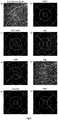
- Analysis of A-Scans from the healthy control retinas allowed us to establish a reflectivity threshold ( Figure 1 ). When A-Scan optical reflectivity value is below the chosen threshold, the location of the A-Scan is identified as a location of LOR and considered for quantification of the LOR area ratio.
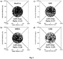
- the following pertains to an analysis of SD-OCT LOR area ratios in the Central Subfield. An analysis was performed separately for each of the segmented layers generating for each one an SD-OCT optical reflectivity map for the diabetic eyes, with NPDR only, with NPDR and SME and NPDR with CME, and compared with healthy control eyes ( Figure 3 and Table 2).
- the SD-OCT optical reflectivity images identify well the changes in extracellular space and their distribution in the macular region. Lower optical reflectivity represented by more clear spaces is well demonstrated in the INL when comparing central subfields from patients with different degrees of edema ( Figure 3 ). In the eyes examined there was a good correlation between increases in retinal thickness and LOR area ratios in the INL and OPL (Table 2).
- the LOR ratios identify well the main sites of leakage and also the areas of late leakage shown in FA. Furthermore, the areas of late leakage on FA are identified by sites of increased extracellular space in specific retinal layers, demonstrating different involvement in different eyes of the different retinal layers. It became apparent from the examinations performed that as the severity of leakage increased there was more involvement of the neighboring retinal layers of the INL.
- the methodology here presented shows the sites of leakage identified by FA in other retinal diseases besides diabetes.
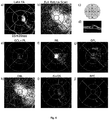
- the location of extracellular space increases identified well the leakage sites and, furthermore, the involvement of the different retinal layers in eyes with central serous chorioretinopathy, macular edema after cataract surgery and branch retinal vein occlusion ( Figure 6 ).
- the figures show the increased visibility of the retinal vascular lesions demonstrated by OCT-Microangiography and the location of increases in extracellular space involving different layers of the retina.
- Combination of OCT-Leakage and OCT-Microangiography methodologies allow (1) visualization of the traditional angiography distribution and its alteration which can be well-recognized by clinicians using FA, (2) identification of cross-sectional structural OCT images, locating the alterations in the retina, (3) identification and localization of sites of low LOR ratio, i.e., areas of increased extracellular space corresponding to alterations of the BRB, and (4) identification of sites of neovascularization and capillary dropout in the retinal vasculature.
- Breakdown of the BRB is a frequent alteration occurring in the retina due to diabetes and other retinal vascular diseases and has major clinical implications. It is of major relevance that the presently available therapies for macular edema act mainly by correcting the alteration of the BRB. Monitoring the alterations of the BRB is, therefore, of major clinical relevance.
- FA is used routinely in clinical practice, to show and demonstrate alterations of the BRB, but is an invasive method relying on intravenous injection of fluorescein which may be associated to severe adverse events.
- OCT-Microangiography has become available replacing much of the information given by FA, such as visualization of areas of capillary dropout in a non-invasive manner.
- OCT-Microangiography cannot visualize dye leakage, i.e., alteration of the BRB [3].
- OCT-Leakage is able to identify the location of the increases of retinal extracellular space in the different layers of the retina thus being able to identify the retinal cells more affected and the potential impact of the retinal tissue damage. This information is expected to offer new insights into the progression and recovery of macular edema with particular value regarding prognosis for visual acuity recovery.
- the method and device here described is able to detect and locate non-invasively, the sites of leakage, i.e., alteration of the BRB in retinal diseases, being, therefore, expected to replace FA. It offers, furthermore, an added value by identifying the changes occurring in the different layers of the retina.
- OCT-Leakage location and quantification is able to complement OCT-Microangiography thus allowing full information on the retinal circulation namely capillary closure, vascular morphology and alteration of BRB by using non-invasive OCT based methodologies.
- OCT techniques may, therefore, replace the widely used method of FA which is invasive and associated with risks.
- certain embodiments of the invention as described herein may be incorporated as code (e.g., a software algorithm or program) residing in firmware and/or on computer useable medium having control logic for enabling execution on a computer system having a computer processor, such as any of the servers described herein.
- a computer system typically includes memory storage configured to provide output from execution of the code which configures a processor in accordance with the execution.
- the code can be arranged as firmware or software, and can be organized as a set of modules, including the various modules and algorithms described herein, such as discrete code modules, function calls, procedure calls or objects in an object-oriented programming environment. If implemented using modules, the code can comprise a single module or a plurality of modules that operate in cooperation with one another to configure the machine in which it is executed to perform the associated functions, as described herein.
Landscapes
- Health & Medical Sciences (AREA)
- Engineering & Computer Science (AREA)
- Life Sciences & Earth Sciences (AREA)
- Physics & Mathematics (AREA)
- General Health & Medical Sciences (AREA)
- Medical Informatics (AREA)
- Nuclear Medicine, Radiotherapy & Molecular Imaging (AREA)
- Radiology & Medical Imaging (AREA)
- Heart & Thoracic Surgery (AREA)
- Animal Behavior & Ethology (AREA)
- Veterinary Medicine (AREA)
- Public Health (AREA)
- Biophysics (AREA)
- Ophthalmology & Optometry (AREA)
- Biomedical Technology (AREA)
- Surgery (AREA)
- Molecular Biology (AREA)
- Quality & Reliability (AREA)
- General Physics & Mathematics (AREA)
- Computer Vision & Pattern Recognition (AREA)
- Theoretical Computer Science (AREA)
- Eye Examination Apparatus (AREA)
Claims (15)
- Automatisiertes Verfahren zur Erkennung von Stellen von Veränderungen der Retina anhand der Daten einer optischen Kohärenztomografie (OCT) eines Auges, wobei die genannten Daten die Daten eines OCT A-Scans eines Areals des Auges umfassen und alle Daten des OCT A-Scans eine Reihe von Reflektivitätswerten entlang der Tiefenrichtung der Daten des OCT A-Scans umfassen, wobei das genannte Verfahren umfasst:Segmentierung der Daten des OCT A-Scans in Retinaschichten;Erkennung der Position des A-Scans als eine Stelle geringer optischer Reflexion (LOR) für jede Retinaschicht und jeden OCT A-Scan, wenn der Wert der optischen Reflektivität des A-Scans unterhalb eines im Voraus festgelegten Grenzwerts liegt;Erzeugung eines zweidimensionalen Bilds der erkannten Stellen für jede Retinaschicht.
- Verfahren nach Anspruch 1, wobei der Grenzwert im Voraus gemäß der optischen Reflektivität einer gesunden Population festgelegt wird.
- Verfahren nach einem der vorangehenden Ansprüche, wobei der im Voraus festgelegte Grenzwert 20 ist, wenn mit einem "OCT Angioplex" gemessen wird, oder 10, wenn mit einem "OCT Cirrus 5000" gemessen wird.
- Verfahren nach einem der vorangehenden Ansprüche, bei dem im Voraus OCT A-Scans eines Areals des Auges durchgeführt werden, wobei jeder A-Scan eine Reihe von Reflektivitätswerten entlang der Tiefenrichtung des Scans umfasst.
- Verfahren nach einem der vorangehenden Ansprüche, bei dem die Segmentierung der Retinaschichten aus den Daten des OCT A-Scans entsprechend der optischen Reflektivität jeder identifizierten Retinaschicht erfolgt.
- Verfahren nach einem der vorangehenden Ansprüche, das ferner die Vorverarbeitung der Daten des OCT A-Scans umfasst, wobei die genannte Vorverarbeitung eine oder mehrere Speckle-Verfahren zur Reduktion oder Normalisierung umfasst.
- Verfahren nach einem der vorangehenden Ansprüche, wobei die Schritte des Verfahrens Pixel für Pixel des zweidimensionalen Bildes, das dem Areal des Auges entspricht, ausgeführt werden und das Verfahren das Anzeigen der genannten Bilder über ein Computerdisplay und/oder das Speichern der Bilder auf einem Datenspeichermedium umfasst.
- Verfahren nach dem vorangehenden Anspruch, umfassend die Anzeige von OCT-Mikroangiographiedaten auf demselben Computerdisplay, insbesondere Seite an Seite oder überlagert, mit den En-face-Bildern der optischen Reflektivität aller segmentierten Retinaschichten.
- Verfahren nach einem der vorangehenden Ansprüche umfassend die Segmentierung von 6 bis 11 Retinaschichten, insbesondere von 7 Retinaschichten.
- Verfahren nach einem der vorangehenden Ansprüche, wobei das Verfahren zur Identifizierung von Zunahmen der interzellulären Flüssigkeit oder Zunahmen des extrazellulären Raums in der Retina oder Veränderungen der Blut-Retina-Schranke verwendet wird.
- Verfahren nach einem der vorangehenden Ansprüche, wobei das Verfahren zur Identifizierung von Stellen von Veränderungen der Blut-Retina-Schranke verwendet wird.
- Nichtflüchtiges Speichermedium, einschließlich Programmanweisungen zur Implementierung einer Vorrichtung für die Erkennung von Stellen geringer optischer Reflektivität über eine optische Kohärenztomografie, wobei die Programmanweisungen Anweisungen für die Durchführung des Verfahrens nach einem der Ansprüche 1-11 enthalten.
- Vorrichtung nach dem vorangehenden Anspruch, umfassend das genannte nichtflüchtige Speichermedium und einen elektronischen Datenprozessor.
- Vorrichtung zur Erkennung von Stellen geringer optischer Reflektivität über eine optische Kohärenztomografie, wobei die genannte Vorrichtung so konfiguriert ist, dass sie das Verfahren nach einem der Ansprüche 1-11 ausführt.
- Vorrichtung nach den Ansprüchen 13 oder 14 umfassend ein OCT-Gerät, insbesondere ferner umfassend ein Computerdisplay zur Anzeige der erkannten Stellen mit geringer optischer Reflexivität.
Applications Claiming Priority (5)
| Application Number | Priority Date | Filing Date | Title |
|---|---|---|---|
| PT10841815 | 2015-04-29 | ||
| PT10881215 | 2015-09-10 | ||
| PT10902815 | 2015-12-10 | ||
| IBPCT/IB2015/060078 | 2015-12-31 | ||
| PCT/IB2016/052462 WO2016174637A1 (en) | 2015-04-29 | 2016-04-29 | Method and device for the non-invasive indirect identification of sites of alterations of the blood-retinal barrier |
Publications (2)
| Publication Number | Publication Date |
|---|---|
| EP3289565A1 EP3289565A1 (de) | 2018-03-07 |
| EP3289565B1 true EP3289565B1 (de) | 2020-08-12 |
Family
ID=56113015
Family Applications (1)
| Application Number | Title | Priority Date | Filing Date |
|---|---|---|---|
| EP16727840.7A Active EP3289565B1 (de) | 2015-04-29 | 2016-04-29 | Verfahren und vorrichtung zur nichtinvasiven indirekten identifizierung von stellen von veränderungen der blut-retina-schranke |
Country Status (3)
| Country | Link |
|---|---|
| US (1) | US11234591B2 (de) |
| EP (1) | EP3289565B1 (de) |
| WO (1) | WO2016174637A1 (de) |
Families Citing this family (3)
| Publication number | Priority date | Publication date | Assignee | Title |
|---|---|---|---|---|
| DE102015009641A1 (de) * | 2015-07-24 | 2017-01-26 | Carl Zeiss Meditec Ag | Verfahren zur Klassifizierung der Katarakt eines Auges |
| US11989877B2 (en) * | 2018-09-18 | 2024-05-21 | MacuJect Pty Ltd | Method and system for analysing images of a retina |
| US20230414094A1 (en) * | 2022-06-23 | 2023-12-28 | Oregon Health & Science University | Volume-based layer-independent framework for detection of retinal pathology |
Family Cites Families (7)
| Publication number | Priority date | Publication date | Assignee | Title |
|---|---|---|---|---|
| US7593559B2 (en) * | 2005-11-18 | 2009-09-22 | Duke University | Method and system of coregistrating optical coherence tomography (OCT) with other clinical tests |
| EP2293714B1 (de) * | 2008-06-02 | 2014-08-13 | Lightlab Imaging, Inc. | Quantitative verfahren zur erfassung von gewebekennwerten auf bildern aus einer optischen kohärenztomographie |
| US20120150029A1 (en) * | 2008-12-19 | 2012-06-14 | University Of Miami | System and Method for Detection and Monitoring of Ocular Diseases and Disorders using Optical Coherence Tomography |
| JP5701024B2 (ja) * | 2010-11-26 | 2015-04-15 | キヤノン株式会社 | 画像処理装置及び方法 |
| CN103858134A (zh) * | 2011-08-09 | 2014-06-11 | 光视有限公司 | 光学相干断层摄影术中对特征的运动校正和标准化 |
| US9877646B2 (en) | 2011-10-11 | 2018-01-30 | Carl Zeiss Meditec, Inc. | Assessment of retinal disruption |
| US9713424B2 (en) * | 2015-02-06 | 2017-07-25 | Richard F. Spaide | Volume analysis and display of information in optical coherence tomography angiography |
-
2016
- 2016-04-29 EP EP16727840.7A patent/EP3289565B1/de active Active
- 2016-04-29 US US15/568,161 patent/US11234591B2/en active Active
- 2016-04-29 WO PCT/IB2016/052462 patent/WO2016174637A1/en not_active Ceased
Non-Patent Citations (1)
| Title |
|---|
| None * |
Also Published As
| Publication number | Publication date |
|---|---|
| US11234591B2 (en) | 2022-02-01 |
| EP3289565A1 (de) | 2018-03-07 |
| WO2016174637A1 (en) | 2016-11-03 |
| US20180146850A1 (en) | 2018-05-31 |
Similar Documents
| Publication | Publication Date | Title |
|---|---|---|
| de Sisternes et al. | Localization of damage in progressive hydroxychloroquine retinopathy on and off the drug: inner versus outer retina, parafovea versus peripheral fovea | |
| Cardoso et al. | Systematic evaluation of optical coherence tomography angiography in retinal vein occlusion | |
| Lauermann et al. | Prevalences of segmentation errors and motion artifacts in OCT-angiography differ among retinal diseases | |
| Kwon et al. | Parapapillary deep-layer microvasculature dropout and visual field progression in glaucoma | |
| CA3014998C (en) | Methods and apparatus for reducing artifacts in oct angiography using machine learning techniques | |
| Sadda et al. | Error correction and quantitative subanalysis of optical coherence tomography data using computer-assisted grading | |
| Viola et al. | Choroidal findings in dome-shaped macula in highly myopic eyes: a longitudinal study | |
| Onal et al. | Quantitative analysis of structural alterations in the choroid of patients with active Behçet uveitis | |
| Mwanza et al. | New developments in optical coherence tomography imaging for glaucoma | |
| Byeon et al. | Foveal ganglion cell layer damage in ischemic diabetic maculopathy: correlation of optical coherence tomographic and anatomic changes | |
| Stetson et al. | OCT minimum intensity as a predictor of geographic atrophy enlargement | |
| Rao et al. | Comparing the structure–function relationship at the macula with standard automated perimetry and microperimetry | |
| Aghdam et al. | Optical coherence tomography angiography features of optic nerve head drusen and nonarteritic anterior ischemic optic neuropathy | |
| Choi et al. | Microstructure of the optic disc pit in open-angle glaucoma | |
| Cunha-Vaz et al. | Agreement between OCT leakage and fluorescein angiography to identify sites of alteration of the blood–retinal barrier in diabetes | |
| Arthur et al. | Agreement in assessing cup-to-disc ratio measurement among stereoscopic optic nerve head photographs, HRT II, and Stratus OCT | |
| CA2776437A1 (en) | Diagnostic method and apparatus for predicting potential preserved visual acuity | |
| Qin et al. | OCT angiography and cone photoreceptor imaging in geographic atrophy | |
| EP3289565B1 (de) | Verfahren und vorrichtung zur nichtinvasiven indirekten identifizierung von stellen von veränderungen der blut-retina-schranke | |
| Schranz et al. | Correlation of vascular and fluid‐related parameters in neovascular age‐related macular degeneration using deep learning | |
| JP6815798B2 (ja) | 眼科撮影装置及び眼科画像処理装置 | |
| Fujimoto et al. | Three-Dimensional Volume Calculation of Intrachoroidal Cavitation Using Deep-Learning–Based Noise Reduction of Optical Coherence Tomography | |
| Siggel et al. | Comparison of automated versus manually modified OCT angiography en face slabs for detection of choroidal neovascularization | |
| Wanek et al. | Feasibility of a method for en face imaging of photoreceptor cell integrity | |
| Rothenbuehler et al. | Three-dimensional analysis of submacular perforating scleral vessels by enhanced depth imaging optical coherence tomography |
Legal Events
| Date | Code | Title | Description |
|---|---|---|---|
| STAA | Information on the status of an ep patent application or granted ep patent |
Free format text: STATUS: THE INTERNATIONAL PUBLICATION HAS BEEN MADE |
|
| PUAI | Public reference made under article 153(3) epc to a published international application that has entered the european phase |
Free format text: ORIGINAL CODE: 0009012 |
|
| STAA | Information on the status of an ep patent application or granted ep patent |
Free format text: STATUS: REQUEST FOR EXAMINATION WAS MADE |
|
| 17P | Request for examination filed |
Effective date: 20171024 |
|
| AK | Designated contracting states |
Kind code of ref document: A1 Designated state(s): AL AT BE BG CH CY CZ DE DK EE ES FI FR GB GR HR HU IE IS IT LI LT LU LV MC MK MT NL NO PL PT RO RS SE SI SK SM TR |
|
| AX | Request for extension of the european patent |
Extension state: BA ME |
|
| DAV | Request for validation of the european patent (deleted) | ||
| DAX | Request for extension of the european patent (deleted) | ||
| GRAP | Despatch of communication of intention to grant a patent |
Free format text: ORIGINAL CODE: EPIDOSNIGR1 |
|
| STAA | Information on the status of an ep patent application or granted ep patent |
Free format text: STATUS: GRANT OF PATENT IS INTENDED |
|
| INTG | Intention to grant announced |
Effective date: 20200219 |
|
| GRAS | Grant fee paid |
Free format text: ORIGINAL CODE: EPIDOSNIGR3 |
|
| GRAA | (expected) grant |
Free format text: ORIGINAL CODE: 0009210 |
|
| STAA | Information on the status of an ep patent application or granted ep patent |
Free format text: STATUS: THE PATENT HAS BEEN GRANTED |
|
| AK | Designated contracting states |
Kind code of ref document: B1 Designated state(s): AL AT BE BG CH CY CZ DE DK EE ES FI FR GB GR HR HU IE IS IT LI LT LU LV MC MK MT NL NO PL PT RO RS SE SI SK SM TR |
|
| REG | Reference to a national code |
Ref country code: CH Ref legal event code: EP |
|
| REG | Reference to a national code |
Ref country code: IE Ref legal event code: FG4D |
|
| REG | Reference to a national code |
Ref country code: DE Ref legal event code: R096 Ref document number: 602016041830 Country of ref document: DE |
|
| REG | Reference to a national code |
Ref country code: AT Ref legal event code: REF Ref document number: 1302285 Country of ref document: AT Kind code of ref document: T Effective date: 20200915 |
|
| REG | Reference to a national code |
Ref country code: LT Ref legal event code: MG4D |
|
| REG | Reference to a national code |
Ref country code: NL Ref legal event code: MP Effective date: 20200812 |
|
| PG25 | Lapsed in a contracting state [announced via postgrant information from national office to epo] |
Ref country code: FI Free format text: LAPSE BECAUSE OF FAILURE TO SUBMIT A TRANSLATION OF THE DESCRIPTION OR TO PAY THE FEE WITHIN THE PRESCRIBED TIME-LIMIT Effective date: 20200812 Ref country code: NO Free format text: LAPSE BECAUSE OF FAILURE TO SUBMIT A TRANSLATION OF THE DESCRIPTION OR TO PAY THE FEE WITHIN THE PRESCRIBED TIME-LIMIT Effective date: 20201112 Ref country code: GR Free format text: LAPSE BECAUSE OF FAILURE TO SUBMIT A TRANSLATION OF THE DESCRIPTION OR TO PAY THE FEE WITHIN THE PRESCRIBED TIME-LIMIT Effective date: 20201113 Ref country code: SE Free format text: LAPSE BECAUSE OF FAILURE TO SUBMIT A TRANSLATION OF THE DESCRIPTION OR TO PAY THE FEE WITHIN THE PRESCRIBED TIME-LIMIT Effective date: 20200812 Ref country code: BG Free format text: LAPSE BECAUSE OF FAILURE TO SUBMIT A TRANSLATION OF THE DESCRIPTION OR TO PAY THE FEE WITHIN THE PRESCRIBED TIME-LIMIT Effective date: 20201112 Ref country code: HR Free format text: LAPSE BECAUSE OF FAILURE TO SUBMIT A TRANSLATION OF THE DESCRIPTION OR TO PAY THE FEE WITHIN THE PRESCRIBED TIME-LIMIT Effective date: 20200812 Ref country code: LT Free format text: LAPSE BECAUSE OF FAILURE TO SUBMIT A TRANSLATION OF THE DESCRIPTION OR TO PAY THE FEE WITHIN THE PRESCRIBED TIME-LIMIT Effective date: 20200812 |
|
| REG | Reference to a national code |
Ref country code: AT Ref legal event code: MK05 Ref document number: 1302285 Country of ref document: AT Kind code of ref document: T Effective date: 20200812 |
|
| PG25 | Lapsed in a contracting state [announced via postgrant information from national office to epo] |
Ref country code: PL Free format text: LAPSE BECAUSE OF FAILURE TO SUBMIT A TRANSLATION OF THE DESCRIPTION OR TO PAY THE FEE WITHIN THE PRESCRIBED TIME-LIMIT Effective date: 20200812 Ref country code: LV Free format text: LAPSE BECAUSE OF FAILURE TO SUBMIT A TRANSLATION OF THE DESCRIPTION OR TO PAY THE FEE WITHIN THE PRESCRIBED TIME-LIMIT Effective date: 20200812 Ref country code: RS Free format text: LAPSE BECAUSE OF FAILURE TO SUBMIT A TRANSLATION OF THE DESCRIPTION OR TO PAY THE FEE WITHIN THE PRESCRIBED TIME-LIMIT Effective date: 20200812 Ref country code: NL Free format text: LAPSE BECAUSE OF FAILURE TO SUBMIT A TRANSLATION OF THE DESCRIPTION OR TO PAY THE FEE WITHIN THE PRESCRIBED TIME-LIMIT Effective date: 20200812 Ref country code: IS Free format text: LAPSE BECAUSE OF FAILURE TO SUBMIT A TRANSLATION OF THE DESCRIPTION OR TO PAY THE FEE WITHIN THE PRESCRIBED TIME-LIMIT Effective date: 20201212 |
|
| PG25 | Lapsed in a contracting state [announced via postgrant information from national office to epo] |
Ref country code: EE Free format text: LAPSE BECAUSE OF FAILURE TO SUBMIT A TRANSLATION OF THE DESCRIPTION OR TO PAY THE FEE WITHIN THE PRESCRIBED TIME-LIMIT Effective date: 20200812 Ref country code: RO Free format text: LAPSE BECAUSE OF FAILURE TO SUBMIT A TRANSLATION OF THE DESCRIPTION OR TO PAY THE FEE WITHIN THE PRESCRIBED TIME-LIMIT Effective date: 20200812 Ref country code: SM Free format text: LAPSE BECAUSE OF FAILURE TO SUBMIT A TRANSLATION OF THE DESCRIPTION OR TO PAY THE FEE WITHIN THE PRESCRIBED TIME-LIMIT Effective date: 20200812 Ref country code: DK Free format text: LAPSE BECAUSE OF FAILURE TO SUBMIT A TRANSLATION OF THE DESCRIPTION OR TO PAY THE FEE WITHIN THE PRESCRIBED TIME-LIMIT Effective date: 20200812 Ref country code: CZ Free format text: LAPSE BECAUSE OF FAILURE TO SUBMIT A TRANSLATION OF THE DESCRIPTION OR TO PAY THE FEE WITHIN THE PRESCRIBED TIME-LIMIT Effective date: 20200812 |
|
| REG | Reference to a national code |
Ref country code: DE Ref legal event code: R097 Ref document number: 602016041830 Country of ref document: DE |
|
| PG25 | Lapsed in a contracting state [announced via postgrant information from national office to epo] |
Ref country code: AL Free format text: LAPSE BECAUSE OF FAILURE TO SUBMIT A TRANSLATION OF THE DESCRIPTION OR TO PAY THE FEE WITHIN THE PRESCRIBED TIME-LIMIT Effective date: 20200812 Ref country code: AT Free format text: LAPSE BECAUSE OF FAILURE TO SUBMIT A TRANSLATION OF THE DESCRIPTION OR TO PAY THE FEE WITHIN THE PRESCRIBED TIME-LIMIT Effective date: 20200812 Ref country code: ES Free format text: LAPSE BECAUSE OF FAILURE TO SUBMIT A TRANSLATION OF THE DESCRIPTION OR TO PAY THE FEE WITHIN THE PRESCRIBED TIME-LIMIT Effective date: 20200812 |
|
| PLBE | No opposition filed within time limit |
Free format text: ORIGINAL CODE: 0009261 |
|
| STAA | Information on the status of an ep patent application or granted ep patent |
Free format text: STATUS: NO OPPOSITION FILED WITHIN TIME LIMIT |
|
| PG25 | Lapsed in a contracting state [announced via postgrant information from national office to epo] |
Ref country code: SK Free format text: LAPSE BECAUSE OF FAILURE TO SUBMIT A TRANSLATION OF THE DESCRIPTION OR TO PAY THE FEE WITHIN THE PRESCRIBED TIME-LIMIT Effective date: 20200812 |
|
| 26N | No opposition filed |
Effective date: 20210514 |
|
| PG25 | Lapsed in a contracting state [announced via postgrant information from national office to epo] |
Ref country code: IT Free format text: LAPSE BECAUSE OF FAILURE TO SUBMIT A TRANSLATION OF THE DESCRIPTION OR TO PAY THE FEE WITHIN THE PRESCRIBED TIME-LIMIT Effective date: 20200812 |
|
| PG25 | Lapsed in a contracting state [announced via postgrant information from national office to epo] |
Ref country code: SI Free format text: LAPSE BECAUSE OF FAILURE TO SUBMIT A TRANSLATION OF THE DESCRIPTION OR TO PAY THE FEE WITHIN THE PRESCRIBED TIME-LIMIT Effective date: 20200812 |
|
| PG25 | Lapsed in a contracting state [announced via postgrant information from national office to epo] |
Ref country code: MC Free format text: LAPSE BECAUSE OF FAILURE TO SUBMIT A TRANSLATION OF THE DESCRIPTION OR TO PAY THE FEE WITHIN THE PRESCRIBED TIME-LIMIT Effective date: 20200812 |
|
| PG25 | Lapsed in a contracting state [announced via postgrant information from national office to epo] |
Ref country code: LU Free format text: LAPSE BECAUSE OF NON-PAYMENT OF DUE FEES Effective date: 20210429 |
|
| REG | Reference to a national code |
Ref country code: BE Ref legal event code: MM Effective date: 20210430 |
|
| PG25 | Lapsed in a contracting state [announced via postgrant information from national office to epo] |
Ref country code: LI Free format text: LAPSE BECAUSE OF NON-PAYMENT OF DUE FEES Effective date: 20210430 Ref country code: CH Free format text: LAPSE BECAUSE OF NON-PAYMENT OF DUE FEES Effective date: 20210430 |
|
| PG25 | Lapsed in a contracting state [announced via postgrant information from national office to epo] |
Ref country code: IE Free format text: LAPSE BECAUSE OF NON-PAYMENT OF DUE FEES Effective date: 20210429 |
|
| PG25 | Lapsed in a contracting state [announced via postgrant information from national office to epo] |
Ref country code: IS Free format text: LAPSE BECAUSE OF FAILURE TO SUBMIT A TRANSLATION OF THE DESCRIPTION OR TO PAY THE FEE WITHIN THE PRESCRIBED TIME-LIMIT Effective date: 20201212 |
|
| PG25 | Lapsed in a contracting state [announced via postgrant information from national office to epo] |
Ref country code: BE Free format text: LAPSE BECAUSE OF NON-PAYMENT OF DUE FEES Effective date: 20210430 |
|
| PG25 | Lapsed in a contracting state [announced via postgrant information from national office to epo] |
Ref country code: PT Free format text: LAPSE BECAUSE OF FAILURE TO SUBMIT A TRANSLATION OF THE DESCRIPTION OR TO PAY THE FEE WITHIN THE PRESCRIBED TIME-LIMIT Effective date: 20201214 |
|
| PG25 | Lapsed in a contracting state [announced via postgrant information from national office to epo] |
Ref country code: HU Free format text: LAPSE BECAUSE OF FAILURE TO SUBMIT A TRANSLATION OF THE DESCRIPTION OR TO PAY THE FEE WITHIN THE PRESCRIBED TIME-LIMIT; INVALID AB INITIO Effective date: 20160429 |
|
| P01 | Opt-out of the competence of the unified patent court (upc) registered |
Effective date: 20230519 |
|
| PG25 | Lapsed in a contracting state [announced via postgrant information from national office to epo] |
Ref country code: CY Free format text: LAPSE BECAUSE OF FAILURE TO SUBMIT A TRANSLATION OF THE DESCRIPTION OR TO PAY THE FEE WITHIN THE PRESCRIBED TIME-LIMIT Effective date: 20200812 |
|
| PG25 | Lapsed in a contracting state [announced via postgrant information from national office to epo] |
Ref country code: MK Free format text: LAPSE BECAUSE OF FAILURE TO SUBMIT A TRANSLATION OF THE DESCRIPTION OR TO PAY THE FEE WITHIN THE PRESCRIBED TIME-LIMIT Effective date: 20200812 |
|
| PG25 | Lapsed in a contracting state [announced via postgrant information from national office to epo] |
Ref country code: TR Free format text: LAPSE BECAUSE OF FAILURE TO SUBMIT A TRANSLATION OF THE DESCRIPTION OR TO PAY THE FEE WITHIN THE PRESCRIBED TIME-LIMIT Effective date: 20200812 |
|
| PG25 | Lapsed in a contracting state [announced via postgrant information from national office to epo] |
Ref country code: MT Free format text: LAPSE BECAUSE OF FAILURE TO SUBMIT A TRANSLATION OF THE DESCRIPTION OR TO PAY THE FEE WITHIN THE PRESCRIBED TIME-LIMIT Effective date: 20200812 |
|
| PGFP | Annual fee paid to national office [announced via postgrant information from national office to epo] |
Ref country code: DE Payment date: 20250428 Year of fee payment: 10 |
|
| PGFP | Annual fee paid to national office [announced via postgrant information from national office to epo] |
Ref country code: GB Payment date: 20250428 Year of fee payment: 10 |
|
| PGFP | Annual fee paid to national office [announced via postgrant information from national office to epo] |
Ref country code: FR Payment date: 20250428 Year of fee payment: 10 |