EP3328453B1 - Verfahren zur herstellung eines osteogenen knochentransplants - Google Patents
Verfahren zur herstellung eines osteogenen knochentransplants Download PDFInfo
- Publication number
- EP3328453B1 EP3328453B1 EP16753497.3A EP16753497A EP3328453B1 EP 3328453 B1 EP3328453 B1 EP 3328453B1 EP 16753497 A EP16753497 A EP 16753497A EP 3328453 B1 EP3328453 B1 EP 3328453B1
- Authority
- EP
- European Patent Office
- Prior art keywords
- effluent
- bone graft
- anticoagulant
- osteogenic
- cell fraction
- Prior art date
- Legal status (The legal status is an assumption and is not a legal conclusion. Google has not performed a legal analysis and makes no representation as to the accuracy of the status listed.)
- Active
Links
Images


Classifications
-
- A—HUMAN NECESSITIES
- A61—MEDICAL OR VETERINARY SCIENCE; HYGIENE
- A61L—METHODS OR APPARATUS FOR STERILISING MATERIALS OR OBJECTS IN GENERAL; DISINFECTION, STERILISATION OR DEODORISATION OF AIR; CHEMICAL ASPECTS OF BANDAGES, DRESSINGS, ABSORBENT PADS OR SURGICAL ARTICLES; MATERIALS FOR BANDAGES, DRESSINGS, ABSORBENT PADS OR SURGICAL ARTICLES
- A61L27/00—Materials for grafts or prostheses or for coating grafts or prostheses
- A61L27/36—Materials for grafts or prostheses or for coating grafts or prostheses containing ingredients of undetermined constitution or reaction products thereof, e.g. transplant tissue, natural bone, extracellular matrix
- A61L27/38—Materials for grafts or prostheses or for coating grafts or prostheses containing ingredients of undetermined constitution or reaction products thereof, e.g. transplant tissue, natural bone, extracellular matrix containing added animal cells
- A61L27/3804—Materials for grafts or prostheses or for coating grafts or prostheses containing ingredients of undetermined constitution or reaction products thereof, e.g. transplant tissue, natural bone, extracellular matrix containing added animal cells characterised by specific cells or progenitors thereof, e.g. fibroblasts, connective tissue cells, kidney cells
-
- A—HUMAN NECESSITIES
- A61—MEDICAL OR VETERINARY SCIENCE; HYGIENE
- A61L—METHODS OR APPARATUS FOR STERILISING MATERIALS OR OBJECTS IN GENERAL; DISINFECTION, STERILISATION OR DEODORISATION OF AIR; CHEMICAL ASPECTS OF BANDAGES, DRESSINGS, ABSORBENT PADS OR SURGICAL ARTICLES; MATERIALS FOR BANDAGES, DRESSINGS, ABSORBENT PADS OR SURGICAL ARTICLES
- A61L27/00—Materials for grafts or prostheses or for coating grafts or prostheses
- A61L27/50—Materials characterised by their function or physical properties, e.g. injectable or lubricating compositions, shape-memory materials, surface modified materials
- A61L27/54—Biologically active materials, e.g. therapeutic substances
-
- A—HUMAN NECESSITIES
- A61—MEDICAL OR VETERINARY SCIENCE; HYGIENE
- A61L—METHODS OR APPARATUS FOR STERILISING MATERIALS OR OBJECTS IN GENERAL; DISINFECTION, STERILISATION OR DEODORISATION OF AIR; CHEMICAL ASPECTS OF BANDAGES, DRESSINGS, ABSORBENT PADS OR SURGICAL ARTICLES; MATERIALS FOR BANDAGES, DRESSINGS, ABSORBENT PADS OR SURGICAL ARTICLES
- A61L2300/00—Biologically active materials used in bandages, wound dressings, absorbent pads or medical devices
- A61L2300/20—Biologically active materials used in bandages, wound dressings, absorbent pads or medical devices containing or releasing organic materials
- A61L2300/23—Carbohydrates
- A61L2300/236—Glycosaminoglycans, e.g. heparin, hyaluronic acid, chondroitin
-
- A—HUMAN NECESSITIES
- A61—MEDICAL OR VETERINARY SCIENCE; HYGIENE
- A61L—METHODS OR APPARATUS FOR STERILISING MATERIALS OR OBJECTS IN GENERAL; DISINFECTION, STERILISATION OR DEODORISATION OF AIR; CHEMICAL ASPECTS OF BANDAGES, DRESSINGS, ABSORBENT PADS OR SURGICAL ARTICLES; MATERIALS FOR BANDAGES, DRESSINGS, ABSORBENT PADS OR SURGICAL ARTICLES
- A61L2300/00—Biologically active materials used in bandages, wound dressings, absorbent pads or medical devices
- A61L2300/40—Biologically active materials used in bandages, wound dressings, absorbent pads or medical devices characterised by a specific therapeutic activity or mode of action
- A61L2300/412—Tissue-regenerating or healing or proliferative agents
-
- A—HUMAN NECESSITIES
- A61—MEDICAL OR VETERINARY SCIENCE; HYGIENE
- A61L—METHODS OR APPARATUS FOR STERILISING MATERIALS OR OBJECTS IN GENERAL; DISINFECTION, STERILISATION OR DEODORISATION OF AIR; CHEMICAL ASPECTS OF BANDAGES, DRESSINGS, ABSORBENT PADS OR SURGICAL ARTICLES; MATERIALS FOR BANDAGES, DRESSINGS, ABSORBENT PADS OR SURGICAL ARTICLES
- A61L2300/00—Biologically active materials used in bandages, wound dressings, absorbent pads or medical devices
- A61L2300/40—Biologically active materials used in bandages, wound dressings, absorbent pads or medical devices characterised by a specific therapeutic activity or mode of action
- A61L2300/42—Anti-thrombotic agents, anticoagulants, anti-platelet agents
-
- A—HUMAN NECESSITIES
- A61—MEDICAL OR VETERINARY SCIENCE; HYGIENE
- A61L—METHODS OR APPARATUS FOR STERILISING MATERIALS OR OBJECTS IN GENERAL; DISINFECTION, STERILISATION OR DEODORISATION OF AIR; CHEMICAL ASPECTS OF BANDAGES, DRESSINGS, ABSORBENT PADS OR SURGICAL ARTICLES; MATERIALS FOR BANDAGES, DRESSINGS, ABSORBENT PADS OR SURGICAL ARTICLES
- A61L2430/00—Materials or treatment for tissue regeneration
- A61L2430/02—Materials or treatment for tissue regeneration for reconstruction of bones; weight-bearing implants
Definitions
- the present disclosure relates to osteogenic bone grafts including concentrated cell fractions of mononuclear cell populations and an anticoagulant, as well as processes for preparing the same.
- Non-unions in particular those involving large segmental defects in long bones, present a significant healing challenge.
- One method of healing these defects involves the use of autograft obtained from the medullary canal of a long bone, for example, a femur or tibia, using a surgical reamer irrigator aspirator (RIA) system.
- RIA surgical reamer irrigator aspirator
- This system has the advantages of being able to harvest a larger quantity of autograft, with lower morbidity and greater rapidity, as compared to, for example, iliac crest bone grafting.
- RIA bone grafts are emerging as the new "gold standard" as it relates to the use of bone grafts, either in repair of large segmental defects or other clinical situations requiring bone grafts.
- One problem that remains is that a large quantity of cells including mesenchymal stem cells, endothelial progenitors, and other progenitor cells, which become available during harvesting are currently lost or under
- surgical reamers utilize a rotating cutting head similar to a drill displaced at the distal end of a drive shaft.
- Bone cutting devices for use in intramedullary reaming typically use a flexible drive shaft because the medullary canals of bones are seldom straight and usually will have some degree of curvature.
- Most reamers also have a central longitudinal bore through both the reamer and the drive shaft. The central bore is intended to receive a long, small diameter guide pin or wire which is initially inserted into the medullary canal to act as a track for the advancing reamer.
- Reamers are used in orthopedic surgery to prepare the medullary canals of bone for surgical procedures such as total hip and knee replacement, nail insertion to stabilize a long bone fracture, an intramedullary osteotomy, and bone harvesting for grafting purposes.
- Blood coagulation or clotting
- Blood coagulation is one of the body's natural means of stopping bleeding. It comprises a complex cascade of many clotting factors, some always present in the blood and some released from damaged tissue and activated platelets.
- platelets When the lining of a blood vessel is broken, platelets are attracted forming a platelet plug.
- platelets have thrombin receptors on their surfaces that bind serum thrombin molecules, which in turn convert soluble fibrinogen in the serum into fibrin at the wound site. Fibrin forms long strands of tough insoluble protein that are bound to the platelets.
- Factor XIII completes the cross-linking of fibrin so that it hardens and contracts.
- the cross-linked fibrin forms a mesh atop the platelet plug that completes the clot.
- the end-product is a mesh of fibrin strands in which blood cells are trapped to form a solid mass that seeks to stop bleeding. The entrapment of cells that promote bone growth by fibrin clots can be deleterious to the regeneration of bone tissue at the site of the trauma injury.
- the blood clotting cascade begins almost instantly after a reamer head damages the endothelium lining of any blood vessels or other tissue in the medullary canal of the femur or tibia. Once intiated, this process is normally irreversible and in certain instances all useful cellular graft material, as well as fatty tissues residing in the medullary canal that becomes atomized by the cutting of the reaming head, can become trapped in the clot matrix, rendering it unusable as a bone graft.
- US2007036766 relates to "a method for preparation of an autologous tissue graft composition
- Bone marrow is harvested from the patient.
- the nucleated cells of the bone marrow are subsequently concentrated.
- Autologous thrombin is purified from a volume of whole blood also taken from the patient.
- the bone marrow aspirate or bone marrow concentrate is then combined with the purified autologous thrombin to form a coagulated tissue graft material that may be used alone or in conjunction with other graft materials”.
- the present invention relates to methods of preparing an osteogenic bone graft according to the appended claims 1-11 and to an osteogenic bone graft composition according to claims 12-15.
- the methods can include:
- Methods of the disclosure can include:
- the present disclosure also describes methods of preparing an osteogenic bone graft including:
- harvesting includes aspirating the intramedullary canal of a long bone with a surgical reaming device.
- the anticoagulant is continuously and proportionately introduced into the effluent.
- the continuous and proportionate introduction of the anticoagulant is advantageous as a way in which to arrest the clotting and protect the regenerative potential of the bone forming and blood vessel forming cells. Suffusing the harvested effluent with quick-acting anticoagulants can arrest the clotting cascade and provide enhanced cell viability of the harvested material.
- the clotting cascade begins as soon as the tissue damage occurs, the sooner the anticoagulant can be proportionately distributed into the effluent after it emerges from the medullary canal, to interrupt the clotting cascade, the more useful the harvested cellular material and mononuclear cell (MNC) population will be for bone regeneration.
- MNC mononuclear cell
- preparing the osteogenic bone graft includes combining the concentrated cell fraction with an osteoconductive or osteoinductive matrix.
- the effluent is filtered prior to collecting, where the step of filtering removes coarse residue from the effluent.
- the method can include retaining autologous bone graft from the filtered coarse residue.
- preparing the osteogenic bone graft can include combining the concentrated cell fraction with the autologous bone graft obtained from the coarse residue.
- preparing the osteogenic bone graft can include combining the concentrated cell fraction with a secondary autologous bone graft, where the secondary autologous bone graft is obtained from an anatomical site distinct from the harvesting site.
- preparing the osteogenic bone graft can include combining the concentrated cell fraction with an allograft, or a synthetic bone substitute.
- the present disclosure is also directed to osteogenic bone grafts according to claim 12.
- the osteogenic bone grafts can include a concentrated cell fraction including a medullary canal derived mononuclear cell population; and an anticoagulant compound.
- the osteogenic bone graft includes a homogenous mixture of the concentrated cell fraction and the anticoagulant compound.
- the osteogenic bone graft can be further combined with an osteoinductive or an osteoconductive matrix.
- the osteoconductive or osteoinductive matrix is an allograft, a synthetic bone substitute, or an autologous bone graft, or a combination thereof.
- the concentration of the mononuclear cell population in the bone graft composition is the range of about 1.0 x 10 6 per ml to about 1000.0 x 10 6 per ml.
- aspiration stream As used herein "aspiration stream,” “aspirant,” and “aspirating stream,” and derivatives thereof are defined as any and all material that is harvested from the intramedullary canal of a long bone during a surgical reaming or equivalent surgical procedure.
- Such material can include, without limitation, irrigation fluids, bone tissue and cells, endosteal cells and tissue, fat tissue and cells, bone marrow and marrow stroma, vascular cells and tissue, stem and/or progenitor cells (such as mesenchymal, hematopoietic, and endothelial stem/progenitor cells) and blood (including its individual constituents such as platelets, red blood cells, plasma, granulocytes, lymphocytes, and monocytes).
- effluent stream and "effluent” and derivatives thereof are defined as constituents of the aspiration stream that can be separated, when desired, from the aspiration stream by some process.
- the separation process may include but is not limited to filtration. Where filtration is utilized to separate the effluent from the aspiration stream, the effluent can constitute anything that passes through the filter. Therefore, as defined herein, "effluent stream” and “effluent” and derivatives thereof are considered to be a constituent of the "aspiration stream” such that any reference to a process or processes performed upon or to the "aspiration stream" prior to a separation/filtration process or step would necessarily include the "effluent stream.”
- coarse residue is defined as the remainder of the aspiration stream that is not part of the effluent.
- the "coarse residue” is anything in the effluent that does not pass through a filter.
- the components of the “coarse residue” can vary depending upon the average pore size of the filter, but will include, typically, components having a cross-sectional dimension larger than the average pore size of the filter.
- Typical components of the "coarse residue” can include, for example, bone particles, agglomerations or aggregations of fat, and clots, as well as combinations of the foregoing.
- the present disclosure describes methods of preparing an osteogenic bone graft.
- the methods can include:
- the methods disclosed herein can, according to one embodiment, isolate mononuclear cells (MNCs), and by extension, subsets of this cell population (e.g. mesenchymal stem cells) from effluent obtained intraoperatively from a surgical procedure, for example, reaming of the intramedullary canal of long bone; e.g., a femur.
- the effluent can be considered a heterogeneous biologic fluid that includes, among other things, several biologic components that are aspirated from the intramedullary canal.
- An exemplary non-limiting list can include, for example, bone marrow and marrow stroma, endosteal tissue, adipose (yellow marrow), endo cortical bone chips, metaphyseal bone chips, platelets, and other commonly known bone or marrow related tissues.
- the cells that can be contained within the effluent includes the following: red blood cells, granulocytes, and mononuclear cells. Many cell types comprise the mononuclear cell population.
- An exemplary, and non-limiting list can include, for example: CD45+ (white blood cells), CD45-, CD 34+ and CD133+ (hematopoietic stem cells), C105+, CD 271+, mesenchymal stem cells (MSCs), endothelial cells, endothelial progenitor cells, bone cells, bone lining cells, t-cells, stromal cells and adipocytes.
- CD45+ white blood cells
- CD45-, CD 34+ and CD133+ hematopoietic stem cells
- C105+ CD 271+
- MSCs mesenchymal stem cells
- endothelial cells endothelial progenitor cells
- bone cells bone lining cells
- t-cells stromal cells
- adipocytes adipocytes.
- the overall mononuclear cell population contained within a typically harvested effluent volume constitutes a very small fraction, in certain instances, less than 1%, of the total volume of the effluent.
- the remainder volume of the effluent is mainly red blood cells, irrigant, other fluids, plasma, fat and fatty tissues.
- methods for preparing an osteogenic bone graft include harvesting a volume of cellular material from the intramedullary canal of a long bone.
- the harvesting can be accomplished utilizing a surgical reamer in combination with an aspiration stream.
- the harvesting further includes an irrigation stream.
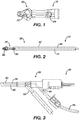
- Surgical reamers are well known in the art. An exemplary reamer is shown and described in U.S. Pat. No. 6,332,886 .
- a surgical reaming device 10 incudes a reamer head 20, located at a distal end 30 of the reaming device.
- a flexible cannulated tube 40 is connected to the reamer head 20 at a distal end of the tube 40 and extends proximally between the reamer head 20 and the proximal end 50 of the device 10.
- Either the reamer head 20, the distal end of the tube 40, or both can have one or more openings 22, 24 that are in fluid communication with the cannulation of the tube 40.
- the cannulated tube 40 can have more than one cannulation, for example, two cannulations within the tube.
- multiple cannulations can extend separately and coextensively from one another within the tube 40 such that each of the cannulations can maintain an independent and distinct fluid flow.
- One or more ports 52, 54 such as, for example, two ports, are located at the proximal end 50 of the device 10 and are in fluid communication with the cannulation of the tube 40. Accordingly, in one embodiment, each of the one or more ports 52, 54 can connect to each of the one or more of the cannulations within the tube 40, respectively, such that each of the one or more ports 52, 54 can be in separate and independent fluid communication with the each of the one or more openings 22, 24 at distal end of the device.
- a surgical reaming device 10 includes two proximal ports 52, 54, an irrigation port 54 and an aspiration port 52.
- the surgical reaming device 10 further includes a flexible tube 40 including two independent coextensive cannulations; an irrigation stream cannulation, and an aspiration stream cannulation.
- the device 10 includes one or more irrigation stream openings 22 connected to the irrigation stream cannulation, and separately, one or more aspiration stream openings 24 connected to the aspiration stream cannulation.
- an irrigation stream 44 is introduced by coupling a fluid supply 56 to the irrigation port 54 of surgical reamer 10, and an aspiration stream 42 is formed by coupling a vacuum source to the aspiration port 52.
- the irrigation stream 44 can flow in a direction from the fluid supply 56, through the irrigation port 54, further into the irrigation stream cannulation, and exit the device through the irrigation stream opening 22 into the intramedullary canal.
- the aspiration stream 42 can flow in a direction from the intramedullary canal into the aspiration stream cannulation through the aspiration stream openings 24, and exit the surgical reaming device through the aspiration stream port 52.
- access to the intramedullary canal is gained via an opening made with a surgical awl, drill, or other like device.
- the fluid supply 56 is opened, starting the irrigation stream 44 through the device 10 as detailed above.
- the reamer head 20 is activated and begins rotation.
- the vacuum source is activated and the reamer head 20 is inserted into the canal.
- the irrigation stream 44 begins to flow into the intramedullary canal space.
- the aspiration stream 42 will begin to flow as previously described, harvesting a volume of cellular material including a mononuclear cell population.
- the aspiration stream 42 can, according to one embodiment, define the contents of the effluent as well as include other components including the coarse residue.
- the aspiration stream 42 includes the volume of harvested cellular material including the mononuclear cell population of the effluent as described above.
- the methods for preparing an osteogenic bone graft can further include filtering the effluent prior to the collecting of the effluent.
- filtering the effluent includes filtering in order to remove a coarse residue, as well as other large tissue fragments, from the effluent in order to separate the effluent from the remainder of the aspiration stream.
- the aspiration stream exiting from the surgical reamer flows to a filter assembly that can include a filter, and also can include an intermediate collection point disposed at a flow point downstream of the filter, and which can collect the effluent.
- the filter can have an average pore size in the range of about 500 microns.
- the step of filtering captures an amount of coarse residue at a surface of the filter.
- the step of filtering retains an amount of autologous bone graft contained within the coarse residue. The retained autologous bone graft can be subsequently used in preparing the osteogenic bone graft as will be more fully described below.
- methods for preparing an osteogenic bone graft include introducing an anticoagulant compound into a volume of cellular material.
- Methods for preparing an osteogenic bone graft are disclosed which include introducing an anticoagulant compound into a harvested volume of cellular material.
- methods of preparing an osteogenic bone graft include:
- methods of preparing an osteogenic bone graft include:
- Anticoagulant compounds are well known in the art.
- An exemplary, non-exclusive list of anticoagulants can include for example, heparin, anticoagulant citrate dextrose-A (ACDA), citrate phosphate dextrose (CPD), citrate phosphate dextrose adenine (CPDA), and bivalarudin, and combinations thereof.
- the anticoagulant compound is introduced into the volume of cellular material in a range of about 2 parts to about 10 parts (by weight) of effluent to anticoagulant compound.
- the anticoagulant compound is introduced into the volume of cellular material in a range of about 6 parts to about 8 parts (by weight) of effluent to anticoagulant compound.
- the anticoagulant compound is introduced into the effluent in an amount sufficient to arrest coagulation for an amount of time sufficient to obtain the concentrated cell fraction.
- the amount of time sufficient to arrest coagulation with the anti-coagulant can vary, and can be for example, less than about 8 hours, less than about 7 hours, less than about 6 hours, less than about 5 hours. less than about 4 hours, less than about 3 hours, less than about 2 hours, or less than about 1 hour.
- the anticoagulant compound is continuously and proportionately introduced into the effluent from an anticoagulant container .
- a proportional introduction would allow for a sample of a given volume of effluent that has been mixed with anticoagulant to have approximately the same ratio of effluent to anticoagulant than another such sample taken at another point in the process.
- the proportionate introducing of the anticoagulant compound is by way of static mixing of the effluent and the anticoagulant, such as for example, a proportionate introduction of the anticoagulant compound and the aspirant accomplished through static mixing of the aspirant and anticoagulant.
- static mixing chambers are called "static mixers" in that the chamber remains fixed at an in-line location and the mixing is caused by the simultaneous, tortuous passage of the two liquids.
- the static mixing is a tortuous mixing.
- the introduction of the anticoagulant compound to the effluent occurs as they pass through an in-line mixing chamber with an interior geometry that causes tortuous mixing of the anticoagulant compound and the effluent such that by the time these two fluids passage through the in-line mixing chamber a homogenous distribution of the anticoagulant compound within the effluent is completed.
- the anticoagulant can be metered into the effluent, upstream of the filter assembly (i.e., into the aspiration stream), and mixed with the effluent during passage through a static mixer.
- the anticoagulant can mix thoroughly upstream of the filter assembly with both the cellular material, containing the mononuclear cell population, and the coarse residue, containing the autologous bone graft, in in an amount sufficient to arrest coagulation.
- the anticoagulant may be introduced within the filter assembly, and can also mix thoroughly with both the cellular material, containing the mononuclear cell population, and the coarse residue, containing the autologous bone graft.
- the anticoagulant can be metered into the effluent downstream of the filter assembly and mixed with the effluent during passage through a static mixer.
- the anticoagulant container includes a 0.2 micron pore size filter to prevent airborne microbes entering the container to replace the volume of anticoagulant dispensed through a one-way valve, such as a duckbill valve, that opens only in response to the vacuum that generates, and pulls the aspiration stream through the filter, which has been previously described.
- the aspiration stream can be pulled into a suction canister positioned downstream of the filter, and in certain embodiments, the suction canister and filter can be considered to form a filter assembly.
- the anticoagulant compound is introduced in a manner that prevents it from travelling upstream to the surgical reaming device and entering the medullary canal of the patient.
- the anticoagulant is introduced into the irrigation stream; for example, continuously introduced into the irrigation stream, such that the anticoagulant can be introduced into the volume of cellular material within the medullary canal and harvested in the aspiration stream.
- the anticoagulant is contained in a rigid container of a fixed volume (and equipped with an air displacement filter as desired) that is plumbed into the effluent line.
- the anticoagulant is contained in a flexible container such as a standard IV bag, which is then connected to the vacuum line at the static mixer via a standard barb fitting.
- the anticoagulant line may be connected to the primary (vacuum line) via a Y shaped fitting.
- the anticoagulant and effluent are combined in a manner designed to induce turbulent mixing, for example a nozzle designed for this purpose.
- the anticoagulant is pumped into the effluent using a peristaltic pump to induce positive pressure.
- the anticoagulant is drip fed into the effluent line under the influence of gravity.
- the anticoagulant is pumped under the influence of pressure applied to the walls of a flexible container (e.g. blood pressure cuff).
- methods for preparing an osteogenic bone graft include collecting the effluent including the volume of cellular material containing the anticoagulant compound at a collection point.
- collecting the effluent includes a first step of collecting at an intermediate collection point.
- the intermediate collection point can include a structure, such as, for example, a suction canister, that is part of the filter assembly, and collects the effluent after it has been filtered.
- the anticoagulant compound and the effluent have been proportionately mixed prior to collecting at the intermediate collection point.
- the effluent is subsequently collected at a processing collection point.
- the processing collection point can include a device capable of processing the effluent such that a desired concentrated cell fraction can be separated from the effluent. Such separation devices are known in the art, and can include, for example, centrifuges.
- methods for preparing an osteogenic bone graft include separating a concentrated cell fraction from the effluent, the concentrated cell fraction including the mononuclear cell population and the anticoagulant compound.
- a device can separate the concentrated cell fraction from the effluent, such as for example, through centrifugation.
- Exemplary separation devices are shown and described, for example in U.S. Pat. No. 8,747,289 .
- the individual cellular components of the cellular material can have varying densities.
- the effluent can be processed through multiple spin cycles. After the completion of each spin cycle, the undesired fractions of the processed effluent can be decanted or otherwise removed from the separation device. In such a manner, a concentrated cell fraction is separated from the effluent for use in preparing the osteogenic bone graft; the concentrated cell fraction including the mononuclear cell population and the anticoagulant compound.
- the concentrated cell fraction can be separated from the effluent during a centrifugation process, such as, for example, by removing the remainder fractions of the effluent during centrifugation.
- the original cellular material has a total mononuclear cell population and the concentrated cell fraction contains at least 80% of the mononuclear cell population of the cellular material.
- the concentrated cell fraction contains at least 90% of the mononuclear cell population of the cellular material.
- the osteogenic bone grafts include a concentrated cell fraction including a medullary canal derived mononuclear cell population; and an anticoagulant compound.
- the osteogenic bone graft can be further combined with an osteoinductive or an osteoconductive matrix.
- the osteoconductive or osteoinductive matrix is an allograft, a synthetic bone substitute, or an autologous bone graft, or a combination thereof.
- the concentration of the mononuclear cell population in the bone graft composition is the range of about 1.0 x 10 6 per ml to about 1000.0 x 10 6 per ml (prior to combining the osteogenic bone graft with an osteoconductive or osteoinductive matrix).
- the osteogenic bone graft can be an injectable composition, a moldable or pliable composition, or a rigid solid composition.
- the osteogenic bone graft can include a secondary amount of anticoagulant, where the secondary amount of anticoagulant can provide a therapeutic effect at the bone graft implantation site.
- the matrix can be a porous matrix.
- the porous nature of the matrix can allow for uptake of the concentrated cell fraction and anticoagulant within the matrix.
- the matrix can be non-porous.
- the concentrated cell fraction and anticoagulant can be blended or otherwise mixed with the matrix to provide a uniform distribution with the matrix.
- preparing the osteogenic bone graft includes combining the concentrated cell fraction with an osteoconductive or osteoinductive matrix.
- preparing the osteogenic bone graft can include combining the concentrated cell fraction with the autologous bone graft obtained from the coarse residue.
- preparing the osteogenic bone graft can include combining the concentrated cell fraction with a secondary autologous bone graft, where the secondary autologous bone graft is obtained from an anatomical site distinct from the harvesting site.
- preparing the osteogenic bone graft can include combining the concentrated cell fraction with an allograft, or a synthetic bone substitute.
- preparing the osteogenic bone graft includes adding an additional amount of anticoagulant to the bone graft, where the amount added is sufficient to increase the overall concentration of anticoagulant in the bone graft to a desired therapeutic level.
- a method of preparing the osteogenic bone graft includes applying the osteogenic bone graft to a bone defect site is also disclosed.
- the method includes performing all of the disclosed steps intraoperatively in a single procedure. This has the advantage of reducing the number of surgical procedures, as well as reducing the need to store the cells, which could result in contamination and loss of cell viability.
- the intraoperative performance of the steps is within 6 hours.
- the method of preparing the osteogenic bone graft can further include storing an excess amount of the concentrated cell fraction and anticoagulant, and can further include preparing a second, third, or more, osteogenic bone grafts.
- the method of preparing the osteogenic bone graft can include cryoprotecting, freezing, and cryogenically storing one or more excess portions of the concentrated cell fraction and anticoagulant, and can include preparing a second, third, or more, osteogenic bone grafts from the cryopreserved excess portions.
- the osteoconductive or osteoinductive matrix according to the present disclosure can include autologous bone (autograft), allogenic bone (allograft), as well as synthetic bone substitutes.
- Autologous bone can be harvested from the medullary canal as previously described above, and can also be taken from a secondary anatomical site such as the iliac crest. Autologous bone offers less risk of rejection because it has originated from the patient's own body. Additionally, autologous bone can also provide osteoinductive and osteogenic properties in addition to having osteoconductive properties.
- Autologous bone scaffolds with high solid bone content has a higher osteoconductive potential than autologous bone that contains a lower solid bone content.
- Allogenic bone scaffolds offer the same osteoconductive properties as autologous scaffolds. Allogenic scaffolds can be obtained from cadaveric samples, for example, from a tissue bank.
- the osteoconductive or osteoinductive matrix includes a synthetic bone substitute.
- the synthetic bone substitute can be porous or non-porous.
- the term "porous" includes, but is not limited to, macroporosity (mean pore diameter greater than or equal to 100 um), mesoporosity (mean pore diameter less than 100 um but greater than or equal to 10 um) and microporosity (mean pore diameter less than 10 um).
- the pores may be of any size, shape or distribution, or within a predetermined tolerance.
- the pores can be interconnecting or non-interconnecting.
- the diameter of the pores can range in size up to about 750 um.
- the pore sizes range up to about 500 um, with approximately 75% of the pores being at least 100 um in size and the remaining 25% of the pores being no more than 10 um in size.
- the synthetic bone substitute includes a ceramic bone substitute, such as, for example, a calcium phosphate based compound.
- a ceramic bone substitute such as, for example, a calcium phosphate based compound.
- Suitable examples of calcium phosphates include amorphous calcium phosphate, crystalline calcium phosphate, or any combination thereof.
- the calcium phosphate compound can be an apatite.
- Apatites are a group of calcium phosphate minerals, usually referring to hydroxyapatite Ca 10 (PO 4 ) 6 (OH) 2 , fluoroapatite Ca 10 (PO 4 ) 6 (F) 2 , chlorapatite Ca 10 (PO 4 ) 6 (Cl) 2 and bromapatite Ca 10 (PO 4 ) 6 (Br) 2 and can further include both silicate (SiO 4 4- ) and carbonate (CO 3 2- ) substituted hydroxyapatites, where the substitution is for one or more of the hydroxy and/or phosphate groups.
- the ceramic bone substitute includes beta-tricalcium phosphate Ca 3 (PO 4 ) 2 , (b-TCP).
- the osteoconductive or osteoinductive matrix can be of any shape as desired for the particular bone defect to be repaired.
- the matrix is a monolithic composition that can be either porous or non-porous. Suitable shapes can include, for example, spherical, cubic, wedge-shaped, oblong, cylindrical, or combinations thereof.
- the matrix can be a plurality of porous or non-porous granules.
- the specific surface area of the matrix can vary. For example, when the matrix is a porous granule, the specific surface area can range from about 0.1m 2 /g to about 100m 2 /g.
- the osteoconductive or osteoinductive matrix may be ceramic bone substitute particles or granules of any size or shape.
- the granules can be obtained by grinding or milling a calcium compound to a desired particle size or particle diameter.
- the mean diameter of the granules range in size from about 0.05 mm to about 10 mm.
- the mean diameter of the granules range in size from about 0.075 mm to about 5 mm.
- the mean diameter of the granules range in size from about 0.075 mm to about 1 mm.
- the mean diameter of the granules range in size from about 1.4 mm to about 2.8 mm.
- the mean diameter of the granules range in size from about 2.8 mm to about 5.6 mm.
- the mean diameter of the granules range in size from about 0.1 mm to about 0.750 mm
- the osteoconductive or osteoinductive matrix can be further combined with a polymeric binder, such that the osteogenic bone graft could be formed, for example, into a moldable or pliable implant that could be shaped as desired to fit the area of the bone to be repaired.
- the polymeric binder can include polymers such as homopolymers and copolymers (i.e., polymers including two or more different monomeric units), as well as polymer and copolymer blends, mixtures and combinations.
- the polymer can be a resorbable polymer, a non-resorbable polymer, or a combination thereof.
- the polymeric binder includes a resorbable polymer, and the polymeric binder is substantially free of a non-resorbable polymer.
- the polymeric binder is resorbable in vivo and includes a resorbable polymer.
- the polymer(s) of the polymeric binder can also include a synthetic polymer, a non-synthetic polymer (i.e., a polymer obtained from a plant or animal), or a combination thereof.
- Suitable polymers useful for preparing the polymeric binder include, but are not limited to, homopolymers or copolymers of monomers selected from L-lactide; L-lactic acid; D-lactide; D-lactic acid; glycolide; alpha-hydroxybutyric acid; alpha-hydroxyvaleric acid; alpha-hydroxyacetic acid; alpha-hydroxycaproic acid; alpha-hydroxyheptanoic acid; alpha-hydroxydecanoic acid; alpha-hydroxymyristic acid; alpha-hydroxyoctanoic acid; alpha-hydroxystearic acid; hydroxybutyrate; hydroxyvalerate; beta-propiolactide; beta-propiolactic acid; gamma-caprolactone; beta-caprolactone; epsilon-caprolactone; gamma-butyrolactone; pivalolactone; tetramethylglycolide; tetramethylglycolic acid; dimethylglycolic acid; trimethylene carbonate
- the polymeric binder includes resorbable polymers.
- resorbable polymers include, e.g., polymers derived from monomers selected from L-lactic acid, D-lactic acid, L-lactide, D-lactide, D,L-lactide, glycolide, a lactone, a lactam, epsilon-caprolactone, trimethylene carbonate, a cyclic carbonate, a cyclic ether, para-dioxanone, beta-hydroxybutyric acid, beta-hydroxypropionic acid, beta-hydroxyvaleric acid, saccharides, collagen, fibrin, albumin; and any combination of two or more of the foregoing.
- the polymeric binder includes a resorbable synthetic polymer.
- resorbable synthetic polymers include, e.g., a poly(L-lactide) (co)polymer, a poly(D,L-lactide) (co)polymer, a polyglycolide (co)polymer, a polycaprolactone (co)polymer, a poly(tetramethylglycolic acid) (co)polymer, a polydioxanone (co) polymer, a polyhydroxybutyrate (co)polymer, a polyhydroxyvalerate (co)polymer, a poly(L-lactide-co-glycolide) copolymer, a poly(glycolide-co-trimethylene carbonate) copolymer, a poly(glycolide-co-caprolactone) copolymer, a poly(glycolide-co-dioxanone-co-trim
- the osteogenic bone graft can consist essentially of the concentrated cell fraction including a medullary canal derived mononuclear cell population and an anticoagulant compound, such that the bone graft does not include any components that materially alter the function of the osteogenic bone graft to therapeutically affect the repair of a bone defect.
- the osteogenic bone graft can consist essentially of the concentrated cell fraction including a medullary canal derived mononuclear cell population, an anticoagulant compound, and an osteoinductive or an osteoconductive matrix, such that the bone graft does not include any components that materially alter the function of the osteogenic bone graft to therapeutically affect the repair of a bone defect.
- Samples of cellular material were harvested in procedures (labeled Run 1 through 10 or Patient 1 through 10) from human long bones utilizing a commercially available surgical reamer from Depuy Synthes (Reamer/Irrigator/Aspirator (RIA)). The amount of each harvested effluent that was processed is shown in Table 3 below.
- the effluent was collected intraoperatively from a line placed in between the filter assembly and a vacuum source. Additional filters may optionally be used in order to filter out debris that would otherwise cause fouling of the processing system. Additional filters may also be added in order to collect a further population of cells that may otherwise be clinically valuable.
- An anticoagulant in this case, Heparin, was introduced into the effluent in a controlled manner at a ratio of about 6-8 parts effluent to anticoagulant (by weight). The anticoagulant was introduced downstream of the filter assembly. The anticoagulant was proportionately introduced to the effluent through a static mixer in a manner that prevented it from travelling upstream into the surgical reaming device.
- the effluent and anticoagulant were collected in an intermediate collection chamber (e.g. a suction canister) positioned downstream of the filter prior to being decanted into a processing collection point. Samples of the pre-processed effluent were taken to be used in later testing and analysis.
- an intermediate collection chamber e.g. a suction canister
- the processing collection point was the single use disposable cartridge, available with the commercially available SynGenX-Lab System. Each of the cartridges is capable of processing up to 240 ml of collected effluent stream and up to 4 cartridges may be placed in a SynGenX-Lab centrifuge (total effluent volume that can be processed in a single cycle is approximately 1000 ml). The harvested effluent from each of the runs was separately processed and was evenly mixed and partitioned into separate cartridges.
- the centrifuge cycle is programmable and may include a "fast spin cycle” (during which the stratification of cells according to density in the main compartment occurs) and “slow spin cycle” (during which depletion of red cells to a dedicated compartment and harvest of clinically relevant cells to a harvest compartment occurs).
- the SyngenX-Lab System includes the cartridge and a reusable battery operated, firmware instructed control module, which contains an accelerometer and four infrared (IR) sensors.
- the accelerometer assures that the stratification of cell fractions according to density occurs at the correct gravity and the IR sensors assure that the predetermined volume of RBCs are transferred to the depletion compartment of the cartridge and the predetermined volume of MNCs, platelets and plasma are transferred to the harvest compartment of the cartridge.
- the processing system can be capable of depleting the excess red blood cells, irrigant, plasma, and granulocytes from the effluent. It is also capable of separating a concentrated cell fraction (as shown in the data below) including over 80% of the MNCs from the effluent.
- the plasma fraction, the RBC fraction and the concentrated cell fraction containing the mononuclear cell population (commonly referred to as the "buffy coat") were removed from each of the cartridges into a tube and labelled for further testing and analysis.
- mass balance is defined as the number of target cells recovered in the harvest chamber, red cell depletion chamber and main processing chamber (of the single-use disposable processing cartridge described in U.S. Pat. No. 8,747,289 ), at the end of a run cycle, compared, as a percentage, to the total number of target cells residing in the full volume of collected RIA effluent.
- 7-Aminoactinomycin D is a fluorescent chemical compound used as a cell viability stain. 7-AAD does not readily pass through intact cell membranes, so upon testing cells with compromised membranes will stain with 7-AAD, while live cells with intact cell membranes will remain dark.
- Annexin A5 is used as a non-quantitative probe to detect cells that have expressed phosphatidylserine (PS) on the cell surface, an event found in apoptosis as well as other forms of cell death.
- the annexin A5 affinity assay typically uses a conjugate of annexin V and a fluorescent or enzymatic label, biotin or other tags, or a radioelement, in a suitable buffer.
- the assay combines annexin V staining of PS and PE membrane events with the staining of proteins in the cell nucleus with propidium iodide (PI) or 7-Aminoactinomycin D (AAD-7), distinguishing viable cells from apoptotic cells and necrotic cells. Detection occurs by flow cytometry.
- Test data from Table 2 demonstrates that the viability of the harvested cells is maintained throughout the processing of the effluent.
- Test data demonstrates mononuclear cell concentrations having values in the 10 6 /ml range.
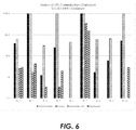
- Table 3 Flow Cytometry analysis of cell recovery Run RIA Vol. (mL) Harvest Vol. (mL) Harvest MNC count (10 9 ) MNC (%) Recovery Harvest CD34+ count (10 9 ) CD34+ (%) Recovery 1 960.0 39.2 N/A N/A N/A N/A 2 958.1 41.2 0.40 89.4 0.0262 97.1 3 225.3 10.5 0.17 101.4 0.0072 103.1 4 953.3 38.5 0.10 95.8 0.0016 99.0 5 956.9 35.9 7.54 95.5 0.5518 96.8 6 1000.6 38.8 0.19 102.6 0.0019 103.1 7 757.6 33.0 1.66 83.1 0.5464 94.2 8 757.6 24.9 0.41 93.7 0.0081 98.1 9 548.9 27.9 0.75 96.0 0.0361 96.9 10 951.3 20.7 0.84 94.5 0.0126 97.6
- test data in Table 3 demonstrates MNC recovery of greater than 90% and greater than 94% CD34+ recovery in runs 2-10.
- the effluent from Patient 1 was not analyzed.
- the data demonstrate that the colony forming capacity of the harvested MNC concentrate was greater ( ⁇ 1.5 to 35 times) than RIA effluent itself. Further, the data also demonstrate that the colony forming cells reside almost entirely within the concentrated cell fraction rather than other constituent layers (e.g., RBC layer and Supernatent layer), except for run 7 where cell separation was incomplete (below average MNC recovery).
Landscapes
- Health & Medical Sciences (AREA)
- Life Sciences & Earth Sciences (AREA)
- Chemical & Material Sciences (AREA)
- Engineering & Computer Science (AREA)
- Biomedical Technology (AREA)
- Medicinal Chemistry (AREA)
- Animal Behavior & Ethology (AREA)
- Transplantation (AREA)
- Veterinary Medicine (AREA)
- Public Health (AREA)
- Dermatology (AREA)
- General Health & Medical Sciences (AREA)
- Oral & Maxillofacial Surgery (AREA)
- Epidemiology (AREA)
- Zoology (AREA)
- Urology & Nephrology (AREA)
- Cell Biology (AREA)
- Chemical Kinetics & Catalysis (AREA)
- Botany (AREA)
- Molecular Biology (AREA)
- Materials For Medical Uses (AREA)
- Prostheses (AREA)
Claims (15)
- In vitro-Verfahren zur Herstellung eines osteogenen Knochentransplantats, umfassend:Einbringen einer Antikoagulansverbindung in einen Ausfluss, wobei der Ausfluss ein Volumen an zellulärem Material einschließlich einer mononukleären Zellpopulation aus dem Markkanal eines Röhrenknochens einschließt, wobei die Antikoagulansverbindung kontinuierlich und proportional in den Ausfluss eingebracht wird;Sammeln des Ausflusses, der das Volumen an zellulärem Material einschließt, welches die Antikoagulansverbindung enthält, an einem Sammelpunkt;Trennen einer konzentrierten Zellfraktion aus dem Ausfluss, wobei die konzentrierte Zellfraktion die mononukleäre Zellpopulation und die Antikoagulansverbindung einschließt; undHerstellen eines osteogenen Knochentransplantats aus der konzentrierten Zellfraktion.
- Verfahren nach Anspruch 1, wobei die Antikoagulansverbindung in einem Verhältnis im Bereich von etwa 2 Teilen bis etwa 10 Teilen, bezogen auf das Gewicht, Ausfluss zu Antikoagulansverbindung eingebracht wird.
- Verfahren nach Anspruch 1, wobei die proportionale Einbringung der Antikoagulansverbindung aufwendiges Mischen des Ausflusses und des Antikoagulans ist.
- Verfahren nach einem der vorhergehenden Ansprüche, wobei das Verfahren des Weiteren Filtrieren des Ausflusses vor dem Sammeln des Ausflusses einschließt, wobei das Filtrieren vorzugsweise einen groben Rückstand aus dem Ausfluss entfernt.
- Verfahren nach Anspruch 4, des Weiteren umfassend den Schritt des Einbehaltens einer Menge an autologem Knochentransplantat aus dem groben Rückstand.
- Verfahren nach einem der vorhergehenden Ansprüche, wobei Sammeln des Ausflusses zuerst Sammeln an einem Zwischensammelpunkt und nachfolgendes Sammeln an einem Verarbeitungssammelpunkt einschließt.
- Verfahren nach einem der vorhergehenden Ansprüche, wobei die konzentrierte Zellfraktion mindestens 80 % und vorzugsweise mindestens 90 % der mononukleären Zellpopulation des zellulären Materials enthält.
- Verfahren nach einem der vorhergehenden Ansprüche, wobei der Schritt des Herstellens des osteogenen Knochentransplantats Kombinieren der konzentrierten Zellfraktion mit einer osteokonduktiven oder osteoinduktiven Matrix einschließt, wobei die osteokonduktive oder osteoinduktive Matrix vorzugsweise ein Allotransplantat oder ein synthetischer Knochenersatz ist.
- Verfahren nach Anspruch 5, wobei der Schritt des Herstellens des osteogenen Knochentransplantats Kombinieren der konzentrierten Zellfraktion mit einer osteokonduktiven oder osteoinduktiven Matrix einschließlich des autologen Knochentransplantats einschließt.
- Verfahren nach einem der vorhergehenden Ansprüche, des Weiteren umfassend Hinzufügen einer zusätzlichen Menge an Antikoagulans zu dem osteogenen Knochentransplantat.
- Verfahren nach Anspruch 4, wobei die Einbringung des Antikoagulans vorgeordnet zu dem Filtrieren erfolgt, oder
wobei die Einbringung des Antikoagulans nachgeordnet zu dem Filtrieren erfolgt. - Osteogene Knochentransplantatzusammensetzung, die gemäß dem Verfahren nach einem der Ansprüche 1 bis 11 hergestellt worden ist, umfassend:eine konzentrierte Zellfraktion, die eine von einem Markkanal abgeleitete mononukleäre Zellpopulation einschließt; undeine Antikoagulansverbindung.
- Osteogene Knochentransplantatzusammensetzung nach Anspruch 12, des Weiteren umfassend eine osteokonduktive oder osteoinduktive Matrix, wobei die osteokonduktive oder osteoinduktive Matrix vorzugsweise ein Allotransplantat, ein synthetischer Knochenersatz oder ein autologes Knochentransplantat oder eine Kombination davon ist.
- Osteogene Knochentransplantatzusammensetzung nach einem der Ansprüche 12 bis 13, wobei die Konzentration der mononukleären Zellpopulation in der Knochentransplantatzusammensetzung im Bereich von 1,0 x 10⁶/mL bis etwa 1000,0 x 106 pro ml liegt.
- Osteogene Knochentransplantatzusammensetzung nach einem der Ansprüche 12 bis 14, wobei die Antikoagulansverbindung ausgewählt ist aus der Gruppe bestehend aus: Heparin, ACDA, CPD, CPDA und Bivalarudin sowie Kombinationen davon.
Applications Claiming Priority (2)
| Application Number | Priority Date | Filing Date | Title |
|---|---|---|---|
| US201562199480P | 2015-07-31 | 2015-07-31 | |
| PCT/US2016/043048 WO2017023542A1 (en) | 2015-07-31 | 2016-07-20 | Method of preparing an osteogenic bone graft |
Publications (2)
| Publication Number | Publication Date |
|---|---|
| EP3328453A1 EP3328453A1 (de) | 2018-06-06 |
| EP3328453B1 true EP3328453B1 (de) | 2021-03-24 |
Family
ID=56694213
Family Applications (1)
| Application Number | Title | Priority Date | Filing Date |
|---|---|---|---|
| EP16753497.3A Active EP3328453B1 (de) | 2015-07-31 | 2016-07-20 | Verfahren zur herstellung eines osteogenen knochentransplants |
Country Status (8)
| Country | Link |
|---|---|
| US (2) | US11278644B2 (de) |
| EP (1) | EP3328453B1 (de) |
| JP (1) | JP6812430B2 (de) |
| CN (1) | CN108025113B (de) |
| AU (1) | AU2016303310B2 (de) |
| BR (1) | BR112018002009B1 (de) |
| CA (1) | CA2994125C (de) |
| WO (1) | WO2017023542A1 (de) |
Families Citing this family (6)
| Publication number | Priority date | Publication date | Assignee | Title |
|---|---|---|---|---|
| US10646235B2 (en) | 2017-10-26 | 2020-05-12 | DePuy Synthes Products, Inc. | Guide wire seal for reamer irrigator aspirator system |
| CN111388027B (zh) * | 2020-03-25 | 2023-07-25 | 中国人民解放军总医院第六医学中心 | 骨髓采集装置 |
| CN114259276B (zh) * | 2021-12-24 | 2024-06-18 | 中国人民解放军陆军军医大学第一附属医院 | 一种基于骨髓抽吸富集系统的高活性骨构建方法 |
| CN114177203B (zh) * | 2021-12-27 | 2024-02-13 | 博雅干细胞科技有限公司 | 脐带血浓缩细胞治疗卵巢早衰的细胞治疗剂 |
| CN114196619B (zh) * | 2021-12-27 | 2023-11-07 | 深圳博雅感知医疗科技有限公司 | 治疗卵巢早衰的动员外周血浓缩细胞治疗剂 |
| US20230390070A1 (en) * | 2022-06-07 | 2023-12-07 | Warsaw Orthopedic, Inc. | Methods and apparatus for coating bone particles using a mesh |
Family Cites Families (11)
| Publication number | Priority date | Publication date | Assignee | Title |
|---|---|---|---|---|
| CA2360867C (en) | 1999-02-03 | 2008-08-05 | James M. Green | Surgical reamer and method of using same |
| US20060278588A1 (en) | 2002-05-24 | 2006-12-14 | Woodell-May Jennifer E | Apparatus and method for separating and concentrating fluids containing multiple components |
| US20070276352A1 (en) | 2002-06-04 | 2007-11-29 | Stemcor Systems, Inc. | Removable device and method for tissue disruption |
| AU2003248714A1 (en) * | 2002-06-17 | 2003-12-31 | Trimedyne, Inc. | Devices and methods for minimally invasive treatment of degenerated spinal discs |
| US7291450B2 (en) | 2003-03-28 | 2007-11-06 | Smith & Nephew, Inc. | Preparation of a cell concentrate from a physiological solution |
| US20070055282A1 (en) * | 2003-03-31 | 2007-03-08 | The Cleveland Clinic Foundation | Apparatus and method for harvesting bone marrow |
| US20070036766A1 (en) * | 2005-08-09 | 2007-02-15 | Sherwin Kevy | Tissue graft composition comprising autologous bone marrow and purified autologous thrombin |
| US8785191B2 (en) | 2007-08-27 | 2014-07-22 | University Of Connecticut | Concentration of stem cells obtained during orthopaedic surgeries |
| KR101946441B1 (ko) | 2010-03-18 | 2019-04-22 | 세스카 쎄라퓨틱스 인코포레이티드 | 특정 세포 군집 이외의 것을 고갈시켜 혈액 또는 골수 중의 특정 세포 군집을 정제하기 위한 시스템 |
| EP2943206A4 (de) * | 2013-01-12 | 2017-01-04 | Cesca Therapeutics Inc. | Schnelle infusion von autologen knochenmarksstammzellen |
| GB2530224B (en) | 2013-07-16 | 2020-08-05 | Univ Leland Stanford Junior | Enhancement of osteogenic potential of bone grafts |
-
2016
- 2016-07-20 EP EP16753497.3A patent/EP3328453B1/de active Active
- 2016-07-20 CN CN201680045112.6A patent/CN108025113B/zh active Active
- 2016-07-20 US US15/747,942 patent/US11278644B2/en active Active
- 2016-07-20 CA CA2994125A patent/CA2994125C/en active Active
- 2016-07-20 AU AU2016303310A patent/AU2016303310B2/en active Active
- 2016-07-20 BR BR112018002009-5A patent/BR112018002009B1/pt not_active IP Right Cessation
- 2016-07-20 WO PCT/US2016/043048 patent/WO2017023542A1/en not_active Ceased
- 2016-07-20 JP JP2018525502A patent/JP6812430B2/ja active Active
-
2022
- 2022-03-21 US US17/699,765 patent/US12194195B2/en active Active
Non-Patent Citations (1)
| Title |
|---|
| None * |
Also Published As
| Publication number | Publication date |
|---|---|
| AU2016303310B2 (en) | 2021-02-18 |
| EP3328453A1 (de) | 2018-06-06 |
| US11278644B2 (en) | 2022-03-22 |
| US20220202992A1 (en) | 2022-06-30 |
| CN108025113A (zh) | 2018-05-11 |
| CN108025113B (zh) | 2022-06-10 |
| US20180221539A1 (en) | 2018-08-09 |
| US12194195B2 (en) | 2025-01-14 |
| JP6812430B2 (ja) | 2021-01-13 |
| AU2016303310A1 (en) | 2018-02-22 |
| BR112018002009A2 (pt) | 2018-09-18 |
| CA2994125A1 (en) | 2017-02-09 |
| CA2994125C (en) | 2023-08-08 |
| BR112018002009B1 (pt) | 2021-06-08 |
| JP2018525193A (ja) | 2018-09-06 |
| WO2017023542A1 (en) | 2017-02-09 |
Similar Documents
| Publication | Publication Date | Title |
|---|---|---|
| US12194195B2 (en) | Method of preparing an osteogenic bone graft | |
| EP3294306B1 (de) | Knochenfragment und gewebeverarbeitungssystem | |
| US20080269762A1 (en) | Method and device for repair of cartilage defects | |
| Kevy et al. | Comparison of methods for point of care preparation of autologous platelet gel | |
| CA2439747C (en) | Composite bone marrow graft material with method and kit | |
| CN101072572B (zh) | 富含结缔组织生长成分的骨髓组分的分离及其促进结缔组织形成的用途 | |
| EP2806914B1 (de) | System und verfahren zur entnahme einer mit definierten zellen wie erythrozytenreichem plasma angereicherten zellulären probe | |
| US6872184B2 (en) | Tissue collection apparatus | |
| AU2002250173A1 (en) | Composite bone marrow graft material with method and kit | |
| EP2644208A1 (de) | Verfahren zur herstellung eines implantatmaterials | |
| US11931087B2 (en) | Bone fragment and tissue processing system | |
| US11278336B2 (en) | Osteomedullary tissue processing system | |
| US20070036766A1 (en) | Tissue graft composition comprising autologous bone marrow and purified autologous thrombin | |
| Harrell et al. | Novel Technology to Increase Concentrations of Stem and Progenitor Cells in Marrow Aspiration | |
| Kirimlioglu et al. | Determination of the optimum platelet-rich plasma method for the essential amount of platelets | |
| KR101187555B1 (ko) | 골유도를 위한 골응괴 제조방법 및 그 방법으로 제조된 골응괴 | |
| Harrell et al. | Marrow Cellution Bone Marrow Aspiration System and Related Concentrations of Stem and Progenitor Cells | |
| Harrell et al. | COMPARATIVE ANALYSIS OF MARROW CELLUTIONS AND THE BMAC® HARVEST®/TERUMO® SYSTEM | |
| JPH0658924A (ja) | 血液から血清を採取する方法 |
Legal Events
| Date | Code | Title | Description |
|---|---|---|---|
| STAA | Information on the status of an ep patent application or granted ep patent |
Free format text: STATUS: THE INTERNATIONAL PUBLICATION HAS BEEN MADE |
|
| PUAI | Public reference made under article 153(3) epc to a published international application that has entered the european phase |
Free format text: ORIGINAL CODE: 0009012 |
|
| STAA | Information on the status of an ep patent application or granted ep patent |
Free format text: STATUS: REQUEST FOR EXAMINATION WAS MADE |
|
| 17P | Request for examination filed |
Effective date: 20180206 |
|
| AK | Designated contracting states |
Kind code of ref document: A1 Designated state(s): AL AT BE BG CH CY CZ DE DK EE ES FI FR GB GR HR HU IE IS IT LI LT LU LV MC MK MT NL NO PL PT RO RS SE SI SK SM TR |
|
| AX | Request for extension of the european patent |
Extension state: BA ME |
|
| DAV | Request for validation of the european patent (deleted) | ||
| DAX | Request for extension of the european patent (deleted) | ||
| STAA | Information on the status of an ep patent application or granted ep patent |
Free format text: STATUS: EXAMINATION IS IN PROGRESS |
|
| 17Q | First examination report despatched |
Effective date: 20190529 |
|
| GRAP | Despatch of communication of intention to grant a patent |
Free format text: ORIGINAL CODE: EPIDOSNIGR1 |
|
| STAA | Information on the status of an ep patent application or granted ep patent |
Free format text: STATUS: GRANT OF PATENT IS INTENDED |
|
| INTG | Intention to grant announced |
Effective date: 20200506 |
|
| GRAJ | Information related to disapproval of communication of intention to grant by the applicant or resumption of examination proceedings by the epo deleted |
Free format text: ORIGINAL CODE: EPIDOSDIGR1 |
|
| STAA | Information on the status of an ep patent application or granted ep patent |
Free format text: STATUS: EXAMINATION IS IN PROGRESS |
|
| INTC | Intention to grant announced (deleted) | ||
| GRAP | Despatch of communication of intention to grant a patent |
Free format text: ORIGINAL CODE: EPIDOSNIGR1 |
|
| STAA | Information on the status of an ep patent application or granted ep patent |
Free format text: STATUS: GRANT OF PATENT IS INTENDED |
|
| INTG | Intention to grant announced |
Effective date: 20201028 |
|
| GRAS | Grant fee paid |
Free format text: ORIGINAL CODE: EPIDOSNIGR3 |
|
| GRAA | (expected) grant |
Free format text: ORIGINAL CODE: 0009210 |
|
| STAA | Information on the status of an ep patent application or granted ep patent |
Free format text: STATUS: THE PATENT HAS BEEN GRANTED |
|
| AK | Designated contracting states |
Kind code of ref document: B1 Designated state(s): AL AT BE BG CH CY CZ DE DK EE ES FI FR GB GR HR HU IE IS IT LI LT LU LV MC MK MT NL NO PL PT RO RS SE SI SK SM TR |
|
| REG | Reference to a national code |
Ref country code: GB Ref legal event code: FG4D |
|
| REG | Reference to a national code |
Ref country code: CH Ref legal event code: EP |
|
| REG | Reference to a national code |
Ref country code: IE Ref legal event code: FG4D |
|
| REG | Reference to a national code |
Ref country code: AT Ref legal event code: REF Ref document number: 1373814 Country of ref document: AT Kind code of ref document: T Effective date: 20210415 Ref country code: DE Ref legal event code: R096 Ref document number: 602016054841 Country of ref document: DE Ref country code: CH Ref legal event code: NV Representative=s name: E. BLUM AND CO. AG PATENT- UND MARKENANWAELTE V, CH |
|
| REG | Reference to a national code |
Ref country code: LT Ref legal event code: MG9D |
|
| PG25 | Lapsed in a contracting state [announced via postgrant information from national office to epo] |
Ref country code: NO Free format text: LAPSE BECAUSE OF FAILURE TO SUBMIT A TRANSLATION OF THE DESCRIPTION OR TO PAY THE FEE WITHIN THE PRESCRIBED TIME-LIMIT Effective date: 20210624 Ref country code: HR Free format text: LAPSE BECAUSE OF FAILURE TO SUBMIT A TRANSLATION OF THE DESCRIPTION OR TO PAY THE FEE WITHIN THE PRESCRIBED TIME-LIMIT Effective date: 20210324 Ref country code: GR Free format text: LAPSE BECAUSE OF FAILURE TO SUBMIT A TRANSLATION OF THE DESCRIPTION OR TO PAY THE FEE WITHIN THE PRESCRIBED TIME-LIMIT Effective date: 20210625 Ref country code: FI Free format text: LAPSE BECAUSE OF FAILURE TO SUBMIT A TRANSLATION OF THE DESCRIPTION OR TO PAY THE FEE WITHIN THE PRESCRIBED TIME-LIMIT Effective date: 20210324 Ref country code: BG Free format text: LAPSE BECAUSE OF FAILURE TO SUBMIT A TRANSLATION OF THE DESCRIPTION OR TO PAY THE FEE WITHIN THE PRESCRIBED TIME-LIMIT Effective date: 20210624 |
|
| PG25 | Lapsed in a contracting state [announced via postgrant information from national office to epo] |
Ref country code: LV Free format text: LAPSE BECAUSE OF FAILURE TO SUBMIT A TRANSLATION OF THE DESCRIPTION OR TO PAY THE FEE WITHIN THE PRESCRIBED TIME-LIMIT Effective date: 20210324 Ref country code: RS Free format text: LAPSE BECAUSE OF FAILURE TO SUBMIT A TRANSLATION OF THE DESCRIPTION OR TO PAY THE FEE WITHIN THE PRESCRIBED TIME-LIMIT Effective date: 20210324 Ref country code: SE Free format text: LAPSE BECAUSE OF FAILURE TO SUBMIT A TRANSLATION OF THE DESCRIPTION OR TO PAY THE FEE WITHIN THE PRESCRIBED TIME-LIMIT Effective date: 20210324 |
|
| REG | Reference to a national code |
Ref country code: NL Ref legal event code: MP Effective date: 20210324 |
|
| REG | Reference to a national code |
Ref country code: AT Ref legal event code: MK05 Ref document number: 1373814 Country of ref document: AT Kind code of ref document: T Effective date: 20210324 |
|
| PG25 | Lapsed in a contracting state [announced via postgrant information from national office to epo] |
Ref country code: NL Free format text: LAPSE BECAUSE OF FAILURE TO SUBMIT A TRANSLATION OF THE DESCRIPTION OR TO PAY THE FEE WITHIN THE PRESCRIBED TIME-LIMIT Effective date: 20210324 |
|
| PG25 | Lapsed in a contracting state [announced via postgrant information from national office to epo] |
Ref country code: LT Free format text: LAPSE BECAUSE OF FAILURE TO SUBMIT A TRANSLATION OF THE DESCRIPTION OR TO PAY THE FEE WITHIN THE PRESCRIBED TIME-LIMIT Effective date: 20210324 Ref country code: CZ Free format text: LAPSE BECAUSE OF FAILURE TO SUBMIT A TRANSLATION OF THE DESCRIPTION OR TO PAY THE FEE WITHIN THE PRESCRIBED TIME-LIMIT Effective date: 20210324 Ref country code: EE Free format text: LAPSE BECAUSE OF FAILURE TO SUBMIT A TRANSLATION OF THE DESCRIPTION OR TO PAY THE FEE WITHIN THE PRESCRIBED TIME-LIMIT Effective date: 20210324 Ref country code: AT Free format text: LAPSE BECAUSE OF FAILURE TO SUBMIT A TRANSLATION OF THE DESCRIPTION OR TO PAY THE FEE WITHIN THE PRESCRIBED TIME-LIMIT Effective date: 20210324 Ref country code: SM Free format text: LAPSE BECAUSE OF FAILURE TO SUBMIT A TRANSLATION OF THE DESCRIPTION OR TO PAY THE FEE WITHIN THE PRESCRIBED TIME-LIMIT Effective date: 20210324 |
|
| PG25 | Lapsed in a contracting state [announced via postgrant information from national office to epo] |
Ref country code: IS Free format text: LAPSE BECAUSE OF FAILURE TO SUBMIT A TRANSLATION OF THE DESCRIPTION OR TO PAY THE FEE WITHIN THE PRESCRIBED TIME-LIMIT Effective date: 20210724 Ref country code: PL Free format text: LAPSE BECAUSE OF FAILURE TO SUBMIT A TRANSLATION OF THE DESCRIPTION OR TO PAY THE FEE WITHIN THE PRESCRIBED TIME-LIMIT Effective date: 20210324 Ref country code: PT Free format text: LAPSE BECAUSE OF FAILURE TO SUBMIT A TRANSLATION OF THE DESCRIPTION OR TO PAY THE FEE WITHIN THE PRESCRIBED TIME-LIMIT Effective date: 20210726 Ref country code: RO Free format text: LAPSE BECAUSE OF FAILURE TO SUBMIT A TRANSLATION OF THE DESCRIPTION OR TO PAY THE FEE WITHIN THE PRESCRIBED TIME-LIMIT Effective date: 20210324 Ref country code: SK Free format text: LAPSE BECAUSE OF FAILURE TO SUBMIT A TRANSLATION OF THE DESCRIPTION OR TO PAY THE FEE WITHIN THE PRESCRIBED TIME-LIMIT Effective date: 20210324 |
|
| REG | Reference to a national code |
Ref country code: DE Ref legal event code: R097 Ref document number: 602016054841 Country of ref document: DE |
|
| PG25 | Lapsed in a contracting state [announced via postgrant information from national office to epo] |
Ref country code: ES Free format text: LAPSE BECAUSE OF FAILURE TO SUBMIT A TRANSLATION OF THE DESCRIPTION OR TO PAY THE FEE WITHIN THE PRESCRIBED TIME-LIMIT Effective date: 20210324 Ref country code: AL Free format text: LAPSE BECAUSE OF FAILURE TO SUBMIT A TRANSLATION OF THE DESCRIPTION OR TO PAY THE FEE WITHIN THE PRESCRIBED TIME-LIMIT Effective date: 20210324 Ref country code: DK Free format text: LAPSE BECAUSE OF FAILURE TO SUBMIT A TRANSLATION OF THE DESCRIPTION OR TO PAY THE FEE WITHIN THE PRESCRIBED TIME-LIMIT Effective date: 20210324 |
|
| PLBE | No opposition filed within time limit |
Free format text: ORIGINAL CODE: 0009261 |
|
| STAA | Information on the status of an ep patent application or granted ep patent |
Free format text: STATUS: NO OPPOSITION FILED WITHIN TIME LIMIT |
|
| PG25 | Lapsed in a contracting state [announced via postgrant information from national office to epo] |
Ref country code: SI Free format text: LAPSE BECAUSE OF FAILURE TO SUBMIT A TRANSLATION OF THE DESCRIPTION OR TO PAY THE FEE WITHIN THE PRESCRIBED TIME-LIMIT Effective date: 20210324 |
|
| 26N | No opposition filed |
Effective date: 20220104 |
|
| PG25 | Lapsed in a contracting state [announced via postgrant information from national office to epo] |
Ref country code: MC Free format text: LAPSE BECAUSE OF FAILURE TO SUBMIT A TRANSLATION OF THE DESCRIPTION OR TO PAY THE FEE WITHIN THE PRESCRIBED TIME-LIMIT Effective date: 20210324 |
|
| REG | Reference to a national code |
Ref country code: BE Ref legal event code: MM Effective date: 20210731 |
|
| PG25 | Lapsed in a contracting state [announced via postgrant information from national office to epo] |
Ref country code: IS Free format text: LAPSE BECAUSE OF FAILURE TO SUBMIT A TRANSLATION OF THE DESCRIPTION OR TO PAY THE FEE WITHIN THE PRESCRIBED TIME-LIMIT Effective date: 20210724 Ref country code: LU Free format text: LAPSE BECAUSE OF NON-PAYMENT OF DUE FEES Effective date: 20210720 |
|
| PG25 | Lapsed in a contracting state [announced via postgrant information from national office to epo] |
Ref country code: IE Free format text: LAPSE BECAUSE OF NON-PAYMENT OF DUE FEES Effective date: 20210720 Ref country code: BE Free format text: LAPSE BECAUSE OF NON-PAYMENT OF DUE FEES Effective date: 20210731 |
|
| PG25 | Lapsed in a contracting state [announced via postgrant information from national office to epo] |
Ref country code: HU Free format text: LAPSE BECAUSE OF FAILURE TO SUBMIT A TRANSLATION OF THE DESCRIPTION OR TO PAY THE FEE WITHIN THE PRESCRIBED TIME-LIMIT; INVALID AB INITIO Effective date: 20160720 |
|
| PG25 | Lapsed in a contracting state [announced via postgrant information from national office to epo] |
Ref country code: CY Free format text: LAPSE BECAUSE OF FAILURE TO SUBMIT A TRANSLATION OF THE DESCRIPTION OR TO PAY THE FEE WITHIN THE PRESCRIBED TIME-LIMIT Effective date: 20210324 |
|
| PG25 | Lapsed in a contracting state [announced via postgrant information from national office to epo] |
Ref country code: MK Free format text: LAPSE BECAUSE OF FAILURE TO SUBMIT A TRANSLATION OF THE DESCRIPTION OR TO PAY THE FEE WITHIN THE PRESCRIBED TIME-LIMIT Effective date: 20210324 |
|
| PG25 | Lapsed in a contracting state [announced via postgrant information from national office to epo] |
Ref country code: MT Free format text: LAPSE BECAUSE OF FAILURE TO SUBMIT A TRANSLATION OF THE DESCRIPTION OR TO PAY THE FEE WITHIN THE PRESCRIBED TIME-LIMIT Effective date: 20210324 |
|
| PGFP | Annual fee paid to national office [announced via postgrant information from national office to epo] |
Ref country code: GB Payment date: 20250529 Year of fee payment: 10 |
|
| PGFP | Annual fee paid to national office [announced via postgrant information from national office to epo] |
Ref country code: FR Payment date: 20250610 Year of fee payment: 10 |
|
| PGFP | Annual fee paid to national office [announced via postgrant information from national office to epo] |
Ref country code: DE Payment date: 20250604 Year of fee payment: 10 |
|
| PGFP | Annual fee paid to national office [announced via postgrant information from national office to epo] |
Ref country code: IT Payment date: 20250623 Year of fee payment: 10 |
|
| PGFP | Annual fee paid to national office [announced via postgrant information from national office to epo] |
Ref country code: CH Payment date: 20250801 Year of fee payment: 10 |
|
| PG25 | Lapsed in a contracting state [announced via postgrant information from national office to epo] |
Ref country code: TR Free format text: LAPSE BECAUSE OF FAILURE TO SUBMIT A TRANSLATION OF THE DESCRIPTION OR TO PAY THE FEE WITHIN THE PRESCRIBED TIME-LIMIT Effective date: 20210324 |