EP3638164B1 - Systeme, und vorrichtung zur behandlung von glaukom - Google Patents
Systeme, und vorrichtung zur behandlung von glaukom Download PDFInfo
- Publication number
- EP3638164B1 EP3638164B1 EP18818723.1A EP18818723A EP3638164B1 EP 3638164 B1 EP3638164 B1 EP 3638164B1 EP 18818723 A EP18818723 A EP 18818723A EP 3638164 B1 EP3638164 B1 EP 3638164B1
- Authority
- EP
- European Patent Office
- Prior art keywords
- eye
- distal end
- inserter
- wedge
- rod
- Prior art date
- Legal status (The legal status is an assumption and is not a legal conclusion. Google has not performed a legal analysis and makes no representation as to the accuracy of the status listed.)
- Active
Links
Images


Classifications
-
- A—HUMAN NECESSITIES
- A61—MEDICAL OR VETERINARY SCIENCE; HYGIENE
- A61F—FILTERS IMPLANTABLE INTO BLOOD VESSELS; PROSTHESES; DEVICES PROVIDING PATENCY TO, OR PREVENTING COLLAPSING OF, TUBULAR STRUCTURES OF THE BODY, e.g. STENTS; ORTHOPAEDIC, NURSING OR CONTRACEPTIVE DEVICES; FOMENTATION; TREATMENT OR PROTECTION OF EYES OR EARS; BANDAGES, DRESSINGS OR ABSORBENT PADS; FIRST-AID KITS
- A61F9/00—Methods or devices for treatment of the eyes; Devices for putting in contact-lenses; Devices to correct squinting; Apparatus to guide the blind; Protective devices for the eyes, carried on the body or in the hand
- A61F9/007—Methods or devices for eye surgery
- A61F9/00781—Apparatus for modifying intraocular pressure, e.g. for glaucoma treatment
-
- A—HUMAN NECESSITIES
- A61—MEDICAL OR VETERINARY SCIENCE; HYGIENE
- A61F—FILTERS IMPLANTABLE INTO BLOOD VESSELS; PROSTHESES; DEVICES PROVIDING PATENCY TO, OR PREVENTING COLLAPSING OF, TUBULAR STRUCTURES OF THE BODY, e.g. STENTS; ORTHOPAEDIC, NURSING OR CONTRACEPTIVE DEVICES; FOMENTATION; TREATMENT OR PROTECTION OF EYES OR EARS; BANDAGES, DRESSINGS OR ABSORBENT PADS; FIRST-AID KITS
- A61F2220/00—Fixations or connections for prostheses classified in groups A61F2/00 - A61F2/26 or A61F2/82 or A61F9/00 or A61F11/00 or subgroups thereof
- A61F2220/0008—Fixation appliances for connecting prostheses to the body
- A61F2220/0016—Fixation appliances for connecting prostheses to the body with sharp anchoring protrusions, e.g. barbs, pins, spikes
-
- A—HUMAN NECESSITIES
- A61—MEDICAL OR VETERINARY SCIENCE; HYGIENE
- A61F—FILTERS IMPLANTABLE INTO BLOOD VESSELS; PROSTHESES; DEVICES PROVIDING PATENCY TO, OR PREVENTING COLLAPSING OF, TUBULAR STRUCTURES OF THE BODY, e.g. STENTS; ORTHOPAEDIC, NURSING OR CONTRACEPTIVE DEVICES; FOMENTATION; TREATMENT OR PROTECTION OF EYES OR EARS; BANDAGES, DRESSINGS OR ABSORBENT PADS; FIRST-AID KITS
- A61F2250/00—Special features of prostheses classified in groups A61F2/00 - A61F2/26 or A61F2/82 or A61F9/00 or A61F11/00 or subgroups thereof
- A61F2250/0004—Special features of prostheses classified in groups A61F2/00 - A61F2/26 or A61F2/82 or A61F9/00 or A61F11/00 or subgroups thereof adjustable
- A61F2250/0013—Special features of prostheses classified in groups A61F2/00 - A61F2/26 or A61F2/82 or A61F9/00 or A61F11/00 or subgroups thereof adjustable for adjusting fluid pressure
-
- A—HUMAN NECESSITIES
- A61—MEDICAL OR VETERINARY SCIENCE; HYGIENE
- A61F—FILTERS IMPLANTABLE INTO BLOOD VESSELS; PROSTHESES; DEVICES PROVIDING PATENCY TO, OR PREVENTING COLLAPSING OF, TUBULAR STRUCTURES OF THE BODY, e.g. STENTS; ORTHOPAEDIC, NURSING OR CONTRACEPTIVE DEVICES; FOMENTATION; TREATMENT OR PROTECTION OF EYES OR EARS; BANDAGES, DRESSINGS OR ABSORBENT PADS; FIRST-AID KITS
- A61F2250/00—Special features of prostheses classified in groups A61F2/00 - A61F2/26 or A61F2/82 or A61F9/00 or A61F11/00 or subgroups thereof
- A61F2250/0058—Additional features; Implant or prostheses properties not otherwise provided for
- A61F2250/0067—Means for introducing or releasing pharmaceutical products into the body
-
- A—HUMAN NECESSITIES
- A61—MEDICAL OR VETERINARY SCIENCE; HYGIENE
- A61F—FILTERS IMPLANTABLE INTO BLOOD VESSELS; PROSTHESES; DEVICES PROVIDING PATENCY TO, OR PREVENTING COLLAPSING OF, TUBULAR STRUCTURES OF THE BODY, e.g. STENTS; ORTHOPAEDIC, NURSING OR CONTRACEPTIVE DEVICES; FOMENTATION; TREATMENT OR PROTECTION OF EYES OR EARS; BANDAGES, DRESSINGS OR ABSORBENT PADS; FIRST-AID KITS
- A61F2250/00—Special features of prostheses classified in groups A61F2/00 - A61F2/26 or A61F2/82 or A61F9/00 or A61F11/00 or subgroups thereof
- A61F2250/0058—Additional features; Implant or prostheses properties not otherwise provided for
- A61F2250/0069—Sealing means
Definitions
- the present disclosure relates to the treatment of glaucoma, and more particularly, to medical devices and methods for creating a drainage pathway to divert aqueous humor out of the anterior chamber of the eye such that pressure within the eye is reduced.
- Aqueous humor is produced by the eye's ciliary body and flows from the ciliary body into the anterior chamber, out through a spongy tissue at the front of the eye called the trabecular meshwork and into a drainage canal.
- continuous drainage of aqueous humor keeps intraocular pressure at a normal level.
- proper circulation of aqueous humor is disrupted, causing the level of intraocular pressure to be elevated.
- fluid does not flow freely through the trabecular meshwork, causing an increase in intraocular pressure, damage to the optic nerve and vision loss. Reduction of intraocular pressure is a means of stopping the progression of optic nerve damage, which if untreated can lead to blindness.
- the suprachoroidal space is a space in the eye that lies between the sclera and the choroid. It is known that aqueous humor in the suprachoroidal space can drain therefrom and cause a reduction in intraocular pressure. Although it is not well understood where aqueous humor drains to once it reaches the suprachoroidal space, there are references to aqueous humor draining into the choroid vessels as well as into the venous plexus of the sclera and to the episcleral veins.
- CyPass ® Microstent that includes a tubular body with an internal lumen that drains aqueous humor from the anterior chamber of the eye into the suprachoroidal space of the eye to lower intraocular pressure in the eye.
- US 7862531 , US 4521210 , US 2013/165840 and WO 207/087061 each disclose devices that include a body with an elongated groove or slot in the surface for fluid drainage.
- a device for implantation into the suprachoroidal space of the eye to promote drainage of aqueous humor from the anterior chamber of the eye to the suprachoroidal space of the eye in order to reduce intraocular pressure.
- the device may be made from a flexible, bio-inert, and biocompatible material that can be inserted into the suprachoroidal space of the eye using an ab interno approach, conform to the curvature of the tissue surrounding the suprachoroidal space (i.e., the sclera and the choroid), and remain in place for a long period of time.
- the device includes an elongated body extending axially from a distal end to a proximal end.
- the distal end of elongate body forms a wedge with a leading distal edge.
- the wedge with leading distal edge can facilitate penetration into and spreading open the tissue of the suprachoroidal space of the eye.
- the elongate body has one or more outer surfaces that define at least one open groove extending from at or near the proximal end towards the distal end of the body.
- the at least one open groove is configured such that aqueous humor flows along the open groove from the anterior chamber of the eye to the suprachoroidal space of the eye. Due to the open nature of the open groove, the flow path of the aqueous humor that flows along the open groove can be bounded by ocular tissue disposed adjacent the open groove along the length of the open groove. The open groove terminates at an abutment defined by the elongated body
- the body of the device has an upper outer surface and a lower outer surface that are substantially planar in form.
- barbs extend from the upper and lower surfaces. The barbs may be tapered to permit insertion in one direction and resist removal in an opposite direction.
- the device is formed from a soft flexible polymeric material.
- soft flexible polymeric material includes poly(styrene-block-isobutylene-block-styrene) (SIBS), styrene ethylene butylene styrene (SEBS), polyhexene, polypropylene, polyethylene, and combinations thereof.
- SIBS poly(styrene-block-isobutylene-block-styrene)
- SEBS styrene ethylene butylene styrene
- polyhexene polypropylene
- polyethylene and combinations thereof.
- the material may have a hardness of Shore 30A to 60A.
- a system includes the device and an inserter coupled to the device.
- the inserter is configured to hold the device while positioning the distal end of the device in the suprachoroidal space and to decouple from the device to deploy the device in a desired location in the suprachoroidal space.
- the inserter may include a handle and at least one rigid member configured for longitudinal translation relative to the handle. Each rigid member is configured for longitudinal translation in a corresponding open groove of the device.
- the inserter holds the device with each rigid member in an extended configuration in which the rigid member extends along at least a portion of the corresponding open groove of the device.
- the inserter is configured to decouple from the device by reconfiguring each rigid member from the extended configuration to a retracted configuration in which the rigid member is removed from the open groove of the device.
- rigid means that the inserter will not bend or buckle under a range of forces (e.g., axial and radial compressive forces) that may be imparted to the inserter by the hand of the user when the inserter is introduced into the eye, as described in greater detail below.
- flexible means that the device, if unsupported by the inserter, will bend or buckle under the axial and radial compressive forces that may be imparted to the inserter during its use.
- the at least one rod is coupled to a slide member configured for actuation by a user's hand.
- the inserter handle defines a longitudinal slot extending axially along the length of the handle and parallel with the at least one rod, and the slide member is configured to slide within the slot to move the rod between the extended and retracted configurations.
- a method of implanting the device includes providing the device coupled to the inserter, introducing the device into the eye while maintaining the handle outside of the eye, positioning the device at a desired implanted position in the suprachoroidal space, and, with the device positioned at the desired implanted position, decoupling the inserter from the device.
- positioning the device at a desired implanted position in the suprachoroidal space includes positioning the distal end of the device in the suprachoroidal space and positioning the proximal end of the device in the anterior chamber. At the desired position, the device may extend about 0.5 mm to 1 mm into the anterior chamber.
- Fig. 1 shows details of an anterior segment of an eye.
- An anterior segment of an eye 1 includes a cornea 2, anterior chamber 3, iris 4, lens 5, conjunctiva 6, Tenon's Capsule 7, sclera 8, ciliary muscle 9, choroid 10, and suprachoroidal space 11.
- the suprachoroidal space 11 is bounded between the choroid 10 and the sclera 8.
- the choroid 10 contains blood vessels that interpenetrate the upper part of the ciliary body 9. Those blood vessels become more organized into a more distinct layer more posterior to the anterior chamber 3.
- a clear corneal incision 13, or a precisely articulated incision used in the cornea 2, is used to access the anterior chamber 3 during ab interno anterior segment surgery such as cataract surgery. Clear corneal incision 13 can be made with a scalpel or with a laser.
- the anterior chamber 3 is typically filled with a visco-elastic material during cataract surgery and procedures of this nature.
- Dotted circle 12 denotes a portion of interest to this disclosure and is shown in greater detail in Fig. 2 .
- the suprachoroidal space 11 is situated just below a scleral spur 20.
- a surgeon looking through a gonio lens (not shown) into the anterior chamber 3 through the cornea 2 can readily identify the scleral spur 20 and the entrance to the suprachoroidal space 11.
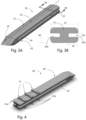
- Fig. 3A shows an embodiment of a glaucoma drainage device 30, hereinafter referred to as a "wedge", that is configured for insertion into the suprachoroidal space 11, as shown in Fig. 5 , for example, and described in greater detail below.
- the wedge 30 is comprised of an elongated body 31, hereinafter referred to as a "rod”, which is shown as being a filleted rectangular (rectangular in terms of the overall planform) rod with a tapered distal end 32.
- the rod 31 has outer surfaces 42 (top surface) and 43 (bottom surface) that define at least one open groove or channel 33 extending from at or near a proximal end 50 towards the distal end 32.
- the groove or channel 33 extends parallel to the longitudinal axis A-A.
- the upper surface 42 and lower surface 43 are planar when the wedge 30 is laid on a flat surface and not subject to external forces.
- the open groove or channel 33 may also or alternatively be formed in either or both of the upper or lower surfaces 42 and 43.
- the taper angle of the distal end 32 is shallower than that shown in Fig. 3A so that the taper extends further and may extend all the way to a proximal end 50.
- proximal and distal refer to positions along axis A-A in Fig. 3A .
- the slot(s) 33 can be continuous throughout the entire length of the wedge 30 or it can stop anywhere along its length for example at an abutment 34 at or near the distal end 32 of the rod 31.
- Fig. 3B shows a cross-section of wedge 30 along section 3B-3B in Fig. 3A showing two slots 33a and 33b on each side of wedge 30.
- the cross-section has the appearance of an I-beam having two horizontal flanges, one on the top and a second on the bottom, spaced vertically by a vertical web.
- the wedge 30 is lumen-less and grooves or channels 33 formed on the outside surface(s) of the rod 31 are open. The grooves or channels 33 enable fluid to flow alongside the outer surface(s) of the wedge 30 and diffuse from the anterior chamber 3 into the suprachoroidal space 11, as will be described in greater detail below.
- the axial length of wedge 30 measured along axis A-A from the distal end 32 to the proximal end 50 can be from 3 mm to 10 mm, and preferably 6 mm.
- the cross-sectional dimensions depicted on Fig. 3B are width 37 of 0.5 mm to 1 mm, and preferably 0.75 mm.
- the height 36 is 0.4 to 0.8 mm, and preferably 0.5 mm.
- the width of slots 33, denoted as 38 can be from 0.05 mm to 0.25 mm, preferably 0.15 mm.
- An indentation or depth 39 of slots 33 into wedge 30 are less than 40% of the width 37 of wedge 30 and can range from 0.05 mm to 0.4 mm, and preferably 0.25 mm.
- Fig. 7 shows a cross section of a wedge 70 that has two lobes 70a and 70b that intersect thereby defining a set of open grooves or channels 73a and 73b on the outer surface of the wedge 70.
- Fig. 8 shows a cross-section of a wedge 80 that has three lobes 80a, 80b, and 80c, which intersect defining a set of three open grooves or channels 83a, 83b, and 83c on the outer surface of the wedge 80.
- the wedge 30 is made from any biomaterial including polyolefins such as poly(styrene-block-isobutylene-block-styrene) (SIBS), styrene ethylene butylene styrene (SEBS), polyhexene, polypropylene, polyethylene, and the like, as well as copolymers of the above.
- SIBS poly(styrene-block-isobutylene-block-styrene)
- SEBS styrene ethylene butylene styrene
- polyhexene polypropylene
- polyethylene polyethylene
- Other materials comprising the wedge 30 can include but are not limited to silicone rubber (polydimethylsiloxane and polyphenylsiloxane and copolymers thereof), polyurethane such as polyether urethane, polycarbonate urethane, polysilicone urethane, polyisobutylene urethane and other polyurethanes used for medical implantation; fluorinated polymers can also be used such as polyvinyldifluoride (PVDF) and fluorinated versions of the above.
- PVDF polyvinyldifluoride
- Other materials can be used for this embodiment include stiffer materials such as PEEK, polyimide, polysulfone, ridged polyurethane, polyamide, etc.
- Biological materials can also be used for the wedge such as crosslinked gelatin (porcine, equine, bovine, feline, etc.) crosslinked polysaccharides (gellen, pectin, hyaluronic acid, methyl cellulose, and the like).
- the preferred materials are those that are biocompatible and significantly flexible to take on the shape of the suprachoroidal space.
- a preferred material to be used in forming the wedge 30 is poly(styrene-block-isobutylene-block-styrene) (SIBS) of Shore 30A to 60A hardness as described in detail in U.S. Patents 9,101,444 ; 9,044,301 ; 7,837,644 ; 7,594,899 ; and 7,431,709 .
- the wedge 30 may be extruded as a long, contoured monofilament, which can be cut to length.
- the extruded, cut monofilament can then be heat-formed at one end (e.g., the distal end 32) to form features (e.g., abutment 34 and taper of the distal end 32) of the wedge 30 that may have not been formed by extrusion and cutting.
- Fig. 4 shows another embodiment of a wedge 30' which has a plurality of barbs 41 on an upper surface 42' and on a lower surface 43'.
- the barbs 41 are configured to engage the tissue defining the suprachoroidal space 11 so that when the wedge 30' in introduced into the suprachoroidal space 11, the barbs 41 will help retain and fixate the wedge 30' in its implanted position and inhibit migration or ejection of the wedge 30' back out of the suprachoroidal space 11. While the barbs 41 are shown as having flat outer surfaces 41a (parallel with the upper and lower surfaces 42' and 43') in Fig.
- the barbs can also have outer surfaces 41a that extend at non-zero angles relative to the upper and lower surfaces 42' and 43' to allow easy insertion into the suprachoroidal space 11, while resisting removal in the other direction.
- the barbs 41 are shown protruding from the surfaces of the wedge 30', the barbs 41 can also be indents, ridges, or grooves (not shown) formed in the surface.
- the wedge 30 is configured for implantation at least partly in the suprachoroidal space 11.
- the distal end 32 of the wedge 30 is located in the suprachoroidal space 11 and the proximal end 50 is located in the anterior chamber 3, as shown in Fig. 5 .
- the wedge 30 conforms to the curved shape of the suprachoroidal space 11, which is parallel to the curvature of the eye.
- the wedge 30 may extend 0.5 mm to 1 mm into the anterior chamber 3. This spacing can help prevent closure of the suprachoroidal space 11 around the proximal end 50 of the wedge 30, which, if closed, would cut off the flow of fluid from the anterior chamber 3 to the suprachoroidal space 11.
- Fig. 6 shows an end-view 60 (viewed from the proximal end 50 looking distally) of wedge 30 in the implanted configuration shown in Fig. 5 , as seen from the anterior chamber 3 using optical coherence tomography (OTC).
- OTC optical coherence tomography
- the wedge 30 can be implanted alone or in conjunction with one or more therapeutic agents. These therapeutic agents can be injected into the eye at the time of surgery or coated on the device or embedded within the device to elute therefrom. In addition, these therapeutic agents can be injected periodically following implantation of the wedge. Also, the wedge 30 may be formed from a biodegradable polymer matrix or coated with a biodegradable polymer matrix, where the biodegradable polymer matrix is loaded with a therapeutic agent that can be released from the matrix into the eye over time. The biodegradable polymer matrix can degrade over time in vivo (in the implanted position in the eye) and such degradation can be required to achieve the desired release rate of the therapeutic agent from the matrix into the eye over time.
- the biodegradable polymer matrix can be selected from the group consisting of one or more biodegradable polymers in varying combinations, such as polymers, copolymers, and block polymers.
- biodegradable polymers include polyglycolides, polylactides, polycaprolactones, polyglycerol sebacate, polycarbonates e.g.
- biopolyesters such as poly( ⁇ -hydroxyalcanoate)s (PHAs) and derived compounds, polyethylene oxide, polybutylene terepthalate, polydioxanones, hybrids, composites, collagen matrices with growth modulators, proteoglycans, glycosaminoglycans, vacuum formed SIS (small intestinal submucosa), fibers, chitin, and dextran. Any of these biodegradable polymers may be used alone or in combination with these or other biodegradable polymers in varying compositions.
- the biodegradable polymer matrix preferably includes biodegradable polymers such as polylactide (PLA), polyglycolic acid (PGA) polymer, poly (e-caprolactone) (PCL), polyacrylates, polymethacryates, or other copolymers.
- the pharmaceutical drug may be dispersed throughout the biodegradable polymeric matrix.
- the pharmaceutical drug may diffuse out from the biodegradable polymeric matrix to elute the drug and/or the pharmaceutical drug may separate from within the biodegradable polymer matrix and diffuse out from the biodegradable polymeric matrix to elute the drug. Examples of such a biodegradable polymer matrix are described in U.S. Patent 8,685,435 (Nivaggioli et al. ).
- the therapeutic agents(s) can include anti-proliferation agents that prevent or delay cell division, for example, by inhibiting replication of DNA, and/or by inhibiting spindle fiber formation, and/or by inhibiting cell migration) or other agents that minimize fibrosis. Examples of such therapeutic agents follow.
- therapeutic agents include the following: Visudyne, Lucentis (rhuFab V2 AMD), Combretastatin A4 Prodrug, SnET2, H8, VEGF Trap, Cand5, LS 11 (Taporfin Sodium), AdPEDF, RetinoStat, Integrin, Panzem, Retaane, Anecortave Acetate, VEGFR-1 mRNA, ARGENT cell-signalling technology, Angiotensin II Inhibitor, Accutane for Blindness, Macugen (PEGylated aptamer), PTAMD, Optrin, AK-1003, NX 1838, Antagonists of avb3 and 5, Neovastat, Eos 200-F and any other VEGF inhibitor.
- therapeutic agents can be used such as: mitomycin C, 5-fluorouracil, dexamethasone, corticosteroids (corticosteroid triamcinolone acetonide is most common), modified toxins, methotrexate, adriamycin, radionuclides (e.g., such as disclosed in U.S. Pat. No.
- protein kinase inhibitors including staurosporin, which is a protein kinase C inhibitor, as well as a diindolo alkaloids and stimulators of the production or activation of TGF-beta, including tamoxifen and derivatives of functional equivalents, e.g., plasmin, heparin, compounds capable of reducing the level or inactivating the lipoprotein Lp(a) or the glycoprotein apolipoprotein(a) thereof), nitric oxide releasing compounds (e.g., nitroglycerin) or analogs or functional equivalents thereof, paclitaxel or analogs or functional equivalents thereof (e.g., taxotere or an agent based on Taxol ® , whose active ingredient is paclitaxel), inhibitors of specific enzymes (such as the nuclear enzyme DNA topoisomerase II and DAN polymerase, RNA polyermase, aden
- therapeutic agents include the following: peptidic or mimetic inhibitors, such as antagonists, agonists, or competitive or non-competitive inhibitors of cellular factors that may trigger proliferation of cells or pericytes (e.g., cytokines (for example, interleukins such as IL-1), growth factors (for example, PDGF, TGF-alpha or -beta, tumor necrosis factor, smooth muscle-and endothelioal—derived growth factors such as endothelin or FGF), homing receptors (for example, for platelets or leukocytes), and extracellular matrix receptors (for example, integrins).
- cytokines for example, interleukins such as IL-1
- growth factors for example, PDGF, TGF-alpha or -beta, tumor necrosis factor, smooth muscle-and endothelioal—derived growth factors such as endothelin or FGF
- homing receptors for example, for platelets or leukocytes
- therapeutic agents include the following: subfragments of heparin, triazolopyrimidine (for example, trapidil, which is a PDGF antagonist), lovastatin, and prostaglandins E1 or I2.
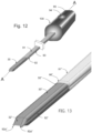
- Figs. 9 to 13 show an inserter 90 that can be used to insert the wedge 30 into the suprachoroidal space 11 on one side of the eye using an ab interno approach via the clear corneal incision 13 ( Fig. 1 ) on a diametrically opposite side of the eye.
- the inserter 90 includes a handle 100 ( Fig. 12 ) that is made from a rigid material which may include medical grade polymers or metals such as polycarbonate, polypropylene, polysulfone, polyimide, polyamide, polyurethane, ABS, polymethylmethacrylate, and the like.
- Metals can include iron, stainless steel, nickel, titanium, gold, platinum and alloys of the above.
- the inserter includes an elongated guide rod 91 that is coupled to the handle 100 and extends from a proximal end 96 at the distal end of the handle 100 to a distal end 97 spaced axially along axis A-A from the proximal end 96. Slots 93 are formed on opposite sides of the guide rod 91 and the slots may extend into the handle 100.
- the inserter includes rods 92 which are coupled to the handle 100 and area configured to translate in the slots 93.
- the rods 92 and the guide rod 91 may be formed from metal (e.g., aluminum, stainless steel, titanium) and may be planar or prebent or curved to facilitate positioning of the wedge 30 in the eye, as will be described in greater detail below.
- Each rod 92 has a thickness and width sufficient that they fit in and slide relative to open groove or channel 33. Also, the rods may have a width that is about the width 39 of the open groove or channel 33.
- a thumb slide 94 is rigidly attached to the rods 92 within the handle 100 and the thumb slide 94 is configured to translate with the rods 92.
- the handle defines a slot 95 in which the thumb slide 94 translates.
- the handle 100, slots 95 and 93, and rods 92 extend parallel to axis A-A. Translational movement of the thumb slide 94 in the slot 95 causes corresponding movement of the rods 92 in their slots 93.
- the rods 92 can be positioned between an extended position shown in Figs. 9 and 11 , in which the thumb slide 94 is moved toward a distal end of the slot 95, and a retracted configuration as shown in Fig. 10 , in which the thumb slide 94 is moved towards an opposite, proximal end of the slot 95.
- the movement of the thumb slide 94 and the rods 92 may be used to implant the wedge 30 in the suprachoroidal space 11 as described in greater detail below.
- the inserter 90 functions as follows.
- the inserter 90 and the wedge 30 are coupled together, as shown, for example, in Fig. 11 .
- the slots 93 of the extension rod 93 are configured to align with open grooves or slots 33 of the wedge 30 so that rods 92 may span slots 33 and 93 in the extended configuration to couple the wedge 30 to the inserter 90, as shown in Figs. 11 and 12 .
- rods 92 of inserter 90 are received in slots 33a and 33b of wedge 30 such that distal ends 92a ( Fig. 9 ) of the rods 92 engage the abutment 34 of wedge 30.
- the proximal end 50 of the wedge 30 engages or otherwise abuts the distal end 97 of the extension rod 91.
- the wedge 30 may be pre-assembled with the inserter 90 and provided to a user as a kit.
- the user holds the inserter 90 while introducing the distal end 32 of the wedge 30 first into the clear corneal incision 13 with the use of a gonioscope (not shown) as well as a viscous fluid (not shown) in the anterior chamber 3 to maintain it open.
- the distal end 97 of the extension rod 91 follows the wedge 30 and is introduced into the eye.
- the axial length of the extension rod 91 is sufficient that the handle 100 remains outside of the eye at all times during use of the inserter 90.
- the width and height of the extension rod 91 are preferably equal to or less than the width 37 and height 36 of the wedge 30 so that the extension rod 91 does not enlarge the pathway through the eye caused by the positioning of the wedge 30 during its implantation.
- the distal end 32 of the wedge 30 is pushed by at least one of the extension rod 91 and the rods 92 and advanced diametrically across the anterior chamber 3 from the corneal incision 13 towards the scleral spur 20.
- the distal end 32 of the wedge 30 is advanced through the trabecular meshwork 14, and between the interface of the scleral spur 20 and the ciliary body, and finally into the suprachoroidal space 11 (just to the left of it in Fig. 5 ).
- the wedge 30 By pushing the ends 92a of the rods 92 against abutment 34 and/or by pushing the distal end 97 of the extension rod 91 against the proximal end 50 of the wedge 30, the wedge 30 can be pushed into suprachoroidal space 11.
- the thumb slide 94 can be retracted proximally in slot 95, which also retracts the rods 92 proximally from the grooves 33a and 33b.
- the extension rod 91 of the inserter 90 can be retracted from the eye, leaving the wedge 30 implanted in the eye in the implanted configuration shown in Fig. 5 .
- the inserter 90 includes a spring or other actuation mechanism to automatically retract the thumb slide 94 from the extended position when the thumb slide 94 is pressed (i.e., pressed downwardly in Fig. 11 ).
- the inserter 90 may include a lock mechanism configured lock the position of the thumb slide 94 in the extended configuration shown in Fig. 11 to prevent inadvertent deployment or decoupling of the wedge 90.
- the inserter handle 100 can be grasped and held in a user's hand so that the user's thumb can actuate the thumb slide 94.
- Portions of the rods 92 can have dimensions that are larger than the dimensions of the slots 33 (i.e., they can have a radially outer portion that is outside the grooves 33).
- the rods may have a "T" shaped cross-section and have an outer beam portion that extends along a plane perpendicular to the portion of the rods received in the slots 33.
- the outer beam portion may have dimensions larger than those of the slots 33 to provide the rods 92 more columnar strength (slots 33 will flex larger).
- Fig. 13 shows another alternate embodiment of the inserter 90 and the wedge 30 in which a wedge 30" is formed like wedge 30 but does not have abutments 34 and in which ends 92a" are formed with sharp cutting surfaces that extend distally of the distal end 32" of the wedge 30".
- the sharp cutting surfaces of ends 92a" can facilitate cutting tissue ahead of the inclined surfaces at the distal end 32" of the wedge 30".
Landscapes
- Health & Medical Sciences (AREA)
- Ophthalmology & Optometry (AREA)
- Heart & Thoracic Surgery (AREA)
- Surgery (AREA)
- Engineering & Computer Science (AREA)
- Biomedical Technology (AREA)
- Nuclear Medicine, Radiotherapy & Molecular Imaging (AREA)
- Vascular Medicine (AREA)
- Life Sciences & Earth Sciences (AREA)
- Animal Behavior & Ethology (AREA)
- General Health & Medical Sciences (AREA)
- Public Health (AREA)
- Veterinary Medicine (AREA)
- Prostheses (AREA)
- Radiation-Therapy Devices (AREA)
Claims (14)
- Eine Glaukomdrainagevorrichtung (30; 30'; 70; 80), wobei die Vorrichtung Folgendes beinhaltet:einen lumenlosen länglichen Körper (31), der sich axial von einem distalen Ende (32) zu einem proximalen Ende (50) erstreckt, wobei das distale Ende (32) als ein Keil mit einer führenden distalen Kante gebildet ist, wobei der Körper (31) eine oder mehrere äußere Oberflächen (42, 43; 42', 43') aufweist, die mindestens eine offene Rille (33) definieren, welche sich von an oder nahe dem proximalen Ende (50) auf das distale Ende (32) des Körpers zu erstreckt;wobei der längliche Körper (31) konfiguriert ist, um in dem suprachoroidalen Raum (11) eines Auges zu sitzen und sich in die vordere Kammer (3) des Auges zu erstrecken, um zu gestatten, dass wässrige Körperflüssigkeit aus der vorderen Kammer des Auges entlang der offenen Rille (33a, 33b; 73a, 73b; 83a, 83b, 83c) aus der vorderen Kammer (3) des Auges in den suprachoroidalen Raum (11) des Auges fließt, unddadurch gekennzeichnet, dass die offene Rille (33a, 33b; 73a, 73b; 83a, 83b, 83c) an einem durch den länglichen Körper (31) definierten Widerlager (34) endet.
- Vorrichtung gemäß Anspruch 1, wobei:der Körper eine Vielzahl von offenen Rillen (33a, 33b; 73a, 73b; 83a, 83b, 83c) definiert, die sich parallel zueinander von dem proximalen Ende (50) auf das distale Ende (32) des Körpers (31) zu erstrecken, wobei jede offene Rille (33a, 33b; 73a, 73b; 83a, 83b, 83c) an einem durch den länglichen Körper (31) definierten Widerlager (34) endet,wobei das Widerlager (34) proximal von dem distalen Ende (32) mit Abstand angeordnet ist.
- Vorrichtung gemäß Anspruch 1, wobei:
der Körper (31) aus einem Material mit einer Shore-Härte von 30A bis 60A gebildet ist. - Vorrichtung gemäß Anspruch 3, wobei:
der Körper (31) aus mindestens einem von Poly(styrol-block-isobutylen-block-styrol) (SIBS), Styrol-Ethylen-Butylen-Styrol (SEBS), Polyhexen, Polypropylen und Polyethylen gebildet ist. - Vorrichtung gemäß Anspruch 1, wobei:
der Körper (31) eine obere äußere Oberfläche (42, 42') und eine untere äußere Oberfläche (43; 43') aufweist, die planar sind. - Vorrichtung gemäß Anspruch 5, wobei:
der Körper einen oder mehrere Widerhaken (41) auf der oberen und der unteren äußeren Oberfläche (42', 43') aufweist, wobei die Widerhaken (41) konfiguriert sind, um in Augengewebe einzugreifen. - Vorrichtung gemäß Anspruch 6, wobei:die Widerhaken (41) flache äußere Oberflächen (41a) aufweisen, die zu der oberen undder unteren äußeren Oberfläche (42', 43') parallel sind.
- Ein Glaukomvorrichtungskit, das Folgendes beinhaltet:eine Glaukomdrainagevorrichtung (30; 30'; 70; 80), die einen lumenlosen länglichen Körper (31) umfasst, der sich axial von einem distalen Ende (32) zu einem proximalen Ende (50) erstreckt, wobei das distale Ende (32) als ein Keil mit einer führenden distalen Kante gebildet ist, wobei der Körper (31) eine oder mehrere äußere Oberflächen (42, 43; 42', 43') aufweist, die mindestens eine offene Rille (33; 73a, 73b; 83a, 83b, 83c) definieren, welche sich von an oder nahe dem proximalen Ende (50) auf das distale Ende (32) des Körpers zu erstreckt;wobei der längliche Körper (31) konfiguriert ist, um in dem suprachoroidalen Raum (11) eines Auges zu sitzen und sich in die vordere Kammer (3) des Auges zu erstrecken, um zu gestatten, dass wässrige Körperflüssigkeit aus der vorderen Kammer (3) des Auges entlang der offenen Rille (33; 73a, 73b; 83a, 83b, 83c) aus der vorderen Kammer (3) des Auges in den suprachoroidalen Raum (11) des Auges fließt, undwobei die offene Rille (33; 73a, 73b; 83a, 83b, 83c) an einem durch den länglichen Körper (31) definierten Widerlager (34) endet; undeine Einführeinrichtung (90), die mit der Glaukomdrainagevorrichtung (30; 30'; 70; 80) gekoppelt ist, wobei die Einführeinrichtung (90) konfiguriert ist, um die Glaukomdrainagevorrichtung (30; 30'; 70; 80) in den suprachoroidalen Raum (11) zu bringen und dort zu positionieren und um sich von der Glaukomdrainagevorrichtung (30; 30'; 70; 80) zu trennen, um die Glaukomdrainagevorrichtung (30; 30'; 70; 80) in den suprachoroidalen Raum (11) einzusetzen;wobei die Einführeinrichtung (90) Folgendes umfasst: einen Griff (100) und mindestens einen Stab (92), der zur Längsverschiebung relativ zu dem Griff (100) in der mindestens einen offenen Rille (33; 73a, 73b; 83a, 83b, 83c) konfiguriert ist, wobei der mindestens eine Stab (92) eine ausgestreckte Konfiguration aufweist, in der sich der Stab (92) in einem Abschnitt der offenen Rille (33; 73a, 73b; 83a, 83b, 83c) befindet und in das durch den länglichen Körper (31) definierte Widerlager (34) eingreift.
- Kit gemäß Anspruch 8, wobei:der Körper (31) eine Vielzahl von offenen Rillen (33a, 33b; 73a, 73b; 83a, 83b, 83c) definiert, die sich parallel zueinander von dem proximalen Ende (50) auf das distale Ende (32) des Körpers zu erstrecken, wobei jede offene Rille (33a, 33b; 73a, 73b; 83a, 83b, 83c) an einem durch den länglichen Körper (31) definierten Widerlager (34) endet, unddie Einführeinrichtung eine Vielzahl von Stäben umfasst, wobei jeder Stab eine ausgestreckte Konfiguration aufweist, in der sich der Stab in einem Abschnitt einer entsprechenden offenen Rille (33; 73a, 73b; 83a, 83b, 83c) befindet und in das durch den länglichen Körper (31) definierte Widerlager (34) eingreift.
- Kit gemäß Anspruch 9, wobei:
die Einführeinrichtung (90) konfiguriert ist, um sich von der Glaukomdrainagevorrichtung (30; 30'; 70; 80) zu trennen, indem der mindestens eine Stab (92) aus der ausgestreckten Konfiguration in eine zurückgezogene Konfiguration, in der sich der Stab (92) nicht in einem Abschnitt der offenen Rille (33; 73a, 73b; 83a, 83b, 83c) der Glaukomdrainagevorrichtung (30; 30'; 70; 80) befindet, rekonfiguriert wird. - Kit gemäß Anspruch 10, wobei:
der mindestens eine Stab (92; 92") mit einem Gleitelement (94) gekoppelt ist, das zur Betätigung durch eine Hand eines Benutzers konfiguriert ist, wobei der Einführeinrichtungsgriff (100) einen Längsschlitz (95) definiert, der sich axial entlang der Länge des Griffs (100) und parallel zu dem mindestens einen Stab (92) erstreckt, wobei das Gleitelement (94) konfiguriert ist, um innerhalb des Schlitzes (95) zu gleiten, um den Stab (92) zwischen der ausgestreckten und der zurückgezogenen Konfiguration zu bewegen. - Kit gemäß Anspruch 11, wobei:
die Einführeinrichtung (90) einen mit dem Gleitelement (94) gekoppelten Betätigungsmechanismus umfasst, wobei der Betätigungsmechanismus konfiguriert ist, um das Gleitelement (94) automatisch zurückzuziehen. - Kit gemäß Anspruch 11, wobei:
die Einführeinrichtung (90) eine Sperre umfasst, die konfiguriert ist, um die Position des Gleitelements (94) in der ausgestreckten Konfiguration zu sperren. - Vorrichtung gemäß Anspruch 1, wobei:
das Widerlager (34) proximal von dem distalen Ende (32) des länglichen Körpers (31) mit Abstand angeordnet ist.
Priority Applications (1)
| Application Number | Priority Date | Filing Date | Title |
|---|---|---|---|
| EP23164959.1A EP4223263B1 (de) | 2017-06-13 | 2018-05-24 | Systeme, und vorrichtung zur behandlung von glaukom |
Applications Claiming Priority (2)
| Application Number | Priority Date | Filing Date | Title |
|---|---|---|---|
| US201762518944P | 2017-06-13 | 2017-06-13 | |
| PCT/US2018/034301 WO2018231485A1 (en) | 2017-06-13 | 2018-05-24 | Systems, methods, and apparatus for treatment of glaucoma |
Related Child Applications (2)
| Application Number | Title | Priority Date | Filing Date |
|---|---|---|---|
| EP23164959.1A Division-Into EP4223263B1 (de) | 2017-06-13 | 2018-05-24 | Systeme, und vorrichtung zur behandlung von glaukom |
| EP23164959.1A Division EP4223263B1 (de) | 2017-06-13 | 2018-05-24 | Systeme, und vorrichtung zur behandlung von glaukom |
Publications (3)
| Publication Number | Publication Date |
|---|---|
| EP3638164A1 EP3638164A1 (de) | 2020-04-22 |
| EP3638164A4 EP3638164A4 (de) | 2021-03-10 |
| EP3638164B1 true EP3638164B1 (de) | 2023-05-10 |
Family
ID=64660581
Family Applications (2)
| Application Number | Title | Priority Date | Filing Date |
|---|---|---|---|
| EP23164959.1A Active EP4223263B1 (de) | 2017-06-13 | 2018-05-24 | Systeme, und vorrichtung zur behandlung von glaukom |
| EP18818723.1A Active EP3638164B1 (de) | 2017-06-13 | 2018-05-24 | Systeme, und vorrichtung zur behandlung von glaukom |
Family Applications Before (1)
| Application Number | Title | Priority Date | Filing Date |
|---|---|---|---|
| EP23164959.1A Active EP4223263B1 (de) | 2017-06-13 | 2018-05-24 | Systeme, und vorrichtung zur behandlung von glaukom |
Country Status (8)
| Country | Link |
|---|---|
| US (2) | US11517476B2 (de) |
| EP (2) | EP4223263B1 (de) |
| JP (2) | JP7309624B2 (de) |
| CN (1) | CN110913809B (de) |
| CA (1) | CA3077101A1 (de) |
| ES (2) | ES2946142T3 (de) |
| TW (1) | TWI769264B (de) |
| WO (1) | WO2018231485A1 (de) |
Families Citing this family (10)
| Publication number | Priority date | Publication date | Assignee | Title |
|---|---|---|---|---|
| TWI695716B (zh) * | 2019-03-26 | 2020-06-11 | 巨晰光纖股份有限公司 | 眼球排水之分流支架 |
| CN111759582A (zh) * | 2019-04-02 | 2020-10-13 | 巨晰光纤股份有限公司 | 眼球排水的分流支架 |
| CN119279913A (zh) | 2019-06-14 | 2025-01-10 | 安特雷克公司 | 支架 |
| CA3183402A1 (en) | 2020-05-20 | 2021-11-25 | Iantrek, Inc. | System for shaping and implanting biologic intraocular stent for increased aqueous outflow and lowering of intraocular pressure |
| US12453656B2 (en) | 2020-05-20 | 2025-10-28 | Iantrek, Inc. | System for shaping and implanting biologic intraocular stent for increased aqueous outflow and lowering of intraocular pressure |
| CN114432034A (zh) * | 2021-12-22 | 2022-05-06 | 山东百多安医疗器械股份有限公司 | 一种可降解聚乳酸青光眼引流管 |
| CN114939190B (zh) * | 2022-06-14 | 2024-01-12 | 健诺维(成都)生物科技有限公司 | 一种用于青光眼治疗的引流管材料及其制备方法 |
| WO2024112747A1 (en) | 2022-11-23 | 2024-05-30 | Iantrek, Inc. | Devices and systems for cutting, loading, and delivering biologic intraocular implants for increased aqueous outflow and lowering of intraocular pressure |
| CN115969612A (zh) * | 2022-12-20 | 2023-04-18 | 重庆医科大学附属第三医院(捷尔医院) | 可生物降解的用于治疗青光眼的房水引流装置及其制备方法 |
| CN119499038B (zh) * | 2023-08-25 | 2025-10-03 | 苏州朗目医疗科技有限公司 | 一种眼部植入物 |
Family Cites Families (47)
| Publication number | Priority date | Publication date | Assignee | Title |
|---|---|---|---|---|
| US4521210A (en) * | 1982-12-27 | 1985-06-04 | Wong Vernon G | Eye implant for relieving glaucoma, and device and method for use therewith |
| US4897255A (en) | 1985-01-14 | 1990-01-30 | Neorx Corporation | Metal radionuclide labeled proteins for diagnosis and therapy |
| FR2721499B1 (fr) * | 1994-06-22 | 1997-01-03 | Opsia | Implant de trabéculectomie. |
| US5601094A (en) * | 1994-11-22 | 1997-02-11 | Reiss; George R. | Ophthalmic shunt |
| CN2287027Y (zh) | 1996-09-18 | 1998-08-05 | 张军锋 | 青光眼微型眼压调节器 |
| US6004302A (en) * | 1997-08-28 | 1999-12-21 | Brierley; Lawrence A. | Cannula |
| US8313454B2 (en) * | 1997-11-20 | 2012-11-20 | Optonol Ltd. | Fluid drainage device, delivery device, and associated methods of use and manufacture |
| US20050119601A9 (en) * | 1999-04-26 | 2005-06-02 | Lynch Mary G. | Shunt device and method for treating glaucoma |
| US7708711B2 (en) * | 2000-04-14 | 2010-05-04 | Glaukos Corporation | Ocular implant with therapeutic agents and methods thereof |
| US7951155B2 (en) * | 2002-03-15 | 2011-05-31 | Glaukos Corporation | Combined treatment for cataract and glaucoma treatment |
| ES2449496T3 (es) | 2003-12-05 | 2014-03-20 | Innfocus, Inc. | Dispositivo mejorado de implante para glaucoma |
| US8685435B2 (en) | 2004-04-30 | 2014-04-01 | Allergan, Inc. | Extended release biodegradable ocular implants |
| US7862531B2 (en) * | 2004-06-25 | 2011-01-04 | Optonol Ltd. | Flow regulating implants |
| US7837644B2 (en) | 2004-12-03 | 2010-11-23 | Innfocus, Llc | Glaucoma implant device |
| US7594899B2 (en) * | 2004-12-03 | 2009-09-29 | Innfocus, Llc | Glaucoma implant device |
| EP3838236B1 (de) | 2006-01-17 | 2025-08-13 | Alcon Inc. | Glaukombehandlungsvorrichtung |
| US7909789B2 (en) | 2006-06-26 | 2011-03-22 | Sight Sciences, Inc. | Intraocular implants and methods and kits therefor |
| US8911496B2 (en) * | 2006-07-11 | 2014-12-16 | Refocus Group, Inc. | Scleral prosthesis for treating presbyopia and other eye disorders and related devices and methods |
| WO2008061043A2 (en) | 2006-11-10 | 2008-05-22 | Glaukos Corporation | Uveoscleral shunt and methods for implanting same |
| CN101199440B (zh) | 2006-12-12 | 2010-07-07 | 张传忠 | 泪道引流装置及放置该装置的泪道探条 |
| NZ583858A (en) * | 2007-09-07 | 2012-08-31 | Quadra Logic Tech Inc | Lacrimal implant detection with detection device |
| AU2008351361B2 (en) * | 2008-02-28 | 2014-01-30 | Hollister Incorporated | Fluid drainage catheter having an external flow path |
| CN201216683Y (zh) | 2008-06-23 | 2009-04-08 | 陈颖平 | 眼球角膜巩膜穿刺刀 |
| CH700142A1 (de) | 2008-12-22 | 2010-06-30 | Grieshaber Ophthalmic Res Foun | Implantat zum einführen in den schlemmschen kanal eines auges. |
| US8425473B2 (en) * | 2009-01-23 | 2013-04-23 | Iscience Interventional Corporation | Subretinal access device |
| ES2817775T3 (es) * | 2009-01-28 | 2021-04-08 | Alcon Inc | Implante ocular con calidades de rigidez |
| CN102458509B (zh) * | 2009-05-15 | 2015-04-22 | I科学干预公司 | 用于视网膜下导管插入的装置 |
| US8764696B2 (en) * | 2009-06-16 | 2014-07-01 | Mobius Therapeutics, Inc. | Medical drainage devices with carbon-based structures for inhibiting growth of fibroblasts |
| CA2774610A1 (en) | 2009-09-21 | 2011-03-24 | Vidus Ocular, Inc. | Uveoscleral drainage device |
| JP2013508096A (ja) * | 2009-10-23 | 2013-03-07 | イバンティス インコーポレイテッド | 眼内移植システムおよび眼内移植方法 |
| US8771216B2 (en) * | 2009-11-06 | 2014-07-08 | University Hospitals Of Cleveland | Fluid communication device and method of use thereof |
| US8845572B2 (en) | 2009-11-13 | 2014-09-30 | Grieshaber Ophthalmic Research Foundation | Method and device for the treatment of glaucoma |
| US20120245505A1 (en) | 2009-12-16 | 2012-09-27 | Robinson Michael R | Intracameral devices for sustained delivery |
| US8663150B2 (en) * | 2011-12-19 | 2014-03-04 | Ivantis, Inc. | Delivering ocular implants into the eye |
| US9101444B2 (en) * | 2012-01-12 | 2015-08-11 | Innfocus, Inc. | Method, surgical kit and device for treating glaucoma |
| US9241832B2 (en) | 2012-04-24 | 2016-01-26 | Transcend Medical, Inc. | Delivery system for ocular implant |
| EP3228286A1 (de) | 2012-09-17 | 2017-10-11 | Novartis AG | Expandierende augenimplantatvorrichtungen |
| CN105358105A (zh) * | 2013-03-14 | 2016-02-24 | 以色列哈尼塔镜片有限公司 | 小型青光眼分流器 |
| US9592151B2 (en) | 2013-03-15 | 2017-03-14 | Glaukos Corporation | Systems and methods for delivering an ocular implant to the suprachoroidal space within an eye |
| CN103181842B (zh) | 2013-03-25 | 2014-12-10 | 中国人民解放军第三军医大学第一附属医院 | 眼科用板层巩膜刀 |
| US9649223B2 (en) | 2013-06-13 | 2017-05-16 | Innfocus, Inc. | Inserter for tubular medical implant devices |
| US9585790B2 (en) * | 2013-11-14 | 2017-03-07 | Aquesys, Inc. | Intraocular shunt inserter |
| US9044301B1 (en) | 2013-11-25 | 2015-06-02 | Innfocus, Inc. | Methods, systems and devices for treating glaucoma |
| WO2016004223A1 (en) * | 2014-07-01 | 2016-01-07 | Cao Ariel | Methods and devices for implantation of intraocular pressure sensors |
| CN104490515A (zh) * | 2014-12-18 | 2015-04-08 | 肖真 | 青光眼阀和青光眼引流装置 |
| CN104984420A (zh) | 2015-07-20 | 2015-10-21 | 王文 | 一种引流管 |
| CN105434103B (zh) | 2015-12-29 | 2017-07-07 | 北京大学人民医院 | 一种微型青光眼引流植入装置及系统 |
-
2018
- 2018-05-24 ES ES18818723T patent/ES2946142T3/es active Active
- 2018-05-24 CA CA3077101A patent/CA3077101A1/en active Pending
- 2018-05-24 JP JP2019568712A patent/JP7309624B2/ja active Active
- 2018-05-24 CN CN201880039591.XA patent/CN110913809B/zh active Active
- 2018-05-24 US US16/621,079 patent/US11517476B2/en active Active
- 2018-05-24 WO PCT/US2018/034301 patent/WO2018231485A1/en not_active Ceased
- 2018-05-24 EP EP23164959.1A patent/EP4223263B1/de active Active
- 2018-05-24 ES ES23164959T patent/ES3063055T3/es active Active
- 2018-05-24 EP EP18818723.1A patent/EP3638164B1/de active Active
- 2018-06-07 TW TW107119574A patent/TWI769264B/zh active
-
2022
- 2022-10-31 US US17/977,830 patent/US20230051779A1/en active Pending
-
2023
- 2023-07-04 JP JP2023110024A patent/JP7445809B2/ja active Active
Also Published As
| Publication number | Publication date |
|---|---|
| JP2020523134A (ja) | 2020-08-06 |
| ES3063055T3 (en) | 2026-04-15 |
| EP4223263C0 (de) | 2026-02-11 |
| JP2023118852A (ja) | 2023-08-25 |
| EP3638164A1 (de) | 2020-04-22 |
| JP7309624B2 (ja) | 2023-07-18 |
| TWI769264B (zh) | 2022-07-01 |
| CN110913809A (zh) | 2020-03-24 |
| TW201902435A (zh) | 2019-01-16 |
| EP3638164A4 (de) | 2021-03-10 |
| ES2946142T3 (es) | 2023-07-13 |
| WO2018231485A1 (en) | 2018-12-20 |
| JP7445809B2 (ja) | 2024-03-07 |
| CN110913809B (zh) | 2023-10-03 |
| EP4223263B1 (de) | 2026-02-11 |
| US20200188172A1 (en) | 2020-06-18 |
| EP4223263A1 (de) | 2023-08-09 |
| US20230051779A1 (en) | 2023-02-16 |
| US11517476B2 (en) | 2022-12-06 |
| CA3077101A1 (en) | 2018-12-20 |
Similar Documents
| Publication | Publication Date | Title |
|---|---|---|
| US20230051779A1 (en) | Systems, methods, and apparatus for treatment of glaucoma | |
| JP6810090B2 (ja) | 緑内障を治療するための方法、外科キット及び器具 | |
| US7837644B2 (en) | Glaucoma implant device | |
| US7594899B2 (en) | Glaucoma implant device | |
| US20070118065A1 (en) | Glaucoma Implant Device | |
| US20070141116A1 (en) | Glaucoma Implant Device | |
| HK40028232A (en) | Systems and apparatus for treatment of glaucoma | |
| HK40028232B (en) | Systems and apparatus for treatment of glaucoma |
Legal Events
| Date | Code | Title | Description |
|---|---|---|---|
| STAA | Information on the status of an ep patent application or granted ep patent |
Free format text: STATUS: THE INTERNATIONAL PUBLICATION HAS BEEN MADE |
|
| PUAI | Public reference made under article 153(3) epc to a published international application that has entered the european phase |
Free format text: ORIGINAL CODE: 0009012 |
|
| STAA | Information on the status of an ep patent application or granted ep patent |
Free format text: STATUS: REQUEST FOR EXAMINATION WAS MADE |
|
| 17P | Request for examination filed |
Effective date: 20191127 |
|
| AK | Designated contracting states |
Kind code of ref document: A1 Designated state(s): AL AT BE BG CH CY CZ DE DK EE ES FI FR GB GR HR HU IE IS IT LI LT LU LV MC MK MT NL NO PL PT RO RS SE SI SK SM TR |
|
| AX | Request for extension of the european patent |
Extension state: BA ME |
|
| DAV | Request for validation of the european patent (deleted) | ||
| DAX | Request for extension of the european patent (deleted) | ||
| REG | Reference to a national code |
Ref country code: HK Ref legal event code: DE Ref document number: 40028232 Country of ref document: HK |
|
| A4 | Supplementary search report drawn up and despatched |
Effective date: 20210208 |
|
| RIC1 | Information provided on ipc code assigned before grant |
Ipc: A61F 9/007 20060101AFI20210202BHEP |
|
| GRAP | Despatch of communication of intention to grant a patent |
Free format text: ORIGINAL CODE: EPIDOSNIGR1 |
|
| STAA | Information on the status of an ep patent application or granted ep patent |
Free format text: STATUS: GRANT OF PATENT IS INTENDED |
|
| INTG | Intention to grant announced |
Effective date: 20221213 |
|
| GRAS | Grant fee paid |
Free format text: ORIGINAL CODE: EPIDOSNIGR3 |
|
| GRAA | (expected) grant |
Free format text: ORIGINAL CODE: 0009210 |
|
| STAA | Information on the status of an ep patent application or granted ep patent |
Free format text: STATUS: THE PATENT HAS BEEN GRANTED |
|
| AK | Designated contracting states |
Kind code of ref document: B1 Designated state(s): AL AT BE BG CH CY CZ DE DK EE ES FI FR GB GR HR HU IE IS IT LI LT LU LV MC MK MT NL NO PL PT RO RS SE SI SK SM TR |
|
| REG | Reference to a national code |
Ref country code: GB Ref legal event code: FG4D |
|
| REG | Reference to a national code |
Ref country code: AT Ref legal event code: REF Ref document number: 1566010 Country of ref document: AT Kind code of ref document: T Effective date: 20230515 Ref country code: CH Ref legal event code: EP |
|
| REG | Reference to a national code |
Ref country code: DE Ref legal event code: R096 Ref document number: 602018049731 Country of ref document: DE |
|
| REG | Reference to a national code |
Ref country code: IE Ref legal event code: FG4D |
|
| P01 | Opt-out of the competence of the unified patent court (upc) registered |
Effective date: 20230525 |
|
| REG | Reference to a national code |
Ref country code: ES Ref legal event code: FG2A Ref document number: 2946142 Country of ref document: ES Kind code of ref document: T3 Effective date: 20230713 |
|
| REG | Reference to a national code |
Ref country code: LT Ref legal event code: MG9D |
|
| REG | Reference to a national code |
Ref country code: NL Ref legal event code: MP Effective date: 20230510 |
|
| REG | Reference to a national code |
Ref country code: AT Ref legal event code: MK05 Ref document number: 1566010 Country of ref document: AT Kind code of ref document: T Effective date: 20230510 |
|
| PG25 | Lapsed in a contracting state [announced via postgrant information from national office to epo] |
Ref country code: SE Free format text: LAPSE BECAUSE OF FAILURE TO SUBMIT A TRANSLATION OF THE DESCRIPTION OR TO PAY THE FEE WITHIN THE PRESCRIBED TIME-LIMIT Effective date: 20230510 Ref country code: PT Free format text: LAPSE BECAUSE OF FAILURE TO SUBMIT A TRANSLATION OF THE DESCRIPTION OR TO PAY THE FEE WITHIN THE PRESCRIBED TIME-LIMIT Effective date: 20230911 Ref country code: NO Free format text: LAPSE BECAUSE OF FAILURE TO SUBMIT A TRANSLATION OF THE DESCRIPTION OR TO PAY THE FEE WITHIN THE PRESCRIBED TIME-LIMIT Effective date: 20230810 Ref country code: NL Free format text: LAPSE BECAUSE OF FAILURE TO SUBMIT A TRANSLATION OF THE DESCRIPTION OR TO PAY THE FEE WITHIN THE PRESCRIBED TIME-LIMIT Effective date: 20230510 Ref country code: AT Free format text: LAPSE BECAUSE OF FAILURE TO SUBMIT A TRANSLATION OF THE DESCRIPTION OR TO PAY THE FEE WITHIN THE PRESCRIBED TIME-LIMIT Effective date: 20230510 |
|
| PG25 | Lapsed in a contracting state [announced via postgrant information from national office to epo] |
Ref country code: RS Free format text: LAPSE BECAUSE OF FAILURE TO SUBMIT A TRANSLATION OF THE DESCRIPTION OR TO PAY THE FEE WITHIN THE PRESCRIBED TIME-LIMIT Effective date: 20230510 Ref country code: PL Free format text: LAPSE BECAUSE OF FAILURE TO SUBMIT A TRANSLATION OF THE DESCRIPTION OR TO PAY THE FEE WITHIN THE PRESCRIBED TIME-LIMIT Effective date: 20230510 Ref country code: LV Free format text: LAPSE BECAUSE OF FAILURE TO SUBMIT A TRANSLATION OF THE DESCRIPTION OR TO PAY THE FEE WITHIN THE PRESCRIBED TIME-LIMIT Effective date: 20230510 Ref country code: LT Free format text: LAPSE BECAUSE OF FAILURE TO SUBMIT A TRANSLATION OF THE DESCRIPTION OR TO PAY THE FEE WITHIN THE PRESCRIBED TIME-LIMIT Effective date: 20230510 Ref country code: IS Free format text: LAPSE BECAUSE OF FAILURE TO SUBMIT A TRANSLATION OF THE DESCRIPTION OR TO PAY THE FEE WITHIN THE PRESCRIBED TIME-LIMIT Effective date: 20230910 Ref country code: HR Free format text: LAPSE BECAUSE OF FAILURE TO SUBMIT A TRANSLATION OF THE DESCRIPTION OR TO PAY THE FEE WITHIN THE PRESCRIBED TIME-LIMIT Effective date: 20230510 Ref country code: GR Free format text: LAPSE BECAUSE OF FAILURE TO SUBMIT A TRANSLATION OF THE DESCRIPTION OR TO PAY THE FEE WITHIN THE PRESCRIBED TIME-LIMIT Effective date: 20230811 |
|
| PG25 | Lapsed in a contracting state [announced via postgrant information from national office to epo] |
Ref country code: FI Free format text: LAPSE BECAUSE OF FAILURE TO SUBMIT A TRANSLATION OF THE DESCRIPTION OR TO PAY THE FEE WITHIN THE PRESCRIBED TIME-LIMIT Effective date: 20230510 |
|
| REG | Reference to a national code |
Ref country code: CH Ref legal event code: PL |
|
| PG25 | Lapsed in a contracting state [announced via postgrant information from national office to epo] |
Ref country code: SK Free format text: LAPSE BECAUSE OF FAILURE TO SUBMIT A TRANSLATION OF THE DESCRIPTION OR TO PAY THE FEE WITHIN THE PRESCRIBED TIME-LIMIT Effective date: 20230510 |
|
| REG | Reference to a national code |
Ref country code: BE Ref legal event code: MM Effective date: 20230531 |
|
| PG25 | Lapsed in a contracting state [announced via postgrant information from national office to epo] |
Ref country code: SM Free format text: LAPSE BECAUSE OF FAILURE TO SUBMIT A TRANSLATION OF THE DESCRIPTION OR TO PAY THE FEE WITHIN THE PRESCRIBED TIME-LIMIT Effective date: 20230510 Ref country code: SK Free format text: LAPSE BECAUSE OF FAILURE TO SUBMIT A TRANSLATION OF THE DESCRIPTION OR TO PAY THE FEE WITHIN THE PRESCRIBED TIME-LIMIT Effective date: 20230510 Ref country code: RO Free format text: LAPSE BECAUSE OF FAILURE TO SUBMIT A TRANSLATION OF THE DESCRIPTION OR TO PAY THE FEE WITHIN THE PRESCRIBED TIME-LIMIT Effective date: 20230510 Ref country code: LU Free format text: LAPSE BECAUSE OF NON-PAYMENT OF DUE FEES Effective date: 20230524 Ref country code: LI Free format text: LAPSE BECAUSE OF NON-PAYMENT OF DUE FEES Effective date: 20230531 Ref country code: EE Free format text: LAPSE BECAUSE OF FAILURE TO SUBMIT A TRANSLATION OF THE DESCRIPTION OR TO PAY THE FEE WITHIN THE PRESCRIBED TIME-LIMIT Effective date: 20230510 Ref country code: DK Free format text: LAPSE BECAUSE OF FAILURE TO SUBMIT A TRANSLATION OF THE DESCRIPTION OR TO PAY THE FEE WITHIN THE PRESCRIBED TIME-LIMIT Effective date: 20230510 Ref country code: CZ Free format text: LAPSE BECAUSE OF FAILURE TO SUBMIT A TRANSLATION OF THE DESCRIPTION OR TO PAY THE FEE WITHIN THE PRESCRIBED TIME-LIMIT Effective date: 20230510 Ref country code: CH Free format text: LAPSE BECAUSE OF NON-PAYMENT OF DUE FEES Effective date: 20230531 |
|
| REG | Reference to a national code |
Ref country code: DE Ref legal event code: R097 Ref document number: 602018049731 Country of ref document: DE |
|
| PG25 | Lapsed in a contracting state [announced via postgrant information from national office to epo] |
Ref country code: MC Free format text: LAPSE BECAUSE OF FAILURE TO SUBMIT A TRANSLATION OF THE DESCRIPTION OR TO PAY THE FEE WITHIN THE PRESCRIBED TIME-LIMIT Effective date: 20230510 |
|
| REG | Reference to a national code |
Ref country code: IE Ref legal event code: MM4A |
|
| PG25 | Lapsed in a contracting state [announced via postgrant information from national office to epo] |
Ref country code: MC Free format text: LAPSE BECAUSE OF FAILURE TO SUBMIT A TRANSLATION OF THE DESCRIPTION OR TO PAY THE FEE WITHIN THE PRESCRIBED TIME-LIMIT Effective date: 20230510 |
|
| PLBE | No opposition filed within time limit |
Free format text: ORIGINAL CODE: 0009261 |
|
| STAA | Information on the status of an ep patent application or granted ep patent |
Free format text: STATUS: NO OPPOSITION FILED WITHIN TIME LIMIT |
|
| PG25 | Lapsed in a contracting state [announced via postgrant information from national office to epo] |
Ref country code: IE Free format text: LAPSE BECAUSE OF NON-PAYMENT OF DUE FEES Effective date: 20230524 |
|
| 26N | No opposition filed |
Effective date: 20240213 |
|
| PG25 | Lapsed in a contracting state [announced via postgrant information from national office to epo] |
Ref country code: IE Free format text: LAPSE BECAUSE OF NON-PAYMENT OF DUE FEES Effective date: 20230524 |
|
| PG25 | Lapsed in a contracting state [announced via postgrant information from national office to epo] |
Ref country code: SI Free format text: LAPSE BECAUSE OF FAILURE TO SUBMIT A TRANSLATION OF THE DESCRIPTION OR TO PAY THE FEE WITHIN THE PRESCRIBED TIME-LIMIT Effective date: 20230510 |
|
| PG25 | Lapsed in a contracting state [announced via postgrant information from national office to epo] |
Ref country code: SI Free format text: LAPSE BECAUSE OF FAILURE TO SUBMIT A TRANSLATION OF THE DESCRIPTION OR TO PAY THE FEE WITHIN THE PRESCRIBED TIME-LIMIT Effective date: 20230510 Ref country code: BE Free format text: LAPSE BECAUSE OF NON-PAYMENT OF DUE FEES Effective date: 20230531 |
|
| PG25 | Lapsed in a contracting state [announced via postgrant information from national office to epo] |
Ref country code: BG Free format text: LAPSE BECAUSE OF FAILURE TO SUBMIT A TRANSLATION OF THE DESCRIPTION OR TO PAY THE FEE WITHIN THE PRESCRIBED TIME-LIMIT Effective date: 20230510 |
|
| PG25 | Lapsed in a contracting state [announced via postgrant information from national office to epo] |
Ref country code: BG Free format text: LAPSE BECAUSE OF FAILURE TO SUBMIT A TRANSLATION OF THE DESCRIPTION OR TO PAY THE FEE WITHIN THE PRESCRIBED TIME-LIMIT Effective date: 20230510 |
|
| PGFP | Annual fee paid to national office [announced via postgrant information from national office to epo] |
Ref country code: DE Payment date: 20250624 Year of fee payment: 8 |
|
| PGFP | Annual fee paid to national office [announced via postgrant information from national office to epo] |
Ref country code: GB Payment date: 20250528 Year of fee payment: 8 Ref country code: ES Payment date: 20250602 Year of fee payment: 8 |
|
| PGFP | Annual fee paid to national office [announced via postgrant information from national office to epo] |
Ref country code: IT Payment date: 20250523 Year of fee payment: 8 |
|
| PGFP | Annual fee paid to national office [announced via postgrant information from national office to epo] |
Ref country code: FR Payment date: 20250522 Year of fee payment: 8 |
|
| PG25 | Lapsed in a contracting state [announced via postgrant information from national office to epo] |
Ref country code: CY Free format text: LAPSE BECAUSE OF FAILURE TO SUBMIT A TRANSLATION OF THE DESCRIPTION OR TO PAY THE FEE WITHIN THE PRESCRIBED TIME-LIMIT; INVALID AB INITIO Effective date: 20180524 |
|
| PG25 | Lapsed in a contracting state [announced via postgrant information from national office to epo] |
Ref country code: HU Free format text: LAPSE BECAUSE OF FAILURE TO SUBMIT A TRANSLATION OF THE DESCRIPTION OR TO PAY THE FEE WITHIN THE PRESCRIBED TIME-LIMIT; INVALID AB INITIO Effective date: 20180524 |
|
| PG25 | Lapsed in a contracting state [announced via postgrant information from national office to epo] |
Ref country code: TR Free format text: LAPSE BECAUSE OF FAILURE TO SUBMIT A TRANSLATION OF THE DESCRIPTION OR TO PAY THE FEE WITHIN THE PRESCRIBED TIME-LIMIT Effective date: 20230510 |