WO1993007487A1 - Method of detecting mesitylene-resistant staphylococcus aureus, novel peptide, and dna coding for same - Google Patents

Method of detecting mesitylene-resistant staphylococcus aureus, novel peptide, and dna coding for same Download PDF

Info

Publication number
WO1993007487A1
WO1993007487A1 PCT/JP1992/001317 JP9201317W WO9307487A1 WO 1993007487 A1 WO1993007487 A1 WO 1993007487A1 JP 9201317 W JP9201317 W JP 9201317W WO 9307487 A1 WO9307487 A1 WO 9307487A1
Authority
WO
WIPO (PCT)
Prior art keywords
ser
phe
asp
leu
lys
Prior art date
Legal status (The legal status is an assumption and is not a legal conclusion. Google has not performed a legal analysis and makes no representation as to the accuracy of the status listed.)
Ceased
Application number
PCT/JP1992/001317
Other languages
English (en)
French (fr)
Inventor
Yoshiyuki Kamio
Kazuo Izaki
Hiroshi Kita
Current Assignee (The listed assignees may be inaccurate. Google has not performed a legal analysis and makes no representation or warranty as to the accuracy of the list.)
Mitsubishi Kagaku Iatron Inc
Original Assignee
Iatron Laboratories Inc
Mitsubishi Kagaku Iatron Inc
Priority date (The priority date is an assumption and is not a legal conclusion. Google has not performed a legal analysis and makes no representation as to the accuracy of the date listed.)
Filing date
Publication date
Priority claimed from JP12995692A external-priority patent/JPH05249119A/ja
Application filed by Iatron Laboratories Inc, Mitsubishi Kagaku Iatron Inc filed Critical Iatron Laboratories Inc
Priority to EP93906349A priority Critical patent/EP0597110A4/en
Priority to CA002097912A priority patent/CA2097912A1/en
Publication of WO1993007487A1 publication Critical patent/WO1993007487A1/ja
Anticipated expiration legal-status Critical
Ceased legal-status Critical Current

Links

Classifications

    • CCHEMISTRY; METALLURGY
    • C07ORGANIC CHEMISTRY
    • C07KPEPTIDES
    • C07K14/00Peptides having more than 20 amino acids; Gastrins; Somatostatins; Melanotropins; Derivatives thereof
    • C07K14/195Peptides having more than 20 amino acids; Gastrins; Somatostatins; Melanotropins; Derivatives thereof from bacteria
    • C07K14/305Peptides having more than 20 amino acids; Gastrins; Somatostatins; Melanotropins; Derivatives thereof from bacteria from Micrococcaceae (F)
    • C07K14/31Peptides having more than 20 amino acids; Gastrins; Somatostatins; Melanotropins; Derivatives thereof from bacteria from Micrococcaceae (F) from Staphylococcus (G)
    • GPHYSICS
    • G01MEASURING; TESTING
    • G01NINVESTIGATING OR ANALYSING MATERIALS BY DETERMINING THEIR CHEMICAL OR PHYSICAL PROPERTIES
    • G01N33/00Investigating or analysing materials by specific methods not covered by groups G01N1/00 - G01N31/00
    • G01N33/48Biological material, e.g. blood, urine; Haemocytometers
    • G01N33/50Chemical analysis of biological material, e.g. blood, urine; Testing involving biospecific ligand binding methods; Immunological testing
    • G01N33/53Immunoassay; Biospecific binding assay; Materials therefor
    • G01N33/569Immunoassay; Biospecific binding assay; Materials therefor for microorganisms, e.g. protozoa, bacteria, viruses
    • G01N33/56911Bacteria
    • G01N33/56938Staphylococcus

Definitions

  • the present invention relates to a novel peptide specifically produced by methicillin-resistant Staphylococcus aureus (hereinafter sometimes referred to as MRSA), a DNA encoding the peptide, and the MRS using an antibody against the peptide.
  • MRSA methicillin-resistant Staphylococcus aureus
  • Staphylococcus aureus is a facultative anaerobic gram-positive cocci that is widely distributed in nature and causes nosocomial and opportunistic infections, such as pyeloneitis, cystitis, pus eruptions, localized pus, and bone marrow It is regarded as a causative agent of inflammation and sepsis.
  • staphylococcus aureus methicillin-resistant Staphylococcus aureus (MRS A), which has high resistance to various antibiotics due to chromosomal mutation, exists. This MRSA can proliferate in the body of people with reduced resistance, such as the elderly, newborns, and cancer patients, causing pneumonia, sepsis, and death. Therefore, the proper diagnosis of MRSA infection is of crucial clinical importance.
  • Methods for detecting MRSA include separating and identifying MRSA from biological fluids (blood, urine, saliva, etc.) and drug resistance tests. However, it is necessary to go through complicated procedures to determine MRSA. there were. Despite the need for early, sensitive, and accurate diagnosis of MRSA infection, no method has ever been available to meet these needs.
  • the present inventors have found that there is a peptide specifically produced by MRSA, synthesize an oligonucleotide probe using the partial amino acid sequence of the peptide (that is, leucocidin), and use the probe to synthesize an oligonucleotide probe.
  • mouth DNA encoding icosidine and its precursor (proleucocidin) was cloned, and leukocidin S and F, and proleukocidin S and F were successfully expressed using the cloned DNA.
  • Antibodies against S and F, and proleukocidin S and F were prepared and evaluated as antibodies for MRSA detection.As a result, it was found that MRSA could be detected quickly, accurately and specifically.
  • the present invention is based on such findings.
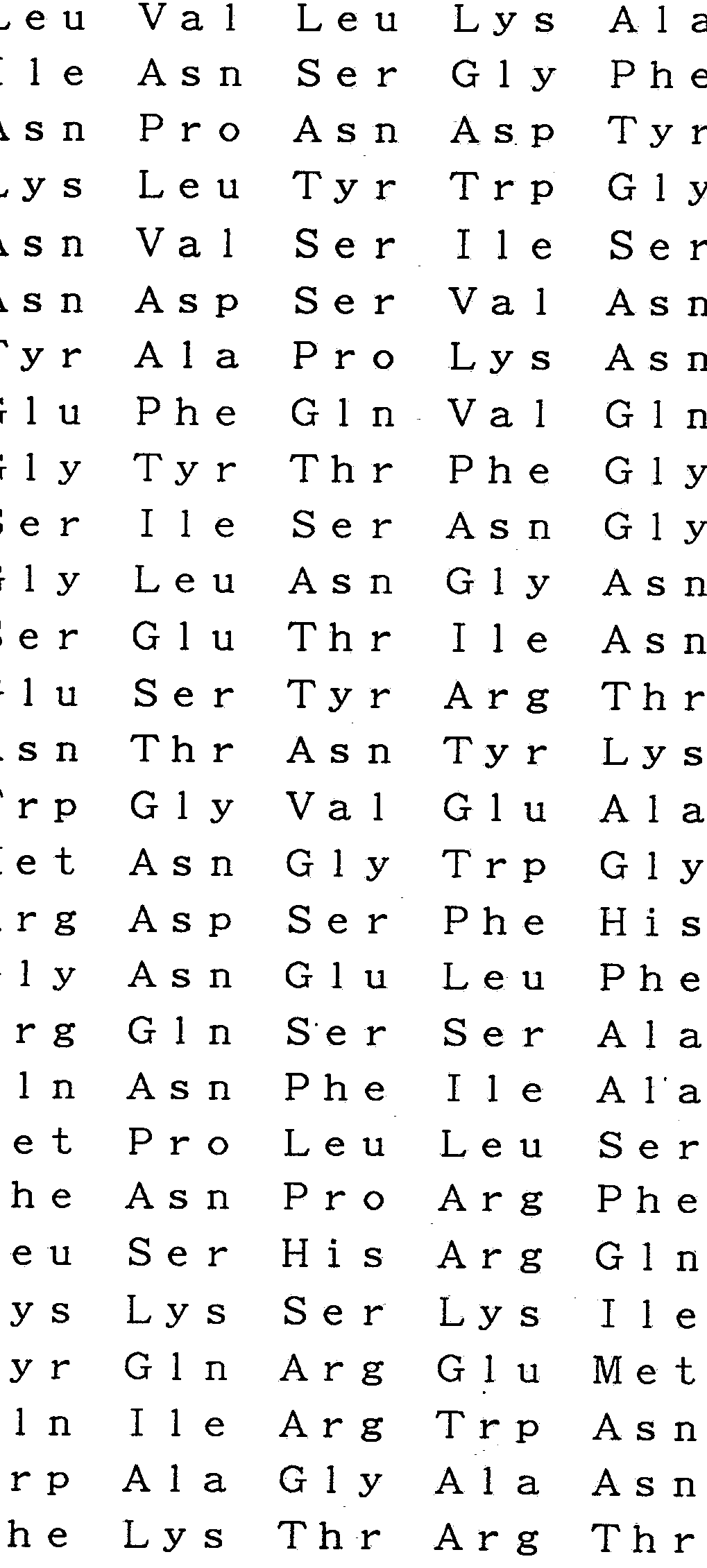
  • Ser r G 1 G in Lys Ser A la Phe As Ser Asp Le u Ph e Va 1 G 1 y Tyr r ys Pro Ro is Ser ys Asp P ro A rg As p Ty r P he Va 1 P ro As p SerG 1 u Le u P ro P ro Le u Va 1 G 1 n Ser G 1 y P he A sn Pr o Ser P he I 1 e A 1 a Thr Va 1 Ser H is G 1 u Lys G 1 y Ser S er As p Th r S er G 1 u Ph e G 1 u I 1 e Th r Ty r G 1 A rg A sn Met As p V a 1 Th r H is A 1 a I 1 e Lys A rg S er Th r H is Ty r G 1 y A sn S er Ty r Le u Asp G 1 H is A rg Va 1 H is A
  • the present invention relates to a DNA having a peptide having the following amino acid sequence or a DNA comprising a base sequence encoding the peptide.
  • the present invention provides
  • the present invention relates to a peptide having the following amino acid sequence or a DNA comprising a base sequence encoding the peptide.
  • the present invention provides a method for isolating chromosomal DNA from the cells of methicillin-resistant Staphylococcus aureus, digesting the DNA fragment with a restriction enzyme, inserting the obtained DNA fragment into a vector, and transforming a host with the vector to obtain a DNA.
  • a library was prepared, and leucocidin or procoticidine was obtained from the DNA library using a synthetic DNA encoding a partial amino acid sequence of leucocidin or proleucocidin purified from the cells of methicillin-resistant Staphylococcus aureus as a probe. And then clone the obtained DNA to produce a DNA encoding leucocidin or proleucocidin.
  • the present invention provides a recombinant DNA capable of replicating by linking a DNA encoding leucocidin or proleucocidin to an expression vector, transforming a host with the recombinant DNA, and transforming the transformant. Culturing to express the above-mentioned DNA encoding leucocidin or proleucocidin to produce oral leucocidin or proleucocidin, and isolating the obtained leucocidin or proleucocidin, characterized in that: We fight for Kosijin's recipe.
  • FIG. 1 is a graph showing the results of purifying the S fraction of leucocidin from MRSA strain No. 4 by HP LC equipped with a TSK gel SP-5PW column. The S fraction was quantified by immunoblotting (the stippled part in Fig. 1 is the S fraction.)
  • FIG. 2 shows the results of SDS-PAGE electrophoresis patterns of purified S fraction preparations from MRS A strain No. 4 and Staphylococcus aureus V8.
  • Lane 1 is the S fraction from Staphylococcus aureus V8
  • lane 2 is the S fraction from MRSA strain No. 4
  • lane 3 is the molecular weight marker.
  • the molecular weight markers used are as follows.
  • Serum albumin (66.2 kDa)
  • ovalbumin 45 kDa
  • FIG. 3 is an explanatory diagram showing a state in which a Hindlll-Xbal fragment (2.2 kb) has been inserted into a plasmid vector PUC119.
  • the abbreviations in Fig. 3 are as follows.
  • FIG. 4 is a restriction map of the Hindlll-Xbal fragment (2.2 kb).
  • the leucocidin (S fraction) coding region is indicated by an arrow.
  • the double dashed line is the inserted 2.2 kb fragment, and the single dashed line is the polylinker region of vector plasmid pUC119.
  • Abbreviations are as follows.
  • FIG. 5 shows the nucleotide sequence of the leucocidin (S fraction) coding region and the amino acid sequence of leucocidin (S fraction) deduced therefrom.
  • the sequence is the first nucleotide of the recognition base sequence of the restriction enzyme Hindyl as the first,
  • the last nucleotide of PSRK 1-1 DNA is designated as 1,466.
  • the two underlined parts are the ribosome binding (estimated) sites, and the rectangles are the (estimated) promoter sites.
  • Parallel arrows indicate three inverted repeats downstream of the translation stop codon, one underlined part is the signal sequence, and the wedge-shaped part is the S fraction precursor. Is the processing site.
  • FIG. 6 shows the results of electrophoresis of the in vitro translation product encoded by the plasmid pSRK91.
  • Lane 1 is the in vitro product encoded by pSRK91 DNA
  • lane 2 is the in vitro product encoded by pUC119
  • PS is the S fraction precursor.
  • FIG. 7 shows the results of Western plotting
  • (A) lane 1 is the purified S fraction
  • lane 2 is the cell extract of E. coli (p SRK91)
  • lane 3 is the E. coli (P UC
  • lane 4 is a culture of E. coli (PSRK91)
  • lane 5 is a culture of E. coli (PUC 119).
  • (B) shows the results of analysis of the S fraction in the cell extract from Escherichia coli DH5a (pSRK1), lane 1 in the presence of IPTG, lane 2 in the absence of IPTG, and lane 3 It is a fraction standard product.
  • FIG. 8 shows the results of Western blotting of the cell fraction of Escherichia coli DH5a (pSRK91). Lane 1 is the standard S protein, Lane 2 is the shocked cell fraction, Lane 3 is the periplasmic space, Lane 4 is the whole cell fraction, and Lane 5 is the culture medium .
  • FIG. 9 is a graph showing the results of purifying the F fraction of leucocidin from MRS A strain No. 4 by HPLC equipped with a TSK gel SP-5 PW column. The F fraction was quantified by immunoblotting. (The shaded area in Fig. 9 is the F fraction.)
  • FIG. 10 shows the results of SDS-PAGE electrophoresis patterns of purified F fraction preparations from MRSA strain No. 4 and Staphylococcus aureus V8.
  • Lane 1 is the F fraction from Staphylococcus aureus V8, and lane 2 is the MRSA This is the F fraction from strain No. 4, and lane 3 is a molecular weight marker.
  • the molecular weight markers used are the same as those in FIG.
  • FIG. 11 is an explanatory diagram showing a state in which an EcoRI-EcoRI fragment (4. Okb) was inserted into a plasmid vector pUC119.
  • the abbreviations in Fig. 11 have the same meanings as those in Fig. 3.
  • C means C 1 a I
  • FIG. 12 is a restriction map of EcoRI—EcoRI fragment (4. Okb). Leucocidin (F fraction) The coding region is indicated by a thick arrow containing F. The double dashed line is the inserted 4 Okb fragment, and the single dashed line is the Bolilinker region of the vector plasmid PUC119. The abbreviations are the same as those in Fig. 4. C represents C1aI.
  • FIG. 13 shows the nucleotide sequence of the leucocidin (F fraction) coding region and the amino acid sequence of leucocidin (F fraction) deduced therefrom.
  • the sequence is shown with the first nucleotide of the recognition nucleotide sequence of the restriction enzyme EcoRI as the first nucleotide and the last nucleotide of the PFRK92 DNA as the second nucleotide.
  • the part enclosed by the rectangle is the (presumed) promoter site.
  • the parallel arrow indicates the inverted repeat sequence downstream of the translation stop codon, the single underlined part is the signal sequence, and the wedge-shaped part is the processing site of the F fraction precursor. is there.
  • FIG. 14 shows the results of electrophoresis against the in vitro translation product encoded by plasmid PFRK92.
  • Lane 1 is the in vitro product encoded by PFRK92 DNA
  • lane 2 is the in vitro product encoded by PUC119
  • PF is the F fraction precursor ⁇
  • FIG. 15 shows the results of Western blotting
  • lane 2 is the purified F fraction
  • lane 1 is a cell extract of E. coli (PFRK92)
  • lane 3 is E. coli (PUC I 19).
  • Lane 4 is a culture of Escherichia coli (P FRK 92)
  • Lane 5 is a culture of Escherichia coli (P UC 11).
  • Fig. 16 shows the Western blot of the cell fraction of E. coli DH5a ( P FRK92). The results of blotting are shown. Lane 1 is the standard F protein, lane 3 is the shocked cell fraction, and lane 2 is the periplasmic space. BEST MODE FOR CARRYING OUT THE INVENTION
  • One cytopathic exotoxin produced by Staphylococcus aureus is composed of two protein fractions: a fast-eluted F fraction and a slow-eluting S fraction when separated by cation exchange chromatography.
  • the S and F fractions are inactive by themselves, but both become active in cooperation, and the cells are converted to human polynuclear leukocytes. It was known to cause toxic changes.
  • the chemical structure of each of these fractions and their relationship with the function have not been known at all. According to the findings of the present inventor by gene engineering techniques, as described later in detail, the S fraction is a protein consisting of 286 amino acids (mature form: leucocidin).
  • the protein was found to be produced intracellularly as a precursor (proleucocidin) consisting of 315 amino acids. It was also found that the F fraction was a protein consisting of 298 amino acids (mature form: leucocidin), and the protein was produced in the cell as a precursor consisting of 323 amino acids (open leucocidin). .
  • proleucocidin a precursor consisting of 315 amino acids.
  • F fraction was a protein consisting of 298 amino acids (mature form: leucocidin), and the protein was produced in the cell as a precursor consisting of 323 amino acids (open leucocidin).
  • the leucocidin S and F fractions are contained in the culture supernatant of methicillin-resistant Staphylococcus aureus (MRSA), respectively.
  • Zinc chloride is added to the culture supernatant to precipitate it, and the precipitate is treated with sodium phosphate.
  • the crude product obtained is purified by ion exchange chromatography to obtain the leucocidin S and F fractions. .
  • the obtained leucocidin S fraction and F The partial amino acid sequence of the fraction can be elucidated, for example, by Edman degradation.
  • a probe can be synthesized by selecting an appropriate part of the amino acid partial sequence determined in the above section (A).
  • the DNA used as a probe can be synthesized by a known method [for example, a phosphoramidite method using an automatic DNA synthesizer, edited by The Biochemical Society of Japan: Michido Kenkyu I, 127, (1986) or Matt. eucci et al .: Tetrahe dr on Lett., 21, 719, (1980)] can be used.
  • the above-described method of Maniatis et al. Can be used for a PUC-based or pBR-based vector.
  • the probe prepared in the above section (B) can be used. First, the colonies are baked on a nylon or nitrocellulose filter membrane (paking). Further, each probe obtained in the above (B) is
  • Examples of the vector used for subcloning include a plasmid of the pUC series or the PBR series.
  • the phage DNA or plasmid DNA is taken out from the plaque or colony selected in the above screening step, purified, digested with an appropriate restriction enzyme, and inserted into a subcloning vector.
  • the recombinant vector thus obtained is introduced into, for example, Hanahan et al., Mo, by the method described in Biol. 166, 557-5 & 0, (1983).
  • Examples of the host cell include Escherichia coli, Bacillus subtilis or yeast, for example, those derived from Escherichia coli K12 strain.
  • the transformants thus obtained can be selected by, for example, a method described in the above-mentioned document of Maniatis et al., For example, a method using isopropylthiogalactosidase (IPTG).
  • alasmid DNA can be purified by, for example, the alkaline-SDS method or the boiling method described in the above-mentioned document by Mani-AtiS et al. If necessary, salt ⁇ ; cesium ultracentrifugation can be used.
  • Leukocidin can be expressed by transforming a competent cell of E. coli strain YA21.
  • host cells for expression include Escherichia coli, Bacillus subtilis or yeast, such as Escherichia coli MM294, DH1, DH5, JM109, HB101, GC508 or CES201, in addition to Escherichia coli YA21 described above. it can
  • a vector that can be used in the present invention there can be mentioned a pUC system or a pBR system which is a Co 1 El system plasmid vector.
  • phage-based vectors include ⁇ phage-derived ⁇ gt10, ⁇ gt 11 ⁇ Charon4A, input gtWES'AB, EMBL3, and EMBL4.
  • Examples of expression vectors for yeast include, for example, PYES2.0, pAH9, pMAC561.PLG669.pMA91, PAM82, pMC2010, pOP, pTE432, pSD922, and the like.
  • Examples of Bacillus subtilis expression vectors For example, pPL608, pKTH50, pKTH51, pKTH53, PKTH38. PHY300, pLK, etc., and expression vectors for animal cells (eg, COS-7 cells, Bowes melanoma cells, CH0 cells) include: For example, PMT, pSV, PCD, pMDSG, pBPV and the like can be mentioned.
  • the obtained transformant is cultured in a suitable known medium until the cell density reaches a sufficient concentration. Subsequently, the cells are disrupted, for example, by sonication, and the disrupted liquid is centrifuged, for example, to obtain a cell supernatant containing proteins, nucleic acids, and the like. Can be purified.
  • an antibody ie, an antiserum
  • a known method for example, a method described in “Immunobiochemical Research Method” of the Seminal Chemistry Laboratory Course (edited by the Biochemical Society of Japan).
  • Monoclonal antibodies can also be prepared. Label these antibodies as necessary by known methods.
  • a non-radiolabeled antibody for example, an enzyme-labeled antibody, a biotinylated antibody, a digoxigeninated antibody or a chemiluminescent substance, or a fluorescent substance, Antibody
  • an enzyme-labeled antibody for example, an enzyme-labeled antibody, a biotinylated antibody, a digoxigeninated antibody or a chemiluminescent substance, or a fluorescent substance, Antibody
  • the liquid sample that can be used in the inspection method according to the present invention is not particularly limited as long as it is a sample suspected of containing MRSA.
  • a sample suspected of containing MRSA for example, blood, saliva, urine, immobilized free cells, tissue sections and the like can be used.
  • the liquid sample is directly or a solution diluted with an appropriate diluent, or cultured as necessary, and the supernatant is used as a test sample, and the antibody is used as a test sample.
  • the test is performed by immunological means.
  • a test sample can be separated by SDS-polyacrylamide gel electrophoresis, electrically adsorbed to a nitrocellulose filter, and identified using a labeled primary antibody or a labeled secondary antibody ( The so-called western blotting method).
  • an agglutination reaction by an antigen-antibody reaction is possible.
  • the antibody may be sensitized to an insoluble carrier such as polystyrene (so-called latex agglutination reaction). Further, it can be applied to other known Imno-Assy systems (ELISA method, EIA method, CLIA method, etc.). These detections can be easily performed by detecting the signal visually or optically, and from the results, the presence or absence of MRSA infection can be diagnosed.
  • an insoluble carrier such as polystyrene (so-called latex agglutination reaction).
  • buffer A 05M acetate buffer (PH5.2) (hereinafter sometimes referred to as buffer A)
  • ammonium sulfate was added until saturation, and the mixture was left under stirring for 60 minutes.
  • the resulting turbidity was collected by centrifugation (15,000 ⁇ g at 4 for 20 minutes).
  • the obtained precipitate was suspended in buffer A and dialyzed against the same buffer A.
  • the dialyzed S and F fractions were loaded on a TSK 3? -5? Gel column (0.75 x 7.5 cm), respectively, and 0.051 ⁇ 1 acetic acid containing 25 ml of 0.4 M salt was added. Salt using the same amount of buffer B as buffer (115.2)
  • the S fraction eluted at a salt concentration of 0.75 M and the F fraction eluted at a salt concentration of 0.06 to 0.08 M were collected.
  • the results are shown in Fig. 1 (S fraction) and Fig. 9 (F fraction).
  • the marker protein of MRSA in the obtained S fraction and F fraction was electrophoretically uniform. The results are shown in Fig.
  • Lane 1 in FIGS. 2 and 10 are the S fraction and the F fraction, respectively, obtained by treating Staphylococcus V8 (ATCC 27733), which is a standard strain of MRS A, in the same manner as described above.
  • Lane 2 is the S and F fractions obtained in Example 1.
  • the primary structure of the MRSA marker protein obtained in Example 1 was determined by Edman degradation using the Protein Sequencer Model del 6600 (Mi-llipore Milligene Biosearch), and the S fraction was separated from the amino terminus.
  • the amino acid sequence up to the 50th position was represented by the following formula (1)
  • the F fraction revealed that the amino acid sequence up to the 41st position from the amino terminal was represented by the following formula (2).
  • Example 3 Probe synthesis A probe was synthesized to clone leucocidin DNA. Probe synthesis was carried out by a conventional method using an automatic type 381 DNA synthesizer (Applied Biosystems).
  • a 53-mer DNA represented by the following formula (3) corresponding to the amino acids from the amino terminus to the 18th amino acid in the amino acid sequence determined in Example 2, and the 25th to 40th amino acids
  • a 47-mer DNA represented by the following formula (4) corresponding to the following and a 20-mer DNA represented by the following formula (5) corresponding to the 25th to 31st amino acids are synthesized.
  • An 18-mer DNA represented by the following formula (7) was synthesized and used as a mixed probe.
  • I is an inosine residue, and the 5 'side is described on the left and the 3' side is described on the right.
  • AAT (or C) AAA (or G) TGG GGI GU ACI CA (5)
  • GTI AAA (or G) AAA (or G) GTI GAT (or C) GA (6)
  • Example 1 The strain No. 4 of MRS A used in Example 1 was described in Example 1.
  • the cells were cultured under the conditions described above, and the cells were collected.
  • the obtained cells were suspended in a 10-fold amount of 1 OmM Tris buffer (PH7.5) and treated with lysostaphin (Sigma) (501 / m1).
  • 0.1 volume of 10% sodium dodecyl sulfate sodium salt hereinafter sometimes referred to as “SDS”
  • 10 mM Tris (hydroxymethyl) amino methane monohydrochloride buffer containing 1 OmM-EDTA (PH7 9) hereinafter sometimes referred to as Tris buffer 1
  • the solubilized solution added ribonucleolytic enzyme (Sigma) such that the final concentration 2 g / m 1, after incubation for 1 hour at 37 e C, comprising the protease K to a final concentration 50 GZm 1 In addition, 37. Digested with C for 2 hours.
  • the DNA of MRS A was extracted from the protease digested solution using a phenol-water-mouthed form system, and ethyl alcohol was added to the aqueous layer to precipitate the DNA, and the chromosomal DNA was wound on a glass rod.
  • This DNA was dissolved in Tris buffer (pH 7.6) containing 1 mM EDTA (hereinafter sometimes referred to as Tris buffer 3) and used as an MRSA-DNA standard in the following experiments.
  • the DNA5 1 (15> ug / m 1) of MRSA obtained in Example 4, restriction enzymes H in dill (20 units), 10 OmM- MgC 1 2, 1 OmM dithiothreitol I tall containing 10 Omm Tris buffer (PH 7.5) Digestion was carried out at 37 C for 4 hours in a reaction solution prepared by adding 2 u1 and distilled water to 201, followed by restriction enzyme XbaI (20 units), 10 OmM-MgC12, 10 Tris buffer (PH7.5) (2 ju 1) containing mM dithiothreitol, 5 OmM salt, 0.1% bovine serum albumin (hereinafter sometimes referred to as BSA) and distilled water were added to make 201.
  • the DNA was partially digested in the reaction solution at 37 eC for 10 minutes to prepare a DNA fragment of MRSA (S fraction).
  • the F fraction was performed using the restriction enzyme EcoR I (20 units).
  • PUC119 plasmid DNA (Takara Shuzo; 19.5 £ 1) 3 3 salt 1.01 and 0.1 1 Scan buffer (P H8. 0) 2.
  • 5M bacterial alkaline phosphatase over peptidase (Takara Shuzo, E. coli A 1 9) (hereinafter sometimes referred to as BAP) and 2 ⁇ ⁇ addition, 6 5 E C At 1 o'clock.
  • BAP-treated PUC119 plasmid DNA was extracted under the conditions described in Example 4 by adding Tris buffer 3 (251) and 3M sodium acetate (15z1) to the reaction solution.
  • Example 6 One platinum loop of ampicillin-susceptible Escherichia coli DH5a (tutsubongene) with the plasmid e SRK91 E. pFRK92 was used to remove 10 g g liters of tryptone, 10 g liters of yeast extract and 5 g Z liters of sodium chloride. was inoculated into XYT medium 5 m 1, take the culture 360 ⁇ 1 was cultured overnight at 37 C, Utsurire fresh 2 XYT medium 36m 1, at 37 e C, until turbidity of 60011111 is 0.4 Shaking culture was continued. Take 25 ml of the culture, collect the cells by centrifugation, and cool 5 OmM
  • Example 6 The transformed bacteria obtained in Example 6 were inoculated on LB agar plate medium containing ampicillin, and cultured at 37 overnight.
  • Co1ony / P1aqeScreenNEF-978X (DuPont) was placed on an agar plate cooled to 4C for 30 minutes and left at room temperature for about 3 minutes.
  • the screen with the bacteria was removed from the agar plate and immersed in 0.5 N—NaOH (750 l) twice at room temperature (for 2 minutes each time). Subsequently, the same operation was performed twice using 1 M Tris buffer (pH 7.5), and the screen on which the DNA was blotted was dried at room temperature for 30 minutes. Dried
  • the Colony / Plaque Screen was immersed in 0.1 XS SC at room temperature for 5 minutes, washed twice with stirring, and dried at room temperature for 1 hour.
  • the Co 1 ony / P 1 aque Screen that has been hybridized with the MR SA DNA probe, washed, and dried is placed in a photo-sensitive cassette, fixed, and film (Kodak—X—OMAT—AR XAR-5) was superposed, exposed to light at 17 CTC or room temperature, developed, and transformed colonies were determined.
  • six strains of N 0.11 to No. 16 were obtained from the hybridization with the above-mentioned formulas (3) to (5). From the hybridization with the probes of the above formulas (6) and (7), 5 strains of No. 21 to No. 25 were selected.
  • Example 8 MRS A—DNA size in transformed colonies
  • the 11 types of bacteria No. 11 to No. 16 and No. 21 to No. 25 were determined. Inoculate 25 ml of LB medium containing 50 g / m1 ampicillin and incubate overnight at 37 C. Take 1 ml of culture solution, transfer to 200 ml of fresh homogeneous medium, and transfer turbidity of 600 ⁇ . Culture was continued until 0.4. The cells were collected by centrifugation, suspended in 4 ml of 25 mM Tris buffer (PH 8.0) containing 5 OmM glucose and 1 OmMEDTA, and lysozyme was added to the suspension to a concentration of 7.5 mg ml.
  • Tris buffer PH 8.0
  • the purified plasmid DNA was digested with XbaI and Hindill for the S fraction and with EcoRI for the F fraction under the conditions described in Example 5, and then 0.8% agarose ( Funakoshi, GA-001), Tris-HCl 4.4 gZ liter, glacial acetic acid 1.142 g / l and 0.5 M-EDTA 2 m 1 liter in TEA buffer for 1.5 hours An electrophoresis was performed. After completion of the electrophoresis, the agarose gel was immersed in 1.5 M saline containing 0.5N-0-1 at room temperature with stirring for 45 minutes to denature the DNA. Subsequently, the agarose gel was neutralized by immersion in a Tris buffer solution (pH 8.0) containing 1.5 M salt for 45 minutes at room temperature with stirring. Place the gene on the agarose gel containing the denatured DNA.
  • Tris buffer solution pH 8.0
  • FIGS. 4 and 12 Restriction enzyme maps of plasmid PSRK91 and plasmid pFRK92 are shown in FIGS. 4 and 12.
  • the arrow above p SRK91 is the site where the leucocidin S-fraction gene is present, and the tip of the arrow corresponds to the C-terminal of the S-fraction protein.
  • pSRK91—: a derivative of UpSRK91, and the arrow below them indicates the direction in which the base sequence was determined.
  • the thick arrow indicated by F is the site where the leucocidin F-fraction dispatch gene exists, and the tip of the arrow corresponds to the C-terminal of the F-fraction protein.
  • nucleotide sequence was determined by the didoxy method using the DYE primer.
  • the nucleotide sequence and the amino acid sequence deduced from the nucleotide sequence are shown in Tables 1 and 5, and Tables 2 and 13.
  • the amino acid sequence is shown by one letter code
  • the amino acid sequence is shown by three letter code.
  • VVO DV ⁇ V ⁇ ⁇ VO 3 ⁇ vn IOSJV u ⁇ OJ ⁇ l
  • VOV 000 VV ⁇ ⁇ OVV ⁇ 3V VOV ⁇ D ⁇
  • TAGCTAGTAA AAC ACGGTC GCC AAC AGTAA TTGTGACGAC CGTGTTTTGA TTTATTATCT TAGTAAGACT GC C ATTCTTT TTCTC AATGT GAGATATAAA GGAATAGCTA C AATTAAAGT GAATATTACG CCTGGAATCG CGTTTAAC AA CACTACCC AC AC AGGTAAAT TTAAAATAAT AGATAATAGT AGCCTAGATA CCAAACTGCC TAATAC ACTT GATAAAACTA ATGATAGTAC ATTTATTTTC AATAAATAAA C AACTGC AAT AGCTATAACT CTAAATATAA TAGAAATAAT C ACATTAATT GGATTAAATA CGCC AAATAT C AGTAATAAT ACGCTTGATA AAAGCCCACC TAAAAAGTAC TTTTTAATTC C AAAGAAAGC TAATATC AAT AATGCTGCCG GTGC AGATAA TTGAAAATCTGGTA TAATGGACGG TATTTTCAAA ACTGCC
  • the rabbit was immunized by a conventional method to prepare an antiserum.
  • the obtained antiserum was fractionated with ammonium sulfate (50% saturation), and the precipitate was collected by centrifugation.
  • the precipitate was dissolved in PBS, fractionated again with ammonium sulfate (33% saturation), and the precipitate was collected by centrifugation.
  • This precipitate was dissolved in 17.5 mM phosphate buffer (pH 6.8), and the buffer was separated.
  • the IgG fraction purified by a DEAE-cellulose column was used as an antibody.
  • DH5a (PSRK91) and DH5a (pFRK92) were cultured at 37 in an LB medium containing ampicillin (100 g / ml) at 37 (mide xpotentialpase). After washing once with 10 mM Tris HC1 buffer (pH 7.5), the cells were shocked by the method of Heppe 1 [Science, 156, 1451-1455 (1967)], and the periplasmic space (outer and inner membranes) And a shock cell fraction was obtained. Western blotting was performed on these fractions in the same manner as in (b-2) above. The results are shown in FIG. 8 and FIG.
  • proleukocidin S About 32 kb of proleukocidin S is present in the DH5 (PSRK91) periplasmic space (the gap between the outer and inner membranes), and about 34 kb of proleukocidin F is present in the DH5 periplasmic space (pFRK92). Between the membrane and the intima). From the above results, it was found that preproleukocidin S obtained from transformed bacteria (35 kd) and proleukocidin S (32 kd), and prepro-lycocidin F (37 kd) and proleukocidin F (34 kd) can be used as antigens for producing anti-leucocidin F antibodies, It was found that it could be used for MR SA diagnosis.
  • Example 10 Evaluation of leucocidin as an MRSA detection marker
  • MRSA21 strain (a clinical strain obtained from Shinshu University School of Medicine) was cultured under the conditions of Example 1, and the partially purified proleucocidin fraction was subjected to Western plotting under the conditions described in Example 9. As a result, it was confirmed that all the specimens used in the test produced oral lysocidin (S and F). Similarly, tests with normal Staphylococcus aureus did not find any that produced leucocidin.
  • MRSA specifically produces leucocidin (S fraction and F fraction) and prolipicidin (3 fraction and fraction), and serves as a marker for MRSA identification.
  • S and F fractions leucocidin
  • prolipicidin 3 fraction and fraction

Landscapes

  • Health & Medical Sciences (AREA)
  • Life Sciences & Earth Sciences (AREA)
  • Chemical & Material Sciences (AREA)
  • Molecular Biology (AREA)
  • Immunology (AREA)
  • Engineering & Computer Science (AREA)
  • Biomedical Technology (AREA)
  • Biochemistry (AREA)
  • Medicinal Chemistry (AREA)
  • Hematology (AREA)
  • General Health & Medical Sciences (AREA)
  • Organic Chemistry (AREA)
  • Urology & Nephrology (AREA)
  • Food Science & Technology (AREA)
  • Gastroenterology & Hepatology (AREA)
  • Cell Biology (AREA)
  • Physics & Mathematics (AREA)
  • Analytical Chemistry (AREA)
  • Biotechnology (AREA)
  • Virology (AREA)
  • General Physics & Mathematics (AREA)
  • Pathology (AREA)
  • Microbiology (AREA)
  • Biophysics (AREA)
  • Genetics & Genomics (AREA)
  • Proteomics, Peptides & Aminoacids (AREA)
  • Tropical Medicine & Parasitology (AREA)
  • Preparation Of Compounds By Using Micro-Organisms (AREA)
  • Peptides Or Proteins (AREA)
  • Measuring Or Testing Involving Enzymes Or Micro-Organisms (AREA)
  • Medicines That Contain Protein Lipid Enzymes And Other Medicines (AREA)

Description

明 細 書 メチシリン耐性黄色ブドウ球菌の検出方法、 新規べプチド及びそれをコードする D N A 技術分野
本発明は、 メチシリン耐性黄色ブドゥ球菌( Methicillin Resistant Staphylococcus aureus;以下、 M R S Aと称することがある) が特異的に 産生する新規のペプチド、 そのペプチドをコードする DNA、 及び前記 ペプチドに対する抗体を用いる前記 MRS Aの検出方法に関する。 背景技術
黄色ブドウ球菌は、 通性嫌気性のグラム陽性球菌で、 自然界に広く分 布し、 院内感染や日和見感染の原因となる微生物であり、 腎盂炎、 膀胱 炎、 膿迦疹、 限局性膿症、 骨髄炎、 敗血症等の起炎菌として重視されて いる。 黄色ブドウ球菌の中に、 染色体変異により多種の抗生物質に対し て高度の耐性を有するメチシリン耐性黄色ブドウ球菌 (MRS A ) が存 在する。 この MRSAは、 高齢者や新生児、 ガン患者等のような抵抗力 の衰えた人の体内で増殖し、 肺炎や敗血症等を引き起こし、 死亡させる ことがある。 従って、 MRS A感染を適格に診断することは臨床的に極 めて重要である。
MRSAの検出方法としては、 生体液(血液、 尿、 唾液等) から MR SAを分離して同定する方法や薬剤耐性試験が行われているが、 確定す るには煩雑な手順を経る必要があった。 MRSA感染の診断は、早期に、 高感度で、 正確に判定する必要があるにもかかわらず、 これらの要望に 応じることのできる手法は従来存在しなかった。
本発明者は、 MRS Aが特異的に産生するべプチドが存在 ることを 見出し、 そのペプチド (即ち、 ロイコシジン〉 の部分アミノ酸配列を利 用してオリゴヌクレオチドプローブを合成し、 そのプローブを用いて口 ィコシジン及びその前駆体(プロロイコシジン〉 をコードする DNAを クローン化し、 更にクローン化 DN Aを用いてロイコシジン S及び F、 並びにプロロイコシジン S及び Fを発現させることに成功した。 また、 前記ロイコシジン S及び F、 並びにプロロイコシジン S及び Fに対する 抗体を作成し、 MRS A検出用の抗体としての評価を行ったところ、 迅 速かつ正確で特異的に MR S Aを検出することができることを見出した。 本発明はかかる知見に基づくものである。 発明の開示
従って、 本発明は、 式
A 1 a A s n As p Thr G 1 u Asp l i e G 1 y
L y s G 1 y S e r Asp l i e G 1 u l i e l i e
L y s A r g Thr 1 u Asp L y s Th r Se r
A s n Ly s Trp G 1 y Va 1 Thr G 1 n A s n l i e G i n P h e As p P h e V a 1 L y s As p
Thr し ys T y r A s n し ys Asp A l a Leu l i e Le L y s Me t Gi n G 1 P h e l i e
Se r Se r A r g Thr Thr Ty r Ty r A s n
Ty r L y s Ly s Thr A s n Hi s V a 1 L y s
A l a Me t A r g Trp P r o P h e Gi n Ty'r
As n l i e G 1 Le u L y s Thr A s n Asp
L y s Ty r V a 1 Se r Leu l i e As n T y r
Le Pr o L y s As n し ys l i e G 1 u Se r
Thr As n Va 1 Se r G i n Thr Le u G 1 y
Ty r As n l i e G 1 G 1 As n Phe G 1 n
Se r A l a Pr o Se r Le u G 1 G 1 y As n
G 1 Se r Phe As n Tyr Se r L y s Se r l i e Se r Tyr Thr G 1 n G i n As n T r
V a 1 Se r G 1 u Va 1 G 1 u G i n G 1 n As n
Se r し ys Se r Va 1 Leu Trp G 1 V a 1
Ly s A l a As n Se r Phe A l a Th r G 1 u
Se r G 1 G i n L y s Se r A l a Phe As S e r Asp Le u Ph e Va 1 G 1 y Ty r し y s P r o H i s S e r し y s As p P r o A r g As p Ty r P h e Va 1 P r o As p S e r G 1 u Le u P r o P r o Le u Va 1 G 1 n S e r G 1 y P h e A s n Pr o S e r P h e I 1 e A 1 a Th r Va 1 S e r H i s G 1 u L y s G 1 y S e r S e r As p Th r S e r G 1 u Ph e G 1 u I 1 e Th r Ty r G 1 A r g A s n Me t As p V a 1 Th r H i s A 1 a I 1 e L y s A r g S e r Th r H i s Ty r G 1 y A s n S e r Ty r Le u As p G 1 H i s A r g Va 1 H i s A s n A 1 a P h e Va 1 A s n A r g A s n Ty r Th r V a 1 し y s Ty r G 1 u Va 1 A s n T r p し y s Th r H i s G 1 u I 1 e L y s Va 1 し y s G 1 y G 1 n A s n
のアミノ酸配列を有するペプチド又はそのペプチドをコードする塩基配 列を含む DN Aに関するものである。
また、 本発明は式
Me t Le u し y s A s n L y s I 1 e Le u A 1 a Th r Th r Le u S e r Va 1 S e r Leu Le u A l a P r o le u A 1 a A s n Pr o Le u し 'e u G 1 u A s n A 1 a L y s A 1 a A 1 a A s n As p Th r G 1 u Asp I 1 e G 1 y し y s G 1 y S e r As p I 1 e G 1 u I 1 e I 1 e し y s A r g Th r G 1 u As p L y s Th r S e r A s n し y s T r p G 1 Va 1 Th r G 1 n A s n I 1 e G 1 n P h e As p P h e Va 1 し y s As p Th r L y s Ty r A s n L y s As p A 1 a Le u I 1 e Le u し y s Met G 1 n G 1 y P h e I 1 e S e r S e r A r g Th r Th r Ty r Ty r A s n Ty r L y s し y s Th r A s n H i s Va 1 L y s A 1 a Me t A r g T r p P r o Ph e G 1 n Ty r A s n I 1 e G 1 y Le u L y s Th r A s n As p L y s Ty r Va 1 S e r Le u I 1 e A s n Ty r し e u- P r o L y s A s n L y s I 1 e G 1 u S e r Th r A s n Va 1 S e r G 1 n Th r Leu G 1 Ty r As n I 1 e G 1 G 1 y As n Phe G 1 II S e r A 1 a P r o S e r Le u G 1 y G 1 y As n G 1 S e r Phe A s n Ty r S e r し y s S e r I 1 e S e r Ty r Th r G 1 n G 1 n As n Ty r Va 1 S e r G 1 u Va 1 G 1 u G 1 n G 1 n As n S e r し y s S e r Va 1 Leu T r p G 1 Va 1 し y s A 1 a As n S e r Phe A 1 a Th r G 1 u S e r G 1 y 1 n し y s S e r A 1 a Phe Asp S e r As p Leu Phe Va 1 G 1 Ty r し y s P r o H i s S e r L y s As p P r o A r g As p Ty r Phe Va 1 Pro As p S e r G 1 u Le u P r o P r o Leu Va 1 G 1 n S e r G 1 y Phe As n P r o S e r Phe I 1 e A 1 a Th r Va 1 S e r H i s G 1 u し y s G 1 y S e r S e r As p Th r S e r G 1 u Phe G 1 u I 1 e Th r Ty r G 1 A r g As n Me t Asp Va 1 Th r H i s A 1 a I 1 e L y s A r g S e r Th r H i s Ty r G 1 As n S e r Ty r Leu As p G 1 y H i s A r g Va 1 H i s A s n A 1 a Phe Va 1 As n A r g As n T y r Th r Va 1 し y s T y r G 1 u Va 1 As n T r p し y s Th r H i s G 1 u I 1 e L y s Va 1 L y s G 1 y G 1 n As n
のアミノ酸配列を有するペプチド又はそのペプチドをコードする塩基配 列を含む DNAにも鬨する。
更にまた、 本発明は、 式
A 1 a G 1 u G 1 y L y s I 1 e Th r P r o Va 1 S e r Va 1 L y s L y s Va 1 As p As p L y s Va 1 Th r Le u T y r L y s Th r Th r A 1 a Th r A 1 a Asp S e r Asp L y s Phe し y s I 1 e S e r G 1 n I 1 e し e u Th r Phe As n Phe I 1 e し y s As p L y s S e r Ty r As p L y s As p Th r Leu Va 1 Le u し y s A 1 a Th r G 1 As n I 1 e As n S e r G 1 y Phe V a 1 L y s P r o A s n P r o Asn As T y r Asp P h e S e r L y s Le u Ty r T r p G 1 y A 1 a L y s Ty r A s n V a 1 S e r I 1 e S e r S e r G 1 n S e r A s n As p S e r V a 1 A s n A 1 a V a 1 As p Ty r A 1 a P r o L y s A s n G 1 n A s n G 1 u G 1 u P h e G 1 n Va 1 G 1 n A s n Th r Le u G 1 y Ty r Th r Ph e G 1 y G 1 y As p I 1 e S e r I 1 e S e r A s n G 1 y Le u S e r G I y G 1 y Le u A s n G 1 A s n Th r A 1 a P h e S e r G 1 u Th r I 1 e A s n Ty r L y s G 1 n G 1 u S e r Ty r A r g Th r Leu S e r A r g A s n Th r A s n T y r し y s A s n Va 1 G 1 y T r p G 1 y Va 1 G 1 u A 1 a H i s し y s I 1 e Me t A s n G 1 y T r p G 1 y Pr o Ty r G 1 A r g As p S e r Ph e H i s P r o Th r Ty r G 1 y A s n G 1 u Le u P h e Le u A 1 a G 1 y A r g G 1 n S e r S e r A 1 a Ty r A 1 a G 1 y G 1 n A s n P h e I 1 e A 1 a G 1 n H i s G 1 n Me t P r o Le u Le u S e r A r g S e r A s n P h e A s n P r o A r g P h e L e u S e r Va 1 Le u S e r H i s A r g G 1 n As p A 1 a A 1 a し y s し y s S e r し y s I 1 e Tli r Va 1 Th r Ty r G 1 n A r g G 1 u Me t As p Le u Ty r G 1 n I 1 e A r g T r p A s n G 1 y P h e Ty r T r p A 1 a G 1 y A 1 a A s n Ty r L y s A s n Ph e し y s Th r A r g Th r P h e L y s S e r Th r Ty r G 1 u I 1 e As p T r p G 1 u A s n H i s し y s Va 1 し y s Le u Le u As p Th r し y s G 1 u Th r G 1 u A s n A s n し y s
のアミノ酸配列を有するペプチド又はそのペプチドをコードする塩基配 列を含む DN Aに関する。
また、 本発明は、 式
Me t L y s Me t A s n L y s Le u V a 1 L y s 卜 ss6asod.0/ε6 OAV
S w w
Ϊ V ^
I T
ci ΐ I o ^ V VB u s .
v Y d s ά s
JL
d V V d V s ft s- ci 1 I
ΐ I s Tc
S v二 a s-.
Ii J, v n v! s T
ν 1 I v ί u Ό s u s
V V d V d u s Λ u s d o s d.
卜Ι S d Τ
ΐ I S V u s
V V <3 s S u s S -i
卜 v ΐ d s
Ϊ Λ d 1 Ό Ξ V u n s
dI n T
V ί I s u二 s V ^ d s
vI ϊ Ό u s S T.
I rL ΐ
, L v d s
v v V S u s J u s i S .ί , L v二 ft! u s.- d v d s
vJ.づ u s ^
g vI si T
Ϊ I d v i u s V.
I d o T
V d v S v S o u s u s
ΐ H s n
V s ΐ! V ν dB d s G .
v, L ΐ S Λοί, Ϊ ITO ΐ..
Ϊ I A s n G 1 y P h e Ty r T r p A 1 a G 1 y A 1 a A s n Ty r し y s A s n P h e し y s Th r A r g Th r P h e し y s S e r Th r Ty r G 1 u I 1 e As p T r p G 1 u A s n H i s し y s Va 1 し y s Leu Leu As p Th r し y s G 1 u Th r G 1 u A s n A s n し y s
のアミノ酸配列を有するぺプチド又はそのべプチドをコードする塩基配 列を含む DN Aに関する。
更には、 本発明は、 メチシリン耐性黄色ブドウ球菌の菌体から染色体 DNAを分離し、 制限酵素で消化し、 得られた DNA断片をベクターに 揷入し、 そのベクターで宿主を形質転換して D N Aライブラリーを作製 し、メチシリン耐性黄色ブドウ球菌の菌体から精製したロイコシジン又 はプロロイコシジンの部分アミノ酸配列をコードする合成 DN Aをプロ ーブとして用いて前記 DNAライブラリーからロイコシジン又はプロ口 ィコシジンをコードする DN Aを選び、 得られた DN Aをクローン化す ることを特徴とする、 ロイコシジン又はプロロイコシジンをコードする DN Aの製法に鬨する。
そして、 本発明は、 ロイコシジン又はプロロイコシジンをコードする DN Aを発現ベクターに連結して複製可能な組換え DN Aを作製し、 こ の組換え D N Aで宿主を形質転換し、 その形質転換体を培養して前記の ロイコシジン又はプロロイコシジンをコードする DN Aを発現させて口 ィコシジン又はプロロイコシジンを産生させ、 得られたロイコシジン又 はプロロイコシジンを単離する特徴とする、 ロイコシジン又はプロロイ コシジンの製法に鬨する。
更に、 本発明は、 プロロイコシジン又はロイコシジンに対する抗体を 用いることを特徴とする、 メチシリン耐性黄色ブドウ球菌の検出方法に も鬨する。 本明細書の塩基配列において、 Aはアデニン残基、 Cはシト シン残基、 Gはグァニン残基そして Tはチミン残基の意味である。 図面の簡単な説明 第 1図は、 TSKゲル SP— 5PWカラムを備えた HP LCによって、 MRSA菌株 No . 4からロイコシジンの S画分を精製した結果を示す グラフである。 S画分をィムノブロッテイングによって定量した (第 1 図の点描部が S画分である。 )
第 2図は、 MRS A菌株 No. 4及び黄色ブドウ球菌 V8からの精製 S画分調製物の SDS— PAGE電気泳動パタ一ンの結果を示す。 レー ン 1は黄色ブドウ球菌 V 8からの S画分であり、 レーン 2は MRS A菌 株 No. 4からの S画分であり、 レーン 3は分子量マーカーである。 使 用した分子量マーカーは以下のとおりである。
1…ホスホリラーゼ( 97. 4kDa) 、
2…ゥシ血清アルブミン ( 66. 2kDa) 、
3…ォバルブミン(45 kD a ) 、
4…ゥシカルボニックアンヒドラーゼ ( 31 kD a ) 、
5…大豆トリプシンインヒビター ( 21. 5kDa) 、
6…リゾチーム ( 14: 4kD a ) 。
第 3図は、 プラスミドベクター P UC 119に H i n dlll-Xb al断 片(2. 2kb ) を挿入した状態を示す説明図である。 第 3図の略号は 以下の通りである。
H〜H i n. dill ; E E c o R I ; X Xb a I ; B B amH I ; K — K'p n I。
第 4図は、 H i n dlll-Xb al断片( 2. 2kb ) の制限酵素地図で ある。 ロイコシジン (S画分) コード領域を矢印で示す。 2本線部分は 揷入された 2. 2 kb断片であり、 1本線部分はベクタープラスミド p UC 119のポリリンカ一領域である。 略号は以下の通りである。
^■•H i n dill ; E E c o R I ; X Xb a I ; B B amH I ; K 〜Kp n I。
第 5図は、 ロイコシジン( S画分) コード領域の塩基配列及びそれか ら演繹されるロイコシジン(S画分) のアミノ酸配列である。 配列は、 制限酵素 H i n dillの認識塩基配列の最初のヌクレオチドを 1番目とし、 P S R K 1 - 1 DN Aの最後のヌクレオチドを 1 , 466番目として 示す。 2本の下線で示した部分はリボソーム結合(推定)部位であり、 長方形で囲った部分は (推定)プロモーター部位である。 平行方向の矢 印は翻訳停止コドンの下流にある 3か所の逆位反復配列を示し、 1本の 下線で示した部分はシグナル配列であり、 クサビ型で示した部分は S画 分前駆体のプロセシング部位である。
第 6図は、 プラスミド p SRK 9 1がコードするインビトロ翻訳産生 物に鬨する電気泳動の結果を示す。 レーン 1は p SRK9 1 DNAがコ ードするインビトロ産生物であり、 レーン 2は p UC 1 1 9がコードす るインビ卜ロ産生物であり、 P Sは S画分前駆体である。
第 7図は、 ウェスタンプロッティングの結果を示し、 ( A) のレーン 1は精製 S画分であり、 レーン 2は大腸菌(p SRK 91 ) の細胞抽出 物であり、 レーン 3は大腸菌(P UC 1 1 9 ) の細胞抽出物であり、 レ ーン 4は大腸菌(P SRK 91 ) の培養液であり、 レーン 5は大腸菌 ( P U C 1 19 ) の培養液である。
( B ) は大腸菌 DH 5 a ( p SRK 1 ) からの細胞抽出液中の S画分 の分析結果であり、 レーン 1は I PTG存在下、 レーン 2は I PTG不 在下、 そしてレーン 3は S画分標準品である。
第 8図は、 大腸菌 DH 5 a ( p S R K 91 ) の細胞画分のウェスタン ブロッテイングの結果を示す。 レーン 1は標準 Sタンパク質であり、 レ ーン 2はショックを与えた細胞画分であり、 レーン 3はペリプラズム空 間であり、 レーン 4は全細胞画分であり、 レーン 5は培養液である。 第 9図ほ、 TSKゲル S P— 5 PWカラムを備えた HPLCによって、 MRS A菌株 No . 4からロイコシジンの F画分を精製した結果を示す グラフである。 F画分をィムノブロッテイングによって定量した。 (第 9図の斜線部が F画分である。 )
第 1 0図は、 MRSA菌株 N o . 4及び黄色ブドウ球菌 V8からの精 製 F画分調製物の SD S— PAGE電気泳動パターンの結果を示す。 レ ーン 1は黄色ブドウ球菌 V8からの F画分であり、 レーン 2は MRSA 菌株 No. 4からの F画分であり、 レーン 3は分子量マーカーである。 使用した分子量マーカーは第 2図のものと同じである。
第 11図は、 プラスミドベクター一 pUC 119に Ec oRI— Ec oRI断片(4. Okb ) を挿入した状態を示す説明図である。 第 11 図の略号は第 3図の略号と同じ意味である。 但し Cは C 1 a Iを意味す る
第 12図は、 EcoRI— EcoRI断片(4. Okb ) の制限酵素 地図である。 ロイコシジン(F画分) コード領域を、 Fを含む太い矢印 で示す。 2本線部分は揷入された 4· Okb断片であり、 1本線部分は ベクタープラスミド PUC 1 19のボリリンカ一領域である。 略号は第 4図の略号と同じである。 なお Cは C 1 a Iを示す。
第 13図は、 ロイコシジン(F画分) コード領域の塩基配列及びそれ から演繹されるロイコシジン(F画分)のアミノ酸配列である。配列は、 制限酵素 E c o R Iの認識塩基配列の最初のヌクレオチドを 1番目とし、 PFRK92DNAの最後のヌクレオチドを 2, 012番目として示す。 長方形で囲った部分は (推定) プロモーター部位である。 平行方向の矢 印は翻訳停止コドンの下流にある逆位反復配列を示し、 1本の下線で示 した部分はシグナル配列であり、 クサビ型で示した部分は F画分前駆体 のプロセシング部位である。
第 14図は、 プラスミド PFRK92がコードするインビトロ翻訳産 生物に鬨する電気泳動の結果を示す。 レーン 1は P F RK 92 D N Aが コードするインビトロ産生物であり、 レーン 2は P UC 1 1 9がコード するインビト口産生物であり、 P Fは F画分前駆体である β
第 15図は、 ウェスタンブロッテイングの結果を示し、 レーン 2は精 製 F画分であり、 レーン 1は大腸菌(PFRK92 ) の細胞抽出物であ り、 レーン 3は大腸菌(P UC I 1 9 ) の細胞抽出物であり、 レーン 4 は大腸菌 ( P FRK 92 ) の培養液であり、 レーン 5は大腸菌(P UC 11 ) の培養液である。
第 16図は、 大腸菌 DH5a (PFRK92 ) の細胞画分のウェスタ ンブロッティングの結果を示す。 レーン 1は標準 Fタンパク質であり、 レーン 3はショックを与えた細胞画分であり、 レーン 2はペリプラズム 空間である。 発明を実施するための最良の形態
黄色ブドゥ球菌が産生する或る細胞変性外毒素は、 2つのタンパク質 画分〔即ち、 カチオン交換クロマトグラフィーで分離した場合に早く溶 出される F画分( f a s t— e l u t e d)及び遅く溶出される S画分 ( s l ow— e l u t e d) 〕 とからなり、 これらの S画分及び F画分 はそれぞれ単独では不活性であるが、 両者が協同して活性型となり、 ヒ トゃラビットの多核性白血球に細胞毒性変化を起こすことが知られてい た。 しかしながら、 これらの各画分の化学構造やその機能との鬨係等は 従来全く分かっていなかった。 本発明者が遣伝子工学の手法により見出 したところによれば、 後に詳述するように、 前記の S画分はアミノ酸 2 86個からなるタンパク質(成熟体:ロイコシジン) であり、 そのタン パク質は細胞内においてアミノ酸 315個からなる前駆体(プロロイコ シジン) として生成されていることが分かった。 また、 前記の F画分は アミノ酸 298個からなるタンパク質(成熟体:ロイコシジン)であり、 そのタンパク質は細胞内においてアミノ酸 323個からなる前駆体(プ 口ロイコシジン) として生成されていることも分かった。 以下、 ロイコ シジン及びプロロイコシジンをコードする D N Aのクローニング及びそ の D N Aの発現について説明する。
( A ) ロイコシジン S画分及び F画分の精製及びァミノ酸部分配列の解 明
ロイコシジン S画分及び F画分は、 それぞれメチシリン耐性黄色ブド ゥ球菌(MRSA) の培養上清に含まれる。 この培養上清に塩化亜鉛を 加えて沈殿させ、 この沈殿をリン酸ナトリウムで処理して得られる粗精 製品をイオン交換クロマトグラフィーにより精製してロイコシジン S画 分及び F画分を得ることができる。 得られたロイコシジン S画分及び F 画分のアミノ酸の部分配列は、 例えばエドマン分解等により、 解明する ことができる。
(B) プローブの合成
前記(A)項で決定したアミノ酸の部分配列の適当な部分を選択して プローブを合成することができる。 プローブとして用いる DNAの合成 には、 公知の方法〔例えば、 DNA自動合成機を利用するホスホアミダ ィト法、 日本生化学会編:道伝子研究法 I, 1一 27, ( 1986 )又 は M att eucc iら: Te tr ahe d r on Le t t. , 21, 719, ( 1980 ) 〕 を使用することができる。
(C) DNAの抽出
培養上清にロイコシジン( S画分及び F画分) を産生しているメチシ リン耐性黄色ブドウ球菌(MRSA) から DN Aを抽出する。 染色体 D NAは、菌体を 50 mMグルコース及び 25mMトリス緩衝液(P H7. 9) 中でリゾスタフインで処理した後、 SDSで溶出させ、 抽出する。 抽出 DNAは、 Ma n i a t i sらの方法〔Ma n i a t i sら: Mo l e cu l ar C l on i ng, A Lab o rat o ry Manua l , Co l d Sp r i ng Ha rb o r Lab orat o ry, New Yo rk, ( 1982) 〕 を用いて精製すれば よい。
(D )ベクターへの DNAの揷入
DNAをベクターへ揷入するには、 例えば、 P UC系又は pBR系の ベクターへ前記の Man i at i sらの方法を利用することができる。
(E)スクリーニング
前記(D)項で作成された DN Aライブラリーから、 目的遺伝子を含 むコロニーをスクリーニングするには、 前記(B)項で作成されたプロ ーブを用いることができる。 まず、 コロニーをナイロンメンブラン又は ニトロセルロース等のフィルターメンブラン上に焼き付ける (ペイキン グ) 。 また、 前記(B)項で得られたそれぞれのプローブを、
Man i at i sらの前記文献に記載の方法等により 〔32P〕等の放射 性同位体でラベルする。 前記フィルターメンブラン上に焼き付けたプラ ークと、 前記32 P等でラベルしたプローブとをハイプリダイゼーシヨン させ、 目的遺伝子を含むプラークのスクリーニングを行うことができる。 更に、 DNAライブラリーからの、 目的遣伝子を含むプラークのスクリ 一二ングに、 G l ove r : DNA C l o n i n , 1 , 51— 52, IRし P r e s sに記載されている方法を用いることもできる。 即ち、 目的とする DNAから発現したタンパク質の活性を検出したり、 そのタ ンパク質に特異的な抗体を用いて目的とする DN Aから発現したタンパ ク質を検出することにより、 目的とする DNAを同定する、 抗体による スクリーニング方法である。
( F ) サブクローニング
サブクローニングに用いるベクターとしては、 例えば p UC系列又は PBR系列のプラスミドを挙げることができる。 前記のスクリーニング 工程によって選択されたプラーク又はコロニーからファージ DN A又は プラスミド DNAを取り出して精製し、 適当な制限酵素で消化し、 サブ クローニング用ベクターに挿入する。 こうして得られる組換えベクター を、 例えば、 Hanahanら : Mo に B i o l . 166, 557 — 5&0, ( 1983 ) に記載の方法によって、 コンビテント細胞に導 入する。 宿主細胞としては、 大腸菌、 枯草菌又は酵母、 例えば、 大腸菌 K 12株由来のものを挙げることができる。 こうして得られた形質転換 体を、 例えば、 Ma n i a t i sらの前記文献に記載の方法、 例えば、 ィソプロピルチオガラクトシダーゼ ( I PTG) を用いる方法によって 選定することができる。
(G) プラスミド DN Aの調製
サブクローニングによって得られたクローンから、 例えば、 Ma— n i a t i sらの前記文献に記載のアルカリ— SDS法、 又はボイリン グ法によってアラスミド DNAを精製することができる。 必要により、 塩^;セシウム超遠心法を用いることもできる。
(H) DN Aの構造解析 前記(G)項で得られたプラスミド DNAを、 各種の制限酵素で切断 して制限酵素地図を作成する。 更に、 ジデォキシ法〔Sa n ge rら: J. Mo l . B i o l . 143, 161 -178, ( 1980 ) 〕等に よって、 ヌクレオチド配列を決定する。
( I )発現
前記(G)項で得られたプラスミド DNAを利用し、 例えば、
Man i a t i sらの前述の文献に記載の方法により、 大腸菌 Y A 21 株のコンビテント細胞を形質転換してロイコシジンを発現させることが できる。 発現用の宿主細胞としては、 前記の大腸菌 Y A 21株の他、 大 腸菌、 枯草菌又は酵母、 例えば、 大腸菌 MM294、 DH 1、 DH5、 JM109、 HB 101、 GC 508又は CES201等を挙げること ができる
更に、 本発明で利用することのできるベクターとしては Co 1 E l系 プラスミドべクターである p U C系又は p B R系を挙げることができる。 また、 ファージ系ベクターとしては、 λファージ由来の λ g t 10、 λ g t 11 ν Char o n4A、 入 gtWES ' AB、 EMBL3、 EM BL4等を挙げることができる。 また、 酵母用の発現ベクターとしては、 例えば、 PYES2. 0、 pAH9、 pMAC 561. P LG669. pMA91、 PAM82、 pMC2010、 pOP、 pTE432、 p SD922等を挙げることができ、 枯草菌用の発現ベクターとしては、 例えば、 pPL608、 pKTH50、 pKTH51、 pKTH53、 PKTH38. pHY300、 pLK等、 そして、 動物細胞 (例えば、 COS— 7細胞、 Bowe s黒色腫細胞、 C H 0細胞)用の発現べクタ 一としては、 例えば、 PMT、 p SV、 P CD、 pMDSG、 pBPV 等を挙げることができる。
得られた形質転換体を適当な公知の培地において、 細胞密度が十分な 濃度に達するまで培養する。 続いて、 例えば超音波処理して細胞を破砕 し、 その破碎液を、 例えば遠心分離してタンパク質や核酸等を含有する 細胞上清を得、 その細胞上清を公知の方法によって処理してロイコシジ ンを精製することができる。
精製したロイコシジン又はプロロイコシジンを免疫原として用い、 公 知の方法、 例えば続生化学実験講座(日本生化学会編) の 「免疫生化学 研究法」等に記載の方法に従って、 抗体即ち、 抗血清又ほモノクローナ ル抗体を調製することができる。 これらの抗体に必要に応じて公知の方 法で標識を付ける。 例えば、 プロロイコシジン又はロイコシジンの検出 を、 ウェスタンプロットハイブリッド法によって行う場合には、 非放射 性標識抗体(例えば、 酵素標識抗体、 ピオチン化抗体、 ジゴキシゲニン 化抗体又は化学発光物質、 蛍光物質で標識した抗体) を用いることがで きる。
本発明による検査方法で用いることのできる液体試料は、 M R S Aを 含有している疑いのある試料であれば特に制限されない。 例えば、 血液、 唾液、 尿、 固定化遊離細胞、 組織切片等を用いることができる。
本発明による検査方法を実施する際には、 まず前記液体試料を直接、 あるいは適当な希釈液によって希釈した液、 又は必要に応じて培養し、 その上清を被検試料とし、 前記の抗体を用いて免疫学的手段等によって 検査を行う。 例えば、 被検試料を S D S—ポリアクリルアミドゲル電気 泳動法によって分離し、 電気的にニトロセルロースフィルターに吸着さ せ、 標識した一次抗体か、 又は標識した二次抗体を用いて同定すること ができる (いわゆるウェスタンブロッテイング法) 。 別に、 抗原—抗体 反応による凝集反応も可能である。 この場合、 抗体はポリスチレン等の 不溶性担体に感作した状態でもよい (いわゆるラテックス凝集反応) 。 また、 その他公知のィムノアツセィ系 (E L I S A法、 E I A法、 C L I A法等)への応用が可能である。 これらの検出は、 その信号を目視的 あるいは光学的な手段によって検知することによって容易に行うことが でき、 その結果から M R S A感染の有無を診断することができる。 実施例
以下、 実施例によって本発明を具体的に説明するが、 これらは本発明 の範囲を限定するものではない。
実施例 1 : MRS Aのマーカータンパク質の精製
培地 10リットル中に酵母エキス (オリエンタル酵母工業) 250 g、 カザミノ酸(D i f c o ) 200g、 グリセ口燐酸ナトリウム (和光純 薬) 200 g. Na2HP04 - 12H20-60 . KH2 P044 g 及び 0. 1%乳酸ナトリウム (和光純薬) を含む培地(以下、 A培地と 称することがある) に MRS A菌株 No. 4 〔大阪大学微生物病研究所 に、 RIMD3109025 (MRSANo . 4) として寄託されいる〕 を接種し、 37eCで振盪培養した。 22時間後に培養液を 4 eC及び 11 , O O OXgで 20分間遠心して菌体を除き、 上清 10リットルを 得た。 この上清に終末濃度が 0. 1Mになるように塩化亜鉛を加え、 撹 拌下に放置し、 生じた沈殿を 4eC及び 8, 000 §で15分間の遠心 処理して集めた。 得られた沈殿を、 0. 4M燐酸ナトリウム緩衝液 (PH6. 5) 1リットルに懸濁し、 生じた燐酸亜鉛の沈殿を、 4°Cに て 10, O O OXgで 20分間の遠心により除いた。 上清を 0. 2M食 塩含有。 . 05M酢酸緩衝液(PH5. 2 ) (以下、 緩衝液 Aと称する ことがある) 20リツトルに対して透析した後、 硫酸アンモニゥムを飽 和になるまで加え、 攪拌下に 60分間放置した。 生じた濁りを、 遠心 (4で、 15, 000Xg、 20分間) して集めた。 得られた沈殿を緩 衝液 Aに懸濁し、 同じ緩衝液 Aに対して透析した。 透析後、 緩衝液 Aで 平街化した CM— S e ph a d ex C50カラム ( 2. 5 X 50 c m ) に載せ、 緩衝液 A 500 m 1と 1. 2 M食塩含有 0. 05 M酢酸緩衝液 (pH5. 2 ) (以下、 緩衝液 Bと称することがある) 500m lとを 用いた食塩の直線溏度勾配により、 吸着しているタンパク質を溶出した。 食塩濃度 0. 9 Mで溶出される S画分及び食塩濃度 0. 5 Mで溶出され る F面分を集め、 緩衝液 Aに対して透析した。
透析した S画分及び F画分は、 それぞれ TSK 3?— 5? ゲルの カラム (0. 75X7. 5 cm) に載せ、 25 m 1の 0. 4 M食塩を含 む 0. 051\1酢酸緩衝液( 115. 2) と同量の緩衝液 Bを用いた食塩 の直線漉度勾配により精製し、 食塩濃度 0. 75Mで溶出される S画分、 及び食塩濃度 0. 06〜0. 08Mで溶出される F画分を集めた。 結果 を第 1図( S画分)及び第 9図(F画分) に示す。 得られた S画分及び F画分(第 1図の点描部及び第 9図の斜線部) の MRSAのマーカータ ンパク質は、 電気泳動的に均一であった。 結果を第 2図 ( S画分)及び 第 10図(F画分) に示す。 第 2図及び第 10図のレーン 1は、 それぞ れ MRS Aの標準株であるブドウ球菌 V8 ( ATCC 27733 ) を前 記と同様に処理して得た S画分及び F画分であり、 レーン 2は本実施例 1で得た S画分及び F画分である。
実施例 2 : MRSAのマ一カータンパク質の一次構造
前記実施例 1で得られた M R S Aのマーカータンパク質の一次構造を P r o t e i n S e q u e n c e r Mo d e l 6600 (M i— l l i p o r e M i l l i g e n B i o s e a r c h社) を用いて、 エドマン分解法により、 S画分はァミノ末端から 50番目までのァミノ 酸配列が次の式( 1 ) で表され、 F画分はァミノ末端から 41番目まで のアミノ酸配列が次の式( 2 ) で表されることを明らかにした。
Ala Asn Asp Thr Glu Asp lie Gly Lys Gly Ser Asp lie Glu lie lie Lys Arg Thr Glu Asp Lys Thr Ser Asn Lys Tip Gly Val Thr Gin Asn lie Gin Phe Asp Phe Val Lys Asp Thr Lys T r Asn Lys Asp Ala Leu lie Leu ( 1 )
Ala Glu Gly Lys lie Thr Pro Val Ser Val Lys Lys Val Asp Asp Lys Val Thr Leu Tyr Lys Thr Thr Ala Thr Ala Asp Ser Asp Lys Phe Lys lie Ser Glu lie Leu Thr Phe Asn Phe ( 2)
S画分及び F画分の有する生物活性〔Pretlow,ら, Immunology, 24, 85-92, (1973), Rahmanら, Biochem. Biophys. Res. Conunun., 181, 138-144, 1991に記載の方法で測定〕 、 精製した S画分及び F画分の分子量(第 2 図及び第 1 0図参照) 、 及び前記のアミノ酸組成より、 実施例 1で得ら れた MRS Aのマーカータンパク質はロイコシジンであることが分かる。 実施例 3 :プローブの合成 ロイコシジンの DN Aをクローニングするためにプローブを合成した。 プローブの合成は 381型自動 DN A合成装置(アプライドバイオシス テム社) を用い、 常法により行った。 S画分については、 実施例 2で決 定したアミノ酸配列のァミノ末端から 18番目までのアミノ酸に対応す る以下の式( 3 )で表される 53マーの DNA、 25番目から 40番目 のアミノ酸に対応する以下の式(4 ) で表される 47マーの DNA及び 25番目から 31番目のアミノ酸に対応する以下の式( 5 ) で表される 20マーの DNAを合成し、 F画分については、 実施例 2で決定したァ ミノ酸配列の 10番目から 15番目までのアミノ酸に対応する以下の式 ( 6 )で表される 17マーの DNA、 及び 16番目から 21番目までの アミノ酸に対応する以下の式( 7 ) で表される 18マーの DN Aを合成 し、 混合プローブとして用いた。 なお、 以下の各式で Iはイノシン残基 であり、 5 ' 側を左側に、 3 ' 側を右側に記載する。
Figure imgf000020_0001
ΑΉ ΑΉ AAAf又は G) AG (3 )
AAT (又は C) AAA(又は G) GGGGI GTI ACI CAA(又は G) AAT(又は。) ΑΉ CAA(又は G) 1ΊΤ(又は C)GAT (又は C) ΠΤ(又は C) GTI ΑΑΑ(又は G)GA (4 )
AAT (又は C) AAA(又は G) TGG GGI GU ACI CA ( 5 )
GTI AAA (又は G) AAA(又は G) GTI GAT(又は C) GA ( 6 )
AAA (又は G) σΠ ACI C(又は T)TI TAT(又は C) AAA(又は G) ( 7 ) 実施例 4 : MRS A— DNAの抽出と精製
前記実施例 1で使用した MRS Aの菌株 No . 4を、 実施例 1に記载 した条件で培養して菌体を集めた。 得られた菌体を 10倍量の 1 OmM トリス緩衝液(PH7. 5 ) に懸濁し、 リゾスタフイン (シグマ社) ( 50 1 /m 1 ) で処理した。 次に、 全体量の 0. 1容量の 10%ド デシル硫酸ナトリウム塩(以下、 SDSと称することがある) 、 及び 1 OmM— EDTA含有の 10mMトリス (ヒドロキシメチル) ァミノ メタン一塩酸緩衝液(PH7. 9 ) (以下、 トリス緩衝液 1と称するこ とがある) を加えて可溶化した。 可溶化された液に、 リボ核酸分解酵素 (シグマ社) を終末濃度 2 g/m 1になるように加え、 37eCで 1時 間保温した後、 プロテアーゼ Kを終末濃度 50 gZm 1になるように 加えて、 37。Cで 2時間消化した。 プロテアーゼ消化液から、 フエノー ル 水 クロ口ホルムの系で MRS Aの DN Aを抽出し、 ェチルアルコ 一ルを水層に加えて DN Aを沈殿させ、 染色体 DN Aをガラス棒に卷き 取った。 この DNAを 1 mM— EDTA含有のトリス緩衝液(pH 7. 6 ) (以下、 トリス緩衝液 3と称することがある) に溶かし、 MR SA— DNA標品として、 以下の実験に用いた。
実施例 5 : MRSA— DNA断片とプラスミド DNAとの結合
前記実施例 4で得た MRSAの DNA5 1 ( 15>u g/m 1 ) を、 制限酵素 H i n dill ( 20単位) 、 10 OmM— MgC 12、 1 OmM ジチオスレィトール含有 10 OmMトリス緩衝液(pH7. 5 ) 2 u 1 及び蒸留水を加えて 20 1にした反応液中で、 37 Cで 4時間消化し、 更に制限酵素 Xb a I ( 20単位) 、 10 OmM— MgC 12、 10 mMジチオスレィトール、 5 OmM食塩、 0. 1%牛血清アルブミン (以下、 BS Aと称することがある)含有トリス緩衝液(PH7. 5 ) 2 ju 1及び蒸留水を加えて 20 1にした反応液中で 37eCにて 10分 間、 部分消化して、 MRSAの DNA断片を調製した ( S画分) 。 F画 分については制限酵素 Ec oR I ( 20単位) を用いて行った。
他方、 同じ条件で S画分として制限酵素 Xb a I及び H i n dillを用 い、 F画分として制限酵素 E c oR Iを用い、 PUC 1 19プラスミド D NA (宝酒造; 19. 5 £ 1 ) に 3 Μ食塩 1. 0 1、 0. 1 Μトリ ス緩衝液(P H8. 0 ) 2. 5M 1. 細菌由来のアルカリフォスファタ ーゼ(宝酒造、 大腸菌 A 1 9 ) (以下、 BAPと称することがある) 2 ί ΐを加え、 6 5ECで 1時閭処理した。 B AP処理した P UC 1 1 9プ ラスミド DNAを、 反応液にトリス緩衝液 3 ( 25 1 )及び 3M酢酸 ナトリウム ( 1 5 z 1 ) を加えて実施例 4に記載した条件で抽出し、 ェ チルアルコールを加えて沈殿させ、 室温で 30分間乾燥させ、 蒸留水 3 QM 1に溶解した。 前記の方法で調製した MRS A— DNA断片 0. 45 gと BAP処理した p UC l 1 9プラスミド DNA— 0. 1 5U S ( 8. 74u l ) の混合液に、 D N Aリガーゼ( T a k a r a DNA L i g a t i o n K i t ) の A液を 8容量、 B液を 1 容置加え、 16 °Cで一夜反応させた。 反応終了後の本反応液を溶液 Iと 呼ぶ。
実施例 6 :プラスミド e SRK91盈び pFRK92による邋菌の アンピシリン感受性大腸菌 DH5 a (二ツボンジーン) の一白金耳を、 トリプトン 1 0 gZリットル、 酵母エキス 10 gノリットル及び食塩 5 gZリットルの組成の 2 XYT培地 5 m 1に接種し、 37 Cで一夜培養 した培養液 360〃 1を採り、 新鮮な 2 XYT培地 36m 1に移レ、 37eCで、 60011111の濁度が0. 4になるまで振盪培養を続けた。 そ の培養液 25m lを採り、 遠心により菌体を集め、 冷 5 OmM
CaC 12 ( 12. 5m 1 ) に懸濁した。 この懸濁液を溶液 IIと呼ぶ。 実施例 5で調製した溶液 I ( 20 1 ) と前記溶液 II ( 200 ί 1 ) を混合し、 0〜5eCで 30分間放置した後、 温度を 42でに上げて 45 秒間更に放置した。 次いで、 37eCに予熱したアンピシリン含有トリプ トン 10 gZリットル、 酵母エキス 5 gZリットル及び食塩 10 gZリ ットルの組成で、 P H7. 5の Lu r i a— B e r t a n i培地(以下、 LB培地と称することがある) 780〃 1を加え、 37°Cで 1時間振盪 培養した。 この培養液を、 37'Cに保持したアンピシリン含有 LB培地 の寒天平板上にプレイティングし、 37でで一夜培養した。 実施例 7 : MRS Aのロイコシジンのクローンのスクリーニング
実施例 6で得られた形質転換処理を受けた菌を、 アンピシリン含有 L B寒天平板培地に接種し、 37 で一夜培養した。 4 Cに 30分間置い て冷やした寒天平板上に C o 1 o n y/P 1 a q u e S c r e e n NE F— 978X (D uP o n t社) を載せ、 室温で約 3分間放置した。 菌が付着したスクリーンを寒天平板上より取り外し、 室温で 2回 (各回 2分間) 、 0. 5 N— N aOH ( 750 l ) に浸漬した。 続いて、 1 Mトリス緩衝液(p H 7. 5 ) で同様な操作を 2回行ってから、 DNA をブロットしたスクリーンを室温で 30分間乾かした。 乾かした
C o l o n y/P l a q u e S c r e e nをプラスチック袋に入れ、 0. 2%ボリビニルピロリドン (分子量 40 , 000 ) 、 0. 2%フィ コール(分子量 40 , 000 ) 、 0. 2%牛血清アルブミン、 0. 0 5 Mトリスー塩酸緩衝液(p H 7. 5 ) 、 1 M食塩、 0. 1 %燐酸ナトリ ゥム、 1. 0%ラウリル硫酸ナトリウム (分子量 500 , 000 )及び 変性した鮭精子 DN A ( 1 O Ou s/m 1 ) の組成のハイブリダィゼィ シヨン用緩衝液 5 m 1を加えて封じた。 65 で一夜放置した後、 32 P で標識した実施例 3で合成したプローブの溶液 1 00^ 1 ( 2 n g-D NA) を加え、 更に 65 eCで一夜放置した。 処理の終わった
C o l o n y/P l a q u e S c r e e nを、 室温で 5分間、 0. 1 X S SCに浸漬し、 撹拌下に 2回洗浄し、 室温で 1時間乾かした。 MR S Aの DNAプローブとのハイブリダィゼイシヨン、 洗浄、 及び乾燥の 済んだ C o 1 o n y/P 1 a q u e S c r e e nを感光用カセットに 入れて固定し、 フィルム (K o d a k— X— OMAT— AR XAR - 5 ) を重ね、 一 7 CTC又は室温で感光させ、 現像して、 形質転換され たコロニーの判定を行った。 判定の結果、 前記した釣菌 1 , 000株中 から、 前記式( 3 ) 〜( 5 ) のァローブとのハイブリダィゼイションか らは N 0. 1 1〜N o . 1 6の 6株が選抜され、前記式( 6 )及び( 7 ) のプローブとのハイブリダィゼイシヨンからは N o . 2 1〜N o . 25 の 5株が選抜された。 実施例 8:形質転換コロニー中の MRS A— DNAの大きさ
コロニーハイブリダィゼイシヨンで選抜したコロニーに組み込まれて いる MRS A— DNAの大きさを決定するために、 前記 No. 11〜 No. 16及び No. 21〜No. 25の 11種の菌をそれぞれ 1白金 耳 50 g/m 1のアンピシリン含有 LB培地 25m 1に接種し、 37 Cで一夜振盪培養した後、 培養液 lm 1を採り、 新鮮な同種の培地 200mlに移し、 600ηπιの濁度が 0. 4になるまで培養を続けた。 菌体を遠心により集め、 5 OmMグルコース及び 1 OmMEDTAを含 有する 25 mMトリス緩衝液(PH8. 0 ) 4m lに懸濁し、 その懸濁 液にリゾチームを 7. 5mg m 1になるように加え、 約 0でで処理し た。 30分閭経過した後、 1%ラウリル硫酸ナトリウム含有 0. 2N -NaOH8m 1を反応液に加え、 更に 10分間、 約 0°Cに保持した。 最後に、 5 M酢酸カリウムを加えて約 0でに 10分間置いた後、 ベック マン JA20の遠心機で 19 , 000回転で 30分間遠心した。 得られ た上清を 0. 6容量のイソプロパノールと混合し、 沈殿を集めた。 この 沈殿に 75%エチルアルコール 30m 1を加え、 更に、 同一条件で遠心 し、 沈殿を集めて 5分間真空乾燥した。 乾燥した沈殿を、 トリス緩衝液 3 (2. 48ml ) に溶かし、 塩化セシウム 2. 91 g及びェチジゥム ブロマイド ( 5mgZm 1 ) 0. 43m lを加え、 ベックマン ALA 1 03. 3の遠心機で 65, 000回転で一夜遠心し、 環状 DNAが局在 する下側のバンドを採った。 集めた画分を、 まず 1—ブタノールで処理 して混在するェチジゥムブ口マイドを除去し、 次いでエチルアルコール で処理してアラスミド DNAを沈殿させ、 得られた沈殿をトリス緩衝液 3に溶かした。
前記の精製プラスミド DNAを、 実施例 5に記述した条件で S画分に ついては Xb a I及び H i n dillで、 また F画分については E c oR I で消化した後、 0. 8%ァガロース (フナコシ、 GA— 001 ) 、 トリ ス塩酸 4. 4gZリットル、 氷酢酸 1. 142g/リットル及び 0. 5 M— EDTA2m 1 リットルの組成の TE A緩衝液中で 1. 5時間電 気泳動を行った。 電気泳動終了後、 ァガロースゲルを 0. 5N— 0?1含有1. 5 M食塩水に室温で撹拌下に 45分間浸漬し、 DNA を変性させた。 次いで、 ァガロースゲルを 1. 5M食塩含有トリス緩衝 液(PH8. 0 ) で、 室温で撹拌下に、 45分間浸漬して中和した。 変 性させた DNAが載っているァガロースゲルの上に、 Ge ne
Sc re e n P l u s NEF— 976 (DuPひ n t社) を重ね、 一夜キャピラリーブロッテイングし、 実施例 7に記载した方法でハイブ リダィゼイシヨンを行った。 このサザンハイブリダィゼイシヨンの結果 より、 形質転換菌 No. 11及び No. 2.1に組み込まれている MRS A由来の DNAの大きさはそれぞれ約 2. 2 k bと約 4 k bであった。 ロイコシジン ( S画分) の遣伝子 DNAが挿入されたプラスミドを p S RK91と命名し、 ロイコシジン ( F画分) の遺伝子 DNAが挿入され たプラスミドを P FRK 92と命名した。 プラスミド p SRK 91及び プラスミド PFRK92の構造を第 3図及び第 11図に示す。 プラスミ ド P SRK 91及びプラスミド p FRK 92の制限酵素地図を第 4図及 び第 12図に示す。 なお、 第 4図において、 p SRK91の上に示した 矢印はロイコシジン S画分遣伝子が存在する箇所であり、 矢印の先端は S画分タンパク質の C末端に相当する。 また、 p SRK91— :U p S RK91の誘導体でありそれらの下に示した矢印は塩基配列を決定した 方向を示す。 第 12図の PFRK92において、 Fで示した太い矢印は ロイコシジン F画分遣伝子が存在する箇所であり、 矢印の先端は F画分 タンパク質の C末端に相当する。 また、 複数の細い矢印は塩基配列を決 定した方向を示す。 更に、 塩基配列を DYEプライマーを利用してジデ ォキシ法によって決定した。 その塩基配列と、 その塩基配列から演繹さ れるアミノ酸配列を第 1表及び第 5図、 並びに第 2表及び第 13図に示 した。 なお、 第 5図においてアミノ酸配列は 1文字表記で示し、第 13 図においてァミノ酸配列は 3文字表記で示してある。 Ζ 0 Z d 丄 s u s ν 9 s S d s v n I -o DO i VVV XVV vov VVV 丄 VO VV ) ≤ I
q丄 5 J v s 3 I I 9 ΐ I n ΐ O ΐ I d s v vov VVV OiV 丄丄 v VVO V丄 V V )
o τ
9 s 6 I Ό s q ^ ΐ o 3 Ϊ I d s v q丄
VVV O丄 V OVO WO 丄 v ≤- d s v u s v "B I V "B I V s q •e I v u s v n ΐ Ό VO VV ΟΟΌ Λ.ΟΌ VVV iVV ΥΥΌ
0 T—
n Q q n 9 o . ^ s γ ^ \ V π θ q o d E I V VXX V丄丄 Ό 丄 VV OOO 丄丄 O 丄〇〇
03- n 3 n 3 q -i 3 S ϊ ΕΛ _i 9 s n 9 q •i q丄 q丄 丄丄 O V丄丄 OOV Ό丄 Ό 丄〇丄 V丄丄 vov
B ϊ V n a q 3 I I s Α"ί u s V s Αη, η 9 q
丄〇£> V丄丄 V丄 V VVV 丄 VV VVV iiO O V
丄丄 VVVO OVOV-D
VVVOV V V VVV丄 VVV丄丄 Ό VV1 VV X V 丄丄 V丄丄 V丄 V V VVVOO J.OOVV 丄 VV丄 VL丄 VV丄 JLOVOVVOIVV 丄丄£)丄丄 VVO丄 Ό OiVViVO
VVVVVVVvLO丄 丄 丄丄 OVV丄丄
丄 D VV€>丄 丄 VVV丄 LOOVV VVVVOVVVOO 丄丄 丄丄丄丄 OOVX VVVVO VOJ.O J.VJLV 丄 OVV丄丄 V丄丄丄 i OVVOOViV VOVVOi OV VOV丄丄丄 3丄丄 V VVXXOOV O
丄丄 VOVOVVOO VVVV丄 ννννν VVi VOVV V〇V丄丄丄丄 V丄 Ό 丄丄丄 VV〇VVOV vvvv oo vv VVVO丄 V3丄丄 V ΌΟ丄 V£)V丄丄丄丄 OVV iVVXVV V丄 VV丄丄 Ό VVO 丄〇丄丄 V丄 VVV丄 VVV丄丄 OOVVO
lI£I0/r6df/JDd .8W0/£6 OAV f3 ≤ τ τ 0 Τ Τ
3 I I u sv ^丄 ^ ΐ Ό η θ q •ι q丄 u ϊ O a s
〇丄¥ XVV Ο V VOO V丄丄 V〇 V Ό VO 丄 £)V
S O T 00 I ΐ u s ν -ί ¾ -Ϊ 3 S n Ϊ O s ΐ I s q u s v Ό Ο Ο VV V0 V 丄 0丄 VVO 丄丄 V VVV VV ≤ 6
s q o a d π 9 T «ι u s v θ ΐ I n 9 T Ί 9 S VVV 丄〇 0 V丄丄 丄 V丄 VV 丄丄 V V丄丄 丄; D丄
06 ≤ 8
I Β Λ J 丄 S ^[ d s V u s v -i q s A 1 n's q 丄丄 ) 丄 V丄 VVV 丄 VO 丄 VV VOV VVV V丄丄
08
^ I Ό 3 ΐ I u s v ■ί u I O 3 q d o Jt d d ·ί丄 丄 Ό Ό 丄丄 V VV V VVO D丄丄 VO 3 oox S L 0 L
3 Λ γ .3 •e I v s I ^ A s ΐ H u s v j q丄
VO 0 O V VVV 丄丄 Ό 丄 VD VV O V
≤ 9 09 s q S i q J u s v ■i J J q丄 J q丄 VVV VVV 丄 V丄 O VV 0 V丄 丄 V丄 vov vov ≤≤
9 S •ί 9 S 3 I I 3 q d & ΐ Ό u t O
VOV 丄〇丄 οον 丄丄 V o丄丄 VOO VVO 。丄 v
0≤
s C ΓΙ 9 q 9 ΐ I in a q E ϊ v d s V s u s v Ό VV V丄丄 νχν ΌΧΟ 丄〇3 VO vvv 3VV
0
•i ^丄 s q q丄 d s v s q ΐ θ ¾ d d sv 丄 V丄 VVV vov OVV Ό丄 Ό 丄丄丄 丄
0
u I Ό a I I u s v u ΐ Ό Ί ¾ ΐ "ΒΛ ^ I Ό J VVO 丄丄 V VV VVO 丄〇¥ Ό丄 Ό ΟΌΌ .I£I0/Z6df/I3d[ OAV GGT GGT AAT TTC CAA TCA GCC CCA
G 1 y G 1 A s n Phe G i n S e r A l a P r o
120
TCA CTC GGT GGT AAT GGA TCA TTT
S e r Le u G 1 y G 1 y A s n G 1 y S e r Ph e
125 130
AAC TAT TCT AAA TCG ATT AGC TAT
A s n Ty r S e r L y s S e r l i e S e r Ty r
135
AC A CAA CAA AAT TAT GTA AGT GAA
Th r G 1 n G i n A s n Ty r Va 1 S e r G 1 u
140 145
GTA GAA CAA CAA AAC TCA AAA AGT
Va 1 G 1 u G 1 n G i n A s n S e r L y s S e r
150 1 55
GTT TTA TGG GGC GTC AAA GC G AAT
V a 1 Le u Trp G 1 y V a 1 L y s A l a A s n
160
TCA TTC GCC ACT GAA TCA GGT CAA
S e r Phe A l a Th r G 1 u S e r G 1 G i n
165 170
AAA TCA GC A TTT GAT AGC GAT TTA
L y,s S e r A l a Ph e As p S e r As p Le u
175
TTT GTA GGC TAC AAA CCT CAT AGT
Phe Va 1 G 1 y Ty r L y s P r o H i s S e r
180 185
AAA GAT CCT AGA GAT TAT TTC GTT
L y s As p P r o A r g As p T y r Ph e V a 1
190 1 5
CCA GAC AGT GAG TTA CCT CCT CTT
P r o As p S e r G 1 u Le u P r o P r o Le u
200 丄丄 V丄丄 V丄丄 v〇 ΌΌ丄 ¥丄〇丄 VOV 丄〇 )丄丄303丄 V 丄 丄 )V丄 丄 VVV丄 VV3丄 V VVV3丄 V丄 VO丄
II S V U I。 y I ) 丄 VV OVD VOO
083
s ΐ Β Λ 3 I I π ΐ s H Ί q丄 S C q
VVV OVV oxv VVO VO 丄 3 v OVV L Z 0 L Z
d J丄 u s v ϊ ΒΛ n ΐ O 丄 s A' t Β Λ Ί q丄
ΌΌ丄 丄 VV 丄 O OVO ov VVV 丄丄 Ό 丄
092 u s v u s v ΐ Β Λ θ q d •e I v u s v 丄 VvL OVV vov VV VXD 〇丄丄 VOO VV s ΐ Η ΐ Β Λ 5 J v s ΐ H 尺 l Ό d S V n θ q ^丄 〇丄3 vov VO VOO OVO V丄丄 丄 VI
Z
3 S u s v 6 \ Ό J 丄 s ΐ H -i q丄 ^ a S S -i v O V OVV ΟΌΌ 丄 V丄 丄 VO oov V。丄 VOV
O Z
s q θ ΐ I B ΐ V s T H -Ϊ q丄 1 Β Λ d s v
VVV 丄丄 V ΟΟΌ VO 丄 v O O VO oxv
0 Z
u s v S J v ^ ΐ O ■i •ϊ q丄 3 ΐ I n I D
OVV vov VOO QV丄 丄〇¥ 丄丄 V VVO
Figure imgf000029_0001
ozz n ΐ Ό 9 S j q丄 d s V 3 s _ι s s ΐ Ό s q VVO oov vov 丄 VO oov ¥〇丄 丄 ΌΌ VVV n ΐ Ό s ΐ H Ί 3 s ΐ Β Λ j q丄 B I V 3 Ϊ I a q d VVO 丄 vo 丄〇丄 VXD vov ODO Ο丄 V
Figure imgf000029_0002
O I Z ≤ 0 Ζ
-I θ S J d u s v ^ i o θ S u I Ό 1 Β Λ VOX 丄〇〇 OVV t-L 丄 £)V VVO .I£l0/r6df/lOd LSPL0/£6 OAS.
LZ τ 0 τ
d s ν d S V ΐ ΒΛ s q s yf q T 3 s T "ΒΛ OVO 丄 VO VVV VVV VJLO ΟΌΥ
T +
o J d q丄 9 I I s q 尺 \ Ό n ΐ Ό •ε I v u s Y VO 0 VO V VXV VVV WO 丄〇£>
≤一
B I V -i M I Ό -J θ S n 9 q π o η 3 q η θ q 丄 丄 丄〇丄 V丄丄 V丄丄 V丄丄 o τ -
B ΐ V -Ϊ s S q丄 B I V ΐ ¾Λ θ S 3 S ΥΟΌ Ό丄 V 丄〇丄 VO V Ό OOi VOX oz- I ΒΛ π 9 q s u s ν q.3 Μ s i q.3 M vvv VVV vLVV Ό丄 V VVV Ό丄 V iVOJLXVV OVOVO-DVVVO 丄 OOVV〇vLVVV Ό丄 ¥〇丄〇¥€)¥¥ 丄丄 丄 )£) 丄 VVV丄丄 Ό丄 OV丄 V丄 OVV VOVJ.VVVXOO 丄丄 VOO丄 VV丄 V OOXOVOV VO VOOOVOV丄丄丄 V iOVO WOO O V丄丄 V03DV ¥〇丄¥€)¥¥^丄 iVOOOJ.VOJ.0 VO J.OXVOO V OVVVOVVOOO V丄丄 ον丄丄 VVV Ό丄丄丄 VVO〇OV VO ViVOOOVV ox oovvvvv -DXVO O V -D VOVOOOOJLV
VJL V OJLO V VVOV丄 Ό丄丄〇丄 ooxoov xov XOVOVOVVO 丄丄3〇丄丄丄 V丄丄 vov v oo v OVVViOViVO l〇〇VVV〇Vi〇
O )V丄 Ό丄丄丄 V丄 丄丄 VO〇£)V丄 丄丄丄 丄 V VVVVO OOVO 丄 VVO丄 OV〇〇O Oi VO XVVO
DO VO O VOO VVVOV丄 V丄丄丄 〇V丄丄 OOVVQV OXVOOXO VVV VVVJ.OOOVO J. OVOOVOVVXV VVVX-DOVO D O VViOO J.0 V 丄 丄 O丄丄丄〇V
Ll£W/Z6df/JDd .8K0/£6 OAV
82 AAA GTT ACT TTA TAC AAA AC A AC A
L y s V a 1 Th r Le u Ty r L y s Th r Th r
20
GCC AC A GC A GAT TCT GAT AAG TTT
A l a Th r A l a As p S e r As p L y s Phe
25 30
AAA ATT TC A C AG ATT TTA AC A TTT
L y s l i e S e r G i n l i e Le u Th r Phe
35
AAT TTC ATC AAA GAT AAA AGT TAT
A s n Phe l i e L y s As p L y s S e r T y r
40 45
GAT AAA GAT ACT TTA GTA CTT AAA
As p L y s As p Th r Le u V a 1 Le u L y s
50 55
GCT ACT GGG AAT ATT AAC TC A GGC
A l a Thr G 1 y A s n l i e A s n S e r G 1
60
TTT GTG AAA CCT AAT CCT AAT GAC
Phe V a 1 L y s P r o A s n P r o A s n As p
65 70
TAT GAC TTT TC A AAA TTA TAT TGG
T y, r As p Phe S e r L y s Le u T y r Trp
75
GGA GCT AAA TAC AAT GTA TCT ATA
G 1 y A l a L y s Ty r A s n Va 1 Se r l i e
80 85
AGC TCA CAA TCT AAT GAT TC A GTA
Se r Se r G i n Se r A s n As p Se r V a 1
90 95
AAC GCT GTT GAT TAT GC A CCA AAA
Asn A l a V a 1 As p T y r A l a Pr o L y s
100 に ! l> ! > > Q Q Q Q O
> O CA > > t 0 *1
s ffi i o > > 口 0 a |>
C CA y G
A GG
Figure imgf000032_0001
AT L Z
θ ϊ I n I O ^ q丄 9 s s 1 a q d -i q丄 丄丄 V VVO vx vov VOX VVV 丄丄丄 vov
0 L Z
3 Λ γ ^ q丄 s q 3 q d u s v s 1 J 丄 u s v VOV VVV JL L
Figure imgf000033_0001
OVV VVV VX 丄 VV
092;
Β ΐ V & I o E ΐ V 丄 ^ I Ό u s v VOD ΟΌΟ VOO 3V丄 丄丄 OOO 丄 VV ≤≤ S
d J丄 S a v 3 ΐ I u ΐ Ό ュ n a T d s v
Ό丄 丄丄 V VVO DV丄 V丄丄 VO 3丄 v n I O S J V u ΐ O J ^ l A ■I q丄 3 I I VVO 丄 Ό VVO 丄 V丄 丄:) V V丄 Ό vov 丄丄 v s q 9 S s q S q B I V E I v d s v u i o VVV 丄〇丄 VVV VVV 0Ό 丄 VO VVO
S -i v s ΐ H -i θ S n 3 t ΒΛ -J 9 S π a q θ q d VOV D VO VO VXD V丄 Ό OOV V丄丄 丄丄丄
O Z Z
S J v 。 d u s v u s v J 3 s 3 Λ γ -l 9 s
VOV 000 丄 VV 丄丄 OVV 丄 3V VOV 丄 D丄
≤ T s O I Z
n s T π a q O Λ d ] ΛΙ u I O s T H u ΐ Ό ^ I V
V丄丄 V丄丄 VOO VVO ovo VVO 000
^ O Z 0 O Z
3 ΐ I u s v u I O ^ I O B I V B I v
V丄 V o丄丄 3 VV VVO ΌΌ 丄〇。 OV丄 VDO ≤ 6 T
9 S -ί θ S u \ o S α V ^ I Ό E I V n θ l s q d O V GOV VVO VOV 丄〇 ) v丄丄 つ丄丄 . ieio/∑6cif /iod LSPL0/£6 OAV GAT TGG GAA AAT C AC AAA GTG AAA Asp T r p G 1 u A s n Hi s L y s Va 1 し y s 280 285
TTG TTA GAT AC A AAA GAA ACT GAA
Leu Leu Asp Th r L y s G 1 u Th r G 1 u
290 295
AAC AAT AAA
A s n A s n L y s
298
TAGCTAGTAA AAC ACGGTC G C C AAC AGTAA TTGTGACGAC CGTGTTTTGA TTTATTATCT TAGTAAGACT GC C ATTCTTT TTCTC AATGT GAGATATAAA GGAATAGCTA C AATTAAAGT GAATATTACG CCTGGAATCG CGTTTAAC AA CACTACCC AC AC AGGTAAAT TTAAAATAAT AGATAATAGT AGCCTAGATA CCAAACTGCC TAATAC ACTT GATAAAACTA ATGATAGTAC ATTTATTTTC AATAAATAAA C AACTGC AAT AGCTATAACT CTAAATATAA TAGAAATAAT C ACATTAATT GGATTAAATA CGCC AAATAT C AGTAATAAT ACGCTTGATA AAAGCCCACC TAAAAAGTAC TTTTTAATTC C AAAGAAAGC TAATATC AAT AATGCTGCCG GTGC AGATAA TTGAAAATCT AATCCTGGTA TAATGGACGG TATTTTCAAA ACTGCCAAAA TGGTTAAAAT CGCAGCC ATG AC ACTAATTT GAGTAATATC TTTTGATGTC ATACTAAAAC CCCTATACCG TTTCATAAAC TTCTTGCTTC GGTGTGC TTT CTAAAAATGA TATGTAATGA TTTAAATCAA TACAATCGTC C ACAAATATT ATTCTGCCTC CATATCTCGT ATTAACTGGT TTAATATC AA ATAATC 実施例 9
( a ) プラスミド p SRK 91及び p FRK92がコードするインビト 口翻訳産生物の同定 プラスミド P SRK91 (実施例 7の形質転換菌 No. 1 1 )及びプ ラスミド P FRK92 (実施例 7の形質転換菌 No . 21 ) を用いての ィンビトロ産生物をトランスクリプシヨン Zトランスレーション法
C Z u d a y , Ann Re v. Ge ne t . 7, 267— 287
( 1 974 ) 〕 によって確認した。 即ち、 〔35S〕メチォニンでラベル されたィンビトロ産生物を SD S— PAGE上で電気泳動し、 オートラ ジオグラフィーを行った。 結果を第 6図及び第 14図に示す。 P SRK 9 1では約 35 k dのポリペプチド (P S ) を確認することができ、 P FRK92では約 37kdのポリペプチド (PF) を確認することがで きた。 同様に行った P UC 1 1 9には見られない。 実施例 1において S 画分の分子量が約 32 k dであり、 F画分の分子量が約 34 k dであつ たことから、 この結果は約 35k dのポリペプチド (PS)及び約 37 kdのポリペプチド ( PF ) がそれぞれ前駆体(p r emat ur e d, S—プロティン; プリプロロイコシジン S :及び F—プロティン ; プリ プロロイコシジン F) であることを示唆している。
( b ) ィンビボにおける S画分及び F画分の同定
( b— 1〉抗体の作製
実施例 1で調製したロイコシジンを抗原とし、 常法によりゥサギに免 疫し、 抗血清を調製した。 得られた抗血清を硫安分画(50%飽和) し、 遠心分離によって沈殿を分取した。 沈殿を PBSに溶解し、 再度硫安分 画(33%飽和) し、 遠心分離によって沈殿を分取した。 この沈殿を 1 7. 5mMリン酸緩衝液(pH6. 8 ) に溶かし、 同緩衝液に対して透 析を行ない、 続けて DEAE—セルロースカラムにより精製した I gG 画分を抗体として用いた。
(b— 2 )実施例 7の形質転換菌 No. 1 1及び形質転換菌 No. 21 をアンピシリン含有 LB培地にて I PTG ( 2mM) の存在下で培養
( 5 X 10⁸ c e 1 1 /m 1 : m i d Lo pha s e ) した。 遠 心分離によって細胞を集め、 1 OmMトリスー HC 1緩衝液(pH 7. 5 ) で 1回洗浄し、 超音波処理してから遠心処理 ( 15000 X S , 1 0分間)した。 この菌体抽出液を実施例 1の条件で部分精製した。 先の培養液及び菌体抽出部分精製物を S D S— P A G E上で電気泳動し、 上記抗体を用いてウェスタンブロッテイングを行った。 なお、 二次抗体 として、 アル力リフォスファターゼ標識抗ゥサギ抗体を用いて検出した。 結果を第 7図の A及び第 15図に示す。 同様にして I PTG不存在下に よる試験結果を第 7図の Bに示す。
これらの結果から、 分子量 32 k d及び分子量 34k dの各成熟体 (mat u r e d t yp e ) は、 培養液から確認することはできない が、 超音波処理して調製された菌体抽出液からそれぞれ確認することが できた。 また、 DH5な (p SRK91 )及び DH 5or ( p F RK 92 ) でのみ確認され、 DH 5 (P UC 1 1 9 ) においては、 培養液及び菌 体抽出液の両方で確認すること-ができない。 DH5 ( p SRK91 ) においては、 I PTGを除いても、 その菌体抽出液から 32 kdの成熟 体(プロロイコシジン S ) を確認することができる。 また DH5 ( p FRK92 ) においても同様であった。
(C ) DH5な ( p SRK91 )及び DH5な (p FRK 92 ) 中の発 現 S面分及び F画分の位置
DH5a (P SRK91 )及び DH5 a (p FRK92 ) をアンピシ リン含有( 100 g/m 1 ) LB培地で 37でにて培養した (m i d e xp o ne nt i a l p ha s e ) 。 10 mMトリス H C 1緩衝液 (pH7. 5 ) で 1回洗浄し、 He p p e 1の方法〔S c i e n c e, 156 , 1451— 1455 ( 1967) ] によりショックを与え、 ペリプラズム空閩(外膜と内膜の間隙)とショック細胞画分を得た。 これ らの画分に対して前記(b— 2 ) と同様にウェスタンブロッテイングを 行った。 結果を第 8図及び第 16図に示す。 約 32kbのプロロイコシ ジン Sは、 DH5な ( P SRK91 )のペリプラズム空間(外膜と内膜 の間隙)に存在し、 約 34kbのプロロイコシジン Fは、 DH5ひ (p FRK92 ) のペリプラズム空間(外膜と内膜の間隙)に存在している。 以上の結果から、 形質転換菌より得られたプリプロロイコシジン S ( 35 k d )及びプロロイコシジン S ( 32 k d ) 、 並びにプリプロ口 ィコシジン F ( 37 k d )及びプロロイコシジン F ( 34 k d ) は、 抗 ロイコシジン F抗体を作製するための抗原として使うことができ、 MR S A診断への利用が可能であることがわかった。
実施例 10 : MRSA検出マーカーとしてのロイコシジンの評価
MRSA21株(信州大学医学部から入手の臨床株) を実施例 1の条 件で培養し、 部分精製したプロロイコシジン画分を、 実施例 9記載の条 件下でウェスタンプロットした。 その結果、 試験に用いた検体は総て口 ィコシジン ( S及び F ) を産生していることが確認された。 同様に、 通 常の黄色ブドウ球菌について試験した結果、 ロイコシジンを産生してい るものは見い出されなかった。
従来、 MRS Aに特異的なマーカーがなく、 MRS Aの検出には、 菌 の分離等の操作を必要とするため、 時間と手間がかかっていた。 本発明 では、 MRSAが特異的にロイコシジン ( S画分及びF画分) 、 プロ口 ィコシジン ( 3画分及び 画分) を産生していることを明らかにし、 M RS A同定のマーカーになることを示した。 このロイコシジン ( S画分 及び F画分)及びプロロイコシジン(3画分及び 画分) の全構造を初 めて決定し、 道伝子操作の技術を用いて MRS Aの免疫学的診断に用い る抗体調製に必要な抗原を産生させることに成功した。 その結果、 MR S A感染患者の早期発見に役立ち、 問題になつている M R S Aの院内感 染防止に貢献することができる。 更に、 ロイコシジン及びプロロイコシ ジンをコードしている塩基配列を明らかにした結果、 M R S Aの確定診 断が可能になった。

Claims

3/07487 請 求 の 範 囲
1. プロロイコ ン又はロイコシジンに対する抗体を用いることを 特徴とする、 メチシリン耐性黄色ブドウ球菌の検出方法。
2. 式
Al a AS H AS p Th r G 1 u As p I 1 e G 1
Ly s G 1 y S e r Asp I 1 e G 1 u I 1 e I 1 e
Ly s A r g Th r G 1 u Asp L y s Th r S e r
As n Ly s T r P G 1 y Va 1 Th r G 1 n As n l i e G i n P h e As p P h e Va 1 し y s As p
Th r し y s Ty r A s n し y s As p A 1 a Le u l i e Le u し y s Me t G 1 n G 1 y P h e I 1 e
S e r S e r A r g Th r Th r Ty r Ty r As n
Ty r L y s L y s Th r A s n H i s Va 1 L y s
A 1 a Me t A r S T r p P ro P h e G 1 n Ty r
A s n l i e G 1 y Le u L y s Th r As n Asp
L y s Ty r Va 1 S e r Le u I 1 e A s n Ty r Leu Pro し y s A s n し y s I 1 e G 1 u S e r
Th r A s n V a 1 S e r G 1 n Th r Leu G 1 y
Ty r A s n I I e G 1 G 1 A s n P h e G 1 n
S e r A l a P r o S e r Leu G 1 G 1 y As n
G I y S e r P h e A s n Ty r S e r し y s S e r l i e S e r Ty r Th r G 1 n G 1 n As n Ty r
Va 1 Se r G 1 u Va 1 G 1 u G 1 n G 1 n As n
S e r L y s S e r Va 1 Leu T r p G 1 Va 1
L y s A l a As n S e r P h e A 1 a Th r G 1 u
S e r G 1 G 1 n し y s S e r A 1 a P h e As p S e r As p し e u P h e Va 1 G 1 Ty r し y s
Pro Hi s Se r L y s Asp P ro A r g Asp
Ty r P h e Va 1 P r o Asp S e r G 1 u Leu P r o Pr o し e u Va 1 G 1 n S e r G 1 P h e
Asn Pr o Se r P e I 1 e A 1 a Th r Va 1
S e r Hi s G 1 u し y s G 1 S e r S e r Asp Th r S e r G 1 u P h e G 1 u l i e Th r Ty r
G 1 A r g A s n Me t A s p Va 1 T h r H i s
A l a l i e Ly s A r g S e r Th r H i s Ty r
G 1 y A s n S e r Ty r L e u A s p G 1 y H i s
A r g V a 1 H i s A s n A l a P h e V a 1 A s n
A r g A s n Ty r Th r Va 1 L y s Ty r G 1 u
Va 1 A s n T r p Ly s Th r H i s G 1 u l i e
L y s V a 1 L y s G 1 y G i n A s n
のァミノ酸配列を有するペプチド。
3. 式
Me t L e u L y s A s n L y s l i e L e u A l a
Th r Th r L e u S e r V a 1 S e r L e u L e u
A l a P r o L e u A l a A s n P r o L e u L e u
G 1 u A s n A l a L y s A 1 a A l a A s n A s p
Th r G 1 u A S P l i e G l y Ly s G 1 y S e r
A s p l i e G 1 u l i e l i e L y s A r g Th r
G 1 u A s p L y s Th r S e r A s n L y s Tr p
G l y V a 1 Th r G i n A s n l i e G i n Ph e
A s p Ph e V a 1 L y s A s p Th r L y s T y r
A s n L y s A s p A l a L e u l i e L e u し y s
Me t G i n G l y P h e l i e S e r S e r A r g
Th r Th r T y r T y r A s n T y r L y s L y s
Th r A s n H i s Va 1 L y s A l a Me t A r g
T r p P r o P h e G i n Ty r A s n l i e G l y
L e u L y s Th r A s n A s p L y s T y r V a 1
S e r L e u l i e A s n T y r L e u P r o L y s
A s n し y s l i e G 1 u S e r Th r A s n V a 1
S e r G i n Th r L e u G l y Ty r A s n l i e
G l y G l y A s n P h e G i n S e r A l a P r o
S e r L e u G l y G l y A s n G l y S e r P h e
As n T y r S e r L y s S e r l i e S e r T y r
Th r G i n G i n A s n Ty r V a 1 S e r G 1 u
V a 1 G 1 u G i n G i n A s n S e r L y s S e r
V a 1 L e u T r p G l y Va 1 L y s A l a A s n S e r Phe Al a Th r G 1 u S e r G 1 G i n L y s S e r A l a Phe As p Se r A S P Leu P h e V a 1 G 1 y Ty r L y s P ro H i s Se r L y s As p Pro A r g As p Ty r Phe V a 1 P r o As p Se r G l u Leu P r o P r o Le u Va 1 G 1 n Se r G 1 Phe A s n P r o Se r Phe I 1 e Al a Thr Va 1 Se r H i s G l u L y s G 1 y Se r Se r Asp Thr Se r G l u Phe G 1 u l i e Th r Ty r G 1 A r g A s n Met As p V 1 Thr H i s A l a l i e Ly s A r g S e r Thr H i s Ty r G 1 y A s n Se r Ty r Leu Asp G 1 H i s A r g Va 1 H i s As n A 1 a Phe Va 1 A s n A r g A s n Ty r Th r Va 1 L y s Ty r G 1 u V a 1 A s n Trp L y s Th r Hi s G 1 u l i e L y s Va 1 L y s G 1 G 1 n A s n
のアミノ酸配列を有するペプチド。
4. 式
A 1 a As n A S P Th r G 1 As p l i e G 1 y L y s G 1 Se r AS P l i e G 1 u l i e l i e L y s A r g Thr G l u A S P L y s Thr Se r A s n L y s Trp G 1 y Va 1 Thr G 1 n A s n I 1 e G 1 n Phe As p Phe Va 1 L y s As p Th r L y s Ty r As n L y S AS P A l a Leu I 1 e Leu Ly s Me t Gi n G 1 y Phe l i e S e r Se r A r g Thr Thr Ty r Ty r A s n Ty r L y s L y s Th r A s n H i s V a 1 L y s A 1 a Met A r g Trp P r o Phe G i n T y r A s n I I e G 1 y Leu し y s Thr A s n Asp し y s Ty r Va 1 Se r Le u l i e A s n Ty r Leu P r o L y s A s n Ly s l i e G 1 u Se r Th r As n Va 1 Se r G 1 n Th r Le u G 1 y Ty r As n l i e G 1 y G 1 y A s n Phe G i n S e r A 1 a P r o Se r Leu G 1 y G 1 y A s n G 1 y Se r Phe A s n Ty r Se r L y s Se r l i e Se r Ty r Th r G i n G i n A s n Ty r
V a 1 Se r G 1 u V a 1 G 1 u G i n G i n A s n Se r L y s Se r V a 1 Le u Trp G 1 y V a 1 L y s A l a A s n Se r Phe A l a Th r G 1 u Se r G 1 y G i n L y s Se r A l a Phe As p Se r As p Le u Ph e Va 1 G 1 y Ty r Ly s P r o Hi s Se r L y s As p P r o A r g As p T y r Phe V a 1 P r o As p Se r G 1 u Le u P r o P r o Le u V a 1 G i n Se r G 1 y Phe A s n P r o Se r Phe l i e A l a Th r V a 1 Se r H i s G 1 u L y s G 1 Se r Se r Asp Th r Se r G l u Phe G 1 u l i e Th r Ty r G 1 y A r g A s n Me t As p V a 1 Th r H i s A l a l i e Ly s A r g Se r Th r H i s Ty r G 1 y A s n Se r Ty r Le u As p G 1 y H i s A r g V a 1 H i s A s n A l a Phe Va 1 As n A r g A s n Ty r Th r V a 1 L y s Ty r G 1 u
V a 1 A s n Trp L y s Th r H i s G 1 u l i e L y s V a 1 L y s G 1 y G i n A s n
のアミノ酸配列を有するペプチドをコードする塩基配列を含む DN A
5. 式
Me t Leu L y s A s n L y s l i e Le u A l a
Th r Th r Le u Se r V a 1 Se r Le u Leu
A】 a P r o Le u A l a A s n P r o Le u Le u
G 1 u A s n A l a L y s A l a A l a As n As p
Th r G 1 u A S P l i e G 1 y L y s G 1 y Se r
As p l i e G 1 u l i e l i e L y s A r g Th r
G 1 u As p L y s Th r Se r A s n L y s Trp
G 1 y V a 1 Th r G i n A s n l i e G i n Phe
As Phe V a 1 L y s As p Th r L y s T y r
A s n L y s As p A l a Le u l i e Le u L y s
Me t G i n G l y Phe l i e Se r Se r A r g
Th r Thr Ty r Ty r A s n Ty r L y s L y s 3/07487
Th r A s n H i s V a 1 Ly s A 1 a Me t A r g
Tr p P r o P h e G 1 n Ty r As n I 1 e G 1 y
Le u し y s Th r A s n As P L y s Ty r V a 1
S e r Le u l i e A s n Ty r Le u P r o L y s
A s n L y s l i e G 1 u S e r Th r As n Va 1
S e r G 1 n Th r Le u G 1 y Ty r As n I 1 e
G 1 G 1 A s n Phe G 1 n S e r A 1 a P r o
S e r Le u G 1 G 1 As n G 1 y S e r Phe
A s n Ty r S e r L y s S e r I 1 e S e r Ty r
Th r G i n G 1 n A s n Ty r Va 1 S e r G 1 u
V a 1 G 1 u G i n G i n As n S e r L y s S e r Va 1 Le u Tr p G 1 V a 1 L y s A 1 a As n S e r Ph e A l a Th r G 1 u S e r G 1 G 1 n L y s S e r A l a Ph e As P S e r As p Le u Ph e Va 1 G 1 Ty r L y s P r o H i s S e r L y s As p P r o A r g As P Ty r Ph e Va 1 P r o As p S e r 1 u L e u P r o P r o Le u Va 1 G i n S e r G 1 y P h e As n P r o S e r Ph e l i e A l a Th r V a 1 S e r H i s G 1 u し y s G 1 y S e r S e r As P Th r S e r G 1 u Ph e G 1 u l i e Th r Ty r G 1 y A r g As n Me t As p V a 1 Th r H i s A 1 a I 1 e し y s A r g S e r Thr H i s Ty r G 1 y As n S e r T y r Le u As p G 1 H i s A r g V a 1 H i s A s n A l a Phe V a 1 As n A r g As n Ty r Th r Va 1 し y s Ty r G 1 u V a 1 A s n Tr p Ly s Th r H i s G 1 u I I e し y s V a 1 し y s G 1 G i n A s n
のアミノ酸配列を有するペプチドをコードする塩基配列を含む DN A
6. 式
A l a G 1 u G 1 y Ly s I I e Th r P r o Va 1
S e r Va 1 L y s L y s V a 1 As p As p L y s
V a 1 Th r Le u Ty r し y s Th r Th r A l a Th r A l a As p S e r A s P L y s Ph e L y s £//sz6Jodrd £60M
v Λ dトI w I u s n^ T
v I HI ΐ d s d s
「V J ΐ Λ vβ s s ./ί d s .
d - v二 v ^ u s u s..
o
. L
w ΐ I
v Λ u s d d
ο ΐi卜
Ξ v s u
ϊ v o V s u s u
1 v J. s u
. L V S .1
V u s
Ϊ Ϊ V O ^コ
d J ΐ o n u
S S Ϊ V. w ^
V二i u
Figure imgf000043_0001
u
ΐ H s
s
v s.
v V s ft ^ u s
I ΐ ΐ v d s
ί Λ
Ό v ΐI v u w d s n T " V S S U - T 3/07487
のアミノ酸配列を有するペプチド
7. 式
Met し y s Me t As n し y s Le u Va 1 L y s S e r S e r Va 1 A 1 a Th r S e r Me t A 1 a Leu Leu Leu Leu S e r G 1 y Th r A 1 a As n A 1 a G 1 u G 1 y し y s I 1 e Th r P r o Va 1 S e r Va 1 し y s L y s V a 1 As p As p L y s Va 1 Th r Le u Ty r し y s Th r Th r A 1 a Th r A 1 a As p S e r As p し y s Phe L y s I 1 e S e r G 1 n I 1 e Leu Th r Phe As n Ph e I 1 e し y s As p し y s S e r Ty r As p L y s Asp Th r Le u Va 1 Leu L y s A l a Th r G 1 y As n I 1 e As n S e r G 1 P h e Va 1 し y s P r o As n P r o As n Asp Ty r Asp Phe S e r し y s Leu Ty r T r p G 1 A 1 a し y s Ty r As n Va 1 S e r I 1 e S e r S e r G 1 n S e r As n As S e r Va 1 As n A 1 a Va 1 As p Ty r A 1 a P r o し y s As n G 1 n As n G 1 u G 1 u Phe G 1 n Va 1 G 1 n As n Th r leu G 1 y Ty r Th r Phe G 1 G 1 As p I 1 e S e r I 1 e S e r As n G 1 y Leu S e r G 1 y G 1 y Leu As n G 1 y As n Th r A 1 a Phe S e r G 1 u Th r I 1 e As n Ty r し y s G 1 n G 1 u S e r Ty r A r Th r Leu S e r A r g As n Th r As n Ty r し y s As n Va 1 G 1 T r p G 1 V a 1 G 1 u A 1 Hi s L y s I 1 e Me t As n G 1 T r p G 1 Pr o Ty r G 1 y A r g As p S e r Phe H i s Pro Th r Ty r G 1 y As n G 1 u eu P h e Le u A 1 a G 1 y A r g G 1 n S e r S e r A 1 a Ty r A 1 a G 1 y G 1 n As n Phe I 1 e A 1 a G 1 n H i s G 1 n Me t P r o e u Leu S e r A r g S e r As n Phe As n P r o A r g Ph e Leu S e r V a 1 Le u S e r H i s A r g G 1 n As p A 1 a A 1 a し y s し y s S e r し y s I 1 e Th r V a 1 Th r Ty r G 1 n A r g G 1 u Me t As p Le u Ty r G 1 n I 1 e A r g T r p A s n G 1 y P h e Ty r T r p A 1 a G 1 y A 1 a A s n Ty r し y s A s n P h e し y s Th r A r g Th r Ph e し y s S e r Th r Ty r G 1 u I 1 e As p T r p G 1 u A s n H i s し y s Va 1 し y s Le u Le u As p Th r し y s G 1 u Th r G 1 u As n A s n L y s
のァミノ酸配列を有するペプチド
8. 式
A 1 a G 1 u G 1 y し y s I 1 e Th r P r o Va 1 S e r Va 1 し y s し y s Va 1 As p As p し y s Va 1 Th r し e u Ty r L y s Th r Th r A 1 a Th r A 1 a As p S e r Asp し y s P h e し y s I 1 e S e r G 1 n I 1 e Le u Th r P h e A s n P h e I 1 e し y s As p し y s S e r T y r As p し y s As p Th r Le u V a 1 Le u し y s A 1 a Th r G 1 A s n I 1 e A s n S e r G 1 y P h e Va 1 し y s Pr o A s n P r o A s n As p T y r As p P h e S e r し y s Le u Ty r T r p G 1 y A 1 a し y s T y r A s n V a 1 S e r I 1 e S e r S e r G 1 n S e r A s n As p S e r Va 1 A s n A 1 a Va 1 As p Ty r A 1 a P r o し y s A s n G 1 n A s n G 1 u G 1 u P h e G 1 n V a 1 G 1 n A s n Th r Le u G 1 y Ty r Th r P h e G 1 y G 1 y A s p I 1 e S e r I 1 e S e r A s n G 1 y Le u S e r G 1 y G 1 y Le u A s n G 1 y A s n Th r A 1 a P h e S e r G 1 u Th r I 1 e A s n Ty r L y s G 1 n G 1 u S e r Ty r A r g Th r Le u S e r A r g A s n Th r A s n Ty r し y s A s n Va 1 G 1 T r p G 1 y Va 1 G 1 u A 1 a H i s し y s I 1 e Me t A s n G 1 y T r p G 1 y Pr o Ty r G 1 y A r g As p S e r P h e H i s Pr o Th r Ty r G 1 A s n G 1 u Le u Phe Leu A 1 a G 1 y A r g G 1 n S e r S e r A 1 a Ty r A 1 a G 1 G 1 n A s n Phe I 1 e A 1 a G 1 n Hi s G 1 n Met Pr o Leu Le u S e r A r g S e r A s n Phe A s n P r o A r g Phe Leu S e r Va 1 Leu S e r H i s A r g G 1 n Asp A 1 a A I a し y s L y s S e r L y s I 1 e Th r Va 1 Th r Ty r G 1 n A r g G 1 u Me t Asp eu Ty r G 1 n I 1 e A r g T r p A s n G 1 Phe Ty r T r p A 1 a G 1 y A 1 a A s n Ty r し y s A s n Phe L y s Th r A r g Th r Phe L y s S e r Th r Ty r G 1 u I 1 e As p T r p G 1 u A s n H i s L y s Va 1 し y s Leu Leu Asp Th r し y s G 1 u Th r G 1 u A s n A s n し y s
のアミノ酸配列を有するペプチドをコードする塩基配列を含む DN A
9. 式
Met し y s Me t As n L y s Le u Va 1 し y s S e r S e r Va 1 A 1 a Th r S e r Me t A 1 a Leu Le Leu Leu S e r G 1 y Th r A 1 a A s n A 1 a G 1 u G 1 y L y s I 1 e Th r Pr o Va 1 S e r Va 1 し y s し y s Va 1 As p As p L y s Va 1 Th r Leu Ty r し y s Th r Th r A 1 a Th r A 1 a As p S e r As p し y s Phe L y s I 1 e S e r G 1 n I 1 e Le u Th r Phe A s n Phe I 1 e L y s As p し y s S e r Ty r Asp L y s Asp Th r Le u Va 1 Leu し y s A 1 a Th r G 1 As n I 1 e As n S e r G 1 y Phe Va 1 し y s P r o As n Pr o As n As p Ty r As p Phe S e r L y s Leu T y r T r p G 1 A 1 a し y s Ty r As n Va 1 S e r I 1 e S e r S e r G 1 n S e r As n As p S e r Va 1 As n A 1 a Va 1 As p Ty r A 1 a P r o L y s A s n G 1 n As n G 1 u G 1 u Phe G 1 n Va 1 G 1 n A s n Th r Le u G 1 y Ty r Th r P h e G 1 y G 1 y As p I 1 e S e r I 1 e S e r A s n G 1 y Le u S e r G 1 y G 1 y Le u A s n G 1 y A s n Th r A 1 a P h e S e r G 1 u Th r I 1 e A s n T y r し y s G 1 n G 1 u S e r Ty r A r g Th r Le u S e r A r g A s n Th r A s n Ty r し y s A s n Va 1 G 1 y T r p G 1 y Va 1 G 1 u A 1 a H i s L y s I 1 e Me t A s n G 1 T r p G 1 y P r o Ty r G 1 y A r g As p S e r Phe H i s P r o Th r Ty r G 1 y A s n G 1 u Le u P h e Le u A 1 a G 1 y A r g G 1 n S e r S e r A 1 a T y r A 1 a G 1 y G 1 n A s n P h e I 1 e A l a G 1 n H i s G 1 n Me t P r o Le u Le u S e r A r g S e r A s n Ph e A s n P r o A r g Ph e Le u S e r Va 1 Le u S e r H i s A r g G 1 n As p A 1 a A 1 a し y s L y s S e r し y s I 1 e Th r V a 1 Th r T y r G 1 n A r g G 1 u Me t As p Le u T y r G 1 n I 1 e A r g T r p A s n G 1 y P h e Ty r T r p A 1 a G 1 y A 1 a A s n Ty r し y s A s n P h e し y s Th r A r g Th r P h e L y s S e r Th r Ty r G 1 u I 1 e As T r p G 1 u A s n H i s し y s V a 1 し y s Le u Le u As p Th r し y s G 1 u Th r G 1 u A s n A s n し y s
のアミノ酸配列を有するペプチドをコードする塩基配列を含む DNA。
10. メチシリン耐性黄色ブドウ球菌の菌体から染色体 DNAを分離 し、 制限酵素で消化し、 得られた DNA断片をベクターに挿入し、 その ベクターで宿主を形質転換して D N Aライブラリーを作製し、 メチシリ ン耐性黄色ブドウ球菌の菌体から精製したロイコシジン又はプロロイコ シジンの部分アミノ酸配列をコードする合成 DN Aをプローブとして用 いて前記 DNAライブラリーからロイコシジン又はプロロイコシジンを コードする DN Aを選び、 得られた DN Aをクレーン化することを特徴 とする、 ロイコシジン又はプロロイコシジンをコードする DN Aの製法。
1 1 . ロイコシジン又はプロロイコシジンをコードする D N Aを発現 ベクターに連結して複製可能な組換え D N Aを作製し、 この組換え D N Aで宿主を形質転換し、 その形質転換体を培養して前記のロイコシジン 又はプロロイコシジンをコードする D N Aを発現させてロイコシジン又 はプロロイコシジンを産生させ、 得られたロイコシジン又はプロロイコ シジンを単離する特徴とする、 ロイコシジン又はプロロイコシジンの製 法。
PCT/JP1992/001317 1991-10-09 1992-10-09 Method of detecting mesitylene-resistant staphylococcus aureus, novel peptide, and dna coding for same Ceased WO1993007487A1 (en)

Priority Applications (2)

Application Number Priority Date Filing Date Title
EP93906349A EP0597110A4 (en) 1991-10-09 1992-10-09 METHOD FOR DETECTING MESITYLENRESISTANT STAPHYLOCOCCUS AUREUS, A NEW PEPTIDE AND FOR THE SAME CODING DNA.
CA002097912A CA2097912A1 (en) 1991-10-09 1992-10-09 Method of detecting methicillin-resistant staphylococcus aureus, novel peptides, and dnas encoding the same

Applications Claiming Priority (4)

Application Number Priority Date Filing Date Title
JP3/290742 1991-10-09
JP29074291 1991-10-09
JP12995692A JPH05249119A (ja) 1991-10-09 1992-04-23 メチシリン耐性黄色ブドウ球菌の検出方法、新規ペプチド及びそれをコードするdna
JP4/129956 1992-04-23

Publications (1)

Publication Number Publication Date
WO1993007487A1 true WO1993007487A1 (en) 1993-04-15

Family

ID=26465204

Family Applications (1)

Application Number Title Priority Date Filing Date
PCT/JP1992/001317 Ceased WO1993007487A1 (en) 1991-10-09 1992-10-09 Method of detecting mesitylene-resistant staphylococcus aureus, novel peptide, and dna coding for same

Country Status (3)

Country Link
EP (1) EP0597110A4 (ja)
CA (1) CA2097912A1 (ja)
WO (1) WO1993007487A1 (ja)

Cited By (2)

* Cited by examiner, † Cited by third party
Publication number Priority date Publication date Assignee Title
JP2004500883A (ja) * 2000-06-20 2004-01-15 ユニヴァーシティー オヴ シェフィールド 抗原性ポリペプチド
CN112903995A (zh) * 2021-01-15 2021-06-04 西北农林科技大学 一种比色/荧光探针、检测玉米赤霉烯酮的试纸条及应用

Families Citing this family (2)

* Cited by examiner, † Cited by third party
Publication number Priority date Publication date Assignee Title
JP5183635B2 (ja) * 2006-10-18 2013-04-17 ビオメリュー Pvl産生黄色ブドウ球菌のインビトロにおける診断方法
WO2008154101A1 (en) 2007-06-06 2008-12-18 The Government Of The United States Of America As Represented By The Secretary Of The Department Of Health And Human Services Psm peptides as vaccine targets against methicillin-resistant staphylococcus aureus

Family Cites Families (1)

* Cited by examiner, † Cited by third party
Publication number Priority date Publication date Assignee Title
GB8426463D0 (en) * 1984-10-19 1984-11-28 Technology Licence Co Ltd Monoclonal antibodies

Non-Patent Citations (4)

* Cited by examiner, † Cited by third party
Title
Biochem. Biophys. Res. Commun., Vol. 181, No. 1, 1991, RAHMAN A. et al., "Nucleotide sequence of leukocidin S-component gene LUKS from methicillin resistant Staphylococcus-aureus", p. 138-144. *
Biochem. Biophys. Res. Commun., Vol. 181, No. 2, 1992, RAHMAN A. et al., "Molecular cloning and nucleotide sequence of leukoxidin F-component gene LUKF from methicillin resistant Staphy lococcus-aureus", p. 640-646. *
Infect. Immun., Vol. 56, No. 1, 1988, MORINAGA N. et al., "Changes in binding of Staphylococcal leukocidin to HL-60 cells during differentiation induced by DMSO", p. 2479-2483. *
See also references of EP0597110A4 *

Cited By (3)

* Cited by examiner, † Cited by third party
Publication number Priority date Publication date Assignee Title
JP2004500883A (ja) * 2000-06-20 2004-01-15 ユニヴァーシティー オヴ シェフィールド 抗原性ポリペプチド
US8568731B2 (en) 2000-06-20 2013-10-29 Biosynexus Incorporated Staphylococcus aureus antigenic polypeptides and compositions
CN112903995A (zh) * 2021-01-15 2021-06-04 西北农林科技大学 一种比色/荧光探针、检测玉米赤霉烯酮的试纸条及应用

Also Published As

Publication number Publication date
EP0597110A4 (en) 1995-05-03
CA2097912A1 (en) 1993-04-10
EP0597110A1 (en) 1994-05-18

Similar Documents

Publication Publication Date Title
Bren et al. The N terminus of the flagellar switch protein, FliM, is the binding domain for the chemotactic response regulator, CheY
Cordwell et al. Identification of membrane‐associated proteins from Campylobacter jejuni strains using complementary proteomics technologies
KR101528169B1 (ko) 아비박테리움 파라갈리나룸 항체의 검출방법 및 키트
JPH07501210A (ja) Moraxella catarrhalisの有用な抗原に関する方法と組成
McAtee et al. Characterization of a Helicobacter pylori vaccine candidate by proteome techniques
Wong et al. Structure-function analysis of the adherence-binding domain on the pilin of Pseudomonas aeruginosa strains PAK and KB7
JPS62228286A (ja) ボルデテラ・パ−タツシス染色体DNAのEcoRIフラグメント
Heras et al. Structural and functional characterization of three DsbA paralogues from Salmonella enterica serovar typhimurium
Hoehn et al. The major anaerobically induced outer membrane protein of Neisseria gonorrhoeae, Pan 1, is a lipoprotein
US20030224476A1 (en) Method of producing transglutaminase reactive compound
Otaka et al. Yeast ribosomal proteins: I. Characterization of cytoplasmic ribosomal proteins by two-dimensional gel electrophoresis
JPH06506114A (ja) 新規な遺伝子及びIgA結合蛋白質の製造方法
WO2003074004A2 (en) Method of producing antigens
Chamberlain et al. Acylation of the 47-kilodalton major membrane immunogen of Treponema pallidum determines its hydrophobicity
CN101759781B (zh) 一种细菌表层黏附蛋白及其用途
Kahan et al. The structural gene for the ribosomal protein S18 in Escherichia coli: II. Chemical studies on the protein S18 having an altered electrophoretic mobility
WO1993007487A1 (en) Method of detecting mesitylene-resistant staphylococcus aureus, novel peptide, and dna coding for same
US20030219853A1 (en) Method of cross-linking a compound
Aitken et al. Amino and carboxy terminal sequences of the DNA-binding protein HU from the Cyanobacterium Synechocystis PCC 6701 (ATCC 27170)
CN112062858A (zh) 一种用于诊断泡型棘球蚴病的串联蛋白及其克隆表达方法
Markl et al. Subunit-specific monoclonal antibodies to tarantula hemocyanin, and a common epitope shared with calliphorin
EP0726314A1 (en) FIMBRILLIN PROTEIN OF $i(PORPHYROMONAS GINGIVALIS)
CN110746496A (zh) 一种鲍曼不动杆菌的pal重组蛋白及其编码基因和它们的应用
CN110128540B (zh) 一种基于便携式血糖仪的二抗
Bridgen et al. Amino Acid Sequence Around the Catalytic Site in Glyceraldehyde-3-Phosphate Dehydrogenase from Bacillus stearothermophilus

Legal Events

Date Code Title Description
AK Designated states

Kind code of ref document: A1

Designated state(s): CA US

AL Designated countries for regional patents

Kind code of ref document: A1

Designated state(s): AT BE CH DE DK ES FR GB GR IE IT LU MC NL SE

WWE Wipo information: entry into national phase

Ref document number: 2097912

Country of ref document: CA

ENP Entry into the national phase

Ref document number: 1993 70478

Country of ref document: US

Date of ref document: 19930608

Kind code of ref document: A

WWE Wipo information: entry into national phase

Ref document number: 1993906349

Country of ref document: EP

WWP Wipo information: published in national office

Ref document number: 1993906349

Country of ref document: EP

WWW Wipo information: withdrawn in national office

Ref document number: 1993906349

Country of ref document: EP