WO1999041282A1 - Therapeutic uses of keratinocyte growth factor-2 - Google Patents
Therapeutic uses of keratinocyte growth factor-2 Download PDFInfo
- Publication number
- WO1999041282A1 WO1999041282A1 PCT/US1999/003018 US9903018W WO9941282A1 WO 1999041282 A1 WO1999041282 A1 WO 1999041282A1 US 9903018 W US9903018 W US 9903018W WO 9941282 A1 WO9941282 A1 WO 9941282A1
- Authority
- WO
- WIPO (PCT)
- Prior art keywords
- polypeptide
- seq
- kgf
- amino acid
- ser
- Prior art date
- Legal status (The legal status is an assumption and is not a legal conclusion. Google has not performed a legal analysis and makes no representation as to the accuracy of the status listed.)
- Ceased
Links
Classifications
-
- C—CHEMISTRY; METALLURGY
- C07—ORGANIC CHEMISTRY
- C07K—PEPTIDES
- C07K14/00—Peptides having more than 20 amino acids; Gastrins; Somatostatins; Melanotropins; Derivatives thereof
- C07K14/435—Peptides having more than 20 amino acids; Gastrins; Somatostatins; Melanotropins; Derivatives thereof from animals; from humans
- C07K14/475—Growth factors; Growth regulators
- C07K14/50—Fibroblast growth factor [FGF]
-
- C—CHEMISTRY; METALLURGY
- C07—ORGANIC CHEMISTRY
- C07K—PEPTIDES
- C07K14/00—Peptides having more than 20 amino acids; Gastrins; Somatostatins; Melanotropins; Derivatives thereof
- C07K14/435—Peptides having more than 20 amino acids; Gastrins; Somatostatins; Melanotropins; Derivatives thereof from animals; from humans
- C07K14/475—Growth factors; Growth regulators
-
- A—HUMAN NECESSITIES
- A61—MEDICAL OR VETERINARY SCIENCE; HYGIENE
- A61P—SPECIFIC THERAPEUTIC ACTIVITY OF CHEMICAL COMPOUNDS OR MEDICINAL PREPARATIONS
- A61P1/00—Drugs for disorders of the alimentary tract or the digestive system
-
- A—HUMAN NECESSITIES
- A61—MEDICAL OR VETERINARY SCIENCE; HYGIENE
- A61P—SPECIFIC THERAPEUTIC ACTIVITY OF CHEMICAL COMPOUNDS OR MEDICINAL PREPARATIONS
- A61P11/00—Drugs for disorders of the respiratory system
-
- A—HUMAN NECESSITIES
- A61—MEDICAL OR VETERINARY SCIENCE; HYGIENE
- A61P—SPECIFIC THERAPEUTIC ACTIVITY OF CHEMICAL COMPOUNDS OR MEDICINAL PREPARATIONS
- A61P11/00—Drugs for disorders of the respiratory system
- A61P11/02—Nasal agents, e.g. decongestants
-
- A—HUMAN NECESSITIES
- A61—MEDICAL OR VETERINARY SCIENCE; HYGIENE
- A61P—SPECIFIC THERAPEUTIC ACTIVITY OF CHEMICAL COMPOUNDS OR MEDICINAL PREPARATIONS
- A61P13/00—Drugs for disorders of the urinary system
- A61P13/08—Drugs for disorders of the urinary system of the prostate
-
- A—HUMAN NECESSITIES
- A61—MEDICAL OR VETERINARY SCIENCE; HYGIENE
- A61P—SPECIFIC THERAPEUTIC ACTIVITY OF CHEMICAL COMPOUNDS OR MEDICINAL PREPARATIONS
- A61P13/00—Drugs for disorders of the urinary system
- A61P13/10—Drugs for disorders of the urinary system of the bladder
-
- A—HUMAN NECESSITIES
- A61—MEDICAL OR VETERINARY SCIENCE; HYGIENE
- A61P—SPECIFIC THERAPEUTIC ACTIVITY OF CHEMICAL COMPOUNDS OR MEDICINAL PREPARATIONS
- A61P27/00—Drugs for disorders of the senses
- A61P27/02—Ophthalmic agents
-
- A—HUMAN NECESSITIES
- A61—MEDICAL OR VETERINARY SCIENCE; HYGIENE
- A61P—SPECIFIC THERAPEUTIC ACTIVITY OF CHEMICAL COMPOUNDS OR MEDICINAL PREPARATIONS
- A61P27/00—Drugs for disorders of the senses
- A61P27/16—Otologicals
-
- A—HUMAN NECESSITIES
- A61—MEDICAL OR VETERINARY SCIENCE; HYGIENE
- A61P—SPECIFIC THERAPEUTIC ACTIVITY OF CHEMICAL COMPOUNDS OR MEDICINAL PREPARATIONS
- A61P35/00—Antineoplastic agents
- A61P35/02—Antineoplastic agents specific for leukemia
-
- A—HUMAN NECESSITIES
- A61—MEDICAL OR VETERINARY SCIENCE; HYGIENE
- A61P—SPECIFIC THERAPEUTIC ACTIVITY OF CHEMICAL COMPOUNDS OR MEDICINAL PREPARATIONS
- A61P7/00—Drugs for disorders of the blood or the extracellular fluid
-
- A—HUMAN NECESSITIES
- A61—MEDICAL OR VETERINARY SCIENCE; HYGIENE
- A61P—SPECIFIC THERAPEUTIC ACTIVITY OF CHEMICAL COMPOUNDS OR MEDICINAL PREPARATIONS
- A61P7/00—Drugs for disorders of the blood or the extracellular fluid
- A61P7/04—Antihaemorrhagics; Procoagulants; Haemostatic agents; Antifibrinolytic agents
-
- A—HUMAN NECESSITIES
- A61—MEDICAL OR VETERINARY SCIENCE; HYGIENE
- A61P—SPECIFIC THERAPEUTIC ACTIVITY OF CHEMICAL COMPOUNDS OR MEDICINAL PREPARATIONS
- A61P7/00—Drugs for disorders of the blood or the extracellular fluid
- A61P7/06—Antianaemics
-
- A—HUMAN NECESSITIES
- A61—MEDICAL OR VETERINARY SCIENCE; HYGIENE
- A61K—PREPARATIONS FOR MEDICAL, DENTAL OR TOILETRY PURPOSES
- A61K38/00—Medicinal preparations containing peptides
Definitions
- the present invention relates to the administration of Keratinocyte Growth Factor-2 (KGF-2) to increase levels of platelets, fibrinogen, albumin, globulin and total serum protein. Further, the present invention relates to administering KGF-2 to protect or treat the bladder and prostate. Moreover, the present invention relates to administering KGF-2 to stimulate growth of nasal, oral, and esophageal mucosa, lacrimal glands, salivary glands and Goblet cells.
- KGF-2 Keratinocyte Growth Factor-2
- thrombocytopenia is a condition in which there is an abnormally small number of platelets in the circulating blood (Stedman's Medical Dictionary, 26th edition, Marj ory Spray car, Editor ( 1995). Thrombocytopenia results from various causes, but ultimately occurs when platelets are destroyed, sequestered in the body, or not produced. The differential diagnosis of thrombocytopenia is extensive and complex, and there is a significant overlap among disorders (Doyle
- Thrombocytopenia may be caused by a variety of mechanisms including, but not limited to, drug induced hypersensitivity, idiopathic thrombocytopenia purpura (ITP), posttransfusion purpura, neonatal thrombocytopenia, bone marrow deficiencies identified with metastatic tumors to the bone, aplastic anemia, myelofibrosis, acute and monocytic leukemia, microangiopathic hemolytic anemia which includes disseminated intravascular coagulation (DIC), thrombotic thrombocytopenic purpura (TTP), hemolytic-uremic syndrome, prosthetic valve hemolytic syndrome, cancer chemotherapy, Zieve's syndrome, sepsis, HELLP preclamptic syndrome, megaloblastic anemia due to B21 and folic acid deficiency, infections such as peritonitis (without septicemia), congenital rubella syndrome, HIV-1 virus infections, Epstein-Barr infectious mononucleosis, rheumatoid-collagen diseases
- Fibrinogen is an abundant plasma glycoprotein that is synthesized in the liver. Thrombin sequentially cleaves fibrinopeptides A and B from the ⁇ and ⁇ chains of fibrinogen to produce fibrin monomer, which then polymerizes to form a fibrin clot which is the final major step in the coagulation process. Mutations have been identified which alter the release of fibrinopeptides from the ⁇ and ⁇ chains of fibrinogen, the rate of polymerization of fibrin monomers, and the sites for fibrin cross-linking.
- Hypofibrinogenemia refers to a condition in which there is an abnormally low concentration of fibrinogen in the circulating blood plasma (Stedman's Medical Dictionary, 26th edition, Marjory Spraycar, Editor (1995)). Hypofibrinogenemia may be caused by a variety of conditions or afflictions including, but not limited to, abnormal hepatic synthesis such as that associated with acute hepatitis or cirrhosis, and disseminated intravascular coagulation (DIC).
- abnormal hepatic synthesis such as that associated with acute hepatitis or cirrhosis
- DIC disseminated intravascular coagulation
- Albumin the major serum protein, is considered to be responsible for maintenance of normal serum colloid osmotic pressure, transport of certain hormones and maintaining an endogenous source of amino acids (Buehler, B.A.
- hypoalbuminemia is a condition in which there is an abnormally low concentration of albumin in the circulating blood.
- the serum albumin level is one of several clinical parameters of the status of general health. There is a marked correlation between low albumin levels and the incidence of morbidity and mortality in hospitalized patients. Therefore, it is not surprising to find that hypoalbuminemia is a common finding among hospitalized patients.
- Hypoalbuminemia is known to be associated with delayed wound healing. The hypoalbuminemic state interferes with the normal functioning of the gastrointestinal tract. Qualitative changes in the albumin molecule which occur in renal disease may damage the nephron. Low serum albumin levels may adversely affect the coagulation system (Doweiko, J.P., and Nompleggi, D.J. J.P.E.N. J. Parenter. Enteral. Nutr. 15: 476-483 (1991)).
- Hypoalbuminemia can be caused by a variety of afflictions or conditions including, but not limited to, hemorrhages, burns, exudates, rheumatic diseases, granulomatous processes, most bacterial infections, viral infections accompanied by tissue destruction, tissue necrosis, vasculitis, ulcerative bowel disease, serositis, subacute bacterial endocarditis, parasitic infestations, acute and chronic liver disease, amyloidosis, malnutrition, malignancy, congestive heart failure, constrictive pericarditis, cardiac valvular disease, nephrotic syndrome, trauma and crush injuries, gastrointestinal and lymphatic fistulae, and protein-losing gastroenteropathies (Clinical guide to laboratory tests. (3rd ed). Philadelphia, W.B. Saunders Company, 1995; Clinical Laboratory Medicine. Clinical application of laboratory data. (6th ed.) St. Louis, Mosby, 1995).
- Globulin is the name for a family of proteins precipitated from plasma or serum by half-saturation with ammonium sulphate. Globulins may be fractionated by solubility, electrophoresis, ultracentrifugation, and other separation methods into many subgroups, the main subgroups being ⁇ -, ⁇ -, and ⁇ -globulins. These differ with respect to associated lipids or carbohydrates and in their content of many physiologically important factors.
- Globulins include immunoglobulins in the ⁇ , and ⁇ fractions, lipoproteins in the ⁇ and ⁇ fractions, gluco- or mucoproteins (orosomucoid, haptoglobulin), and metal binding and metal transporting proteins (such as transferrin, siderophilin, ceruloplasmin).
- Other substances found in globulin fractions are: macroglobulin, plasminogen, prothrombin, euglobulin, antihemophilic globulin, fibrinogen, and cryoglobulin (Stedman's Medical Dictionary, 26th edition, Marjory Spraycar, Editor (1995)). Certain reasonably predictable changes take place in plasma protein levels in response to acute illness.
- Hypoglobulinemia refers to an abnormally low concentration of globulin in the circulating plasma. Hypoglobulinemia may result from a variety of conditions or afflictions including, but not limited to, alpha- 1 antityrpsin deficiencies, severe liver disease, estrogen therapy, megaloblastic anemia, hypogammaglobulinemiaand aggammaglobulinemia (Clinical guide to laboratory tests. (3rded). Philadelphia, W.B. Saunders Company, 1995; Clinical Laboratory Medicine. Clinical application of laboratory data. (6th ed.) St. Louis, Mosby, 1995). A decrease in total serum protein is associated with protein loss (protein- losing gastroenteropathies, acute burns, nephrotic syndrome) and decreased synthesis of protein (chronic liver disease, malabsorption syndrome, malnutrition, and agammaglobulinemia).
- Hemorrhagic cystitis is a syndrome associated with certain disease states as well as exposure to drugs, viruses, and toxins. It manifests as diffuse bleeding of the endothelial lining of the bladder. Treatment includes intravesical, systemic, and nonpharmacologic therapies (West, NJ. Pharmacotherapy 17: 696-706 (1997)).
- Loss of the ability to produce adequate amounts of saliva and tears is a maj or clinical problem affecting millions of people and there are few therapeutic options for these sufferers.
- Patients with xerostomia, or dry mouth have difficulty swallowing, have painful cracks in their mouths, and experience a decrease in their ability to taste. This condition may be caused by Sjogren's syndrome, as a secondary event to radiation used with patients with head and neck tumors, and to drugs.
- Patients with Sjogren's syndrome sometimes have keratoconjunctivitis sicca or dry eye. This condition may be caused by damage to the lacrimal gland due to Sjogren's syndrome, sarcoidosis, aging, HIV infection, burns, etc.
- Sinus infections usually occur in the setting of upper respiratory tract infections, allergies, or anatomic defects (within the sinuses or nasal septum) and may lead to symptoms of headache, facial pain, fever, and purulent rhinorrhea.
- the symptoms of nasal allergy and chronic ethmoid sinusitis overlap and treatment failure in allergic patients may suggest possible chronic sinusitis.
- Chronic ethmoidal sinusitis may be the leading cause of rhinorrhea and nasal obstruction in patients with perennial allergies.
- Acute sinusitis tends to occur in patients with a history of rhinitis, which may be allergic or non-allergic in origin. Patients with anatomic variants (Evans KL. Drugs
- cystic fibrosis (Brihaye et al, Acta Otorhinolaryngol Belg 57(4):323-337 (1997); Ramsey et al, J. Allergy Clin. Immunol. 90(3Pt2):547-552 (1992); Davidson et al, Laryngoscope 705(4Ptl):354-358 (1995); Jones et al, Int. J. Pediatr. Otorhinolaryngol 28(l):25-32 (1993)) are also at high risk for developing sinusitis. In particular, it is known that occlusion of the sinus ostia starts the cycle of events, which lead to and sustain sinusitis.
- Surgical treatment of the nasal sinuses is only considered when medical measures fail to control the sinusitis.
- the goals of treatment are identical: to improve ventilation and to facilitate or restore sinus drainage.
- An additional goal is that surgical treatment may sometimes improve the penetration of topical drugs into the sinus.
- Up to 250,000 procedures are performed each year in the United States.
- the mucosal surface is stripped away or damaged.
- the underlying bone within the sinus may be exposed for periods of up to 6 months before the mucosa is fully reconstituted and recovery of ciliary density may require up to 2 years to achieve.
- the present invention provides isolated nucleic acid molecules comprising a polynucleotide encoding the keratinocyte growth factor (KGF-2) having the amino acid sequence is shown in Figure 1 [SEQ ID NO:2] or the amino acid sequence encoded by the cDNA clone deposited as ATCC Deposit Number 75977 on December 16, 1994.
- the nucleotide sequence determined by sequencing the deposited KGF-2 clone which is shown in Figure 1 [SEQ ID NO : 1 ] , contains an open reading frame encoding a polypeptide of 208 amino acid residues, including an initiation codon at positions 1-3, with a predicted leader sequence of about 35 or 36 amino acid residues, and a deduced molecular weight of about 23.4 kDa.
- the amino acid sequence of the mature KGF-2 is shown in Figure 1, amino acid residues about 36 or 37 to 208 [SEQ ID NO:2].
- polypeptide of the present invention has been identified as a member of the FGF family, more particularly the polypeptide has been identified as KGF- 2 as a result of amino acid sequence homology with other members of the FGF family.
- novel mature polypeptides which are KGF-2 as well as biologically active and diagnostically or therapeutically useful fragments, analogs and derivatives thereof.
- the polypeptides of the present invention are of human origin.
- isolated nucleic acid molecules encoding human KGF-2 including mRNAs, DNAs, cDNAs, genomic DNA, as well as antisense analogs thereof, and biologically active and diagnostically or therapeutically useful fragments thereof.
- KGF-2 can also be used to increase the levels of plasma fibrinogen which may be found in conditions of abnormal hepatic synthesis (such as that associated with acute hepatitis or cirrhosis) and disseminated intravascular coagulation.
- KGF-2 can further be used to increase levels of serum albumin in patients with hypoalbuminemia.
- KGF-2 can also be used to increase the levels of serum globulin found in patients with hypoglobulinemia.
- KGF-2 can also be used to increase the levels of total serum protein in patients with protein loss and decreased protein synthesis.
- KGF-2 can further be used to inhibit toxic effects on the prostate and bladder and to reduce the extent of ulceration caused by cystitis.
- KGF-2 can also be used to stimulate the growth of sinus epithelia, nasal mucosa, lacrimal glands and salivary glands.
- Figures 1A-1C illustrate the cDNA and corresponding deduced amino acid sequence of the polypeptide of the present invention.
- the initial 35 or 36 amino acid residues represent the putative leader sequence (underlined) .
- the standard one letter abbreviations for amino acids are used.
- Sequencing inaccuracies are a common problem when attempting to determine polynucleotide sequences. Sequencing was performed using a 373 Automated DNA sequencer (Applied Biosystems, Inc.). Sequencing accuracy is predicted to be greater than 97% accurate. [SEQ ID NO:l]
- Figures 2A-2D are an illustration of a comparison of the amino acid sequence of the polypeptide of the present invention and other fibroblast growth factors. [SEQ ID NOS: 13-22]
- Figures 3 A-3D show the full length mRNA and amino acid sequence for the KGF-2 gene. [SEQ ID NOS:23 and 24]
- Figures 4A-4E show an analysis of the KGF-2 amino acid sequence.
- Alpha, beta, turn and coil regions; hydrophilicity and hydrophobicity; amphipathic regions; flexible regions; antigenic index and surface probability are shown.
- amino acid residues amino acid residues 41 —109 in Figure 1 [SEQ ID NO: 2] correspond to the shown highly antigenic regions of the KGF-2 protein.
- Hydrophobic regions (Hopp- Woods Plot) fall below the median line (negative values) while hydrophilic regions (Kyte-Doolittle Plot) are found above the median line (positive values, e.g. amino acid residues 41—109).
- the plot is over the entire 208 amino acid ORF.
- Figure 5 shows the DNA sequence and the protein expressed from the pQE60-Cy s37 construct [SEQ ID NOS :29 and 30] .
- the expressed KGF-2 protein contains the sequence from Cysteine at position 37 to Serine at position 208 with a 6X(His) tag attached to the N-terminus of the protein.
- Figure 6 (A) shows the stimulation of normal primary epidermal keratinocyte proliferation by KGF-2.
- B shows the stimulation of normal primary epidermal keratinocyte proliferation by KGF-2 ⁇ 33.
- C shows the stimulation of normal primary epidermal keratinocyte proliferation by KGF-2 ⁇ 28.
- KGF-2 human normal primary epidermal keratinocytes were incubated with various concentrations of KGF-2, KGF-2 ⁇ 33 or KGF-2 ⁇ 28 for three days.
- alamarBlue was then added for 16 hr and the intensity of the red color converted from alamarBlue by the cells was measured by the difference between O.D. 570 nm and O.D. 600 nm.
- KGM complete keratinocyte growth media
- KBM negative control with keratinocyte basal media
- Figure 7 shows the stimulation of thymidine incorporation by KGF-2 and FGF7 in Baf3 cells transfected with FGFRlb and FGFR2.
- KGF-2 right panel
- FGF7 left panel
- Y-axis represents the amount of [ 3 H]thymidine incorporation (cpm) into DNA of Baf3 cells.
- X-axis represents the final concentration of KGF-2 or FGF7 added to the tissue culture media.
- B shows the stimulation of thymidine incorporation by KGF-2 ⁇ 33 in Baf3 cells transfected with FGFR2iiib
- C shows the stimulation of thymidine incorporation by KGF-2 (white bar)
- KGF-2 ⁇ 33 black bar
- KGF-2 ⁇ 28 grey bar
- Figure 8 shows a schematic representation of the pHE4-5 expression vector (SEQ ID NO: 147) and the subcloned KGF-2 cDNA coding sequence. The locations of the kanamycin resistance marker gene, the KGF-2 coding sequence, the oriC sequence, and the laclq coding sequence are indicated.
- Figure 9 shows the nucleotide sequence of the regulatory elements of the pHE promoter (SEQ ID NO: 148).
- the two lac operator sequences the Shine- Delgamo sequence (S/D), and the terminal Hindlll and Ndel restriction sites
- Figure 10 shows the DNA and protein sequence [SEQ ID NOS:38 and 39] for the E.coli optimized full length KGF-2.
- Figures 11A and B show the DNA and protein sequences [SEQ ID NOS:42, 43, 54, and 55] for the E.coli optimized mature KGF-2.
- Figure 12 shows the DNA and the encoded protein sequence [SEQ ID NOS:65 and 66] for the KGF-2 deletion construct comprising amino acids 36 to 208 of KGF-2.
- Figure 13 shows the DNA and the encoded protein sequence [SEQ ID NOS:67 and 68] for the KGF-2 deletion construct comprising amino acids 63 to
- Figure 14 shows the DNA and the encoded protein sequence [SEQ ID NOS:69 and 70] for the KGF-2 deletion construct comprising amino acids 77 to 208 of KGF-2.
- Figure 15 shows the DNA and the encoded protein sequence [SEQ ID NOS:69 and 70] for the KGF-2 deletion construct comprising amino acids 77 to 208 of KGF-2.
- Figure 15 shows the DNA and the encoded protein sequence [SEQ ID NOS:69 and 70] for the KGF-2 deletion construct comprising amino acids 77 to 208 of KGF-2.
- Figure 15 shows the DNA and the encoded protein sequence [SEQ ID NOS:69 and 70] for the KGF-2 deletion construct comprising amino acids 77 to 208 of KGF-2.
- KGF-2 deletion construct comprising amino acids 93 to 208 of KGF-2.
- Figure 16 shows the DNA and the encoded protein sequence [SEQ ID NOS:73 and 74] for the KGF-2 deletion construct comprising amino acids 104 to 208 of KGF-2.
- Figure 17 shows the DNA and the encoded protein sequence [SEQ ID NOS:75 and 76] for the KGF-2 deletion construct comprising amino acids 123 to 208 of KGF-2.
- Figure 18 shows the DNA and the encoded protein sequence [SEQ ID NOS:77 and 78] for the KGF-2 deletion construct comprising amino acids 138 to 208 of KGF-2.
- Figure 19 shows the DNA and the encoded protein sequence [SEQ ID NOS:79 and 80] for the KGF-2 deletion construct comprising amino acids 36 to
- Figure 20 shows the DNA and the encoded protein sequence [SEQ ID NOS:81 and 82] for the KGF-2 deletion construct comprising amino acids 63 to 153 of KGF-2.
- Figure 21 shows the DNA sequence for the KGF-2 Cysteine-37 to Serine mutant construct [SEQ ID NO:83].
- Figure 22 shows the DNA sequence for the KGF-2 Cysteine-37/Cysteine- 106 to Serine mutant construct [SEQ ID NO: 84].
- Figure 23 shows the proliferation of bladder epithelium following ip or sc administration of KGF-2 ⁇ 33.
- Figure 24 shows the proliferation of prostatic epithelial cells after systemic administration of KGF-2 ⁇ 33.
- Figure 25 shows the effect of KGF-2 ⁇ 33 on bladder wall ulceration in a cyclophosphamide-induced hemorrhagic cystitis model in the rat.
- Figure 26 shows the effect of KGF-2 ⁇ 33 on bladder wall thickness in a cyclophosphamide-induced cystitis rat model.
- Figure 27 provides an overview of the study design to determine whether KGF-2 ⁇ 33 induces proliferation of normal epithelia in rats when administered systemically using SC and IP routes.
- Figure 28 Normal Sprague Dawley rats were injected daily with KGF-2
- KGF-2 ⁇ 33 (5 mg/kg; HG03411-E2) or buffer and sacrificed one day after the final injection. A blinded observer counted the proliferating cells in ten randomly chosen fields per animals at a 10 x magnification. SC administration of KGF-2 ⁇ 33 elicited a significant proliferation after one day which then returned to normal by 2 days. KGF-2 ⁇ 33 given ip stimulated proliferation from 1 -3 days but only the results from days 1 and 3 were statistically significant.
- FIG. 29 Normal Sprague Dawley rats were injected daily with KGF-2 ⁇ 33 (5 mg/kg; HG03411-E2) or buffer and sacrificed one day after the final injection. A blinded observer counted the proliferating cells in ten randomly chosen fields per animal at a 10 x magnification. KGF-2 ⁇ 33 given ip stimulated proliferation over the entire study period while sc administration of KGF-2 ⁇ 33 did not increase the proliferation at any time point.
- FIG 30 Normal Sprague Dawley rats were injected daily with KGF-2 ⁇ 33 (5 mg/kg; HG03411-E2) or buffer and sacrificed one day after the final injection. A blinded observer counted the proliferating cells in one cross-section per animal at a 10 x magnification. KGF-2 ⁇ 33 given sc elicited a significant increase in proliferation after 1 , 2, and 3 days of daily administration. When KGF-2 ⁇ 33 was given ip, proliferation was seen after 2 and 3 days only.
- Figure 31 demonstrates KGF-2 ⁇ 33 induced proliferation in normal rat lung.
- Figure 32 shows a morphometric assessment of the number of BrdU positive cells in the parotid gland revealed dramatic proliferation over the seven day study period. Note that BrdU incorporation returned to normal after a seven day rest period. Measurements were performed in a blinded manner.
- Figure 33 shows the weight of the parotid glands from animals treated daily with KGF-2 or buffer are expressed as mg per g of body weight. Only after seven daily intravenous injections of KGF-2 did the organ weight significantly increase and this normalized after a seven day rest period.
- Figure 34 shows the proliferative effect of KGF-2 on the submandibular gland was apparent after one injection (day 1). It remained active over three doses but after seven daily injections was no longer inducing a significant increase in proliferation. All measurements were performed in a blinded manner.
- Figure 35 shows the weight of the submandibular glands obtained from rats treated daily with KGF-2 showed a significant increase in size after two injections. This enlargement peaked after seven daily injections. After one week of recovery the weight of the submandibular gland from rats treated with KGF-2 had began to normalize but was still significant larger than tissue taken from buffer treated control rats.
- Figure 36 shows the proliferation of the lacrimal gland after 1, 2 and 3 daily i.v. injections of KGF-2 ⁇ 33. Animals were used in groups of 6, and the proliferation of the tissue was assessed by a blinded observer using a computerized morphometry unit. The proliferation results are expressed as the percentage of a region of interest that has proliferating cells (% ROI). The error bars reflect the SEM. Statistical analysis was performed using StatView. A factorial ANOVA was performed followed by a Scheffe post-hoc test and statistical significant was defined as p ⁇ 0.05.
- Figure 37 shows the effectof KGF-2 intravenous injection at days 1, 2, 3 and 7 on the cornea.
- the results express the replicating epithelia as number of cells per mm of tissue. An increase in the number of proliferating cells following
- FIG 38 shows the results of KGF-2 intravenous injection at days 1, 2,
- results express the replicating epithelia as number of cells per mm of tissue. An increase in the number of proliferating cells following KGF-2 treatment on days 1, 2 and 3 was observed. These results were statistically significant.
- Figures 39 (A) and (B) show the results of KGF-2 ⁇ 33 daily intravenous injections on pilocarpine-induced salivation.
- Figure 39(A) shows KGF-2 ⁇ 33 treatment induces a large increase in saliva production in the treated animals.
- Figure 39(B) shows that the concentration of amylase in saliva is decreased following the daily KGF-2 ⁇ 33 injections.
- Figure 40 shows the results of KGF-2 intravenous injection at days 1, 2,
- FIG 41 shows the results of KGF-2 ⁇ 33 intravenous injection at days
- Figure 42 shows the effect of KGF-2 ⁇ 33 on the number of goblet cells in the conjunctiva in male rats intravenously injected with either KGF-2 ⁇ 33 or buffer at days 1, 2, 3, and 7.
- Figure 43 shows the effect of intravenous KGF-2 on lung proliferation.
- Figure 44 shows the effect of nebulized KGF-2 on proliferation in normal rat lungs.
- Male Lewis rats inhaled nebulized buffer or KGF-2 ⁇ 33 (56 or 12 mg/rat) .
- Twenty four hours post-nebulization animals received BrdU ( 1 OOmg/kg, i.p.) 2 hours prior to euthanasia.
- BrdU 1 OOmg/kg, i.p.
- Figure 45 shows the of prophylactic KGF-2 ⁇ 33 on body weights in a bleomycin-induced lung injury rat model.
- Male rats were given intratracheal doses of KGF-2 ⁇ 33 (0.5, 1 and 5 mg/kg) of buffer. Treatments were given on day 0 and day 1. Bleomycin was given on day 3. Animals were weighed on alternate days until the 14 th day. Statistical analysis was done using an unpaired t-test. *compared with the no treatment group; **compared to the KGF-2 buffer group.
- Figure 46 shows the effect of prophylactic KGF-2 ⁇ 33 on fibrosis score in bleomycin-induced lung injury rat model.
- Figure 47 shows the effect of prophylactic KGF-2 ⁇ 33 on bleomycin- induced lung injury rat model.
- Figure 48 shows the effect of KGF-2 ⁇ 33 on bladder capacity in cyclophosphamide-induced cystitis rat model.
- Male SD rats (200g) received KGF-2 ⁇ 33 in doses of 0.1 mg/kg, 0.3 mg/kg, 1.0 mg/kg or 3.0 mg/kg intravenously.
- 200 mg/kg of cyclophosphamide (CP) was administered i.p. Animals were sacrificed on day 4. After urine was removed from the bladder, it was filled with formalin until it leaked out of the urethra. The volume of formalin injected into the bladders was recorded as the bladder capacity. (* compared to CP only control; f compared to buffer control).
- Figure 49 shows the effect of KGF-2 ⁇ 33 on bladder wet weight in cyclophosphamide-induced cystitis rat model.
- Male SD rats (200g) received KGF-2 ⁇ 33 in doses of 0.1 mg/kg, 0.3 mg/kg, 1.0 mg/kg or 3.0 mg/kg intravenously.
- 200 mg/kg of cyclophosphamide (CP) was administered i.p. Animals were sacrificed on day 4. Bladders were fixed in formalin, harvested, weighed and then placed in tissue cassettes for histological analysis. (* compared to CP only control; f compared to buffer control).
- Figure 50 shows the effect of KGF-2 ⁇ 33 and mesna on bladder ulceration of cyclophosphamide-induced cystitis.
- Male S.D. rats (200g) received either buffer or 5.0 mg/kg KGF-2 ⁇ 33 intravenously on day 0.
- 200 mg/kg of cyclophosphamide (CP) i.p and Mesna (20 ⁇ g/g or 40 ⁇ g/g, i.v.). Animals were sacrificed on day 3 and bladders were fixed internally with formalin before tissue harvest. Saline was used as a normal control.
- Statistical analysis was performed using the unpaired t-test where significance is found if p ⁇ 0.05. *Compared to CP only; ** compared to buffer.
- Figure 51 shows the effect of KGF-2 ⁇ 33 and mesna on percent normal urothelium of cyclophosphamide-induced cystitis.
- Male S.D. rats (200g) received either buffer or 5.0 mg/kg KGF-2 ⁇ 33 intravenously on day 0.
- 200 mg/kg of cyclophosphamide (CP) i.p and Mesna (20 ⁇ g/g or 40 ⁇ g/g, i.v.). Animals were sacrificed on day 3 and bladders were fixed internally with formalin before tissue harvest. Saline was used as a normal control.
- Statistical analysis was performed using the unpaired t-test where significance is found if p ⁇ 0.05. ""Compared to CP only; """compared to buffer.
- Figure 52 shows the effect of KGF-2 ⁇ 33 and mesna on bladder wall thickness of cyclophosphamide-induced cystitis.
- Male S.D. rats (200g) received either buffer or 5.0 mg/kg KGF-2 ⁇ 33 intravenously on day 0.
- 200 mg/kg of cyclophosphamide (CP) i.p and Mesna (20 ⁇ g/g or 40 ⁇ g/g, i.v.). Animals were sacrificed on day 3 and bladders were fixed internally with formalin before tissue harvest. Saline was used as a normal control.
- Statistical analysis was performed using the unpaired t-test where significance is found if p
- Figure 53 shows the synergistic effect of KGF-2 ⁇ 33 and mesna on bladder capacity of cyclophosphamide-induced cystitis.
- Cyclophosphamide 300 mg/kg, i.p. was administered on day 1 to all treatment groups with the exception of the saline control. One group was added as a CP control with no treatment.
- animals received BrdU (lOOmg/kg, i.p.) 2 hours prior to euthanasia. After urine was removed from the bladder, it was filled with formalin until it leaked out of the urethra. The volume of formalin injected into the bladder was recorded as the bladder capacity. * Compared to CP only control; f compared to buffer control.
- Figure 54 shows the synergistic effect of KGF-2 ⁇ 33 and mesna on bladder wet weight of cyclophosphamide-induced cystitis.
- Cyclophosphamide 300 mg/kg, i.p. was administered on day 1 to all treatment groups with the exception of the saline control. One group was added as a CP control with no treatment.
- animals received BrdU (lOOmg/kg, i.p.) 2 hours prior to euthanasia.
- Bladders were fixed with 10% neutral-buffered formalin, weighed and placed in tissue cassettes for histological analysis. *Compared to CP only control; f compared to buffer control.
- nucleic acid which encodes for the polypeptide having the deduced amino acid sequence of Figure 1 (SEQ ID NO: 2) or for the polypeptide encoded by the cDNA of the clone deposited as ATCC Deposit No. 75977 on December 16, 1994 at the American Type Culture Collection, Patent Depository, 10801 University Boulevard, Manassas, VA 20110-2209. Nucleic Acid Molecules
- nucleotide sequences determined by sequencing a DNA molecule herein were determined using an automated DNA sequencer (such as the Model 373 from Applied Biosystems, Inc.), and all amino acid sequences of polypeptides encoded by DNA molecules determined herein were predicted by translation of a DNA sequence determined as above. Therefore, as is known in the art for any DNA sequence determined by this automated approach, any nucleotide sequence determined herein may contain some errors. Nucleotide sequences determined by automation are typically at least about 90% identical, more typically at least about 95% to at least about
- nucleotide sequence 99.9% identical to the actual nucleotide sequence of the sequenced DNA molecule.
- the actual sequence can be more precisely determined by other approaches including manual DNA sequencing methods well known in the art.
- a single insertion or deletion in a determined nucleotide sequence compared to the actual sequence will cause a frame shift in translation of the nucleotide sequence such that the predicted amino acid sequence encoded by a determined nucleotide sequence will be completely different from the amino acid sequence actually encoded by the sequenced DNA molecule, beginning at the point of such an insertion or deletion.
- each "nucleotide sequence" set forth herein is presented as a sequence of deoxyribonucleotides (abbreviated A, G , C and T).
- nucleotide sequence of a nucleic acid molecule or polynucleotide is intended, for a DNA molecule or polynucleotide, a sequence of deoxyribonucleotides, and for an RNA molecule or polynucleotide, the corresponding sequence of ribonucleotides (A, G, C and U), where each thymidine deoxyribonucleotide (T) in the specified deoxyribonucleotide sequence is replaced by the ribonucleotide uridine (U).
- RNA molecule having the sequence of SEQ ID NO:l set forth using deoxyribonucleotide abbreviations is intended to indicate an RNA molecule having a sequence in which each deoxyribonucleotide A, G or C of SEQ ID NO: 1 has been replaced by the corresponding ribonucleotide A, G or C, and each deoxyribonucleotide T has been replaced by a ribonucleotide U.
- isolated nucleic acid molecule(s) is intended a nucleic acid molecule, DNA or RNA, which has been removed from its native environment
- recombinant DNA molecules contained in a vector are considered isolated for the purposes of the present invention.
- Further examples of isolated DNA molecules include recombinant DNA molecules maintained in heterologous host cells or purified (partially or substantially) DNA molecules in solution.
- Isolated RNA molecules include in vivo or in vitro RNA transcripts of the DNA molecules of the present invention. Isolated nucleic acid molecules according to the present invention further include such molecules produced synthetically.
- Isolated nucleic acid molecules of the present invention include DNA molecules comprising an open reading frame (ORF) with an initiation codon at positions 1-3 of the nucleotide sequence shown in Figure 1 (SEQ ID NO : 1 ) ; DNA molecules comprising the coding sequence for the mature KGF-2 protein shown in Figure 1 (last 172 or 173 amino acids) (SEQ ID NO:2); and DNA molecules which comprise a sequence substantially different from those described above but which, due to the degeneracy of the genetic code, still encode the KGF-2 protein.
- ORF open reading frame
- SEQ ID NO:2 DNA molecules comprising the coding sequence for the mature KGF-2 protein shown in Figure 1 (last 172 or 173 amino acids)
- the genetic code is well known in the art. Thus, it would be routine for one skilled in the art to generate the degenerate variants described above.
- a polynucleotide encoding a polypeptide of the present invention may be obtained from a human prostate and fetal lung.
- a fragment of the cDNA encoding the polypeptide was initially isolated from a library derived from a human normal prostate.
- the open reading frame encoding the full length protein was subsequently isolated from a randomly primed human fetal lung cDNA library. It is structurally related to the FGF family. It contains an open reading frame encoding a protein of 208 amino acid residues of which approximately the first 35 or 36 amino acid residues are the putative leader sequence such that the mature protein comprises 173 or 172 amino acids.
- the protein exhibits the highest degree of homology to human keratinocyte growth factor with 45% identity and 82% similarity over a 206 amino acid stretch. It is also important that sequences that are conserved through the FGF family are found to be conserved in the protein of the present invention.
- results from nested PCR of KGF-2 cDNA from libraries showed that there were potential alternative spliced forms of KGF-2.
- PCR products of 0.2 kb and 0.4 kb were obtained from various cDNA libraries.
- a 0.2 kb size was the expected product for KGF-2 while the 0.4 kb size may result from an alternatively spliced form of KGF-2.
- the 0.4 kb product was observed in libraries from stomach cancer, adult testis, duodenum and pancreas.
- the polynucleotide of the present invention may be in the form of RNA or in the form of DNA, which DNA includes cDNA, genomic DNA, and synthetic DNA.
- the DNA may be doublestranded or single-stranded, and if single stranded may be the coding strand or non-coding (anti-sense) strand.
- the coding sequence which encodes the mature polypeptide may be identical to the coding sequence shown in Figure 1 (SEQ ID NO: 1) or that of the deposited clone or may be a different coding sequence which coding sequence, as a result of the redundancy or degeneracy of the genetic code, encodes the same mature polypeptide as the DNA of Figure 1 (SEQ ID NO:l) or the deposited cDNA.
- the polynucleotide which encodes for the predicted mature polypeptide of Figure 1 (SEQ ID NO:2) or for the predicted mature polypeptide encoded by the deposited cDNA may include: only the coding sequence for the mature polypeptide; the coding sequence for the mature polypeptide and additional coding sequence such as a leader or secretary sequence or aproprotein sequence; the coding sequence for the mature polypeptide (and optionally additional coding sequence) and non-coding sequence, such as intron or non-coding sequence 5' and/or 3' of the coding sequence for the predicted mature polypeptide.
- a full length mRNA has been obtained which contains 5' and 3' untranslated regions of the gene ( Figure 3 (SEQ ID NO:23)).
- the actual KGF-2 polypeptide encoded by the deposited cDNA comprises about 208 amino acids, but may be anywhere in the range of 200-220 amino acids; and the actual leader sequence of this protein is about 35 or 36 amino acids, but may be anywhere in the range of about 30 to about 40 amino acids.
- the term "polynucleotide encoding a polypeptide" encompasses a polynucleotide which includes only coding sequence for the polypeptide as well as a polynucleotide which includes additional coding and/or non-coding sequence.
- the present invention further relates to variants of the hereinabove described polynucleotides which encode for fragments, analogs and derivatives of the polypeptide having the deduced amino acid sequence of Figure 1 (SEQ ID NO:2) or the polypeptide encoded by the cDNA of the deposited clone.
- the variant of the polynucleotide may be a naturally occurring allelic variant of the polynucleotide or a nonnaturally occurring variant of the polynucleotide.
- the present invention includes polynucleotides encoding the same predicted mature polypeptide as shown in Figure 1 (SEQ ID NO:2) or the same predicted mature polypeptide encoded by the cDNA of the deposited clone as well as variants of such polynucleotides which variants encode for a fragment, derivative or analog of the polypeptide of Figure 1 (SEQ ID NO:2) or the polypeptide encoded by the cDNA of the deposited clone.
- nucleotide variants include deletion variants, substitution variants and addition or insertion variants.
- the present invention includes polynucleotides encoding mimetic peptides of KGF-2 which can be used as therapeutic peptides.
- Mimetic KGF-2 peptides are short peptides which mimic the biological activity of the KGF-2 protein by binding to and activating the cognate receptors of KGF-2.
- Mimetic KGF-2 peptides can also bind to and inhibit the cognate receptors of KGF-2.
- KGF-2 receptors include, but are not limited to, FGFR2iiib and FGFRliiib.
- Such mimetic peptides are obtained from methods such as, but not limited to, phage display or combinatorial chemistry. For example the method disclosed by
- the polynucleotide may have a coding sequence which is a naturally occurring allelic variant of the coding sequence shown in Figure 1 (SEQ ID NO:l) or of the coding sequence of the deposited clone.
- an allelic variant is an alternate form of a polynucleotide sequence which may have a substitution, deletion or addition of one or more nucleotides, which does not substantially alter the function of the encode polypeptide.
- the present invention also includes polynucleotides, wherein the coding sequence for the mature polypeptide may be fused in the same reading frame to a polynucleotide sequence which aids in expression and secretion of a polypeptide from a host cell, for example, a leader sequence which functions as a secretory sequence for controlling transport of a polypeptide from the cell.
- the polypeptide having a leader sequence is a preprotein and may have the leader sequence cleaved by the host cell to form the mature form of the polypeptide.
- the polynucleotides may also encode for proprotein which is the mature protein plus additional 5' amino acid residues.
- a mature protein having a prosequence is a proprotein and is an inactive form of the protein.
- the polynucleotide of the present invention may encode for a mature protein, or for a protein having a prosequence or for a protein having both prosequence and a presequence (leader sequence).
- the polynucleotides of the present invention may also have the coding sequence fused in frame to a marker sequence which allows for purification of the polypeptide of the present invention.
- the marker sequence may be a hexahistidine tag supplied by a pQE-9 vector to provide for purification of the mature polypeptide fused to the marker in the case of a bacterial host, or, for example, the marker sequence may be a hemagglutinin (HA) tag when a mammalian host, e.g. COS-7 cells, is used.
- the HA tag corresponds to an epitope derived from the influenza hemagglutinin protein (Wilson, I. et al. Cell 37:161
- gene means the segment of DNA involved in producing a polypeptide chain; it includes regions preceding and following the coding region (leader and trailer) as well as intervening sequences (introns) between individual coding segments (exons).
- Fragments of the full length gene of the present invention may be used as a hybridization probe for a cDNA library to isolate the full length cDNA and to isolate other cDNAS which have a high sequence similarity to the gene or similar biological activity.
- Probes of this type preferably have at least 30 bases and may contain, for example, 50 or more bases.
- the probe may also be used to identify a cDNA clone corresponding to a full length transcript and a genomic clone or clones that contain the complete gene including regulatory and promotor regions, exons, and introns.
- An example of a screen comprises isolating the coding region of the gene by using the known DNA sequence to synthesize an oligonucleotide probe. Labeled oligonucleotides having a sequence complementary to that of the gene of the present invention are used to screen a library of human cDNA, genomic DNA or cDNA to determine which members of the library the probe hybridizes to.
- nucleic acid molecules comprising a polynucleotide having a nucleotide sequence at least 90% identical, and more preferably at least 95%, 96%, 97%, 98% or 99% identical to (a) a nucleotide sequence encoding the full-length KGF-2 polypeptide having the complete amino acid sequence in Figure 1 (SEQ ID NO:2), including the predicted leader sequence; (b) a nucleotide sequence encoding the mature KGF-2 polypeptide (full-length polypeptide with the leader removed) having the amino acid sequence at positions about 36 or 37 to 208 in Figure 1 (SEQ ID NO:2); (c) a nucleotide sequence encoding the full-length KGF-2 polypeptide having the complete amino acid sequence including the leader encoded by the cDNA clone contained in ATCC Deposit No.
- a polynucleotide having a nucleotide sequence at least, for example,
- nucleotide sequence of the polynucleotide is identical to the reference sequence except that the polynucleotide sequence may include up to five point mutations per each 100 nucleotides of the reference nucleotide sequence encoding the KGF-2 polypeptide.
- nucleotide sequence at least 95% identical to a reference nucleotide sequence
- up to 5% of the nucleotides in the reference sequence may be deleted or substituted with another nucleotide, or a number of nucleotides up to 5% of the total nucleotides in the reference sequence may be inserted into the reference sequence.
- the query sequence may be an entire sequence shown of
- SEQ ID NO:l the ORF (open reading frame), or any fragment specified as described herein.
- nucleic acid molecule or polypeptide is at least 90%, 95%, 96%, 97%, 98% or 99% identical to a nucleotide sequence of the presence invention can be determined conventionally using known computer programs.
- a preferred method for determining the best overall match between a query sequence (a sequence of the present invention) and a subject sequence, also referred to as a global sequence alignment, can be determined using the FASTDB computer program based on the algorithm of Brutlag etal. (Comp. App. Biosci. (1990) 6:237-245.)
- sequence alignment the query and subject sequences are both DNA sequences.
- An RNA sequence can be compared by converting U's to T's.
- the percent identity is corrected by calculating the number of bases of the query sequence that are 5' and 3' of the subject sequence, which are not matched/aligned, as a percent of the total bases of the query sequence. Whether a nucleotide is matched/aligned is determined by results of the FASTDB sequence alignment. This percentage is then subtracted from the percent identity, calculated by the above FASTDB program using the specified parameters, to arrive at a final percent identity score. This corrected score is what is used for the purposes of the present invention. Only bases outside the 5' and 3' bases of the subject sequence, as displayed by the FASTDB alignment, which are not matched/aligned with the query sequence, are calculated for the purposes of manually adjusting the percent identity score.
- a 90 base subject sequence is aligned to a 100 base query sequence to determine percent identity.
- the deletions occur at the 5' end of the subject sequence and therefore, the FASTDB alignment does not show a matched/alignment of the first 10 bases at 5' end.
- the 10 unpaired bases represent 10% of the sequence (number of bases at the 5' and 3' ends not matched/total number of bases in the query sequence) so 10% is subtracted from the percent identity score calculated by the FASTDB program. If the remaining 90 bases were perfectly matched the final percent identity would be 90%.
- a 90 base subject sequence is compared with a 100 base query sequence.
- deletions are internal deletions so that there are no bases on the 5' or 3' of the subject sequence which are not matched/aligned with the query.
- percent identity calculated by FASTDB is not manually corrected.
- bases 5' and 3' of the subject sequence which are not matched/aligned with the query sequence are manually corrected for. No other manual corrections are to made for the purposes of the present invention.
- the KGF-2 variants may contain alterations in the coding regions, non- coding regions, or both.
- polynucleotide variants containing alterations which produce silent substitutions, additions, or deletions, but do not alter the properties or activities of the encoded polypeptide are preferred.
- variants in which 5-10, 1-5, or 1-2 amino acids are substituted, deleted, or added in any combination are also preferred.
- KGF-2 polynucleotide variants can be produced for a variety of reasons, e.g., to optimize codon expression for a particular host (change codons in the human mRNA to those preferred by a bacterial host such as E. coli).
- nucleic acid molecule is at least 90%, 95%, 96%, 97%, 98% or 99% identical to, for instance, the nucleotide sequence shown in Figure 1 (SEQ ID NO: 1) or to the nucleotides sequence of the deposited cDNA clone may also be determined conventionally using known computer programs such as the Bestfit program (Wisconsin Sequence Analysis
- the parameters are set, of course, such that the percentage of identity is calculated over the full length of the reference nucleotide sequence and that gaps in homology of up to 5% of the total number of nucleotides in the reference sequence are allowed.
- the present application is directed to nucleic acid molecules at least 90%, 95%, 96%, 97%, 98% or 99% identical to the nucleic acid sequence shown in
- Figure 1 or to the nucleic acid sequence of the deposited cDNA, irrespective of whether they encode a polypeptide having KGF-2 activity. This is because even where a particular nucleic acid molecule does not encode a polypeptide having KGF-2 activity, one of skill in the art would still know how to use the nucleic acid molecule, for instance, as a hybridization probe or a polymerase chain reaction (PCR) primer.
- PCR polymerase chain reaction
- nucleic acid molecules of the present invention that do not encode a polypeptide having KGF-2 activity include, inter alia, (1) isolating the KGF-2 gene or allelic variants thereof in a cDNA library; (2) in situ hybridization (e.g., "FISH") to metaphase chromosomal spreads to provide precise chromosomal location of the KGF-2 gene, as described in Verma etal, Human Chromosomes: A Manual of Basic Techniques, Pergamon Press, New York (1988); and Northern Blot analysis for detecting KGF-2 mRNA expression in specific tissues.
- FISH in situ hybridization
- nucleic acid molecules having sequences at least 90%, 95%, 96%, 97%, 98% or 99% identical to the nucleic acid sequence shown in Figure 1 [SEQ ID NO:l] or to the nucleic acid sequence of the deposited cDNA which do, in fact, encode a polypeptide having KGF-2 protein activity.
- a polypeptide having KGF-2 activity is intended polypeptides exhibiting activity similar, but not necessarily identical, to an activity of the wild-type KGF- 2 protein of the invention or an activity that is enhanced over that of the wild-type
- KGF-2 protein (either the full-length protein or, preferably, the mature protein), as measured in a particular biological assay.
- KGF-2 activity Assays of KGF-2 activity are disclosed, for example, in the Examples below. These assays can be used to measure KGF-2 activity of partially purified or purified native or recombinant protein. KGF-2 stimulates the proliferation of epidermal keratinocytes but not mesenchymal cells such as fibroblasts. Thus, "a polypeptide having KGF-2 protein activity" includes polypeptides that exhibit the KGF-2 activity in the keratinocyte proliferation assay set forth in the Examples below and will bind to the FGF receptor isoforms 1 -iiib and 2-iiib.
- a polypeptide having KGF-2 protein activity will exhibit substantially similar activity as compared to the KGF-2 protein (i.e., the candidate polypeptide will exhibit greater activity or not more than about tenfold less and, preferably, not more than about twofold less activity relative to the reference KGF-2 protein).
- nucleic acid molecules having a sequence at least 90%, 95%, 96%, 97%, 98%, or 99% identical to the nucleic acid sequence of the deposited cDNA or the nucleic acid sequence shown in Figure 1 [SEQ ID NO:l] will encode a polypeptide "having
- KGF-2 protein activity since degenerate variants of these nucleotide sequences all encode the same polypeptide, this will be clear to the skilled artisan even without performing the above described comparison assay. It will be further recognized in the art that, for such nucleic acid molecules that are not degenerate variants, a reasonable number will also encode a polypeptide having KGF-2 protein activity. This is because the skilled artisan is fully aware of amino acid substitutions that are either less likely or not likely to significantly effect protein function (e.g., replacing one aliphatic amino acid with a second aliphatic amino acid). For example, guidance concerning how to make phenotypically silent amino acid substitutions is provided in Bowie, J. U.
- the present invention further relates to polynucleotides which hybridize to the hereinabove-described sequences if there is at least 70%, preferably at least 90%, and more preferably at least 95% and still more preferably 96%, 97%, 98%, 99% identity between the sequences.
- the present invention particularly relates to polynucleotides which hybridize under stringent conditions to the hereinabove- described polynucleotides.
- stringent conditions means hybridization will occur only if there is at least 95% and preferably at least 97% identity between the sequences.
- polypeptides which hybridize to the hereinabove described polynucleotides in a preferred embodiment encode polypeptides which either retain substantially the same biological function or activity as the mature polypeptide encoded by the cDNAs of Figure 1 (SEQ ID NO:l) or the deposited cDNA(s).
- stringent hybridization conditions includes overnight incubation at 42°C in a solution comprising: 50% formamide, 5x SSC (750 mM NaCl, 75mM trisodium citrate), 50 mM sodium phosphate (pH 7.6), 5x
- Denhardt's solution 10% dextran sulfate, and 20 ⁇ g/ml denatured, sheared salmon sperm DNA, followed by washing the filters in 0. lx SSC at about 65 °C.
- the polynucleotide may have at least 20 bases, preferably
- polynucleotide of the present invention 30 bases, and more preferably at least 50 bases which hybridize to a polynucleotide of the present invention and which has an identity thereto, as hereinabove described, and which may or may not retain activity.
- polynucleotides may be employed as probes for the polynucleotide of SEQ ID NO:l, for example, for recovery of the polynucleotide or as a diagnostic probe or as a PCR primer.
- polynucleotides hybridizing to a larger portion of the reference polynucleotide for instance, a portion 50, 75, 100, 125, 150, 175, 200, 225, 250, 275, 300, 325, 350, 375, 400, 425, 450, 475, 500, 525, 550, 575, 600, 625, 650, 675, 700, 725, or 750 nt in length, or even to the entire length of the reference polynucleotide, are also useful as probes according to the present invention, as are polynucleotides corresponding to most, if not all, of the nucleotide sequence of the deposited cDNA or the nucleotide sequence as shown in Figure 1 [SEQ ID NO: 1].
- portions are useful diagnostically either as a probe according to conventional DNA hybridization techniques or as primers for amplification of a target sequence by the polymerase chain reaction (PCR), as described, for instance, in Molecular Cloning, A Laboratory Manual, 2nd. edition, edited by Sambrook, J., Fritsch, E. F. and Maniatis, T., (1989), Cold
- generating polynucleotides which hybridize to a portion of the KGF-2 cDNA molecule would be routine to the skilled artisan.
- restriction endonuclease cleavage or shearing by sonication of the KGF-2 cDNA clone could easily be used to generate DNA portions of various sizes which are polynucleotides that hybridize to a portion of the KGF-2 cDNA molecule.
- the hybridizing polynucleotides of the present invention could be generated synthetically according to known techniques.
- a polynucleotide which hybridizes only to a poly A sequence such as the 3' terminal poly (A) tract of the KGF-2 cDNA shown in Figure 1 [SEQ ID NO: 1]), or to a complementary stretch of T (or U) resides, would not be included in a polynucleotide of the invention used to hybridize to a portion of a nucleic acid of the invention, since such a polynucleotide would hybridize to any nucleic acid molecule containing a poly (A) stretch or the complement thereof (e.g., practically any double-stranded cDNA clone).
- the invention further provides isolated nucleic acid molecules comprising a polynucleotide encoding an epitope-bearing portion of the KGF-2 protein.
- isolated nucleic acid molecules are provided encoding polypeptides comprising the following amino acid residues in Figure 1 (SEQ ID NO:2), which the present inventors have determined are antigenic regions of the KGF-2 protein:
- Gly41-Asn71 GQDMVSPEATNSSSSSFSSPSSAGRHVRSYN [SEQ ID NO:25];
- Lys91-Serl09 KIEKNGKVSGTKKENCPYS [SEQ ID NO:26];
- Figure 1 SEQ ID NO:2
- Glnl70-Glnl75 of Figure 1 SEQ ID NO:2
- Methods for generating such epitope-bearing portions of KGF-2 are described in detail below.
- the present invention further relates to a polypeptide which has the deduced amino acid sequence of Figure 1 (SEQ ID NO:2) or which has the amino acid sequence encoded by the deposited cDNA, as well as fragments, analogs and derivatives of such polypeptide.
- the actual KGF-2 polypeptide encoded by the deposited cDNA comprises about 208 amino acids, but may be anywhere in the range of 200-220 amino acids; and the actual leader sequence of this protein is about 35 or 36 amino acids, but may be anywhere in the range of about 30 to about 40 amino acids.
- fragment when referring to the polypeptide, of Figure 1 (SEQ ID NO:2) or that encoded by the deposited cDNA, means a polypeptide which retains essentially the same biological function or activity as such polypeptide.
- an analog includes a protein which can be activated by cleavage of the protein portion to produce an active mature polypeptide.
- polypeptide of the present invention may be a recombinant polypeptide, a natural polypeptide or a synthetic polypeptide, preferably a recombinant polypeptide.
- the fragment, derivative or analog of the polypeptide of Figure 1 (SEQ ID NO: 1
- the amino acid residues may be (i) one in which one or more of the amino acid residues are substituted with a conserved or non- conserved amino acid residue (preferably a conserved amino acid residue) and such substituted amino acid residue may or may not be one encoded by the genetic code, or (ii) one in which one or more of the amino acid residues includes a substituent group, or (iii) one in which the mature polypeptide is fused with another compound, such as a compound to increase the half-life of the polypeptide (for example, polyethylene glycol), or (iv) one in which the additional amino acids are fused to the mature polypeptide, such as a leader or secretary sequence or a sequence which is employed for purification of the mature polypeptide or a protein sequence.
- a conserved or non- conserved amino acid residue preferably a conserved amino acid residue
- substituted amino acid residue may or may not be one encoded by the genetic code
- one or more of the amino acid residues includes a substituent group
- peptide and oligopeptide are considered synonymous (as is commonly recognized) and each term can be used interchangeably as the context requires to indicate a chain of at least to amino acids coupled by peptidyl linkages.
- polypeptide is used herein for chains containing more than ten amino acid residues . All oligopeptide and polypeptide formulas or sequences herein are written from left to right and in the direction from amino terminus to carboxy terminus.
- the invention further includes variations of the KGF-2 polypeptide which show substantial KGF-2 polypeptide activity or which include regions of KGF-2 protein such as the protein portions discussed below.
- Such mutants include deletions, insertions, inversions, repeats, and type substitutions (for example, substituting one hydrophilic residue for another, but not strongly hydrophilic for strongly hydrophobic as a rule). Small changes or such "neutral" amino acid substitutions will generally have little effect on activity.
- the present invention includes mimetic peptides of KGF-2 which can be used as therapeutic peptides.
- Mimetic KGF-2 peptides are short peptides which mimic the biological activity of the KGF-2 protein by binding to and activating the cognate receptors of KGF-2.
- Mimetic KGF-2 peptides can also bind to and inhibit the cognate receptors of KGF-2.
- KGF-2 receptors include, but are not limited to, FGFR2iiib and FGFRliiib.
- Such mimetic peptides are obtained from methods such as, but not limited to, phage display or combinatorial chemistry. For example, the method disclosed by Wrighton et al. Science 273:458-463
- polypeptides and polynucleotides of the present invention are preferably provided in an isolated form, and preferably are purified to homogeneity.
- the polypeptides of the present invention are preferably in an isolated form.
- isolated polypeptide is intended a polypeptide removed from its native environment.
- a polypeptide produced and/or contained within a recombinant host cell is considered isolated for purposes of the present invention.
- polypeptides that have been purified, partially or substantially, from a recombinant host cell or a native source.
- polypeptides of the present invention include the polypeptide of SEQ ID NO:2 (in particular the mature polypeptide) as well as polypeptides which have at least 90%, 95%, 96%, 97%, 98%, 99% similarity (more preferably at least 90%, 95%, 96%, 97%, 98%, 99% identity) to the polypeptide of SEQ ID NO:2 and also include portions of such polypeptides with such portion of the polypeptide (such as the deletion mutants described below) generally containing at least 30 amino acids and more preferably at least 50 amino acids.
- similarity between two polypeptides is determined by comparing the amino acid sequence and its conserved amino acid substitutes of one polypeptide to the sequence of a second polypeptide.
- % similarity for two polypeptides is intended a similarity score produced by comparing the amino acid sequences of the two polypeptides using the Bestfit program (Wisconsin Sequence Analysis Package, Version 8 for Unix, Genetics Computer Group, University Research Park, 575 Science Drive, Madison, WI 53711) and the default settings for determining similarity. Bestfit uses the local homology algorithm of Smith and Waterman (Advances in Applied Mathematics 2: 482-489, 1981 ) to find the best segment of similarity between two sequences.
- polypeptide having an amino acid sequence at least, for example, 95% "identical" to a reference amino acid sequence of a KGF-2 polypeptide is intended that the amino acid sequence of the polypeptide is identical to the reference sequence except that the polypeptide sequence may include up to five amino acid alterations per each 100 amino acids of the reference amino acid of the KGF-2 polypeptide.
- up to 5% of the amino acid residues in the reference sequence may be deleted or substituted with another amino acid, or a number of amino acids up to 5% of the total amino acid residues in the reference sequence may be inserted into the reference sequence.
- These alterations of the reference sequence may occur at the amino or carboxy terminal positions of the reference amino acid sequence or anywhere between those terminal positions, interspersed either individually among residues in the reference sequence or in one or more contiguous groups within the reference sequence.
- a polypeptide having an amino acid sequence at least, for example, 95% "identical" to a query amino acid sequence of the present invention it is intended that the amino acid sequence of the subject polypeptide is identical to the query sequence except that the subject polypeptide sequence may include up to five amino acid alterations per each 100 amino acids of the query amino acid sequence.
- the amino acid sequence of the subject polypeptide may include up to five amino acid alterations per each 100 amino acids of the query amino acid sequence.
- up to 5% of the amino acid residues in the subject sequence may be inserted, deleted, (indels) or substituted with another amino acid.
- These alterations of the reference sequence may occur at the amino or carboxy terminal positions of the reference amino acid sequence or anywhere between those terminal positions, interspersed either individually among residues in the reference sequence or in one or more contiguous groups within the reference sequence.
- any particular polypeptide is at least 90%, 95%, 96%, 97%, 98% or 99% identical to, for instance, the amino acid sequences shown in SEQ ID NO:2 or to the amino acid sequence encoded by deposited DNA clone can be determined conventionally using known computer programs.
- a preferred method for determining the best overall match between a query sequence (a sequence of the present invention) and a subject sequence can be determined using the FASTDB computer program based on the algorithm of Brutlag etal. (Comp. App. Biosci. (1990) 6:237-245).
- the query and subject sequences are either both nucleotide sequences or both amino acid sequences.
- the result of said global sequence alignment is in percent identity.
- the percent identity is corrected by calculating the number of residues of the query sequence that are N- and C-terminal of the subject sequence, which are not matched/aligned with a corresponding subject residue, as a percent of the total bases of the query sequence. Whether a residue is matched/aligned is determined by results of the FASTDB sequence alignment. This percentage is then subtracted from the percent identity, calculated by the above FASTDB program using the specified parameters, to arrive at a final percent identity score. This final percent identity score is what is used for the purposes of the present invention.
- a 90 amino acid residue subject sequence is aligned with a 100 residue query sequence to determine percent identity.
- the deletion occurs at the N-terminus of the subject sequence and therefore, the FASTDB alignment does not show a matching/alignment of the first 10 residues at the N-terminus.
- the 10 unpaired residues represent 10% of the sequence (number of residues at the N- and C- termini not matched/total number of residues in the query sequence) so 10% is subtracted from the percent identity score calculated by the
- FASTDB program If the remaining 90 residues were perfectly matched the final percent identity would be 90%.
- a 90 residue subject sequence is compared with a 100 residue query sequence. This time the deletions are internal deletions so there are no residues at the N- or C-termini of the subject sequence which are not matched/aligned with the query. In this case the percent identity calculated by FASTDB is not manually corrected.
- residue positions outside the N- and C-terminal ends of the subject sequence, as displayed in the FASTDB alignment, which are not matched/aligned with the query sequence are manually corrected for. No other manual corrections are to made for the purposes of the present invention.
- any particular polypeptide is at least 90%, 95%, 96%, 97%, 98% or 99% identical to, for instance, the amino acid sequence shown in Figure 1 [SEQ ID NO:2] or to the amino acid sequence encoded by deposited cDNA clone may also be determined conventionally using known computer programs such the Bestfit program (Wisconsin Sequence Analysis Package,
- polypeptides of the present invention can be used to raise polyclonal and monoclonal antibodies, which are useful in diagnostic assays for detecting KGF-2 protein expression as described below or as agonists and antagonists capable of enhancing or inhibiting KGF-2 protein function.
- polypeptides can be used in the yeast two-hybrid system to "capture" KGF-2 protein binding proteins which are also candidate agonist and antagonist according to the present invention.
- the yeast two hybrid system is described in Fields and Song, Nature 340:245-246 (1989).
- the invention provides a peptide or polypeptide comprising an epitope-bearing portion of a polypeptide of the invention.
- the epitope of this polypeptide portion is an immunogenic or antigenic epitope of a polypeptide of the invention.
- An "immunogenic epitope" is defined as a part of a protein that elicits an antibody response when the whole protein is the immunogen. These immunogenic epitopes are believed to be confined to a few loci on the molecule.
- a region of a protein molecule to which an antibody can bind is defined as an "antigenic epitope.”
- the number of immunogenic epitopes of a protein generally is less than the number of antigenic epitopes. See, for instance, Geysen et al, Proc. Natl. Acad. Sci. USA 57:3998- 4002 (1983).
- peptides or polypeptides bearing an antigenic epitope i.e., that contain a region of a protein molecule to which an antibody can bind
- relatively short synthetic peptides that mimic part of a protein sequence are routinely capable of eliciting an antiserum that reacts with the partially mimicked protein. See, for instance, Sutcliffe, J. G., Shinnick, T. M., Green, N. and Learner, R. A. (1983) Antibodies that react with predetermined sites on proteins. Science 219:660-666.
- Peptides capable of eliciting protein-reactive sera are frequently represented in the primary sequence of a protein, can be characterized by a set of simple chemical rules, and are confined neither to immunodominant regions of intact proteins (i.e., immunogenic epitopes) nor to the amino or carboxyl terminals. Peptides that are extremely hydrophobic and those of six or fewer residues generally are ineffective at inducing antibodies that bind to the mimicked protein; longer, soluble peptides, especially those containing proline residues, usually are effective. Sutcliffe et al., supra, at 661.
- 18 of 20 peptides designed according to these guidelines containing 8-39 residues covering 75% of the sequence of the influenza virus hemagglutinin HA1 polypeptide chain, induced antibodies that reacted with the HA 1 protein or intact virus; and 12/12 peptides from the MuLV polymerase and 18/18 from the rabies glycoprotein induced antibodies that precipitated the respective proteins.
- Antigenic epitope-bearing peptides and polypeptides of the invention are therefore useful to raise antibodies, including monoclonal antibodies, that bind specifically to a polypeptide of the invention.
- a high proportion of hybridomas obtained by fusion of spleen cells from donors immunized with an antigen epitope-bearing peptide generally secrete antibody reactive with the native protein.
- the antibodies raised by antigenic epitope-bearing peptides or polypeptides are useful to detect the mimicked protein, and antibodies to different peptides may be used for tracking the fate of various regions of a protein precursor which undergoes post-translational processing.
- the peptides and anti-peptide antibodies may be used in a variety of qualitative or quantitative assays for the mimicked protein, for instance in competition assays since it has been shown that even short peptides (e.g., about 9 amino acids) can bind and displace the larger peptides in immunoprecipitation assays. See, for instance, Wilson etal, Cell 37:1 '61 '-778 (1984) at 777 '.
- the anti- peptide antibodies of the invention also are useful for purification of the mimicked protein, for instance, by adsorption chromatography using methods well known in the art.
- Antigenic epitope-bearing peptides and polypeptides of the invention designed according to the above guidelines preferably contain a sequence of at least seven, more preferably at least nine and most preferably between about 15 to about 30 amino acids contained within the amino acid sequence of a polypeptide of the invention.
- peptides or polypeptides comprising a larger portion of an amino acid sequence of a polypeptide of the invention, containing about 30, 40, 50, 60, 70, 80, 90, 100, or 150 amino acids, or any length up to and including the entire amino acid sequence of a polypeptide of the invention also are considered epitope-bearing peptides or polypeptides of the invention and also are useful for inducing antibodies that react with the mimicked protein.
- the amino acid sequence of the epitope-bearing peptide is selected to provide substantial solubility in aqueous solvents (i.e., the sequence includes relatively hydrophilic residues and highly hydrophobic sequences are preferably avoided); and sequences containing proline residues are particularly preferred.
- antigenic polypeptides or peptides that can be used to generate KGF-2-specific antibodies include the following:
- Gly41-Asn71 GQDMVSPEATNSSSSSFSSPSSAGRHVRSYN [SEQ ID NO:25];
- the epitope-bearing peptides and polypeptides of the invention may be produced by any conventional means for making peptides or polypeptides including recombinant means using nucleic acid molecules of the invention.
- a short epitope-bearing amino acid sequence may be fused to a larger polypeptide which acts as a carrier during recombinant production and purification, as well as during immunization to produce anti-peptide antibodies.
- Epitope-bearing peptides also may be synthesized using known methods of chemical synthesis. For instance, Houghten has described a simple method for synthesis of large numbers of peptides, such as 10-20 mg of 248 different 13 residue peptides representing single amino acid variants of a segment of the HA1 polypeptide which were prepared and characterized (by ELISA-type binding studies) in less than four weeks. Houghten, R. A. (1985) General method for the rapid solid-phase synthesis of large numbers of peptides: specificity of antigen-antibody interaction at the level of individual amino acids. Proc. Natl. Acad. Sci. USA 52:5131-5135.
- animals may be immunized with free peptide; however, anti-peptide antibody titer may be boosted by coupling of the peptide to a macromolecular carrier, such as keyhole limpet hemacyanin (KLH) or tetanus toxoid.
- KLH keyhole limpet hemacyanin
- peptides containing cysteine may be coupled to carrier using a linker such as m-maleimidobenzoyl-N-hydroxysuccinimide ester (MBS), while other peptides may be coupled to carrier using a more general linking agent such as glutaraldehyde.
- a linker such as m-maleimidobenzoyl-N-hydroxysuccinimide ester (MBS)
- MBS m-maleimidobenzoyl-N-hydroxysuccinimide ester
- glutaraldehyde m-maleimidobenzoyl-N-hydroxysuccinimide ester
- Animals such as rabbits, rats and mice are immunized with either free or carrier-coupled peptides, for instance, by intraperitoneal and/or intradermal injection of emulsions containing about 100 ⁇ g peptide or carrier protein and Freund's adjuvant.
- booster injections may be needed, for instance, at intervals of about two weeks, to provide a useful titer of anti-peptide antibody which can be detected, for example, by ELISA assay using free peptide adsorbed to a solid surface.
- the titer of anti-peptide antibodies in serum from an immunized animal may be increased by selection of anti-peptide antibodies, for instance, by adsorption to the peptide on a solid support and elution of the selected antibodies according to methods well known in the art.
- Immunogenic epitope-bearing peptides of the invention i.e., those parts of a protein that elicit an antibody response when the whole protein is the immunogen, are identified according to methods known in the art. For instance, Geysen et al. , supra, discloses a procedure for rapid concurrent synthesis on solid supports of hundreds of peptides of sufficient purity to react in an enzyme-linked immunosorbent assay. Interaction of synthesized peptides with antibodies is then easily detected without removing them from the support. In this manner a peptide bearing an immunogenic epitope of a desired protein may be identified routinely by one of ordinary skill in the art.
- the immunologically important epitope in the coat protein of foot-and-mouth disease virus was located by Geysen et al with a resolution of seven amino acids by synthesis of an overlapping set of all 208 possible hexapeptides covering the entire 213 amino acid sequence of the protein. Then, a complete replacement set of peptides in which all 20 amino acids were substituted in turn at every position within the epitope were synthesized, and the particular amino acids conferring specificity for the reaction with antibody were determined.
- peptide analogs of the epitope-bearing peptides of the invention can be made routinely by this method.
- non-peptide analogs of the epitope-bearing peptides of the invention also can be made routinely by these methods.
- KGF-2 polypeptides of the present invention and the epitope-bearing fragments thereof described above can be combined with parts of the constant domain of immunoglobulins (IgG), resulting in chimeric polypeptides. These fusion proteins facilitate purification and show an increased half-life in vivo.
- novel variants of KGF-2 are also described. These can be produced by deleting or substituting one or more amino acids of KGF-2. Natural mutations are called allelic variations. Allelic variations can be silent (no change in the encoded polypeptide) or may have altered amino acid sequence.
- Muteins and deletions can show, e.g., enhanced activity or increased stability. In addition, they could be purified in higher yield and show better solubility at least under certain purification and storage conditions. Set forth below are examples of mutations that can be constructed.
- Native KGF-2 is relatively unstable in the aqueous state and it undergoes chemical and physical degradation resulting in loss of biological activity during processing and storage. Native KGF-2 is also prone to aggregation in aqueous solution, at elevated temperatures and it becomes inactivated under acidic conditions.
- the deletion of 3 and 8 amino acids had full activity. More deletions of KGF have been described in PCT/IB95/00971.
- the deletion of carboxyterminal amino acids can enhance the activity of proteins.
- One example is interferon gamma that shows up to ten times higher activity by deleting ten amino acid residues from the carboxy terminus of the protein (D ⁇ beli et al, J. of Biotechnology 7:199-216
- one aspect of the invention is to provide polypeptide analogs of
- KGF-2 and nucleotide sequences encoding such analogs that exhibit enhanced stability e.g., when exposed to typical pH, thermal conditions or other storage conditions.
- KGF-2 polypeptides are shown below (numbering starts with the first amino acid in the protein (Met) ( Figure 1 (SEQ ID NO:2))):
- Preferred embodiments include the N-terminal deletions Ala (63) ⁇ Ser
- N-terminal and C-terminal deletion mutants are described in Examples 13 and 16 (c) of the specification and include: Ala (39) —
- Ser (208) (SEQ ID NO: 116); Pro (47) - Ser (208) of Figure 1 (SEQ ID NO:2);
- Other preferred C-terminal deletion mutants include: Met (1 ), Thr (36), or Cys (37) - Lys (153) of Figure 1 (SEQ ID NO:
- deletion mutants having amino acids deleted from both the N- terminus and the C-terminus.
- Such mutants include all combinations of the N-terminal deletion mutants and C-terminal deletion mutants described above, e.g., Ala (39) ⁇ His (200) of Figure 1 (SEQ ID NO:2), Met (44) - Arg (193) of Figure 1 (SEQ ID NO:2), Ala (63) -- Lys (153) of Figure 1 (SEQ IDNO:2), Ser (69) - Lys (153) of Figure 1 (SEQ ID NO:2), etc. etc. etc . . . . Those combinations can be made using recombinant techniques known to those skilled in the art.
- N-terminal deletion mutants are provided by the present invention.
- Such mutants include those comprising the amino acid sequence shown in Figure 1 (SEQ ID NO:2) except for a deletion of at least the first 38 N-terminal amino acid residues (i.e. , a deletion of at least Met ( 1 ) ⁇ Gin
- the deletion will include at least the first 38 N- terminal amino acid residues (i.e., a deletion of at least Met (1) ⁇ Gin (38)) but not more than the first 137 N-terminal amino acid residues of Figure 1 (SEQ ID NO:2).
- the deletion will include at least the first 46 N-terminal amino acid residues but not more than the first 137 N-terminal amino acid residues of Figure 1 (SEQ ID NO:2).
- the deletion will include at least the first 62 N-terminal amino acid residues but not more than the first 137 N-terminal amino acid residues of Figure 1 (SEQ ID NO:2).
- the deletion will include at least the first 68 N-terminal amino acid residues but not more than the first 137 N-terminal amino acid residues of Figure 1 (SEQ ID NO:2).
- the deletion will include at least the first 76 N-terminal amino acid residues but not more than the first 137 N-terminal amino acid residues of Figure 1 (SEQ ID NO:2).
- the deletion will include at least the first 92 N-terminal amino acid residues but not more than the first 137
- the deletion will include at least the first 103 N-terminal amino acid residues but not more than the first 137 N-terminal amino acid residues of Figure 1 (SEQ ID NO:2).
- the deletion will include at least the first 122 N-terminal amino acid residues but not more than the first 137 N-terminal amino acid residues of Figure 1 (SEQ ID NO:2).
- the present invention is also directed to all combinations of the above described ranges, e.g., deletions of at least the first 62 N-terminal amino acid residues but not more than the first 68 N-terminal amino acid residues of Figure 1 (SEQ ID NO: 1
- C-terminal deletion mutants are provided by the present invention.
- the N-terminal amino acid residue of said C-terminal deletion mutants is amino acid residue 1 (Met), 36 (Thr), or 37 (Cys) of Figure 1 (SEQ ID NO:2).
- Such mutants include those comprising the amino acid sequence shown in Figure 1 (SEQ ID NO:2) except for a deletion of at least the last C-terminal amino acid residue (Ser (208)) but not more than the last 55 C- terminal amino acid residues (i.e., a deletion of amino acid residues Glu (154) - Ser (208)) of Figure 1 (SEQ ID NO:2).
- the deletion will include at least the last C-terminal amino acid residue but not more than the last 65 C- terminal amino acid residues of Figure 1 (SEQ ID NO:2).
- the deletion will include at least the last 10 C-terminal amino acid residues but not more than the last 55 C-terminal amino acid residues of Figure 1 (SEQ ID NO:2).
- the deletion will include at least the last 20 C-terminal amino acid residues but not more than the last 55 C-terminal amino acid residues of Figure 1 (SEQ ID NO:2).
- the deletion will include at least the last 30 C- terminal amino acid residues but not more than the last 55 C-terminal amino acid residues of Figure 1 (SEQ ID NO:2).
- the deletion will include at least the last 40 C-terminal amino acid residues but not more than the last 55 C- terminal amino acid residues of Figure 1 (SEQ ID NO:2).
- the deletion will include at least the last 50 C-terminal amino acid residues but not more than the last 55 C-terminal amino acid residues of Figure 1 (SEQ ID NOT2).
- the present invention is also directed to all combinations of the above described ranges, e.g., deletions of at least the last C-terminal amino acid residue but not more than the last 10 C-terminal amino acid residues of Figure 1 (SEQ IDNO:2); deletions of at least the last C-terminal amino acid residue but not more than the last 20 C-terminal amino acid residues of Figure 1 (SEQ ID NO: 2); deletions of at least the last C-terminal amino acid residue but not more than the last 30 C- terminal amino acid residues of Figure 1 (SEQ ID NO:2); deletions of at least the last C-terminal amino acid residue but not more than the last 40 C-terminal amino acid residues of Figure 1 (SEQ ID NO:2); deletions of at least the last 10 C- terminal amino acid residues but not more than the last 20 C-terminal amino acid residues of Figure 1 (SEQ ID NO:2); deletions of at least the last 10 C-terminal amino acid residues
- deletion mutants having amino acids deleted from both the N- terminal and C-terminal residues.
- Such mutants include all combinations of the N-terminal deletion mutants and C-terminal deletion mutants described above.
- Such mutants include those comprising the amino acid sequence shown in Figure 1 (SEQ ID NO:2) except for a deletion of at least the first 46 N-terminal amino acid residues but not more than the first 137 N-terminal amino acid residues of Figure 1 (SEQ ID NO:2)
- a deletion can include at least the first 62, 68, 76, 92, 103, or 122 N-terminal amino acids but not more than the first 137 N-terminal amino acid residues of Figure 1 (SEQ ID NO:2) and a deletion of at least the last 10, 20, 30,
- a further aspect of the present invention also includes the substitution of amino acids.
- Native mature KGF-2 contains 44 charged residues, 32 of which carry a positive charge.
- substitution of one or more of these clustered residues with amino acids carrying a negative charge or a neutral charge may alter the electrostatic interactions of adjacent residues and may be useful to achieve increased stability and reduced aggregation of the protein. Aggregation of proteins cannot only result in a loss of activity but be problematic when preparing pharmaceutical formulations, because they can be immunogenic (Pinckard et al, Clin. Exp. Immunol.
- KGF-2 molecules may include one or more amino acid substitutions, deletions or additions, either from natural mutation or human manipulation.
- Examples of some preferred mutations are: Ala (49) Gin, Asn (51) Ala, Ser (54) Val, Ala (63) Pro, Gly (64) Glu, Val (67) Thr, Trp (79) Val, Arg (80) Lys, Lys (87) Arg, Tyr (88) T ⁇ , Phe (89) Tyr, Lys (91) Arg, Ser (99) Lys, Lys (102) Gin, Lys 103(Glu), Glu (104) Met, Asn (105) Lys, Pro (107) Asn, Ser (109) Asn, Leu (111) Met, Thr (114) Arg, Glu(l 17) Ala, Val (120) He, Val (123) lie, Ala (125)
- Ala (49) Gin is intended that the Ala at position 49 of Figure 1 (SEQ ID NO:2) is replaced by Gin.
- Changes are preferably of minor nature, such as conservative amino acid substitutions that do not significantly affect the folding or activity of the protein. Examples of conservative amino acid substitutions known to those skilled in the art are set forth below:
- Aromatic phenylalanine tryptophan tyrosine
- the number of amino acid substitutions a skilled artisan would make depends on many factors, including those described above. Generally speaking, the number of substitutions for any given KGF-2 polypeptide will not be more than 50, 40, 30, 20, 10, 5, or 3, depending on the objective. For example, a number of substitutions that can be made in the C-terminus of KGF-2 to improve stability are described above and in Example 22.
- Amino acids in KGF-2 that are essential for function can be identified by methods well known in the art, such as site-directed mutagenesis or alanine-scanning mutagenesis (Cunningham and Wells, Science 244 :1081-1085 (1989). The latter procedure introduces single alanine mutations at every residue in the molecule. The resulting mutant molecules are then tested for biological activity such as receptor binding or in vitro and in vivo proliferative activity. (See, e.g., Examples lO and 11). Sites that are critical for ligand-receptor binding can also be determined by structural analysis such as crystallization, nuclear magnetic resonance or photoaffinity labeling. (See for example: Smith et al, J.
- Another aspect of the present invention substitutions of serine for cysteine at amino acid positions 37 and 106 and 150.
- An uneven number of cy steins means that at least one cysteine residue is available for intermolecular crosslinks or bonds that can cause the protein to adopt an undesirable tertiary structure.
- Novel KGF-2 proteins that have one or more cysteine replaced by serine or e.g. alanine are generally purified at a higher yield of soluble, correctly folded protein. Although not proven, it is believed that the cysteine residue at position 106 is important for function. This cysteine residue is highly conserved among all other
- a further aspect of the present invention are fusions of KGF2 with other proteins or fragments thereof such as fusions or hybrids with other FGF proteins, e.g. KGF (FGF-7), bFGF, aFGF, FGF-5, FGF-6, etc.
- FGF-7 KGF
- FGF-7 bFGF
- aFGF FGF-5
- FGF-6 FGF-6
- Fusions with parts of the constant domain of immunoglobulins show often an increased half-life time in vivo. This has been shown, e.g., for chimeric proteins consisting of the first two domains of the human CD4-polypeptide with various domains of the constant regions of the heavy or light chains of mammalian immunoglobulins (European Patent application, Publication No. 394 827, Traunecker et al., Nature 331, 84-86 (1988). Fusion proteins that have a disulfide-linked dimeric structure can also be more efficient in binding monomeric molecules alone (Fountoulakis et al, J. of Biochemistry, 270: 3958-3964, (1995)). Antigenic/hydrophilic parts of KGF-2
- Hydrophilic parts are known to be mainly at the outside (surface) of proteins and, therefore, available for antibodies recognizing these regions. Those regions are also likely to be involved in the binding of KGF-2 to its receptor(s). Synthetic peptides derived from these areas can interfere with the binding of KGF-2 to its receptor(s) and, therefore, block the function of the protein. Synthetic peptides from hydrophilic parts of the protein may also be agonistic, i.e. mimic the function of KGF-2.
- the present invention is further directed to isolated polypeptides comprising a hydrophilic region of KGF-2 wherein said polypeptide is not more than 150 amino acids in length, preferably not more than 100, 75, or 50 amino acids in length, which comprise one or more of the above described KGF-2 hydrophilic regions.
- the KGF wild type and analogs may be further modified to contain additional chemical moieties not normally part of the protein.
- Those derivatized moieties may improve the solubility, the biological half life or abso ⁇ tion of the protein.
- the moieties may also reduce or eliminate any desirable side effects of the proteins and the like, an overview for those moieties can be found in REMINGTON'S PHARMACEUTICAL SCIENCES, 18th ed., Mack Publishing
- PEG Polyethylene glycol
- PEG molecules are connected to the protein via a reactive group found on the protein.
- Amino groups e.g. on lysines or the amino terminus of the protein are convenient for this attachment among others.
- the present invention also relates to vectors which include the isolated
- DNA molecules of the present invention are vectors which are genetically engineered with the recombinant vectors, and the production of KGF-2 polypeptides or fragments thereof by recombinant techniques.
- Fragments or portions of the polypeptides of the present invention may be employed for producing the corresponding full-length polypeptide by peptide synthesis; therefore, the fragments may be employed as intermediates for producing the full-length polypeptides. Fragments or portions of the polynucleotides of the present invention may be used to synthesize full-length polynucleotides of the present invention.
- the present invention also relates to vectors which include polynucleotides of the present invention, host cells which are genetically engineered with vectors of the invention and the production of polypeptides of the invention by recombinant techniques.
- Host cells are genetically engineered (transduced or transformed or transfected) with the vectors of this invention which may be, for example, a cloning vector or an expression vector.
- the vector may be, for example, in the form of a plasmid, a viral particle, a phage, etc.
- the engineered host cells can be cultured in conventional nutrient media modified as appropriate for activating promoters, selecting transformants or amplifying the KGF-2 genes.
- the culture conditions such as temperature, pH and the like, are those previously used with the host cell selected for expression, and will be apparent to the ordinarily skilled artisan.
- the polynucleotides of the present invention may be employed for producing polypeptides by recombinant techniques.
- the polynucleotide may be included in any one of a variety of expression vectors for expressing a polypeptide.
- Such vectors include chromosomal, nonchromosomal and synthetic DNA sequences, e.g., derivatives of SV40; bacterial plasmids; phage DNA; baculovirus; yeast plasmids; vectors derived from combinations of plasmids and phage DNA, viral DNA such as vaccinia, adenovirus, fowl pox virus, and pseudorabies.
- any other vector may be used as long as it is replicable and viable in the host.
- the appropriate DNA sequence may be inserted into the vector by a variety of procedures.
- the DNA sequence is inserted into an appropriate restriction endonuclease site(s) by procedures known in the art. Such procedures and others are deemed to be within the scope of those skilled in the art.
- the DNA sequence in the expression vector is operatively linked to an appropriate expression control sequences) (promoter) to direct cDNA synthesis.
- promoter an appropriate expression control sequences
- the expression vector also contains a ribosome binding site for translation initiation and a transcription terminator.
- the vector may also include appropriate sequences for amplifying expression.
- the expression vectors preferably contain one or more selectable marker genes to provide a phenotypic trait for selection of transformed host cells such as dihydrofolate reductase or neomycin resistance for eukaryotic cell culture, or such as tetracycline or ampicillin resistance in E. coli.
- the vector containing the appropriate DNA sequence as hereinabove described, as well as an appropriate promoter or control sequence may be employed to transform an appropriate host to permit the host to express the protein.
- bacterial cells such as E. coli, Streptomyces, Salmonella typhimurium
- fungal cells such as yeast
- insect cells such as Drosophila S2 and Spodoptera Sf9
- animal cells such as CHO, COS or Bowes melanoma
- adenoviruses plant cells, etc.
- the selection of an appropriate host is deemed to be within the scope of those skilled in the art from the teachings herein. More particularly, the present invention also includes recombinant constructs comprising one or more of the sequences as broadly described above.
- the constructs comprise a vector, such as a plasmid or viral vector, into which a sequence of the invention has been inserted, in a forward or reverse orientation.
- the construct further comprises regulatory sequences, including, for example, a promoter, operably linked to the sequence.
- regulatory sequences including, for example, a promoter, operably linked to the sequence.
- Bacterial pQ ⁇ 70, pQE60, pQE-9 (Qiagen), pBS, pDIO, phagescript, psiX174, pbluescript SK, pbsks, pNH8A, pNHl ⁇ a, pNHl 8 A, pNH46A (Stratagene); ptrc99a, pKK223-3, pKK233-3, pDR540, pRIT5
- any other plasmid or vector may be used as long as they are replicable and viable in the host.
- the present invention further includes novel expression vectors comprising operator and promoter elements operatively linked to nucleotide sequences encoding a protein of interest.
- novel expression vectors comprising operator and promoter elements operatively linked to nucleotide sequences encoding a protein of interest.
- pHE4-5 is described in detail below.
- components of the pHE4-5 vector include: 1) a neomycinphosphotransferase gene as a selection marker, 2) an E. coli origin of replication, 3) a T5 phage promoter sequence, 4) two lac operator sequences, 5) a Shine-Delgarno sequence, 6) the lactose operon repressor gene (laclq).
- the origin of replication (oriC) is derived from pUC19 (LTI, Gaithersburg, MD). The promoter sequence and operator sequences were made synthetically. Synthetic production of nucleic acid sequences is well known in the art. CLONTECH 95/96 Catalog, pages 215-216, CLONTECH, 1020 East
- a nucleotide sequence encoding a KGF-2 polypeptide is operatively linked to the promoter and operator by inserting the nucleotide sequence between the Ndel and Asp718 sites of the pHE4-5 vector.
- the pHE4-5 vector contains a laclq gene.
- Z clq is an allele of the lacl gene which confers tight regulation of the lac operator.
- the laclq gene encodes a repressor protein which binds to lac operator sequences and blocks transcription of down-stream (i. e., 3') sequences.
- the laclq gene product dissociates from the lac operator in the presence of either lactose or certain lactose analogs, e.g. , isopropyl B-D-thiogalactopyranoside (IPTG).
- IPTG isopropyl B-D-thiogalactopyranoside
- the promoter/operator sequences of the pHE4-5 vector comprise a T5 phage promoter and two lac operator sequences. One operator is located 5' to the transcriptional start site and the other is located 3' to the same site. These operators, when present in combination with the laclq gene product, confer tight repression of down-stream sequences in the absence of a lac operon inducer, e.g., IPTG. Expression of operatively linked sequences located down-stream from the lac operators may be induced by the addition of a lac operon inducer, such as IPTG. Binding of a lac inducer to the laclq proteins results in their release from the lac operator sequences and the initiation of transcription of operatively linked sequences. Lac operon regulation of gene expression is reviewed in Devlin, T., TEXTBOOK OF BIOCHEMISTRY WITH CLINICAL CORRELATIONS, 4th Edition (1997), pages 802-807.
- the pHE4 series of vectors contain all of the components of the pHE4-5 vector except for the KGF-2 coding sequence.
- Features of the pHE4 vectors include optimized synthetic T5 phage promoter, lac operator, and Shine- Delagarno sequences. Further, these sequences are also optimally spaced so that expression of an inserted gene may be tightly regulated and high level of expression occurs upon induction.
- bacterial promoters suitable for use in the production of proteins of the present invention include the E. coli lacl and lacZ promoters, the T3 and T7 promoters, the gpt promoter, the lambda PR and PL promoters and the trp promoter.
- Suitable eukaryotic promoters include the CMV immediate early promoter, the HSV thymidine kinase promoter, the early and late SV40 promoters, the promoters of retroviral LTRs, such as those of the Rous Sarcoma Virus (RSV), and metallothionein promoters, such as the mouse metallothionein-I promoter.
- the pHE4-5 vector also contains a Shine-Delgarno sequence 5' to the
- the present invention is also directed to expression vector useful for the production of the proteins of the present invention.
- This aspect of the invention is exemplified by the pHE4-5 vector (SEQ ID NO: 147).
- Promoter regions can be selected from any desired gene using CAT (chloramphenicol transferase) vectors or other vectors with selectable markers.
- Two appropriate vectors are pKK232-8 and pCM7.
- Particular named bacterial promoters include lad, lacZ, T3, T7, gpt, lambda P R , P L and t ⁇ .
- Eukaryotic promoters include CMV immediate early, HSV thymidine kinase, early and late SV40, LTRs from retrovirus, and mouse metallothionein-I. Selection of the appropriate vector and promoter is well within the level of ordinary skill in the art.
- the present invention relates to host cells containing the above-described constructs.
- the host cell can be a higher eukaryotic cell, such as a mammalian cell, or a lower eukaryotic cell, such as a yeast cell, or the host cell can be a prokaryotic cell, such as a bacterial cell.
- Introduction of the construct into the host cell can be effected by calcium phosphate transfection, DEAE-Dextran mediated transfection, or electroporation (Davis, L. et al, Basic Methods in Molecular Biology (1986)).
- the constructs in host cells can be used in a conventional manner to produce the gene product encoded by the recombinant sequence.
- the polypeptides of the invention can be synthetically produced by conventional peptide synthesizers.
- Mature proteins can be expressed in mammalian cells, yeast, bacteria, or other cells under the control of appropriate promoters. Cell-free translation systems can also be employed to produce such proteins using RNAs derived from the DNA constructs of the present invention. Appropriate cloning and expression vectors for use with prokaryotic and eukaryotic hosts are described by Sambrook et al, Molecular Cloning: A Laboratory Manual, Second Edition, Cold Spring Harbor, N.Y. (1989), the disclosure of which is hereby inco ⁇ orated by reference.
- Enhancers are cis-acting elements of DNA, usually about from
- SV40 enhancer on the late side of the replication origin bp 100 to 270
- a cytomegalovirus early promoter enhancer the polyoma enhancer on the late side of the replication origin
- adenovirus enhancers examples including the SV40 enhancer on the late side of the replication origin bp 100 to 270, a cytomegalovirus early promoter enhancer, the polyoma enhancer on the late side of the replication origin, and adenovirus enhancers.
- secretion of the translated protein into the lumen of the endoplasmic reticulum, into the periplasmic space or into the extracellular environment appropriate secretion signals may be inco ⁇ orated into the expressed polypeptide.
- the signals may be endogenous to the polypeptide or they may be heterologous signals.
- the polypeptide may be expressed in a modified form, such as a fusion protein, and may include not only secretion signals, but also additional heterologous functional regions. For instance, a region of additional amino acids, particularly charged amino acids, may be added to the N-terminus of the polypeptide to improve stability and persistence in the host cell, during purification, or during subsequent handling and storage. Also, peptide moieties may be added to the polypeptide to facilitate purification. Such regions may be removed prior to final preparation of the polypeptide. The addition of peptide moieties to polypeptides to engender secretion or excretion, to improve stability and to facilitate purification, among others, are familiar and routine techniques in the art.
- a preferred fusion protein comprises a heterologous region from immunoglobulin that is useful to solubilize receptors.
- EP-A-O 464 533 (Canadian counte ⁇ art 2045869) discloses fusion proteins comprising various portions of constant region of immunoglobin molecules together with another human protein or part thereof.
- the Fc part in fusion protein is thoroughly advantageous for use in therapy and diagnosis and thus results, for example, in improved pharmacokinetic properties (EP-A 0232262).
- Fc portion proves to be a hindrance to use in therapy and diagnosis, for example when the fusion protein is to be used as antigen for immunizations.
- human proteins such as, shIL5-receptor has been fused with Fc portions for the pu ⁇ ose of high- throughput screening assays to identify antagonists of hIL-5. See, D. Bennett et al., Journal of Molecular Recognition, Vol. 8 52-58 (1995) and K. Johanson et al., The Journal oj Biological Chemistry, Vol.270,No. 16, pp 9459-9471 (1995).
- recombinant expression vectors will include origins of replication and selectable markers permitting transformation of the host cell, e.g., the ampicillin resistance gene of E. coli and S. cerevisiae TRPl gene, and a promoter derived from a highly-expressed gene to direct transcription of a downstream structural sequence.
- promoters can be derived from operons encoding glycolytic enzymes such as 3 -phosphogly cerate kinase (PGK), ⁇ -factor, acid phosphatase, or heat shock proteins, among others.
- the heterologous structural sequence is assembled in appropriate phase with translation initiation and termination sequences, and preferably, a leader sequence capable of directing secretion of translated protein into the periplasmic space or extracellular medium.
- the heterologous sequence can encode a fusion protein including an N-terminal identification peptide imparting desired characteristics, e.g., stabilization or simplified purification of expressed recombinant product.
- Useful expression vectors for bacterial use are constructed by inserting a structural DNA sequence encoding a desired protein together with suitable translation initiation and termination signals in operable reading phase with a functional promoter.
- the vector will comprise one or more phenotypic selectable markers and an origin of replication to ensure maintenance of the vector and to, if desirable, provide amplification within the host.
- Suitable prokaryotic hosts for transformation include E. coli, Bacillus subtilis, Salmonella typhimurium and various species within the genera Pseudomonas, Streptomyces, and Staphylococcus, although others may also be employed as a matter of choice.
- useful expression vectors for bacterial use can comprise a selectable marker and bacterial origin of replication derived from commercially available plasmids comprising genetic elements of the well known cloning vector pBR322 (ATCC 37017).
- cloning vector pBR322 ATCC 37017
- Such commercial vectors include, for example, pKK223-3 (Pharmacia Fine Chemicals, Uppsala, Sweden) and GEM1 (Promega Biotec, Madison, WI, USA) These pBR322 "backbone" sections are combined with an appropriate promoter and the structural sequence to be expressed.
- the selected promoter is induced by appropriate means (e.g., temperature shift or chemical induction) and cells are cultured for an additional period.
- Microbial cells employed in expression of proteins can be disrupted by any convenient method, including freeze-thaw cycling, sonication, mechanical disruption, or use of cell lysing agents, such methods are well known to those skilled in the art.
- mammalian cell culture systems can also be employed to express recombinant protein.
- mammalian expression systems include the
- COS-7 lines of monkey kidney fibroblasts described by Gluzman, Cell 23:115 (1981), and other cell lines capable of expressing a compatible vector, for example, the C127, 3T3, CHO, HeLa and BHK cell lines.
- Mammalian expression vectors will comprise an origin of replication, a suitable promoter and enhancer, and also any necessary ribosome binding sites, polyadenylation site, splice donor and acceptor sites, transcriptional termination sequences, and 5' flanking nontranscribed sequences. DNA sequences derived from the SV40 splice, and polyadenylation sites may be used to provide the required nontranscribed genetic elements.
- the KGF-2 polypeptide can be recovered and purified from recombinant cell cultures by methods including ammonium sulfate or ethanol precipitation, acid extraction, anion or cation exchange chromatography, phosphocellulose chromatography, hydrophobic interaction chromatography, affinity chromatography, hydroxylapatite chromatography and lectin chromatography. Protein refolding steps can be used, as necessary, in completing configuration of the mature protein. Finally, high performance liquid chromatography (HPLC) can be employed for final purification steps.
- HPLC high performance liquid chromatography
- polypeptides of the present invention may be a naturally purified product, or a product of chemical synthetic procedures, or produced by recombinant techniques from a prokaryotic or eukaryotic host (for example, by bacterial, yeast, higher plant, insect and mammalian cells in culture). Depending upon the host employed in a recombinant production procedure, the polypeptides of the present invention may be glycosylated or may be non-glycosylated. Polypeptides of the invention may also include an initial methionine amino acid residue.
- KGF-2 is intended to refer to the full- length and mature forms of KGF-2 described herein and to the KGF-2 analogs, derivatives and mutants described herein.
- the invention is directed to increasing platelet level or number in blood for the pu ⁇ ose of alleviating thrombocytopenia. Determining the true cause of thrombocytopenia is a difficult and challenging clinical problem. Thrombocytopenia results from various causes, but ultimately occurs when platelets are destroyed, sequestered in the body, or not produced. The differential diagnosis of thrombocytopenia is extensive and complex, and there is a significant overlap among disorders (Doyle B. and Porter D.L., A.A.C.N. Clin. Issues 1997 Aug; 8(3): 469-480).
- Thrombocytopenia may be caused by a variety of mechanisms including, but not limited to, drug induced hypersensitivity, idiopathic thrombocytopenia pupura (ITP), posttransfusion purpura, neonatal thrombocytopenia, bone marrow deficiencies identified with metastatic tumors to the bone, aplastic anemia, myelofibrosis, acute and monocytic leukemia, microangiopathic hemolytic anemia which includes disseminated intravascular coagulation (DIC), thrombotic thrombocytopenic pu ⁇ ura (TTP), hemolytic- uremic syndrome, prosthetic valve hemolytic syndrome, cancer chemotherapy, Zieve's syndrome, sepsis, HELLP preeclamptic syndrome, megaloblastic anemia due to B21 and folic acid deficiency, infections such as peritonitis (without septicemia), congenital rubella syndrome, HIV-1 virus infections, Epstein-Barr infectious mononucleosis, rheumatoid-collagen
- the present invention provides a process for administering KGF-2 polypeptides to an individual for alleviating thrombocytopenia. Suitable doses, formulations, and administration routes are described below.
- Fibrinogen is a glycoprotein that is synthesized in the liver. Fibrinogen can give rise to fibrin split products (FDPs) in the following manner. Normally, thrombin catalyzes conversion of fibrinogen to fibrin by splitting two fibrinopeptide molecules, known as fibrinopeptide A and B, from the central portion of fibrinogen. This action exposes polymerization sites on the remaining portion of the fibrinogen molecule, which is now called fibrin monomer. Fibrin monomers spontaneously aggregate together in side-to-side and end-to-end configurations to form a fibrin gel. Thrombin also activates factor XIII, which helps introduce cross-linking isopeptide bonds between the fibrin monomers to form stabilized insoluble fibrin polymer.
- FDPs fibrin split products
- Fibrin polymer forms the scaffolding for blood clots.
- Fibrinolysins are naturally occurring or acquired enzymes that may attack either fibrinogen or fibrin or both, splitting off FDPs, which, in turn, are broken into smaller pieces. These split products may form a complex with fibrin monomers and interfere with polymerization.
- Fibrinogen degradation products can be produced by action of primary fibrinolysin on fibrinogen and also by action of plasmin on fibrinogen and fibrin monomers or fibrin clots formed in a variety of conditions, normal and abnormal. Plasmin attacks intravascular blood clots formed as part of normal hemostasis (e.g., trauma or surgery) as well as blood clots that produce disease (e.g., thrombosis or embolization).
- Decreased plasma fibrinogen levels may occur from decreased liver production resulting from acute hepatitis or cirrhosis, from the action of fibrinolysins which are enzymes that destroy fibrin and may also attack fibrinogen, and from conversion of fibrinogen to fibrin that is too extensive to permit adequate replacement of the fibrinogen. Decreased levels of plasma fibrinogen may be found in conditions of abnormal hepatic synthesis such as that associated with acute hepatitis or cirrhosis.
- the major etiology of hypofibrinogenemia other than severe liver disease is fibrinogen depletion caused by disseminated intravascular coagulation (DlC)(Clinical guide to laboratory tests. (3rd ed). Philadelphia, W.B . Saunders Company, 1995 ; Clinical Laboratory
- a process for utilizing KGF-2 polypeptides to enhance or increase the fibrinogen level or number in plasma is provided.
- a KGF-2 polypeptide is administered to an individual for the pu ⁇ ose of alleviating hypofibrinogenemia. Suitable doses, formulations, and administration routes are described below.
- Albumin is a serum protein produced by the liver which is most active in maintaining the serum oncotic pressure. Serum albumin also acts as a transport protein for some drugs and a few other substances. Hypoalbuminemia can be caused by a variety of mechanisms including, but not limited to, hemorrhages, burns, exudates, rheumatic diseases, granulomatous processes, most bacterial infections, viral infections accompanied by tissue destruction, tissue necrosis, vasculitis, ulcerative bowel disease, serositis, subacute bacterial endocarditis, parasitic infestations, acute and chronic liver disease, amyloidosis, malnutrition, malignancy, congestive heart failure, constrictive pericarditis, cardiac valvular disease, nephrotic syndrome, trauma and crush injuries, gastrointestinal and lymphatic fistulae, and protein-losing gastroenteropathies (Clinical guide to laboratory tests. (3rded). Philadelphia, W.B. Saunders Company, 1995; Clinical Laboratory Medicine. Clinical application oflabor
- a process for utilizing KGF-2 polypeptides to enhance or increase the level or number of serum albumin Preferably, a KGF-2 polypeptide is administered to an individual for the pu ⁇ ose of alleviating hypoalbuminemia. Suitable doses, formulations, and administration routes are described below.
- Serum globulin refers to a heterologous group of proteins such as glycoproteins, lipoproteins, and immunoglobulins.
- the globulins form the main transport system for various substances as well as constituting the antibody system, the clotting proteins, complement, and certain special duty substances such as the "acute reaction" proteins.
- Hypoglobulinemia may result from a number of conditions or afflictions including, but not limited to, alpha- 1 antity ⁇ sin deficiencies, severe liver disease, estrogen therapy, megaloblastic anemia, hypogammaglobulinemia and aggammaglobuinemia (Clinical guide to laboratory tests. (3rded). Philadelphia, W.B. Saunders Company, 1995; Clinical
- a process for utilizing KGF-2 polypeptides to enhance or increase the level or number of serum globulin Preferably, a KGF-2 polypeptide is administered to an individual for the pu ⁇ ose of alleviating hypoglobulinemia. Suitable doses, formulations, and administration routes are described below.
- KGF-2 can also be used to increase the levels of total serum protein in individuals with protein loss and/or decreased protein synthesis.
- Human serum contains many proteins including albumin, globulins, prothrombin, fibrinogen, and other proteins synthesized exclusively by hepatocytes. Extensive liver injury may lead to decreased levels of these proteins (Clinical guide to laboratory tests. (3rd ed). Philadelphia, W.B. Saunders Company, 1995; Clinical Laboratory Medicine. Clinical application oflaboratory data. (6th ed.) St. Louis, Mosby, 1995).
- Serum albumin levels are influenced by a variety of nonhepatic factors, most notably nutritional status, hormonal factors, and plasma oncotic pressure. Nephrotic syndrome or protein-losing enteropathy may lead to depressed serum albumin levels.
- Serum globulins include alpha and beta globulins as well as serum immunoglobulins.
- Alpha- 1 globulins include alpha- 1 antitrypsin, alpha- 1 acid glycoprotein, alpha- 1-fetoprotein, and certain carrier proteins such as cortisol-binding protein (transcortin) and thyroxine-binding globulin.
- Alpha- 1 globulin is absent or nearly so in alpha- 1 antitrypsin deficiency (Clinical guide to laboratory tests. (3rd ed). Philadelphia, W.B. Saunders Company, 1995; Clinical Laboratory Medicine. Clinical application oflaboratory data. (6th ed.) St. Louis, Mosby, 1995).
- Alpha-2 globulins include haptoglobulin, alpha-2 macroglobulin, and ceruloplasmin.
- Haptoglobulin levels are decreased in severe liver disease, in patients on estrogen therapy, in megaloblastic anemia, and also in conditions in which free hemoglobin appears in the blood (RBC hemolysis) (Clinical guide to laboratory tests. (3rd ed). Philadelphia, W.B. Saunders Company, 1995; Clinical Laboratory Medicine. Clinical application of laboratory data. (6th ed.) St. Louis, Mosby, 1995). Ceruloplasmin levels are decreased in Wilson's disease, malnutrition, nephrotic syndrome, and protein losing enteropathy. Beta globulins include transferrin, beta-lipoprotein, and several components of complement. Transferrin is frequently decreased in protein malnutrition.
- Gamma globulins include immunoglobulins IgG, Ig A, IgM, IgD, and IgE.
- the gammaglobulins are decreased in hyporgammaglobulinemia and agammaglobulinemia (Clinical guide to laboratory tests. (3rd ed). Philadelphia, W.B. Saunders Company, 1995; Clinical Laboratory Medicine. Clinical application of laboratory data. (6th ed.) St. Louis, Mosby, 1995).
- serum contains a number of enzymes which have been used to distinguish and assess hepatocellular injury and biliary tract dysfunction or obstruction.
- AST aspartate aminotransferase
- ALT alanine aminotransferase
- Uremia may lead to spuriously low aminotransferase levels
- total protein loss can be caused by a variety of mechanisms including, but not limited to, protein-losing gastroenteropathies, acute burns, and nephrotic syndrome.
- Decreased protein synthesis can be caused by a variety " of mechanisms including, but not limited to, chronic liver disease, malabso ⁇ tion syndrome, malnutrition, and agammaglobulinemia.
- a process for utilizing KGF-2 polypeptides to enhance or increase the level or number of total serum protein is administered to an individual for the pu ⁇ ose of alleviating diseases or conditions associated with protein loss. Suitable doses, formulations, and administration routes are described below.
- Hemorrhagic cystitis is a syndrome associated with certain disease states as well as exposure to drugs, viruses, and toxins. It manifests as diffuse bleeding of the endothelial lining of the bladder.
- Known treatments include intravesical, systemic, and nonpharmacologic therapies (West, N.J., Pharmacotherapy 17:696-706 (1997).
- Some cytotoxic agents used clinically have side effects resulting in the inhibition of the proliferation of the normal epithelial in the bladder, leading to potentially life-threatening ulceration and breakdown in the epithelial lining.
- cyclophosphamide is a cytotoxic agent which is biotransformed principally in the liver to active alkylating metabolites by a mixed function microsomal oxidase system. These metabolites interfere with the growth of susceptible rapidly proliferating malignant cells. The mechanism of action is believed to involve cross-linking of tumor cell DNA (Physicians' Desk reference, 1997).
- Cyclophosphamide is one example of a cytotoxic agent which causes hemorrhagic cystitis in some patients, a complication which can be severe and in some cases fatal. Fibrosis of the urinary bladder may also develop with or without cystitis. This injury is thought to be caused by cyclophosphamide metabolites excreted in the urine. Hematuria caused by cyclophosphamide usually is present for several days, but may persist. In severe cases medical or surgical treatment is required. Instances of severe hemorrhagic cystitis result in discontinued cyclophosphamide therapy.
- urinary bladder malignancies generally occur within two years of cyclophosphamide treatment and occurs in patients who previously had hemorrhagic cystitis (CYTOXAN (cyclophosphamide) package insert). Cyclophosphamide has toxic effects on the prostate and male reproductive systems. Cyclophosphamide treatment can result in the development of sterility, and result in some degree of testicular atrophy.
- the present invention provides a method of stimulating proliferation of bladder epithelium and prostatic epithelial cells by administering to an individual an effective amount of a KGF-2 polypeptide. More importantly, as
- KGF-2 can be used to reduce damage caused by cytotoxic agents having side effects resulting in the inhibition of bladder and prostate epithelial cell proliferation.
- KGF-2 can be administered either before, after, or during treatment with or exposure to the cytotoxic agent.
- a method of reducing damage caused by an inhibition of the normal proliferation of epithelial cells of the bladder or prostate by administering to an individual an effective amount of KGF-2.
- inhibitors of normal proliferation of bladder or prostate epithelium include radiation therapy (causing acute or chronic radiation damage) and cytoxic agents such as chemotherapeutic or antineoplastic drugs including, but not limited to, cyclophosphamide, busulfan, and ifosfamide.
- KGF-2 is administered to reduce or prevent fibrosis and ulceration of the urinary bladder.
- KGF-2 is administered to reduce or prevent hemorrhagic cystitis. Suitable doses, formulations, and administration routes are described below.
- Sinus infections usually occur in the setting of upper respiratory tract infections, allergies, or anatomic defects (within the sinuses or nasal septum) and may lead to symptoms of headache, facial pain, fever, and purulent rhinorrhea.
- the symptoms of nasal allergy and chronic ethmoid sinusitis overlap and treatment failure in allergic patients may suggest possible chronic sinusitis.
- Chronic ethmoidal sinusitis may be the leading cause of rhinorrhea and nasal obstruction in patients with perennial allergies. (Bertrand et al, Ada Otorhinolaryngol Belg 51(4):221-231 (1997)).
- Chronic sinusitis is identified when symptoms of nasal obstruction, rhinorrhea, post nasal drip, intermittent facial pain (Evans KL. Drugs 56(1): 59-71 (1998)) and anosmia (Jones et al, Int. J. Pediatr. Otorhinolaryngol. 28(l):25-32 (1993)) persist for 3 months or more.
- chronic sinusitis is also associated with mucosal edema, mucous hypersecretion, and persistent or recurrent infection.
- Acute sinusitis tends to occur in patients with a history of rhinitis, which may be allergic or non-allergic in origin.
- Patients with anatomic variants (Evans KL. Drugs 56(1): 59-71 (1998)) and cystic fibrosis (Brihaye et al, Ada Otorhinolaryngol Belg 57(4):323-337 (1997); Ramsey et al, J. Allergy Clin. Immunol 90(3Pt2):547-552 (1992); Davidson et al, Laryngoscope 705(4Ptl):354-358 (1995); Jones et al, Int. J. Pediatr. Otorhinolaryngol.
- the present invention provides a method of stimulating the healing of mucosa of the sinus following damage by surgery or other pathological conditions by adminstration of KGF-2. Further, the present invention, KGF-2 is administered to stimulate the healing wounds located in the nasal and sinus mucosa.
- the present invention also provides for KGF-2 administration to improve function of the sinuses by increasing and/or protecting ciliary density and mucosal integrity.
- Another aspect of the invention is to provide a method decreasing sinus infection, scarring, polyps, cyst formation and recurrance as well as increasing olfaction by KGF-2 adminstration. Suitable doses, formulations, and administration routes are described below.
- Saliva is secreted from three major salivary glands: the parotid, sublingual and submandibular (or submaxillary) salivary glands. Tears are secreted from lacrimal glands. Loss of the ability to produce adequate amounts of saliva and tears is a major clinical problem affecting millions of people and there are few therapeutic options for these sufferers. Patients with xerostomia, or dry mouth, do not produce adequate amounts of saliva, have difficulty swallowing, have painful cracks in their mouths, and experience a decrease in their ability to taste.
- This condition may be caused by Sjogren's syndrome, as a secondary event to radiation used with patients with head and neck tumors, and to drugs. Patients with Sjogren's syndrome sometimes have keratoconjunctivitis sicca or dry eye. This condition may be caused by damage to the lacrimal gland due to Sjogren's syndrome, sarcoidosis, aging, HIV infection, burns, etc. (Lemp, M.A, Adv. Exp.
- keratoconjunctivitis sicca This clinical disorder is associated with meibomian gland dysfunction resulting in a deficiency in the superficial lipid layer of the tear film. Drop out of meibomian glands can be visualized by transillumination biomicroscopy of the eyelids.
- the second class being aqueous deficient dry eye.
- This clinical disorder may be immunologic (Sjogren's syndrome) or non- immunologic (non-Sjogren's syndrome).
- Aqueous tear deficiencies lead to ocular surface disease, termed keratoconjunctivitis sicca, which results from either pathology in the lacrimal gland or in mucin-producing epithelial cells.
- the lacrimal gland produces epidermal growth factor, which may play a role in ocular surface wound healing (Wilson, S.E., American Journal of Ophthalmology 111 (6): 763-765 (1991)).
- epidermal growth factor may play a role in ocular surface wound healing (Wilson, S.E., American Journal of Ophthalmology 111 (6): 763-765 (1991)).
- endogenous growth factors in lacrimal secretions provides the rationale for the use of topically applied keratinocyte growth factor to support the growth and differentiation of the conjunctival epithelium.
- atrophy or losses of secretory acini in the lacrimal gland contribute to defective tear production.
- KGF-2 effects on the proliferative rate of cells in the salivary and lacrimal glands in normal rats were investigated in Example 24.
- One intravenous administration of KGF-2 induced an almost forty-fold increase in the proliferation of serous cells in the parotid gland, an eighteen-fold elevation in the proliferation of serous and mucous cells in the submandibular gland, and a ten-fold increase in the proliferation of serous cells in the lacrimal gland.
- the enhanced proliferation in the salivary and lacrimal gland cells induced by KGF-2 was reversible.
- KGF-2 produces a dramatic increase in the proliferation of normal serous and mucous secretory cells in salivary and lacrimal glands of normal rats but does not appear to elicit a proliferative response of ductal cells. (See, Example 24). This effect on the salivary and lacrimal glands reverses upon removal of treatment suggesting KGF-2 would prove safe and effective in promoting regeneration of the cells of these glands.
- KGF-2 ⁇ 33 has shown proliferative effects on secretory tissues such as salivary glands and the pancreas
- the effect of systemic administration of KGF - 2 ⁇ 33 on the lacrimal glands was also investigated in Example 25.
- the results demonstrates that the lacrimal gland proliferates after 1, 2, and 3 daily i.v. treatments with KGF-2 ⁇ 33.
- the gland fails to demonstrate elevated proliferation after 7 daily administrations of this growth factor, a situation that has been observed in many organs and tissues. (See, Example 25).
- topical or systemic administration of KGF-2 ⁇ 33 would be expected stimulate a therapeutic increase in the secretory capacity of the glands by its proliferative effect on lacrimal epithelial cells.
- the invention provides for a method of stimulating the growth of the parotid, sublingual and submandibular (or submaxillary) salivary gland cells by administration of KGF-2 to a patient in need thereof.
- the invention also provides a method of stimulating the growth of lacrimal gland cells by administration of KGF-2 to a patient in need thereof.
- KGF- 2 can be administered to treat or prevent keratoconjunctivitis sicca, xerostomia or other pathologies and injuries to the lacrimal gland, salivary gland or mucosa caused by radiation therapy, autoimmune disease, sarcoidosis, aging, HIV infection, burns, etc. Suitable doses, formulations, and administration routes are described below.
- KGF-2 administration also stimulates proliferation of Goblet cells in the respiratory epithelium of the nasal air passage way (Example 37).
- KGF-2 administration also results in proliferation of Goblet cells in the conjunctiva.
- the present inventors have further demonstrated that KGF-2 administration stimulates proliferation of Goblet cells in the large and small intestines.
- the present invention further provides a method for stimulating proliferation of Goblet cells by administration of KGF-2 to an individual in need thereof.
- Stimulating Goblet cell proliferation can be used for a number of pu ⁇ oses, including treating or preventing dry eye and radiation induced damage (such as during cancer therapies). Suitable doses, formulations, and administration routes are described below.
- KGF-2 stimulates proliferation of lung epithelial cells.
- KGF-2 can be administered prophylactically to reduce or prevent damage to the lungs caused by various pathological states.
- KGF-2 can also be administered during or after a damaging event occurs to promote healing.
- KGF-2 can stimulate proliferation and differentiation and promote the repair of alveoli and bronchiolar epithelium to prevent, attenuate, or treat acute or chronic lung damage.
- Emphysema which results in the progressive loss of alveoli, and inhalation inj uries, i. e.
- KGF-2 could be used to stimulate the proliferation of and differentiation of type II pneumocytes, which may help treat or prevent disease such as hyaline membrane diseases, such as infant respiratory distress syndrome and bronchopulmonary dysplasia, in premature infants.
- KGF-2 stimulates proliferation of lung epithelial cells by direct intratracheal administration. Further, administration of nebulized KGF-2 also stimulates cell proliferation as does KGF-2 administered intraveneously.
- KGF-2 is useful prophylactically for lung fibrosis.
- compositions As used herein, by “individual” is intended an animal, preferably a mammal (such as apes, cows, horses, pigs, boars, sheep, rodents, goats, dogs, cats, chickens, monkeys, rabbits, ferrets, whales, and dolphins), and more preferably a human.
- a mammal such as apes, cows, horses, pigs, boars, sheep, rodents, goats, dogs, cats, chickens, monkeys, rabbits, ferrets, whales, and dolphins
- the KGF-2 polypeptides of the present invention may be employed in combination with a suitable pharmaceutical carrier to comprise a pharmaceutical composition.
- a suitable pharmaceutical carrier to comprise a pharmaceutical composition.
- Such compositions comprise a therapeutically effective amount of the polypeptide, agonist or antagonist and a pharmaceutically acceptable carrier or excipient.
- a carrier includes but is not limited to saline, buffered saline, dextrose, water, glycerol, ethanol, and combinations thereof.
- the formulation should suit the mode of administration.
- the invention also provides a pharmaceutical pack or kit comprising one or more containers filled with one or more of the ingredients of the pharmaceutical compositions of the invention.
- Associated with such containers can be a notice in the form prescribed by a governmental agency regulating the manufacture, use or sale of pharmaceuticals or biological products, which notice reflects approval by the agency of manufacture, use or sale for human administration.
- the polypeptides, agonists and antagonists of the present invention may be employed in conjunction with other therapeutic compounds.
- the polypeptide having KGF-2 activity may be administered in pharmaceutical compositions in combination with one or more pharmaceutically acceptable excipients. It will be understood that, when administered to a human patient, the total daily usage of the pharmaceutical compositions of the present invention will be decided by the attending physician within the scope of sound medical judgment.
- the specific therapeutically effective dose level for any particular patient will depend upon a variety of factors including the type and degree of the response to be achieved; the specific composition an other agent, if any, employed; the age, body weight, general health, sex and diet of the patient; the time of administration, route of administration, and rate of excretion of the composition; the duration of the treatment; drugs (such as a chemotherapeutic agent) used in combination or coincidental with the specific composition; and like factors well known in the medical arts.
- Suitable formulations, known in the art can be found in Remington 's Pharmaceutical Sciences (latest edition), Mack Publishing Company, Easton, PA.
- the KGF-2 composition to be used in the therapy will be formulated and dosed in a fashion consistent with good medical practice, taking into account the clinical condition of the individual patient (especially the side effects of treatment with KGF-2 alone), the site of delivery of the KGF-2 composition, the method of administration, the scheduling of administration, and other factors known to practitioners.
- the "effective amount" of KGF-2 for pu ⁇ oses herein is thus determined by such considerations.
- the pharmaceutical compositions may be administered in a convenient manner such as by the oral, topical, intravenous, intraperitoneal, intramuscular, intraarticular, subcutaneous, intranasal, intratracheal, intraocular, inhalation, or intradermal routes.
- the pharmaceutical compositions are administered in an amount which is effective for treating and/or prophylaxis of the specific indication.
- the KGF-2 dosage is from about 1 ⁇ g/kg to about 30 mg/kg body weight daily, taking into account the routes of administration, symptoms, etc. However, the dosage can be as low as 0.001 ⁇ g/kg.
- dosages are preferably administered from about 0.01 ⁇ g to 9 mg per cm 2 .
- dosages are preferably administered from about 0.001 ⁇ g/ml to about 10 mg/ml, and more preferably from about 0.05 mg/ml to about 4 mg/ml.
- the total pharmaceutically effective amount of the KGF-2 administered parenterally per more preferably dose will be in the range of about 1 ⁇ g/kg/day to 100 mg/kg/day of patient body weight, although, as noted above, this will be subject to therapeutic discretion.
- the KGF-2 is typically administered at a dose rate of about 1 ⁇ g/kg/hour to about 50 ⁇ g/kg/hour, either by 1-4 injections per day or by continuous subcutaneous infusions, for example, using a mini-pump.
- An intravenous bag solution or bottle solution may also be employed.
- KGF-2 is administered intravenously at dose range of about 0.5 mg/kg to about 30 mg/kg to treat cystitis, hypoalbuminemia, hypofibrinogemmia, hypogammaglobulinemia, hemorrhagic cystitis, stimulating salivary gland epithelia, lung damage or injuries, stimulating sinus and nasal mucosa.
- KGF-2 is preferably nebulized and administered by inhalation at a dose range of about 6 mg to about 20 mg to treat pulmonary diseases and conditions as well as for nasal and sinus epithelium stimulation.
- KGF-2 polypeptide is also suitably administered by sustained-release systems.
- sustained-release compositions include semipermeable polymer matrices in the form of shaped articles, e.g., films, or mirocapsules.
- Sustained-release matrices include polylactides (U.S. Pat. No.
- Sustained- release KGF-2 compositions also include liposomally entrapped KGF-2.
- Liposomes containing KGF-2 are prepared by methods known per se: DE 3,218,121; Epstein, et al, Proc. Natl. Acad. Sci. USA 52:3688-3692 (1985); Hwang et al, Proc. Natl. Acad. Sci. USA 77:4030-4034 (1980); EP 52,322; EP 36,676; EP 88,046; EP 143,949; EP 142,641; Japanese Pat. Appl. 83-118008;
- the liposomes are of the small (about 200-800 Angstroms) unilamellar type in which the lipid content is greater than about 30 mol. percent cholesterol, the selected proportion being adjusted for the optimal KGF-2 therapy.
- the KGF-2 is formulated generally by mixing it at the desired degree of purity, in a unit dosage injectable form (solution, suspension, or emulsion), with a pharmaceutically acceptable carrier, i.e., one that is non-toxic to recipients at the dosages and concentrations employed and is compatible with other ingredients of the formulation.
- a pharmaceutically acceptable carrier i.e., one that is non-toxic to recipients at the dosages and concentrations employed and is compatible with other ingredients of the formulation.
- the formulation preferably does not include oxidizing agents and other compounds that are known to be deleterious to polypeptides.
- KGF-2 may also be administered to the eye to treat lacrimal gland injuries, disorders and pathologies in animals and humans as a drop, or within ointments, gels, liposomes, or biocompatible polymer discs, pellets or carried within contact lenses.
- the intraocular composition may also contain a physiologically compatible ophthalmic vehicle as those skilled in the art can select using conventional criteria.
- the vehicles may be selected from the known ophthalmic vehicles which include but are not limited to water, polyethers such s polyethylene glycol 400, polyvinyls such as polyvinyl alcohol, povidone, cellulose derivatives such as carboxymethylcellulose, methylcellulose and hydroxypropyl methylcellulose, petroleumn derivatives such as mineral oil and white petrolatum, animal fats such as lanolin, vegetable fats such as peanut oil, polymers of acrylic acid such as carboxylpolymethylene gel, polysaccharides such as dextrans and glycosaminoglycans such as sodium chloride and potassium, chloride, zinc chloride and buffer such as sodium bicarbonate or sodium lactate. High molecular weight molecules can also be used.
- Physiologically compatible preservatives which do not inactivate the KGF-2 present in the composition include alcohols such as chlorobutanol, benzalknonium chloride and EDTA, or any other appropriate preservative known to those skilled in the art.
- KGF-2 can also be intranasally administered to the nasal mucosa to treat disorders, injuries and pathologies of the nasal mucosa and sinus epithelia in animals and humans as drops or in a spray form.
- Various types of nasal delivery devices are well known in the art.
- peptides are administered in an aqueous solution are often administered by means of metered-dose spray pumps.
- suspensions will generally be aqueous solutions which contain a physiologically compatible vehicle as those skilled in the art can select using conventional criteria.
- the suspension may be prepared from water alone (e.g., sterile or pyrogen-free water), water and a physiologcially acceptable non aqueous vehicle (e.g., ethanol, propylene glycol, polyethylene glycols such as PEG 400, etc.).
- a physiologcially acceptable non aqueous vehicle e.g., ethanol, propylene glycol, polyethylene glycols such as PEG 400, etc.
- Such suspensions may additionally contain other excipients, for example, preservatives (e.g., benzalkonium chloride, phenylethylalchohol, and other known quaternary amines, etc.), wetting agents/surfactants (e.g., polysorbates such as Tween 80; sorbitan esters such as Span 80), buffering agents (e.g., acetic acid/sodium acetate, citric acid/disodium hydrogen phosphate, sodium dihydrogen phosphate/disodium hydrogen phosphate, and citric acid/sodium citrate, etc.), osmotic pressure controlling agents (e.g., sodium chloride), muscosal abso ⁇ tion enhancers (e.g., bile salts, monolauryl esters of macrogols, phospholipids and fusidate derivatives), and viscosity enhancers (e.g., acacia, bentonite, carboxymethylcellulose, ge
- KGF-2 intranasal administration may also be achieved by means of an aerosol formulation in which the peptide composition is provided in a pressurized pack with a suitable propellant such as a chlorofluorocarbon (CFC), a hydrofluorocarbon (HFC), carbon dioxide or other suitable gas.
- a suitable propellant such as a chlorofluorocarbon (CFC), a hydrofluorocarbon (HFC), carbon dioxide or other suitable gas.
- CFC chlorofluorocarbon
- HFC hydrofluorocarbon
- the carrier is a parenteral carrier, more preferably a solution that is isotonic with the blood of the recipient.
- carrier vehicles include water, saline, Ringer's solution, and dextrose solution.
- Non-aqueous vehicles such as fixed oils and ethyl oleate are also useful herein, as well as liposomes. Suitable formulations, known in the art, can be found in Remington 's
- the carrier suitably contains minor amounts of additives such as substances that enhance isotonicity and chemical stability.
- additives such as substances that enhance isotonicity and chemical stability.
- Such materials are non-toxic to recipients at the dosages and concentrations employed, and include buffers such as phosphate, citrate, succinate, acetic acid, and other organic acids or their salts; antioxidants such as ascorbic acid; low molecular weight (less than about ten residues) polypeptides, e.g., polyarginine or tripeptides; proteins, such as serum albumin, gelatin, or immunoglobulins; hydrophilic polymers such as polyvinylpyrrolidone; amino acids, such as glycine, glutamic acid, aspartic acid, or arginine; monosaccharides, disaccharides, and other carbohydrates including cellulose or its derivatives, glucose, mannose, or dextrins; chelating agents such as EDTA; sugar alcohols such as mannitol or sorbi
- KGF-2 to be used for therapeutic administration must be sterile. Sterility is readily accomplished by filtration through sterile filtration membranes (e.g., 0.2 micron membranes).
- Therapeutic KGF-2 compositions generally are placed into a container having a sterile access port, for example, an intravenous solution bag or vial having a stopper pierceable by a hypodermic injection needle.
- KGF-2 ordinarily will be stored in unit or multi-dose containers, for example, sealed ampules or vials, as an aqueous solution or as a lyophilized formulation for reconstitution.
- a lyophilized formulation 10 ml vials are filled with 5 ml of sterile-filtered 1% (w/v) aqueous KGF-2 solution, and the resulting mixture is lyophilized.
- the infusion solution is prepared by reconstituting the lyophilized KGF-2 using bacteriostatic Water-for-Injection. Dosaging may also be arranged in a patient specific manner to provide a predetermined concentration of an KGF-2 activity in the blood, as determined by an RI A technique, for instance.
- compositions of the invention may be administered orally, rectally, parenterally, intracisternally, intradermally, intravaginally, intraperitoneally, topically (as by powders, ointments, gels, creams, drops or transdermal patch), bucally, or as an oral or nasal spray.
- pharmaceutically acceptable carrier is meant a non-toxic solid, semisolid or liquid filler, diluent, encapsulating material or formulation auxiliary of any type.
- parenteral refers to modes of administration which include intravenous, intramuscular, intraperitoneal, intrasternal, subcutaneous and intraarticular injection and infusion.
- KGF-2 formulations are described in U.S. Provisional Application No. 60/068,493, filed December 22, 1997, and the U.S. Non Provisional Application, filed on December 22, 1998 (Attorney Docket No.
- the KGF-2 polypeptide of the present invention may be employed by expression of the polypeptide in vivo, which is often referred to as "gene therapy".
- the gene therapy method relates to the introduction of nucleic acid (DNA, RNA and antisense DNA or RNA) sequences encoding KGF-2 polypeptides into an animal to achieve expression of a KGF-2 polypeptide.
- nucleic acid DNA, RNA and antisense DNA or RNA sequences encoding KGF-2 polypeptides
- Such gene therapy and delivery techniques are known in the art. See, for example, WO 90/11092, which is herein inco ⁇ orated by reference.
- the KGF-2 polynucleotide sequences preferably have a therapeutic effect after being taken up by a cell.
- polynucleotides that are themselves therapeutic are anti-sense DNA and RNA; DNA coding for an anti-sense RNA; or DNA coding for tRNA or rRNA to replace defective or deficient endogenous molecules.
- a promoter may be operably linked to a DNA sequence encoding for an antisense RNA.
- the antisense RNA oligonucleotide hybridizes to the mRNA in vivo and blocks translation of an mRNA molecule into a polypeptide (Okano, J Neurochem 56:560 (1991)).
- the antisense RNA must be of sufficient length and complementarity to prevent translation of its target mRNA.
- the heterologous polynucleotides can also code for therapeutic polypeptides.
- Therapeutic polypeptides include those that can compensate for defective or deficient polypeptide in an animal, or those that act through toxic effects to limit or remove harmful cells from the body.
- the therapeutic polypeptide comprise KGF-2 polypeptides.
- cells from a patient may be engineered with a polynucleotide (DNA or RNA) comprising KGF-2 polynucleotide operably linked to a promoter ex vivo, with the engineered cells then being provided to a patient to be treated with the KGF-2 polypeptide.
- a polynucleotide DNA or RNA
- KGF-2 polynucleotide operably linked to a promoter ex vivo
- the cells to be engineered may be any cell type where KGF-2 may have a therapeutic effect.
- Such cells include, but are not limited to, muscle cells, epithelial cells, bladder, prostate, testes, lacrimal gland, salivary gland, sinus epithelia, conjunctiva, bone marrow stem and progenitor cells, lung, liver, pancreas, esophagus, etc. . .
- These engineered cells may be reintroduced into the patient through direct injection to the tissue of origin, the tissues surrounding the tissue of origin, veins or arteries; or through catheter injection.
- cells may be engineered in vivo for expression of a therapeutic polypeptide in vivo by, for example, procedures known in the art.
- the constructs can be delivered by any method that delivers materials to the cells of an animal, such as injection into the interstitial space of tissues.
- the constructs may be delivered in a pharmaceutically acceptable liquid or aqueous carrier well known to those skilled in the art.
- the KGF-2 polynucleotide constructs may be delivered as naked polynucleotides.
- naked polynucleotides is meant that the polynucleotides are free from any delivery vehicle that acts to assist, promote, or facilitate entry into the cell, including viral sequences, viral particles, liposome formulation, lipofectin, precipitating agents and the like. Such methods are well known in the art and described, for example, in U.S. Patent Nos. 5,593,972, 5,589,466, and 5,580,859.
- the naked polynucleotides used in the invention can be those which do not integrate into the genome of the host cell.
- the naked polynucleotides used in the invention may integrate into the genome of the host cell by, for example, homologous recombination, as discussed below.
- the naked KGF-2 polynucleotide construct is contained in a plasmid.
- Suitable expression vectors for include, but are not limited to, vectors such as pRSVcat (ATCC 37152), pSVL and MSG (Pharmacia, Uppsala, Sweden), pSV2dhfr (ATCC 37146) and pBC12MI (ATCC 67109). Additional suitable plasmids are discussed in more detail above.
- the naked polynucleotides can be administered to any tissue (such as muscle tissue) or organ, as described above. In another embodiment, the naked polynucleotides are administered to the tissue surrounding the tissue of origin. In another embodiment, the naked polynucleotides are administered systemically, through intravenous injection.
- the naked polynucleotides are delivered by any method known in the art, including, but not limited to, direct needle injection at the delivery site, intravenous injection (especially portal vein injection), topical administration, catheter infusion, and so-called "gene guns”. These delivery methods are known in the art and discussed in more detail below.
- an effective dosage amount of polynucleotide will be in the range of from about 0.05 ⁇ g/kg body weight to about 50 mg/kg body weight.
- the dosage will be from about 0.005 mg/kg to about 20 mg/kg and more preferably from about 0.05 mg/kg to about 5 mg/kg.
- the appropriate and effective dosage of the polynucleotide construct can readily be determined by those of ordinary skill in the art and may depend on the condition being treated and the route of administration.
- the constructs may also be delivered with delivery vehicles such as viral sequences, viral particles, liposome formulations, lipofectin, precipitating agents, etc. Such methods of delivery are known in the art.
- delivery vehicles such as viral sequences, viral particles, liposome formulations, lipofectin, precipitating agents, etc.
- delivery vehicles such as viral sequences, viral particles, liposome formulations, lipofectin, precipitating agents, etc.
- delivery vehicles such as viral sequences, viral particles, liposome formulations, lipofectin, precipitating agents, etc.
- delivery vehicles such as viral sequences, viral particles, liposome formulations, lipofectin, precipitating agents, etc.
- the polynucleotide construct can be delivered specifically to hepatocytes through the method of ⁇ u et al., J. Biol. Chem. 264:6985-16981 (1989).
- the KGF-2 polynucleotide constructs are complexed in a liposome preparation.
- Liposomal preparations for use in the instant invention include cationic (positively charged), anionic (negatively charged) and neutral preparations.
- cationic liposomes are particularly preferred because a tight charge complex can be formed between the cationic liposome and the polyanionic nucleic acid.
- Cationic liposomes have been shown to mediate intracellular delivery of plasmid DNA (Feigner et al. , Proc. Natl. Acad. Sci. USA (1987) 54:7413-7416); mRNA (Malone et al, Proc. Natl. Acad. Sci. USA (1989) 56:6077-6081 ); and purified transcription factors (Debs et al. ,
- Cationic liposomes are readily available.
- N[l-2,3-dioleyloxy)propyl]-N,N,N-triethylammonium(DOTMA) liposomes are particularly useful and are available under the trademark Lipofectin, from GIBCO BRL, Grand Island, N.Y. (See, also, Feigner et al, Proc. Natl Acad. Sci. USA
- liposomes include transfectace (DDAB/DOPE) and DOTAP/DOPE (Boehringer).
- cationic liposomes can be prepared from readily available materials using techniques well known in the art. See, e.g. PCT Publication No. WO 90/11092 for a description of the synthesis of DOTAP (l,2-bis(oleoyloxy)-3-
- DOTMA liposomes Preparation of DOTMA liposomes is explained in the literature, see, e.g., P. Feigner et al. , Proc. Natl. Acad. Sci. USA 54:7413-7417. Similar methods can be used to prepare liposomes from other cationic lipid materials. Similarly, anionic and neutral liposomes are readily available, such as from Avanti Polar Lipids (Birmingham, Ala.), or can be easily prepared using readily available materials.
- Such materials include phosphatidyl, choline, cholesterol, phosphatidyl ethanolamine, dioleoylphosphatidyl choline (DOPC), dioleoylphosphatidyl glycerol (DOPG), dioleoylphoshatidyl ethanolamine (DOPE), among others. These materials can also be mixed with the DOTMA and DOTAP starting materials in appropriate ratios. Methods for making liposomes using these materials are well known in the art.
- DOPC dioleoylphosphatidyl choline
- DOPG dioleoylphosphatidyl glycerol
- DOPE dioleoylphosphatidyl ethanolamine
- DOPG/DOPC vesicles can be prepared by drying 50 mg each of DOPG and DOPC under a stream of nitrogen gas into a sonication vial. The sample is placed under a vacuum pump overnight and is hydrated the following day with deionized water.
- the sample is then sonicated for 2 hours in a capped vial, using a Heat Systems model 350 sonicator equipped with an inverted cup (bath type) probe at the maximum setting while the bath is circulated at 15 °C.
- negatively charged vesicles can be prepared without sonication to produce multilamellar vesicles or by extrusion through nucleopore membranes to produce unilamellar vesicles of discrete size.
- Other methods are known and available to those of skill in the art.
- the liposomes can comprise multilamellar vesicles (MLVs), small unilamellar vesicles (SUVs), or large unilamellar vesicles (LUVs), with SUVs being preferred.
- MLVs multilamellar vesicles
- SUVs small unilamellar vesicles
- LUVs large unilamellar vesicles
- the various liposome-nucleic acid complexes are prepared using methods well known in the art. See, e.g., Straubinger et al, Methods of Immunology (1983), 101:512-521.
- MLVs containing nucleic acid can be prepared by depositing a thin film of phospholipid on the walls of a glass tube and subsequently hydrating with a solution of the material to be encapsulated.
- SUVs are prepared by extended sonication of MLVs to produce a homogeneous population of unilamellar liposomes.
- the material to be entrapped is added to a suspension of preformed MLVs and then sonicated.
- liposomes containing cationic lipids the dried lipid film is resuspended in an appropriate solution such as sterile water or an isotonic buffer solution such as 10 mM Tris/NaCl, sonicated, and then the preformed liposomes are mixed directly with the DNA.
- the liposome and DNA form a very stable complex due to binding of the positively charged liposomes to the cationic DNA.
- SUVs find use with small nucleic acid fragments.
- LUVs are prepared by a number of methods, well known in the art. Commonly used methods include Ca 2+ -EDTA chelation (Papahadjopoulos et al, Biochim. Biophys. Ada (1975) 594:483; Wilson et al, Cell (1979) 17:11); ether injection (Deamer, D. and Bangham, A., Biochim. Biophys. Ada (1976) 445:629; Ostro et al, Biochem. Biophys. Res. Commun. (1977) 76:836; Fraley et al, Proc. Natl. Acad. Sci. USA (1979) 76:3348); detergent dialysis (Enoch, H.
- cationic lipids include dipalmitoyl- phophatidylethanolamine 5-carboxyspen-nylamide (DPPES); 5- carboxyspermylglycine dioctadecylamide (DOGS); dimethyldioctdecyl- ammonium bromide (DDAB); and ( ⁇ )-N,N-dimethyl-N-[2- (sperminecarboxamido)ethyl]-2,3-bis(dioleyloxy)-l -propaniminium pentahydrochloride (DOSPA).
- DPES dipalmitoyl- phophatidylethanolamine 5-carboxyspen-nylamide
- DOGS 5- carboxyspermylglycine dioctadecylamide
- DDAB dimethyldioctdecyl- ammonium bromide
- DOSPA dipalmitoyl- phophatidylethanolamine 5-carboxyspen
- Non-diether cationic lipids such as DL-1,2- dioleoyl-3-dimethylaminopropyl- ⁇ -hydroxyethylammonium (DORI diester), 1,2- O-dioleyl-3-dimethylaminopropyl- ⁇ -hydroxyethylammonium (DORIE diether), 1 -O-oleyl-2-oleoyl-3 -dimethylaminopropyl- ⁇ -hydroxyethylammom ' um (DORI ester/ether), and their salts promote in vivo gene delivery.
- Cationic cholesterol derivatives suchas, ⁇ 3 ⁇ [N-N',N'-dimethylamino)ethane]-carbomoyl ⁇ -cholesterol
- Preferred cationic lipids include: ( ⁇ )-N-(2-hydroxyethyl)-N,N-dimethyl- 2,3-bis(tetradecyloxy)-l -propaniminium bromide; 3,5-(N,N-di-lysyl)diamino- benzoylglycyl-3-(DL-l,2-dioleoyl-dimethylaminopropyl- ⁇ -hydroxyethylamine) (DLYS-DABA-GLY-DORI diester); 3,5-(NN-dilysyl)-diaminobenzoyl-3-(DL-
- lipids ( ⁇ )-N-(2-hydroxyethyl)-N,N-dimethyl- 2,3-bis(tetradecyloxy)-l-propaniminium bromide and l,2-dioleoyl-sn-glycero-3- phosphoethanolamine; and ( ⁇ )-N-(2-hydroxyethyl)-N,N-dimethyl-2,3- bis(tetradecyloxy)-l-propaniminium bromide, and l,2-dioleoyl-sn-glycero-3- phosphoethanolamine in a 1:1 ratio.
- the lipid formulations may have a cationic lipid alone, or also include a neutral lipid such as cardiolipin, phosphatidylcholine, phosphatidylethanolamine, dioleoylphosphatylcholine, dioleoylphosphatidyl-ethanolamine, 1 ,2-dioleoyl-sn- gly cero-3 -phosphatidylethanolamine (DOPE), sphingomyelin, and mono-, di- or tri-acylglycerol).
- a neutral lipid such as cardiolipin, phosphatidylcholine, phosphatidylethanolamine, dioleoylphosphatylcholine, dioleoylphosphatidyl-ethanolamine, 1 ,2-dioleoyl-sn- gly cero-3 -phosphatidylethanolamine (DOPE), sphingomyelin, and mono-, di- or tri-acylglycerol).
- Lipid formulations may also have cationic lipid together with a lysophosphatide.
- the lysophosphatide may have a neutral or a negative head group.
- Useful lysophosphatides include lysophosphatidylcholine, lysophosphatidyl-ethanolamine, and 1-oleoyl lysophosphatidylcholine.
- Lysophosphatide lipids are present
- Other additives such as cholesterol, fatty acid, ganglioside, glycolipid, neobee, niosome, prostaglandin, sphingolipid, and any other natural or synthetic amphiphiles, can be used.
- a preferred molar ratio of cationic lipid to neutral lipid in these lipid formulations is from about 9:1 to about 1:9; an equimolar ratio is more preferred in the lipid-containing formulation in a 1 :2 ratio of lysolipid to cationic lipid.
- the ratio of DNA to liposomes will be from about 10: 1 to about 1:10.
- the ratio will be from about 5 : 1 to about 1:5. More preferably, the ratio will be about 3:1 to about 1:3. Still more preferably, the ratio will be about 1 :1.
- U.S. Patent No. 5,676,954 reports on the injection of genetic material, complexed with cationic liposomes carriers, into mice.
- WO 94/9469 provide cationic lipids for use in transfecting DNA into cells and mammals.
- WO 94/9469 provide methods for delivering DNA-cationic lipid complexes to mammals.
- cells are be engineered, ex vivo or in vivo, using a retroviral particle containing RNA which comprises a polynucleotide encoding KGF-2 operably linked to a promoter.
- Retroviruses from which the retroviral plasmid vectors may be derived include, but are not limited to, Moloney Murine
- Leukemia Virus Leukemia Virus, spleen necrosis virus, Rous sarcoma Virus, Harvey Sarcoma Virus, avian leukosis virus, gibbon ape leukemia virus, human immunodeficiency virus, Myeloproliferative Sarcoma Virus, and mammary tumor virus.
- the retroviral plasmid vector is employed to transduce packaging cell lines to form producer cell lines.
- packaging cells which may be transfected include, but are not limited to, the PE501 , PA317, ⁇ -2, ⁇ -AM, PA12, T19-14X, VT-19-17-H2, ⁇ CRE, ⁇ CRIP, GP+E-86, GP+envAml2, andDANcell lines as described in Miller, Human Gene Therapy 1 :5-14 (1990), which is inco ⁇ orated herein by reference in its entirety.
- the vector may transduce the packaging cells through any means known in the art. Such means include, but are not limited to, electroporation, the use of liposomes, and CaPO 4 precipitation.
- the retroviral plasmid vector may be encapsulated into a liposome, or coupled to a lipid, and then administered to a host.
- the producer cell line generates infectious retroviral vector particles which include a KGF-2 polynucleotide operably linked to a promoter. Such retroviral vector particles then may be employed, to transduce eukaryotic cells, either in vitro or in vivo. The transduced eukaryotic cells will express the desired polypeptide.
- cells are engineered, ex vivo or in vivo, with the KGF-2 polynucleotide operably linked to a promoter contained in an adenovirus vector.
- Adenovirus can be manipulated such that it encodes and expresses the desired gene product, and at the same time is inactivated in terms of its ability to replicate in a normal lytic viral life cycle. Adenovirus expression is achieved without integration of the viral DNA into the host cell chromosome, thereby alleviating concerns about insertional mutagenesis.
- adenoviruses have been used as live enteric vaccines for many years with an excellent safety profile (Schwartz, A. R. et al. (1974) Am. Rev. Respir.
- adenovirus mediated gene transfer has been demonstrated in a number of instances including transfer of alpha- 1 -antitrypsin and CFTR to the lungs of cotton rats (Rosenfeld, M. A. et al. (1991) Science 252:431-434; Rosenfeld etal, (1992) Cell 65:143-155). Furthermore, extensive studies to attempt to establish adenovirus as a causative agent in human cancer were uniformly negative (Green, M. et al. (1979) Proc. Natl. Acad. Sci. USA 76:6606).
- Suitable adenoviral vectors useful in the present invention are described, for example, in Kozarsky and Wilson, Curr. Opin. Genet. Devel 5:499-503
- the adenovirus vector Ad2 is useful and can be grown in human 293 cells. These cells contain the El region of adenovirus and constitutively express Ela and Elb, which complement the defective adenoviruses by providing the products of the genes deleted from the vector.
- adenoviruses used in the present invention are replication deficient.
- Replication deficient adenoviruses require the aid of a helper virus and/or packaging cell line to form infectious particles.
- the resulting virus is capable of infecting cells and can express a polynucleotide of interest which is operably linked to a promoter, for example, the KGF-2 polynucleotide of the present invention, but cannot replicate in most cells.
- Replication deficient adenoviruses may be deleted in one or more of all or a portion of the following genes: Ela, Elb, E3, E4, E2a, or LI through L5.
- the cells are engineered, ex vivo or in vivo, using an adeno-associated virus (AAV).
- AAVs are naturally occurring defective viruses that require helper viruses to produce infectious particles (Muzyczka, N.,
- an appropriate AAV vector for use in the present invention will include all the sequences necessary for DNA replication, encapsidation, and host-cell integration.
- the KGF-2 polynucleotide construct is inserted into the AAV vector using standard cloning methods, such as those found in Sambrook et al, Molecular Cloning: A Laboratory Manual, Cold Spring Harbor Press
- the recombinant AAV vector is then transfected into packaging cells which are infected with a helper virus, using any standard technique, including lipofection, electroporation, calcium phosphate precipitation, etc.
- Appropriate helper viruses include adenoviruses, cytomegaloviruses, vaccinia viruses, or he ⁇ es viruses.
- Another method of gene therapy involves operably associating heterologous control regions (e.g., a promoter of interest) and endogenous polynucleotide sequences (e.g., KGF-2) viahomologous recombination (see, e.g., U.S. Patent No. 5,641,670, issued June 24, 1997; International Publication No. WO 96/29411 , published September 26, 1996; International Publication No. WO 94/12650, published August 4, 1994; Koller et al, Proc. Natl. Acad. Sci. USA
- This method involves the activation of a gene which is present in the target cells, but which is not normally expressed in the cells, or is expressed at a lower level than desired.
- Polynucleotide constructs are made, using standard techniques known in the art, which contain the promoter of interest with targeting sequences flanking the promoter of interest.
- the targeting sequence is sufficiently complementary to an endogenous sequence to permit homologous recombination of the promoter- targeting sequence with the endogenous sequence.
- the targeting sequence will be sufficiently near the 5' end of the desired endogenous polynucleotide sequence so the promoter will be operably linked to the endogenous sequence upon homologous recombination.
- the promoter and the targeting sequences can be amplified using PCR.
- the amplified promoter contains distinct restriction enzyme sites on the 5' and 3' ends.
- the 3' end of the first targeting sequence contains the same restriction enzyme site as the 5' end of the amplified promoter and the 5' end of the second targeting sequence contains the same restriction site as the 3' end of the amplified promoter.
- the amplified promoter and targeting sequences are digested and ligated together.
- the promoter-targeting sequence construct is delivered to the cells, either as naked polynucleotide, or in conjunction with transfection-facilitating agents, such as liposomes, viral sequences, viral particles, whole viruses, lipofection, precipitating agents, etc., described in more detail above.
- transfection-facilitating agents such as liposomes, viral sequences, viral particles, whole viruses, lipofection, precipitating agents, etc.
- the promoter-targeting sequence can be delivered by any method, included direct needle injection, intravenous injection, topical administration, catheter infusion, particle accelerators, etc. The methods are described in more detail below.
- the promoter-targeting sequence construct is taken up by cells.
- KGF-2 polynucleotides of the present invention may be used in gene therapy to treat metabolic, infectious and other diseases described herein.
- a KGF-2 polynucleotide of the present invention is operably linked a promoter so as to alleviate the symptoms of, or cures the disease to be treated as described in detail herein.
- Any mode of administration of any of the above-described polynucleotides constructs can be used so long as the mode results in the expression of one or more molecules in an amount sufficient to provide a therapeutic effect. This includes direct needle injection, systemic injection,
- biolistic injectors i.e., "gene guns”
- particle accelerators i.e., "gene guns”
- gelfoam sponge depots other commercially available depot materials
- osmotic pumps e.g., Alza minipumps
- oral or suppositorial solid (tablet or pill) pharmaceutical formulations and decanting or topical applications during surgery.
- direct injection of naked calcium phosphate-precipitated plasmid into rat liver and rat spleen or a protein-coated plasmid into the portal vein has resulted in gene expression of the foreign gene in the rat livers (Kaneda et al, Science 243:315 (1989)).
- a preferred method of local administration is by direct injection.
- a recombinant molecule of the present invention complexed with a delivery vehicle is administered by direct injection into or locally within the area of the liver.
- Administration of a composition locally within the area of the liver refers to injecting the composition centimeters and preferably, millimeters within the liver.
- Another method of local administration is to contact a KGF-2 poiynucleotide-promoter construct of the present invention in or around a surgical wound.
- a patient can undergo surgery and the polynucleotide construct can be coated on the surface of tissue inside the wound or the construct can be injected into areas of tissue inside the wound.
- compositions useful in systemic administration include recombinant molecules of the present invention complexed to a targeted delivery vehicle of the present invention.
- Suitable delivery vehicles for use with systemic administration comprise liposomes comprising ligands for targeting the vehicle to a particular site, for example, ligands for targeting the vehicle to a tissue of interest. Targeting vehicles for other tissues and organs are well known to skilled artisans.
- Intravenous injections can be performed using methods standard in the art. Aerosol delivery can also be performed using methods standard in the art (see, for example, Stribling et al. , Proc. Natl. Acad. Sci. USA 189: 1211-11281, 1992, whichis inco ⁇ orated herein by reference).
- Oral delivery can be performed by complexing a polynucleotide construct of the present invention to a carrier capable of withstanding degradation by digestive enzymes in the gut of an animal. Examples of such carriers, include plastic capsules or tablets, such as those known in the art.
- Topical delivery can be performed by mixing a polynucleotide construct of the present invention with a lipophilic reagent (e.g., DMSO) that is capable of passing into the skin.
- a lipophilic reagent e.g., DMSO
- Determining an effective amount of substance to be delivered can depend upon a number of factors including, for example, the chemical structure and biological activity of the substance, the age and weight of the animal, the precise condition requiring treatment and its severity, and the route of administration.
- the frequency of treatments depends upon a number of factors, such as the amount of polynucleotide constructs administered per dose, as well as the health and history of the subject. The precise amount, number of doses, and timing of doses will be determined by the attending physician or veterinarian. —
- compositions of the present invention can be administered to any animal, preferably to mammals and birds.
- Preferred mammals include humans, dogs, cats, mice, rats, rabbits sheep, cattle, horses and pigs, with humans being particularly preferred.
- the DNA sequence encoding KGF-2, ATCC # 75977, is initially amplified using PCR oligonucleotide primers corresponding to the 5' and 3' end sequences of the processed KGF-2 cDNA (including the signal peptide sequence).
- the 5' oligonucleotide primer has the sequence
- 5' CCCCACATGTGGAAATGGATACTGACACATTGTGCC 3' contains an Afl III restriction enzyme site including and followed by 30 nucleotides of KGF-2 coding sequence starting from the presumed initiation codon.
- the 3' sequence contains an Afl III restriction enzyme site including and followed by 30 nucleotides of KGF-2 coding sequence starting from the presumed initiation codon.
- 5' CCCAAGCTTCCACAAACGTTGCCTTCCTCTATGAG 3' contains complementary sequences to Hind III site and is followed by 26 nucleotides of KGF-2.
- the restriction enzyme sites are compatible with the restriction enzyme sites on the bacterial expression vector pQE-60 (Qiagen, Inc. Chatsworth, CA).
- pQE-60 encodes antibiotic resistance (Amp 1 ) , a bacterial origin of replication (ori), an IPTG-regulatable promoter operator (P/0), a ribosome binding site (RBS) , a 6-His tag and restriction enzyme sites.
- pQE-60 is then digested with Ncol and Hindlll.
- the amplified sequences are ligated into pQE-60 and are inserted in frame.
- the ligation mixture is then used to transform E. coli strain Ml 5/rep 4 (Qiagen, Inc.) by the procedure described in Sambrook, J., et al, Molecular Cloning: A Laboratory Manual, Cold Spring Laboratory Press, (1989).
- M15/rep4 contains multiple copies of the plasmid pREP4, which expresses the lad repressor and also confers kanamycin resistance (Kan 1 ) .
- Transformants are identified by their ability to grow on LB plates and ampicillin/kanamycin resistant colonies are selected. Plasmid DNA is isolated and confirmed by restriction analysis. Clones containing the desired constructs are grown overnight (O/N) in liquid culture in LB media supplemented with both Amp (100 ug/ml) and Kan (25 ug/ml) . The O/N culture is used to inoculate a large culture at a ratio of 1 : 100 to 1 :250. The cells are grown to an optical density 600 (O.D. 600 ) of between 0.4 and 0.6. IPTG (“Isopropyl-B-D-thiogalacto pyranoside”) is then added to a final concentration of 1 mM.
- O.D. 600 optical density 600
- IPTG interacts with the lad repressor to cause it to dissociate from the operator, forcing the promoter to direct transcription.
- Cells are grown an extra 3 to 4 hours. Cells are then harvested by centrifugation. The cell pellet is solubilized in the chaotropic agent 6 Molar Guanidnine HC1. After clarification, solubilized KGF-2 is purified from this solution by chromatography on a Heparin affinity column under conditions that allow for tight binding of the proteins (Hochuli, E., et al, J. Chromatography 477 : 177- 184 (1984)). KGF-2 (75% pure) is eluted from the column by high salt buffer.
- Example 2
- the DNA sequence encoding KGF-2, ATCC # 75977, is initially amplified using PCR oligonucleotide primers corresponding to the 5' and 3' sequences of the truncated version of the KGF-2 polypeptide.
- the truncated version comprises the polypeptide minus the 36 amino acid signal sequence, with a methionine and alanine residue being added just before the cysteine residue which comprises amino acid 37 of the full-length protein.
- the 5' oligonucleotide primer has the sequence 5' CATGCCATGGCGTGCCAAGCCCTTGGTCAGGACATG 3' (SEQ ID
- No. 5 contains an Ncol restriction enzyme site including and followed by 24 nucleotides of KGF-2 coding sequence.
- the 3' sequence 5' CCCAAGCTTCCACAAACGTTGCCTTCCTC TATGAG 3' contains complementary sequences to Hind III site and is followed by 26 nucleotides of the KGF-2 gene.
- the restriction enzyme sites are compatible with the restriction enzyme sites on the bacterial expression vector pQE-60 (Qiagen, Inc. Chatsworth, CA).
- pQE-60 encodes antibiotic resistance (Amp 1 ) , a bacterial origin of replication (ori), an IPTG-regulatable promoter operator (P/0) , a ribosome binding site (RBS) , a 6-His tag and restriction enzyme sites.
- pQE-60 is then digested with Ncol and Hindlll. The amplified sequences are ligated into pQE-60 and are inserted in frame. The ligation mixture is then used to transform E. coli strain Ml 5/rep 4 (Qiagen, Inc.) by the procedure described in Sambrook, J., et al, Molecular Cloning: A Laboratory Manual, Cold Spring Laboratory Press, (1989).
- M15/rep4 contains multiple copies of the plasmid pREP4, which expresses the lad repressor and also confers kanamycin resistance (Kan 1 ).
- Transformants are identified by their ability to grow on LB plates and ampicillin/kanamycin resistant colonies are selected. Plasmid DNA is isolated and confirmed by restriction analysis. Clones containing the desired constructs are grown overnight (O/N) in liquid culture in LB media supplemented with both Amp (100 ug/ml) and Kan (25 ug/ml). The O/N culture is used to inoculate a large culture at a ratio of 1 : 100 to 1 :250. The cells are grown to an optical density 600 (O.D. 600 ) of between 0.4 and 0.6. IPTG (“Isopropyl-B-D-thiogalacto pyranoside”) is then added to a final concentration of 1 mM.
- O.D. 600 optical density 600
- IPTG induces by inactivating the laci repressor, clearing the P/O leading to increased gene expression.
- Cells are grown an extra 3 to 4 hours. Cells are then harvested by centrifugation. The cell pellet is solubilized in the chaotropic agent 6 Molar Guanidine HC1. After clarification, solubilized KGF-2 is purified from this solution by chromatography on a Heparin affinity column under conditions that allow for tight binding the proteins (Hochuli, E. et al, J. Chromatography
- KGF-2 protein is eluted from the column by high salt buffer.
- the 5' primer has the sequence 5' GCGGGATCCGCCATCATGTGGAAATGGATACTCAC 3' (SEQ ID No. 7) and contains a BamHl restriction enzyme site (in bold) followed by 6 nucleotides resembling an efficient signal for the initiation of translation in eukaryotic cells (Kozak, M., J. Mol. Biol, 196:941-950 (1987)) and just behind the first 17 nucleotides of the KGF-2 gene (the initiation codon for translation "ATG" is underlined).
- the 3' primer has the sequence
- 5' GCGCGGTACCACAAACGTTGCCTTCCT 3' (SEQ ID No. 8) and contains the cleavage site for the restriction endonuclease Asp718 and 19 nucleotides complementary to the 3' non-translated sequence of the KGF-2 gene.
- the amplified sequences are isolated from a 1% agarose gel using a commercially available kit from Qiagen, Inc., Chatsworth, CA. The fragment is then digested with the endonucleases BamHl and Asp718 and then purified again on a 1% agarose gel. This fragment is designated F2.
- the vector p A2 (modification of pVL941 vector, discussed below) is used for the expression of the KGF-2 protein using the baculovirus expression system (for review see: Summers, M.D. & Smith, G.E., A manual of methods for baculovirus vectors and insect cell culture procedures, Texas Agricultural Experimental Station Bulletin No. 1555 (1987)).
- This expression vector contains the strong polyhedrin promoter of the Autographa califomica nuclear polyhidrosis virus (AcMNPV) followed by the recognition sites for the restriction endonucleases BamHl and Asp718.
- the polyadenylation site of the simian virus (SV) 40 is used for efficient polyadenylation.
- beta-galactosidase gene from E. coli is inserted in the same orientation as the polyhedrin promoter followed by the polyadenylation signal of the polyhedrin gene.
- the polyhedrin sequences are flanked at both sides by viral sequences for the cell-mediated homologous recombination of co- transfected wild-type viral DNA.
- Many other baculovirus vectors could be used such as pAc373, pVL941 and pAcIMl (Luckow, V.A. & Summers, M.D., Virology, 770:31-39).
- the plasmid is digested with the restriction enzymes BamHl and Asp718.
- the DNA is then isolated from a 1 % agarose gel using the commercially available kit (Qiagen, Inc., Chatsworth, CA). This vector DNA is designated V2.
- Fragment F2 and the plasmid V2 are ligated with T4 DNA ligase.
- E. coli HB101 cells are then transformed and bacteria identified that contained the plasmid (pBacKGF-2) with the KGF-2 gene using PCR with both cloning oligonucleotides. The sequence of the cloned fragment is confirmed by DNA sequencing.
- plasmid pBacKGF-2 5 ⁇ g of the plasmid pBacKGF-2 is co-transfected with 1.0 ⁇ g of a commercially available linearized baculovirus ("BaculoGoldTM baculovirus
- plaque assay After four days the supernatant is collected and a plaque assay performed similar as described by Summers and Smith (supra). As a modification an agarose gel with "Blue Gal” (Life Technologies Inc., Gaithersburg) is used which allows an easy isolation of blue stained plaques. (A detailed description of a "plaque assay” can also be found in the user's guide for insect cell culture and baculovirology distributed by Life Technologies Inc., Gaithersburg, page 9-10). Four days after the serial dilution, the viruses are added to the cells and blue stained plaques are picked with the tip of an Eppendorf pipette. The agar containing the recombinant viruses is then resuspended in an Eppendorf tube containing 200 ⁇ l of Grace's medium.
- the agar is removed by a brief centrifugation and the supernatant containing the recombinant baculovirus is used to infect Sf 9 cells seeded in 35 mm dishes. Four days later the supernatants of these culture dishes are harvested and then stored at 4°C.
- Sf9 cells are grown in Grace's medium supplemented with 10% heat- inactivated FBS.
- the cells are infected with the recombinant baculovirus V- KGF-2 at a multiplicity of infection (MOI) of 2.
- MOI multiplicity of infection
- a typical mammalian expression vector contains the promoter element, which mediates the initiation of transcription of mRNA, the protein coding sequence, and signals required for the termination of transcription and polyadenylation of the transcript. Additional elements include enhancers, Kozak sequences and intervening sequences flanked by donor and acceptor sites for RNA splicing.
- Suitable expression vectors for use in practicing the present invention include, for example, vectors such as pSVL and pMSG (Pharmacia,
- Mammalian host cells that could be used include, human Hela, 283, H9 and Jurkart cells, mouse NIH3T3 and C127 cells, Cos 1, Cos 7 and CV1, African green monkey cells, quail QC1-3 cells, 293T cells, mouse L cells and Chinese hamster ovary cells.
- the gene can be expressed in stable cell lines that contain the gene integrated into a chromosome.
- the co-transfection with a selectable marker such as dhfr, gpt, neomycin, hygromycin allows the identification and isolation of the transfected cells.
- the transfected gene can also be amplified to express large amounts of the encoded protein.
- the DHFR dihydrofolate reductase
- Another useful selection marker is the enzyme glutamine synthase(GS) (Mu ⁇ hyet ⁇ /., BiochemJ. 227:211-219 (1991); Bebbingtonet ⁇ /.,
- the expression vectors pCl and pC4 contain the strong promoter (LTR) of the Rous Sarcoma Virus (Cullen et al. , Molecular and Cellular Biology, 438-447 (March, 1985)) plus a fragment of the CMV-enhancer (Boshart et al, Cell 41:521 -530 (1985)). Multiple cloning sites, e.g., with the restriction enzyme cleavage sites BamHl, Xbal and Asp718, facilitate the cloning of the gene of interest.
- the vectors contain in addition the 3 ' intron, the polyadenylation and termination signal of the rat preproinsulin gene.
- plasmid, KGF-2 HA was derived from a vector pcDNAI/Amp (Invitrogen) containing: 1) SV40 origin of replication, 2) ampicillin resistance gene, 3) E. coli replication origin, 4) CMV promoter followed by a poly linker region, a S V40 intron and polyadenylation site.
- the HA tag correspond to an epitope derived from the influenza hemagglutinin protein as previously described (Wilson, I., etal, Cell 37:161, (1984)). The infusion of HA tag to the target protein allows easy detection of the recombinant protein with an antibody that recognizes the HA epitope.
- the plasmid construction strategy is described as follows: The DNA sequence encoding KGF-2, ATCC # 75977, is constructed by PCR using two primers: the 5' primer
- 5' TAACGAGGATCCGCCATCATGTGGAAATGGATACTGACAC 3' contains a BamHl site followed by 22 nucleotides of KGF-2 coding sequence starting from the initiation codon; the 3' sequence
- 5' TAAGCACTCGAGTGAGTGTACCACCATTGGAAGAAATG 3* contains complementary sequences to an Xhol site, HA tag and the last 26 nucleotides of the KGF-2 coding sequence (not including the stop codon). Therefore, the PCR product contains a BamHl site, KGF-2 coding sequence followed by an Xhol site, an HA tag fused in frame, and a translation termination stop codon next to the HA tag.
- the PCR amplified DNA fragment and the vector, pcDNA-3'HA are digested with BamHl and Xhol restriction enzyme and ligated resulting in pcDNA-3'HA-KGF-2. The ligation mixture is transformed into E.
- Plasmid pCl is used for the expression of KFG-2 protein.
- Plasmid pCl is a derivative of the plasmid pSV2-dhfr [ATCC Accession No. 37146]. Both plasmids contain the mouse DHFR gene under control of the SV40 early promoter. Chinese hamster ovary- or other cells lacking dihydrofolate activity that are transfected with these plasmids can be selected by growing the cells in a selective medium (alpha minus MEM, Life Technologies) supplemented with the chemotherapeutic agent methotrexate. The amplification of the DHFR genes in cells resistant to methotrexate (MTX) has been well documented (see, e.g., Alt,
- Plasmid pCl contains for the expression of the gene of interest a strong promoter of the long terminal repeat (LTR) of the Rouse Sarcoma Virus (Cullen, et al, Molecular and Cellular Biology, March 1985:438-4470) plus a fragment isolated from the enhancer of the immediate early gene of human cytomegalovirus (CMV) (Boshart et ⁇ /. , Ce/747:521-530, 1985). Downstream of the promoter are the following single restriction enzyme cleavage sites that allow the integration of the genes: BamHl, Pvull, and Nrul.
- LTR long terminal repeat
- CMV cytomegalovirus
- the plasmid contains translational stop codons in all three reading frames followed by the 3 ' intron and the polyadenylation site of the rat preproinsulin gene.
- Other high efficient promoters can also be used for the expression, e.g., the human ⁇ -actin promoter, the SV40 early or late promoters or the long terminal repeats from other retroviruses, e.g., HIV and HTLVI.
- the polyadenylation of the mRNA other signals, e.g., from the human growth hormone or globin genes can be used as well.
- Stable cell lines carrying a gene of interest integrated into the chromosomes can also be selected upon co-transfection with a selectable marker such as gpt, G418 or hygromycin. It is advantageous to use more than one selectable marker in the beginning, e.g., G418 plus methotrexate.
- the plasmid pC 1 is digested with the restriction enzyme BamHl and then dephosphorylated using calf intestinal phosphates by procedures known in the art.
- the vector is then isolated from a 1% agarose gel.
- the DNA sequence encoding KFG-2, ATCC No. 75977, is amplified using PCR oligonucleotide primers corresponding to the 5 ' and 3 ' sequences of the gene:
- the 5 ' primer has the sequence 5'TAACGAGGATCCGCCATCATGTGGAAATGGATACTGACAC 3' (SEQ ID NO: 1)
- Biol. 196:947-950 (1987) is appropriately located in the vector portion of the construct.
- the 3 ' primer has the sequence 5' TAAGCAGGATCCTGAGTGTACCACCATTGGAAGAAATG 3' (SEQ ID NO. 10) containing the BamHl restriction followed by nucleotides complementary to the last 26 nucleotides of the KGF-2 coding sequence set out in Figure 1 (SEQ ID NO:l), not including the stop codon.
- amplified fragments are isolated from a 1% agarose gel as described above and then digested with the endonuclease BamHl and then purified again on a 1% agarose gel.
- the isolated fragment and the dephosphorylated vector are then ligated with T4 DNA ligase.
- E. coli HB101 cells are then transformed and bacteria identified that contained the plasmid pCl. The sequence and orientation of the inserted gene is confirmed by DNA sequencing.
- Chinese hamster ovary cells lacking an active DHFR enzyme are used for transfection.
- 5 ⁇ g of the expression plasmid Cl are cotransfected with 0.5 ⁇ g of the plasmid pSVneo using the lipofecting method (Feigner et al, supra).
- the plasmid pSV2-neo contains a dominant selectable marker, the gene neo from Tn5 encoding an enzyme that confers resistance to a group of antibiotics including G418.
- the cells are seeded in alpha minus MEM supplemented with 1 mg/ml
- the cells are trypsinized and seeded in hybridoma cloning plates (Greiner, Germany) and cultivated for 10-14 days. After this period, single clones are trypsinized and then seeded in 6-well petri dishes using different concentrations of methotrexate (25 nM, 50 nM, 100 nM, 200 nM, 400 nM). Clones growing at the highest concentrations of methotrexate are then transferred to new 6-well plates containing even higher concentrations of methotrexate (500 nM, 1 ⁇ M, 2 ⁇ M, 5 ⁇ M). The same procedure is repeated until clones grow at a concentration of 100 ⁇ M.
- the expression of the desired gene product is analyzed by Western blot analysis and SDS-PAGE.
- a PCR product is derived from the cloned cDNA in the pA2 vector used for insect cell expression of KGF-2.
- the primers used for this PCR were: 5'ATTAACCCTCACTAAAGGGAGGCCATGTGGAAATGGATACTGACA
- the first primer contains the sequence of a T3 promoter 5' to the ATG initiation codon.
- the second primer is complimentary to the 3' end of the KGF-2 open reading frame, and encodes the reverse complement of a stop codon.
- the resulting PCR product is purified using a commercially available kit from Qiagen. 0.5 ⁇ g of this DNA is used as a template for an in vitro transcription-translation reaction. The reaction is performed with a kit commercially available from Promega under the name of TNT.
- the assay is performed as described in the instructions for the kit, using radioactively labeled methionine as a substrate, with the exception that only Vi of the indicated volumes of reagents are used and that the reaction is allowed to proceed at 33 °C for 1.5 hours.
- Fibroblasts are obtained from a subject by skin biopsy.
- the resulting tissue is placed in tissue-culture medium and separated into small pieces. Small chunks of the tissue are placed on a wet surface of a tissue culture flask, approximately ten pieces are placed in each flask.
- the flask is turned upside down, closed tight and left at room temperature over night. After 24 hours at room temperature, the flask is inverted and the chunks of tissue remain fixed to the bottom of the flask and fresh media (e.g., Ham's F12 media, with 10% FBS, penicillin and streptomycin, is added.) This is then incubated at 37°C for approximately one week. At this time, fresh media is added and subsequently changed every several days.
- fresh media e.g., Ham's F12 media, with 10% FBS, penicillin and streptomycin
- pMV-7 (Kirschmeier, P.T. et al, DNA, 7:219-25 (1988)) flanked by the long terminal repeats of the Moloney murine sarcoma virus, is digested with EcoRI and Hindlll and subsequently treated with calf intestinal phosphatase.
- the linear vector is fractionated on agarose gel and purified, using glass beads.
- the cDNA encoding a polypeptide of the present invention is amplified using PCR primers which correspond to the 5' and 3' end sequences respectively.
- the 5' primer containing an EcoRI site and the 3' primer further includes a Hindlll site.
- Equal quantities of the Moloney murine sarcoma virus linear backbone and the amplified EcoRI and Hindlll fragment are added together, in the presence of
- T4 DNA ligase The resulting mixture is maintained under conditions appropriate for ligation of the two fragments.
- the ligation mixture is used to transform bacteria HB101, which are then plated onto agar-containing kanamycin for the pu ⁇ ose of confirming that the vector had the gene of interest properly inserted.
- the amphotropic pA317 or GP+aml2 packaging cells are grown in tissue culture to confluent density in Dulbecco's Modified Eagles Medium (DMEM) with 10% calf serum (CS), penicillin and streptomycin.
- DMEM Dulbecco's Modified Eagles Medium
- CS calf serum
- the MSV vector containing the gene is then added to the media and the packaging cells are transduced with the vector.
- the packaging cells now produce infectious viral particles containing the gene (the packaging cells are now referred to as producer cells).
- Fresh media is added to the transduced producer cells, and subsequently, the media is harvested from a 10 cm plate of confluent producer cells.
- the spent media containing the infectious viral particles, is filtered through a millipore filter to remove detached producer cells and this media is then used to infect fibroblast cells.
- Media is removed from a sub-confluent plate of fibroblasts and quickly replaced with the media from the producer cells. This media is removed and replaced with fresh media. If the titer of virus is high, then virtually all fibroblasts will be infected and no selection is required. If the titer is very low, then it is necessary to use a retroviral vector that has a selectable marker, such as neo or his.
- the engineered fibroblasts are then injected into the host, either alone or after having been grown to confluence on cytodex 3 microcarrier beads.
- the fibroblasts now produce the protein product.
- MTN Multiple Tissue Northern
- H human tissues
- IM human immune system tissues
- the KGF-2 mRNA was relatively abundant in heart, pancreas, placenta and ovary. A minor mRNA species of about 5.2 kb was also observed ubiquitously. The identity of this 5.2 kb mRNA species was not clear. It is possible that the 5.2 kb transcript encodes an alternatively spliced form of KGF-2 or a third member of the KGF family.
- the KGF-2 cDNA was 4.1 kb, consistent with the size of the mRNA of 4.2 kb.
- Dermal keratinocytes are cells in the epidermis of the skin. The growth and spreading of keratinocytes in the skin is an important process in wound healing. A proliferation assay of keratinocyte is therefore a valuable indicator of protein activities in stimulating keratinocyte growth and consequently, wound healing.
- Keratinocytes are, however, difficult to grow in vitro. Few keratinocyte cell lines exist. These cell lines have different cellular and genetic defects. In order to avoid complications of this assay by cellular defects such as loss of key growth factor receptors or dependence of key growth factors for growth, primary dermal keratinocytes are chosen for this assay. These primary keratinocytes are obtained from Clonetics, Inc. (San Diego, CA).
- Keratinocyte proliferation assay with alamarBlue alamarBlue is a viable blue dye that is metabolized by the mitochondria when added to the culture media. The dye then turns red in tissue culture supernatants. The amounts of the red dye may be directly quantitated by reading difference in optical densities between 570 nm and 600 nm. This reading reflects cellular activities and cell number.
- Normal primary dermal keratinocytes (CC-0255, NHEK-Neo pooled) are purchased from Clonetics, Inc. These cells are passage 2. Keratinocytes are grown in complete keratinocyte growth media (CC-3001 , KGM; Clonetics, Inc.) until they reach 80% confluency. The cells are trypsinized according to the manufacturer's specification. Briefly, cells were washed twice with Hank's balanced salt solution. 2-3 ml of trypsin was added to cells for about 3-5 min at room temperature. Trypsin neutralization solution was added and cells were collected. Cells are spun at 600 xg for 5 min at room temperature and plated into new flasks at 3,000 cells per square centimeter using pre-warmed media.
- KGF-2 Cys37-Ser208 with a 6X (His) tag (SEQ ID NOs: 29-30; Figure 5) and N-terminal deletion mutants KGF-2 ⁇ 33 and KGF- 2 ⁇ 28 were active in stimulating epidermal keratinocyte growth
- normal primary human epidermal keratinocytes were incubated with the E. coli-expressed and purified KGF-2 protein (batch number E3), KGF-2 ⁇ 33 (batch number El) and KGF-2 ⁇ 28 (batch number E2).
- the KGF-2 protein stimulated the growth of epidermal keratinocytes with an EC50 of approximately 5 ng/ml, equivalent to that of FGF7/KGF-1 ( Figure 6A).
- KGF-2 ⁇ 33 is more potent in stimulating keratinocyte proliferation than the "Cys (37)" KGF-2 described above and the KGF-2 ⁇ 28.
- FGF7/KGF was known to induce mitogenesis of epithelial cells by binding to and specifically activating the FGFR2iiib form (Miki et al. Science 251:12-15 (1991)).
- the cells were incubated for 2 days at 37 °C. To each wll, 1 ⁇ Ci of 3 H-thymidine was then added in a volume of 50 ⁇ l. Cells were harvested after 4-5 hours by filtration through glass fiber paper. Inco ⁇ orated 3 H-thymidine was counted on a Wallac beta plate scintillation counter.
- KGF-2 protein Thr (36) - Ser (208) of Figure 1 (SEQ ID NO:2) with a N-terminal Met added thereto
- KGF-2 protein Thr (36) - Ser (208) of Figure 1 (SEQ ID NO:2) with a N-terminal Met added thereto
- FGFR2iiib isoform As indicated by 3 H-thymidine inco ⁇ oration ( Figure 7A).
- KGF-2 did not have any effects on cells expressing the FGFR3iiib or the FGFR4 forms of the receptor.
- FGF7/KGF stimulated the proliferation of cells expressing the KGF receptor, FGFR2iiib but not FGFRl iiib isoform.
- the difference between KGF-2 and FGF7/KGF was intriguing.
- aFGF stimulated its receptors, FGFRl iiib and iiic and bFGF stimulated its receptor FGFR2iiic.
- KGF-2 binds to FGFR2iiib isoform and stimulates mitogenesis.
- KGF-2 also binds FGFRl iiib isoform and stimulates mitogenesis.
- FGFs or KGF-1 and -2 both bind to and activate the FGF 2iiib receptor (FGFR 2iiib).
- the proliferative effects of KGF-2 ⁇ 33 in mitogenesis assays were tested using cells expressing one of the following FGF receptor isoforms: FGFR2iiib or FGFR2iiic (the 2iiic receptor-transfected cells are included as a negative control). The experiments were performed as above in part A of this example.
- BaF3 cells were grown in RPMI containing 10% bovine calf serum (BCS - not fetal serum), 10% conditioned medium from cultures of WEHI3 cells (grown in RPMI containing 5%BCS), 50nM ⁇ -mercaptoethanol, L-Glu (2% of a 100X stock) and pen/strep (1% of a 100X stock).
- BCS - not fetal serum 10% bovine calf serum
- conditioned medium from cultures of WEHI3 cells
- 50nM ⁇ -mercaptoethanol 2% of a 100X stock
- pen/strep 1% of a 100X stock
- BaF3 cells (22,000/well) were plated in a 96-well plate in 150 ⁇ l of RPMI medium containing 10% BCS and 1 ⁇ g/ml heparin.
- the cells were incubated in a final volume of 200 ⁇ l for 48 hours at 37 C.
- Basal medium RPMI medium containing 10% BCS and 1 ⁇ g/ml heparin
- KGF-2 Thr (36) - Ser (208) with N-terminal Met added
- KGF-2 ⁇ 33 and KGF-2 ⁇ 28 proteins strongly stimulated the proliferation of BaF3 cells expressing the KGF receptor, FGFR2iiib isoform, as indicated by 3 H-thymidine inco ⁇ oration ( Figures 7A-C).
- the KGF-2 proteins did not have any effects on cells expressing the FGFR2iiic forms of the receptor.
- KGF-2 proteins bind to FGFR2iiib isoform and stimulates mitogenesis.
- KGF-2 ⁇ 33 was able to stimulate the proliferation of the BaF3 cells better than the KGF-2 (Thr (36) - Ser (208)).
- a second PCR reaction was set up using 1 ⁇ l of the first PCR reaction with KFG-2 sythetic primer 6 as the 3' primer and KGF-2 synthetic 5' BamHl as the 5' primer using the same conditions as described above for 25 cycles.
- the product produced by this final reaction was restricted with Avail and BamHl.
- the KGF-2 construct of Example 1 was restricted with Avail and Hindlll and the fragment was isolated. These two fragments were cloned into pQE-9 restricted with BamHl and Hindlll in a three fragment ligation.
- KGF-2 Synthetic Primer 4: GGTGAAAGAGAACAGTTTACGCCAACGAACGTCACCCTGCAGGTG
- KGF-2 Synthetic 5' BamHl AAAGGATCCATGTGGAAATGGATACTGACCCACTGC (SEQ ID NO:37)
- E. coli expression system In order to further increase expression levels of the mature form of KGF-2 in an E. coli expression system, the codons of the amino terminal portion of the gene were optimized to highly used E. coli codons.
- a truncated form of KGF-2 was constructed starting at threonine 36.
- E. coli synthetic KGF-2 from Example 10 A was used as a template in a PCR reaction using BspHI 5' KGF-2 as the 5' primer (sequence given below) and Hindlll 3' KGF-2 as the 3' primer (sequence given below). Amplification was performed using standard conditions as given above in Example 10 A for 25 cycles.
- a second PCR reaction was set up to amplify the 3' region of the gene using primers 18066 and 18065 under the same conditions as described above for 25 rounds.
- the resulting products were separated on an agarose gel.
- Gel slices containing the product were diluted in lOmM Tris, ImM EDTA, pH 7.5
- primer 18169 as the 5' primer
- primer 18065 as the 3' primer.
- the product was amplified for 25 cycles using the same conditions as above.
- the product produced by this final reaction was and restricted with Eco Rl and Hindlll, and cloned into pQE60, which was also cut with Eco Rl and Hindlll (pQE6 now).
- Deletion mutants were constructed from the 5' terminus and 3' terminus of KGF-2 gene using the optimized KGF-2 construct from Example 10 A as a template. The deletions were selected based on regions of the gene that might negatively affect expression in E. coli. For the 5' deletion the primers listed below were used as the 5' primer. These primers contain the indicated restriction site and an ATG to code for the initiator methionine. The KGF-2 (FGF-12) 208 amino acid 3' Hindlll primer was used for the 3' primer. PCR amplification for
- FGF-12 62aa/153aa 128aa 3' Hindlll was used as the 3' primer with FGF-12 62aa/208aa as the 5' primer.
- the nomenclature of the resulting clones indicates the first and last amino acid of the polypeptide that results from the deletion.
- KGF-236aa/l 53aa indicates that the first amino acid of the deletion mutant is amino acid 36 and the last amino acid is amino acid 153 of KGF-2.
- each mutant has N-terminal Met added thereto.
- FGF 12 36aa 208aa
- FGF 12 63aa/208aa
- FGF 12 93aa 208aa 5' Ncol GGACAGCCATGGTTCGTTGGCGTAAACTG [SEQ ID NO:58] FGF 12 93aa 208aa:
- FGF 12 104aa 208aa
- FGF 12 123aa 208aa 5'NcoI GGACCCCCATGGTCAAAGCCATTAACAGCAAC [SEQ IDNO:61]
- FGF 12 138aa/208aa
- C-37 mutation primers 5457 5' Bsphl and 5258 173aa 3' Hindlll were used to amplify the KGF-2 (FGF-12) template from Example 10 A.
- Primer 5457 5' Bsphl changes cysteine 37 to a serine.
- Amplification was done using the standard conditions outlined above in Example 10 A for 25 cycles.
- the resulting product was restricted with BspHI and Hindlll and cloned into E. coli expression vector pQE60, digested with BspHI and Hindlll.
- PCR reactions was used as template in a subsequent reaction using, as a 5' primer, 5453 BspHI, and as a 3' primer, 5258 Hindlll. Amplification for 25 rounds was performed using standard conditions as set forth in Example 10. The resulting product was restricted with BspHI and Hindlll and cloned into the E. coli expression vector pQE60, which was restricted with Ncol and Hindlll.
- Example 10 B The DNA sequence encoding the optimized mature protein described in Example 10 B (i.e., amino acids T36 through S208 of KGF-2) was cloned into plasmid pQE-9 (Qiagen). E. coli (M15/rep4;Qiagen) were grown to stationary phase overnight at 37 °C in LB containing 100 ⁇ g/ml Ampicillin and 25 ⁇ g/ml
- Kanamycin This culture was used to innoculate fresh LB media containing containing 100 ⁇ g/ml Ampicillin and 25 ⁇ g/ml Kanamycin at a dilution of 1 :50.
- the cells were grown at 37 °C to an O.D. 595 of 0.7, induced by the addition of isopropyl 1 -thio-b-D-galactopyranoside (IPTG) to a final concentration of ImM. After 3-4 hours, the cells were harvested by centrifugation, and resuspended in a buffer containing 60mM NaPO 4 and 360mM NaCl at a ratio of 5 volumes of buffer: 1 volume of cell paste. After disruption in a Mautin Gaulin, the extract was adjusted to pH to 8.0 by the addition of NaOH and clarified by centrifugation. The clarified soluble extract was applied to a Poros HS-50 column
- KGF-2 (FGF-12)-containing fractions that eluted at about 500mM to about 750 mM NaCl were pooled, diluted and re-applied to an CM-20 column to concentrate. Finally, the protein was seperated on a gel filtration column (S-75; Pharmacia) in 40mM NaOAC pH6.5; 150mM NaCl (Batch E-5) Alternatively, the gel filtration column was run in Phosphate Buffered Saline (PBS, Batch E-4). KGF-2 containing fractions were pooled and protein concentration determined by Bio-Rad Protein Assay. Proteins were judged to be >90% pure by SDS-PAGE.
- KGF-2 ⁇ 33 KGF-2 aa 69-208 (SEQ ID NO:96) which removes the first 68 amino acids of the pre-processed KGF2 was generated.
- the rationale for creating this deletion variant was based on the following observations. Firstly, mature KGF2 (KGF-2 aa 36-208) contains an uneven number (three) of cysteine residues which can lead to aggregation due to intra-molecular disulphide bridge formation.
- the KGF ⁇ 33 deletion variant contains only two cysteine residues, which reduces the potential for intra-molecular disulphide bridge formation and subsequent aggregation.
- KGF ⁇ 33 deletion variant removes a poly-serine stretch which is not present in KGF-1 and does not appear to be important for activity, but may hinder expression of the protein in E. coli. Thus, removal of the poly-serine stretch may increase expression levels of active KGF-2 protein.
- expression of KGF ⁇ 33 in E.coli results in natural cleavage of KGF-2 between residues 68 and 69. Thus, it is anticipated that KGF2 ⁇ 33 will be processed efficiently and will be stable in E.coli.
- pQE6 two oligonucleotide primers (5952 and 19138) complementary to the desired region of KGF2 were synthesized with the following base sequence.
- Primer 19138 5' GGGCCCAAGCTTATGAGTGTACCACCAT 3' (SEQ ID NO: 1
- Primer 5952 In the case of the N-terminal primer (5952), an Afllll restriction site was inco ⁇ orated, while in the case of the C-terminal primer (19138) a Hindlll restriction site was inco ⁇ orated. Primer 5952 also contains an ATG sequence adjacent and in frame with the KGF2 coding region to allow translation of the cloned fragment in E.coli, while primer 19138 contains two stop codons (preferentially utilized in E.coli) adjacent and in frame with the KGF2 coding region which ensures correct translational termination in E.coli.
- the Polymerase Chain Reaction was performed using standard conditions well known to those skilled in the art and the nucleotide sequence for the mature KGF-2 (aa 36-208) (constructed in Example IOC) as template.
- the resulting amplicon was restriction digested with Afllll and Hindlll and subcloned into
- two oligonucleotide primers (6153 and 6150) corresponding to the desired region of KGF2 were synthesized with the following base sequence.
- Primer 6153 5' CCGGCGGATCCCATATGTCTTACAACCACCTGCAGG 3' (SEQ ID NO:93)
- Primer 6150 5' CCGGCGGTACCTTATTATGAGTGTACCACCATTGG 3' (SEQ ID NO:94)
- N-terminal primer (6153) an Ndel restriction site was inco ⁇ orated
- C-terminal primer (6150) an Asp718 restriction site was inco ⁇ orated.
- Primer 6153 also contains an ATG sequence adjacent and in frame with the KGF2 coding region to allow translation of the cloned fragment in E.coli
- primer 6150 contains two stop codons (preferentially utilized in E.coli) adjacent and in frame with the KGF2 coding region which ensures correct translational termination in E.coli.
- the Polymerase Chain Reaction was performed using standard conditions well known to those skilled in the art and the nucleotide sequence for the mature KGF-2 (aa 36-208) (constructed in Example 10C) as template.
- the resulting amplicon was restriction digested with Ndel and Asp718 and subcloned into
- the codons of the complete gene were optimized to match those most highly used in E.coli.
- the template utilised to generate the KGF2 ⁇ 33 was codon optimized within the N-terminal region, the C-terminal amino acids (84-208) required optimization.
- amino acids 172-208 were codon optimized to generate KGF2 ⁇ 33(sl72-208). This was achieved by an overlapping PCR strategy. Oligonucleotides PM07 and PM08 (corresponding to amino acids 172-208) were combined and annealed together by heating them to 70 °C and allowing them to cool to 37 °C. The annealed oligonucleotides were then utilized as template for a standard PCR reaction which was directed by primers PM09 and PM 10.
- oligonucleotides PM05 which overlaps with the Pstl site within coding region of KGF2
- PM11 were used to amplify the region ofKGF2 corresponding to amino acids 84-172.
- the product of the first PCR reaction corresponding to codon optimized amino acids 172-208
- the product of the second PCR reaction corresponding to codon non-optimized amino acids 84-172 were combined and used as template for a standard PCR reaction directed by oligonucleotides PM05 and PM 10.
- the resulting amplicon was digested with Pstl /Hindlll and sub-cloned into Pstl/HindlH digested pQE6KGF2 ⁇ 33, effectively substituting the corresponding non codon optimized region, and creating pQE6KGF2 ⁇ 33(sl72-208).
- a synthetic gene codon optimized for the region of KGF2 corresponding to amino acids 84-172 was generated utilising overlapping oligonucleotides. Firstly, four oligonucleotides (PM31, PM32, PM33 and PM 34) were combined and seven cycles of the following PCR was performed: 94°C, 30 sees; 46.5 °C, 30 sees; and 72°C, 30 sees.
- a second PCR reaction directed by primers PM35 and PM36 was then performed following standard procedures, utilizing 1 ⁇ l of the first PCR reaction as template.
- the resulting codon optimized gene fragment was then digested with Pstl/Sall and subcloned into Pstl/Sall digested pQE6KGF2 ⁇ 33(sl72-208) to create a fully optimized KGF2 encoding gene, pQE6KGF2 ⁇ 33s.
- KGF2 ⁇ 33s was
- PM09 GCCACATACATTTGTCGACCGTT (SEQ ID NO: 100)
- PM10 GGGCCCAAGCTTAAGAGTG (SEQ ID NO:101)
- PM35 GCGGCGTCGACCGTTGTGCTGCCAG (SEQ ID NO: 107)
- PM36 GCGGCCTGCAGGGTGACGTTCGTTGG (SEQ ID NO: 108)
- KGF2 ⁇ 4 amino acids 39-208 which removes the first 38 amino acids of pre-processed KGF2 was constructed, including the cysteine at position 37.
- the resulting KGF2 deletion molecule contains an even number of cysteines, problems due to aggregation caused by intra-molecular disulphide bridge formation should be reduced, resulting in an enhanced level of expresssion of active protein.
- pQE6 two oligonucleotide primers (PM61 and 19138) were synthesized with the following base sequence.
- PM61 N-terminal primer
- C-terminal primer (19138) a Hindlll restriction site was inco ⁇ orated.
- PM61 also contains an ATG sequence adjacent and in frame with the KGF2 coding region to allow translation of the cloned fragment in E.coli, while 19138 contains a stop codon (preferentially utilized in E.coli) adjacent to and in frame with the KGF2 coding region which ensures correct translational termination in E.coli.
- the Polymerase Chain Reaction was performed using standard conditions well known to those skilled in the art and the full length KGF2 (aa 36-208) as template (constructed in Example IOC).
- the resulting amplicon was restriction digested with Ncol and Hindlll and subcloned into NcoI/HindlH digested pQE6 protein expression vector.
- KGF-2 The carboxyl terminus of KGF-2 is highly charged. The density of these charged residues may affect the stability and consequently the solubility of the protein.
- muteins that might stabilize the protein in solution a series of mutations were created in this region of the gene.
- point mutants 194 R/E, 194 R/Q, 191 K/E, 191 K/Q, 188R/E, 188R/Q the 5952 KGF ⁇ 33 5' Afl III 5' primer was used with the indicated 3' primers, which contain the appropriate point mutations for KGF-2, in PCR reactions using standard conditions well known to those skilled in the art with KGF-2 ⁇ 33 as template.
- the resulting products were restricted with Afllll and Hind III and cloned into the E. coli expression vector, pQE60 restricted with Ncol and Hind III.
- AAACGGTAAAGTTTCTGGGACCAAGAAGGAGAACTGCCCGTACAG CATCCTGGAGATAACATCAGTAGAAATCGGAGTTG TTGCCGTCAAAGCCATTAACAGCAACTATTACTTAGCCATGAACAA GAAGGGGAAACTCTATGGCTCAAAAGAATTTAAC AATGACTGTAAGCTGAAGGAGAGGATAGAGGAAAATGGATACAAT ACCTATGCATCATTTAACTGGCAGCATAATGGGAG
- KGF-2 ⁇ 33,194 R/E Amino acid sequence MS YNHLQGDVRWRKLFSFTKYFLKIEKNGKVSGTKKENCP YSILEITS
- KGF-2 ⁇ 33,191 K/E Construction The following primers were used:
- AAACGGTAAAGTTTCTGGGACCAAGAAGGAGAACTGCCCGTACAG CATCCTGGAGATAACATCAGTAGAAATCGGAGTTG TTGCCGTCAAAGCCATTAACAGCAACTATTACTTAGCCATGAACAA GAAGGGGAAACTCTATGGCTCAAAAGAATTTAAC AATGACTGTAAGCTGAAGGAGAGGATAGAGGAAAATGGATACAAT
- KGF-2 ⁇ 33, 183K/E Construction For mutation 183K E , two PCR reactions were set up for oligonucleotide site directed mutagenesis of this lysine. In one reaction, 5952 KGF ⁇ 33 5' Afllll was used as the 5' primer, and KGF2 183aa K to E antisense was used as the 3' primer in the reaction. In a second reaction, KGF2 5' 183aa K to E sense was used as the 5' primer, and KGF2 3' Hindlll TAA stop was used as the 3' primer.
- KGF-2 ⁇ 33 was used as template for these reactions.
- the reactions were amplified under standard conditions well known to those skilled in the art.
- One microliter from each of these PCR reactions was used as template in a subsequent reaction using, as a 5' primer, 5453 Bsphl, and as a 3' primer, 5258 Hindlll.
- Amplification was performed using standard conditions well known to those skilled in the art.
- the resulting product was restricted with Afl III and Hindlll and cloned into the E. coli expression vector pQE60, which was restricted with
- KGF-2 ⁇ 33 is capable of stimulating urinary bladder proliferation in normal rats and that there is a therapeutic effect of KGF-2 ⁇ 33 in arat model of cyclophosphamide-induced hemorrhagic cystitis.
- cytotoxic agents used clinically have side effects resulting in the inhibition of the proliferation of the normal epithelium in the bladder, leading to potentially life-threatening ulceration and breakdown in the epithelial lining of the bladder.
- cyclophosphamide causes hemorrhagic cystitis in some patients, a complication which can be severe and in some cases fatal. Fibrosis of the urinary bladder may also develop with or without cystitis. This injury is thought to be caused by cyclophosphamide metabolites excreted in the urine. Hematuria caused by cyclophosphamide usually is present for several days, but may persist. In severe cases medical or surgical treatment is required.
- Cyclophosphamide has toxic effects on the prostate and male reproductive systems. Cyclophosphamide treatment can result in the development of sterility, and result in some degree of testicular atrophy. Effects of KGF-2 ⁇ 33 on Normal Bladder AT estes and Prostate Experimental Design
- KGF-2 ⁇ 33 was administered at a dose of 5 mg/kg/day.
- Daily ip or sc injections of recombinant KGF-2 ⁇ 33 or buffer (40 mM sodium acetate + 150 mM NaCl at pH 6.5) were administered for a period of 1 -7 days and the rats were sacrificed the following day.
- additional animals were injected ip daily for 7 days with KGF-2 ⁇ 33 or buffer and sacrificed after a 7 day treatment-free period. On the day of sacrifice, rats were injected ip with 100 mg/kg of BrdU.
- Sections were read by blinded observers. The number of proliferating cells was counted in 10 random fields per animal at a 1 Ox magnification for the prostate. To assess the effects of KGF-2 ⁇ 33 in the bladder, cross-sections of these tissues were prepared and the number of proliferating and non-proliferating cells were counted in ten random fields at 20x magnification. The results are expressed as the percentage of labeled to unlabeled cells. Data are presented as mean + SEM. Statistical analyses (two-tailed unpaired t-test) were performed with the StatView Software Package and statistical significance is defined as p ⁇ 0.05.
- the urinary bladder and prostate gland were paraffin embedded, cross-sectioned and stained with H&E and a mouse anti-BrdU monoclonal antibody.
- the extent of urothelial damage was assessed using the following scoring system: Bladders were graded by two independent observers to describe the extent of the loss of urothelium. (Urothelial damage was scored as 0, 25%, 50%, 75% and 100% loss of the urothelium). In addition, the thickness of the bladder wall was measured at 10 random sites per section and expressed in ⁇ m.
- bladders were thick and rigid. Upon injection of 10% formalin, very little expansion of the bladders was noted. However, in the groups pretreated with KGF-2 ⁇ 33, a greater elasticity of the bladder was noted upon direct injection with formalin suggesting a lesser degree of fibrosis.
- Figure 25 shows the results of KGF-2 ⁇ 33 pretreatment on the extent of ulceration in the bladder.
- the bladders appeared normal histologically and no ulceration of the urothelium was observed.
- Administration of 200 mg/kg i.p. of cyclophosphamide resulted in ulceration of the bladder epithelium that was between 25 and 50% of the total epithelial area (with a mean of 37%).
- FIG. 26 shows the effects of KGF-2 ⁇ 33 on the thickness of the urinary bladder wall which includes epithelium, smooth muscle layers and the serosal surface. In groups treated with buffer alone, the thickness of the bladder wall is approximately 40 ⁇ m. Treatment with cyclophosphamide results in a 5 fold increase in bladder wall thickness to 210 ⁇ m.
- Prostate Gland In rats receiving buffer and cyclophosphamide, marked atrophy of the prostatic glands (acini) was observed accompanied by enlargement of interstitial spaces with remarkable edema when compared to normals. In addition, epithelial cells lining the prostatic glands were observed to be much shorter and less dense than in corresponding normal prostatic tissue. KGF-2 ⁇ 33 pretreatment at both 1 mg/kg and 5 mg/kg displayed a normal histological appearance of the prostatic gland. No increase in the interstitial spaces or edema was observed, and the epithelial cells lining the prostatic glands were similar in size and density to normal prostatic tissue.
- KGF-2 specifically induces proliferation of bladder epithelial cells and the epithelial cells lining the prostatic glands.
- the results also demostrate that KGF-2 specifically results in a significant reduction in the extent of ulceration in cyclophosphamide-induced hemorrhagic cystitis.
- KGF-2 a member of the FGF family, induces proliferation of normal human and rat keratinocytes. It has approximately 57% homology to KGF- 1 (a member of the FGF family).
- KGF-1 has been reported to induce proliferation of epithelia of many organs (Housley et al, Keratinocyte growth factor induces proliferation of hepatocytes and epithelial cells throughout the rat gastrointestinal tract. J Clin Invest 94: 1764-1777 (1994); Ulichet ⁇ /., Keratinocyte growth factor is a growth factor for type II pneumocytes in vivo. J Clin Invest 93: 1298-1306
- Keratinocyte growth factor is a growth factor for mammary epithelium in vivo. The mammary epithelium of lactating rats is resistant to the proliferative action of keratinocyte growth factor.
- KGF-2 ⁇ 33 (HG03411-E2) was administered at a dose of 5 mg/kg/day.
- additional animals were injected ip daily for 7 days with KGF-2 ⁇ 33 or buffer and sacrificed after a 7 day treatment-free period.
- kidney Since the kidney has many functionally discrete areas, the proliferation was assessed in a coronal cross-section taken through the center of one kidney per animal. To assess the effects of KGF-2 ⁇ 33 in the esophagus and bladder, cross-sections of these tissues were prepared and the number of proliferating and non-proliferating cells were counted in ten random fields at a lOx and 20x magnification, respectively. The results are expressed as the percentage of labeled to unlabeled cells.
- KGF-2 ⁇ 33 When administered ip, KGF-2 ⁇ 33 induced a rapid proliferation of hepatocytes (solid squares) (Figure 28) after 1 injection and this augmented mitotic activity persisted for three days, returning to normal after 7 days of daily injections. In contrast to the dramatic effect ip administration of KGF-2 exerted on the liver, when given sc (solid circle, Figure 28) this growth factor demonstrated minor effects. Proliferation was elevated after one day of treatment but returned to normal values after two daily injections.
- KGF-2 ⁇ 33 induced proliferation of renal epithelia when given either by the sc or ip route but the former induced a greater effect.
- SC administration induced a rapid increase in proliferation (solid circles) that peaked after 2 days which then returned to normal after 7 daily treatments (Figure 30).
- IP administration of KGF-2 elicited a short-lived, small burst of proliferation in the kidney that was centered in the region containing collecting ducts.
- Daily sc treatment induced a prolonged, exaggerated proliferation in this area.
- KGF-2 ⁇ 33 is capable of stimulating lung proliferation in normal rats following intratracheal administration (administration of KGF-2 ⁇ 33 directly to the lung).
- rats were injected ip with 100 mg/kg of BrdU. Two hours later the rats were killed by C02 asphyxiation. Lungs were inflated with 10% buffered formalin via intratracheal catheter, and saggital sections of lung were paraffin embedded. To detect BrdU inco ⁇ oration into replicating cells, five micron sections were subjected to immunohistochemical procedures using a mouse anti-BrdU monoclonal antibody and the ABC Elite detection system. The sections were lightly counterstained with hematoxylin.
- Sections were read by two blinded observers. The number of proliferating cells was counted in 10 random fields per section at a 20 x magnification. The results are expressed as the number of BrdU positive cells per field. Data are presented as mean ⁇ SEM. Statistical analyses (unpaired t-test) were performed with the Instat v2.0.1 and statistical significance is defined as p ⁇ 0.05.
- KGF-2 ⁇ 33 Intratracheal injection of KGF-2 ⁇ 33 at 1 and 5 mg/kg resulted in an increase in proliferation of lung epithelial cells as shown in Figure 31.
- KGF-2 fibroblast growth factor family
- FGF-10 has been shown to induce proliferation of mouse keratinocytes and rat prostate epithelial cells mv/ ' tro (Igarashi,M.,et ⁇ /.,J. Biol. Chem. 275:13230-13235 (1998);Emoto, H., et al, J. Biol Chem. 272:23191-23194 (1997); and Lu, W., et al, FASEB Journal 72:A1463 (1998)).
- the effects of KGF-2 on the proliferative rate of salivary and lacrimal glands in normal rats were studied in this example.
- KGF-2 ⁇ 33 (formulated in 40 mM sodium acetate and 150 mM sodium chloride at pH 6.5) at a dose of 5 mg/kg or with buffer (40 mM sodium acetate and 150 mM sodium chloride at pH 6.5).
- Systemic treatments were administered by the intravenous route daily for a period of 1 -7 days and rats were sacrificed 24 hours after the last treatment.
- additional animals were injected daily for 7 days with KGF-2 ⁇ 33 or buffer and sacrificed after a 7 day treatment-free period.
- KGF- 2 ⁇ 33 induced a three-fold increase in the weight of the parotid gland after seven daily injections but its size had returned to normal after a seven day recovery period (Figure 33).
- KGF-2 ⁇ 33 treatment significantly increased proliferation of the submandibular gland. Both serous and mucous secretory cells are affected although the ducts appear to be resistant to the proliferation-enhancing action of KGF-2 ⁇ 33.
- the proliferation was quantitated and the results shown in Figure 34.
- KGF-2 ⁇ 33 enhanced growth of cells after 1-3 daily treatments. After seven days there remained a slight elevation in proliferation but this did not attain statistical significance. Unlike the parotid gland, this increase in cellular proliferation was rapidly reflected in an increase in gland weight that started to normalize after treatment was withdrawn for 7 days (Figure 35).
- the parotid gland is one of the three major salivary glands and is composed of serous cells that secrete a watery substance that contains enzymes such as amylase and lysozyme.
- the submandibular gland contains both serous and mucous secretory cells and secretes a thicker liquid (Wheater's functional histology. A text and colour atlas, New York: Churchill Livingstone, Ed. 3 (1993)).
- KGF-2 induced a dramatic increase in the proliferation of the parotid and submandibular glands within 24 hours of the first intravenous injection.
- the augmented proliferation of the parotid gland remained over the seven day treatment period while the submandibular gland had normalized during seven days of daily administration of KGF-2.
- EGF epidermal growth factor
- Ohlsson et al. reported an increase in both the serous and ductal cells following EGF administration in the mouse (Ohlsson, B., et al., Pancreas 14:94-98 (1997)).
- the proliferation occurred in the parotid and submandibular glands after three and seven days of continuous treatment, respectively. There was a higher index of labeling of the ductal rather than the serous cells in the parotid gland and the submandibular gland with 12 and 3 fold increases, respectively.
- the proliferation of the serous cells in the parotid and submandibular gland reached maximums of 5 and 2 fold, respectively. There was no change in organ weights. Breider et al.
- KGF- 1 Another growth factor has been reported to induce some changes in the salivary gland.
- KGF- 1 which shares some homology with KGF-2, that has been reported to induce excessive salivation in transgenic mice within five days of birth (Guo, L., et ⁇ /., EMBO J. 72:973-986 (1993)).
- the submandibular gland of these mice showed an undifferentiated mo ⁇ hology with few or no secretory granules.
- KGF-2 administered systemically in rats over a period of one week, did not induce such a change in these cells.
- KGF-2 induced dramatic elevations in the proliferation of the saliva and tear producing cells of the salivary and lacrimal glands. It appeared to have little effect on the ductal cells in these organs although careful analysis of both cell types was not performed.
- KGF-2 ⁇ 33 Since KGF-2 ⁇ 33 has shown proliferative effects on secretory tissues such as salivary glands and the pancreas, the effect of systemic administration of KGF-
Landscapes
- Health & Medical Sciences (AREA)
- Chemical & Material Sciences (AREA)
- Life Sciences & Earth Sciences (AREA)
- Organic Chemistry (AREA)
- Medicinal Chemistry (AREA)
- General Health & Medical Sciences (AREA)
- Veterinary Medicine (AREA)
- Chemical Kinetics & Catalysis (AREA)
- Nuclear Medicine, Radiotherapy & Molecular Imaging (AREA)
- Pharmacology & Pharmacy (AREA)
- General Chemical & Material Sciences (AREA)
- Animal Behavior & Ethology (AREA)
- Public Health (AREA)
- Engineering & Computer Science (AREA)
- Bioinformatics & Cheminformatics (AREA)
- Hematology (AREA)
- Biochemistry (AREA)
- Biophysics (AREA)
- Proteomics, Peptides & Aminoacids (AREA)
- Molecular Biology (AREA)
- Genetics & Genomics (AREA)
- Diabetes (AREA)
- Toxicology (AREA)
- Zoology (AREA)
- Gastroenterology & Hepatology (AREA)
- Urology & Nephrology (AREA)
- Pulmonology (AREA)
- Oncology (AREA)
- Otolaryngology (AREA)
- Ophthalmology & Optometry (AREA)
- Medicines That Contain Protein Lipid Enzymes And Other Medicines (AREA)
- Peptides Or Proteins (AREA)
Abstract
Description


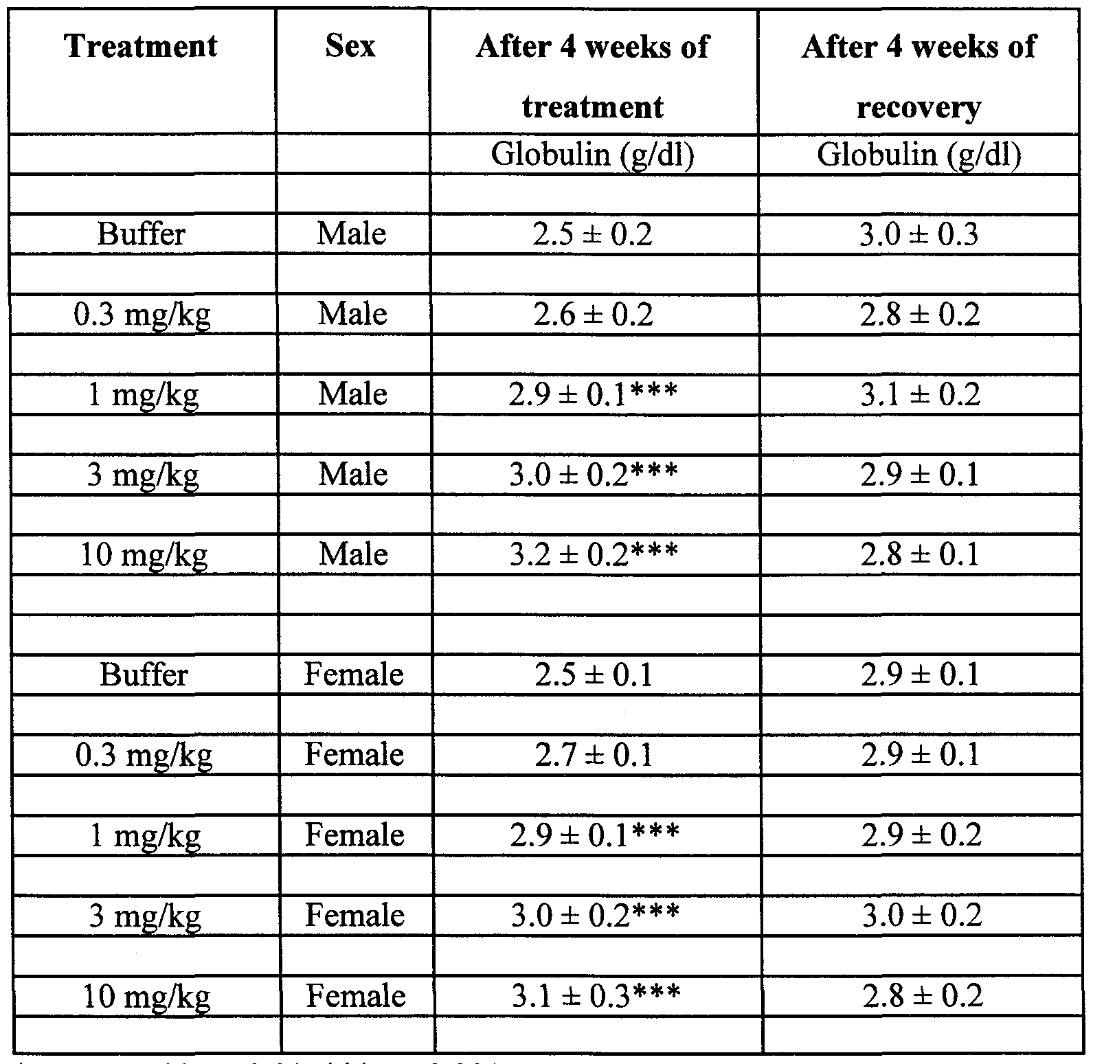

Claims
Priority Applications (6)
| Application Number | Priority Date | Filing Date | Title |
|---|---|---|---|
| AU28677/99A AU762519B2 (en) | 1998-02-13 | 1999-02-12 | Therapeutic uses of keratinocyte growth factor-2 |
| EP99909486A EP1054900A4 (en) | 1998-02-13 | 1999-02-12 | THERAPEUTIC USES OF KERATINOCYTE GROWTH FACTOR-2 |
| NZ506422A NZ506422A (en) | 1998-02-13 | 1999-02-12 | Therapeutic uses of keratinocyte growth factor -2 |
| KR1020007008814A KR20010040906A (en) | 1998-02-13 | 1999-02-12 | Therapeutic uses of keratinocyte growth factor-2 |
| CA002320515A CA2320515A1 (en) | 1998-02-13 | 1999-02-12 | Therapeutic uses of keratinocyte growth factor-2 |
| JP2000531473A JP2002507546A (en) | 1998-02-13 | 1999-02-12 | Therapeutic use of keratinocyte growth factor-2 |
Applications Claiming Priority (4)
| Application Number | Priority Date | Filing Date | Title |
|---|---|---|---|
| US7458598P | 1998-02-13 | 1998-02-13 | |
| US60/074,585 | 1998-02-13 | ||
| US11438798P | 1998-12-30 | 1998-12-30 | |
| US60/114,387 | 1998-12-30 |
Publications (2)
| Publication Number | Publication Date |
|---|---|
| WO1999041282A1 true WO1999041282A1 (en) | 1999-08-19 |
| WO1999041282A8 WO1999041282A8 (en) | 2000-07-06 |
Family
ID=26755823
Family Applications (1)
| Application Number | Title | Priority Date | Filing Date |
|---|---|---|---|
| PCT/US1999/003018 Ceased WO1999041282A1 (en) | 1998-02-13 | 1999-02-12 | Therapeutic uses of keratinocyte growth factor-2 |
Country Status (9)
| Country | Link |
|---|---|
| US (1) | US6599879B1 (en) |
| EP (1) | EP1054900A4 (en) |
| JP (1) | JP2002507546A (en) |
| KR (1) | KR20010040906A (en) |
| CN (1) | CN1295579A (en) |
| AU (1) | AU762519B2 (en) |
| CA (1) | CA2320515A1 (en) |
| NZ (1) | NZ506422A (en) |
| WO (1) | WO1999041282A1 (en) |
Cited By (9)
| Publication number | Priority date | Publication date | Assignee | Title |
|---|---|---|---|---|
| WO2002077155A3 (en) * | 2001-01-08 | 2003-01-03 | Human Genome Sciences Inc | Keratinocyte growth factor-2 |
| US6599879B1 (en) | 1998-02-13 | 2003-07-29 | Human Genome Sciences, Inc. | Therapeutic uses of keratinocyte growth factor-2 |
| US6653284B2 (en) | 1997-12-22 | 2003-11-25 | Human Genome Sciences, Inc. | Keratinocyte growth factor-2 formulations |
| US6693077B1 (en) | 1995-02-14 | 2004-02-17 | Human Genome Sciences, Inc. | Keratinocyte growth factor-2 |
| EP1421974A1 (en) * | 2002-06-10 | 2004-05-26 | D.M.G. Italia Srl | Carboxy methyl beta-1, 3 glucan containing ophthalmic solution |
| US6869927B1 (en) | 1997-12-22 | 2005-03-22 | Human Genome Sciences, Inc. | Keratinocyte growth factor-2 formulations |
| US6903072B2 (en) | 1995-02-14 | 2005-06-07 | Human Genome Sciences, Inc. | Keratinocyte growth factor-2 |
| US7232667B2 (en) | 1995-02-14 | 2007-06-19 | Human Genome Sciences, Inc. | Keratinocyte growth factor-2 polynucleotides |
| CN114073526A (en) * | 2020-08-21 | 2022-02-22 | 上海中医药大学附属曙光医院 | Renal fibrosis assessment method based on magnetic resonance elastography and serological examination |
Families Citing this family (11)
| Publication number | Priority date | Publication date | Assignee | Title |
|---|---|---|---|---|
| CN1302109C (en) * | 2004-04-21 | 2007-02-28 | 中国人民解放军军事医学科学院基础医学研究所 | Horny cell growth factor mutant and its preparation method and use thereof |
| US7223737B1 (en) | 2004-08-13 | 2007-05-29 | Alcon, Inc. | Method of treating dry eye disorders using glycosides |
| CA2588449A1 (en) * | 2004-11-24 | 2006-06-01 | Therakine Corporation | An implant for intraocular drug delivery |
| JP5637551B2 (en) * | 2005-12-28 | 2014-12-10 | キッセイ薬品工業株式会社 | Pharmaceuticals for the treatment of salivary gland disorders |
| CN100434117C (en) * | 2006-06-06 | 2008-11-19 | 武汉大学 | A suppository for treating chronic cervicitis |
| EP2026822A4 (en) * | 2006-06-12 | 2012-07-04 | Therakine Ltd | Topical treatment for diseases of eye surface |
| CN104173392A (en) * | 2008-05-30 | 2014-12-03 | 戴纳米斯制药公司 | Natural product inhibitors of 3dg |
| CN107893081B (en) * | 2017-12-27 | 2021-01-12 | 温州大学 | Gene sequence, expression vector and production method of human keratinocyte growth factor-2 |
| US20230285536A1 (en) * | 2020-06-24 | 2023-09-14 | Trustees Of Tufts College | Edible cell based vaccines |
| CN118105466A (en) * | 2024-02-04 | 2024-05-31 | 温州医科大学 | Application of KGF-2 in the treatment of dry eye |
| CN121182776B (en) * | 2025-11-25 | 2026-02-03 | 山东理工大学 | Phosphorylcholine cytidylyltransferase mutant and application thereof |
Citations (4)
| Publication number | Priority date | Publication date | Assignee | Title |
|---|---|---|---|---|
| US5677278A (en) * | 1993-06-29 | 1997-10-14 | Chiron Corporation | Truncated keratinocyte growth factor (KGF) having increased biological activity |
| US5703047A (en) * | 1992-09-21 | 1997-12-30 | Board Of Regents, The University Of Texas System | Methods and treatments for corneal healing with growth factors |
| US5731170A (en) * | 1989-01-31 | 1998-03-24 | The United States Of America As Represented By The Department Of Health And Human Services | DNA encoding a growth factor specific for epithelial cells |
| US5824643A (en) * | 1993-03-26 | 1998-10-20 | Amgen Inc. | Therapeutic uses of keratinocyte growth factor |
Family Cites Families (10)
| Publication number | Priority date | Publication date | Assignee | Title |
|---|---|---|---|---|
| JP3578761B2 (en) | 1993-03-26 | 2004-10-20 | アムジエン・インコーポレーテツド | Use of epidermal keratinocyte growth factor for therapeutic purposes |
| CA2185671A1 (en) | 1994-03-15 | 1995-09-21 | Prizm Pharmaceuticals, Inc. | Heparin-binding growth factors for gene therapy and anterior eye disorders |
| US6077692A (en) | 1995-02-14 | 2000-06-20 | Human Genome Sciences, Inc. | Keratinocyte growth factor-2 |
| ATE241639T1 (en) * | 1995-02-14 | 2003-06-15 | Human Genome Sciences Inc | KERATINOCYTE GROWTH FACTOR 2 |
| US5773252A (en) | 1995-06-05 | 1998-06-30 | Human Genome Sciences, Inc. | Fibroblast growth factor 15 |
| CN1234071A (en) * | 1996-08-13 | 1999-11-03 | 人类基因组科学公司 | Keratinocyte grouth factor -2(KGF-2 or fibroblast growth factor-12, FGF-12) |
| KR20010033484A (en) | 1997-12-22 | 2001-04-25 | 휴먼 게놈 사이언시즈, 인크. | Keratinocyte growth factor-2 formulations |
| US6599879B1 (en) | 1998-02-13 | 2003-07-29 | Human Genome Sciences, Inc. | Therapeutic uses of keratinocyte growth factor-2 |
| KR20020010920A (en) | 1999-06-02 | 2002-02-06 | 추후제출 | Keratinocyte growth factor-2 formulations |
| MXPA02000152A (en) | 1999-07-02 | 2002-07-30 | Human Genome Sciences Inc | Keratinocyte growth factor-2. |
-
1999
- 1999-02-12 US US09/248,998 patent/US6599879B1/en not_active Expired - Fee Related
- 1999-02-12 JP JP2000531473A patent/JP2002507546A/en active Pending
- 1999-02-12 CA CA002320515A patent/CA2320515A1/en not_active Abandoned
- 1999-02-12 EP EP99909486A patent/EP1054900A4/en not_active Withdrawn
- 1999-02-12 AU AU28677/99A patent/AU762519B2/en not_active Ceased
- 1999-02-12 NZ NZ506422A patent/NZ506422A/en unknown
- 1999-02-12 KR KR1020007008814A patent/KR20010040906A/en not_active Ceased
- 1999-02-12 WO PCT/US1999/003018 patent/WO1999041282A1/en not_active Ceased
- 1999-02-12 CN CN99804591A patent/CN1295579A/en active Pending
Patent Citations (5)
| Publication number | Priority date | Publication date | Assignee | Title |
|---|---|---|---|---|
| US5731170A (en) * | 1989-01-31 | 1998-03-24 | The United States Of America As Represented By The Department Of Health And Human Services | DNA encoding a growth factor specific for epithelial cells |
| US5703047A (en) * | 1992-09-21 | 1997-12-30 | Board Of Regents, The University Of Texas System | Methods and treatments for corneal healing with growth factors |
| US5824643A (en) * | 1993-03-26 | 1998-10-20 | Amgen Inc. | Therapeutic uses of keratinocyte growth factor |
| US5677278A (en) * | 1993-06-29 | 1997-10-14 | Chiron Corporation | Truncated keratinocyte growth factor (KGF) having increased biological activity |
| US5843883A (en) * | 1993-06-29 | 1998-12-01 | Chiron Corporation | Methods of wound healing and treating hyperproliferative diseases of epidermis comprising administering trunicated keratinocyte growh factor (KGF) having increased biological activity |
Non-Patent Citations (1)
| Title |
|---|
| See also references of EP1054900A4 * |
Cited By (10)
| Publication number | Priority date | Publication date | Assignee | Title |
|---|---|---|---|---|
| US6693077B1 (en) | 1995-02-14 | 2004-02-17 | Human Genome Sciences, Inc. | Keratinocyte growth factor-2 |
| US6903072B2 (en) | 1995-02-14 | 2005-06-07 | Human Genome Sciences, Inc. | Keratinocyte growth factor-2 |
| US6916786B2 (en) | 1995-02-14 | 2005-07-12 | Human Genome Sciences, Inc. | Keratinocyte growth factor-2 |
| US7232667B2 (en) | 1995-02-14 | 2007-06-19 | Human Genome Sciences, Inc. | Keratinocyte growth factor-2 polynucleotides |
| US6653284B2 (en) | 1997-12-22 | 2003-11-25 | Human Genome Sciences, Inc. | Keratinocyte growth factor-2 formulations |
| US6869927B1 (en) | 1997-12-22 | 2005-03-22 | Human Genome Sciences, Inc. | Keratinocyte growth factor-2 formulations |
| US6599879B1 (en) | 1998-02-13 | 2003-07-29 | Human Genome Sciences, Inc. | Therapeutic uses of keratinocyte growth factor-2 |
| WO2002077155A3 (en) * | 2001-01-08 | 2003-01-03 | Human Genome Sciences Inc | Keratinocyte growth factor-2 |
| EP1421974A1 (en) * | 2002-06-10 | 2004-05-26 | D.M.G. Italia Srl | Carboxy methyl beta-1, 3 glucan containing ophthalmic solution |
| CN114073526A (en) * | 2020-08-21 | 2022-02-22 | 上海中医药大学附属曙光医院 | Renal fibrosis assessment method based on magnetic resonance elastography and serological examination |
Also Published As
| Publication number | Publication date |
|---|---|
| WO1999041282A8 (en) | 2000-07-06 |
| CA2320515A1 (en) | 1999-08-19 |
| JP2002507546A (en) | 2002-03-12 |
| EP1054900A4 (en) | 2004-12-22 |
| EP1054900A1 (en) | 2000-11-29 |
| AU762519B2 (en) | 2003-06-26 |
| AU2867799A (en) | 1999-08-30 |
| CN1295579A (en) | 2001-05-16 |
| NZ506422A (en) | 2002-10-25 |
| US6599879B1 (en) | 2003-07-29 |
| KR20010040906A (en) | 2001-05-15 |
Similar Documents
| Publication | Publication Date | Title |
|---|---|---|
| EP0950100B1 (en) | KERATINOCYTE GROWTH FACTOR-2 (KGF-2 OR FIBROBLAST GROWTH FACTOR-12, FGF-12) Mutants | |
| EP1054900A1 (en) | Therapeutic uses of keratinocyte growth factor-2 | |
| US6916786B2 (en) | Keratinocyte growth factor-2 | |
| EP1063293A2 (en) | Transforming growth factor alpha H1 | |
| US7611865B2 (en) | Polynucleotides encoding truncated glial cell line-derived neurotrophic factor | |
| JP2002505873A (en) | Vascular endothelial growth factor 2 | |
| JP2007509984A (en) | Pigment epithelium-derived factor, novel biological activity thereof and method of use thereof | |
| EP1303605A2 (en) | Nucleic acids, vectors, host cells, polypeptides, and uses thereof | |
| MXPA01012387A (en) | Keratinocyte growth factor-2 formulations. | |
| CA2210444C (en) | Keratinocyte growth factor-2 | |
| WO1996039506A1 (en) | Fibroblast growth factor-14 | |
| JPH07103156B2 (en) | Osteogenic growth polypeptides identified from regenerated bone marrow | |
| US20070281890A1 (en) | Fibroblast Growth Factor-13 | |
| US20030186383A1 (en) | Dendritic cell-derived growth factor | |
| WO2006054278A2 (en) | Variants of pigment epithelium derived factor and uses thereof | |
| EP0832216A1 (en) | Fibroblast growth factor 15 | |
| EP1247862A2 (en) | Keratinocyte growth factor-2 (KGF-2 or fibroblast growth factor-12, FGF-12) | |
| MXPA00007884A (en) | Therapeutic uses of keratinocyte growth factor-2 | |
| AU1017202A (en) | Keratinocyte growth factor-2 (kgf-2 or fibroblast growth fact or-12, fgf-12) | |
| AU711647C (en) | Fibroblast growth factor 15 | |
| MXPA97006854A (en) | Factor 14 of fibroblas growth | |
| MXPA97008493A (en) | Factor 15 of fibroblas growth | |
| CN101298613A (en) | transforming growth factor α-HII |
Legal Events
| Date | Code | Title | Description |
|---|---|---|---|
| WWE | Wipo information: entry into national phase |
Ref document number: 99804591.8 Country of ref document: CN |
|
| AK | Designated states |
Kind code of ref document: A1 Designated state(s): AL AM AT AU AZ BA BB BG BR BY CA CH CN CU CZ DE DK EE ES FI GB GD GE GH GM HR HU ID IL IN IS JP KE KG KP KR KZ LC LK LR LS LT LU LV MD MG MK MN MW MX NO NZ PL PT RO RU SD SE SG SI SK SL TJ TM TR TT UA UG US UZ VN YU ZW |
|
| AL | Designated countries for regional patents |
Kind code of ref document: A1 Designated state(s): GH GM KE LS MW SD SZ UG ZW AM AZ BY KG KZ MD RU TJ TM AT BE CH CY DE DK ES FI FR GB GR IE IT LU MC NL PT SE BF BJ CF CG CI CM GA GN GW ML MR NE SN TD TG |
|
| 121 | Ep: the epo has been informed by wipo that ep was designated in this application | ||
| DFPE | Request for preliminary examination filed prior to expiration of 19th month from priority date (pct application filed before 20040101) | ||
| AK | Designated states |
Kind code of ref document: C1 Designated state(s): AL AM AT AU AZ BA BB BG BR BY CA CH CN CU CZ DE DK EE ES FI GB GD GE GH GM HR HU ID IL IN IS JP KE KG KP KR KZ LC LK LR LS LT LU LV MD MG MK MN MW MX NO NZ PL PT RO RU SD SE SG SI SK SL TJ TM TR TT UA UG US UZ VN YU ZW |
|
| AL | Designated countries for regional patents |
Kind code of ref document: C1 Designated state(s): GH GM KE LS MW SD SZ UG ZW AM AZ BY KG KZ MD RU TJ TM AT BE CH CY DE DK ES FI FR GB GR IE IT LU MC NL PT SE BF BJ CF CG CI CM GA GN GW ML MR NE SN TD TG |
|
| CFP | Corrected version of a pamphlet front page | ||
| CR1 | Correction of entry in section i | ||
| ENP | Entry into the national phase |
Ref document number: 2320515 Country of ref document: CA Ref document number: 2320515 Country of ref document: CA Kind code of ref document: A |
|
| WWE | Wipo information: entry into national phase |
Ref document number: PA/a/2000/007884 Country of ref document: MX Ref document number: 1020007008814 Country of ref document: KR |
|
| ENP | Entry into the national phase |
Ref document number: 2000 531473 Country of ref document: JP Kind code of ref document: A |
|
| WWE | Wipo information: entry into national phase |
Ref document number: 506422 Country of ref document: NZ Ref document number: 28677/99 Country of ref document: AU |
|
| WWE | Wipo information: entry into national phase |
Ref document number: 1999909486 Country of ref document: EP |
|
| WWP | Wipo information: published in national office |
Ref document number: 1999909486 Country of ref document: EP |
|
| REG | Reference to national code |
Ref country code: DE Ref legal event code: 8642 |
|
| WWP | Wipo information: published in national office |
Ref document number: 1020007008814 Country of ref document: KR |
|
| WWG | Wipo information: grant in national office |
Ref document number: 28677/99 Country of ref document: AU |
|
| WWW | Wipo information: withdrawn in national office |
Ref document number: 1999909486 Country of ref document: EP |
|
| WWR | Wipo information: refused in national office |
Ref document number: 1020007008814 Country of ref document: KR |