WO2000008206A1 - A novel method of diagnosing, monitoring, staging, imaging and treating lung cancer - Google Patents
A novel method of diagnosing, monitoring, staging, imaging and treating lung cancer Download PDFInfo
- Publication number
- WO2000008206A1 WO2000008206A1 PCT/US1999/016247 US9916247W WO0008206A1 WO 2000008206 A1 WO2000008206 A1 WO 2000008206A1 US 9916247 W US9916247 W US 9916247W WO 0008206 A1 WO0008206 A1 WO 0008206A1
- Authority
- WO
- WIPO (PCT)
- Prior art keywords
- lsg
- levels
- patient
- cancer
- lung cancer
- Prior art date
- Legal status (The legal status is an assumption and is not a legal conclusion. Google has not performed a legal analysis and makes no representation as to the accuracy of the status listed.)
- Ceased
Links
Classifications
-
- A—HUMAN NECESSITIES
- A61—MEDICAL OR VETERINARY SCIENCE; HYGIENE
- A61K—PREPARATIONS FOR MEDICAL, DENTAL OR TOILETRY PURPOSES
- A61K51/00—Preparations containing radioactive substances for use in therapy or testing in vivo
- A61K51/02—Preparations containing radioactive substances for use in therapy or testing in vivo characterised by the carrier, i.e. characterised by the agent or material covalently linked or complexing the radioactive nucleus
- A61K51/04—Organic compounds
- A61K51/08—Peptides, e.g. proteins, carriers being peptides, polyamino acids, proteins
- A61K51/10—Antibodies or immunoglobulins; Fragments thereof, the carrier being an antibody, an immunoglobulin or a fragment thereof, e.g. a camelised human single domain antibody or the Fc fragment of an antibody
- A61K51/1093—Antibodies or immunoglobulins; Fragments thereof, the carrier being an antibody, an immunoglobulin or a fragment thereof, e.g. a camelised human single domain antibody or the Fc fragment of an antibody conjugates with carriers being antibodies
-
- A—HUMAN NECESSITIES
- A61—MEDICAL OR VETERINARY SCIENCE; HYGIENE
- A61K—PREPARATIONS FOR MEDICAL, DENTAL OR TOILETRY PURPOSES
- A61K49/00—Preparations for testing in vivo
- A61K49/06—Nuclear magnetic resonance [NMR] contrast preparations; Magnetic resonance imaging [MRI] contrast preparations
- A61K49/08—Nuclear magnetic resonance [NMR] contrast preparations; Magnetic resonance imaging [MRI] contrast preparations characterised by the carrier
- A61K49/10—Organic compounds
- A61K49/14—Peptides, e.g. proteins
- A61K49/16—Antibodies; Immunoglobulins; Fragments thereof
-
- A—HUMAN NECESSITIES
- A61—MEDICAL OR VETERINARY SCIENCE; HYGIENE
- A61P—SPECIFIC THERAPEUTIC ACTIVITY OF CHEMICAL COMPOUNDS OR MEDICINAL PREPARATIONS
- A61P35/00—Antineoplastic agents
-
- C—CHEMISTRY; METALLURGY
- C07—ORGANIC CHEMISTRY
- C07K—PEPTIDES
- C07K14/00—Peptides having more than 20 amino acids; Gastrins; Somatostatins; Melanotropins; Derivatives thereof
- C07K14/435—Peptides having more than 20 amino acids; Gastrins; Somatostatins; Melanotropins; Derivatives thereof from animals; from humans
- C07K14/46—Peptides having more than 20 amino acids; Gastrins; Somatostatins; Melanotropins; Derivatives thereof from animals; from humans from vertebrates
- C07K14/47—Peptides having more than 20 amino acids; Gastrins; Somatostatins; Melanotropins; Derivatives thereof from animals; from humans from vertebrates from mammals
- C07K14/4701—Peptides having more than 20 amino acids; Gastrins; Somatostatins; Melanotropins; Derivatives thereof from animals; from humans from vertebrates from mammals not used
- C07K14/4748—Tumour specific antigens; Tumour rejection antigen precursors [TRAP], e.g. MAGE
-
- G—PHYSICS
- G01—MEASURING; TESTING
- G01N—INVESTIGATING OR ANALYSING MATERIALS BY DETERMINING THEIR CHEMICAL OR PHYSICAL PROPERTIES
- G01N33/00—Investigating or analysing materials by specific methods not covered by groups G01N1/00 - G01N31/00
- G01N33/48—Biological material, e.g. blood, urine; Haemocytometers
- G01N33/50—Chemical analysis of biological material, e.g. blood, urine; Testing involving biospecific ligand binding methods; Immunological testing
- G01N33/53—Immunoassay; Biospecific binding assay; Materials therefor
- G01N33/575—Immunoassay; Biospecific binding assay; Materials therefor for cancer
- G01N33/5752—Immunoassay; Biospecific binding assay; Materials therefor for cancer of the lungs
-
- G—PHYSICS
- G01—MEASURING; TESTING
- G01N—INVESTIGATING OR ANALYSING MATERIALS BY DETERMINING THEIR CHEMICAL OR PHYSICAL PROPERTIES
- G01N33/00—Investigating or analysing materials by specific methods not covered by groups G01N1/00 - G01N31/00
- G01N33/48—Biological material, e.g. blood, urine; Haemocytometers
- G01N33/50—Chemical analysis of biological material, e.g. blood, urine; Testing involving biospecific ligand binding methods; Immunological testing
- G01N33/53—Immunoassay; Biospecific binding assay; Materials therefor
- G01N33/575—Immunoassay; Biospecific binding assay; Materials therefor for cancer
- G01N33/5758—Immunoassay; Biospecific binding assay; Materials therefor for cancer involving compounds serving as markers for tumours, cancers or neoplasias, e.g. cellular determinants, receptors, heat shock/stress proteins, A-protein, oligosaccharides or metabolites
-
- A—HUMAN NECESSITIES
- A61—MEDICAL OR VETERINARY SCIENCE; HYGIENE
- A61K—PREPARATIONS FOR MEDICAL, DENTAL OR TOILETRY PURPOSES
- A61K39/00—Medicinal preparations containing antigens or antibodies
Definitions
- This invention relates, in part, to newly developed assays for detecting, diagnosing, monitoring, staging, prognosticating, imaging and treating cancers, particularly lung cancer.
- Lung cancer is the second most prevalent type of cancer for both men and women in the United States and is the most common cause of cancer death in both sexes .
- Lung cancer can result from a primary tumor originating in the lung or a secondary tumor which has spread from another organ such as the bowel or breast.
- Primary lung cancer is divided into three main types; small cell lung cancer; non-small cell lung cancer; and mesothelioma.
- Small cell lung cancer is also called "Oat Cell” lung cancer because the cancer cells are a distinctive oat shape.
- Squamous cell cancer is the most common type of lung cancer. It develops from the cells that line the airways. Adenocarcinoma also develops from the cells that line the airways. However, adenocarcinoma develops from a particular type of cell that produces mucus (phlegm) . Large cell lung cancer has been thus named because the cells look large and rounded when they are viewed under a microscope.
- Mesothelioma is a rare type of cancer which affects the covering of the lung called the pleura. Mesothelioma is often caused by exposure to asbestos.
- Secondary lung cancer is cancer that has started somewhere else in the body (for example, the breast or bowel) and spread to the lungs .
- Choice of treatment for secondary lung cancer depends on where the cancer started. In other words, cancer that has spread from the breast should respond to breast cancer treatments and cancer that has spread from the bowel should respond to bowel cancer treatments.
- the stage of a cancer indicates how far a cancer has spread. Staging is important because treatment is often decided according to the stage of a cancer.
- the staging is different for non-small cell and for small cell cancers of the lung.
- Non- small cell cancer can be divided into four stages. Stage I is very localized cancer with no cancer in the lymph nodes. Stage II cancer has spread to the lymph nodes at the top of the affected lung. Stage III cancer has spread near to where the cancer started. This can be to the chest wall, the covering of the lung (pleura) , the middle of the chest (mediastinum) or other lymph nodes. Stage IV cancer has spread to another part of the body.
- small cell lung cancers can spreads quite early in development of the disease, small cell lung cancers are divided into only two groups. These are: limited disease, that is cancer that can only be seen in one lung and in nearby lymph nodes; and extensive disease, that is cancer that has spread outside the lung to the chest or to other parts of the body. Further, even if spreading is not apparent on the scans, it is likely that some cancer cells will have broken away and traveled through the bloodstream or lymph system. To be safe, it is therefore preferred to treat small cell lung cancers as if they have spread, whether or not secondary cancer is visible. Because surgery is not typically used to treat small cell cancer, except in very early cases, the staging is not as critical as it is with some other types of cancer. Chemotherapy with or without radiotherapy is often employed. The scans and tests done at first will be used later to see how well a patient is responding to treatment.
- WO 98/56951 discloses a set of contiguous and partially overlapping cDNA sequences and polypeptides encoded thereby, designated as LS170. These sequences are suggested to be useful in detecting, diagnosing, staging, monitoring, prognosticating, in vivo imaging, preventing or treating, and determining the predisposition of an individual to disease and conditions of the lung, such as lung cancer.
- the LS170-specific polynucleotide is taught to have at least 50% identity with a polynucleotide selected from the group consisting of SEQ ID NO: 1-9 as disclosed in WO 98/56951.
- SEQ ID NO : 1 taught in WO 98/56951 overlaps with an LSG, SEQ ID NO: 4, used in the instant invention.
- methods are provided for detecting, diagnosing, monitoring, staging, prognosticating, in vivo imaging and treating lung cancer via five (5) Lung Specific Genes (LSG) .
- LSGs refer, among other things, to native proteins expressed by the genes comprising the polynucleotide sequences of any of SEQ ID NO: 1, 2, 3, 4, or 5.
- Procedures used for detecting, diagnosing, monitoring, staging, and prognosticating lung cancer are of critical importance to the outcome of the patient. For example, patients diagnosed with early lung cancer generally have a much greater five-year survival rate as compared to the survival rate for patients diagnosed with distant metastasized lung cancer. New diagnostic methods which are more sensitive and specific for detecting early lung cancer are clearly needed. Lung cancer patients are closely monitored following initial therapy and during adjuvant therapy to determine response to therapy and to detect persistent or recurrent disease of metastasis. There is clearly a need for a lung cancer marker which is more sensitive and specific in detecting lung cancer, its recurrence and progression.
- stage determination has potential prognostic value and provides criteria for designing optimal therapy.
- pathological staging of lung cancer is preferable over clinical staging because the former gives a more accurate prognosis.
- clinical staging would be preferred were it at least as accurate as pathological staging because it does not depend on an invasive procedure to obtain tissue for pathological evaluation.
- Staging of lung cancer would be improved by detecting new markers in cells, tissues, or bodily fluids which could differentiate between different stages of invasion.
- a method of diagnosing metastatic lung cancer in a patient having such cancer which is not known to have metastasized by identifying a human patient suspected of having lung cancer that has metastasized; analyzing a sample of cells, tissues, or bodily fluid from such patient for LSG; comparing the LSG levels in such cells, tissues, or bodily fluid with levels of LSG in preferably the same cells, tissues, or bodily fluid type of a normal human control, wherein an increase in LSG levels in the patient versus the normal human control is associated with a cancer which has metastasized.
- Also provided by the invention is a method of staging lung cancer in a human which has such cancer by identifying a human patient having such cancer; analyzing a sample of cells, tissues, or bodily fluid from such patient for LSG; comparing LSG levels in such cells, tissues, or bodily fluid with levels of LSG in preferably the same cells, tissues, or bodily fluid type of a normal human control sample, wherein an increase in LSG levels in the patient versus the normal human control is associated with a cancer which is progressing and a decrease in the levels of LSG is associated with a cancer which is regressing or in remission.
- the method comprises identifying a human patient having such cancer that is not known to have metastasized; periodically analyzing a sample of cells, tissues, or bodily fluid from such patient for LSG; comparing the LSG levels in such cells, tissue, or bodily fluid with levels of LSG in preferably the same cells, tissues, or bodily fluid type of a normal human control sample, wherein an increase in LSG levels in the patient versus the normal human control is associated with a cancer which has metastasized.
- the method comprises identifying a human patient having such cancer; periodically analyzing a sample of cells, tissues, or bodily fluid from such patient for LSG; comparing the LSG levels in such cells, tissue, or bodily fluid with levels of LSG in preferably the same cells, tissues, or bodily fluid type of a normal human control sample, wherein an increase in LSG levels in the patient versus the normal human control is associated with a cancer which is progressing and a decrease in the levels of LSG is associated with a cancer which is regressing or in remission.
- antibodies against the LSGs or fragments of such antibodies which can be used to detect or image localization of the LSGs in a patient for the purpose of detecting or diagnosing a disease or condition.
- Such antibodies can be polyclonal or monoclonal, or prepared by molecular biology techniques.
- the term "antibody”, as used herein and throughout the instant specification is also meant to include aptamers and single-stranded oligonucleotides such as those derived from an in vi tro evolution protocol referred to as SELEX and well known to those skilled in the art.
- Antibodies can be labeled with a variety of detectable labels including, but not limited to, radioisotopes and paramagnetic metals.
- antibodies or fragments thereof can also be used as therapeutic agents in the treatment of diseases characterized by expression of a LSG.
- the antibody can be used without or with derivatization to a cytotoxic agent such as a radioisotope, enzyme, toxin, drug or a prodrug.
- the present invention relates to diagnostic assays and methods, both quantitative and qualitative for detecting, diagnosing, monitoring, staging, prognosticating, in vivo imaging and treating cancers by comparing levels of LSG with those of LSG in a normal human control.
- levels of LSG as used herein means levels of the native protein expressed by the gene comprising the polynucleotide sequence of any of SEQ ID NO: 1, 2, 3, 4, or 5.
- levels of LSG as used herein means levels of the native mRNA encoded by the gene comprising any of the polynucleotide sequences of SEQ ID NO: 1, 2, 3,
- a diagnostic assay in accordance with the invention for diagnosing over- expression of LSG protein compared to normal control bodily fluids, cells, or tissue samples may be used to diagnose the presence of cancers, including lung cancer. Any of the five LSGs may be measured alone in the methods of the invention, or all together or any combination of the five.
- All the methods of the present invention may optionally include measuring the levels of other cancer markers as well as LSG.
- Other cancer markers, in addition to LSG, useful in the present invention will depend on the cancer being tested and are known to those of skill in the art.
- the present invention provides methods for diagnosing the presence of lung cancer by analyzing for changes in levels of LSG in cells, tissues or bodily fluids compared with levels of LSG in cells, tissues or bodily fluids of preferably the same type from a normal human control , wherein an increase in levels of LSG in the patient versus the normal human control is associated with the presence of lung cancer.
- a positive result indicating the patient being tested has cancer is one in which cells, tissues, or bodily fluid levels of the cancer marker, such as LSG, are at least two times higher, and most preferable are at least five times higher, than in preferably the same cells, tissues, or bodily fluid of a normal human control.
- the cancer marker such as LSG
- the present invention also provides a method of diagnosing metastatic lung cancer in a patient having lung cancer which has not yet metastasized for the onset of metastasis.
- a human cancer patient suspected of having lung cancer which may have metastasized is identified. This is accomplished by a variety of means known to those of skill in the art. For example, in the case of lung cancer, patients are typically diagnosed with lung cancer following traditional detection methods .
- determining the presence of LSG level in cells, tissues, or bodily fluid is particularly useful for discriminating between lung cancer which has not metastasized and lung cancer which has metastasized.
- Existing techniques have difficulty discriminating between lung cancer which has metastasized and lung cancer which has not metastasized and proper treatment selection is often dependent upon such knowledge .
- the cancer marker level measured in such cells, tissues, or bodily fluid is LSG, and is compared with levels of LSG in preferably the same cells, tissue, or bodily fluid type of a normal human control. That is, if the cancer marker being observed is just LSG in serum, this level is preferably compared with the level of LSG in serum of a normal human patient. An increase in the LSG in the patient versus the normal human control is associated with lung cancer which has metastasized.
- a positive result indicating the cancer in the patient being tested or monitored has metastasized is one in which cells, tissues, or bodily fluid levels of the cancer marker, such as LSG, are at least two times higher, and more preferably are at least five times higher, than in preferably the same cells, tissues, or bodily fluid of a normal patient.
- Normal human control as used herein includes a human patient without cancer and/or non cancerous samples from the patient; in the methods for diagnosing metastasis or monitoring for metastasis, normal human control preferably includes samples from a human patient that is determined by reliable methods to have lung cancer which has not metastasized such as samples from the same patient prior to metastasis .
- the invention also provides a method of staging lung cancer in a human patient.
- the method comprises identifying a human patient having such cancer and analyzing a sample of cells, tissues, or bodily fluid from such patient for LSG.
- the measured LSG levels are then compared to levels of LSG in preferably the same cells, tissues, or bodily fluid type of a normal human control sample, wherein an increase in LSG levels in the patient versus the normal human control is associated with a cancer which is progressing and a decrease in the levels of LSG is associated with a cancer which is regressing or in remission.
- a method of monitoring lung cancer in a human having such cancer for the onset of metastasis comprises identifying a human patient having such cancer that is not known to have metastasized; periodically analyzing a sample of cells, tissues, or bodily fluid from such patient for LSG; comparing the LSG levels in such cells, tissue, or bodily fluid with levels of LSG in preferably the same cells, tissues, or bodily fluid type of a normal human control sample, wherein an increase in LSG levels in the patient versus the normal human control is associated with a cancer which has metastasized.
- a method of monitoring the change in stage of lung cancer in a human having such cancer is identifying a human patient having such cancer that is not known to have metastasized.
- the method comprises identifying a human patient having such cancer; periodically analyzing a sample of cells, tissues, or bodily fluid from such patient for LSG; comparing the LSG levels in such cells, tissue, or bodily fluid with levels of LSG in preferably the same cells, tissues, or bodily fluid type of a normal human control sample, wherein an increase in LSG levels in the patient versus the normal human control is associated with a cancer which is progressing in stage and a decrease in the levels of LSG is associated with a cancer which is regressing in stage or in remission.
- Monitoring such patient for onset of metastasis is periodic and preferably done on a quarterly basis. However, this may be more or less frequent depending on the cancer, the particular patient, and the stage of the cancer.
- Assay techniques that can be used to determine levels of gene expression, such as LSG of the present invention, in a sample derived from a host are well-known to those of skill in the art.
- Such assay methods include radioimmunoassays , reverse transcriptase PCR (RT-PCR) assays, immunohistochemistry assays, in si tu hybridization assays, competitive-binding assays, Western Blot analyses, ELISA assays and proteomic approaches.
- RT-PCR reverse transcriptase PCR
- immunohistochemistry assays in si tu hybridization assays
- competitive-binding assays Western Blot analyses
- ELISA assays are frequently preferred to diagnose a gene's expressed protein in biological fluids.
- An ELISA assay initially comprises preparing an antibody, if not readily available from a commercial source, specific to LSG, preferably a monoclonal antibody.
- a reporter antibody generally is prepared which binds specifically to LSG.
- the reporter antibody is attached to a detectable reagent such as radioactive, fluorescent or enzymatic reagent, for example horseradish peroxidase enzyme or alkaline phosphatase.
- antibody specific to LSG is incubated on a solid support, e.g., a polystyrene dish, that binds the antibody. Any free protein binding sites on the dish are then covered by incubating with a non-specific protein such as bovine serum albumin.
- a non-specific protein such as bovine serum albumin.
- the sample to be analyzed is incubated in the dish, during which time LSG binds to the specific antibody attached to the polystyrene dish. Unbound sample is washed out with buffer.
- a reporter antibody specifically directed to LSG and linked to horseradish peroxidase is placed in the dish resulting in binding of the reporter antibody to any monoclonal antibody bound to LSG. Unattached reporter antibody is then washed out.
- Reagents for peroxidase activity including a colorimetric substrate are then added to the dish.
- the amount of color developed in a given time period is proportional to the amount of LSG protein present in the sample. Quantitative results typically are obtained by reference to a standard curve.
- a competition assay may be employed wherein antibodies specific to LSG attached to a solid support and labeled LSG and a sample derived from the host are passed over the solid support and the amount of label detected attached to the solid support can be correlated to a quantity of LSG in the sample.
- Nucleic acid methods may be used to detect LSG mRNA as a marker for lung cancer.
- Polymerase chain reaction (PCR) and other nucleic acid methods such as ligase chain reaction (LCR) and nucleic acid sequence based amplification (NASABA)
- PCR polymerase chain reaction
- LCR ligase chain reaction
- NASABA nucleic acid sequence based amplification
- RT-PCR reverse- transcriptase PCR
- cDNA complementary DNA
- RT-PCR can thus reveal by amplification the presence of a single species of mRNA. Accordingly, if the mRNA is highly specific for the cell that produces it, RT-PCR can be used to identify the presence of a specific type of cell.
- Hybridization to clones or oligonucleotides arrayed on a solid support can be used to both detect the expression of and quantitate the level of expression of that gene.
- a cDNA encoding the LSG gene is fixed to a substrate.
- the substrate may be of any suitable type including but not limited to glass, nitrocellulose, nylon or plastic.
- At least a portion of the DNA encoding the LSG gene is attached to the substrate and then incubated with the analyte, which may be RNA or a complementary DNA (cDNA) copy of the RNA, isolated from the tissue of interest.
- Hybridization between the substrate bound DNA and the analyte can be detected and quantitated by several means including but not limited to radioactive labeling or fluorescence labeling of the analyte or a secondary molecule designed to detect the hybrid. Quantitation of the level of gene expression can be done by comparison of the intensity of the signal from the analyte compared with that determined from known standards. The standards can be obtained by in vi tro transcription of the target gene, quantitating the yield, and then using that material to generate a standard curve.
- 2D electrophoresis is a technique well known to those in the art. Isolation of individual proteins from a sample such as serum is accomplished using sequential separation of proteins by different characteristics usually on polyacrylamide gels. First, proteins are separated by size using an electric current. The current acts uniformly on all proteins, so smaller proteins move farther on the gel than larger proteins. The second dimension applies a current perpendicular to the first and separates proteins not on the basis of size but on the specific electric charge carried by each protein. Since no two proteins with different sequences are identical on the basis of both size and charge, the result of a 2D separation is a square gel in which each protein occupies a unique spot. Analysis of the spots with chemical or antibody probes, or subsequent protein microsequencing can reveal the relative abundance of a given protein and the identity of the proteins in the sample.
- Bodily fluids useful in the present invention include blood, urine, saliva, or any other bodily secretion or derivative thereof.
- Blood can include whole blood, plasma, serum, or any derivative of blood.
- Antibodies against LSG can also be used in vivo in patients with disease of the lung. Specifically, antibodies against an LSG can be injected into a patient suspected of having a disease of the lung for diagnostic and/or therapeutic purposes.
- the use of antibodies for in vivo diagnosis is well known in the art.
- antibody-chelators labeled with Indium-111 have been described for use in the radioimmunoscintographic imaging of carcinoembryonic antigen expressing tumors (Sumerdon et al . Nucl . Med. Biol. 1990 17:247-254).
- these antibody-chelators have been used in detecting tumors in patients suspected of having recurrent colorectal cancer (Griffin et al . J. Clin. One.
- Antibodies with paramagnetic ions as labels for use in magnetic resonance imaging have also been described (Lauffer, R.B. Magnetic Resonance in Medicine 1991 22:339- 342) .
- Antibodies directed against LSGs can be used in a similar manner. Labeled antibodies against an LSG can be injected into patients suspected of having a disease of the lung such as lung cancer for the purpose of diagnosing or staging of the disease status of the patient. The label used will be selected in accordance with the imaging modality to be used. For example, radioactive labels such as Indium-111, Technetium-99m or Iodine-131 can be used for planar scans or single photon emission computed tomography (SPECT) .
- SPECT single photon emission computed tomography
- Positron emitting labels such as Fluorine-19 can be used in positron emission tomography.
- Paramagnetic ions such as Gadlinium (III) or Manganese (II) can used in magnetic resonance imaging (MRI) .
- Localization of the label within the lung or external to the lung permits determination of the spread of the disease.
- the amount of label within the lung also allows determination of the presence or absence of cancer in the lung.
- an antibody against an LSG can also have a therapeutic benefit.
- the antibody may exert its therapeutic effect alone.
- the antibody is conjugated to a cytotoxic agent such as a drug, toxin or radionuclide to enhance its therapeutic effect.
- Drug monoclonal antibodies have been described in the art for example by Garnett and Baldwin, Cancer Research 1986 46:2407-2412. The use of toxins conjugated to monoclonal antibodies for the therapy of various cancers has also been described by Pastan et al . Cell 1986 47:641-648). Yttrium-90 labeled monoclonal antibodies have been described for maximization of dose delivered to the tumor while limiting toxicity to normal tissues (Goodwin and Meares Cancer Supplement 1997 80:2675-2680).
- cytotoxic radionuclides including, but not limited to Copper-67, Iodine- 131 and Rhenium- 186 can also be used for labeling of antibodies against LSGs.
- Antibodies which can be used in these in vivo methods include both polyclonal and monoclonal antibodies and antibodies prepared via molecular biology techniques. Antibody fragments can also be used.
- Subsetting is similar to library comparison but allows the identification of clones expressed in a pool of libraries and absent or expressed at a lower level in a second pool of libraries .
- Transcript Imaging lists all of the clones in a single library or a pool of libraries based on abundance. Individual clones can then be examined using Electronic Northerns to determine the tissue sources of their component ESTs.
- Incyte has identified subsets of ESTs with a potential protein function based on homologies to known proteins. Some examples in this database include Transcription Factors and Proteases. Some leads were identified by searching in this database for clones whose component ESTs showed disease specificity.
- Real-Time quantitative PCR with fluorescent Taqman probes is a quantitation detection system utilizing the 5'- 3' nuclease activity of Taq DNA polymerase.
- the method uses an internal fluorescent oligonucleotide probe (Taqman) labeled with a 5' reporter dye and a downstream, 3' quencher dye.
- Taqman internal fluorescent oligonucleotide probe
- the 5' -3' nuclease activity of Taq DNA polymerase releases the reporter, whose fluorescence can then be detected by the laser detector of the Model 7700 Sequence Detection System (PE Applied Biosystems, Foster City, CA, USA) .
- Amplification of an endogenous control is used to standardize the amount of sample RNA added to the reaction and normalize for Reverse Transcriptase (RT) efficiency.
- Either cyclophilin, glyceraldehyde-3 -phosphate dehydrogenase (GAPDH) or 18S ribosomal RNA (rRNA) was used as this endogenous control.
- GPDH glyceraldehyde-3 -phosphate dehydrogenase
- rRNA 18S ribosomal RNA
- Quantitation relative to the "calibrator" can be obtained using the standard curve method or the comparative method
- the tissue distribution and the level of the target gene were evaluated for every example in normal and cancer tissue.
- PSA Prostate Specific Antigen
- PLA2 Phospholipase A2
- mRNA expression data showed overexpression of the mRNA in 8 out of the 12 prostate matching samples analyzed (66%) .
- the tissue specificity for PLA2 was not as good as the one described for PSA.
- small intestine, liver, and pancreas also showed high levels of mRNA expression for PLA2.
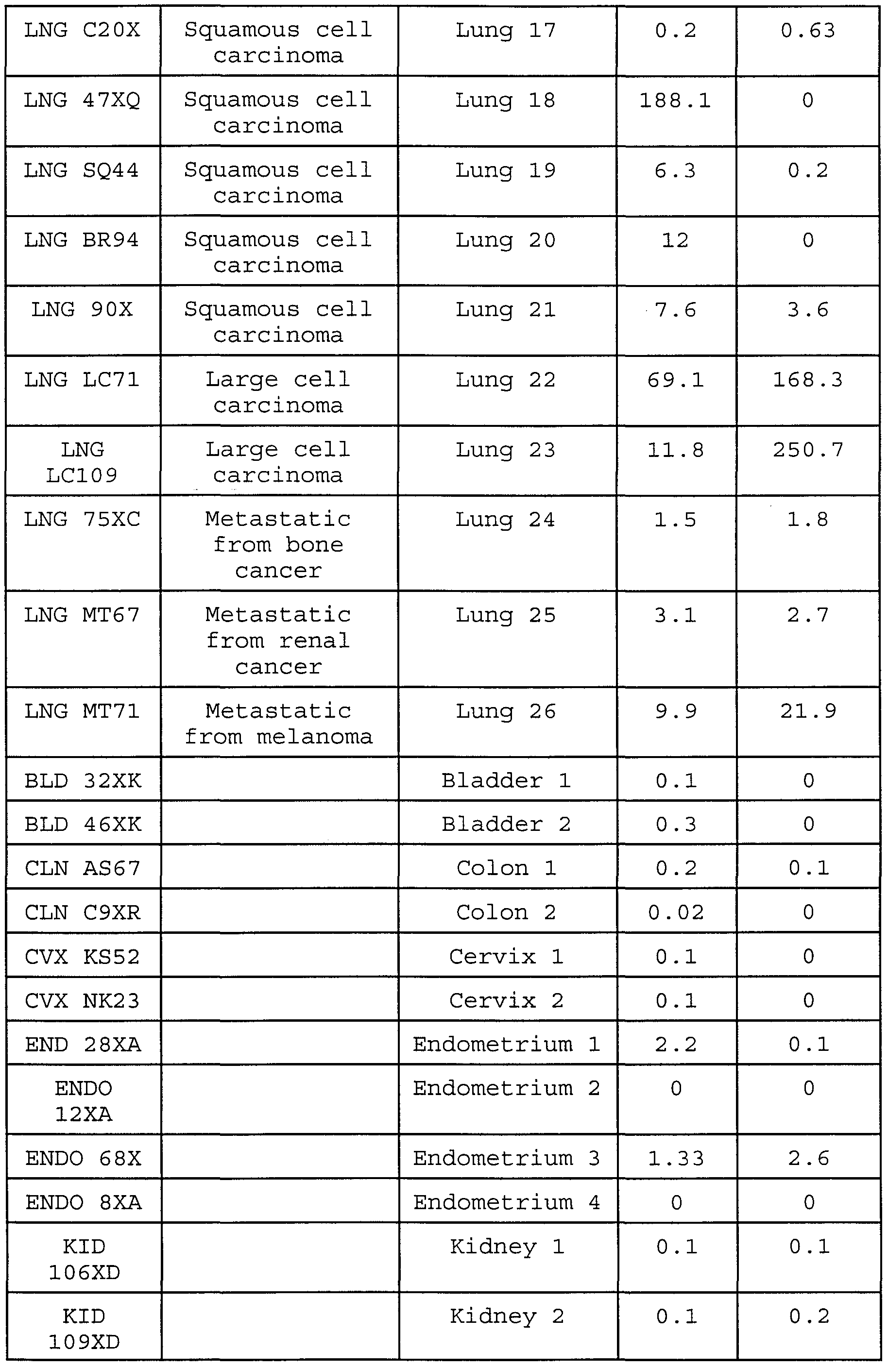
- RNA samples are commercially available pools, originated by pooling samples of a particular tissue from different individuals.
- the absolute numbers in Table 2 were obtained analyzing pools of samples of a particular tissue from different individuals. They can not be compared to the absolute numbers originated from RNA obtained from tissue samples of a single individual in Table 3.
- the absolute numbers depicted in Table 3 are relative levels of expression of Lngl09 (SEQ ID NO: 5) in 57 pairs of matching samples. All the values are compared to normal small intestine (calibrator) .
- a matching pair is formed by mRNA from the cancer sample for a particular tissue and mRNA from the normal adjacent sample for that same tissue from the same individual .
- the level of mRNA expression in cancer samples and the isogenic normal adjacent tissue from the same individual was compared. This comparison provides an indication of specificity for the cancer stage (e.g. higher levels of mRNA expression in the cancer sample compared to the normal adjacent) .
- Table 3 shows overexpression of Lngl09 in 16 primary lung cancer tissues compared with their respective normal adjacent (lung samples #1, 3, 4, 5, 7, 8, 9, 10, 11, 13, 14, 15, 18, 19, 20, and 21) . There was overexpression in the cancer tissue for 70% of the lung matching samples tested (total of 23 lung matching samples) .
- RNA samples are commercially available pools, originated by pooling samples of a particular tissue from different individuals.
- the absolute numbers in Table 4 were obtained analyzing pools of samples of a particular tissue from different individuals. They can not be compared to the absolute numbers originated from RNA obtained from tissue samples of a single individual in Table 5.
- the absolute numbers depicted in Table 5 are relative levels of expression of LngllO in 60 pairs of matching samples. All the values are compared to normal testis (calibrator) .
- a matching pair is formed by mRNA from the cancer sample for a particular tissue and mRNA from the normal adjacent sample for that same tissue from the same individual .
- the level of mRNA expression in cancer samples and the isogenic normal adjacent tissue from the same individual was compared. This comparison provides an indication of specificity for the cancer stage (e.g. higher levels of mRNA expression in the cancer sample compared to the normal adjacent) .
- Table 5 shows overexpression of LngllO in 18 primary lung cancer samples compared with their respective normal adjacent (lung samples #1, 4, 5, 6, 7, 8, 9, 10, 13, 14, 15, 16, 17, 18, 19, 21, 23 and 24) . There is overexpression in the cancer tissue for 75% of the lung matching samples tested (total of 24 primary lung matching samples) .
- LngllO being a diagnostic marker for lung cancer.
- SEQ ID NO: 6 The amino acid sequence encoded by the open reading frame of LngllO is depicted in SEQ ID NO: 6.
Landscapes
- Health & Medical Sciences (AREA)
- Life Sciences & Earth Sciences (AREA)
- Chemical & Material Sciences (AREA)
- Immunology (AREA)
- Medicinal Chemistry (AREA)
- Engineering & Computer Science (AREA)
- General Health & Medical Sciences (AREA)
- Molecular Biology (AREA)
- Urology & Nephrology (AREA)
- Hematology (AREA)
- Biomedical Technology (AREA)
- Public Health (AREA)
- Animal Behavior & Ethology (AREA)
- Physics & Mathematics (AREA)
- Proteomics, Peptides & Aminoacids (AREA)
- Organic Chemistry (AREA)
- Veterinary Medicine (AREA)
- Biochemistry (AREA)
- Biotechnology (AREA)
- Pathology (AREA)
- General Physics & Mathematics (AREA)
- Analytical Chemistry (AREA)
- Food Science & Technology (AREA)
- Pharmacology & Pharmacy (AREA)
- Nuclear Medicine, Radiotherapy & Molecular Imaging (AREA)
- Microbiology (AREA)
- Cell Biology (AREA)
- Epidemiology (AREA)
- Chemical Kinetics & Catalysis (AREA)
- Biophysics (AREA)
- Radiology & Medical Imaging (AREA)
- General Chemical & Material Sciences (AREA)
- Gastroenterology & Hepatology (AREA)
- Toxicology (AREA)
- Genetics & Genomics (AREA)
- Optics & Photonics (AREA)
- Zoology (AREA)
- Measuring Or Testing Involving Enzymes Or Micro-Organisms (AREA)
- Peptides Or Proteins (AREA)
- Preparation Of Compounds By Using Micro-Organisms (AREA)
Abstract
Description


Claims
Priority Applications (4)
| Application Number | Priority Date | Filing Date | Title |
|---|---|---|---|
| EP99935685A EP1104486A4 (en) | 1998-08-04 | 1999-07-19 | NEW METHOD FOR DIAGNOSING, MONITORING, STADIUM CLASSIFICATION, IMAGING AND TREATING LUNG CANCER |
| JP2000563828A JP3524061B2 (en) | 1998-08-04 | 1999-07-19 | Novel way to diagnose, monitor, stage, image and treat lung cancer |
| CA002347656A CA2347656A1 (en) | 1998-08-04 | 1999-07-19 | A novel method of diagnosing, monitoring, staging, imaging and treating lung cancer |
| US09/762,028 US6869592B1 (en) | 1998-08-04 | 1999-07-19 | Method and antibody for imaging lung cancer |
Applications Claiming Priority (2)
| Application Number | Priority Date | Filing Date | Title |
|---|---|---|---|
| US9523398P | 1998-08-04 | 1998-08-04 | |
| US60/095,233 | 1998-08-04 |
Related Child Applications (2)
| Application Number | Title | Priority Date | Filing Date |
|---|---|---|---|
| US09/762,028 A-371-Of-International US6869592B1 (en) | 1998-08-04 | 1999-07-19 | Method and antibody for imaging lung cancer |
| US10/843,968 Division US20040203065A1 (en) | 1998-08-04 | 2004-05-12 | Novel method of diagnosing, monitoring, staging, imaging and treating lung cancer |
Publications (1)
| Publication Number | Publication Date |
|---|---|
| WO2000008206A1 true WO2000008206A1 (en) | 2000-02-17 |
Family
ID=22250833
Family Applications (1)
| Application Number | Title | Priority Date | Filing Date |
|---|---|---|---|
| PCT/US1999/016247 Ceased WO2000008206A1 (en) | 1998-08-04 | 1999-07-19 | A novel method of diagnosing, monitoring, staging, imaging and treating lung cancer |
Country Status (4)
| Country | Link |
|---|---|
| EP (1) | EP1104486A4 (en) |
| JP (1) | JP3524061B2 (en) |
| CA (1) | CA2347656A1 (en) |
| WO (1) | WO2000008206A1 (en) |
Cited By (13)
| Publication number | Priority date | Publication date | Assignee | Title |
|---|---|---|---|---|
| WO2002068633A3 (en) * | 2000-11-22 | 2003-03-27 | Diadexus Inc | Compositions and methods relating to lung specific genes and proteins |
| WO2002018576A3 (en) * | 2000-08-28 | 2003-04-17 | Diadexus Inc | Compositions and methods relating to lung specific genes |
| WO2002040673A3 (en) * | 2000-11-20 | 2003-06-05 | Diadexus Inc | Compositions and methods relating to lung specific genes and proteins |
| WO2002046224A3 (en) * | 2000-10-26 | 2003-08-21 | Diadexus Inc | Compositions and methods relating to lung specific genes and proteins |
| WO2002064788A3 (en) * | 2000-11-20 | 2003-12-11 | Diadexus Inc | Compositions and methods relating to lung specific genes and proteins |
| US6869592B1 (en) | 1998-08-04 | 2005-03-22 | Diadexus, Inc. | Method and antibody for imaging lung cancer |
| US7527933B2 (en) | 2002-11-22 | 2009-05-05 | Ganymed Pharmaceuticals Ag | Genetic products differentially expressed in tumors and the use thereof |
| US7951781B2 (en) | 2006-11-02 | 2011-05-31 | University Of Iowa Research Foundation | Methods and compositions related to PLUNC surfactant polypeptides |
| US9044382B2 (en) | 2004-05-18 | 2015-06-02 | Ganymed Pharmaceuticals Ag | Genetic products differentially expressed in tumors and the use thereof |
| US9212228B2 (en) | 2005-11-24 | 2015-12-15 | Ganymed Pharmaceuticals Ag | Monoclonal antibodies against claudin-18 for treatment of cancer |
| US9512232B2 (en) | 2012-05-09 | 2016-12-06 | Ganymed Pharmaceuticals Ag | Antibodies against Claudin 18.2 useful in cancer diagnosis |
| US10093736B2 (en) | 2012-11-13 | 2018-10-09 | Biontech Ag | Agents for treatment of claudin expressing cancer diseases |
| WO2024252154A1 (en) * | 2023-06-07 | 2024-12-12 | Curenetics Ltd | Lung cancer biomarkers |
Citations (2)
| Publication number | Priority date | Publication date | Assignee | Title |
|---|---|---|---|---|
| US5589579A (en) * | 1994-07-19 | 1996-12-31 | Cytoclonal Pharmaceutics, Inc. | Gene sequence and probe for a marker of non-small cell lung carinoma |
| WO1998020143A1 (en) * | 1996-11-05 | 1998-05-14 | Abbott Laboratories | Reagents and methods useful for detecting diseases of the lung |
Family Cites Families (3)
| Publication number | Priority date | Publication date | Assignee | Title |
|---|---|---|---|---|
| EP0695760A1 (en) * | 1994-08-05 | 1996-02-07 | F. Hoffmann-La Roche Ag | Novel tumor marker for lung cancer |
| CA2292788A1 (en) * | 1997-06-11 | 1998-12-17 | Abbott Laboratories | Reagents and methods useful for detecting diseases of the lung |
| WO1999060160A1 (en) | 1998-05-21 | 1999-11-25 | Diadexus Llc | A novel method of diagnosing, monitoring, and staging lung cancer |
-
1999
- 1999-07-19 WO PCT/US1999/016247 patent/WO2000008206A1/en not_active Ceased
- 1999-07-19 JP JP2000563828A patent/JP3524061B2/en not_active Expired - Fee Related
- 1999-07-19 CA CA002347656A patent/CA2347656A1/en not_active Abandoned
- 1999-07-19 EP EP99935685A patent/EP1104486A4/en not_active Withdrawn
Patent Citations (2)
| Publication number | Priority date | Publication date | Assignee | Title |
|---|---|---|---|---|
| US5589579A (en) * | 1994-07-19 | 1996-12-31 | Cytoclonal Pharmaceutics, Inc. | Gene sequence and probe for a marker of non-small cell lung carinoma |
| WO1998020143A1 (en) * | 1996-11-05 | 1998-05-14 | Abbott Laboratories | Reagents and methods useful for detecting diseases of the lung |
Non-Patent Citations (1)
| Title |
|---|
| See also references of EP1104486A4 * |
Cited By (26)
| Publication number | Priority date | Publication date | Assignee | Title |
|---|---|---|---|---|
| US6869592B1 (en) | 1998-08-04 | 2005-03-22 | Diadexus, Inc. | Method and antibody for imaging lung cancer |
| WO2002018576A3 (en) * | 2000-08-28 | 2003-04-17 | Diadexus Inc | Compositions and methods relating to lung specific genes |
| WO2002046224A3 (en) * | 2000-10-26 | 2003-08-21 | Diadexus Inc | Compositions and methods relating to lung specific genes and proteins |
| WO2002040673A3 (en) * | 2000-11-20 | 2003-06-05 | Diadexus Inc | Compositions and methods relating to lung specific genes and proteins |
| WO2002064788A3 (en) * | 2000-11-20 | 2003-12-11 | Diadexus Inc | Compositions and methods relating to lung specific genes and proteins |
| WO2002068633A3 (en) * | 2000-11-22 | 2003-03-27 | Diadexus Inc | Compositions and methods relating to lung specific genes and proteins |
| US7527933B2 (en) | 2002-11-22 | 2009-05-05 | Ganymed Pharmaceuticals Ag | Genetic products differentially expressed in tumors and the use thereof |
| US10414824B2 (en) | 2002-11-22 | 2019-09-17 | Ganymed Pharmaceuticals Ag | Genetic products differentially expressed in tumors and the use thereof |
| US8088588B2 (en) | 2002-11-22 | 2012-01-03 | Ganymed Pharmaceuticals Ag | Genetic products differentially expressed in tumors and the use of thereof |
| US8586047B2 (en) | 2002-11-22 | 2013-11-19 | Ganymed Pharmaceuticals Ag | Genetic products differentially expressed in tumors and the use thereof |
| US8637012B2 (en) | 2002-11-22 | 2014-01-28 | Ganymed Pharmaceuticals Ag | Genetic products differentially expressed in tumors and the use thereof |
| US9044382B2 (en) | 2004-05-18 | 2015-06-02 | Ganymed Pharmaceuticals Ag | Genetic products differentially expressed in tumors and the use thereof |
| US9775785B2 (en) | 2004-05-18 | 2017-10-03 | Ganymed Pharmaceuticals Ag | Antibody to genetic products differentially expressed in tumors and the use thereof |
| US9212228B2 (en) | 2005-11-24 | 2015-12-15 | Ganymed Pharmaceuticals Ag | Monoclonal antibodies against claudin-18 for treatment of cancer |
| US9751934B2 (en) | 2005-11-24 | 2017-09-05 | Ganymed Pharmaceuticals Ag | Monoclonal antibodies against claudin-18 for treatment of cancer |
| US9499609B2 (en) | 2005-11-24 | 2016-11-22 | Ganymed Pharmaceuticals Ag | Monoclonal antibodies against claudin-18 for treatment of cancer |
| US10017564B2 (en) | 2005-11-24 | 2018-07-10 | Ganymed Pharmaceuticals Gmbh | Monoclonal antibodies against claudin-18 for treatment of cancer |
| US10174104B2 (en) | 2005-11-24 | 2019-01-08 | Ganymed Pharmaceuticals Gmbh | Monoclonal antibodies against claudin-18 for treatment of cancer |
| US10738108B2 (en) | 2005-11-24 | 2020-08-11 | Astellas Pharma Inc. | Monoclonal antibodies against claudin-18 for treatment of cancer |
| US11739139B2 (en) | 2005-11-24 | 2023-08-29 | Astellas Pharma Inc. | Monoclonal antibodies against Claudin-18 for treatment of cancer |
| US7951781B2 (en) | 2006-11-02 | 2011-05-31 | University Of Iowa Research Foundation | Methods and compositions related to PLUNC surfactant polypeptides |
| US9512232B2 (en) | 2012-05-09 | 2016-12-06 | Ganymed Pharmaceuticals Ag | Antibodies against Claudin 18.2 useful in cancer diagnosis |
| US10053512B2 (en) | 2012-05-09 | 2018-08-21 | Ganymed Pharmaceuticals Ag | Antibodies against claudin 18.2 useful in cancer diagnosis |
| US11976130B2 (en) | 2012-05-09 | 2024-05-07 | Astellas Pharma Inc. | Antibodies against claudin 18.2 useful in cancer diagnosis |
| US10093736B2 (en) | 2012-11-13 | 2018-10-09 | Biontech Ag | Agents for treatment of claudin expressing cancer diseases |
| WO2024252154A1 (en) * | 2023-06-07 | 2024-12-12 | Curenetics Ltd | Lung cancer biomarkers |
Also Published As
| Publication number | Publication date |
|---|---|
| JP3524061B2 (en) | 2004-04-26 |
| JP2002522046A (en) | 2002-07-23 |
| EP1104486A4 (en) | 2002-07-17 |
| EP1104486A1 (en) | 2001-06-06 |
| CA2347656A1 (en) | 2000-02-17 |
Similar Documents
| Publication | Publication Date | Title |
|---|---|---|
| US7858325B2 (en) | Method of diagnosing, monitoring, staging, imaging and treating prostate cancer | |
| US8029787B2 (en) | Method of diagnosing, monitoring, staging, imaging and treating various cancers | |
| US6902892B1 (en) | Method of diagnosing, monitoring, staging, imaging and treating prostate cancer | |
| JP3422776B2 (en) | Novel method for diagnosing, monitoring, staging, imaging and treating breast cancer | |
| EP1131095A1 (en) | Method of diagnosing, monitoring, staging, imaging and treating prostate cancer | |
| WO2000008206A1 (en) | A novel method of diagnosing, monitoring, staging, imaging and treating lung cancer | |
| US20050266483A1 (en) | Novel method of diagnosing, monitoring, staging, imaging and treating colon cancer | |
| WO2000023108A1 (en) | Method of diagnosing, monitoring, staging, imaging and treating prostate cancer | |
| WO2000020640A1 (en) | A novel method of diagnosing, monitoring, staging, imaging and treating gastrointestinal cancers | |
| US6869592B1 (en) | Method and antibody for imaging lung cancer | |
| US6962779B1 (en) | Method of diagnosing, monitoring, staging, imaging and treating gastrointestinal cancers | |
| US6737040B1 (en) | Method and antibody for imaging breast cancer | |
| CA2346326A1 (en) | A novel method of diagnosing, monitoring, staging and treating gynecological and prostatic cancers | |
| WO2000012761A1 (en) | A novel antibody for the diagnosis of bladder cancer |
Legal Events
| Date | Code | Title | Description |
|---|---|---|---|
| AK | Designated states |
Kind code of ref document: A1 Designated state(s): CA JP US |
|
| AL | Designated countries for regional patents |
Kind code of ref document: A1 Designated state(s): AT BE CH CY DE DK ES FI FR GB GR IE IT LU MC NL PT SE |
|
| 121 | Ep: the epo has been informed by wipo that ep was designated in this application | ||
| DFPE | Request for preliminary examination filed prior to expiration of 19th month from priority date (pct application filed before 20040101) | ||
| ENP | Entry into the national phase |
Ref document number: 2347656 Country of ref document: CA Ref country code: CA Ref document number: 2347656 Kind code of ref document: A Format of ref document f/p: F |
|
| WWE | Wipo information: entry into national phase |
Ref document number: 1999935685 Country of ref document: EP |
|
| WWE | Wipo information: entry into national phase |
Ref document number: 09762028 Country of ref document: US |
|
| WWP | Wipo information: published in national office |
Ref document number: 1999935685 Country of ref document: EP |
|
| WWW | Wipo information: withdrawn in national office |
Ref document number: 1999935685 Country of ref document: EP |