WO2001069254A2 - Visually detectable dye labelled biomolecules for analysis - Google Patents

Visually detectable dye labelled biomolecules for analysis Download PDF

Info

Publication number
WO2001069254A2
WO2001069254A2 PCT/US2001/007885 US0107885W WO0169254A2 WO 2001069254 A2 WO2001069254 A2 WO 2001069254A2 US 0107885 W US0107885 W US 0107885W WO 0169254 A2 WO0169254 A2 WO 0169254A2
Authority
WO
WIPO (PCT)
Prior art keywords
biomolecule
group
linear atoms
formula
alkyl
Prior art date
Legal status (The legal status is an assumption and is not a legal conclusion. Google has not performed a legal analysis and makes no representation as to the accuracy of the status listed.)
Ceased
Application number
PCT/US2001/007885
Other languages
French (fr)
Other versions
WO2001069254A3 (en
Inventor
Susan Ann Bevers
Rodrigo Bohn Andrade
Kiril Stefan Alexandrov
Zoran Zare Zdraveski
Current Assignee (The listed assignees may be inaccurate. Google has not performed a legal analysis and makes no representation or warranty as to the accuracy of the list.)
GENIGMA Corp
Original Assignee
GENIGMA Corp
Priority date (The priority date is an assumption and is not a legal conclusion. Google has not performed a legal analysis and makes no representation as to the accuracy of the date listed.)
Filing date
Publication date
Application filed by GENIGMA Corp filed Critical GENIGMA Corp
Priority to AU2001245643A priority Critical patent/AU2001245643A1/en
Priority to EP01918584A priority patent/EP1266222A2/en
Publication of WO2001069254A2 publication Critical patent/WO2001069254A2/en
Publication of WO2001069254A3 publication Critical patent/WO2001069254A3/en
Anticipated expiration legal-status Critical
Ceased legal-status Critical Current

Links

Classifications

    • GPHYSICS
    • G01MEASURING; TESTING
    • G01NINVESTIGATING OR ANALYSING MATERIALS BY DETERMINING THEIR CHEMICAL OR PHYSICAL PROPERTIES
    • G01N33/00Investigating or analysing materials by specific methods not covered by groups G01N1/00 - G01N31/00
    • G01N33/48Biological material, e.g. blood, urine; Haemocytometers
    • G01N33/50Chemical analysis of biological material, e.g. blood, urine; Testing involving biospecific ligand binding methods; Immunological testing
    • G01N33/58Chemical analysis of biological material, e.g. blood, urine; Testing involving biospecific ligand binding methods; Immunological testing involving labelled substances
    • G01N33/583Chemical analysis of biological material, e.g. blood, urine; Testing involving biospecific ligand binding methods; Immunological testing involving labelled substances with non-fluorescent dye label
    • CCHEMISTRY; METALLURGY
    • C09DYES; PAINTS; POLISHES; NATURAL RESINS; ADHESIVES; COMPOSITIONS NOT OTHERWISE PROVIDED FOR; APPLICATIONS OF MATERIALS NOT OTHERWISE PROVIDED FOR
    • C09BORGANIC DYES OR CLOSELY-RELATED COMPOUNDS FOR PRODUCING DYES, e.g. PIGMENTS; MORDANTS; LAKES
    • C09B5/00Dyes with an anthracene nucleus condensed with one or more heterocyclic rings with or without carbocyclic rings
    • C09B5/62Cyclic imides or amidines of peri-dicarboxylic acids of the anthracene, benzanthrene, or perylene series
    • CCHEMISTRY; METALLURGY
    • C09DYES; PAINTS; POLISHES; NATURAL RESINS; ADHESIVES; COMPOSITIONS NOT OTHERWISE PROVIDED FOR; APPLICATIONS OF MATERIALS NOT OTHERWISE PROVIDED FOR
    • C09BORGANIC DYES OR CLOSELY-RELATED COMPOUNDS FOR PRODUCING DYES, e.g. PIGMENTS; MORDANTS; LAKES
    • C09B69/00Dyes not provided for by a single group of this subclass
    • CCHEMISTRY; METALLURGY
    • C09DYES; PAINTS; POLISHES; NATURAL RESINS; ADHESIVES; COMPOSITIONS NOT OTHERWISE PROVIDED FOR; APPLICATIONS OF MATERIALS NOT OTHERWISE PROVIDED FOR
    • C09BORGANIC DYES OR CLOSELY-RELATED COMPOUNDS FOR PRODUCING DYES, e.g. PIGMENTS; MORDANTS; LAKES
    • C09B69/00Dyes not provided for by a single group of this subclass
    • C09B69/10Polymeric dyes; Reaction products of dyes with monomers or with macromolecular compounds
    • GPHYSICS
    • G01MEASURING; TESTING
    • G01NINVESTIGATING OR ANALYSING MATERIALS BY DETERMINING THEIR CHEMICAL OR PHYSICAL PROPERTIES
    • G01N33/00Investigating or analysing materials by specific methods not covered by groups G01N1/00 - G01N31/00
    • G01N33/48Biological material, e.g. blood, urine; Haemocytometers
    • G01N33/50Chemical analysis of biological material, e.g. blood, urine; Testing involving biospecific ligand binding methods; Immunological testing
    • G01N33/53Immunoassay; Biospecific binding assay; Materials therefor
    • G01N33/531Production of immunochemical test materials
    • G01N33/532Production of labelled immunochemicals

Definitions

  • This invention relates to the detection of biomolecules.
  • the invention relates to biomarkers for the labeling, visual detection and quantification of biomolecules.
  • the invention provides visually detectable biomolecules and reagents for their preparation, as well as methods for visually detecting a biomolecule and for determining the size of a biomolecule.
  • Fluorescent dyes have been widely used as an alternative to radioactive biomarkers.
  • background fluorescence from components present in a test sample may interfere with an accurate determination of the fluorescence of the fluorescent label.
  • the label itself may be photolabile and exhibit bleaching when subjected to the fluorescence detection conditions.
  • Perylenes and related dyes have high photochemical persistency (chemical, thermal, and photochemical stability) and high fluorescence quanta yield and are used in a variety of reprographic processes, solar cells, photovoltaic devices, and dye lasers.
  • Bair U.S. Patent No. 4,719,236, teaches perylene derivatives useful as biocidal agents, particularly antitumor agents.
  • perylene derivatives have been used primarily as pigments and fluorescent dyes.
  • Perylene dyes of various colors and light-absorbing properties have been reported.
  • Becker S. et al, Chem. Eur. ⁇ ., 6, 21, 3984,( 2000) report the synthesis of thermotropic perylenedicarboximide chromophores that show a color change from blue to orange.
  • Holtrup F.O. et al, Tetrahedron, 53, 20, 6847, (1997) report the synthesis of purple benzoylperyleneimides that exhibit high thermal and photochemical stability. Langhals H. and Jona W., Angew. Chem. Int.
  • perylene dyes for the visual detection of biomolecules has not been described.
  • biomarkers that permit visual detection of biomolecules without prior illumination or chemical or enzymatic activation.
  • biomarkers should be intensely colored and should be available in a variety of colors.
  • the present invention provides visually detectable biomolecules and reagents for their preparation, as well as methods for visually detecting a biomolecule and for determining the size of a biomolecule.
  • the labeled biomolecules of the invention are intensely colored and can be readily observed by visual inspection, without prior illumination or chemical or enzymatic activation.
  • visually detectable biomolecules of a variety of colors may be obtained.
  • the invention provides a visually detectable biomolecule of formula B-(-L-(D) ⁇ , m and n are each an integer from one to about 5;
  • B is a biomolecule
  • L at each occurrence, is a spacer group comprising from one to about 10 linear atoms, where L is attached to B by means of an ester, amide, phosphate, phosphorothioate, phosphonate, thioester, or disulfide linkage, and where the remaining linear atoms in L are selected from the group consisting of carbon, oxygen, nitrogen, and sulfur; wherein the linear atoms in L can be optionally substituted and optionally can be included in a ring; and
  • D at each occurrence, is a radical of a photostable visible dye, wherein each D has one and only one linkage to a biomolecule, provided that D is not unsubstituted perylenyl.
  • the visually detectable biomolecule has the formula B-(-L-(P) ⁇ , wherein m and n are each an integer from one to about 5;
  • B is a biomolecule
  • L at each occurrence, is a spacer group comprising from one to about 10 linear atoms, where L is attached to B by means of an ester, amide, phosphate, phosphorothioate, phosphonate, thioester, or disulfide linkage, and where the remaining linear atoms in L are selected from the group consisting of carbon, oxygen, nitrogen, and sulfur; wherein the linear atoms in L can be optionally substituted and optionally can be included in a ring; and
  • P at each occurrence, is a radical of a perylene, anthracene, naphthalene, or pyrene derivative, wherein each P has one and only one linkage to a biomolecule.
  • the visually detectable biomolecule has a defined molecular weight, and is useful as a molecular weight standard.
  • the invention further provides a kit for determining the size of a test biomolecule, comprising a collection of two or more such visually detectable biomolecules of defined molecular weight.
  • the invention provides a method for determining the size of a test biomolecule, comprising (a) subjecting a visually detectable biomolecule kit according to the invention to conditions under which the biomolecule standards migrate to different positions according to molecular weight, thereby producing a visual ladder of biomolecule standards; (b) subjecting the test biomolecule to the same conditions employed in step (a); and (c) comparing the migration of the test biomolecule to the visual ladder of biomolecule standards to determine the molecular weight of the test biomolecule.
  • the invention provides a method for visually detecting a biomolecule, comprising: (a) providing a biological system with a visually detectable biomolecule of formula B-(-L-(D) ⁇ or B-(-L-(P) ⁇ , wherein B, L, D, P, m, and n are as defined above for the first aspect of the invention; and (b) detecting the biomolecule by its visible properties.
  • the invention provides reactive dyes for use in preparing the visually detectable biomolecules of the invention.
  • the reactive dye has the formula (D) ⁇ -L-X, wherein
  • D is a radical of a photostable visible dye
  • L is a spacer group comprising from one to about 10 linear atoms selected from the group consisting of carbon, oxygen, nitrogen, and sulfur; wherein the linear atoms in L can be optionally substituted and optionally can be included in a ring; and
  • Y 1 and Y 2 are independently dialkylamino, N-heterocyclic radical, or OZ, where Z is a protecting group.
  • the reactive dye has the formula (P) ⁇ -L-X, wherein P is a radical of a derivative of perylene, anthracene, naphthalene, or pyrene, and n, L, and X are as described above for the reactive dye of formula (D) flush-L-X.
  • the reactive dye has the formula(P) ⁇ -L-X, wherein
  • P is a radical of a perylene derivative having a formula selected from the group consisting of:
  • R 1 , R 2 , R 3 , R 4 , R 5 , R 6 , R 7 , R 8 , R 9 , and R 10 are radicals independently selected from the group consisting of hydrogen, halogen, cyano, hydroxy, alkoxy, amino, alkylamino, dialkylamino, heterocyclic radical, alkyl, and aryl, wherein the alkyl and aryl groups may be optionally substituted, and wherein adjacent radicals can form a carbocyclic or heterocyclic ring; p is 0, 1, or 2;
  • R 11 is C r C 6 alkyl, C 6 -C 10 aryl, or (C 6 -C 10 )ar(C C 6 )alkyl;
  • R 12 and R 13 are independently - alkyl, C 6 -C 10 aryl, or (C 6 - C 10 )ar(C 1 -C 6 )alkyl;
  • L is a spacer group comprising from one to about 10 linear atoms selected from the group consisting of carbon, oxygen, nitrogen, and sulfur; wherein the linear atoms in L can be optionally substituted and optionally can be included in a ring; and
  • X is a reactive group that enables attachment to an amino, hydroxy, carboxyl, or sulfhydryl group on a biomolecule.
  • the invention provides a method for visually detecting a biomolecule, comprising contacting a biomolecule with a reactive dye of formula (D) ⁇ -L-X, wherein
  • D is a radical of a photostable visible dye
  • L is a spacer group comprising from one to about 10 linear atoms selected from the group consisting of carbon, oxygen, nitrogen, and sulfur; wherein the linear atoms in L can be optionally substituted and optionally can be included in a ring
  • X is a reactive group that enables attachment to an amino, hydroxy, carboxyl, or sulfhydryl group on a biomolecule; whereby a visually detectable biomolecule of formula B-(-L-(D) ⁇ is produced; and detecting the biomolecule by its visible properties.
  • the reactive dye has the formula (P) n -L-X, wherein
  • P is a radical of a perylene, anthracene, naphthalene, or pyrene derivative
  • L is a spacer group comprising from one to about 10 linear atoms selected from the group consisting of carbon, oxygen, nitrogen, and sulfur; wherein the linear atoms in L can be optionally substituted and optionally can be included in a ring; and
  • X is a reactive group that enables attachment to an amino, hydroxy, carboxyl, or sulfhydryl group on a biomolecule.
  • the present invention provides methods for detecting biomolecules that are an alternative to currently employed methods that rely on fluorescent, chemiluminescent, or radioactive biomarkers.
  • the invention provides visually detectable biomolecules and reagents for their preparation, as well as methods for visually detecting a biomolecule and for determining the size of a biomolecule.
  • the invention provides a visually detectable biomolecule of formula B-(-L-(D) resort, m and n are each an integer from one to about 5;
  • B is a biomolecule
  • L at each occurrence, is a spacer group comprising from one to about 10 linear atoms, where L is attached to B by means of an ester, amide, phosphate, phosphorothioate, phosphonate, thioester, or disulfide linkage, and where the remaining linear atoms in L are selected from the group consisting of carbon, oxygen, nitrogen, and sulfur; wherein the linear atoms in L can be optionally substituted and optionally can be included in a ring; and
  • the term "biomolecule” refers to any of a variety of biological materials, including nucleic acids, carbohydrates, amino acids, polypeptides, glycoproteins, hormones, and mixtures thereof. More specifically, the term is intended to include, without limitation, RNA, DNA, oligonucleotides, modified or derivatized nucleotides, enzymes, receptors, receptor ligands (including hormones), antibodies, antigens, and toxins, as well as bacteria, viruses, blood cells, and tissue cells.
  • the visually detectable biomolecules of the invention are prepared, as further described herein, by contacting a biomolecule with a visible dye having a reactive group that enables attachment to an amino, hydroxy, carboxyl, or sulfhydryl group on the biomolecule.
  • visible and “visually detectable” are used herein to refer to substances that are observable by visual inspection, without prior illumination, or chemical or enzymatic activation. Such visually detectable substances absorb and emit light in a region of the spectrum ranging from about 400 to about 800 nm. Preferably, such substances are intensely colored, preferably having a molar extinction coefficient of at least about 40,000, more preferably at least about 50,000, still more preferably at least about 60,000, yet still more preferably at least about 70,000, and most preferably at least about 80,000 M "1 cm “1 .
  • the biomolecules of the invention may be detected by observation with the naked eye, or with the aid of a optically based detection device, including, without limitation, absorption spectrophotometers, transmission light microscopes, digital cameras and scanners.
  • L is a spacer group between the biomolecule, B, and the visible dye, D.
  • the structure of L is not critical, so long as it does not interfere with the function of B or prevent detection of the visible chromophore D.
  • L comprises from one to about 10 linear atoms, where L is attached to a ring atom in D and is attached to B by means of an ester amide, phosphate, phosphorothioate, phosphonate, thioester, or disulfide linkage.
  • the remaining linear atoms in L are preferably selected from the group consisting of carbon, oxygen, nitrogen and sulfur, any of which atoms optionally may be included in a carbocyclic or heterocyclic ring.
  • the linear carbon atoms in L optionally can be substituted with a substituent selected from the group consisting of halo, hydroxy, nitro, haloalkyl, alkyl, alkaryl, aryl, aralkyl, alkoxy, aryloxy, amino, acylamino, alkylcarbamoyl, arylcarbamoyl, aminoalkyl, alkoxycarbonyl, carboxy, hydroxyalkyl, alkanesulfonyl, arenesulfonyl, alkanesulfonamido, arenesulfonamido, aralkylsulfonamido, alkylcarbonyl, acyloxy, cyano, and ureido.
  • a substituent selected from the group consisting of halo, hydroxy, nitro, haloalkyl, alkyl, alkaryl, aryl, aralkyl, alkoxy, aryloxy, amino, acy
  • a linear nitrogen atom in L optionally can be substituted with acyl, sulfonyl, alkyl, alkaryl, aryl, aralkyl, alkoxycarbonyl.
  • a linear sulfur atom in L optionally can be oxidized.
  • the biomolecule is preferably attached to more than one dye molecule in order to enhance the sensitivity of detection.
  • each P has only one linkage to a biomolecule.
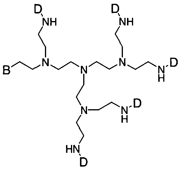
  • L may comprise a dendrimer.
  • dendrimer refers to a structure having multiple arms so that more than one dye molecule may be attached to a single spacer group. An example of a biomolecule of the invention comprising such a dendrimer structure is shown below:
  • the term "photostable visible dye” refers to a chemical moiety that is visually detectable, as defined hereinabove, and is not significantly altered or decomposed upon exposure to light.
  • the photostable visible dye does not exhibit significant bleaching or decomposition after being exposed to light for at least one hour. More preferably, the visible dye is stable after exposure to light for at least 12 hours, still more preferably at least 24 hours, still yet more preferably at least one week, and most preferably at least one month.
  • Nonlimiting examples of photostable visible dyes suitable for use in the compounds and methods of the invention include azo dyes, thioindigo dyes, quinacridone pigments, dioxazine, phthalocyanine, perinone, diketopyrrolopyrrole, quinophthalone, and truarycarbonium.
  • the visually detectable biomolecule has the formula B-(-L-(P) ⁇ , wherein m and n are each an integer from one to about 5;
  • B is a biomolecule
  • L at each occurrence, is a spacer group comprising from one to about 10 linear atoms, where L is attached to B by means of an ester, amide, phosphate, phosphorothioate, phosphonate, thioester, or disulfide linkage, and where the remaining linear atoms in L are selected from the group consisting of carbon, oxygen, nitrogen, and sulfur; wherein the linear atoms in L can be optionally substituted and optionally can be included in a ring; and P, at each occurrence, is a radical of a perylene, anthracene, naphthalene, or pyrene derivative, wherein each P has one and only one linkage to a biomolecule.
  • P is an imide, bisimide or hydrazamimide derivative of perylene, anthracene, naphthalene, or pyrene.
  • P is a perylene imide, perylene bisimide, or perylene hydrazamimide, wherein L is preferably attached to the imide nitrogen.
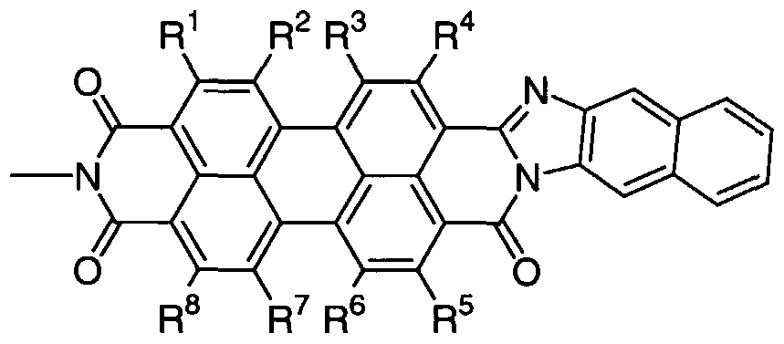
  • P has a formula selected from the group consisting of:
  • R 1 , R 2 , R 3 , R 4 , R 5 , R 6 , R 7 , R 8 , R 9 , and R 10 are radicals independently selected from the group consisting of hydrogen, halogen, cyano, hydroxy, alkoxy, amino, alkylamino, dialkylamino, heterocyclic radical, alkyl, and aryl, wherein the alkyl and aryl groups may be optionally substituted, and wherein adjacent radicals can form a carbocyclic or heterocyclic ring; p is 0, 1, or 2;
  • R 11 is C C 6 alkyl, C 6 -C 10 aryl, or (C 6 -C 10 )ar(C 1 -C 6 )alkyl;
  • R 12 and R 13 are independently C.-C 6 alkyl, C 6 -C 10 aryl, or (C 6 - C 10 )ar(C 1 -C 6 )alkyl;
  • the visually detectable biomolecule has a defined molecular weight, and is useful as a molecular weight standard.
  • the invention further provides a kit for determining the size of a test biomolecule, comprising a collection of two or more such visually detectable biomolecules of defined molecular weight. When subjected to electrophoresis, the biomolecule standards migrate to different positions according to molecular weight, thereby producing a visual biomolecule ladder.
  • Biomolecule ladders such as DNA ladders, are common reagents for determining the size of test biomolecules.
  • the invention provides a method for determining the size of a test biomolecule, comprising (a) subjecting a visually detectable biomolecule kit according to the invention to conditions under which the biomolecule standards migrate to different positions according to molecular weight, thereby producing a visual ladder of biomolecule standards; (b) subjecting the test biomolecule to the same conditions employed in step (a); and (c) comparing the migration of the test biomolecule to the visual ladder of biomolecule standards to determine the molecular weight of the test biomolecule.
  • any conditions suitable for separating molecules according to molecular weight may be used for practice of the method according to this aspect of the invention.
  • separation conditions include chromatographic and electrophoretic techniques.
  • a biomolecule test sample such as a DNA sample
  • a biomolecule ladder are loaded in adjacent wells on an electrophoresis gel, such as an agarose or polyacrylamide gel.
  • the sample is separated by electrophoresis through the gel, and the size of the test biomolecule is determined by comparing its migration with the bands of known size in the biomolecule ladder.
  • electrophoresis gel such as an agarose or polyacrylamide gel.
  • the size of the test biomolecule is determined by comparing its migration with the bands of known size in the biomolecule ladder.
  • the biomolecule ladders of the present invention are immediately visible to the naked eye, even while they are still running in the gel.
  • biomarkers can be utilized as purification tools, obviating the need for analytical or biochemical assaying of fractions.
  • biomarkers require additional steps for visualization or are unstable to light for the time periods necessary to effect separation.
  • the visually detectable biomolecules of the invention can be simply observed by visual inspection during analytical or preparative separations.
  • chromatographic methods are used to separate the biomolecules.
  • Such chromatographic methods may include, without limitation, paper chromatography, thin layer chromatography, including preparative thin layer chromatography, and column chromatography.
  • suitable chromatography conditions include normal phase and reverse phase silica gel chromatography, size exclusion chromatography, and ion exchange chromatography.
  • art-recognized electrophoretic methods are used to separate the biomolecules. It will be apparent to one skilled in the art that the chromatographic or electrophoretic conditions may be selected and modified to achieve optimum separation and /or purification of the biomolecule of interest.
  • the visually detectable biomolecules of the invention are also useful for a wide variety of biochemical and biomedical applications in which there is a need to determine the presence, location, or quantity of a particular biomolecule.
  • the invention provides a method for visually detecting a biomolecule, comprising: (a) providing a biological system with a visually detectable biomolecule of formula B-(-L-(D) ⁇ or B-(-L-(P) ⁇ , wherein B, L, D, P, m, and n are as described above for the first aspect of the invention; and (b) detecting the biomolecule by its visible properties.
  • the phrase "detecting the biomolecule by its visible properties” means that the biomolecule, without illumination or chemical or enzymatic activation, is observed with the naked eye, or with the aid of a optically based detection device, including, without limitation, absorption spectrophotometers, transmission light microscopes, digital cameras and scanners.
  • a densitometer may be used to quantify the amount of visually detectable biomolecule present.
  • the relative quantity of the biomolecule in two samples can be determined by measuring relative optical density. If the stoichiometry of dye molecules per biomolecule is known, and the extinction coefficient of the dye molecule is known, then the absolute concentration of the biomolecule can also be determined from a measurement of optical density.
  • biological system is used to refer to any solution or mixture comprising one or more biomolecules in addition to the visually detectable biomolecule.
  • biological systems include cells, cell extracts, tissue samples, electrophoretic gels, assay mixtures, and hybridization reaction mixtures.
  • the methods of the invention are useful for microarray and high- throughput screening applications.
  • Microarray technologies have successfully allowed for the rapid study of vast numbers of genes, and have become an indispensable tool for molecular biologists studying the genome.
  • MacBeath G. et al, Science 289: 1760 (2000) describes protein microarrays, wherein proteins are arrayed on a glass surface.
  • Nyquist R.M. et al, Langmuir, 16: 1793 (2000) describes efforts to apply this technology to carbohydrates.
  • the visually detectable biomolecules of the invention permit rapid and inexpensive detection of binding in microarrays, including protein, nucleic acid, carbohydrate, and glycoprotein microarrays.
  • the visually detectable biomolecule B-(-L-(D) ⁇ or B-(-L-(P) ⁇ of the invention is used in a two-step detection process.
  • the visually detectable biomolecule is used as a probe to detect a second component in the biological system, to which it specifically binds.
  • the biological system is contacted with the visually detectable biomolecule to permit binding of the biomolecule to the second component.
  • the mixture is preferably washed to remove visually detectable biomolecules that are nonspecifically bound.
  • the intensity of color is then indicative of the amount of the second component present in the biological system.
  • B is streptavidin
  • the visually detectable biomolecule is used as a probe to detect a biotin- labeled component in the biological system.
  • a streptavidin-labeled component in the biological system can be detected with a visually detectable biomolecule in which B is biotin.
  • B is an antibody, preferably a monoclonal antibody, and the visually detectable biomolecule can be used to detect an antigen for which the antibody is specific.
  • the visually detectable antibody can be used in biological assays, such as sandwich assays.
  • the visually detectable antibody can also be used to detect cell surface antigens, and can be used for cell sorting or for cell staining in tissue samples.
  • the method can also be used to determine the density of cell surface antigens, such as cell surface receptors.
  • the method employs a plurality of visually detectable biomolecules of the invention, each B being specific for a different component in the biological system.
  • each D exhibits different visible properties, e.g., absorption spectrum.
  • each of the plurality of visually detectable biomolecules has a different color.
  • B is a nucleic acid
  • the visually detectable biomolecule is used as a probe to detect a nucleic acid component in the biological system having a complementary sequence.
  • the biological system is preferably contacted with the visually detectable nucleic acid probe according to any of the standard hybridization conditions known in the art. Both the probe and the target nucleic acid may comprise RNA, DNA, modified nucleic acids, or combinations thereof.
  • the visually detectable biomolecules of the invention may be prepared by any suitable method.
  • the biomolecule is contacted with a reactive dye reagent having a reactive group that enables attachment to an amino, hydroxy, carboxyl, or sulfhydryl group on a biomolecule.
  • the invention provides reactive dyes for use in preparing the visually detectable biomolecules of the invention.
  • the reactive dye has the formula (D) n -L-X, wherein
  • D is a radical of a photostable visible dye
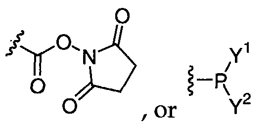
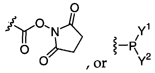
  • L is a spacer group comprising from one to about 10 linear atoms selected from the group consisting of carbon, oxygen, nitrogen, and sulfur; wherein the linear atoms in L can be optionally substituted and optionally can be included in a ring; and X is
  • Y 1 and Y 2 are independently dialkylamino, N-heterocyclic radical, or OZ, where Z is a protecting group.
  • X is an electrophilic moiety that enables attachment to a nucleophilic amino, hydroxy, or sulfhydryl group on the biomolecule.
  • the reactive group X preferably has the formula -P(Y 1 )(Y 2 ), which enables attachment to a hydroxy group on the nucleic acid.
  • Y 1 is OZ, where Z is preferably selected from the group consisting of alkyl, allyl, aryl, or cyanoalkyl, and Y 2 is dialkylamino. Most preferably, Y 1 is cyanoethyl and Y 2 is diisopropylamino.
  • standard phosphoramidite chemistry is employed to attach the dye to the biomolecule.
  • X is preferably an isocyanate, isothiocyanate, dichlorotriazine, or activated ester, such as an N-hydroxysuccinimide ester.
  • X is preferably an activated disulfide moiety, such as a pyridyldisulfide.
  • X is a nucleophilic moiety that enables attachment to a carboxyl group on the biomolecule.
  • X is preferably hydroxy or amino.
  • Reaction of X with a carboxyl group on the biomolecule can be effected by treatment with a coupling reagent, such as ethylcarbodiimide hydrochloride (EDC • HC1).
  • EDC • HC1 ethylcarbodiimide hydrochloride
  • the carboxyl group can be activated prior to reaction with X, for example by conversion to an activated ester moiety such as an N-hydroxysuccinimide ester.
  • the reactive dye has the formula (P) n -L-X, wherein P is a radical of a perylene, anthracene, naphthalene, or pyrene derivative;
  • L is a spacer group comprising from one to about 10 linear atoms selected from the group consisting of carbon, oxygen, nitrogen, and sulfur; wherein the linear atoms in L can be optionally substituted and optionally can be included in a ring; and
  • Y 1 and Y 2 are independently dialkylamino, N-heterocyclic radical, or OZ, where Z is a protecting group.
  • the reactive dye has the formula (P),-L-X, wherein
  • P is a radical of a perylene derivative having a formula selected from the group consisting of:
  • R 1 , R 2 , R 3 , R 4 , R 5 , R 6 , R 7 , R 8 , R 9 , and R 10 are radicals independently selected from the group consisting of hydrogen, halogen, cyano, hydroxy, alkoxy, amino, alkylamino, dialkylamino, heterocyclic radical, alkyl, and aryl, wherein the alkyl and aryl groups may be optionally substituted, and wherein adjacent radicals can form a carbocyclic or heterocyclic ring; p is 0, 1, or 2; R 11 is C C 6 alkyl, C 6 -C 10 aryl, or (C 6 -C 10 )ar(C C 6 )alkyl;
  • R 12 and R 13 are independently C,-C 6 alkyl, C 6 -C 10 aryl, or (C 6 - C 10 )ar(C r C 6 )alkyl;
  • L is a spacer group comprising from one to about 10 linear atoms selected from the group consisting of carbon, oxygen, nitrogen, and sulfur; wherein the linear atoms in L can be optionally substituted and optionally can be included in a ring; and
  • X is a reactive group that enables attachment to an amino, hydroxy, carboxyl, or sulfhydryl group on a biomolecule.
  • the invention provides a method for visually detecting a biomolecule, comprising contacting a biomolecule with a reactive dye of formula (D) ⁇ -L-X or (P) ⁇ -L-X, as defined above, whereby a visually detectable biomolecule of formula B-(-L-(D) ⁇ or B-(-L-(P) m ) H is produced; and detecting the biomolecule by its visible properties.
  • alkyl refers to straight and branched chain aliphatic groups having from 1 to 12 carbon atoms, preferably 1-8 carbon atoms, and more preferably 1-6 carbon atoms, which may be optionally substituted with one, two or three substituents. Unless otherwise apparent from context, the term “alkyl” is meant to include saturated, unsaturated, and partially unsaturated aliphatic groups. Preferred saturated alkyl groups include, without limitation, methyl, ethyl, propyl, isopropyl, butyl, isobutyl, sec- butyl, tert-butyl, pentyl, and hexyl.
  • carbocyclic group as employed herein includes saturated and partially unsaturated cyclic hydrocarbon groups having 3 to 12 carbons, preferably 3 to 8 carbons, and more preferably 3 to 6 carbons, wherein the cycloalkyl group additionally may be optionally substituted.
  • Preferred carbocyclic groups include, without limitation, cyclopropyl, cyclobutyl, cyclopentyl, cyclopentenyl, cyclohexyl, cyclohexenyl, cycloheptyl, and cyclooctyl.
  • aryl is a C 6 -C, 4 aromatic moiety comprising one to three aromatic rings, which may be optionally substituted.
  • the aryl group is a C 6 -C 10 aryl group.
  • Preferred aryl groups include, without limitation, phenyl, naphthyl, anthracenyl, and fluorenyl.
  • An “aralkyl” or “arylalkyl” group comprises an aryl group covalently linked to an alkyl group, either of which may independently be optionally substituted or unsubstituted.
  • the aralkyl group is (C 1 -C 6 )alk(C 6 -C 10 )aryl, including, without limitation, benzyl, phenethyl, and naphthylmethyl.
  • An "alkaryl” or “alkylaryl” group is an aryl group having one or more alkyl substituents. Examples of alkaryl groups include, without limitation, tolyl, xylyl, mesityl, ethylphenyl, tert-butylphenyl, and methylnaphthyl.
  • a “heterocyclic” group is a ring structure having from about 3 to about 8 atoms, wherein one or more atoms are selected from the group consisting of N, O, and S.
  • the heterocyclic group may be optionally substituted on carbon at one or more positions.
  • the heterocyclic group may also independently be substituted on nitrogen with alkyl, aryl, aralkyl, alkylcarbonyl, alkylsulfonyl, arylcarbonyl, arylsulfonyl, alkoxycarbonyl, aralkoxycarbonyl, or on sulfur with oxo or lower alkyl.
  • heterocyclic groups include, without limitation, epoxy, aziridinyl, tetrahydrofuranyl, pyrrolidinyl, piperidinyl, piperazinyl, thiazolidinyl, oxazolidinyl, oxazolidinonyl, and morpholino.
  • the heterocyclic group is fused to an aryl or heteroaryl group. Examples of such fused heterocyles include, without limitation, tetrahydroquinoline and dihydrobenzofuran.
  • heteroaryl refers to groups having 5 to 14 ring atoms, preferably 5, 6, 9, or 10 ring atoms; having 6, 10, or 14 ⁇ electrons shared in a cyclic array; and having, in addition to carbon atoms, between one and about three heteroatoms selected from the group consisting of N, O, and S.
  • Preferred heteroaryl groups include, without limitation, thienyl, benzothienyl, furyl, benzofuryl, dibenzofuryl, pyrrolyl, imidazolyl, pyrazolyl, pyridyl, pyrazinyl, pyrimidinyl, indolyl, quinolyl, isoquinolyl, quinoxalinyl, tetrazolyl, oxazolyl, thiazolyl, and isoxazolyl.
  • a "substituted" alkyl, cycloalkyl, aryl, heteroaryl, or heterocyclic group is one having between one and about four, preferably between one and about three, more preferably one or two, non-hydrogen substituents.
  • Suitable substituents include, without limitation, halo, hydroxy, nitro, haloalkyl, alkyl, alkaryl, aryl, aralkyl, alkoxy, aryloxy, amino, acylamino, alkylcarbamoyl, arylcarbamoyl, aminoalkyl, alkoxycarbonyl, carboxy, hydroxyalkyl, alkanesulfonyl, arenesulfonyl, alkanesulfonamido, arenesulfonamido, aralkylsulfonamido, alkylcarbonyl, acyloxy, cyano, and ureido groups.
  • halogen or "halo" as employed herein refers to chlorine, bromine, fluorine, or iodine.
  • acyl refers to an alkylcarbonyl or arylcarbonyl substituent.
  • acylamino refers to an amide group attached at the nitrogen atom.
  • carbamoyl refers to an amide group attached at the carbonyl carbon atom.
  • the nitrogen atom of an acylamino or carbamoyl substituent may be additionally substituted.
  • sulfonamido refers to a sulfonamide substituent attached by either the sulfur or the nitrogen atom.
  • amino is meant to include NH,, alkylamino, arylamino, and cyclic amino groups.
  • ureido refers to a substituted or unsubstituted urea moiety.
  • a capping reagent such as methylamine or benzylamine (but not limited to these two) can be attached to the other end of the perylene moiety in the following manner:
  • 1,7-dibromo- 3.4,9, 10-Perylenetetracarboxylicdianhydride E
  • the starting material, l,7-dibromoperylene-3,4,9,10-tetracarboxylic dianhydride can be obtained by selective bromination of perylene-3,4,9,10- tetracarboxylic dianhydride in 100% by weight sulfuric acid (monohydrate), as described by Bohm A.et al, Patent No. DE19547210..
  • An expedient procedure comprises first stirring perylene-3,4,9,10-tetracarboxylic dianhydride in the sulfuric acid for 2-6 hours and then heating this mixture, after adding a halogenation catalyst such as iodine (preferably 30-40 mmol per mole of anhydride) to the reaction temperature (generally 80-90 °C). At this point, the bromine is added slowly dropwise (usually over 6-10 hours), preferably using 2- 2.5 mol of bromine (Br 2 ) per mole of anhydride. After cooling to room temperature and displacing the unreacted bromine by nitrogen, water is added, a little at a time, in order to reduce the concentration of sulfuric acid to about 85- 88% by weight.
  • a halogenation catalyst such as iodine (preferably 30-40 mmol per mole of anhydride)
  • solvents include toluene, m-cresol, N-methylpyrolidone.
  • a capping reagent such as methylamine or benzylamine (but not limited to these two) can be attached to the other end of the perylene moiety in the following manner: 1 ,7-dibromo-N- 2- ⁇ ( 2-OtButyldimethylsilylethoxykthyl] N , -2-methyl-3.4.9.10- Perylenetetracarboxylic diimide (G).
  • the reaction is stopped with 2-3 drops of methanol and the phosphitylated product is precipitated into cold hexane (0°C).
  • the product is filtered, washed with cold hexane and rinsed from the filter with dichloromethane.
  • the product is concentrated to yield 50-90 molar percent of Reactive Dye II.
  • the dye is prepared according to Langhals H. et al, Liebigs Ann., 481, (1995), and the phosphoramidite is prepared according to the previously listed methods.
  • Fully acetylated high-mannoside bearing a nonanoate spacer is prepared as described by Grice P. et al, Chem. Eur. J. 3: 431 (1997).
  • To a solution of the fully acetylated high-mannoside (1 mmol, 1 equiv) and Reactive Dye V (1 mmol, 1 equiv) in 3 mL diethyl ether is added DCC (1 mmol, 1 equiv) and a catalytic amount of DMAP.
  • the reaction mixture is stirred for lhr at room temperature and filtered. Concentration and purification by silica gel chromatography affords a fully protected neoglycoconjugate.
  • DNA sequences are assembled using standard phosphoramidite protocols. Oligonucleotide with the sequence 5'-GTAGGTAAG-3' were assembled on the synthesizer. The DMT is removed from the terminal 5' hydroxyl, and the synthesis is interrupted. The CPG containing the oligonucleotide is removed from the synthesizer, dried in vacuo and poured into a small flask. To the CPG is added 30 ⁇ mol of the perylene phosphoramidite Reactive Dye I dissolved in 0.50 mL of anhydrous dichloromethane and 0.50 mL of 0.5 M tetrazole in acetonitrile. The reaction is mixed in an argon atmosphere for 1 hour. The CPG is then filtered and rinsed with dichloromethane to remove unincorporated phosphoramidite.
  • the sequences are deprotected in concentrated ammonia at 55 °C overnight.
  • the oligonucleotides are purified by denaturing polyacrylamide gel electrophoresis using 20% polyacrylamide/1% bisacrylamide/7M urea gels in 89 mM tris-borate buffer containing 2mM Na2EDTA, pH 8.0 (IX TBE buffer). The bands are visualized by UV shadowing as well as by the naked eye, and are excised.
  • the pure oligonucleotides are electroeluted in 0.5X TBE buffer using a Schleicher and Schuell Elutrap. All oligonucleotides are desalted before use by running the solution through a G-25 TE column according to the manufacturers specifications (Roche).
  • Example 8 Incorporation of Reactive Dye IV into oligonucleotides at the 3' position
  • Example 10 Labeling streptavidin with Reactive Dye IV
  • Streptavidin (5 mg/ml) and biotinylated DNA probes are purchased from Roche.
  • a 10-mer oligonucleotide is labeled with a Biotin Chem-Link following the supplied instructions.
  • Conjugations of streptavidin and the Reactive Dye IV are performed by dissolving Reactive Dye IV in aqueous media at a concentration of 10 mg/ml and at varied dye-to-protein molar ratios.
  • a water-miscible organic cosolvent, such as DMSO may be added to enhance solubility of the reactive dye compound.
  • reaction mixtures are incubated for 90 min at room temperature, quenched with the addition of hydroxylamine (final concentration of 0.15 M at pH 8.0) and incubated for an additional 30 min.
  • the conjugates are purified from the unreacted dye by size-exclusion chromatography using Bio-Gel P-30 (BioRad).
  • the biomarker-streptavidin conjugate is incubated with the biotinylated oligonucleotides by adding 300 ⁇ l buffer (100 mM MES (Sigma), 1M NaCl (Sigma), 0.05% Tween 20 (Pierce)), 24.0 ⁇ l BSA 50 mg/ml, 6.0 ⁇ l biotinylated oligo 0.5 mg/ml, 6.0 ⁇ l of biomarker- streptavidin conjugate 1 mg/ml, and 264 ⁇ l of H z O. The reaction is incubated for 30 min, and 10 ⁇ l aliquots are removed and mixed with equal volume of a 50% sucrose loading dye.
  • the samples are loaded on a 4% to 20% TBE, the system is loaded with IX TBE buffer, and the gel is run at 150 volts for 1 hr.
  • the biomarker-streptavidin conjugated biotinylated oligonucleotide is visualized as a black band running in the gel and recorded by a digital camera and a computer scanner. The location of the oligonucleotide is confirmed by staining the gel with the fluorescent nucleic acid dye SYBR Green I (Molecular Probes).
  • Example 11 Staining and microscopic visualization of human lymphocytes with Reactive Dye IV conjugated to sheep antimouse IgG antibody
  • peripheral lymphocytes are treated at zero degrees for 30 minutes with mouse anti-Beta2-microglobulin (0.25 ⁇ g/10 6 cells).
  • the cells are washed twice with DMEM buffer and are then treated with Reactive Dye IV-labeled sheep anti-mouse-IgG antibody (1 ⁇ g per 10 6 cells). After a 30 minute incubation at 0 °C, the excess antibody is removed and the cells are again washed twice with DMEM buffer. Aliquots of the cells are fixed on slides for analysis by microscopy by a Zeiss inverted microscope with high Numerical Aperature objectives-60X and 100X.
  • the stained lymphocytes on the slide are visualized by an ICCD (intensified CCD) or EB CDD (Electron bombarded CCD) camera attached to an image digitizer and television monitor.
  • the cells stained by this method are visible under the microscope.
  • use of the primary mouse anti-Beta2 - microglobulin antibody is omitted but the staining and analysis are otherwise carried out as described above.
  • the control sample is not visible under the microscope, indicating that Reactive Dye IV-labeled sheep anti-mouse antibody does not give significant nonspecific binding to lymphocytes.
  • Oligodeoxynucleotides are synthesized on an Applied Biosystems 381A DNA Synthesizer. Protected nucleotide phosphoramidites for the four DNA bases and four RNA bases are commonly available, e.g., from Glenn Research. Oligonucleotides of various sizes are synthesized and tagged with a Reactive Dye phosphoramidite, as described in Example 7. The dye-labeled oligonucleotides are then purified by chromatographic or electrophoretic methods, and desalted by standard methods.
  • DNA or RNA sequences of varying sizes, each having a different color dye attached, are synthesized as described above.
  • the labeled oligonucleotides are then mixed to give a ladder useful for tracking the sizes of unknown single stranded oligonucleotides.
  • sequences can be hybridized with their complementary sequences, combined and used as a ladder for measuring the migration of double stranded samples.
  • DNA and RNA duplexes are hybridized by heating the complementary strands in solution for 5 min at 80 °C and then letting the mixture cool for 14-20 h.
  • DNA binding protein for DNA oligonucleotide modified with a DNA damaging agent DNA binding protein for DNA oligonucleotide modified with a DNA damaging agent
  • DNA is labeled with Reactive Dye I, hybridized to its complementary sequence, and modified with cz ' s-diaminedichloroplatinum(II) (cisplatin).
  • Binding reactions are carried out in 15 ⁇ l reactions containing 20 mM Tris base, 5 mM MgCl 2 , 2.5 mM CaCl 2 , 0.1 mM DTT, 0.01 mM EDTA, and 50 ng of nonspecific chicken erythrocyte competitor DNA. Binding is performed at 30 min on ice. Samples are then loaded onto 4% (29:1 acrylamide:bis) native polyacrylamide gels containing 1 x TAE (90 mM Tris base (pH 8.0), 2.0 mM EDTA, 90 mM boric acid) and 5% sucrose, and separated by electrophoresis at room temperature in lx TAE at ⁇ 25 mA (140V) for 2 h.
  • 1 x TAE 90 mM Tris base (pH 8.0), 2.0 mM EDTA, 90 mM boric acid
  • Example 14 Applications of perylene Reactive Dyes in microarrays DNA /RNA Micro-array
  • RNA oligonucleotides are synthesized and biotinylated as described.
  • An array is built of complementary and non- complementary sequences on a glass slide as directed on P. Brown's web page (http://cmam.stanford.edu/pbrown/ .
  • the probes are hybridized and then stained in a buffer containing red biomarker-streptavidin conjugate as described above (Example 9).
  • the hybridization results yield a red positive pattern of hybridization that is visualized by an ICCD (intensified CCD) or EB CDD (Electron bombarded CCD) camera attached to an image digitizer.
  • ICCD intensified CCD
  • EB CDD Electrode bombarded CCD
  • the labeled protein is diluted into PBS, pH 7.5 supplemented with 0.1% Tween- 20 (v/v) and 1% BSA (w/v), to a concentration of 0.5 mg/ml.
  • To the slide is applied 0.55 ml of protein solution, using a PC500 CoverWell incubation chamber (Grace Biolabs). Following a 1-hour incubation at room temperature, the slides are rinsed with PBS and then washed 3 times for 3 min each with PBST (PBS supplemented with 0.1% Tween-20). The slides are rinsed twice with PBS and centrifuged at 200g for 1 min to remove excess buffer.
  • the slides are visualized by an ICCD (intensified CCD) or EB CDD (Electron bombarded CCD) camera attached to an image digitizer.
  • ICCD intensified CCD
  • EB CDD Electrode bombarded CCD

Landscapes

  • Health & Medical Sciences (AREA)
  • Life Sciences & Earth Sciences (AREA)
  • Immunology (AREA)
  • Engineering & Computer Science (AREA)
  • Chemical & Material Sciences (AREA)
  • Molecular Biology (AREA)
  • Biomedical Technology (AREA)
  • Hematology (AREA)
  • Urology & Nephrology (AREA)
  • Biotechnology (AREA)
  • Biochemistry (AREA)
  • Cell Biology (AREA)
  • Food Science & Technology (AREA)
  • Medicinal Chemistry (AREA)
  • Physics & Mathematics (AREA)
  • Analytical Chemistry (AREA)
  • Microbiology (AREA)
  • General Health & Medical Sciences (AREA)
  • General Physics & Mathematics (AREA)
  • Pathology (AREA)
  • Organic Chemistry (AREA)
  • Measuring Or Testing Involving Enzymes Or Micro-Organisms (AREA)
  • Investigating Or Analysing Materials By The Use Of Chemical Reactions (AREA)

Abstract

This invention relates to the detection of biomolecules. In particular, the invention relates to biomarkers for the labeling, visually detection and quantification of biomolecules. The invention provides visually detectable biomolecules and reagents for their preparation, as well as methods for visually detecting a biomolecule and for determining the site of a biomolecule. The labeled biomolecules of the invention are intensely colored and can be readily observed by visual inspection, without prior illumination or chemical or enzymatic activation.

Description

BIOMARKERS FOR THE LABELING, VISUAL DETECTION AND QUANTIFICATION OF BIOMOLECULES
(Attorney Docket No. 111543.120)
BACKGROUND OF THE INVENTION
Field of the Invention
This invention relates to the detection of biomolecules. In particular, the invention relates to biomarkers for the labeling, visual detection and quantification of biomolecules. The invention provides visually detectable biomolecules and reagents for their preparation, as well as methods for visually detecting a biomolecule and for determining the size of a biomolecule.
Background of the Invention
Methods for detecting biomolecules typically rely upon the use of fluorescent, chemiluminescent, or radioactive biomarkers. Each of these classes of biomarkers suffers limitations, however. For example, the use of radioactive labels requires careful attention to safety and regulatory protocols, and disposal of the resultant radioactive waste is both costly and potentially hazardous.
Fluorescent dyes have been widely used as an alternative to radioactive biomarkers. However, background fluorescence from components present in a test sample may interfere with an accurate determination of the fluorescence of the fluorescent label. Furthermore, the label itself may be photolabile and exhibit bleaching when subjected to the fluorescence detection conditions.
Tizard R. et al, Proc. Natl. Acad. Sci. USA, 87: 4514-4518 (1990) describes the use of 1,2-dioxetanes as chemiluminescent probes. Upon chemical or enzymatic activation, the dioxetane decomposes, with the emission of light.
These biomarkers suffer the disadvantage of being consumed when subjected to the detection protocol. Perylenes and related dyes have high photochemical persistency (chemical, thermal, and photochemical stability) and high fluorescence quanta yield and are used in a variety of reprographic processes, solar cells, photovoltaic devices, and dye lasers. Bair, U.S. Patent No. 4,719,236, teaches perylene derivatives useful as biocidal agents, particularly antitumor agents. However, perylene derivatives have been used primarily as pigments and fluorescent dyes. Mullen et al, U.S. Patent No. 5,986,099 and U.S. Patent No. 6,124,458, describe the synthesis of substituted quaterrylene tetracarboxylic acid diimides and their use as pigments or fluorescent dyes. Langhals and Jona, U.S. Patent No. 6,166,210, describes perylene imide monocarboxylic acids for use as colorants. Langhals and Ismael, U.S. Patent No. 6,143,890, teaches that perylenehydrazamides absorb at longer wavelength and exhibit increased lightfastness as compared to perylenebisimides. Langhals and Gold, U.S. Patent No. 5,929,239 describes bifluorophoric perylene colorants. Hao et al, U.S. Patent Nos. 5,874,580, 5,886,160, 6,013776, 6,013,777, and 6,127,549 describe carbamate derivatives of various chromophores, including perylene.
Perylene dyes of various colors and light-absorbing properties have been reported. For example, Becker S. et al, Chem. Eur. }., 6, 21, 3984,( 2000) report the synthesis of thermotropic perylenedicarboximide chromophores that show a color change from blue to orange. Holtrup F.O. et al, Tetrahedron, 53, 20, 6847, (1997) report the synthesis of purple benzoylperyleneimides that exhibit high thermal and photochemical stability. Langhals H. and Jona W., Angew. Chem. Int. Ed., 37, 7,952 (1998)show the synthesis of bi- and trichromophoric perylene- 3, 4:9,10-bis(dicarboximide)s with increased fluorescent properties. Zhao Y. and Wasielewski M.R., Tetrahedron Letters, 40, 7047, (1999), report the synthesis of a dialkylamino modified perylene dye that affords green products that exhibit intense optical absorption bands at 700 nm wavelength.
Unlike a variety of common fluorophores, however, the perylene chromophore has rarely been used as a biomolecular probe, apparently due to the strongly hydrophobic character of the molecule and difficulties with regiospecific labeling of biomolecules. Balakin K.V. et al, Nucleosides & Nucleotides, 18, 1279 (1999), report the synthesis of oligodeoxynucleotides bearing a 3'-terminal perylene-containing pseudonucleoside. The authors describe the use of these oligodeoxynucleotides as hybridization probes, which are detected by fluorescence anisotropy. Balakin K.V. et al Biosensors &
Bioelectronics 13, 771, (1998), describes the synthesis of a 13-mer containing a 5'- (3-perylene)acetic acid residue, which shows no response in the fluorescence spectrum upon hybridization to the complementary sequence. Bevers S.A. et al, JACS, 122(25); 11004, (1998), and JACS, 120(42); 5905, (2000) report the synthesis of naphthalene- and perylene-based linkers for the stabilization of hairpin triplexes, and for duplex and triplex stabilization. However, the use of perylene dyes for the visual detection of biomolecules has not been described.
There is thus a need in the art for biomarkers that permit visual detection of biomolecules without prior illumination or chemical or enzymatic activation. Ideally, such biomarkers should be intensely colored and should be available in a variety of colors.
BRIEF SUMMARY OF THE INVENTION
The present invention provides visually detectable biomolecules and reagents for their preparation, as well as methods for visually detecting a biomolecule and for determining the size of a biomolecule. The labeled biomolecules of the invention are intensely colored and can be readily observed by visual inspection, without prior illumination or chemical or enzymatic activation. By appropriate selection of the biomarker, as described herein, visually detectable biomolecules of a variety of colors may be obtained.
In a first aspect, therefore, the invention provides a visually detectable biomolecule of formula B-(-L-(D) π, m and n are each an integer from one to about 5;
B is a biomolecule;
L, at each occurrence, is a spacer group comprising from one to about 10 linear atoms, where L is attached to B by means of an ester, amide, phosphate, phosphorothioate, phosphonate, thioester, or disulfide linkage, and where the remaining linear atoms in L are selected from the group consisting of carbon, oxygen, nitrogen, and sulfur; wherein the linear atoms in L can be optionally substituted and optionally can be included in a ring; and
D, at each occurrence, is a radical of a photostable visible dye, wherein each D has one and only one linkage to a biomolecule, provided that D is not unsubstituted perylenyl.
In a preferred embodiment, the visually detectable biomolecule has the formula B-(-L-(P) π, wherein m and n are each an integer from one to about 5;
B is a biomolecule; L, at each occurrence, is a spacer group comprising from one to about 10 linear atoms, where L is attached to B by means of an ester, amide, phosphate, phosphorothioate, phosphonate, thioester, or disulfide linkage, and where the remaining linear atoms in L are selected from the group consisting of carbon, oxygen, nitrogen, and sulfur; wherein the linear atoms in L can be optionally substituted and optionally can be included in a ring; and
P, at each occurrence, is a radical of a perylene, anthracene, naphthalene, or pyrene derivative, wherein each P has one and only one linkage to a biomolecule.
In another preferred embodiment according to this aspect of the invention, the visually detectable biomolecule has a defined molecular weight, and is useful as a molecular weight standard. The invention further provides a kit for determining the size of a test biomolecule, comprising a collection of two or more such visually detectable biomolecules of defined molecular weight.
In another aspect, therefore, the invention provides a method for determining the size of a test biomolecule, comprising (a) subjecting a visually detectable biomolecule kit according to the invention to conditions under which the biomolecule standards migrate to different positions according to molecular weight, thereby producing a visual ladder of biomolecule standards; (b) subjecting the test biomolecule to the same conditions employed in step (a); and (c) comparing the migration of the test biomolecule to the visual ladder of biomolecule standards to determine the molecular weight of the test biomolecule.
In another aspect, the invention provides a method for visually detecting a biomolecule, comprising: (a) providing a biological system with a visually detectable biomolecule of formula B-(-L-(D) π or B-(-L-(P) π, wherein B, L, D, P, m, and n are as defined above for the first aspect of the invention; and (b) detecting the biomolecule by its visible properties.
In yet another aspect, the invention provides reactive dyes for use in preparing the visually detectable biomolecules of the invention. In one embodiment, the reactive dye has the formula (D)π-L-X, wherein
D is a radical of a photostable visible dye;
L is a spacer group comprising from one to about 10 linear atoms selected from the group consisting of carbon, oxygen, nitrogen, and sulfur; wherein the linear atoms in L can be optionally substituted and optionally can be included in a ring; and
X is
Figure imgf000007_0001
wherein Y1 and Y2 are independently dialkylamino, N-heterocyclic radical, or OZ, where Z is a protecting group.
In a preferred embodiment, the reactive dye has the formula (P)π-L-X, wherein P is a radical of a derivative of perylene, anthracene, naphthalene, or pyrene, and n, L, and X are as described above for the reactive dye of formula (D)„-L-X.
In another embodiment, the reactive dye has the formula(P)π-L-X, wherein
P is a radical of a perylene derivative having a formula selected from the group consisting of:
Figure imgf000008_0001
wherein
R1, R2, R3, R4, R5, R6, R7, R8, R9, and R10 are radicals independently selected from the group consisting of hydrogen, halogen, cyano, hydroxy, alkoxy, amino, alkylamino, dialkylamino, heterocyclic radical, alkyl, and aryl, wherein the alkyl and aryl groups may be optionally substituted, and wherein adjacent radicals can form a carbocyclic or heterocyclic ring; p is 0, 1, or 2;
R11 is CrC6 alkyl, C6-C10 aryl, or (C6-C10)ar(C C6)alkyl;
R12 and R13 are independently - alkyl, C6-C10 aryl, or (C6- C10)ar(C1-C6)alkyl;
L is a spacer group comprising from one to about 10 linear atoms selected from the group consisting of carbon, oxygen, nitrogen, and sulfur; wherein the linear atoms in L can be optionally substituted and optionally can be included in a ring; and
X is a reactive group that enables attachment to an amino, hydroxy, carboxyl, or sulfhydryl group on a biomolecule. In another aspect, the invention provides a method for visually detecting a biomolecule, comprising contacting a biomolecule with a reactive dye of formula (D)π-L-X, wherein
D is a radical of a photostable visible dye; L is a spacer group comprising from one to about 10 linear atoms selected from the group consisting of carbon, oxygen, nitrogen, and sulfur; wherein the linear atoms in L can be optionally substituted and optionally can be included in a ring; and X is a reactive group that enables attachment to an amino, hydroxy, carboxyl, or sulfhydryl group on a biomolecule; whereby a visually detectable biomolecule of formula B-(-L-(D) π is produced; and detecting the biomolecule by its visible properties.
In a preferred embodiment according to this aspect of the invention, the reactive dye has the formula (P)n-L-X, wherein
P is a radical of a perylene, anthracene, naphthalene, or pyrene derivative;
L is a spacer group comprising from one to about 10 linear atoms selected from the group consisting of carbon, oxygen, nitrogen, and sulfur; wherein the linear atoms in L can be optionally substituted and optionally can be included in a ring; and
X is a reactive group that enables attachment to an amino, hydroxy, carboxyl, or sulfhydryl group on a biomolecule. DETAILED DESCRIPTION OF THE PREFERRED EMBODIMENTS
The present invention provides methods for detecting biomolecules that are an alternative to currently employed methods that rely on fluorescent, chemiluminescent, or radioactive biomarkers. In particular, the invention provides visually detectable biomolecules and reagents for their preparation, as well as methods for visually detecting a biomolecule and for determining the size of a biomolecule.
The patent and scientific literature referred to herein establishes knowledge that is available to those with skill in the art. The issued patents, applications, and references that are cited herein are hereby incorporated by reference to the same extent as if each was specifically and individually indicated to be incorporated by reference. In the case of inconsistencies, the present disclosure will prevail.
In a first aspect, the invention provides a visually detectable biomolecule of formula B-(-L-(D) „, m and n are each an integer from one to about 5;
B is a biomolecule;
L, at each occurrence, is a spacer group comprising from one to about 10 linear atoms, where L is attached to B by means of an ester, amide, phosphate, phosphorothioate, phosphonate, thioester, or disulfide linkage, and where the remaining linear atoms in L are selected from the group consisting of carbon, oxygen, nitrogen, and sulfur; wherein the linear atoms in L can be optionally substituted and optionally can be included in a ring; and
D, at each occurrence, is a radical of a photostable visible dye, wherein each D has one and only one linkage to a biomolecule. For purposes of the present invention, the term "biomolecule" refers to any of a variety of biological materials, including nucleic acids, carbohydrates, amino acids, polypeptides, glycoproteins, hormones, and mixtures thereof. More specifically, the term is intended to include, without limitation, RNA, DNA, oligonucleotides, modified or derivatized nucleotides, enzymes, receptors, receptor ligands (including hormones), antibodies, antigens, and toxins, as well as bacteria, viruses, blood cells, and tissue cells. The visually detectable biomolecules of the invention are prepared, as further described herein, by contacting a biomolecule with a visible dye having a reactive group that enables attachment to an amino, hydroxy, carboxyl, or sulfhydryl group on the biomolecule.
The terms "visible" and "visually detectable" are used herein to refer to substances that are observable by visual inspection, without prior illumination, or chemical or enzymatic activation. Such visually detectable substances absorb and emit light in a region of the spectrum ranging from about 400 to about 800 nm. Preferably, such substances are intensely colored, preferably having a molar extinction coefficient of at least about 40,000, more preferably at least about 50,000, still more preferably at least about 60,000, yet still more preferably at least about 70,000, and most preferably at least about 80,000 M"1 cm"1. The biomolecules of the invention may be detected by observation with the naked eye, or with the aid of a optically based detection device, including, without limitation, absorption spectrophotometers, transmission light microscopes, digital cameras and scanners.
L is a spacer group between the biomolecule, B, and the visible dye, D. The structure of L is not critical, so long as it does not interfere with the function of B or prevent detection of the visible chromophore D. Preferably, L comprises from one to about 10 linear atoms, where L is attached to a ring atom in D and is attached to B by means of an ester amide, phosphate, phosphorothioate, phosphonate, thioester, or disulfide linkage. The remaining linear atoms in L are preferably selected from the group consisting of carbon, oxygen, nitrogen and sulfur, any of which atoms optionally may be included in a carbocyclic or heterocyclic ring. The linear carbon atoms in L optionally can be substituted with a substituent selected from the group consisting of halo, hydroxy, nitro, haloalkyl, alkyl, alkaryl, aryl, aralkyl, alkoxy, aryloxy, amino, acylamino, alkylcarbamoyl, arylcarbamoyl, aminoalkyl, alkoxycarbonyl, carboxy, hydroxyalkyl, alkanesulfonyl, arenesulfonyl, alkanesulfonamido, arenesulfonamido, aralkylsulfonamido, alkylcarbonyl, acyloxy, cyano, and ureido. A linear nitrogen atom in L optionally can be substituted with acyl, sulfonyl, alkyl, alkaryl, aryl, aralkyl, alkoxycarbonyl. A linear sulfur atom in L optionally can be oxidized.
In some embodiments, the biomolecule is preferably attached to more than one dye molecule in order to enhance the sensitivity of detection. However, each P has only one linkage to a biomolecule. To further enhance sensitivity, L may comprise a dendrimer. For purposes of the invention, the term "dendrimer" refers to a structure having multiple arms so that more than one dye molecule may be attached to a single spacer group. An example of a biomolecule of the invention comprising such a dendrimer structure is shown below:
Figure imgf000012_0001
For purposes of the invention, the term "photostable visible dye" refers to a chemical moiety that is visually detectable, as defined hereinabove, and is not significantly altered or decomposed upon exposure to light. Preferably, the photostable visible dye does not exhibit significant bleaching or decomposition after being exposed to light for at least one hour. More preferably, the visible dye is stable after exposure to light for at least 12 hours, still more preferably at least 24 hours, still yet more preferably at least one week, and most preferably at least one month. Nonlimiting examples of photostable visible dyes suitable for use in the compounds and methods of the invention include azo dyes, thioindigo dyes, quinacridone pigments, dioxazine, phthalocyanine, perinone, diketopyrrolopyrrole, quinophthalone, and truarycarbonium.
In a preferred embodiment, the visually detectable biomolecule has the formula B-(-L-(P) π, wherein m and n are each an integer from one to about 5;
B is a biomolecule;
L, at each occurrence, is a spacer group comprising from one to about 10 linear atoms, where L is attached to B by means of an ester, amide, phosphate, phosphorothioate, phosphonate, thioester, or disulfide linkage, and where the remaining linear atoms in L are selected from the group consisting of carbon, oxygen, nitrogen, and sulfur; wherein the linear atoms in L can be optionally substituted and optionally can be included in a ring; and P, at each occurrence, is a radical of a perylene, anthracene, naphthalene, or pyrene derivative, wherein each P has one and only one linkage to a biomolecule.
As used herein, the term "perylene derivative" is intended to include any substituted perylene that is visually detectable. However, the term is not intended to include perylene itself. The terms "anthracene derivative", "naphthalene derivative", and "pyrene derivative" are used analogously. In some preferred embodiments, P is an imide, bisimide or hydrazamimide derivative of perylene, anthracene, naphthalene, or pyrene. Preferably, P is a perylene imide, perylene bisimide, or perylene hydrazamimide, wherein L is preferably attached to the imide nitrogen. In certain preferred embodiments, P has a formula selected from the group consisting of:
Figure imgf000014_0001
Figure imgf000014_0002
wherein
R1, R2, R3, R4, R5, R6, R7, R8, R9, and R10 are radicals independently selected from the group consisting of hydrogen, halogen, cyano, hydroxy, alkoxy, amino, alkylamino, dialkylamino, heterocyclic radical, alkyl, and aryl, wherein the alkyl and aryl groups may be optionally substituted, and wherein adjacent radicals can form a carbocyclic or heterocyclic ring; p is 0, 1, or 2;
R11 is C C6 alkyl, C6-C10 aryl, or (C6-C10)ar(C1-C6)alkyl;
R12 and R13 are independently C.-C6 alkyl, C6-C10 aryl, or (C6- C10)ar(C1-C6)alkyl; In one preferred embodiment according to this aspect of the invention, the visually detectable biomolecule has a defined molecular weight, and is useful as a molecular weight standard. The invention further provides a kit for determining the size of a test biomolecule, comprising a collection of two or more such visually detectable biomolecules of defined molecular weight. When subjected to electrophoresis, the biomolecule standards migrate to different positions according to molecular weight, thereby producing a visual biomolecule ladder.
Biomolecule ladders, such as DNA ladders, are common reagents for determining the size of test biomolecules. Thus, in another aspect, the invention provides a method for determining the size of a test biomolecule, comprising (a) subjecting a visually detectable biomolecule kit according to the invention to conditions under which the biomolecule standards migrate to different positions according to molecular weight, thereby producing a visual ladder of biomolecule standards; (b) subjecting the test biomolecule to the same conditions employed in step (a); and (c) comparing the migration of the test biomolecule to the visual ladder of biomolecule standards to determine the molecular weight of the test biomolecule.
Any conditions suitable for separating molecules according to molecular weight may be used for practice of the method according to this aspect of the invention. Nonlimiting examples of such separation conditions include chromatographic and electrophoretic techniques. Typically, a biomolecule test sample, such as a DNA sample, and a biomolecule ladder are loaded in adjacent wells on an electrophoresis gel, such as an agarose or polyacrylamide gel. The sample is separated by electrophoresis through the gel, and the size of the test biomolecule is determined by comparing its migration with the bands of known size in the biomolecule ladder. Currently available ladders require additional steps for visualization. By contrast, the biomolecule ladders of the present invention are immediately visible to the naked eye, even while they are still running in the gel. More generally, visible biomarkers can be utilized as purification tools, obviating the need for analytical or biochemical assaying of fractions. Currently available biomarkers require additional steps for visualization or are unstable to light for the time periods necessary to effect separation. By contrast, the visually detectable biomolecules of the invention can be simply observed by visual inspection during analytical or preparative separations. In certain preferred embodiments, chromatographic methods are used to separate the biomolecules. Such chromatographic methods may include, without limitation, paper chromatography, thin layer chromatography, including preparative thin layer chromatography, and column chromatography. Nonlimiting examples of suitable chromatography conditions include normal phase and reverse phase silica gel chromatography, size exclusion chromatography, and ion exchange chromatography. In certain other preferred embodiments, art-recognized electrophoretic methods are used to separate the biomolecules. It will be apparent to one skilled in the art that the chromatographic or electrophoretic conditions may be selected and modified to achieve optimum separation and /or purification of the biomolecule of interest.
The visually detectable biomolecules of the invention are also useful for a wide variety of biochemical and biomedical applications in which there is a need to determine the presence, location, or quantity of a particular biomolecule. In another aspect, therefore, the invention provides a method for visually detecting a biomolecule, comprising: (a) providing a biological system with a visually detectable biomolecule of formula B-(-L-(D) π or B-(-L-(P) π, wherein B, L, D, P, m, and n are as described above for the first aspect of the invention; and (b) detecting the biomolecule by its visible properties.
For purposes of the invention, the phrase "detecting the biomolecule by its visible properties" means that the biomolecule, without illumination or chemical or enzymatic activation, is observed with the naked eye, or with the aid of a optically based detection device, including, without limitation, absorption spectrophotometers, transmission light microscopes, digital cameras and scanners. A densitometer may be used to quantify the amount of visually detectable biomolecule present. For example, the relative quantity of the biomolecule in two samples can be determined by measuring relative optical density. If the stoichiometry of dye molecules per biomolecule is known, and the extinction coefficient of the dye molecule is known, then the absolute concentration of the biomolecule can also be determined from a measurement of optical density.
As used herein, the term "biological system" is used to refer to any solution or mixture comprising one or more biomolecules in addition to the visually detectable biomolecule. Nonlimiting examples of such biological systems include cells, cell extracts, tissue samples, electrophoretic gels, assay mixtures, and hybridization reaction mixtures.
The methods of the invention are useful for microarray and high- throughput screening applications. Microarray technologies have successfully allowed for the rapid study of vast numbers of genes, and have become an indispensable tool for molecular biologists studying the genome. MacBeath G. et al, Science 289: 1760 (2000) describes protein microarrays, wherein proteins are arrayed on a glass surface. Nyquist R.M. et al, Langmuir, 16: 1793 (2000) describes efforts to apply this technology to carbohydrates. The visually detectable biomolecules of the invention permit rapid and inexpensive detection of binding in microarrays, including protein, nucleic acid, carbohydrate, and glycoprotein microarrays.
In some embodiments, the visually detectable biomolecule B-(-L-(D) π or B-(-L-(P) π of the invention is used in a two-step detection process. In these embodiments, the visually detectable biomolecule is used as a probe to detect a second component in the biological system, to which it specifically binds. Preferably, the biological system is contacted with the visually detectable biomolecule to permit binding of the biomolecule to the second component. The mixture is preferably washed to remove visually detectable biomolecules that are nonspecifically bound. The intensity of color is then indicative of the amount of the second component present in the biological system.
For example, in one particularly preferred embodiment, B is streptavidin, and the visually detectable biomolecule is used as a probe to detect a biotin- labeled component in the biological system. Likewise, a streptavidin-labeled component in the biological system can be detected with a visually detectable biomolecule in which B is biotin. It will thus be apparent to those of skill in the art that the visually detectable biomolecules of the invention are readily adapted for use in sandwich assays. In another particularly preferred embodiment, B is an antibody, preferably a monoclonal antibody, and the visually detectable biomolecule can be used to detect an antigen for which the antibody is specific. For example, the visually detectable antibody can be used in biological assays, such as sandwich assays. The visually detectable antibody can also be used to detect cell surface antigens, and can be used for cell sorting or for cell staining in tissue samples. The method can also be used to determine the density of cell surface antigens, such as cell surface receptors.
In certain preferred embodiments, the method employs a plurality of visually detectable biomolecules of the invention, each B being specific for a different component in the biological system. Preferably, each D exhibits different visible properties, e.g., absorption spectrum. More preferably, each of the plurality of visually detectable biomolecules has a different color. Thus, the method according to this embodiment permits multiple components in a biological system to be simultaneously visualized and distinguished. In another particularly preferred embodiment, B is a nucleic acid, and the visually detectable biomolecule is used as a probe to detect a nucleic acid component in the biological system having a complementary sequence. The biological system is preferably contacted with the visually detectable nucleic acid probe according to any of the standard hybridization conditions known in the art. Both the probe and the target nucleic acid may comprise RNA, DNA, modified nucleic acids, or combinations thereof.
The visually detectable biomolecules of the invention may be prepared by any suitable method. Typically, the biomolecule is contacted with a reactive dye reagent having a reactive group that enables attachment to an amino, hydroxy, carboxyl, or sulfhydryl group on a biomolecule.
In another aspect, therefore, the invention provides reactive dyes for use in preparing the visually detectable biomolecules of the invention. In one embodiment, the reactive dye has the formula (D)n-L-X, wherein
D is a radical of a photostable visible dye;
L is a spacer group comprising from one to about 10 linear atoms selected from the group consisting of carbon, oxygen, nitrogen, and sulfur; wherein the linear atoms in L can be optionally substituted and optionally can be included in a ring; and X is
Figure imgf000019_0001
wherein Y1 and Y2 are independently dialkylamino, N-heterocyclic radical, or OZ, where Z is a protecting group.
In some preferred embodiments, X is an electrophilic moiety that enables attachment to a nucleophilic amino, hydroxy, or sulfhydryl group on the biomolecule.
When the biomolecule to which the reactive dye is to be attached is a nucleic acid, the reactive group X preferably has the formula -P(Y1)(Y2), which enables attachment to a hydroxy group on the nucleic acid. Preferably, Y1 is OZ, where Z is preferably selected from the group consisting of alkyl, allyl, aryl, or cyanoalkyl, and Y2 is dialkylamino. Most preferably, Y1 is cyanoethyl and Y2 is diisopropylamino. In these embodiments, standard phosphoramidite chemistry is employed to attach the dye to the biomolecule. When the reactive dye is to be attached to an amino or hydroxy group of a biomolecule other than a nucleic acid, X is preferably an isocyanate, isothiocyanate, dichlorotriazine, or activated ester, such as an N-hydroxysuccinimide ester. When the reactive dye is to be attached to a sulfhydryl group on the biomolecule, X is preferably an activated disulfide moiety, such as a pyridyldisulfide.
In some other preferred embodiments, X is a nucleophilic moiety that enables attachment to a carboxyl group on the biomolecule. In these embodiments, X is preferably hydroxy or amino. Reaction of X with a carboxyl group on the biomolecule can be effected by treatment with a coupling reagent, such as ethylcarbodiimide hydrochloride (EDC • HC1). Alternatively, the carboxyl group can be activated prior to reaction with X, for example by conversion to an activated ester moiety such as an N-hydroxysuccinimide ester.
In a preferred embodiment, the reactive dye has the formula (P)n-L-X, wherein P is a radical of a perylene, anthracene, naphthalene, or pyrene derivative;
L is a spacer group comprising from one to about 10 linear atoms selected from the group consisting of carbon, oxygen, nitrogen, and sulfur; wherein the linear atoms in L can be optionally substituted and optionally can be included in a ring; and
X is
Figure imgf000021_0001
wherein Y1 and Y2 are independently dialkylamino, N-heterocyclic radical, or OZ, where Z is a protecting group.
In another embodiment, the reactive dye has the formula (P),-L-X, wherein
P is a radical of a perylene derivative having a formula selected from the group consisting of:
Figure imgf000021_0002
Figure imgf000021_0003
wherein
R1, R2, R3, R4, R5, R6, R7, R8, R9, and R10 are radicals independently selected from the group consisting of hydrogen, halogen, cyano, hydroxy, alkoxy, amino, alkylamino, dialkylamino, heterocyclic radical, alkyl, and aryl, wherein the alkyl and aryl groups may be optionally substituted, and wherein adjacent radicals can form a carbocyclic or heterocyclic ring; p is 0, 1, or 2; R11 is C C6 alkyl, C6-C10 aryl, or (C6-C10)ar(C C6)alkyl;
R12 and R13 are independently C,-C6 alkyl, C6-C10 aryl, or (C6- C10)ar(CrC6)alkyl;
L is a spacer group comprising from one to about 10 linear atoms selected from the group consisting of carbon, oxygen, nitrogen, and sulfur; wherein the linear atoms in L can be optionally substituted and optionally can be included in a ring; and
X is a reactive group that enables attachment to an amino, hydroxy, carboxyl, or sulfhydryl group on a biomolecule.
In yet another aspect, the invention provides a method for visually detecting a biomolecule, comprising contacting a biomolecule with a reactive dye of formula (D)π-L-X or (P)π-L-X, as defined above, whereby a visually detectable biomolecule of formula B-(-L-(D) π or B-(-L-(P)m)H is produced; and detecting the biomolecule by its visible properties.
Definitions
The term "alkyl" as employed herein refers to straight and branched chain aliphatic groups having from 1 to 12 carbon atoms, preferably 1-8 carbon atoms, and more preferably 1-6 carbon atoms, which may be optionally substituted with one, two or three substituents. Unless otherwise apparent from context, the term "alkyl" is meant to include saturated, unsaturated, and partially unsaturated aliphatic groups. Preferred saturated alkyl groups include, without limitation, methyl, ethyl, propyl, isopropyl, butyl, isobutyl, sec- butyl, tert-butyl, pentyl, and hexyl. The term "carbocyclic group" as employed herein includes saturated and partially unsaturated cyclic hydrocarbon groups having 3 to 12 carbons, preferably 3 to 8 carbons, and more preferably 3 to 6 carbons, wherein the cycloalkyl group additionally may be optionally substituted. Preferred carbocyclic groups include, without limitation, cyclopropyl, cyclobutyl, cyclopentyl, cyclopentenyl, cyclohexyl, cyclohexenyl, cycloheptyl, and cyclooctyl.
An "aryl" group is a C6-C,4 aromatic moiety comprising one to three aromatic rings, which may be optionally substituted. Preferably, the aryl group is a C6-C10 aryl group. Preferred aryl groups include, without limitation, phenyl, naphthyl, anthracenyl, and fluorenyl. An "aralkyl" or "arylalkyl" group comprises an aryl group covalently linked to an alkyl group, either of which may independently be optionally substituted or unsubstituted. Preferably, the aralkyl group is (C1-C6)alk(C6-C10)aryl, including, without limitation, benzyl, phenethyl, and naphthylmethyl. An "alkaryl" or "alkylaryl" group is an aryl group having one or more alkyl substituents. Examples of alkaryl groups include, without limitation, tolyl, xylyl, mesityl, ethylphenyl, tert-butylphenyl, and methylnaphthyl. A "heterocyclic" group is a ring structure having from about 3 to about 8 atoms, wherein one or more atoms are selected from the group consisting of N, O, and S. The heterocyclic group may be optionally substituted on carbon at one or more positions. The heterocyclic group may also independently be substituted on nitrogen with alkyl, aryl, aralkyl, alkylcarbonyl, alkylsulfonyl, arylcarbonyl, arylsulfonyl, alkoxycarbonyl, aralkoxycarbonyl, or on sulfur with oxo or lower alkyl. Preferred heterocyclic groups include, without limitation, epoxy, aziridinyl, tetrahydrofuranyl, pyrrolidinyl, piperidinyl, piperazinyl, thiazolidinyl, oxazolidinyl, oxazolidinonyl, and morpholino. In certain preferred embodiments, the heterocyclic group is fused to an aryl or heteroaryl group. Examples of such fused heterocyles include, without limitation, tetrahydroquinoline and dihydrobenzofuran.
As used herein, the term "heteroaryl" refers to groups having 5 to 14 ring atoms, preferably 5, 6, 9, or 10 ring atoms; having 6, 10, or 14 π electrons shared in a cyclic array; and having, in addition to carbon atoms, between one and about three heteroatoms selected from the group consisting of N, O, and S. Preferred heteroaryl groups include, without limitation, thienyl, benzothienyl, furyl, benzofuryl, dibenzofuryl, pyrrolyl, imidazolyl, pyrazolyl, pyridyl, pyrazinyl, pyrimidinyl, indolyl, quinolyl, isoquinolyl, quinoxalinyl, tetrazolyl, oxazolyl, thiazolyl, and isoxazolyl. As employed herein, a "substituted" alkyl, cycloalkyl, aryl, heteroaryl, or heterocyclic group is one having between one and about four, preferably between one and about three, more preferably one or two, non-hydrogen substituents. Suitable substituents include, without limitation, halo, hydroxy, nitro, haloalkyl, alkyl, alkaryl, aryl, aralkyl, alkoxy, aryloxy, amino, acylamino, alkylcarbamoyl, arylcarbamoyl, aminoalkyl, alkoxycarbonyl, carboxy, hydroxyalkyl, alkanesulfonyl, arenesulfonyl, alkanesulfonamido, arenesulfonamido, aralkylsulfonamido, alkylcarbonyl, acyloxy, cyano, and ureido groups.
The term "halogen" or "halo" as employed herein refers to chlorine, bromine, fluorine, or iodine.
As herein employed, the term "acyl" refers to an alkylcarbonyl or arylcarbonyl substituent.
The term "acylamino" refers to an amide group attached at the nitrogen atom. The term "carbamoyl" refers to an amide group attached at the carbonyl carbon atom. The nitrogen atom of an acylamino or carbamoyl substituent may be additionally substituted. The term "sulfonamido" refers to a sulfonamide substituent attached by either the sulfur or the nitrogen atom. The term "amino" is meant to include NH,, alkylamino, arylamino, and cyclic amino groups.
The term "ureido" as employed herein refers to a substituted or unsubstituted urea moiety.
EXAMPLES
Example 1: Synthesis of Reactive Dye I:
N-2-{[2-O-(2-cyanoethyl diisopropylchlorophosphino)- ethoxy] ethyl} N'-2-methyl-3,4,9/10/-Perylenetetracarboxylic Diimide
Figure imgf000025_0001
2-(2-Q-tButyldimethylsilylethoxy)ethylamine (A). To 5.0 g (48 mmol) of 2-(2-aminoethoxy)ethanol dissolved in anhydrous pyridine (50 mL) was added 7.2 g (48 mmol) tert-butyldimethylsilyl chloride and 4.9 g (72 mmol) of imidazole. The reaction mixture was stirred overnight, until TLC analysis indicated the absence of starting material and the formation of new products. Pyridine was removed in vacuo. The mixture was dissolved in dichloromethane (50 mL) and was washed with an aqueous solution of saturated sodium bicarbonate (2 x 50 mL). The organic layer was dried over anhydrous sodium sulfate. The mixture was then vacuum filtered, the filter was washed with dichloromethane, and the product was purified by flash chromatography to yield (A). N-2-r(2-OtButyldimethylsilylethoxy)ethynperylene-3.4:9.10-tetracarboxylic-3.4- anhydride-9,10-carboximide (B)
To 2.0 g (5.1 mmol) perylenetetracarboxylic dianhydride suspended in anhydrous pyridine or quinoline (25 mL) was added 1.16 g (5.1 mmol) 2-(2-0- t-butyldimethylsilylethoxy)ethylamine (A) and 1.1 g (5.1 mmol) zinc acetate dihydrate. The reaction mixture was refluxed overnight, until TLC analysis indicated the absence of starting material and the formation of products. Pyridine was removed in vacuo. The mixture was suspended in chloroform and loaded onto a large silica column. The product was purified by flash chromatography to give (B) as a dark red solid (50-90% yield). Other possible solvents include toluene, m-cresol, and N-methylpyrolidone. A capping reagent such as methylamine or benzylamine (but not limited to these two) can be attached to the other end of the perylene moiety in the following manner:
N- 2-r(2-OtButyldimethylsilylethoxy)ethyn N,-2-methyl-3.4.9.10-perylene- tetracarboxylic diimide (Q. To 2.0 g (5.1 mmol) N-2-[(2-OtButyldimethylsilylethoxy)ethyl]perylene-
3,4:9,10-tetracarboxylic-3,4- anhydride-9,10-carboximide (B) suspended in anhydrous pyridine or quinoline (25 mL) is added 5.1 mmol of methylamine and 5.1 mmol of zinc acetate dihydrate. The reaction mixture is refluxed overnight, until TLC analysis indicates the absence of starting material and the formation of products. Pyridine is removed in vacuo. The mixture is suspended in chloroform, loaded onto a large silica column, and purified by flash chromatography to give (C) as a dark red solid (50-90% yield).
N-2-f(2-hydroxyethoxy')ethyl1-N'-2-methyl-3,4,9.10,-perylenetetracarboxylic diimide (D). To 4.2 mmol of N- 2-[(2-OtButyldimethylsilylethoxy)ethyl] N'-2-methyl-
3,4,9,10-perylenetetracarboxylic diimide (C) dissolved in anhydrous pyridine (50 mL) is added HF/Pyridine (70%) until the starting material is consumed, as monitored by TLC analysis. Pyridine is removed in vacuo. The mixture is resuspended in chloroform and purified by flash chromatography to yield 50- 90% of compound (D).
N-2-([2-0-(2-cyanoethyl diisopropylchlorophosphino)ethoxy1ethyl} N'-2- methyl-3,4.9.10,-perylenetetracarboxylic diimide (Reactive Dye I).
To 0.055 mmol of N-2-[(2-hydroxyethoxy)ethyl]-N'-2-methyl-3,4,9,10,- perylenetetracarboxylic diimide (D) dissolved in anhydrous dichloromethane (1 mL) at 0 °C is added 0.048 mL (0.28 mmol) diisopropylethylamine followed by 0.025 mL (0.11 mmol) 2-cyanoethyl diisopropylchlorophosphoramidite. The reaction is slowly brought to 25 °C and stirred for 30 minutes. When TLC analysis indicates the absence of starting material and the presence of a new product, the reaction is stopped with 2-3 drops of methanol, and the phosphitylated product is precipitated into cold hexane (0 °C). The product is filtered, washed with cold hexane and rinsed from the filter with dichloromethane. The product is concentrated to yield 50-90 molar percent of Reactive Dye I as a red solid.
Example 2: Synthesis of Reactive Dye II: l,7-dipyrolo-N-2-{[2-O-(2- cyanoethyl diisop_opylchlorophosphino)ethoxy]ethyl} N'-2- methyl-S^^lO -perylenetetracarboxylic diimide
Figure imgf000027_0001
2-(2-Q-tButyldimethylsilylethoxy)ethylamine (A).
To 5.0 g (48 mmol) of 2-(2-aminoethoxy)ethanol dissolved in anhydrous pyridine (50 mL) was added 7.2 g (48 mmol) fert-butyldimethylsilyl chloride and 4.9 g (72 mmol) of imidazole. The reaction mixture was stirred overnight, until TLC analysis indicated the absence of starting material and formation of new products. Pyridine was removed in vacuo. The mixture was dissolved in dichloromethane (50 mL), washed with an aqueous solution of saturated sodium bicarbonate (2 x 50 mL), and dried over anhydrous sodium sulfate. The mixture was then vacuum filtered and the filter was washed with dichloromethane. The product was purified by flash chromatography to yield 3.5 g (16 mmol) of (A) as brown oil (36% yield).
1,7-dibromo- 3.4,9, 10-Perylenetetracarboxylicdianhydride (E). The starting material, l,7-dibromoperylene-3,4,9,10-tetracarboxylic dianhydride, can be obtained by selective bromination of perylene-3,4,9,10- tetracarboxylic dianhydride in 100% by weight sulfuric acid (monohydrate), as described by Bohm A.et al, Patent No. DE19547210.. An expedient procedure comprises first stirring perylene-3,4,9,10-tetracarboxylic dianhydride in the sulfuric acid for 2-6 hours and then heating this mixture, after adding a halogenation catalyst such as iodine (preferably 30-40 mmol per mole of anhydride) to the reaction temperature (generally 80-90 °C). At this point, the bromine is added slowly dropwise (usually over 6-10 hours), preferably using 2- 2.5 mol of bromine (Br2) per mole of anhydride. After cooling to room temperature and displacing the unreacted bromine by nitrogen, water is added, a little at a time, in order to reduce the concentration of sulfuric acid to about 85- 88% by weight. Working up the reaction mixture to the 1,7-dibromoperylene- 3,4,9,10-tetracarboxylic dianhydride (E) can be carried out by filtering off the precipitated product, washing it with 85-88% by weight sulfuric acid, stirring it into water, filtering the mixture again, washing the product with water and then drying it. l,7-dibromo- N-2-r(2-OtButyldimethylsilylethoxy)ethyl1perylene-3,4:9,10- tetracarboxylic-3,4- anhydride-9,10-carboximide (F).
To 5.1 mmol of 1,7-dibromo-perelynetetracarboxylicdianhydride (E) suspended in anhydrous pyridine or quinoline (25 mL) is added 5.1 mmol of 2- (2-0-t-butyldimethylsilylethoxy)ethylamine (A) and 5.1 mmol zinc acetate dihydrate. The reaction mixture is refluxed overnight, until TLC analysis indicates the absence of starting material and the formation of products. Pyridine is removed in vacuo. The mixture is suspended in chloroform, loaded onto a large silica column, and purified by flash chromatography to give (E) as a dark red solid (50-90% yield). Other possible solvents include toluene, m-cresol, N-methylpyrolidone. A capping reagent such as methylamine or benzylamine (but not limited to these two) can be attached to the other end of the perylene moiety in the following manner: 1 ,7-dibromo-N- 2-\ ( 2-OtButyldimethylsilylethoxykthyl] N,-2-methyl-3.4.9.10- Perylenetetracarboxylic diimide (G).
To 5.1 mmol 1,7-dibromo-N'- 2-[(2- OtButyldimethylsilylethoxy)ethyl]imide-3,4,9,10-Perylenetetracarboxylic anhydride (E) suspended in anhydrous pyridine or quinoline (25 mL) is added 5.1 mmol methylamine and 5.1 mmol zinc acetate dihydrate. The reaction mixture is refluxed overnight, until TLC analysis indicates the absence of starting material and the formation of products. Pyridine is removed in vacuo. The mixture is suspended in chloroform, loaded onto a large silica column, and purified by flash chromatography to give (G) as a dark red solid (50-90% yield).
1.7-dipyrolo-N- 2-[(2-OtButyldimethylsilylethoxy)ethyl] N'-2-methyl-3,4,9,10- Perylenetetracarboxylic diimide (H)
1,7-dibromo-N- 2-[(2-OtButyldimethylsilylethoxy)ethyl] N'-2-methyl- 3,4,9,10-perylenetetracarboxylic diimide (G) is dissolved in pyrrolidine. The solution is heated to 60 °C under dry nitrogen for 24 hours with stirring. Excess pyrrolidine is removed in vacuo and the residue is purified by column chromatography on silica to yield a green solid.
1.7-dipyrolo-N-2-r(2-hydroxyethoxy thyl]-N'-2-methyl-3,4,9,10,-ρerylenetetra- carboxylic Diimide (IV To 4.2 mmol 1,7-dipyrolo-N- 2-[(2-OtButyldimethylsilylethoxy)ethyl] N'-
2-methyl-3,4,9,10-perylenetetracarboxylic diimide (H) dissolved in anhydrous pyridine (50 mL) is added tetrabutylammonium fluoride until the silyl protecting group is cleaved, as monitored by TLC analysis. Pyridine is removed in vacuo. The mixture is resuspended in chloroform and purified by flash chromatography to yield 50-90% of compound (I).
1.7-dipyrolo-N-2- ( r2-Q-(2-cyanoe thyl diisopropylchlorophosphino)ethoxy1ethyl) N'-2-methyl-3,4,9,10,- Perylenetetracarboxylic Diimide (Reactive Dye II).
To 0.055 mmol l,7-dipyrolo-N-2-[(2-hydroxyethoxy)ethyl]-N'-2-methyl- 3,4,9,10,-perylenetetracarboxylic diimide (F) dissolved in anhydrous dichloromethane (1 mL at 0 °C) is added 0.048 mL (0.28 mmol) diisopropylethylamine followed by 0.025 mL (0.11 mmol) 2-cyanoethyl diisopropylchlorophosphoramidite. The reaction is slowly brought to 25 °C and stirred for 30 minutes, until TLC analysis on alumina indicates the absence of starting material and the presence of a new product. The reaction is stopped with 2-3 drops of methanol and the phosphitylated product is precipitated into cold hexane (0°C). The product is filtered, washed with cold hexane and rinsed from the filter with dichloromethane. The product is concentrated to yield 50-90 molar percent of Reactive Dye II.
Example 3: Synthesis of Reactive Dye III:
N-2-{ [2-O-(2-cyanoethyl diisopropylchlorophosphino)- ethoxy]ethyl}-naptho[2',3':4,5]imidazo[l,2-b]anthra[2,l,9- def:6/5,10-d',e'/f']diisoquinoline-lΛ16(2H)-trione
Figure imgf000030_0001
The dye is prepared according to Langhals H. et al, Liebigs Ann., 481, (1995), and the phosphoramidite is prepared according to the previously listed methods.
Example 4: Synthesis of Reactive Dye IV:
N-2-[N-hydroxysuccinimidylpropanoyl]- naptho[2',3':4/5]imidazo[l/2-b]anthra[2/l,9-d/e,f:6,5,10-d',e',f']- diisoquinoline-l,3,16(2H)-trione
Figure imgf000031_0001
To a solution of N-hydroxysuccinimide (1 mmol, 1 equiv) and the carboxylic acid precursor (1 mmol, 1 equiv) in 7 mL diethyl ether is added dicyclohexylcarbodiimide (1 mmol, 1 equiv). The reaction mixture is stirred for 2 hr at room temperature and then filtered. Purification by column chromatography on silica gel affords Reactive Dye IV in 50-90% yield. The carboxylic acid precursor is prepared in accordance with procedures described by Langhals H., et al, Liebigs Ann., 481, (1995).
Example 5: Synthesis of Reactive Dye V
Figure imgf000031_0002
To a solution of azido protected Reactive Dye V precursor (1 mmol, 1 equiv) in 5 mL THF is added H.O (10 mmol, 10 equiv) and tributyl phosphine (3 mmol, 3 equiv). The reaction mixture is stirred for lhr and then partitioned between methylene chloride and brine. The aqueous was back extracted once with methylene chloride and the combined organics were dried over anhydrous sodium sulfate. Concentration and purification by silica gel chromatography affords Reactive Dye V. The azido precursor is prepared in accordance with procedures described by Langhals, H., et. al, Liebigs Ann., 481, (1995). Example 6: Derivatization of Carbohydrates with Reactive Dye V
Synthesis of High-mannose Glycoconjugate
Figure imgf000032_0001
Fully acetylated high-mannoside bearing a nonanoate spacer is prepared as described by Grice P. et al, Chem. Eur. J. 3: 431 (1997). To a solution of the fully acetylated high-mannoside (1 mmol, 1 equiv) and Reactive Dye V (1 mmol, 1 equiv) in 3 mL diethyl ether is added DCC (1 mmol, 1 equiv) and a catalytic amount of DMAP. The reaction mixture is stirred for lhr at room temperature and filtered. Concentration and purification by silica gel chromatography affords a fully protected neoglycoconjugate. To a solution of fully acetylated mannoside-Reactive Dye V conjugate in anhydrous methanol at room temperature is added a catalytic amount of sodium methoxide. The reaction mixture is stirred at this temperature for 1 hr, quenched with Amberlite 120 plus acidic resin and filtered. Concentration and purification by reverse-phase silica gel chromatography affords the pure glycoconjugate.
Example 7: Incorporation of perylene Reactive Dye I into oligonucleotides at the 5' position
DNA sequences are assembled using standard phosphoramidite protocols. Oligonucleotide with the sequence 5'-GTAGGTAAG-3' were assembled on the synthesizer. The DMT is removed from the terminal 5' hydroxyl, and the synthesis is interrupted. The CPG containing the oligonucleotide is removed from the synthesizer, dried in vacuo and poured into a small flask. To the CPG is added 30 μmol of the perylene phosphoramidite Reactive Dye I dissolved in 0.50 mL of anhydrous dichloromethane and 0.50 mL of 0.5 M tetrazole in acetonitrile. The reaction is mixed in an argon atmosphere for 1 hour. The CPG is then filtered and rinsed with dichloromethane to remove unincorporated phosphoramidite.
The sequences are deprotected in concentrated ammonia at 55 °C overnight. The oligonucleotides are purified by denaturing polyacrylamide gel electrophoresis using 20% polyacrylamide/1% bisacrylamide/7M urea gels in 89 mM tris-borate buffer containing 2mM Na2EDTA, pH 8.0 (IX TBE buffer). The bands are visualized by UV shadowing as well as by the naked eye, and are excised. The pure oligonucleotides are electroeluted in 0.5X TBE buffer using a Schleicher and Schuell Elutrap. All oligonucleotides are desalted before use by running the solution through a G-25 TE column according to the manufacturers specifications (Roche).
Example 8: Incorporation of Reactive Dye IV into oligonucleotides at the 3' position
Figure imgf000033_0001
To a solution of DMT protected hydroxyprolinol (1 equiv, 1 mmol) in 3 mL dichloromethane is added Reactive Dye IV (1 equiv, 1 mmol). The reaction mixture is stirred for lhr and partitioned between ethyl acetate and water. The aqueous layer is back extracted with ethyl acetate and the combined organics are dried over anhydrous sodium sulfate. Concentration and purification by silica-gel chromatography affords pure hydroxyprolinol which is then coupled to succinylated aminoalkyl CPG using conditions described by Balakin, K. V. et. al, Nucleosides & Nucleotides, 18 (6&7), 1279, (1999) to give (?). This ensures the effective introduction of Reactive Dye II into the 3' position of oligonucleotides in the course of automated nucleic acid synthesis.
Example 9: Labeling antibody with Reactive Dye IV
Following procedures similar to those described in Waggoner, U.S. Patent No. 6,048,982, to 1 mg of sheep anti-mouse-IgG antibody in 250 μL of 0.1 M sodium carbonate /bicarbonate (pH 9.2) is added 10 μl of Reactive Dye IV solution (4.42 mg/ml DMSO), giving a molar ratio of dye:protein of 10:1. The reaction mixture is stirred at 22 °C for 2 hours, and then is passed over a Sephadex G-15 desalting column (2.5 ml bed volume) that is preconditioned with phosphate buffered saline. The dye-conjugated protein obtained from the column has an apparent ratio of about 5 dyes /protein. Other reactive dyes bearing N-hydroxysuccinimide esters, isothiocyanate, or dichlorotriazine groups are attached to antibody molecules by essentially the identical procedure.
Example 10: Labeling streptavidin with Reactive Dye IV Streptavidin (5 mg/ml) and biotinylated DNA probes are purchased from Roche. A 10-mer oligonucleotide is labeled with a Biotin Chem-Link following the supplied instructions. Conjugations of streptavidin and the Reactive Dye IV are performed by dissolving Reactive Dye IV in aqueous media at a concentration of 10 mg/ml and at varied dye-to-protein molar ratios. A water-miscible organic cosolvent, such as DMSO, may be added to enhance solubility of the reactive dye compound. The reaction mixtures are incubated for 90 min at room temperature, quenched with the addition of hydroxylamine (final concentration of 0.15 M at pH 8.0) and incubated for an additional 30 min. The conjugates are purified from the unreacted dye by size-exclusion chromatography using Bio-Gel P-30 (BioRad). The biomarker-streptavidin conjugate is incubated with the biotinylated oligonucleotides by adding 300 μl buffer (100 mM MES (Sigma), 1M NaCl (Sigma), 0.05% Tween 20 (Pierce)), 24.0 μl BSA 50 mg/ml, 6.0 μl biotinylated oligo 0.5 mg/ml, 6.0 μl of biomarker- streptavidin conjugate 1 mg/ml, and 264 μl of HzO. The reaction is incubated for 30 min, and 10 μl aliquots are removed and mixed with equal volume of a 50% sucrose loading dye. The samples are loaded on a 4% to 20% TBE, the system is loaded with IX TBE buffer, and the gel is run at 150 volts for 1 hr. The biomarker-streptavidin conjugated biotinylated oligonucleotide is visualized as a black band running in the gel and recorded by a digital camera and a computer scanner. The location of the oligonucleotide is confirmed by staining the gel with the fluorescent nucleic acid dye SYBR Green I (Molecular Probes).
Example 11: Staining and microscopic visualization of human lymphocytes with Reactive Dye IV conjugated to sheep antimouse IgG antibody
Following a procedure analogous to that described in Waggoner, U.S. Patent No. 6,048,982, freshly isolated peripheral lymphocytes are treated at zero degrees for 30 minutes with mouse anti-Beta2-microglobulin (0.25 μg/106 cells). The cells are washed twice with DMEM buffer and are then treated with Reactive Dye IV-labeled sheep anti-mouse-IgG antibody (1 μg per 106 cells). After a 30 minute incubation at 0 °C, the excess antibody is removed and the cells are again washed twice with DMEM buffer. Aliquots of the cells are fixed on slides for analysis by microscopy by a Zeiss inverted microscope with high Numerical Aperature objectives-60X and 100X. Under the microscope the stained lymphocytes on the slide are visualized by an ICCD (intensified CCD) or EB CDD (Electron bombarded CCD) camera attached to an image digitizer and television monitor. The cells stained by this method are visible under the microscope. In a control experiment, use of the primary mouse anti-Beta2 - microglobulin antibody is omitted but the staining and analysis are otherwise carried out as described above. The control sample is not visible under the microscope, indicating that Reactive Dye IV-labeled sheep anti-mouse antibody does not give significant nonspecific binding to lymphocytes.
Example 12: Synthesis of DNA and RNA ladders, visible standards of predetermined sizes
Oligodeoxynucleotides are synthesized on an Applied Biosystems 381A DNA Synthesizer. Protected nucleotide phosphoramidites for the four DNA bases and four RNA bases are commonly available, e.g., from Glenn Research. Oligonucleotides of various sizes are synthesized and tagged with a Reactive Dye phosphoramidite, as described in Example 7. The dye-labeled oligonucleotides are then purified by chromatographic or electrophoretic methods, and desalted by standard methods.
DNA or RNA sequences of varying sizes, each having a different color dye attached, are synthesized as described above. The labeled oligonucleotides are then mixed to give a ladder useful for tracking the sizes of unknown single stranded oligonucleotides.
In addition, the above sequences can be hybridized with their complementary sequences, combined and used as a ladder for measuring the migration of double stranded samples. DNA and RNA duplexes are hybridized by heating the complementary strands in solution for 5 min at 80 °C and then letting the mixture cool for 14-20 h. The three duplexes are mixed, and samples of 50 ng in a 5 μL volume are loaded onto 4% (29:1, acrylamide:bis) native polyacrylamide gels containing 1 X TAE (90 mM Tris base (pH 8.0, 2.0 mM EDTA, 90 mM boric acid and 5% sucrose and separated by electrophoresis at room temperature in 1 X TAE at approximately 25 mA (140 V) for 1 h. The three bands of different color are distinctly visible after running the gel for five minutes. Example 13: Band shift experiment: determination of binding affinity of a
DNA binding protein for DNA oligonucleotide modified with a DNA damaging agent
DNA is labeled with Reactive Dye I, hybridized to its complementary sequence, and modified with cz's-diaminedichloroplatinum(II) (cisplatin).
Binding reactions are carried out in 15 μl reactions containing 20 mM Tris base, 5 mM MgCl2, 2.5 mM CaCl2, 0.1 mM DTT, 0.01 mM EDTA, and 50 ng of nonspecific chicken erythrocyte competitor DNA. Binding is performed at 30 min on ice. Samples are then loaded onto 4% (29:1 acrylamide:bis) native polyacrylamide gels containing 1 x TAE (90 mM Tris base (pH 8.0), 2.0 mM EDTA, 90 mM boric acid) and 5% sucrose, and separated by electrophoresis at room temperature in lx TAE at ~25 mA (140V) for 2 h. Binding of MutS to perylene labeled 24-base pair (bp) DNA probes containing cisplatin-DNA adducts is readily observed by the retarded migration of the labeled probe through the gel. The level of binding is reflected in the fraction of shifted probe. Amounts of bound and unbound perylene labeled probe are determined by densiometric quantitative analysis. The Kd(app)is determined by a nonlinear least squares fitting of the binding data to the standard Hill equation.
Example 14: Applications of perylene Reactive Dyes in microarrays DNA /RNA Micro-array
As a proof of principle, 20bp RNA oligonucleotides are synthesized and biotinylated as described. An array is built of complementary and non- complementary sequences on a glass slide as directed on P. Brown's web page (http://cmam.stanford.edu/pbrown/ . The probes are hybridized and then stained in a buffer containing red biomarker-streptavidin conjugate as described above (Example 9). The hybridization results yield a red positive pattern of hybridization that is visualized by an ICCD (intensified CCD) or EB CDD (Electron bombarded CCD) camera attached to an image digitizer. When the identical procedure is carried out with a non-complementary sequence, no visual hybridization is detected. Protein microarray: Interaction of protein G and immunoglobulin IgG
Following the procedure described by G. MacBeath and S. Schreiber, Science, 289, 1760 (2000), glass slides are chemically derivatized and proteins are arrayed on the glass. The protein is spotted on a single aldehyde slide using a split pin arrayer constructed following directions on P. Brown's web page(http://cmαm .stanford.edu/pbrown/). Goat-anti-Mouse IgG (Pierce) is labeled with the Reactive Dye IV, as described above for Streptavidin. To probe the slides, the labeled protein is diluted into PBS, pH 7.5 supplemented with 0.1% Tween- 20 (v/v) and 1% BSA (w/v), to a concentration of 0.5 mg/ml. To the slide is applied 0.55 ml of protein solution, using a PC500 CoverWell incubation chamber (Grace Biolabs). Following a 1-hour incubation at room temperature, the slides are rinsed with PBS and then washed 3 times for 3 min each with PBST (PBS supplemented with 0.1% Tween-20). The slides are rinsed twice with PBS and centrifuged at 200g for 1 min to remove excess buffer. The slides are visualized by an ICCD (intensified CCD) or EB CDD (Electron bombarded CCD) camera attached to an image digitizer.
Example 15: Derivatization of Carbohydrates with Reactive dye IV
Synthesis of High-mannose glycoconjugate (T)
Figure imgf000038_0001
(J) To a solution of fully acetylated 5-amino mannoside (1 mmol, 1 equiv) in 3 mL dichloromethane is added Reactive dye IV (1 mmol, 1 equiv). The reaction mixture is stirred for 1 hr and partitioned between ethyl acetate and water. The aqueous layer is back extracted with ethyl acetate x 2. The combined organics are dried over anhydrous sodium sulfate. The mixture is concentrated and purified via flash silica gel column chromatography. To a solution of fully acetylated mannoside-Reactive Dye IV conjugate in anhydrous methanol (2 mL) is added a catalytic amount of sodium methoxide (0.2 mmol, 0.2 equiv). The reaction mixture is stirred for 4 hr at room temperature, and then quenched by the addition of Amberlite 120 acidic resin. Filtration, concentration and purification by reverse-phase silica gel chromatography affords pure (J).
Carbohydrate microarray
Following the procedure described by G. MacBeath and S. Schreiber, Science, 289, 1760 (2000), glass slides are chemically derivatized and proteins are arrayed on the glass. The protein is spotted on a single aldehyde slide using a split pin arrayer constructed following directions on P. Brown's web page(http://cmαrn.stanford.edu/pbrown/). Gpl20 is purchased from Pierce. To probe the slides, the carbohydrate-biomarker conjugate is diluted into PBS, pH 7.5 supplemented with 0.1% Tween-20 (v/v) and 1% BSA (w/v) to a concentration of 0.5 mg/ml. To the slide is applied 0.55 ml of protein solution, using a PC500 CoverWell incubation chamber (Grace Biolabs). Following a 1-hour incubation at room temperature, the slides are rinsed with PBS and then washed 3 times for 3 min each with PBST (PBS supplemented with 0.1% Tween-20). The slides are rinsed twice with PBS and centrifuged at 200g for 1 min to remove excess buffer. The expected binding interactions are visualized by an ICCD (intensified CCD) or EB CDD (Electron bombarded CCD) camera attached to an image digitizer.
While the foregoing invention has been described in some detail for purposes of clarity and understanding, it will be appreciated by one skilled in the art from a reading of this disclosure that various changes in form and detail can be made without departing from the true scope of the invention and appended claims.

Claims

WHAT IS CLAIMED IS:
1. A visually detectable biomolecule of formula B-(-L-(D) „, wherein m and n are each an integer from one to about 5;
B is a biomolecule; L, at each occurrence, is a spacer group comprising from one to about 10 linear atoms, where L is attached to B by means of an ester, amide, phosphate, phosphorothioate, phosphonate, thioester, or disulfide linkage, and where the remaining linear atoms in L are selected from the group consisting of carbon, oxygen, nitrogen, and sulfur; wherein the linear atoms in L can be optionally substituted and optionally can be included in a ring; and
D, at each occurrence, is a radical of a photostable visible dye, wherein each P has one and only one linkage to a biomolecule, provided that D is not unsubstituted perylenyl.
2. A visually detectable biomolecule of formula B-(-L-(P) π, wherein m and n are each an integer from one to about 5;
B is a biomolecule;
L, at each occurrence, is a spacer group comprising from one to about 10 linear atoms, where L is attached to B by means of an ester, amide, phosphate, phosphorothioate, phosphonate, thioester, or disulfide linkage, and where the remaining linear atoms in L are selected from the group consisting of carbon, oxygen, nitrogen, and sulfur; wherein the linear atoms in L can be optionally substituted and optionally can be included in a ring; and P, at each occurrence, is a radical of a perylene, anthracene, naphthalene, or pyrene derivative, wherein each P has one and only one linkage to a biomolecule.
3. The visually detectable biomolecule of claim 1, wherein B is selected from the group consisting of nucleic acids, carbohydrates, amino acids, and polypep tides.
4. The visually detectable biomolecule of claim 1, wherein B is a nucleic acid.
5. The visually detectable biomolecule of claim 1, wherein B is selected from the group consisting of enzymes, receptors, receptor ligands, antibodies, and glycoproteins.
6. The visually detectable biomolecule of claim 2, wherein P is a radical of a perylene derivative.
7. The visually detectable biomolecule of claim 6, wherein the perylene derivative is a perylene imide, perylene bisimide, or perylene hydrazamimide.
8. The visually detectable biomolecule of claim 7, wherein P has the formula:
Figure imgf000042_0001
wherein R1, R2, R3, R4, R5, R6, R7, R8, R9, and R10 are radicals independently selected from the group consisting of hydrogen, halogen, cyano, hydroxy, alkoxy, amino, alkylamino, dialkylamino, heterocyclic radical, alkyl, and aryl, wherein the alkyl and aryl groups may be optionally substituted, and wherein adjacent radicals can form a carbocyclic or heterocyclic ring.
The visually detectable biomolecule of claim 8, wherein P has the formula:
Figure imgf000043_0001
wherein p is 0, 1, or 2;
R1, R2, R3, R4, R5, R6, R7, R8, R9, and R10 are radicals independently selected from the group consisting of hydrogen, halogen, cyano, hydroxy, alkoxy, amino, alkylamino, dialkylamino, heterocyclic radical, alkyl, and aryl, wherein the alkyl and aryl groups may be optionally substituted, and wherein adjacent radicals can form a carbocyclic or heterocyclic ring; and
R11 is Q-C6 alkyl, C6-C10 aryl, or (C6-C10)ar(C C6) alkyl.
10. The visually detectable biomolecule of claim 8, wherein P has the formula:
Figure imgf000043_0002
wherein R1, R2, R3, R4, R5, R6, R7, R8, R9, and R10 are radicals independently selected from the group consisting of hydrogen, halogen, cyano, hydroxy, alkoxy, amino, alkylamino, dialkylamino, heterocyclic radical, alkyl, and aryl, wherein the alkyl and aryl groups may be optionally substituted, and wherein adjacent radicals can form a carbocyclic or heterocyclic ring.
11. The visually detectable biomolecule of claim 7, wherein P has the formula:
Figure imgf000044_0001
wherein R1, R2, R3, R4, R5, R6, R7, R8, R9, and R10 are radicals independently selected from the group consisting of hydrogen, halogen, cyano, hydroxy, alkoxy, amino, alkylamino, dialkylamino, heterocyclic radical, alkyl, and aryl, wherein the alkyl and aryl groups may be optionally substituted, and wherein adjacent radicals can form a carbocyclic or heterocyclic ring; and R12 and R13 are independently - alkyl, C6-C10 aryl, or (C6-C10)ar(C1- alkyl.
12. A visually detectable biomolecule standard of formula B-(-L-(D)m)(1, wherein m and n are each an integer from one to about 5; B is a biomolecule;
L is a spacer group comprising from one to about 10 linear atoms, where L is attached to B by means of an ester, amide, phosphate, phosphorothioate, phosphonate, thioester, or disulfide linkage, and where the remaining linear atoms in L are selected from the group consisting of carbon, oxygen, nitrogen, and sulfur; wherein the linear atoms in L can be optionally substituted and optionally can be included in a ring; and D, at each occurrence, is a radical of a photostable visible dye, wherein each D has one and only one linkage to a biomolecule, provided that D is not unsubstituted perylenyl; wherein the standard has a defined molecular weight.
13. A visually detectable biomolecule standard of formula B-(-L-(P) π, wherein m and n are each an integer from one to about 5;
B is a biomolecule;
L is a spacer group comprising from one to about 10 linear atoms, where L is attached to B by means of an ester, amide, phosphate, phosphorothioate, phosphonate, thioester, or disulfide linkage, and where the remaining linear atoms in L are selected from the group consisting of carbon, oxygen, nitrogen, and sulfur; wherein the linear atoms in L can be optionally substituted and optionally can be included in a ring; and P, at each occurrence, is a radical of a perylene, anthracene, naphthalene, or pyrene derivative, wherein each P has one and only one linkage to a biomolecule; wherein the standard has a defined molecular weight.
14. A kit for determining the size of a test biomolecule, comprising a collection of two or more visually detectable biomolecule standards of formula B-(-L-(D) (1, wherein m and n are each an integer from one to about 5;
B is a biomolecule;
L is a spacer group comprising from one to about 10 linear atoms, where L is attached to B by means of an ester, amide, phosphate, phosphorothioate, phosphonate, thioester, or disulfide linkage, and where the remaining linear atoms in L are selected from the group consisting of carbon, oxygen, nitrogen, and sulfur; wherein the linear atoms in L can be optionally substituted and optionally can be included in a ring; and D, at each occurrence, is a radical of a photostable visible dye, wherein each D has one and only one linkage to a biomolecule; wherein each standard has a defined molecular weight.
15. A kit for determining the size of a test biomolecule, comprising a collection of two or more visually detectable biomolecule standards of formula B-(-L-(P) π, wherein m and n are each an integer from one to about 5;
B is a biomolecule;
L is a spacer group comprising from one to about 10 linear atoms, where L is attached to B by means of an ester, amide, phosphate, phosphorothioate, phosphonate, thioester, or disulfide linkage, and where the remaining linear atoms in L are selected from the group consisting of carbon, oxygen, nitrogen, and sulfur; wherein the linear atoms in L can be optionally substituted and optionally can be included in a ring; and P, at each occurrence, is a radical of a perylene, anthracene, naphthalene, or pyrene derivative, wherein each P has one and only one linkage to a biomolecule; wherein each standard has a defined molecular weight.
16. A method for determining the size of a test biomolecule, comprising:
(a) subjecting the visually detectable biomolecule kit according to claim 14 or 15 to conditions under which the biomolecule standards migrate to different positions according to molecular weight, thereby producing a visual ladder of biomolecule standards;
(b) subjecting the test biomolecule to the same conditions employed in step (a); and
(c) comparing the migration of test biomolecule to the visual ladder of biomolecule standards to determine the molecular weight of the test biomolecule.
17. A method for visually detecting a biomolecule, comprising
(a) providing a biological system with a visually detectable biomolecule of formula B-(-L-(D) n, wherein m and n are each an integer from one to about 5; B is a biomolecule;
L is a spacer group comprising from one to about 10 linear atoms, where L is attached to B by means of an ester, amide, phosphate, phosphorothioate, phosphonate, thioester, or disulfide linkage, and where the remaining linear atoms in L are selected from the group consisting of carbon, oxygen, nitrogen, and sulfur; wherein the linear atoms in L can be optionally substituted and optionally can be included in a ring; and
D, at each occurrence, is a radical of a photostable visible dye, wherein each D has one and only one linkage to a biomolecule, provided that D is not unsubstituted perylenyl; and
(b) detecting the biomolecule by its visible properties.
18. A method for visually detecting a biomolecule, comprising
(a) providing a biological system with a visually detectable biomolecule of formula B-(-L-(P) n, wherein m and n are each an integer from one to about 5;
B is a biomolecule;
L is a spacer group comprising from one to about 10 linear atoms, where L is attached to B by means of an ester, amide, phosphate, phosphorothioate, phosphonate, thioester, or disulfide linkage, and where the remaining linear atoms in
L are selected from the group consisting of carbon, oxygen, nitrogen, and sulfur; wherein the linear atoms in L can be optionally substituted and optionally can be included in a ring; and P, at each occurrence, is a radical of a perylene, anthracene, naphthalene, or pyrene derivative, wherein each P has one and only one linkage to a biomolecule; and
(b) detecting the biomolecule by its visible properties.
19. A method for visually detecting a biomolecule, comprising: (a) contacting a biomolecule with a reactive dye of (D)π-L-X, wherein D is a radical of a photostable visible dye, provided that D is not unsubstituted perylenyl;
L is a spacer group comprising from one to about 10 linear atoms selected from the group consisting of carbon, oxygen, nitrogen, and sulfur; wherein the linear atoms in L can be optionally substituted and optionally can be included in a ring; and
X is a reactive group that enables attachment to an amino, hydroxy, carboxyl, or sulfhydryl group on a biomolecule; whereby a visually detectable biomolecule of formula B-(-L-(D) π is produced; and
(b) detecting the biomolecule by its visible properties.
20. A method for visually detecting a biomolecule, comprising:
(a) contacting a biomolecule with a reactive dye of formula (P)π-L-X, wherein
P is a radical of a perylene, anthracene, naphthalene, or pyrene derivative;
L is a spacer group comprising from one to about 10 linear atoms selected from the group consisting of carbon, oxygen, nitrogen, and sulfur; wherein the linear atoms in L can be optionally substituted and optionally can be included in a ring; and
X is a reactive group that enables attachment to an amino, hydroxy, carboxyl, or sulfhydryl group on a biomolecule; whereby a visually detectable biomolecule of formula B-(-L-(P) „ is produced; and (b) detecting the biomolecule by its visible properties.
21. The method of claim 19, wherein B is selected from the group consisting of nucleic acids, carbohydrates, amino acids, and polypeptides.
22. The method of claim 19, wherein B is a nucleic acid.
23. The method of claim 19, wherein B is selected from the group consisting of enzymes, receptors, receptor ligands, antibodies, and glycoproteins.
24. The method of claim 19, wherein the perylene derivative is a perylene imide, perylene bisimide, or perylene hydrazamimide.
25. The method of claim 24, wherein P has the formula:
Figure imgf000050_0001
wherein
R1, R2, R3, R4, R5, R6, R7, R8, R9, and R10 are radicals independently selected from the group consisting of hydrogen, halogen, cyano, hydroxy, alkoxy, amino, alkylamino, dialkylamino, heterocyclic radical, alkyl, and aryl, wherein the alkyl and aryl groups may be optionally substituted, and wherein adjacent radicals can form a carbocyclic or heterocyclic ring.
26. The method of claim 24, wherein P has the formula:
Figure imgf000051_0001
wherein p is 0, 1, or 2; and R11 is -C6 alkyl, C6-C10 aryl, or (C6-C10)ar(Cj- alkyl.
27. The method of claim 24, wherein P has the formula:
Figure imgf000051_0002
28. A reactive dye of formula(D)π-L-X, wherein n is an integer from 1 to about 5;
D is a radical of a photostable visible dye, provided that D is not unsubstituted perylenyl; L is a spacer group comprising from one to about 10 linear atoms selected from the group consisting of carbon, oxygen, nitrogen, and sulfur; wherein the linear atoms in L can be optionally substituted and optionally can be included in a ring; and
X is
Figure imgf000051_0003
wherein Y1 and Y2 are independently dialkylamino, N-heterocyclic radical, or OZ, where Z is a protecting group.
29. A reactive dye of formula(P)π-L-X, wherein n is an integer from 1 to about 5;
P is a radical of a perylene, anthracene, naphthalene, or pyrene derivative;
L is a spacer group comprising from one to about 10 linear atoms selected from the group consisting of carbon, oxygen, nitrogen, and sulfur; wherein the linear atoms in L can be optionally substituted and optionally can be included in a ring; and
X is
Figure imgf000052_0001
wherein Y1 and Y2 are independently dialkylamino, N-heterocyclic radical, or OZ, where Z is a protecting group.
30. A reactive dye of formula (P)π-L-X, wherein n is an integer from 1 to about 5;
P is a radical of a perylene derivative having a formula selected from the group consisting of:
Figure imgf000052_0002
Figure imgf000053_0001
wherein
R1, R2, R3, R4, R5, R6, R7, R8, R9, and R10 are radicals independently selected from the group consisting of hydrogen, halogen, cyano, hydroxy, alkoxy, amino, alkylamino, dialkylamino, heterocyclic radical, alkyl, and aryl, wherein the alkyl and aryl groups may be optionally substituted, and wherein adjacent radicals can form a carbocyclic or heterocyclic ring; p is 0, 1, or 2; R11 is C C6 alkyl, C6-C10 aryl, or (C6-C10)ar(C C6)alkyl;
L is a spacer group comprising from one to about 10 linear atoms selected from the group consisting of carbon, oxygen, nitrogen, and sulfur; wherein the linear atoms in L can be optionally substituted and optionally can be included in a ring; and X is a reactive group that enables attachment to an amino, hydroxy, carboxyl, or sulfhydryl group on a biomolecule.
31. The reactive dye of claim 30, wherein X has the formula:
Figure imgf000053_0002
wherein Y1 and Y2 are independently dialkylamino, N-heterocyclic radical, or OZ, where Z is a protecting group.
32. The reactive dye of claim 29 or 31, wherein Z is alkyl, allyl, aryl, or cyanoalkyl.
33. The reactive dye of claim 29 or 31, wherein Y1 is cyanoethyl and Y2 is diisopropylamino.
PCT/US2001/007885 2000-03-14 2001-03-13 Visually detectable dye labelled biomolecules for analysis Ceased WO2001069254A2 (en)

Priority Applications (2)

Application Number Priority Date Filing Date Title
AU2001245643A AU2001245643A1 (en) 2000-03-14 2001-03-13 Biomarkers for the labeling, visual detection and quantification of biomolecules
EP01918584A EP1266222A2 (en) 2000-03-14 2001-03-13 Visually detectable dye labelled biomolecules for analysis

Applications Claiming Priority (4)

Application Number Priority Date Filing Date Title
US18926400P 2000-03-14 2000-03-14
US60/189,264 2000-03-14
US20918800P 2000-06-05 2000-06-05
US60/209,188 2000-06-05

Publications (2)

Publication Number Publication Date
WO2001069254A2 true WO2001069254A2 (en) 2001-09-20
WO2001069254A3 WO2001069254A3 (en) 2002-05-30

Family

ID=26884953

Family Applications (1)

Application Number Title Priority Date Filing Date
PCT/US2001/007885 Ceased WO2001069254A2 (en) 2000-03-14 2001-03-13 Visually detectable dye labelled biomolecules for analysis

Country Status (4)

Country Link
US (1) US20020012947A1 (en)
EP (1) EP1266222A2 (en)
AU (1) AU2001245643A1 (en)
WO (1) WO2001069254A2 (en)

Cited By (14)

* Cited by examiner, † Cited by third party
Publication number Priority date Publication date Assignee Title
WO2004076563A1 (en) * 2003-02-28 2004-09-10 MAX-PLANCK-Gesellschaft zur Förderung der Wissenschaften e.V. Monofunctionalized perylenetetracarboxylic acid bisimides
EP2062944A1 (en) 2007-11-20 2009-05-27 Max-Planck-Gesellschaft zur Förderung der Wissenschaften e.V. Water-soluble rylene dyes, methods for preparing the same and uses thereof as fluorescent labels for biomolecules
CN103543274A (en) * 2013-11-07 2014-01-29 南京祥中生物科技有限公司 Visual biological chip
JP2016534107A (en) * 2013-08-22 2016-11-04 ソニー株式会社 Water-soluble fluorescent dye or colored dye and method of using the same
CN107454903A (en) * 2015-02-26 2017-12-08 索尼公司 Include the water soluble fluorescence or illuminating colour of conjugation group
US9851359B2 (en) 2016-04-06 2017-12-26 Sony Corporation Of America Ultra bright dimeric or polymeric dyes with spacing linker groups
US10036754B2 (en) 2014-01-16 2018-07-31 Sony Corporation Water soluble fluorescent or colored dyes and methods for their use
US10865310B2 (en) 2015-05-11 2020-12-15 Sony Corporation Of America Ultra bright dimeric or polymeric dyes
US11084932B2 (en) 2015-02-26 2021-08-10 Sony Group Corporation Phenylethynylnaphthalene dyes and methods for their use
US11142647B2 (en) 2016-04-01 2021-10-12 Sony Group Corporation Ultra bright dimeric or polymeric dyes
US11370922B2 (en) 2016-05-10 2022-06-28 Sony Corporation Ultra bright polymeric dyes with peptide backbones
US11390754B2 (en) 2016-05-10 2022-07-19 Sony Corporation Compositions comprising a polymeric dye and a cyclodextrin and uses thereof
US11434377B2 (en) 2016-04-01 2022-09-06 Sony Corporation Ultra bright dimeric or polymeric dyes with rigid spacing groups
US11685835B2 (en) 2016-05-11 2023-06-27 Sony Corporation Ultra bright dimeric or polymeric dyes

Families Citing this family (20)

* Cited by examiner, † Cited by third party
Publication number Priority date Publication date Assignee Title
CN100445283C (en) * 2006-11-22 2008-12-24 天津大学 Preparation method of photoresponsive azobenzene-peryleneimide compound
WO2010147998A2 (en) 2009-06-15 2010-12-23 Marquette University Quenched fluorophores conjugated to peptides via linkers containing dithio groups
EP2295502A1 (en) 2009-08-20 2011-03-16 Ruprecht-Karls-Universität Heidelberg Fluorescent perylene derivatives for direct detection of heparin
EP3464477A1 (en) 2016-06-06 2019-04-10 Sony Corporation Ionic polymers comprising fluorescent or colored reporter groups
JP7312929B2 (en) 2016-07-29 2023-07-24 ソニーグループ株式会社 Superbright dimer or polymer dyes and methods for their preparation
KR20260011205A (en) 2017-10-05 2026-01-22 소니그룹주식회사 Programmable polymeric drugs
WO2019071153A1 (en) 2017-10-05 2019-04-11 Sony Corporation Programmable dendritic drugs
CN111836645A (en) 2017-11-16 2020-10-27 索尼公司 Programmable Polymeric Drugs
US12539334B2 (en) 2018-01-12 2026-02-03 Sony Group Corporation Phosphoalkyl polymers comprising biologically active agents
EP3737417B1 (en) 2018-01-12 2025-08-27 Sony Group Corporation Phosphoalkyl ribose polymers comprising biologically active compounds
CN111565756A (en) 2018-01-12 2020-08-21 索尼公司 Polymers with Rigid Spacer Groups Containing Biologically Active Compounds
KR102742386B1 (en) 2018-03-19 2024-12-16 소니그룹주식회사 Use of divalent metals to enhance fluorescence signals
CN118480073A (en) 2018-03-21 2024-08-13 索尼公司 Polymeric tandem dyes with linker groups
JP7580689B2 (en) 2018-06-27 2024-11-12 ソニーグループ株式会社 Polymeric dyes with deoxyribose-containing linker groups
KR20210032434A (en) 2018-07-13 2021-03-24 소니 주식회사 Polymeric dyes with backbone containing organophosphate units
WO2021062176A2 (en) 2019-09-26 2021-04-01 Sony Corporation Polymeric tandem dyes with linker groups
EP4038081A1 (en) 2019-09-30 2022-08-10 Sony Group Corporation Nucleotide probes
EP4256078A1 (en) 2020-12-07 2023-10-11 Sony Group Corporation Spacing linker group design for brightness enhancement in dimeric or polymeric dyes
CN115181065B (en) * 2022-08-08 2023-07-21 河南大学 Perylene bisimide derivative and application thereof
CN119410365B (en) * 2024-11-01 2025-09-26 四川大学 Bromine-doped carbon dots and nanomicelle assembly thereof, preparation method and use thereof

Family Cites Families (7)

* Cited by examiner, † Cited by third party
Publication number Priority date Publication date Assignee Title
CA1185177A (en) * 1981-07-17 1985-04-09 Larry E. Morrison Non-radiative energy transfer immunochemical technique
NL8401213A (en) * 1984-04-16 1985-11-18 Rijksuniversiteit CHEMILUMINESCENT COMPOUNDS AND INTERMEDIATES, THEIR PREPARATION AND THEIR USE IN DETERMINING OR DETERMINING ORGANIC COMPOUNDS.
DE4018830A1 (en) * 1990-06-12 1991-12-19 Langhals Heinz New perylene dyestuffs synthesised from perylene blue - used as biochemical tracers, dyestuffs and pigments, in optical light gathering systems, in polymerisation reactions, etc.
DE59408909D1 (en) * 1993-10-13 1999-12-16 Ciba Sc Holding Ag New fluorescent dyes
WO1997025314A1 (en) * 1996-01-11 1997-07-17 Florida State University Novel dicationic perylene dye and use for precipitation and quantitation of dna
US6166210A (en) * 1997-12-15 2000-12-26 Ciba Specialty Chemicals Corporation Perylene imide monocarboxylic acids
DE19900063A1 (en) * 1999-01-04 2000-07-27 Heinz Langhals Naphthalene and perylene hydrazamimides - a new class of dyes

Cited By (26)

* Cited by examiner, † Cited by third party
Publication number Priority date Publication date Assignee Title
WO2004076563A1 (en) * 2003-02-28 2004-09-10 MAX-PLANCK-Gesellschaft zur Förderung der Wissenschaften e.V. Monofunctionalized perylenetetracarboxylic acid bisimides
US7795432B2 (en) 2003-02-28 2010-09-14 Max-Planck-Gesellschaft Zur Forderung Der Wissenschaften E.V. Monofunctionalized perylenetetracarboxylic acid bisimides
US8541583B2 (en) 2003-02-28 2013-09-24 Max-Planck-Gesellschaft Zur Forderung Der Wissenschaften E.V. Monofunctionalized perylenetetracarboxylic acid bismides
EP2062944A1 (en) 2007-11-20 2009-05-27 Max-Planck-Gesellschaft zur Förderung der Wissenschaften e.V. Water-soluble rylene dyes, methods for preparing the same and uses thereof as fluorescent labels for biomolecules
WO2009065508A3 (en) * 2007-11-20 2011-10-13 MAX-PLANCK-Gesellschaft zur Förderung der Wissenschaften e.V. Water-soluble rylene dyes, methods for preparing the same and uses thereof as fluorescent labels for biomolecules
KR101847748B1 (en) * 2013-08-22 2018-04-10 소니 주식회사 Water soluble fluorescent or colored dyes and methods for their use
EP3019559A4 (en) * 2013-08-22 2017-04-05 Sony Corporation Water soluble fluorescent or colored dyes and methods for their use
US9765220B2 (en) 2013-08-22 2017-09-19 Sony Corporation Water soluble fluorescent or colored dyes and methods for their use
JP2016534107A (en) * 2013-08-22 2016-11-04 ソニー株式会社 Water-soluble fluorescent dye or colored dye and method of using the same
US10435563B2 (en) 2013-08-22 2019-10-08 Sony Corporation Water soluble fluorescent or colored dyes and methods for their use
CN103543274A (en) * 2013-11-07 2014-01-29 南京祥中生物科技有限公司 Visual biological chip
US10036754B2 (en) 2014-01-16 2018-07-31 Sony Corporation Water soluble fluorescent or colored dyes and methods for their use
CN107454903A (en) * 2015-02-26 2017-12-08 索尼公司 Include the water soluble fluorescence or illuminating colour of conjugation group
JP2018507863A (en) * 2015-02-26 2018-03-22 ソニー株式会社 Water-soluble fluorescent dyes or colored dyes containing conjugated groups
CN107454903B (en) * 2015-02-26 2021-01-01 索尼公司 Water-soluble fluorescent or colored dyes containing conjugated groups
US11084932B2 (en) 2015-02-26 2021-08-10 Sony Group Corporation Phenylethynylnaphthalene dyes and methods for their use
US11827661B2 (en) 2015-02-26 2023-11-28 Sony Group Corporation Water soluble fluorescent or colored dyes comprising conjugating groups
US10865310B2 (en) 2015-05-11 2020-12-15 Sony Corporation Of America Ultra bright dimeric or polymeric dyes
US11981820B2 (en) 2016-04-01 2024-05-14 Sony Group Corporation Ultra bright dimeric or polymeric dyes
US11434377B2 (en) 2016-04-01 2022-09-06 Sony Corporation Ultra bright dimeric or polymeric dyes with rigid spacing groups
US11142647B2 (en) 2016-04-01 2021-10-12 Sony Group Corporation Ultra bright dimeric or polymeric dyes
US10866244B2 (en) 2016-04-06 2020-12-15 Sony Corporation Of America Ultra bright dimeric or polymeric dyes with spacing linker groups
US9851359B2 (en) 2016-04-06 2017-12-26 Sony Corporation Of America Ultra bright dimeric or polymeric dyes with spacing linker groups
US11390754B2 (en) 2016-05-10 2022-07-19 Sony Corporation Compositions comprising a polymeric dye and a cyclodextrin and uses thereof
US11370922B2 (en) 2016-05-10 2022-06-28 Sony Corporation Ultra bright polymeric dyes with peptide backbones
US11685835B2 (en) 2016-05-11 2023-06-27 Sony Corporation Ultra bright dimeric or polymeric dyes

Also Published As

Publication number Publication date
US20020012947A1 (en) 2002-01-31
EP1266222A2 (en) 2002-12-18
WO2001069254A3 (en) 2002-05-30
AU2001245643A1 (en) 2001-09-24

Similar Documents

Publication Publication Date Title
US20020012947A1 (en) Biomarkers for the labeling, visual detection and quantification of biomolecules
KR102742386B1 (en) Use of divalent metals to enhance fluorescence signals
JP4790598B2 (en) Meso-substituted cyanine dye labeling reagent
EP3294811B1 (en) Ultra bright dimeric or polymeric dyes
EP3455299B1 (en) Compositions comprising a polymeric dye and a cyclodextrin and uses thereof
JP4927253B2 (en) Hardened trimethine cyanine dye
US5486616A (en) Method for labeling and detecting materials employing arylsulfonate cyanine dyes
EP0743987B1 (en) Probes labelled with energy transfer coupled dyes
US6048982A (en) Cyanine dyes as labeling reagents for detection of biological and other materials by luminescence methods
US9139868B2 (en) Silicon and germanium dyes for use in genetic identity
US6225050B1 (en) Cyanine dyes as labeling reagents for detection of biological and other materials by luminescence methods
EP0097667B1 (en) Dna fragments marked at least at one of the ends thereof by modified ribonucleotides recognizable by related molecules and method for analyzing such dna fragments
EP0702727B1 (en) Dyes designed for high sensitivity detection of double-stranded dna
KR20210032434A (en) Polymeric dyes with backbone containing organophosphate units
US20030224391A1 (en) Rigidized trimethine cyanine dyes
JPH0145026B2 (en)
JP2019523787A (en) Ionic polymers containing fluorescent or colored reporter groups
WO1994017397A1 (en) Dna complexes with dyes designed for energy transfer as fluorescent markers
US20020146736A1 (en) Cyanine dyes as labeling reagents for detection of biological and other materials by luminescence methods
WO2022125564A9 (en) Spacing linker group design for brightness enhancement in dimeric or polymeric dyes
EP1278803B1 (en) Sulfonated ¬8,9 benzophenoxazine dyes and the use of their labelled conjugates
EP0747448B1 (en) Rigidized monomethine cyanine dyes
AU2001259351A1 (en) Sulfonated (8,9)benzophenoxazine dyes and the use of their labelled conjugates
JP2002523783A (en) Energy transfer dye
Wang et al. Tethered thiazole orange intercalating dye for development of fibre-optic nucleic acid biosensors

Legal Events

Date Code Title Description
AK Designated states

Kind code of ref document: A2

Designated state(s): AE AG AL AM AT AU AZ BA BB BG BR BY BZ CA CH CN CR CU CZ DE DK DM DZ EE ES FI GB GD GE GH GM HR HU ID IL IN IS JP KE KG KP KR KZ LC LK LR LS LT LU LV MA MD MG MK MN MW MX MZ NO NZ PL PT RO RU SD SE SG SI SK SL TJ TM TR TT TZ UA UG UZ VN YU ZA ZW

AL Designated countries for regional patents

Kind code of ref document: A2

Designated state(s): GH GM KE LS MW MZ SD SL SZ TZ UG ZW AM AZ BY KG KZ MD RU TJ TM AT BE CH CY DE DK ES FI FR GB GR IE IT LU MC NL PT SE TR BF BJ CF CG CI CM GA GN GW ML MR NE SN TD TG

121 Ep: the epo has been informed by wipo that ep was designated in this application
DFPE Request for preliminary examination filed prior to expiration of 19th month from priority date (pct application filed before 20040101)
AK Designated states

Kind code of ref document: A3

Designated state(s): AE AG AL AM AT AU AZ BA BB BG BR BY BZ CA CH CN CR CU CZ DE DK DM DZ EE ES FI GB GD GE GH GM HR HU ID IL IN IS JP KE KG KP KR KZ LC LK LR LS LT LU LV MA MD MG MK MN MW MX MZ NO NZ PL PT RO RU SD SE SG SI SK SL TJ TM TR TT TZ UA UG UZ VN YU ZA ZW

AL Designated countries for regional patents

Kind code of ref document: A3

Designated state(s): GH GM KE LS MW MZ SD SL SZ TZ UG ZW AM AZ BY KG KZ MD RU TJ TM AT BE CH CY DE DK ES FI FR GB GR IE IT LU MC NL PT SE TR BF BJ CF CG CI CM GA GN GW ML MR NE SN TD TG

WWE Wipo information: entry into national phase

Ref document number: 2001918584

Country of ref document: EP

WWP Wipo information: published in national office

Ref document number: 2001918584

Country of ref document: EP

NENP Non-entry into the national phase

Ref country code: JP

WWW Wipo information: withdrawn in national office

Ref document number: 2001918584

Country of ref document: EP