WO2002028904A2 - Human anti-cd40 antibodies - Google Patents
Human anti-cd40 antibodies Download PDFInfo
- Publication number
- WO2002028904A2 WO2002028904A2 PCT/US2001/030857 US0130857W WO0228904A2 WO 2002028904 A2 WO2002028904 A2 WO 2002028904A2 US 0130857 W US0130857 W US 0130857W WO 0228904 A2 WO0228904 A2 WO 0228904A2
- Authority
- WO
- WIPO (PCT)
- Prior art keywords
- human
- cell
- monoclonal antibody
- fragment
- cells
- Prior art date
- Legal status (The legal status is an assumption and is not a legal conclusion. Google has not performed a legal analysis and makes no representation as to the accuracy of the status listed.)
- Ceased
Links
Classifications
-
- C—CHEMISTRY; METALLURGY
- C07—ORGANIC CHEMISTRY
- C07K—PEPTIDES
- C07K16/00—Immunoglobulins [IG], e.g. monoclonal or polyclonal antibodies
- C07K16/18—Immunoglobulins [IG], e.g. monoclonal or polyclonal antibodies against material from animals or humans
- C07K16/28—Immunoglobulins [IG], e.g. monoclonal or polyclonal antibodies against material from animals or humans against receptors, cell surface antigens or cell surface determinants
- C07K16/2878—Immunoglobulins [IG], e.g. monoclonal or polyclonal antibodies against material from animals or humans against receptors, cell surface antigens or cell surface determinants against the NGF-receptor/TNF-receptor superfamily, e.g. CD27, CD30, CD40, CD95
-
- A—HUMAN NECESSITIES
- A61—MEDICAL OR VETERINARY SCIENCE; HYGIENE
- A61P—SPECIFIC THERAPEUTIC ACTIVITY OF CHEMICAL COMPOUNDS OR MEDICINAL PREPARATIONS
- A61P1/00—Drugs for disorders of the alimentary tract or the digestive system
-
- A—HUMAN NECESSITIES
- A61—MEDICAL OR VETERINARY SCIENCE; HYGIENE
- A61P—SPECIFIC THERAPEUTIC ACTIVITY OF CHEMICAL COMPOUNDS OR MEDICINAL PREPARATIONS
- A61P1/00—Drugs for disorders of the alimentary tract or the digestive system
- A61P1/04—Drugs for disorders of the alimentary tract or the digestive system for ulcers, gastritis or reflux esophagitis, e.g. antacids, inhibitors of acid secretion, mucosal protectants
-
- A—HUMAN NECESSITIES
- A61—MEDICAL OR VETERINARY SCIENCE; HYGIENE
- A61P—SPECIFIC THERAPEUTIC ACTIVITY OF CHEMICAL COMPOUNDS OR MEDICINAL PREPARATIONS
- A61P1/00—Drugs for disorders of the alimentary tract or the digestive system
- A61P1/16—Drugs for disorders of the alimentary tract or the digestive system for liver or gallbladder disorders, e.g. hepatoprotective agents, cholagogues, litholytics
-
- A—HUMAN NECESSITIES
- A61—MEDICAL OR VETERINARY SCIENCE; HYGIENE
- A61P—SPECIFIC THERAPEUTIC ACTIVITY OF CHEMICAL COMPOUNDS OR MEDICINAL PREPARATIONS
- A61P17/00—Drugs for dermatological disorders
- A61P17/06—Antipsoriatics
-
- A—HUMAN NECESSITIES
- A61—MEDICAL OR VETERINARY SCIENCE; HYGIENE
- A61P—SPECIFIC THERAPEUTIC ACTIVITY OF CHEMICAL COMPOUNDS OR MEDICINAL PREPARATIONS
- A61P19/00—Drugs for skeletal disorders
- A61P19/02—Drugs for skeletal disorders for joint disorders, e.g. arthritis, arthrosis
-
- A—HUMAN NECESSITIES
- A61—MEDICAL OR VETERINARY SCIENCE; HYGIENE
- A61P—SPECIFIC THERAPEUTIC ACTIVITY OF CHEMICAL COMPOUNDS OR MEDICINAL PREPARATIONS
- A61P25/00—Drugs for disorders of the nervous system
-
- A—HUMAN NECESSITIES
- A61—MEDICAL OR VETERINARY SCIENCE; HYGIENE
- A61P—SPECIFIC THERAPEUTIC ACTIVITY OF CHEMICAL COMPOUNDS OR MEDICINAL PREPARATIONS
- A61P29/00—Non-central analgesic, antipyretic or antiinflammatory agents, e.g. antirheumatic agents; Non-steroidal antiinflammatory drugs [NSAID]
-
- A—HUMAN NECESSITIES
- A61—MEDICAL OR VETERINARY SCIENCE; HYGIENE
- A61P—SPECIFIC THERAPEUTIC ACTIVITY OF CHEMICAL COMPOUNDS OR MEDICINAL PREPARATIONS
- A61P35/00—Antineoplastic agents
-
- A—HUMAN NECESSITIES
- A61—MEDICAL OR VETERINARY SCIENCE; HYGIENE
- A61P—SPECIFIC THERAPEUTIC ACTIVITY OF CHEMICAL COMPOUNDS OR MEDICINAL PREPARATIONS
- A61P35/00—Antineoplastic agents
- A61P35/02—Antineoplastic agents specific for leukemia
-
- A—HUMAN NECESSITIES
- A61—MEDICAL OR VETERINARY SCIENCE; HYGIENE
- A61P—SPECIFIC THERAPEUTIC ACTIVITY OF CHEMICAL COMPOUNDS OR MEDICINAL PREPARATIONS
- A61P35/00—Antineoplastic agents
- A61P35/04—Antineoplastic agents specific for metastasis
-
- A—HUMAN NECESSITIES
- A61—MEDICAL OR VETERINARY SCIENCE; HYGIENE
- A61P—SPECIFIC THERAPEUTIC ACTIVITY OF CHEMICAL COMPOUNDS OR MEDICINAL PREPARATIONS
- A61P37/00—Drugs for immunological or allergic disorders
-
- A—HUMAN NECESSITIES
- A61—MEDICAL OR VETERINARY SCIENCE; HYGIENE
- A61P—SPECIFIC THERAPEUTIC ACTIVITY OF CHEMICAL COMPOUNDS OR MEDICINAL PREPARATIONS
- A61P37/00—Drugs for immunological or allergic disorders
- A61P37/02—Immunomodulators
-
- A—HUMAN NECESSITIES
- A61—MEDICAL OR VETERINARY SCIENCE; HYGIENE
- A61P—SPECIFIC THERAPEUTIC ACTIVITY OF CHEMICAL COMPOUNDS OR MEDICINAL PREPARATIONS
- A61P37/00—Drugs for immunological or allergic disorders
- A61P37/02—Immunomodulators
- A61P37/06—Immunosuppressants, e.g. drugs for graft rejection
-
- A—HUMAN NECESSITIES
- A61—MEDICAL OR VETERINARY SCIENCE; HYGIENE
- A61P—SPECIFIC THERAPEUTIC ACTIVITY OF CHEMICAL COMPOUNDS OR MEDICINAL PREPARATIONS
- A61P37/00—Drugs for immunological or allergic disorders
- A61P37/08—Antiallergic agents
-
- A—HUMAN NECESSITIES
- A61—MEDICAL OR VETERINARY SCIENCE; HYGIENE
- A61P—SPECIFIC THERAPEUTIC ACTIVITY OF CHEMICAL COMPOUNDS OR MEDICINAL PREPARATIONS
- A61P43/00—Drugs for specific purposes, not provided for in groups A61P1/00-A61P41/00
-
- A—HUMAN NECESSITIES
- A61—MEDICAL OR VETERINARY SCIENCE; HYGIENE
- A61P—SPECIFIC THERAPEUTIC ACTIVITY OF CHEMICAL COMPOUNDS OR MEDICINAL PREPARATIONS
- A61P7/00—Drugs for disorders of the blood or the extracellular fluid
- A61P7/02—Antithrombotic agents; Anticoagulants; Platelet aggregation inhibitors
-
- A—HUMAN NECESSITIES
- A61—MEDICAL OR VETERINARY SCIENCE; HYGIENE
- A61P—SPECIFIC THERAPEUTIC ACTIVITY OF CHEMICAL COMPOUNDS OR MEDICINAL PREPARATIONS
- A61P7/00—Drugs for disorders of the blood or the extracellular fluid
- A61P7/04—Antihaemorrhagics; Procoagulants; Haemostatic agents; Antifibrinolytic agents
-
- A—HUMAN NECESSITIES
- A01—AGRICULTURE; FORESTRY; ANIMAL HUSBANDRY; HUNTING; TRAPPING; FISHING
- A01K—ANIMAL HUSBANDRY; AVICULTURE; APICULTURE; PISCICULTURE; FISHING; REARING OR BREEDING ANIMALS, NOT OTHERWISE PROVIDED FOR; NEW BREEDS OF ANIMALS
- A01K2217/00—Genetically modified animals
- A01K2217/05—Animals comprising random inserted nucleic acids (transgenic)
-
- A—HUMAN NECESSITIES
- A61—MEDICAL OR VETERINARY SCIENCE; HYGIENE
- A61K—PREPARATIONS FOR MEDICAL, DENTAL OR TOILETRY PURPOSES
- A61K39/00—Medicinal preparations containing antigens or antibodies
- A61K2039/505—Medicinal preparations containing antigens or antibodies comprising antibodies
-
- C—CHEMISTRY; METALLURGY
- C07—ORGANIC CHEMISTRY
- C07K—PEPTIDES
- C07K2317/00—Immunoglobulins specific features
- C07K2317/20—Immunoglobulins specific features characterized by taxonomic origin
- C07K2317/21—Immunoglobulins specific features characterized by taxonomic origin from primates, e.g. man
-
- C—CHEMISTRY; METALLURGY
- C12—BIOCHEMISTRY; BEER; SPIRITS; WINE; VINEGAR; MICROBIOLOGY; ENZYMOLOGY; MUTATION OR GENETIC ENGINEERING
- C12N—MICROORGANISMS OR ENZYMES; COMPOSITIONS THEREOF; PROPAGATING, PRESERVING, OR MAINTAINING MICROORGANISMS; MUTATION OR GENETIC ENGINEERING; CULTURE MEDIA
- C12N2799/00—Uses of viruses
- C12N2799/02—Uses of viruses as vector
- C12N2799/021—Uses of viruses as vector for the expression of a heterologous nucleic acid
- C12N2799/026—Uses of viruses as vector for the expression of a heterologous nucleic acid where the vector is derived from a baculovirus
Definitions
- the invention relates to human antibodies capable of binding to CD40, methods of using the antibodies, and treatment of antibody-mediated disease in humans.
- the CD40 antigen is a glycoprotein expressed on the cell surface of B cells and other cells, including dendritic cells. During B-cell differentiation, the molecule is first expressed on pre-B cells and then disappears from the cell surface when the B cell becomes a plasma cell. Crosslinking of the CD40 molecules with anti- CD40 antibodies mediates a variety of effects on B cells.
- the CD40 antigen is known to be related to the human nerve growth factor (NGF) receptor and tumor necrosis factor- ⁇ (TNF- ⁇ ) receptor, suggesting that CD40 is a receptor for a ligand with important functions in B-cell activation. CD40 is a key element of immune responses.
- CD40L antigen-presenting cells by its ligand, termed CD40L or CD 154
- CD40L antigen-presenting cells
- Engagement of CD40 on B lymphocytes provides a costimulatory signal to the B cell that drives antibody production.
- blocking of CD40 engagement and activation has the potential to suppress antibody and cell mediation immune responses.
- Anti-CD40 antagonist antibodies could be used to treat autoimmune disease such as systemic lupus, psoriasis, multiple sclerosis, inflammatory bowel disease (Crohn's disease), and rheumatoid arthritis.
- Such antibodies could also be used to prevent rejection of organ and tissue grafts by suppressing autoimmune responses, to treat lymphomas by depriving malignant B lymphocytes of the activating signal provided by CD40, and to deliver toxins to CD40-bearing cells in a specific manner.
- mouse monoclonal antibodies such as 5D12 have been disclosed that bind to CD40 without providing an activating signal. These antibodies have the ability to inhibit immune responses in vivo and in vitro.
- mouse antibodies cannot be used to treat human disease because they elicit human anti-mouse antibodies that hinder the effectiveness of the treatment. Therefore, there is a need in the art for antibodies of comparable specificity but composed of a human amino acid sequence.
- Particular autoimmune diseases contemplated for treatment by this method include systematic lupus erythematosus (SLE), primary biliary cirrhosis (PBC), and idiopathic thrombocytopenic purpura (ITP). It is another object of the invention to provide a method of inhibiting growth of tumor cells, including Non-Hodgkins Lymphoma.
- the monoclonal antibody is 15B8, 20C4, 13E4, 12D9, or 9F7.
- Figure 1 is a bar graph illustrating the effect of anti-CD40 antibodies on proliferation of B cells stimulated by CD40L-expressing cells.
- Figure 2 is a bar graph illustrating the effect of anti-CD40 antibodies on B cell proliferation.
- Figure 3 is a bar graph illustrating the effect of CD40 antibodies on anti- IgM-induced B cell proliferation.
- Figure 4 is a bar graph illustrating the effect of crosslinked anti-CD40 antibody on proliferation of human peripheral B cells.
- Figure 5 is a comparison of the amino acid sequences of the light chains of five human anti-CD40 antibodies and 5H7.
- the SEQ ID NOs are: 5H7: SEQ ID NOs: 19, 25
- Figure 6 is a comparison of the amino acid sequences of the heavy chains of five human anti-CD40 antibodies and 5H7.
- the SEQ ID NOs are: 5H7: SEQ ID NOs:31, 37 9F7: SEQ ID NOs:32, 38 15B8: SEQ ID NOs:33, 39 12D9: SEQ ID NOs:34, 40 20C4: SEQ ID NOs:35, 41 13E4: SEQ ID NOs:36, 42
- Figure 7 shows the results of FACS analysis of monoclonal antibody binding to cells expressing CD40, 15B8, showing that the antibody stained peripheral blood cells from three species: humans; Rhesus; and cynomologus macaques.
- Figure 8 provides the DNA and amino acid sequences for the vK region of human monoclonal antibody 12D9, SEQ ID NOs:43 and 44, respectively.
- Figure 9 provides the DNA and amino acid sequences for the heavy chain constant region of human monoclonal antibody 12D9, SEQ ID NOs:l and 2, respectively.
- Figure 10 provides the DNA sequences for the vK.l and vhl regions of human monoclonal antibody 20C4, SEQ ID NOs:3 and 4, respectively.
- Figure 11 provides the DNA sequences for the vK.l and vhl regions of human monoclonal antibody 9F7, SEQ ID NOs:5 and 6, respectively.
- Figure 12 provides the DNA sequences for the vK.3 and vhl regions of human monoclonal antibody 15B8, SEQ ID NOs:7 and 8, respectively.
- Figure 13 provides the DNA sequence for the vhl regions of human monoclonal antibodies 13E4 and 12D9, SEQ ID NOs:9 and 10, respectively.
- Figure 14 provides the amino acid sequences for the following regions of the indicated human monoclonal antibodies: 9F7VH1 : SEQ ID NO: 11 12D9VH1: SEQ ID NO:12 15B8VH1 SEQ ID NO: 13 20C4VH1 : SEQ ID NO: 14
- Figure 15 shows that anti-CD40 antibody MS81 stimulated proliferation of NHL cells in the presence and absence of IL-4, using cells from one patient.
- Figure 16 shows that anti-CD40 antibody MS81 stimulated proliferation of NHL cells in the presence and absence of IL-4, using cells from a second patient.
- Figure 17 shows that anti-CD40 antibody 15B8 inhibits the proliferation of NHL cells in one patient.
- Figure 18 shows the dose response to 15B8 in proliferation of NHL cells from a rituxan-sensitive patient. The cells were stimulated with CD40L and IL-4.
- Figure 19 shows representative dose-response curves for 15B8 effect on proliferation of human B cells stimulated by CD40L using cells from three individuals.
- Antibodies are constructed of several regions, a crucial region being the complementarity determining region, or CDR.
- CDR complementarity determining region
- the phrase "complementarity determining region” refers to amino acid sequences which together define the binding affinity and specificity of the natural Fv region of a native immunoglobulin binding site.
- constant region refers to the portion of the antibody molecule that confers effector functions.
- mouse constant regions were substituted by human constant regions.
- the constant regions of the subject humanized antibodies were derived from human immunoglobulins. However, these humanized antibodies still elicited an unwanted and potentially dangerous immune response in humans and there was a loss of affinity.
- the human monoclonal anti-CD40 antibodies of the present invention address the shortcomings of prior art monoclonal antibodies. Accordingly, the human monoclonal antibodies of the invention are preferably produced using transgenic animals that are engineered to contain human immunoglobulin loci.
- transgenic animals that are engineered to contain human immunoglobulin loci.
- WO 98/24893 discloses transgenic animals having a human Ig locus wherein the animals do not produce functional endogenous immunoglobulins due to the inactivation of endogenous heavy and light chain loci.
- WO 91/10741 also discloses transgenic non- primate mammalian hosts capable of mounting an immune response to an immunogen, wherein the antibodies have primate constant and/or variable regions, and wherein the endogenous immunoglobulin-encoding loci are substituted or inactivated.
- WO 94/02602 discloses non-human mammalian hosts having inactivated endogenous Ig loci and functional human Ig loci.
- U.S. Patent No. 5,939,598 discloses methods of making transgenic mice in which the mice lack endogenous heavy claims, and express an exogenous immunoglobulin locus comprising one or more xenogeneic constant regions. Using a transgenic animal described above, an immune response can be produced to a selected antigenic molecule, in this case CD40, and antibody-producing cells can be removed from the animal and used to produce hybridomas that secrete human monoclonal antibodies.
- antibody refers to polyclonal antibodies, monoclonal antibodies, single-chain antibodies, and fragments thereof such as Fab, F(ab') 2 , F v , and other fragments which retain the antigen binding function of the parent antibody.
- the term "monoclonal antibody” refers to an antibody composition having a homogeneous antibody population.
- the term is not limited regarding the species or source of the antibody, nor is it intended to be limited by the manner in which it is made.
- the term encompasses whole immunoglobulins as well as fragments such as Fab, F(ab') 2 , F v , and others which retain the antigen binding function of the antibody.
- Monoclonal antibodies of any mammalian species can be used in this invention. In practice, however, the antibodies will typically be of rat or murine origin because of the availability of rat or murine cell lines for use in making the required hybrid cell lines or hybridomas to produce monoclonal antibodies.
- single chain antibodies refer to antibodies prepared by determining the binding domains (both heavy and light chains) of a binding antibody, and supplying a linking moiety which permits preservation of the binding function. This forms, in essence, a radically abbreviated antibody, having only that part of the variable domain necessary for binding to the antigen. Determination and construction of single chain antibodies are described in U.S. Pat. No. 4,946,778 to Ladner et al.
- CD40 antigen epitope refers to a molecule which is capable of immunoreactivity with the anti-CD40 monoclonal antibodies of this invention, excluding the CD40 antigen itself.
- CD40 antigen epitopes may comprise proteins, protein fragments, peptides, carbohydrates, lipids, and other molecules, but for the purposes of the present invention are most commonly proteins, short oligopeptides, oligopeptide mimics (i.e., organic compounds which mimic the antibody binding properties of the CD40 antigen), or combinations thereof. Suitable oligopeptide mimics are described, inter alia, in PCT application US91/04282.
- the antibodies of the current invention are produced by transgenic mice bearing human immunoglobulin loci, and bind to a human CD40 antigen on the surface of a human cell, particularly a B cell.
- These antibodies may be polyclonal antibodies, monoclonal antibodies, single-chain antibodies, and fragments thereof.
- Monoclonal antibodies 15B8, 20C4, 13E4, 12D9, and 9F7 are prepared as described in the Examples.
- Other antibodies of the invention may be prepared similarly using mice transgenic for human immunoglobulin loci or by other methods known n the art and/or described herein.
- Polyclonal sera may be prepared by conventional methods.
- a solution containing the CD40 antigen is first used to immunize a suitable animal, in the present invention a transgenic animal, preferably a mouse bearing human immunoglobulin loci.
- Sf9 cells expressing CD40 are used as the immunogen.
- Immunization can also be performed by mixing or emulsifying the antigen-containing solution in saline, preferably in an adjuvant such as Freund's complete adjuvant, and injecting the mixture or emulsion parenterally (generally subcutaneously or intramuscularly). A dose of 50-200 ⁇ g/injection is typically sufficient. Immunization is generally boosted 2-6 weeks later with one or more injections of the protein in saline, preferably using Freund's incomplete adjuvant. One may alternatively generate antibodies by in vitro immunization using methods known in the art, which for the purposes of this invention is considered equivalent to in vivo immunization.
- an adjuvant such as Freund's complete adjuvant
- Polyclonal antisera are obtained by bleeding the immunized animal into a glass or plastic container, incubating the blood at 25°C for one hour, followed by incubating at 4°C for 2-18 hours.
- the serum is recovered by centrifugation (e.g., 1,000 x g for 10 minutes). About 20-50 ml per bleed may be obtained from rabbits.
- Monoclonal antibodies can be prepared using the method of Kohler and Milstein, Nature 256:495-96 (1975), or a modification thereof.
- a mouse is immunized as described above.
- the spleen and optionally several large lymph nodes
- the spleen cells may be screened (after removal of nonspecifically adherent cells) by applying a cell suspension to a plate or well coated with the protein antigen.
- B-cells expressing membrane-bound immunoglobulin specific for the antigen bind to the plate, and are not rinsed away with the rest of the suspension.
- Resulting B-cells, or all dissociated spleen cells are then induced to fuse with myeloma cells to form hybridomas, and are cultured in a selective medium (e.g., hypoxanthine, aminopterin, thymidine medium, "HAT").
- a selective medium e.g., hypoxanthine, aminopterin, thymidine medium, "HAT”
- the resulting hybridomas are plated by limiting dilution, and are assayed for the production of antibodies which bind specifically to the desired immunizing cell-surface antigen (and which do not bind to unrelated antigens).
- the selected MAb-secreting hybridomas are then cultured either in vitro (e.g., in tissue culture bottles or hollow fiber reactors), or in vivo (as ascites in mice).
- antibody can be produced in a cell line such as a CHO cell line, as disclosed in U.S. Patent Nos. 5,545,403, 5,545,405, and 5,998,144, incorporated herein by reference. Briefly, the cell line is transfected with vectors capable of expressing a light chain and a heavy chain, respectively. By transfecting the two proteins on separate vectors, chimeric antibodies can be produced. Another advantage is the correct glycosylation of the antibody.
- Fully human antibodies to CD40 are obtained by immunizing transgenic mice.
- One such mouse is referred to as a Xenomouse, and is disclosed in U.S. Patent Nos. 6,075,181; 6,091,001; and 6,114,598, all of which are incorporated herein by reference.
- mice transgenic for the human IgG2 heavy chain locus and the human K light chain locus were immunized with Sf9 cells expressing human CD40. Mice can also be transgenic for other isotypes. Production of the Sf9 (Spodoptera frugiperda) cells is disclosed in de Boer, U.S. Patent No. 6,004,552, incorporated herein by reference.
- sequences encoding human CD40 were recombined into a baculovirus using transfer vectors as described by de Boer.
- the plasmids were co-transfected with wild-type baculovirus DNA into Sf9 cells.
- Recombinant baculovirus-infected Sf9 cells were identified and clonally purified.
- Mice were injected intraperitoneally (IP) at day 0 and day 14 with 5 x
- the five hybridomas showing the optimal inhibitory activity were designated 15B8.8.6 (15B8), 20C4.1.6 (20C4), 13E4.12.il (13E4), 12D9.9.10 (12D9), 9F7.9.11.1 (9F7), and 15B8.7.2. None of these hybridomas showed significant ability to induce proliferation in resting human peripheral blood B cells.
- hybridoma antibodies The relative binding properties of these hybridoma antibodies was examined by flow cytometry, as described in detail in the Examples. Briefly, the antibodies compared exhibited differences in affinity despite their ability to recognize the same or closely related epitopes. For example, MAb 15B8 blocked binding of MAb 20C4 to human CD20 + peripheral blood lymphocytes, but MAb 20C4 did not block MAb 15B8 binding to the CD20 + lymphocytes.
- the differential CD40 binding of hybridomas is shown in Table 4 (Example 4).
- Four hybridomas tested (15B8, 20C4, 12D9, and 9F7) produced monoclonal antibodies that stained peripheral blood cells from three species: humans; Rhesus; and cynomologus macaques ( Figure 7).
- mRNA was prepared from the hybridomas and RT-PCR was performed on the mRNAs using standard procedures. The PCR products were analyzed on gels, sequenced, and translated.
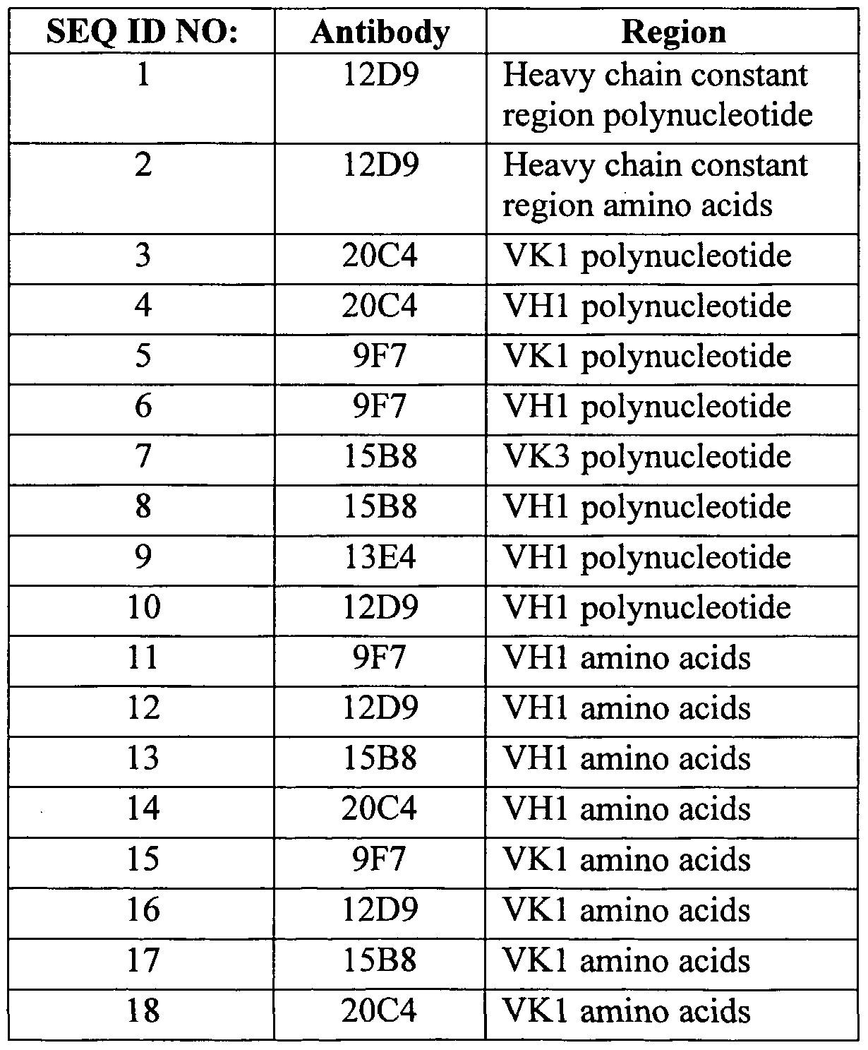
- the polynucleotide and amino acid sequences are provided in SEQ ID NOs: 1-18, as shown in Table 7, Example 11.
- results obtained using the five disclosed monoclonal antibodies indicate that these antibodies, as well as fragments and chimeric forms thereof, have antagonistic features that make them suitable for a number of clinical applications, including treatment of autoimmune diseases, treatment of transplantation reactions and rejections, as adjuvant therapies for gene therapies and protein therapies, and in inhibiting growth of tumor cells, including Non-Hodgkins Lymphoma cells. Additional uses include treatment of any disease mediated by a CD40-expressing malignant cell, and use to treat diseases related to the proliferation, activation, or regulation of cells expressing CD40.
- Table 1 The activity of the five MAb's is summarized in Table 1.
- the invention encompasses not only the five monoclonal antibodies described herein, but also antibodies differing from these but retaining the CDR; and antibodies with one or more amino acid addition(s), deletion(s), or substitution(s), wherein the activity is measured by inhibition of B cell proliferation and/or antibody secretion.
- the invention also encompasses de-immunized antibodies, which can be produced as described in, for example, WO 98/52976, "Method for the Production of Non-Immunogenic Proteins," which is incorporated by reference herein.
- fusion proteins comprising a monoclonal antibody of the invention, or a fragment thereof, which fusion proteins can be synthesized or expressed from corresponding polynucleotide vectors, as is known in the art.
- the antibodies of the present invention can have sequence variations produced using methods described in, for example, Patent Publication Nos. EP 0 983 303 Al, WO 00/34317, and WO 98/52976, incorporated herein by reference. For example, it has been shown that sequences within the CDR can cause an antibody to bind to MHC Class II and trigger an unwanted helper T cell response. A conservative substitution can allow the antibody to retain binding activity yet lose its ability to trigger an unwanted T cell response. Any such conservative or non-conservative substitutions can be made using art-recognized methods, and the resulting antibodies will fall within the scope of the invention.
- the invention provides amino acid sequences for light chains and heavy chains of the preferred monoclonal antibodies (SEQ ID NOS. 1-18).
- the sequences are aligned as shown in Figures 5 and 6. These alignments indicate specific amino acid positions that are more amenable to substitution without loss of the desired biological activities of the antibody.
- plasmids that will encode variants of these sequences.
- the variant antibodies can be routinely tested for antagonistic activity, affinity, specificity, and agonistic activity using methods described herein.
- an antibody produced by any of the methods described above, or any other method not disclosed herein, will fall within the scope of the invention if it possesses at least one of the following biological activities: inhibition of immunoglobulin secretion by human peripheral B cells stimulated by T cells; inhibition of proliferation of human peripheral B cells stimulated by Jurkat T cells; and inhibition of proliferation of human peripheral B cells stimulated by CD40L-expressing cells.
- the antibodies will also exhibit a single site binding affinity (K D ) of at least 10 "5 M, preferably at least IO '6 - 10 '7 M, more preferably at least 10 "8 M, and most preferably at least IO "9 M, such as 10 "10 M, as measured using a standard assay such as Biacore, which is know in the art, in comparison with appropriate controls. Binding affinity of 10 "11 M, IO “13 M, 10 "15 M, IO "17 M and IO “19 M can also be achieved. These assays are automated, and allow the measurement of MAb specificity and cross- reactivity, which can also be assayed using standard techniques known in the art. Details of the Biacore assays are provided in Biacore 's "BIAapplications handbook.” Methods described in WO 01/27160 can be used to modulate the binding affinity.
- K D single site binding affinity
- the antibodies may be labeled using conventional techniques. Suitable labels include fluorophores, chromophores, radioactive atoms (particularly 32 P and 125 I), electron-dense reagents, enzymes, and ligands having specific binding partners. Enzymes are typically detected by their activity. For example, horseradish peroxidase is usually detected by its ability to convert 3,3',5,5'-tetramethylbenzidine (TMB) to a blue pigment, quantifiable with a spectrophotometer. "Specific binding partner” refers to a protein capable of binding a ligand molecule with high specificity, as for example in the case of an antigen and a monoclonal antibody specific therefor.
- 125 I may serve as a radioactive label or as an electron- dense reagent.
- HRP may serve as enzyme or as antigen for a MAb.
- MAbs and avidin also require labels in the practice of this invention: thus, one might label a MAb with biotin, and detect its presence with avidin labeled with 125 I, or with an anti-biotin MAb labeled with HRP.
- Other permutations and possibilities will be readily apparent to those of ordinary skill in the art, and are considered as equivalents within the scope of the instant invention.
- Antibodies for use in the invention can be produced using any suitable technique.
- WO 01/27160 discloses a method of conferring donor CDR binding affinity onto an antibody acceptor variable region framework. The method can also be used to optimize the binding affinity of a variable region or an antibody, such as to enhance the binding affinity.
- Methods for producing humanized antibodies having one or more CDR's are disclosed in U.S. Patent No. 6,180,370. Methods of producing antibodies that have been optimized for administration to humans are disclosed in WO 00/34317, which describes the production of proteins that are rendered less immunogenic or non-immunogenic.
- U.S. Patent No. 5,514,548 discloses methods for selection of ligand binding proteins, such as antibodies, that bind with high affinity to a target ligand.
- U.S. Patent No. 5,877,397 disclosed transgenic non-human animals capable of producing heterologous antibodies. All of these patents and patent publications are incorporated herein by reference.
- the antibodies of this invention are administered at a concentration that is therapeutically effective to prevent or treat antibody-mediated diseases such as allergies, SLE, PBC, ITP, multiple sclerosis, psoriasis, Crohn's disease, graft rejection, and B cell lymphoma.
- the antibodies may be formulated using a variety of acceptable excipients known in the art.
- the antibodies are administered by injection, either intravenously or intraperitoneally. Methods to accomplish this administration are known to those of ordinary skill in the art. It may also be possible to obtain compositions which may be topically or orally administered, or which may be capable of transmission across mucous membranes.
- formulants may be added to the antibodies.
- a liquid formulation is preferred.
- these formulants may include oils, polymers, vitamins, carbohydrates, amine acids, salts, buffers, albumin, surfactants, or bulking agents.
- carbohydrates include sugar or sugar alcohols such as mono-, di-, or polysaccharides, or water soluble glucans.
- the saccharides or glucans can include fructose, dextrose, lactose, glucose, mannose, sorbose, xylose, maltose, sucrose, dextran, pullulan, dextrin, alpha and beta cyclodextrin, soluble starch, hydroxethyl starch and carboxymethylcellulose, or mixtures thereof.
- Sucrose is most preferred.
- "Sugar alcohol” is defined as a C to C 8 hydrocarbon having an —OH group and includes galactitol, inositol, mannitol, xylitol, sorbitol, glycerol, and arabitol. Mannitol is most preferred.
- sugars or sugar alcohols mentioned above may be used individually or in combination. There is no fixed limit to amount used as long as the sugar or sugar alcohol is soluble in the aqueous preparation.
- the sugar or sugar alcohol concentration is between 1.0 w/v % and 7.0 w/v %, more preferable between 2.0 and 6.0 w/v %.
- amino acids include levorotary (L) forms of carnitine, arginine, and betaine; however, other amino acids may be added.
- Preferred polymers include polyvinylpyrrolidone (PVP) with an average molecular weight between 2,000 and 3,000, or polyethylene glycol (PEG) with an average molecular weight between 3,000 and 5,000.
- a buffer in the composition it is also preferred to use a buffer in the composition to minimize pH changes in the solution before lyophilization or after reconstitution.
- Most any physiological buffer may be used, but titrate, phosphate, succinate, and glutamate buffers or mixtures thereof are preferred. Most preferred is a citrate buffer.
- the concentration is from 0.01 to 0.3 molar.
- Surfactants that can be added to the formulation are shown in EP Nos. 270,799 and 268,110.
- antibodies can be chemically modified by covalent conjugation to a polymer to increase their circulating half-life, for example. Preferred polymers, and methods to attach them to peptides, are shown in U.S. Pat. Nos.
- Preferred polymers are polyoxyethylated polyols and polyethylene glycol (PEG).
- PEG is soluble in water at room temperature and has the general formula: R(O ⁇ CH 2 — CH 2 ) réelle O--R where R can be hydrogen, or a protective group such as an alkyl or alkanol group.
- R can be hydrogen, or a protective group such as an alkyl or alkanol group.
- the protective group has between 1 and 8 carbons, more preferably it is methyl.
- n is a positive integer, preferably between 1 and 1,000, more preferably between 2 and 500.
- the PEG has a preferred average molecular weight between 1000 and 40,000, more preferably between 2000 and 20,000, most preferably between 3,000 and 12,000.
- PEG has at least one hydroxy group, more preferably it is a terminal hydroxy group. It is this hydroxy group which is preferably activated to react with a free amino group on the inhibitor.
- the type and amount of the reactive groups may be varied to achieve a covalently conjugated PEG/antibody of the present invention.
- Water soluble polyoxyethylated polyols are also useful in the present invention. They include polyoxyethylated sorbitol, polyoxyethylated glucose, polyoxyethylated glycerol (POG), etc. POG is preferred. One reason is because the glycerol backbone of polyoxyethylated glycerol is the same backbone occurring naturally in, for example, animals and humans in mono-, di-, triglycerides. Therefore, this branching would not necessarily be seen as a foreign agent in the body.
- the POG has a preferred molecular weight in the same range as PEG.
- the structure for POG is shown in Knauf et al., J. Bio. Chem. 263:15064-15070 (1988), and a discussion of POG/IL-2 conjugates is found in U.S. Pat. No. 4,766,106, both of which are hereby incorporated by reference in their entireties.
- liposome Another drug delivery system for increasing circulatory half-life is the liposome.
- Methods of preparing liposome delivery systems are discussed in Gabizon et al., Cancer Research 42:4734 (1982); Cafiso, Biochem Biophys Acta 649:129 (1981); and Szoka, Ann Rev Biophys Eng 9:467 (1980).
- Other drug delivery systems are known in the art and are described in, e.g., Poznansky et al., Drug Delivery Systems (R. L. Juliano, ed., Oxford, N.Y. 1980), pp. 253-315; M. L. Poznansky, Pharm Revs 36:277 (1984).
- the liquid pharmaceutical composition is preferably lyophilized to prevent degradation and to preserve sterility.
- Methods for lyophilizing liquid compositions are known to those of ordinary skill in the art.
- the composition may be reconstituted with a sterile dilutent (Ringer's solution, distilled water, or sterile saline, for example) which may include additional ingredients.
- the composition is preferably administered to subjects using those methods that are known to those skilled in the art.
- the antibodies and compositions of this invention are used to treat human patients to prevent or treat antibody-mediated diseases such as allergies, SLE, PBC and ITP.
- the preferred route of administration is parenterally.
- the compositions of this invention will be formulated in a unit dosage injectable form such as a solution, suspension or emulsion, in association with a pharmaceutically acceptable parenteral vehicle.
- a pharmaceutically acceptable parenteral vehicle are inherently nontoxic and nontherapeutic. Examples of such vehicles are saline, Ringer's solution, dextrose solution, and Hanks' solution.
- Nonaqueous vehicles such as fixed oils and ethyloleate may also be used.
- a preferred vehicle is 5% dextrose in saline.
- the vehicle may contain minor mounts of additives such as substances that enhance isotonicity and chemical stability, including buffers and preservatives.
- compositions are administered so that antibodies are given at a dose between 1 ⁇ g/kg and 20 mg/kg, more preferably between 20 ⁇ g/kg and I 0 mg/kg, most preferably between 1 and 7 mg/kg.
- it is given as an infusion or as a bolus dose, to increase circulating levels by 10-20 fold and for 4-6 hours after the bolus dose. Continuous infusion may also be used after the bolus dose.
- the antibodies may be infused at a dose between 5 and 20 ⁇ g/kg/minute, more preferably between 7 and 15 ⁇ g/kg/minute.
- Suitable treatment regimens are disclosed in WO 00/27428 and WO 00/27433, which are incorporated herein by reference.
- Non-Hodgkin's Lymphoma originates from components of the spleen, thymus and lymph nodes (Jandl J.H., Non-Hogkin's lymphomas, in Jandl JH (ed): Blood, Textbook of Hematology, Boston, MA, Little Brown, 1996, pp. 853-887). It consists of a group of lymphocytic malignancies that are derived primarily from B and T cells. Patients with low grade NHL are usually non-responsive to radiation therapy and chemotherapy. This low response rate and the high probability of relapse contribute to the median patient survival time of fewer than 9 years.
- CD40 chronic lymphcytic leukemia
- Fas a malignancy that plays an important role in the pathogenesis of low grade B lineage malignancies, including chronic lymphcytic leukemia (CLL) and NHL (Ghia P., Adv.
- lymphoma cells Studies in low grade NHL suggests the disease onset is due to the accumulation of the lymphoma cells as a results of reduction in apoptosis through the Fas pathway and increase in the survival signal through CD40 (Ghia P., Adv. Cancer Res., 2000, 79: 157-73). This may explain the insensitivity of the lymphoma cells to chemo or radiation therapy, which specifically target actively proliferating cells.
- the invention further relates to a new NHL therapy comprising the use of an antibody to CD40 to block the survival signal for the NHL cells. This strategy is supported by a number of observations in the published scientific literature. CD40 is expressed on the surface of B cells throughout B-cell development.
- CD40 provides a survival signal for malignant B cells and stimulates their growth in vitro (Romano M.F. et al., Leuk. Lymphoma, 2000 Jan., 5 ⁇ 5(3-4):255-62; Furman R.R., J. Immunol., 2000 Feb. 15, 164(4):2200-6; Kitada S., Br. J. Haematol, 1999 Sep., 70tf(4):995-1004; Romano M.F., Blood, 1998 Aug. 1, P2(3):990-5; Jacob A., Leuk. Res., 1998 Apr., 22(4):379-82; Wang D., Br. J.
- CD40 is expressed on lymphoma cells in 86% of patients with B-lineage NHL (Uckun F.M., Blood, 1990 Dec. 15, 7d(12):2449-56).
- the discovery of CD40/CD40L co-expression in the same B-cell lymphoma cells raises the possibility of an autocrine growth signal loop in NHL patients (Clodi K., Br. J.
- an antagonist anti- CD40 antibody may have therapeutic value in NHL.
- 15B8 a human IgG2 subtype anti-human CD40 monoclonal antibody generated by immunization of transgenic mice bearing the human IgG2 heavy chain locus and the human K light chain locus (Xenomouse, Abgenix) was used.
- 15B8 was tested using malignant B cells (NHL cells) obtained from NHL patients who were either rituximab treated or na ⁇ ve.
- Rituximab is an anti-CD20 monoclonal antibody for the treatment of relapsed or refractory low grade or follicular NHL.
- tumor cells were co-cultured with irradiated CD40-ligand (CD40L) transfected feeder cells (Arpin, C, Science, 1995, 268:720-722) in the presence or absence of the B cell growth factor Interleukin-4 (IL-4).
- CD40L irradiated CD40-ligand
- IL-4 B cell growth factor Interleukin-4
- Antibodies agonist anti-CD40 MS81, or antagonist anti-CD40 15B8 or isotype control huIgG2 of indicated concentration (from 0.01 ⁇ g/ml to 10 ⁇ g/ml) were then added to the culture. Following incubation at 37°C for 48 hours, cultured cells were pulsed with H-thymidine for 18 hours. The cells were then harvested and analyzed for the amount of 3 H-thymidine incorporation (Schultz, J.L., Proc. Natl Acad. Sci. USA, 1995, 2:8200-8204). All sample conditions were in triplicate.
- lymphomas There are two types of preclinical models that are currently used for evaluation of human antigen-specific Mabs in therapeutic development for lymphomas.
- One model is the xenograft mouse in vivo model, where the EBV-transformed lymphoma cell lines, such as Daudi (Burkitt lymphoma) or Raji (Burkitt lymphoma) cells, are xenografted into SCID/Nude mice.
- the results only reflect effects on the particular immortal cell line, which is derived from one EBV- transformed cell.
- Burkitt lymphoma cells are lymphoblastoid cells (Ambinder R.F., Cancer Treat.
- CD40 signaling leads to growth arrest in EBV-transformed Burkitt lymphoma cell lines (Fukuda M., Viral Immunol, 2000, 73(2):215-29; Baker M.P., Blood, 1998 Oct. 15, 2(8):2830-43).
- an antagonist anti- CD40 MAb (15B8) in the xenograft models will not be able to predict the response to the antibody (15B8) by ⁇ HL patients.
- the other model is the in vitro growth inhibition assay of lymphomas cells from ⁇ HL patients, which was used herein.
- the advantage is that the results predicate the sensitivity of the lymphoma cells from ⁇ HL patients to the agent (15B8) tested.
- the results are obtained from in vitro study under defined conditions.
- a previously published study reported that a rat anti-mouse CD40, which failed to induce ADCC and CDD in vitro, showed good efficacy in two syngeneic mouse B lymphoma models (BCL1 and A31) (Tutt A.L., J. Immunol, 1998 Sep. 15, 161 (6):3176-85).
- the anti-tumor effect on the anti-mouse CD40 occurred slower in time than an anti-Id tested.
- the anti-mouse CD40 may operate by blocking critical growth signals that are dependent on the expression of surface CD40 not direct signaling like anti-Id in the mouse models tested. This study suggests that the blocking of CD40/CD40L signaling by an anti-CD40 could be efficacious in vivo.
- 15B8 did not bind to the Fc ⁇ receptors in vitro and failed to induce ADCC and CDC in vitro since it is of human IgG2 subtype. 15B8 is of similar property to the rat anti- mouse CD40. This data supports the hypothesis that 15B8 will be beneficial to ⁇ HL patients, especially Rituxan-resistant patients.
- the concentrations of human IgM and IgG were estimated by ELISA. 96- well ELISA plates were coated with 2 ⁇ g/ml goat anti-human IgG MAb (The Jackson
- PBMQ Peripheral Blood Mononuclear Cells
- Ficoll-Paque solution low endotoxin, Pharmacia
- 20ml of Ficoll-Paque solution was added per 50 ml polystyrene tube, in 3 tubes, 30 minutes before adding the blood.
- the Ficoll- Paque solution was warmed up to room temperature.
- 3L of bleach in 1:10 dilution was prepared, and used to wash all the tubes and pipettes contacting the blood.
- the blood was layered on the top of the Ficoll-Paque solution without disturbing the Ficoll layer, at 1.5ml blood/lml of Ficoll-Paque.
- the tubes were centrifuged at 1700 rpm for 30 minutes at room temperature with the brake on the centrifuge turned off.
- the second layer which contains the B and T lymphocytes, was collected using a sterile Pasteur pipette, and place in two 50 ml polystyrene tubes.
- the collection was diluted with 3x the volume of cold RPMI with no additives, and the tubes were centrifuged at 1000 RPM for 10 minutes.
- the media was removed by aspiratation, and the cells from both 50 ml tubes were resuspended in a total of 10ml cold RPMI (with additives) and transferred to a 15ml tube.
- the cells were counted using the hemacytometer, then centrifuged at 1000 RPM for 10 minutes.
- the media was removed and the cells resuspended in 4mls RPMI. This fraction contained the PBMC.
- the tube containing the PBMC and beads was placed into the magnetic holder for 2 minutes, then the solution was transferred to a new 5ml tube in the magnetic holder. After 2 minutes, the solution was transferred to a new 15ml tube. This step was repeated four more times, and the solutions of the first four times were collected in the 15ml tube, then centrifuged at 1000 RPM for 5 minutes. This step produced the pellet for T cell separation.
- lOO ⁇ l RPMI (with additives) was added to collect the beads, and the solution was transferred into a 0.7ml tube.
- lO ⁇ l of Dynal DetachaBeads were added into the suspension at room temperature, and it was allowed to rotate for 45 minutes.
- the suspension was transferred into a new 5ml tube and 3mls of RPMI (with additives) was added.
- the tube was placed in the magnetic holder for 2 minutes.
- the solution was transferred into a new 5ml tube in the holder for 2 minutes, then to a 15ml tube.
- the previous step was repeated three more times, collecting the solution in the 15ml tube.
- the 15ml tube was centrifuged at lOOORPM for 10 minutes, and the cells resuspended in 10ml RMPI.
- the washing step was repeated 2 more times for a total of 3 washes.
- the cells were counted before the last centrifugation. This step completed the B cell purification. Cells were stored in 90% FCS and 10% DMSO and frozen at - 80°C. Isolation of the T Cells.
- the human T cell Enrichment Column (R&D systems, anti-hCD3 column kit) was prepared using 20ml of IX column wash buffer by mixing 2ml of 10X column wash buffer and 18ml of sterile distilled water. The column was cleaned with 70% ethanol and placed on top of a 15ml tube. The top cap of the column was removed first to avoid drawing air into the bottom of the column. Next, the bottom cap was removed, and the tip was cleaned with 70% ethanol. The fluid within the column was allowed to drain into the 15ml tube. A new sterile 15ml tube was placed under the column after the column buffer had drained to the level of the white filter. The B cell depleted PBMC fraction was suspended in 1ml of buffer and added it to the top of the column.
- the cells were allowed to incubate with the column at room temperature for 10 minutes.
- the T cells were eluted from the column with 4 aliquots of 2ml each of IX column wash buffer.
- the collected T cells were centrifuged at 1000 RPM for 5 minutes.
- the supernatant was removed and the cells resuspended in lOmls RPMI. Cells were counted and centrifuged one more time. The supernatant was removed, completing the T cell purification.
- Cells were stored in 90% FCS and 10% DMSO and frozen at -80°C.
- the RPMI composition contained 10 % FCS (inactivated at 56°C for 45 min.), 1% Pen Strep (lOOu ml Penicillin, 0.1 ⁇ g/ml Streptomycin), 1% Glutamate, 1% sodium puravate, 50 ⁇ M 2-ME.
- Ramos cells (10 6 cells/sample) were incubated in 100 ⁇ l primary antibody (10 ⁇ g/ml in PBS-BSA) for 20 min at 4°C. After 3 washes with PBS-BSA or HBSS- BSA, the cells were incubated in 100 ⁇ l FITC-labeled F(ab') 2 fragments of goat anti- (human IgG) antibodies (Caltaq) for 20 min at 4°C. After 3 washes with PBS-BSA and 1 wash with PBS, the cells were resuspended in 0.5 ml PBS. Analyses were performed with a FACSCAN V (Becton Dickinson, San Jose, Calif). Generation of Hybridoma Clones
- Splenocytes from immunized mice were fused with SP2/0 or P3 x 63Ag8.653 murine myeloma cells at a ratio of 10:1 using 50% polyethylene glycol as previously described by de Boer et al., J. Immunol. Meth. 773:143 (1988).
- the fused cells were resuspended in complete IMDM medium supplemented with hypoxanthine (0.1 mM), aminopterin (0.01 mM), thymidine (0.016 mM) and 0.5 ng/ml hIL-6 (Genzyme, Cambridge, Mass.).
- the fused cells were then distributed between the wells of 96-well tissue culture plates, so that each well contained 1 growing hybridoma on average. After 10-14 days the supernatants of the hybridoma populations were screened for specific antibody production. For the screening of specific antibody production by the hybridoma clones, the supernatants from each well were pooled and tested for anti-CD40 activity specificity by ELISA first. The positives were then used for fluorescent cell staining of EBV-transformed B cells as described for the FACS Assay above. Positive hybridoma cells were cloned twice by limiting dilution in IMDM/FBS containing 0.5 ng/ml hIL-6.
- EXAMPLE 1 EXPRESSION OF HUMAN CD40 IN SF9 CELLS Sf9 insect cells infected with recombinant virus Autographa californica baculovirus (AcNPV), encoding CD40, were cultured for 48 hours in 24-well plates. After removal of the tissue culture medium the plates were incubated for 45 minutes at room temperature (RT) with 0.25 ml of antibody in PBS with 1% BSA (PBS-BSA).
- Bound peroxidase activity was revealed by the addition of an assay mixture prepared by diluting 0.5 ml of 2 mg/ml 3,3',5,5'-tetramethylbenzidine in ethanol to 10 ml with 10 mM sodium acetate, 10 mM EDTA buffer (pH 5.0) and adding 0.03% (v/v) H. 2 O 2 . The reaction was stopped after 10 minutes by adding 100 ⁇ l of 1 M H 2 SO 4 .
- mice transgenic for the human IgG2 heavy chain locus and the human K light chain locus were immunized with Sf9 cells expressing human CD40.
- the method of immunization was carried out as described by de Boer, U.S. Patent No. 6,004,552. Briefly, mice were injected intraperitoneally at day 0 and day 14 with 5 x IO 6 Sf9 cells infected with AcCD40 virus. At day 21, 100 ⁇ l of serum was obtained to test for the presence of specific antibodies. After a rest period of at least two weeks, the mice received a final injection with 5 x 10° cells infected with AcCD40 virus. Three days after this last injection, the spleen cells were used for cell fusion.
- mice were selected for fusion based on the reactivity of their sera with recombinant CD40 in an ELISA.
- Spleen cells from immunized mice were fused with mouse myeloma cells (NS/0) by the method of Kohler and Milstein, Nature 256:495-96 (1975), with modifications.
- Hybridomas grown in HAT selective medium were selected for further characterization based on their ability to bind CD40 in an ELISA.
- Hybridomas that produced antibodies that bound nontransfected Sf9 cell lysate or anti- mouse light chain antibody were dropped from consideration.
- Hybridomas that produced CD40-binding antibodies that did not bind Sf9 cell lysate or anti-mouse light chain antibody were subcloned.
- Hybridoma antibodies were also tested for their ability to stain Ramos lymphoma cells, which express human CD40 on their surfaces. The concentration of antibodies giving half-maximal CD40 binding as measured by ELISA is shown below in Table 2.
- Hybridoma antibodies were then selected for their ability to inhibit the production of IgM by human peripheral blood B cells stimulated with anti-CD28- activated human peripheral blood T cells (Figure 1). Hybridoma antibodies were further screened for their ability to inhibit proliferation of human peripheral blood B cells induced by CD40L and anti-IgM ( Figure 2). Hybridomas were also screened for their ability to induce proliferation in resting human peripheral blood B cells ( Figure 3). Some hybridomas, such as 36C4-G2, exhibited marked stimulatory activity. Thus, even when able to bind CD40, not all human antibodies exhibit the desired inhibitory effect. Four hybridomas were selected from approximately 36 as having the optimal inhibitory activity.
- EXAMPLE 3 BINDING PROPERTIES OF SELECTED HYBRIDOMAS Four hybridomas were selected based on their inhibitory effect on B cell activation as described above in Example 2. Their binding properties were determined by BIAcore evaluation using soluble recombinant CD40 as the mobile phase with the anti-CD40 antibodies being captured on the sensor surface. The inhibitory antibodies exhibited various binding affinities, which are suitable for the uses described herein, as shown in Table 3.
- the pre-coated plates were washed 3 x with PBS.
- the T cells were irradiated with 3000 Rad.
- the B cells were resuspended in RPMI(+) to IO 4 per ml. lOO ⁇ l of B cells were added into the well, then anti-CD40 antibodies were added into the well.
- the T cells were resuspended in RPMI(+) to 10 5 per ml.
- Human recombinant IL2 to 200u/ml was added (Chiron, lOu/ ⁇ l water solution stored at -20°C) was added in the cell suspension. A lOO ⁇ l suspension was taken into each well, and mixed well with the B cells and antibodies.
- EXAMPLE 6 INHIBITION OF PROLIFERATION OF JURKAT-STIMULATED HUMAN PERIPHERAL B CELLS B cells were purified as described above. IO 4 purified B cells, 10 5 irradiated Jurkat cells (3000 Rad), and antibodies to be tested were added into anti- CD3-coated 96-well plates. The plates were incubated at 37°C for four days, with labeling of the cells with H-thymidine during the last 18 hours. The cells were harvested and counted. The results are shown below in Table 6.
- B cells (1 x IO 4 per well) were cultured in 200 ⁇ l RPMI supplemented with 10% fetal calf serum in U-bottom 96-well microtiter plates. B cells were stimulated by addition of immobilized anti-(IgM) antibodies (5 ⁇ g/ml, Sigma). Varying concentrations of MAbs were added at the onset of the microcultures and proliferation was assessed at day 4 by measurement of the incorporation of H-thymidine after 18 hour pulsing. The results are shown in Figure 2.
- Example 2 and RT-PCR was performed on the mRNAs. Two sets of primers were used to generate PCR products: a universal or pool of heavy and light chain family primers; and then family-specific primers. The PCR products were analyzed on gels, sequenced, and translated. The polynucleotide and amino acid sequences are provided in the Sequence Listing as summarized in Table 7.
- FIGS 5, 6, and 8-14 Specific regions of the antibodies are shown in Figures 5, 6, and 8-14. This information can be used to design additional monoclonal antibodies for use according to the invention. These monoclonal antibodies may differ from those described herein, by substitution of one or more of the framework or CDR regions. The monoclonal antibodies also may differ by substitution of one or more amino acids, which are shown to differ in certain regions of the framework and CDR ( Figures 5 and 6). Once the amino acid sequence is designed, routine procedures can be used to construct a corresponding polynucleotide sequence for expression of the monoclonal antibody. Expression and purification of the monoclonal antibodies is performed using methods known in the art, such as those disclosed in U. S. Patent Nos. 5,545,403, 5,545,405, and 5,998,144, which are incorporated herein by reference.
- B cells from tumor infiltrated lymph nodes were obtained from one antibody naive, one rituximab-sensitive and one rituximab-resistant NHL patient.
- the NHL cells were studied under four different culture conditions; no added antibody (medium); addition of human isotype antibody IgG2 (control); addition of anti-CD40 antibody MS81 (agonistic antibody); and addition of 15B8. All antibodies were tested 1, 2, and 5 ⁇ g/mL in the presence or absence of IL-4.
- the NHL cells from two patients were cultured as described above under the same four conditions in the presence of IL-4 (2 ng/ml). B-cell proliferation was measured by 3 H-thymidine incorporation as described above.
- Anti-CD40 antibody 15B8 at concentration of 1, 2 and 5 ⁇ g/mL, did no stimulate NHL cells to proliferate in either the presence or absence of IL-4.
- an agonistic anti-CD40 antibody MS 81
- MS 81 an agonistic anti-CD40 antibody tested at the same concentration, stimulated NHL-cell proliferation or in the presence and absence of IL-4 in all patient samples. Representative results from one patient are shown below (Fig. 15 and Fig. 16). Results from the NHL cells from the two patients in the presence of IL-4 and three patients in the absence of IL-4 were comparable. These results indicate that 15B8 is not an agonist anti-CD40 antibody and does not stimulate proliferation of NHL cells from rituximab- sensitive, naive or -resistant NHL patients in vitro.
- NHL cells FACS analysis of the NHL cells was performed with either a direct labelled 15B8-FITC or 15B8 plus anti-HuIgG2-FITC to confirm that CD40 is expressed on the surface the NHL cells tested and that 15B8 binds to the NHL cells.
- the 15B8 binding-positive cell population in any given patient is about 66% to 91%.
- NHL cells from patients were cultured as described in Example 12 in suspension over CD40L-expressing feeder cells under four different conditions: no added antibody (medium); addition of human isotype antibody IgG2 (control); addition of anti-CD40 antibody MS81 (agonistic antibody); and addition of 15B8. All antibodies were added at concentration of 1, 2, and 5 ⁇ g/mL in the presence or the absence of IL-4.
- the NHL cells from one antibody naive, two rituximab-sensitive and five rituximab-resistant patients (8 patients in total) were cultured under the same four conditions as described above in the presence of IL-4 (2 ng/ml). NHL cells from three rituximab-sensitive and four rituximab-resistant patients (7 patients in total) were cultured under similar conditions in the absence of IL-4.
- the NHL cell proliferation was measured by H-thymidine incorporation.
- NHL cells from patients were cultured with murine L-cells expressing human CD40L in the presence of medium, agonist anti-CD40 (MS81), antagonist anti-CD40 (15B8) or huIgG2 isotype control in vitro.
- the proliferation of the NHL cells was measured by 3H-thymidine incorporation (data from one rituximab-sensitive patient is not in the table for the cpm of CD40L is ⁇ 2000).
- Table 9 shows the inhibitory effect of 15B8 on proliferation of NHL cells from one antibody naive, two rituximab-sensitive (data from both patient samples were repeated twice reproducibly) and five rituximab-resistant patients (8 patients in total) stimulated by both CD40L and IL-4 in vitro.
- 15B8 significantly (p ⁇ 0.05) inhibited the CD40L and IL-4-mediated proliferation of the NHL cells.
- the degree of inhibition ranged from 18-69% at high dose (5 or 10 ⁇ g/ml) in samples from all 8 patients in vitro.
- There is a statistically significant dose response of this inhibitory effect by 15B8 (p ⁇ 0.005) Fig. 18 shows one representative dose response curve) at 15B8 concentration range of 0.01 - 10 ⁇ g/ml.
- NHL cells from patients were cultured with murine L-cells expressing human CD40L in the presence of IL-4 (human interleukin-4) at 2 ng/ml under conditions described in Table 1.
- IL-4 human interleukin-4
- 15B8 was tested in several in vitro assays described below using cells from humans and five different primate species, including champanzee (chimp), marmoset, cynomologus mokey, rhesus monkey and baboon. 15B8 does not activate human peripheral blood B cell and does not cause PBMC proliferation in vitro in human, chimp and marmoset. Activation of CD40 on human B cells obtained from peripheral blood leads to proliferation of the B cells (van Kooten C, J. Leukoc. BioL, 2000 Jan., ⁇ 57(1):2-17; Denton M.D., Pediatr.
- 15B8 was further compared to CD40L for stimulation of human PBMC proliferation using freshly isolated human PBMC. As summarized in Table 10 below, 15B8 does not stimulate human PBMC proliferation in vitro as measured by 3-H methyl-thymidine incorporation (John E. Coligan et al., Current Protocols in Immunology, Vol. 13:12, John Wiley & Sons, Inc., 1991 ; Kwekkeboom J., Immunology, 1993 Jul, 7P(3):439-444) in samples tested from sixteen volunteers at concentration range of 0.2-5 ⁇ g/ml.
- B cells/PBMCs were cultured in vitro in the presence of CD40L, 15B8 or huIgG2 isotype control.
- the fold-increase for CD40L shown in the table is the ratio of the CD40L cpm to the cpm of huIG2 at 5 ⁇ g/ml.
- CD40L CD40L transfected CHO cells, fixed with formaldehyde before the experiments.
- Annexin V staining on the cell surface can be used as an early apoptosis marker (Ju S.T., Int. Rev. Immunol, 1999, 7S(5-6):485-513).
- Human B cells were purified from peripheral blood and incubated with 15B8. FACS analysis was used to detect cells with positive staining of Annexin V and anti-FasL. There was no significant difference on the surface staining by the two reagents between cells incubated with 15B8 or the isotype control (huIgG2) antibody. This result shows that 15B8 does not induce apoptosis of human B cells in vitro. These data provide further evidence that 15B8 is not an agonistic anti-CD40 antibody for human B cells.
- 15B8 cross-reacts with CD40 expressed on the surface of CD20 positive PBMCs from primates.
- 15B8 can activate CD40 on B cells from other primate species such as chimps and marmosets
- the same proliferation assay were carried out using freshly isolated chimp and marmoset PBMC from fifteen chimps and five marmosets. Similar to the results with the human PBMC, 15B8 did not stimulate the proliferation in vitro of PBMCs from six chimps and five marmosets at 1 and 5 ⁇ g/ml concentration (Table 3 above). 15B8 also did not up-regulate the expression of activation marker, CD69, in the three chimp-PBMC samples tested (Table 4). 15B8 did not show any effect on FasL expression and apoptosis in chimp PBMCs similar to human PBMC controls after 24 and 48 hours simulation in vitro in all samples from six chimps tested.
- Cross-linking 15B8 by a secondary antibody fixed to plastic surface did not increase its potency to stimulate B cell proliferation (data not shown).
- 15B8 did not stimulate proliferation of the cells. This observation indicates a reduced risk of 15B8 being stimulative (agonistic) for B cell proliferation in case of induction of anti-15B8 (HAHA) or Fc binding to other Fc receptor expressing cells when administered in vivo.
- 15B8 does not initiate an activation signal in human B cells/PBMCs nor in chimp/marmoset PBMCs in vitro. Therefore, 15B8 is not an agonistic anti-CD40 antibody in humans, chimps and marmosets.
- EXAMPLE 15 15B8 is AN ANTAGONIST ANTI-CD40 ANTIBODY IN HUMANS, CHIMPS AND MARMOSETS IN VITRO.
- 15B8 is an antagonist anti-CD40
- its ability to inhibit CD40-CD40L interaction was tested in a CD40L mediated-human B cell proliferation assay (Kwekkeboom J., Immunology, 1993 Jul, 7P(3):439-444).
- a transfected CHO cell line expressing human CD40L was used to stimulate the proliferation of purified human peripheral blood B cells or PBMCs. Human B cells from ten healthy volunteers and human PBMCs from three healthy volunteers were tested. In all the samples tested, 15B8 suppressed CD40L-expressing CHO cells mediated-proliferation by 42-88% at concentration range from 0.2 - 5 ⁇ g/ml (Table 12).
- Figure 19 shows representative dose- response curves using cells from three individuals.
- the no-effect dose of 15B8 is 0.008 ⁇ g/ml and reaches saturating dose at 0.2 ⁇ g/ml (Figure 19). This observation indicates that 15B8, as an antagonist anti-CD40 antibody, can inhibit the growth signals in human B cells and PBMCs provided by cell surface-expressed CD40L. Table 12
- B cells/PBMCs were cultured in vitro with CD40L expressing CHO cells in the presence of 15B8 or huIgG2 control.
- CD40L transfected CHO cells were fixed with formaldehyde before the experiments.
- the proliferation of cells was measured by 3H-thymidine incorporation.
- Activated B cells undergo a number of biological response such as proliferation and antibody production.
- the activation of B cells by T cell-dependent antigens involves CD4 + T-helper (Th) cells.
- This T cell helper process is mediated by a concerted effort of the interaction of CD40 on the B cells with the CD40L on the Th cells surface together with the interactions of other co-stimulatory factors and cytokinesDenton M.D., Pediatr. Transplant., 1998 Feb., 2(1):6-15; Evans D.E., J. Immunol, 2000 Jan. 15, 164(2):6S8-97; Noelle R.J., Agents Actions Suppl, 1998, 49:17-22; Lederman S. et al., Curr. Opin.
- 15B8 can block T-helper cell mediated B cell antibody production.
- purified human peripheral blood B cells were cultured in the presence of purified irradiate T cells activated with anti-CD3 antibody.
- An ELISA assay was used to measure the level of IgM production.
- 15B8 reduced IgM production by about 30% in this assay (data not shown). Therefore, 15B8 can reduce T cell-mediated B cell immunoglobulin production.
- 15B8 inhibits CD40L induced Bpcell/PBMC proliferation in human, chimp and marmoset, and inhibits T cell inducted antibody production by purified human B cells in vitro.
- EXAMPLE 16 15B8 IS AN AGONIST ANTI-MONKEY (CYNOMOLGUS, RHESUS AND BABOON) CD40
- 15B8 was found to stimulate cynomolgus monkey PBMC to proliferate in vitro as measured by 3 H methyl-thymidine incorporation (Table 13 below).
- 15B8 stimulated the proliferation of the PBMCs by 6 - 129.7 folds compare to the huIgG2 control in the tewnety-two samples from seventeen monkeys tested (samples from five monekys were tested twice) (Table 13 below).
- the proliferation stimulated by 15B8 is 14 - 24 folds in four samples from to monkeys and about 1.25 or 1.85 fold in two samples from two monkeys (Table 13).
- 15B8 may be at the limit of over-saturating dose for its proliferation-stimulatory effect on PBMCs from cynomolgus monkey.
- 15B8 is an agonist antibody to CD40 expressed on peripheral blood B cells from cynomolgus monkeys in vitro.
- 15B8 stimulated proliferation of PBMCs from rhesus monkeys and baboons in vitro (Table 13 below).
- the agonist activity of 15B8 is shown using the PBMCs from five rhesus monkey and three baboons (Table 13).
- PBMCs were cultured in vitro in the presence of CD40L, 15B8 or huIgG2 control.
- the fold-increase for CD40L shown in the table is the ratio of the CD40L cpm to the cpm of huIgG2 at 5 ⁇ g/ml.
- CD40L transfected CHO cells were fixed with formaldehyde before the experiments.
- N/A means not measured or not successful.
- EXAMPLE 17 15B8 IS AN AGONIST ANTI CD40 ANTIBODY IN VIVO IN CYNOMOLGUS MONKEYS..
- 15B8 can stimulate proliferation and up-regulation of cell surface activation markers in PBMCs from cynomolgus monkeys in vitro.
- 15B8 is an agonist anti-CD40 antibody in the monkeys in vivo
- a study was performed to examine the biodistribution of 15B8 and the fate of affected peripheral B cells (i.e. extravastion, apoptosis, activation status, or complement lysis) [Biodistribution 15B8.72 Antibodies following Intravenous Administration to Non-Naive Male and Female Cynomolgus Monkeys (SNBL.218.3, SNBL USA)].
- Cynomolgus monkeys (1 female and 2 males) received a single intravenous administration of 3 mg/kg 15B8. The following parameters were monitored: clinical signs, food consumption, body weight, pharmacokinetics, serum complement (CH50), flow cytometry for B cells (including apoptotic B cells), T cells, and monocytes. B cell CD40 receptor saturation with 15B8 was also measured. Animals were necropsied 24 hours after receiving the single dose of 15B8, and standard organs were weighed. Pre-study surgical biopsies of spleen and axiliary lymph nodes were taken to serve as baseline controls. At necropsy, lymphoid and non-lymphoid tissues were sampled for histopathology and immunohistochemistry. Tissues were immunostained with antibodies against CD3, CD40, CD20, CD27, and CD38 antigens. Preliminary results of the study are discussed below.
- CD20, CD27, CD40 and CD86 antibodies revealed increases in these markers in splenic and lymph node follicles, which correlated with the follicular hyperplasia seen in these same tissues.
- Increased staining of CD20 and CD40 were limited to the spleen and lymph node while there was some additional staining of hepatic tissue with CD27 and of hepatic Kupffer cells and inflammatory cells by CD86.
- CD86 staining was also increased in thymic medullary cells and adrenal interstitial leukocytes. There were no changes in the immunostaining of CD3 in 15B8-treated animals as compared to controls.
- 15B8 is an agonistic anti-CD40 antibody in cynomolgus and rhesus monkeys and baboons, and an antagonistic antibody in humans, chimpanzees and marmosets.
- 15B8 is an anti-human CD40 specific monoclonal antibody with human IgG2 subtype and with cross-reactivity to CD40 from non-human primates only.
- 15B8 was shown to be an antagonistic anti-CD40 to the CD40 expressed on human B cells, PBMCs from human, chimp and marmoset.
- 15B8 was shown to have agonistic activity when bound to the CD40 expressed on PBMCs from monkeys (cynomolgus, rhesus and baboon) in vitro. This agonistic activity of 15B8 was confirmed in vivo in cynomolgus monkeys. When tested in primary culture of lymphoma cells from Rituxan-sensitive and resistant NHL patients, 15B8 has no agonist activity in the presence and absence of IL-4. 15B8 can also inhibit CD40L stimulated growth of the lymphoma cells from the similar group of patients under both conditions.
- B cell malignancies such as non-Hodgkin's lymphoma (NHL)
- NHL non-Hodgkin's lymphoma
Landscapes
- Health & Medical Sciences (AREA)
- Chemical & Material Sciences (AREA)
- Organic Chemistry (AREA)
- Life Sciences & Earth Sciences (AREA)
- Medicinal Chemistry (AREA)
- General Health & Medical Sciences (AREA)
- Animal Behavior & Ethology (AREA)
- General Chemical & Material Sciences (AREA)
- Chemical Kinetics & Catalysis (AREA)
- Nuclear Medicine, Radiotherapy & Molecular Imaging (AREA)
- Pharmacology & Pharmacy (AREA)
- Public Health (AREA)
- Veterinary Medicine (AREA)
- Engineering & Computer Science (AREA)
- Bioinformatics & Cheminformatics (AREA)
- Immunology (AREA)
- Hematology (AREA)
- Biochemistry (AREA)
- Rheumatology (AREA)
- Oncology (AREA)
- Proteomics, Peptides & Aminoacids (AREA)
- Molecular Biology (AREA)
- Genetics & Genomics (AREA)
- Biophysics (AREA)
- Diabetes (AREA)
- Pulmonology (AREA)
- Transplantation (AREA)
- Orthopedic Medicine & Surgery (AREA)
- Physical Education & Sports Medicine (AREA)
- Dermatology (AREA)
- Pain & Pain Management (AREA)
- Neurosurgery (AREA)
- Neurology (AREA)
- Biomedical Technology (AREA)
- Gastroenterology & Hepatology (AREA)
- Medicines Containing Antibodies Or Antigens For Use As Internal Diagnostic Agents (AREA)
- Peptides Or Proteins (AREA)
- Preparation Of Compounds By Using Micro-Organisms (AREA)
- Medicines That Contain Protein Lipid Enzymes And Other Medicines (AREA)
- Measuring Or Testing Involving Enzymes Or Micro-Organisms (AREA)
Abstract
Description


Claims
Priority Applications (6)
| Application Number | Priority Date | Filing Date | Title |
|---|---|---|---|
| JP2002532486A JP4271440B2 (en) | 2000-10-02 | 2001-10-02 | Human anti-CD40 antibody |
| AU2002211366A AU2002211366A1 (en) | 2000-10-02 | 2001-10-02 | Human anti-cd40 antibodies |
| DE60143535T DE60143535D1 (en) | 2000-10-02 | 2001-10-02 | HUMAN ANTIBODIES AGAINST CD40 |
| CA002424296A CA2424296A1 (en) | 2000-10-02 | 2001-10-02 | Human anti-cd40 antibodies |
| AT01979395T ATE489403T1 (en) | 2000-10-02 | 2001-10-02 | HUMAN ANTIBODIES AGAINST CD40 |
| EP01979395A EP1326896B1 (en) | 2000-10-02 | 2001-10-02 | Human anti-cd40 antibodies |
Applications Claiming Priority (2)
| Application Number | Priority Date | Filing Date | Title |
|---|---|---|---|
| US23755600P | 2000-10-02 | 2000-10-02 | |
| US60/237,556 | 2000-10-02 |
Publications (2)
| Publication Number | Publication Date |
|---|---|
| WO2002028904A2 true WO2002028904A2 (en) | 2002-04-11 |
| WO2002028904A3 WO2002028904A3 (en) | 2003-02-06 |
Family
ID=22894238
Family Applications (4)
| Application Number | Title | Priority Date | Filing Date |
|---|---|---|---|
| PCT/US2001/030961 Ceased WO2002028480A2 (en) | 2000-10-02 | 2001-10-02 | Methods of therapy for b-cell malignancies |
| PCT/US2001/030859 Ceased WO2002028905A2 (en) | 2000-10-02 | 2001-10-02 | Human anti-cd40 antibodies |
| PCT/US2001/030857 Ceased WO2002028904A2 (en) | 2000-10-02 | 2001-10-02 | Human anti-cd40 antibodies |
| PCT/US2001/030963 Ceased WO2002028481A2 (en) | 2000-10-02 | 2001-10-02 | Methods of therapy for b-cell malignancies using antagonist anti-cd40 antibodies |
Family Applications Before (2)
| Application Number | Title | Priority Date | Filing Date |
|---|---|---|---|
| PCT/US2001/030961 Ceased WO2002028480A2 (en) | 2000-10-02 | 2001-10-02 | Methods of therapy for b-cell malignancies |
| PCT/US2001/030859 Ceased WO2002028905A2 (en) | 2000-10-02 | 2001-10-02 | Human anti-cd40 antibodies |
Family Applications After (1)
| Application Number | Title | Priority Date | Filing Date |
|---|---|---|---|
| PCT/US2001/030963 Ceased WO2002028481A2 (en) | 2000-10-02 | 2001-10-02 | Methods of therapy for b-cell malignancies using antagonist anti-cd40 antibodies |
Country Status (10)
| Country | Link |
|---|---|
| US (5) | US7288252B2 (en) |
| EP (3) | EP1326896B1 (en) |
| JP (6) | JP4202127B2 (en) |
| AT (3) | ATE327004T1 (en) |
| AU (4) | AU2001296507A1 (en) |
| CA (2) | CA2424749A1 (en) |
| DE (2) | DE60143535D1 (en) |
| ES (1) | ES2357051T3 (en) |
| PT (1) | PT1326896E (en) |
| WO (4) | WO2002028480A2 (en) |
Cited By (29)
| Publication number | Priority date | Publication date | Assignee | Title |
|---|---|---|---|---|
| WO2005044854A3 (en) * | 2003-11-04 | 2005-08-11 | Chiron Corp | Antagonist anti-cd40 monoclonal antibodies and methods for their use |
| WO2005044304A3 (en) * | 2003-11-04 | 2005-08-25 | Chiron Corp | Use of antagonist anti-cd40 antibodies for treatment of chronic lymphocytic leukemia |
| WO2005044855A3 (en) * | 2003-11-04 | 2005-09-15 | Chiron Corp | Use of antagonist anti-cd40 monoclonal antibodies for treatment of multiple myeloma |
| US7063845B2 (en) | 2000-04-28 | 2006-06-20 | Gemini Science, Inc. | Human anti-CD40 antibodies |
| WO2006125117A2 (en) | 2005-05-18 | 2006-11-23 | Novartis Ag | Methods for diagnosis and treatment of diseases having an autoimmune and/or inflammatory component |
| US7193064B2 (en) | 2001-04-27 | 2007-03-20 | Kirin Beer Kabushiki Kaisha | Anti-CD40 monoclonal antibody |
| WO2007053767A1 (en) * | 2005-11-01 | 2007-05-10 | Novartis Ag | Uses of anti-cd40 antibodies |
| EP1844815A1 (en) | 2003-11-04 | 2007-10-17 | Novartis Vaccines and Diagnostics, Inc. | Combination therapy comprising anti-CD20 and anti-CD40 antibodies for the treatment of B cell-related cancers |
| WO2007124299A2 (en) | 2006-04-21 | 2007-11-01 | Novartis Ag | Antagonist anti-cd40 antibody pharmaceutical compositions |
| EP2301575A1 (en) | 2003-11-04 | 2011-03-30 | Novartis Vaccines and Diagnostics, Inc. | Methods of therapy for solid tumors expressing the CD40 cell-surface antigen |
| US7939642B2 (en) | 2005-04-09 | 2011-05-10 | Fusion Antibodies Limited | Antibody and uses thereof |
| WO2012075111A1 (en) | 2010-11-30 | 2012-06-07 | Novartis Ag | Uses of anti-cd40 antibodies in combination therapy for b cell-related cancers |
| US8277810B2 (en) | 2003-11-04 | 2012-10-02 | Novartis Vaccines & Diagnostics, Inc. | Antagonist anti-CD40 antibodies |
| US8828396B2 (en) | 2010-11-15 | 2014-09-09 | Novartis Ag | Silent Fc variants of anti-CD40 antibodies |
| US9624310B2 (en) | 2011-11-17 | 2017-04-18 | Gundram Jung | Methods of using bi-specific antibodies for treating B-cell-mediated autoimmune diseases or auto-reactive B-cells |
| WO2017220989A1 (en) | 2016-06-20 | 2017-12-28 | Kymab Limited | Anti-pd-l1 and il-2 cytokines |
| CN108503708A (en) * | 2017-09-01 | 2018-09-07 | 北京智仁美博生物科技有限公司 | Anti-human CD47 antibody and application thereof |
| WO2018219327A1 (en) | 2017-06-01 | 2018-12-06 | 江苏恒瑞医药股份有限公司 | Anti-cd40 antibody, antigen binding fragment thereof and medical use thereof |
| WO2019241730A2 (en) | 2018-06-15 | 2019-12-19 | Flagship Pioneering Innovations V, Inc. | Increasing immune activity through modulation of postcellular signaling factors |
| US10590202B2 (en) | 2012-11-19 | 2020-03-17 | Baliopharm Ag | Recombinant bispecific antibody binding to CD20 and CD95 |
| WO2020108611A1 (en) | 2018-11-30 | 2020-06-04 | 江苏恒瑞医药股份有限公司 | Anti-cd40 antibody, antigen binding fragment and pharmaceutical use thereof |
| WO2020108621A1 (en) | 2018-11-30 | 2020-06-04 | 江苏恒瑞医药股份有限公司 | Cd40 antibody pharmaceutical composition and use thereof |
| WO2020227159A2 (en) | 2019-05-03 | 2020-11-12 | Flagship Pioneering Innovations V, Inc. | Methods of modulating immune activity |
| WO2021127217A1 (en) | 2019-12-17 | 2021-06-24 | Flagship Pioneering Innovations V, Inc. | Combination anti-cancer therapies with inducers of iron-dependent cellular disassembly |
| WO2022006179A1 (en) | 2020-06-29 | 2022-01-06 | Flagship Pioneering Innovations V, Inc. | Viruses engineered to promote thanotransmission and their use in treating cancer |
| WO2022212784A1 (en) | 2021-03-31 | 2022-10-06 | Flagship Pioneering Innovations V, Inc. | Thanotransmission polypeptides and their use in treating cancer |
| WO2023278641A1 (en) | 2021-06-29 | 2023-01-05 | Flagship Pioneering Innovations V, Inc. | Immune cells engineered to promote thanotransmission and uses thereof |
| WO2024077191A1 (en) | 2022-10-05 | 2024-04-11 | Flagship Pioneering Innovations V, Inc. | Nucleic acid molecules encoding trif and additionalpolypeptides and their use in treating cancer |
| WO2024151687A1 (en) | 2023-01-09 | 2024-07-18 | Flagship Pioneering Innovations V, Inc. | Genetic switches and their use in treating cancer |
Families Citing this family (54)
| Publication number | Priority date | Publication date | Assignee | Title |
|---|---|---|---|---|
| JP4202127B2 (en) | 2000-10-02 | 2008-12-24 | ノバルティス バクシンズ アンド ダイアグノスティックス,インコーポレーテッド | B cell malignancy treatment method using antagonist anti-CD40 antibody |
| US7906492B2 (en) | 2001-01-16 | 2011-03-15 | Sloan-Kettering Institute For Cancer Research | Therapy-enhancing glucan |
| US7507724B2 (en) | 2001-01-16 | 2009-03-24 | Sloan-Kettering Institute For Cancer Research | Therapy-enhancing glucan |
| US20050136055A1 (en) * | 2003-12-22 | 2005-06-23 | Pfizer Inc | CD40 antibody formulation and methods |
| US20070292439A1 (en) * | 2004-04-27 | 2007-12-20 | Novartis Vaccines And Diagnostics, Inc. | Antagonist Anti-Cd40 Monoclonal Antibodies and Methods for Their Use |
| WO2006125143A2 (en) * | 2005-05-18 | 2006-11-23 | Novartis Ag | Methods for diagnosis and treatment of proliferative disorders mediated by cd40 signaling |
| US8323644B2 (en) * | 2006-01-17 | 2012-12-04 | Sloan-Kettering Institute For Cancer Research | Therapy-enhancing glucan |
| US20090053221A1 (en) * | 2006-01-17 | 2009-02-26 | Cheung Nai-Kong V | Immune response enhancing glucan |
| WO2007130493A2 (en) * | 2006-05-03 | 2007-11-15 | Regents Of The University Of Colorado | Cd40 agonist antibody/type1 interferon synergistic adjuvant combination, conjugates containing and use thereof as a therapeutic to enhance cellular immunity |
| US20090074711A1 (en) * | 2006-09-07 | 2009-03-19 | University Of Southhampton | Human therapies using chimeric agonistic anti-human cd40 antibody |
| CA2693053C (en) * | 2007-05-30 | 2021-01-05 | Xencor, Inc. | Methods and compositions for inhibiting cd32b expressing cells |
| MX2010005099A (en) * | 2007-11-09 | 2010-05-27 | Novartis Ag | Uses of anti-cd40 antibodies. |
| WO2009094391A1 (en) * | 2008-01-23 | 2009-07-30 | Xencor, Inc. | Optimized cd40 antibodies and methods of using the same |
| AU2010222928B2 (en) | 2008-07-16 | 2012-11-29 | Baylor Research Institute | Antigen presenting cell targeted vaccines |
| EP2406289B1 (en) | 2009-03-10 | 2017-02-22 | Baylor Research Institute | Antigen presenting cell targeted anti-viral vaccines |
| DK2406286T3 (en) * | 2009-03-10 | 2016-08-22 | Baylor Res Inst | Anti-cd40 antibodies and uses thereof |
| WO2011109280A1 (en) | 2010-03-05 | 2011-09-09 | Lerner Research Institute | Methods and compositions to treat immune-mediated disorders |
| EP2552967A4 (en) | 2010-04-02 | 2014-10-08 | Amunix Operating Inc | Binding fusion proteins, binding fusion protein-drug conjugates, xten-drug conjugates and methods of making and using same |
| HUE065915T2 (en) | 2011-03-11 | 2024-06-28 | Beth Israel Deaconess Medical Ct Inc | Anti-cd40 antibodies and uses thereof |
| CA2831294A1 (en) | 2011-03-25 | 2012-10-04 | Baylor Research Institute | Compositions and methods to immunize against hepatitis c virus |
| BR112013026828A2 (en) * | 2011-04-21 | 2016-11-29 | Bristol Myers Squibb Co | cd40 antagonizing antibody polypeptides |
| JP2016011258A (en) * | 2012-10-26 | 2016-01-21 | 株式会社ペルセウスプロテオミクス | Anti-human CD40 monoclonal antibody and use thereof |
| JP7037884B2 (en) | 2014-01-13 | 2022-03-17 | ベイラー リサーチ インスティテュート | New vaccines for HPV and HPV-related diseases |
| WO2015134988A1 (en) | 2014-03-07 | 2015-09-11 | Bristol-Myers Squibb Company | Method of using antibody polypeptides that antagonize cd40 to treat ibd |
| CA2963720C (en) | 2014-10-29 | 2024-05-14 | Seattle Genetics, Inc. | Dosage and administration of non-fucosylated anti-cd40 antibodies |
| AU2015360502A1 (en) | 2014-12-10 | 2017-06-29 | Regents Of The University Of Minnesota | Genetically modified cells, tissues, and organs for treating disease |
| US10174121B2 (en) | 2015-05-29 | 2019-01-08 | Abbvie, Inc. | Anti-CD40 antibodies |
| WO2017040932A1 (en) | 2015-09-04 | 2017-03-09 | Primatope Therapeutics Inc. | Humanized anti-cd40 antibodies and uses thereof |
| CR20180177A (en) | 2015-09-30 | 2018-06-22 | Janssen Biotech Inc | Agonistic antibodies specifically binding human cd40 and methods of use |
| EP3468997B1 (en) | 2016-06-08 | 2023-09-13 | Xencor, Inc. | Treatment of igg4-related diseases with anti-cd19 antibodies crossbinding to cd32b |
| SG11201811431VA (en) | 2016-07-14 | 2019-01-30 | Genmab As | Multispecific antibodies against cd40 and cd137 |
| US10610104B2 (en) | 2016-12-07 | 2020-04-07 | Progenity, Inc. | Gastrointestinal tract detection methods, devices and systems |
| US20210138213A1 (en) | 2017-03-30 | 2021-05-13 | Progenity, Inc. | Treatment of a disease of the gastrointestinal tract with an immune modulatory agent released using an ingestible device |
| SG11202000198QA (en) | 2017-08-04 | 2020-02-27 | Genmab As | Binding agents binding to pd-l1 and cd137 and use thereof |
| US10610585B2 (en) | 2017-09-26 | 2020-04-07 | Inserm (Institut National De La Sante Et De La Recherche Medicale) | Methods and compositions for treating and preventing HIV |
| US12227565B2 (en) | 2018-06-20 | 2025-02-18 | Biora Therapeutics, Inc. | Method of formulating a pharmaceutical composition comprising administering an immune modulator to the small intestine |
| WO2019246312A1 (en) | 2018-06-20 | 2019-12-26 | Progenity, Inc. | Treatment of a disease of the gastrointestinal tract with an immunomodulator |
| EP3860653A1 (en) | 2018-10-05 | 2021-08-11 | INSERM (Institut National de la Santé et de la Recherche Médicale) | Methods and systems for controlling the agonistic properties of antibody variable domains by light |
| US20220249814A1 (en) | 2018-11-19 | 2022-08-11 | Progenity, Inc. | Methods and devices for treating a disease with biotherapeutics |
| NL2022494B1 (en) * | 2019-02-01 | 2020-08-19 | Lava Therapeutics B V | Novel CD40-binding antibodies |
| CN113993893A (en) * | 2019-02-01 | 2022-01-28 | 拉法医疗有限公司 | Novel CD40 binding antibodies |
| KR20220109436A (en) | 2019-12-03 | 2022-08-04 | 에보텍 인터내셔널 게엠베하 | Interferon-conjugated antigen binding proteins and uses thereof |
| CN121197633A (en) | 2019-12-13 | 2025-12-26 | 比特比德科有限责任公司 | Ingestible device for delivering therapeutic agents to the gastrointestinal tract |
| FI4284512T3 (en) | 2021-01-28 | 2025-05-22 | Regeneron Pharma | Compositions and methods for treating cytokine release syndrome |
| KR20230169135A (en) | 2021-03-09 | 2023-12-15 | 젠맵 에이/에스 | Multispecific binding agents for CD40 and CD137 in therapy |
| CA3219376A1 (en) | 2021-05-07 | 2022-11-10 | Redkik Oy | Risk probability assessment for cargo shipment operations and methods of use thereof |
| BR112023026111A2 (en) * | 2021-06-28 | 2024-03-05 | Jiangsu Hengrui Pharmaceuticals Co Ltd | ANTI-CD40 ANTIBODY, ANTIGEN BINDING FRAGMENT AND MEDICAL USE THEREOF |
| JP2024525758A (en) | 2021-07-13 | 2024-07-12 | ビオンテック・ソシエタス・エウロパエア | Multispecific binding agents to CD40 and CD137 in combination therapy for cancer - Patents.com |
| EP4393937A4 (en) | 2021-08-26 | 2025-12-31 | Duality Biologics Shanghai Co Ltd | STERODIC COMPOUND AND CONJUGATE OF IT |
| US12234294B2 (en) | 2021-11-05 | 2025-02-25 | Kiniksa Pharmaceuticals, Gmbh | Pharmaceutical composition of a humanized anti-CD40 antibody |
| US20250249278A1 (en) | 2022-04-14 | 2025-08-07 | Institut National de la Santé et de la Recherche Médicale | Methods for controlling the tumor cell killing by light |
| JP2025541738A (en) | 2022-12-01 | 2025-12-23 | ビオンテック・ソシエタス・エウロパエア | Multispecific antibodies against CD40 and CD137 in combination with anti-PD1AB and chemotherapy |
| WO2025184603A2 (en) | 2024-03-01 | 2025-09-04 | Regeneron Pharmaceuticals, Inc. | The use of cd40 inhibitors for inhibiting an immune response and enabling immunogen administration and re-administration |
| WO2025184567A1 (en) | 2024-03-01 | 2025-09-04 | Regeneron Pharmaceuticals, Inc. | Methods and compositions for re-dosing aav using anti-cd40 antagonistic antibody to suppress host anti-aav antibody response |
Family Cites Families (43)
| Publication number | Priority date | Publication date | Assignee | Title |
|---|---|---|---|---|
| US59427A (en) * | 1866-11-06 | Improvement in fences | ||
| US142358A (en) * | 1873-09-02 | Improvement in the purification of illuminating-gas | ||
| US4355023A (en) * | 1980-09-30 | 1982-10-19 | The Massachusetts General Hospital | Antibody fragment compositions and process |
| NZ201918A (en) | 1981-09-18 | 1987-04-30 | Genentech Inc | N-terminal methionyl analogues of bovine growth hormone |
| US4689299A (en) * | 1982-09-30 | 1987-08-25 | University Of Rochester | Human monoclonal antibodies against bacterial toxins |
| US4708871A (en) | 1983-03-08 | 1987-11-24 | Commonwealth Serum Laboratories Commission | Antigenically active amino acid sequences |
| US4816567A (en) | 1983-04-08 | 1989-03-28 | Genentech, Inc. | Recombinant immunoglobin preparations |
| NZ212051A (en) * | 1984-05-18 | 1988-10-28 | Univ Australian | Immune response suppression; certain epipolythio- dioxopiperazine derivatives and their preparation |
| US5068223A (en) * | 1985-09-09 | 1991-11-26 | Board Of Regents, University Of Texas System | Hydrophobic peptide esters and amides |
| US4676980A (en) | 1985-09-23 | 1987-06-30 | The United States Of America As Represented By The Secretary Of The Department Of Health And Human Services | Target specific cross-linked heteroantibodies |
| US5182368A (en) * | 1986-06-13 | 1993-01-26 | Ledbetter Jeffrey A | Ligands and methods for augmenting B-cell proliferation |
| US4923872A (en) * | 1986-08-26 | 1990-05-08 | Warner-Lambert Co. | Analogues of pyrrolo[3,2d]pyrimidin-4-ones |
| US4831175A (en) | 1986-09-05 | 1989-05-16 | The United States Of America As Represented By The Secretary Of The Department Of Health And Human Services | Backbone polysubstituted chelates for forming a metal chelate-protein conjugate |
| ES2051726T3 (en) * | 1987-01-17 | 1994-07-01 | Asta Medica Ag | PROCEDURE FOR PREPARING INJECTABLE PRODUCTS CONTAINING AZELASTIN AND THEOFILINE OR AZELASTIN AND BETA-MIMETICS. |
| US5530101A (en) * | 1988-12-28 | 1996-06-25 | Protein Design Labs, Inc. | Humanized immunoglobulins |
| IL162181A (en) | 1988-12-28 | 2006-04-10 | Pdl Biopharma Inc | A method of producing humanized immunoglubulin, and polynucleotides encoding the same |
| US5100899A (en) * | 1989-06-06 | 1992-03-31 | Roy Calne | Methods of inhibiting transplant rejection in mammals using rapamycin and derivatives and prodrugs thereof |
| EP0434879A1 (en) | 1989-12-14 | 1991-07-03 | Schering-Plough | Method of making factor dependent human B cell lines |
| WO1992000092A1 (en) | 1990-07-02 | 1992-01-09 | Bristol-Myers Squibb Company | Ligand for cd28 receptor on b cells and methods |
| AU661360B2 (en) | 1991-10-25 | 1995-07-20 | Immunex Corporation | Novel cytokine |
| EP1291360A1 (en) | 1991-12-13 | 2003-03-12 | Xoma Corporation | Methods and materials for preparation of modified antibody variable domains and therapeutic uses thereof |
| CA2089229C (en) | 1992-02-14 | 2010-04-13 | Alejandro A. Aruffo | Cd40cr receptor and ligands therefor |
| AU4554493A (en) | 1992-07-07 | 1994-01-31 | Eleanor N. Fish | Interferon receptor binding peptides |
| EP0651797B1 (en) * | 1992-07-09 | 2004-01-28 | Chiron Corporation | A method for generation of antibodies to cell surface molecules |
| US5397703A (en) | 1992-07-09 | 1995-03-14 | Cetus Oncology Corporation | Method for generation of antibodies to cell surface molecules |
| US5874082A (en) * | 1992-07-09 | 1999-02-23 | Chiron Corporation | Humanized anti-CD40 monoclonal antibodies and fragments capable of blocking B cell proliferation |
| AU5098493A (en) | 1992-08-21 | 1994-03-15 | Schering Corporation | Cd40 ligand, anti cd40 antibodies, and soluble cd40 |
| DE614989T1 (en) | 1993-02-17 | 1995-09-28 | Morphosys Proteinoptimierung | Method for in vivo selection of ligand binding proteins. |
| US5595721A (en) | 1993-09-16 | 1997-01-21 | Coulter Pharmaceutical, Inc. | Radioimmunotherapy of lymphoma using anti-CD20 |
| ES2211884T3 (en) | 1993-10-01 | 2004-07-16 | Immunex Corporation | ANTIBODIES AGAINST CD40. |
| EP0751781B1 (en) * | 1993-12-23 | 2004-05-26 | Immunex Corporation | Use of monoclonal antibodies or soluble oligomeric ligands in the manufacture of a medicament for the prevention or treatment of neoplastic disorders |
| EP0823941A4 (en) * | 1995-04-28 | 2001-09-19 | Abgenix Inc | Human antibodies derived from immunized xenomice |
| MY133346A (en) | 1999-03-01 | 2007-11-30 | Biogen Inc | Kit for radiolabeling ligands with yttrium-90 |
| US20020102208A1 (en) | 1999-03-01 | 2002-08-01 | Paul Chinn | Radiolabeling kit and binding assay |
| WO2000063395A1 (en) | 1999-04-16 | 2000-10-26 | F. Hoffmann-La Roche Ag | Nucleic acids encoding cd40/cd40l chimeric polypeptides, methods for their production and uses thereof |
| US6946129B1 (en) | 1999-06-08 | 2005-09-20 | Seattle Genetics, Inc. | Recombinant anti-CD40 antibody and uses thereof |
| ATE363290T1 (en) | 1999-10-04 | 2007-06-15 | Novartis Vaccines & Diagnostic | CD40 ANTAGONIST FOR THE TREATMENT OF PSORIASIS |
| US20020028178A1 (en) * | 2000-07-12 | 2002-03-07 | Nabil Hanna | Treatment of B cell malignancies using combination of B cell depleting antibody and immune modulating antibody related applications |
| WO2001034649A2 (en) | 1999-11-09 | 2001-05-17 | Chiron Corporation | Compositions and methods for treating autoimmune diseases and transplant rejections |
| AU2001259215A1 (en) * | 2000-04-28 | 2001-11-12 | La Jolla Institute For Allergy And Immunology | Human anti-cd40 antibodies and methods of making and using same |
| US20030059427A1 (en) | 2000-04-28 | 2003-03-27 | Force Walker R. | Isolation and characterization of highly active anti-CD40 antibody |
| US7063845B2 (en) | 2000-04-28 | 2006-06-20 | Gemini Science, Inc. | Human anti-CD40 antibodies |
| JP4202127B2 (en) * | 2000-10-02 | 2008-12-24 | ノバルティス バクシンズ アンド ダイアグノスティックス,インコーポレーテッド | B cell malignancy treatment method using antagonist anti-CD40 antibody |
-
2001
- 2001-10-02 JP JP2002532304A patent/JP4202127B2/en not_active Expired - Fee Related
- 2001-10-02 WO PCT/US2001/030961 patent/WO2002028480A2/en not_active Ceased
- 2001-10-02 AU AU2001296507A patent/AU2001296507A1/en not_active Abandoned
- 2001-10-02 JP JP2002532486A patent/JP4271440B2/en not_active Expired - Lifetime
- 2001-10-02 AU AU2001296545A patent/AU2001296545A1/en not_active Abandoned
- 2001-10-02 AT AT01977428T patent/ATE327004T1/en not_active IP Right Cessation
- 2001-10-02 WO PCT/US2001/030859 patent/WO2002028905A2/en not_active Ceased
- 2001-10-02 PT PT01979395T patent/PT1326896E/en unknown
- 2001-10-02 CA CA002424749A patent/CA2424749A1/en not_active Abandoned
- 2001-10-02 AU AU2001296547A patent/AU2001296547A1/en not_active Abandoned
- 2001-10-02 AT AT06005656T patent/ATE541860T1/en active
- 2001-10-02 EP EP01979395A patent/EP1326896B1/en not_active Expired - Lifetime
- 2001-10-02 WO PCT/US2001/030857 patent/WO2002028904A2/en not_active Ceased
- 2001-10-02 AU AU2002211366A patent/AU2002211366A1/en not_active Abandoned
- 2001-10-02 AT AT01979395T patent/ATE489403T1/en active
- 2001-10-02 DE DE60143535T patent/DE60143535D1/en not_active Expired - Lifetime
- 2001-10-02 ES ES01979395T patent/ES2357051T3/en not_active Expired - Lifetime
- 2001-10-02 DE DE60119945T patent/DE60119945T2/en not_active Expired - Lifetime
- 2001-10-02 WO PCT/US2001/030963 patent/WO2002028481A2/en not_active Ceased
- 2001-10-02 CA CA002424296A patent/CA2424296A1/en not_active Abandoned
- 2001-10-02 EP EP01977428A patent/EP1322383B9/en not_active Expired - Lifetime
- 2001-10-02 EP EP06005656A patent/EP1717251B1/en not_active Expired - Lifetime
- 2001-10-02 US US10/380,223 patent/US7288252B2/en not_active Expired - Fee Related
-
2007
- 2007-08-31 US US11/849,107 patent/US7445780B2/en not_active Expired - Fee Related
- 2007-11-19 JP JP2007299859A patent/JP2008156339A/en active Pending
- 2007-11-26 JP JP2007305086A patent/JP2008074870A/en active Pending
-
2008
- 2008-08-28 JP JP2008220544A patent/JP4838831B2/en not_active Expired - Fee Related
- 2008-10-06 US US12/246,029 patent/US7611708B2/en not_active Expired - Fee Related
-
2009
- 2009-06-16 JP JP2009143746A patent/JP2009201523A/en active Pending
- 2009-09-28 US US12/568,373 patent/US7820170B2/en not_active Expired - Fee Related
-
2010
- 2010-09-27 US US12/891,415 patent/US8088383B2/en not_active Expired - Fee Related
Cited By (68)
| Publication number | Priority date | Publication date | Assignee | Title |
|---|---|---|---|---|
| US7063845B2 (en) | 2000-04-28 | 2006-06-20 | Gemini Science, Inc. | Human anti-CD40 antibodies |
| US7537763B2 (en) | 2000-04-28 | 2009-05-26 | Kyowa Hakko Kirin Co., Ltd. | Anti-CD40 monoclonal antibody |
| US7193064B2 (en) | 2001-04-27 | 2007-03-20 | Kirin Beer Kabushiki Kaisha | Anti-CD40 monoclonal antibody |
| US8277810B2 (en) | 2003-11-04 | 2012-10-02 | Novartis Vaccines & Diagnostics, Inc. | Antagonist anti-CD40 antibodies |
| WO2005044854A3 (en) * | 2003-11-04 | 2005-08-11 | Chiron Corp | Antagonist anti-cd40 monoclonal antibodies and methods for their use |
| WO2005044306A3 (en) * | 2003-11-04 | 2005-09-15 | Chiron Corp | Use of antagonist anti-cd40 antibodies for treatment of autoimmune and inflammatory diseases and organ transplant rejection |
| CN1938045B (en) * | 2003-11-04 | 2014-03-26 | 诺华疫苗和诊断公司 | Use of cd40 antibody for treating autoimmune diseases, inflammatory diesease and organ transplanting rejection |
| WO2005044294A3 (en) * | 2003-11-04 | 2005-09-15 | Chiron Corp | Method of therapy for cancers expressing the cd40 antigen |
| CN1938045A (en) * | 2003-11-04 | 2007-03-28 | 希龙公司 | Methods of therapy for solid tumors expressing the cd40 cell-surface antigen |
| JP2007510664A (en) * | 2003-11-04 | 2007-04-26 | カイロン コーポレイション | Use of antagonist anti-CD40 monoclonal antibodies for the treatment of chronic lymphocytic leukemia |
| US8637032B2 (en) | 2003-11-04 | 2014-01-28 | Novartis Vaccines And Diagnostics, Inc. | Antagonist anti-CD40 monoclonal antibodies and methods for their use |
| JP2007512243A (en) * | 2003-11-04 | 2007-05-17 | カイロン コーポレイション | A method for treating cancer expressing CD40 antigen. |
| EP1844815A1 (en) | 2003-11-04 | 2007-10-17 | Novartis Vaccines and Diagnostics, Inc. | Combination therapy comprising anti-CD20 and anti-CD40 antibodies for the treatment of B cell-related cancers |
| WO2005044855A3 (en) * | 2003-11-04 | 2005-09-15 | Chiron Corp | Use of antagonist anti-cd40 monoclonal antibodies for treatment of multiple myeloma |
| JP2008500956A (en) * | 2003-11-04 | 2008-01-17 | カイロン コーポレイション | Use of antagonist anti-CD40 monoclonal antibodies for the treatment of multiple myeloma |
| JP2008502309A (en) * | 2003-11-04 | 2008-01-31 | カイロン コーポレイション | Antagonist anti-CD40 monoclonal antibodies and methods for using them |
| WO2005044304A3 (en) * | 2003-11-04 | 2005-08-25 | Chiron Corp | Use of antagonist anti-cd40 antibodies for treatment of chronic lymphocytic leukemia |
| EP2149585A1 (en) | 2003-11-04 | 2010-02-03 | Novartis Vaccines and Diagnostics, Inc. | Antagonist anti-CD40 monoclonal antibodies and methods for their use |
| EP2236172A1 (en) | 2003-11-04 | 2010-10-06 | Novartis Vaccines and Diagnostics, Inc. | Combination therapy comprising anti-CD20 and anti-CD40 antibodies for the treatment of B cell-related cancers |
| EP2243491A1 (en) | 2003-11-04 | 2010-10-27 | Novartis Vaccines and Diagnostics, Inc. | Use of antagonist anti-CD40 monoclonal antibodies for treatment of chronic lymphocytic leukemia |
| EP2243492A1 (en) | 2003-11-04 | 2010-10-27 | Novartis Vaccines and Diagnostics, Inc. | Use of antagonist anti-cd40 monoclonal antibodies for treatment of multiple myeloma |
| EP2248830A1 (en) | 2003-11-04 | 2010-11-10 | Novartis Vaccines and Diagnostics, Inc. | Use of antagonist anti-CD40 antibodies for treatment of autoimmune and inflammatory diseases and organ transplant rejection |
| EP2255828A1 (en) | 2003-11-04 | 2010-12-01 | Novartis Vaccines and Diagnostics, Inc. | Methods of therapy for cancers expressing the CD40 antigen |
| AU2004287879B2 (en) * | 2003-11-04 | 2011-01-06 | Novartis Vaccines And Diagnostics, Inc. | Antagonist anti-CD40 monoclonal antibodies and methods for their use |
| EP2301575A1 (en) | 2003-11-04 | 2011-03-30 | Novartis Vaccines and Diagnostics, Inc. | Methods of therapy for solid tumors expressing the CD40 cell-surface antigen |
| AU2004287482B2 (en) * | 2003-11-04 | 2011-04-14 | Novartis Vaccines And Diagnostics, Inc. | Use of antagonist anti-CD40 antibodies for treatment of autoimmune and inflammatory diseases and organ transplant rejection |
| KR101128777B1 (en) | 2003-11-04 | 2012-04-13 | 조마 테크놀로지 리미티드 | Use of antagonist anti-cd40 monoclonal antibodies for treatment of multiple myeloma |
| KR101125127B1 (en) * | 2003-11-04 | 2012-03-16 | 노바티스 백신즈 앤드 다이아그노스틱스 인코포레이티드 | Use of antagonist anti-cd40 antibodies for treatment of autoimmune and inflammatory diseases and organ transplant rejection |
| AU2004287879C1 (en) * | 2003-11-04 | 2011-05-26 | Novartis Vaccines And Diagnostics, Inc. | Antagonist anti-CD40 monoclonal antibodies and methods for their use |
| JP4765037B2 (en) * | 2003-11-04 | 2011-09-07 | ノバルティス バクシンズ アンド ダイアグノスティックス,インコーポレーテッド | Use of antagonist anti-CD40 monoclonal antibodies for the treatment of chronic lymphocytic leukemia |
| JP4765040B2 (en) * | 2003-11-04 | 2011-09-07 | ノバルティス バクシンズ アンド ダイアグノスティックス,インコーポレーテッド | Use of antagonist anti-CD40 monoclonal antibodies for the treatment of multiple myeloma |
| AU2004287480B2 (en) * | 2003-11-04 | 2011-09-15 | Novartis Vaccines And Diagnostics, Inc. | Use of antagonist anti-CD40 antibodies for treatment of chronic lymphocytic leukemia |
| US7939642B2 (en) | 2005-04-09 | 2011-05-10 | Fusion Antibodies Limited | Antibody and uses thereof |
| US8747849B2 (en) | 2005-04-09 | 2014-06-10 | Fusion Antibodies Limited | Antibody and uses thereof |
| EP2312315A1 (en) | 2005-05-18 | 2011-04-20 | Novartis AG | Methods for diagnosis and treatment of diseases having an autoimmune and/or inflammatory component |
| WO2006125117A2 (en) | 2005-05-18 | 2006-11-23 | Novartis Ag | Methods for diagnosis and treatment of diseases having an autoimmune and/or inflammatory component |
| US8926979B2 (en) | 2005-11-01 | 2015-01-06 | Novartis Ag | Treatment of cancer or pre-malignant conditions using anti-CD40 antibodies |
| AU2006308606B2 (en) * | 2005-11-01 | 2013-03-28 | Novartis Ag | Uses of anti-CD40 antibodies |
| AU2006308606C1 (en) * | 2005-11-01 | 2013-07-25 | Novartis Ag | Uses of anti-CD40 antibodies |
| WO2007053767A1 (en) * | 2005-11-01 | 2007-05-10 | Novartis Ag | Uses of anti-cd40 antibodies |
| WO2007124299A2 (en) | 2006-04-21 | 2007-11-01 | Novartis Ag | Antagonist anti-cd40 antibody pharmaceutical compositions |
| US8945564B2 (en) | 2006-04-21 | 2015-02-03 | Novartis Ag | Antagonist anti-CD40 antibody pharmaceutical compositions |
| US11124578B2 (en) | 2010-11-15 | 2021-09-21 | Novartis Ag | Method of treating transplant rejection with silent Fc variants of anti-CD40 antibodies |
| US8828396B2 (en) | 2010-11-15 | 2014-09-09 | Novartis Ag | Silent Fc variants of anti-CD40 antibodies |
| US9221913B2 (en) | 2010-11-15 | 2015-12-29 | Novartis Ag | Silent Fc variants of anti-CD40 antibodies |
| US9688768B2 (en) | 2010-11-15 | 2017-06-27 | Novartis Ag | Silent Fc variants of anti-CD40 antibodies |
| US9828433B2 (en) | 2010-11-15 | 2017-11-28 | Novartis Ag | Nucleic acids encoding silent Fc variants of anti-CD40 antibodies |
| US10323096B2 (en) | 2010-11-15 | 2019-06-18 | Novartis Ag | Nucleic acids encoding silent Fc variants of anti-CD40 antibodies |
| WO2012075111A1 (en) | 2010-11-30 | 2012-06-07 | Novartis Ag | Uses of anti-cd40 antibodies in combination therapy for b cell-related cancers |
| US9624310B2 (en) | 2011-11-17 | 2017-04-18 | Gundram Jung | Methods of using bi-specific antibodies for treating B-cell-mediated autoimmune diseases or auto-reactive B-cells |
| US10590202B2 (en) | 2012-11-19 | 2020-03-17 | Baliopharm Ag | Recombinant bispecific antibody binding to CD20 and CD95 |
| WO2017220990A1 (en) | 2016-06-20 | 2017-12-28 | Kymab Limited | Anti-pd-l1 antibodies |
| WO2017220988A1 (en) | 2016-06-20 | 2017-12-28 | Kymab Limited | Multispecific antibodies for immuno-oncology |
| WO2017220989A1 (en) | 2016-06-20 | 2017-12-28 | Kymab Limited | Anti-pd-l1 and il-2 cytokines |
| WO2018219327A1 (en) | 2017-06-01 | 2018-12-06 | 江苏恒瑞医药股份有限公司 | Anti-cd40 antibody, antigen binding fragment thereof and medical use thereof |
| US11525005B2 (en) | 2017-06-01 | 2022-12-13 | Jiangsu Hengrui Medicine Co., Ltd. | Anti-CD40 antibody, antigen binding fragment thereof and medical use thereof |
| CN108503708B (en) * | 2017-09-01 | 2021-07-30 | 北京智仁美博生物科技有限公司 | Anti-human CD47 antibody and use thereof |
| CN108503708A (en) * | 2017-09-01 | 2018-09-07 | 北京智仁美博生物科技有限公司 | Anti-human CD47 antibody and application thereof |
| WO2019241730A2 (en) | 2018-06-15 | 2019-12-19 | Flagship Pioneering Innovations V, Inc. | Increasing immune activity through modulation of postcellular signaling factors |
| WO2020108621A1 (en) | 2018-11-30 | 2020-06-04 | 江苏恒瑞医药股份有限公司 | Cd40 antibody pharmaceutical composition and use thereof |
| WO2020108611A1 (en) | 2018-11-30 | 2020-06-04 | 江苏恒瑞医药股份有限公司 | Anti-cd40 antibody, antigen binding fragment and pharmaceutical use thereof |
| WO2020227159A2 (en) | 2019-05-03 | 2020-11-12 | Flagship Pioneering Innovations V, Inc. | Methods of modulating immune activity |
| WO2021127217A1 (en) | 2019-12-17 | 2021-06-24 | Flagship Pioneering Innovations V, Inc. | Combination anti-cancer therapies with inducers of iron-dependent cellular disassembly |
| WO2022006179A1 (en) | 2020-06-29 | 2022-01-06 | Flagship Pioneering Innovations V, Inc. | Viruses engineered to promote thanotransmission and their use in treating cancer |
| WO2022212784A1 (en) | 2021-03-31 | 2022-10-06 | Flagship Pioneering Innovations V, Inc. | Thanotransmission polypeptides and their use in treating cancer |
| WO2023278641A1 (en) | 2021-06-29 | 2023-01-05 | Flagship Pioneering Innovations V, Inc. | Immune cells engineered to promote thanotransmission and uses thereof |
| WO2024077191A1 (en) | 2022-10-05 | 2024-04-11 | Flagship Pioneering Innovations V, Inc. | Nucleic acid molecules encoding trif and additionalpolypeptides and their use in treating cancer |
| WO2024151687A1 (en) | 2023-01-09 | 2024-07-18 | Flagship Pioneering Innovations V, Inc. | Genetic switches and their use in treating cancer |
Also Published As
Similar Documents
| Publication | Publication Date | Title |
|---|---|---|
| EP1326896B1 (en) | Human anti-cd40 antibodies | |
| WO2003029296A1 (en) | Human anti-cd40 antibodies | |
| US6899879B2 (en) | Method for treating an IgE-mediated disease in a patient using anti-CD40 monoclonal antibodies | |
| EP0896585B1 (en) | Humanized anti-cd40 monoclonal antibodies and fragments capable of blocking b cell activation | |
| PT1391464E (en) | Anti-cd40 monoclonal antibody | |
| EP0945465B1 (en) | Antagonistic monoclonal antibodies to human CD40 |
Legal Events
| Date | Code | Title | Description |
|---|---|---|---|
| AK | Designated states |
Kind code of ref document: A2 Designated state(s): AE AG AL AM AT AU AZ BA BB BG BR BY BZ CA CH CN CO CR CU CZ DE DK DM DZ EC EE ES FI GB GD GE GH GM HR HU ID IL IN IS JP KE KG KP KR KZ LC LK LR LS LT LU LV MA MD MG MK MN MW MX MZ NO NZ PH PL PT RO RU SD SE SG SI SK SL TJ TM TR TT TZ UA UG US UZ VN YU ZA ZW |
|
| AL | Designated countries for regional patents |
Kind code of ref document: A2 Designated state(s): GH GM KE LS MW MZ SD SL SZ TZ UG ZW AM AZ BY KG KZ MD RU TJ TM AT BE CH CY DE DK ES FI FR GB GR IE IT LU MC NL PT SE TR BF BJ CF CG CI CM GA GN GQ GW ML MR NE SN TD TG |
|
| DFPE | Request for preliminary examination filed prior to expiration of 19th month from priority date (pct application filed before 20040101) | ||
| 121 | Ep: the epo has been informed by wipo that ep was designated in this application | ||
| WWE | Wipo information: entry into national phase |
Ref document number: 2424296 Country of ref document: CA |
|
| WWE | Wipo information: entry into national phase |
Ref document number: 2002532486 Country of ref document: JP |
|
| WWE | Wipo information: entry into national phase |
Ref document number: 2001979395 Country of ref document: EP |
|
| WWP | Wipo information: published in national office |
Ref document number: 2001979395 Country of ref document: EP |
|
| REG | Reference to national code |
Ref country code: DE Ref legal event code: 8642 |