WO2003020403A1 - Removal of metabolic components from blood - Google Patents
Removal of metabolic components from blood Download PDFInfo
- Publication number
- WO2003020403A1 WO2003020403A1 PCT/AU2002/001217 AU0201217W WO03020403A1 WO 2003020403 A1 WO2003020403 A1 WO 2003020403A1 AU 0201217 W AU0201217 W AU 0201217W WO 03020403 A1 WO03020403 A1 WO 03020403A1
- Authority
- WO
- WIPO (PCT)
- Prior art keywords
- blood
- plasma
- pore size
- cathode
- anode
- Prior art date
- Legal status (The legal status is an assumption and is not a legal conclusion. Google has not performed a legal analysis and makes no representation as to the accuracy of the status listed.)
- Ceased
Links
Classifications
-
- B—PERFORMING OPERATIONS; TRANSPORTING
- B01—PHYSICAL OR CHEMICAL PROCESSES OR APPARATUS IN GENERAL
- B01D—SEPARATION
- B01D61/00—Processes of separation using semi-permeable membranes, e.g. dialysis, osmosis or ultrafiltration; Apparatus, accessories or auxiliary operations specially adapted therefor
- B01D61/42—Electrodialysis; Electro-osmosis ; Electro-ultrafiltration; Membrane capacitive deionization
-
- A—HUMAN NECESSITIES
- A61—MEDICAL OR VETERINARY SCIENCE; HYGIENE
- A61M—DEVICES FOR INTRODUCING MEDIA INTO, OR ONTO, THE BODY; DEVICES FOR TRANSDUCING BODY MEDIA OR FOR TAKING MEDIA FROM THE BODY; DEVICES FOR PRODUCING OR ENDING SLEEP OR STUPOR
- A61M1/00—Suction or pumping devices for medical purposes; Devices for carrying-off, for treatment of, or for carrying-over, body-liquids; Drainage systems
- A61M1/14—Dialysis systems; Artificial kidneys; Blood oxygenators ; Reciprocating systems for treatment of body fluids, e.g. single needle systems for hemofiltration or pheresis
- A61M1/16—Dialysis systems; Artificial kidneys; Blood oxygenators ; Reciprocating systems for treatment of body fluids, e.g. single needle systems for hemofiltration or pheresis with membranes
-
- A—HUMAN NECESSITIES
- A61—MEDICAL OR VETERINARY SCIENCE; HYGIENE
- A61M—DEVICES FOR INTRODUCING MEDIA INTO, OR ONTO, THE BODY; DEVICES FOR TRANSDUCING BODY MEDIA OR FOR TAKING MEDIA FROM THE BODY; DEVICES FOR PRODUCING OR ENDING SLEEP OR STUPOR
- A61M1/00—Suction or pumping devices for medical purposes; Devices for carrying-off, for treatment of, or for carrying-over, body-liquids; Drainage systems
- A61M1/14—Dialysis systems; Artificial kidneys; Blood oxygenators ; Reciprocating systems for treatment of body fluids, e.g. single needle systems for hemofiltration or pheresis
- A61M1/16—Dialysis systems; Artificial kidneys; Blood oxygenators ; Reciprocating systems for treatment of body fluids, e.g. single needle systems for hemofiltration or pheresis with membranes
- A61M1/1601—Control or regulation
- A61M1/1603—Regulation parameters
- A61M1/1605—Physical characteristics of the dialysate fluid
-
- A—HUMAN NECESSITIES
- A61—MEDICAL OR VETERINARY SCIENCE; HYGIENE
- A61M—DEVICES FOR INTRODUCING MEDIA INTO, OR ONTO, THE BODY; DEVICES FOR TRANSDUCING BODY MEDIA OR FOR TAKING MEDIA FROM THE BODY; DEVICES FOR PRODUCING OR ENDING SLEEP OR STUPOR
- A61M1/00—Suction or pumping devices for medical purposes; Devices for carrying-off, for treatment of, or for carrying-over, body-liquids; Drainage systems
- A61M1/36—Other treatment of blood in a by-pass of the natural circulatory system, e.g. temperature adaptation, irradiation ; Extra-corporeal blood circuits
- A61M1/3621—Extra-corporeal blood circuits
- A61M1/3623—Means for actively controlling temperature of blood
-
- B—PERFORMING OPERATIONS; TRANSPORTING
- B01—PHYSICAL OR CHEMICAL PROCESSES OR APPARATUS IN GENERAL
- B01D—SEPARATION
- B01D57/00—Separation, other than separation of solids, not fully covered by a single other group or subclass, e.g. B03C
- B01D57/02—Separation, other than separation of solids, not fully covered by a single other group or subclass, e.g. B03C by electrophoresis
-
- A—HUMAN NECESSITIES
- A61—MEDICAL OR VETERINARY SCIENCE; HYGIENE
- A61M—DEVICES FOR INTRODUCING MEDIA INTO, OR ONTO, THE BODY; DEVICES FOR TRANSDUCING BODY MEDIA OR FOR TAKING MEDIA FROM THE BODY; DEVICES FOR PRODUCING OR ENDING SLEEP OR STUPOR
- A61M2202/00—Special media to be introduced, removed or treated
- A61M2202/04—Liquids
- A61M2202/0496—Urine
- A61M2202/0498—Urea
-
- A—HUMAN NECESSITIES
- A61—MEDICAL OR VETERINARY SCIENCE; HYGIENE
- A61M—DEVICES FOR INTRODUCING MEDIA INTO, OR ONTO, THE BODY; DEVICES FOR TRANSDUCING BODY MEDIA OR FOR TAKING MEDIA FROM THE BODY; DEVICES FOR PRODUCING OR ENDING SLEEP OR STUPOR
- A61M2205/00—General characteristics of the apparatus
- A61M2205/15—Detection of leaks
-
- A—HUMAN NECESSITIES
- A61—MEDICAL OR VETERINARY SCIENCE; HYGIENE
- A61M—DEVICES FOR INTRODUCING MEDIA INTO, OR ONTO, THE BODY; DEVICES FOR TRANSDUCING BODY MEDIA OR FOR TAKING MEDIA FROM THE BODY; DEVICES FOR PRODUCING OR ENDING SLEEP OR STUPOR
- A61M2205/00—General characteristics of the apparatus
- A61M2205/33—Controlling, regulating or measuring
- A61M2205/3317—Electromagnetic, inductive or dielectric measuring means
-
- A—HUMAN NECESSITIES
- A61—MEDICAL OR VETERINARY SCIENCE; HYGIENE
- A61M—DEVICES FOR INTRODUCING MEDIA INTO, OR ONTO, THE BODY; DEVICES FOR TRANSDUCING BODY MEDIA OR FOR TAKING MEDIA FROM THE BODY; DEVICES FOR PRODUCING OR ENDING SLEEP OR STUPOR
- A61M2205/00—General characteristics of the apparatus
- A61M2205/33—Controlling, regulating or measuring
- A61M2205/3324—PH measuring means
-
- A—HUMAN NECESSITIES
- A61—MEDICAL OR VETERINARY SCIENCE; HYGIENE
- A61M—DEVICES FOR INTRODUCING MEDIA INTO, OR ONTO, THE BODY; DEVICES FOR TRANSDUCING BODY MEDIA OR FOR TAKING MEDIA FROM THE BODY; DEVICES FOR PRODUCING OR ENDING SLEEP OR STUPOR
- A61M2205/00—General characteristics of the apparatus
- A61M2205/33—Controlling, regulating or measuring
- A61M2205/3331—Pressure; Flow
- A61M2205/3344—Measuring or controlling pressure at the body treatment site
-
- A—HUMAN NECESSITIES
- A61—MEDICAL OR VETERINARY SCIENCE; HYGIENE
- A61M—DEVICES FOR INTRODUCING MEDIA INTO, OR ONTO, THE BODY; DEVICES FOR TRANSDUCING BODY MEDIA OR FOR TAKING MEDIA FROM THE BODY; DEVICES FOR PRODUCING OR ENDING SLEEP OR STUPOR
- A61M2205/00—General characteristics of the apparatus
- A61M2205/33—Controlling, regulating or measuring
- A61M2205/3368—Temperature
Definitions
- the present invention relates to removal of an unwanted component or components, particularly metabolic products, from blood or plasma using membrane-based electrophoresis.
- Marker molecules in particular urea, are used as a measure of treatment adequacy. Urea does not have pathophysiologic effects and the urea concentration in dialysis patients suggest it is not a major toxin. However, urea generation reflects the generation of toxic products of protein metabolism. All the toxins produced due to renal, or other organ failure have yet to be identified. Thus a clearance of 65% urea is considered to be an adequate renal dialysis session. The majority (85%) of end stage renal disease (ESRD) patients are treated using a technique called haemodialysis which uses a passive diffusion strategy. Patients on haemodialysis, however, live far from normal lives.
- ESRD end stage renal disease
- Haemodialysis involves passing blood through an artificial kidney, where uraemic toxins, such as salts and urea (low molecular weight molecules), diffuse across a semipermeable membrane into isotonic dialysate, resulting in reduced toxin concentration in the blood.
- uraemic toxins such as salts and urea (low molecular weight molecules)
- uraemic toxins such as middle molecule toxins (eg ⁇ -2-microglobulin) and phosphate represent significant limitations of current renal dialysis technology.
- middle molecule toxins eg ⁇ -2-microglobulin
- manufactures and nephrologists are attempting to increase the surface area of the artificial kidneys and prolong the patient treatment times.
- a blood priming is required for dialysis, there is a limit to the surface area of an artificial kidney. The limit will be reached when the blood priming volume (which goes to waste) exceeds the human blood regeneration time.
- increasing therapy times reduces the quality of life of a patient and increases the medical and ancillary staff requirements.
- kidney functions to remove excess water, salts and small proteins from the blood circulation.
- Nitrogenous wastes removed by the kidney include urea, the final metabolic destiny of excess dietary nitrogen, creatinine which is produced during muscle activity, and uric acid, an endpoint product of nucleotide metabolism.
- Current renal dialysis technology relies on equilibrium/diffusion principles and transmembrane pressure to remove nitrogenous wastes, salts and excess water from the bloodstream of patients experiencing chronic or acute renal failure. This requires two to three hours of dialysis treatment on three or four occasions each week.
- Electrophoresis technology can be used to perform blood dialysis for purposes of renal replacement therapy, such that these deficiencies in conventional dialysis could be addressed. These deficiencies can be addressed by including the application of an electrical potential through a blood dialysis chamber to accelerate the removal of charged solutes such as phosphate ions and proteins, as well as charged nitrogenous wastes and other salt ions such as sodium, potassium, chloride and so on. Electrophoresis technology can be applied to the removal of specific proteins from the blood or plasma circulations, with the intention of treating disease symptoms mediated by those proteins. Examples of such disease states include rheumatoid arthritis and a host of other autoantibody mediated autoimmune diseases, which could be treated by the selective removal of autoantibody or other disease related proteins from the patients blood circulation.
- dialysis is a therapy which eliminates the toxic wastes from the body due to kidney failure.
- haemodialysis haemodialysis and peritoneal dialysis.
- Haemodialysis is usually performed in dialysis centers, where the treatment entails dialysis for approximately 4 hours three times a week. This sharply interferes with the quality of life of patients and also their productivity to the community at large.
- the present technology entails the re-routing of blood from the body to a filter made of plastic capillaries. The blood is purified when the waste products diffuse from the blood across the membrane of these tiny capillaries. The blood is then return to the body, usually via a vein in the arm.
- the main advantage to this system is that patient training is not required.
- the main disadvantages are that dialysis graft failure is common and there is lack of freedom on the part of the patient because of the requirement to report to a dialysis center for treatment.
- Beta-2-microglobulin ( ⁇ 2m) associated amyloidosis is a serious and debilitating complication that affects long term dialysis patients. Under normal physiological conditions, free circulating ⁇ 2m can be found in plasma at low concentrations (1-3 mg/l). However, this level can be up to fifty times higher in long term dialysis patients.
- Beta-2 microglobulin is an 11.9 kDa non-glycosylated protein comprising a single polypeptide chain of 99 amino acid residues. It is encoded by a single gene on chromosome 15, and is synthesised with an 18 residue signal peptide.
- ⁇ 2m is ubiquitously expressed on the surface of all nucleated cells where it functions as the light chain of HLA class I molecule via a non-covalent association with the heavy chain and is required for transport and expression of the complex at the cell surface. It has also been shown to have amino acid sequence homology with the constant domain of lgG( CH3 ) and the alpha-3 domain of the heavy chain HLA class I.
- Several isoforms of ⁇ 2m have been described, the native ⁇ 2m and more acidic variants which are found in long term dialysis patients, possessing isoelectric points of 5.7 and 4.8 to 5.3 respectively.
- Phosphate retention plays a pivotal role in the pathogenesis of secondary hyperparathyroidism in dialysis patients by reducing renal synthesis of 1.25(OH) 2 vitamin D3.
- the control of serum phosphate levels is central to strategies for the treatment of hyperparathyroidism.
- Several approaches that can be implemented in the control of phosphate levels include dietary phosphate limitation, phosphate binding drug administration and enhancement of phosphate dialysis removal (PDR).
- Existing renal dialysis modalities employ diffusive and/or convective means to remove contaminating molecules from blood.
- Blood is passed through a disposable cartridge where the blood travels in one direction and dialysate, separated from the blood stream by a semi-permeable membrane, flows in the opposing direction.
- Diffusive removal means that when blood is dialysed against a physiological dialysate solution, contaminant molecules present in blood, but absent from the dialysate, diffuse down their concentration gradient, out of the blood stream and into the dialysate stream. This is the mode of contaminant removal employed in 'conventional renal dialysis'. Dialysis of this type is usually adequate for the removal of low molecular weight solutes, but is entirely inadequate for the removal of larger blood components or contaminants like ⁇ 2 microglobulin.
- Convective removal means that blood is processed over a membrane at a pressure sufficient to force fluid (but not cells) through the membrane. This is referred to as hemofiltration, and allows removal of blood contaminants that are carried out by the bulk flow of fluid from the blood. Some fluid removal is the balanced by the infusion of a substitution solution into the patient to maintain correct body fluid balance.
- Diffusive and convective means of solute removal may be combined in the dialysis mode known as hemodiafiltration, in which blood, maintained at a relatively high pressure, is dialysed against a dialysate solution maintained at a lower pressure. Solutes are able to diffuse across the semi-permeable membrane, while the simultaneous bulk fluid removal from blood, induced by transmembrane pressure, adds to the rate of solute removal.
- a substitution solution is also used to maintain fluid balance, and may be added to blood before or after processing in the dialysis cartridge.
- GradiflowTM is a membrane-based preparative electrophoresis technology developed by Gradipore Limited (Australia) in which separations are achieved using the dual strategy of molecular charge and size.
- the distinguishing features of this technology are a set of hydrogel membranes and the application of an electrical potential across these membranes to drive a separation.
- the use of these features allows the selective removal of contaminants or a product from complex starting materials, which has been demonstrated in a number of protein purifications.
- the present inventors have now developed a membrane-based electrophoresis system suitable for treating blood an plasma to remove unwanted metabolic products.
- the present invention provides an electrophoresis system for removing or reducing the concentration of a metabolic component or components from blood or plasma of a subject, the system comprising: (a) a cathode in a cathode zone; (b) an anode in an anode zone, the anode disposed relative to the cathode so as to be adapted to generate an electric field in an electric field area therebetween upon application of an electric potential between the cathode and the anode;
- a second ion-permeable barrier having a defined pore size and pore size distribution disposed between the cathode zone and the first barrier so as to define a treatment chamber therebetween;
- the system further includes: (h) means adapted to return treated blood or plasma to the subject.
- the metabolic component can be any unwanted metabolic component in blood or plasma such as small compounds and middle weight proteins.
- the metabolic components are solutes including phosphates, nitrogenous wastes like urea and uric acid, or macromolecules including beta-2 microglobulin and other unwanted proteins including autoantibodies.
- the system according to the present invention is particularly suitable as an adjunct to standard dialysis treatments.
- the subject can be a patient with renal abnormalities which requires renal dialysis, or a patient with liver abnormalities, for example.
- the cathode zone and the anode zone are supplied with suitable dialysate or buffer solutions by any suitable pumping means.
- the blood or plasma is supplied to the treatment chamber by any suitable pumping means.
- the first and second barriers can have the same defined pore size and pore size distribution or different defined pore size and pore size distribution.
- the barriers are hydrogel membranes formed from polyaccrylamide or any other suitable polymer.
- the membranes preferably have a defined pore size and pore size distribution which will not allow the movement of serum albumin.
- the barriers or membranes have a nominal molecular mass cut-off of less than about 60 kDa.
- the selection of the molecular mass cut-off of the barriers or membranes will depend on the sample being processed and the metabolic components to be removed. It will be appreciated, however, that other membrane chemistries or constituents can be used for the present invention. There maybe situations where two or more treatment chambers are required. If so, then the system according to the present invention may further include a third ion-permeable barrier having a defined pore size and pore size distribution disposed in the electric field area forming a second treatment chamber. Similarly a plurality of treatment chambers may be required. If so, then the system according to the present invention may further include a plurality of ion-permeable barriers having defined pore sizes and pore size distributions disposed in the electric field area forming a plurality of treatment chambers.
- the barriers or membranes forming the treatment chamber are provided as a cartridge or cassette positioned between the electrode zones of the systems.
- the cartridge or cassette is removable from an electrophoresis apparatus adapted to contain or receive the cartridge.
- the electrodes can be housed in the electrophoresis apparatus or positioned in the cartridge.
- the cartridge can be disposable or adapted for reuse. As different membranes may be required for different treatments, combinations can be supplied, in sterile form if necessary, for single use. Alternatively, the cartridge can be disassembled and fresh barriers or membranes inserted.
- the electrode zones and the treatment chamber are configured to allow flow of the respective dialysate and blood / plasma forming streams. In this form, large blood or plasma volumes can be processed quickly and efficiently.
- the dialysate is moved or recirculated through the electrode zones from a reservoir by a suitable pumping means.
- peristaltic pumps are used as the pumping means for moving the dialysate and blood / plasma.
- the cooling means is selected from gas, liquid or solid heat transfer system or systems, cooled fluid jacket heat exchanger, or peltier cooler.
- the cooling means is a gas, liquid or solid heat transfer system or systems.
- the blood / plasma can also be recirculated from the subject to the system and returned to the subject over a period of time until the amount unwanted metabolic products is reduced.
- the system is typically built in modular form from components that are biocompatible and are easily disassembled for cleaning, disinfection or sterilisation.
- blood or plasma is removed from the subject, passed through the cooling means to reduce the temperature of the blood or plasma, and passed into the treatment chamber.
- Dialysate is provided to the electrode zones, an electric potential is applied to the electric field area causing at least one metabolic component in the blood or plasma to move to through at least one barrier or membrane into the respective cathode zone or anode zone.
- the blood or plasma is returned to the subject.
- the blood or plasma may be continuously treated via recirculation from the patient through the system or batch processed by holding the blood or plasma in the treatment chamber.
- the system according to the present invention is suitable for renal dialysis of animals, particularly humans.
- the components of systems have been found to be biocompatible and do not adversely affect blood components when put back into patients after dialysis.
- the present invention provides a method for removing or reducing the concentration or amount of a metabolic component in blood or plasma of a subject, the method comprising: (a) placing blood or plasma from the subject in a treatment chamber of an electrophoresis system according to the first aspect of the present invention; (b) .
- the blood or plasma is passed through the cooling means prior to being passed to the treatment chamber. It has been found by the present inventors that it is preferable to cool the blood or plasma prior to being passed to the treatment chamber.
- the blood or plasma is preferably cooled to a temperature below physiological temperature (about 37°C) by an amount of the heating that is expected to occur when the blood or plasma in treated in the treatment chamber.
- physiological temperature about 37°C
- treatment should not result in the blood or plasma being substantially heated above physiological temperature (about 37°C). Cooling the blood or plasma prior to treatment overcomes any problems with over heating the blood or plasma during treatment.
- the blood or plasma is passed through the cooling means of the electrophoresis system according to the first aspect of the present invention to reduce the temperature of the blood or plasma prior to being passed to the treatment chamber of the electrophoresis system. This step assists in preventing the blood or plasma being heated above physiological temperature of about 37°C.
- the blood and plasma can be warmed up to the desired temperature before being returned to the subject.
- the subject is a renal dialysis patient or a liver failure patient.
- the blood or plasma is preferably recirculated between the subject and the treatment chamber.
- the barriers can have a molecular mass cut-off close to the apparent molecular mass of metabolic component. It will be appreciated, however, that the barrier may have any required molecular mass cut-off depending on the application.
- cellular and biomolecular components of the blood or plasma are substantially retained in the treatment chamber, or if entering a barrier, being substantially prevented from entering the electrode chambers.
- the metabolic components are solutes including phosphates, nitrogenous wastes like urea and uric acid, or middle weight proteins including beta-2 microglobulin and other unwanted proteins including autoantibodies.
- the barriers are membranes have a molecular mass cut-off of between about 3 and about 60 kDa to ensure that larger proteins such as albumin are not lost from the blood or plasma. It will be appreciated, however, that other size membranes may be applicable, depending on the treatment process required. A number of different barriers or membranes may also be used in a desired or useful configuration.
- the electric potential applied during the method should preferably not substantially adversely affect the cells or proteins present in blood or plasma.
- An electric potential of up to about 100 volts has been found to be suitable. It will be appreciated, however, that other voltages may be used.
- Flow rate of the blood / plasma sample or dialysate can influence the separation of the metabolic components. Rates of 20 ml/min have been found to be suitable. However, increasing the scale of the separation also increases the flow rates. It would be expected that flow rates between 200-800 ml/min would be achievable in a scaled-up electrophoresis system. Depending upon the application of the electrophoresis system (adjunct or stand alone unit), flow rates between 20 - 1000 ml/min would be anticipated.
- the voltage and/or current applied varies depending on the application. Typically up to about 100 volts can used but choice and variation of voltage will depend on the configuration of the apparatus, dialysate and the sample to be treated. Importantly, the voltage used should not result in 'over heating' the blood or plasma resulting in hemolysis or inactivation or destruction of blood or plasma components.
- the use of an optimised cooling system will enable the electrical potential applied across the system to be increased. Scaling-up the electrophoresis system will also result in increasing the electrical potential applied across the system.
- the electric potential may be periodically stopped and/or reversed to cause movement of a constituent having entered a barrier or membrane to move back into the sample in the chamber, while substantially not causing any constituents that have passed completely through a barrier or membrane to pass back through the barrier or membrane.
- Reversal of the electric potential is an option but another alternative is a resting period. Resting (a period without an electric potential being applied) is an optional step that can replace or be included before or after an optional electrical potential reversal. This resting technique can often be practised for specific applications as an alternative or adjunct to reversing the potential.
- at least one blood component has undergone treatment such as diffusive hemodialysis, convective hemodialysis, hemofiltration, hemodiafiltration, or combinations prior to, subsequent to, or concurrently with step (a).
- the present invention provides a method for renal dialysis of a subject, the method comprising carrying out haemodialysis on blood or plasma of the subject followed by treating the blood or plasma of the subject to the method according to the second aspect of the present invention.
- a second treatment process using the method according to the second aspect of the present invention has the potential to selectively remove these components.
- the method comprises:
- the components can be phosphates or proteins such as beta-2 microglobulin or autoantibodies. It will be appreciated, however, that other unwanted metabolic components can also be removed in this process.
- the present invention relates to use of the system according to the first aspect of the present invention in the dialysis of renal patents.
- the present invention relates to a combination of an electrophoresis system adapted to assist in the dialysis of patients together with an artificial kidney, a plasma separating device, or an apheresis device.
- Figure 1 shows a schematic of configuration of a membrane-based electrophoresis system according to the present invention.
- Figure 2 shows urea removal from plasma, spiked blood and spiked plasma.
- Figure 3 shows creatinine removal from plasma, spiked blood and spiked plasma.
- Figure 4 shows uric acid removal from plasma, spiked blood and spiked plasma.
- Figure 5 shows phosphate removal from spiked plasma.
- Figure 6 shows effect of an electrical potential on the transfer of ⁇ -2- microglobulin and phosphate through a 50 kDa separation membrane using a Tris Borate buffer (pH 8.4).
- Figure 7 shows effect of pH on the transfer of ⁇ -2-microglobulin and phosphate through a 50 kDa separation membrane using a 200V potential.
- Figure 8 shows effect of membrane pore size on the transfer of ⁇ -2- microglobulin and phosphate using a 200V potential.
- Figure 9 shows movement of albumin, spiked ⁇ -2-microglobulin and phosphate from blood and plasma through a 100 kDa and 200 kDa separation membrane using a 63V potential.
- Figure 10 shows beta-2 microglobulin removal was not due to membrane absorption.
- the lower series of bars represent the percentage of beta-2 microglobulin removed from S1
- the upper bars represent the percentage beta-2 microglobulin collected in S2, from S1 , after the treatment.
- Figure 11 shows comparison of performance of an electrophoresis system according to the present invention compared with current dialytic therapies in the removal of beta-2 microglobulin.
- the current dialysis therapies described in this graph are haemodialysis (HD) membranes composed of currently available commercial material (cuprophan, cellulose acetate, polyacrylonitrile (PAN), polysulphone and polymethylmethacrylate (PMMA)).
- Figure 12 shows effect of membrane based electrophoresis treatment on haemolysis.
- Figure 13 shows effect of membrane based electrophoresis treatment on coagulation (APTT clot time).
- Figure 14 shows effect of membrane based electrophoresis treatment on coagulation (TAT formation).
- Figure 15 shows effect of membrane based electrophoresis treatment on complement activation (C3a).
- Figure 16 shows effect of membrane based electrophoresis treatment on platelet activation in whole blood.
- Figure 17 shows effect of membrane based electrophoresis treatment on lymphocyte activation in whole blood.
- Figure 18 shows effect of membrane based electrophoresis treatment on neutrophil activation in whole blood.
- Figure 19 shows temperature variations in S1 and S2 in variously configured membrane-based electrophoresis system. Numbering of each bar graph is set out below. ; Graph 1; normal flow, ice cooled dialysate, 2 streams, RS power supply; Graph 2: U/S counter current flow, ice cooled dialysate, 2 streams, RS power supply; Graph 3: normal flow, room temperature, 2 streams, RS power supply; Graph 4: normal flow, room temperature, 1 stream (S1), RS power supply; Graph 5: counter current flow, room temperature, 1 stream (S1), RS power supply; Graph 6: blood, normal flow, room temperature, 2 streams, RS power supply; Graph 7: blood, S1 counter current flow, room temperature, 2 streams, RS power supply; Graph 8: blood, normal flow, room temperature, 1 stream (S1), RS power supply; Graph 9: blood, counter current flow, room temperature, 1 stream (S1), RS power supply; Graph 10: blood, normal flow, room temperature, 2
- Figure 20 shows effect of treatment using an electrophoresis system according to the present invention on haemolysis.
- Figure 21 shows effect of treatment using an electrophoresis system according to the present invention on coagulation, thrombin/antithrombin III complex (TAT) formation.
- TAT thrombin/antithrombin III complex
- Figure 22 shows effect of treatment using an electrophoresis system according to the present invention on complement (C3a) activation.
- Figure 23 shows effects of different voltage on haemolysis levels.
- Figure 24 shows blood circuit in an experimental sheep model.
- Figure 25 shows plasma circuit in an experimental sheep model.
- Figure 26 shows removal of salt ions from heparinised blood when using 1x Fresenius buffer in an electrophoresis system according to the present invention at 80V.
- Figure 27 shows removal of salt ions from heparinised plasma when using 1x Fresenius buffer in an electrophoresis system according to the present invention at 80V.
- Figure 28 shows removal of salt ions from heparinised blood when using slightly diluted Fresenius buffer in electrophoresis system according to the present invention at 80V. Fresenius buffer was diluted such that the Na+ concentration was reduced from 139.8 mM to 130 mM.
- Figure 29 shows removal of salt ions from heparinised blood when using Lactate peritoneal dialysis fluid in electrophoresis system according to the present invention at 80V.
- Figure 30 shows removal of salt ions from heparinised blood when using 1/2x Fresenius buffer in electrophoresis system according to the present invention at 80V. Glucose was used to make up the osmolarity to the 1x Fresenius osmolarity of 296.1 mM.
- Figure 31 shows effect of different buffers and buffer concentrations on haemolysis using electrophoresis system according to the present invention.
- abbreviations are used to describe particular compartments or processes associated with membrane-based electrophoresis and its uses in blood treatment, as well as metabolic parameters tested. Below is a list of the abbreviations, with a brief description.
- Buffer stream or Dialysate stream- The stream circulating on the outside of the membranes. Dialysate usually in the form of Fresenius was placed in this stream.
- C3a - Complement factor C3a A protein that takes part in the complement cascade to finally produce membrane attack complex during an immune response.
- Cartridge configuration 1 stream (dialysis) - A membrane cartridge constructed using one restriction membrane and one separation membrane which formed one stream (S1). The cartridge was typically configured with the restriction membrane on top and the separation membrane on the bottom, resulting in the sample stream being loaded into S1.
- the convention for describing the cartridge configuration was from top to bottom; top restriction - separation eg 100-100 kDa molecular mass cut-off.
- Cartridge configuration 2 streams - A membrane cartridge constructed using one separation membrane between two restriction membranes forming two streams (S1 and S2).
- the convention for describing the cartridge configuration was from top to bottom; top restriction - separation - bottom restriction eg 3-100- 3 kDa molecular mass cut-off.
- C-bilirubin Conjugated bilirubin.
- DF100 - Membrane-based electrophoresis model developed for the present invention termed Dialysis Flow 100.
- PCV Packed Cell Volume.
- RBC Red blood cells.
- S1 - Stream 1 of an electrophoresis system S2 - Stream 2 of an electrophoresis system.
- T-bilirubin - Total bilirubin.
- WCC White Cell Count.
- FIG. 1 A schematic diagram of an electrophoresis system 10 utilizing a separation unit 20 is shown in Figure 1 for the purpose of illustrating the general functionality of a system suitable for the present invention.
- two electrode zones (cathode zone 22, anode zone 24) are connected to an anolyte flow circuit.
- the flow circuit 40 comprises an anolyte reservoir 42, anolyte tubing 44, and anolyte pump 46.
- Sample flow circuit 48 contains cooling means 50, tubing 52 and pump 54. Sample 56 flows from a subject 50 to cooling means in the form of a heat exchanger 60 through tubing 52 to pump 54, then through inlet into treatment chamber 26. In one embodiment, the flow directions of the anolyte 36 and sample 56 in the treatment chamber 26 are opposite. Sample 56 exits separation unit 20 at outlet and flows through tubing 52, returning to subject 50 through tubing 52. In another embodiment, the flow directions of sample 56 in the treatment chamber and anolyte 36 are the same. Preferably, all tubing 44 and 52 is peristaltic tubing that is autoclavable, chemically resistant, and biologically inert. One such tubing is Masterflex® C- FLEX® 50 A tubing.
- pumps 46 and 54 are preferably peristaltic pumps that are not in contact with anolyte 36 and sample 56.
- heat exchanger 68 is constructed from stainless steel, although other materials known in the art are suitably used.
- heat exchanger 68 is autoclavable, chemically resistant, biologically inert and capable of facilitating heat exchange of the blood or plasma sample.
- the separation unit 20 further comprises electrodes 88a and 88b.
- the respective electrodes are located in the cathode and anode zones and are separated from the treatment chamber by ion-permeable barriers.
- Electrodes 88a and 88b are suitably standard electrodes or preferably are formed from platinum coated titanium expanded mesh, providing favourable mechanical properties, even distribution of the electric field, long service life and cost efficiency. Electrodes 88a and 88b are preferably located relatively close to ion-permeable barriers 30 and 32 providing better utilization of the applied potential and diminished heat generation. A distance of about 0.1 to 6 mm has been found to be suitable for the system. For larger versions, the distance will depend on the number and type of ion-permeable barriers, and the size and volume of the electrode zones and treatment chamber. Preferred distances would be in the order of about 0.1 mm to about 10 mm.
- Separation unit 20 also preferably comprises electrode connectors 78 that are used for connecting separation unit 20 to power supply 72.
- power supply 72 is external to separation unit 20, however, separation unit 20 is configurable to accept internal power supply 72.
- Electrode connectors 78 are preferably autoclavable.
- Movement of metabolic components from blood or plasma sample is achieved when an electric potential is applied to separation unit 20.
- Selection of the electric field strength (potential) varies depending on the separation.
- the electric field strength varies between 1 V/cm to about 500 V/cm, preferably between 10 V/cm to 80 V/cm and leads to currents of up to about 1 A.
- phosphate and problematic middle molecules are not adequately removed.
- the system and methods according to the present invention's combined characteristics of protein removal and electrolyte dialysis has significant potential in the treatment of renally compromised patients.
- the electrophoresis system and methods according to the present invention uses an electrical potential to drive separations, molecules with a large charge:mass ratio, such as phosphate, were removed very quickly.
- Middle molecule toxins which tend to be proteins with a molecular weight less than albumin, have a specific charge at a particular pH. By using the charge on a protein, and the molecular weight cut off of the membrane, reduction of middle molecule proteins can be achieved.
- the small volume (12 ml) nitrogenous waste removal experiments involved evaluating whether The electrophoresis system could remove spiked uraemic toxins (urea, uric acid, creatinine, phosphate and ⁇ -2-microglobulin) from buffer.
- the buffer was then replaced with normal and spiked plasma, blood and renal patient dialysate.
- the renal patient dialysate was used as a alternative source for middle molecule toxin ⁇ -2-microglobulin reduction experiments.
- a dialysis electrophoresis system prototype was developed.
- the dialysis prototype was designated Dialysis Flow 100 (DF100 - the differences between the BF200 and DF100 are described below).
- the DF100 reduced haemolysis and maintained the same biocompatibility aspects of the BF200.
- Aim was to evaluate the electrophoresis system for its ability to remove uraemic wastes (urea, creatinine and uric acid) and phosphate from spiked buffer, plasma and whole blood.
- Plasma was obtained from renal dialysis patients with elevated levels of ⁇ 2 microglobulin (12 kDa. pi 5.8). This plasma was placed in the S1 of the electrophoresis system BF200. Bicarbonate haemodialysis dialysate (Fresenius, Medical Care South East Asia Pty Ltd, Smithfield NSW Australia) was placed in the S2 and buffer stream. A 63V (RS power supply) potential was placed across a 3-200-3 kDa membrane arrangement in conventional configuration. Samples were taken from the S1 and S2 and assayed for removal of ⁇ 2-microglobulin using Behring nephelometer 100 analyser and Dade Behring nephelometry reagents (Dade Behring, Germany). A 63V potential was used in order to enable movement of a protein. Due to a protein's lower charge to mass ratio than the molecules used above, a higher potential was required. Results
- Creatinine carries a slight positive charge at physiological conditions resulting in voltage dependent removal. After 1 hour 41% of creatinine was removed from normal plasma samples with 14% and 25% removal obtained from spiked blood and spiked plasma respectively ( Figure 3).
- uric acid carries a negative charge, hence removal was also found to be dependent on voltage. After 1 hour 89% of the uric acid was cleared from normal plasma samples. The removal from spiked blood was 66% and 96% was cleared from spiked plasma (Figure 4).
- Phosphate also has a negative charge at pH 7.4, again leading to a voltage dependent removal rate. From spiked plasma, 98% of the starting phosphate concentration was removed after one hour ( Figure 5).
- the electrophoresis system was capable of removing uraemic toxins from small blood and plasma volumes. These results indicate the potential of The electrophoresis system in the treatment of renally compromised patients.
- the removal of the uremic toxins by either passive diffusion or by a voltage dependant means suggested a roll for The electrophoresis system as a stand alone dialysis unit or an adjunct to existing dialysis technologies.
- the most significant benefit was observed in the removal of phosphate, a problematic molecule which requires constant control in dialysis patients. ⁇ -2-microglobulin and Phosphate Removal
- ⁇ -2-microglobulin (Research Diagnostics, Inc) and phosphate were spiked into 20 ml stream 1 buffer (buffer + 20 mg/ml dextran - blocking agent) to a final concentration of 0.1 mg/ml (3.23 mmol/l).
- Stream 2 had 10 ml buffer + dextran.
- Samples were taken from both streams 1 and 2 at times 0, 5, 10, 15, 20, 30, 45, 60 and 62 minutes. At 60 minutes, the voltage was switched off and the streams allowed to recirculate for 2 minutes prior to a final sample being taken. Each experiment was performed in triplicate, with triplicate analysis of each time point sample.
- Sample (300 ⁇ l) of a 20 mg/ml stock solution of ⁇ -2-microglobulin and 30 ⁇ L of a 200 mg/ml stock solution of phosphate were spiked into 60 ml of buffer to give final concentrations of 0.1 mg/ml.
- the electrophoresis system moved both ⁇ -2-microglobulin and phosphate.
- the movement of phosphate was dependent upon the electrical potential, but independent of pH, ionic strength and membrane pore size ( Figure 6, Figure 7 and Figure 8). Movement of ⁇ -2-microglobulin however, was dependent upon the membrane pore size, pH, ionic strength and the applied electrical potential ( Figure 7).
- Steam 1 contained 30 ml of fresh heparinized plasma or blood, from volunteers with normal kidney function.
- the blood and plasma in S1 were spiked with commercial ⁇ -2-microglobulin and phosphate to a final concentration of 0.1 mg/ml (3.23 mmol/L).
- Fifteen ml buffer plus 20 mg/ml dextran and 12 units/ml heparin was placed into S2.
- Samples were taken from both streams S1 and S2 at times 0, 5, 10, 15, 20, 30, 45, 60, 90, 120 and 122 minutes. At 120 minutes, a 63V electrical potential was switched off and the streams allowed to recirculate for 2 minutes prior to a final sample being taken.
- Each experiment was performed in duplicate, with triplicate analysis of each time point sample.
- Sample (150 ⁇ l) of a 20 mg/ml ⁇ -2-microglobulin stock and 15 ⁇ l of a 200 mg/ml stock of phosphate was spiked into 30 ml of blood or plasma to give a final ⁇ -2-microglobulin and phosphate concentration of 0.1 mg/ml.
- ⁇ -2-microglobulin was from dialysis patients plasma.
- S1 contained 30 ml of dialysis patient plasma was used. Fifteen ml buffer plus 20 mg/ml dextran and 12 units/ml heparin was placed into S2. Samples were taken from both streams S1 and S2 at times 0, 5, 10, 15, 20, 30, 45 and 60 minutes. At 60 minutes, a 63V electrical potential was switched off and the streams allowed to recirculate for 2 minutes prior to a final sample being taken. Each experiment was performed in duplicate, with triplicate analysis of each time point sample.
- albumin was removed as a second protein marker for separations.
- a membrane with a pore size suitable to the application will be used.
- a pore size which excludes the removal of albumin would be recommended.
- phosphate removal rates were higher, with 56-64% removal from spiked blood and 79-81% removal from spiked plasma ( Figure 9). The high percentage phosphate removal suggests a role for the electrophoresis system in the treatment of uraemic patients.
- Figure 11 illustrates the effective removal of beta-2 microglobulin in a 1 hour electrophoresis system treatment compared to current 4 hourly dialysis treatments. The results suggest that up to 60% of ⁇ -2-microglobulin was removed. compared to approximately 0 - 40% removal by current haemodialysis treatments.
- Haemolysis was measured using absorbances for haemoglobin at 540nm. The amount of lysis was calculated as a percentage of total possible lysis and the difference between start and end or the various time points taken as the lysis occurring.
- TAT thrombin anti-thrombin III complex
- TAT Formation of TAT was determined using Enzygnost TAT ELISA kits (Dade Behring, Germany).
- C3a was assayed using C3a-des arg ELISA kits from Quidel (California, USA) (supplied by Thermo Trace).
- Platelet and lymphocyte activation analysis of was performed by flow cytometry.
- Whole blood was processed in the electrophoresis system with and without the application of an electrical potential.
- For platelet activation aliquots were taken and stained with CD61 PerCP and C62P PE monoclonal antibodies (Becton Dickinson, New Jersey, USA) and gated based on FSC/SSC- and CD61- staining properties.
- CD45 PerCP and CD62L PE monoclonal antibodies (Becton Dickinson, New Jersey, USA) were used as measures of activation. Lymphocytes were identified using a FSC/SSC/CD45 gate and analysed for CD62L expression.
- CD45 PerCP and CD62L PE monoclonal antibodies were used as measures of activation. Neutrophils were identified using a FSC/SSC/CD45 gate and analysed for CD62L expression.
- the percentage haemolysis was measured to determine the effect of an electrical potential on anticoagulated whole blood. According to international standards, less than 5% haemolysis is acceptable for a medical device. The data demonstrated that with and without the application of an electrical potential, the observed haemolysis was within the acceptable range ( Figure 12). The results also indicated that an electrical potential increases haemolysis on recirculating blood. However, after a single pass through the electric field only 0.05% of red blood cells were lysed.
- PerCP and CD62L PE monoclonal antibodies monoclonal antibodies. Lymphocytes were identified using a FSC/SSC/CD45 gate and analysed for CD62L expression. The positive control, treatment with phorbol myristate acetate (PMA), caused a rapid loss of surface CD62L. Treatment of whole blood with and without an electrical potential caused no significant change to lymphocyte CD62L expression (Figure 17).
- PMA phorbol myristate acetate
- BF200 two electrophoresis devices were used; BF200 and DF100.
- the BF200 was an early model predominantly used for protein purifications/separations.
- the DF100 was designed for use as a small scale clinical dialysis device according to the present invention.
- the separation unit and housing used to produce the DF100 was designed to be easily disassembled and sanitised by a variety of means, including autoclaving and chemical methods.
- the housing unit was able to enclose open vessels in S1 , S2 and the buffer tank and prevent any cross-contamination between these areas.
- the separation unit was designed to be easily disassembled for cleaning purposes and also included an area for cartridge leakage decontamination.
- the DF100 also had far fewer dead zones or sharp internal surfaces than found in a BF200. All components for the DF100 could be sanitised by heat or chemical methods.
- the BF200 contained a mixture of tubing, which included general laboratory grade PVC and silicone with components using Pharmed (all tubing from Masterflex). As the BF200 contained a range of different tubing which could illicit different responses from blood, the use of a single tubing type was introduced in the DF100.
- the initial tubing type chosen for the DF100 was C- Flex (Masterflex) as the manufacturers reported C-Flex to have "excellent biocompatibility". C-Flex did not produce any bio-incompatible responses when tested, however, it did not wear well after prolonged use.
- the pumps tended to release small fragments of tubing during an experiment into the streams S1 , S2 or buffer.
- Temperature measurements during blood and plasma experiments in the BF200 suggested that the ice bucket method of controlling temperature was not ideal when working with blood/plasma.
- Analysis of different configurations (blood/plasma, parallel/counter current flow, plus/minus cooling, different power packs, single/multiple passes) of the BF200 demonstrated that, depending on the configuration, temperature could change between 5°C - 22°C ( Figure 19).
- the results from the BF200 study did show that an average 8°C increase occurred through the separation unit. An increase of 8°C from a physiological 37°C blood sample was considered to be unacceptable.
- the DF100 was configured to cool blood/plasma before entering the separation unit, the reverse of the BF200 and current dialysis therapies.
- the DF100 fluid circuit design resulted in blood and buffer being cooled by an external heat exchanger/chiller unit before entering the separation unit.
- a heat exchange/chiller was used in place of the ice bucket to provide better temperature control of the system.
- the development of the DF100 was to improve haemolysis, blood temperature control and sanitisation was followed by testing to determine whether the modifications altered the biocompatibility of the electrophoresis system.
- Haemolysis The percentage haemolysis was measured to determine the effect of an electrical potential on red blood cell lysis in anticoagulated whole blood. According to international standards, less than 5% haemolysis is acceptable for a medical device. The data demonstrated that with and without the application of an electrical potential, the observed haemolysis was within the acceptable range. The results also indicated that an electrical potential increases haemolysis on recirculating blood. The increased haemolysis was observed when an 80V electrical potential was applied to re-circulating blood. The application of 80V resulted in the lysis of less than 1% of red blood cells, which is less than the accepted international standards (5%) and is putatively caused by increase in temperature. However, after blood was recirculated with a 63.3V electrical potential, less than 0.5% haemolysis was observed. The haemolysis using 63.3V was within the error range of the 0V background values ( Figure 20).
- TAT formation in heparinised blood indicated that coagulation activation did not occur.
- Blood (100 ml) was passed through the DF100 for one hour using a consort power supply limited to 2A and 150W.
- the voltages applied were 63V, 80V, 100V, 125V, 150V and 200V.
- the haemolysis was measured at 0, 5, 10, 15, 30, 45 and 60 minutes, using the spectrophotometric method described previously in this document.
- the aim of this study was to use an in vivo ovine model to determine the biocompatibility of the electrophoresis system technology.
- Six sheep were treated using the electrophoresis system and the effects on cells and plasma were analysed.
- Six additional sheep were used to determine the baseline effects of the extracorporeal circuit without the electrophoresis system component.
- Biocompatibility was assessed in the treatment of both whole blood and plasma using an extracorporeal circuit and access via a carotid-jugular shunt. Biocompatibility was assessed by measuring the effect of experimental and control procedures on haematological and biochemical parameters.
- Sheep were used as the model to investigate the biocompatibility of the electrophoresis system when treating blood (Figure 24) and plasma (Figure 25).
- the sheep 99 used were healthy cross bred wethers, approximately 50 to 70 kg in weight and 2 years in age. In all procedures, vascular access was gained via direct cutdown to the animal's carotid artery and external jugular vein under general anaesthetic.
- a shunt 100 was inserted between carotid artery and jugular vein according to procedures outlined in An Ovine Carotid Jugular Shunt Model for Haemocompatibility Testing of Biomaterials (Tatarinoff V, Poole-Warren LA, Tunstell A and Schindhelm K.
- the extracorporeal circuit was primed with normal saline with 10000 IU heparin added to the last bag.
- the electrophoresis system cartridge was primed directly to drain.
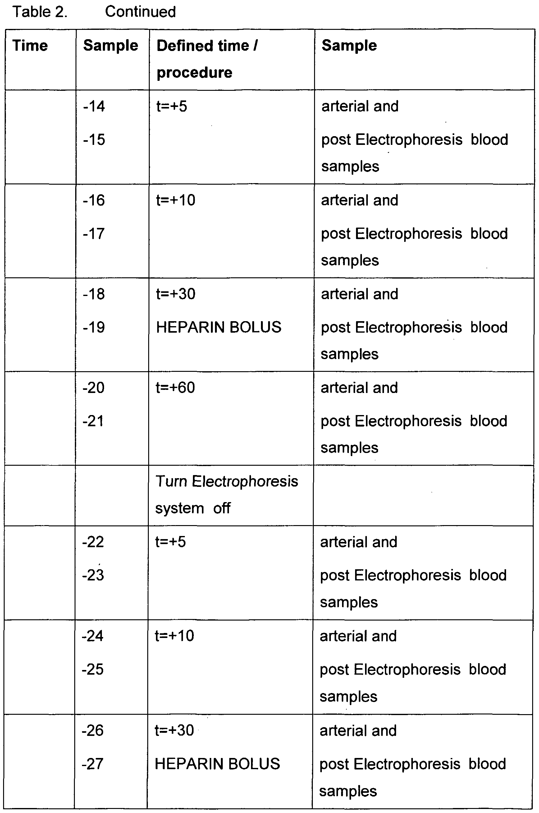
- Anticoagulation was achieved by an initial bolus of heparin (5000 IU) followed by 5000 IU boluses of heparin hourly during the procedure.
- the anticoagulation regime was supplemented with continuous infusion of heparin into the electrophoresis system line.
- the heparin infusion was achieved by using a pump 105 to transport the heparin solution 104 through a commercial sterile heparin line 106 into the arteriovenous circuit 101.
- a baseline blood sample was collected from the shunt.
- the arterial 101 and venous 113 lines were then connected to the carotid artery and jugular vein respectively and blood flow established through the blood circuit, using a Gambro blood pump 103.
- Blood was allowed to circulate for 10 minutes before arterial 102 and venous 112 blood samples were collected.
- plasma flow was then established for a further five minutes before another set of arterial 102 and venous 112 blood samples was collected.
- the electrophoresis system 108 process consisted of the cartridge together with a heat exchanger in line before the cartridge and the ancillary equipment required to maintain the temperature of the blood or plasma, along with a pump to transport the blood to the electrophoresis system 108.
- the electrophoresis system procedure was performed with and without electrical activation on whole blood ( Figure 24) or plasma ( Figure 25) separated from cellular components and reconstituted following processing.
- whole blood was treated, blood was taken directly from the arteriovenous circuit 101 through tube 107 using the pump associated with the electrophoresis system 108.
- the blood was then treated by the electrophoresis system 108 and re-integrated, through tube 109, in a blood collection vessel 110.
- the blood was the returned to the sheep 99 through tube 111 , sample port 112 and tubing 113.
- a membrane plasma separator 114 was used, in which plasma exited from the plasma outlet 115, while the cellular material of blood exited from the blood outlet 117.
- Plasma was transported from the plasma separator 114, with pump 116 from the plasma outlet port 115 and reconstituted with the cellular material through tube 117, into a blood collection vessel 110.
- the integrated whole blood in blood vessel 110 was transported back to the sheep 99, through tube 111 , sample port 112 and tube 113.
- plasma was taken directly from the plasma separator 114 using the pump associated with the electrophoresis system 108.
- the plasma was then treated by the electrophoresis system 108 and transported through tubing 109 and re-integrated with the cellular components of blood in the blood collection vessel 110.
- the blood was the returned to the sheep 99 via tubing 111 , sample port 112 and tubing 113.
- Pulse, respiration rate, oxygen partial pressure (Sp0 2 ) and temperature were recorded at 10 minute intervals during all procedures.
- Blood and urine samples taken before, during and after the procedure were tested to determine any effects of the procedure on the cellular components of the blood as well as liver function and electrolytes. Blood samples were tested by an external laboratory for the analytes set out in Table 5: Tablet Procedure chart group 1 -whole blood with the electrophoresis system
- a weighted mean equivalent to an "area under the curve" was calculated for each one-hour period. This was calculated using the trapezoidal method. Note that when the average is calculated this way, the observations at 30 and 60 minutes carry much more weight than the observations at 0, 5 and 10 minutes. Note also that the pre-procedure (baseline) sample was not included in the calculation.
- the sheep factor is a catch-all that includes the random variation due the particular sheep selected for the procedure as well as all other day-to-day variation that cannot otherwise be accounted for.
- the concentration of a solute during the procedure might reflect the baseline, pre-procedure level. Ideally, the analysis can take this into account.
- the experiment design was of a 'repeated measures type' with two between-sheep factors, PL and GR, and two within-sheep factors, P and SI.
- Tests of the between-sheep factors, PL and GR were performed using analysis of co-variance on the means, calculated over the eight observations (4 periods x 2 sites) for each sheep, with the baseline level (the initial arterial sample) as the covariate. This method is recommended by Frison and Pocock (Frison L and Pocock SJ: Repeated measures in clinical trials: Analysis using mean summary statistics and its implications for design. Statistics in Medicine 11 : 1685-1704 (1992)) as the optimum way to adjust for the effect of variation in baseline levels.
- this method determines if the average level X in the experiments with the electrophoresis system in the circuit different from the average level in experiments without the electrophoresis system .
- Overall tests of the within-sheep factors, P and SI, and their interactions with PL and GR were performed using repeated-measures analysis of variance with the Huynh-Feldt correction factor (StataCorp. 1999. Stata Statistical Software: Release 6.0. College Station TX: Stata Corporation). These tests determine if the levels of Y changed during the procedure and whether the pattern of differences was affected by the electrophoresis system .
- a number of contrasts among the levels of P and SI were calculated for each sheep and each contrast was tested as the dependent variable in an ordinary univariate analysis of variance against PL and GR, including an additional test that the overall mean of the variable is different from zero.
- the F-ratio for GR would be a test of the P1 ⁇ GR interaction.
- Four contrasts among the time periods were tested. The coefficients are listed below along with the contrast S1 comparing arterial and venous.
- P1 is a contrast between B and A periods (corresponding to power on and power off when the electrophoresis system was used).
- P2 compares the second AB cycle with the first and P3 compares the B-A difference in the second cycle with the B- A difference in the first cycle.
- These three contrasts are orthogonal (the dot products of their coefficient vectors is zero) and together they account for all variation among the 4 time periods. Contrast P4 simply compares B and A during the first cycle.
- biocompatibility does not have a single, comprehensive and uniformly accepted definition.
- Jonathan Black Black J. Biological performance of materials: fundamentals of biocompatibility, 2nd edition, Dekker, New York, 1992) described it as "biological performance in a specific application that is judged suitable to that situation”. It is a concept that assesses the effect of a procedure, device or material on the physiology of a living system.
- the aim of this study was to assess the biocompatibility of the electrophoresis system.
- the electrophoresis system poses several issues relating to biocompatibility. Firstly, the design and materials of the cartridge, the membranes, the heat exchanger and the tubing may all affect the biocompatibility of the procedure. Secondly, the effect of the electrical field on the blood or plasma was yet to be established in an ex vivo setting.
- GLDH levels were stable in the control and treatment animals whereas alkaline phosphatase levels tended to increase over the course of the procedures.
- AST and GammaGT levels were relatively stable for all animals throughout the procedures with a slow decline in AST seen over the course of the procedures in most animals.
- the statistical analysis suggested that there was a significant difference between arterial and venous levels of Gamma GT unrelated to the presence of the electrophoresis system.
- B-OHBUT and CK levels were relatively stable for all animals throughout the procedures with a slow increase seen over the course of the procedures in most animals.
- One electrophoresis system treated blood animal exhibited a sharp decrease in CK levels when the electrical field was deactivated for the first time. The level in this animal then remained low for the remainder of the procedure.
- Venous levels of CK were consistently higher than arterial by an average of 2.5 U/l across all groups and periods.
- Biochemical values such as urea, creatinine and most electrolytes were relatively stable during the extracorporeal procedures as expected.
- Bicarbonate levels showed small increases in nearly all animals with the pattern consistent across the four groups. Venous levels were consistently lower than arterial by an average of 0.6 mM across all groups and periods.
- WCC White cell numbers
- Lymphocyte, eosinophil and monocyte levels were relatively stable in most animals.
- Lactate buffer and various concentrations of Fresenius buffer were used in blood and plasma electrophoresis system runs.
- the electrophoresis system runs were sampled at 0, 5, 10, 15, 30, 45 and 60 minutes. Blood was centrifuged and the plasma analysed for free haemoglobin. The blood plasma and plasma samples were assayed for electrolyte and glucose concentrations by the NATA accredited pathology laboratory.
- electrolytes During the larger volume uraemic waste removal experiments, electrolytes
- lactate buffer (Baxter PD dialysate) replaced the acetate-bicarbonate dialysate.
- the concentration of acetate- bicarbonate buffer was reduced. It was anticipated that by reducing the concentration of sodium chloride in the dialysate, its consequential build up would be reduced. By reducing the sodium concentration of the dialysate from 139.8 mM to 130 mM, only a minor change in sodium and chloride concentrations were observed after The electrophoresis system treatment (Figure 29).
- Dialysate could be prepared by replacing excess sodium and chloride with other electrolytes which were found to be reduced during the treatment.
- electrolytes could be replaced with other non-ionic molecules which will maintain the isotonicity of the dialysate.
- Another alternative would be to change the current re-circulating dialysate system, to a single pass system.
- the mortality rate for liver failure is 80-90% with a rapid death (a week or two), without an effective liver transplant.
- a rapid death a week or two
- the build-up of toxic nitrogenous products i.e. ammonia, trytophan, benzodiazepines, GABA, acetaminophen
- foreign biological components i.e. ammonia, trytophan, benzodiazepines, GABA, acetaminophen
- electrolytes and water imbalance within the body can cause serious damage.
- Current technologies are developing techniques to bridge the time until a suitable transplant can be identified. These technologies use a variety of filter types to remove the build up of excess molecules and toxins which have accumulated in blood.
- the electrophoresis system has been shown capable of removing metabolic products, excess salts, selective protein removal and control fluid imbalances as well as being a biocompatible system. Therefore, the system according to the present invention has the potential to be incorporated in liver dialysis treatments, as an adjunct, to assist in increasing the bridge to finding
- uremic toxins which had a low charge to mass ratio under physiological conditions, such as urea (neutral) were not removed as efficiently by the system and methods according to the present invention.
- problematic uremic toxins such as phosphate and charged middle molecule toxins, would be enhanced with the use of The electrophoresis system as an adjunct to existing technologies.
- the system according to the present invention could be combined with any of the existing medical technologies, including haemodialysis, hemofiltration, hemodialfiltration, REDY sorbent haemodialysis and/or even plasmaphersis.
- the heat exchanger can be modified to be made or coated with different biocompatible material such as metals (eg titanium) and polymers;
- the entire heat exchanger system could be modified to suit the application by introducing different means to control temperature eg peltier cooling systems;
- Modification to the separation unit such as change the inlet and outlet ports to accommodate viscous biological fluids such as blood and plasma; modify the cartridge to be less turbulent and angular; modify the grid structure to be thinner or incorporate different patterns to suit the biological fluid being processed; include measures to reduce or control the effects of venous or arterial pressures on the cartridge; introduce a means to introduce anticoagulant in an automated fashion before the heat exchanger and/or the separation unit; use pumps or devices which direct or transport fluid and do not damage or lysis cells; appropriate monitors to measure fluid parameters, clearance levels and safety aspects during the treatment; use buffers which incorporate a range of constituents to suit individual treatments; use buffers which enhance treatments; use single or recirculating buffer systems, or a partial recirculating system; control fluid levels; use the properties of different membranes to enhance treatments.
- the electrically motivated dialysis according to the present invention uses a fundamentally different motive force, namely an electric field, to remove contaminating solutes from patient blood.
- an electric field for renal dialysis applications, or for the removal of uraemic toxins from blood, has not been previously described in the literature.
- the removal of solutes and small proteins from patient blood according to the present invention depends on the electrophoretic mobility of the and small proteins, which is a function of the contaminating molecules charge to mass ratio. This basis for solute removal is different to previous modes of solute removal, which are based purely on the size or diffusive mobility of the contaminant molecule.
- middle molecules A middle molecule that plagues long term renal dialysis patients is ⁇ 2-microglobulin, which accumulates in the blood and then polymerises in bones and joints, with significant clinical consequences.
- ⁇ 2-microglobulin A middle molecule that plagues long term renal dialysis patients is ⁇ 2-microglobulin, which accumulates in the blood and then polymerises in bones and joints, with significant clinical consequences.
- electrophoresis was used in experiments to reduced ⁇ 2-microglobulin.
- Dialysis treatments not only needed to be able to remove waste metabolites and other problematic molecules, these treatment modalities needed to be safe for the patient.
- An important aspect of safety is the biocompatibility of the membrane materials and treatment system. Then present investigations showed that membrane-based electrophoresis treatments had improved biocompatibility in the areas of coagulation and complement when compared to existing membrane types currently in clinical use. Similarly, there were no adverse effects of membrane-based electrophoresis processing on any of these standard biocompatibility measurements.
Landscapes
- Health & Medical Sciences (AREA)
- Heart & Thoracic Surgery (AREA)
- Engineering & Computer Science (AREA)
- Urology & Nephrology (AREA)
- Vascular Medicine (AREA)
- Life Sciences & Earth Sciences (AREA)
- Animal Behavior & Ethology (AREA)
- Veterinary Medicine (AREA)
- Hematology (AREA)
- Anesthesiology (AREA)
- Biomedical Technology (AREA)
- General Health & Medical Sciences (AREA)
- Public Health (AREA)
- Water Supply & Treatment (AREA)
- Chemical & Material Sciences (AREA)
- Chemical Kinetics & Catalysis (AREA)
- Emergency Medicine (AREA)
- Cardiology (AREA)
- Electrochemistry (AREA)
- Environmental & Geological Engineering (AREA)
- Molecular Biology (AREA)
- External Artificial Organs (AREA)
- Electrostatic Separation (AREA)
Abstract
Description




Claims
Priority Applications (3)
| Application Number | Priority Date | Filing Date | Title |
|---|---|---|---|
| AU2002325669A AU2002325669B2 (en) | 2001-09-04 | 2002-09-04 | Removal of metabolic components from blood |
| JP2003524705A JP4181038B2 (en) | 2001-09-04 | 2002-09-04 | Removal of metabolic components from blood |
| EP02759922A EP1434645A4 (en) | 2001-09-04 | 2002-09-04 | EXTRACTION OF METABOLIC BLOOD CONSTITUENTS |
Applications Claiming Priority (2)
| Application Number | Priority Date | Filing Date | Title |
|---|---|---|---|
| AUPR7485 | 2001-09-04 | ||
| AUPR7485A AUPR748501A0 (en) | 2001-09-04 | 2001-09-04 | Renal dialysis |
Publications (1)
| Publication Number | Publication Date |
|---|---|
| WO2003020403A1 true WO2003020403A1 (en) | 2003-03-13 |
Family
ID=3831435
Family Applications (1)
| Application Number | Title | Priority Date | Filing Date |
|---|---|---|---|
| PCT/AU2002/001217 Ceased WO2003020403A1 (en) | 2001-09-04 | 2002-09-04 | Removal of metabolic components from blood |
Country Status (5)
| Country | Link |
|---|---|
| EP (1) | EP1434645A4 (en) |
| JP (1) | JP4181038B2 (en) |
| CN (1) | CN1551795A (en) |
| AU (1) | AUPR748501A0 (en) |
| WO (1) | WO2003020403A1 (en) |
Cited By (17)
| Publication number | Priority date | Publication date | Assignee | Title |
|---|---|---|---|---|
| EP2087916A1 (en) | 2008-02-11 | 2009-08-12 | ICinnovation BV | Electrosorption device for the purification of blood and other fluids |
| EP2446908A1 (en) | 2010-11-02 | 2012-05-02 | ICinnovation BV | Electrosorption and decomposition device for the purification of blood and other fluids |
| JP5121449B2 (en) * | 2005-05-26 | 2013-01-16 | 株式会社サインポスト | Presentation method of disease onset prediction |
| WO2014096499A1 (en) * | 2012-12-20 | 2014-06-26 | Servicio Andaluz De Salud | Device for inducing therapeutic hypothermia |
| EP2862584A1 (en) | 2013-10-21 | 2015-04-22 | ICinnovation BV | Electrosorption and decomposition device for the purification of blood and other fluids |
| WO2016187217A2 (en) | 2015-05-18 | 2016-11-24 | The Board Of Trustees Of The Leland Stanford Junior University | Methods and compositions for treating aging-associated impairments |
| US9682181B2 (en) | 2013-01-04 | 2017-06-20 | Fresenius Medical Care Deutschland Gmbh | Device and method for removing protein-bound toxins from the blood of patients using a high-frequency, electromagnetic field and an electrostatic direct current field |
| US10487148B2 (en) | 2010-01-28 | 2019-11-26 | The Board Of Trustees Of The Leland Stanford Junior University | Methods and compositions for treating aging-associated impairments |
| US10525187B2 (en) | 2012-12-21 | 2020-01-07 | Fresenius Medical Care Deutschland Gmbh | Apparatus for removing protein-bound toxins from blood plasma |
| US10617744B2 (en) | 2015-06-15 | 2020-04-14 | The Board Of Trustees Of The Leland Stanford Junior University | Methods and compositions for treating aging-associated conditions |
| US10626399B2 (en) | 2010-01-28 | 2020-04-21 | The Board Of Trustees Of The Leland Stanford Junior University | Methods of treating cognitive symptoms of an aging-associated impairment by modulating C-C chemokine receptor type 3 (CCR3) |
| US10688130B2 (en) | 2013-12-09 | 2020-06-23 | The Board Of Trustees Of The Leland Stanford Junior University | Methods and compositions for treating aging-associated conditions |
| US10688154B2 (en) | 2011-04-08 | 2020-06-23 | The Board Of Trustees Of The Leland Stanford Junior University | Methods of neuroprotection involving macrophage colony stimulating factor receptor agonists |
| CN112040999A (en) * | 2018-08-17 | 2020-12-04 | 华盛顿大学 | Apparatus and method for photo-oxidation of urea |
| US10905779B2 (en) | 2013-12-09 | 2021-02-02 | The Board Of Trustees Of The Leland Stanford Junior University | Methods for screening human blood products comprising plasma using immunocompromised rodent models |
| US11236340B2 (en) | 2010-01-28 | 2022-02-01 | The Board Of Trustees Of The Leland Stanford Junior University | Method of reducing the effects of aging-associated impairment of neurogenesis comprising modulating c-c chemokine receptor type 3 (CCR3) |
| US11383010B2 (en) | 2011-07-05 | 2022-07-12 | Baxter International Inc. | Method of dialysis for removing protein-bound toxins from the blood of patients using high-frequency electromagnetic fields |
Families Citing this family (9)
| Publication number | Priority date | Publication date | Assignee | Title |
|---|---|---|---|---|
| JP4947693B2 (en) * | 2006-07-14 | 2012-06-06 | 学校法人北里研究所 | Method for separating albumin and biomolecules bound to albumin |
| KR101848527B1 (en) * | 2010-06-28 | 2018-04-12 | 옵토플루이드 테크놀로지스 오유 | A method and device for determining content of the middle and protein bound uremic toxins in a biological fluid |
| DE102012025052A1 (en) * | 2012-12-20 | 2014-06-26 | Fresenius Medical Care Deutschland Gmbh | Hämodiafiltrationsverfahren |
| KR101552519B1 (en) | 2014-05-13 | 2015-09-14 | 계명대학교 산학협력단 | Fat separation system and method for prevention of cardiovascular disease |
| CN104404008A (en) * | 2014-12-10 | 2015-03-11 | 辽宁省海洋水产科学研究院 | Method for separating and purifying sea urchin multifunctional actin |
| CN109358105B (en) * | 2018-09-26 | 2021-01-05 | 陕西绿盾环境工程研究院有限公司 | Device and method for reducing kasugamycin content in kasugamycin residues |
| CN110817802B (en) * | 2019-10-24 | 2023-01-24 | 邯郸钢铁集团有限责任公司 | System and method for preparing ultrapure hydrogen by using composite purification process |
| CN111407943A (en) * | 2020-04-25 | 2020-07-14 | 宁波高新区零零七工业设计有限公司 | Virus adsorption separation harmless treatment equipment and treatment method thereof |
| CN113181458A (en) * | 2021-04-22 | 2021-07-30 | 浙大宁波理工学院 | Bioreactor for artificial liver |
Citations (2)
| Publication number | Priority date | Publication date | Assignee | Title |
|---|---|---|---|---|
| US4276140A (en) * | 1980-01-10 | 1981-06-30 | Ionics Inc. | Electrodialysis apparatus and process for fractionating protein mixtures |
| JPS62172965A (en) * | 1986-01-27 | 1987-07-29 | 日東電工株式会社 | Blood treatment apparatus |
Family Cites Families (4)
| Publication number | Priority date | Publication date | Assignee | Title |
|---|---|---|---|---|
| US3870617A (en) * | 1971-03-30 | 1975-03-11 | Rhone Poulenc Sa | Apparatus for forced flow electrophoresis |
| US3989613A (en) * | 1973-05-16 | 1976-11-02 | The Dow Chemical Company | Continuous balanced flow fixed boundary electrophoresis |
| FR2568485B1 (en) * | 1984-08-06 | 1990-03-23 | Rhone Poulenc Rech | PROTEIN-CONTAINING ELECTROPHORESIS APPARATUS FOR USE, IN PARTICULAR FOR FRACTIONATION OF HUMAN PLASMA |
| AUPP576598A0 (en) * | 1998-09-07 | 1998-10-01 | Life Therapeutics Limited | Cassette for macromolecule purification |
-
2001
- 2001-09-04 AU AUPR7485A patent/AUPR748501A0/en not_active Abandoned
-
2002
- 2002-09-04 CN CNA028173619A patent/CN1551795A/en active Pending
- 2002-09-04 JP JP2003524705A patent/JP4181038B2/en not_active Expired - Fee Related
- 2002-09-04 EP EP02759922A patent/EP1434645A4/en not_active Withdrawn
- 2002-09-04 WO PCT/AU2002/001217 patent/WO2003020403A1/en not_active Ceased
Patent Citations (2)
| Publication number | Priority date | Publication date | Assignee | Title |
|---|---|---|---|---|
| US4276140A (en) * | 1980-01-10 | 1981-06-30 | Ionics Inc. | Electrodialysis apparatus and process for fractionating protein mixtures |
| JPS62172965A (en) * | 1986-01-27 | 1987-07-29 | 日東電工株式会社 | Blood treatment apparatus |
Non-Patent Citations (2)
| Title |
|---|
| DATABASE WPI Week 198736, Derwent World Patents Index; Class S03, AN 1987-252867, XP003005722 * |
| See also references of EP1434645A4 * |
Cited By (24)
| Publication number | Priority date | Publication date | Assignee | Title |
|---|---|---|---|---|
| JP5121449B2 (en) * | 2005-05-26 | 2013-01-16 | 株式会社サインポスト | Presentation method of disease onset prediction |
| EP2092944A2 (en) | 2008-02-11 | 2009-08-26 | ICinnovation BV | Electrosorption device for the purification of blood and other fluids |
| EP2087916A1 (en) | 2008-02-11 | 2009-08-12 | ICinnovation BV | Electrosorption device for the purification of blood and other fluids |
| US11912998B2 (en) | 2010-01-28 | 2024-02-27 | The Board Of Trustees Of The Leland Stanford Junior University | Method of treating aging-associated cognitive impairment by reducing CCR3 |
| US10487148B2 (en) | 2010-01-28 | 2019-11-26 | The Board Of Trustees Of The Leland Stanford Junior University | Methods and compositions for treating aging-associated impairments |
| US10626399B2 (en) | 2010-01-28 | 2020-04-21 | The Board Of Trustees Of The Leland Stanford Junior University | Methods of treating cognitive symptoms of an aging-associated impairment by modulating C-C chemokine receptor type 3 (CCR3) |
| US11236340B2 (en) | 2010-01-28 | 2022-02-01 | The Board Of Trustees Of The Leland Stanford Junior University | Method of reducing the effects of aging-associated impairment of neurogenesis comprising modulating c-c chemokine receptor type 3 (CCR3) |
| WO2012060700A1 (en) | 2010-11-02 | 2012-05-10 | Icinnovation Bv | Electrosorption and decomposition device for the purification of blood and other fluids |
| EP2446908A1 (en) | 2010-11-02 | 2012-05-02 | ICinnovation BV | Electrosorption and decomposition device for the purification of blood and other fluids |
| US10688154B2 (en) | 2011-04-08 | 2020-06-23 | The Board Of Trustees Of The Leland Stanford Junior University | Methods of neuroprotection involving macrophage colony stimulating factor receptor agonists |
| US11383010B2 (en) | 2011-07-05 | 2022-07-12 | Baxter International Inc. | Method of dialysis for removing protein-bound toxins from the blood of patients using high-frequency electromagnetic fields |
| WO2014096499A1 (en) * | 2012-12-20 | 2014-06-26 | Servicio Andaluz De Salud | Device for inducing therapeutic hypothermia |
| US10525187B2 (en) | 2012-12-21 | 2020-01-07 | Fresenius Medical Care Deutschland Gmbh | Apparatus for removing protein-bound toxins from blood plasma |
| US9682181B2 (en) | 2013-01-04 | 2017-06-20 | Fresenius Medical Care Deutschland Gmbh | Device and method for removing protein-bound toxins from the blood of patients using a high-frequency, electromagnetic field and an electrostatic direct current field |
| EP2862584A1 (en) | 2013-10-21 | 2015-04-22 | ICinnovation BV | Electrosorption and decomposition device for the purification of blood and other fluids |
| US9878084B2 (en) | 2013-10-21 | 2018-01-30 | Icinnovation Bv | Redox controlled electrosorption and decomposition device for the purification of blood and other fluids |
| WO2015060716A1 (en) | 2013-10-21 | 2015-04-30 | Icinnovation Bv | Redox controlled electrosorption and decomposition device for the purification of blood and other fluids |
| US10688130B2 (en) | 2013-12-09 | 2020-06-23 | The Board Of Trustees Of The Leland Stanford Junior University | Methods and compositions for treating aging-associated conditions |
| US10905779B2 (en) | 2013-12-09 | 2021-02-02 | The Board Of Trustees Of The Leland Stanford Junior University | Methods for screening human blood products comprising plasma using immunocompromised rodent models |
| EP3892315A1 (en) | 2015-05-18 | 2021-10-13 | The Board of Trustees of the Leland Stanford Junior University | Methods and compositions for treating aging-associated impairments |
| WO2016187217A2 (en) | 2015-05-18 | 2016-11-24 | The Board Of Trustees Of The Leland Stanford Junior University | Methods and compositions for treating aging-associated impairments |
| US10617744B2 (en) | 2015-06-15 | 2020-04-14 | The Board Of Trustees Of The Leland Stanford Junior University | Methods and compositions for treating aging-associated conditions |
| US11141469B2 (en) | 2015-06-15 | 2021-10-12 | The Board Of Trustees Of The Leland Stanford Junior University | Methods and compositions for treating aging-associated conditions |
| CN112040999A (en) * | 2018-08-17 | 2020-12-04 | 华盛顿大学 | Apparatus and method for photo-oxidation of urea |
Also Published As
| Publication number | Publication date |
|---|---|
| JP2005527247A (en) | 2005-09-15 |
| EP1434645A4 (en) | 2006-12-13 |
| JP4181038B2 (en) | 2008-11-12 |
| AUPR748501A0 (en) | 2001-09-27 |
| CN1551795A (en) | 2004-12-01 |
| EP1434645A1 (en) | 2004-07-07 |
Similar Documents
| Publication | Publication Date | Title |
|---|---|---|
| US7066900B2 (en) | Removal of metabolic components from blood | |
| EP1434645A1 (en) | Removal of metabolic components from blood | |
| JP2009056323A (en) | Blood-related dialysis and processing | |
| EP3383449B1 (en) | An implantable renal replacement therapy | |
| EP2214751A1 (en) | Method and device for differentiation of substances | |
| US20110042313A1 (en) | Bioequivalence dialysis | |
| AU2002325669B2 (en) | Removal of metabolic components from blood | |
| US20210113753A1 (en) | Dialysate-free wearable renal replacement system | |
| de Francisco et al. | Hemodiafiltration with on-line endogenous reinfusion | |
| AU2002325669A1 (en) | Removal of metabolic components from blood | |
| Opatrný Jr et al. | Does treatment modality have an impact on anemia in patients with chronic renal failure? Effect of low‐and high‐flux biocompatible dialysis | |
| Kurihara et al. | Continuous hemofiltration model using porcine blood for comparing filter life | |
| Kawanishi et al. | How to prescribe hemodialysis or hemodiafiltration in order to ameliorate dialysis-related symptoms and complications | |
| Cantaluppi et al. | Blood purification for sepsis | |
| JP4947693B2 (en) | Method for separating albumin and biomolecules bound to albumin | |
| Malchesky | 8.3 EXTRACORPOREAL ARTIFICIAL ORGANS | |
| Orihara et al. | Can ion exchange resins be used for extracorporeal circulation therapy of hyperkalemia? | |
| Bazaev et al. | A wearable device for low-flow detoxification of human body by peritoneal dialysis | |
| AU2004200567B2 (en) | Blood-Related Dialysis and Treatment | |
| Gagel et al. | Advances in Membrane, Dialyzer Design and Related Monitoring Technology for Hemodiafiltration: Translating Bench Side Innovations to Bedside Applications | |
| Lee et al. | Hematologic and biochemical effects of the gradiflow in an ex vivo ovine model | |
| Bazaev et al. | In vitro and in vivo trials of wearable artificial kidney | |
| Stefoni et al. | Combined hemodialysis-hemoperfusion treatment reduces the time of substitutive therapy in chronic uremia | |
| Huang | A study of novel and optimal technology for hemodialysis | |
| Mydlik et al. | Middle molecular substances in acute renal failure |
Legal Events
| Date | Code | Title | Description |
|---|---|---|---|
| AK | Designated states |
Kind code of ref document: A1 Designated state(s): AE AG AL AM AT AU AZ BA BB BG BR BY BZ CA CH CN CO CR CU CZ DE DK DM DZ EC EE ES FI GB GD GE GH GM HR HU ID IL IN IS JP KE KG KP KR KZ LC LK LR LS LT LU LV MA MD MG MK MN MW MX MZ NO NZ OM PH PL PT RO RU SD SE SG SI SK SL TJ TM TN TR TT TZ UA UG UZ VC VN YU ZA ZM ZW Kind code of ref document: A1 Designated state(s): AE AG AL AM AT AU AZ BA BB BG BY BZ CA CH CN CO CR CU CZ DE DM DZ EC EE ES FI GB GD GE GH HR HU ID IL IN IS JP KE KG KP KR LC LK LR LS LT LU LV MA MD MG MN MW MX MZ NO NZ OM PH PL PT RU SD SE SG SI SK SL TJ TM TN TR TZ UA UG UZ VC VN YU ZA ZM |
|
| AL | Designated countries for regional patents |
Kind code of ref document: A1 Designated state(s): GH GM KE LS MW MZ SD SL SZ TZ UG ZM ZW AM AZ BY KG KZ MD RU TJ TM AT BE BG CH CY CZ DE DK EE ES FI FR GB GR IE IT LU MC NL PT SE SK TR BF BJ CF CG CI CM GA GN GQ GW ML MR NE SN TD TG Kind code of ref document: A1 Designated state(s): GH GM KE LS MW MZ SD SL SZ UG ZM ZW AM AZ BY KG KZ RU TJ TM AT BE BG CH CY CZ DK EE ES FI FR GB GR IE IT LU MC PT SE SK TR BF BJ CF CG CI GA GN GQ GW ML MR NE SN TD TG |
|
| 121 | Ep: the epo has been informed by wipo that ep was designated in this application | ||
| DFPE | Request for preliminary examination filed prior to expiration of 19th month from priority date (pct application filed before 20040101) | ||
| WWE | Wipo information: entry into national phase |
Ref document number: 2002325669 Country of ref document: AU |
|
| WWE | Wipo information: entry into national phase |
Ref document number: 2003524705 Country of ref document: JP |
|
| WWE | Wipo information: entry into national phase |
Ref document number: 20028173619 Country of ref document: CN |
|
| WWE | Wipo information: entry into national phase |
Ref document number: 2002759922 Country of ref document: EP |
|
| WWP | Wipo information: published in national office |
Ref document number: 2002759922 Country of ref document: EP |
|
| WWG | Wipo information: grant in national office |
Ref document number: 2002325669 Country of ref document: AU |