WO2004112840A2 - Peptide conjugate for magnetic resonance imaging - Google Patents

Peptide conjugate for magnetic resonance imaging Download PDF

Info

Publication number
WO2004112840A2
WO2004112840A2 PCT/IB2004/002210 IB2004002210W WO2004112840A2 WO 2004112840 A2 WO2004112840 A2 WO 2004112840A2 IB 2004002210 W IB2004002210 W IB 2004002210W WO 2004112840 A2 WO2004112840 A2 WO 2004112840A2
Authority
WO
WIPO (PCT)
Prior art keywords
peptide
pro
ala
leu
patient
Prior art date
Legal status (The legal status is an assumption and is not a legal conclusion. Google has not performed a legal analysis and makes no representation as to the accuracy of the status listed.)
Ceased
Application number
PCT/IB2004/002210
Other languages
French (fr)
Other versions
WO2004112840A3 (en
Inventor
Marc Port
Olivier Rousseaux
Christelle Medina
Claire Corot
Irène GUILBERT
Jean Sébastien RAYNAUD
Current Assignee (The listed assignees may be inaccurate. Google has not performed a legal analysis and makes no representation or warranty as to the accuracy of the list.)
Guerbet SA
Original Assignee
Guerbet SA
Priority date (The priority date is an assumption and is not a legal conclusion. Google has not performed a legal analysis and makes no representation as to the accuracy of the date listed.)
Filing date
Publication date
Priority claimed from FR0307694A external-priority patent/FR2856689A1/en
Application filed by Guerbet SA filed Critical Guerbet SA
Priority to DE602004030789T priority Critical patent/DE602004030789D1/en
Priority to US10/560,807 priority patent/US20060239913A1/en
Priority to EP04743873A priority patent/EP1635878B1/en
Priority to JP2006516596A priority patent/JP2007521254A/en
Priority to AT04743873T priority patent/ATE493151T1/en
Publication of WO2004112840A2 publication Critical patent/WO2004112840A2/en
Publication of WO2004112840A3 publication Critical patent/WO2004112840A3/en
Anticipated expiration legal-status Critical
Ceased legal-status Critical Current

Links

Classifications

    • A—HUMAN NECESSITIES
    • A61—MEDICAL OR VETERINARY SCIENCE; HYGIENE
    • A61K—PREPARATIONS FOR MEDICAL, DENTAL OR TOILETRY PURPOSES
    • A61K49/00—Preparations for testing in vivo
    • A61K49/0002—General or multifunctional contrast agents, e.g. chelated agents
    • A—HUMAN NECESSITIES
    • A61—MEDICAL OR VETERINARY SCIENCE; HYGIENE
    • A61K—PREPARATIONS FOR MEDICAL, DENTAL OR TOILETRY PURPOSES
    • A61K49/00—Preparations for testing in vivo
    • A61K49/06—Nuclear magnetic resonance [NMR] contrast preparations; Magnetic resonance imaging [MRI] contrast preparations
    • A61K49/08—Nuclear magnetic resonance [NMR] contrast preparations; Magnetic resonance imaging [MRI] contrast preparations characterised by the carrier
    • A61K49/085—Nuclear magnetic resonance [NMR] contrast preparations; Magnetic resonance imaging [MRI] contrast preparations characterised by the carrier conjugated systems
    • A—HUMAN NECESSITIES
    • A61—MEDICAL OR VETERINARY SCIENCE; HYGIENE
    • A61K—PREPARATIONS FOR MEDICAL, DENTAL OR TOILETRY PURPOSES
    • A61K49/00—Preparations for testing in vivo
    • A61K49/06—Nuclear magnetic resonance [NMR] contrast preparations; Magnetic resonance imaging [MRI] contrast preparations
    • A61K49/08—Nuclear magnetic resonance [NMR] contrast preparations; Magnetic resonance imaging [MRI] contrast preparations characterised by the carrier
    • A61K49/10—Organic compounds
    • A61K49/14—Peptides, e.g. proteins
    • C—CHEMISTRY; METALLURGY
    • C07—ORGANIC CHEMISTRY
    • C07K—PEPTIDES
    • C07K5/00—Peptides containing up to four amino acids in a fully defined sequence; Derivatives thereof
    • C07K5/04—Peptides containing up to four amino acids in a fully defined sequence; Derivatives thereof containing only normal peptide links
    • C07K5/10—Tetrapeptides
    • C07K5/1002—Tetrapeptides with the first amino acid being neutral
    • C07K5/1005—Tetrapeptides with the first amino acid being neutral and aliphatic

Definitions

  • the invention relates to new compounds and compositions for the imaging diagnostic of pathologies, namely for cardiovascular diseases, more precisely atherosclerosis disease.
  • These compounds are contrast agents useful namely in the field of magnetic resonance imaging MRI, but also in other imaging fields such as nuclear medicine, X-ray, ultrasounds, optical imaging.
  • These compounds comprise at least a targeting moiety linked to at least a signal moiety.
  • a targeting entity is capable of targeting at least one marker of a pathologic state and/or area that are over or under expressed in, a pathologic state and/or area compared to the non pathologic ones. These compounds are called specific compounds, the targeting entity being called biovector. Numerous signal entities/moieties are already known, such as linear or macrocyclic chelates of paramagnetic metal ion for MRI and of radionucleides for nuclear medicine.
  • Such chelates are described in the documents EP 71 564, EP 448 191 , WO 02/48119, US 6 399 043, WO 01/51095, EP 203 962 , EP 292 689 , EP 425 571 , EP 230 893 , EP 405 704, EP 290 047, US 6 123 920, EP 292 689, EP 230 893, US 6 403 055, WO 02/40060, US 6 458 337, US 6 264 914, US 6 221 334, WO 95/31444, US 5 573 752, US 5 358 704.
  • Chelates commonly used are for example DTPA, DTPA BMA, DTPA BOPTA, DO3A, HPDO3A, TETA, DOTA (1 , 4,7,10-tetracyclododecane-N,N , ,N",N"'-tetraacetic acid), PCTA and their derivatives.
  • Products on the market are namely for example Dotarem ® and Magnevist ⁇ he signal is measured in MRI by the relaxivity in water which is in the order of 3 to 10 mM-1s-1 Gd-1for such chelates.
  • a broad spectrum of clinically different diseases such as myocardial infarction, stroke, abdominal aneurysms and lower limb ischemia are basically related to atherosclerosis. Most of their acute manifestations share a common pathogenic feature : rupture of an atherosclerotic plaque with superimposed thrombosis. Plaque rupture, which accounts for approximately 70% of fatal acute myocardial infarctions and of symptomatic carotid lesions, is the ultimate complication of a vulnerable plaque. Vulnerable plaques include thrombosis-prone plaques as well as those with a high probability of undergoing rapid progression, thus becoming culprit plaques. They are characterized by a large lipid core, a thin cap and macrophage-dense inflammation on or beneath their surface. The risk of acute ischemic event for an individual is determined by the number of vulnerable plaques and the current challenge is to stratify such a risk.
  • Angiography is strictly an anatomic imaging tool and is unable to evaluate coronary plaque dimension and composition. Other modalities are catheter- based and, therefore, have a limited clinical applicability.
  • Intravascular ultrasound provides some information on plaque morphology but image resolution and sensitivity are still insufficient to reliably distinguish vulnerable plaque deposits.
  • Optical coherence tomography better delineates between intimal wall and plaque but its penetration depth is low.
  • Angioscopy may be used to detect lipid-rich plaques and to visualize thrombus, whereas thermography is very sensitive to superficial inflammation.
  • both techniques are unable to examine the deep layers of the arterial wall and to estimate cap thickness.
  • MRI magnetic resonance imaging
  • MMPs matrix metalloproteinases
  • a contrast molecule which can be detected namely with MRI, will be useful to image MMP activity and to non invasively detect vulnerable plaques and improve patients' risk stratification.
  • MMP-8 per milligram of tissue in advanced atherosclerotic carotid lesions.
  • MMP-13 As the sensitivity of MRI in vivo is relatively low compared to scintigraphic imaging techniques, for instance, there is a need to compensate for the low levels of MMPs in the lesions in order to generate a sufficient signal intensity. This requirement may be achieved by using a compound which targets nonselectively the majority of MMPs and, thereby, will allow a high local concentration of the contrast agent.
  • the applicant has now prepared imaging compounds comprising a biovector with good affinity for MMP-1 , MMP-2, MMP-3, MMP-8, MMP-9 ; in particular MMP-3 are surexpressed in lesional plaques. Specific compounds for the targeting of MMPs are described in the prior art.
  • WO 01/60416 describes compounds that comprise a targeting entity towards MMPs coupled to a linear or macrocyclic chelate signal entity.
  • a targeting entity towards MMPs coupled to a linear or macrocyclic chelate signal entity According to applicant's knowledge based namely on biological assays, such compounds of the prior art are not sufficiently efficient for a very satisfying in vivo diagnosis, due to their relative low relaxivity which is in the order of 5 to 10 mMol-1s-1 Gd-1 and/or their lack of affinity or selectivity.
  • affinity or selectivity there still remains a serious need for new products that are effectively efficient in imaging diagnostic in vivo
  • the peptidic MMP inhibitor used as biovector by the applicant is described in Biochemical and Biophysical research Communications, vol 199, 3, 1994, pages 1442-1446 and in US 5 100 874 incorporated by reference. But the coupling to a signal entity of this particular peptidic MMP inhibitor, among the huge amount of possible MMP targeting entities and inhibitors known with equivalent or higher affinity or selectivity for MMPs, was neither described nor suggested for diagnostic imaging and specially for cardiovascular disease diagnostic. Further, according to the applicant's knowledge, the clinical trials relating to MMP target entities in the therapeutic field focus on cancer therapy and are not engaged in the cardiovascular domain.
  • the invention relates to a diagnostic agent comprising a compound of formula (I) (PEPT ⁇ DE)M - (LINKER)n2 - (SIGNAL)n3
  • PEPTIDE is chosen in the group : a) the peptide of formula X1 - X2 - X3 - X4 - NHOH (II), wherein
  • X1 is absent or X1 is a residue of glycine and, X2 is a residue of an amino acid selected from proline, hydroxyproline, thioproline and alanine, X3 is a residue of an amino acid selected from glutamine, glutamic acid, leucine, isoleucine and phenylalanine and X4 is a residue of an alpha-amino acid selected from glycine, alanine, valine, leucine ; and the carboxyl group of alpha-amino acid X1 forms a peptide bond together with the amino group of alpha-amino acid X2, the carboxyl group of alpha-amino acid and acid X2 forms a peptide bond together with the amino group of alpha-amino acid X3, the carboxyl group of alpha-amino acid X3 forms a peptide bond together with the amino group of alpha-amino acid X4 and the carboxyl group of al
  • SIGNAL is a signal entity for medical imaging
  • LINKER eventually absent represents a chemical link between PEPTIDE and SIGNAL ; and the pharmaceutical salts thereof.
  • the amino acids may be either D or L amino acids.
  • peptide functionally equivalent to peptide (II) refers to peptides that have a chemical structure allowing them to be coupled to the chelate, and a biological activity such that the activity of the diagnostic compound (I) is comparable to the activity of compound exemplified above towards MMPs, within the range of 20 to 200 %, typically at least 80% of the activity of exemplified compounds.
  • the activity towards MMPs may be equivalent towards each MMP targeted by the peptides or only towards certain of them ; thus the activity towards MMPs relates to the global MMPs targeting activity such that the compound is useful in term of medical imaging diagnostic of cardiovascular/atheroma diseases and in particular of vulnerable plaque detection.
  • the peptide is preferably chosen so that the concentration which inhibits by 50% the activity of MMPs (IC50) is less than 10 ⁇ M.
  • IC50 concentration which inhibits by 50% the activity of MMPs
  • the IC50 is between 0.5 and 5 ⁇ M.
  • peptides with higher IC50 are also included in the invention if they give effectively good results in vivo imaging, in particular any peptide allowing to visualize the plaques in the ex vivo test exemplified in the detailed description.
  • PEPTIDE is a peptide X1 - X2 - X3 - X4 -
  • NHOH (II) wherein X1 is absent or X1 is glycine, X2 is a residue of an amino acid selected from proline, hydroxyproline, thioproline, X3 is a residue of an amino acid selected from leucine, isoleucine and phenylalanine and X4 is a residue of an alpha-amino acid selected from glycine, alanine.
  • PEPTIDE is X-NHOH with X chosen among : Abz-Gly-Pro-D-Leu-D-Ala, HBz-Gly-Pro-D-Leu-D-Ala, Abz-Gly-Pro-Leu- Ala, Bz-Gly-Pro-D-Leu-D-Ala, Bz-Gly-Pro-Leu-Ala, HPP-Pro-D-Leu-D-Ala, HPP-Pro-Leu-Ala, Z-Pro-D-Leu-D-Ala, Z-Pro-Leu-Ala.
  • PEPTIDE p-aminobenzoyl-Gly-Pro-D-Leu-D-Ala-NHOH is very satisfying for plaque imaging.
  • the hydroxamates peptides derivatives terminate NHOH, instead of peptides CO2H terminal.
  • the peptides can be carried out by processes which can be divided roughly into (A) and (B) below:
  • Prefered cations of inorganic bases suitable for salifying the complexes comprise, in particular, alkali or alkaline-earth metal ions such as potassium, sodium, calcium, magnesium.
  • organic bases comprise those of primary, secondary and tertiary amines, such as ethanolamine, diethanolamine, morpholine, glucamine, N-methylglucamine,N, N- dimethylglucamine.
  • - Preferred anions of inorganic acids comprise, in particular, the ions of halo acids such as chlorides, bromides, iodides or other ions such as sulfate.
  • Preferred anions of organic acids comprise those of acids conventionally used in pharmaceutical technique for the salification of basic substances, such as acetate, succinate, citrate, fumarate, maleate, oxalate, trifluoroacetate
  • Preferred cations and anions of amino acids comprise, for example, those of taurine, glycine, lysine, arginine or omithine or of the aspartic and glutamic acids.
  • SIGNAL is a linear or macrocyclic chelate.
  • Chelates (chelators, chelating ligands) for magnetic resonance imaging contrast agents are selected to form stable complexes with paramagnetic metal ions, such as Gd (III), Dy (III), Fe (III), Mn (III) and Mn (II), include the residue of a polyaminopolycarboxylic acid, either linear or cyclic, in racemic or optically active form, such as ethylenediaminotetracetic acid (EDTA), diethylenetriaminopentaacetic acid (DTPA), N-[2-aminopolycarboxylic acid, either linear or cyclic, in racemic or optically active form, such as ethylenediaminotetracetic acid (EDTA), diethylenetriaminopentaacetic acid (DTPA), N-[2-aminopolycarboxylic acid, either linear or cyclic, in racemic or optically active form, such as ethylenediaminotetracetic acid (EDTA), di
  • Usable chelates may also be DOTA gadofluorins, DO3A, HPDO3A, TETA, TRITA, HETA, DOTA-NHS, M4DOTA, M4DO3A, PCTA and their derivatives, 2-benzyl-DOTA,alpha- (2-phenethyl) 1 ,4,7,10 tetraazacyclododecane-1-acetic-4,7,10-tris (methylacetic) acid, 2benzyl- cyclohexyldiethylenetriaminepentaacetic acid, 2-benzyl-6methyl-DTPA, and 6,6"-bis [N, N, N", N"tetra (carboxymethyl)aminomethyl)-4'- (3-amino-4- methoxyphenyl)- 2,2' : 6', 2"-terpyridine, N,N'-bis-(pyridoxal-5-phosphate) ethylenediamine- N, N'-diacetic acid (DPDP) and ethylenedin
  • Suitable chelating ligands as well as the processes for their preparation are described for instance in the following patents : EP-A-230,893, EP-A- 255,471 , EP-A-299,795, EP-A-325,762, EP-A-565,930, EP-A-594,734, US- A-4,885,363, EP-A-773,936, WO-A-9426313, WO-A-9426754, WO-A- 9426755, WO-A-9519347, WO-A-9731005, WO-A-9805625, WO-A- 9805626, WO-A-9935133, WO-A-9935134, and WO-A-0146207, which are incorporated herein by reference.
  • Preferred chelating ligands are linear and macrocyclic polyaminopolycarboxylic acids, in racemic or optically active form.
  • DTPA DTPA
  • DO3A DO3A
  • HPDO3A HPDO3A
  • DOTMA PCTA
  • SIGNAL may be of general formula or derivatives thereof described in detail in WO 01/60416 and US 6 221 334.
  • Preferred paramagnetic metal ions include ions of transition and lanthanide metals (i.e. metals having atomic number of 21 to 29, 42, 43, 44, or 57 to 71 ).
  • transition and lanthanide metals i.e. metals having atomic number of 21 to 29, 42, 43, 44, or 57 to 71 .
  • ions of Mn, Fe, Co, Ni, Eu, Gd, Dy, Tm, and Yb are preferred, with those of Mn, Fe, Eu, Gd, and Dy being more preferred and Gd being the most preferred.
  • the metal chelate is selected to form stable complexes with the metal ion chosen for the particular application.
  • Chelators or bonding moieties for diagnostic radiopharmaceuticals are selected to form stable complexes with the radioisotopes that have imageable gamma ray or positron emissions, such as 99Tc, 95Tc, 111 In, 62Cu 60Cu 64Cu, 67Ga, 68Ga, 86Y.
  • Chelators for technetium, copper and gallium isotopes are selected preferably from diaminedithiols, monoamine-monoamidedithiols, triamide- monothiols, monoamine-diamide-monothiols, diaminedioximes, and hydrazines.
  • the chelators are generally tetradentate with donor atoms selected from nitrogen, oxygen and sulfur.
  • Preferred reagents are comprised of chelators having amine nitrogen and thiol sulfur donor atoms and hydrazine bonding units.
  • the thiol sulfur atoms and the hydrazines may bear a protecting group which can be displaced either prior to using the reagent to synthesize a radiopharmaceutical or preferably in situ during the synthesis of the radiopharmaceutical.
  • Chelators for 1111n and 86Y are typically selected from cyclic and acyclic polyaminocarboxylates such as DTPA, DOTA,D03A, 2benzyl-DOTA, alpha- (2-phenethyl) 1 ,4,7,10-tetraazazcyclododecane1 -acetic-4, 7,10-tris (methylacetic) acid, 2-benzylcyclohexyldiethylenetriaminepentaacetic acid, 2-benzyl-6-methyl
  • the detectable moieties can also be bound to more than one peptide through a spacer LINKER containing a multiplicity of binding sites.
  • the numbers n1 , n2, n3 are chosen with conformity of the chemical structure and so that the diagnostic activity is obtained.
  • the LINKER consists of an alkylidene, alkenylidene, alkynylidene, cycloalkylidene, arylidene, or aralkylidene radical that can be substituted and be interrupted by heteroatoms such as oxygen, nitrogen, and sulphur.
  • said spacer arm consists of an aliphatic, straight or branched chain, that effectively separates the reactive moieties of the spacer so that ideally the spatial configuration of the molecule of the PEPTIDE is not influenced by the presence of the MRI detectable moiety and the PEPTIDE is thus more easily recognized by its MMP target.
  • Said chain may be interrupted by groups such as, -O-, -S-, - CO-, -NR-, -CS- and the like groups or by aromatic rings such as phenylene radicals, and may bear substituents such as -OR, -SR, -NRR1 , - COOR, -CONRR1 , and the like substituents, wherein R and R1 , each independently, may be a hydrogen atom or an organic group. More globally, appropriate linker groups include, but are not limited to, alkyl and aryl groups, including substituted alkyl and aryl groups and heteroalkyl (particularly oxo groups) and heteroaryl groups, including alkyl amine groups, as defined as follows.
  • Alkyl groups include straight or branched chain alkyl group, with straight chain alkyl groups being preferred. If branched, it may be branched at one or more positions, and unless specified, at any position.
  • the alkyl group may range from about 1 to about 15 carbon atoms (C1-C15), with a preferred embodiment utilizing from about 1 to about 10 carbon atoms (C1- C10), with C1 to C5 being particularly preferred, although in some embodiments the alkyl group may be larger.
  • cycloalkyl groups such as C5 and C6 rings, and heterocyclic rings with nitrogen, oxygen, sulfur or phosphorus.
  • Alkyl also includes heteroalkyl, with heteroatoms of sulfur, oxygen, nitrogen, and silicone being preferred.
  • Alkyl includes substituted alkyl groups.
  • aryl group or “aromatic group” or grammatical equivalents herein is meant an aromatic monocyclic or polycyclic hydrocarbon moiety generally containing 5 to 14 carbon atoms (although larger polycyclic rings structures may be made) and any carbocylic ketone or thioketone derivative thereof, wherein the carbon atom with the free valence is a member of an aromatic ring.
  • Aromatic groups include arylene groups and aromatic groups with more than two atoms removed. For the purposes of this application aromatic includes heterocycle.
  • Heterocycle or “heteroaryl” means an aromatic group wherein 1 to 5 of the indicated carbon atoms are replaced by a heteroatom chosen from nitrogen, oxygen, sulfur, phosphorus, boron and silicon wherein the atom with the free valence is a member of an aromatic ring, and any heterocyclic ketone and thioketone derivative thereof.
  • heterocycle includes thienyl, furyl, pyrrolyl, pyrimidinyl, oxalyl, indolyl, purinyl, quinolyl, isoquinolyl, thiazolyl, imidozyl, etc.
  • the aryl group may be substituted with a substitution group.
  • Usable linker groups include p-aminobenzyl, substituted p-aminobenzyl, diphenyl and substituted diphenyl, alkyl furan such as benzylfuran, carboxy, and straight chain alkyl groups of 1 to 10 carbons in length.
  • Preferred linkers include p-aminobenzyl, methyl, ethyl, propyl, butyl, pe ' ntyl, hexyl, acetic acid, propionic acid, aminobutyl, p-alkyl phenols, 4-alkylimidazoIe, carbonyls, OH, COOH, glycols such as PEG.
  • ethylene glycol or "(poly)ethylene glycol” herein is meant a -(O-CH2-CH2)n- group, although each carbon atom of the ethylene group may also be singly or doubly substituted, i.e. -(O-CR2-CR2)n-, with R as described above.
  • Ethylene glycol derivatives with other heteroatoms in place of oxygen i.e. -(N-CH2-
  • SIGNAL is a carrier lipidic and/or polymeric system capable of vehicling chelate signal entities.
  • carrier systems are known, namely lipidic nanodroplets (emulsions of nanoparticles) such as described in WO 03/062198 incorporated by reference. This document describes the proof of concept of the specific imaging with nanodroplets carrying a biovector targeting alpha v beta 3 receptor.
  • Other parent technologies may be used such as the ones described in US 6 403 056 incorporated by reference.
  • One nanodroplet includes typically 10 000 to 100 000 chelates. Many nanoparticulate emulsions may be used.
  • W095/03829 describes oil emulsions where the drug is dispersed or solubilized inside an oil droplet and the oil droplet is targeted to a specific location by means of a ligand.
  • US 5,542, 935 describes site-specific drug delivery using gas-filled perfluorocarbon microspheres. The targeting entity delivery is accomplished by permitting the microspheres to home to the target and then effecting their rupture. Low boiling perfluoro compounds are used to form the particles so that the gas bubbles can form. It is possible to employ emulsions wherein the nanoparticles are based on high boiling perfluorocarbon liquids such as those described in US 5,958, 371.
  • the liquid emulsion contains nanoparticles comprised of relatively high boiling perfluorocarbons surrounded by a coating which is composed of a lipid and/or surfactant.
  • the surrounding coating is able to couple directly to a targeting moiety or can entrap an intermediate component which is covalently coupled to the targeting moiety, optionally through a linker.
  • a possible emulsion is a nanoparticulate system containing a high boiling perfluorocarbon as a core and an outer coating that is a lipid/surfactant mixture which provides a vehicle for binding a multiplicity of copies of one or more desired components to the nanoparticle.
  • perfluorocarbon compound is perfluorodecalin, perfluorooctane, perfluorodichlorooctane, perfluoro-n-octyl bromide, perfluoroheptane, perfluorodecane, perfluorocyclohexane, perfluoromorpholine, perfluorotripropylamine, perfluortributylamine, perfluorodimethylcyclohexane, perfluorotrimethylcyclohexane, perfluorodicyclohexyl ether, perfluoro-n-butyltetrahydrofuran, and compounds that are structurally similar to these compounds and are partially or fully halogenated (including at least some fluorine substituents) or partially or fully fluorinated including perfluoroalkylated ether, polyether or crown ether.
  • the lipid/surfactants used to form an outer coating on the nanoparticles include typically natural or synthetic phospholipids, fatty acids, cholesterols.lysolipids, sphingomyelins, and the like, including lipid conjugated polyethylene glycol.
  • Various commercial anionic, cationic, and nonionic surfactants can also be employed, including Tweens, Spans, Tritons, and the like.
  • Some surfactants are themselves fluorinated, such as perfluorinated alkanoic acids such as perfluorohexanoic and perfluorooctanoic acids, perfluorinated alkyl sulfonamide, alkylene quaternary ammonium salts and the like.
  • perfluorinated alcohol phosphate esters can be employed.
  • Cationic lipids included in the outer layer may be advantageous in entrapping ligands such as nucleic acids, in particular aptamers.
  • the lipid/surfactant coated nanoparticles are typically formed by microfluidizing a mixture of the fluorocarbon lipid which forms the core and the lipid/surfactant mixture which forms the outer layer in suspension in aqueous medium to form an emulsion.
  • the lipid/surfactants may already be coupled to additional ligands when they are coated onto the nanoparticles, or may simply contain reactive groups for subsequent coupling.
  • the components to be included in the lipid/surfactant layer may simply be solubilized in the layer by virtue of the solubility characteristics of the ancillary material. Sonication or other techniques may be required to obtain a suspension of the lipid/surfactant in the aqueous medium.
  • At least one of the materials in the lipid/surfactant outer layer comprises a linker or functional group which is useful to bind the additional desired component or the component may already be coupled to the material at the time the emulsion is prepared.
  • a linker or functional group which is useful to bind the additional desired component or the component may already be coupled to the material at the time the emulsion is prepared.
  • various types of bonds and linking agents may be employed. Typical methods for forming such coupling include formation of amides with the use of carbodiamides, or formation of sulfide linkages through the use of unsaturated components such as maleimide.
  • coupling agents include, for example, glutaraldehyde, propanedial or butanedial, 2-iminothiolane hydrochloride, bifunctional N- hydroxysuccinimide esters such as disuccinimidyl suberate, disuccinimidyl tartrate, bis [2- (succinimidooxycarbonyloxy) ethyl] sulfone, heterobifunctional reagents such asN- (5-azido-2-nitrobenzoyloxy) succinimide, succinimidyl4- (N-maleimidomethyl) cyclohexane- 1- carboxylate, and succinimidyl 4- (p-maleimidophenyl) butyrate.homobifunctional reagents such as1 , 5-difluoro-2,4- dinitrobenzene, 4, 4'-difluoro-3, 3'-dinitrodiphenylsulfone, 4,4'- diisothiocyano-2, 2'-d
  • Linkage can also be accomplished by acylation, sulfonation, reductive amination, and the like.
  • a multiplicity of ways to couple, covalently, a desired ligand to one or more components of the outer layer is known in the art.
  • the ligand itself may be included in the surfactant layer if its properties are suitable. For example, if the ligand contains a highly lipophilic portion, it may itself be embedded in the lipid/surfactant coating. Further, if the ligand is capable of direct adsorption to the coating, this too will effect its coupling.
  • Other useful encapsulation system may be used, if they exhibit sufficient hydrophily towards the PEPTIDE, for instance: mono or multilayers lipidic systems, liposomes, saccharidic polymers.
  • SIGNAL is a metal nanoparticle based on iron such as ultra small superparamagnetic particles USPIO generally comprising an iron oxide or hydroxide.
  • such magnetic particles comprise a ferrite, especially maghemite ( ⁇ Fe 2 O 3 ) and magnetite (Fe 3 O 4 ), or also mixed ferrites of cobalt (Fe 2 CoO 4 ) or of manganese (Fe 2 MnO 4 ).
  • suitably coated ferromagnetic or superparamagnetic particles are for instance those described in US patents 4,770,183, 4,827,945, 5,707,877, 6,123,920, and 6,207,134 having a coating materials, i.e., polymers such as polysaccharides, carbohydrates, polypeptides, organosilanes, proteins, and the like, gelatin-aminodextran, or starch and polyalkylene oxides, at the condition that they can be functionalised to allow binding of the particle to the spacer or directly to the PEPTIDE.
  • a coating materials i.e., polymers such as polysaccharides, carbohydrates, polypeptides, organosilanes, proteins, and the like, gelatin-aminodextran, or starch and polyalkylene oxides, at the condition that they can be functionalised to allow binding of the particle to the spacer or directly to the PEPTIDE.
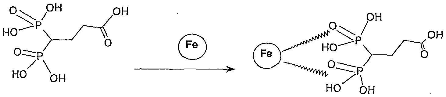
  • particles are coated with a phosphate, phosphonate, bisphosphonate or
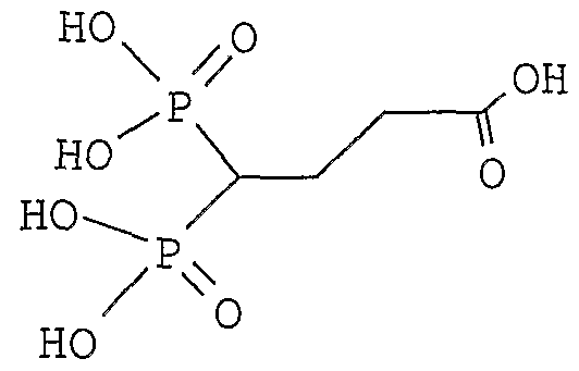
  • a preferred gem- bisphosphonate coating is of formula X-L-CH (PO 3 H 2 ) 2 where the biphosphontate part is linked to the particle and where: X is a chemical function able to react with the PEPTIDE L is an organic group linking X to the function gem-bisphosphonate -CH(PO 3 H 2 ) 2 .
  • L represents a substituted or unsubstituted aliphatic group, and more preferably a group -(CH 2 ) P - where p is an integer from 1 to 5.
  • L represents a group LrCONH-L 2 and more preferably a group -(CH 2 )n-NHCO-(CH 2 ) m - where n and m represent an integer from 0 to 5.
  • the X end of the gem-bisphosphonate compound of formula (I) is chosen in such a manner that it is capable of reacting and forming a covalent bond with a group present on the PEPTIDE biovector.
  • preferred X groups in particular :
  • the SIGNAL entity is a fluorescent probe for fluorescent imaging.
  • filtered light or a laser with a defined bandwidth is used as a source of excitation light.
  • the excitation light travels through body tissues. When it encounters a near infrared fluorescent molecule ("contrast agent”), the excitation light is absorbed. The fluorescent molecule then emits light (fluorescence) spectrally distinguishable (slightly longer wavelength) from the excitation light.
  • fluorochromes destinated to the targeting of MMP have been described, for instance reminded in US2004015062.
  • Enzyme-sensitive molecular probes have been synthesized and which are capable of fluorescence activation at 600-900 nm. These probes are described namely in US 6,083,486.
  • Fluorescent probes i.e., excitation at shorter wavelength and emission at longer wavelength
  • fluorescent probes are to be used in living systems, the choice is generally limited to the near infrared spectrum (600-1000 nm) to maximize tissue penetration by minimizing absorption by physiologically abundant absorbers such as hemoglobin ( ⁇ 550 nm) or water (>1200 nm).
  • physiologically abundant absorbers such as hemoglobin ( ⁇ 550 nm) or water (>1200 nm).
  • the fluorochromes are designed to emit at 800+/-50 nm.
  • NIRF molecules A variety of NIRF molecules have been described and/or are commercially available, including: Cy5.5 (Amersham, Arlington Heights, III.); NIR-1 (Dojindo, Kumamoto, Japan); 1RD382 (LI-COR, Lincoln, Nebr.); La Jolla Blue (Diatron, Miami, Fla.); ICG (Akorn, Lincolnshire, III.); and ICG derivatives (Serb Labs, Paris, France). Quantum dots derivatives (inorganic fluorophores comprising nanocristal) may also be used.
  • Another aspect of the present invention contemplates a method of imaging cardiovascular pathologies associated with extracellular matrix degradation, such as atherosclerosis, heart failure, and restenosis in a patient involving: (1 ) administering a paramagnetic metallopharmaceutical of the present invention capable of localizing the loci of the cardiovascular pathology to a patient by injection or infusion; and (2) imaging the patient using magnetic resonance imaging or planar CT or SPECT gamma scintigraphy, or positron emission tomography or sonography.
  • the invention also relates to a method for assessing vulnerable plaques combining a diagnostic imaging with a product of the invention and/or a morphologic analysis of the plaques and/or a study of stenoses.
  • the invention also relates to:
  • a method of detecting, imaging or monitoring the presence of matrix MMPs in a patient comprising the steps of: a) administering to said patient a diagnostic agent described above; and b) acquiring an image of a site of concentration of said diagnostic agent in the patient by a diagnostic imaging technique.
  • a method of detecting, imaging or monitoring a pathological disorder associated with MMPs activity in a patient comprising the steps of: a) administering to said patient a diagnostic agent described above ; and c) acquiring an image of a site of concentration of said diagnostic agent in the patient by a diagnostic imaging technique.
  • Atherosclerosis is coronory atherosclerosis or cerebrovascular atherosclerosis.
  • the MRI detectable species (I) according to the present invention may be administered to patients for imaging in an amount sufficient to give the desired contrast with the particular technique used in the MRI. Generally, dosages of from about 0.001 to about 5.0 mmoles of MRI detectable species (I) per kg of body weight are sufficient to obtain the desired contrast. For most MRI applications preferred dosages of imaging metal compound will be in the range of from 0.001 to 2.5 mmoles per kg of body weight.
  • the MRI detectable species (I) of the present invention can be employed for the manufacture of a contrast medium for use in a method of diagnosis by MRI involving administering said contrast medium to a human or animal being and generating an image of at least part of said human or animal being.
  • the MRI detectable species (I) of the present invention may be formulated with conventional pharmaceutical aids, such as emulsifiers, stabilisers, antioxidant agents, osmolality adjusting agents, buffers, pH adjusting agents, and the like agents, and may be in a form suitable for parenteral administration, e.g. for infusion or injection.
  • conventional pharmaceutical aids such as emulsifiers, stabilisers, antioxidant agents, osmolality adjusting agents, buffers, pH adjusting agents, and the like agents.
  • the MRI detectable species (I) according to the present invention may be in conventional administration forms such as solutions, suspensions, or dispersions in physiologically acceptable carriers media, such as water for injection.
  • Parenterally administrable forms should be sterile and free from physiologically unacceptable agents, and have low osmolality to minimize irritation and other adverse effects upon administration.
  • These parenterally administrable solutions can be prepared as customarily done with injectable solutions. They may contain additives, such as anti-oxidants, stabilizers, buffers, etc., which are compatible with the chemical structure of the MRI detectable species (I) and which will not interfere with the manufacture, storage and use thereof.
  • Compound B was obtained by grafting a human MMPs inhibitor of formula II p-aminobenzoyl-Gly-Pro-D-Leu-D-Ala-NHOH on Gd 3+ chelator (DOTA) appropriately functionalised for coupling with an isothiocyanate linker , at a ratio 1 :1.
  • DOTA Gd 3+ chelator
  • compound B displayed a molecular weight of 1210 Da and showed relaxivity values similar to that of gadoteric acid (Gd-DOTA, Dotarem ⁇ , GUERBET, France), a non specific product which was used as the reference compound.
  • the MMP-inhibitor of compound B consisted of a water soluble tetrapeptidyl hydroxamic acid, purchased from Bachem (Budendorf, Switzerland). In vitro MMP inhibition assay
  • compound B or the corresponding peptide hydroxamate was tested in vitro, on human enzymes.
  • test was validated by using TIMP-1 or GM6001 as a standard inhibitor of MMPs see table 2).
  • ApoE-KO mice of 11-12 weeks were fed a cholesterol-rich Western-type diet (Tecklad, Madison, USA) for 4 months.
  • This model of atherosclerosis was fully characterized: it contained extensive atherosclerotic plaques in the heart and aortic arch, where the plaques occupied 34% of the total lumen area.
  • this animal model showed intense lipid staining, as well as the presence of macrophages, MMP-2 and MMP-3 which accounted for 15%, 10-20% and 20-40% of the plaque areas, respectively.
  • Muscle and different organs were removed, as well as some artery specimens (aortic arch, carotid arteries, thoracic aorta, abdominal aorta and femoral artery). Finally, an aliquot of the blood sample was centrifuged and the Gd 3+ contained in all the collected samples was quantified by induced coupled plasma - mass spectrometry (ICP-MS) after acidic mineralization.
  • ICP-MS induced coupled plasma - mass spectrometry
  • Specimen preparation - Watanabe Heritable Hyperlipidemic rabbits (CAP, Olivet, France), which develop hypercholesterolemia and subsequent atherosclerosis due to a genetic deficiency of LDL receptors, were profoundly anaesthetized.
  • the arterial specimen was then kept frozen at -20°C; it was gently and extemporarily unfrozen for experimental purpose.
  • Detection of the contrast media by ex vivo imaging and Gd quantification For ex vivo MRI, the aortic slices were transferred in a new 24-wells plate and embedded in a semi-solid agar-agar gel (0.8% m/v) at room temperature. Images were performed on a 2.35 T MRI system (Biospec, Bruker, Germany), using a 7 cm inner diameter birdcage coil and a 200 mT/m insert gradient.
  • the gel was removed from the aortic segments and the Gd contained in each precisely weighed sample was quantified by ICP-MS after acidic mineralization.
  • Immunohistochemistry In order to validate this ex vivo screening test and to correlate the MRI signal obtained for compound B with the targeted MMPs, we performed immunohistochemical analysis. The assays were conducted on incubated and washed sections originating from WHHL rabbits and humans. Regarding rabbits, only the antibodies recognizing MMP-2 (polyclonal antibody, Calbiochem, Darmstadt, Germany) and MMP-9 (monoclonal antibody, Oncogene Research Products, San Diego, USA) were commercially available and validated.
  • MMP-2 polyclonal antibody, Calbiochem, Darmstadt, Germany
  • MMP-9 monoclonal antibody, Oncogene Research Products, San Diego, USA
  • MMP-1 horseradish anti-MMP-1 polyclonal antibody, Chemicon, Temecula, CA, USA
  • MMP-2 rabbit anti-rat MMP-2 polyclonal antibody, Chemicon, Temecula, CA, USA
  • MMP-3 rabbit anti-MMP-3 polyclonal antibody, Chemicon, Temecula, CA, USA
  • MMP-7 rabbit anti-MMP-7 polyclonal antibody, Chemicon, Temecula, CA, USA
  • MMP-9 goat anti-MMP-9 polyclonal antibody, Santa Cruz Biotechnology, Santa Cruz, CA, USA
  • Biodistribution assay The biodistribution of compound B and its reference compound Gd-DOTA in the plasma, main organs and artery specimen of ApoE-KO mice is presented in figure 1.
  • both contrast agents showed no significant difference in plasma, kidney and muscle accumulation, contrary to the liver uptake.
  • the concentrations of compound B and Dotarem ⁇ were poor in the main organs (i.e. ⁇ 1 % ID), except in the kidney which is the organ of excretion.
  • the two contrast agents were cleared rapidly from the body, as the percent of injected dose found in the plasma was approximately 1-2% (half-life of elimination in plasma: 15 min).
  • the compound B allowed to differentiate between vulnerable plaques, which showed a strong positivity for MMP-1 , -2, -3, -7 and -9 according to immunohistochemical analysis, and silent plaques, which contained a lower amount of MMPs.
  • vulnerable plaques which showed a strong positivity for MMP-1 , -2, -3, -7 and -9 according to immunohistochemical analysis
  • silent plaques which contained a lower amount of MMPs.
  • the compound B may be interesting for assessing the inflammatory degree, and hence the vulnerability, of human atherosclerotic plaques.
  • Table 1 experimental conditions for the in vitro MMP-inhibition assay.
  • Enzyme 7 nM 0.35 ⁇ M 6 nM concentration (enzyme isolated from (enzyme isolated from (recombinant (activated human rheumatoid human rheumatoid enzyme expressed synovial fibroblast) synovial fibroblast) in sF9 cells) enzyme)
  • Validation IC50 of TIMP on MMP-1 2.9E-09 M
  • Validation IC50 of GM6001 on MMP-2 7.0E-10 M
  • Validation IC50 of TIMP on MMP-3 4.8E-09 M
  • the figure 1 enclosed shows the biodistribution of compound B versus Gd- DOTA in plasma and main organs (left), as well as in the vascular wall of artery specimen (right) of ApoE-KO mice in a 100% C57BI/6 background (** : P ⁇ 0.01).
  • the figure 2 shows the T1 MRI signal with compound B (left) compared to the control Dotarem (right).
  • the oil obtained is solubilized in 125 ml of toluene. 0.14 g of para- toluenesulfonic acid are added. The mixture is brought to reflux for 24 hours with a Dean-Stark trap and is then concentrated to dryness under vacuum. The produdt is extracted with 500 ml of CH 2 CI 2 and is then washed twice with 250 ml of water. The organic phase is dried over MgSO 4 and concentrated under vacuum.
  • the crude product is purified on 625 g of Merck Geduran® silica gel (40-63 ⁇ m). Elution: CH 2 CI 2 /acetone - 50/50 18.4 g are isolated with a yield of 71 %.
  • the nitric ferrofluid is stirred for 15 min with 200 ml of HNO 3 [2M], and the supernatant is removed by magnetic separation.
  • the nitric ferrofluid is brough to reflux with 600 ml of water and 200 ml of Fe(NO 3 ) 3 [1 M] for 30 min. The supernatant is removed by magnetic separation.
  • the nitric ferrofluid is stirred for 15 min with 200 ml of HNO 3 [2M], the supernatant being removed by magnetic separation.
  • the nitric ferrofluid is washed 3 times with 3 liters of acetone, and is then taken up with 400 ml of water. The solution is evaporated under vacuum until a final volume of 250 ml is obtained.
  • SQUID diameter of the nongrafted particle (p) estimated by deconvolution of magnetization curves measured on a SQUID magnetometer
  • the nitric ferrofluid is brought to reflux with 1300 ml of water and 700 ml of Fe(NO 3 ) 3 [1M] for 30 min (600 rpm). The supernatant is removed by magnetic separation.
  • the nitric ferrofluid is stirred for 15 min with 3 liters of HNO 3 [2M], the supernatant being removed by magnetic separation.
  • the nitric ferrofluid is washed 3 times with 3 liters of acetone, and is then taken up with 600 ml of water. The solution is evaporated under vacuum until a final volume of 250 ml is obtained.
  • Z ave Hydrodynamic size by PCS in unimodal mode.
  • the flocculate is isolated by magnetic separation and is then washed 3 times with 3 liters of water.
  • Example 2.18 100 ml of a solution of Sub example 2.12 at 0.285 M/L are ultrafiltered through a PALL ® 30 KD stirring cell. 202 mg of compound PEPTIDE aminobenzoyl-Gly-Pro-D-Leu-D-Ala- NHOH from compound B are added to this solution. The pH is adjusted to 6.1 with QS HCI [0.1 N].

Landscapes

  • Health & Medical Sciences (AREA)
  • Life Sciences & Earth Sciences (AREA)
  • Chemical & Material Sciences (AREA)
  • General Health & Medical Sciences (AREA)
  • Veterinary Medicine (AREA)
  • Animal Behavior & Ethology (AREA)
  • Public Health (AREA)
  • Epidemiology (AREA)
  • Organic Chemistry (AREA)
  • Radiology & Medical Imaging (AREA)
  • Nuclear Medicine, Radiotherapy & Molecular Imaging (AREA)
  • Medicinal Chemistry (AREA)
  • Biochemistry (AREA)
  • Genetics & Genomics (AREA)
  • Molecular Biology (AREA)
  • Proteomics, Peptides & Aminoacids (AREA)
  • Biophysics (AREA)
  • Medicines Containing Antibodies Or Antigens For Use As Internal Diagnostic Agents (AREA)
  • Magnetic Resonance Imaging Apparatus (AREA)
  • Peptides Or Proteins (AREA)

Abstract

The invention relates to new compounds and compositions for the imaging diagnostic of pathologies, namely for cardiovascular diseases, more precisely atherosclerosis disease. These compounds are contrast agents useful namely in the field of magnetic resonance imaging MRI and nuclear medicine. The compounds comprise a particular peptidic MMP inhibitor coupled with a signal entity.

Description

Title : COMPOUND FOR DIAGNOSTIC IMAGING
The invention relates to new compounds and compositions for the imaging diagnostic of pathologies, namely for cardiovascular diseases, more precisely atherosclerosis disease. These compounds are contrast agents useful namely in the field of magnetic resonance imaging MRI, but also in other imaging fields such as nuclear medicine, X-ray, ultrasounds, optical imaging. These compounds comprise at least a targeting moiety linked to at least a signal moiety.
A targeting entity is capable of targeting at least one marker of a pathologic state and/or area that are over or under expressed in, a pathologic state and/or area compared to the non pathologic ones. These compounds are called specific compounds, the targeting entity being called biovector. Numerous signal entities/moieties are already known, such as linear or macrocyclic chelates of paramagnetic metal ion for MRI and of radionucleides for nuclear medicine. Such chelates are described in the documents EP 71 564, EP 448 191 , WO 02/48119, US 6 399 043, WO 01/51095, EP 203 962 , EP 292 689 , EP 425 571 , EP 230 893 , EP 405 704, EP 290 047, US 6 123 920, EP 292 689, EP 230 893, US 6 403 055, WO 02/40060, US 6 458 337, US 6 264 914, US 6 221 334, WO 95/31444, US 5 573 752, US 5 358 704. Chelates commonly used are for example DTPA, DTPA BMA, DTPA BOPTA, DO3A, HPDO3A, TETA, DOTA (1 , 4,7,10-tetracyclododecane-N,N,,N",N"'-tetraacetic acid), PCTA and their derivatives. Products on the market are namely for example Dotarem ® and Magnevist © he signal is measured in MRI by the relaxivity in water which is in the order of 3 to 10 mM-1s-1 Gd-1for such chelates. There is still a serious need for a new contrast agent product able to target specifically atherosclerotic lesions. Atherosclerosis is the most prevalent disease of modern society. A broad spectrum of clinically different diseases such as myocardial infarction, stroke, abdominal aneurysms and lower limb ischemia are basically related to atherosclerosis. Most of their acute manifestations share a common pathogenic feature : rupture of an atherosclerotic plaque with superimposed thrombosis. Plaque rupture, which accounts for approximately 70% of fatal acute myocardial infarctions and of symptomatic carotid lesions, is the ultimate complication of a vulnerable plaque. Vulnerable plaques include thrombosis-prone plaques as well as those with a high probability of undergoing rapid progression, thus becoming culprit plaques. They are characterized by a large lipid core, a thin cap and macrophage-dense inflammation on or beneath their surface. The risk of acute ischemic event for an individual is determined by the number of vulnerable plaques and the current challenge is to stratify such a risk.
Conventional imaging techniques are unable to detect and help characterize vulnerable plaques, especially those of the coronary arteries. Angiography is strictly an anatomic imaging tool and is unable to evaluate coronary plaque dimension and composition. Other modalities are catheter- based and, therefore, have a limited clinical applicability. Intravascular ultrasound provides some information on plaque morphology but image resolution and sensitivity are still insufficient to reliably distinguish vulnerable plaque deposits. Optical coherence tomography better delineates between intimal wall and plaque but its penetration depth is low. Angioscopy may be used to detect lipid-rich plaques and to visualize thrombus, whereas thermography is very sensitive to superficial inflammation. However, both techniques are unable to examine the deep layers of the arterial wall and to estimate cap thickness.
A very promising technique is the magnetic resonance imaging (MRI) technique. On atherosclerotic carotid plaques, it is able to visualize intraplaque hemorrhage and fibrous cap rupture, but also to detect intraluminal thrombi and differentiate their age. It is, therefore, a potentially attractive diagnostic tool for risk stratification of patients with recent onset of cerebral ischemic symptoms. Until now however, it lacks sufficient spatial resolution for accurate measurements of cap thickness and characterization of the coronary atherosclerotic lesions. Thus for a predictive diagnostic there is a need for a physiological characterisation of the plaques further to their morphological study. An alternative strategy for identification, by MRI, of coronary vulnerable plaques may be to apply a molecular imaging approach based on the detection of a specific marker. One such marker is represented by the matrix metalloproteinases (MMPs), a family of zinc-containing endoproteinases which are overexpressed in active atherosclerotic lesions and promote plaque instability by degrading the fibrillar collagen of the fibrous cap. Thus, a contrast molecule, which can be detected namely with MRI, will be useful to image MMP activity and to non invasively detect vulnerable plaques and improve patients' risk stratification.
Although little is known about the amounts of MMPs accumulating within human vulnerable plaques, some studies have reported a surexpression of
MMP-8 per milligram of tissue in advanced atherosclerotic carotid lesions.
These levels were considered similar to those obtained for MMP-1 and
MMP-13 . As the sensitivity of MRI in vivo is relatively low compared to scintigraphic imaging techniques, for instance, there is a need to compensate for the low levels of MMPs in the lesions in order to generate a sufficient signal intensity. This requirement may be achieved by using a compound which targets nonselectively the majority of MMPs and, thereby, will allow a high local concentration of the contrast agent. The applicant has now prepared imaging compounds comprising a biovector with good affinity for MMP-1 , MMP-2, MMP-3, MMP-8, MMP-9 ; in particular MMP-3 are surexpressed in lesional plaques. Specific compounds for the targeting of MMPs are described in the prior art. For instance WO 01/60416 describes compounds that comprise a targeting entity towards MMPs coupled to a linear or macrocyclic chelate signal entity. According to applicant's knowledge based namely on biological assays, such compounds of the prior art are not sufficiently efficient for a very satisfying in vivo diagnosis, due to their relative low relaxivity which is in the order of 5 to 10 mMol-1s-1 Gd-1 and/or their lack of affinity or selectivity. Thus there still remains a serious need for new products that are effectively efficient in imaging diagnostic in vivo
Surprisingly, while assessing very promising compounds with high relaxivity, the applicant has now shown that a particular peptidic MMP inhibitor coupled to a signal entity gives effective very good results for the diagnostic imaging despite the relatively low relaxivity of the signal entity. The compound prepared by the applicant is indeed very successful for the specific diagnosis of a disease associated with vulnerable plaques, compared to a non specific control compound Dotarem. The affinity of exemplified compounds for MMPs was tested in vitro on purified MMPs as well as ex vivo on WHHL rabbit arteries and human endarterectomy specimens. The biodistribution was studied in a mouse model of atherosclerosis showing increased MMP expression. It is reminded here that a very high number of MMP targeting molecules are described in the prior art, which exhibit a high structural diversity, reviewed namely in : -Current Medicinal Chemistry, 2001 , 8, 425-474 -Chem rev, 1999, 99, 2735-2776 -DDT vol 1 , n°1 , jan 1996, Elsevier Science, 16-17 -Bioconjugate Chem, 2001 , 12, 964-971 It is also reminded that over 150 US patents or patent applications cover MMP inhibitors and a lot more cover MMP targeting entities. The peptidic MMP inhibitor used as biovector by the applicant is described in Biochemical and Biophysical research Communications, vol 199, 3, 1994, pages 1442-1446 and in US 5 100 874 incorporated by reference. But the coupling to a signal entity of this particular peptidic MMP inhibitor, among the huge amount of possible MMP targeting entities and inhibitors known with equivalent or higher affinity or selectivity for MMPs, was neither described nor suggested for diagnostic imaging and specially for cardiovascular disease diagnostic. Further, according to the applicant's knowledge, the clinical trials relating to MMP target entities in the therapeutic field focus on cancer therapy and are not engaged in the cardiovascular domain.
Thus according to a first aspect the invention relates to a diagnostic agent comprising a compound of formula (I) (PEPTΪDE)M - (LINKER)n2 - (SIGNAL)n3
Wherein
1 ) PEPTIDE is chosen in the group : a) the peptide of formula X1 - X2 - X3 - X4 - NHOH (II), wherein
X1 is absent or X1 is a residue of glycine and, X2 is a residue of an amino acid selected from proline, hydroxyproline, thioproline and alanine, X3 is a residue of an amino acid selected from glutamine, glutamic acid, leucine, isoleucine and phenylalanine and X4 is a residue of an alpha-amino acid selected from glycine, alanine, valine, leucine ; and the carboxyl group of alpha-amino acid X1 forms a peptide bond together with the amino group of alpha-amino acid X2, the carboxyl group of alpha-amino acid and acid X2 forms a peptide bond together with the amino group of alpha-amino acid X3, the carboxyl group of alpha-amino acid X3 forms a peptide bond together with the amino group of alpha-amino acid X4 and the carboxyl group of alpha- amino acid X4 forms an amido together with -NHOH ; and the hydrogen atom of the amino group in said alpha-amino acid X1 may be replaced with a member X0 selected from an alkyl or an aryl group, preferably chosen in the group consisting of acetyl, benzoyl (Bz), benzyloxy, t-butyloxycarbonyl, benzyloxycarbonyl (Z), p-aminobenzoyl (ABz), p-amino-benzyl, p-hydroxybenzoyl (HBz), 3-p- hydroxyphenylpropionyl (HPP). b) a peptide functionally equivalent to a peptide of especially a) c) a peptidic fragment of (II) functionally equivalent to a peptide of a) or b)
2) SIGNAL is a signal entity for medical imaging
3) LINKER eventually absent represents a chemical link between PEPTIDE and SIGNAL ; and the pharmaceutical salts thereof.
The amino acids may be either D or L amino acids.
The term peptide functionally equivalent to peptide (II) refers to peptides that have a chemical structure allowing them to be coupled to the chelate, and a biological activity such that the activity of the diagnostic compound (I) is comparable to the activity of compound exemplified above towards MMPs, within the range of 20 to 200 %, typically at least 80% of the activity of exemplified compounds. The activity towards MMPs may be equivalent towards each MMP targeted by the peptides or only towards certain of them ; thus the activity towards MMPs relates to the global MMPs targeting activity such that the compound is useful in term of medical imaging diagnostic of cardiovascular/atheroma diseases and in particular of vulnerable plaque detection. In particular the peptide is preferably chosen so that the concentration which inhibits by 50% the activity of MMPs (IC50) is less than 10 μM. Preferably the IC50 is between 0.5 and 5 μM. However peptides with higher IC50 are also included in the invention if they give effectively good results in vivo imaging, in particular any peptide allowing to visualize the plaques in the ex vivo test exemplified in the detailed description.
Compounds with X0 comprising an aromatic group are also included in the invention.
According to an embodiment PEPTIDE is a peptide X1 - X2 - X3 - X4 -
NHOH (II) wherein X1 is absent or X1 is glycine, X2 is a residue of an amino acid selected from proline, hydroxyproline, thioproline, X3 is a residue of an amino acid selected from leucine, isoleucine and phenylalanine and X4 is a residue of an alpha-amino acid selected from glycine, alanine.
According to an embodiment PEPTIDE is a peptide
X1 - X2 - X3 - X4 - NHOH (II) wherein X1 is glycine, X2 is proline, X3 is leucine, X4 is alanine.
According to an embodiment PEPTIDE is X-NHOH with X chosen among : Abz-Gly-Pro-D-Leu-D-Ala, HBz-Gly-Pro-D-Leu-D-Ala, Abz-Gly-Pro-Leu- Ala, Bz-Gly-Pro-D-Leu-D-Ala, Bz-Gly-Pro-Leu-Ala, HPP-Pro-D-Leu-D-Ala, HPP-Pro-Leu-Ala, Z-Pro-D-Leu-D-Ala, Z-Pro-Leu-Ala.
In particular the PEPTIDE p-aminobenzoyl-Gly-Pro-D-Leu-D-Ala-NHOH is very satisfying for plaque imaging.
The hydroxamates peptides derivatives terminate NHOH, instead of peptides CO2H terminal. The peptides can be carried out by processes which can be divided roughly into (A) and (B) below:
(A) Process where a compound of the formula Boc-X4 -NHOBzl is used as starting material; the peptide chain is extended on the Boc-N group side first to form the group X3 -X4 -, which is converted, via the group X2 -X3 -X4- into the group X1 -X2 -X3 -X4 -; and finally the O-benzyl on the hydroxamic acid side is eliminated to give the desired compound; and (B) Process where a compound of the formula Boc-X4 -OR2 is used as starting material to synthesize the corresponding peptide derivative: X1 -X2 -X3 -X4 -OR2 (with R2 : methyl or ethyl group)
Document US 5 100 874 describes precisely the preparation of such peptides, which is also available at BACHEM company (www.Bachem.com). In the above mentioned processes, any means conventionally used in the peptide synthetic chemistry may be employed as specific means for condensing amino acids for formation of peptide chains ; for protecting with protecting groups the amino, imino, carboxyl and/or hydroxyl groups which may be present in their structure; and for eliminating such protecting groups. Concerning the pharmaceutical salts :
- Prefered cations of inorganic bases suitable for salifying the complexes comprise, in particular, alkali or alkaline-earth metal ions such as potassium, sodium, calcium, magnesium.
- Preferred cations of organic bases comprise those of primary, secondary and tertiary amines, such as ethanolamine, diethanolamine, morpholine, glucamine, N-methylglucamine,N, N- dimethylglucamine.
- Preferred anions of inorganic acids comprise, in particular, the ions of halo acids such as chlorides, bromides, iodides or other ions such as sulfate.
- Preferred anions of organic acids comprise those of acids conventionally used in pharmaceutical technique for the salification of basic substances, such as acetate, succinate, citrate, fumarate, maleate, oxalate, trifluoroacetate Preferred cations and anions of amino acids comprise, for example, those of taurine, glycine, lysine, arginine or omithine or of the aspartic and glutamic acids.
According to an embodiment, SIGNAL is a linear or macrocyclic chelate. Chelates (chelators, chelating ligands) for magnetic resonance imaging contrast agents are selected to form stable complexes with paramagnetic metal ions, such as Gd (III), Dy (III), Fe (III), Mn (III) and Mn (II), include the residue of a polyaminopolycarboxylic acid, either linear or cyclic, in racemic or optically active form, such as ethylenediaminotetracetic acid (EDTA), diethylenetriaminopentaacetic acid (DTPA), N-[2-
[bis(carboxymethyl)amino]-3-(4-ethoxyphenyI)propyl]-N-[2- [bis(carboxymethyl)amino]ethyl]-L-glycine (EOB-DTPA), N,N-bis[2- [bis(carboxymethyl)amino]ethyl]-L-glutamic acid (DTPA-GLU), N,N-bis[2- [bis(carboxymethyl)amino]ethyl]-L-lysine (DTPA-LYS), the DTPA mono- or bis-amide derivatives, such as N,N-bis[2-
[carboxymethyl[(methylcarbamoyl)methyl]amino]ethyl] glycine (DTPA- BMA), 4-carboxy-5,8,11-tris(carboxymethyl)-1-phenyl-2-oxa-5,8,11- triazatridecan-13-oic acid (BOPTA), 1 , 4,7,10-tetraazacyclododecan- 1 ,4,7,10-tetraacetic acid (DOTA), 1 , 4,7,10-tetraazacyclododecan-1 , 4,7- triacetic acid (DO3A), 10-(2-hydroxypropyl)-1 , 4,7,10-tetraazacyclododecan- 1 ,4,7-triacetic acid (HPDO3A) 2-methyl-1 , 4,7,10-tetraazacyclododecan- 1 ,4,7,10-tetraacetic acid (MCTA), ( alpha , alpha ', alpha ", alpha '")- tetramethyl-1 ,4,7,10-tetraazacyclododecan- 1 ,4,7,10-tetraacetic acid (DOTMA), 3,6,9,15-tetraazabicyclo[9.3.1]pentadeca-1 (15),11 ,13-triene- 3,6,9-triacetic acid (PCTA), or of a derivative thereof wherein one or more of the carboxylic groups are in the form of the corresponding salts, esters, or amides; or of a corresponding compound wherein one or more of the carboxylic groups is replaced by a phosphonic and/or phosphinic group, such as for instance 4-carboxy-5,11-bis(carboxymethyl)-1-phenyl-12- [(phenylmethoxy)methyl]-8-(phosphonomethyl)-2-oxa-5,8, 11 -triazatridecan- 13-oic acid, N,N'-[(phosphonomethylimino)di-2,1-ethanediyl]bis[N- (carboxymethyl)glycine], N,N'-[(phosphonomethylimino)di-2,1~ ethanediyl]bis[N-(phosphonomethyl)glycine], N,N'-
[(phosphinomethylimino)di-2,1-ethanediyl]bis[N-(carboxymethyl)glycine], 1 ,4,7, 10-tetraazacyclododecane-1 ,4,7,10- tetrakis[methylen(methylphosphonic)]acid, or 1 ,4,7,10- tetraazacyclododecane-1 ,4,7, 10-tetrakis[methylen(methylphosphinic)]acid. Usable chelates may also be DOTA gadofluorins, DO3A, HPDO3A, TETA, TRITA, HETA, DOTA-NHS, M4DOTA, M4DO3A, PCTA and their derivatives, 2-benzyl-DOTA,alpha- (2-phenethyl) 1 ,4,7,10 tetraazacyclododecane-1-acetic-4,7,10-tris (methylacetic) acid, 2benzyl- cyclohexyldiethylenetriaminepentaacetic acid, 2-benzyl-6methyl-DTPA, and 6,6"-bis [N, N, N", N"tetra (carboxymethyl)aminomethyl)-4'- (3-amino-4- methoxyphenyl)- 2,2' : 6', 2"-terpyridine, N,N'-bis-(pyridoxal-5-phosphate) ethylenediamine- N, N'-diacetic acid (DPDP) and ethylenedinitrilotetrakis (methylphosphonic) acid (EDTP).
Suitable chelating ligands as well as the processes for their preparation are described for instance in the following patents : EP-A-230,893, EP-A- 255,471 , EP-A-299,795, EP-A-325,762, EP-A-565,930, EP-A-594,734, US- A-4,885,363, EP-A-773,936, WO-A-9426313, WO-A-9426754, WO-A- 9426755, WO-A-9519347, WO-A-9731005, WO-A-9805625, WO-A- 9805626, WO-A-9935133, WO-A-9935134, and WO-A-0146207, which are incorporated herein by reference. Preferred chelating ligands are linear and macrocyclic polyaminopolycarboxylic acids, in racemic or optically active form.
Most preferred are (DTPA), (DOTA), (BOPTA), (DO3A), (HPDO3A), (DOTMA), (PCTA) and their derivatives.
The appropriate chelates are not limited to this list ; other chelates in compounds (I) with good efficiency in imaging diagnostic is appropriate. In particular SIGNAL may be of general formula or derivatives thereof described in detail in WO 01/60416 and US 6 221 334.
Figure imgf000012_0001
Figure imgf000012_0002
Preferred paramagnetic metal ions include ions of transition and lanthanide metals (i.e. metals having atomic number of 21 to 29, 42, 43, 44, or 57 to 71 ). In particular ions of Mn, Fe, Co, Ni, Eu, Gd, Dy, Tm, and Yb are preferred, with those of Mn, Fe, Eu, Gd, and Dy being more preferred and Gd being the most preferred.
In nuclear medicine diagnostic, the metal chelate is selected to form stable complexes with the metal ion chosen for the particular application. Chelators or bonding moieties for diagnostic radiopharmaceuticals are selected to form stable complexes with the radioisotopes that have imageable gamma ray or positron emissions, such as 99Tc, 95Tc, 111 In, 62Cu 60Cu 64Cu, 67Ga, 68Ga, 86Y. Chelators for technetium, copper and gallium isotopes are selected preferably from diaminedithiols, monoamine-monoamidedithiols, triamide- monothiols, monoamine-diamide-monothiols, diaminedioximes, and hydrazines. The chelators are generally tetradentate with donor atoms selected from nitrogen, oxygen and sulfur. Preferred reagents are comprised of chelators having amine nitrogen and thiol sulfur donor atoms and hydrazine bonding units. The thiol sulfur atoms and the hydrazines may bear a protecting group which can be displaced either prior to using the reagent to synthesize a radiopharmaceutical or preferably in situ during the synthesis of the radiopharmaceutical.
Chelators for 1111n and 86Y are typically selected from cyclic and acyclic polyaminocarboxylates such as DTPA, DOTA,D03A, 2benzyl-DOTA, alpha- (2-phenethyl) 1 ,4,7,10-tetraazazcyclododecane1 -acetic-4, 7,10-tris (methylacetic) acid, 2-benzylcyclohexyldiethylenetriaminepentaacetic acid, 2-benzyl-6-methyl
DTPA, and 6,6"-bis [N, N, N", N"-tetra (carboxymethyl) aminomethyl4'- (3- amino-4-methoxyphenyl)-2, 2' : 6', 2"-terpyridine.
In the above formula (I) PEPTIDE and SIGNAL can be bound together either directly (n2 = 0) or through a spacer LINKER. It is also possible to have, in the above formula (I), one MRI targeting entity (detectable moiety) bound to more than one peptide. In a realisation n1 = n2 = n3 =1. In another realisation, in order to increase the signal, several chelates are used. For instance n1 = n2=1 and n3 = 2 to 8 ; for instance the linker is a divisor chemically linked to at least two chelates such as a 1 ,3,5 triazine divisor. When the MRI compounds of formula (I) contain more than one MRI detectable moiety (chelate), said detectable moieties can also be bound to more than one peptide through a spacer LINKER containing a multiplicity of binding sites. The numbers n1 , n2, n3 are chosen with conformity of the chemical structure and so that the diagnostic activity is obtained. For instance the LINKER consists of an alkylidene, alkenylidene, alkynylidene, cycloalkylidene, arylidene, or aralkylidene radical that can be substituted and be interrupted by heteroatoms such as oxygen, nitrogen, and sulphur. In a preferred embodiment said spacer arm consists of an aliphatic, straight or branched chain, that effectively separates the reactive moieties of the spacer so that ideally the spatial configuration of the molecule of the PEPTIDE is not influenced by the presence of the MRI detectable moiety and the PEPTIDE is thus more easily recognized by its MMP target. Said chain may be interrupted by groups such as, -O-, -S-, - CO-, -NR-, -CS- and the like groups or by aromatic rings such as phenylene radicals, and may bear substituents such as -OR, -SR, -NRR1 , - COOR, -CONRR1 , and the like substituents, wherein R and R1 , each independently, may be a hydrogen atom or an organic group. More globally, appropriate linker groups include, but are not limited to, alkyl and aryl groups, including substituted alkyl and aryl groups and heteroalkyl (particularly oxo groups) and heteroaryl groups, including alkyl amine groups, as defined as follows.
Alkyl groups include straight or branched chain alkyl group, with straight chain alkyl groups being preferred. If branched, it may be branched at one or more positions, and unless specified, at any position. The alkyl group may range from about 1 to about 15 carbon atoms (C1-C15), with a preferred embodiment utilizing from about 1 to about 10 carbon atoms (C1- C10), with C1 to C5 being particularly preferred, although in some embodiments the alkyl group may be larger. Also included within the definition of an alkyl group are cycloalkyl groups such as C5 and C6 rings, and heterocyclic rings with nitrogen, oxygen, sulfur or phosphorus. Alkyl also includes heteroalkyl, with heteroatoms of sulfur, oxygen, nitrogen, and silicone being preferred. Alkyl includes substituted alkyl groups. By "aryl group" or "aromatic group" or grammatical equivalents herein is meant an aromatic monocyclic or polycyclic hydrocarbon moiety generally containing 5 to 14 carbon atoms (although larger polycyclic rings structures may be made) and any carbocylic ketone or thioketone derivative thereof, wherein the carbon atom with the free valence is a member of an aromatic ring. Aromatic groups include arylene groups and aromatic groups with more than two atoms removed. For the purposes of this application aromatic includes heterocycle. "Heterocycle" or "heteroaryl" means an aromatic group wherein 1 to 5 of the indicated carbon atoms are replaced by a heteroatom chosen from nitrogen, oxygen, sulfur, phosphorus, boron and silicon wherein the atom with the free valence is a member of an aromatic ring, and any heterocyclic ketone and thioketone derivative thereof. Thus, heterocycle includes thienyl, furyl, pyrrolyl, pyrimidinyl, oxalyl, indolyl, purinyl, quinolyl, isoquinolyl, thiazolyl, imidozyl, etc. As for alkyl groups, the aryl group may be substituted with a substitution group.
Usable linker groups include p-aminobenzyl, substituted p-aminobenzyl, diphenyl and substituted diphenyl, alkyl furan such as benzylfuran, carboxy, and straight chain alkyl groups of 1 to 10 carbons in length. Preferred linkers include p-aminobenzyl, methyl, ethyl, propyl, butyl, pe'ntyl, hexyl, acetic acid, propionic acid, aminobutyl, p-alkyl phenols, 4-alkylimidazoIe, carbonyls, OH, COOH, glycols such as PEG. By "ethylene glycol" or "(poly)ethylene glycol" herein is meant a -(O-CH2-CH2)n- group, although each carbon atom of the ethylene group may also be singly or doubly substituted, i.e. -(O-CR2-CR2)n-, with R as described above. Ethylene glycol derivatives with other heteroatoms in place of oxygen (i.e. -(N-CH2-
CH2)n- or -(S-CH2-CH2)n-, or with substitution groups) are also appropriate. Squarate linkers are also usable.
According to another embodiment, SIGNAL is a carrier lipidic and/or polymeric system capable of vehicling chelate signal entities. Several carrier systems are known, namely lipidic nanodroplets (emulsions of nanoparticles) such as described in WO 03/062198 incorporated by reference. This document describes the proof of concept of the specific imaging with nanodroplets carrying a biovector targeting alpha v beta 3 receptor. Other parent technologies may be used such as the ones described in US 6 403 056 incorporated by reference. One nanodroplet includes typically 10 000 to 100 000 chelates. Many nanoparticulate emulsions may be used. For example W095/03829 describes oil emulsions where the drug is dispersed or solubilized inside an oil droplet and the oil droplet is targeted to a specific location by means of a ligand. US 5,542, 935 describes site-specific drug delivery using gas-filled perfluorocarbon microspheres. The targeting entity delivery is accomplished by permitting the microspheres to home to the target and then effecting their rupture. Low boiling perfluoro compounds are used to form the particles so that the gas bubbles can form. It is possible to employ emulsions wherein the nanoparticles are based on high boiling perfluorocarbon liquids such as those described in US 5,958, 371. The liquid emulsion contains nanoparticles comprised of relatively high boiling perfluorocarbons surrounded by a coating which is composed of a lipid and/or surfactant. The surrounding coating is able to couple directly to a targeting moiety or can entrap an intermediate component which is covalently coupled to the targeting moiety, optionally through a linker. A possible emulsion is a nanoparticulate system containing a high boiling perfluorocarbon as a core and an outer coating that is a lipid/surfactant mixture which provides a vehicle for binding a multiplicity of copies of one or more desired components to the nanoparticle. The construction of the basic particles and the formation of emulsions containing them, regardless of the components bound to the outer surface is described in the above- cited patents to the present applicants, US 5 690 907, 5 780 010, 5 958 371.
Useful perfluorocarbon emulsions are reminded in US 6 676 963, and include those in which the perfluorocarbon compound is perfluorodecalin, perfluorooctane, perfluorodichlorooctane, perfluoro-n-octyl bromide, perfluoroheptane, perfluorodecane, perfluorocyclohexane, perfluoromorpholine, perfluorotripropylamine, perfluortributylamine, perfluorodimethylcyclohexane, perfluorotrimethylcyclohexane, perfluorodicyclohexyl ether, perfluoro-n-butyltetrahydrofuran, and compounds that are structurally similar to these compounds and are partially or fully halogenated (including at least some fluorine substituents) or partially or fully fluorinated including perfluoroalkylated ether, polyether or crown ether.
The lipid/surfactants used to form an outer coating on the nanoparticles (that will contain the coupled ligand or entrap reagents for binding desired components to the surface) include typically natural or synthetic phospholipids, fatty acids, cholesterols.lysolipids, sphingomyelins, and the like, including lipid conjugated polyethylene glycol. Various commercial anionic, cationic, and nonionic surfactants can also be employed, including Tweens, Spans, Tritons, and the like. Some surfactants are themselves fluorinated, such as perfluorinated alkanoic acids such as perfluorohexanoic and perfluorooctanoic acids, perfluorinated alkyl sulfonamide, alkylene quaternary ammonium salts and the like. In addition, perfluorinated alcohol phosphate esters can be employed. Cationic lipids included in the outer layer may be advantageous in entrapping ligands such as nucleic acids, in particular aptamers.
The lipid/surfactant coated nanoparticles are typically formed by microfluidizing a mixture of the fluorocarbon lipid which forms the core and the lipid/surfactant mixture which forms the outer layer in suspension in aqueous medium to form an emulsion. In this procedure, the lipid/surfactants may already be coupled to additional ligands when they are coated onto the nanoparticles, or may simply contain reactive groups for subsequent coupling. Alternatively, the components to be included in the lipid/surfactant layer may simply be solubilized in the layer by virtue of the solubility characteristics of the ancillary material. Sonication or other techniques may be required to obtain a suspension of the lipid/surfactant in the aqueous medium. Typically, at least one of the materials in the lipid/surfactant outer layer comprises a linker or functional group which is useful to bind the additional desired component or the component may already be coupled to the material at the time the emulsion is prepared. For coupling by covalently binding the targeting peptide or other organic moiety (such as a chelating agent for a paramagnetic metal) to the components of the outer layer, various types of bonds and linking agents may be employed. Typical methods for forming such coupling include formation of amides with the use of carbodiamides, or formation of sulfide linkages through the use of unsaturated components such as maleimide. Other coupling agents include, for example, glutaraldehyde, propanedial or butanedial, 2-iminothiolane hydrochloride, bifunctional N- hydroxysuccinimide esters such as disuccinimidyl suberate, disuccinimidyl tartrate, bis [2- (succinimidooxycarbonyloxy) ethyl] sulfone, heterobifunctional reagents such asN- (5-azido-2-nitrobenzoyloxy) succinimide, succinimidyl4- (N-maleimidomethyl) cyclohexane- 1- carboxylate, and succinimidyl 4- (p-maleimidophenyl) butyrate.homobifunctional reagents such as1 , 5-difluoro-2,4- dinitrobenzene, 4, 4'-difluoro-3, 3'-dinitrodiphenylsulfone, 4,4'- diisothiocyano-2, 2'-disulfonic acid stilbene,p-phenylenediisothiocyanate, carbonylbis (L- methionine p-nitrophenyl ester), 4,4'-dithiobisphenylazide, erythritolbiscarbonate and bifunctional imidoesters such as dimethyl adipimidate hydrochloride, dimethyl suberimidate, dimethyl 3,3'- dithiobispropionimidate hydrochloride and the like. Linkage can also be accomplished by acylation, sulfonation, reductive amination, and the like. A multiplicity of ways to couple, covalently, a desired ligand to one or more components of the outer layer is known in the art. The ligand itself may be included in the surfactant layer if its properties are suitable. For example, if the ligand contains a highly lipophilic portion, it may itself be embedded in the lipid/surfactant coating. Further, if the ligand is capable of direct adsorption to the coating, this too will effect its coupling. Other useful encapsulation system may be used, if they exhibit sufficient hydrophily towards the PEPTIDE, for instance: mono or multilayers lipidic systems, liposomes, saccharidic polymers.
According to an other embodiment, SIGNAL is a metal nanoparticle based on iron such as ultra small superparamagnetic particles USPIO generally comprising an iron oxide or hydroxide. Preferably, such magnetic particles comprise a ferrite, especially maghemite (γ Fe2O3) and magnetite (Fe3O4), or also mixed ferrites of cobalt (Fe2CoO4) or of manganese (Fe2MnO4). Examples of suitably coated ferromagnetic or superparamagnetic particles are for instance those described in US patents 4,770,183, 4,827,945, 5,707,877, 6,123,920, and 6,207,134 having a coating materials, i.e., polymers such as polysaccharides, carbohydrates, polypeptides, organosilanes, proteins, and the like, gelatin-aminodextran, or starch and polyalkylene oxides, at the condition that they can be functionalised to allow binding of the particle to the spacer or directly to the PEPTIDE. In a realisation, particles are coated with a phosphate, phosphonate, bisphosphonate or gem-bisphosphonate coating. A preferred gem- bisphosphonate coating is of formula X-L-CH (PO3H2)2 where the biphosphontate part is linked to the particle and where: X is a chemical function able to react with the PEPTIDE L is an organic group linking X to the function gem-bisphosphonate -CH(PO3H2)2 .
According to one particularly preferred embodiment, L represents a substituted or unsubstituted aliphatic group, and more preferably a group -(CH2)P- where p is an integer from 1 to 5. According to another preferred embodiment, L represents a group LrCONH-L2 and more preferably a group -(CH2)n-NHCO-(CH2)m- where n and m represent an integer from 0 to 5. The X end of the gem-bisphosphonate compound of formula (I) is chosen in such a manner that it is capable of reacting and forming a covalent bond with a group present on the PEPTIDE biovector. For more information concerning these coupling processes reference may be made in particular to the work Bioconjugate techniques, Greg T. Hermanson, 1995, Publisher : Academic, San Diego, Calif. There may be mentioned as preferred X groups, in particular :
-COOH,
-NH2, -NCS, -NH-NH2, -CHO, alkylpyrocarbonyl (-CO-O-CO- alk), acylazidyl (-CO-N3), iminocarbonate (-O-C(NH)-NH2), vinylsulphuryl (-S-CH=CH2), pyridyldisulphuryl (-S-S-Py), haloacetyl, maleimidyl, dichlorotriazinyl, halogen, the groups -COOH and -NH2 being especially preferred. The applicant has also studied compounds which coating is a phosphonate or bisphosphonate or their derivatives other than the gem bisphosphonate.
According to another embodiment, the SIGNAL entity is a fluorescent probe for fluorescent imaging. In near infrared fluorescence imaging, filtered light or a laser with a defined bandwidth is used as a source of excitation light. The excitation light travels through body tissues. When it encounters a near infrared fluorescent molecule ("contrast agent"), the excitation light is absorbed. The fluorescent molecule then emits light (fluorescence) spectrally distinguishable (slightly longer wavelength) from the excitation light.
In particular, fluorochromes destinated to the targeting of MMP have been described, for instance reminded in US2004015062. Enzyme-sensitive molecular probes have been synthesized and which are capable of fluorescence activation at 600-900 nm. These probes are described namely in US 6,083,486. Fluorescent probes (i.e., excitation at shorter wavelength and emission at longer wavelength) are ideally suited for studying biological phenomena, as has been done extensively in fluorescence microscopy. If fluorescent probes are to be used in living systems, the choice is generally limited to the near infrared spectrum (600-1000 nm) to maximize tissue penetration by minimizing absorption by physiologically abundant absorbers such as hemoglobin (<550 nm) or water (>1200 nm). Typically the fluorochromes are designed to emit at 800+/-50 nm. A variety of NIRF molecules have been described and/or are commercially available, including: Cy5.5 (Amersham, Arlington Heights, III.); NIR-1 (Dojindo, Kumamoto, Japan); 1RD382 (LI-COR, Lincoln, Nebr.); La Jolla Blue (Diatron, Miami, Fla.); ICG (Akorn, Lincolnshire, III.); and ICG derivatives (Serb Labs, Paris, France). Quantum dots derivatives (inorganic fluorophores comprising nanocristal) may also be used.
Another aspect of the present invention contemplates a method of imaging cardiovascular pathologies associated with extracellular matrix degradation, such as atherosclerosis, heart failure, and restenosis in a patient involving: (1 ) administering a paramagnetic metallopharmaceutical of the present invention capable of localizing the loci of the cardiovascular pathology to a patient by injection or infusion; and (2) imaging the patient using magnetic resonance imaging or planar CT or SPECT gamma scintigraphy, or positron emission tomography or sonography. The invention also relates to a method for assessing vulnerable plaques combining a diagnostic imaging with a product of the invention and/or a morphologic analysis of the plaques and/or a study of stenoses. The invention also relates to:
A method of detecting, imaging or monitoring the presence of matrix MMPs in a patient comprising the steps of: a) administering to said patient a diagnostic agent described above; and b) acquiring an image of a site of concentration of said diagnostic agent in the patient by a diagnostic imaging technique.
A method of detecting, imaging or monitoring a pathological disorder associated with MMPs activity in a patient comprising the steps of: a) administering to said patient a diagnostic agent described above ; and c) acquiring an image of a site of concentration of said diagnostic agent in the patient by a diagnostic imaging technique.
A method above wherein the atherosclerosis is coronory atherosclerosis or cerebrovascular atherosclerosis. - A method of identifying a patient at high risk for transient ischemic attacks or stroke by determining the degree of active atherosclerosis in a patient carrying out the method above.
A method of identifying a patient at high risk for acute cardiac ischemia, myocardial infarction or cardiac death by determining the degree of active atherosclerosis by imaging the patient by the method above.
The MRI detectable species (I) according to the present invention may be administered to patients for imaging in an amount sufficient to give the desired contrast with the particular technique used in the MRI. Generally, dosages of from about 0.001 to about 5.0 mmoles of MRI detectable species (I) per kg of body weight are sufficient to obtain the desired contrast. For most MRI applications preferred dosages of imaging metal compound will be in the range of from 0.001 to 2.5 mmoles per kg of body weight. The MRI detectable species (I) of the present invention can be employed for the manufacture of a contrast medium for use in a method of diagnosis by MRI involving administering said contrast medium to a human or animal being and generating an image of at least part of said human or animal being. For said use the MRI detectable species (I) of the present invention may be formulated with conventional pharmaceutical aids, such as emulsifiers, stabilisers, antioxidant agents, osmolality adjusting agents, buffers, pH adjusting agents, and the like agents, and may be in a form suitable for parenteral administration, e.g. for infusion or injection.
Thus the MRI detectable species (I) according to the present invention may be in conventional administration forms such as solutions, suspensions, or dispersions in physiologically acceptable carriers media, such as water for injection.
Parenterally administrable forms, e.g. i.v. solutions, should be sterile and free from physiologically unacceptable agents, and have low osmolality to minimize irritation and other adverse effects upon administration. These parenterally administrable solutions can be prepared as customarily done with injectable solutions. They may contain additives, such as anti-oxidants, stabilizers, buffers, etc., which are compatible with the chemical structure of the MRI detectable species (I) and which will not interfere with the manufacture, storage and use thereof.
Further aspects of the invention will be described in the detailed description above.
1) Compounds based on gadolinium chelates: preparation and biological assays and imaging.
Contrast medium
Compound B was obtained by grafting a human MMPs inhibitor of formula II p-aminobenzoyl-Gly-Pro-D-Leu-D-Ala-NHOH on Gd3+ chelator (DOTA) appropriately functionalised for coupling with an isothiocyanate linker , at a ratio 1 :1.
Figure imgf000024_0001
Molecular Weight =1209,43 Molecular Formula =C48H65GdN11014S
0.42g of Int1 , 0.34g of peptide Abz-Gly-Pro-Leu-Ala-NHOH from Bachem and 200μL of triethylamine are dissolved in 20mL of DMSO at room temperature. The solution is stirred for 3 hours. 200mL of ether is instilled into the solution. The precipitate is washed with ether, ethanol and dichloromethane.
Analysis: HPLC purity = 92%; HPLC: Column: SYMMETRY C18, 5μm, lOOA, 250*4.6 mm
The chemical purity of this specific contrast medium was more than 90%. In addition, compound B displayed a molecular weight of 1210 Da and showed relaxivity values similar to that of gadoteric acid (Gd-DOTA, Dotarem©, GUERBET, France), a non specific product which was used as the reference compound. The MMP-inhibitor of compound B consisted of a water soluble tetrapeptidyl hydroxamic acid, purchased from Bachem (Budendorf, Switzerland). In vitro MMP inhibition assay
The ability of compound B or the corresponding peptide hydroxamate to inhibit MMP-1 , MMP-2 or MMP-3 activity was tested in vitro, on human enzymes. The reaction was based on the formation of a fluorescent compound produced after MMP-cleavage of a substrate (see table 1 for experimental conditions). Briefly, compound B or the peptide alone was added at 3 different concentrations to a buffer containing the MMP to test. For the control sample, the contrast agent or the peptide was replaced by water. After a pre-incubation period of 30 min at 37°C, the fluorescence intensity was measured using a microplate reader (GeminiXS, Molecular Devices), in order to detect any compound interference with the fluorimetric assay (=F1 ). The enzymatic reaction was then initiated by the addition of the MMP substrate and the mixture was again incubated at 37°C. After a pre-determined time, a second fluorescent measurement was performed (=F2). The enzyme activity was determined by subtracting the signal F1 from F2, and the results were expressed as a percent inhibition of the control enzyme activity.
Moreover, the test was validated by using TIMP-1 or GM6001 as a standard inhibitor of MMPs see table 2).
Biodistribution assay
Animals - the tissue distribution of compound B, when compared to Gd- DOTA, was performed in ApoE-KO mice in a 100% C57BI/6 background. The animals were bred and housed in the Animal Experiment Unit of the Center of Molecular and Vascular Biology, Leuven, Belgium, and had free access to food and water.
ApoE-KO mice of 11-12 weeks were fed a cholesterol-rich Western-type diet (Tecklad, Madison, USA) for 4 months. This model of atherosclerosis was fully characterized: it contained extensive atherosclerotic plaques in the heart and aortic arch, where the plaques occupied 34% of the total lumen area. Moreover, this animal model showed intense lipid staining, as well as the presence of macrophages, MMP-2 and MMP-3 which accounted for 15%, 10-20% and 20-40% of the plaque areas, respectively. Biodistribution - After the diet period, animals were matched for sex and body weight, compounfd B or Gd-DOTA was administered intravenously via the tail vein (n=7 / compound), at a clinical dose of 100 μmol Gd / kg. 60 min post-injection, the animals were anaesthetized and blood was obtained via eye bleeding. The mice were then subjected to a transcardiac perfusion with heparinized saline, in order to clear the intravascular compartment from any residual circulating contrast agent. Muscle and different organs (kidney, liver) were removed, as well as some artery specimens (aortic arch, carotid arteries, thoracic aorta, abdominal aorta and femoral artery). Finally, an aliquot of the blood sample was centrifuged and the Gd3+ contained in all the collected samples was quantified by induced coupled plasma - mass spectrometry (ICP-MS) after acidic mineralization.
Ex vivo magnetic resonance imaging
Specimen preparation - Watanabe Heritable Hyperlipidemic rabbits (CAP, Olivet, France), which develop hypercholesterolemia and subsequent atherosclerosis due to a genetic deficiency of LDL receptors, were profoundly anaesthetized. After their sacrifice by exsanguination, the arterial tree, comprising the aortic arch with the beginning of the efferent arteries, as well as the thoracic aorta and the supra-renal abdominal aorta, was removed in one block and flushed with heparinized Krebs-Ringer Bicarbonate buffer (pH=7.4) in order to clean any remaining blood. The arterial specimen was then kept frozen at -20°C; it was gently and extemporarily unfrozen for experimental purpose.
Samples of fresh carotid artery were obtained from patients (n=21 ) undergoing carotid endarterectomy. Specimen consisted of the common carotid artery, as well as the internal and external carotid arteries. The excised plaques were rinsed in ice-cold saline and they were used in the 2- 4 hours following surgery. Finally, both rabbits and human atherosclerotic segments were cut in exactly 3.0 mm-thick sections, before their contact with the contrast medium and ex vivo imaging.
Ex vivo incubation - All the slices were deposited in a 24-wells culture plate. They were then incubated under stirring at 37°C, in 1.0 ml of Krebs- Ringer Bicarbonate buffer (pH = 7.4), Gd-DOTA or compound B (at a Gd concentration of 5 x 10"4 M). After pre-determined incubation periods of 1h, 3h or 18h, the slices originating from the WHHL rabbits were either immediately imaged, or subjected to a washing during 30 min, 1h or 3h (n=3 for each condition) in 10.0 ml of Krebs-Ringer bicarbonate buffer, with the medium being replaced every hour. Regarding human samples, only the protocol which allowed the best differentiation between the compounds (i.e. an incubation period of 18h, followed by a washing of 30 min) was applied. Afterwards, all the slices were drained and prepared for ex vivo imaging. Further the protocol was applied both regarding to lesional plaques and not lesional plaques specimens.
Specificity testing - To additionally test the specificity of the compound B uptake, we repeated the ex vivo 18h-incubation with a new set of WHHL rabbit atherosclerotic slices which were previously incubated during 30 min with the hydroxamate peptide, at a 10 times higher concentration than COMPOUND B (i.e. 5 x 10"3 M). The samples were subsequently washed during 30 min, as previously described.
Detection of the contrast media by ex vivo imaging and Gd quantification - For ex vivo MRI, the aortic slices were transferred in a new 24-wells plate and embedded in a semi-solid agar-agar gel (0.8% m/v) at room temperature. Images were performed on a 2.35 T MRI system (Biospec, Bruker, Germany), using a 7 cm inner diameter birdcage coil and a 200 mT/m insert gradient. A 3D T1w SNAP sequence was acquired with following parameters (TR/TE/α = 20 ms/4.2 ms/75°, Matrix 256*256*16, resolution 235*175*1000 μm3, duration 21 '49") to have a compromise between resolution, short duration for screening and sections localization. Finally, the gel was removed from the aortic segments and the Gd contained in each precisely weighed sample was quantified by ICP-MS after acidic mineralization.
Immunohistochemistry - In order to validate this ex vivo screening test and to correlate the MRI signal obtained for compound B with the targeted MMPs, we performed immunohistochemical analysis. The assays were conducted on incubated and washed sections originating from WHHL rabbits and humans. Regarding rabbits, only the antibodies recognizing MMP-2 (polyclonal antibody, Calbiochem, Darmstadt, Germany) and MMP-9 (monoclonal antibody, Oncogene Research Products, San Diego, USA) were commercially available and validated. For immunohistochemical analysis on human sections, the following antibodies were used for the detection of MMP-1 (rabbit anti-MMP-1 polyclonal antibody, Chemicon, Temecula, CA, USA), MMP-2 (rabbit anti- rat MMP-2 polyclonal antibody, Chemicon, Temecula, CA, USA), MMP-3 (rabbit anti-MMP-3 polyclonal antibody, Chemicon, Temecula, CA, USA), MMP-7 (rabbit anti-MMP-7 polyclonal antibody, Chemicon, Temecula, CA, USA) and MMP-9 (goat anti-MMP-9 polyclonal antibody, Santa Cruz Biotechnology, Santa Cruz, CA, USA). In both cases, the immunohistochemical analysis were performed according to manufacturer's instructions.
Statistical analysis Data were represented as means ± SEM. Sets of data were then compared with the unpaired non-parametric Mann-Whitney test, using a two-tail P- value (GraphPad Instat3). Differences were considered significant at P < 0.05.
In vitro MMP inhibition assay The results of the human MMP-inhibitory activity of compound B and its corresponding peptidyl hydroxamic acid are presented in table 2. Both compounds were effective in vitro on MMP-1 and -2, at a concentration of 1.0E-05 M, with compound B displaying an increased inhibitory activity when compared to the peptide. In contrast, no activity was noted for the two protease inhibitors on MMP-3. These experimental values matched perfectly with the reported IC50 values of the peptidyl hydroxamic acid on MMP-1 and -3, where the respective IC50 accounted for 1.0E-06 M and 1.5E-04 M. However, the results were slight stronger for MMP-2, as an enzymatic inhibition was already observed at a concentration of 1.OE-05 M for COMPOUND B and the peptide, whereas the IC50 was reported to be at 3.0E-05 M.
Biodistribution assay The biodistribution of compound B and its reference compound Gd-DOTA in the plasma, main organs and artery specimen of ApoE-KO mice is presented in figure 1. One hour post-intravenous injection, both contrast agents showed no significant difference in plasma, kidney and muscle accumulation, contrary to the liver uptake. In general, the concentrations of compound B and Dotarem© were poor in the main organs (i.e. < 1 % ID), except in the kidney which is the organ of excretion. Moreover, the two contrast agents were cleared rapidly from the body, as the percent of injected dose found in the plasma was approximately 1-2% (half-life of elimination in plasma: 15 min).
Regarding the tissue distribution in the artery specimen, it revealed that compound B accumulated preferentially in the arterial wall, with a 3-3.5 times greater concentration when compared to Dotarem (p < 0.01). In addition, COMPOUND B, but not Dotarem©, showed a tendency to stain the arterial regions which contained the most numerous atherosclerotic plaques (carotid arteries and aortic arch). Ex vivo magnetic resonance imaging Assay performed on artery slices originating from Watanabe Heritable Hyperlipidemic rabbits - After the 1 h~, 3h- or 18h- incubation periods, all the sections showed a strong, but diffuse, enhancement with the reference compound Gd-DOTA. This result was in agreement with the amount of Gd measured in the sections by ICP-MS, which attained 3-9 nmol / section (mean value). However, Gd-DOTA disappeared immediately during the first washing step of 30 min. At this moment, the Gd concentration dropped at a mean value inferior to 0.8 nmol / section. By contrast, compound B showed also a strong but more delineated enhancement after all the incubation periods (mean Gd concentration of 5- 11 nmol / section), but this contrast agent was not eluted as fast as Gd- DOTA when the incubation period was longer than 1 h. Indeed, when incubated during 3h or 18h, it still showed a clear signal after 30 min and 1 h of washing, corresponding to a respective mean Gd concentration of 1- 8 nmol / section (figure 2).
Moreover, when we performed an inhibition/competition study with a preliminary incubation with the free peptide, we founded a very faint enhancement only for compound B. This indicated that the inhibition / competition was effective and that compound B was specific for its target. Finally, this atherosclerotic model for ex vivo screening was validated, as it contained the MMPs of interest: the immunohistochemical analysis revealed a strong positivity for MMP-1 and a low expression of MMP-9. \ssay performed on artery slices originating from humans undergoing surgical resection of the carotid artery - As the previous experimental screening conditions (for example: 18h incubation, followed by 30 min washing) allowed to discriminate between the specific and the non specific contrast medium, and as both techniques of contrast medium detection, i.e. MRI ex vivo and Gd quantification by ICP-MS, correlated well, we applied them for the study on the human atheromatous sections. The results showed that only the specific compound B, and not the non specific compound Gd-DOTA, was able to enhance the atherosclerotic human sections, as demonstrated by MRI ex vivo and Gd quantification. In addition, the compound B allowed to differentiate between vulnerable plaques, which showed a strong positivity for MMP-1 , -2, -3, -7 and -9 according to immunohistochemical analysis, and silent plaques, which contained a lower amount of MMPs. One can conclude that the compound B may be interesting for assessing the inflammatory degree, and hence the vulnerability, of human atherosclerotic plaques.
Table 1: experimental conditions for the in vitro MMP-inhibition assay.
Experimenta MMP-1 [1] MMP-2 [2] MMP-3 [2] I procedures
Enzyme 7 nM 0.35 μM 6 nM concentration (enzyme isolated from (enzyme isolated from (recombinant (activated human rheumatoid human rheumatoid enzyme expressed synovial fibroblast) synovial fibroblast) in sF9 cells) enzyme)
Experimental 50 mM Tris-HCI 50 mM Tris-HCI 25 mM Na medium (pH=7.4), 200 mM (pH=7.5), acetate
NaCI, 5 mM CaCI2, 150 mM NaCI, (pH=6.0), 0.02 mM ZnCI2 and 10 mM CaCI2, 150 mM NaCI, 0.05% Brij®35 0.02% NaN3 and 10 mM CaCI2, 0.05% Brij®35 0.02% NaN3 and
0.05% Brij®35
Substrate 10 μM of DNP-Pro- 6 μM of Mca-Arg-Pro- 6 μM of Mca-
Cha-Gly-Cys(Me)- Lys-Pro-Tyr-Ala-Nva- Arg-Pro-Lys-Pro-
His-Ala-Lys(n-Me- Trp-Met-Lys(DNP)- Tyr-Ala-Nva-Trp-
Abz)-NH2 NH2 (NFF-2) Met-Lys(DNP)-
NH2 (NFF-2)
Cleaved Cys(Me)-His-Ala- Mca-Arg-Pro-Lys-Pro- Mca-Arg-Pro- fluorescent Lys(n-Me-Abz)-NH2 Tyr-Ala Lys-Pro-Tyr-Ala compound
Fluorescent λex = 360 nm λex = 340 nm λex = 340 nm detection λem = 465 nm λem = 405 nm λem = 405 nm
(wavelength)
Reaction time 40 min 90 min 90 min [1] DA Bickett, MD. Green, J. Berman, M. Dezube, AS. Howe, PJ. Brown, JT Roth and GM McGeeban. A high throughput fluorigenic substrate for interstitial collagenase (MMP-1) and gelatinase (MMP-9). Anal. Biochem (1993) 212 : 58.
[2] N. Nagase, CG. Fields and GB Fields. Design and characterization of a fluorogenic substrate selectively hydrolyzed by stromelysin 1 (MMP-3). J. Biol. Chem. (1994) 296 : 20952.
Table 2: In vitro effect of the protease inhibitors compound B and its corresponding peptide on human MMP-1 ,-2 and -3 activity, measured by a fluorimetric assay. Results were obtained in duplicate and are expressed as the mean of the percents of inhibition of the control value.
MMP Test Peptide Compound B concentration
[M] % inhibition of control % inhibition of values control values
MMP-1 1.OE-09 9 10
MMP-1 1.0E-07 10 27
MMP-1 1.0E-05 46 86
MMP-2 1.0E-07 5 26
MMP-2 1.0E-05 68 86
MMP-2 1.0E-04 94 100
MMP-3 1.0E-09 0 2
MMP-3 1.0E-07 4 2
MMP-3 1.0E-05 8 5
Validation IC50 of TIMP on MMP-1 = 2.9E-09 M Validation IC50 of GM6001 on MMP-2 = 7.0E-10 M Validation IC50 of TIMP on MMP-3 = 4.8E-09 M
The figure 1 enclosed shows the biodistribution of compound B versus Gd- DOTA in plasma and main organs (left), as well as in the vascular wall of artery specimen (right) of ApoE-KO mice in a 100% C57BI/6 background (** : P < 0.01). The figure 2 shows the T1 MRI signal with compound B (left) compared to the control Dotarem (right).
EXAMPLE 2) Compounds based on iron oxide superparamagnetic particles
Sub example 2.8 : nitric acid ferrofluid (size PCS = 38 nm)
Figure imgf000034_0001
Sub example 2.9 : nitric acid ferrofluid (size PCS = 23,3 nm)
[Fe] [Fe] Fe2O3
PZPIHQ3-
Compound A
Figure imgf000034_0002
Sub example 2.10 : (size PCS = 67,9 nm) Sub Ex 8 + compound A
Figure imgf000035_0001
Sub example 2. 11 : (size PCS = 40,3 nm) Sub Ex 8 + compound A + treatment pH 11
Figure imgf000035_0002
Sub example 12 : (size PCS = 25,6 nm) Sub Ex 9 + compound A
Figure imgf000035_0003
Sub example 2.18 :
Sub Ex 12 + compound PEPTIDE
Preparation of compound A:
Figure imgf000036_0001
1) Diethyl-1-fethoxyphosphoryllvinyl phosphonate
13 g (0.433 mol) of paraformaldehyde and 10 ml (0.097 mol) of diethylamine are solubilized under hot conditions in 250 ml of methanol. 24 g (8.67 x 10"2 mol) of diethyl[ethoxy(propyl)phosphoryl]methyl phosphonate are then added. The mixture is brought to reflux for 24 hours. The reaction medium is concentrated under vacuum. The concentrate is taken up twice with 250 ml of toluene and is then concentrated under vacuum.
The oil obtained is solubilized in 125 ml of toluene. 0.14 g of para- toluenesulfonic acid are added. The mixture is brought to reflux for 24 hours with a Dean-Stark trap and is then concentrated to dryness under vacuum. The produdt is extracted with 500 ml of CH2CI2 and is then washed twice with 250 ml of water. The organic phase is dried over MgSO4 and concentrated under vacuum.
The crude product is purified on 625 g of Merck Geduran® silica gel (40-63 μm). Elution: CH2CI2 /acetone - 50/50 18.4 g are isolated with a yield of 71 %.
Mass spectrum: M/z = 301.4 (ES+) theoretical M = 300.2 C13 spectrum: (s) 148.8 ppm, (t) 134.8 - 131.5 - 128.2 ppm, (s) 62.2 ppm, (s) 16.7 ppm
H1 spectrum: (t) 6.9 - 6.8 - 6.6 ppm, (unresolved peak) 3.9 ppm, (t) 1.15 ppm. 2) Diethyl 2-f2.2-bis(diethylDhosDhoryl)ethyll malonate
1.6 g (0.01 mol) of diethyl malonate, 0.07 g (0.001 mol) of sodium ethoxide and 3 g (0.01 mol) of diethyl[ethoxy(propyl)phosphoryl]vinyl phosphonate are stirred for 15 min in 15 ml of ethanol. [TLC: SiO2; eluent CH2Cl2 /acetone 50/50 - Rf = 0.6].
5 ml of a saturated NH4CI solution are added to the ethanolic solution. The mixture is concentrated under vacuum. The residue is extracted with 30 ml of ethyl acetate and washed twice with 5 ml of water. The organic phase is dried over MgSO and is then evaporated to dryness. The oil obtained is purified on 200 g of Merck Geduran® silica (40-63 μm). Elution CH2CI2 /acetone 50/50 3.8 g are isolated with a yield of 82%. Mass spectrum: M/z 460.9 (ES+), theoretical M = 460.
3) 4.4-Diphosphonobutanoic acid
7 g (15.7 x 10"2 mol) of diethyl 2-[2.2-bis(diethylphosphoryl)ethyl] malonate are brought to reflux for 8 hours in 350 ml of HCI [5N].
The brown oil obtained is purified on 60 g of silanized silica 60 (0.063-0.200 mm) with water elution [HPLC monitoring]. 3.6 g are isolated with a yield of 92%. Mass spectrum: M/z 249 (ES+), theoretical M = 248 HPLC: column : Hypercarb® 250 X 4 mm Detection: 202 nm Isocratic eluent 99/1 : 0.034 N H2SO4 / CH3CN
Sub example 2.8:
A solution of 36 g (0.181 mol) of FeCI2.4H2O and 20 ml of HCI at
37% in 150 ml of H2O is introduced into a mixture consisting of 3 liters of water and 143 ml (0.302 mol) of FeCI3 at 27%. 250 ml of NH4OH at 25% are introduced rapidly with vigorous stirring. The mixture is stirred for 30 min. The liquors are removed by magnetic separation. The ferrofluid is washed 3 times consecutively with 2 liters of water.
The nitric ferrofluid is stirred for 15 min with 200 ml of HNO3 [2M], and the supernatant is removed by magnetic separation.
The nitric ferrofluid is brough to reflux with 600 ml of water and 200 ml of Fe(NO3)3 [1 M] for 30 min. The supernatant is removed by magnetic separation.
The nitric ferrofluid is stirred for 15 min with 200 ml of HNO3 [2M], the supernatant being removed by magnetic separation.
The nitric ferrofluid is washed 3 times with 3 liters of acetone, and is then taken up with 400 ml of water. The solution is evaporated under vacuum until a final volume of 250 ml is obtained.
Figure imgf000038_0001
Ms (emu/cm3) = magnetization at saturation
Z ave = Hydrodynamic size by PCS in unimodal mode
Poly σ: peak polydispersity by PCS
SQUID = diameter of the nongrafted particle (p) estimated by deconvolution of magnetization curves measured on a SQUID magnetometer
Sub example 2.9:
108 g (0.543 mol) of FeCI2 . 4 H2O in 450 ml of H2O is introduced into a solution of 4 liters of water and 429 ml (0.906 mol) of FeCI3 at 27%. 750 ml of NH4OH at 25% are introduced rapidly with stirring (1200 rpm). The mixture is stirred for 30 min. The liquors are removed by magnetic separation. The ferrofluid is washed twice consecutively with 3 liters of water. The nitric ferrofluid is stirred for % H with 3 liters of HNO3 [2M], and the supernatant is removed by magnetic separation.
The nitric ferrofluid is brought to reflux with 1300 ml of water and 700 ml of Fe(NO3)3 [1M] for 30 min (600 rpm). The supernatant is removed by magnetic separation.
The nitric ferrofluid is stirred for 15 min with 3 liters of HNO3 [2M], the supernatant being removed by magnetic separation.
The nitric ferrofluid is washed 3 times with 3 liters of acetone, and is then taken up with 600 ml of water. The solution is evaporated under vacuum until a final volume of 250 ml is obtained.
At this stage, the following characteristics are obtained:
Figure imgf000039_0001
Z ave = Hydrodynamic size by PCS in unimodal mode.
❖ Treatment:
200 ml of the preceding solution are stirred in 2,4 liters of HNO3 for 4 hours. The. supernatant is removed by magnetic separation. The nitric ferrofluid is washed twice with 3 liters of acetone, and is then taken up with 400 ml of water. The solution is evaporated under vacuum until a final volume of 250 ml is obtained.
Figure imgf000039_0002
Sub examples 2.10 to 2.12: Examples of complexation of the magnetic particles (p) by compounds αembisphosphonates Sub example 2.10:
50 ml of Sub example 2.8 at 4.85 M/L are diluted in 3 liters of water.
A solution of 1.3 g (5.24 x 10"3 mol) of compound A from Example 1 in 100 ml of water is introduced dropwise. Stirring is maintained for 30 minutes. The flocculate is isolated by magnetic separation and is then washed 3 times with 3 liters of water.
It is redissolved with 700 ml of water at pH 7.2 with QS NaOH [1 N]. The final solution is filtered through a 0.22 μm membrane.
Iron titer = 0.252 M/L Fe = 61.7 % mass/mass
P = 1.21 % mass/mass
C = 1.04 % mass/mass
Degree of grafting [compound A / Fe] = 1.86 % mol/mol
Sub example 2.11 :
50 ml of Sub example 2.8 at 4.73 M/L are diluted in 3 liters of water.
A solution of 1.3 g (5.24 x 10~3 mol) of compound A from Example 1 in 80 ml of water is introduced dropwise. Stirring is maintained for 30 minutes.
The flocculate is isolated by magnetic separation and is then washed 3 times with 3 liters of water.
It is redissolved with 700 ml of water at pH 11 with QS NaOH [1 N] and then stabilized at pH 7.2 with QS HCI [1 N]. The final solution is filtered through a 0.22 μm membrane. Iron titer = 0.279 M/L Fe = 63.9 % mass/mass
P = 1.38 % mass/mass C = 1.07 % mass/mass Degree of grafting [compound A / Fe] = 1.95% mol/mol
Sub example 2.12: 100 ml of example 2.9 at 2.742 M/L (PCS size = 21.3 nm) are diluted in 3 liters of water.
A solution of 1.5 g (6.03 x 10"3 mol) of compound A from example 1 in 100 ml of water is introduced dropwise. The stirring is maintained for 30 minutes. The floculate is isolated by magnetic separation and is then washed 3 times with 3 liters of water.
It is redissolved with 700 ml of water at pH 11 with QS NaOH [1 N] and then stabilized at pH 7.2 with QS HCI [1 N]. The final solution is filtered through a 0.22 μm membrane.
Iron titer = 0.285 M/L
Fe = 62.9 % mass/mass
P = 1.32 % mass/mass
C = 1.22 % mass/mass
Degree of grafting [compound A / Fe] = 1.90% mol/mol
Relaxivities:
20MHz (0.47 T) - 37°C - in aqueous solution
Figure imgf000041_0001
Example 2.18: 100 ml of a solution of Sub example 2.12 at 0.285 M/L are ultrafiltered through a PALL® 30 KD stirring cell. 202 mg of compound PEPTIDE aminobenzoyl-Gly-Pro-D-Leu-D-Ala- NHOH from compound B are added to this solution. The pH is adjusted to 6.1 with QS HCI [0.1 N].

Claims

1. Diagnostic agent comprising a compound of formula : (PEPTIDE)nl - (LINKER)n2 - (SIGNAL)n3 (I) wherein 1 ) PEPTIDE is chosen in the group : a) X1 - X2 - X3 - X4 - NHOH (II), wherein
X1 is absent or X1 is a residue of an alpha-amino glycine, X2 is a residue of an amino acid selected from proline, hydroxyproline, thioproline and alanine, X3 is a residue of an amino acid selected from glutamine, glutamic acid, leucine, isoleucine and phenylalanine and X4 is a residue of an alpha- amino acid selected from glycine, alanine, valine, leucine ; and the hydrogen atom of the amino group in said alpha-amino acid X1 may be replaced with a member XO selected from the group consisting of acetyl, benzoyl (Bz), benzyloxy, t-butyloxycarbonyl, benzyloxycarbonyl (Z), p-aminobenzoyl (ABz), p-amino-benzyl, p-hydroxybenzoyl (HBz), 3-p- hydroxyphenylpropionyl (HPP).
b) a peptide functionally equivalent to a peptide of a) c) a peptidic fragment of (II) functionally equivalent to a peptide of a) or b)
2) SIGNAL is a signal entity for medical imaging
3) LINKER eventually absent represents a chemical link between PEPTIDE and, SIGNAL
; and the pharmaceutical salts thereof.
2. Diagnostic agent of claim 1 wherein X1 is absent or X1 is glycine, X2 is a residue of an amino acid selected from proline, hydroxyproline, thioproline, X3 is a residue of an amino acid selected from leucine, isoleucine and phenylalanine and X4 is a residue of an alpha-amino acid selected from glycine, alanine.
3. Diagnostic agent of claim 1 wherein PEPTIDE is X-NHOH with X chosen among : Abz-Gly-Pro-D-Leu-D-Ala, HBz-Gly-Pro-D-Leu-D-Ala, Abz- Gly-Pro-Leu-Ala, Bz-Gly-Pro-D-Leu-D-Ala, Bz-Gly-Pro-Leu-Ala, HPP-Pro- D-Leu-D-Ala, HPP-Pro-Leu-Ala, Z-Pro-D-Leu-D-Ala, Z-Pro-Leu-Ala.
4. Diagnostic agent of claim 1 to 3 wherein PEPTIDE is p-aminobenzoyl- Gly-Pro-D-Leu-D-Ala-NHOH.
5. Diagnostic agent of claim 1 to 4 wherein SIGNAL is macrocyclic or linear chelate chosen among DTPA, DOTA, DTPA BMA, BOPTA, DO3A, HPDO3A, TETA, TRITA, HETA, M4DOTA, DOTMA, MCTA, PCTA and the derivatives thereof.
6. Diagnostic agent of claim 1 to 4 wherein SIGNAL is a lipidic nanoparticule, a liposome, a nanocapsule wherein the SIGNAL is a carrier of a diagnostic metal chelate.
7. Diagnostic agent of claim 1 to 6 wherein said agent is coupled to a metal element M chosen among an ion of a paramagnetic metal of atomic number 21-29, 42-44, or 58-70, namely Gd, or a radionucleide, typically "Tc, 117Sn, 111ln, 97Ru, 67Ga, 68Ga, 89Zr, 177Lu, 47Sc, 105Rh; 188Re, 60Cu, 62Cu, 64Cu, 67Cu, 90Y, 159Gd, 149Pr, 166Ho.
8. Diagnostic agent of claim 1 to 4 wherein SIGNAL is an iron oxide particle.
9. Diagnostic agent of claim 8 wherein the particle is coated with a gem- bisphosphonate.
10. Use of a compound of claim 9 for the diagnostic of a cardiovascular/atheroma disease.
11. Use of compound of claim 1 to 9 for the preparation of an agent for the diagnostic of a cardiovascular/atheroma disease.
SUBSTITUTE SHEET (RULE 261
12. Method of preparation of a compound of claim 1 to 8 comprising the coupling of a peptide X1 -X2 -X3 -X4-NHOH and a SIGNAL entity.
13 Method of detecting, imaging or monitoring the presence of matrix metalloproteinase in a patient comprising the steps of: a) administering to said patient a diagnostic agent of claim 1 to 9 ; and b) acquiring an image of a site of concentration of said diagnostic agent in the patient by a diagnostic imaging technique.
14 Method of detecting, imaging or monitoring a pathological disorder associated with matrix metalloproteinase activity in a patient comprising the steps of: a) administering to said patient a diagnostic agent according to claim 1 to 9 ; and c) acquiring an image of a site of concentration of said diagnostic agent in the patient by a diagnostic imaging technique.
15. Method according to claim 14, wherein the atherosclerosis is coronory atherosclerosis or cerebrovascular atherosclerosis.
16. Method of identifying a patient at high risk for transient cerebral ischemic attacks or stroke by determining the degree of active atherosclerosis in a patient comprising carrying out the method of claim 15.
17 Method of identifying a patient at high risk for acute cardiac ischemia, myocardial infarction or cardiac death by determining the degree of active atherosclerosis by imaging the patient by the method of claim 15.
PCT/IB2004/002210 2003-06-25 2004-06-17 Peptide conjugate for magnetic resonance imaging Ceased WO2004112840A2 (en)

Priority Applications (5)

Application Number Priority Date Filing Date Title
DE602004030789T DE602004030789D1 (en) 2003-06-25 2004-06-17 PEPTIDE CONJUGATE FOR THE MAGNETIC RESONANCE TOMOGRAPHY OF MATRIX METALLOPROTEINASES
US10/560,807 US20060239913A1 (en) 2003-06-25 2004-06-17 Peptide conjugate for magnetic resonance imaging
EP04743873A EP1635878B1 (en) 2003-06-25 2004-06-17 Peptide conjugate for magnetic resonance imaging of matrix metalloproteinases
JP2006516596A JP2007521254A (en) 2003-06-25 2004-06-17 Diagnostic imaging compounds
AT04743873T ATE493151T1 (en) 2003-06-25 2004-06-17 PEPTIDE CONJUGATE FOR MATRIX METALLOPROTEINASE MAGNETIC RESONANCE TOMOGRAPHY

Applications Claiming Priority (4)

Application Number Priority Date Filing Date Title
FR0307694A FR2856689A1 (en) 2003-06-25 2003-06-25 SPECIFIC COMPOUNDS WITH HIGH RELAXIVITY
FR03/07694 2003-06-25
US50542303P 2003-09-25 2003-09-25
US60/505,423 2003-09-25

Publications (2)

Publication Number Publication Date
WO2004112840A2 true WO2004112840A2 (en) 2004-12-29
WO2004112840A3 WO2004112840A3 (en) 2005-03-24

Family

ID=33542625

Family Applications (1)

Application Number Title Priority Date Filing Date
PCT/IB2004/002210 Ceased WO2004112840A2 (en) 2003-06-25 2004-06-17 Peptide conjugate for magnetic resonance imaging

Country Status (4)

Country Link
US (1) US20060239913A1 (en)
EP (1) EP1635878B1 (en)
JP (1) JP2007521254A (en)
WO (1) WO2004112840A2 (en)

Cited By (6)

* Cited by examiner, † Cited by third party
Publication number Priority date Publication date Assignee Title
WO2007042504A3 (en) * 2005-10-07 2008-02-28 Guerbet Sa Compounds comprising a biological target recognizing part, coupled to a signal part capable of complexing gallium
FR2914304A1 (en) * 2007-03-28 2008-10-03 Guerbet Sa COMPOUNDS FOR THE DIAGNOSIS OF DISEASES ASSOCIATED WITH EXPRESSION OF VCAM.
WO2009053597A3 (en) * 2007-10-05 2009-10-15 Guerbet Novel method for preparing nanoparticles covered with an organic stabilising layer coupled to targeting ligands
EP2474553A3 (en) * 2007-03-28 2012-08-22 Guerbet Compounds for diagnosing apoptosis
US8986650B2 (en) 2005-10-07 2015-03-24 Guerbet Complex folate-NOTA-Ga68
US9732047B2 (en) 2008-01-08 2017-08-15 Lantheus Medical Imaging, Inc. N-alkoxyamide conjugates as imaging agents

Families Citing this family (6)

* Cited by examiner, † Cited by third party
Publication number Priority date Publication date Assignee Title
US9072789B2 (en) * 2007-07-20 2015-07-07 The Trustees Of Princeton University Nano-particle surface modification
WO2009155335A2 (en) * 2008-06-19 2009-12-23 University Of Maryland, Baltimore Conjugates of 19f mr imaging tracers and chemotherapeutic agents for drug quantification and drug dose individualization
WO2011041701A2 (en) * 2009-10-02 2011-04-07 The Brigham And Women's Hospital, Inc. Inplantable contrast agents and methods
GB201411907D0 (en) 2014-07-03 2014-08-20 King S College London Targeted imaging agent
GB201610738D0 (en) 2016-06-20 2016-08-03 Ge Healthcare As Chelate compounds
WO2018051197A1 (en) * 2016-09-19 2018-03-22 The Hong Kong Polytechnic University Chiral cyclen compounds and their uses

Family Cites Families (34)

* Cited by examiner, † Cited by third party
Publication number Priority date Publication date Assignee Title
US4647447A (en) * 1981-07-24 1987-03-03 Schering Aktiengesellschaft Diagnostic media
US4885363A (en) * 1987-04-24 1989-12-05 E. R. Squibb & Sons, Inc. 1-substituted-1,4,7-triscarboxymethyl-1,4,7,10-tetraazacyclododecane and analogs
US4827945A (en) * 1986-07-03 1989-05-09 Advanced Magnetics, Incorporated Biologically degradable superparamagnetic materials for use in clinical applications
US4770183A (en) * 1986-07-03 1988-09-13 Advanced Magnetics Incorporated Biologically degradable superparamagnetic particles for use as nuclear magnetic resonance imaging agents
JP2573006B2 (en) * 1987-12-17 1997-01-16 富士薬品工業株式会社 New hydroxamic acid derivatives
US4885056A (en) * 1988-09-02 1989-12-05 Motorola Inc. Method of reducing defects on semiconductor wafers
US5695739A (en) * 1989-06-30 1997-12-09 Schering Aktiengesellschaft Derivatized DTPA complexes, pharmaceutical agents containing these compounds, their use, and processes for their production
US5258704A (en) * 1989-09-25 1993-11-02 General Electric Company Electronic watthour meter
US5542935A (en) * 1989-12-22 1996-08-06 Imarx Pharmaceutical Corp. Therapeutic delivery systems related applications
DE4009119A1 (en) * 1990-03-19 1991-09-26 Schering Ag 1,4,7,10-TETRAAZACYCLODODECANE-BUTYLTRIOLS, METHOD FOR THE PRODUCTION THEREOF AND PHARMACEUTICAL AGENTS CONTAINING THEM
US5466609A (en) * 1990-10-31 1995-11-14 Coulter Corporation Biodegradable gelatin-aminodextran particle coatings of and processes for making same
JP3878208B2 (en) * 1994-04-08 2007-02-07 ブラッコ インターナショナル ベスローテン フェンノートシャップ Aromatic amide compounds and their chelates
EP0783325B2 (en) * 1994-09-27 2004-03-31 Amersham Health AS Contrast agent
US5690907A (en) * 1995-06-08 1997-11-25 The Jewish Hospital Of St. Louis Avidin-biotin conjugated emulsions as a site specific binding system
US5958371A (en) * 1995-06-08 1999-09-28 Barnes-Jewish Hospital Site specific binding system, nuclear imaging compositions and methods
US5780010A (en) * 1995-06-08 1998-07-14 Barnes-Jewish Hospital Method of MRI using avidin-biotin conjugated emulsions as a site specific binding system
GB9600427D0 (en) * 1996-01-10 1996-03-13 Nycomed Imaging As Contrast media
US6458337B1 (en) * 1996-08-02 2002-10-01 Dibra S.P.A Diagnostic imaging contrast agent with improved in serum relaxivity
US6403055B1 (en) * 1996-08-02 2002-06-11 Dibra S.P.A. Diagnostic imaging contrast agent with improved in serum relaxivity
ATE314097T1 (en) * 1996-10-28 2006-01-15 Amersham Health As CONTRAST AGENTS
GB9708265D0 (en) * 1997-04-24 1997-06-18 Nycomed Imaging As Contrast agents
US6120751A (en) * 1997-03-21 2000-09-19 Imarx Pharmaceutical Corp. Charged lipids and uses for the same
US6083486A (en) * 1998-05-14 2000-07-04 The General Hospital Corporation Intramolecularly-quenched near infrared fluorescent probes
US6093382A (en) * 1998-05-16 2000-07-25 Bracco Research Usa Inc. Metal complexes derivatized with folate for use in diagnostic and therapeutic applications
US6656448B1 (en) * 2000-02-15 2003-12-02 Bristol-Myers Squibb Pharma Company Matrix metalloproteinase inhibitors
US6989139B2 (en) * 2000-02-15 2006-01-24 Bristol-Myers Squibb Pharma Company Matrix metalloproteinase inhibitors
US6676963B1 (en) * 2000-10-27 2004-01-13 Barnes-Jewish Hospital Ligand-targeted emulsions carrying bioactive agents
US7383076B2 (en) * 2000-11-27 2008-06-03 The General Hospital Corporation Fluorescence-mediated molecular tomography
DE10064467C2 (en) * 2000-12-15 2002-10-31 Schering Ag Lithium complexes of N- (1-hydroxymethyl-2,3-dihydroxypropyl) -1,4,7-triscarboxymethyl-1,4,7,10-tetraazacyclododecane, their preparation and use
US6517814B2 (en) * 2001-01-09 2003-02-11 Bristol-Myers Squibb Pharma Company Macrocyclic chelants useful for metallopharmaceuticals
US20020122806A1 (en) * 2001-03-05 2002-09-05 Chinnaiyan Arul M. Compositions and methods for in situ and in vivo imaging of cells and tissues
DE10135355C1 (en) * 2001-07-20 2003-04-17 Schering Ag Conjugates of macrocyclic metal complexes with biomolecules and their use in the preparation of NMR and radiodiagnostic agents and radiotherapy
KR20050034642A (en) * 2002-05-10 2005-04-14 파마시아 코퍼레이션 Peptide compounds and their use as protease substrates
FR2848850B1 (en) * 2002-12-20 2005-06-24 Guerbet Sa NOVEL MAGNETIC PARTICLES COMPOSITIONS COVERED WITH GEM-BISPHOSPHONATE DERIVATIVES.

Non-Patent Citations (1)

* Cited by examiner, † Cited by third party
Title
None

Cited By (13)

* Cited by examiner, † Cited by third party
Publication number Priority date Publication date Assignee Title
JP2013189454A (en) * 2005-10-07 2013-09-26 Guerbet Compound comprising moiety for recognizing biological target coupled to signal part capable of forming complex with gallium
JP2009519209A (en) * 2005-10-07 2009-05-14 ゲルベ A compound comprising a moiety for recognition of a biological target coupled to a signal moiety capable of complexing with gallium
WO2007042504A3 (en) * 2005-10-07 2008-02-28 Guerbet Sa Compounds comprising a biological target recognizing part, coupled to a signal part capable of complexing gallium
US8926945B2 (en) 2005-10-07 2015-01-06 Guerbet Compounds comprising a biological target recognizing part, coupled to a signal part capable of complexing gallium
US8986650B2 (en) 2005-10-07 2015-03-24 Guerbet Complex folate-NOTA-Ga68
FR2914304A1 (en) * 2007-03-28 2008-10-03 Guerbet Sa COMPOUNDS FOR THE DIAGNOSIS OF DISEASES ASSOCIATED WITH EXPRESSION OF VCAM.
WO2008125464A1 (en) * 2007-03-28 2008-10-23 Guerbet Compounds for the diagnosis of diseases associated with vcam expression
JP2010522727A (en) * 2007-03-28 2010-07-08 ゲルベ Compound for diagnosis of diseases related to VCAM expression
EP2322538A1 (en) * 2007-03-28 2011-05-18 Guerbet Compounds for the diagnosis of diseases associated with VCAM expression
EP2474553A3 (en) * 2007-03-28 2012-08-22 Guerbet Compounds for diagnosing apoptosis
US8367039B2 (en) 2007-03-28 2013-02-05 Guerbet Compounds for the diagnosis of diseases associated with VCAM expression
WO2009053597A3 (en) * 2007-10-05 2009-10-15 Guerbet Novel method for preparing nanoparticles covered with an organic stabilising layer coupled to targeting ligands
US9732047B2 (en) 2008-01-08 2017-08-15 Lantheus Medical Imaging, Inc. N-alkoxyamide conjugates as imaging agents

Also Published As

Publication number Publication date
WO2004112840A3 (en) 2005-03-24
US20060239913A1 (en) 2006-10-26
JP2007521254A (en) 2007-08-02
EP1635878A2 (en) 2006-03-22
EP1635878B1 (en) 2010-12-29

Similar Documents

Publication Publication Date Title
KR100539041B1 (en) Perfluoroalkyl-Containing Metal Complexes and Their Use in NMR Diagnostics
CN101305006B (en) Compounds containing short aminoalcohol chains and metal complexes for medical imaging
JP6542748B2 (en) CEST system showing concentration-independent response
CN108368067B (en) Dimeric contrast agent
SK177997A3 (en) Cascade polymer complexes
EP1635878B1 (en) Peptide conjugate for magnetic resonance imaging of matrix metalloproteinases
JP2003201258A (en) Targeting multimeric imaging agents through multidentate binding
EP1572246A2 (en) Novel compositions of magnetic particles covered with gem-bisphosphonate derivatives
US8268810B2 (en) Lipophilic chelates and their use in imaging
JP2014114318A (en) Diagnostic compound of diseases associated with vcam expression
JPH09500115A (en) Functionalized tripodal ligands for imaging
US7780952B2 (en) Agents for magnetic imaging method
SK1572003A3 (en) Use of metal complexes containing perfluoroalkyl as contrast agents in MR-imaging for the representation of plaques, tumours and necroses
US20090104124A1 (en) Paramagnetic Complexes with Pendant Crown Compounds Showing Improved Targeting- Specificity as MRI Contrast Agents
JP5475454B2 (en) Metal chelates with perfluorinated PEG groups, methods for their formulation and use thereof
US20100143250A1 (en) Compounds for the diagnosis of apoptosis
CZ2003341A3 (en) Perfluoroalkylated complexes with polar residues, process for their preparation and their use
US9402924B2 (en) Diagnostic compounds for targeting a chemokine receptors
Bonnet et al. MRI contrast agents
CA2243316C (en) Metal complexes that contain perfluoroalkyl, process for their production and their use in nmr diagnosis
ES2835065T3 (en) Contrast agents

Legal Events

Date Code Title Description
AK Designated states

Kind code of ref document: A2

Designated state(s): AE AG AL AM AT AU AZ BA BB BG BR BW BY BZ CA CH CN CO CR CU CZ DE DK DM DZ EC EE EG ES FI GB GD GE GH GM HR HU ID IL IN IS JP KE KG KP KR KZ LC LK LR LS LT LU LV MA MD MG MK MN MW MX MZ NA NI NO NZ OM PG PH PL PT RO RU SC SD SE SG SK SL SY TJ TM TN TR TT TZ UA UG US UZ VC VN YU ZA ZM ZW

AL Designated countries for regional patents

Kind code of ref document: A2

Designated state(s): GM KE LS MW MZ NA SD SL SZ TZ UG ZM ZW AM AZ BY KG KZ MD RU TJ TM AT BE BG CH CY CZ DE DK EE ES FI FR GB GR HU IE IT LU MC NL PL PT RO SE SI SK TR BF BJ CF CG CI CM GA GN GQ GW ML MR NE SN TD TG

121 Ep: the epo has been informed by wipo that ep was designated in this application
WWE Wipo information: entry into national phase

Ref document number: 2006516596

Country of ref document: JP

WWE Wipo information: entry into national phase

Ref document number: 2004743873

Country of ref document: EP

WWP Wipo information: published in national office

Ref document number: 2004743873

Country of ref document: EP

WWE Wipo information: entry into national phase

Ref document number: 2006239913

Country of ref document: US

Ref document number: 10560807

Country of ref document: US

WWP Wipo information: published in national office

Ref document number: 10560807

Country of ref document: US