ANTHOCYANIN COMPOUNDS AND METHODS OF USE THEREOF
[0001] This application claims the benefit of U.S. Provisional Patent Application No.
60/543,716, filed February 11, 2004, which is incorporated by reference herein in its entirety.
[0002] All patents, patent applications and publications cited herein are hereby incorporated by reference in their entirety. The disclosures of these publications in their entireties are hereby incorporated by reference into this application in order to more fully describe the state of the art as known to those skilled therein as of the date of the invention described and claimed herein.
[0001] A portion of the disclosure of this patent document contains material that is subject to copyright protection. The copyright owner has no objection to the facsimile reproduction by anyone of the patent document or the patent disclosure, as it appears in the Patent and Trademark Office patent file or records, but otherwise reserves all copyright rights whatsoever.
BACKGROUND OF THE INVENTION
[0002] Anthocyanins are a class of polyphenolic compounds, and these water-soluble pigments account, in part, for the red, purple and blue colors of fruits and flowers. Anthocyanins belong to the flavonoid family of compounds and occur in nature as glycosides (anthocyanosides). A particularly rich source of anthocyanins is the bilberry plant. While anthocyanins have the structure of flavylium cations (2-phenyl-benzopyrylium salts), six different aglycones (the non-sugar portion of the anthocyanin) are known to occur naturally, all varying in degree of unsaturation and in the number and arrangement of hydroxyl and methyl groups. In addition, each aglycone can be glycosylated by different sugars, the most common being glucose, although galactose, arabinose, rhamnose and xylose residues are sometimes found. Although there are anecdotal suggestions that bilberry extracts may improve vision, there is no currently no evidence that bilberry extracts or anthocyanins can protect or improve vision.
SUMMARY OF THE INVENTION
[0003] The present invention is directed to methods for treating or preventing eye diseases or disorders by administering one or more anthocyanins and/or Anthocyanin Derivatives (ADs). These methods are based in part on the present findings that
anthocyanins can prevent and/or reduce cell death and damage induced by A2E. Further, the present invention provides methods for identifying particular anthocyanins and ADs that are effective for treating or preventing eye diseases or disorders, and compositions comprising ADs and specific mixtures of anthocyanins.
[0004] In one aspect, the present invention provides an anthocyanin derivative comprising a compound having the structure according to formula (IIA) or (IIB) (collectively referred to as "the Anthocyanin Derivatives"):
[0005]
(IIA) and pharmaceutically acceptable salts therof, wherein
[0006] R1, R2 and R3 are each independently -H, -OH, -O-td-Ce alkyl) or R10; [0007] R4 andR5 are each independently -H, -OH or R10; [0008] R° and R' are each independently -H, -OH, -OR 1ι0υ or -CH2OR ( 1'0.
[0009] R and R are each independently -OH or R ιo. ; and
[0010] R is a group derived from an antioxidant, wherein a compound of formula
(IIA) can have only one R10 group.
(IIB) and pharmaceutically acceptable salts thereof, wherein [0011] R .11, R2z and RJ are each independently -H, -OH, -O-(Cι-C6 alkyl) or R 1ι0υ;.
[0012] R andR are each independently -H, -OH or R ιo.
[0013] R6 is -H, -OH, -OR10 or -CH2OR10;
[0014] R and R are each independently -OH or R . 10 ;. and
[0015] R10 is a group derived from an antioxidant, wherein a compound of formula (IIB) can have only one R10 group.
[0016] In another aspect, the invention provides an anthocyanin derivative comprising a compound depicted by formula (IIA) wherein: R1 is -OCH3; R2, R3, R8 and R9 are -OH; R4 is -H or -OH; R5 is -H or -OH; R6 is R10, -OR10, -H, -OH or -CH2OR10; and R7 is R10, -OR10, - H, -OH or -CH2OR10.
[0017] In another aspect, the invention provides an anthocyanin derivative comprising a compound depicted by formula (IIB) wherein: R1 is -OCH3; R2, R3, R8 and R9 are -OH; R4 is -OH; R5 is -OH; and R6 is -CH2OR10.
[0018] In yet another aspect, the invention provides an anthocyanin derivative comprising a compound depicted by formula (IIA) wherein: R 1 , R , R R and R 0 are -OH; R " is
-OCH3; R4 is -H or -OH; R5 is -H or -OH; R6 is R10, -OR10, -H, -OH or -CH2OR10; and R7 is R10, -OR10, -H, -OH or -CH2OR10.
[0019] In another aspect, the invention provides an anthocyanin derivative comprising a compound depicted by formula (IIA) wherein: R1 and R3 are OCH3; R2, R8 and R9 are - OH; R4 is -H or -OH; R5 is -H or -OH; R6 is R10, -OR10, -H, -OH or -CH2OR10; and R7 is R10, -OR10, -H, -OH or -CH2OR10.
[0020] In another aspect, the invention provides an anthocyanin derivative comprising a compound depicted by formula (IIB) wherein: R1 and R3 are OCH3; R2, R8 and R9 are -OH; R4 is -OH; R5 is -OH; and R6 is -CH2OR10.
[0021] In another aspect, the invention provides an anthocyanin derivative comprising a compound depicted by formula (IIA) wherein: R1, R2, R3, R8 and R9 are -OH; R4 is -H or - OH; R5 is -H or -OH; R6 is R10, -OR10, -H, -OH or -CH2OR10; and R7 is R10, -OR10, -H, -OH or -CH2OR10.
[0022] In another aspect, the invention provides an anthocyanin derivative comprising a compound depicted by formula (IIA) wherein: R1 is -OCH3; R2, R8 and R9 are -OH; R3 is - H; R4 is -H or -OH; R5 is -H or -OH; R6 is R10, -OR10, -H, -OH or -CH2OR10; and R7 is R10, - OR10, -H, -OH or -CH2OR10.
[0023] In another aspect, the invention provides an anthocyanin derivative comprising a compound depicted by formula (IIB) wherein: R1 is -OCH3; R2, R8 and R9 are -OH; R3 is - H; R4 is -OH; R5 is -OH; and R6 is -CH2OR10.
[0024] In another aspect, the invention provides an anthocyanin derivative comprising a compound depicted by formula (IIA) wherein: R1, R2, R8 and R9 are -OH; R3 is -H; R4 is - H or -OH; R5 is -H or -OH; R6 is R10, -OR10, -H, -OH or -CH2OR10; and R7 is R10, -OR10, -H, -OH or -CH2OR10.
[0025] In another aspect, the invention provides an anthocyanin derivative comprising a compound depicted by formula (IIB) wherein: R1, R2, R8 and R9 are -OH; R3 is -H; R4 is - OH; R5 is -OH; and R6 is -CH2OR10.
[0026] In another aspect, the invention provides an anthocyanin derivative comprising a compound depicted by formula (IIA) wherein: R1 and R3 are -H; R2, R8 and R9 are -OH; R4 is -H or -OH; R5 is -H or -OH; R6 is R10, -OR10, -H, -OH or -CH2OR10; and R7 is R10, -OR10, - H, -OH or -CH2OR10.
[0027] In another aspect, the invention provides an anthocyanin derivative comprising a compound depicted by formula (IIB) wherein: R1 and R3 are -H; R2, R8 and R9 are -OH; R4 is -OH; R5 is -OH; and R6 is R10.
[0028] In one aspect, the present invention provides Anthocyanin Derivatives according to formula (IIA) or (IIB), wherein group R10 is derived from a caffeic acid, a carotenoid, a linoleic acid, curcumin, or vitamin E.
[0029] In one embodiment, for the Anthocyanin Derivatives of formula (IIA) or (IIB),
R10 is a group of formula (III):
[0030] wherein R11, R12, R13, R14 and R15 are each independently -H, -OH or -O-(Cι-
C6 alkyl).
[0031] In yet another aspect, the present invention provides an anthocyanin derivative having the structure depicted below by formula VI, V, VI, VII, VIII, IX, X, XI, XII, XIII, TV, XV, XVI, XVII, XVIII, XIX, XX, XXI or XXII.
[0032] In a further embodiment, the present invention provides a composition comprising an Anthocyanin Derivative and a physiologically acceptable carrier or vehicle.
[0033] In another aspect, the present invention provides a method for treating an eye disease in a subject comprising administering to the subject an amount of an anthocyanin and/or an Anthocyanin Derivative of formula (IIA) or formula (IIB) effective to ameliorate the eye disease and thereby treat the disease. In all aspects of the present invention, the Anthocyanin Derivatives can comprise, for example, one or more of delphinidin 3-O- galactoside, delphinidin 3-O-glucoside, cyanidin 3-O-galactoside, cyaniding 3-O-glucoside, petunidin 3-O-galactoside, cyanidin 3-O-arabinoside, petunidin 3-O-glucoside, peonidin 3-O-
galactoside, peonidin 3-O-glucoside, malvidin 3-O-glycoside, or any combination thereof. In various embodiments, the amount of the anthocyanin and/or the anthocyanin derivative can comprise from about 5 mg to about 50 mg, from about 50 mg to about 100 mg, or from about 100 mg to about 500 mg per kg of the body weight of the subject. The anthocyanin(s) and/or the Anthocyanin Derivatives can be administered to a subject by an ophthalmic delivery, an oral delivery, a nasal delivery, or by injection. In order to maximize delivery to the retinal pigment epithelium, the anthocyanins and the Anthocyanin Derivatives can be, for example, injected into the vitreous or subretinal compartments of the eye.
[0034] In another aspect, the anthocyanins and/or the Anthocyanin Derivatives can be administered in combination with isotretinoin (13-cώ-retinoic acid), all-trαns-retinol- dehydrogenase, an anti-phosphatidylethanolamine moiety, vitamin E, zinc, an anti-vascular endothelial growth factor moiety, or any combination thereof.
[0035] The methods of administering one or more anthocyanins and/or Anthocyanin
Derivatives are directed to treating or preventing eye diseases or disorders, such as age- related macular degeneration (AMD), juvenile macular degeneration, retinitis pigmentosa, choroidal neovascular membrane (CNVM), macular hole, pattern dystrophy, cone-rod dystrophy, bulls-eye maculopathy cataracts or glaucoma. When the treatment or prevention is directed to AMD, AMD can be a neovascular or a non-neovascular condition. When the treatment or prevention is directed to juvenile macular degeneration, the degeneration can be cone-rod dystrophy, corneal dystrophy, Fuch's dystrophy, Sorsby's macular dystrophy, Best disease, Stargadt's disease, or juvenile retinoschisis. When the treatment or prevention is directed to pattern dystrophy, pattern dystrophy can comprise adult-onset foveomacular vitelliform dystrophy, butterfly-shaped pigment dystrophy, reticular dystrophy of the retinal pigment epithelium, multifocal pattern dystrophy simulating fundus flavimaculatus or fundus pulverulentus.
[0036] In another aspect, the present invention provides a method for conferring resistance to blue-light damage to retinal pigment epithelial (RPE) cells in a subject comprising administering to the subject an amount of one or more antlιocyanin(s) and/or Anthocyanin Derivatives effective to quench singlet oxygen in the RPE cells, thereby increasing resistance to blue-light damage in the RPE cells.
[0037] In yet another aspect, the present invention provides a method for reducing
RPE cell death in a subject comprising administering to the subject an amount of one or more
anthocyanin(s) and/or anthocyanin derivative(s) effective to quench singlet oxygen in the RPE cells, thereby reducing RPE cell death in the subject.
[0038] Further, the present invention provides a method for reducing or preventing lipofuscin accumulation in a RPE cell in a subject comprising administering to the subject an amount of one or more anthocyanin(s) and or anthocyanin derivative(s) effective to prevent or reduce lipofuscin levels in the RPE cell. The lipofuscin can comprise A2E and/or A2E- PE.
[0039] In another aspect, the present invention provides a method for a stabilizing an
RPE cell membrane which comprises contacting the RPE cell with an amount of one or more anthocyanin(s) and/or anthocyanin derivative(s). In such a method, the cell membrane can comprise a plasma membrane, a lysosomal membrane, a nuclear membrane or a mitochondrial membrane. Further, the method can stabilize cell membranes that are subject to damage by lipid peroxidation.
[0040] In another aspect, a method for treating or preventing an eye disease in a subject comprises contacting a cell in vitro with one or more anthocyanin(s) and/or anthocyanin derivative(s); and implanting the cell into an eye of the subject, thereafter treating or preventing an eye disease in the subject. A number of different cell types or cell lines are contemplated for implantion, such as a retinal progenitor cell, a stem cell or a retinal pigment epithelial cell. The cells for implantation can be obtained from the subject itself, or the cells can be obtained from tissue culture cell lines or from donors other than the subject. Prior to implantation, the cells can be manipulated or modified by integrating an άbcr gene cassette or an all-tra«,s-retinol-dehydrogenase gene cassette into the genome of the cell in vitro. Alternatively, gene cassettes can be stably or transiently transfected into the cells.
[0041] The present invention is also directed to compositions comprising anthocyanins and Anthocyanin Derivatives. For example, the present invention provides a therapeutic composition comprising one or more anthocyanin(s) and/or anthocyanin derivative(s) and a carbomer or a polymer. The polymer can comprise a biodegradable microsphere. In another aspect, the invention provides a composition comprising one or more anthocyanin(s) and/or anthocyanin derivative(s) encapsulated in liposomes. In another aspect, the invention provides an topical ophthalmic composition comprising one or more anthocyanin(s) and or anthocyanin derivative(s) solubilized in a pharmaceutically acceptable solution.
[0042] The present invention also provides a particular anthocyanin mixture- composition, wherein the anthocyanin composition comprises delphinidin 3-O-galactoside, delphinidin 3-O-glucoside, cyanidin 3-O-galactoside, cyanidin 3-O-glucoside, petunidin 3-O- galactoside, cyanidin 3-O-arabinoside, petunidin 3-O-glucoside, peonidin 3-O-galactoside, peonidin 3-O-glucoside, and malvidin 3-O-glycoside. Additionally, an anthocyanin composition is provided that consists essentially of one or more of delphinidin 3-O- galactoside, delphinidin 3-O-glucoside, cyanidin 3-O-galactoside, cyanidin 3-O-glucoside, petunidin 3-O-galactoside, cyanidin 3-O-arabinoside, petunidin 3-O-glucoside, peonidin 3-O- galactoside, peonidin 3-O-glucoside, and malvidin 3-O-glycoside. Further, an anthocyanin composition is provided, wherein delphinidin 3 -galactoside, delphinindin 3-glucoside, cyanidin 3-galactoside and cyanidin 3-glucoside comprise greater than about 50% of the anthocyanins in the anthocyanin composition.
[0043] The present invention also provides a method for identifying an anthocyanin and/or anthocyanin derivative composition useful for treating or preventing macular degeneration comprising: administering one or more anthocyanin(s) and/or anthocyanin derivate(s) to a first abcr gene deficient animal; administering a negative control to a second abcr gene deficient animal; recovering retinal pigment epithelial cells from the first and second abcr gene deficient animals; and comparing the health of the recovered retinal pigment epithelial cells from the first and second abcr gene deficient animals, whereby a therapeutically effective composition of anthocyanin(s) is determined. In relation to comparing the health between RPE cell populations, the methods can comprise assaying A2E levels; assaying A2E subcellular localization; assaying plasma membrane integrity; assaying cell viability levels; DNA damage, lipid peroxidation; changes in gene expression; formation of advanced glycation endproducts (AGEs); or any combination thereof.
[0044] In another aspect, the present invention provides a method for identifying an anthocyanin effective against A2E-mediated cell damage comprising: providing a plant extract comprising a plurality of anthocyanins; isolating anthocyanin fractions from the plant extract by reverse-phase high-pressure liquid chromatography, wherein each of the anthocyanin fractions comprise a distinct anthocyanin; and testing the anthocyanin fractions for activity against A2E-mediated cell damage. This method for identifying an anthocyanin effective against A2E-mediated damage can further comprise incubating a first population of cells with one of the anthocyanin fractions; incubating a second population of cells with a negative control; incubating the first population and second population of cells with A2E; and
comparing the health of the first and second population of cells, wherein the comparing comprises assaying A2E levels; assaying A2E subcellular localization; assaying plasma membrane integrity; assaying cell viability levels; DNA damage, lipid peroxidation; changes in gene expression; formation of advanced glycation endproducts (AGEs); or any combination thereof.
[0045]
BRIEF DESCRIPTION OF THE DRAWINGS
[0046] Figure 1. Reverse phase HPLC profile of anthocyanins isolated from water- soluble extracts of bilberry. A lyophilized bilberry extract was first obtained from Pharmanex, Inc., and the organic phase of the bilberry extract was removed by EtOAc extraction prior to HPLC anthocyanin isolation. HPLC was conducted with a Vydac C18, 10mm, 22 X 250 mm, with a gradient mode A:B, 95:5 to75:25 (A: 5% formic acid, B: acetonitrile). Detection was performed at 520 nm.
[0047] Figure 2. Structures of anthocyanins obtained from the HPLC bilberry aqueous fractions. The anthocyanin obtained from each fraction is identified by "Al" through "A10" as shown in Figure 1.
[0048] Figure 3. A2E (2-{2-[2,6-Dimethyl-8-(2,6,6-trimethyl-cyclohex-l-enyl)-octa- l,3,5,7-tetraenyl]-4-[4-methyl-6-(2,6,6-trimethyl-cyclohex-l-enyl)-hexa-l,3,5-trienyl]- pyridin-l-yl}-ethanol) is pyridinium bisretinoid compound and fluorophore, which is a constitutent of RPE lipofuscin. On HPLC analysis of hydrophobic extracts of human RPE, A2E presents as a prominent peak along with its slightly less polar photoisomer iso-A2E and additional minor isomers. A2E has a rather unique structure - it consists of a positively charged pyridine ring with two side arms each derived from all-traws-retinal. NMR studies indicate that iso-A2E is a cis double bond isomer - cis at position C 13- 14 of one of the side arms of the molecule. The minor isomers are putative cis isomers at other positions.
[0049] Figure 4. Although A2E accumulates in RPE cells, its synthesis actually begins in the photoreceptor cell outer segments and it is generated as a byproduct of the retinoid cycle. A2E forms from reactions with first one all-trα^s-retinal (ATR) molecule and then another ATR molecule to form first the precursor A2-PE (Hexadecanoic acid 2-[(2-{2- [2,6-dimethyl-8-(2,6,6-trimethyl-cyclohex-l-enyl)-octa-l,3,5,7-tetraenyl]-4-[4-methyl-6- (2,6,6-trimethyl-cyclohex-l-enyl)-hexa-l,3,5-trienyl]-pyridin-l-yl}-ethoxy)-hydroxy-
phosphoryloxy]-l-hexadecanoyloxy-ethyl ester)), and after phosphate hydrolysis A2E is liberated. The source of all-trαws-retinal for A2E formation is the visual cycle, with A2E formation being a byproduct of the visual cycle. Not all of the all-trαrø-retinal forms A2E - only the fraction that escapes reduction to all-trans-retinol. But there is enough inefficiency in the system that over time, enough all-trαMs-retinal escapes to lead to a progressive accumulation of the non-degradable A2E. Since the availability of ATR for A2E formation is light driven, the implication is that light exposure determined the rate of A2E formation - (light of all wavelengths in the visible spectrum).
[0050] Figure 5. A2E (structure on the left) has the tendency to perturb cell membranes because it has an amphiphilic structure - with a polar head and two hydrophobic side-arms. Factors that are important in terms of A2E interactions with membranes are its amphiphilic structure, its bulky wedge-shape and the behaviour of its counter-ion (putatively chloride). The photoisomer iso-A2E (structure on the right) is expected to penetrate the membrane more easily because it is more streamlined. A2E becomes sequested within the lysosomal compartment of the cell, and A2E may traverse the plasma membrane by endovesiculation.
[0051] Figure 6. A cell culture model to study A2E cell toxicity. A2E is incubated with cells, for example the human RPE cell line - ARPE-19. A2E, as depicted by the black ovals, accumulates in the cells from the culture medium. As ARPE-19 cells do not have endogenous A2E, ARPE-19 cells that are not incubated with A2E provide a negative control.
[0052] Figure 7. Detection of internalized A2E by confocal microscopy. Confocal microscopy images were taken of RPE cells incubated with A2E (left and middle image) and without A2E (right image) in the cell culture system depicted in Figure 6. A2E is shown by the green fluorescence in a punctate perinuclear pattern. The perinuclear pattern is typical of the distribution of lysosomes, which is where A2E localizes in RPE cells in vivo.
[0053] Figure 8. A2E fluorescence co-localizes with an acidophilic dye
("lysotracker") that targets to lysosomes, indicating that A2E accumulates in the lysosomal compartment of the cultured cells, just as A2E does in vivo. A2E (green fluorescence) was simulatenously incubated with the lysosomal marker (red fluorescence). By confocal microscopy, many A2E-containing intracellular granules were found to coloclaize with the lysotracker probe. The colocalization is apparent in the overlay of the A2E and lysotracker images, as colocalization is indicated by the yellow fluorescence (from the merging of the red
and green fluorescence). Conversely, colocalization with the mitochondrial marker ("mitotracker") was not observed.
[0054] Figure 9A and 9B. The plasma membrane as a model for the effects of A2E on membrane integrity. Figure 9A: When A2E is introduced to the cells, it perturbs membrane integrity as indicated by the leakage of the cytoplasmic enzyme lactate dehydrogenase (LDH) out of the cells. Figure 9B: Using the plasma membrane as a general model for the effects of A2E on membrane integrity (including lysosomal or mitochondrial membranes, for example), A2E was incubated (6 hours incubation) at various concentrations, and at concentrations greater than 10 μM, membrane disruption occurred as manifested by LDH levels in the cell culture medium after 24 hours.
[0055] Figure 10. A2E induces membrane blebbing. In these experiments, a lentivirus vector (CMV promotor and VSV-G envelope protein) was used to transduce GFP in ARPE-19 cells. For viral transduction, 105-107 infectious units were added to the medium for 24 hours (70% confluent). Wild-type GFP (of small size - 238 aa) localizes to the cytoplasm and can diffuse throughout. A2E-induced blebbing can be a result of loss of adhesion between the membrane and the cytoskeleton.
[0056] Figure 11. To aid in the understanding how the structure of A2E determines the manner in which it interacts with membranes, a single side arm counterpart to A2E, a compound called A1E (MW 352.7), has been synthesized. A1E is a non-physiological single side armed pyridinium molecule that retains both hydrophobic and hydrophilic elements (ends). The comparison of A2E and A1E is useful because A2E has the same positively charged hydrophilic head group - pyridine ring - but only one hydrophobic side arm. A1E and A2E have the same hydrophilic head group (pyridinium ring) yet the structure of A1E is more typical of a detergent. The finding that A1E penetrates and disrupts the cell membrane (as indicated by loss of viability) faster than A2E could be an effect of shape rather than just size. That A2E enters more sluggishly is consistent with the view that A2E may traverse the plasma membrane by endovesiculation. Since A2E becomes sequestered within the lysosomal compartment of the cell, these findings are important to understanding whether and how A2E may interact with lysosomal membranes.
[0057] Figure 12A and 12B. A1E accumulates in RPE cells at a faster rate than A2E and causes more pronounced cell death. A1E and A2E were incubated with RPE cells at 20 μM for 2 days. Nuclei were stained with DAPI (microscopy filter set for DAPI, max 325,
emission above 400). Figure 12A shows that A1E fluorescence is readily apparent in the cells (A IE presents as yellow intracellular fluorescence). A2E did not yet accumulate to detectable levels. Figure 12B shows that A1E induced more rapid membrane permeabilization, as indicated by the ability of a membrane impermeable dye to penetrate into the cells and stain the nuclei. The bar graph in Figure 12B presents percent nonviable cells in A1E and A2E-treated cultures after 24 hours of incubation. Nonviable nuclei were labeled with Dead Red and all nuclei with DAPI. Percent nonviable cells (mean +/- SEM; 6 fields/experiment, 3-4 experiments).
[0058] Figure 13. Figure 13 depicts confocal microscopic images of ARPE-19 cells incubated with A1E. The images show that A1E does accumulate in RPE cells, and A1E is fairly evenly distributed within the cytoplasm. Cell borders were labeled by immunostaining with antibody to ZO-1 (red) and nuclei were staining with propidium iodide (red). Internalized A1E presents as a green autofluorescence (imaging with FITC-appropriate filters). Each image is a single optical section (1 μM).
[0059] Figure 14A-C. A1E does not localize to the lysosomal compartment. Figure
14A shows A1E autofluorescence (green signal) that was imaged using FITC-appropriate filters. Figure 14B shows lysosomes that were labeled with Lysotracker Red DND, a membrane diffusible acidophilic fluorophore (red signal). Figure 14C shows the merging of red and green fluorescence of Figures 14A and 14B, and reveals that A1E autofluorescence does not co-localize with Lysotracker Red.
[0060] Figure 15. Protective effects of anthocyanin pre-treatment on blue light- induced A2E epoxidation. The loss of A2E that accompanies A2E-epoxidation, was quantified by HPLC after A2E was irradiated at 430 nm in the presence and absence of the anthocyanins delphinidin galactoside (del-gal), delphinidin glucoside (del-glc), cyanidin galactoside (cya-gal) and cyanidin glucoside (cya-glc). Data are the mean +/- SEM of 3 experiments, p < 0.05 as compared to 430 nm irradiated A2E in the absence of anthocyanins (A2E 430 nm) (ANOVA).
[0061] Figure 16. Anthocyanins inhibit A2E-epoxidation by quenching singlet oxygen. The consumption of A2E that accompanies A2E-epoxidation, was quantified by HPLC after A2E was exposed to singlet oxygen generated from the endoperoxide of 1,4- dimethylnaphthalene (20 mM) with and without the addition of the delphinidin galactoside, delphinidin glucoside, cyanidin galactoside and cyanidin glucoside. The symbols (+) and (-)
indicate the presence or absence, respectively, of a compound; p <0.001 as compared to a mixture of A2E and endoperoxide in the absence of anthocyanin (ANOVA).
[0062] Figure 17A and 17B. Anthocyanins protect A2E-laden RPE from blue light- induced death. Figure 17A depicts the incorporation of delphinidin-3 -galactoside (del-gal) and cyanidin-galactoside (cya-gal) by ARPE-19 cells. Phase-contrast images. Scale bar, 10 mM. Figure 17B shows that anthocyanins decrease the amount of A2E mediated blue light induced cell death. RPE cells that had accumulated A2E were incubated with delphinidin galactoside and cyanidin galactoside for 3 days and were then irradiated at 430 nm. Values are mean +/- SEM of 3 experiments, * p < 0.001 as compared to the absence of anthocyanin.
[0063] Figure 18. In cultured RPE that have incorporated anthocyanins, the accumulation of A2E is retarded. The accumulation of A2E and iso-A2E was quantified by HPLC and normalized to cellular protein levels. Values are mean +/- SEM of 3 experiments, p < 0.001.
[0064] Figure 19. Anthocyanins protect A2E-laden RPE from blue light-induced death. Figure 19 depicts that different types of anthocyanins can have varying degrees of protection against A2E mediated cell death, as discussed in Example 5.
[0065] Figure 20 depicts the basic structure of anthocyanins. When R] is -OCH3, R2 is -OH and R3 is -OH, then the aglycone component of the anthocyanin is petunidin. Also, when R! is -OH, R2 is -OH and R3 is -OCH , then the aglycone is petunidin. When Ri is - OCH3, R2 is -OH and R3 is -OCH , then the aglycone is malvidin. When R is -OH, R2 is - OH and R3 is -OH, then the aglycone is delphinidin. When R] is -OCH3, R2 is -OH and R3 is -H, then the aglycone is peeonidin. When R] is -OH, R2 is -OH and R3 is -H, then the aglycone is cyanidin. i can be unsubstituted, or it can be a sugar, for example, galactose, glucose, arabinose, rhamnose and xylose. Where the source of anthocyanins is bilberry, then R4 is galactose, glucose or arabinose.
[0066] Figure 21 depicts the basic structures of Anthocyanin Derivatives of the present invention.
[0067] Figure 22 depicts one possible anthocyanin derivative (formula V) of the present invention.
[0068] Figure 23 shows the synthesis of an anthocyanin derivative (2) (formula (V)) where the reaction is described in Example 8.
[0069] Figure 24 shows that an anthocyanin derivative (formula (V)), i.e., an acylated anthocyanin by caffeic acid, provides more than 2-times protection against A2E degradation than unmodified anthocyanin. The concentration of anthocyanin ("Cy", which stands for cyanidin-3-O-glucose) and anthocyanin derivative ("AcylCy") used was 50 μM.
DETAILED DESCRIPTION OF THE INVENTION
[0070] The present invention relates to compositions for and methods of treating or preventing eye disorders and diseases through the use of anthocyanins and Anthocyanin Derivatives. The present invention also provides anthocyanin and/or anthocyanin derivative- based therapeutic compositions and methods that involve specific effects against A2E- mediated cellular damage. Methods are also provided which identify the anthocyanins and Anthocyanin Derivatives that can prevent or reduce A2E-mediated cellular damage, and the dose amount and formulations that provide such therapeutic effects.
Definitions and Abbreviations The terms used herein having following meaning: The term "-d-Cό alkyl" as used herein, refers to a straight chain or branched non-cyclic hydrocarbon having from 1 to 6 carbon atoms, wherein one of the hydrocarbon's hydrogen atoms has been replaced with a single bond. Representative straight chain -Cι-C6 alkyls include -methyl, -ethyl, -zz-propyl, -ra-butyl, -«-pentyl, and -n-hexyl. Representative branched -Cι-C6 alkyls include -isopropyl, -sec-butyl, -isobutyl, -tert-butyl, -isopentyl, -neopentyl, 1-methylbutyl, 2-methylbutyl, 3-methylbutyl, 1,1-dimethylpropyl, 1,2-dimethylpropyl, 1-methylpentyl, 2-methylpentyl, 3-methylpentyl, 4-methylpentyl, 1-ethylbutyl, 2-ethylbutyl, 3-ethylbutyl, 1,1-dimethylbutyl, 1,2-dimethylbutyl, 1,3-dimethylbutyl, 2,2-dimethylbutyl, 2,3-dimethylbutyl, 3,3-dimethylbutyl, -isopropyl, -sec-butyl, -isobutyl, -neohexyl, -isohexyl, and the like. Representative "pharmaceutically acceptable salts" include, e.g., water-soluble and water-insoluble salts, such as the acetate, amsonate (4,4-diaminostilbene-2, 2 - disulfonate), benzenesulfonate, benzonate, bicarbonate, bisulfate, bitartrate, borate, bromide, butyrate, calcium edetate, camphorsulfonate, camsylate, carbonate, chloride, citrate, clavulariate, dihydrochloride, edetate, edisylate, estolate, esylate, fiunarate, fumarate, gluceptate, gluconate, glutamate, glycollylarsanilate, hexafluorophosphate, hexylresorcinate, hydrabamine, hydrobromide, hydrochloride, hydroxynaphthoate, iodide, isothionate, lactate, lactobionate, laurate, malate, maleate, mandelate, mesylate, methylbromide, methylnitrate,
methylsulfate, mucate, napsylate, nitrate, N-methylglucamine ammonium salt, 3-hydroxy-2- naphthoate, oleate, oxalate, palmitate, pamoate (l,l-methene-bis-2-hydroxy-3-naphthoate, einbonate), pantothenate, phosphate/diphosphate, picrate, polygalacturonate, propionate, p-toluenesulfonate, salicylate, stearate, subacetate, succinate, sulfate, sulfosaliculate, suramate, tannate, tartrate, teoclate, tosylate, triethiodide, and valerate salts. The term "subject," as used herein, includes, but is not limited to, a non- human animal, such as a cow, monkey, horse, sheep, pig, chicken, turkey, quail, cat, dog, mouse, rat, rabbit, or guinea pig; and a human. In one embodiment, a subject is a human.
[0071] Anthocyanins
[0072] Anthocyanins are water-soluble pigments that impart colors ranging from violet and blue to most shades of red to flowers and other plant parts. Chemically, anthocyanins can be classified as both flavonoid and phenolic. In terms of their function within plants, anthocyanins absorb ultraviolet (UV) radiation thereby protecting plant DNA from sunlight damage.
[0073] The present invention contemplates the use, isolation, identification and formulation of therapeutic anthocyanins from a wide range of plants (or from non-natural sources, such as plant cells grown in tissue culture). Most colored fruits and vegetables can be a source of anthocyanins. For example, the fruits and vegetables include, but are not limited to, bilberries, blueberries, blackberries, strawberries, currants, cranberries, cherries, raspberries, grapes, elderberries, hibiscus flowers, bell peppers, red cabbage, pears, plums, purple com, legumes and violet sweet potatoes.
[0074] Anthocyanins are comprised of an aglycone portion and a sugar portion. The structural formula (I) shown below (and Figure 20) presents a basic anthocyanin structure, which comprises an aglycone structure (which is also called an anthocyanidin, i.e., an anthocyanin without a sugar component) and a sugar (R ) that is attached to the aglycone at position 3.
[0075]
wherein: R
1, R
2 and R
3 are each independently -H, -OH or -OCH
3; and and R
4 is a sugar, for example, R
4 can comprise galactose, glucose, arabinose, xylose or rhamnose. When anthocyanins are isolated from plant sources, different plants provide different mixtures of anthocyanins. Thus, for example, when anthocyanins are isolated from bilberry plants, R
4is either a galactose, glucose or arabinose.
[0076] For anthocyanins isolated from nature, six different aglycones are known to comprise the anthocyanin compound: petunidin, malvidin, delphinidin, peonidin, cyanidin and pelargonidin. Aglycones can vary in their degree of unsaturation and in the number and arrangement of hydroxyl and methyl groups. For example, in respect to Formula (I) above (and Figure 20), when R1 is -OCH3, R2 is -OH and R3 is -OH, then the aglycone is petunidin. Also, when R1 is -OH, R2 is -OH and R3 is -OCH3, then the aglycone is petunidin. When R1 is -OCH3, R2 is -OH and R3 is -OCH3, then the aglycone is malvidin. When R1 is -OH, R2 is -OH and R3 is -OH, then the aglycone is delphinidin. When R1 is -OCH3, R2 is -OH and R3 is -H, then the aglycone is peonidin. When R1 is -OH, R2 is -OH and R3 is -H, then the aglycone is cyanidin. When R1 and R3 are -H and R2 is -OH, then the aglyclone is pelargonidin. In the present invention, R4 can be substituted with soluble antioxidants for embodiments that relate to anthocyanidin derivatives. In relation to anthocyanin embodiments, R4 is a sugar, for example, galactose, glucose, arabinose, rhamnose or xylose.
[0077] Anthocyanins can be purified or isolated from extracts derived from plant sources by a variety of means, such as by chromatography, (J.-P. Goiffon, M. Brun and M.-J. Bourrier, High-performance liquid chromatography of red fruit anthocyanins. Journal of
Chromatography, 537, 101-121 (1991); see also PCT Publication No. WO 02/17945). In Example 1, anthocyanins were isolated from bilberry extracts with application of reverse- phase high pressure liquid chromatography (HPLC) and gradient elution. Ten individual anthocyanins, all monoglycosylated, were identified and purified (see, Figures 1 and 2). The anthocyanins identified from the HPLC fractions were delphinidin-3-O-galactoside (del-gal), delphinidin-3-O-glucoside (del-glc), cyanidin-3-O-galactoside (cya-gal), cyanidin-3-O- glucoside (cya-glc), petunidin-3-O-galactoside (pet-gal), cyanidin-3-O-arabinoside (cya-ara), petunidin-3-O-glucoside (pet-glc), peonidin-3-O-galactoside (peo-gal), peonidin-3-O- glucoside (peo-glc), and malvidin-3-O-glucoside (mal-glc). Of these 10 anthocyanins isolated from the extract, the predominant compounds were delphinidin-3-O-galactoside, delphinidin-3-O-glucoside, cyanidin-3-O-galactoside and cyanidin-3-O-glucoside, where the predominant compounds constituted greater than about 50 % of the mixture.
[0078] The present invention provides compositions where the composition comprises greater than about 75%, 80%, 85%, 90%, 95%, 96%, 97%, 98%, or 99% anthocyanin. In such compositions, the anthocyanin component of the composition can comprise a mixture of specific anthocyanins. For example, the mixture of specific anthocyanins in an anthocyanin composition can comprise delphinidin-3-O-galactoside (del- gal), delphinidin-3-O-glucoside (del-glc), cyanidin-3-O-galactoside (cya-gal), cyanidin-3-O- glucoside (cya-glc), petunidin-3-O-galactoside (pet-gal), cyanidin-3-O-arabinoside (cya-ara), petunidin-3-O-glucoside (pet-glc), peonidin-3-O-galactoside (peo-gal), peonidin-3-O- glucoside (peo-glc), and malvidin-3-O-glucoside (mal-glc). Further, in one embodiment of such a mixture, delphinidin-3-O-galactoside, delphinidin-3-O-glucoside, cyanidin-3-O- galactoside and cyanidin-3-O-glucoside constitute greater than about 50 % of the anthocyanin mixture.
[0079] The present invention contemplates isolating essentially any anthocyanin by methods such as HPLC in order to subsequently test the anthocyanins and specific mixtures of anthocyanins for therapeutic uses against eye disorders and diseases. Examples of additional anthocyanins that can be tested include, but are not limited to, pelargonidin-3-O- glucoside, cyanidin-3-O-(6-malonyl-glucoside), pelargonidin-3-O-(6-malonyl-glucoside), peonidin-3-O-(6-malonyl glucoside), delphinidin-3-O-arabinoside, petunidin-3-O- arabinoside, malvidin-3-O-galactoside, peonidin-3-O-arabinose and malvidin-3-O-arabinose.
[0080] Anthocyanin Derivatives
[0081] In the present invention, an anthocyanin derivative can be a compound represented by formula (IIA) or formula (IIB):
(IIA).
(IIB).
[0082] wherein R1, R2, R3, R4, R5' R6, R7, R8, R9 and R10 are defined above for the compounds of formulas (IIA) and (IIB).
In the present invention, an anthocyanin derivative comprises an anthocyanin that is derivatized by the conjugation of an antioxidant compound to a free hydroxyl group of the anthocyanin. In one embodiment, the antioxidant compound is an organic molecule with
antioxidant properties. The conjugation of an antioxidant compound to a free hydroxyl group can occur via a dehydrogenation or a condensation reaction, such that the oxygen of the free hydroxyl group is released as part of a water molecule as the byproduct of the reaction and that the antioxidant compound is then covalently attached to the anthocyanin molecule. [0083] Essentially any antioxidant compound can be attached to an anthocyanin to form an anthocyanin derivative. Examples of antioxidant compounds that can be used to make Anthocyanin Derivatives include, but are not limited to: catechin; gallocatechin; 2- caffeoyl-oxy-3-(2-(4-hydroxybenzyl)-4,5-dihydroxy)phenylpropionic-acid; 3,4-dicaffeoyl- quinic-acid; 3,5-dicaffeoyl-quinic-acid; 3-O-caffeoylquinic-acid; 4-allyl-pyrocatechol; 4- terpineol; 5-O-caffeoylquinic-acid; 6"-O-acetyl-daidzin; 6"-O-acetyl-genistin; 6-gingerol; 6- shogaol; acetyl-eugenol; alanine; alantolactone; alizarin; allantoin; allicin; alliin; allixin; allyl-mercaptan; alpha-boswellic-acid; alpha-tocopherol; alpha-tocotrienol; amentoflavone; andrographolide; anethole; anisole; apigenin; apocynin; apomorphine; ascorbic-acid; atractylon; aucubin; baicalin; berbamine; beta-amyrin-acetate; beta-carotene; beta-sitosterol; betanin; bilobalide; bilobetin; bixin; boldine; brevifolin; brucine; butein; caffeic-acid; campesterol; camphene; capsaicin; capsanthin; camosic-acid; camosol; carnosolic-acid; carotene; carvacrol; catechin; catechol; chamazulene; chlorogenic-acid; chrysoeriol; coclaurine; coniferyl-alcohol; corilagin; corypalmine; crocetin; curcumin; cysteine; daidzein; daidzin; dehydrocurdione; delta-5-avenasterol; delta-7-avenasterol; delta-tocopherol; diallyl- disulfide; diallyl-sulfide; diallyl-tetrasulfide; diallyl-trisulfide; dihydroquercetin; dimethyl- sulfoxide; dioscorin; ellagic-acid; ellagitannin; embelin; epicatechin; epicatechin-3-O-gallate; epicatechin-gallate; epigallocatechin; epigallocatechin-3-O-gallate; eriodictyol; escin; esculin; ethyl-gallate; eugenol; ferulic-acid; feruloylhistamine; fisetin; forsythoside-B; fraxin; fumaric-acid; galangin; gallic-acid; gamma-terpinene; gamma-tocopherol; gam a- tocotrienol; gardenoside; geniposide; geniposidic-acid; genistein; genistin; gentisic-acid; geraniin; ginkgetin; ginkgolide-A; ginkgolide-B; ginkgolide-C; glutathione; glycitein; glycyrrhetic-acid; glycyrrhetinic-acid; glycyrrhizin; gossypetin; gossypol; hamamelitannin; heptadecanoic-acid; hesperidin; hispidulin; histidine; honokiol; humulone; hydroquinone; hydroxy-chavicol; hyperin; hyperoside; ionol; isochlorogenic-acid; isoeugenol; isoferulic- acid; isoorientin; isorhamnetin; isorosmanol; isoterchebin; isothymonin; isovitexin; kaempferol; kaempferol-7-O-rhamnoside; labiatic-acid; lauric-acid; linalyl-acetate; linoleic acid; lupeol; lupulone; lutein; luteolin; luteolin-3,-O-(3"-O-acetyl)-beta-d-glucuronide; luteolin-3 '-O-(4"-O-acetyl)-beta-d-glucuronide; luteolin-4'-O-glucoside; luteolin-7- glucuronide; luteolin-7-O-beta-glucoside; lycopene; magnolol; maltol; malvin; mangiferin;
mannitol; methionine; methyl-eugenol; methyl-gallate; morin; myrcene; myricetin; myricitrin; myristic-acid; myristicin; myristoleic-acid; naringenin; naringin; nasunin; neoandrographolide; neohesperidin; norbixin; oleanolic-acid; oleuropein; orientin; oxyacanthine; oxyresveratrol; p-coumaric-acid; p-hydroxy-benzoic-acid; p-hydroxycinnamic- acid; palmitic-acid; penta-O-galloyl-beta-d-glucose; pentadecanoic-acid; phytic-acid; piceatannol; piceid; pinoresinol; piperine; plumbagin; polydatin; protocatechualdehyde; protocatechuic-acid; pterostilbene; puerarin; pyrocatechol; pyrogallol; resorcinol; resveratrol; rhamnetin; robinetin; robinin; rosmadial; rosmanol; rosmaridiphenol; rosmarinic-acid; rosmariquinone; rutin; s-allyl-cysteine-sulfoxide; salicylic-acid; salvianolic-acid-A; sanguinarine; schizandrin; sciadopitysin; scopoletin; scutellarein; secoisolariciresinol; sesamin; sesaminol; sesamol; sesamolin; shikimic-acid; silybin; silymarin; sinapic-acid; sparteine; spiraeoside; squalene; stigmasterol; sucrose; syringaldehyde; syringic-acid; tangeretin; tannic-acid; tannin; taurine; taxifolin; terpinen-4-ol; tetrandrine; theaflavin; thymol; thymoquinone; tocopherol; tr 7?s-anethole; tra«s-ferulic-acid; rricin; tridecanoic- acid; trimethylamine; tryptophan; tyrosol; ubiquinone; ursolic-acid; uvaol; vanillic-acid; vanillin; verbascoside; vicenin; vitamin E; vitexin; and wogonin. Any of the above antioxidants can be conjugated to a free hydroxyl group of an anthocyanin, for example, by means of a condensation or a dehydrogenation reaction.
[0084] In one embodiment, the present invention provides Anthocyanin Derivatives having an antioxidant conjugated to a free hydroxyl group of the anthocyanin, wherein the antioxidant is caffeic acid, a carotenoid, linoleic acid, curcumin, or vitamin E.
[0085] In another embodiment, the invention provides an anthocyanin derivative comprising an antioxidant as the R1, R2 or R3 group of formula (IIA) or (IIB), such that the antioxidant is conjugated to the "B Ring" phenolic portion of the aglycone portion of the anthocyanin. In another embodiment, the invention provides an anthocyanin derivative comprising an antioxidant as the R4, R5, R6 or R7 group of formula (IIA) or (IIB), such that the antioxidant is conjugated to the sugar portion of the anthocyanin. In yet another embodiment, an anthocyanin derivative comprises an antioxidant as the R8 or R9 group of formula (IIA) or (IIB), such that the antioxidant is conjugated to the "A Ring" phenolic portion of the agyclone portion of the anthocyanin.
[0086] In one embodiment, the invention provides an anthocyanin derivative as a petunidin comprising derivative, where the anthocyanin derivative comprises a compound according to formula (IIA), wherein: R1 is -OCH3; R2, R3, R8 and R9 are -OH; R4 is -H or -
OH; R5 is -H or -OH; R6 is R10, -OR10, -H, -OH or -CH2OR10; and R7 is R10, -OR10, -H, -OH or -CH2OR10.
[0087] In one embodiment, the invention provides an anthocyanin derivative as a petunidin comprising derivative, where the anthocyanin derivative comprises a compound according to formula (IIB), wherein: R1 is -OCH3; R2, R3, R8 and R9 are -OH; R4 is -OH; R5 is -OH; and R6 is -CH2OR10.
[0088] In another embodiment, the invention provides an anthocyanin derivative as a malvidin comprising derivative, where the anthocyanin derivative comprises a compound according to formula (IIA), wherein: R1, R2, R8 and R9 are -OH; R3 is -OCH3; R4 is -H or - OH; R5 is -H or -OH; R6 is R10, -OR10, -H, -OH or -CH2OR10; and R7 is R10, -OR10, -H, -OH or -CH2OR10.
[0089] In another embodiment, the invention provides an anthocyanin derivative as a malvidin comprising derivative, where the anthocyanin derivative comprises a compound according to formula (IIA), wherein: R1 and R3 are OCH3; R2, R8 and R9 are -OH; R4 is -H or -OH; R5 is -H or -OH; R6 is R10, -OR10, -H, -OH or -CH2OR10; and R7 is R10, -OR10, -H, -OH or -CH2OR10.
[0090] In another embodiment, the invention provides an anthocyanin derivative as a malvidin comprising derivative, where the anthocyanin derivative comprises a compound according to formula (IIB), wherein: R1 and R3 are OCH3; R2, R8 and R9 are -OH; R4 is -OH; R5 is -OH; and R6 is -CH2OR10.
[0091] In another embodiment, the invention provides an anthocyanin derivative as a delphinidin comprising derivative, where the anthocyanin derivative comprises a compound according to formula (IIA), wherein: R1, R2, R3, R8 and R9 are -OH; R4 is -H or -OH; R5 is - H or -OH; R6 is R10, -OR10, -H, -OH or -CH2OR10; and R7 is R10, -OR10, -H, -OH or - CH2OR10.
[0092] In another embodiment, the invention provides an anthocyanin derivative as a peonidin comprising derivative, where the anthocyanin derivative comprises a compound according to formula (IIA), wherein: R1 is -OCH3; R2, R8 and R9 are -OH; R3 is -H; R4 is -H or -OH; R5 is -H or -OH; R6 is R10, -OR10, -H, -OH or -CH2OR10; and R7 is R10, -OR10, -H, - OH or -CH2OR10.
[0093] In another embodiment, the invention provides an anthocyanin derivative as a peonidin comprising derivative, where the anthocyanin derivative comprises a compound
according to formula (IIB), wherein: R1 is -OCH3; R2, R8 and R9 are -OH; R3 is -H; R4 is - OH; R5 is -OH; and R6 is -CH2OR10.
[0094] In another embodiment, the invention provides an anthocyanin derivative as a cyanidin comprising derivative, where the anthocyanin derivative comprises a compound according to formula (IIA), wherein: R1, R2, R8 and R9 are -OH; R3 is -H; R4 is -H or -OH; R5 is -H or -OH; R6 is R10, -OR10, -H, -OH or -CH2OR10; and R7 is R10, -OR10, -H, -OH or - CH2OR10.
[0095] In another embodiment, the invention provides an anthocyanin derivative as a cyanidin comprising derivative, where the anthocyanin derivative comprises a compound according to formula (IIB), wherein: R1, R2, R8 and R9 are -OH; R3 is -H; R4 is -OH; R5 is - OH; and R6 is -CH2OR10.
[0096] In another embodiment, the invention provides an anthocyanin derivative as a pelargonidin comprising derivative, where the anthocyanin derivative comprises a compound according to formula (IIA), wherein: R and R are -H; R , R and R are -OH; R is -H or - OH; R5 is -H or -OH; R6 is R10, -OR10, -H, -OH or -CH2OR10; and R7 is R10, -OR10, -H, -OH or -CH2OR10.
[0097] In another embodiment, the invention provides an anthocyanin derivative as a pelargonidin comprising derivative, where the anthocyanin derivative comprises a compound according to formula (IIB), wherein: R1 and R3 are -H; R2, R8 and R9 are -OH; R4 is -OH; R5 is -OH; and R6 is R10.
[0098] In the anthocyanin derivative embodiments comprising a petunidin, malvidin, delphinidin, peonidin, cyanidin or pelargonidin component, and wherein the sugar component of derivatized to have an antioxidant molecule attached to it, the sugar component can comprise, for example, a glycoside derivative, including a β-D-glucose, a β- D-galactose, a β- D-arabinose, a β- a D-rhamnose or a β-D-xylose sugar derivative. For example, in formula (IIA), when R4, R5 and R6 are -OH; and R7 is -CH2OR10 (wherein R10 comprises an antioxidant compound); then the sugar component of the anthocyanin derivative comprises a β- D-glucose or β-D-galactose derivative. In formula (IIB), when R4 and R5 are -OH; and R6 is R10 (wherein R10 comprises an antioxidant compound), then the anthocyanin derivative comprises an β-D-arabinose derivative.
In one embodiment, group R , 10 of the Anthocyanin Derivatives of formulas (IIA) or (IIB) is a group of formula (III):
[0099] wherein R11, R , R , R14 and R15 are each independently -H, -OH or -O-(d-C6 alkyl). In various embodiments, when R10 is a group of formula (III), the phenyl moiety of the group of formula (III) can have the following independent substitution patterns: (i) p-OH; (ii) m, p-di-OH; (iii) m,p-di-OCH3; (iv) m-OCH3; or (v) p-OH, wherein "p" represents a para substituent on the phenyl moiety of the group of formula (III) and "m" represents a meta substituent on the phenyl moiety of the group of formula (III).
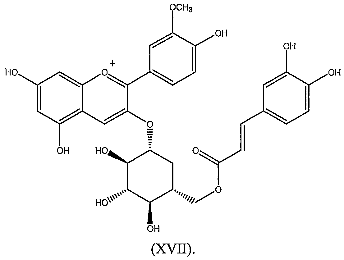
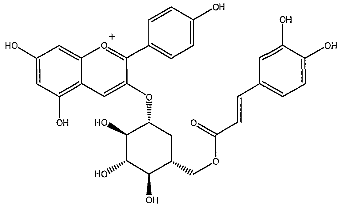
[00100] Illustrative examples of anthocyanin derivatives include:
and pharmaceutically acceptable salts thereof.
[00101] The present invention encompasses compositions, formulations and methods directed to Anthocyanin Derivatives. Any method, mixture or formulation directed to anthocyanins described herein is also meant to encompass Anthocyanin Derivatives.
Methods for Making the Anthocyanin Derivatives of Formulas (IIA) and (IIB) The Anthocyanin Derivatives of the present invention can be made using methodology known to those of ordinary skill in the art or by using the synthetic procedures outlined below in Schemes 1 and 2. Scheme 1 shows a method for making the Anthocyanin Derivatives of formulas (IIA) ) aanndd ((IIIIB) using an triazole ester coupling reaction to add group R10 to the anthocyanin aglycone.
Scheme 1
wherein Y is -CHR
5- or-CHR
5-CHR
6-; and R
1, R
2, R
3, R
4, R
5, R
6, R
7, R
8, R
9 and R
10 are as defined above for the compounds of formulas (IIA) and (IIB).
An antioxidant compound having a carboxylic acid moiety (1) can be reacted with l,l'-carbonyldi(l, 2, 4-triazole) to provide a triazole substituted intermediate of formula 2. The intermediate of formula 2 can then be coupled with an anthocyanin aglycone of formula 3 in presence of l,8-diazabicyclo[5.4.0]undec-7-ene (DBU) to provide the corresponding anthocyanin derivative of formula (IIA) or (IIB).
Scheme 2 shows a method for making the Anthocyanin Derivatives of formulas (IIA) and (IIB) using an olefin metathesis reaction to add group R10 to the anthocyanin aglycone.
allyl-Br R
10-OH
10O base 5 DMF
anthocyanin derivatives of formula (IIA) or (IIB)
A hydroxy group of an anthocyanin aglycone of formula 3 can be derivatized using acryloyl chloride to provide an anthocyanin acrylate intermediate of formula 4. In a separate reaction, an antioxidant compound having a hydroxy moiety (5) can be reacted with allyl bromide to provide an allylic ether intermediate of formula 6. The intermediate of formula 4 can then be coupled with an intermediate of formula 6 via an olefin metathesis reaction using a ruthenium catalyst of formula 7 to provide the coπesponding anthocyanin derivative of formula (IIA) or (IIB).
[00102] Lipofuscin andA2E
[00103] The accumulation of lipofuscin in retinal cells such as retinal pigment epithelial (RPE) cells has been implicated in various eye diseases and disorders. Lipofuscin is not a distinct chemical compound, but a generic name given to an autofluorescent intracellular material that is distributed widely amongst the post-mitotic cells of different organs of the body. With advancing age, and with various eye diseases and disorders, there is a marked increase in the lipofuscin granule content in RPE cells. RPE cell lipofuscin contains lipids, proteins and a heterogeneous mixture of different fluorophores. The major fluorophore in RPE cell lipofuscin is A2E, and the chemical name for A2E is 2-{2-[2,6- Dimethyl-8-(2,6,6-trimethyl-cyclohex-l-enyl)-octa-l,3,5,7-tetraenyl]-4-[4-methyl-6-(2,6,6- trimethyl-cyclohex-l-enyl)-hexa-l,3,5-trienyl]-pyridin-l-yl}-ethanol.
[00104] A2E is a pyridinium bisretinoid compound that is generated as a byproduct of the visual cycle (see Figure 4). The cells involved in visual transduction are the rod and cone photoreceptor cells. Rods and cones are specialized for responding to dim light and color vision respectively. The outer segments of the photoreceptor cells are replete with light-capturing proteins consisting of rod or cone opsin bound through a Schiff base linkage to the chromophore 1 l-cts-retinal. It is the isomerization of the retinal chromophore form the ll-cis to the εdl-trans form by a photon of light that initiates phototransduction. Photoreceptor cells in turn are dependent on cooperative interactions with the underlying retinal pigment epithelium. The RPE cells contain the machinery to regenerate 11-czs-retinal from all-tra s-retinal, the latter being released from opsin after photoisomerization. RPE cells also phagocytose and degrade outer segment membrane that is discarded daily by the photoreceptor cells to balance the continual production of new disks at the base of the photoreceptor segments.
[00105] However, not all of the all-trα/zs-retinal (ATR) is regenerated into 11- cis-retinal by the visual cycle enzymes (i.e., all-trαrø-retinol dehydrogenase, isomerase and l l-cts-retinol dehydrogenase). Some ATR escapes to lead to a progressive accumulation of the non-degradable A2E. A2E is generated from two molecules of ATR and ethanolamine, where ATR is released from photoactivated rhodopsin and ethanolamine being the head group of phosphatidylethanolamine (PE), an abundant membrane phospholipid. On HPLC analysis of hydrophobic extracts of human RPE, A2E presents as a prominent peak along with its slightly less polar photoisomer iso-A2E (see Figure 3).
[00106] Thus, for the purposes of the present invention, methods that relate to
A2E-induced cellular damage also encompass lipofuscin induced damage and other lipofuscin-component induced damage.
[00107] Anthocyanin Testing and Treatment of Lipofuscin/A2E-Related
Diseases and Disorders
[00108] Studies concerned with examining associations between RPE lipofuscin and RPE cell death, have shown that the major constituent of RPE lipofuscin, the bisretinoid fluorophore A2E, can perturb cellular membranes (Spaπow, J.R. et al, Invest. Ophthalmol. Vis. Set, (1999) 40: 2988-2995) and can mediate blue light damage to RPE (Sparrow, J.R. et al, Invest. Ophthalmol. Vis. Set, (2001) 42: 1356-1362; Spaπow, J.R. et al, Invest. Ophthalmol. Vis. Set, (2000) 41: 1981-1989; Spaπow, J.R. et al, Invest. Ophthalmol. Vis. Sci, (2002) 43: 1222-1227). The photoexcixation of A2E leads to the generation of singlet oxygen and the addition of the latter to carbon-carbon double bonds along the retinoid-derived side-arms of A2E such that epoxides are formed. In this way, A2E is converted to a mixture of compounds (A2E-epoxides) bearing epoxides of varying numbers. These highly reactive epoxides have been shown mediate cell damage (Spaπow, J.R. et al, J. Biol. Chem., (2003) 278: 18207-18213). Ultimately, the photochemical events provoked by the iπadiation of A2E in RPE cells initiates cell death (Spaπow et al, (2000)) by way of a pathway that involves the participation of cysteine-dependent proteases (caspases) to cleave cellular substrates and that is modulated by the mitochondrial protein bcl-2 (Spaπow et al, (2002)).
[00109] The present invention provides methods of administering anthocyanins and/or Anthocyanin Derivatives to subjects to treat eye diseases and disorders where lipofuscin or lipfuscin component related damage (i.e., A2E-related damage) may be
involved. For the purposes of the present invention, A2E-related damage includes iso-A2E related damage (see, Figure 5). Eye diseases and disorders where lipofuscin or A2E-related damage may be involved include, but are not limited to, age-related macular degeneration (AMD), juvenile macular degeneration, retinitis pigmentosa, choroidal neovascular membrane (CNVM), macular hole, pattern dystrophy, cataracts or glaucoma. Types of juvenile macular degeneration that are contemplated for treatment include, but are not limited to, cone-rod dystrophy, comeal dystrophy, Fuch's dystrophy, Sorsby's macular dystrophy, Best disease, Stargadt's disease, and juvenile retinoschisis. Types of pattern dystrophy that are contemplated for treatment include, but are not limited to, adult-onset foveomacular vitelliform dystrophy, butterfly-shaped pigment dystrophy, reticular dystrophy of the retinal pigment epithelium, multifocal pattern dystrophy simulating fundus flavimaculatus and fundus pulverulentus.
[00110] One method of treatment relates to conferring resistance to blue-light damage to retinal pigment epithelial (RPE) cells in a subject comprising administering to the subject an amount of one or more anthocyanins and/or Anthocyanin Derivatives effective to quench singlet oxygen in the RPE cells, thereby increasing resistance to blue-light damage in the RPE cells. Another method is directed to reducing RPE cell death in a subject comprising administering to the subject an amount of one or more anthocyanins and/or Anthocyanin Derivatives effective to quench singlet oxygen in the RPE cells, thereby reducing RPE cell death in the subject. Yet another method is directed to reducing or preventing lipofuscin or A2E accumulation in a RPE cell in a subject comprising administering to the subject an amount of one or more anthocyanins and/or Anthocyanin Derivatives effective to prevent or reduce lipofuscin or A2E levels in the RPE cell. Additionally a method is provided for a stabilizing an RPE cell membrane which comprises contacting the RPE cell with an amount of one or more anthocyanins and/or Anthocyanin Derivatives. The present invention also contemplates administering anthocyanins anthocyanins and/or Anthocyanin Derivatives using the above-mentioned methods in combination with one or more of the following entities: isotretinoin (13-cώ-retinoic acid), dehydrogenases (including all-trα«s-retinol- dehydrogenase), a agents that target dehydrogenases (i.e., agents that repress activity of 11- czs-retinol dehydrogenase (see, US Patent Application Publication No. US2003/0032078), isomerase, RmP protein, an anti-phosphatidylethanolamine moiety, vitamin E, zinc, an anti- vascular endothelial growth factor moiety, or any combination thereof.
[00111] The present invention also provides a method for treating or preventing an eye disease in a subject comprising contacting a cell in vitro with one or more anthocyanins and/or Anthocyanin Derivatives; and implanting the cell into an eye of the subject, thereafter treating or preventing an eye disease in the subject. The cell can be a retinal pigment epithelial cell obtained from donors, from the subject itself or from cell lines. In consideration of degenerative eye diseases, the cell can be a stem cell or a retinal progenitor cell, such that after implantation, the cell can help to regenerate damaged tissue, such as damaged retinal pigment epithelium. Prior to implantation, the cell can be treated in vitro to accumulate anthocyanins and/or Anthocyanin Derivatives and to be transfected with a gene, such as an isomerase gene, a dehydrogenase gene, an abcr gene, or any combination thereof.
[00112] To determine whether certain anthocyanins or anthocyanin combinations can confer the above-stated therapeutic effects, the present invention provides both in vitro and in vivo methods to screen or test anthocyanins anthocyanins and/or Anthocyanin Derivatives in relation to the various types of lipofuscin or A2E-mediated damage. For example, the present invention provides methods for and a cell culture model to identify whether or not a particular anthocyanin and/or an AD, mixture, dose amount, or formulation can protect against A2E-mediated cell damage. Figure 6 shows the basic methodology of the cell culture testing/screening method. In one aspect of the cell culture method, a human RPE cell line, ARPE-19 is grown in cell culture. Since ARPE-19 cells do not have endogenous A2E, ARPE-19 cells that are not incubated with A2E provides a negative control for testing the effects of anthocyanins and/or Anthocyanin Derivatives on preventing or reducing A2E mediated damage. Figures 7 and 8 show that ARPE-19 cells accumulate A2E in a manner that is consistent with how RPE cells accumulate A2E in vivo. Figures 7 and 8 show that A2E accumulates in the lysosomal compartment of ARPE-19 cells. Additionally, the cell culture model system can test the therapeutic effects of anthocyanins and/or Anthocyanin Derivatives with other cell types, including primary cells that are isolated from animal donors. Further, the cell culture model system can be modified such that anthocyanin testing is conducted on cells obtained from donors that are afflicted with an eye disease or disorder such that these cells do not have to be incubated with lipofuscin, A2E or some other lipofuscin component. The cell types can include, but are not limited to, primary RPE cells, RPE progenitor cells, stem cells, ARPE-19 cells and any RPE cell line.
[00113] In one variation of the cell culture model, the present invention provides a method to test whether anthocyanins and/or Anthocyanin Derivatives (for the purposes of the present invention, testing anthocyanins and/or Anthocyanin Derivatives refers to testing individual anthocyanins and/or Anthocyanin Derivatives, mixtures, doses or formulations) can protect against the deleterious effects that lipofuscin, A2E or other lipofuscin components have on membrane integrity. In this variation, cultured cells, such as ARPE-19 cells, are incubated with A2E (see, Figure 9). When A2E accumulates in cells, it perturbs membrane integrity as indicated by the leakage of the cytoplasmic enzyme lactate dehydrogenase (LDH) out of the cells. This leakage can be detected by a variety of methods known in the art, such as by testing samples of the cell culture media for the presence of LDH by absorbance (Figure 9B), ELISA or Western or by testing for ability of dyes, which are otherwise not permeable, to enter the cell and label the nucleus. As shown in Figure 9B, incubation with increasing amounts of A2E results in increasing amounts of LDH leakage. Thus, a method for testing whether anthocyanins can protect against A2E-mediated cell membrane damage comprises: incubating cells with or without anthocyanins and/or Anthocyanin Derivatives, incubating cells with A2E, and assaying for leakage of LDH. Particular anthocyanins and/or Anthocyanin Derivatives, mixtures, doses or formulations that decrease the leakage of LDH indicates a potential therapeutic composition.
[00114] In another variation of the cell culture model, the present invention provides a method to test whether anthocyanins and/or Anthocyanin Derivatives can protect against A2E mediated blue light-induced damage. As blue light-exposed A2E-laden RPE cells exhibit a propensity for apoptosis (Spaπow et al, J. Biol. Chem. (2003) 278(20): 18207-18213; Spaπow et al, Invest. Ophth. & Vis. Set, (2000) 41(7): 1981-1989), the present invention provides methods to test whether anthocyanins and/or Anthocyanin Derivatives can reduce A2E-mediated blue-light damage, as indicated by a reduction of A2E epoxidation. Upon blue-light iπadiation of cells containing A2E, A2E self-generates singlet oxygen with the singlet oxygen in turn reacting with A2E to generate epoxides at carbon-carbon double bounds, such that the reduction of A2E levels upon blue-light iπadiation indicates A2E- epoxidation and subsequent apoptosis. For example, in Figure 15 and Example 2, cells incubated with various anthocyanins reduce the loss of A2E by blue-light induced A2E- epoxidation. Figure 16 and Example 3 present an alternative method to test anthocyanins and/or Anthocyanin Derivatives protection, as A2E-epoxidation is catalyzed by endoperoxide. However, the results in Figure 16 are similar, as the anthocyanins repress the
formation of A2E-epoxides. Anthocyanin repression of A2E-epoxidation is shown to be linked to preventing blue-light induced A2E-mediated apoptosis (Figure 17 and Example 4). Interestingly, the cya-gal anthocyanin is more effective than del-gal in preventing the apoptosis. Thus, in this variation, cultured cells, such as ARPE-19 cells, are incubated with A2E and with or without anthocyanins and/or Anthocyanin Derivatives (different types, mixtures, doses, formulations). The incubated cells are then exposed to blue-light (i.e., around 430 nm) or endoperoxide and are assayed for A2E levels and/or for the percentage of cells that are viable or non-viable. Anthocyanins and/or Anthocyanin Derivatives that reduce A2E-epoxidation or the percent of non-viable cells indicate that they are therapeutic candidates. Further, doses or formulations of anthocyanins and/or Anthocyanin Derivatives that reduce A2E-epoxidation or the percent of non-viable cells indicate potential therapeutically effective amounts or formulations.
[00115] In yet another variation of the cell culture model, the present invention provides a method to test whether anthocyanins and/or Anthocyanin Derivatives can protect against A2E cellular accumulation. This method comprises the incubation of cells with A2E and with or without anthocyanins and/or Anthocyanin Derivatives, followed by assaying the cells for their intracellular levels of A2E. As can be seen in Figure 18, del-gal and cya-gal anthocyanins reduce the accumulation of A2E (and iso-A2E) in cells.
[00116] In vivo Testing of Anthocyanins
[00117] The above cell culture models can be used as assay methods in conjunction with animal models to test whether anthocyanins and/or Anthocyanin Derivatives can have a therapeutic effect against A2E-related damage in vivo. For example, the knock-out mouse, abcX" is deficient in both copies of the abcr gene and manifests symptoms of Stargadt's disease and other lipofuscin-accumulation related diseases/disorders: (i) slow photoreceptor degeneration, (ii) delayed recovery of rod sensitivity following light exposure, (iii) elevated all-trαws-retinal and reduced all-trfms-retinol in photoreceptor outer-segments following a photobleach assay, (iv) constitutively elevated phosphatidylethanolamine (PE) in outer- segments, and (v) accumulation of lipofuscin (and A2E) in RPE cells elevated levels of Schiff base conjugate between all-trans-retinal and phosphatidylethanolamine; elevated levels of all-trans-retinal dimer-PE conjugates (Weng, J. et al, Cell, (1999), 98: 13-23; Mate, N.L. et al, Proc. Natl. Acad. Sci. USA, (2000) 97: 7154-7159; Mata, N.L. et al, Invest. Ophthalmol. Visual Sci, (2001), 42: 1685-1690).
[00118] Thus, the present invention provides a method to test whether anthocyanins and/or Anthocyanin Derivatives (including compounds, mixtures, doses and formulations) can protect against lipofuscin related damage in vivo. This method comprises administering anthocyanins and/or Anthocyanin Derivatives to a first abcr gene deficient animal; administering a negative control to a second abcr gene deficient animal; recovering retinal pigment epithelial cells (or other eye cells) from the first and second abcr gene deficient animals; and comparing the health of the recovered retinal pigment epithelial cells from the first and second abcr gene deficient animals, whereby a therapeutically effective composition of anthocyanins and/or Anthocyanin Derivatives is determined.
[00119] Comparing the health of the cells recovered from abcr gene deficient animals comprises the various cell culture methods discussed supra and additional methods. Thus, the methods to compare the health of the cells include, but are not limited to, comparing intracellular A2E levels, comparing intracellular lipofuscin levels, comparing A2E subcellular localization, comparing percent of cell viability within the recovered populations, comparing sensitivity to blue-light mediated apoptosis, comparing sensitivity to A2E- epoxidation, and comparing LDH leakage. Intracellular A2E levels can be quantitatively determined by HPLC analysis, and qualitatively determined by microscopy, including fluorescent confocal microscopy and electron microscopy. Histological methods can also be utilized to determine the relative levels of cell viability in the retinal pigment epithelium. Additionally, the visual function of abcr gene deficient mice treated and not treated with anthocyanin can be tested by ERG analysis (Radu, R. et al, Proc. Natl. Acad. Sci. USA, (2003) 100(8): 4742-4747).
[00120] In all of the methods of the present invention that compares the health of cells treated or untreated with anthocyanins and/or Anthocyanin Derivatives, those treatments that show a reduction or prevention of cell damage (including cell membrane integrity) or viability, A2E or lipofuscin accumulation, A2E-epoxidation, etc., as compared to the negative controls (i.e., animals or cells not treated with anthocyanins and/or Anthocyanin Derivatives); the reduction or prevention indicates that the treatment comprises an amount of anthocyanins and/or Anthocyanin Derivatives effective to provide a therapeutic effect. Thus, in this sense, a therapeutic effect provided by anthocyanins and/or Anthocyanin Derivatives relates to at least: the prevention or reduction of lipofuscin accumulation, iso-A2E accumulation, A2E accumulation, A2E-epoxidation, cell membrane integrity, cell viability, or visual function.
[00121] Additionally, in all of the methods of the present invention that compares the health of cells treated or untreated with anthocyanins and/or Anthocyanin Derivatives, the methods can further comprise testing the health of cells treated or untreated with anthocyanins and/or Anthocyanin Derivatives in combination with isotretinoin (13-cis- retinoic acid), dehydrogenases (including all-tra/zs-retinol-dehydrogenase, l l-cts-retinol dehydrogenase), isomerase, RmP protein, an anti-phosphatidylethanolamine moiety, vitamin E, zinc, an anti-vascular endothelial growth factor moiety, or any combination thereof.
[00122] Administration Methods and Formulations
[00123] The methods of administering anthocyanins and Anthocyanin Derivatives, including anthocyanin mixtures, anthocyanin derivative compounds and formulations thereof, contemplate essentially any method of delivery, including topical ophthalmic delivery, an oral delivery, a nasal delivery, an implant delivery, or an injection delivery. Topical ophthalmic delivery can include, for example, delivery of anthocyanins and/or derivatives by eye drops or a cream/ointment. When the anthocyanin and or derivative is to be injected into the eye, the injection can be into the vitreous or subretinal compartment of an eye.
[00124] The purpose of injection into the vitreous or subretinal compartment is to maximize or enhance the amount of anthocyanins and Anthocyanin Derivatives that come into contact with posterior areas of the eye, such as the retinal pigment epithelium. However, as administration may be conducted on a repeated basis, repetitive insertion into the eye can cause complications such as retinal detachment, posterior dislocation, endophthalmitis, vitreous hemoπhage and cataract formation. To address this concern, a prolonged or controlled delivery can be accomplished by administering intravitreous polymer implants or microspheres containing anthocyanins and/or Anthocyanin Derivatives. The choice of anthocyanins and/or derivatives in the intravitreous polymer implants is made according to size. For example, as the internal limiting membrane of the retina prevents diffusion of substances larger than about 40,000 Daltons to about 70,000 Daltons from diffusing into the retina from the vitreous, anthocyanins and derivatives smaller than at least about 70,000 Daltons are selected for intravitreous polymer implant delivery and for intravitreous injection delivery. Alternatively, anthocyanins and derivatives smaller than about 40,000 Daltons are selected for intravitreous polymer implant and injection delivery.
[00125] Injectable or implantable formulations can comprise biodegradable polymer microspheres that are attached to anthocyanins and Anthocyanin Derivatives and provide a
controlled or prolonged/sustained release of the anthocyanins and Anthocyanin Derivatives. The biodegradable polymer microspheres can comprise poly(L-lactic acid) and poly(glycolic acid), wherein the poly(glycolic acid) can comprise poly(D,L-lactide-co-glycolide)(PLGA). The polymer microspheres can be injected or implanted into the vitreous (Moritera, T. et al, Invest. Ophthalmol. Vis. Sci, (1991) 32:1785) as discussed above, or directly injected to retinal pigment epithelial cells by subretinal injection (Ogura, Y. and Kimura, H, Survey Ophthalmol, (1995), 39, Suppl. 1, S17-S24). In the subretinal injection, a device, such as a glass micropipette, is inserted transvitreally through a sclerotomy and stereotactically manipulated through the subretinal space. The subretinal delivery allows direct delivery of the microspheres to the retinal pigment epithelial because the RPE cells will phagocytose the microspheres and its attached anthocyanin cargo. Such a delivery may be favorable in certain circumstances, because anthocyanins will be delivered directly to lipofuscin, and hence, A2E. The delivery brings anthocyanins and A2E into direct contact because membrane vesicles carrying phagocytosed substances merge and localize to the lysosomal compartment, where lipofuscin accumulates. Biodegradable polymers can also be implanted into the sclera (Kunou, N. et al, J. Biomed. Mater. Res., (2000), 51: 635-641).
[00126] Another formulation of the present invention comprises liposomes that encapsulate anthocyanins and/or Anthocyanin Derivatives. A benefit of a liposome formulation is that it allows for non-ocular injection. Liposomes can be injected to the arm of a subject, for example, whereby the liposomes travel throughout the bloodstream. Targeted delivery of the anthocyanins by the liposomes can be accomplished by light-targeted delivery (LTD). With LTD, anthocyanins and/or Anthocyanin Derivatives are encapsulated in heat- sensitive liposomes. The liposomes do not disassemble or degrade under normal physiological temperatures. To release the anthocyanins and derivatives from the liposomes at the retinal pigment epithelium, a light beam iπadiates the choroidal neovascularization (CNV) and suπounding tissues at a wavelength strongly absorbed by blood. The RPE, choriocapillaris and CNV are the first to reach the temperature necessary to melt the circulating liposomes (about 41 °C) (Zeimer, R. and Goldberg, M.F., Adv. Drug Deliv. Reviews, (2001), 52: 49-61).
[00127] If anthocyanins and/or Anthocyanin Derivatives are to be administered topically to the eye, the formulation can comprise a saline solution. Such a saline solution can be an eye drop formulation, where an anthocyanin or an anthocyanin derivative compound can be esterified to a isopropyl-ester prodrug such that it can then be hydrolyzed
in the cornea by esterases to a free hydroxyl form. If anthocyanins and/or Anthocyanin Derivatives are to be administered via the digestive system, then the formulation can comprise a carbomer or a polymer such that the anthocyanin and/or derivatives are tableted, or are stabilized in a solution, syrup, ointment or cream.
[00128] It is to be understood and expected that variations in the principles of the invention herein disclosed in an exemplary embodiment can be made by one skilled in the art and it is intended that such modifications, changes, and substitutions are included within the scope of the present invention.
[00129] The examples set forth below illustrate several embodiments of the invention. These examples are for illustrative purposes only, and are not meant to be limiting.
[00130] EXAMPLES Materials and Methods
[00131] Reagents: Bilberry extract was from Nature's Resource Premium Herb
(Mission Hills, CA); ethanolamine and trifluoroacetic acid were purchased from Aldrich Chemical Company, Inc. (Milwaukee, WI); HEPES were obtained from Sigma (St. Louis, MO); acetonitrile was purchased from Fisher (Fair Lawn, NJ) and Dulbecco's phosphate buffered saline (DPBS) from GIBCO-BRL. HP-20 resin was purchased from Supelco (Bellefonte, PA). All other chemicals were from Sigma. A2E was synthesized as previously described (Parish, CA. et al, Proc. Natl. Acad. Sci. USA, (1998) 95: 14609-14613).
[00132] Cell culture, treatment and illumination: Human adult RPE cells (ARPE-19;
American Type Culture Collection, Manassas VA) lacking endogenous A2E (Spaπow et al, (1999)) were grown as previously described 18, 20. To generate A2E-laden RPE, nonconfluent cultures were allowed to accumulate A2E from a 20μM concentration in media 18. For some experiments, cultures were treated prior to blue light illumination with anthocyanins (500 μM) dissolved in PBS buffer solution. Cells were subsequently exposed to 430 nm illumination (0.36 mW/mm2) as previously described 19-21. To assess cell viability, cultures were exposed to a 430 nm light line (0.8 mm wide; 0.34 mW/mm2).
[00133] Evaluation of the singlet oxygen quenching activity of anthocyanins: To measure the singlet oxygen quenching activity of anthocyanins, A2E (100 mM) and
anthocyanins (500 μM) were incubated together with a single oxygen generator, the endoperoxide of 1,4-dimethylnaphthalene (10 mM in 100 mL of methanol) (Ben-Shabat; Spaπow). The mixture was stiπed for 14 - 15 h in the dark at room temperature following which A2E was quantified by HPLC using A2E as external standard.
[00134] Mass spectrometry: FAB-MS for oxidized A2E was performed on a JEOL
JMS-HX110A/110A tandem MS (Akishima, Tokyo Japan), using 10 kV acceleration voltage and fitted with a Xe beam FAB gun (6 kV) on the MS-1 ion source. 3-Nitrobenzyl alcohol was used as matrix. Liquid chromatography-mass spectrometry (LCMS) analysis of anthocyanins was performed on a Q-TOF detector (Micromass, Manchester, UK) with electrospray ionization (ESI) source. ESI capillary voltage was 3 kV. The sample was introduced with a flow injection mode using 80% of acetonitrile and 0.1% of TFA in water as a solvent. Flow rate was 0.2ml/min.
[00135] Assay of cell viability: Apoptotic cells and membrane-permeabilized cells were labeled with the membrane impermeant dye Dead Red (Molecular Probes, Eugene OR) and the nuclei of all cells with DAPI, as previously described 21. Counting was performed from digital images, nonviable cells were expressed as a proportion of the total number of cells in an illuminated field and means were compared by ANOVA followed by the Student- Newman Keul multiple comparison test. The level of significance was 0.05.
[00136] Activity-guided fractionation of bilberry and isolation of anthocyanins:
Bilberry extract (10 g) was dissolved in 250 ml of deionized water, and fractionated between the organic and aqueous phases by extracting an aqueous solution of bilberry with 250 ml of ethyl acetate three times. HP-20 dianion resin was used to divide aqueous layer into 10 subtractions (H2O : MeOH = 100 : 0 a 0 : 100). The fractions were assayed for their ability to inhibit A2E-epoxidation by exposing preparations of A2E (20 μM in DPBS) with and without each fraction (250 μg/ml), to 430nm light for 10 min (0.36 mW/mm2). Anthocyanins were isolated by preparative HPLC of the aqueous layer of bilberry extract. Each peak was collected for the mass spectroscopy.
[00137] HPLC: A HP 1100 Series HPLC equipped with photodiode aπay detector was used with a reverse phase C18 column (150 mm x 4.6 mm, Cosmosil 5C18, Nacalai Tesque, Japan) for quantitation studies of anthocyanins and a Vydac C18 (250 mm x 22 mm, 10 μm, Grace Vydac, CA) for anthocyanin separation. Detection was at 520 nm. A2E was quantitated with the following gradient of acetonitrile in water (containing 0.1 % TFA): 85-
95 % (10 min), 95-100 (5 min), 100 % (5 min); flow rate 0.8 ml/min with monitoring at 430 nm. In some experiments, A2E was extracted from RPE cells with CHC13 after homogenizing with glass tissue homogenizer. A2E was used as external standard for quantitation and A2E levels were normalized to cellular protein concentration after protein assay by BioRad Bradford method. Anthocyanins were quantitated with the following gradient of 5 % formic acid in acetonitrile: 95-90 % (5 min), 90-85 % (10 min); flow rate 0.8 ml/min at 520 nm. To isolate anthocyanins by preparative HPLC, a solvent system of 5 % formic acid in acetonitrile was used as the mobile phase with the following gradient: 93 % (v/v, 30 min), 93-90 % (10 min), 90-87 % (30 min), 87-77 % (20 min), 77-75 % (10 min), 75-30 % (20 min); flow rate 4.0 ml/min at 520 nm; injection volume 100 μl (40mg/ml).
EXAMPLE 1: Anthocyanins Isolated From Bilberry Extracts
[00138] The application of reverse-phase HPLC and gradient elution, 10 individual anthocyanins (see Figures 1 and 2), all monoglycosylated, were resolved in the water-soluble extract of bilberry and identified by LC-MS. The sequence of elution of the compounds and coπesponding mass-to charge ratio (m/z) as determined by mass spectrum was delphinidin-3- galactoside (del-gal; peak 1), m/z 465; delphinidin-3 -glucoside (del-glc; peak 2), m/z 465; cyanidin-3-galactoside (cya-gal; peak 3), m/z 449; cyanidin-3-glucoside (cya-glc; peak 4), m/z 449; petunidin-3 -galactoside (pet-gal; peak 5), m/z 479; cyanidin-3-arabinoside (cya-ara; peak 6), m/z 419; petunidin-3-glucoside (pet-glc; peak 7), m/z 479; peonidin-3 -galactoside (peo-gal; peak 8), m/z 463; peonidin-3-glucoside (peo-glc; peak 9), m/z 463; malvidin-3- glucoside (mal-glc; peak 10), m/z 493. This elution order is consistent with their known hydrophobicities (da Costa et al, 2000; Goiffon et al.). Of the 10 anthocyanins isolated from the extract, the predominant compounds were delphinidin 3-galactoside (Del-Gal), delphinidin 3-glucoside (Del-Glc), cyanidin 3-galactoside (Cya-Gal) and cyanidin 3- glucoside (Cya-Glc).
EXAMPLE 2: Anthocyanins Protect Against A2E-Epoxidation in a Cell-Free System
[00139] Given that anthocyanins are purported to have antioxidant properties, anthocyanins were tested as to whether they could inhibit light-induced epoxidation of A2E. For these experiments, A2E in aqueous media was exposed to 430 nm illumination in the presence and absence of the anthocyanins del-gal, del-glc, cya-gal and cya-glc and the
mixtures were subsequently analyzed by quantifying the consumption of A2E as A2E- epoxidation occuπed. Blue light iπadiation resulted in an HPLC profile that revealed a decrease in the absorbance of the A2E peak as compared with the control non-illuminated sample (Figure 15). This change signified a 12% loss of A2E. This photooxidation associated diminution in A2E was attenuated, however, when the iπadiated mixture contained del-gal (57% reduction in A2E), del-glc (46%) cya-gal (54%) and cya-glc (53%).
EXAMPLE 3: Anthocyanins Quench Singlet Oxygen
[00140] Since singlet oxygen mediates the epoxidation of A2E, it is advantageous to determine whether the anthocyanins inhibit A2E-epoxidation by quenching singlet oxygen. To this end, singlet oxygen was generated from the endoperoxide of 1,4- dimethylnaphthalene; the latter decomposes to release singlet oxygen. It has been previously demonstrated that for the generation of A2E-epoxides, the endoperoxide can substitute for blue light iπadiation. Accordingly, mixtures of A2E, 1,4-dimethyl naphthalene endoperoxide and del-gal or cya-gal in methanol were incubated for 14-15 hours at room temperature. As shown in Fig. 16, quantification of A2E by HPLC revealed a 96% decrease (p < 0.001) in the level of A2E following its incubation with the singlet oxygen generator but without anthocyanins. Addition of anthocyanins to the A2E - endoperoxide mixture shielded A2E such that its loss was reduced to 62% (del-gal) and 63% (cya-gal) of control A2E levels not incubated with endoperoxide (p < 0.001). These results demonstate that anthocyanins protect against A2E-epoxidation by quenching singlet oxygen. EXAMPLE 4 Anthocyanins Protect A2E-laden RPE Cells From Blue Light-Induced Damage
[00141] To study the anthocyanins in a cell system, ARPE-19 cells were incubated with del-gal and cya-gal ( 500 μM) for 2-5 days. Incorporation of anthocyanins into the cells was confirmed by light microscopy (Fig 17A). Separation of membrane, mitochondrial and cytosolic fractions, showed that anthocyanins were not only incorporated into the plasma membrane (~90%), but also penetrated into the cell cytosol (~ 10%). When incubated with anthocyanins for up to 5 days, the ARPE-19 cells showed no evidence of cell toxicity (data not shown).
[00142] The capacity for anthocyanins to confer a cellular resistance to blue light damage was evident when ARPE-19 cells that had accumulated A2E were incubated with
del-gal and cya-gal for 3 days and were then illuminated with blue light. Pre-treatment with del-gal and cya-gal reduced cell death by 60% and 33%, respectively (Figure 17). [00143] The protection confeπed by anthocyanins to A2E epoxidation was further shown in Figure 19. ARPE-19 cells that had accumulated A2E were incubated with 100 μM of the following anthocyanins: del-gal, del-glc, del-ara, cya-gal, cya-glc, cya-ara, pet-gal, pet- glc, pet-ara, mal-gal, mal-glc, mal-ara, and the aglycones themselves without sugar substituents (i.e., delphinidin, cyaniding, petunidin, and malvidin). The cells were then exposed to blue-light, and cell viability was assessed. In Figure 19, the % protection refers to the percentage of cells that survived the blue-light induced A2E epoxidation mediated apoptosis. As can be observed, the percent protection of anthocyanins can vary according to which aglyclone (and which sugar is attached to the aglyclone (or no sugar)) is incubated with the cells. Although delphinidin species may possess the most potent antioxidant activity due to the fact that is has three OH groups at the 3', 4' and 5' positions (as opposed to the other aglycones having one or two OH groups at these positions - see brief description for Figure 20 and Figure 20); in this example, the malvidin species were most effective in protecting the cells against A2E-epoxidation mediated death, potentially due a greater stability of malvidin as compared to the other aglyclone species. [00144] EXAMPLE 5 Anthocyanins Inhibit Lipid Peroxidation [00145] Anthocyanins reduced cellular incorporation of A2E.
[00146] In experiments in which cultured RPE cells were incubated with anthocyanins before allowing the cells to accumulate A2E, it was consistently observed by microscopic examination that the amount of A2E amassed was reduced as compared to cells that had not been pre-treated with anthocyanins. To confirm these observations by quantitative assay, the amounts of A2E and iso-A2E, the latter being a less abundant photoisomer, were quantified by HPLC to determine the cellular incorporation of these fiuorophores. Consistent with previous observation that A2E and iso-A2E reach photoequilibium at a ratio 4:1 (A2E:iso-A2E) (Parish), A2E was present at a 3.8-fold greater abundance than iso-A2E (Fig. 6). Under conditions in which the cells were pre-treated with anthocyanins , cellular incorporation of both A2E and iso-A2E was reduced. The decrease due to del-gal was 29% for both A2E and iso-A2E while cya-gal reduced the accumulation of A2E and iso-A2E by
46%o and 44%>, respectively. Despite the reduced incorporation of A2E/isoA2E, the ratio of these photoisomers remained constant (Fig. 6).
EXAMPLE 6 Anthocyanins Reduce Cellular Incorporation of A2E [00147] To study how anthocyanin can reduce the RPE cell toxicity induced by high concentration of A2E, cellular localization of incorporated anthocyanins was measured by HPLC quantitation of anthocyanin in different cellular compartment. Most of incorporated anthocyanins were located at cellular membrane fraction (Fig. 6), as is supposed by previous reports about the cellular localization of anthocyanin and its affinity on membrane bound carrier protein (ref). To clarify whether this kind of properties of anthocyanin is able to interrupt RPE cellular incorporation of A2E, anthocyanins were pretreated to RPE cells before A2E (20 μM) treatment, and then quantitated intracellular A2E by HPLC. Cellular incorporation of A2E was significantly blocked by pretreatment of anthocyanin (Fig. 18). EXAMPLE 7: Anthocyanins Reduce A2E-Mediated RPE Cell Death [00148] A2E has detergent-like properties that can critically damage cell membranes.
As anthocyanins were reported to interact with cell membrane, the effect of anthocyanins on high concentration of A2E (100 μM) insult to RPE cells was tested. This concentration of A2E showed severe toxicity in cells in 50-100 μM A2E for 2 hours manifested by permeability of membrane to lactate dehydrogenase (Spaπow, 1999). EXAMPLE 8: Synthesis of an Anthocyanin Derivative (see Figure 23)
[00149] Caffeic acid (1 eq.) and l,l'-dicarbonylimidazole (1.1 eq.) were dissolved in
THF and stiπed for overnight under room temperature. The reaction solution was extracted with diethylether and then extract was washed with saturated NaHCO3 and NaCl aqueous solution respectively. The ether layer was dried with Na2SO4 and then filtered. The filtrate was evaporated to leave yellow powder, 3-(3,4-Dihydroxy-phenyl)-l-imidazol-l-yl- propenone (see Figure 23; 1). Cyanidin-3-O-glucoside was reacted with 1 in THF/CH3CN (1:1 mixture) as a solvent and stiπed for overnight at room temperature. The reaction solution was concentrated and purified with reverse phase HPLC (H2O and CH3CN with TFA as solvent), such that the anthocyanin derivative (see Figure 23; 2; formula (V)) was isolated.
Although cyanidin-3-O-glucoside was used as the anthocyanin starting material, other anthocyanins can readily be used with this protocol, for example, other anthocyanins include: delphinidin-3-O-galactoside (del-gal), delphinidin-3-O-glucoside (del-glc), cyanidin-3-O- galactoside (cya-gal), cyanidin-3-O-arabinoside (cya-ara), petunidin-3-O-galactoside (pet- gal), petunidin-3-O-glucoside (pet-glc), petunidin-3-O-arabinoside (pet-ara), peonidin-3-O- galactoside (peo-gal), peonidin-3-O-glucoside (peo-glc), peonidin-3-O-arabinoside (peo-ara), malvidin-3-O-galactoside (mal-gal), malvidin-3-O-arabinoside (mal-ara) and malvidin-3-O- glucoside (mal-glc) (see Examples 10-24). [00150] EXAMPLE 9: Anthocyanin Derivative with Improved Antioxidant Properties (see Figure 24) [00151] As shown in Example 4, anthocyanins have antioxidant properties as they could inhibit light-induced epoxidation of A2E. Thus, an anthocyanin derivative (formula (V)) was tested to measure its antioxidant properties. For these experiments, A2E in aqueous media was exposed to 430 nm illumination in the presence and absence of anthocyanins (see Figure 24, "Cy") and the anthocyanin derivative ("AcylCy"). The mixtares were subsequently analyzed by quantifying the consumption of A2E as A2E-epoxidation occuπed. Blue light iπadiation resulted in an HPLC profile that revealed a decrease in the absorbance of the A2E peak ("A2E + BL") as compared with the control non-illuminated sample ("A2E"). This photooxidation associated diminution in A2E was attenuated, however, when the iπadiated mixture contained an anthocyanin ("A2E+Cy+BL"). The photoxidation was even more attenuated when the iπadiated mixture contained the anthocyanin derivative ("A2E+AcylCy+BL").
EXAMPLE 10: Synthesis of a Delphinidin-3-O-Glucoside Derivative
[00152] Caffeic acid (1 eq.) and l,l'-dicarbonylimidazole (1.1 eq.) are dissolved in
THF and stiπed for overnight under room temperature. The reaction solution is extracted with diethylether and then the extract is washed with saturated NaHCO3 and NaCl aqueous solution respectively. The ether layer is dried with Na2SO4 and then filtered. The filtrate is evaporated to leave yellow powder, 3-(3,4-Dihydroxy-phenyl)-l-imidazol-l-yl-propenone.
Delphinidin-3-O-glucoside (del-glc) is reacted with 1 in THF/CH3CN (1:1 mixture) as a solvent and stiπed for overnight at room temperature. The reaction solution is concentrated and purified with reverse phase HPLC (H2O and CH3CN with TFA as solvent), such that an anthocyanin derivative (delphinidin-3-O-(6-O-caffeoyl-glucoside)) according to formula (VT) below is isolated.
EXAMPLE 11: Synthesis of a Delphinidin-3-O-Galactoside Derivative
[00153] Caffeic acid (1 eq.) and l,l'-dicarbonylimidazole (1.1 eq.) are dissolved in
THF and stiπed for overnight under room temperature. The reaction solution is extracted with diethylether and then the extract is washed with saturated NaHCO3 and NaCl aqueous solution respectively. The ether layer is dried with Na2SO and then filtered. The filtrate is evaporated to leave yellow powder, 3-(3,4-Dihydroxy-phenyl)-l-imidazol-l-yl-propenone. Delphinidin-3-O-galactoside (del-gal) is reacted with 1 in THF/CH3CN (1:1 mixtare) as a solvent and stiπed for overnight at room temperature. The reaction solution is concentrated and purified with reverse phase HPLC (H2O and CH3CN with TFA as solvent), such that an anthocyanin derivative (delphinidin-3-O-(6-O-caffeoyl-galactoside)) according to formula (VII) below is isol. ' '
EXAMPLE 12: Synthesis of a Delphinidin-3-O-Arabinoside Derivative
[00154] Caffeic acid (1 eq.) and l,l'-dicarbonylimidazole (1.1 eq.) are dissolved in
THF and stirred for overnight under room temperatare. The reaction solution is extracted with diethylether and then the extract is washed with saturated NaHCO3 and NaCl aqueous solution respectively. The ether layer is dried with Na2SO4 and then filtered. The filtrate is evaporated to leave yellow powder, 3-(3,4-Dihydroxy-phenyl)-l-imidazol-l-yl-propenone. Delphinidin-3-O-arabinose (del-ara) is reacted with 1 in THF/CH3CN (1:1 mixtare) as a solvent and stiπed for overnight at room temperature. The reaction solution is concentrated and purified with reverse phase HPLC (H2O and CH3CN with TFA as solvent), such that an anthocyanin derivative (delphinidin-3-O-(5-O-caffeoyl-arabinoside)) according to formula (VIII) below is isolated.
(VIII).
EXAMPLE 13: Synthesis of a Cyanidin-3-O-Galactoside Derivative
[00155] Caffeic acid (1 eq.) and l,l'-dicarbonylimidazole (1.1 eq.) are dissolved in
THF and stiπed for overnight under room temperatare. The reaction solution is extracted with diethylether and then the extract is washed with satarated NaHCO3 and NaCl aqueous solution respectively. The ether layer is dried with Na2SO and then filtered. The filtrate is evaporated to leave yellow powder, 3-(3,4-Dihydroxy-phenyl)-l-imidazol-l-yl-propenone. Cyanidin-3-O-galactoside (cya-gal) is reacted with 1 in THF/CH3CN (1:1 mixture) as a solvent and stiπed for overnight at room temperatare. The reaction solution is concentrated and purified with reverse phase HPLC (H2O and CH3CN with TFA as solvent), such that an anthocyanin derivative (cyanidin-3-O-(6-O-caffeoyl-galactoside)) according to formula (IX) below is isolated. [00156]
EXAMPLE 14: Synthesis of a Cvanidin-3-O-Arabinoside Derivative
[00157] Caffeic acid (1 eq.) and l,l'-dicarbonylimidazole (1.1 eq.) are dissolved in
THF and stiπed for overnight under room temperature. The reaction solution is extracted with diethylether and then the extract is washed with satarated NaHCO3 and NaCl aqueous solution respectively. The ether layer is dried with Na2SO and then filtered. The filtrate is evaporated to leave yellow powder, 3-(3,4-Dihydroxy-phenyl)-l-imidazol-l-yl-propenone. Cyanidin-3-O-arabinoside (cya-ara) is reacted with 1 in THF/CH3CN (1:1 mixtare) as a solvent and stiπed for overnight at room temperatare. The reaction solution is concentrated and purified with reverse phase HPLC (H2O and CH3CN with TFA as solvent), such that an anthocyanin derivative (cyanidin-3-O-(5-O-caffeoyl-arabinoside)) according to formula (X) below is isolated.
(X).
EXAMPLE 15: Synthesis of a Petanidin-3-O-Glucoside Derivative
[00158] Caffeic acid (1 eq.) and l,l'-dicarbonylimidazole (1.1 eq.) are dissolved in
THF and stiπed for overnight under room temperatare. The reaction solution is extracted with diethylether and then the extract is washed with saturated NaHCO3 and NaCl aqueous solution respectively. The ether layer is dried with Na2S04 and then filtered. The filtrate is evaporated to leave yellow powder, 3-(3,4-Dihydroxy-phenyl)-l-imidazol-l-yl-propenone. Petanidin-3-O-glucoside (pet-glc) is reacted with 1 in THF/CH3CN (1:1 mixture) as a solvent and stiπed for overnight at room temperatare. The reaction solution is concentrated and purified with reverse phase HPLC (H2O and CH3CN with TFA as solvent), such that an anthocyanin derivative (petunidin-3-O-(6-O-caffeoyl-glucoside)) according to formula (XI) below is isolated.
EXAMPLE 16: Synthesis of a Petunidin-3-O-Galactoside Derivative
[00159] Caffeic acid (1 eq.) and l,l'-dicarbonylimidazole (1.1 eq.) are dissolved in
THF and stiπed for overnight under room temperatare. The reaction solution is extracted with diethylether and then the extract is washed with satarated NaHCO3 and NaCl aqueous solution respectively. The ether layer is dried with Na2SO4 and then filtered. The filtrate is evaporated to leave yellow powder, 3-(3,4-Dihydroxy-phenyl)-l-imidazol-l-yl-propenone. Petunidin-3-O-galactoside (pet-gal) is reacted with 1 in THF/CH3CN (1:1 mixtare) as a solvent and stiπed for overnight at room temperatare. The reaction solution is concentrated and purified with reverse phase HPLC (H2O and CH3CN with TFA as solvent), such that an anthocyanin derivative (petanidin-3-O-(6-O-caffeoyl-galactoside)) according to formula (XII) below is isolated.
(XII).
EXAMPLE 17: Synthesis of a Petanidin-3-O-Arabinoside Derivative
[00160] Caffeic acid (1 eq.) and l,l'-dicarbonylimidazole (1.1 eq.) are dissolved in
THF and stiπed for overnight under room temperatare. The reaction solution is extracted with diethylether and then the extract is washed with saturated NaHCO3 and NaCl aqueous
solution respectively. The ether layer is dried with Na2SO4 and then filtered. The filtrate is evaporated to leave yellow powder, 3-(3,4-Dihydroxy-phenyl)-l-imidazol-l-yl-ρropenone. • Petunidin-3-O-arabinoside (pet-ara) is reacted with 1 in THF/CH3CN (1:1 mixtare) as a solvent and stiπed for overnight at room temperature. The reaction solution is concentrated and purified with reverse phase HPLC (H2O and CH3CN with TFA as solvent), such that an anthocyanin derivative (petanidin-3-O-(5-O-caffeoyl-arabinoside)) according to formula (XIII) below is isolated.
EXAMPLE 18: Synthesis of a Malvidin-3-O-Glucoside Derivative
[00161] Caffeic acid (1 eq.) and l,l'-dicarbonylimidazole (1.1 eq.) are dissolved in
THF and stiπed for overnight under room temperatare. The reaction solution is exfracted with diethylether and then the extract is washed with saturated NaHCO3 and NaCl aqueous solution respectively. The ether layer is dried with Na2SO4 and then filtered. The filtrate is evaporated to leave yellow powder, 3-(3,4-Dihydroxy-phenyl)-l-imidazol-l-yl-propenone. Malvidin-3-O-glucoside (mal-glc) is reacted with 1 in THF/CH3CN (1:1 mixtare) as a solvent and stiπed for overnight at room temperatare. The reaction solution is concentrated and
purified with reverse phase HPLC (H2O and CH3CN with TFA as solvent), such that an anthocyanin derivative (malvidin-3-O-(6-O-caffeoyl-glucoside)) according to formula (XIV) below is isolated. [00162]
EXAMPLE 19: Synthesis of a Malvidin-3-O-Galactoside Derivative
[00164] Caffeic acid (1 eq.) and l,l'-dicarbonylimidazole (1.1 eq.) are dissolved in
THF and stiπed for overnight under room temperatare. The reaction solution is extracted with diethylether and then the extract is washed with saturated NaHCO
3 and NaCl aqueous solution respectively. The ether layer is dried with Na
2SO
4 and then filtered. The filtrate is evaporated to leave yellow powder, 3-(3,4-Dihydroxy-phenyl)-l-imidazol-l-yl-propenone. Malvidin-3-O-galactoside (mal-gal) is reacted with 1 in THF/CH
3CN (1:1 mixtare) as a solvent and stiπed for overnight at room temperatare. The reaction solution is concentrated and purified with reverse phase HPLC (H
2O and CH
3CN with TFA as solvent), such that an anthocyanin derivative (malvidin-3-O-(6-O-caffeoyl-galactoside)) according to formula (XV) below is isolated.
(XV).
EXAMPLE 20: Synthesis of a Malvidin-3-O-Arabinoside Derivative
[00165] Caffeic acid (1 eq.) and l,l'-dicarbonylimidazole (1.1 eq.) are dissolved in
THF and stiπed for overnight under room temperature. The reaction solution is extracted with diethylether and then the exfract is washed with saturated NaHCO3 and NaCl aqueous solution respectively. The ether layer is dried with Na2SO4 and then filtered. The filtrate is evaporated to leave yellow powder, 3-(3,4-Dihydroxy-phenyl)-l-imidazol-l-yl-propenone. Malvidin-3-O-arabinoside (mal-ara) is reacted with 1 in THF/CH CN (1:1 mixtare) as a solvent and stiπed for overnight at room temperatare. The reaction solution is concentrated and purified with reverse phase HPLC (H2O and CH3CN with TFA as solvent), such that an anthocyanin derivative (malvidin-3-O-(5-O-caffeoyl-arabinoside)) according to formula (XVI) below is isolated.
(XVI).
EXAMPLE 21: Synthesis of a Peonidin-3-O-Glucoside Derivative
[00166] Caffeic acid (1 eq.) and l,l'-dicarbonylimidazole (1.1 eq.) are dissolved in
THF and stiπed for overnight under room temperatare. The reaction solution is exfracted with diethylether and then the extract is washed with saturated NaHCO3 and NaCl aqueous solution respectively. The ether layer is dried with Na2SO4 and then filtered. The filtrate is evaporated to leave yellow powder, 3-(3,4-Dihydroxy-phenyl)-l-imidazol-l-yl-propenone. Peonidin-3-O-glucoside (peo-glc) is reacted with 1 in THF/CH3CN (1:1 mixtare) as a solvent and stiπed for overnight at room temperature. The reaction solution is concentrated and purified with reverse phase HPLC (H2O and CH3CN with TFA as solvent), such that an anthocyanin derivative (peonidin-3-O-(6-O-caffeoyl-glucoside)) according to formula (XVTI) below is isolated.
[00167]
EXAMPLE 22: Synthesis of a Peonidin-3-O-Galactoside Derivative
[00168] Caffeic acid (1 eq.) and l,l'-dicarbonylimidazole (1.1 eq.) are dissolved in
THF and stiπed for overnight under room temperature. The reaction solution is extracted with diethylether and then the extract is washed with satarated NaHCO3 and NaCl aqueous solution respectively. The ether layer is dried with Na2SO4 and then filtered. The filtrate is evaporated to leave yellow powder, 3-(3,4-Dihydroxy-phenyl)-l-imidazol-l-yl-propenone. Peonidin-3-O-glucoside (peo-gal) is reacted with 1 in THF/CH3CN (1:1 mixture) as a solvent and stirred for overnight at room temperature. The reaction solution is concentrated and purified with reverse phase HPLC (H2O and CH3CN with TFA as solvent), such that an anthocyanin derivative (peonidin-3-O-(6-O-caffeoyl-galactoside)) according to formula (XVIII) below is isolated.
EXAMPLE 23: Synthesis of a Peonidin-3-O-Arabinoside Derivative
[00169] Caffeic acid (1 eq.) and l,l'-dicarbonylimidazole (1.1 eq.) are dissolved in
THF and stiπed for overnight under room temperatare. The reaction solution is exfracted with diethylether and then the extract is washed with saturated NaHCO3 and NaCl aqueous solution respectively. The ether layer is dried with Na2SO4 and then filtered. The filtrate is
evaporated to leave yellow powder, 3-(3,4-Dihydroxy-phenyl)-l-imidazol-l-yl-propenone. Peonidin-3-O-arabinoside (peo-ara) is reacted with 1 in THF/CH3CN (1:1 mixtare) as a solvent and stiπed for overnight at room temperatare. The reaction solution is concentrated and purified with reverse phase HPLC (H2O and CH CN with TFA as solvent), such that an anthocyanin derivative (peonidin-3-O-(5-O-caffeoyl-arabinoside)) according to formula (XIX) below is isolated.
(XLX).
EXAMPLE 24: Synthesis of a Pelargonidin-3-O-Glucoside Derivative
[00170] Caffeic acid (1 eq.) and l,l'-dicarbonylimidazole (1.1 eq.) are dissolved in
THF and stiπed for overnight under room temperatare. The reaction solution is extracted with diethylether and then the exfract is washed with satarated NaHCO3 and NaCl aqueous solution respectively. The ether layer is dried with Na2SO4 and then filtered. The filtrate is evaporated to leave yellow powder, 3-(3,4-Dihydroxy-phenyl)-l-imidazol-l-yl-propenone. Pelargonidin-3-O-glucoside (pel-glc) is reacted with 1 in THF/CH3CN (1:1 mixtare) as a solvent and stiπed for overnight at room temperatare. The reaction solution is concentrated and purified with reverse phase HPLC (H2O and CH3CN with TFA as solvent), such that an anthocyanin derivative (pelargonidin-3-O-(6-O-caffeoyl-glucoside)) according to formula (XX) below is isolated. [00171]
EXAMPLE 25: Synthesis of a Pelargonidin-3-O-Galactoside Derivative
[00172] Caffeic acid (1 eq.) and l,l'-dicarbonylimidazole (1.1 eq.) are dissolved in
THF and stiπed for overnight under room temperatare. The reaction solution is exfracted with diethylether and then the exfract is washed with saturated NaHCO3 and NaCl aqueous solution respectively. The ether layer is dried with Na2SO4 and then filtered. The filtrate is evaporated to leave yellow powder, 3-(3,4-Dihydroxy-phenyl)-l-imidazol-l-yl-propenone. Pelargonidin-3-O-galactoside (pel-gal) is reacted with 1 in THF/CH3CN (1:1 mixture) as a solvent and stiπed for overnight at room temperatare. The reaction solution is concentrated and purified with reverse phase HPLC (H2O and CH3CN with TFA as solvent), such that an anthocyanin derivative (pelargonidin-3-O-(6-O-caffeoyl-galactoside)) according to formula (XXI) below is isolated. [00173]
(XXI).
EXAMPLE 26: Synthesis of a Pelargonidin-3-O-Arabinoside Derivative
[00174] Caffeic acid (1 eq.) and l,l'-dicarbonylimidazole (1.1 eq.) are dissolved in
THF and stiπed for overnight under room temperature. The reaction solution is exfracted with diethylether and then the extract is washed with saturated NaHCO3 and NaCl aqueous solution respectively. The ether layer is dried with Na2SO4 and then filtered. The filtrate is evaporated to leave yellow powder, 3-(3,4-Dihydroxy-phenyl)-l-imidazol-l-yl-propenone. Pelargonidin-3-O-arabinoside (pel-ara) is reacted with 1 in THF/CH3CN (1:1 mixture) as a solvent and stiπed for overnight at room temperatare. The reaction solution is concentrated and purified with reverse phase HPLC (H2O and CH3CN with TFA as solvent), such that an anthocyanin derivative (pelargonidin-3-O-(5-O-caffeoyl-arabinoside)) according to formula (XXII) below is isolated.