WO2009096196A1 - 抗がん剤感受性の判定方法 - Google Patents
抗がん剤感受性の判定方法 Download PDFInfo
- Publication number
- WO2009096196A1 WO2009096196A1 PCT/JP2009/000374 JP2009000374W WO2009096196A1 WO 2009096196 A1 WO2009096196 A1 WO 2009096196A1 JP 2009000374 W JP2009000374 W JP 2009000374W WO 2009096196 A1 WO2009096196 A1 WO 2009096196A1
- Authority
- WO
- WIPO (PCT)
- Prior art keywords
- sensitivity
- cancer
- protein
- anticancer
- anticancer agent
- Prior art date
- Legal status (The legal status is an assumption and is not a legal conclusion. Google has not performed a legal analysis and makes no representation as to the accuracy of the status listed.)
- Ceased
Links
Images


Classifications
-
- A—HUMAN NECESSITIES
- A61—MEDICAL OR VETERINARY SCIENCE; HYGIENE
- A61K—PREPARATIONS FOR MEDICAL, DENTAL OR TOILETRY PURPOSES
- A61K31/00—Medicinal preparations containing organic active ingredients
- A61K31/28—Compounds containing heavy metals
- A61K31/282—Platinum compounds
-
- A—HUMAN NECESSITIES
- A61—MEDICAL OR VETERINARY SCIENCE; HYGIENE
- A61K—PREPARATIONS FOR MEDICAL, DENTAL OR TOILETRY PURPOSES
- A61K45/00—Medicinal preparations containing active ingredients not provided for in groups A61K31/00 - A61K41/00
- A61K45/06—Mixtures of active ingredients without chemical characterisation, e.g. antiphlogistics and cardiaca
-
- A—HUMAN NECESSITIES
- A61—MEDICAL OR VETERINARY SCIENCE; HYGIENE
- A61P—SPECIFIC THERAPEUTIC ACTIVITY OF CHEMICAL COMPOUNDS OR MEDICINAL PREPARATIONS
- A61P35/00—Antineoplastic agents
-
- A—HUMAN NECESSITIES
- A61—MEDICAL OR VETERINARY SCIENCE; HYGIENE
- A61P—SPECIFIC THERAPEUTIC ACTIVITY OF CHEMICAL COMPOUNDS OR MEDICINAL PREPARATIONS
- A61P35/00—Antineoplastic agents
- A61P35/02—Antineoplastic agents specific for leukemia
-
- A—HUMAN NECESSITIES
- A61—MEDICAL OR VETERINARY SCIENCE; HYGIENE
- A61P—SPECIFIC THERAPEUTIC ACTIVITY OF CHEMICAL COMPOUNDS OR MEDICINAL PREPARATIONS
- A61P43/00—Drugs for specific purposes, not provided for in groups A61P1/00-A61P41/00
-
- G—PHYSICS
- G01—MEASURING; TESTING
- G01N—INVESTIGATING OR ANALYSING MATERIALS BY DETERMINING THEIR CHEMICAL OR PHYSICAL PROPERTIES
- G01N27/00—Investigating or analysing materials by the use of electric, electrochemical, or magnetic means
- G01N27/62—Investigating or analysing materials by the use of electric, electrochemical, or magnetic means by investigating the ionisation of gases, e.g. aerosols; by investigating electric discharges, e.g. emission of cathode
-
- G—PHYSICS
- G01—MEASURING; TESTING
- G01N—INVESTIGATING OR ANALYSING MATERIALS BY DETERMINING THEIR CHEMICAL OR PHYSICAL PROPERTIES
- G01N33/00—Investigating or analysing materials by specific methods not covered by groups G01N1/00 - G01N31/00
- G01N33/48—Biological material, e.g. blood, urine; Haemocytometers
- G01N33/50—Chemical analysis of biological material, e.g. blood, urine; Testing involving biospecific ligand binding methods; Immunological testing
- G01N33/5005—Chemical analysis of biological material, e.g. blood, urine; Testing involving biospecific ligand binding methods; Immunological testing involving human or animal cells
- G01N33/5008—Chemical analysis of biological material, e.g. blood, urine; Testing involving biospecific ligand binding methods; Immunological testing involving human or animal cells for testing or evaluating the effect of chemical or biological compounds, e.g. drugs, cosmetics
- G01N33/5011—Chemical analysis of biological material, e.g. blood, urine; Testing involving biospecific ligand binding methods; Immunological testing involving human or animal cells for testing or evaluating the effect of chemical or biological compounds, e.g. drugs, cosmetics for testing antineoplastic activity
-
- G—PHYSICS
- G01—MEASURING; TESTING
- G01N—INVESTIGATING OR ANALYSING MATERIALS BY DETERMINING THEIR CHEMICAL OR PHYSICAL PROPERTIES
- G01N33/00—Investigating or analysing materials by specific methods not covered by groups G01N1/00 - G01N31/00
- G01N33/48—Biological material, e.g. blood, urine; Haemocytometers
- G01N33/50—Chemical analysis of biological material, e.g. blood, urine; Testing involving biospecific ligand binding methods; Immunological testing
- G01N33/53—Immunoassay; Biospecific binding assay; Materials therefor
- G01N33/575—Immunoassay; Biospecific binding assay; Materials therefor for cancer
- G01N33/5758—Immunoassay; Biospecific binding assay; Materials therefor for cancer involving compounds serving as markers for tumours, cancers or neoplasias, e.g. cellular determinants, receptors, heat shock/stress proteins, A-protein, oligosaccharides or metabolites
-
- G—PHYSICS
- G01—MEASURING; TESTING
- G01N—INVESTIGATING OR ANALYSING MATERIALS BY DETERMINING THEIR CHEMICAL OR PHYSICAL PROPERTIES
- G01N2333/00—Assays involving biological materials from specific organisms or of a specific nature
- G01N2333/435—Assays involving biological materials from specific organisms or of a specific nature from animals; from humans
- G01N2333/46—Assays involving biological materials from specific organisms or of a specific nature from animals; from humans from vertebrates
- G01N2333/47—Assays involving proteins of known structure or function as defined in the subgroups
- G01N2333/4701—Details
- G01N2333/4727—Calcium binding proteins, e.g. calmodulin
-
- G—PHYSICS
- G01—MEASURING; TESTING
- G01N—INVESTIGATING OR ANALYSING MATERIALS BY DETERMINING THEIR CHEMICAL OR PHYSICAL PROPERTIES
- G01N2800/00—Detection or diagnosis of diseases
- G01N2800/52—Predicting or monitoring the response to treatment, e.g. for selection of therapy based on assay results in personalised medicine; Prognosis
Definitions
- the present invention relates to an anticancer drug sensitivity determination marker used for determining whether or not a cancer of a target patient has therapeutic reactivity to an anticancer drug to be used, and its application.
- anticancer agents such as alkylating agents, platinum preparations, antimetabolites, anticancer antibiotics, and anticancer plant alkaloids. These anticancer agents may or may not be effective depending on the type of cancer. However, it is known that some types of cancer that are recognized as effective may or may not be effective depending on the individual patient. Whether or not an anticancer drug shows an effect on such individual patient's cancer is called anticancer drug sensitivity.
- Oxaliplatin (SP-4-2)-[(1R, 2R) -cyclohexane-1,2-diamine- ⁇ N, ⁇ N ′] [ethanedioato (2-)- ⁇ O 1 , ⁇ O 2 ] platinum (IUPAC) is the third It is a next generation platinum complex antineoplastic agent.
- the drug price was listed in combination with the continuous intravenous administration of levofolinate and fluorouracil (FOLFOX4 method) for advanced / recurrent colorectal cancer.
- FOLFOX4 method for advanced recurrent colorectal cancer, the survival rate with 5-FU / LV therapy, which was performed until the early 1990s, was 10 to 12 months, whereas the survival period with FOLFOX therapy with oxaliplatin was It has almost doubled in 19.5 months.
- stage II / III cases the usefulness of FOLFOX therapy compared to 5-FU / LV therapy in postoperative adjuvant therapy has been reported. It is a drug that can be expected to expand and benefit patients.
- Factors related to oxaliplatin's therapeutic response are mainly: 1) Enhanced ability to remove and repair damaged DNA caused by oxaliplatin 2) Inactivation of oxaliplatin (activated) in cells (detoxification) 3) It is thought to be related to a decrease in the intracellular accumulation of oxaliplatin, and as a therapeutic response and prognostic factor in oxaliplatin + 5-FU combination therapy for colorectal cancer patients 1) -3 ) Related clinical studies are being conducted.
- ERCC1 Exclusion repair cross-complementing group 1
- NER nucleotide excision repair
- SNPs single nucleotide polymorphisms
- Non-patent Documents 2 and 3 In base excision repair (BER), an X-ray repair cross that encodes a protein that is thought to be involved in efficient repair of DNA single-strand breaks formed by exposure to an alkylating agent or the like. -The relationship between genetic polymorphism of the complementing group 1 (XRCC1) with an Arg399Gln amino acid mutation and tumor reduction effect was reported (Non-patent Document 4). It has been reported that there is no effect (Non-Patent Document 2). DNA mismatch repair (MMR) is thought to be involved in the decrease in sensitivity to cisplatin, but in vitro studies report that MMR is not involved in the repair of DNA damage sites by oxaliplatin. (Non-Patent Document 5).
- MMR DNA mismatch repair
- Glutathione-S-transferase is one of the enzymes responsible for the phase II reaction of detoxification and metabolism, and catalyzes the formation of a conjugate between a DNA platinum adduct and glutathione. Activate.
- GSTP1 has a high expression level in colorectal cancer and is associated with a genetic polymorphism accompanied by amino acid mutation of Ile105Val and survival time (median survival time: Ile / Ile 7.9 months, Ile / Val 13.3 months and Val / Val 24.9 months) (non-patent document 6).
- Non-patent Document 7 organic cation transporters (OCTs) are involved in the transport and sensitivity of oxaliplatin into cells
- OCTs organic cation transporters
- ATP7A A relationship between transporters involved in transport of copper and heavy metals such as ATP7B and sensitivity has also been reported (Non-Patent Documents 8 and 9).
- clinical studies have not been conducted on the relationship between these expressions and oxaliplatin treatment response.
- Non-Patent Documents 11 and 12 Non-Patent Documents 11 and 12
- oxaliplatin has a different therapeutic response depending on the type of cancer than cisplatin.
- the cellular response of cancer cells to platinum DNA adducts responsible for the cytotoxic activity of oxaliplatin has not been elucidated.
- a clear biomarker that can predict treatment responsiveness to chemotherapy with platin has not yet been established.
- An object of the present invention is to provide an anticancer drug sensitivity determination marker capable of discriminating treatment responsiveness of individual patients and a new cancer treatment means using the same.
- the present inventors cultured human cancer cell lines, and determined their sensitivity to anticancer agents using surface enhanced laser desorption / ionization time-of-flight mass spectrometry (SELDI-TOF MS) for these intracellular proteins.
- SELDI-TOF MS surface enhanced laser desorption / ionization time-of-flight mass spectrometry
- a protein whose expression level increases with a decrease in sensitivity to an anticancer agent was found, and the protein was detected as a peak at m / z 10,800 to 11,400 in a mass spectrometer. It was found that these are three types of proteins.
- the proteins were further examined, they were found to be calcium binding proteins S100A7, S100A8 and S100A10, which are known as members of the S100 protein family having a calcium binding EF-hand motif.
- this invention provides the anticancer agent sensitivity determination marker containing calcium binding protein S100A7, S100A8, or S100A10.
- the present invention also provides an anticancer drug sensitivity determination method characterized by measuring S100A7, S100A8 or S100A10 in a specimen.
- the present invention also provides a kit for carrying out a method for determining sensitivity to an anticancer drug, comprising a protocol for measuring the S100A7, S100A8 or S100A10 in a specimen.
- the present invention provides a method for screening an anticancer agent sensitivity enhancer using the suppression of expression of S100A7, S100A8 or S100A10 as an index.
- this invention provides the anticancer agent sensitivity enhancer obtained by said screening method.
- this invention provides the composition for cancer treatment which combines said anticancer agent sensitivity enhancer and the anticancer agent used as the object of sensitivity enhancement. Furthermore, the present invention provides use of a combination of the above anticancer drug sensitivity-enhancing agent and an anticancer agent targeted for sensitivity enhancement for the production of a therapeutic drug for cancer. Furthermore, the present invention provides a method for treating cancer, characterized by administering the anticancer agent sensitivity-enhancing agent described above and an anticancer agent that is an object of sensitivity enhancement.
- the anticancer drug sensitivity determination marker of the present invention By using the anticancer drug sensitivity determination marker of the present invention, the anticancer drug sensitivity of individual patients can be accurately determined before the start of treatment, so that an anticancer drug having a high therapeutic effect can be selected. Furthermore, unnecessary side effects can be avoided because it is possible to avoid the use of anticancer agents that are not effective. In addition, because treatment schedules using anticancer drugs are long-term, the sensitivity of anticancer drugs to cancer over time can be determined by determining anticancer drug sensitivity for each treatment cycle even during treatment. Evaluation is possible and it is possible to determine whether or not to continue treatment.
- Two-dimensional electrophoretic development views of two types of colon cancer cell lines HT-29 (protein A1 highly expressed) and COLO320 (protein A1 low expressed) and spot selection for LC / MS / MS analysis are shown. It is a figure which shows the correlation with the peak intensity of protein A2, and the oxaliplatin sensitivity in each cancer cell line. Is a diagram showing a relationship between sensitivity to oxaliplatin in each cancer cell line and the peak intensity of the protein A3 (IC 50 values for oxaliplatin, classified into two groups 5.0 ⁇ M as a cut-off value comparison).
- the anticancer drug sensitivity determination marker in the present invention includes S100A7, S100A8, or S100A10. These proteins are obtained on a surface enhanced laser desorption / ionization time-of-flight mass spectrometer (SELDI-TOF MS) at m / z 10,800-10,900 (S100A8), m / z 11,000-11,100. Proteins detected as peaks of (S100A10) and m / z 11,300 to 11,400 (S100A7).
- SELDI-TOF MS surface enhanced laser desorption / ionization time-of-flight mass spectrometer
- Protein A proteins S100A7, S100A8, and S100A10 (hereinafter also referred to as Protein A) were examined for the expression of intracellular proteins in cultured cancer cells using SELDI TOF MS, as shown in Examples below.
- IC 50 value cisplatin sensitivity
- IC 50 value irinotecan sensitivity or SN-38 sensitivity
- the expression level of protein A is low in cancer cells sensitive to oxaliplatin, cisplatin, irinotecan or SN-38
- the expression level of protein A is low in cancer cells sensitive to oxaliplatin, cisplatin, irinotecan or SN-38. it was high.
- protein A is an anticancer agent sensitivity determination marker, and further a platinum complex anticancer agent or plant alkaloid anticancer agent sensitivity determination marker, particularly oxaliplatin, cisplatin, irinotecan, SN-38 or a salt thereof. It is useful as a susceptibility determination marker.
- S100A8 and S100A7 may bind to S100A10 (Journal of Proteome Research 2005; 4: 1717-1721). There is also a possibility that these conjugates can also be used as sensitivity markers for anticancer agents.
- S100A10 is composed of its own dimer and annexin A2 (Annexin-2, Annexin II, Lipocortin II, Calpactin I heavy chain, Chromabindin-8, p36, Protein I, Prandial anti-AP Since it is also known to form a tetramer, annexin A2 may be used as a determination marker for anticancer drug sensitivity, similar to S100A10.
- an anticancer agent used as the target of the anticancer agent sensitivity determination marker of this invention for example, oxaliplatin, cyclophosphamide (ifosfamide), thiotepa (thiotepa), melphalan ( melphalan, busulfan, nimustine, ranimustine, dacarbazine (da), procarbazine, templatin, cisplatin, cisplatin, cisplatin, cisplatin, cisplatin, cisplatin methotrexate , Pemetrexed, fluorouracil, tegafur / uracil (tegaful erac), doxyfluridine, tegafur / gimeracil / citabin, tegafur / citabin enocitabine, gemcitabine, 6-mercaptopurine, fludarabine, pentostatin, cladribine, hydroxyurea, hydroxyurea
- protein A in a sample may be measured.
- a biological sample derived from a subject having cancer such as blood, serum, plasma, a cancer tissue biopsy specimen, a cancer excision surgical specimen, stool, urine, ascites, pleural effusion, Although cerebrospinal fluid, sputum, etc. are mentioned, serum is particularly preferable.
- lip, oral and pharyngeal cancer typified by pharyngeal cancer
- digestive organ cancer typified by esophageal cancer
- stomach cancer colon / rectal cancer
- lung cancer Respiratory and intrathoracic organ cancer, bone and joint cartilage cancer, cutaneous malignant melanoma, squamous cell carcinoma and other skin cancers, mesothelioma and mesothelioma Tissue cancer, breast cancer, uterine cancer, female genital cancer represented by ovarian cancer, male genital cancer represented by prostate cancer, urinary tract cancer represented by bladder cancer, brain tumor Representative eye, brain and central nervous system cancer, thyroid and other endocrine adenocarcinoma, non-Hodgkin lymphoma, lymphoid leukemia, lymphoid tissue, hematopoietic tissue and related tissue cancer, and these as the primary focus Cancer of metastatic tissue that does, especially gastric cancer and colorectal cancer
- the measuring means of protein A in the specimen can be measured by, for example, SELDI-TOF MS, immunological measurement method or the like.
- an immunoassay using an anti-protein A antibody is preferable.
- the anti-protein A antibody used may be a monoclonal antibody or a polyclonal antibody. More specifically, for example, radioimmunoassay, enzyme immunoassay, fluorescent immunoassay, luminescence immunoassay, immunoprecipitation method, immunoturbidimetric method, Western blot, immunostaining, immunodiffusion method, etc. can be mentioned, preferably Western blot
- an enzyme immunoassay is particularly preferred, such as Western blotting and enzyme-linked immunosorbent assay (ELISA) (for example, sandwich ELISA).
- the protein A concentration in a biological sample derived from a cancer patient before administration of the anticancer agent is measured, and the concentration of protein A is higher than a predetermined standard concentration If it has the determined concentration, it can be determined that the cancer is not sensitive to the anticancer agent of interest.
- the sensitivity of the cancer to the target anticancer agent over time is measured. Therefore, it can be a marker for determining whether or not to continue treatment.
- the determination marker can also be used as a marker for avoiding the development of unnecessary side effects or the progression of cancer and the increase in side effects associated with continued invalid treatment.
- concentration of protein A is determined to be lower than a predetermined standard concentration, it can be determined that the cancer is sensitive to the target anticancer agent, and thus a therapeutic effect can be expected. It can also be used as a marker for actively selecting patients.
- anti-cancer drugs can be screened using protein A as an index. That is, in vitro, for example, if a cell killing effect appears after exposure of various cancer cell lines having a high protein A concentration with a substance, the substance may be a platinum complex anticancer agent such as oxaliplatin, irinotecan, etc. It is also an effective anticancer agent for cancers with low sensitivity to plant alkaloid anticancer agents and the aforementioned existing anticancer agents. In vivo, for example, if a tumor reduction effect appears after administration of a substance in a cancer-bearing animal that has a high concentration of protein A in its biological sample, the substance is a platinum complex such as oxaliplatin.
- plant alkaloid anticancer agents such as irinotecan
- cancers having low sensitivity to the existing anticancer agents By performing screening using protein A as an index using various cancer cell lines with a high concentration of protein A, or animals bearing cancer that have a high concentration of protein A in their biological samples, Anti-tumor effects even against platinum complex anticancer agents such as oxaliplatin, plant alkaloid anticancer agents such as irinotecan, and cancers with low sensitivity to the above existing anticancer agents Whether it is useful as an agent can be determined. A significant effect can be expected from the viewpoint of reducing labor and costs associated with the development of anticancer drugs.
- kits including a protocol for measuring protein A in a specimen.
- the kit includes a protein A measurement reagent, a method for using the measurement reagent, a standard for determining the presence or absence of anticancer drug sensitivity, and the like.
- the standard includes the standard concentration of protein A, the concentration judged to be high, the concentration judged to be low, the factors affecting the measurement results, the degree of the influence, and the like. It can be set for each cancer drug. Using the reference, it can be determined as described above.
- an anticancer drug sensitivity enhancer can be screened. That is, a substance that suppresses the expression of protein A in vitro or in vivo enhances anticancer drug sensitivity.
- a substance that decreases the concentration of protein A in various cancer cell lines in the absence or presence of an anticancer drug is a substance that enhances the sensitivity of the anticancer drug (an anticancer drug).
- Sensitizer In vivo, a substance that enhances the decrease in protein A concentration before or during administration of an anticancer drug in a cancer-bearing animal is a substance that enhances the sensitivity of the anticancer drug (anticancer drug sensitivity). Enhancer).
- the combined form of the anticancer agent sensitivity-enhancing agent and the anticancer agent targeted for sensitivity enhancement may be a single composition containing both of these components. There may be. In addition, these components may be different administration routes.
- Anticancer agents to be used here are the same as described above, and are oxaliplatin, cyclophosphamide, ifosfamide, thiotepa, melphalan, busulfan.
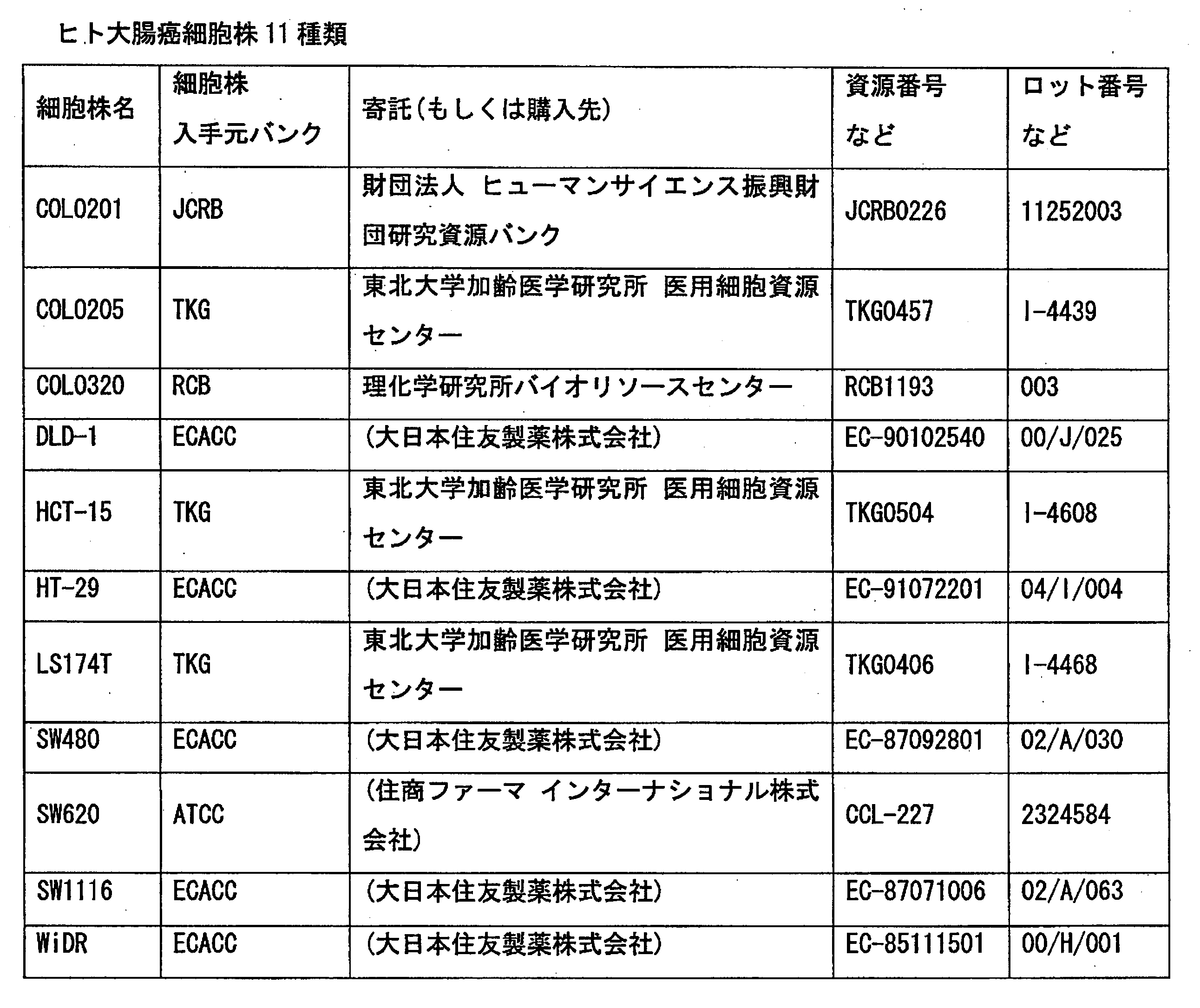
- Example 1 (1) Method (a) Cells used 11 types of human colorectal cancer cell lines (COLO201, COLO205, COLO320, DLD-1, HCT-15, HT-29, LS174T, SW480, SW620, SW1116, WiDR) are as follows (Table) Obtained from 1). For culture, adherent cells were ⁇ 100 mm / Tissue Culture Dish (IWAKI), and floating cells were ⁇ 100 mm / Non-Treated Dish (IWAKI), medium (RPMI 1640, 2 mM Glutamine, 10% Fetal Bovine Serum), 37 ° C., 5%. Performed under CO 2 conditions.
- IWAKI Tissue Culture Dish
- IWAKI Non-Treated Dish
- medium RPMI 1640, 2 mM Glutamine, 10% Fetal Bovine Serum
- Example 2 (1) Method Seven types of human colon cancer cell lines (DLD-1, HCT-15, HT-29, LS174T, SW480, SW620, WiDR) are used, and the drug is cisplatinum (II) dichloride (SIGMA). The cell viability after 48 hours exposure at 0 to 1000 ⁇ mol / L was evaluated by MTS assay in the same manner as in Example 1, and the IC 50 value was calculated. Intracellular protein extraction, protein expression analysis, and correlation analysis with cisplatin (CDDP) sensitivity were performed in the same manner as in Example 1.
- DLD-1, HCT-15, HT-29, LS174T, SW480, SW620, WiDR cisplatinum (II) dichloride
- Example 3 (1) Method The cells used are 5 types of human colorectal cancer cell lines (COLO320, HCT-15, HT-29, LS174T, HCT116 (the bank from which HCT116 was obtained is ECACC)), and the drug is irinotecan active metabolite (SN-38, The cell viability after exposure for 72 hours at 0 to 1000 nmol / L was evaluated by MTS assay in the same manner as in Example 1, and the IC 50 value was calculated. Intracellular protein extraction, sample preparation, and protein chip preparation were performed in the same manner as in Example 1. Analysis was performed using ProteinChip TM Reader (Model PCS4000 Personal Edition, Bio-Rad), and Mass Range 0 to 70,000 Daltons, Focus. Analysis was performed under the conditions of Mass 11,000 Dalton, Energy 3000 nJ, and a total of 265 shots per sample. Protein expression comparison analysis was performed using Ciphergen Express TM Data Manager 3.0.
- Example 4 In Example 1, in order to investigate the property of the protein (protein A1) detected as a peak at m / z 11,000 to 11,100, the change in peak intensity with a change in pH and the CM10 chip array are different. The presence or absence of detection in a protein chip array in which chemical modification was applied to the chip surface was examined.
- Example 2 Results In Example 1, the candidate proteins detected as peaks at m / z 11,000 to 11,100 under the analysis conditions of pH 4.5 using the CM10 chip array were the CM10 chip array and the Q10 chip array. In any case, a remarkable peak decrease was observed at pH 7.0 to 7.5, and the isoelectric point (pI) of this candidate protein was estimated to be pI 7.0 to 7.5. The detection was also confirmed in the IMAC30 chip array whose chip surface was activated with NiSO 4 .
- Example 5 (identification of candidate protein A1) (1) Estimation of Molecular Weight of Candidate Protein A1 For intracellular protein extraction samples, protein expression using SELDI-TOF MS using bovine insulin (5733.51 Da) and equine cytochrome c (123360.96 Da) of known molecular weight as Calibrant Internal calibration was performed under the same analysis conditions as in the analysis (CM10 chip array and 50 mM sodium acetate buffer, pH 4.5). As a result, the molecular weight of the candidate protein A1 peak was estimated to be 11072 Da (FIG. 7).
- IEF lysis buffer (6M urea, 2M thiourea, 3% CHAPS, 1% Triton X-100, DeStrike reagent, GE Healthcare Bioscience) was added to the precipitate, followed by stirring and sonication at room temperature.
- the prepared IEF sample solution is centrifuged, and the supernatant is applied to Immobiline DryStripe gel (pH 3 to 10 non-linear, 13 cm, GE Healthcare Bioscience), and the gel is swollen and then subjected to isoelectric focusing. (5000V, 15hr).
- Immobiline DryStripe gel was equilibrated in sample buffer (6Murea, 20% glycerol, 2% DTT, 2% SDS, 100 mM Tris-HCl pH 8.8) for two-dimensional electrophoresis.
- Immobiline Dry Strip gel was converted to an electrophoretic sample at a constant current of 25 mA.
- the gel was 10-18%, 16 ⁇ 16 cm polyacrylamide gradient gel (Biocraft), and the gel after electrophoresis was stained with CBB G-250 and decolorized with 5% acetic acid.
- GS-800 calibrated Imaging Densitometer Bio-Rad
- PDQuest software Bio-Rad
- protein A1 (molecular weight 11,072, pI 7.3) identified in spot 4 shown in FIG. 8 was obtained by SELDI-TOF MS analysis using a protein chip array. Consistent with the results (molecular weight 11072, pI 7.0-7.5).
- the candidate protein A1 detected as a peak at m / z 11,000 to 11,100 showing a correlation with oxaliplatin sensitivity is S100A10 (S100 calcium-binding protein A10, Calpactin-1 light chain, Calpactin I light. chain, p10 protein, p11, Cellular ligand of annexin II).
- Example 6 Intracellular protein extraction samples of cancer cell lines were analyzed under the same conditions as in (1) method (e) of Example 1 using a CM10 protein chip array and 50 mM Tris-HCl buffer (pH 8.0). As a result, two protein peaks, protein A2 and protein A3, showing an association with oxaliplatin sensitivity were found. The peak intensity of protein A2 showed a significant correlation with oxaliplatin (L-OHP) sensitivity (IC 50 value) of each cancer cell line (FIG. 9).
- L-OHP oxaliplatin
- the cancer cell line is highly sensitive and low sensitive with a cut-off value of 5.0 ⁇ M, which corresponds to the peak blood concentration at the time of clinical use of oxaliplatin.
- the peak intensity of protein A3 was significantly increased in the low sensitivity group (FIG. 10).
- the cell line LS174T was excluded from the analysis target because the S / N ratio was less than 5.
- protein A2 and protein A3 exist in the vicinity of S100A10.
- protein A2 is 10835 Da
- protein A3 was estimated to be 11340 Da.
- Example 2 15 types of buffers of pH 3.0 to 10.0 were used for each of the CM10 chip array and the Q10 chip array, and the change in each peak intensity was analyzed by SELDI-TOF MS. As a result, 10835 Da The peak indicating pH markedly decreased at pH 6.5 to 7.0, and the peak indicating 11340 Da increased sharply from pH 6.0 to 6.5. It was estimated to be 7.0 and 6.0 to 6.5.
- Example 7 1) Method After extracting intracellular protein by the same method as in Example 1 using the high expression cell HT-29 and the low expression cell COLO320 with peaks detected at 11,000 to 11,100 as m / z. Then, a protein amount of 10 ⁇ g per lane was applied to a 16.5% polyacrylamide gel, and SDS-PAGE was performed at a constant voltage of 100V. After electrophoresis, the protein was blotted on a PVDF membrane using a drive blotting system (iBlot TM , invitrogen), blocked, and then anti-S100A10 monoclonal antibody (Purified Mouse Anti-Annexin II monoclonal chain, S100A10).
- iBlot TM drive blotting system
- a primary antibody reaction was performed using BD Transaction Laboratories) and an endogenous protein using an anti-GAPDH monoclonal antibody (anti-GAPDH monoclonal antibody, Ambion).
- an anti-GAPDH monoclonal antibody anti-GAPDH monoclonal antibody, Ambion.
- CDP-Star TM chemiluminescent substrate was added as a reaction substrate to emit light, and detection was performed with a lumino image analyzer (LAS-4000mini, FUJIFILM).
- Chemiluminescent Western Blot Immunodetection Kit was used as the blocking reagent, secondary antibody, and reaction substrate.
Landscapes
- Health & Medical Sciences (AREA)
- Life Sciences & Earth Sciences (AREA)
- Chemical & Material Sciences (AREA)
- Engineering & Computer Science (AREA)
- Immunology (AREA)
- General Health & Medical Sciences (AREA)
- Medicinal Chemistry (AREA)
- Biomedical Technology (AREA)
- Hematology (AREA)
- Urology & Nephrology (AREA)
- Molecular Biology (AREA)
- Veterinary Medicine (AREA)
- Public Health (AREA)
- Pharmacology & Pharmacy (AREA)
- Animal Behavior & Ethology (AREA)
- General Physics & Mathematics (AREA)
- Pathology (AREA)
- Biochemistry (AREA)
- Analytical Chemistry (AREA)
- Physics & Mathematics (AREA)
- Epidemiology (AREA)
- Cell Biology (AREA)
- Food Science & Technology (AREA)
- Microbiology (AREA)
- Biotechnology (AREA)
- Chemical Kinetics & Catalysis (AREA)
- Bioinformatics & Cheminformatics (AREA)
- General Chemical & Material Sciences (AREA)
- Organic Chemistry (AREA)
- Nuclear Medicine, Radiotherapy & Molecular Imaging (AREA)
- Tropical Medicine & Parasitology (AREA)
- Toxicology (AREA)
- Oncology (AREA)
- Electrochemistry (AREA)
- Acyclic And Carbocyclic Compounds In Medicinal Compositions (AREA)
- Pharmaceuticals Containing Other Organic And Inorganic Compounds (AREA)
- Investigating Or Analysing Biological Materials (AREA)
- Medicines That Contain Protein Lipid Enzymes And Other Medicines (AREA)
- Other Investigation Or Analysis Of Materials By Electrical Means (AREA)
Abstract
Description
1)オキサリプラチンによりもたらされる損傷DNAの除去・修復能の亢進
2)細胞内におけるオキサリプラチン(活性化体)の不活化(解毒)
3)オキサリプラチンの細胞内蓄積量の低下
などが関与していると考えられており、大腸癌患者を対象にしたオキサリプラチン+5-FU併用療法における治療反応性や予後予測因子として1)~3)と関連する臨床研究が行われている。
かかる知見に基づき、さらに検討した結果、がん患者由来の生体試料中の当該S100A7、S100A8又はS100A10の濃度を測定すれば、当該がん患者のがんが抗がん剤に対する感受性を有するか否かを判定できること、また、この物質の発現抑制を指標とすれば抗がん剤感受性亢進剤のスクリーニングが可能になること、さらに当該抗がん剤感受性亢進剤と感受性亢進の対象となる抗がん剤を併用すれば、当該抗がん剤の治療効果が飛躍的に向上することを見出し、本発明を完成した。
また、本発明は、検体中の前記S100A7、S100A8又はS100A10を測定することを特徴とする抗がん剤感受性の判定方法を提供するものである。
また、本発明は、検体中の前記S100A7、S100A8又はS100A10を測定するためのプロトコールを含むことを特徴とする抗がん剤感受性の判定方法を実施するためのキットを提供するものである。
さらに本発明は、前記S100A7、S100A8又はS100A10の発現抑制を指標とする抗がん剤感受性亢進剤のスクリーニング方法を提供するものである。
さらにまた本発明は、上記のスクリーニング方法により得られた抗がん剤感受性亢進剤を提供するものである。
さらに本発明は、上記の抗がん剤感受性亢進剤と、感受性亢進の対象となる抗がん剤とを組み合せてなるがん治療用組成物を提供するものである。
さらに本発明は、上記の抗がん剤感受性亢進剤と、感受性亢進の対象となる抗がん剤との組み合せの、がん治療薬製造のための使用を提供するものである。
さらに本発明は、上記の抗がん剤感受性亢進剤と、感受性亢進の対象となる抗がん剤とを投与することを特徴とするがん治療方法を提供するものである。
さらに、また、このマーカーを用いれば、抗がん剤感受性を亢進させる薬剤がスクリーニングでき、その対象となった抗がん剤と抗がん剤感受性亢進剤とを併用すれば、がん治療効果が飛躍的に向上する。
対象とする抗がん剤に対して感受性を有さない場合は、その薬効を期待することができず、抗がん剤による副作用のみが発現すると考えられるため、本発明における抗がん剤感受性判定マーカーは、不必要な副作用の発現もしくは無効な治療継続に伴うがんの進行や副作用の増大を回避するためのマーカーとしても使用できる。
また、タンパク質Aの濃度が所定の標準濃度より低いと判断される濃度を有する場合は、そのがんは対象とする抗がん剤に対して感受性を有すると判定できるため、治療効果を期待できる患者を積極的に選出するためのマーカーとしても使用できる。
ここで用いられる対象となる抗がん剤としては、前記と同様であり、オキサリプラチン、シクロフォスファミド(cyclophosphamide)、イフォスファミド(ifosfamide)、チオテパ(thiotepa)、メルファラン(melphalan)、ブスルファン(busulfan)、ニムスチン(nimustine)、ラニムスチン(ranimustine)、ダカルバジン(dacarbazine)、プロカルバジン(procarbazine)、テモゾロミド(temozolomide)、シスプラチン(cisplatin)、カルボプラチン(carboplatin)、ネダプラチン(nedaplatin)、メトトレキサート(methotrexate)、ペメトレキセド(pemetrexed)、フルオロウラシル(fluorouracil)、テガフール/ウラシル(tegaful・uracil)、ドキシフルリジン(doxifluridine)、テガフール/ギメラシル/オテラシル(tegaful・gimeracil・oteracil)、カペシタビン(capecitabine)、シタラビン(cytarabine)、エノシタビン(enocitabine)、ゲムシタビン(gemcitabine)、6-メルカプトプリン(6-mercaptopurine)、フルダラビン(fuludarabin)、ペントスタチン(pentostatin)、クラドリビン(cladribine)、ヒドロキシウレア(hydroxyurea)、ドキソルビシン(doxorubicin)、エピルビシン(epirubicin)、ダウノルビシン(daunorubicin)、イダルビシン(idarubicine)、ピラルビシン(pirarubicin)、ミトキサントロン(mitoxantrone)、アムルビシン(amurubicin)、アクチノマイシンD(actinomycin D)、ブレオマイシン(bleomycine)、ペプレオマイシン(pepleomycin)、マイトマイシンC(mytomycin C)、アクラルビシン(aclarubicin)、ジノスタチン(zinostatin)、ビンクリスチン(vincristine)、ビンデシン(vindesine)、ビンブラスチン(vinblastine)、ビノレルビン(vinorelbine)、パクリタキセル(paclitaxel)、ドセタキセル(docetaxel)、イリノテカン(irinotecan)、イリノテカン活性代謝物(SN-38)、ノギテカン(nogitecan、topotecan)、エトポシド(etoposide)、プレドニゾロン(prednisolone)、デキサメタゾン(dexamethasone)、タモキシフェン(tamoxifen)、トレミフェン(toremifene)、メドロキシプロゲステロン(medroxyprogesterone)、アナストロゾール(anastrozole)、エキセメスタン(exemestane)、レトロゾール(letrozole)、リツキシマブ(rituximab)、イマチニブ(imatinib)、ゲフィチニブ(gefitinib)、ゲムツズマブ・オゾガマイシン(gemtuzumab ozogamicin)、ボルテゾミブ(bortezomib)、エルロチニブ(erlotinib)、セツキシマブ(cetuximab)、ベバシズマブ(bevacizumab)、スニチニブ(sunitinib)、ソラフェニブ(sorafenib)、ダサチニブ(dasatinib)、パニツムマブ(panitumumab)、アスパラギナーゼ(asparaginase)、トレチノイン(tretinoin)、三酸化ヒ素(arsenic trioxide)、又はそれらの塩、又はそれらの活性代謝物等が挙げられる。このうち、白金錯体系抗がん剤及び植物アルカロイド系抗がん剤が好ましく、特にオキサリプラチン、シスプラチン、イリノテカン、SN-38又はそれらの塩が好ましい。
(1)方法
(a)使用細胞
ヒト大腸癌細胞株11種類(COLO201、COLO205、COLO320、DLD-1、HCT-15、HT-29、LS174T、SW480、SW620、SW1116、WiDR)は、以下(表1)より入手した。
培養は、接着細胞はφ100mm/Tissue Culture Dish(IWAKI)、浮遊細胞はφ100mm/Non-Treated Dish(IWAKI)にて、培地(RPMI 1640、2mM Glutamine、10% Fetal Bovine Serum)、37℃、5%CO2の条件下にて行なった。
オキサリプラチン(L-OHP)原末は、株式会社ヤクルト本社より入手した。
各細胞において、オキサリプラチン0~1000μmol/Lにて48時間曝露後の細胞生存率をMTS assay(CellTiter96TMAQueous One Solution Cell Proliferation Assay、Promega)にて評価し、IC50値(オキサリプラチン未処理wellに対して細胞数を50%抑制する濃度)を算出して各細胞におけるオキサリプラチン感受性とした。感受性の評価は異なる継代数の細胞にて4回行い、その平均値と標準偏差を算出した。
接着細胞はdishより培地を除き氷冷PBSで3回洗浄後、ラバーポリスマンにて剥がしてかき集め、浮遊細胞は細胞浮遊液をPBSにて再懸濁して遠心する形で3回洗浄後の細胞懸濁液を1.5mLマイクロチューブに移した。4℃にて1,200×g、10分間遠心して細胞を集め、上清を除いた後に細胞溶解バッファー(9mol/L Urea、2% CHAPS、1mM DTT、プロテアーゼインヒビターカクテル(Sigma))400μLを加え、氷冷下で超音波破砕処理を行なった後、4℃にて16,000×g、20分間遠心し、上清を液体窒素にて急速凍結後、分析するまで-80℃にて保存した。上清の一部を用いてタンパク質定量(DC Protein Assay Kit、Bio-Rad)を行なった。
細胞溶解バッファー(プロテアーゼインヒビターを除く)にて5mg/mLに調整し、さらに、pH4.5の希釈/洗浄バッファー(50mM sodium acetate buffer)(以下、バッファー)にて1mg/mLに調整したサンプル100μLを、同バッファーにて前処理した陽イオン交換チップアレイ(CM10、Bio-Rad)のスポットにapplyし、1時間インキュベートして反応させた後、バッファーにて3回wash、milliQ水にて2回リンスした。風乾後、energy absorbing molecule(EAM:50% ACN/0.5% TFA溶液によるsinapinic acidの飽和溶液)を、1.0μLを0.5μLずつ2回に分けて各スポットにapplyし、スポット表面が乾いた後、プロテインチップアレイの分析を行なった。
タンパク質発現解析は、表面エンハンス型レーザー脱離イオン化飛行時間型質量分析計(surface-enhanced laser desorption/ionization time-of-flight mass spectrometry:SELDI-TOF MS)にて行なった。分析機器としては、ProteinChipTM Reader(Model PBSIIC、Bio-Rad)を用い、mass-to-charge ratio(m/z)のopitimization range 2,000~30,000 Daltons、Laser intensity 220、Detector sensitivity 8、1サンプルあたり合計104 shotsの条件にて分析を行った。signal-to-noise ratio(S/N比)5以上のピークの抽出とタンパク質発現比較解析は、CiphergenExpressTM Data Manager 3.0を用いて行なった。
SELDI-TOF MSにより検出されたすべてのタンパク質ピークについて、各細胞のIC50値とピーク強度の関係について相関分析を行なった。IC50値の対数値とピーク強度の関係について線形回帰分析を行い、P値<0.05、決定係数(r2;r,Pearson相関係数)>0.5を満たすピークをオキサリプラチン感受性関連候補タンパク質として選出した。相関分析及び線形回帰分析は、SPSS 15.0J for Windows(登録商標)(バージョン15.0.1)にて行なった。
(a)11種類のヒト大腸癌細胞株におけるオキサリプラチン感受性の評価
各細胞におけるIC50値は0.84±0.20~29.7±13.6μMであり、その感受性には大きな幅が認められた(図1)。
SELDI-TOF MSを用いた前述の分析条件による発現解析により、全部で42個のピークを検出した。
相関分析及び線形回帰分析により、m/zとして11,000~11,100に検出されたピークがオキサリプラチン感受性(IC50値)と有意な相関性をもつことを見出した(図2、図3)。
(1)方法
使用細胞はヒト大腸癌細胞株7種類(DLD-1、HCT-15、HT-29、LS174T、SW480、SW620、WiDR)、薬剤はシスプラチン(cis-Diammineplatinum(II)dichrolide、SIGMA)を使用し、0~1000μmol/Lにて48時間曝露後の細胞生存率を実施例1と同様にMTS assayにて評価し、IC50値を算出した。細胞内タンパク質の抽出、タンパク質発現解析およびシスプラチン(CDDP)感受性との相関分析は実施例1と同様に行なった。
(a)7種類のヒト大腸癌細胞株におけるシスプラチン感受性の評価
各細胞におけるIC50値は3.33±0.80~26.9±11.6μMであり、その感受性には大きな幅が認められた。
相関分析及び線形回帰分析により、m/zとして11,000~11,100に検出されたピークがシスプラチン感受性(IC50値)と相関を示す傾向を認めた(図4)。さらに、シスプラチン投与時における総プラチナの薬物動態学的パラメータ(ブリプラチンTM注インタビューフォームより)にもとづくと、シスプラチン臨床使用における1回あたりの最高投与量(100mg/m2)を投与した際のピーク血中濃度(Cmax)は、およそ9.5~11.0μM(日本人の体表面積を1.50~1.73m2として計算)と計算されることから、各がん細胞株のシスプラチンに対するIC50値について10μMをカットオフ値として、高感受性及び低感受性の2群に分類した。その結果、m/zとして11,000~11,100に検出されたピークのピーク強度は低感受性群において有意な上昇を認めた(図5)。
(1)方法
使用細胞はヒト大腸癌細胞株5種類(COLO320、HCT-15、HT-29、LS174T、HCT116(HCT116の入手元バンクはECACC))、薬剤はイリノテカン活性代謝物(SN-38、株式会社ヤクルト本社より入手)を使用し、0~1000nmol/Lにて72時間曝露後の細胞生存率を実施例1と同様にMTS assayにて評価し、IC50値を算出した。細胞内タンパク質の抽出、サンプル調整およびプロテインチップの作成は実施例1と同様に行い、分析はProteinChipTM Reader(Model PCS4000 Personal Edition、Bio-Rad)を用い、Mass Range 0~70,000 Daltons、Focus Mass 11,000 Dalton、Energy 3000 nJ、1サンプルあたり合計265shotsの条件にて分析を行なった。タンパク質発現比較解析は、CiphergenExpressTM Data Manager 3.0を用いて行なった。
(a)5種類のヒト大腸癌細胞株におけるSN-38感受性の評価
各細胞におけるIC50値は3.39~33.7nMであり、その感受性には大きな幅が認められた。
SN-38の薬物動態学的パラメータ(カンプトTMインタビューフォームより)にもとづくと、大腸癌治療レジメン(180mg/m2/日)によるイリノテカン投与1回目に得られるSN-38の血中濃度曲線下面積(AUCSN-38)は1449nmol.h/Lと計算され、本検討で用いた72時間薬剤曝露により同等の曝露量が得られるSN38の濃度は20nMと算出されることから、各がん細胞株のSN-38に対するIC50値について20nMをカットオフ値として、高感受性及び低感受性の2群に分類した。その結果、m/zとして11,000~11,100に検出されたピークのピーク強度は低感受性群において有意な上昇を認めた(図6)。
実施例1において、m/z 11,000~11,100のピークとして検出されたタンパク質(タンパク質A1)の性質を調べるために、pHの変化に伴うピーク強度の変化、およびCM10チップアレイとは異なる化学修飾がチップ表面に施されたプロテインチップアレイにおける検出の有無について検討を行った。
(a)検討に用いたプロテインチップアレイとバッファー条件
陽イオン交換チップアレイ(CM10、Bio-Rad)及び陰イオン交換チップアレイ(Q10、Bio-Rad)に対しては、pH3.0(50mM Glycine-HCl buffer)、pH3.5(50mM sodium acetate buffer)、pH4.0(50mM sodium acetate buffer)、pH4.5(50mM sodium acetate buffer)、pH5.0(50mM sodium acetate buffer)、pH5.5(50mM sodium acetate buffer)、pH6.0(50mM phosphate buffer)、pH6.5(50mM phosphate buffer)、pH7.0(50mM phosphate buffer)、pH7.5(50mM phosphate buffer)、pH8.0(50mM Tris-HCl buffer)、pH8.5(50mM Tris-HCl buffer)、pH9.0(50mM Glycine-NaOH buffer)、pH9.5(50mM Glycine-NaOH buffer)、pH10.0(50mM Glycine-NaOH buffer)の15種類のバッファーを用い、金属修飾(Immobilized Metal Affinity Capture)チップアレイ(IMAC30、Bio-Rad)に対してはphosphate buffered saline(PBS)を用いた。
CM10及びQ10チップアレイを用いた分析のためのサンプル調整及びプロテインチップの作成は、(a)の各バッファーを用いて実施例1の(1)方法前記(e)に準じて実施した。ただし分析機器としては、ProteinChipTM Reader(Model PCS4000 Personal Edition、Bio-Rad)を用い、Mass Range 0~70,000 Daltons、Focus Mass 11,000 Dalton、Energy 3000 nJ、1サンプルあたり合計265shots の条件にて分析を行なった。
IMAC30チップアレイは50mM NiSO4にてスポット表面の活性化を行い、milliQ水により1回リンスした後、PBSにて前処理を行った。サンプル調整及びチップへのサンプルapply以降の作業は、バッファーとして上記(a)のPBSを用い、実施例1の(1)方法前記(e)に準じて実施した。ただし分析機器としては、ProteinChipTM Reader(Model PCS4000 Personal Edition、Bio-Rad)を用い、Mass Range 0~70,000 Daltons、Focus Mass 11,000 Dalton、Energy 6000nJ、1サンプルあたり合計265shots の条件にて分析を行なった。
実施例1で、CM10チップアレイを用いたpH4.5の分析条件においてm/z 11,000~11,100のピークとして検出された上記候補タンパク質は、CM10チップアレイ及びQ10チップアレイいずれにおいてもpH7.0~7.5において顕著なピークの低下を認め、この候補タンパク質の等電点(pI)は、pI 7.0~7.5と推定された。また、チップ表面をNiSO4により活性化したIMAC30チップアレイにても検出が確認された。
(1)候補タンパク質A1の分子量の推定
細胞内タンパク質抽出サンプルについて、分子量既知のbovine insulin(5733.51Da)及びequine Cytochrome c(12360.96Da)をCalibrantとして用い、SELDI-TOF MSを用いてタンパク質発現解析時と同じ分析条件(CM10チップアレイ及び50mM sodium acetate buffer,pH4.5)にてInternal Calibrationを行なった。その結果、候補タンパク質A1ピークの分子量は11072Daと推定された(図7)。
(精製)
候補タンパク質A1の精製及び同定を目的として、タンパク質発現解析において候補タンパク質が高発現を示した細胞株HT-29及び低発現を示した細胞株COLO320の2細胞株より発現解析時と同様の方法により細胞内タンパク質を抽出した。タンパク質定量の結果にもとづき両サンプルより等量を分取し、TCA沈殿を行なった後、回収した沈殿を冷エタノール/エーテル溶媒にて洗浄し室温にて乾燥した。この沈殿にIEF lysis buffer(6M urea,2M thiourea,3% CHAPS,1% Triton X-100,DeStreak reagent、GEヘルスケア バイオサイエンス)を加え、室温で攪拌及び超音波処理を行なった。
調整したIEFサンプル溶液を遠心し、上清をImmobiline DryStrip gel(pH3~10 non-linear,13cm,GEヘルスケア バイオサイエンス)にapplyし、ゲルの膨潤を行なった後、等電点電気泳動を行なった(5000V,15hr)。
等電点電気泳動終了後、Immobiline DryStrip gelを二次元電気泳動用のsample buffer(6M urea,20% glycerol,2% DTT,2% SDS,100mM Tris-HCl pH8.8)中で平衡化し、平衡化の終了したImmobiline DryStrip gelを25mAの定電流にて電気泳動を行なった。ゲルは10~18%、16×16cmポリアクリルアミドグラジェントゲル(バイオクラフト)を用い、電気泳動後のゲルはCBB G-250により染色後、5%酢酸にて脱色を行なった。
二次元電気泳動の画像取得にはGS-800 calibrated Imaging Densitometer(Bio-Rad)、画像解析はPDQuestソフトウェア(Bio-Rad)を用い比較解析を行った。
プロテインチップを用いたSELDI-TOF MS分析により得られた候補タンパク質A1に関する情報(分子量 11072、pI 7.0~7.5)に基づき、2次元電気泳動にて展開したゲル上の分子量10~15kDa及び、およそpI 6.5~8.0の範囲(中性付近)に分離されたスポットを対象とし、これらの中からCOLO320と比較してHT-29において高い発現を示していた4スポットを選択した(図8)。これらのスポットを切り出し、公知の方法に従ってタンパク質のゲル内トリプシン消化を行った後、LCMS-IT-TOF(液体クロマトグラフ質量分析計、Shimadzu)を用いて分析(MS/MS測定)を行い、得られた結果についてMASCOTデータベース検索を行なった。
がん細胞株の細胞内タンパク質抽出サンプルを、CM10プロテインチップアレイ、50mM Tris-HCl buffer(pH8.0)を用いて、実施例1の(1)方法前記(e)と同様の条件にて分析した結果、オキサリプラチン感受性との関連を示す2つのタンパク質ピーク、タンパク質A2及びタンパク質A3を見出した。
タンパク質A2のピーク強度は各がん細胞株のオキサリプラチン(L-OHP)感受性(IC50値)と有意な相関性を示した(図9)。
また、各がん細胞株のオキサリプラチンに対するIC50値にもとづき、オキサリプラチン臨床使用時のピーク血中濃度にほぼ相当する5.0μMをカットオフ値としてがん細胞株を高感受性及び低感受性の2群に分類すると、タンパク質A3のピーク強度は低感受性群において有意な上昇を認めた(図10)。なお、細胞株LS174Tは、S/N比が5未満であったため、解析対象から除外した。
これらの結果にもとづきデータベース(Expasy TagIdent tool:http://www.expasy.ch/tools/tagident.html)検索した結果、10835Daで示されるピークはS100A8(S100 calcium-binding protein A8、Calgranulin-A、Migration inhibitory factor-related protein 8、MRP-8、p8など)、11340Daで示されるピークはS100A7(S100 calcium-binding protein A7、Psoriasin)であることが示された(図11)。
実施例6と同様にして、SELDI-TOF MSにより分子量13,242のS100A9と推定されるピークについて、オキサリプラチン感受性との関連について検討した。その結果、S100A9発現量とオキサリプラチン感受性との間に有意な関連は認められなかった(図12)。従って、S100A9には抗がん剤感受性との間に関連性はないことが判明した。
1)方法
m/zとして11,000~11,100に検出されたピークの高発現細胞HT-29、および低発現細胞COLO320を用いて、実施例1と同様の方法により細胞内タンパク質を抽出後、16.5%ポリアクリルアミドゲルに1レーンあたりタンパク量10μgをapplyし、100V定電圧にてSDS-PAGEを行なった。泳動後、ドライブロッティングシステム(iBlotTM、invitrogen)を用いてPVDF膜にタンパク質をブロットし、ブロッキングを行なった後、S100A10に対しては抗S100A10モノクローナル抗体(Purified Mouse Anti-Annexin II light chain monoclonal antibody、BD Transduction Laboratories)を、内在性タンパク質については抗GAPDHモノクローナル抗体(anti-GAPDH monoclonal antibody、Ambion)を用いて一次抗体反応を行なった。アルカリホスファターゼ標識抗マウスIgG抗体と二次抗体反応をさせた後、反応基質としてCDP-StarTM chemiluminescent substrateを添加して発光させ、ルミノ・イメージアナライザー(LAS-4000mini、FUJIFILM)により検出した。ブロッキング試薬、二次抗体および反応基質はChemiluminescent Western Blot Immunodetection Kit(WesternBreezeTM、invitrogen)を用いた。
プロテインチップアレイを用いたタンパク質発現解析(実施例1)において、m/zとして11,000~11,100に検出されたピークの高発現細胞HT-29、および低発現細胞COLO320におけるS100A10の発現は、抗S100A10モノクローナル抗体を用いたウエスタンブロット法によって確認された(図13)。
Claims (14)
- カルシウム結合タンパク質S100A7、S100A8又はS100A10を含有する抗がん剤感受性判定マーカー。
- 抗がん剤が、白金錯体系抗がん剤又は植物アルカロイド系抗がん剤である請求項1記載の抗がん剤感受性判定マーカー。
- 抗がん剤が、オキサリプラチン、シスプラチン、イリノテカン、SN-38又はそれらの塩である請求項1又は2記載の抗がん剤感受性判定マーカー。
- 検体中の請求項1~3のいずれか1項記載の抗がん剤感受性判定マーカーを測定することを特徴とする抗がん剤感受性の判定方法。
- 検体が、がんを有する被験者由来の生体試料であることを特徴とする請求項4記載の判定方法。
- 検体が、抗がん剤を投与された、がんを有する被験者由来の生体試料である請求項4又は5記載の判定方法。
- 検体中の請求項1~3のいずれか1項記載の抗がん剤感受性判定マーカーを測定するためのプロトコールを含むことを特徴とする請求項4~6のいずれか1項記載の判定方法を実施するためのキット。
- 検体が、がんを有する被験者由来の生体試料である請求項7記載のキット。
- 検体が、抗がん剤を投与された、がんを有する被験者由来の生体試料である請求項7又は8記載のキット。
- 請求項1~3のいずれか1項記載の抗がん剤感受性判定マーカーの発現抑制を指標とする抗がん剤感受性亢進剤のスクリーニング方法。
- 請求項10記載の方法により得られた抗がん剤感受性亢進剤。
- 請求項11記載の抗がん剤感受性亢進剤と、感受性亢進の対象となる抗がん剤とを組み合せてなるがん治療用組成物。
- 請求項11記載の抗がん剤感受性亢進剤と、感受性亢進の対象となる抗がん剤との組み合せの、がん治療薬製造のための使用。
- 請求項11記載の抗がん剤感受性亢進剤と、感受性亢進の対象となる抗がん剤とを投与することを特徴とするがん治療方法。
Priority Applications (11)
| Application Number | Priority Date | Filing Date | Title |
|---|---|---|---|
| EP09706388.7A EP2241334A4 (en) | 2008-01-31 | 2009-01-30 | METHOD FOR DETERMINING SENSITIVITY AGAINST ANTI-CANCER MEDIUM |
| JP2009551440A JP5461201B2 (ja) | 2008-01-31 | 2009-01-30 | 抗がん剤感受性の判定方法 |
| AU2009208526A AU2009208526A1 (en) | 2008-01-31 | 2009-01-30 | Method for determination of sensitivity to anti-cancer agent |
| US12/865,726 US9089540B2 (en) | 2008-01-31 | 2009-01-30 | Method for determination of sensitivity to anti-cancer agent |
| CN2009801039330A CN101932338A (zh) | 2008-01-31 | 2009-01-30 | 抗癌剂感受性的判定方法 |
| MX2010008496A MX2010008496A (es) | 2008-01-31 | 2009-01-30 | Metodo para la determinacion de sensibilidad a agente anticancerigeno. |
| NZ586972A NZ586972A (en) | 2008-01-31 | 2009-01-30 | Method for determination of sensitivity to anti-cancer agent |
| CA2713296A CA2713296A1 (en) | 2008-01-31 | 2009-01-30 | Method for determination of sensitivity to anti-cancer agent |
| BRPI0906573-3A BRPI0906573A2 (pt) | 2008-01-31 | 2009-01-30 | Método para determinação da sensibilidade para agentes anticâncer |
| UAA201010512A UA101492C2 (ru) | 2008-01-31 | 2009-01-30 | Способ определения чувствительности к противораковому средству |
| IL207169A IL207169A0 (en) | 2008-01-31 | 2010-07-22 | Method for determination of sensitivity to anti-cancer agent |
Applications Claiming Priority (4)
| Application Number | Priority Date | Filing Date | Title |
|---|---|---|---|
| JP2008-021124 | 2008-01-31 | ||
| JP2008021124 | 2008-01-31 | ||
| JP2008223384 | 2008-09-01 | ||
| JP2008-223384 | 2008-09-01 |
Publications (1)
| Publication Number | Publication Date |
|---|---|
| WO2009096196A1 true WO2009096196A1 (ja) | 2009-08-06 |
Family
ID=40912550
Family Applications (1)
| Application Number | Title | Priority Date | Filing Date |
|---|---|---|---|
| PCT/JP2009/000374 Ceased WO2009096196A1 (ja) | 2008-01-31 | 2009-01-30 | 抗がん剤感受性の判定方法 |
Country Status (13)
| Country | Link |
|---|---|
| US (1) | US9089540B2 (ja) |
| EP (1) | EP2241334A4 (ja) |
| JP (1) | JP5461201B2 (ja) |
| KR (1) | KR20100130587A (ja) |
| CN (2) | CN104784706A (ja) |
| AU (1) | AU2009208526A1 (ja) |
| BR (1) | BRPI0906573A2 (ja) |
| CA (1) | CA2713296A1 (ja) |
| IL (1) | IL207169A0 (ja) |
| MX (1) | MX2010008496A (ja) |
| NZ (1) | NZ586972A (ja) |
| RU (1) | RU2010136309A (ja) |
| WO (1) | WO2009096196A1 (ja) |
Cited By (8)
| Publication number | Priority date | Publication date | Assignee | Title |
|---|---|---|---|---|
| WO2011052749A1 (ja) | 2009-10-30 | 2011-05-05 | 学校法人慶應義塾 | 抗がん剤の感受性の判定方法 |
| WO2011052748A1 (ja) * | 2009-10-30 | 2011-05-05 | 学校法人慶應義塾 | 抗がん剤の感受性判定方法 |
| WO2011052750A1 (ja) * | 2009-10-30 | 2011-05-05 | 学校法人慶應義塾 | 抗がん剤の感受性判定マーカー |
| WO2012127984A1 (ja) * | 2011-03-24 | 2012-09-27 | 学校法人慶應義塾 | 抗がん剤感受性の判定マーカー |
| WO2013125675A1 (ja) * | 2012-02-23 | 2013-08-29 | 学校法人慶應義塾 | 併用抗がん剤の感受性判定マーカー |
| EP3550306A1 (en) | 2014-08-26 | 2019-10-09 | Keio University | Anti-cancer agent sensitivity-determining marker |
| WO2020067228A1 (ja) * | 2018-09-28 | 2020-04-02 | 学校法人慶應義塾 | 併用抗がん剤の感受性の判定マーカー |
| JP2022038829A (ja) * | 2020-08-27 | 2022-03-10 | 国立大学法人 大分大学 | 癌治療効果の有効性を予測する方法、予測装置及び予測プログラム |
Families Citing this family (6)
| Publication number | Priority date | Publication date | Assignee | Title |
|---|---|---|---|---|
| WO2012110425A1 (en) * | 2011-02-14 | 2012-08-23 | INSERM (Institut National de la Santé et de la Recherche Médicale) | Methods for monitoring the response to treatment and for treating colorectal cancer |
| SG11201606439VA (en) | 2014-02-07 | 2016-09-29 | Dong Wha Pharm Co Ltd | Anticancer adjuvant composition containing rip3 expression promoter as active ingredient, method for screening for anticancer adjuvant enhancing sensitivity of anticancer drug by promoting rip3 expression, and method for monitoring sensitivity of anticancer drug |
| WO2015119362A1 (ko) * | 2014-02-07 | 2015-08-13 | 아주대학교산학협력단 | Rip3 발현촉진제를 유효성분으로 포함하는 항암보조용 조성물, rip3 발현을 촉진하여 항암제 감수성을 증진시키는 항암 보조제 스크리닝 방법 및 항암제 감수성 모니터링 방법 |
| US10063388B2 (en) | 2015-04-21 | 2018-08-28 | Philips Lighting Holding B.V. | Lighting-control processor and method for operating a lighting-control processor |
| KR101834378B1 (ko) * | 2015-08-05 | 2018-03-06 | 아주대학교산학협력단 | Rip3 발현촉진제를 유효성분으로 포함하는 항암보조용 조성물, rip3 발현을 촉진하여 항암제 감수성을 증진시키는 항암 보조제 스크리닝 방법 및 항암제 감수성 모니터링 방법 |
| WO2019188446A1 (ja) * | 2018-03-29 | 2019-10-03 | 学校法人慶應義塾 | イリノテカンを含む抗がん剤療法の感受性判定マーカー |
Citations (2)
| Publication number | Priority date | Publication date | Assignee | Title |
|---|---|---|---|---|
| WO2007058968A2 (en) * | 2005-11-12 | 2007-05-24 | Bayer Healthcare Llc | Gene expression profiles and methods of use |
| JP2007517058A (ja) * | 2003-12-31 | 2007-06-28 | ザ・ペン・ステート・リサーチ・ファンデーション | 卵巣がんの化学療法に対する抵抗性を予測および克服するための方法ならびに結腸がんの発生を予測するための方法 |
Family Cites Families (9)
| Publication number | Priority date | Publication date | Assignee | Title |
|---|---|---|---|---|
| US20040076955A1 (en) * | 2001-07-03 | 2004-04-22 | Eos Biotechnology, Inc. | Methods of diagnosis of bladder cancer, compositions and methods of screening for modulators of bladder cancer |
| CA2519630A1 (en) * | 2003-03-20 | 2004-10-07 | Dana-Farber Cancer Institute, Inc. | Gene expression in breast cancer |
| JPWO2005007846A1 (ja) * | 2003-04-25 | 2006-08-31 | 財団法人癌研究会 | 腫瘍細胞の抗癌剤に対する感受性を判定する方法 |
| GB0412620D0 (en) * | 2004-06-05 | 2004-07-07 | Univ Belfast | BRCA1 markers |
| MX2008014482A (es) * | 2006-05-19 | 2008-11-27 | Hoffmann La Roche | Uso de proteina s100a 12 como un marcador para cancer colorrectal. |
| CN101040843A (zh) | 2006-09-22 | 2007-09-26 | 济南康泉医药科技有限公司 | 一种含尼莫司汀及其增效剂的抗实体瘤缓释剂 |
| US20100216131A1 (en) * | 2006-12-11 | 2010-08-26 | Rajyalakshmi Luthra | Gene expression profiling of esophageal carcinomas |
| CN101011351A (zh) | 2007-02-12 | 2007-08-08 | 济南帅华医药科技有限公司 | 同载铂类化合物和增效剂的抗癌组合物 |
| CA2680591A1 (en) * | 2007-03-15 | 2008-09-25 | Genomic Health, Inc. | Gene expression markers for prediction of patient response to chemotherapy |
-
2009
- 2009-01-30 JP JP2009551440A patent/JP5461201B2/ja not_active Expired - Fee Related
- 2009-01-30 BR BRPI0906573-3A patent/BRPI0906573A2/pt not_active IP Right Cessation
- 2009-01-30 EP EP09706388.7A patent/EP2241334A4/en not_active Withdrawn
- 2009-01-30 AU AU2009208526A patent/AU2009208526A1/en not_active Abandoned
- 2009-01-30 MX MX2010008496A patent/MX2010008496A/es not_active Application Discontinuation
- 2009-01-30 CN CN201510075217.3A patent/CN104784706A/zh active Pending
- 2009-01-30 NZ NZ586972A patent/NZ586972A/en not_active IP Right Cessation
- 2009-01-30 WO PCT/JP2009/000374 patent/WO2009096196A1/ja not_active Ceased
- 2009-01-30 CA CA2713296A patent/CA2713296A1/en not_active Abandoned
- 2009-01-30 RU RU2010136309/15A patent/RU2010136309A/ru not_active Application Discontinuation
- 2009-01-30 CN CN2009801039330A patent/CN101932338A/zh active Pending
- 2009-01-30 KR KR1020107016145A patent/KR20100130587A/ko not_active Ceased
- 2009-01-30 US US12/865,726 patent/US9089540B2/en not_active Expired - Fee Related
-
2010
- 2010-07-22 IL IL207169A patent/IL207169A0/en unknown
Patent Citations (2)
| Publication number | Priority date | Publication date | Assignee | Title |
|---|---|---|---|---|
| JP2007517058A (ja) * | 2003-12-31 | 2007-06-28 | ザ・ペン・ステート・リサーチ・ファンデーション | 卵巣がんの化学療法に対する抵抗性を予測および克服するための方法ならびに結腸がんの発生を予測するための方法 |
| WO2007058968A2 (en) * | 2005-11-12 | 2007-05-24 | Bayer Healthcare Llc | Gene expression profiles and methods of use |
Non-Patent Citations (14)
| Title |
|---|
| ANTICANCERRES., vol. 21, 2001, pages 3075 - 3079 |
| BR. J. CANCER, vol. 91, 2004, pages 344 - 354 |
| CANCER RES, vol. 61, 2001, pages 8654 - 8658 |
| CANCER RES., vol. 56, 1996, pages 4881 - 4886 |
| CANCER RES., vol. 66, 2006, pages 8847 - 8857 |
| CLIN. CANCER RES., vol. 10, 2004, pages 4661 - 4669 |
| CLIN. CANCER RES., vol. 11, 2005, pages 4770 - 4774 |
| J. BIOL. CHEM., vol. 279, 2004, pages 46113 - 46121 |
| J. CLIN. ONCOL., vol. 19, 2001, pages 4298 - 4304 |
| J. CLIN. ONCOL., vol. 25, 2007, pages 1247 - 1254 |
| J. NATL. CANCER INST., vol. 94, 2002, pages 936 - 942 |
| JOURNAL OF PROTEOME RESEARCH, vol. 4, 2005, pages 1717 - 1721 |
| MOL. PHARMACOL., vol. 66, 2004, pages 25 - 32 |
| See also references of EP2241334A4 |
Cited By (37)
| Publication number | Priority date | Publication date | Assignee | Title |
|---|---|---|---|---|
| WO2011052749A1 (ja) | 2009-10-30 | 2011-05-05 | 学校法人慶應義塾 | 抗がん剤の感受性の判定方法 |
| WO2011052748A1 (ja) * | 2009-10-30 | 2011-05-05 | 学校法人慶應義塾 | 抗がん剤の感受性判定方法 |
| WO2011052750A1 (ja) * | 2009-10-30 | 2011-05-05 | 学校法人慶應義塾 | 抗がん剤の感受性判定マーカー |
| CN102597778A (zh) * | 2009-10-30 | 2012-07-18 | 学校法人庆应义塾 | 抗癌剂感受性的判定方法 |
| CN102597762A (zh) * | 2009-10-30 | 2012-07-18 | 学校法人庆应义塾 | 抗癌剂感受性判定标记 |
| CN105699638B (zh) * | 2009-10-30 | 2018-05-22 | 学校法人庆应义塾 | 抗癌剂感受性的判定方法 |
| EP2495569A4 (en) * | 2009-10-30 | 2013-03-13 | Univ Keio | METHOD FOR DETERMINING SENSITIVITY TO ANTICREME AGENT |
| EP3144670A3 (en) * | 2009-10-30 | 2017-04-26 | Keio University | Anticancer agent sensitivity-determining marker |
| US8765713B2 (en) | 2009-10-30 | 2014-07-01 | Keio University | Method for determination of sensitivity to anti-cancer agent |
| JP5548695B2 (ja) * | 2009-10-30 | 2014-07-16 | 学校法人慶應義塾 | 抗がん剤の感受性判定マーカー |
| JP5548693B2 (ja) * | 2009-10-30 | 2014-07-16 | 学校法人慶應義塾 | 抗がん剤の感受性判定方法 |
| JP5548694B2 (ja) * | 2009-10-30 | 2014-07-16 | 学校法人慶應義塾 | 抗がん剤の感受性の判定方法 |
| US8809362B2 (en) | 2009-10-30 | 2014-08-19 | Keio University | Anticancer agent sensitivity-determining marker |
| EP3081941A1 (en) | 2009-10-30 | 2016-10-19 | Keio University | Method for determination of sensitivity to anti-cancer agent |
| CN102597778B (zh) * | 2009-10-30 | 2016-03-02 | 学校法人庆应义塾 | 抗癌剂感受性的判定方法 |
| US9459254B2 (en) | 2009-10-30 | 2016-10-04 | Keio University | Method for determining sensitivity to an anticancer agent |
| CN105699638A (zh) * | 2009-10-30 | 2016-06-22 | 学校法人庆应义塾 | 抗癌剂感受性的判定方法 |
| CN102597762B (zh) * | 2009-10-30 | 2016-08-03 | 学校法人庆应义塾 | 抗癌剂感受性判定标记 |
| JP2016166881A (ja) * | 2011-03-24 | 2016-09-15 | 学校法人慶應義塾 | 抗がん剤感受性の判定マーカー |
| US9797884B2 (en) | 2011-03-24 | 2017-10-24 | Keio University | Marker for determination of sensitivity to anticancer agent |
| US10309957B2 (en) | 2011-03-24 | 2019-06-04 | Keio University | Method for determining sensitivity to fluorouracil in a subject having colorectal cancer |
| JP5922645B2 (ja) * | 2011-03-24 | 2016-05-24 | 学校法人慶應義塾 | 抗がん剤感受性の判定マーカー |
| WO2012127984A1 (ja) * | 2011-03-24 | 2012-09-27 | 学校法人慶應義塾 | 抗がん剤感受性の判定マーカー |
| JP2018194556A (ja) * | 2012-02-23 | 2018-12-06 | 学校法人慶應義塾 | 併用抗がん剤の感受性判定マーカー |
| US10481160B2 (en) | 2012-02-23 | 2019-11-19 | Keio University | Combined anticancer drug sensitivity-determining marker |
| US9733256B2 (en) | 2012-02-23 | 2017-08-15 | Keio University | Combined anticancer drug sensitivity-determining marker |
| JP2018169409A (ja) * | 2012-02-23 | 2018-11-01 | 学校法人慶應義塾 | 併用抗がん剤の感受性判定マーカー |
| WO2013125675A1 (ja) * | 2012-02-23 | 2013-08-29 | 学校法人慶應義塾 | 併用抗がん剤の感受性判定マーカー |
| JPWO2013125675A1 (ja) * | 2012-02-23 | 2015-07-30 | 学校法人慶應義塾 | 併用抗がん剤の感受性判定マーカー |
| JP2017173334A (ja) * | 2012-02-23 | 2017-09-28 | 学校法人慶應義塾 | 併用抗がん剤の感受性判定マーカー |
| EP3550306A1 (en) | 2014-08-26 | 2019-10-09 | Keio University | Anti-cancer agent sensitivity-determining marker |
| EP3828547A1 (en) | 2014-08-26 | 2021-06-02 | Keio University | Anti-cancer agent sensitivity-determining marker |
| WO2020067228A1 (ja) * | 2018-09-28 | 2020-04-02 | 学校法人慶應義塾 | 併用抗がん剤の感受性の判定マーカー |
| JPWO2020067228A1 (ja) * | 2018-09-28 | 2021-09-24 | 学校法人慶應義塾 | 併用抗がん剤の感受性の判定マーカー |
| JP2024016105A (ja) * | 2018-09-28 | 2024-02-06 | 慶應義塾 | 併用抗がん剤の感受性の判定マーカー |
| JP2022038829A (ja) * | 2020-08-27 | 2022-03-10 | 国立大学法人 大分大学 | 癌治療効果の有効性を予測する方法、予測装置及び予測プログラム |
| JP7384410B2 (ja) | 2020-08-27 | 2023-11-21 | 国立大学法人 大分大学 | 癌治療効果の有効性を予測する方法、予測装置及び予測プログラム |
Also Published As
| Publication number | Publication date |
|---|---|
| NZ586972A (en) | 2012-03-30 |
| IL207169A0 (en) | 2010-12-30 |
| RU2010136309A (ru) | 2012-03-10 |
| MX2010008496A (es) | 2010-08-30 |
| EP2241334A1 (en) | 2010-10-20 |
| US9089540B2 (en) | 2015-07-28 |
| AU2009208526A1 (en) | 2009-08-06 |
| EP2241334A4 (en) | 2013-06-19 |
| CN104784706A (zh) | 2015-07-22 |
| BRPI0906573A2 (pt) | 2015-07-07 |
| KR20100130587A (ko) | 2010-12-13 |
| CA2713296A1 (en) | 2009-08-06 |
| JP5461201B2 (ja) | 2014-04-02 |
| JPWO2009096196A1 (ja) | 2011-05-26 |
| CN101932338A (zh) | 2010-12-29 |
| US20100323034A1 (en) | 2010-12-23 |
Similar Documents
| Publication | Publication Date | Title |
|---|---|---|
| JP5461201B2 (ja) | 抗がん剤感受性の判定方法 | |
| JP5548694B2 (ja) | 抗がん剤の感受性の判定方法 | |
| JP5548695B2 (ja) | 抗がん剤の感受性判定マーカー | |
| JP7308497B2 (ja) | 併用抗がん剤の感受性の判定マーカー | |
| JP6836625B2 (ja) | 抗がん剤の感受性の判定マーカー | |
| JP5548693B2 (ja) | 抗がん剤の感受性判定方法 | |
| HK1148682A (en) | Method for determination of sensitivity to anti-cancer agent |
Legal Events
| Date | Code | Title | Description |
|---|---|---|---|
| WWE | Wipo information: entry into national phase |
Ref document number: 200980103933.0 Country of ref document: CN |
|
| 121 | Ep: the epo has been informed by wipo that ep was designated in this application |
Ref document number: 09706388 Country of ref document: EP Kind code of ref document: A1 |
|
| WWE | Wipo information: entry into national phase |
Ref document number: 2009551440 Country of ref document: JP |
|
| ENP | Entry into the national phase |
Ref document number: 20107016145 Country of ref document: KR Kind code of ref document: A |
|
| WWE | Wipo information: entry into national phase |
Ref document number: 2009706388 Country of ref document: EP |
|
| WWE | Wipo information: entry into national phase |
Ref document number: 207169 Country of ref document: IL |
|
| WWE | Wipo information: entry into national phase |
Ref document number: 586972 Country of ref document: NZ Ref document number: 2009208526 Country of ref document: AU |
|
| WWE | Wipo information: entry into national phase |
Ref document number: 2713296 Country of ref document: CA Ref document number: 5352/DELNP/2010 Country of ref document: IN |
|
| WWE | Wipo information: entry into national phase |
Ref document number: MX/A/2010/008496 Country of ref document: MX |
|
| WWE | Wipo information: entry into national phase |
Ref document number: 12865726 Country of ref document: US |
|
| NENP | Non-entry into the national phase |
Ref country code: DE |
|
| ENP | Entry into the national phase |
Ref document number: 2009208526 Country of ref document: AU Date of ref document: 20090130 Kind code of ref document: A |
|
| WWE | Wipo information: entry into national phase |
Ref document number: 2010136309 Country of ref document: RU |
|
| ENP | Entry into the national phase |
Ref document number: PI0906573 Country of ref document: BR Kind code of ref document: A2 Effective date: 20100727 |