WO2009117675A1 - Low aspect ratio diffusing fiber tip - Google Patents
Low aspect ratio diffusing fiber tip Download PDFInfo
- Publication number
- WO2009117675A1 WO2009117675A1 PCT/US2009/037834 US2009037834W WO2009117675A1 WO 2009117675 A1 WO2009117675 A1 WO 2009117675A1 US 2009037834 W US2009037834 W US 2009037834W WO 2009117675 A1 WO2009117675 A1 WO 2009117675A1
- Authority
- WO
- WIPO (PCT)
- Prior art keywords
- tip assembly
- diffuser
- light
- treatment site
- diffuser tip
- Prior art date
- Legal status (The legal status is an assumption and is not a legal conclusion. Google has not performed a legal analysis and makes no representation as to the accuracy of the status listed.)
- Ceased
Links
Classifications
-
- A—HUMAN NECESSITIES
- A61—MEDICAL OR VETERINARY SCIENCE; HYGIENE
- A61N—ELECTROTHERAPY; MAGNETOTHERAPY; RADIATION THERAPY; ULTRASOUND THERAPY
- A61N5/00—Radiation therapy
- A61N5/06—Radiation therapy using light
- A61N5/0613—Apparatus adapted for a specific treatment
- A61N5/0624—Apparatus adapted for a specific treatment for eliminating microbes, germs, bacteria on or in the body
-
- A—HUMAN NECESSITIES
- A61—MEDICAL OR VETERINARY SCIENCE; HYGIENE
- A61L—METHODS OR APPARATUS FOR STERILISING MATERIALS OR OBJECTS IN GENERAL; DISINFECTION, STERILISATION OR DEODORISATION OF AIR; CHEMICAL ASPECTS OF BANDAGES, DRESSINGS, ABSORBENT PADS OR SURGICAL ARTICLES; MATERIALS FOR BANDAGES, DRESSINGS, ABSORBENT PADS OR SURGICAL ARTICLES
- A61L2/00—Disinfection or sterilisation of materials or objects, in general; Accessories therefor
- A61L2/02—Disinfection or sterilisation of materials or objects, in general; Accessories therefor using physical processes
-
- A—HUMAN NECESSITIES
- A61—MEDICAL OR VETERINARY SCIENCE; HYGIENE
- A61L—METHODS OR APPARATUS FOR STERILISING MATERIALS OR OBJECTS IN GENERAL; DISINFECTION, STERILISATION OR DEODORISATION OF AIR; CHEMICAL ASPECTS OF BANDAGES, DRESSINGS, ABSORBENT PADS OR SURGICAL ARTICLES; MATERIALS FOR BANDAGES, DRESSINGS, ABSORBENT PADS OR SURGICAL ARTICLES
- A61L2/00—Disinfection or sterilisation of materials or objects, in general; Accessories therefor
- A61L2/16—Disinfection or sterilisation of materials or objects, in general; Accessories therefor using chemical substances
- A61L2/18—Liquid substances
-
- A—HUMAN NECESSITIES
- A61—MEDICAL OR VETERINARY SCIENCE; HYGIENE
- A61N—ELECTROTHERAPY; MAGNETOTHERAPY; RADIATION THERAPY; ULTRASOUND THERAPY
- A61N5/00—Radiation therapy
- A61N5/06—Radiation therapy using light
- A61N5/0613—Apparatus adapted for a specific treatment
- A61N5/0616—Skin treatment other than tanning
-
- A—HUMAN NECESSITIES
- A61—MEDICAL OR VETERINARY SCIENCE; HYGIENE
- A61N—ELECTROTHERAPY; MAGNETOTHERAPY; RADIATION THERAPY; ULTRASOUND THERAPY
- A61N5/00—Radiation therapy
- A61N5/06—Radiation therapy using light
- A61N5/067—Radiation therapy using light using laser light
-
- A—HUMAN NECESSITIES
- A61—MEDICAL OR VETERINARY SCIENCE; HYGIENE
- A61B—DIAGNOSIS; SURGERY; IDENTIFICATION
- A61B18/00—Surgical instruments, devices or methods for transferring non-mechanical forms of energy to or from the body
- A61B18/18—Surgical instruments, devices or methods for transferring non-mechanical forms of energy to or from the body by applying electromagnetic radiation, e.g. microwaves
- A61B18/20—Surgical instruments, devices or methods for transferring non-mechanical forms of energy to or from the body by applying electromagnetic radiation, e.g. microwaves using laser
- A61B18/22—Surgical instruments, devices or methods for transferring non-mechanical forms of energy to or from the body by applying electromagnetic radiation, e.g. microwaves using laser the beam being directed along or through a flexible conduit, e.g. an optical fibre; Couplings or hand-pieces therefor
- A61B2018/2255—Optical elements at the distal end of probe tips
- A61B2018/2261—Optical elements at the distal end of probe tips with scattering, diffusion or dispersion of light
-
- A—HUMAN NECESSITIES
- A61—MEDICAL OR VETERINARY SCIENCE; HYGIENE
- A61L—METHODS OR APPARATUS FOR STERILISING MATERIALS OR OBJECTS IN GENERAL; DISINFECTION, STERILISATION OR DEODORISATION OF AIR; CHEMICAL ASPECTS OF BANDAGES, DRESSINGS, ABSORBENT PADS OR SURGICAL ARTICLES; MATERIALS FOR BANDAGES, DRESSINGS, ABSORBENT PADS OR SURGICAL ARTICLES
- A61L2103/00—Materials or objects being the target of disinfection or sterilisation
- A61L2103/05—Living organisms or biological materials
-
- A—HUMAN NECESSITIES
- A61—MEDICAL OR VETERINARY SCIENCE; HYGIENE
- A61N—ELECTROTHERAPY; MAGNETOTHERAPY; RADIATION THERAPY; ULTRASOUND THERAPY
- A61N5/00—Radiation therapy
- A61N5/06—Radiation therapy using light
- A61N5/0601—Apparatus for use inside the body
- A61N5/0603—Apparatus for use inside the body for treatment of body cavities
- A61N2005/0607—Nose
-
- A—HUMAN NECESSITIES
- A61—MEDICAL OR VETERINARY SCIENCE; HYGIENE
- A61N—ELECTROTHERAPY; MAGNETOTHERAPY; RADIATION THERAPY; ULTRASOUND THERAPY
- A61N5/00—Radiation therapy
- A61N5/06—Radiation therapy using light
- A61N2005/063—Radiation therapy using light comprising light transmitting means, e.g. optical fibres
-
- A—HUMAN NECESSITIES
- A61—MEDICAL OR VETERINARY SCIENCE; HYGIENE
- A61N—ELECTROTHERAPY; MAGNETOTHERAPY; RADIATION THERAPY; ULTRASOUND THERAPY
- A61N5/00—Radiation therapy
- A61N5/06—Radiation therapy using light
- A61N2005/065—Light sources therefor
- A61N2005/0655—Tubes
-
- A—HUMAN NECESSITIES
- A61—MEDICAL OR VETERINARY SCIENCE; HYGIENE
- A61N—ELECTROTHERAPY; MAGNETOTHERAPY; RADIATION THERAPY; ULTRASOUND THERAPY
- A61N5/00—Radiation therapy
- A61N5/06—Radiation therapy using light
- A61N2005/0658—Radiation therapy using light characterised by the wavelength of light used
-
- A—HUMAN NECESSITIES
- A61—MEDICAL OR VETERINARY SCIENCE; HYGIENE
- A61N—ELECTROTHERAPY; MAGNETOTHERAPY; RADIATION THERAPY; ULTRASOUND THERAPY
- A61N5/00—Radiation therapy
- A61N5/06—Radiation therapy using light
- A61N2005/0658—Radiation therapy using light characterised by the wavelength of light used
- A61N2005/0659—Radiation therapy using light characterised by the wavelength of light used infrared
-
- A—HUMAN NECESSITIES
- A61—MEDICAL OR VETERINARY SCIENCE; HYGIENE
- A61N—ELECTROTHERAPY; MAGNETOTHERAPY; RADIATION THERAPY; ULTRASOUND THERAPY
- A61N5/00—Radiation therapy
- A61N5/06—Radiation therapy using light
- A61N2005/0664—Details
Definitions
- the present invention generally relates to methods and systems for generating infrared optical radiation in selected energies and dosimetries that will modify the bioenergetic steady-state trans-membrane and mitochondnal potentials of irradiated cells through a depolarization effect, and more particularly, relates to methods and systems for membrane depolarization to potentiate antibiotic compounds m bacte ⁇ al cells, and particularly antibiotic resistant bacterial cells
- the present invention provides an apparatus, systems and methods for microbial reduction using optical energy Specific near infrared wavelength ranges photodamage cell membranes, causing oxidative stress and membrane depolarization Bacte ⁇ a in the field of the optical beam are photodamaged m that ATP production is compromised, efflux pumps are inhibited, cell wall biosynthesis is disrupted, and the bacteria display increased sensitivity to antibiotics hi many cases, optical photodamage can reverse a drug resistance phenotype, permitting the (re)use of common antibiotics against even multiple drug resistance (MDR) sti ains
- MDR drug resistance
- the mvention mcludes a method of effectuating antimicrobial activity at a microbial colonization site m a subject, by applymg a redox modifying and membrane depolarizing dosage of near infrared energy to the site, the near infrared energy having a first wavelength of about 870 nm and a second wavelength of 930 nm, the dosage of near infrared energy being insufficient to cause thermolysis of subject tissues at the site, and applying one or more antimicrobial agents to the microbial colonization site, wherein at least a two-fold log reduction m microbial colonization is observed in the subject at the colonization site
- the invention provides a method of inhibiting bacte ⁇ al viability at a microbial colonization site in a subject, by applying a peptidoglycan biosynthesis inhibiting dosage of near infrared energy to the site, the near infrared energy having a first wavelength of about 870 nm and a second wavelength of 930 nm, the dosage of near infrared energy being insufficient to cause thermolysis of subject tissues at the site, and applying one or more antimicrobial agents to the microbial colonization site whe ⁇ n at least one of the antimicrobial agents binds the active site of a bacte ⁇ al transpeptidase enzyme, wherein at least a two-fold log reduction m microbial colonization is observed m the subject at the colonization site
- the invention includes a method of inhibiting microbial viability at a microbial colonization site in a subject, comprising a) applymg a DNA replication and transcription inhibiting dosage of near infrared energy to the site
- the invention provides a method of reducing the number and viability of microbes at a microbial colonization site in a subject, comprising a) applymg a bacte ⁇ al phospholipid biosynthesis inhibiting dosage of near infrared energy to the site, the near infrared energy having a first wavelength of about 870 nm and a second wavelength of 930 nm, the dosage of near infrared energy being insufficient to cause thermolysis of subject tissues at the site, and b) applying one or more antimicrobial agents to the microbial colonization site, wherein at least a two-fold log reduction m microbial colonization is observed in the subject at the colonization site hi still yet another aspect, the invention provides a method of decontaminating an area of a subject, comprising a) identifying m or on a subject, a wound or infection site or a surgical location m need of a reduction m bacte ⁇ al colonization, b) applymg one or more photodamaging
- the at least one of the first and second reflectors includes a curved reflector
- the first reflector is a diffuse reflector and the second reflector is a specular reflector
- the ratio of the distance between the first reflector and the second reflector along the cavity axis to the outer diameter of the diffusion tube is about 10 or less, about 1 or less, or even about 0 1 or less
- diffusive scatte ⁇ ng material includes a plastic, a glass, a polymer, or a fluid
- the diffusive scatte ⁇ ng mate ⁇ al includes PTFE
- the diffusive scatte ⁇ ng mate ⁇ al is adapted to scatter light m the near infrared
- the reflective cavity and the diffuser tube are autoclavable
- Some embodiments include substantially transparent outer jacket adapted to contain the reflective cavity and the diffuser tube
- the outer jacket is detachable from the reflective cavity and the diffuser tube
- kits for treating an antimicrobial resistant biological contaminate at a treatment site which includes a diffuser tip adapted to receive near infrared therapeutic light from a light delivery system and diffuse the light to illuminate at least a portion of the treatment site, a quantity of an antimicrobial agent, instructions to use the antimicrobial agent m conjunction with the therapeutic light to potentiate the antimicrobial agent to treat the biological contaminate, and suitable packaging
- the diffuser tip is a diffuser tip assembly of the any of the types described herein
- the therapeutic light includes optical radiation substantially in a first wavelength range from about 865 nm to about 875 nm or a second wavelength range having a wavelength from about 925 nm to about 935 nm, or both wavelength ranges
- the diffuser tip assembly is adapted to provide substantially uniform illumination of the illuminated portion of the treatment site
- the diffuser tip assembly is adapted to illuminate the illuminated portion of the treatment site at a power density and an energy density which potentiates the antimicrobial agent at the treatment site without causing substantial photothermal or photomechanical damage to the treatment site
- the diffuser tip assembly is adapted to illuminate the illuminated portion of the treatment site at a power density of about 0 2 W/cm 2 to about 1 W/cm 2 and an energy density from about 100 J/cm 2 to about 400 J/cm 2 at the illuminated target region
- the diffuser tip is adapted to operate for about 30 seconds or more at an operating temperature of 110° F or less
- the diffuser tip assembly is adapted for detachable connection to a distal end of an optical fiber which transmits the therapeutic light from a light source to the distal end of the fiber
- the quantity of antimicrobial agent includes a topical paste
- the antimicrobial agent includes an antibiotic or a pharmacologically acceptable salt thereof, selected from the group consisting of /3-lactams, glycopeptides, cyclic polypeptides, macrolides, ketohdes, amlmouracils, lmcosamides, chloramphenicols, tetracyclines, aminoglycosides, bacitracins, cefazolms, cephalosporins, muprrocms, nitroimidazoles, qumolones and fluoroquinolones, novobiocins, polymixms, catiomc detergent antibiotics, oxazohdmones or other heterocyclic organic compounds, glycylcyclmes, hpopeptides, cyclic lipopeptides, pleuromutilms, and gramicidins, daptomycins, lmezolid
- the antimicrobial agent is ineffective for treating the antimicrobial resistant biological contaminate in the absence of the therapeutic light
- the resistant biological contaminate includes MRSA or MSSA
- the biological contaminate includes an aberrant microbial colonization
- the instructions mclude instruction to use the antimicrobial agent in conjunction with the therapeutic light to potentiate the antimicrobial agent to reduce the level of colonization at the treatment site
- the an aberrant microbial colonization prior to application of the treatment light, has a drug resistant phenotype with respect to the antimicrobial agent
- the diffuser tip is sterilized
- q therapeutic system for treatment of a biological contaminant at a treatment site including an optical radiation generation device configured and arranged to generate near infrared therapeutic light, a controller operatively connected to the optical radiation generation device for controlling dosage of the therapeutic light transmitted to the treatment site at a dosimetry sufficient to produce photodamage m the biological contaminant without causing substantial photothermal or photomechanical damage to biological tissue at the treatment site, a delivery assembly including an optical fiber which directs the therapeutic light to be transmitted to the treatment site, and a diffusei tip adapted to receive the therapeutic light from the delivery assembly and diffuse the therapeutic light to illuminate at least a portion of the treatment site with a prescribed illumination pattern
- the diffuser tip is the diffuser tip of any of the types described herein.
- the therapeutic light includes optical radiation substantially in a first wavelength range from about 865 nm to about 875 nm or a second wavelength range having a wavelength from about 925 nm to about 935 nm, or both wavelength ranges
- the diffuser tip assembly is adapted to provide substantially uniform illumination of at least a portion of the illuminated portion of the treatment site In some embodiments, the diffuser tip assembly is adapted to illuminate the portion of the treatment site at a power density and an energy density which potentiates an antimicrobial application at the treatment site In some embodiments, the diffuser tip assembly is adapted to illuminate the portion of the treatment site at a power density of about 0 3 W/cm 2 to about 7 W/cm 2 and an energy density from about 100 J/cm 2 to about 400 J/cm 2 at the illuminated portion of the treatment site In some embodiments, the diffuser tip is adapted to operate for about 30 seconds or more at an operating temperature of 110° F or less
- the diffuser tip assembly is adapted for detachable connection to a distal end of the optical fiber
- a method treatment of a biological contaminant at a treatment site which includes generating near infrared therapeutic light, controlling dosage of the therapeutic light transmitted to the treatment site at a dosimetry sufficient to produce photodamage in the biological contaminant without causing substantial photothermal or photomechanical damage to biological tissue at the treatment site, directing the therapeutic light to be transmitted to the treatment site, and using a diffuser tip, diffusing the therapeutic light to illuminate at least a portion of the treatment site with a prescribed illumination pattern
- the diffuser tip is a diffuser tip assembly of any of the types desc ⁇ bed herein
- the therapeutic light includes optical radiation substantially m a first wavelength range from about 865 nm to about 875 nm or a second wavelength range having a wavelength from about 925 nm to about 935 nm, or both wavelength ranges
- the diffusing the therapeutic light includes providing substantially uniform illumination of at least a portion of the illuminated portion of the treatment site
- Some embodiment further ⁇ nclude applying a quantity of antimicrobial agent the treatment site, and illuminating the portion of the treatment site at a power density and an energy density which potentiates the antimicrobial application
- the antimicrobial agent is ineffective for treating the antimicrobial resistant biological contammate in the absence of the therapeutic light In some embodiments, the antimicrobial agent is ineffective for treating the biological contaminate in the absence of the therapeutic light In some embodiments, the biological contaminate mcludes an aberrant microbial colonization, and further including includes using the antimicrobial agent in conjunction with the therapeutic light to potentiate the antimicrobial agent to reduce the level of colonization at the treatment site hi some embodiments, the aberrant microbial colonization, pnor to application of the treatment light, has a drug resistant phenotype with respect to the antimicrobial agent In some embodiments, controlling the dosage of the therapeutic light includes controlling the illumination of the portion of the treatment site at a power density of about 0.2 W/cm 2 to about 1 W/cm 2 and an energy density from about 100 J/cm 2 to about 400 J/cm 2 at the illuminated region.
- antimicrobial agents that are appropriate for use in conjunction with optical photodamage to reduce bacterial counts include common antibiotics and pharmacologically acceptable salt thereof, including /3-lactams, glycopeptides, cyclic polypeptides, macrolides, ketolides, anilinouracils, lincosamides, chloramphenicols, tetracyclines, aminoglycosides, bacitracins, cefazolins, cephalosporins, mupirocins, nitroimidazoles, quinolones and fluoroquinolones, novobiocins, polymixins, cationic detergent antibiotics, oxazolidinones or other heterocyclic organic compounds, glycylcyclines, lipopeptides, cyclic lipopeptides, pleuromutilins, and gramicidins, daptomycins, linezolids, ansamycins, carbacephems, carbapenem
- Figure 1 shows a typical phospholipid bilayer
- Figure 2 shows the chemical structure of a phospholipid
- Figure 3 shows dipole effects in phospholipid bilayer membranes (" ⁇ d);
- Figure 4A shows a phospholipid bilayer in bacterial plasma membrane, mammalian mitochondrial membrane, or fugal mitochondrial membrane with a steady-state transmembrane potential prior to NIMELS irradiation.
- Figure 4B shows a transient-state plasma membrane potential in bacterial plasma membrane, mammalian mitochondrial membrane, or fugal mitochondrial membrane after NlMELS irradiation;
- Figure 5 shows a phospholipid bilayer with trans-membrane proteins embedded therein;
- Figure 6 shows a general depiction of electron transport and proton pump;
- Figure 7 shows the effects of NlMELS irradiation (at a single dosimetry) on MRSA trans-membrane potential which is measured by green fluorescence emission intensities in control and lased samples as a function of time in minutes post-lasing;
- Figure 8 shows the effects of NIMELS irradiation (at a single dosimetry) on mitochondrial membrane potential of human embryonic kidney cells, which is measured by red fluorescence emission intensities in control and lased samples; and the effects of NIMELS irradiation (at a single dosimetry) on mitochondrial membrane potential of human embryonic kidney cells, which is measured as ratio of red to green fluorescence in control and lased samples;
- Figure 9 shows the reduction in total glutathione concentration in MRSA as it correlates with reactive oxygen species (ROS) generation in these cells as the result of NIMELS irradiation (at several dosimetries); the decrease in glutathione concentration in lased samples is shown as percentage relative to the control;
- ROS reactive oxygen species
- Figure 10 shows the reduction in total glutathione concentration in human embryonic kidney cells as it correlates with reactive oxygen species (ROS) generation in these cells as the result of NIMELS irradiation (at two different dosimetries); the decrease in glutathione concentration in lased samples is shown as percentage relative to the control;
- ROS reactive oxygen species
- Figure 11 shows the synergistic effects of NIMELS and methicillin in growth inhibition of MRSA colonies; data show methicillin is being potentiated by sub-lethal NIMELS dosimetry; and
- Figure 12 shows the synergistic effects of NIMELS and bacitracin in growth inhibition of MRSA colonies; arrows indicate the growth or a lack thereof of MRSA colonies in the two samples shown; images show that bacitracin is being potentiated by sub- lethal NIMELS dosimetry.
- Figure 13 shows a bar chart depicting the synergistic effects, as indicated by experimental data, of NIMELS with methicillin, penicillin and erythromycin in growth inhibition of MRSA colonies.
- Figure 14 illustrates the detection of decreased membrane potential in E. coli with sub-lethal NIMELS irradiation.
- Figure 15 illustrates the detection of increased glutathione in E. coli with sub-lethal NMELS irradiation.
- Figure 16a illustrates five subjects initially culturing positive for erythromycin resistant MSSA, all showing positive responses to phototherapy.
- Figure 16b illustrates three subjects initially culturing positive for erythromycin resistant MSRA, all showing positive responses to phototherapy.
- Fig. 17 illustrates an exemplary NEVIELS treatment system.
- Figs. 18a-18d illustrate the delivery of treatment light from a NIMELS treatment system.
- Fig. 19 is a perspective illustration of a diffusive fiber tip device.
- Fig. 20 is a cross sectional representation of a diffusive fiber tip device that shows how the light emitted from a fiber optic initially interacts with a diffusion tube without the mirrors that form the reflective cavity.
- Fig. 21 is a perspective illustration of the operation of the diffusing tube of Fig. 20 with the addition of the cavity mirrors.
- Fig. 22 is a cross sectional illustration of a curved mirror embodiment of a diffusive fiber tip assembly.
- Fig. 23 shows intensity profiles observed from the operation of the diffusive tip assembly.
- Fig. 24 is a cross sectional illustration of an exemplary construction of a curved mirror embodiment of the diffusive tip assembly.
- Fig. 25 is a cross sectional illustration of an exemplary construction of the diffusive reflector embodiment of the diffusive fiber tip assembly.
- Fig. 26 is a drawing of a reusable diffusion tip encased in a disposable polypropylene outer jacket.
- Fig 27 illustrates several exemplary embodiments of diffusion tips.
- Fig 28 is an intensity CCD camera scan of a diffusion tip.
- Fig 29 is an illustration of a diffusion tip
- Fig 30 is an illustration of a kit including a diffusion tip
- a NIMELS wavelength includes any wavelength within the ranges of the NTMELS wavelengths desc ⁇ bed, as well as combinations of such wavelengths
- the present invention is directed to methods and systems for enhancing bacterial succeptability to antimicrobial agents thereby reducmg the minimum inhibitory concentration (MIC) of the antimicrobial agent necessary to attenuate or eliminate microbial related pathology and/or enabling therapeutic use of antimicrobial agents that would otherwise be ineffective due to bactenal resistance
- MIC minimum inhibitory concentration
- NIMELS near mfrared optical radiation in selected energies and dosimetries
- ⁇ membrane potential
- NTMELS effect can potentiate existing antimicrobial agents against microbes infecting and causing harm to human or animal hosts
- NTMELS effects will affect many cellular anabolic reactions (e g , cell wall formation) and drug-resistance mechanisms (e g , efflux pumps) that require chemiosmotic electrochemical energy to function
- any membrane bound cellular resistance mechanisms or anabolic reactions that makes use of the membrane potential ⁇ , proton motive force ⁇ p, or the phosphorylation potential ⁇ Gp for their functional energy needs will be affected by the NTMELS effects, and accordingly provide therapeutic targets for the methods and systems of the present invention
- the methods and systems of the present invention utilize optical radiation to sensitize undesirable microbial cells (e g , MRSA infection m skm) without substantial thermal or chemical damage to host tissues
- the applied optical radiation used m accordance with methods and systems of the present invention includes one or more, and preferably two independent wavelengths ranging from about 850 nm to about 900 nm, at a NTMELS dosimetry, as described herein
- wavelengths from about 865 nm to about 875 nm are utilized
- such applied radiation has a wavelength from about 905 nm to about 945 nm at a NIMELS dosimetry
- such applied optical radiation has a wavelength from about 925 nm to about 935 nm
- a wavelength of (or narrow wavelength range including) 930 nm can be employed
- multiple wavelength ranges include 870 and 930 nm, respectively
- the methods and systems of the present invention are used m treating, reducing and/or eliminating the infectious entities known to cause cutaneous or wound infections such as staphyloccocci and enterococci
- Staphyloccoccal and enterococcal infections can involve almost any skm surface on the body, and is known to cause numerous skm conditions such as boils, carbuncles, bullous impetigo and scalded skm syndrome
- one objective of the invention is to prevent or treat staphyloccoccal and enterococcal infections of the host skm, thereby treating the aforementioned conditions
- S aureus is also the cause of staphylococcal food poisoning, ente ⁇ tis, osteomilitis, toxic shock syndrome, endocarditis, meningitis, pneumonia, cystitis, septicemia and post- operative wound infections
- another objective of the invention is to prevent or treat such infections S aureus can be acquired while a patient is m a hospital or long- term care facility, and yet another object of
- a further object of the invention is to prevent or treat drug resistant bacte ⁇ al infections of the host, preferably but not limited to MRSA mfections of the host.
- NIMELS dosimetry denotes the power density (W/cm
- potentiation of an antibacterial agent, it is meant that the methods and systems of this invention counteract the resistance mechanisms m the microbe sufficiently for the agent to inhibit the growth and/or proliferation of said microbe at a lower concentration than in the absence of the present methods and systems
- potentiation means that the agent will inhibit the growth and/or proliferation of pathogenic cells at a therapeutically acceptable dosage, thereby treating the disease state
- Membrane Dipole Potential ⁇ (m contrast to the Transmembrane Potential ⁇ ) refers to the potential formed between the highly hydrated lipid heads (hydrophihc) at the membrane surface and the low polar interior of the bilayer (hydrophobic) Lipid bilayers intrinsically possess a substantial Membrane Dipole Potential ⁇ d arising from the structural organization of dipolar groups and molecules, primarily the ester linkages of the phospholipids and water
- ⁇ d does not depend upon the ions at the membrane surface and will be used herein to describe five different dipole potentials 1) Mammalian Plasma Membrane Dipole Potential ⁇ d-plas-mam, 2) Mammalian Mitochondrial Membrane Dipole Potential ⁇ d-mito-mam,
- Trans-Membrane Potential refers to the elect ⁇ cal potential difference between the aqueous phases separated by a membrane (dimensions mV) and will be given by the symbol ( ⁇ ) ⁇ does depend upon the ions at the membrane surface and will be used herein to describe three different plasma trans-membrane potentials
- Mitochondrial Trans-Membrane Potential refers to the elect ⁇ cal potential difference between the compartments separated by the mitochondrial inner membrane (dimensions mV) and will be used herein to describe two different mitochondrial trans-membrane potentials 1) Mammalian Mitochondrial Trans-Membrane Potential ⁇ -mito-mam
- bacterial plasma trans-membrane potential ( ⁇ -plas- bact) refers to the elect ⁇ cal potential difference m the bacte ⁇ al cell plasma membrane The bacterial plasma membrane potential is generated by the steady-state flow
- P-class ion pump refers to a trans-membrane active transport protem assembly which contains an ATP-bmding site ( ⁇ e , it needs ATP to function) Dunng the transport process, one of the protem subunits is phosphorylated, and the transported ions are thought to move through the phosphorylated subunit
- This class of ion pumps includes the Na + /K + -ATPase pump m the mammalian plasma membrane, which maintains the Na + and K + electrochemical potential ( ⁇ Na + /K + ) and the pH gradients typical of ammal cells
- Another important member of the P-class ion pumps transports protons (H + ions) out of and K + ions in to the cell
- steady-state plasma trans-membrane potential ( ⁇ - steady) refers to the quantitative Plasma Membrane Potential of a mammalian, fungal or bactenal cell before irradiation in accordance with the
- Transient-state plasma membrane potential refers to the Plasma Membrane Potential of a mammalian, fungal or bacte ⁇ al cell after irradiation m accordance with the methods and systems of the present invention whereby the irradiation has changed the bioenergetics of the plasma membrane In a bacteria, ⁇ -tran will also change the redox state of the cell, as the plasma membrane is where the ETS and cytochromes reside ⁇ -tran is a state that would not occur without irradiation using methods of the present invention ⁇ -tran will be used herein to desc ⁇ be three (3) different Transient-state plasma trans-membrane potentials based on species
- steady-state mitochondrial membrane potential ( ⁇ - steady-mito) refers to the quantitative Mitochondrial Membrane Potential of mammalian or fungal mitochondria before irradiation in accordance with the methods and systems of the present invention that would continue into the future, in the absence of such irradiation
- the steady-state flow of electrons and protons across mitochond ⁇ al inner membrane that occurs during normal electron transport and oxidative phosphorylation would be in a steady-state because of a constant flow of conventional redox reactions occurring across the membrane Any modification of this redox state would cause a transient-state mitochond ⁇ al membrane potential ⁇ -steady-mito will be used herein to desc ⁇ be two (2) different steady-state mitochondrial membrane potentials based on species 1) Steady-state mitochondrial mammalian potential ⁇ -steady-mito-mam 2) Steady-state mitochondrial fungal potential ⁇ -steady-mito-fungi
- transient-state mitochondrial membrane potential ( ⁇ - tran-mito-mam oi ⁇ -tran-mito-fungi) refers to the membrane potential of a mammalian or fungal cell after irradiation m accordance with the methods and systems of the present invention whereby the irradiation has changed the bioenergetics of the mitochondrial inner membrane
- ⁇ -tran-mito will also change the redox state of the cell, as the inner mitochondrial membrane is where the electron transport system (ETS) and cytochromes reside ⁇ -tran-mito could also drastically affect (the Proton- motive force) ⁇ p-mito-mam and ⁇ p-mito-fungi, as these mitochondrial (H + ) gradients are generated in the mitochondria, to produce adequate ATP for a myriad of cellular functions
- ⁇ -tran-mito is a state that would not occur without irradiation m accordance with methods and systems of the present invention
- ⁇ HT proton electrochemical gradient
- ⁇ ton electrochemical gradient dimension kJ mol-1
- ⁇ ton electrochemical gradient refers to the electncal and chemical properties across a membrane, particularly proton gradients, and represents a type of cellular potential energy available for work in a cell
- ⁇ l-T is reduced by any means, it is a given that cellular anabolic pathways and resistance mechanisms m the affected cells are inhibited This can be accomplished by combining ⁇ n and Tn to irradiate a target site alone, or can be further enhanced with the simultaneous or sequential administration of a pharmacological agent configured and arranged for delivery to the target site (i e , the co- targetmg of an anabolic pathway with ( ⁇ n and Tn) + (pharmacological
- the term "Ion Electrochemical Gradient ( ⁇ x+)” refers to the electncal and chemical properties across a membrane caused by the concentration gradient of an ion (other than H + ) and represents a type of cellular potential energy available for work m a cell
- the Na + ion electrochemical gradient is maintained across the plasma membrane by active transport OfNa + out of the cell This is a different gradient than the proton electrochemical potential, yet is generated from an ATP coupled pump, said ATP produced du ⁇ ng oxidative phosphorylation from the mammalian mitochond ⁇ al proton-motive force ( ⁇ p-mito-marri)
- ⁇ x + is reduced by any means, it is a given that cellular anabolic pathways and resistance mechanisms in the affected cells are inhibited This can be accomplished by combining ⁇ n and Tn to irradiate a target site alone, or can be further enhanced with the simultaneous or sequential administration of a pharmacological agent configured and arranged for delivery to the target
- the term "co-targeting of a bacte ⁇ al anabolic pathway” refers to (the ⁇ n and Tn lowe ⁇ ng of ( ⁇ l-T) and/oi ( ⁇ x + ) of cells at the target site to affect an anabolic pathway) + (a pharmacological molecule or molecules to affect the same bacte ⁇ al anabolic pathway) and can refer to any of the following bacte ⁇ al anabolic pathways that are capable of being inhibited with pharmacological molecules wherein the targeted anabolic pathway is peptidoglycan biosynthesis that is co-targeted by a pharmacological agent that binds at the active site of the bacte ⁇ al transpeptidase enzymes (penicillin binding proteins) which cross-links peptidoglycan in the bacte ⁇ al cell wall Inhibition of these enzymes ultimately cause cell lysis and death, wherein the targeted bacterial anabolic pathway is peptidoglycan biosynthesis that is co-targeted by
- the term "proton-motive force (Ap)” refers to the sto ⁇ ng of energy (acting like a kind of battery), as a combination of a proton and voltage gradient across a membrane
- the two components of Ap are ⁇ (the transmembrane potential) and ⁇ pH (the chemical gradient of H + )
- Ap consists of the H + transmembrane potential ⁇ (negative (acidic) outside) and a transmembrane pH gradient ⁇ pH (alkaline mside)
- This potential energy stored m the form of an electrochemical gradient is generated by the pumping of hydrogen ions across biological membranes (mitochondrial inner membranes or bacte ⁇ al and fungal plasma membranes) during chemiosmosis
- the Ap can be used for chemical, osmotic, or mechanical work m the cells
- the proton gradient is generally used in oxidative phosphorylation to d ⁇ ve ATP synthesis and can be used to drive efflux pumps in bact
- the term “Bacte ⁇ al Plasma Membrane Proton-motive force ( ⁇ p- plas-Bact)” refers to the potential energy stored in the form of an electrochemical gradient (H + ), across a bactenal plasma membrane, and is generated by the pumping of hydrogen ions across the plasma membrane du ⁇ ng chemiosmosis ⁇ p-plas-Bact is used m oxidative phosphorylation to d ⁇ ve ATP synthesis m the bacte ⁇ al plasma membrane and can be used to d ⁇ ve efflux pumps m bacterial cells
- the term “phosphorylation potential ( ⁇ Gp)” refeis to the ⁇ G for ATP synthesis at any given ATP, ADP and Pi concentrations (dimensions kJ mol ')
- CCCP carbonyl cyanide m- chlorophenylhydrazone
- a highly toxic ionophore and uncoupler of the respiratory chain CCCP increases the conductance of protons through membranes and acts as a classical uncoupler by uncoupling ATP synthesis from the A ⁇ tt and dissipating both the ⁇ and ⁇ pH
- Reactive Oxygen Species includes one of the following catego ⁇ es a) The Superoxide ion radical (O 2 ) b) Hydrogen Peroxide (non-radical) (H 2 O 2 ) c) Hydroxyl radical (*OH) d) Hydroxy ion (OH )
- ROS generally occur through the reaction chain O 2 ⁇ O 2 + 2H+ ⁇ H 2 O 2 ⁇ OH + *0H ⁇ OH (e-) (e-) (e-)
- singlet oxygen refers to (“1O 2 ”) and is formed via an interaction with t ⁇ plet-excited molecules
- Smglet oxygen is a non-radical species with its electrons m anti-parallel spins Because smglet oxygen 10 2 does not have spin restriction of its electrons, it has a very high oxidizing power and is easily able to attack membranes (e g , via polyunsaturated fatty acids, or PUFAs) ammo acid residues, protein and DNA
- the term "NIMELS effect” refers to the modification of the bioenergetic "state" of irradiated cells at the level of the cell's plasma and mitochondrial membranes from ⁇ -steady to ⁇ -trans with the present invention Specifically, the NTMELS effect can weaken cellular anabolic pathways or antimicrobial and/or cancer resistance mechanisms that make use of the proton motive force or the chemiosmotic potential for their energy needs
- pe ⁇ plasmic space or periplasm refers to the space between the plasma membrane and the outer membrane in gram-negative bacte ⁇ a and the space between the plasma membrane and the cell wall in gram-positive bacte ⁇ a and fungi such as the Candida and Trichophyton species
- This pe ⁇ plasmic space is involved m various biochemical pathways including nut ⁇ ent acquisition, synthesis of peptidoglycan, electron transport, and alteration of substances toxic to the cell
- the pe ⁇ plasmic space is of significant clinical importance as it is where ⁇ - lactamase enzymes inactivate penicillin based antibiotics
- the term “efflux pump” refers to an active transport protein assembly which exports molecules from the cytoplasm or pe ⁇ plasm of a cell (such as antibiotics, antifungals, or poisons) for their removal from the cells to the external environment in an energy dependent fashion
- the term “efflux pump inhibitor” refers to a compound or electromagnetic
- a cell that "utilizes an efflux pump resistance mechanism” it is meant that the bacte ⁇ al cell exports anti-bacterial agents from their cytoplasm or pe ⁇ plasm to the external environment of the cell and thereby reduce the concentration of these agents m the cell to a concentration below what is necessary to inhibit the growth and/or proliferation of the bacte ⁇ al cells
- anti-bacterial molecule refers to a chemical or compound that is bacte ⁇ acidal or bacte ⁇ astatic Another p ⁇ ncipal efficacy resides m the present invention's ability to potentiate anti-bacte ⁇ al molecules by inhibiting efflux pump activity in resistant bactenal strains, or inhibiting anabolic reactions and/or resistance mechanisms that require the proton motive force or chemiosmotic potential for energy
- a "sub-inhibitory concentration" of an antibacte ⁇ al agent refers to a concentration that is less than that required to inhibit a majo ⁇ ty of the target cells m the population Generally, a sub-inhibitory concentration refers to a concentration that is less than the Minimum Inhibitory Concentration (MIC)
- MIC Minimum Inhibitory Concentration
- the term "Minimal Inhibitory Concentration” or MIC is defined as the lowest effective or therapeutic concentration that results m inhibition of growth of the microorganism
- the minimum inhibitory concentration (MIC) of an antibacte ⁇ al agent is therefore the maximum dilution of the agent that will still inhibit the growth of a test microorganism
- the minimum bactericidal concentration (MBCs) of an antibacte ⁇ al agent is the lowest concentration of the antimicrobial agent that will prevent the growth of an organism after subculture on to antibiotic-free media
- MLC lethal concentration
- Interaction coefficient is defined as a nume ⁇ cal representation of the magnitude of the bacte ⁇ astatic/bacte ⁇ acidal interaction between the NIMELS laser and/or the antimicrobial molecule, with the target cells
- the present mvention is directed to perturbing cell membrane biological thermodynamics (bioenergetics) and the consequent diminished capacity of the irradiated cells to adequately undergo normal energy transduction and energy transformation
- the methods and systems of the present invention optically alter and modify ⁇ - plas-mam, ⁇ d-mito-mam, ⁇ d-plas-fungi, ⁇ d-mito-fungi and ⁇ d-plas-bact to set m motion further alterations of ⁇ and ⁇ p in the same membranes This is caused by the targeted near infrared irradiation of the C-H covalent bonds m the long chain fatty acids of lipid bilayers, causing a va ⁇ ation in the dipole potential ⁇ d
- membranes lipid bilayers, see, Figure 1
- Figure 2 possess a significant dipole potential ⁇ d arising from the structural association of dipolar groups and molecules, primarily the ester linkages of the phospholipids (Figure 2) and water
- Figure 3 The degree of the dipole potential is usually large, typically several hundreds of millivolts
- the second major potential, a separation of charge across the membrane gives ⁇ se to the trans- membrane potential ⁇
- the trans-membrane potential is defined as the electric potential difference between the bulk aqueous phases at the two sides of the membrane and results from the selective transport of charged molecules across the membrane
- the potential at the cytoplasm side of cell membranes is negative relative to the extracellular physiological solution (Figure 4A)
- the dipole potential ⁇ d constitutes a large and functionally important part of the electrostatic potential of all plasma and mitochond ⁇ al membranes ⁇ d modifies the electric field mside the membrane, producing a virtual positive charge in the apolar bilayer center
- lipid membranes exhibit a substantial (e g , up to six orders of magnitude) difference m
- the penetration rates between positively and negatively charged hydrophobic ions ⁇ d also plays an important role in the membrane permeability for lipophilic ions
- the energy transduction in biological membranes generally involves three interrelated mechanisms
- ⁇ Gp is the ⁇ G for ATP synthesis at any given set of ATP, ADP and P 1 concentrations.
- thermodynamics a state function (state quantity), is a property or a system that depends only on the current state of the system. It does not depend on the way in which the system attained its particular state.
- the present invention facilitates a transition of state in a trans-membrane and/or mitochondrial potential ⁇ , in a temporally dependent manner, to move the bioenergetics of a membrane from a thermodynamic steady- state condition ⁇ -steady to one of energy stress and/or redox stress in a transition state ⁇ -trans.
- the First Law of Thermodynamics and Membranes An elemental aspect of the First Law of Thermodynamics (which holds true for membrane systems) is that the energy of an insulated system is conserved and that heat and work are both considered as equivalent forms of energy Hence, the energy level of a membrane system ( ⁇ d and ⁇ ) can be altered by an mcrease or decrease of mechanical work exerted by a force or pressure actmg, respectively, over a given distance or within an element of volume, and/or non-destructive heat transmitted through a temperature gradient m the membrane
- the vibrations or rotations within the lipid bilayer' s molecular bonds that absorb the infrared photons must cause a net change m the dipole potential of the membrane If the frequency (wavelength) of the infrared radiation matches the vibrational frequency of the absorbing molecule (i e , C-H covalent bonds in long cham fatty acids) then radiation will be absorbed causmg a change in ⁇ d This can happen m ⁇ d-plas-mam, ⁇ d-mito-mam, ⁇ d-plas-fungi, ⁇ d-mito- fungi and ⁇ d-plas-bact In other words, there can be a direct and targeted change m the enthalpy and entropy ( ⁇ H and ⁇ S) of all cellular lipid bilayers with the methods and systems desc ⁇ bed herein
- the present mvention is based upon a combination of insights that have been introduced above and are de ⁇ ved m part from empi ⁇ cal data, which mclude the following It has been appreciated that unique, single infrared wavelengths (about 870 nm and about 930 nm) are each capable of killing bacterial cells (prokaryotes) such as E cob and (eukaryotes) such as Chinese Hampster Ovary (CHO) cells, as a result of the generation and interaction of ROS and/or toxic singlet oxygen reactions.
- prokaryotes such as E cob
- eukaryotes such as Chinese Hampster Ovary (CHO) cells
- the present invention employs these infrared wavelengths, preferably in combination, but at 5 log less power density than is typically found in a confocal laser microscope such as that used in optical traps ( ⁇ to 500,000 w/cm 2 less power) to advantageously exploit the use of such wavelengths for therapeutic laser systems, to cause a bacteriostatic or bacteriocidal effect at an infection site, without causing thermal damage to the hosts tissues.
- cytochrome chains With 870 nm, will additionally alter ⁇ - steady and the redox potential of the membranes that have cytochromes (i.e., bacterial plasma membranes, and fungal and mammalian mitochondria).
- m entropy (m a membrane) is a state function whose change m a reaction desc ⁇ bes the direction of a reaction due to changes m (energy) heat input or output and the associated molecular rearrangements
- the NIMELS effect will modify the entropy "state" of irradiated cells at the level of the lipid bilayer in a temporally dependent manner This mcrease in entropy will alter the ⁇ d of all irradiated membranes (mitochond ⁇ al and plasma) and hence, thermodynamically alter the "steady-state” flow of electrons and protons across a cell membrane ( Figures 6 and 7) This will in turn change the steady-state trans-membrane potential ⁇ -steady to a transient-state membrane potential ( ⁇ -tran) This phenomenon will occur in
- the present invention can act as an optical uncoupler by lowering the ⁇ HT and Ap of the following irradiated membranes 1) Mammalian Mitochond ⁇ al Proton-motive force ( ⁇ p-mito-mamj
- Lipid peroxidation is a prevalent cause of biological cell injury and death m both the microbial and mammalian world In this process, strong oxidents cause the breakdown of membrane phospholipids that contain polyunsaturated fatty acids (PUFA's) The severity of the membrane damage can cause local reductions in membrane fluidity and full disruption of bilayer integrity
- Peroxidation of mitochondrial membranes will have det ⁇ mental consequences on the respiratory chams resulting m madequate production of ATP and collapse of the cellular energy cycle
- Peroxidation of the plasma membrane can affect membrane permeability, disfunction of membrane proteins such as po ⁇ ns and efflux pumps, inhibition of signal transduction and improper cellular respiration and ATP formation (i e , the respiratory chains m prokaryotes are housed in the plasma membranes as prokaryotes do not have mitochondria) Free radical
- a free radical is defined as an atom or molecule that contains an unpaired electron
- An example of the damage that a free radical can do in a biological environment is the one- electron (via an existing or generated free radical) removal from bis-allylic C-H bonds of polyunsaturated fatty acids (PUFAs) that will yield a carbon centered free radical R* + (PUFA)-CH(bis-allyhc C-H bond) ⁇ (PUFA)-C* + RH
- a free radical can also add to a nonradical molecule, producing a free radical product (A* + B -> A-B*) or a nonradical product (A*+B -> A-B)
- a free radical product A* + B -> A-B*
- A*+B -> A-B An example of this would be the hydroxylation of an aromatic compound by *OH Reactive Oxygen Species (ROS)
- Oxygen gas is actually a free radical species
- the (spin rest ⁇ ction) rule generally prevents O 2 from receiving a parr of electrons with parallel spms without a catalyst Consequently O 2 must receive one electron at a time
- Hydrogen peroxide (H 2 O 2 ) is not a good oxidizing agent (by itself) and cannot remove hydrogen from PUFA' s It can, however, cross biological membranes (rather easily) to exert dangerous and harmful effects m other areas of cells
- (H 2 O 2 ) is highly reactive with transition metals mside microcellular environments, (such as Fe +2 and Cu + ) that can then create hydroxyl radicals (*0H) (known as the Fenton Reaction)
- hydroxyl radical is one of the most reactive species known in biology Hydroxyl Radical
- Hydroxyl radicals (*0H) will react with almost all kinds of biological molecules It has a very fast reaction rate that is essentially controlled by the hydroxyl radical (*0H) diffusion rate and the presence (or absence) of a molecule to react near the site of (*0H) creation
- the standard reduction potential (EO') for hydroxyl radical (*0H) is (+2 31 V) a value that is 7 X greater than (H 2 O 2 ), and is catego ⁇ zed as the most reactive among the biologically relevant free radicals Hydroxyl radicals will initiate lipid peroxidation in biological membranes, in addition to damaging proteins and DNA Reactive Oxygen Species Created from the Peroxidation of PUFAs
- alkyl hydroperoxides are not technically radical species but are unstable in the presence of transition metals such as such as Fe +2 and Cu +
- Alkyl peroxyl radicles and alkoxyl radicles are extremely reactive oxygen species and also contribute to the process of propagation of further lipid peroxidation
- the altered redox state of irradiated cells and generation of free radicals and ROS because of the ⁇ - steady + (NIMELS Treatment) ->-> ⁇ - trans phenomenon is another object of the present invention This is an additive effect to further alter cellular bioenergetics and inhibit necessary energy dependent anabolic reactions, potentiate pharmacological therapies, and/or lower cellular resistance mechanisms to antimicrobial, antifungal and antineoplastic molecules
- Antimicrobial resistance is defined as the ability of a microorganism to survive the effects of an antimicrobial drug or molecule Antimicrobial resistance can evolve naturally via natural selection, through a random mutation, or through genetic engineering Also, microbes can transfer resistance genes between one another via mechanisms such as plasmid exchange If a microorganism carries several resistance genes, it is called multi-drug resistant or, colloquially, a "superbug " Multi-drug resistance in pathogenic bacteria and fungi are a serious problem in the treatment of patients infected with such orgamsms At present, it is tremendously expensive and difficult to create or discover new antimicrobial drugs that are safe for human use Also, there have been resistant mutant orgamsms that have evolved challenging all known antimicrobial classes and mechanisms Hence, few antimicrobials have been able to maintain their long-term effectiveness Most of the mechanisms of antimicrobial drug resistance are known
- micro-organisms exhibit resistance to antimicrobials are a) Drug mactivation or modification, b) Alteration of target site, c) Alteration of metabolic pathway, and d) Reduced drug accumulation by decreasing drug permeability and/or increasing active efflux on the cell surface Resistant Microbes
- Staphylococcus aureus is a good example of one of the major resistant bacte ⁇ al pathogens currently plaguing civilization This gram positive bacterium is primarily found on the mucous membranes and skm of close to half of the adult world-wide population S aureus is extremely adaptable to pressure from all known classes of antibiotics S aureus was the first bacte ⁇ um m which resistance to penicillin was found in 1947 Since then, almost complete resistance has been found to methicillm and oxacillin The "superbug" MRSA (methicillm resistant Staphylococcus aureus) was first detected m 1961, and is now ubiquitous m hospitals and communities worldwide Today, more than half of all S aureus infections m the United States are resistant to penicillin, methicillm, tetracycline and erythromycin Recently, m what were the new classes of antibiotics (antimicrobials of last resort) glycopeptides and oxazohdmones, there have
- CA-MRSA community-associated
- potentiators of existmg antimicrobial drugs and inhibitors of drug resistance systems m bacte ⁇ a and fungi Such potentiators and/or inhibitors, if not toxic to humans, would be very valuable for the treatment of patients infected with pathogenic and drug-resistant microbes In the United States, as many as 80% of individuals are colonized with S aureus at some point Most are colonized only intermittently, 20-30% are persistently colomzed Healthcare workers, persons with diabetes, and patients on dialysis all have higher rates of colonization
- the anterior nares are the predominant site of colonization in adults, other potential sites of colonization include the axilla, rectum, and perineum
- Daptomycm's mechanism of action involves a calcium-dependent incorporation of the lipopeptide compound mto the cytoplasmic membrane of bacteria On a molecular level, it is calcium binding between two aspartate residues (in the daptomycm molecule) that decreases its net negative charge and permits it to act better with the negatively charged phospholipids that are typically found m the cytoplasmic membrane of gram-positive bacte ⁇ a
- the effects of daptomycm have been proposed to result from this calcium- dependent action on the bacte ⁇ al cytoplasmic membrane that dissipates the transmembrane membrane elect ⁇ cal potential gradient ⁇ tT This is in effect, a selective chemical depolarization of only bacterial membranes It is well known that the maintenance of a correctly energized cytoplasmic membrane is essential to the survival and growth of bacte ⁇ al cells, nevertheless depol
- Multidrug resistance efflux pumps are now known to be present in gram-positive bacte ⁇ a, gram-negative bacte ⁇ a, and other eukaryotic cells
- Efflux pumps generally have a poly-specificity of transporters that confers a broad-spectrum of resistance mechanisms These can strengthen the effects of other mechamsms of antimicrobial resistance such as mutations of the antimicrobial targets or enzymatic modification of the antimicrobial molecules
- Active efflux for antimicrobials can be clinically relevant for /3-lactam antimicrobials, macrolides, fluoroquinolones, tetracyclines and other important antibiotic famihess
- a microbe With efflux pump-based resistance, a microbe has the capacity to seize an antimicrobial agent or toxic compound and expel it to the exterior (environment) of the cell, thereby reducing the intracellular accumulation of the agent It is generally considered that the over-expression of one or more of these efflux pumps prevents the intracellular accumulation of the agent to thresholds necessary for their biological activity
- the efflux of drugs is coupled to the proton motive force that creates electrochemical potentials and/or the energy necessary (ATP) for the needs of these protein pumps This mcludes 1) Mammalian mitochondrial proton-motive force ( ⁇ p-mito-mam),
- ABC ATP -binding cassette
- SMR small multidrug resistance subfamily of the DMT (drug/metabolite transporters) superfamily
- MATE multi-antimicrobial extrusion subfamily of the MOP (multidrug/oligosaccha ⁇ dyl-lipid/polysaccha ⁇ de flippases) superfamily
- MFS major facilitator superfamily
- RND resistance/nodulation/division superfamily
- the approach of the current mvention to inhibit efflux pumps is a general modification (optical depolarization) of the membranes ⁇ withm the irradiated area, leading to lower electrochemical gradients that will lower the phosphorylation potential ⁇ Gp and energy available for the pumps functional energy needs It is also the object of the present invention to have the same photobiological mechanism inhibit the many different anabolic and energy dnven mechanisms of the target cells, including absorption of nut ⁇ ents for normal growth
- Reserpme inhibits the activity of Bmr and NorA, two gram-positive efflux pumps, by altering the generation of the membrane proton-motive force ⁇ p required for the function of MDR efflux pumps Although these molecules are able to inhibit the ABC transporters involved m the extrusion of antibiotics ( ⁇ e , tetracycline), the concentrations necessary to block bacterial efflux are neurotoxic m humans
- ⁇ -plas-bact uncouplers inhibit peptidoglycan formation with the accumulation of the nucleotide precursors involved in peptidoglycan synthesis, and the inhibition of transport of N-acetylglucosamme (GIcNAc), one of the major biopolymers in peptidoglycan
- tachyplesm that decreases ⁇ -plas-bact m gram positive and gram negative pathogens
- This compound was shown at sub-lethal concentrations to have the ability to potentiate the cell wall synthesis inhibitor /3-lactam antibiotic ampicillm m MRSA
- the invention provides a method of modifying the dipole potential ⁇ d of all membranes within the path of a NIMELS beam ( ⁇ d-plas-mam, ⁇ d-mito-mam, ⁇ d- plas-fungi, ⁇ d-rmto-fungi, and ⁇ d-plas-bact) to set m motion the cascade of further alterations of ⁇ and ⁇ p in the same membranes
- such applied optical radiation may have a wavelength from about 850 nm to about 900 nm, at a NIMELS dosimetry, as described herein In exemplary embodiments, wavelengths from about 865 nm to about 875 nm are utilized In further embodiments, such applied radiation may have a wavelength from about 905 nm to about 945 nm at a NIMELS dosimetry In certain embodiments, such applied optical radiation may have a wavelength from about 925 nm to about 935 nm In representative non-limiting embodiments exemplified hereinafter, the wavelength employed is 930 nm
- Bioenergetic steady-state membrane potentials may be modified, in exemplary embodiments, as noted below, and may employ multiple wavelength ranges including ranges bracketing 870 and 930 nm, respectively
- NIMELS parameters include the average single or additive output power of the laser diodes and the wavelengths (870 nm and 930 nm) of the diodes. This information, combined with the area of the laser beam or beams (cm 2 ) at the target site, the power output of the laser system and the time of irradiation, provide the set of information which may be used to calculate effective and safe irradiation protocols according to the invention
- a new set of parameters are defined that will take mto account the implementation of any different dosimetric value for the NTMELS laser and any MIC value for a given antimicrobial being examined This can be simply tailored to the NIMELS laser system and methods by creating only a set of variables that quantify CFU's of pathogenic organisms withm any given expe ⁇ mental or treatment parameter with the NIMELS system
- NIMELS Potentiation Magnitude Scale measures the NIMELS effect number (Ne) between 1 to 10, where the goal is to gam a Ne of >4 m reduction of CFU count of a pathogen, at any safe combination of antimicrobial concentration and NIMELS dosimetry
- Ne NIMELS effect number
- CFU count is used here for quantifying pathogenic organism, other means of quantification such as, for example, dye detection methods or polymerase cham reaction (PCR) methods can also be used to obtain values for A, B, and Np parameters
- the NIMELS effect number Ne is an interaction coefficient indicating to what extent the combined mhibitory/bacte ⁇ ostatic effect of an antimicrobial drug is synergistic with the NIMELS laser against a pathogen target without significant harm to healthy tissue at the site of pathogen infection
- Np CFU Count of pathogen with (NPMELS + Antimicrobial).
- the present invention provides systems and methods to reduce the MIC of antimicrobial molecules when the area being treated is concomitantly treated with the NEVIELS laser system If the MIC of an antimicrobial is reduced for a localized and resistant local mfection
- decreasmg the MIC of an antimicrobial by the addition of the NIMELS laser (e g , generating a value of Ne that is m one aspect > 1 and in another aspect >4 and yet in another aspect >10), represents a positive step forward in restoring the once lost therapeutic efficacy of antibiotics
- this invention provides methods and systems that will reduced the MIC of antimicrobial molecules necessary to eradicate or at least attenuate microbial pathogens via a depolarization of membranes within the irradiated field which will decrease the membrane potential ⁇ of the irradiated cells This weakened ⁇ will cause an affiliated weakening of the proton motive force ⁇ p, and the associated bioenergetics of all affected membranes It is a further object of the present invention that this "NBVIELS effect" potentiate existing antimicrobial molecules against microbes infecting and causing harm to human hosts
- such applied optical radiation has a wavelength from about 850 run to about 900 nm, at a NTMELS dosimetry, as desc ⁇ bed herem In exemplary embodiments, wavelengths from about 865 nm to about 875 nm are utilized In further embodiments, such applied radiation has a wavelength from about 905 nm to about 945 nm at a NIMELS dosimetry In certain embodiments, such applied optical radiation has a wavelength from about 925 nm to about 935 nm In one aspect, the wavelength employed is 930 nm
- Microbial pathogens that have their bioenergetic systems affected by the NIMELS laser system according to the present invention include microorganisms such as, for example, bacteria, fungi, molds, mycoplasmas, protozoa, and parasites Exemplary embodiments, as noted below may employ multiple wavelength ranges including ranges bracketing 870 and 930 nm, respectively In the methods according to one aspect of the invention, irradiation by the wavelength ranges contemplated are performed independently, in sequence, in a blended ratio, or essentially concurrently (all of which can utilize pulsed and/or continuous-wave, CW, operation).
- NIMELS energy at NIMELS dosimetry to the biological contaminant is applied prior to, subsequent to, or concomitant with the administration of an antimicrobial agent.
- said NIMELS energy at NIMELS dosimetry can be administered after antimicrobial agent has reached a "peak plasma level" in the infected individual or other mammal. It should be noted that the co-administered antimicrobial agent ought to have antimicrobial activity against any naturally sensitive variants of the resistant target contaminant.
- the wavelengths irradiated according to the present methods and systems increase the sensitivity of a contaminant to the level of a similar non-resistant contaminant strain at a concentration of the antimicrobial agent of about 0.5 M or less, about 0.1 M or less, or about 0.01 M or less, about 0.005 M or less or about 0.005 M or less.
- the methods of the invention slow or eliminate the progression of microbial contaminants in a target site, improve at least some symptoms or asymptomatic pathologic conditions associated with the contaminants, and/or increase the sensitivity of the contaminants to an antimicrobial agent.
- the methods of the invention result in a reduction in the levels of microbial contaminants in a target site and/or potentiate the activity of antimicrobial compounds by increasing the sensitivity of a biological contaminant to an antimicrobial agent to which the biological contaminant has evolved or acquired resistance, without an adverse effect on a biological subject.
- the reduction in the levels of microbial contaminants can be, for example, at least 10%, 20%, 30%, 50%, 70%, 100% or more as compared to pretreatment levels. It is preferred that the bacterial reduction be approximately a 2 or 3 log reduction.
- the sensitivity is potentiated by at least 10% and preferably by several orders of magnitude.
- the invention provides a system to implement the methods according to other aspects of the invention.
- a system includes a laser oscillator for generating the radiation, a controller for calculating and controlling the dosage of the radiation, and a delivery assembly (system) for transmitting the radiation to the treatment site through an application region.
- Suitable delivery assemblies/systems include hollow waveguides, fiber optics, and/or free space/beam optical transmission components.
- Suitable free space/beam optical transmission components include collimating lenses and/or aperture stops
- the system utilizes two or more solid state diode lasers to function as a dual wavelength near-mfrared optical source
- the two or more diode lasers may be located in a single housing with a unified control
- the two wavelengths can include emission m two ranges from about 850 nm to about 900 nm and from about 905 nm to about 945 nm
- the laser oscillator of the present invention is used to emit a single wavelength (or a peak value, e g , central wavelength) in one of the ranges disclosed herein In certain embodiments, such a laser is used to emit radiation substantially within the about 865-875 nm and the about 925-935 nm ranges
- Systems according to the present invention can include a suitable optical source for each individual wavelength range desired to be produced
- a suitable solid stated laser diode, a va ⁇ able ultra-short pulse laser oscillator, or an ion-doped (e g , with a suitable rare earth element) optical fiber or fiber laser is used
- a suitable neai infrared laser includes titanium-doped sapphire
- suitable laser sources including those with other types of solid state, liquid, or gas gam (active) media may be used within the scope of the present mvention
- a therapeutic system includes an optical radiation generation system adapted to generate optical radiation substantially m a first wavelength range from about 850 nm to about 900 nm, a delivery assembly for causing the optical radiation to be transmitted through an application region, and a controller operatively connected to the optical radiation generation device foi controlling the dosage of the radiation transmitted through the application region, such that the time integral of the power density and energy density of the transmitted radiation per unit area is below a predetermined threshold.
- therapeutic systems especially adapted to generate optical radiation substantially in a first wavelength range from about 865 nm to about 875 nm
- a therapeutic system includes an optical radiation generation device that is configured to generate optical radiation substantially in a second wavelength range from about 905 nm to about 945 nm, m certain embodiments the noted first wavelength range is simultaneously or concurrently/sequentially produced by the optical radiation generation device Also withm the scope of this embodiment, are therapeutic systems especially adapted to generate optical radiation substantially m a first wavelength range from about 925 nm to about 935 nm
- the therapeutic system can further include a delivery assembly (system) for transmitting the optical radiation in the second wavelength range (and where applicable, the first wavelength range) through an application region, and a controller operatively for controlling the optical radiation generation device to selectively generate radiation substantially in the first wavelength range or substantially in the second wavelength range or any combinations thereof.
- the delivery assembly comprises one or more optical fibers having an end configured and arranged for insertion in patient tissue at a location within an optical transmission range of the medical device, wherein the radiation is delivered at a NIMELS dosimetry to the tissue surrounding the medical device.
- the delivery assembly may further comprise a free beam optical system.
- the controller of the therapeutic system includes a power limiter to control the dosage of the radiation.
- the controller may further include memory for storing a patient's profile and dosimetry calculator for calculating the dosage needed for a particular target site based on the information input by an operator.
- the memory may also be used to store information about different types of diseases and the treatment profile, for example, the pattern of the radiation and the dosage of the radiation, associated with a particular application.
- the optical radiation can be delivered from the therapeutic system to the application site in different patterns.
- the radiation can be produced and delivered as a continuous wave (CW), or pulsed, or a combination of each.
- CW continuous wave
- pulsed pulsed
- two wavelengths of radiation can be multiplexed (optically combined) or transmitted simultaneously to the same treatment site.
- Suitable optical combination techniques can be used, including, but not limited to, the use of polarizing beam splitters (combiners), and/or overlapping of focused outputs from suitable mirrors and/or lenses, or other suitable multiplexing/combining techniques.
- the radiation can be delivered in an alternating pattern, in which the radiation in two wavelengths are alternatively delivered to the same treatment site.
- An interval between two or more pulses may be selected as desired according to NIMELS techniques of the present invention.
- Each treatment may combine any of these modes of transmission.
- the intensity distributions of the delivered optical radiation can be selected as desired. Exemplary embodiments include top-hat or substantially top-hat (e.g., trapezoidal, etc.) intensity distributions. Other intensity distributions, such as Gaussian may be used.
- top-hat or substantially top-hat e.g., trapezoidal, etc.
- Other intensity distributions such as Gaussian may be used.
- illustrative non-limitmg examples of biological contaminants include, but are not limited to, any bacte ⁇ a, such as, for example, Escherichia, Enter ob acter , Bacillus, Campylobacter, Corynebacterium, Klebsiella, Listeria, Mycobacterium, Neiseria, Pseudomonas, Salmonella, Streptococcus, Staphylococcus, Treponema, Vibrio and Yersinia
- any bacte ⁇ a such as, for example, Escherichia, Enter ob acter , Bacillus, Campylobacter, Corynebacterium, Klebsiella, Listeria, Mycobacterium, Neiseria, Pseudomonas, Salmonella, Streptococcus, Staphylococcus, Treponema, Vibrio and Yersinia
- the target site to be irradiated need not be already infected with a biological contaminant
- the methods of the present invention may be used "prophylactically," prior to infection
- Further embodiments include use on medical devices such as catheters, (e g , IV catheter, central venous line, arterial catheter, peripheral catheter, dialysis catheter, pe ⁇ toneal dialysis catheter, epidural catheter), artificial joints, stents, external fixator pms, chest tubes, gastronomy feeding tubes, etc
- irradiation may be palliative as well as prophylactic
- the methods of the invention are used to irradiate a tissue or tissues for a therapeutically effective amount of time for treating or alleviating the symptoms of an infection
- treating or alleviating means reducing, preventing, and/or reversing the symptoms of the individual treated according to the invention, as compared to the symptoms of an individual receiving no such treatment
- the invention is useful in conjunction with a variety of diseases caused by or otherwise associated with any microbial, fungal, and viral infection (see, Harrison's, Principles of Internal Medicine, 13 th Ed , McGraw Hill, New York (1994), the entire teaching of which is incorporated herem by reference)
- the methods and the systems according to the invention are used m concomitance with traditional therapeutic approaches available m the art (see, e g , Goodman and Gilman's, The Pharmacological Basis of Therapeutics, 8th ed, 1990, Pergmon Press, the entire teaching of which is incorporated herein by reference ) to treat an infection by the administration of known antimicrobial agent compositions
- antimicrobial composition refer to compounds and combinations thereof that are administered to an animal, including human, and which inhibit the proliferation of a microbial infection (e g , antibacterial, antifungal, and antiviral)
- a microbial infection e g , antibacterial, antifungal, and antiviral
- the wide breath of applications contemplated include
- the interaction between a target site being treated and the energy imparted is defined by a number of parameters including: the wavelength(s); the chemical and physical properties of the target site; the power density or irradiance of beam; whether a continuous wave (CW) or pulsed irradiation is being used; the laser beam spot size; the exposure time, energy density, and any change in the physical properties of the target site as a result of laser irradiation with any of these parameters.
- the physical properties e.g., absorption and scattering coefficients, scattering anisotropy, thermal conductivity, heat capacity, and mechanical strength
- the target site may also affect the overall effects and outcomes.
- a biological moiety e.g., a mammalian cell, tissue, or organ
- NIMELS dosimetry parameters lie between known photochemical and photo-thermal parameters in an area traditionally used for photodynamic therapy in conjunction with exogenous drugs, dyes, and/or chromophores, yet can function in the realm of photodynamic therapy without the need of exogenous drugs, dyes, and/or chromophores.
- the energy density also expressible as fluence, or the product (or integral) of particle or radiation flux and time — for medical laser applications in the art typically varies between about 1 J/cm 2 to about 10,000 J/cm 2 (five orders of magnitude), whereas the power density (irradiance) varies from about IxIO "3 W/cm 2 to over about 10 12 W/cm 2 (15 orders of magnitude).
- laser exposure duration is the primary parameter that determines the nature and safety of laser-tissue interactions
- thermal vapo ⁇ zation of tissue in vivo non-ablative
- Boulnois 1986 it can be seen that to produce an energy density of 1000 J/cm 2 (see, Table 1) one could use any of the following dosimetry parameters
- This progression desc ⁇ bes a suitable method or basic algorithm that can be used for a NIMELS interaction agamst a biological contaminant m a tissue
- this mathematical relation is a reciprocal correlation to achieve a laser-tissue interaction phenomena
- This ratiomale can be used as a basis for dosimetry calculations for the observed antimicrobial phenomenon imparted by NIMELS energies with insertion of NIMELS expe ⁇ mental data m the energy density and time and power parameters
- a practitioner is able to adjust the power density and time to obtain the desired energy density
- ROS a NEVIELS effect
- ROS can be calculated to reach the threshold energy densities required to reduce the level of a biological contaminant and/or to increase the sensitivity of the biological contaminant to an antimicrobial agent that said contaminant is resistant to, but below the level of "denarurization” and "tissue overheating".
- NEVIELS dosimetries exemplified herein to target microbes in vivo were from about 125 J/cm 2 to about 700 J/cm 2 and preferably 150 J/cm 2 to about 400 J/cm 2 for approximately 100 to 700 seconds. These power values do not approach power values associated with photoablative or photothermal (laser/tissue) interactions.
- the intensity distribution of a collimated laser beam is given by the power density of the beam, and is defined as the ratio of laser output power to the area of the circle in
- the illumination pattern of a 1.5 cm irradiation spot with an incident Gaussian beam pattern of the area 1.77 cm 2 can produce at least six different power density values within the 1.77 cm 2 irradiation area.
- Tn is from about 50 to about 300 seconds; in other embodiments, Tn is from about 75 to about 200 seconds; in yet other embodiments, Tn is from about 100 to about 150 seconds. In in vivo embodiments, Tn is from about 100 to about 1200 seconds.
- NVIELS dosimetry encompasses ranges of power density and/or energy density from a first threshold point at which a subject wavelength according to the invention is capable of optically reducing ⁇ in a target site to a second end-point and/or to increase the sensitivity of the biological contaminant to an antimicrobial agent that said contaminant is resistant to via generation of ROS, immediately before those values at which an intolerable adverse risk or effect is detected (e g , thermal damage such as poration) on a biological moiety
- an intolerable adverse risk or effect e g , thermal damage such as poration
- the stopping point contemplated are those at which the adverse effects are considerable and, thus, undesired (e g , cell death, protein denaturation, DNA damage, morbidity, or mortality)
- undesired e g , cell death, protein denaturation, DNA damage, morbidity, or mortality
- power density ranges can encompass values from about 0 5 W/cm 2 to about 10 W/cm 2 Power densities exemplified herein are from about 0 5 W/cm 2 to about 5 W/cm 2 Power densities in vivo from about 1 5 to about 2 5 W/cm 2 have been shown to be effective for va ⁇ ous microbes with or without coadministration of antibiotics Empirical data appears to indicate that higher power density values are generally used when targeting a biological contaminant m an in vitro setting (e g , plates) rather than in vivo (e g , toe nail)
- the energy density range contemplated herein is greater than 50 J/cm 9 but less than about 25,000 J/cm 7
- the energy density range is from about 750 J/cm 2 to about 7,000 J/cm 2
- the energy density range is from about 1,500 J/cm 2 to about 6,000 J/cm 2
- NIMELS dosimetry values within the power density and energy density ranges contemplated herein for a given circumstance may be empirically done via routine expe ⁇ mentation Practitioners (e g , dentists) using near infrared energies m conjunction with pe ⁇ odontal treatment routinely adjust power density and energy density based on the exigencies associated with each given patient (e g , adjust the parameters as a function of tissue color, tissue architecture, and depth of pathogen invasion)
- laser treatment of a pe ⁇ odontal infection m a light-colored tissue e g , a melanme deficient patient
- antibiotic resistant bacteria may be effectively treated according to the methods of the present invention
- the methods of this invention may be used to augment traditional approaches, to be used m combination with, m lieu of tradition therapy, or even serially as an effective therapeutic approach Accordmgly, the invention may be combined with antibiotic treatment
- antibiotic includes, but is not limited to, /3-lactams, penicillins, and cephalosporins, vancomycins, bacitracins, macrolides (erythromycins), ketolides (telithxomycm), lmcosamides (clindamycin), chloramphenicols, tetracyclines, aminoglycosides (gentamicms), amphote ⁇ cns, amlinouracils, cefazolms, clindamycins, mupirocins, sulfonamides and trimethoprim, ⁇ fampicins, metroni
- the expe ⁇ mental data provided herein supports a universal alteration of ⁇ and ⁇ p among all cell types, and hence leads to the notion that not only the electro-mechanical, but also the electro-dynamical aspects of all cell membranes, have no differing properties that can adequately be separated
- techniques according to the present invention utilize this universal optical depolarizing effect to be independently exploited in non-desired cells, by adding antimicrobial agents to a therapeutic regimen, and potentiating such molecules m (only) non-desired cells
- Such a targeted therapeutic outcome can exploit the NIMELS laser's effect of universal depolarization, with the targeted toxicity of microbial antibiotics, the combination
- MIC Minimum Inhibitory Concentration
- MRSA EXPERIMENTS The following illustrates the general antibacterial methods according to the invention, using a MRSA model for the in vitro Experiments V and VIII-XII.
- HEK293 cells were seeded into approp ⁇ ate wells of a 24-well plate at a density of 1 x 10 5 cells/ml (0 7ml total volume) in Freestyle medium (Invitrogen) Cells were incubated in a humidified incubator at 37 0 C m 8% CO 2 for approximately 48 hours prior to the expe ⁇ ment Cells were approximately 90% confluent at the time of the expe ⁇ ment equating to roughly 3 x 10 5 total cells Immediately p ⁇ or to treatment, cells were washed m pre- warmed phosphate buffer saline (PBS) and overlaid with 2 ml of PBS during treatment
- PBS phosphate buffer saline
- These dyes generally operate by a potential-dependent partitioning between the extracellular medium and either the membrane or the cytoplasm of membranes This occurs by redistribution of the dye via interaction of the voltage potential with an ionic charge on the dye This fluorescence can be eliminated in about 5 minutes by the protonophore carbonyl cyanide m-chlorophenylhydrazone (CCCP), indicating that maintenance of dye concentration is dependent on the mside-negative transmembrane potential maintained by functional ETS and ⁇ p
- ⁇ i fluorescence intensity in a control cell culture (no laser) subjected to carbocyanme dye
- ⁇ 2 is fluorescence intensity in the same cell culture pre-irradiated with sub-lethal dosimetry from the NTMELS laser
- the data mdicates that the fluorescence of cells is dissipated (less than control of unirradiated or "unlased" cells) by pre-treatment (of the cells) with the NTJVIELS laser system, indicating that the NTMELS laser interacted with respiratory processes and oxidative phosphorylation of the cells via the plasma membranes 0 Will uphold that the addition sub-lethal NTMEL irradiation
- BacLightTM Bacterial Membrane Potential Kit (B34950, Invitrogen U S ) The if ⁇ cLightTM Bacterial Membrane Potential Kit provides of carbocyanine dye DiOC2(3)
- DiOC2(3) exhibits green fluorescence in all bacterial cells, but the fluorescence shifts toward red emission as the dye molecules self associate at the higher cytosolic concentrations caused by larger membrane potentials.
- Proton ionophores such as CCCP destroy membrane potential by eliminating the proton gradient, hence causing higher green fluorescence. 0 Detection of membrane potential ⁇ in MRSA
- Green fluorescence emission was calculated using population mean fluorescence intensities for control and lased samples at sub-lethal dosimetry:
- EXAMPLE VI NIMELS IN VITRO TESTS FOR ⁇ -mito HUMAN EMBRYONIC KIDNEY CELLS WITH SUB-LETHAL LASER DOSIMETRY
- ⁇ fluorescence intensity m a mammalian control cell culture mitochond ⁇ a (no laser) subjected to a Mitochond ⁇ al Membrane Potential Detection Kit b) ⁇ 2 is fluorescence intensity m the same mammalian cell culture pre-irradiated with sublethal dosimetry from the NIMELS laser and subjected to a Mitochondrial
- the data shows that the fluorescence of mitochond ⁇ a is dissipated (less than control unlased cells) by pre-treatment (of the cells) with the NIMELS laser system, the results indicate that the NIMELS laser interacted with respiratory processes and oxidative phosphorylation of the cells m mitochond ⁇ a of mammalian cells
- JC-I The loss of mitochond ⁇ al membrane potential ( ⁇ ) is a hallmark for apoptosis
- the APO LOGIX JC-I Assay Kit measures the mitochondrial membrane potential in cells
- JC-I (5,5 ⁇ 6,6'-tetrachloro-l,l ',3,3'-tetraethylbenz- lmidazolylcarbocyanme iodide) exists as a monomer in the cytosol (green) and also accumulates as aggregates m the mitochond ⁇ a which stam red
- JC-I exists in monome ⁇ c form and stams the cytosol green
- the (APO LOGIX JC-I) kit measures membrane potential by conversion of green fluorescence to red fluorescence. The appearance of red color has been measured and plotted, which should only occur in cells with intact membranes, and the ratio of green to red is calculated for both control and lased samples.
- EXAMPLE VII NMELS IN VITRO TESTS FOR REACTIVE OXYGEN SPECIES (ROS)
- ROS reactive oxygen species
- Glutathione is the most abundant thiol (SH) compound in animal tissues, plant tissues, bacteria and yeast. GSH plays many different roles such as protection against reactive oxygen species and maintenance of protein SH groups. During these reactions,
- GSH is converted into glutathione disulfide (GSSG: oxidized form of GSH). Since GSSG is enzymatically reduced by glutathione reductase, GSH is the dominant form in organisms.
- DTNB (5,5'-Dithiobis(2-nitrobenzoic acid)), known as Ellman's Reagent, was developed for the detection of thiol compounds. In 1985, it was suggested that the glutathione recycling system by DTNB and glutathione reductase created a highly sensitive glutathione detection method. DTNB and glutathione (GSH) react to generate 2-nitro-5-thiobenzoic acid and glutathione disulfide (GSSG).
- GSH concentration in a sample solution can be determined by the measurement at 412 nm absorbance.
- GSH is generated from GSSG by glutathione reductase, and reacts with DTNB again to produce 2-nitro-5 -thiobenzoic acid. Therefore, this recycling reaction improves the sensitivity of total glutathione detection.
- ROS will react rapidly and specifically with the target at a rate exceeding the rate of its reduction by the components of the glutathione antioxidant system (catalases, peroxidases, GSH).
- ⁇ -trans-bact is proof of generation of ROS with sub-lethal alteration of Trans-membrane ⁇ -steady-bact to one of ⁇ -trans-bact.
- a reduction in total glutathione in C. albicans at sub-lethal NIMELS dosimetry that alters ⁇ -steady-mito-fungi to ⁇ -trans-mito-fungi and subsequently ⁇ -steady-fungi to one of ⁇ -trans-fungi, is proof of generation of ROS with sub-lethal alteration of Transmembrane ⁇ -steady-mito-fungi to ⁇ -trans-mito-fungi and subsequently ⁇ -steady-fungi to one of ⁇ -trans-fungi.
- Erythromycin is a macrolide antibiotic that has an antibacte ⁇ al spectrum of action very similar to that of the /3-lactam penicillin In the past, it has been effective in the treatment of a wide range of gram-positive bacte ⁇ al infections effecting the skm and respiratory tract, and has been considered one of the safest antibiotics to use In the past, erythromycin has been used for people with allergies to penicillins Erythromycin's mechamsm of action is to prevent growth and replication of bacteria by obstructing bacte ⁇ al protem synthesis This is accomplished because erythromycin binds to the 23 S rRNA molecule in the 5OS of the bacte ⁇ al ⁇ bosome, thereby blocking the exit of the growing peptide chain thus inhibiting the translocation of peptides Erythromycin resistance (as with other marcohdes) is rampant, wide spread, and is accomplished via two significant resistance systems A) modification of the 23 S rRNA in the 5OS ⁇ bosom
- T ⁇ methop ⁇ m is an antibiotic that has histo ⁇ cally been used m the treatment of u ⁇ nary tract infections It is a member of the class of antimicrobials known as dihydro folate reductase inhibitors T ⁇ methop ⁇ m's mechamsm of action is to interfere with the system of bactenal dihydrofolate reductase (DHFR), because it is an analog of dihydro folic acid This causes competitive inhibition of DHFR due to a 1000 fold higher affinity for the enzyme than the natural substrate
- Tetrahydrofolic acid is an essential precursor m the de novo synthesis of the DNA nucleotide thymidylate Bacte ⁇ a are incapable of taking up folic acid from the environment (i e , the infection host) and are thus dependent on their own de novo synthesis of tetrahydrofolic acid Inhibition of the enzyme ultimately prevents DNA replication
- T ⁇ methop ⁇ m resistance generally results from the overproduction of the normal chromosomal DHFR, or drug resistant DHFR enzymes Reports of t ⁇ methop ⁇ m resistance W
- S. aureus have indicated that the resistance is chromosomally of the mediated type or is encoded on large plasmids. Some strains have been reported to exhibit both chromosomal and plasmid-mediated trimethoprim resistance.
- trimethoprim In the gram positive pathogen S. aureus, resistance to trimethoprim is due to genetic 5 mutation, and there have been no reports that trimethoprim is actively effluxed out of cells.
- a major route of drug resistance in bacteria and fungi is the active export (efflux) of antibiotics out of the cells such that a therapeutic concentration in not obtained in the 10 cytoplasm of the cell.
- Active efflux of antibiotics is mediated by a series of transmembrane proteins in the cytoplasmic membrane of gram positive bacteria and the outer membranes of gram negative bacteria.
- antibiotic resistance that is mediated via efflux pumps, is most relevant 15 in gram positive bacteria for marcolides, tetracyclines and fluoroquinolones.
- ⁇ -lactam efflux mediated resistance is also of high clinical relevance.
- 20 a) ⁇ ] is sub-lethal dosimetry from the NDVIEL laser system on MRSA as a control and; b) ⁇ 2 is the same sub-lethal dosimetry from the NIMEL laser system on MRSA with the addition of trimethoprim at resistant MIC just below effectiveness level and; c) ⁇ 3 is the same sub-lethal dosimetry from the NIMEL laser system on MRSA with the addition of erythromycin at resistant MIC just below effectiveness level.
- NIMEL irradiation on normal growth of MRSA colonies.
- ⁇ , - ⁇ 3 0
- Tetracycline is considered a bacteriostatic antibiotic, meaning that it hampers the growth of bacteria by inhibiting protein synthesis. Tetracycline accomplishes this by inhibiting action of the bacterial 30S ribosome through the binding of the enzyme aminoacyl-tRNA. Tetracycline resistance is often due to the acquisition of new genes, which code for energy-dependent efflux of tetracyclines, or for a protein that protects bacterial ⁇ bosomes from the action of tetracyclines
- Rifampin is a bacterial RNA polymerase inhibitor, and functions by directly blocking the elongation of RNA Rifampicin is typically used to treat mycobacte ⁇ al infections, but also plays a role m the treatment of methicillm-resistant Staphylococcus aureus (MRSA) m combmation with fusidic acid, a bacte ⁇ ostatic protein synthesis inhibitor There are no reports of rifampin resistance via efflux pumps m MRSA
- ⁇ i sub-lethal dosimetry from the NEMEL laser system on MRSA as a control
- ⁇ 2 is the same sub-lethal dosimetry from the NIMEL laser system on MRSA with the addition of tetracycline at resistant MIC just below effectiveness level
- ⁇ 3 is the same sub-lethal dosimetry from the NIMEL laser system on MRSA with the addition of rifampin at resistant MIC just below effectiveness level
- Methicillin is a /3-lactam that was previously used to treat infections caused by gram-positive bacteria, particularly /3-lactamase-producing organisms such as S. aureus that would otherwise be resistant to most penicillins, but is no longer clinically used.
- MRSA methicillin-resistant S. aureus
- methicillin acts by inhibiting the synthesis of peptidoglycan (bacterial cell walls). It has been shown in the gram positive bacterium Bacillus subtilis, that the activities of peptidoglycan autolysins are increased (i.e., no longer inhibited) when the ETS was blocked by adding proton conductors. This suggests that ⁇ -plas-bact and ⁇ HT (independent of storing energy for cellular enzymatic functions) potentially has a profound and exploitable influence on cell wall anabolic functions and physiology.
- ⁇ -plas-bact uncouplers inhibit peptidoglycan formation with the accumulation of the nucleotide precursors involved in peptidoglycan synthesis, and the inhibition of transport of N-acetylglucosamine (GIcNAc), one of the major biopolymers in peptidoglycan.
- GIcNAc N-acetylglucosamine
- Bacitracin will potentiate the multiple influences of an optically lowered ⁇ -plas- bact on a growing cell wall (i.e., increased cell wall autolysis, inhibited cell wall synthesis). This is especially relevant in gram positive bacteria such as MRSA, that do not have efflux pumps as resistance mechanisms for cell wall inhibitory antimicrobial compounds.
- ⁇ i sub-lethal dosimetry from the NIMEL laser system on MRSA as a control
- ⁇ 2 is the same sub-lethal dosimetry from the NTMEL laser system on MRSA with the addition of methicillin at resistant MIC just below effectiveness level and
- ⁇ i - ⁇ 2 0
- EXAMPLE XI Assessment of the impact of Sub-lethal doses of NMELS Laser on MRSA with Bacitracin and ⁇ -plas-bact inhibition of cell wall synthesis
- Bacitracin is a mixture of cyclic polypeptides produced by Bacillus subtilis. As a toxic and difficult-to-use antibiotic, bacitracin cannot generally be used orally, but is used topically.
- Bacitracin interferes with the dephosphorylation of the C 55 -isoprenyl pyrophosphate, a molecule which carries the building blocks of the peptidoglycan bacterial cell wall outside of the inner membrane in gram negative organisms and the plasma membrane in gram positive organism.
- ⁇ -plas-bact uncouplers inhibit peptidoglycan formation with the accumulation of the nucleotide precursors involved in peptidoglycan synthesis, and the inhibition of transport of N-acetylglucosamine (GIcNAc), one of the major biopolymers in peptidoglycan.
- GIcNAc N-acetylglucosamine
- Bacitracin potentiates the multiple influences of an optically lowered ⁇ -plas-bact on a growing cell wall (i.e., increased cell wall autolysis, inhibited cell wall synthesis). This is especially relevant in gram positive bacteria such as MRSA, that do not have efflux pumps as resistance mechanisms for cell wall inhibitory antimicrobial compounds.
- ⁇ i sub-lethal dosimetry from the NIMEL laser system on MRSA as a control
- ⁇ 2 is the same sub-lethal dosimetry from the NIMEL laser system on MRSA with the addition of bacitracin at resistant MIC just below effectiveness level and
- ⁇ , - ⁇ 2 0 Will uphold that the addition of bacitracin produces no deleterious effect after sub-lethal
- the NIMELS laser and its concomitant optical ⁇ -plas-bact lowering phenomenon is synergistic with cell wall inhibitory antimicrobials in MRSA. Without wishing to be bound by theory, this most likely functions via the inhibition of anabolic (periplasmic) ATP coupled functions as MRSA does not have efflux pumps for bacitracin.
- NIMELS approach to impact upon the viability of various commonly found microorganisms at the wavelengths described herein.
- the microorganisms exemplified include E. coli K- 12, multi-drug resistant E. coli, Staphylococcus aureus, methicillin-resistant S. aureus, Candida albicans, and Trichophyton rubrum.
- NIMELS parameters include the average single or additive output power of the laser diodes, and the wavelengths (870 nm and 930 nm) of the diodes. This information, combined with the area of the laser beam or beams (cm 2 ) at the target site, provide the initial set of information which may be used to calculate effective and safe irradiation protocols according to the invention.
- the power density of a given laser measures the potential effect of NIMELS at the target site. Power density is a function of any given laser output power and beam area, and may be calculated with the following equations: For a single wavelength:
- Beam Diameter (cm 2 ) Beam Diameter (cm 2 ) Beam area can be calculated by either:
- the total photonic energy delivered into the tissue by one NIMELS laser diode system operating at a particular output power over a certain period is measured in Joules, and is calculated as follows:
- the total photonic energy delivered into the tissue by both NJMELS laser diode systems (both wavelengths) at the same time, at particular output powers over a certain period, is measured in Joules, and is calculated as follows:
- Treatment Time (seconds) Energy (Joules)
- the therapeutic system may also include a computer database storing all researched treatment possibilities and dosimetries.
- the computer (a dosimetry and parameter calculator) in the controller is preprogrammed with algorithms based on the above-described formulas, so that any operator can easily retrieve the data and parameters on the screen, and input additional necessary data (such as: spot size, total energy desired, time and pulse width of each wavelength, tissue being irradiated, bacteria being irradiated) along with any other necessary information, so that any and all algorithms and calculations necessary for favorable treatment outcomes can be generated by the dosimetry and parameter calculator and hence run the laser.
- the bacterial kill rate (as measured by counting Colony Forming Units or CFU on post-treatment culture plates) ranged from 93.7% (multi-drug resistant E. coli) to 100% (all other bacteria and fungi).
- E. coli Kl 2 liquid cultures were grown in Luria Bertani (LB) medium (25 g/L).
- Plates contained 35 mL of LB plate medium (25 g/L LB, 15 g/L bacteriological agar). Culture dilutions were performed using PBS. All protocols and manipulations were performed using sterile techniques. B Growth Kinetics
- Liquid cultures of E coli Kl 2 were set up as desc ⁇ bed previously An aliquot of 100 ⁇ L was removed from the subculture and serially diluted to 1 1200 m PBS This dilution was allowed to incubate at room temperature approximately 2 hours or until no further increase m O D 6O o was observed m order to ensure that the cells in the PBS suspension would reach a static state (growth) with no significant doubling and a relatively consistent number of cells could be ahquoted further for testing
- C albicans ATCC 14053 liquid cultures were grown in YM medium (21g/L, Difco) medium at 37°C
- a standardized suspension was ahquoted into selected wells m a 24-well tissue culture plate following laser treatments, lOO ⁇ L was removed from each well and serially diluted to 1 1000 resultmg m a final dilution of 1 5x10 5 of initial culture 3x100 ⁇ L of each final dilution were spread onto separate plates The plates were then incubated at 37 0 C for approximately 16-20 hours Manual colony counts were performed and recorded A digital photograph of each plate was also taken T rubrum ATCC 52022 liquid cultures were grown m peptone-dextrose (PD) medium at 37 0 C
- PD peptone-dextrose
- EXAMPLE XV DOSIMETRY VALUES FOR NMELS LASER WAVELENGTH 870 NM IN VITRO
- This synergistic ability is significant to human tissue safety, as the 930 nm optical energy heats up tissues at a greater rate than the 870 nm optical energy, and it is beneficial to a mammalian system to produce the least amount of heat possible during treatment.
- CFU colony forming units
- the treated and a control untreated suspension were diluted and plated m triplicate on trypic soy agar with or without 30 ⁇ g/ml methicillm After 24hrs of growth at 37 0 C colonies were counted
- the treated and a control untreated suspension were diluted and plated in pentuplicate on trypic soy agar with or without 30 ⁇ g/ml methicillin
- Example XXI Laser Treatment for Microbial Reduction and Elimination of Nasal Colonization of MRSA
- 1120 dual-wavelength diode laser was employed for this study.
- the laser operates in continuous wave format at two wavelengths, 870 ran (+/- 5 nm) and 930 ran (+/- 5 nm).
- This device is a class II non-significant risk laser device.
- the laser sources of this device are semiconductor laser arrays that are optically coupled to form a single fiber laser output.
- the delivery system consists of a single flexible optical fiber. The device delivers continuous wave laser light only.
- the device is designed specifically to effect microbial cell optical destruction, while preserving and without substantial damage optically or thermally to the human tissue at the infection site being irradiated.
- the NOVEONTM system was designed to harness the known photo-lethal characteristics of these precise energies to kill pathogenic microorganism at far lower energy levels and heat deposition than is generally necessary to kill pathogens using laser-based thermal sterilization means.
- topical intra-nasal antimicrobial agents are recognized as the preferred method for preventing (distal-site) infections because of their demonstrated effectiveness and widespread desire to minimize the use of systemic antimicrobials.
- the design of this protocol includes a number of important factors have been considered. Foremost is the need to assure that the amount of energy used in the Nares is safe for the nasal and nares tissues. Furthermore, significant human and histological tests have been done with the Noveon laser in the areas that the study is treating
- a cylindrical diffusing optical fiber tip for near infrared light delivery was fabricated specifically for uniform illumination of a length of 1.5 cm, to then be placed in a transparent catheter (of given width) to prevent placement too far anteriorly in the nostril, and guarantee a uniform power density at all tissues proximal to the catheter within the nostril.
- the tip included an optically transmissive, light diffusing, fiber tip assembly having an entrance aperture through a proximal reflector, a radiation-scattering, transmissive material (e.g. a poly-tetrafluoroethylene tube) surrounding an enclosed cavity (e.g. a cylindrical void filled with air or another substantially non-scattering, transparent medium), and a distal reflective surface.
- a radiation-scattering, transmissive material e.g. a poly-tetrafluoroethylene tube
- enclosed cavity e.g. a cylindrical void filled with air or another substantially non-scattering, transparent medium
- distal reflective surface e.g. a distal reflective surface
- the scattering medium has a prescribed inner diameter. This inner diameter of the scattering material is designed such that the interaction with this material and the multiple reflections off of the cavity reflectors interact to provide a substantially proscribed axial distribution of laser radiation over the length of the tip apparatus. Suitable choices of tip dimensions provide control over the emitted axial and azimuthal energy distributions.
- Diode lasers in the near infrared range have a very low absorption coefficient in water; hence, they achieve relatively deep optical penetration in tissues that contain 80% water (such as the dermis, the oral mucosa, bone and the gingiva.
- the depth of penetration (before photon absorption) of the greatest amount of the incident energy is about 1.5 cm. This allows the near infrared laser energy to pass through water with minimal absorption, producing thermal effects deeper in the tissue and the photons are absorbed by the deeper tissue pigments.
- This photobiology allows for controlled, deeper soft-tissue irradiation and decontamination, as the photons that emerge from the dispersion tip in a uniform dosimetry from the diffusing tip absorbed by blood and other tissue pigments.
- This investigational protocol was designed to demonstrate that the Noveon Laser treatment is able to produce reduction in Nasal carriage of MRSA in patients with previously "culture positive" history.
- This investigational protocol was an open-label study of subjects who are colonized with MRSA in the nares (nostril). The study was done in two parts.
- the NOVEONTM laser was used for two (2) six-minute treatments in each nostril on day (1) and day (3) of the study.
- the dosimetries used are shown in the Table TT, below.
- the laser was calibrated before the first treatment of the day. Intermittent temperature testing of the treatment site was performed on each subject using a noncontact infrared thermometer (Raytek Minitemp), 30-60 second intervals. If a temperature of 1 10 F degrees was reached, or the patient complained of pain, the laser treatment was interrupted and only resumed when the patient was comfortable. Inturruption only occurred once in 40 treatments (20 nostrils x 2 treatments over three days), and was resumed 30 seconds later to completion.
- Tables 42-44 represent the mean values of the triplicate CFU counts and plating of each swab from each nostril, pre and post laser therapy (for this data set the mean is the sum of the observed and counted CFU 's per plate, divided by the number of counted plates).
- Topical Antimicrobial Antimicrobial paste (generic 2% erythromycin) was 15 placed on a cotton tipped swab for application after phototherapeutic near mfrared radiation
- the swab was inserted approximately 1 cm in to the ante ⁇ or nares of the subject, rotated 360 degrees several times and removed
- the application of erythromycin was maintained for 3 times a day for the remainder of the study
- the laser was calibrated before the first treatment of the day and between each 20 patient
- the NOVEONTM laser was used for four six-minute treatments of the nares at the following sets of dosimetries (Tables 45), which were evaluated for safety in previous studies
- Tables 45 were evaluated for safety in previous studies
- each patient underwent exposure with the Noveon for 7 minutes (energy density - 207 J/cm2) to each anterior nostril on Day 1 and on Day 3
- the treatment was divided into two parts, an approximately 3 -minute exposure using 25 a combination of 870 nm and 930 nm and an approximately 3-mmute exposure of 930 run alone
- Temperatures of the nares were recorded every 30 seconds with an IR temperature thermometer
- the Erythromycin resistant MRSA was completely cleared by culture in all 3 carriers, as was the E-mycin resistant MSSA in four of the five (5) carriers after the second laser treatment on day 3 and remained clear on day 5.
- the E-mycin resistant MSSA baseline count > 1000 CFU's
- No sequelae or adverse events were observed.
- the average maximum temperature of the nares reached in all patients was 99F.
- NOVEONTM laser exposure at a non-damaging energy density and approximately physiologic temperatures, re-sensitized erythromycin resistant MRSA and MSSA to 2% generic erythromycin paste. Photodamage to the organism results in sensitivity to antibiotics in otherwise drug resistant strains.
- the NOVEONTM laser system provides for local reduction of drug resistant microbes and a concomitant reduction of bio-burden in: e.g., wounds, mucosal or cutaneous tissues, and other colonized or infected areas such as surgical sites and tissue/medical device interfaces, which are prone to contamination particularly by nosocomial strains of microbes frequently having multidrug resistance phenotypes.
- FIG. 17 illustrates a schematic diagram of a therapeutic radiation treatment device according one embodiment of the present disclosure.
- the therapeutic system 110 includes an optical radiation generation device 112, a delivery assembly 114, an application region
- the optical radiation generation device includes one or more suitable lasers, Ll and L2 A suitable laser may be selected based on a degree of coherence
- a therapeutic system can include at least one diode laser configured and arranged to produce an output m the near infrared region
- Suitable diode lasers can include a semiconductor mate ⁇ als for producing radiation in desired wavelength ranges, e g , 850nm-900nm and 905nm-945nm
- Suitable diode laser configurations can include cleave-coupled, dist ⁇ ubbed feedback, dist ⁇ ubbed Bragg reflector, vertical cavity surface emitting lasers (VCSELS), etc
- the delivery assembly 114 can generate a "flat-top" energy profile for uniform distribution of energy over large areas
- a diffuser tip 10 may be included which diffuses treatment light with a uniform cylindrical energy profile in an application region 116 (e g a nasal cavity as desc ⁇ bed in the example above)
- the optical radiation generation device 1 12 can include one or more lasers, e g , laser oscillators Ll and L2
- one laser oscillator can be configured to emit optical radiation m a first wavelength range of 850 nm to 900 nm
- the other laser oscillator can be configured to emit radiation m a second wavelength range of 905 nm to 945 nm
- one laser oscillator is configured to emit radiation m a first wavelength range of 865 nm to 875 nm
- the other laser oscillator 28 is configured to emit radiation m a second wavelength range of 925 nm to 935 n
- the delivery assembly 114 includes an elongated flexible optical fiber 118 adapted for delivery of the dual wavelength radiation from the oscillators 26 and 28 to diffuser tip 10 to illuminate the application region 116
- the delivery assembly 14 may have different formats (e g , including safety features to prevent thermal damage) based on the application requirements
- the delivery assembly 114 or a portion thereof (e g tip 10) may be constructed with a size and with a shape for inserting into a patient's body
- the delivery assembly 114 may be constructed with a corneal shape for emitting radiation in a divergmg-conical manner to apply the radiation to a relatively large area Hollow waveguides may be used for the delivery assembly 114 m certain embodiments
- Other size and shapes of the delivery assembly 14 may also be employed based on the requirements of the application site Lti exemplary embodiments, the delivery assembly 114 can be configured for free space or free beam application of the optical radiation, e g , making use of available transmission through tissue
- the controller 118 includes a power lirmter 124 connected to the laser oscillators Ll and L2 for controlling the dosage of the radiation transmitted through the application region 116, such that the time integral of the power density of the transmitted radiation per unit area is below a predetermined threshold, which is set up to prevent damages to the healthy tissue at the application site
- the controller 1 18 may further mclude a memory 126 for storing treatment information of patients
- the stored information of a particular patient may include, but not limited to, dosage of radiation, (for example, including which wavelength, power density, treatment tune, skin pigmentation parameters, etc ) and application site information (for example, including type of treatment site (lesion, cancer, etc ), size, depth, etc )
- the memory 126 may also be used to store information of different types of diseases and the treatment profile, for example, the pattern of the radiation and the dosage of the radiation, associated with a particular type of disease
- the controller 118 may further include a dosimetry calculator 128 to calculate the dosage needed for a particular patient based on the application type and other application site information input mto the controller by a physician
- the controller 118 further mcludes an imaging system for imaging the application site The imaging system gathers application site information based on the images of the application site and transfers the gathered information to the dosimetry calculator 128 for dosage calculation
- a physician also can manually calculate and input information gathered from the images to the controller 118
- the controller may further include a control panel 130 through which, a physician can control the therapeutic system manually
- the therapeutic system 10 also can be controlled by a computer, which has a control platform, for example, a WINDOWS TM based platform
- the parameters such as pulse intensity, pulse width, pulse repetition rate of the optical radiation can be controlled through both the computer and the control panel 30
- Figures 18a-l 8d show different temporal patterns of the optical radiation that can be delivered from the therapeutic system to the application site
- the optical radiation can be delivered m one wavelength range only, for example, in the first wavelength range of 850 nm to 900 run, or m the range of 865 nm to 875 nm, or m the second wavelength range of 905 nm to 945 nm, or m the range of 925 nm to 935 nm, as shown m Figure 18a
- the radiation in the first wavelength range and the radiation m the second wavelength range also can be multiplexed by a multiplex system installed in the optical radiation generation device 112 and delivered to the application site m a multiplexed form, as shown in Figure 18b
- the radiation in the first wavelength range and the radiation m the second wavelength range can be applied to the application site simultaneously without passing through a multiplex system
- Figure 18c shows that the optical radiation can be delivered in an intermission-alternating manner, for example, a first pulse m the first
- treatment system 110 employs a diffusion tip 10 to diffuse therapeutic treatment light delivered from a therapeutic light source by optical fiber 118
- the tip operates to provide a desired illumination profile (i e emitted intensity profile) at the application region 116
- a desired illumination profile i e emitted intensity profile
- Other embodiments of tip 10 may be used to direct treatment light to other areas such as tissue spaces (e g the pe ⁇ odontal pocket or within a joint e g m an orthopedic surgical procedure), interfaces between body tissue and
- I l l - other surfaces e g the surface of an implantable medical device, over a wide area such as a dermal surface, etc
- diffuser tip 10 is an optically transmissive, light diffusing, fiber tip assembly having an entrance aperture through a proximal reflector, a radiation- scattering, transmissive mate ⁇ al surrounding an enclosed void (e g a cylindrical cavity), and a distal reflective surface is disclosed
- a portion of the radiation is scattered in a cylindrical (or partly cylindrical) pattern along the distal portion of the fiber tip Radiation, which is not scattered during this initial pass through the tip, is reflected by at least one surface of the assembly and returned through the tip During this second pass, the remaining radiation, (or a portion of the returning radiation), is scattered and emitted from the proximal portion of the tube
- the scattering medium has a prescribed inner diameter
- This inner diameter of the scatte ⁇ ng material is designed such that the interaction with this mate ⁇ al and the multiple reflections off of the cavity reflectors
- the diffusion techniques and devices disclosed herein are generally applicable for diffusing radiation from an optical fiber to provide a larger exposure area for photo- illummation
- Some embodiments are particularly useful as part of a fiber-optic based medical laser system in which a lower aspect ratio of length to diameter than typical diffuser designs is desirable
- Suitable laser systems include those desc ⁇ bed herein along with those described in U S Patent Application No 11/930,941 filed 31 October 2007 and U S Patent Application Se ⁇ al No 11/981,486 filed 31 October 2007, the entire contents of both of which applications are incorporated herein by reference
- Some embodiments provide substantially uniform energy dist ⁇ bution to a major portion of the exposure area
- Some embodiments provide for constructing and implementing circumferential and/or sideways emitting diffusing tip assemblies for optical fibers to direct laser radiation in a radially outward pattern relative to the fiber's longitudinal axis.
- optical fiber is intended to encompass optically transmissive waveguides of various shapes and sizes.
- Some diffusing tip designs are intended for a higher aspect ratio of length to diameter. Typical aspect ratios for prior art diffusing tip technologies are usually from 20 to 1 and higher, (e.g. 1 mm diameter and 20 mm length). Some embodiments of the diffusion tips described herein allow for producing diffusing tip assemblies with aspect ratios of about 10 or less, about 1 or less, or about .1 or less. For example, one embodiment may be used to produce uniform emission from a diffusing tip with 10mm diameter and 10 mm length. The aspect ratio of this diffusing tip would be 1.
- a diffusive tip assembly for diffusing radiation from an optical fiber.
- the tip assembly includes a light transmissive, tubular housing, alignable with, and adapted to receive, the distal end of the fiber and serve as a diffusive scattering medium for light that has been emitted by the optical fiber.
- the assembly further includes a reflective cavity formed by reflectors on each side of the diffusive tube, such that the light is scattered by the tube on it's first pass through the tube, and is emitted outward to the illumination site. The un-scattered portion of the illumination is reflected back to further interact with the scattering tube. This second pass illumination is then scattered outward by the scattering tube to complement the light emitted on the first pass to produce the desired illumination profile. Additional 2nd, 3rd and 4th reflections with subsequent scattering from the diffusing tube can be added to produce additional homogeneity of the emitted axial energy profiles.
- the reflective surfaces of the apparatus can also be modified to effect non-planar forms.
- Reflective structures are disclosed which control the spatial distribution of the light emitted from the tip. These techniques and structures permit, for example, an evenly distributed orthogonal projection of the radiation.
- the diameter of the tubular scattering material and/ or the length of the diffusing tip can be controlled such that the diffusion of the radiation during the initial and reflected paths are complementary.
- the cumulative energy profile, or fluence, along at least a portion of the fiber tip can be rendered uniform.
- substantially uniform is commonly used in the field of phototherapy to describe light diffusers that possess a uniformity of about +/-15% or less of the average intensity of light emitted from the diffusive tip assembly
- embodiments of tip 10 provide a mechanism for substantially uniform cylindrical illumination of biological structures and other illumination applications
- the diffuser tips desc ⁇ bed herem may be used to apply therapeutic light at NIMELS dosimetry and wavelengths (e g as desc ⁇ bed above) without exhibiting heating to temperatures which are unwanted or intolerable at the treatment site (i e temperatures that would cause substantial thermal damage at the site, or discomfort to a patient undergoing treatment)
- the diffuser tip may absorb about 20% or less of the therapeutic light delivered from a therapeutic source at NIMELS dosimetry and wavelengths
- the diffuser tip may be operated to deliver therapeutic light at NIMELS dosimetry and wavelengths for treatment times on the order tens of seconds or on the order of minutes or more while remaining at an operatmg temperature of 110 F degrees or less, or IOOF degrees or less
- an optical fiber diffusive tip assembly 10 including and optical fiber 12 having a light transmissive core 14, a cladding 16, a proximal first mirror 18, a diffusing tube 20, and a distal second mirror 22
- the end face of fiber 12 is inserted through an aperture 24 m the first mirror 18
- Fig 20 the operation of diffuser tip 10 is shown where the radiation 28 from the fiber 12 expanding at angle defined by numerical aperture NA intersects the diffusing tube 20 As indicated, a distal portion 32 of diffusing tube 20 is illuminated by light which intersects the tub and is scattered radially outward
- Fig 21 shows the operation of diffuser tip 10 where the radiation 28 from the fiber 12 which does not interact with the diffusing tube 20 reflects back into the void 30 from the distal mirror 22
- the diffusing tube may be constructed from many mate ⁇ als with suitable optical and scatte ⁇ ng properties, but preferably from those mate ⁇ als with low absorption (e g less than 20% of the incident intensity) and high scatte ⁇ ng properties, of which expanded poly-tetrafluoroethylene (PTFE) is an example
- PTFE expanded poly-tetrafluoroethylene
- Radiation which propagates into the diffusing mate ⁇ al 20 is efficiently scattered m region 32 with a portion escaping the mate ⁇ al 20 and emergmg into external space
- a portion of the radiation 28 is returned into the void 30 where it is propagated into another region of the diffusing tube 20 or contmues to be reflected by first or second mirrors 18, 22
- the radiation scattered out of tube 20 sums to a cumulative illumination pattern which depends on properties of tip
- Fig 22 shows the diffusive tip assembly 10 Shown are the optical fiber 12, a proximal first mirror 18, a diffusing tube 20, and a distal second mirror 22 having a curved reflective face
- Fig 23 shows a graph of the tip's light intensity (ordmate) as a function of axial position (ordmate) for a planar distal mirror 22 (left graph) and for a curved distal second mirror 22 ( ⁇ ght graph)
- the void in the diffusion tube 20 was larger than preferred to provide a uniform axial intensity distribution
- a properly curved second mirror 22 can mcrease the uniformity of the tip's axial mtensity profile
- Fig 24 shows a method of construction of the curved second mirror 22 where a small form or sphere 34 is placed between the flexible reflective films 36 and the distal backing of the second mirror assembly 22
- a flexible reflective film is 3M Vikuity enhanced specular reflective film
- Fig 25 shows a cross section of the fiber tip 10 having a proximal first mirror 18 with diffuse reflective surface 40
- white Backlight reflector sheet made of a polyester film such as that produced by Kimoto
- the distal mirror 36 may be specular or diffuse, preferably specular
- the proximal mirror may be specular or diffuse, preferably diffuse
- Fig 26 shows an embodiment of diffuser tip 10 where the diffusion tube is enclosed by a disposable, sterile, test tube sized appropriately for the diffusion tip assembly
- a preferred disposable tube is made from Polypropylene, due to its high transmission of visible and near infrared light, non shattering nature and ability to withstand high temperatures Alternate mate ⁇ als could include polycarbonate or Pyrex glass
- diffuser tip 10 is autoclavable and reusable
- diffuser tip 20 is detachbly connected to fiber 12, and is disposable
- Fig 27 shows how the size of void 30 may be adjusted relative to the length of tip 10 m order to maintain a substantially uniform axial intensity distribution
- the length of the tip that can be used to provide uniform illumination is increased up to the the point at which the wall thickness becomes extremely thm Above that pomt, other properties of tip 10 (e g the curvature of one or more of the reflectors) must be adjusted to maintain uniform illumination
- Fig 28 shows an intensity profile of a diffusion tip 10 Axial intensity distribution uniformity of +/- 8% is achieved using the techniques taught herein Note also the lack of any light outside the cavity region This indicates that the cumulative illumination pattern for this tip is a substantially uniform cylindrical pattern characte ⁇ zed by uniform axial intensity distribution directed outward m the radial direction along diffusion tube 20 and substantially prosc ⁇ bed illumination in the axial direction
- the piefe ⁇ ed void size or preferred length for a given void size may be calculated as follows Light from fiber 12 propagates radial outward in a cone from aperture 24 as defined by the numerical aperture NA and the length L of tube 20 The radius of this cone at the distal end of tube 20 is defined as R The radius of void 30 is defined at R VO i d In one embodiment, for uniform axial illumination, the area of the void is chosen to be chosen to be 40% of the area of the cone at the distal end of tube 20 such that
- R V0ld is about 1 89mm
- L is equal to about one half inch
- a kit 300 includes a diffuser tip 10, e g of the types desc ⁇ bed herein, suitable for use with a treatment system of the type desc ⁇ bed above (e g system 110) to provide a desired distribution of treatment light at a target site Tip 10 may be may be detachably connected to the treatment system and may be configured to apply therapeutic light at NIMELS dosimetry and wavelengths without becoming subject to unwanted levels of heating at treatment site
- Kit 300 also includes antimicrobial (e g antibiotic or antifungal) application 301 which is potentiated by treatment light delivered from the diffuser tip
- antimicrobial application 301 may be potentiated by the treatment light to treat a resistant biological contaminate at a treatment site even though the antimicrobial application alone would be ineffective in treating the contaminate
- antimicrobial application 302 may be erythromycin potentiated by NIMELS treatment light to treat antibiotic resistant bacte ⁇ a such as MRSA or MSSA
- antimicrobial application 301 may be a topical application (e g a paste) or may be administered m other ways known m the art (e g ingested orally, administered intravenously, etc )
- Kit 300 may also include instructions 302 instructing the use of antimicrobial application 301 in conjunction with potentiating treatment light diffused by tip 10
- the instructions may provide guidance as to the types of contaminates which may be treated using the kit, along with information regarding suitable dosimetry for the treatment light (e g NIMELS dosimetry)
- the instructions may be provided m any media including p ⁇ nt or electronic formats
- Kit 300 may be contained in suitable packaging 303, e g a box or pouch
- packaging may include ste ⁇ le packaging
- tip 10 may be ste ⁇ hzed and packed in a sterile container
- diffusion tip 10 may include any number of sensors, e g , temperature sensors which may communicate (by wire or wirelessly) with therapeutic system 110 Information provided by these sensors may be used to control applied dosages of therapeutic light, e g for safety purposes
- Tip 10 may also include one or more cooling devices (e.g. a thermoelectric cooler), or attachments suitable for engagement with external cooling devices (e.g. tubular plumbing for circulation of cooling fluids).
- the phrases “light”, “optical”, etc. are not limited to the visible spectrum, but may refer to electromagnetic radiation at any wavelength including, e.g., the infrared.
Landscapes
- Health & Medical Sciences (AREA)
- Life Sciences & Earth Sciences (AREA)
- Engineering & Computer Science (AREA)
- Biomedical Technology (AREA)
- General Health & Medical Sciences (AREA)
- Public Health (AREA)
- Animal Behavior & Ethology (AREA)
- Veterinary Medicine (AREA)
- Radiology & Medical Imaging (AREA)
- Pathology (AREA)
- Nuclear Medicine, Radiotherapy & Molecular Imaging (AREA)
- Epidemiology (AREA)
- Biophysics (AREA)
- Chemical Kinetics & Catalysis (AREA)
- Chemical & Material Sciences (AREA)
- General Chemical & Material Sciences (AREA)
- Physics & Mathematics (AREA)
- Optics & Photonics (AREA)
- Medicines That Contain Protein Lipid Enzymes And Other Medicines (AREA)
- Acyclic And Carbocyclic Compounds In Medicinal Compositions (AREA)
- Radiation-Therapy Devices (AREA)
- Pharmaceuticals Containing Other Organic And Inorganic Compounds (AREA)
Abstract
A kit for treating an antimicrobial resistant biological contaminate at a treatment site is disclosed which includes a diffuser tip adapted to receive near infrared therapeutic light from a light delivery system and diffuse the light to illuminate at least a portion of the treatment site; a quantity of an antimicrobial; agent; instructions to use the antimicrobial agent in conjunction with the therapeutic light to potentiate the antimicrobial agent to treat the biological contaminate; and suitable packaging.
Description
LOW ASPECT RATIO DIFFUSING FIBER TIP
RELATED APPLICATIONS This application is a continuation in-part of applications U S S N l 1/930,941 and
U S S N l 1/981,486, both filed October 31 , 2007, and further claims pπoπty to U S S N 61/097,792 filed September 17, 2008 and 61/038,265 filed March 20, 2008, the contents of all of which are hereby incorporated herein by reference m their entireties
FIELD OF THE INVENTION
The present invention generally relates to methods and systems for generating infrared optical radiation in selected energies and dosimetries that will modify the bioenergetic steady-state trans-membrane and mitochondnal potentials of irradiated cells through a depolarization effect, and more particularly, relates to methods and systems for membrane depolarization to potentiate antibiotic compounds m bacteπal cells, and particularly antibiotic resistant bacterial cells
BACKGROUND OF THE INVENTION
The universal πse of bacteπa, fungi and other biological contaminants resistant to antimicrobial agents presents humanity with a grievous threat to its very existence Since the advent of sulfa drugs (sulfanilamide, first used in 1936) and penicillin (1942, Pfizer Pharmaceuticals), exploitation of significant quantities of antimicrobial agents of all kinds across the planet has created a potent environment for the materialization and spread of resistant contaminants and pathogens Certain resistant contaminants take on an extraordinary epidemiological significance, because of their predominance m hospitals and the general environment Widespread use of antibiotics not only prompts generation of resistant bacteπa, such as, for example, methicillin-resistant staphylococcus aureus (MRSA) and vancomycin-resistant enterococci (VRE), but also creates favorable conditions for infection with the fungal organisms (mycosis), such as, Candida Given the increasing world's population and the prevalence of drug resistant bacteπa and fungi, the πse m incidence of bacterial or fungal infections is anticipated to continue unabated for the foreseeable future
Currently, available therapies for bacteπal infections include administration of antibacterial therapeutics or, in some instances, application of surgical debπdement of the mfected area Because antibacteπal therapies alone are rarely curative, especially in view of newly emergent drug resistant pathogens and the extreme morbidity of highly disfiguπng
surgical therapies, it has been imperative to develop new strategies to treat or prevent microbial infections
Therefore, there exist a need for methods and systems that can reduce the risk of bacteπal mfections, in/at a given target site, without intolerable risks and/or intolerable adverse effects to the host organism (e g , mammalian tissues) other than the targeted microbial contaminants
SUMMARY OF THE INVENTION
The present invention provides an apparatus, systems and methods for microbial reduction using optical energy Specific near infrared wavelength ranges photodamage cell membranes, causing oxidative stress and membrane depolarization Bacteπa in the field of the optical beam are photodamaged m that ATP production is compromised, efflux pumps are inhibited, cell wall biosynthesis is disrupted, and the bacteria display increased sensitivity to antibiotics hi many cases, optical photodamage can reverse a drug resistance phenotype, permitting the (re)use of common antibiotics against even multiple drug resistance (MDR) sti ains
Accordingly, in a first aspect, the mvention mcludes a method of effectuating antimicrobial activity at a microbial colonization site m a subject, by applymg a redox modifying and membrane depolarizing dosage of near infrared energy to the site, the near infrared energy having a first wavelength of about 870 nm and a second wavelength of 930 nm, the dosage of near infrared energy being insufficient to cause thermolysis of subject tissues at the site, and applying one or more antimicrobial agents to the microbial colonization site, wherein at least a two-fold log reduction m microbial colonization is observed in the subject at the colonization site
In another aspect, the invention provides a method of inhibiting bacteπal viability at a microbial colonization site in a subject, by applying a peptidoglycan biosynthesis inhibiting dosage of near infrared energy to the site, the near infrared energy having a first wavelength of about 870 nm and a second wavelength of 930 nm, the dosage of near infrared energy being insufficient to cause thermolysis of subject tissues at the site, and applying one or more antimicrobial agents to the microbial colonization site wheπn at least one of the antimicrobial agents binds the active site of a bacteπal transpeptidase enzyme, wherein at least a two-fold log reduction m microbial colonization is observed m the subject at the colonization site
In another aspect, the invention includes a method of inhibiting microbial viability at a microbial colonization site in a subject, comprising a) applymg a DNA replication and transcription inhibiting dosage of near infrared energy to the site, the near infrared energy having a first wavelength of about 870 nm and a second wavelength of 930 run, the dosage of near infrared energy being insufficient to cause thermolysis of subject tissues at the site, and b) applying one or more antimicrobial agents to the microbial colonization site, wherein at least a two-fold log reduction m microbial colonization is observed m the subject at the colonization site
Pn yet another aspect, the invention provides a method of reducing the number and viability of microbes at a microbial colonization site in a subject, comprising a) applymg a bacteπal phospholipid biosynthesis inhibiting dosage of near infrared energy to the site, the near infrared energy having a first wavelength of about 870 nm and a second wavelength of 930 nm, the dosage of near infrared energy being insufficient to cause thermolysis of subject tissues at the site, and b) applying one or more antimicrobial agents to the microbial colonization site, wherein at least a two-fold log reduction m microbial colonization is observed in the subject at the colonization site hi still yet another aspect, the invention provides a method of decontaminating an area of a subject, comprising a) identifying m or on a subject, a wound or infection site or a surgical location m need of a reduction m bacteπal colonization, b) applymg one or more photodamaging doses of optical radiation to the area without thermally damaging the area, c) applying an antimicrobial agent to the area hi another aspect diffuser tip assembly adapted to receive the distal end of optical fiber is disclosed The assembly includes a reflective cavity including a first reflector positioned proximal the distal end of the received fiber and including an aperture adapted to admit light emitted from the fiber into the cavity, and a second reflector positioned distal the first reflector The assembly also mcludes a diffuser tube positioned between the first and second reflectors about a cavity axis extending from the first reflector to the second reflector, the diffuser tube including an inner void surrounded by an outer portion including a diffusive scatteπng mateπal The cavity and diffuser tube are arranged such that at least a portion of light admitted into the cavity is scattered by the diffusive scattering material out of the tip assembly through the outer portion m a direction transverse to the cavity axis hi some embodiments, the cavity and diffuser tube are configured such that light admitted into the cavity is directed from the aperture towards the second reflector, a portion of the light directed towards the second reflector impinges upon the diffusive scatteπng mateπal and is scattered out of the tip assembly in a direction transverse to the axis, at least a portion of
unscattered light impinges upon the second reflector and is reflected back towards the first reflector, and a portion of the light directed back towards the first reflector impinges upon the diffusive scatteπng mateπal and is scattered out of the tip assembly m a direction transverse to the axis In some embodiments,the cavity and diffuser tube are configured such that light admitted into the cavity travels multiple passes between the first and second reflectors, and on each pass, at least a portion of the light is scattered by the diffusive scatteπng mateπal out of the tip assembly m a direction transverse to the axis In some embodiments, the light scattered out of the tip assembly on each pass combine to produce a cumulative illumination pattern In some embodiments, the cumulative illumination pattern is characteπzed by substantially uniform axial intensity profile along at least a portion of the diffuser tube In some embodiments, the cumulative illumination pattern is characteπzed by a substantially uniform azimuthal illumination profile In some embodiments, the cumulative illumination pattern is characterized by substantially proscπbed illumination m the direction parallel to the axis In some embodiments,the cumulative illumination pattern is a substantially uniform cylindπcal illumination pattern emitted radially from the outer surface of the diffuser tube In some embodiments, the cumulative illumination pattern is determined by at least on at least one chosen from the list consistmg of a length of the diffuser tube, the diameter of the inner void of the diffuser tube, a numeπcal aperture associate with the aperture m the first reflector In some embodiments, the inner void is filled with a substantially transparent non-scatteπng mateπal
In some embodiments, the at least one of the first and second reflectors includes a curved reflector In some embodiments, the first reflector is a diffuse reflector and the second reflector is a specular reflector In some embodiments, the ratio of the distance between the first reflector and the second reflector along the cavity axis to the outer diameter of the diffusion tube is about 10 or less, about 1 or less, or even about 0 1 or less
In some embodiments, diffusive scatteπng material includes a plastic, a glass, a polymer, or a fluid In some embodiments, the diffusive scatteπng mateπal includes PTFE In some embodiments, the diffusive scatteπng mateπal is adapted to scatter light m the near infrared
In some embodiments,the reflective cavity and the diffuser tube are autoclavable Some embodiments include substantially transparent outer jacket adapted to contain the reflective cavity and the diffuser tube In some embodiments, the outer jacket is detachable from the reflective cavity and the diffuser tube The diffuser tip assembly of any of the
preceding claims, where the tip assembly is adapted to scatter about 80% or more of the light delivered from the fiber while absorbmg about 20% or less of the light delivered from the fiber
In another aspect, a kit for treating an antimicrobial resistant biological contaminate at a treatment site is disclosed which includes a diffuser tip adapted to receive near infrared therapeutic light from a light delivery system and diffuse the light to illuminate at least a portion of the treatment site, a quantity of an antimicrobial agent, instructions to use the antimicrobial agent m conjunction with the therapeutic light to potentiate the antimicrobial agent to treat the biological contaminate, and suitable packaging In some embodiments, the diffuser tip is a diffuser tip assembly of the any of the types described herein
In some embodiments, the therapeutic light includes optical radiation substantially in a first wavelength range from about 865 nm to about 875 nm or a second wavelength range having a wavelength from about 925 nm to about 935 nm, or both wavelength ranges In some embodiments,the diffuser tip assembly is adapted to provide substantially uniform illumination of the illuminated portion of the treatment site In some embodiments, the diffuser tip assembly is adapted to illuminate the illuminated portion of the treatment site at a power density and an energy density which potentiates the antimicrobial agent at the treatment site without causing substantial photothermal or photomechanical damage to the treatment site In some embodiments, the diffuser tip assembly is adapted to illuminate the illuminated portion of the treatment site at a power density of about 0 2 W/cm2 to about 1 W/cm2 and an energy density from about 100 J/cm2 to about 400 J/cm2 at the illuminated target region In some embodiments, the diffuser tip is adapted to operate for about 30 seconds or more at an operating temperature of 110° F or less
In some embodiments, the diffuser tip assembly is adapted for detachable connection to a distal end of an optical fiber which transmits the therapeutic light from a light source to the distal end of the fiber
In some embodiments, the quantity of antimicrobial agent includes a topical paste In some embodiments, the antimicrobial agent includes an antibiotic or a pharmacologically acceptable salt thereof, selected from the group consisting of /3-lactams, glycopeptides, cyclic polypeptides, macrolides, ketohdes, amlmouracils, lmcosamides, chloramphenicols, tetracyclines, aminoglycosides, bacitracins, cefazolms, cephalosporins, muprrocms, nitroimidazoles, qumolones and fluoroquinolones, novobiocins, polymixms, catiomc detergent antibiotics, oxazohdmones or other heterocyclic organic compounds, glycylcyclmes, hpopeptides, cyclic lipopeptides, pleuromutilms, and gramicidins,
daptomycins, lmezolids, ansamycms, carbacephems, carbapenems, monobactams, platensimycms, streptogramms and tmidazoles
In some embodiments, the antimicrobial agent is ineffective for treating the antimicrobial resistant biological contaminate in the absence of the therapeutic light In some embodiments, the resistant biological contaminate includes MRSA or MSSA
In some embodiments, the biological contaminate includes an aberrant microbial colonization, and where the instructions mclude instruction to use the antimicrobial agent in conjunction with the therapeutic light to potentiate the antimicrobial agent to reduce the level of colonization at the treatment site In some embodiments, the an aberrant microbial colonization, prior to application of the treatment light, has a drug resistant phenotype with respect to the antimicrobial agent
In some embodiments, the diffuser tip is sterilized
In another aspect, q therapeutic system for treatment of a biological contaminant at a treatment site is disclosed including an optical radiation generation device configured and arranged to generate near infrared therapeutic light, a controller operatively connected to the optical radiation generation device for controlling dosage of the therapeutic light transmitted to the treatment site at a dosimetry sufficient to produce photodamage m the biological contaminant without causing substantial photothermal or photomechanical damage to biological tissue at the treatment site, a delivery assembly including an optical fiber which directs the therapeutic light to be transmitted to the treatment site, and a diffusei tip adapted to receive the therapeutic light from the delivery assembly and diffuse the therapeutic light to illuminate at least a portion of the treatment site with a prescribed illumination pattern
In some embodiments, the diffuser tip is the diffuser tip of any of the types described herein In some embodiments, where the therapeutic light includes optical radiation substantially in a first wavelength range from about 865 nm to about 875 nm or a second wavelength range having a wavelength from about 925 nm to about 935 nm, or both wavelength ranges
In some embodiments, the diffuser tip assembly is adapted to provide substantially uniform illumination of at least a portion of the illuminated portion of the treatment site In some embodiments, the diffuser tip assembly is adapted to illuminate the portion of the treatment site at a power density and an energy density which potentiates an antimicrobial application at the treatment site
In some embodiments, the diffuser tip assembly is adapted to illuminate the portion of the treatment site at a power density of about 0 3 W/cm2 to about 7 W/cm2 and an energy density from about 100 J/cm2 to about 400 J/cm2 at the illuminated portion of the treatment site In some embodiments, the diffuser tip is adapted to operate for about 30 seconds or more at an operating temperature of 110° F or less
In some embodiments, the diffuser tip assembly is adapted for detachable connection to a distal end of the optical fiber
In another aspect, a method treatment of a biological contaminant at a treatment site is disclosed which includes generating near infrared therapeutic light, controlling dosage of the therapeutic light transmitted to the treatment site at a dosimetry sufficient to produce photodamage in the biological contaminant without causing substantial photothermal or photomechanical damage to biological tissue at the treatment site, directing the therapeutic light to be transmitted to the treatment site, and using a diffuser tip, diffusing the therapeutic light to illuminate at least a portion of the treatment site with a prescribed illumination pattern
In some embodiments, the diffuser tip is a diffuser tip assembly of any of the types descπbed herein
In some embodiments, the therapeutic light includes optical radiation substantially m a first wavelength range from about 865 nm to about 875 nm or a second wavelength range having a wavelength from about 925 nm to about 935 nm, or both wavelength ranges
In some embodiments, the diffusing the therapeutic light includes providing substantially uniform illumination of at least a portion of the illuminated portion of the treatment site
Some embodiment further πnclude applying a quantity of antimicrobial agent the treatment site, and illuminating the portion of the treatment site at a power density and an energy density which potentiates the antimicrobial application
In some embodiments, the antimicrobial agent is ineffective for treating the antimicrobial resistant biological contammate in the absence of the therapeutic light In some embodiments, the antimicrobial agent is ineffective for treating the biological contaminate in the absence of the therapeutic light In some embodiments, the biological contaminate mcludes an aberrant microbial colonization, and further including includes using the antimicrobial agent in conjunction with the therapeutic light to potentiate the antimicrobial agent to reduce the level of colonization at the treatment site hi some embodiments, the aberrant microbial colonization, pnor to application of the treatment light, has a drug resistant phenotype with respect to the antimicrobial agent
In some embodiments, controlling the dosage of the therapeutic light includes controlling the illumination of the portion of the treatment site at a power density of about 0.2 W/cm2 to about 1 W/cm2 and an energy density from about 100 J/cm2to about 400 J/cm2 at the illuminated region. Exemplary antimicrobial agents that are appropriate for use in conjunction with optical photodamage to reduce bacterial counts include common antibiotics and pharmacologically acceptable salt thereof, including /3-lactams, glycopeptides, cyclic polypeptides, macrolides, ketolides, anilinouracils, lincosamides, chloramphenicols, tetracyclines, aminoglycosides, bacitracins, cefazolins, cephalosporins, mupirocins, nitroimidazoles, quinolones and fluoroquinolones, novobiocins, polymixins, cationic detergent antibiotics, oxazolidinones or other heterocyclic organic compounds, glycylcyclines, lipopeptides, cyclic lipopeptides, pleuromutilins, and gramicidins, daptomycins, linezolids, ansamycins, carbacephems, carbapenems, monobactams, platensimycins, streptogramins and tinidazoles. Various embodiments may include any of the features of techniques described above, either alone, or in any combination.
BRIEF DESCRIPTION OF THE DRAWINGS
Aspects of the invention may more fully be understood from the following description when read together with the accompanying drawings, which are to be regarded as illustrative in nature, and not limiting. The drawings are not necessarily to scale, emphasis instead being placed on the principles of the invention. In the drawings:
Figure 1 shows a typical phospholipid bilayer;
Figure 2 shows the chemical structure of a phospholipid;
Figure 3 shows dipole effects in phospholipid bilayer membranes ("^d);
Figure 4A shows a phospholipid bilayer in bacterial plasma membrane, mammalian mitochondrial membrane, or fugal mitochondrial membrane with a steady-state transmembrane potential prior to NIMELS irradiation. Figure 4B shows a transient-state plasma membrane potential in bacterial plasma membrane, mammalian mitochondrial membrane, or fugal mitochondrial membrane after NlMELS irradiation;
Figure 5 shows a phospholipid bilayer with trans-membrane proteins embedded therein;
Figure 6 shows a general depiction of electron transport and proton pump;
Figure 7 shows the effects of NlMELS irradiation (at a single dosimetry) on MRSA trans-membrane potential which is measured by green fluorescence emission intensities in control and lased samples as a function of time in minutes post-lasing;
Figure 8 shows the effects of NIMELS irradiation (at a single dosimetry) on mitochondrial membrane potential of human embryonic kidney cells, which is measured by red fluorescence emission intensities in control and lased samples; and the effects of NIMELS irradiation (at a single dosimetry) on mitochondrial membrane potential of human embryonic kidney cells, which is measured as ratio of red to green fluorescence in control and lased samples;
Figure 9 shows the reduction in total glutathione concentration in MRSA as it correlates with reactive oxygen species (ROS) generation in these cells as the result of NIMELS irradiation (at several dosimetries); the decrease in glutathione concentration in lased samples is shown as percentage relative to the control;
Figure 10 shows the reduction in total glutathione concentration in human embryonic kidney cells as it correlates with reactive oxygen species (ROS) generation in these cells as the result of NIMELS irradiation (at two different dosimetries); the decrease in glutathione concentration in lased samples is shown as percentage relative to the control;
Figure 11 shows the synergistic effects of NIMELS and methicillin in growth inhibition of MRSA colonies; data show methicillin is being potentiated by sub-lethal NIMELS dosimetry; and
Figure 12 shows the synergistic effects of NIMELS and bacitracin in growth inhibition of MRSA colonies; arrows indicate the growth or a lack thereof of MRSA colonies in the two samples shown; images show that bacitracin is being potentiated by sub- lethal NIMELS dosimetry.
Figure 13 shows a bar chart depicting the synergistic effects, as indicated by experimental data, of NIMELS with methicillin, penicillin and erythromycin in growth inhibition of MRSA colonies.
Figure 14 illustrates the detection of decreased membrane potential in E. coli with sub-lethal NIMELS irradiation.
Figure 15 illustrates the detection of increased glutathione in E. coli with sub-lethal NMELS irradiation.
Figure 16a illustrates five subjects initially culturing positive for erythromycin resistant MSSA, all showing positive responses to phototherapy. Figure 16b illustrates three subjects initially culturing positive for erythromycin resistant MSRA, all showing positive responses to phototherapy.
Fig. 17 illustrates an exemplary NEVIELS treatment system.
Figs. 18a-18d illustrate the delivery of treatment light from a NIMELS treatment system.
Fig. 19 is a perspective illustration of a diffusive fiber tip device.
Fig. 20 is a cross sectional representation of a diffusive fiber tip device that shows how the light emitted from a fiber optic initially interacts with a diffusion tube without the mirrors that form the reflective cavity.
Fig. 21 is a perspective illustration of the operation of the diffusing tube of Fig. 20 with the addition of the cavity mirrors.
Fig. 22 is a cross sectional illustration of a curved mirror embodiment of a diffusive fiber tip assembly.
Fig. 23 shows intensity profiles observed from the operation of the diffusive tip assembly.
Fig. 24 is a cross sectional illustration of an exemplary construction of a curved mirror embodiment of the diffusive tip assembly.
Fig. 25 is a cross sectional illustration of an exemplary construction of the diffusive reflector embodiment of the diffusive fiber tip assembly.
Fig. 26 is a drawing of a reusable diffusion tip encased in a disposable polypropylene outer jacket.
Fig 27 illustrates several exemplary embodiments of diffusion tips.
Fig 28 is an intensity CCD camera scan of a diffusion tip.
Fig 29 is an illustration of a diffusion tip
Fig 30 is an illustration of a kit including a diffusion tip
DETAILED DESCRIPTION OF THE INVENTION
As used in this specification, the singular forms "a", "an" and "the" also encompass the plural forms of the terms to which they refer, unless the content clearly dictates otherwise For example, reference to "a NIMELS wavelength" includes any wavelength within the ranges of the NTMELS wavelengths descπbed, as well as combinations of such wavelengths
The present invention is directed to methods and systems for enhancing bacterial succeptability to antimicrobial agents thereby reducmg the minimum inhibitory concentration (MIC) of the antimicrobial agent necessary to attenuate or eliminate microbial related pathology and/or enabling therapeutic use of antimicrobial agents that would otherwise be ineffective due to bactenal resistance According to methods and systems of the present invention, near mfrared optical radiation in selected energies and dosimetries (herein known as NIMELS, standing for "near mfrared microbial elimination system") are used to cause a depolarization of membranes withm the irradiated field, that will alter the absolute value of the membrane potential (ΔΨ) of the irradiated cells
This altered ΔΨ will cause an affiliated weakening of the proton motive force Δp, and the bioenergetics of all affected membranes Accordingly, the effects of NIMELS irradiation (NTMELS effect) can potentiate existing antimicrobial agents against microbes infecting and causing harm to human or animal hosts These NTMELS effects will affect many cellular anabolic reactions (e g , cell wall formation) and drug-resistance mechanisms (e g , efflux pumps) that require chemiosmotic electrochemical energy to function Hence, any membrane bound cellular resistance mechanisms or anabolic reactions that makes use of the membrane potential ΔΨ, proton motive force Δp, or the phosphorylation potential ΔGp for their functional energy needs, will be affected by the NTMELS effects, and accordingly provide therapeutic targets for the methods and systems of the present invention
The methods and systems of the present invention utilize optical radiation to sensitize undesirable microbial cells (e g , MRSA infection m skm) without substantial thermal or chemical damage to host tissues
In exemplary embodiments, the applied optical radiation used m accordance with methods and systems of the present invention includes one or more, and preferably two independent wavelengths ranging from about 850 nm to about 900 nm, at a NTMELS dosimetry, as described herein In one aspect, wavelengths from about 865 nm to about 875
nm are utilized In another aspect, such applied radiation has a wavelength from about 905 nm to about 945 nm at a NIMELS dosimetry In one aspect, such applied optical radiation has a wavelength from about 925 nm to about 935 nm In a particular aspect, a wavelength of (or narrow wavelength range including) 930 nm can be employed In some aspects of the present invention, multiple wavelength ranges include 870 and 930 nm, respectively
In one embodiment, the methods and systems of the present invention are used m treating, reducing and/or eliminating the infectious entities known to cause cutaneous or wound infections such as staphyloccocci and enterococci Staphyloccoccal and enterococcal infections can involve almost any skm surface on the body, and is known to cause numerous skm conditions such as boils, carbuncles, bullous impetigo and scalded skm syndrome Accordingly, one objective of the invention is to prevent or treat staphyloccoccal and enterococcal infections of the host skm, thereby treating the aforementioned conditions S aureus is also the cause of staphylococcal food poisoning, enteπtis, osteomilitis, toxic shock syndrome, endocarditis, meningitis, pneumonia, cystitis, septicemia and post- operative wound infections Accordingly, another objective of the invention is to prevent or treat such infections S aureus can be acquired while a patient is m a hospital or long- term care facility, and yet another object of the invention is to prevent or treat nosocomial infections m the host
The widespread use of antibiotics have led to the development of antibiotic-resistant strains of S aureus These strains are called methicillrn resistant staphylococcus aureus (MRSA) Infections caused by MRSA are frequently resistant to a wide vaπety of antibiotics (especially jS-lactams) and are associated with significantly higher rates of morbidity and mortality, higher costs, and longer hospital stays than mfections caused by non-MRSA microorganisms Risk factors for MRSA infection in the hospital include colonization of the nares, surgery, prior antibiotic therapy, admission to intensive care, exposure to a MRSA-colonized patient or health care worker, being m the hospital more than 48 hours, and having an indwelling catheter or other medical device that goes through the skm Thus, a further object of the invention is to prevent or treat drug resistant bacteπal infections of the host, preferably but not limited to MRSA mfections of the host The term "NIMELS dosimetry" denotes the power density (W/cm2) and the energy density (J/cm2) (where 1 Watt = 1 Joule per second) values at which a subject wavelength according to the invention is capable of generating a reactive oxygen species ("ROS") and thereby reduce the level of a biological contaminant m a target site The term also includes irradiating a cell to increase the sensitivity of the biological contaminant through the loweπng of ΔΨ with the concomitant generation of ROS of an antimicrobial or
antineoplastic agent, wherein the contaminant is resistant to the agent otherwise This method can be effected without intolerable risks and/or intolerable side effects on the host subject's tissue other than the biological contaminant
By "potentiation" of an antibacterial agent, it is meant that the methods and systems of this invention counteract the resistance mechanisms m the microbe sufficiently for the agent to inhibit the growth and/or proliferation of said microbe at a lower concentration than in the absence of the present methods and systems In cases where resistance is essentially complete, i e , the agent has no apparent bacteπostatic or bacteriocidal effect on the microbial cells, potentiation means that the agent will inhibit the growth and/or proliferation of pathogenic cells at a therapeutically acceptable dosage, thereby treating the disease state
As used herein, the term "Membrane Dipole Potential Ψά" (m contrast to the Transmembrane Potential ΔΨ) refers to the potential formed between the highly hydrated lipid heads (hydrophihc) at the membrane surface and the low polar interior of the bilayer (hydrophobic) Lipid bilayers intrinsically possess a substantial Membrane Dipole Potential Ψd arising from the structural organization of dipolar groups and molecules, primarily the ester linkages of the phospholipids and water
Ψd does not depend upon the ions at the membrane surface and will be used herein to describe five different dipole potentials 1) Mammalian Plasma Membrane Dipole Potential Ψd-plas-mam, 2) Mammalian Mitochondrial Membrane Dipole Potential Ψd-mito-mam,
3) Fungal Plasma Membrane Dipole Potential Ϋd-plas-fungi,
4) Fungal Mitochondrial Membrane Dipole Potential Ψd-mito-fungi, and
5) Bacteπal Plasma Membrane Dipole Potential Ψd-plas-bact
As used herein, the term "Trans-Membrane Potential" refers to the electπcal potential difference between the aqueous phases separated by a membrane (dimensions mV) and will be given by the symbol (ΔΨ) ΔΨ does depend upon the ions at the membrane surface and will be used herein to describe three different plasma trans-membrane potentials
1) Mammalian Plasma Trans-Membrane Potential ΔΨ-plas-mam 2) Fungal Plasma Trans-Membrane Potential ΔΨ-plas-fungi
3) Bacterial Plasma Trans-Membrane Potential ΔΨ-plas-bact
As used herein, the term "Mitochondrial Trans-Membrane Potential" refers to the electπcal potential difference between the compartments separated by the mitochondrial inner membrane (dimensions mV) and will be used herein to describe two different mitochondrial trans-membrane potentials
1) Mammalian Mitochondrial Trans-Membrane Potential ΔΨ-mito-mam
2) Fungal Mitochondrial Trans-Membrane Potential ΔΨ-mito-fungi
As used herem, the term "bacterial plasma trans-membrane potential (ΔΨ-plas- bact)" refers to the electπcal potential difference m the bacteπal cell plasma membrane The bacterial plasma membrane potential is generated by the steady-state flow
(translocation) of electrons and protons (H+) across the bacteπal plasma membrane that occurs with normal electron transport and oxidative phosphorylation, within the bacteπal plasma membrane A common feature of all electron transport chains is the presence of a proton pump to create a transmembrane proton gradient Although bacteria lack mitochondπa, aerobic bacteria carry out oxidative phosphorylation (ATP production) by essentially the same process that occurs in eukaryotic mitochondπa
As used herem, the term "P-class ion pump" refers to a trans-membrane active transport protem assembly which contains an ATP-bmding site (ι e , it needs ATP to function) Dunng the transport process, one of the protem subunits is phosphorylated, and the transported ions are thought to move through the phosphorylated subunit This class of ion pumps includes the Na+/K+-ATPase pump m the mammalian plasma membrane, which maintains the Na+ and K+ electrochemical potential (ΔNa+/K+) and the pH gradients typical of ammal cells Another important member of the P-class ion pumps, transports protons (H+ ions) out of and K+ ions in to the cell As used herem, the term "steady-state plasma trans-membrane potential (ΔΨ- steady)" refers to the quantitative Plasma Membrane Potential of a mammalian, fungal or bactenal cell before irradiation in accordance with the methods and systems of the present invention that would continue into the future in the absence of such irradiation For example, the steady-state flow of electrons and protons across a bacteπal cell membiane that occurs during normal electron transport and oxidative phosphorylation would be in a steady- state due to a constant flow of conventional redox reactions occurπng across the membrane Conversely any modification of this redox state would cause a transient-state membrane potential ΔΨ-steady will be used herein to descnbe three (3) different steady-state plasma trans-membrane potentials, based on species 1) Steady-state mammalian plasma trans-membrane potential ΔΨ-steady-mam
2) Steady-state fungal plasma trans-membrane potential ΔΨ-steady-fungi
3) Steady-state bacterial plasma trans-membiane potential ΔΨ-steady-bact
As used herein, the term "Transient-state plasma membrane potential (ΔΨ-tran)" refers to the Plasma Membrane Potential of a mammalian, fungal or bacteπal cell after irradiation m accordance with the methods and systems of the present invention whereby the
irradiation has changed the bioenergetics of the plasma membrane In a bacteria, ΔΨ-tran will also change the redox state of the cell, as the plasma membrane is where the ETS and cytochromes reside ΔΨ-tran is a state that would not occur without irradiation using methods of the present invention ΔΨ-tran will be used herein to descπbe three (3) different Transient-state plasma trans-membrane potentials based on species
1 ) Transient-state mammalian plasma trans-membrane potential ΔΨ-tran-mam
2) Transient-state fungal plasma trans-membrane potential ΔΨ-tran-fungi
3) Transient -state bacteπal plasma trans-membrane potential ΔΨ-tran-bact
As used herem, the term "steady-state mitochondrial membrane potential (ΔΨ- steady-mito)" refers to the quantitative Mitochondrial Membrane Potential of mammalian or fungal mitochondria before irradiation in accordance with the methods and systems of the present invention that would continue into the future, in the absence of such irradiation For example, the steady-state flow of electrons and protons across mitochondπal inner membrane that occurs during normal electron transport and oxidative phosphorylation would be in a steady-state because of a constant flow of conventional redox reactions occurring across the membrane Any modification of this redox state would cause a transient-state mitochondπal membrane potential ΔΨ-steady-mito will be used herein to descπbe two (2) different steady-state mitochondrial membrane potentials based on species 1) Steady-state mitochondrial mammalian potential ΔΨ-steady-mito-mam 2) Steady-state mitochondrial fungal potential ΔΨ-steady-mito-fungi
As used herein, the term "transient-state mitochondrial membrane potential (ΔΨ- tran-mito-mam oi ΔΨ-tran-mito-fungi)" refers to the membrane potential of a mammalian or fungal cell after irradiation m accordance with the methods and systems of the present invention whereby the irradiation has changed the bioenergetics of the mitochondrial inner membrane In mammalian and fungal cells, ΔΨ-tran-mito will also change the redox state of the cell, as the inner mitochondrial membrane is where the electron transport system (ETS) and cytochromes reside ΔΨ-tran-mito could also drastically affect (the Proton- motive force) Δp-mito-mam and Δp-mito-fungi, as these mitochondrial (H+) gradients are generated in the mitochondria, to produce adequate ATP for a myriad of cellular functions ΔΨ-tran-mito is a state that would not occur without irradiation m accordance with methods and systems of the present invention ΔΨ-tran-mito will be used herein to descπbe two (2) different transient-state mitochondrial membrane potentials based on species
1) Transient-state mitochondrial mammalian potential ΔΨ-tran-mito-mam
2) Transient-state mitochondrial fungal potential ΔΨ-tran-mito-fungi
As used herein, the term "proton electrochemical gradient" (ΔμHT) (dimensions kJ mol-1) refers to the electncal and chemical properties across a membrane, particularly proton gradients, and represents a type of cellular potential energy available for work in a cell This proton electrochemical potential difference between the two sides of a membrane that engage m active transport involving proton pumps, is at times also called a chemiosmotic potential or proton motive force When Δμl-T is reduced by any means, it is a given that cellular anabolic pathways and resistance mechanisms m the affected cells are inhibited This can be accomplished by combining λn and Tn to irradiate a target site alone, or can be further enhanced with the simultaneous or sequential administration of a pharmacological agent configured and arranged for delivery to the target site (i e , the co- targetmg of an anabolic pathway with (λn and Tn) + (pharmacological molecule or molecules))
As used herein, the term "Ion Electrochemical Gradient (Δμx+)" refers to the electncal and chemical properties across a membrane caused by the concentration gradient of an ion (other than H+) and represents a type of cellular potential energy available for work m a cell In mammalian cells, the Na+ ion electrochemical gradient is maintained across the plasma membrane by active transport OfNa+ out of the cell This is a different gradient than the proton electrochemical potential, yet is generated from an ATP coupled pump, said ATP produced duπng oxidative phosphorylation from the mammalian mitochondπal proton-motive force (Δp-mito-marri) When Δμx+ is reduced by any means, it is a given that cellular anabolic pathways and resistance mechanisms in the affected cells are inhibited This can be accomplished by combining λn and Tn to irradiate a target site alone, or can be further enhanced with the simultaneous or sequential administration of a pharmacological agent configured and arranged for delivery to the target site (i e , the co- targeting of an anabolic pathway with (λn and Tn) + (pharmacological molecule or molecules)
As used herein, the term "co-targeting of a bacteπal anabolic pathway" refers to (the λn and Tn loweπng of (Δμl-T) and/oi (Δμx+) of cells at the target site to affect an anabolic pathway) + (a pharmacological molecule or molecules to affect the same bacteπal anabolic pathway) and can refer to any of the following bacteπal anabolic pathways that are capable of being inhibited with pharmacological molecules wherein the targeted anabolic pathway is peptidoglycan biosynthesis that is co-targeted by a pharmacological agent that binds at the active site of the bacteπal transpeptidase enzymes (penicillin binding proteins) which cross-links peptidoglycan in the bacteπal cell wall Inhibition of these enzymes ultimately cause cell lysis and death, wherein the targeted bacterial anabolic pathway is peptidoglycan
biosynthesis that is co-targeted by a pharmacological agent that binds to acyl-D-alanyl-D- alanine groups in cell wall intermediates and hence prevents incorporation of N- acetylmuramic acid (NAM)- and N-acetylglucosamme (NAG)-peptide subunits into the peptidoglycan matrix (effectively inhibiting peptidoglycan biosynthesis by acting on transglycosylation and/or transpeptidation) thereby preventing the proper formation of peptidoglycan, in gram-positive bacteπa, wherein the targeted bacteπal anabolic pathway is peptidoglycan biosynthesis that is co-targeted by a pharmacological agent that binds with C55-isoprenyl pyrophosphate and prevents pyrophosphatase from interacting with C55- isoprenyl pyrophosphate thus reducing the amount of C55-isoprenyl pyrophosphate that is available for carrying the building blocks peptidoglycan outside of the inner membrane, wherein the targeted anabolic pathway is bacteπal protein biosynthesis that is co-targeted by a pharmacological agent that binds to the 23S iRNA molecule m the subunit 5OS subumt of the bacterial πbosome, causing the accumulation of peptidyl-tRNA in the cell, hence depleting the free tRNA necessary for activation of α-ammo acids, and inhibiting transpeptidation by causmg premature dissociation of peptidyl tRNA from the πbosome, wherein the co-targeted pharmacological agent binds simultaneously to two domains of 23 S RNA of the 50 S bacteπal πbosomal subunit, and can thereby inhibit the formation of the bacteπal πbosomal subunits 5OS and 3OS (πbosomal subunit assembly), wherein the co- targeted pharmacological agent is chlorinated to increases its lipophilicity to penetrate into bacteπal cells, and binds to the 23S portion of the 5OS subunit of bacteπal πbosomes and prevents the translocation of the peptidyl -tRNA from the Ammoacyl site (A-site) to the Peptidyl site (P-site) thereby inhibiting the transpeptidase reaction, which results in an incomplete peptide being released from the πbosome, wherein the targeted anabolic pathway is bacteπal protem biosynthesis that is co-targeted by pharmacological agent that binds to the 3OS bacteπal πbosomal subunit and blocks the attachment of the ammo-acyl tRNA from binding to the acceptor site (A-site) of the πbosome, thereby inhibiting the codon-anticodon interaction and the elongation phase of protein synthesis, wherein the co- targeted pharmacological agent binds more avidly to the bacterial πbosomes, and in a different oπentation from the classical subclass of polyketide antimicrobials having an octahydrotetracene-2-carboxamide skeleton, so that they are active against strains of S aureus with a tet(M) πbosome and tet(K) efflux genetic determinant, wherein the targeted anabolic pathway is bacteπal protein biosynthesis that is co-targeted by a pharmacological agent that bmds to a specific aminoacyl-tRNA synthetase to prevent the esteπfϊcation of a specific ammo acid or its precursor to one of its compatible tRNA's, thus preventing formation of an ammoacyl-tRNA and hence halting the incorporation of a necessary ammo
acid into bacterial proteins, wherein the targeted anabolic pathway is bacteπal protein biosynthesis that is co-targeted by a pharmacological agent that inhibits bacteπal protein synthesis before the initiation phase, by binding the 5OS rRNA through domain V of the 23S rRNA, along with interacting with the 16S rRNA of the 3OS πbosomal subunit, thus preventing binding of the initator of protein synthesis formyl -methionine (f-Met-tRNA), and the 30S nbosomal subunit, wherein the targeted anabolic pathway is bacterial protein biosynthesis that is co-targeted by a pharmacological agent that interacts with the 5OS subunit of bacterial πbosomes at protem L3 in the region of the 23 S rRNA P site near the peptidyl transferase center and hence inhibits peptidyl transferase activity and peptidyl transfer, blocks P-site interactions, and prevents the normal formation of active 50S nbosomal subumts, wherein the targeted anabolic pathway is DNA replication and transcription that is co-targeted by a pharmacological agent that inhibits Topoisomerase II (DNA gyrase) and/or Topoisomerase IV, wherem the targeted anabolic pathway is DNA replication and translation that is co-targeted by a pharmacological agent that inhibits DNA polymerase IHC, the enzyme required for the replication of chromosomal DNA in gram- positive bacteπa, but not present in gram-negative bacteπa, wherein the targeted anabolic pathway is DNA replication and transcπption that is co-targeted by a pharmacological hybird compound that inhibits Topoisomerase II (DNA gyrase) and/or Topoisomerase FV and/or DNA polymerase IIIC, wherein the targeted anabolic pathway is bacterial phospholipid biosynthesis that is co-targeted by a topical pharmacological agent that acts on phosphatidylethanolamme-nch cytoplasmic membranes and works well m combination with other topical synergistic agents, wherein the targeted anabolic pathway is bacterial fatty acid biosynthesis that is co-targeted by a pharmacological agent that inhibits bacteπal fatty acid biosynthesis through the selective targeting of /3-ketoacyl-(acyl-carπer-protem (ACP)) synthase MI (FabF/B), an essential enzymes m type II fatty acid synthesis, wherein the targeted anabolic pathway is maintenance of bacteπal plasma trans-membrane potential ΔΨ-plas-bact and the co-targeting pharmacological agent disrupts multiple aspects of bacteπal cell membrane function on its own, by binding pπmaπly to gram positive cytoplasmic membranes , not penetrating into the cells, and causing depolaπzation and loss of membrane potential that leads to inhibition of protem, DNA and RNA synthesis, wherein the co-targeting pharmacological agent increases the permeability of the bacterial cell wall, and hence allows inorganic cations to travel through the wall m an unrestπcted manner thereby destroying the ion gradient between the cytoplasm and extracellular environment, wherein the targeted anabolic pathway is maintenance of bacterial membrane selective permeability and bacterial plasma trans-membrane potential ΔΨ-plas-bact, and the co-
targeting pharmacological agent is a cationic antibacteπal peptide that is selective for the negatively charged surface of bacteπal membranes relative to the neutral membrane surface of eukaryotic cells and leads to prokaryotic membrane permeabhzation and ultimate perforation and/or disintegration of bacteπal cell membranes, thereby promoting leakage of bacteπal cell contents and a breakdown of the transmembrane potential, wherein the co- targeting pharmacological agent inhibits bacteπa protease Peptide Deformylase, that catalyzes the removal of formyl groups from the N-termim of newly synthesized bacteπal polypeptides, and wherem the co-targeting pharmacological agent inhibits two-component regulatory systems m bacteria, such as the ability to respond to their environment through signal transduction across bacterial plasma membranes, these signal transduction processes being absent m mammalian membranes
As used herein, the term "proton-motive force (Ap)" refers to the stoπng of energy (acting like a kind of battery), as a combination of a proton and voltage gradient across a membrane The two components of Ap are ΔΨ (the transmembrane potential) and ΔpH (the chemical gradient of H+) Stated another way, Ap consists of the H+ transmembrane potential ΔΨ (negative (acidic) outside) and a transmembrane pH gradient ΔpH (alkaline mside) This potential energy stored m the form of an electrochemical gradient, is generated by the pumping of hydrogen ions across biological membranes (mitochondrial inner membranes or bacteπal and fungal plasma membranes) during chemiosmosis The Ap can be used for chemical, osmotic, or mechanical work m the cells The proton gradient is generally used in oxidative phosphorylation to dπve ATP synthesis and can be used to drive efflux pumps in bacteπa, fungi, or mammalian cells including cancerous cells Ap will be used herein to describe four (4) different proton motive forces m membranes, based on species, and is mathematically defined as (ΔP = ΔΨ + ΔpH) 1) Mammalian Mitochondπal Proton-motive force (Ap-mito-mam)
2) Fungal Mitochondπal Proton-motive force (Ap-mιto-Fungι)
3) Fungal Plasma Membrane Proton-motive force (Ap-plas-Fungι)
4) Bacteπal Plasma Membrane Proton-motive force (Ap-plas-Bact)
As used herein, the term "Bacteπal Plasma Membrane Proton-motive force (Δp- plas-Bact)" refers to the potential energy stored in the form of an electrochemical gradient (H+), across a bactenal plasma membrane, and is generated by the pumping of hydrogen ions across the plasma membrane duπng chemiosmosis Δp-plas-Bact is used m oxidative phosphorylation to dπve ATP synthesis m the bacteπal plasma membrane and can be used to dπve efflux pumps m bacterial cells
As used herein, the term "phosphorylation potential (ΔGp)" refeis to the ΔG for ATP synthesis at any given ATP, ADP and Pi concentrations (dimensions kJ mol ')
As used herem the term "CCCP" refers to carbonyl cyanide m- chlorophenylhydrazone, a highly toxic ionophore and uncoupler of the respiratory chain CCCP increases the conductance of protons through membranes and acts as a classical uncoupler by uncoupling ATP synthesis from the Aμtt and dissipating both the ΔΨ and ΔpH
As used herein, the term "Reactive Oxygen Species", includes one of the following categoπes a) The Superoxide ion radical (O2 ) b) Hydrogen Peroxide (non-radical) (H2O2) c) Hydroxyl radical (*OH) d) Hydroxy ion (OH )
These ROS generally occur through the reaction chain O2 ^ O2 + 2H+ ^ H2O2 ^ OH + *0H ^ OH (e-) (e-) (e-) (e-)
As used herein, the term "singlet oxygen" refers to ("1O2") and is formed via an interaction with tπplet-excited molecules Smglet oxygen is a non-radical species with its electrons m anti-parallel spins Because smglet oxygen 102 does not have spin restriction of its electrons, it has a very high oxidizing power and is easily able to attack membranes (e g , via polyunsaturated fatty acids, or PUFAs) ammo acid residues, protein and DNA
As used herein, the term "NIMELS effect" refers to the modification of the bioenergetic "state" of irradiated cells at the level of the cell's plasma and mitochondrial membranes from ΔΨ-steady to ΔΨ-trans with the present invention Specifically, the NTMELS effect can weaken cellular anabolic pathways or antimicrobial and/or cancer resistance mechanisms that make use of the proton motive force or the chemiosmotic potential for their energy needs
As used herein, the term "peπplasmic space or periplasm" refers to the space between the plasma membrane and the outer membrane in gram-negative bacteπa and the space between the plasma membrane and the cell wall in gram-positive bacteπa and fungi such as the Candida and Trichophyton species This peπplasmic space is involved m various biochemical pathways including nutπent acquisition, synthesis of peptidoglycan, electron transport, and alteration of substances toxic to the cell In gram-positive bacteria like MRSA, the peπplasmic space is of significant clinical importance as it is where β- lactamase enzymes inactivate penicillin based antibiotics
As used herein, the term "efflux pump" refers to an active transport protein assembly which exports molecules from the cytoplasm or peπplasm of a cell (such as antibiotics, antifungals, or poisons) for their removal from the cells to the external environment in an energy dependent fashion As used herein, the term "efflux pump inhibitor" refers to a compound or electromagnetic radiation delivery system and method which mterferes with the ability of an efflux pump to export molecules from a cell In particular, the efflux pump inhibitor of this invention is a form of electromagnetic radiation that will interfere with a pump's ability to excrete therapeutic antibiotics, anti-fungal agents, antineoplastic agents and poisons from cells via a modification of the ΔΨ-steady-mam , ΔΨ-steady-fungi or, ΔΨ-steady-bact
By a cell that "utilizes an efflux pump resistance mechanism," it is meant that the bacteπal cell exports anti-bacterial agents from their cytoplasm or peπplasm to the external environment of the cell and thereby reduce the concentration of these agents m the cell to a concentration below what is necessary to inhibit the growth and/or proliferation of the bacteπal cells
As used herein, the term "anti-bacterial molecule (or agent)" refers to a chemical or compound that is bacteπacidal or bacteπastatic Another pπncipal efficacy resides m the present invention's ability to potentiate anti-bacteπal molecules by inhibiting efflux pump activity in resistant bactenal strains, or inhibiting anabolic reactions and/or resistance mechanisms that require the proton motive force or chemiosmotic potential for energy
As used herein, a "sub-inhibitory concentration" of an antibacteπal agent refers to a concentration that is less than that required to inhibit a majoπty of the target cells m the population Generally, a sub-inhibitory concentration refers to a concentration that is less than the Minimum Inhibitory Concentration (MIC) As used herein, the term "Minimal Inhibitory Concentration" or MIC is defined as the lowest effective or therapeutic concentration that results m inhibition of growth of the microorganism The minimum inhibitory concentration (MIC) of an antibacteπal agent is therefore the maximum dilution of the agent that will still inhibit the growth of a test microorganism The minimum bactericidal concentration (MBCs) of an antibacteπal agent is the lowest concentration of the antimicrobial agent that will prevent the growth of an organism after subculture on to antibiotic-free media The minimum lethal concentration (MLC) of an antibacteπal agent is the maximum dilution of the product that will kill the test organism MIC/MLC values can be determined by a number of standard test procedures The most commonly employed methods are the tube dilution method and agar dilution methods Serial dilutions are made of the products m bacteπal growth media The test
organisms are then added to the dilutions of the products, incubated, and scored for growth This procedure is a standard assay for antimicrobials The procedure incorporates the content and intent of the American Society for Microbiology (ASM) recommended methodology As used herein, the term "therapeutically effective amount" of an antibacterial agent refers to a concentration of an agent that will partially or completely relieve one or more of the symptoms caused by the target (pathogenic) cells In particular, a therapeutically effective amount refers to the amount of an agent that (1) reduces, if not eliminates, the population of target microbial cells m the patient's body, (2) inhibits (i e , slows, if not stops) proliferation of the target microbial cells m the patients body, (3) inhibits (i e , slows, if not stops) spread of the infection (4) relieves (if not, eliminates) symptoms associated with the infection The NIMELS effect lowers the therapeutic threshold by sensitizing the microbial targets to the antibiotic agent
As used herein, the term "Interaction coefficient" is defined as a numeπcal representation of the magnitude of the bacteπastatic/bacteπacidal interaction between the NIMELS laser and/or the antimicrobial molecule, with the target cells
Thermodynamics of Energy Transduction m Biological Membranes
The present mvention is directed to perturbing cell membrane biological thermodynamics (bioenergetics) and the consequent diminished capacity of the irradiated cells to adequately undergo normal energy transduction and energy transformation
The methods and systems of the present invention optically alter and modify Ψά- plas-mam, Ψd-mito-mam, Ψd-plas-fungi, Ψd-mito-fungi and Ψd-plas-bact to set m motion further alterations of ΔΨ and Δp in the same membranes This is caused by the targeted near infrared irradiation of the C-H covalent bonds m the long chain fatty acids of lipid bilayers, causing a vaπation in the dipole potential Ψd
To aid with an understanding of the process of this bioenergetic modification, the following descnption of the application of thermodynamics to membrane bioenergetics and energy transduction m biological membranes is presented To begin, membranes (lipid bilayers, see, Figure 1) possess a significant dipole potential Ψd arising from the structural association of dipolar groups and molecules, primarily the ester linkages of the phospholipids (Figure 2) and water These dipolar groups are oriented such that the hydrocarbon phase is positive with respect to the outer membrane regions (Figure 3) The degree of the dipole potential is usually large, typically several hundreds of millivolts The second major potential, a separation of charge across the membrane, gives πse to the trans-
membrane potential ΔΨ The trans-membrane potential is defined as the electric potential difference between the bulk aqueous phases at the two sides of the membrane and results from the selective transport of charged molecules across the membrane As a rule, the potential at the cytoplasm side of cell membranes is negative relative to the extracellular physiological solution (Figure 4A)
The dipole potential Ψd constitutes a large and functionally important part of the electrostatic potential of all plasma and mitochondπal membranes Ψd modifies the electric field mside the membrane, producing a virtual positive charge in the apolar bilayer center As a result of this "positive charge", lipid membranes exhibit a substantial (e g , up to six orders of magnitude) difference m the penetration rates between positively and negatively charged hydrophobic ions Ψd also plays an important role in the membrane permeability for lipophilic ions
Numerous cellular processes, such as binding and insertion of protems (enzymes), lateral diffusion of proteins, ligand-receptor recognition, and certain steps m membrane fusion to endogenous and exogenous molecules, critically depend on the physical properties ^d of the membrane bilayer Studies in model membrane systems have illustrated the ability of mono- and multivalent ions to cause isothermal phase transitions in pure lipids, different phase separations, and a distinct clusteπng of individual components m mixtures In membranes, changes such as these can exert physical influences on the conformational dynamics of membrane-embedded proteins (Figures 4B and 5), and more specifically, on proteins that go through large conformational rearrangements in their transmembrane domains duπng their operating cycles Most importantly, changes in Ψd is believed to modulate membrane enzyme activities
Energy Transduction
The energy transduction in biological membranes generally involves three interrelated mechanisms
1 ) The transduction of redox energy to "free energy" stored in a trans-membrane ionic electrochemical potential also called the membrane proton electrochemical gradient ΔμHT This proton electrochemical potential difference between the two sides of a membrane that engage m active transport involving proton pumps is at times also called a chemiosmotic potential or proton motive force
2) In mammalian cells, the (Na+) ion electrochemical gradient Δμx+ is maintained across the plasma membrane by active transport of (Na+) out of the cell This is a different gradient than the proton electrochemical potential, yet is generated from a (pump) via the ATP
produced during oxidative phosphorylation from the Mammalian Mitochondrial Proton- motive force Δp-mito-mam.
3) The use of this "free energy" to create ATP (energy transformation) to impel active transport across membranes with the concomitant buildup of required solutes and metabolites in the cell is termed the phosphorylation potential ΔGp. In other words, ΔGp is the ΔG for ATP synthesis at any given set of ATP, ADP and P1 concentrations.
Steady-state trans-membrane potential (ΔΨ-steady)
The state of a membrane "system" is in equilibrium when the values of its chemical potential gradient Aμ¥t and E (energy) are temporally independent and there is no flux of energy across the margins of the system. If the membrane system variables of ΔμU1^ and E are constant, but there is a net flux of energy moving across the_system, then this membrane system is in a steady-state and is temporally dependent.
It is this temporally dependent steady-state trans-membrane and/or mitochondrial potential (ΔΨ-steady) of a cell (a respiring, growing and dividing cell) that is of focus. This "steady-state" of the flow of electrons and protons, or Na+/K+ ions across a mitochondrial or plasma membrane during normal electron transport and oxidative phosphorylation, would most likely continue into the future, if unimpeded by an endogenous or exogenous event. Any exogenous modification of the membrane thermodynamics, would bring about a transient-state trans-membrane and/or mitochondrial potential ΔΨ-trans, and this change from ΔΨ-steady to ΔΨ-trans is an object of the present invention.
Mathematical relationships between the state variables ΔΨ-steady and ΔΨ-trans are called equations of state. In thermodynamics, a state function (state quantity), is a property or a system that depends only on the current state of the system. It does not depend on the way in which the system attained its particular state. The present invention facilitates a transition of state in a trans-membrane and/or mitochondrial potential ΔΨ, in a temporally dependent manner, to move the bioenergetics of a membrane from a thermodynamic steady- state condition ΔΨ-steady to one of energy stress and/or redox stress in a transition state ΔΨ-trans. This can occur in ΔΨ-steady-mam, ΔΨ-steady-fungi, ΔΨ-steady-Bact-ΔΨ-steady- mito-mam and ΔΨ-steady-mito-fungi. Not wishing to be bound by theory, it is believed that this transition is caused by the targeted near infrared irradiation of the C-H covalent bonds in the long chain fatty acids of lipid bilayers (with 930 nm wavelength), causing a variation in the dipole potential Ψd, and the targeted near infrared irradiation of cytochrome chains
(with λ of 870 nm), that will concurrently alter ΔΨ-steady and the redox potential of the membranes
The First Law of Thermodynamics and Membranes An elemental aspect of the First Law of Thermodynamics (which holds true for membrane systems) is that the energy of an insulated system is conserved and that heat and work are both considered as equivalent forms of energy Hence, the energy level of a membrane system (Ψd and ΔΨ ) can be altered by an mcrease or decrease of mechanical work exerted by a force or pressure actmg, respectively, over a given distance or within an element of volume, and/or non-destructive heat transmitted through a temperature gradient m the membrane
This law (the law of conservation of energy), posits that the total energy of a system insulated from its surroundings does not change Thus, addition of any amounts of (energy) heat and work to a system must be reflected in a change of the energy of the system
Absorption of infrared radiation
The individual photons of infrared radiation do not contam sufficient energy (e g , as measured m electron-volts) to induce electronic transitions (in molecules) as is seen with photons of ultraviolet radiation Because of this, absorption of infrared radiation is limited to compounds with small energy differences m the possible vibrational and rotational states of the molecular bonds
By definition, for a membrane bilayer to absorb infrared radiation, the vibrations or rotations within the lipid bilayer' s molecular bonds that absorb the infrared photons, must cause a net change m the dipole potential of the membrane If the frequency (wavelength) of the infrared radiation matches the vibrational frequency of the absorbing molecule (i e , C-H covalent bonds in long cham fatty acids) then radiation will be absorbed causmg a change in Ψd This can happen m Ψd-plas-mam, Ψd-mito-mam, Ψd-plas-fungi, Ψd-mito- fungi and Ψd-plas-bact In other words, there can be a direct and targeted change m the enthalpy and entropy (ΔH and ΔS) of all cellular lipid bilayers with the methods and systems descπbed herein
The present mvention is based upon a combination of insights that have been introduced above and are deπved m part from empiπcal data, which mclude the following It has been appreciated that unique, single infrared wavelengths (about 870 nm and about 930 nm) are each capable of killing bacterial cells (prokaryotes) such as E cob and (eukaryotes) such as Chinese Hampster Ovary (CHO) cells, as a result of the generation and
interaction of ROS and/or toxic singlet oxygen reactions. The present invention employs these infrared wavelengths, preferably in combination, but at 5 log less power density than is typically found in a confocal laser microscope such as that used in optical traps (~ to 500,000 w/cm2 less power) to advantageously exploit the use of such wavelengths for therapeutic laser systems, to cause a bacteriostatic or bacteriocidal effect at an infection site, without causing thermal damage to the hosts tissues.
This is done for the expressed purpose of alteration, manipulation and depolarization of the ΔΨ-steady-mam, ΔΨ-steady-fungi, ΔΨ-steady-Bact, ΔΨ-steady-mito- mam and ΔΨ-steady-mito-fungi of all cells within the irradiation field. This is accomplished in the present invention by the targeted near infrared irradiation of the C-H covalent bonds in the long chain fatty acids of lipid bilayers (with 930 nm energy), resulting in a variation in the dipole potentials Ψd-plas-mam, Ψd-mito-mam, Ψd-plas-fungi, Ψd-mito- fungi and Ψd-plas-bact of all biological membranes within the irradiation field. Secondly, the near infrared irradiation of cytochrome chains (with 870 nm), will additionally alter ΔΨ- steady and the redox potential of the membranes that have cytochromes (i.e., bacterial plasma membranes, and fungal and mammalian mitochondria).
Serving as direct chromophores (cytochromes and C-H bonds in long chain fatty acids), there will be a direct enthalpy and entropy change in the molecular dynamics of membrane lipids and cytochromes for all cellular lipid bilayers in the irradiation path of the present invention. This will alter each membrane dipole potential Ψd, and concurrently alter the absolute value of the membrane potential ΔΨ, of all membranes in the irradiated cells.
These changes occur through significantly increased molecular motions (viz. ΔS) of the lipids and metallo-protein reaction centers of the cytochromes, as they absorb energy from the NIMELS system in a linear one-photon process. As even a small thermodynamic shift in either the lipid bilayer and/or the cytochromes would be enough to change the dipole potential Ψd, the molecular shape (and hence the enzymatic reactivity) of an attached electron transport protein, or trans-membrane protein would be rendered less functional. This will directly affect and modify the ΔΨ in all membranes in the irradiated cells.
The NTMELS effect occurs in accordance with methods and systems described herein, importantly, without thermal or ablative mechanical damage to the cell membranes. This combined and targeted low dose irradiation approach is a distinct variation and improvement from existing methods that would otherwise cause actual thermal or mechanical damage to all membranes within the path of a beam of energy.
Membrane Entropy and the Second Law of Thermodynamics
The conversion of heat mto other forms of energy is never perfect, and (according to the Second Law of Thermodynamics) must always be accompanied by an mcrease m entropy Entropy (m a membrane) is a state function whose change m a reaction descπbes the direction of a reaction due to changes m (energy) heat input or output and the associated molecular rearrangements
Even though heat and mechanical energy are equivalent in their fundamental nature (as forms of energy), there are limitations on the ability to convert heat energy into work i e , too much heat can permanently damage the membrane architecture and prevent work or beneficial energy changes in either direction
The NIMELS effect will modify the entropy "state" of irradiated cells at the level of the lipid bilayer in a temporally dependent manner This mcrease in entropy will alter the Ψd of all irradiated membranes (mitochondπal and plasma) and hence, thermodynamically alter the "steady-state" flow of electrons and protons across a cell membrane (Figures 6 and 7) This will in turn change the steady-state trans-membrane potential ΔΨ-steady to a transient-state membrane potential (ΔΨ-tran) This phenomenon will occur in
1) Mammalian Plasma Trans-membrane Potential ΔΨ-plas-mam,
2) Fungal Plasma Trans-membrane Potential ΔΨ-plas-fungi,
3) Bacteπal Plasma Trans-membrane Potential ΔΨ-plas-bact, 4) Mammalian Mitochondπal Trans-membiane Potential ΔΨ-mito-mam, and 5) Fungal Mitochondrial Trans-membiane Potential ΔΨ-mito-fungi
This is a direct result of the targeted enthalpy change at the level of the C-H bonds of the long chain fatty acids m the fluid mosaic membrane, causing a measure of dynamic disorder (m the membrane) which can alter the membranes corporeal properties This fluid mosaic increases in entropy and can disrupt the tertiary and quaternary properties of electron transport protems, cause redox stress, energy stress and subsequent generation of ROS, that will further damage membranes and additionally alter the bioenergetics
Since a pπme function of the electron transport system of respiring cells is to transduce energy under steady-state conditions, techniques according to the present invention are utilized to temporarily, mechano-optically uncouple many of the relevant thermodynamic interactions on that transduction process This can be done with the express intent of alteπng the absolute quantitative value of the proton electrochemical gradient ΔμFT" and proton-motive force and Δp of the membranes This phenomenon can occur, inter aha, in
1) Mammalian Mitochondrial Proton-motive force (Δp-mito-mam),
2) Fungal Mitochondrial Proton-motive force (Δp-mito-Fungi),
3) Fungal Plasma Membrane Proton-motive force (Δp-plas-Fungij, and
4) Bacteπal Plasma Membrane Proton-motive force (Δp-plas-Bact) Such phenomena can in turn decrease the Gibbs free energy value AG available for the phosphorylation and synthesis of ATP (AGp) The present invention carries out these phenomena m order to inhibit the necessary energy dependent anabolic reactions, potentiating pharmacological therapies, and/or lowenng cellular resistance mechanisms (to antimicrobial, antifungal and antineoplastic molecules) as many of these resistance mechanisms make use of the proton motive force or the chemiosmotic potential for their energy needs, to resist and/or efflux these molecules Free Radical Generation m consequence of modifications of ΔΨ-steady
The action of chemical uncouplers for oxidative phosphorylation and other bioenergetic work is believed to depend on the energized state of the membrane (plasma or mitochondrial) Further, it is believed that the energized state of the bacteπal membrane or eukaryotic mitochondrial inner membrane, is an electrochemical proton gradient ΔμET that is established by primary proton translocation events occurring during cellular respiration and electron transport
Agents that directly dissipate (depolarize) the ΔμFf", (e g , by peπneabihzing the coupling membrane to the movement of protons or compensatory ions) short-circuits energy coupling, and inhibit bioenergetic work, by inducing a reduction in the membrane potential ΔΨ-steady This will occur while respiration (primary proton translocation) continues apace For example, the classic uncoupler of oxidative phosphorylation, carbonyl cyanide m-chlorophenylhydrazone (CCCP), induces a reduction m membrane potential ΔΨ-steady and induces a concomitant generation of ROS, as respiration continues These agents (uncouplers) generally cannot be used as antimicrobials, antifungals, or antineoplastics, because their effects are correspondingly toxic to all bacterial, fungal and mammalian cells
However, it has been shown that in many target cells that are resistant to antimicrobials a Δp uncoupler (like CCCP) will collapse the energy gradient required for an efflux pump and hence induce a strong increase in the intracellular accumulation of these drugs These results clearly indicate that some resistance mechanisms (such as drug efflux pumps) are driven by the proton motive force
The scientific findings and experimental data of the present invention show that as a membrane is depolaπzed optically, the generation of ROS further potentiates the depolarization of affected cells, and further potentiate the antibacteπal effects of the present invention Free radical and RQS generation by irradiation with the NIMELS laser
By mechano-optically modifying many of the relevant thermodynamic interactions of the membrane energy transduction process, along with altermg ΔΨ-steady, the present invention can act as an optical uncoupler by lowering the ΔμHT and Ap of the following irradiated membranes 1) Mammalian Mitochondπal Proton-motive force (Δp-mito-mamj
2) Fungal Mitochondrial Proton-motive force (Δp-mito-Fungij
3) Fungal Plasma Membrane Proton-motive foice (Δp-plas-Fungij
4) Bacterial Plasma Membrane Proton-motive force (Δp-plas-Bactj
This lowered Δp will cause a senes of free radicals and radical oxygen species to be generated because of the altered redox state The generation of free radicals and reactive oxygen species has been proven experimentally and described herein with the alteration of ΔΨ-steady to ΔΨ-trans in the following
1) ΔΨ-steady-mam + (NIMELS Treatment) ->-> ΔΨ-trans-mam
2) ΔΨ-steady-fungi + (NIMELS Treatment) -> -> ΔΨ-trans-fungi 3) ΔΨ-steady-bact + (NIMELS Treatment) -> ■-> ΔΨ-trans-bact
4) ΔΨ-mito-fungi + (NIMELS Treatment) ->-> ΔΨ-trans-rmto-fungi
5) ΔΨ-mito-mam + (NIMELS Treatment) -^ -> ΔΨ-trans-mito-mam
The altered redox state and generation of free radicals and ROS because of the ΔΨ- steady + (NIMELS Treatment) -^ -^ ΔΨ- trans phenomenon, can cause seπous further damage to biological membranes such as lipid peroxidation
Lipid peroxidation
Lipid peroxidation is a prevalent cause of biological cell injury and death m both the microbial and mammalian world In this process, strong oxidents cause the breakdown of membrane phospholipids that contain polyunsaturated fatty acids (PUFA's) The severity of the membrane damage can cause local reductions in membrane fluidity and full disruption of bilayer integrity
Peroxidation of mitochondrial membranes (mamalhan cells and fungi) will have detπmental consequences on the respiratory chams resulting m madequate production of ATP and collapse of the cellular energy cycle Peroxidation of the plasma membrane
(bacteria) can affect membrane permeability, disfunction of membrane proteins such as poπns and efflux pumps, inhibition of signal transduction and improper cellular respiration and ATP formation (i e , the respiratory chains m prokaryotes are housed in the plasma membranes as prokaryotes do not have mitochondria) Free radical
A free radical is defined as an atom or molecule that contains an unpaired electron An example of the damage that a free radical can do in a biological environment is the one- electron (via an existing or generated free radical) removal from bis-allylic C-H bonds of polyunsaturated fatty acids (PUFAs) that will yield a carbon centered free radical R* + (PUFA)-CH(bis-allyhc C-H bond) ^ (PUFA)-C* + RH
This reaction can initiate lipid peroxidation damage of biological membranes A free radical can also add to a nonradical molecule, producing a free radical product (A* + B -> A-B*) or a nonradical product (A*+B -> A-B) An example of this would be the hydroxylation of an aromatic compound by *OH Reactive Oxygen Species (ROS)
Oxygen gas is actually a free radical species However, because it contains two unpaired electrons m different 7T-anti-bonding orbitals that have parallel spin m the ground state, the (spin restπction) rule generally prevents O2 from receiving a parr of electrons with parallel spms without a catalyst Consequently O2 must receive one electron at a time There are many significant donors m a cell (prokaryotic and eukaryotic) that are able to stimulate the one-electron reduction of oxygen, that will create an additional radical species
These are generally categoπzed as
The Superoxide ion radical (O2 ) Hydrogen Peroxide (non-radical) (H2O2)
Hydroxyl radical (*OH)
Hydroxy ion (OH ) The Reaction Chain is O2 -> O2 + 2H+ -> H2O2 -> OH + *0H -» OH (e-) (e-) (e-) (e-)
Superoxide
The danger of these molecules to cells is well categorized in the literature Superoxide, for example, can either act as an oxidizing or a reducing agent NADH -> NAD+
Of higher importance to an organism's metabolism, superoxide can reduce cytochrome C It is generally believed that the reaction rates of superoxide (O2 ) with lipids (i e , membranes) protems, and DNA are too slow to have biological significance The protonated form of superoxide hydroperoxyl radical (HOO*) has a lower reduction potential than (O2 ), yet is able to remove hydrogen atoms from PUFA' s Also of note, the pKa value of (HOO*) is 4 8 and the (acid) microenvironment near biologiocal membranes will favor the formation of hydroperoxyl radicals Furthermore, the reaction of superoxide (O2 ) with any free F6 +3 will produce a "perferryl" intermediate which can also react with PUFA' s and induce lipid (membrane) peroxidation Hydrogen Peroxide
Hydrogen peroxide (H2O2) is not a good oxidizing agent (by itself) and cannot remove hydrogen from PUFA' s It can, however, cross biological membranes (rather easily) to exert dangerous and harmful effects m other areas of cells For example, (H2O2) is highly reactive with transition metals mside microcellular environments, (such as Fe+2 and Cu+) that can then create hydroxyl radicals (*0H) (known as the Fenton Reaction) An hydroxyl radical is one of the most reactive species known in biology Hydroxyl Radical
Hydroxyl radicals (*0H) will react with almost all kinds of biological molecules It has a very fast reaction rate that is essentially controlled by the hydroxyl radical (*0H) diffusion rate and the presence (or absence) of a molecule to react near the site of (*0H) creation In fact, the standard reduction potential (EO') for hydroxyl radical (*0H) is (+2 31 V) a value that is 7X greater than (H2O2), and is categoπzed as the most reactive among the biologically relevant free radicals Hydroxyl radicals will initiate lipid peroxidation in biological membranes, in addition to damaging proteins and DNA Reactive Oxygen Species Created from the Peroxidation of PUFAs
Furthermore, the development of lipid peroxidation (from any source) will result in the genesis of three other reactive oxygen intermediate molecules from PUFA's
(a) alkyl hydroperoxides (ROOH),
Like H2O2, alkyl hydroperoxides are not technically radical species but are unstable in the presence of transition metals such as such as Fe+2 and Cu+
(b) alkyl peroxyl radicles (ROO*), and
(c) alkoxyl radicles (RO*)
Alkyl peroxyl radicles and alkoxyl radicles are extremely reactive oxygen species and also contribute to the process of propagation of further lipid peroxidation The altered redox state of irradiated cells and generation of free radicals and ROS because of the ΔΨ-
steady + (NIMELS Treatment) ->-> ΔΨ- trans phenomenon is another object of the present invention This is an additive effect to further alter cellular bioenergetics and inhibit necessary energy dependent anabolic reactions, potentiate pharmacological therapies, and/or lower cellular resistance mechanisms to antimicrobial, antifungal and antineoplastic molecules
ROS overproduction can damage cellular macromolecules, above all lipids Lipid oxidation has been shown to modify both the small-scale structural dynamics of biological membranes as well as their more macroscopic lateral organization and altered a packing density dependent reoπentation of the component of the dipole moment Ψά Oxidative damage of the acyl chains (in lipids) causes loss of double bonds, chain shortening, and the introduction of hydroperoxy groups Hence, these changes are believed to affect the structural characteπstics and dynamics of lipid bilayers and the dipole potential Ψά Antimicrobial Resistance
Antimicrobial resistance is defined as the ability of a microorganism to survive the effects of an antimicrobial drug or molecule Antimicrobial resistance can evolve naturally via natural selection, through a random mutation, or through genetic engineering Also, microbes can transfer resistance genes between one another via mechanisms such as plasmid exchange If a microorganism carries several resistance genes, it is called multi-drug resistant or, colloquially, a "superbug " Multi-drug resistance in pathogenic bacteria and fungi are a serious problem in the treatment of patients infected with such orgamsms At present, it is tremendously expensive and difficult to create or discover new antimicrobial drugs that are safe for human use Also, there have been resistant mutant orgamsms that have evolved challenging all known antimicrobial classes and mechanisms Hence, few antimicrobials have been able to maintain their long-term effectiveness Most of the mechanisms of antimicrobial drug resistance are known
The four mam mechanisms by which micro-organisms exhibit resistance to antimicrobials are a) Drug mactivation or modification, b) Alteration of target site, c) Alteration of metabolic pathway, and d) Reduced drug accumulation by decreasing drug permeability and/or increasing active efflux on the cell surface
Resistant Microbes
Staphylococcus aureus (S aureus) is a good example of one of the major resistant bacteπal pathogens currently plaguing humanity This gram positive bacterium is primarily found on the mucous membranes and skm of close to half of the adult world-wide population S aureus is extremely adaptable to pressure from all known classes of antibiotics S aureus was the first bacteπum m which resistance to penicillin was found in 1947 Since then, almost complete resistance has been found to methicillm and oxacillin The "superbug" MRSA (methicillm resistant Staphylococcus aureus) was first detected m 1961, and is now ubiquitous m hospitals and communities worldwide Today, more than half of all S aureus infections m the United States are resistant to penicillin, methicillm, tetracycline and erythromycin Recently, m what were the new classes of antibiotics (antimicrobials of last resort) glycopeptides and oxazohdmones, there have been reports of significant resistance (Vancomycin since 1996 and Zyvox since 2003)
A new vaπant CA-MRSA, (community acquired MRSA) has also recently emerged as an epidemic, and is responsible for a group of rapidly progressive, fatal diseases including necrotizmg pneumonia, severe sepsis and necrotizing fasciitis Outbreaks of community-associated (CA)-MRSA infections are reported daily m correctional facilities, athletic teams, military recruits, in newborn nurseπes, and among active homosexual men CA-MRSA infections now appear to be almost endemic m many urban regions and cause most CA-S aureus infections
The scientific and medical community has been attempting to find potentiators of existmg antimicrobial drugs and inhibitors of drug resistance systems m bacteπa and fungi Such potentiators and/or inhibitors, if not toxic to humans, would be very valuable for the treatment of patients infected with pathogenic and drug-resistant microbes In the United States, as many as 80% of individuals are colonized with S aureus at some point Most are colonized only intermittently, 20-30% are persistently colomzed Healthcare workers, persons with diabetes, and patients on dialysis all have higher rates of colonization The anterior nares are the predominant site of colonization in adults, other potential sites of colonization include the axilla, rectum, and perineum
Selective Pharmacological Alteration of ΔΨ-Steady State m Bactena
There is a relatively new class of bactericidal antibiotics called the lipopeptides of which daptomycm is the first FDA approved member This antibiotic has demonstrated (in vitro and in vivo) that it can rapidly kill virtually all clinically relevant gram-positive
bacteπa (such as MRSA) via a mechanism of action distmct from those of other antibiotics on the market at present
Daptomycm's mechanism of action involves a calcium-dependent incorporation of the lipopeptide compound mto the cytoplasmic membrane of bacteria On a molecular level, it is calcium binding between two aspartate residues (in the daptomycm molecule) that decreases its net negative charge and permits it to act better with the negatively charged phospholipids that are typically found m the cytoplasmic membrane of gram-positive bacteπa There is generally no interaction with fungi or mammalian cells at therapeutic levels, so it is a very selective molecule The effects of daptomycm have been proposed to result from this calcium- dependent action on the bacteπal cytoplasmic membrane that dissipates the transmembrane membrane electπcal potential gradient ΔμtT This is in effect, a selective chemical depolarization of only bacterial membranes It is well known that the maintenance of a correctly energized cytoplasmic membrane is essential to the survival and growth of bacteπal cells, nevertheless depolaπzation (m this manner) is not m and of itself a bacteπally lethal action For example, the antibiotic valmomycm, which causes depolaπzation in the presence of potassium ions, is bacteπostatic but not bactericidal as would be the case with CCCP
Conversely, m the absence of a proton motive force Δp, the mam component of which is the transmembrane electπcal potential gradient ΔμHT, cells cannot make ATP or take up necessary nutπents needed for growth and reproduction The collapse of ΔμFT" explains the dissimilar (detπmental) effects produced by daptomycm (e g , inhibition of protein, RNA, DNA, peptidoglycan, lipoteichoic acid, and lipid biosynthesis)
Further research into the medical literature concerning the drug daptomycm, suggests that the addition of gentamicm or minocycline (to daptomycm) results in the enhancement of its bactericidal activity against MRSA As both gentamicm and minocycline can be effluxed out of MRSA cells through energy dependent pumps, and are inhibitors of protein synthesis (an anabolic function) at the level of the 3OS bacteπal πbosome, this indicates that dissipation of the transmembrane electπcal potential gradient ΔμtT by daptomycm can potentiate certain antimicrobial drugs This should occur as a result of resistance mechanisms that are rendered less useful by a reduction m the membrane potential ΔΨ and the fact that ATP is not available (i e , the concomitant lowered Δ Gp) for the anabolic function of protem synthesis
Based on the above, it would be possible to optically inhibit the activity of drug efflux pumps and/or anabolic reactions m target cells by safely reducing the membrane potential ΔΨ (ΔΨ-steady + (NIMELS Treatment) ->-> ΔΨ- trans) of the cells in a given target area Methods according to the present invention accomplish this and other tasks with the use of selected infrared wavelengths, e g , about 870 nm and about 930 nm, independent of any exogenous chemical membrane-actmg agents such as daptomycm
Multidrug resistance efflux pumps
Multidrug resistance efflux pumps are now known to be present in gram-positive bacteπa, gram-negative bacteπa, and other eukaryotic cells Efflux pumps generally have a poly-specificity of transporters that confers a broad-spectrum of resistance mechanisms These can strengthen the effects of other mechamsms of antimicrobial resistance such as mutations of the antimicrobial targets or enzymatic modification of the antimicrobial molecules Active efflux for antimicrobials can be clinically relevant for /3-lactam antimicrobials, macrolides, fluoroquinolones, tetracyclines and other important antibiotic famihess
With efflux pump-based resistance, a microbe has the capacity to seize an antimicrobial agent or toxic compound and expel it to the exterior (environment) of the cell, thereby reducing the intracellular accumulation of the agent It is generally considered that the over-expression of one or more of these efflux pumps prevents the intracellular accumulation of the agent to thresholds necessary for their biological activity Universally m microbes, the efflux of drugs is coupled to the proton motive force that creates electrochemical potentials and/or the energy necessary (ATP) for the needs of these protein pumps This mcludes 1) Mammalian mitochondrial proton-motive force (Δp-mito-mam),
2) Fungal mitochondrial proton -motive force (Δp-mito-fungi),
3) Fungal plasma membrane proton-motive force (Δp-plas-fungi), and
4) Bacteπal plasma membrane proton-motive force (Δp-plas-bact)
Phylogenetically, bacteπal antibiotic efflux pumps belong to five superfamilies
(i) ABC (ATP -binding cassette), which are primary active transporters energized by ATP hydrolysis,
(n) SMR [small multidrug resistance subfamily of the DMT (drug/metabolite transporters) superfamily],
(in) MATE [multi-antimicrobial extrusion subfamily of the MOP (multidrug/oligosacchaπdyl-lipid/polysacchaπde flippases) superfamily] , (iv) MFS (major facilitator superfamily), and
(v) RND (resistance/nodulation/division superfamily), which are all secondary active transporters driven by ion gradients
The approach of the current mvention to inhibit efflux pumps is a general modification (optical depolarization) of the membranes ΔΨ withm the irradiated area, leading to lower electrochemical gradients that will lower the phosphorylation potential ΔGp and energy available for the pumps functional energy needs It is also the object of the present invention to have the same photobiological mechanism inhibit the many different anabolic and energy dnven mechanisms of the target cells, including absorption of nutπents for normal growth
Reduction of efflux pump energy Targeting the driving force of the mechanism Today, there are no drugs that belong to the "energy-blocker" family of molecules that have been developed for clinical use as efflux pump inhibitors There are a couple of molecules that have been found to be "general" inhibitors of efflux pumps Two such molecules are reserpme and verapamil These molecules were originally recognized as inhibitors of vesicular monoamine transporters and blockers of transmembrane calcium entry (or calcium ion antagonists), respectively Verapamil is known as an inhibitor of MDR pumps m cancer cells and certain parasites and also improves the activity of tobramycin
Reserpme inhibits the activity of Bmr and NorA, two gram-positive efflux pumps, by altering the generation of the membrane proton-motive force Δp required for the function of MDR efflux pumps Although these molecules are able to inhibit the ABC transporters involved m the extrusion of antibiotics (ι e , tetracycline), the concentrations necessary to block bacterial efflux are neurotoxic m humans
Bacterial Plasma Trans-membrane Potential ΔΨ-plas-bact and cell wall synthesis
Dunng normal cellular metabolism, protons are extruded through the cytoplasmic membrane to form ΔΨ-plas-bact This function also acidifies (lower pH) the narrow region near the bacteπal plasma membrane It has been shown m the gram positive bacteπum Bacillus subtihs, that the activities of peptidoglycan autolysms are increased (i e , no longer inhibited) when the electron transport system was blocked by adding proton conductors This suggests that ΔΨ-plas-bact and ΔμtT (independent of storing energy for cellular
enzymatic functions) potentially has a profound and exploitable influence on cell wall anabolic functions and physiology
In addition, it has been shown that ΔΨ-plas-bact uncouplers inhibit peptidoglycan formation with the accumulation of the nucleotide precursors involved in peptidoglycan synthesis, and the inhibition of transport of N-acetylglucosamme (GIcNAc), one of the major biopolymers in peptidoglycan
Also, there is reference to an antimicrobial compound called tachyplesm that decreases ΔΨ-plas-bact m gram positive and gram negative pathogens (Antimicrobial compositions and pharmaceutical preparations thereof United States Patent 5,610,139, the entire teaching of which is incorporated herein by reference ) This compound was shown at sub-lethal concentrations to have the ability to potentiate the cell wall synthesis inhibitor /3-lactam antibiotic ampicillm m MRSA It is desirable to couple the multiple influences of an optically lowered ΔΨ-plas-bact (i e , increased cell wall autolysis, inhibited cell wall synthesis, and cell wall antimicrobial potentiation) to any other relevant antimicrobial therapy that targets bacterial cell walls This is especially relevant m gram positive bacteπa such as MRSA that do not have efflux pumps as resistance mechanisms for cell wall inhibitory antimicrobial compounds
Cell wall inhibitory compounds do not need to gam entry through a membrane m gram positive bacteria, as is necessary with gram negative bacteria, to exhibit effects against the cell wall Experimental evidence has proven that the NIMELS laser and its concomitant optical ΔΨ-plas-bact lowering phenomenon is synergistic with cell wall inhibitory antimicrobials in MRSA This must function via the inhibition of anabolic (peπplasmic) ATP coupled functions, as MRSA does not have efflux pumps that function on peptidoglycan inhibitory antimicrobials, as they do not need to enter the cell to be effective In one aspect, the invention provides a method of modifying the dipole potential Ψd of all membranes within the path of a NIMELS beam (Ψd-plas-mam, Ψd-mito-mam, Ψd- plas-fungi, Ψd-rmto-fungi, and Ψd-plas-bact) to set m motion the cascade of further alterations of ΔΨ and Δp in the same membranes
The bioenergetic steady-state membrane potentials ΔΨ-steady of all irradiated cells (ΔΨ-steady-mam, ΔΨ-steady-fungi, ΔΨ-steady-Bact, ΔΨ-steady-mito-mam and ΔΨ-steady- mito-fungi) are altered to ΔΨ-trans values (ΔΨ-trans-mam, ΔΨ-trans-fungi, ΔΨ-trans-Bact, ΔΨ-trans-mito-mam and ΔΨ-trans-mito-fungi) This results m a concomitant depolarization and quantifiable alteration in the absolute value of the Δp for all irradiated cells (Δp-mito- mam, Δp-mito-Fungi, Δp-plas-Fungi and Δp-plas-Bact)
These phenomena occur without intolerable risks and/or intolerable adverse effects to biological subjects (e g , a mammalian tissue, cell or certain biochemical preparations such as a protein preparation) in/at the given target site other than the targeted biological contaminants (bacteria and fungi), by irradiating the target site with optical radiation of desired wavelength(s), power density level(s), and/or energy density level(s)
In certain embodiments, such applied optical radiation may have a wavelength from about 850 nm to about 900 nm, at a NIMELS dosimetry, as described herein In exemplary embodiments, wavelengths from about 865 nm to about 875 nm are utilized In further embodiments, such applied radiation may have a wavelength from about 905 nm to about 945 nm at a NIMELS dosimetry In certain embodiments, such applied optical radiation may have a wavelength from about 925 nm to about 935 nm In representative non-limiting embodiments exemplified hereinafter, the wavelength employed is 930 nm
Bioenergetic steady-state membrane potentials may be modified, in exemplary embodiments, as noted below, and may employ multiple wavelength ranges including ranges bracketing 870 and 930 nm, respectively
The NIMELS Potentiation Magnitude Scale (NPMS)
As discussed in more detail supra, NIMELS parameters include the average single or additive output power of the laser diodes and the wavelengths (870 nm and 930 nm) of the diodes This information, combined with the area of the laser beam or beams (cm2) at the target site, the power output of the laser system and the time of irradiation, provide the set of information which may be used to calculate effective and safe irradiation protocols according to the invention
Based on these novel resistance leversal and antimicrobial potentiation interactions available with the NIMELS laser, there needs to be a quantitative value for the "potentiation effect" that will hold true for each unique antimicrobial and laser dosimetry
A new set of parameters are defined that will take mto account the implementation of any different dosimetric value for the NTMELS laser and any MIC value for a given antimicrobial being examined This can be simply tailored to the NIMELS laser system and methods by creating only a set of variables that quantify CFU's of pathogenic organisms withm any given expeπmental or treatment parameter with the NIMELS system
These parameters create a scale called the NIMELS Potentiation Magnitude Scale (NPMS) and exploits the NEVIELS lasers inherent phenomenon of reversing resistance and/or potentiating the MIC of antimicrobial drugs, while also producing a measure of safety against burning and injuring adjacent tissues, with power, and/or treatment time
The NPMS scale measures the NIMELS effect number (Ne) between 1 to 10, where the goal is to gam a Ne of >4 m reduction of CFU count of a pathogen, at any safe combination of antimicrobial concentration and NIMELS dosimetry Although CFU count is used here for quantifying pathogenic organism, other means of quantification such as, for example, dye detection methods or polymerase cham reaction (PCR) methods can also be used to obtain values for A, B, and Np parameters
The NIMELS effect number Ne is an interaction coefficient indicating to what extent the combined mhibitory/bacteπostatic effect of an antimicrobial drug is synergistic with the NIMELS laser against a pathogen target without significant harm to healthy tissue at the site of pathogen infection
The NIMELS potentiation number (Np) is a value indicating whether the antimicrobial at a given concentration is synergistic, or antagonistic, to the pathogen target without harm to healthy tissue Hence, within any given set of standard experimental or treatment parameters - A = CFU Count of pathogen with NPMELS alone,
• B = CFU Count of pathogen with antimicrobial alone,
• Np = CFU Count of pathogen with (NPMELS + Antimicrobial), and
• Ne = (A+B) / 2Np,
Interpretation of NTMELS effect number Ne where
If 2N^> < A + B then the (given) antimicrobial has been successfully potentiated with the
NTMELS laser at the employed concentrations and dosimetries then
IfNe = 1 then there is no potentiation effect IfNe > 1 then there is a potentiation effect If Ne ≥2 then there is at least a 50% potentiation effect on the antimicrobial If Ne >4 then there is at least a 75% potentiation effect on the antimicrobial IfNe >10 then there is at least a 90% potentiation effect on the antimicrobial
Sample calculation 1
• A = 110 CFU - B = 120 CFU
• Np = 75 CFU
• Ne = (1 10 CFU + 120 CFU) / 2(75) = 1 5 Sample calculation 2
- A = 150 CFU • B = 90 CFU
• N/? = 30 CFU
• Ne = (150 CFU + 90 CFU) / 2(30) = 4
In general, it can be advantageous to use a lower dose of antimicrobials when treating microbial infections, as the antimicrobials are expensive and by and large associated with undesirable side effects that can include systemic kidney and/or liver damage Therefore, it is desirable to devise methods to lower and or potentiate the MIC of antimicrobials The present invention provides systems and methods to reduce the MIC of antimicrobial molecules when the area being treated is concomitantly treated with the NEVIELS laser system If the MIC of an antimicrobial is reduced for a localized and resistant local mfection
(e g , skin, diabetic foot, bedsore), the therapeutic efficacy of many of the older, cheaper and safer antimicrobials to treat these infections will be restored Therefore, decreasmg the MIC of an antimicrobial, by the addition of the NIMELS laser (e g , generating a value of Ne that is m one aspect > 1 and in another aspect >4 and yet in another aspect >10), represents a positive step forward in restoring the once lost therapeutic efficacy of antibiotics
Therefore, m one aspect, this invention provides methods and systems that will reduced the MIC of antimicrobial molecules necessary to eradicate or at least attenuate microbial pathogens via a depolarization of membranes within the irradiated field which will decrease the membrane potential ΔΨ of the irradiated cells This weakened ΔΨ will cause an affiliated weakening of the proton motive force Δp, and the associated bioenergetics of all affected membranes It is a further object of the present invention that this "NBVIELS effect" potentiate existing antimicrobial molecules against microbes infecting and causing harm to human hosts
In certain embodiments, such applied optical radiation has a wavelength from about 850 run to about 900 nm, at a NTMELS dosimetry, as descπbed herem In exemplary embodiments, wavelengths from about 865 nm to about 875 nm are utilized In further embodiments, such applied radiation has a wavelength from about 905 nm to about 945 nm at a NIMELS dosimetry In certain embodiments, such applied optical radiation has a wavelength from about 925 nm to about 935 nm In one aspect, the wavelength employed is 930 nm
Microbial pathogens that have their bioenergetic systems affected by the NIMELS laser system according to the present invention include microorganisms such as, for example, bacteria, fungi, molds, mycoplasmas, protozoa, and parasites Exemplary embodiments, as noted below may employ multiple wavelength ranges including ranges bracketing 870 and 930 nm, respectively
In the methods according to one aspect of the invention, irradiation by the wavelength ranges contemplated are performed independently, in sequence, in a blended ratio, or essentially concurrently (all of which can utilize pulsed and/or continuous-wave, CW, operation). Irradiation with NIMELS energy at NIMELS dosimetry to the biological contaminant is applied prior to, subsequent to, or concomitant with the administration of an antimicrobial agent. However, said NIMELS energy at NIMELS dosimetry can be administered after antimicrobial agent has reached a "peak plasma level" in the infected individual or other mammal. It should be noted that the co-administered antimicrobial agent ought to have antimicrobial activity against any naturally sensitive variants of the resistant target contaminant.
The wavelengths irradiated according to the present methods and systems increase the sensitivity of a contaminant to the level of a similar non-resistant contaminant strain at a concentration of the antimicrobial agent of about 0.5 M or less, about 0.1 M or less, or about 0.01 M or less, about 0.005 M or less or about 0.005 M or less.
The methods of the invention slow or eliminate the progression of microbial contaminants in a target site, improve at least some symptoms or asymptomatic pathologic conditions associated with the contaminants, and/or increase the sensitivity of the contaminants to an antimicrobial agent. For example, the methods of the invention result in a reduction in the levels of microbial contaminants in a target site and/or potentiate the activity of antimicrobial compounds by increasing the sensitivity of a biological contaminant to an antimicrobial agent to which the biological contaminant has evolved or acquired resistance, without an adverse effect on a biological subject. The reduction in the levels of microbial contaminants can be, for example, at least 10%, 20%, 30%, 50%, 70%, 100% or more as compared to pretreatment levels. It is preferred that the bacterial reduction be approximately a 2 or 3 log reduction. With regard to sensitivity of a biological contaminant to an antimicrobial agent, the sensitivity is potentiated by at least 10% and preferably by several orders of magnitude.
In another aspect, the invention provides a system to implement the methods according to other aspects of the invention. Such a system includes a laser oscillator for generating the radiation, a controller for calculating and controlling the dosage of the radiation, and a delivery assembly (system) for transmitting the radiation to the treatment site through an application region. Suitable delivery assemblies/systems include hollow waveguides, fiber optics, and/or free space/beam optical transmission components. Suitable
free space/beam optical transmission components include collimating lenses and/or aperture stops
In one form, the system utilizes two or more solid state diode lasers to function as a dual wavelength near-mfrared optical source The two or more diode lasers may be located in a single housing with a unified control The two wavelengths can include emission m two ranges from about 850 nm to about 900 nm and from about 905 nm to about 945 nm The laser oscillator of the present invention is used to emit a single wavelength (or a peak value, e g , central wavelength) in one of the ranges disclosed herein In certain embodiments, such a laser is used to emit radiation substantially within the about 865-875 nm and the about 925-935 nm ranges
Systems according to the present invention can include a suitable optical source for each individual wavelength range desired to be produced For example, a suitable solid stated laser diode, a vaπable ultra-short pulse laser oscillator, or an ion-doped (e g , with a suitable rare earth element) optical fiber or fiber laser is used In one form, a suitable neai infrared laser includes titanium-doped sapphire Other suitable laser sources including those with other types of solid state, liquid, or gas gam (active) media may be used within the scope of the present mvention
Accordmg to one embodiment of the present invention, a therapeutic system includes an optical radiation generation system adapted to generate optical radiation substantially m a first wavelength range from about 850 nm to about 900 nm, a delivery assembly for causing the optical radiation to be transmitted through an application region, and a controller operatively connected to the optical radiation generation device foi controlling the dosage of the radiation transmitted through the application region, such that the time integral of the power density and energy density of the transmitted radiation per unit area is below a predetermined threshold Also within this embodiment, are therapeutic systems especially adapted to generate optical radiation substantially in a first wavelength range from about 865 nm to about 875 nm
According to further embodiments, a therapeutic system includes an optical radiation generation device that is configured to generate optical radiation substantially in a second wavelength range from about 905 nm to about 945 nm, m certain embodiments the noted first wavelength range is simultaneously or concurrently/sequentially produced by the optical radiation generation device Also withm the scope of this embodiment, are therapeutic systems especially adapted to generate optical radiation substantially m a first wavelength range from about 925 nm to about 935 nm
The therapeutic system can further include a delivery assembly (system) for transmitting the optical radiation in the second wavelength range (and where applicable, the first wavelength range) through an application region, and a controller operatively for controlling the optical radiation generation device to selectively generate radiation substantially in the first wavelength range or substantially in the second wavelength range or any combinations thereof.
According to one embodiment, the delivery assembly comprises one or more optical fibers having an end configured and arranged for insertion in patient tissue at a location within an optical transmission range of the medical device, wherein the radiation is delivered at a NIMELS dosimetry to the tissue surrounding the medical device. The delivery assembly may further comprise a free beam optical system.
According to a further embodiment, the controller of the therapeutic system includes a power limiter to control the dosage of the radiation. The controller may further include memory for storing a patient's profile and dosimetry calculator for calculating the dosage needed for a particular target site based on the information input by an operator. In one aspect, the memory may also be used to store information about different types of diseases and the treatment profile, for example, the pattern of the radiation and the dosage of the radiation, associated with a particular application.
The optical radiation can be delivered from the therapeutic system to the application site in different patterns. The radiation can be produced and delivered as a continuous wave (CW), or pulsed, or a combination of each. For example, in a single wavelength pattern or in a multi-wavelength (e.g., dual-wavelength) pattern. For example, two wavelengths of radiation can be multiplexed (optically combined) or transmitted simultaneously to the same treatment site. Suitable optical combination techniques can be used, including, but not limited to, the use of polarizing beam splitters (combiners), and/or overlapping of focused outputs from suitable mirrors and/or lenses, or other suitable multiplexing/combining techniques. Alternatively, the radiation can be delivered in an alternating pattern, in which the radiation in two wavelengths are alternatively delivered to the same treatment site. An interval between two or more pulses may be selected as desired according to NIMELS techniques of the present invention. Each treatment may combine any of these modes of transmission. The intensity distributions of the delivered optical radiation can be selected as desired. Exemplary embodiments include top-hat or substantially top-hat (e.g., trapezoidal, etc.) intensity distributions. Other intensity distributions, such as Gaussian may be used.
One of skill m the art will appreciate that the methods and systems of the invention may be used in conjunction with a variety of biological contaminants generally known to those skilled in the art The following lists are provided solely for the purpose of illustrating the broad scope of microorganisms which may be targeted according to the methods and devices of the present invention and are not intended to limit the scope of the invention
Accordingly, illustrative non-limitmg examples of biological contaminants (pathogens) include, but are not limited to, any bacteπa, such as, for example, Escherichia, Enter ob acter , Bacillus, Campylobacter, Corynebacterium, Klebsiella, Listeria, Mycobacterium, Neiseria, Pseudomonas, Salmonella, Streptococcus, Staphylococcus, Treponema, Vibrio and Yersinia
It will be understood that the target site to be irradiated need not be already infected with a biological contaminant Indeed, the methods of the present invention may be used "prophylactically," prior to infection Further embodiments include use on medical devices such as catheters, (e g , IV catheter, central venous line, arterial catheter, peripheral catheter, dialysis catheter, peπtoneal dialysis catheter, epidural catheter), artificial joints, stents, external fixator pms, chest tubes, gastronomy feeding tubes, etc
In certain instances, irradiation may be palliative as well as prophylactic Hence, the methods of the invention are used to irradiate a tissue or tissues for a therapeutically effective amount of time for treating or alleviating the symptoms of an infection The expression "treating or alleviating" means reducing, preventing, and/or reversing the symptoms of the individual treated according to the invention, as compared to the symptoms of an individual receiving no such treatment
One of skill m the art will appreciate that the invention is useful in conjunction with a variety of diseases caused by or otherwise associated with any microbial, fungal, and viral infection (see, Harrison's, Principles of Internal Medicine, 13th Ed , McGraw Hill, New York (1994), the entire teaching of which is incorporated herem by reference) In certain embodiments, the methods and the systems according to the invention are used m concomitance with traditional therapeutic approaches available m the art (see, e g , Goodman and Gilman's, The Pharmacological Basis of Therapeutics, 8th ed, 1990, Pergmon Press, the entire teaching of which is incorporated herein by reference ) to treat an infection by the administration of known antimicrobial agent compositions The terms "antimicrobial composition", "antimicrobial agent" refer to compounds and combinations thereof that are administered to an animal, including human, and which inhibit the proliferation of a microbial infection (e g , antibacterial, antifungal, and antiviral)
The wide breath of applications contemplated include, for example, a variety of dermatological, podiatric, pediatric, and general medicine to mention but a few. The interaction between a target site being treated and the energy imparted is defined by a number of parameters including: the wavelength(s); the chemical and physical properties of the target site; the power density or irradiance of beam; whether a continuous wave (CW) or pulsed irradiation is being used; the laser beam spot size; the exposure time, energy density, and any change in the physical properties of the target site as a result of laser irradiation with any of these parameters. In addition, the physical properties (e.g., absorption and scattering coefficients, scattering anisotropy, thermal conductivity, heat capacity, and mechanical strength) of the target site may also affect the overall effects and outcomes.
The NTMELS dosimetry denotes the power density (W/cm2) and the energy density (J/cm2; where 1 Watt = 1 Joule/second) values at which a subject wavelength is capable of generating ROS and thereby reducing the level of a biological contaminant in a target site, and/or irradiating the contaminant to increase the sensitivity of the biological contaminant through the lowering of ΔΨ with concomitant generation of ROS to an antimicrobial agent that said contaminant is resistant to without intolerable risks and/or intolerable side effects on a biological moiety (e.g., a mammalian cell, tissue, or organ) other than the biological contaminant.
As discussed in Boulnois 1986, (Lasers Med. Sci. 1:47-66 (1986), the entire teaching of which is incorporated herein by reference), at low power densities (also referred to as irradiances) and/or energies, the laser-tissue interactions can be described as purely optical (photochemical), whereas at higher power densities photo-thermal interactions ensue. In certain embodiments, exemplified hereinafter, NIMELS dosimetry parameters lie between known photochemical and photo-thermal parameters in an area traditionally used for photodynamic therapy in conjunction with exogenous drugs, dyes, and/or chromophores, yet can function in the realm of photodynamic therapy without the need of exogenous drugs, dyes, and/or chromophores.
The energy density — also expressible as fluence, or the product (or integral) of particle or radiation flux and time — for medical laser applications in the art typically varies between about 1 J/cm2 to about 10,000 J/cm2 (five orders of magnitude), whereas the power density (irradiance) varies from about IxIO"3 W/cm2 to over about 1012 W/cm2 (15 orders of magnitude). Upon taking the reciprocal correlation between the power density and the irradiation exposure time, it can be observed that approximately the same energy density is required for any intended specific laser-tissue interaction. As a result, laser exposure duration (irradiation time) is the primary parameter that determines the nature and safety of
laser-tissue interactions For example, if one were mathematically looking for thermal vapoπzation of tissue in vivo (non-ablative) (based on Boulnois 1986), it can be seen that to produce an energy density of 1000 J/cm2 (see, Table 1) one could use any of the following dosimetry parameters
Table 1 Example of Values Deπved on the Basis of the Boulnois Table
POWER TIME ENERGY
DENSITY DENSIT
Y IxI O5 0 01 1000
W/c sec J/cm
_2
IxIO4 0 10 1000
W/c sec J/cm2 m2
IxIO3 1 00 1000
W/c sec J/cm2 m"
This progression descπbes a suitable method or basic algorithm that can be used for a NIMELS interaction agamst a biological contaminant m a tissue In other words, this mathematical relation is a reciprocal correlation to achieve a laser-tissue interaction phenomena This ratiomale can be used as a basis for dosimetry calculations for the observed antimicrobial phenomenon imparted by NIMELS energies with insertion of NIMELS expeπmental data m the energy density and time and power parameters
On the basis of the particular mteractions at the target site being irradiated (such as the chemical and physical properties of the target site, whether contmuous wave (CW) or pulsed irradiation is being used, the laser beam spot size, and any change in the physical properties of the target site, e g , absorption and scattering coefficients, scattering anisotropy, thermal conductivity, heat capacity, and mechamcal strength, as a result of laser irradiation with any of these parameters), a practitioner is able to adjust the power density and time to obtain the desired energy density
The examples provided herein show such relationships m the context of both in vitro and in vivo treatments Hence, m the context of treating subjects, for spot sizes having a diameter of 1 -4 cm, power density values were varied from about 0 2 W/cm2 to about 5 W/cm2 and preferably 0 3 W/cm2 to about 0 7 W/cm2 to stay withm safe and non- damaging/minimally damaging thermal laser-tissue interactions well below the level of "denatuπzation" and "tissue overheating" Other suitable spot sizes may be used With this reciprocal correlation, the threshold energy density needed for a NIMELS interaction with these wavelengths can be maintained independent of the spot-size so long as the
desired energies are delivered. In exemplary embodiments, the optical energy is delivered through a uniform geometric distribution to the tissues (e.g., a fiat-top, or top-hat progression). With such a technique, a suitable NIMELS dosimetry sufficient to generate
ROS (a NEVIELS effect) can be calculated to reach the threshold energy densities required to reduce the level of a biological contaminant and/or to increase the sensitivity of the biological contaminant to an antimicrobial agent that said contaminant is resistant to, but below the level of "denarurization" and "tissue overheating".
NEVIELS dosimetries exemplified herein to target microbes in vivo, were from about 125 J/cm2 to about 700 J/cm2 and preferably 150 J/cm2 to about 400 J/cm2 for approximately 100 to 700 seconds. These power values do not approach power values associated with photoablative or photothermal (laser/tissue) interactions.
The intensity distribution of a collimated laser beam is given by the power density of the beam, and is defined as the ratio of laser output power to the area of the circle in
(cm2) and the spatial distribution pattern of the energy. Hence, the illumination pattern of a 1.5 cm irradiation spot with an incident Gaussian beam pattern of the area 1.77 cm2 can produce at least six different power density values within the 1.77 cm2 irradiation area.
These varying power densities increase in intensity (or concentration of power) over the surface area of the spot from 1 (on the outer periphery) to 6 at the center point. In certain embodiments of the invention, a beam pattern is provided which overcomes this inherent error associated with traditional laser beam emissions. NEvIELS parameters may be calculated as a function of treatment time (Tn) as follows: Tn = Energy Density/Power
Density.
In certain embodiments (see, e.g., the in vitro experiments hereinbelow), Tn is from about 50 to about 300 seconds; in other embodiments, Tn is from about 75 to about 200 seconds; in yet other embodiments, Tn is from about 100 to about 150 seconds. In in vivo embodiments, Tn is from about 100 to about 1200 seconds.
Utilizing the above relationships and desired optical intensity distributions, e.g., flat-top illumination geometries as described herein, a series of in vivo energy parameters have been experimentally proven as effective for NEVIELS microbial decontamination therapy in vitro. A key parameter for a given target site has thus been shown to be the energy density required for NIMELS therapy at a variety of different spot sizes and power densities.
"NEVIELS dosimetry" encompasses ranges of power density and/or energy density from a first threshold point at which a subject wavelength according to the invention is capable of optically reducing ΔΨ in a target site to a second end-point and/or to increase the
sensitivity of the biological contaminant to an antimicrobial agent that said contaminant is resistant to via generation of ROS, immediately before those values at which an intolerable adverse risk or effect is detected (e g , thermal damage such as poration) on a biological moiety One of skill in the art will appreciate that under certain circumstances adverse effects and/or πsks at a target site (e g , a mammalian cell, tissues, or organ) may be tolerated in view of the inherent benefits accruing from the methods of the mvention Accordingly, the stopping point contemplated are those at which the adverse effects are considerable and, thus, undesired (e g , cell death, protein denaturation, DNA damage, morbidity, or mortality) In certain embodiments, e g , for in vivo applications, the power density range contemplated herein is from about 0 25 to about 40 W/cm2 In other embodiments, the power density range is from about 0 5 W/cm2 to about 25 W/cm2 Currently preferred embodiments for decolonizing a microbial site on a subject utilize a power density range from about 0 3 W/cm2 to about 0 7 W/cm2 when antibacterial coumpounds are coadministered Currently preferred embodiments for decolonizing a microbial site on a subject utilize an energy density range from about 125 J/cm2 to about 400 J/cm2 when antibacterial coumpounds are coadministered
In further embodiments, power density ranges can encompass values from about 0 5 W/cm2 to about 10 W/cm2 Power densities exemplified herein are from about 0 5 W/cm2 to about 5 W/cm2 Power densities in vivo from about 1 5 to about 2 5 W/cm2 have been shown to be effective for vaπous microbes with or without coadministration of antibiotics Empirical data appears to indicate that higher power density values are generally used when targeting a biological contaminant m an in vitro setting (e g , plates) rather than in vivo (e g , toe nail) In certain embodiments (see, in vitro examples below), the energy density range contemplated herein is greater than 50 J/cm9 but less than about 25,000 J/cm7 In other embodiments, the energy density range is from about 750 J/cm2 to about 7,000 J/cm2 In yet other embodiments, the energy density range is from about 1,500 J/cm2 to about 6,000 J/cm2 depending on whether the biological contaminant is to be targeted m an in vitro setting (e g , plates) or in vivo (e g , toe nail or surrounding a medical device) In certain embodiments (see, in vivo examples below), the energy density is from about 100 J/cm2 to about 500 J/cm2 In yet other in vivo embodiments, the energy density is from about 175 J/cm2 to about 300 J/cm2 In yet other embodiments, the energy density is from about 200 J/cm2 to about 250 J/cm2 In some embodiments, the energy density is from about 300 J/cm2 to about 700 J/cm~ In some other embodiments, the energy density is from about 300 J/cm2 to
about 500 J/cm2 In yet others, the energy density is from about 300 J/cm2 to about 450 J/cm2
Power densities empirically tested for various in vitro treatment of microbial species were from about 1 W/cm2 to about 10 W/cm2 One of skill m the art will appreciate that the identification of particularly suitable
NIMELS dosimetry values within the power density and energy density ranges contemplated herein for a given circumstance may be empirically done via routine expeπmentation Practitioners (e g , dentists) using near infrared energies m conjunction with peπodontal treatment routinely adjust power density and energy density based on the exigencies associated with each given patient (e g , adjust the parameters as a function of tissue color, tissue architecture, and depth of pathogen invasion) As an example, laser treatment of a peπodontal infection m a light-colored tissue (e g , a melanme deficient patient) will have greater thermal safety parameters than darker tissue, because the darker tissue will absorb near-infrared energy more efficiently, and hence transform these near- infrared energies to heat m the tissues faster Hence, the obvious need for the ability of a practitioner to identify multiple different NDVIELS dosimetry values for different therapy protocols
As illustrated infra, it has been found that antibiotic resistant bacteria may be effectively treated according to the methods of the present invention In addition, it has been found that the methods of this invention may be used to augment traditional approaches, to be used m combination with, m lieu of tradition therapy, or even serially as an effective therapeutic approach Accordmgly, the invention may be combined with antibiotic treatment The term "antibiotic" includes, but is not limited to, /3-lactams, penicillins, and cephalosporins, vancomycins, bacitracins, macrolides (erythromycins), ketolides (telithxomycm), lmcosamides (clindamycin), chloramphenicols, tetracyclines, aminoglycosides (gentamicms), amphoteπcns, amlinouracils, cefazolms, clindamycins, mupirocins, sulfonamides and trimethoprim, πfampicins, metronidazoles, qumolones, novobiocins, polymixms, oxazohdmone class (e g , lmezolid), glycylcyclmes (e g , tigecycline), cyclic hpopeptides (e g , daptomycm), pleuromutilms (e g , retapamulm) and gramicidins and the like and any salts or vaπants thereof It also understood that it is within the scope of the present invention that the tetracyclines include, but are not limited to, immunocyclme, chlortetracyclme, oxytetracychne, demeclocyclme, methacyclme, doxycyclme and minocycline and the like It is also further understood that it is withm the scope of the present invention that aminoglycoside antibiotics include, but are not limited to, gentamicm, amikacin and neomycin, and the like
A common tenet m the search for inhibitors of drug resistance systems m bacteria, or a potentiator of antimicrobial agents has always been that such agents are preferably nontoxic to the mammalian tissues that are infected, in order to have any intrinsic value To accomplish this, most antimicrobials affect bacterial cellular processes that are not common to the mammalian host, and, hence, are less disruptive to host metabolic processes If antimicrobials, potentiators, and/or resistance reversal entities were to also affect the mammalian cells m the same manner as they damage the pathogens, over similar concentrations, they could not be used safely as therapeutic agents
In the current invention, the expeπmental data provided herein supports a universal alteration of ΔΨ and Δp among all cell types, and hence leads to the notion that not only the electro-mechanical, but also the electro-dynamical aspects of all cell membranes, have no differing properties that can adequately be separated This indicates that all cells in the path of the beam are affected with depolarization, not only the pathogenic (non-desired) cells By reaffirming what the photobiology and cellular energetics data of the NIMELS system has already illuminated (i e , that all of membrane energetics are affected m the same way across prokaryotic and eukaryotic species), techniques according to the present invention utilize this universal optical depolarizing effect to be independently exploited in non-desired cells, by adding antimicrobial agents to a therapeutic regimen, and potentiating such molecules m (only) non-desired cells Such a targeted therapeutic outcome can exploit the NIMELS laser's effect of universal depolarization, with the targeted toxicity of microbial antibiotics, the combination being somewhat transient to the metabolism of the host cells but highly disruptive and preferably lethal to the bacteπa
The examples below provide experimental evidence proving the concept of universal optical membrane depolarization coupled to our current understanding of photobiology and cellular energetics and the conservation of thermodynamics as applied to cellular processes
EXAMPLES
The following examples are included to demonstrate exemplary embodiments of the present invention and are not intended to limit the scope of the invention Those of skill in the art, will appreciate that many changes can be made in the specific embodiments and still obtain a like or similar result without departing from the spirit and scope of the present invention
EXAMPLE I
Table 2: MIC values for Susceptible, Intermediate and Resistant S. aureus
Minimum Inhibitory Concentration (MIC) Interpretive Standards (μg/ml) for Staphylococcus sp.
Antimicrobial Agent Susceptible Intermediate Resistant
Penicillin <0.12 >0.25
Methicillin <s - >16
Aminoglycosides
Gentamicin <4 8 >16
Kanamycin <16 32 I >64
Macrolides
Erythromycin <0.5 1-4 >s
Tetracycline
Tetracycline <4 Q o I >16
Fluoroquinolone
Ciprofloxacin <1 2 >4
Folate Pathway Inhibitors
Trimethoprim <8 - >16
Ansamycins
Rifampin <1 2 >4
EXAMPLE II: BACTERIAL METHODS: NIMELS TREATMENT PARAMETERS FOR IN VITRO
MRSA EXPERIMENTS The following illustrates the general antibacterial methods according to the invention, using a MRSA model for the in vitro Experiments V and VIII-XII. A. Experiment Materials and Methods for MRSA:
Table 3 Method for CFU counts
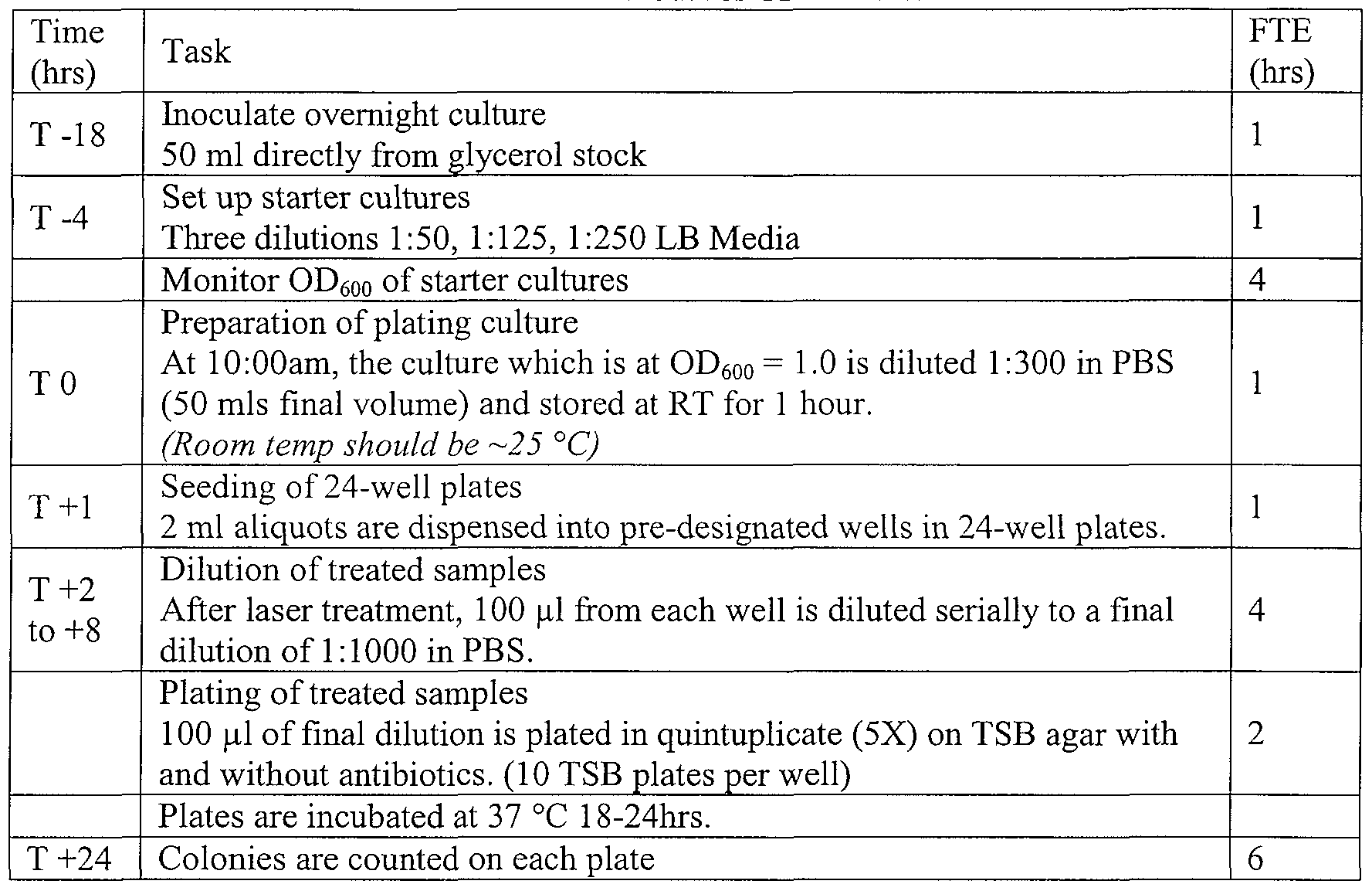
Similar cell culture and kinetic protocols were performed with E coli for all NIMELS irradiation experiments A standardized suspension was ahquoted into selected wells in a 24-well tissue culture plate Following laser treatments, 100 μL was removed from each well and serially diluted to 1 1000 resulting m a final dilution of 1 5x10⁶ of initial culture An aliquot of each final dilution were spread onto separate plates The plates were then incubated at 370C for approximately 16-20 hours Manual colony counts were performed and recorded
Table 4 Method for ΔΨ and ROS Assays
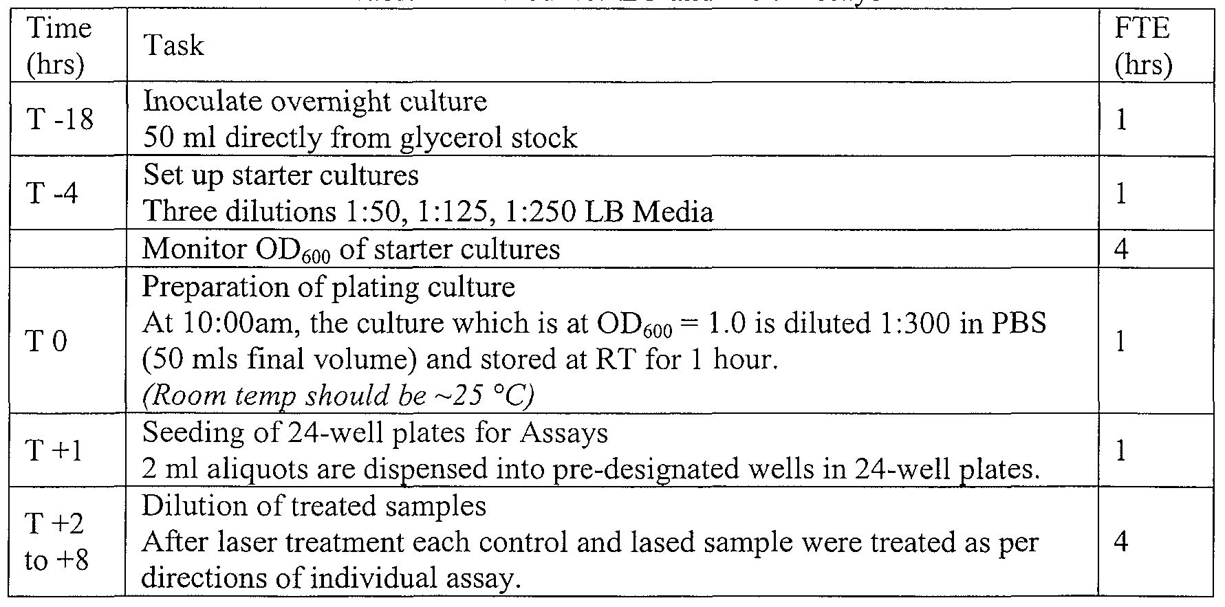
EXAMPLE III MAMMALIAN CELL METHODS NIMELS TREATMENT PARAMETERS FOR IN VITRO HEK293 EXPERIMENTS The following parameters illustrate the general methods according to the invention as applied to HEK293 cells for the in vitro expeπments
A Experiment Materials and Methods for HEK293 cells HEK293 cells were seeded into appropπate wells of a 24-well plate at a density of 1 x 10⁵ cells/ml (0 7ml total volume) in Freestyle medium (Invitrogen) Cells were incubated in a humidified incubator at 37 0C m 8% CO2 for approximately 48 hours prior to the expeπment Cells were approximately 90% confluent at the time of the expeπment equating to roughly 3 x 10⁵ total cells Immediately pπor to treatment, cells were washed m pre- warmed phosphate buffer saline (PBS) and overlaid with 2 ml of PBS during treatment
After laser treatment, cells were mechanically dislodged from the wells and transferred to 1 5 ml centrifuge tubes Mitochondrial membrane potential and total glutathione was determined
EXAMPLE FV NTMELS IN VITRO TESTS FOR CRT+ (YELLO W)AND CRT - (WHITE) S
A UREus EXPERIMENTS
We conducted experiments with crt- (white) mutants of S aureus that were genetically engineered with the crt gene (yellow carotenoid pigment) removed, and these mutants were subjected to previously determined non-lethal doses of NIMELS laser against wild type (yellow) S aureus The purpose of this expeπment was to test for the phenomenon of Radical Oxygen Species (ROS) generation and/or singlet oxygen generation with the NTMELS laser In the scientific literature, Lm et al had previously used a similar model, to test the antioxidant protection activity of the yellow S aureus *caratenoid) pigment against neutrophils (Lm et al , Staphylococcus aureus golden pigment impairs neutrophil killing and promotes virulence through its antioxidant activity, VoI 202, No 2, July 18, 2005 209-215, the entire teaching of which is incorporated herein by reference )
It has previously been determined that the golden color m S aureus is imparted by carotenoid (antioxidant) pigments capable of protecting the organism from oxygen damage, and when a mutant is isolated (crt ) that does not produce such carotenoid pigments, the mutant colomes are "white" in appearance and more susceptible to oxidative killing, and have impaired neutrophil survival
It was found that non-lethal dosimetπes of the NIMELS laser (to wild type S aureus) consistently killed up to 90% of the mutant "white" cells and did not kill the normal S aureus The only genetic difference m the two strains of S aureus is the lack of an antioxidant pigment in the mutant This experimental data strongly suggests that it is the endogenous generation of radical oxygen species and/or smglet oxygen that are killing the "white" S aureus
Table 5 Data
Dl - D4 Yellow Wild Type S aureus D5 - D6 White "crt " Mutant S Aureus

Samples Dl- D4 Yellow Wild Type S. aureus. Sam les D5 - D6 White "crt-" Mutant S. aureus.

There are selected fluorescent dyes that can be taken up by mtact cells and accumulate within the intact cells within 15 to 30 minutes without appreciable staining of other protoplasmic constituents These dye indicators of membrane potential have been available for many years and have been employed to study cell physiology The fluorescence mtensity of these dyes can be easily monitored, as their spectral fluorescent properties are responsive to changes in the value of the trans-membrane potentials ΔΨ- steady
These dyes generally operate by a potential-dependent partitioning between the extracellular medium and either the membrane or the cytoplasm of membranes This occurs by redistribution of the dye via interaction of the voltage potential with an ionic charge on the dye This fluorescence can be eliminated in about 5 minutes by the protonophore carbonyl cyanide m-chlorophenylhydrazone (CCCP), indicating that maintenance of dye concentration is dependent on the mside-negative transmembrane potential maintained by functional ETS and Δp
Hypothesis Testing
The null hypothesis is μi - μ2 = 0 μi is fluorescence intensity in a control cell culture (no laser) subjected to carbocyanme dye μ2 is fluorescence intensity in the same cell culture pre-irradiated with sub-lethal dosimetry from the NTMELS laser The data mdicates that the fluorescence of cells is dissipated (less than control of unirradiated or "unlased" cells) by pre-treatment (of the cells) with the NTJVIELS laser system, indicating that the NTMELS laser interacted with respiratory processes and oxidative phosphorylation of the cells via the plasma membranes 0 Will uphold that the addition sub-lethal NTMEL irradiation on the cell culture has no effect on ΔΨ-steady 0
Will uphold that the addition sub-lethal NTMEL irradiation on the cell culture has a dissipation or depolarization effect on ΔΨ-steady Materials and Methods
BacLight™ Bacterial Membrane Potential Kit (B34950, Invitrogen U S )
The iføcLight™ Bacterial Membrane Potential Kit provides of carbocyanine dye DiOC2(3)
(3,3'-diethyloxacarbocyanine iodide, Component A) and CCCP (carbonyl cyanide 3- chlorophenylhydrazone, Component B), both in DMSO, and a 1 x PBS solution
(Component C).
DiOC2(3) exhibits green fluorescence in all bacterial cells, but the fluorescence shifts toward red emission as the dye molecules self associate at the higher cytosolic concentrations caused by larger membrane potentials. Proton ionophores such as CCCP destroy membrane potential by eliminating the proton gradient, hence causing higher green fluorescence. 0 Detection of membrane potential ΔΨ in MRSA
Green fluorescence emission was calculated using population mean fluorescence intensities for control and lased samples at sub-lethal dosimetry:
Table 7.

Detection of membrane potential ΔΨ in E. coli 0 Red/green ratios were calculated using population mean fluorescence intensities for control and lased samples at sub-lethal dosimetry:
The data shows that μi - μ2 > 0 as the lased cells had less "Green fluorescence" as seen in Figure 19. These E. coli samples showed clear alteration and lowering of ΔΨ - steady-bact to one of ΔΨ -trans-bact with sublethal MMELS dosimetry. 5
EXAMPLE VI: NIMELS IN VITRO TESTS FOR ΔΨ-mito HUMAN EMBRYONIC KIDNEY CELLS WITH SUB-LETHAL LASER DOSIMETRY
Hypothesis Testing: 0 The null hypothesis is μ, - μ2 = 0:
a) μ] is fluorescence intensity m a mammalian control cell culture mitochondπa (no laser) subjected to a Mitochondπal Membrane Potential Detection Kit b) μ2 is fluorescence intensity m the same mammalian cell culture pre-irradiated with sublethal dosimetry from the NIMELS laser and subjected to a Mitochondrial
Membrane Potential Detection Kit
The data shows that the fluorescence of mitochondπa is dissipated (less than control unlased cells) by pre-treatment (of the cells) with the NIMELS laser system, the results indicate that the NIMELS laser interacted with respiratory processes and oxidative phosphorylation of the cells m mitochondπa of mammalian cells
Will uphold that the addition sub-lethal NIMEL irradiation on the mammalian cell culture mitochondπa has no effect on ΔΨ-steady-mito-mam μ, - μ2 > 0 Will uphold that the addition sub-lethal NDVIEL irradiation on the mammalian cell culture has a dissipation or depolaπzation effect on ΔΨ-steady-mito-mam
Mateπals and Methods
Mitochondπal Membrane Potential Detection Kit (APO LOGIX JC-I) (Cell Technology Inc , 950 Rengstorff Ave, Suite D, Mountain View CA 94043)
The loss of mitochondπal membrane potential (ΔΨ) is a hallmark for apoptosis The APO LOGIX JC-I Assay Kit measures the mitochondrial membrane potential in cells In non-apoptotic cells, JC-I (5,5\6,6'-tetrachloro-l,l ',3,3'-tetraethylbenz- lmidazolylcarbocyanme iodide) exists as a monomer in the cytosol (green) and also accumulates as aggregates m the mitochondπa which stam red Whereas, m apoptotic and necrotic cells, JC-I exists in monomeπc form and stams the cytosol green
Table 8 Mamalhan Cell Dosimetries

The (APO LOGIX JC-I) kit measures membrane potential by conversion of green fluorescence to red fluorescence. The appearance of red color has been measured and plotted, which should only occur in cells with intact membranes, and the ratio of green to red is calculated for both control and lased samples.
Clearly in this test, the red fluorescence is reduced in the lased sample while the ratio of green to red increases, indicating depolarization. These results show that μ] - μ2> O and that sub-lethal NIMELS irradiation on the mammalian cell mitochondria has a dissipation or depolarization effect on ΔΨ-steady-mito-mam, indicating a clear reduction in mammalian ΔΨ-steady-mito-mam to ΔΨ-trans-mito-mam.
EXAMPLE VII: NMELS IN VITRO TESTS FOR REACTIVE OXYGEN SPECIES (ROS)
These in vitro tests for generation of reactive oxygen species (ROS) were carried on after laser alteration of bacterial trans-membrane ΔΨ-steady-bact to ΔΨ-trans-bact, ΔΨ- steady-mito-fungi to ΔΨ-trans-mito-fungi, and ΔΨ-steady-mito-mam to ΔΨ-trans-mito-mam with sub-lethal laser dosimetry comparable to those used in ΔΨ tests above in previous examples.
Materials and Methods:
Total Glutathione Quantification Kit (Dojindo Laboratories; Kumamoto Techno Research Park, 2025-5 Tabaru, Mashiki-machi, Kamimashiki-gun; Kumamoto 861-2202, JAPAN) Glutathione (GSH) is the most abundant thiol (SH) compound in animal tissues, plant tissues, bacteria and yeast. GSH plays many different roles such as protection against reactive oxygen species and maintenance of protein SH groups. During these reactions,
GSH is converted into glutathione disulfide (GSSG: oxidized form of GSH). Since GSSG is enzymatically reduced by glutathione reductase, GSH is the dominant form in organisms. DTNB (5,5'-Dithiobis(2-nitrobenzoic acid)), known as Ellman's Reagent, was developed for the detection of thiol compounds. In 1985, it was suggested that the glutathione recycling system by DTNB and glutathione reductase created a highly sensitive glutathione detection method. DTNB and glutathione (GSH) react to generate 2-nitro-5-thiobenzoic acid and glutathione disulfide (GSSG). Since 2-nitro-5- thiobenzoic acid is a yellow colored product, GSH concentration in a sample solution can be determined by the measurement at 412 nm absorbance. GSH is generated from GSSG by glutathione reductase, and reacts with DTNB again to produce 2-nitro-5 -thiobenzoic acid. Therefore, this recycling reaction improves the sensitivity of total glutathione detection.
At significant concentrations ROS will react rapidly and specifically with the target at a rate exceeding the rate of its reduction by the components of the glutathione antioxidant system (catalases, peroxidases, GSH).
Detection of Glutathione in MRSA at sub-lethal NIMELS dosimetry that alters ΔΨ-steady- bact to one of ΔΨ-trans-bact
A reduction in total glutathione in MRSA at sub-lethal NIMELS dosimetry that alters that alters ΔΨ-steady-bact to one of .ΔΨ-trans-bact, is proof of generation of ROS with sub-lethal alteration of Trans-membrane ΔΨ-steady-bact to one of ΔΨ-trans-bact.
Detection of Glutathione in E.coli at sub-lethal NIMELS dosimetry that alters Transmembrane ΔΨ-steady to one of ΔΨ-trans
A reduction in total glutathione in E. coli at sub-lethal NIMELS dosimetry that alters ΔΨ-steady-bact to one of ΔΨ -trans-bact, is evidence of generation of ROS with sub- lethal alteration of Trans-membrane ΔΨ-steady-bact to one of ΔΨ -trans-bact.
Detection of glutathione in C. albicans at sub-lethal NIMELS that alters ΔΨ-steady- mito-fungi to ΔΨ-trans-mito-fungi and subsequently ΔΨ-steady-fungi to one of ΔΨ-trans- fungi.
Detection of Glutathione in C. albicans at sub-lethal NIMELS dosimetry that alters ΔΨ- steady-mito-fungi to ΔΨ-trans-mito-fungi and subsequently ΔΨ-steady-fungi to one of ΔΨ- trans-fungi
A reduction in total glutathione in C. albicans at sub-lethal NIMELS dosimetry that alters ΔΨ-steady-mito-fungi to ΔΨ-trans-mito-fungi and subsequently ΔΨ-steady-fungi to one of ΔΨ-trans-fungi, is proof of generation of ROS with sub-lethal alteration of Transmembrane ΔΨ-steady-mito-fungi to ΔΨ-trans-mito-fungi and subsequently ΔΨ-steady-fungi to one of ΔΨ-trans-fungi.
Detection of Glutathione in HEK-293 (Human Embryonic Kidney Cells) at sub-lethal NIMELS dosimetry that alters ΔΨ-steady-mito-mam to ΔΨ-trans-mito-mam
A reduction in total Glutathione in HEK-293 (Human Embryonic Kidney Cells) with sub-lethal NIMELS dosimetry that alters ΔΨ-steady-mito-mam to ΔΨ-trans-mito-mam, is proof of generation of ROS with NTMELS-mediated sub-lethal alteration of Transmembrane ΔΨ-steady-mito-mam to ΔΨ-trans-mito-mam.
EXAMPLE Vπi Assessment of the impact of Sub-lethal doses of NIMELS Laser on MRSA with Erythromycin and Trimethoprim
In this example, it was determined whether a sub-lethal dose of the NIMEL laser will potentiate the effect of the antibiotic erythromycin more than the antibiotic trimethoprim in MRSA Efflux pumps play a major factor in erythromycin resistance There are no reported trimethoprim efflux pump resistance mechanisms in the gram positive S aureus
Background Erythromycin is a macrolide antibiotic that has an antibacteπal spectrum of action very similar to that of the /3-lactam penicillin In the past, it has been effective in the treatment of a wide range of gram-positive bacteπal infections effecting the skm and respiratory tract, and has been considered one of the safest antibiotics to use In the past, erythromycin has been used for people with allergies to penicillins Erythromycin's mechamsm of action is to prevent growth and replication of bacteria by obstructing bacteπal protem synthesis This is accomplished because erythromycin binds to the 23 S rRNA molecule in the 5OS of the bacteπal πbosome, thereby blocking the exit of the growing peptide chain thus inhibiting the translocation of peptides Erythromycin resistance (as with other marcohdes) is rampant, wide spread, and is accomplished via two significant resistance systems A) modification of the 23 S rRNA in the 5OS πbosomal subumt to msensitivity B) efflux of the drug out of cells
Tπmethopπm is an antibiotic that has histoπcally been used m the treatment of uπnary tract infections It is a member of the class of antimicrobials known as dihydro folate reductase inhibitors Tπmethopπm's mechamsm of action is to interfere with the system of bactenal dihydrofolate reductase (DHFR), because it is an analog of dihydro folic acid This causes competitive inhibition of DHFR due to a 1000 fold higher affinity for the enzyme than the natural substrate
Thus, tπmethopπm inhibits synthesis of the molecule tetrahydrofolic acid Tetrahydrofolic acid is an essential precursor m the de novo synthesis of the DNA nucleotide thymidylate Bacteπa are incapable of taking up folic acid from the environment (i e , the infection host) and are thus dependent on their own de novo synthesis of tetrahydrofolic acid Inhibition of the enzyme ultimately prevents DNA replication
Tπmethopπm resistance generally results from the overproduction of the normal chromosomal DHFR, or drug resistant DHFR enzymes Reports of tπmethopπm resistance
W
S. aureus have indicated that the resistance is chromosomally of the mediated type or is encoded on large plasmids. Some strains have been reported to exhibit both chromosomal and plasmid-mediated trimethoprim resistance.
In the gram positive pathogen S. aureus, resistance to trimethoprim is due to genetic 5 mutation, and there have been no reports that trimethoprim is actively effluxed out of cells.
Efflux Pumps in Bacteria
A major route of drug resistance in bacteria and fungi is the active export (efflux) of antibiotics out of the cells such that a therapeutic concentration in not obtained in the 10 cytoplasm of the cell.
Active efflux of antibiotics (and other deleterious molecules) is mediated by a series of transmembrane proteins in the cytoplasmic membrane of gram positive bacteria and the outer membranes of gram negative bacteria.
Clinically, antibiotic resistance that is mediated via efflux pumps, is most relevant 15 in gram positive bacteria for marcolides, tetracyclines and fluoroquinolones. In gram negative bacteria, β-lactam efflux mediated resistance is also of high clinical relevance.
Hypothesis Testing
The null hypothesis is μt - μ2 = 0 and μi - μ3= 0 where: 20 a) μ] is sub-lethal dosimetry from the NDVIEL laser system on MRSA as a control and; b) μ2 is the same sub-lethal dosimetry from the NIMEL laser system on MRSA with the addition of trimethoprim at resistant MIC just below effectiveness level and; c) μ3 is the same sub-lethal dosimetry from the NIMEL laser system on MRSA with the addition of erythromycin at resistant MIC just below effectiveness level.
25 The data shows that the addition of the antibiotic trimethoprim or erythromycin, after sub-lethal irradiation, results in the reduction in growth of these MRSA colonies, as follows: 0
Will uphold that the addition of trimethoprim produces no deleterious effect after sub-lethal 30 NIMEL irradiation, on normal growth of MRSA colonies.
Will uphold that the addition of trimethoprim produces a deleterious effect after sub-lethal
NIMEL irradiation, on normal growth of MRSA colonies. μ, - μ3= 0
Will uphold that the addition of erythromycin produces no deleterious effect after sub-lethal NIMEL irradiation, on normal growth of MRSA colonies. μ, - μ3 > 0
Will uphold that the addition of erythromycin produces a deleterious effect after sub-lethal NIMEL irradiation, on normal growth of MRSA colonies.
Table 9.


Results:
This experiment clearly showed that under sub-lethal laser parameters with the NIMELS system, μ] - μ2 = 0 and μι - μ3 >= 0. This indicates that an efflux pump is being inhibited, and resistance to erythromycin being reversed by the NIMELS effect on ΔΨ- steady-bact of the MRSA.
EXAMPLE IX: Assessment of the impact of Sub-lethal doses of NIMELS Laser on MRSA with Tetracycline and Rifampin
The purpose of this experiment was to observe if a sub-lethal dose of the NDVIEL laser will potentiate the effect of the antibiotic tetracycline more than the antibiotic rifampin in MRSA. Efflux pumps are well researched, and play a major factor in tetracycline resistance. However, there are no reported rifampin efflux pump resistance mechanisms in the gram positive S. aureus.
This experiment was also previously run with erythromycin and trimethoprim, with data indicating that the NIMELS effect is able to damage efflux pump resistance mechanisms in erythromycin.
Tetracycline:
Tetracycline is considered a bacteriostatic antibiotic, meaning that it hampers the growth of bacteria by inhibiting protein synthesis. Tetracycline accomplishes this by inhibiting action of the bacterial 30S ribosome through the binding of the enzyme aminoacyl-tRNA. Tetracycline resistance is often due to the acquisition of new genes, which
code for energy-dependent efflux of tetracyclines, or for a protein that protects bacterial πbosomes from the action of tetracyclines
Rifampin
Rifampin is a bacterial RNA polymerase inhibitor, and functions by directly blocking the elongation of RNA Rifampicin is typically used to treat mycobacteπal infections, but also plays a role m the treatment of methicillm-resistant Staphylococcus aureus (MRSA) m combmation with fusidic acid, a bacteπostatic protein synthesis inhibitor There are no reports of rifampin resistance via efflux pumps m MRSA
Hypothesis
The null hypothesis is μj - μ2 = 0 and μi - μ3= 0 where a) μi is sub-lethal dosimetry from the NEMEL laser system on MRSA as a control and, b) μ2 is the same sub-lethal dosimetry from the NIMEL laser system on MRSA with the addition of tetracycline at resistant MIC just below effectiveness level and, c) μ3 is the same sub-lethal dosimetry from the NIMEL laser system on MRSA with the addition of rifampin at resistant MIC just below effectiveness level
The data shows that the addition of the antibiotic tetracycline or rifampin, after sublethal irradiation, results m the reduction m growth of these MRSA colonies, as follows μ, - μ2 = 0 Will uphold that the addition of tetracycline produces no deleterious effect after sub-lethal
NIMEL irradiation, on normal growth of MRSA colonies μ, - μ2 > 0
Will uphold that the addition of tetracycline produces a deleterious effect after sub-lethal
NEMEL irradiation, on normal growth of MRSA colonies μ] - μ3= 0
Will uphold that the addition of rifampin produces no deleteπous effect after sub-lethal
NIMEL irradiation, on normal growth of MRSA colonies μi - μs > 0
Will uphold that the addition of rifampin produces a deleteπous effect after sub-lethal NIMEL irradiation, on normal growth of MRSA colonies
Table 10.

Results:
This experiment clearly showed that under sub-lethal laser parameters with the NIMELS system, μi - μ2 = 0 and μ] - μ3 >= 0. This indicates that an efflux pump is being inhibited, and resistance to tetracycline is being reversed by the NTMELS effect on ΔΨ- steady-bact of the MRSA.
EXAMPLE X: Assessment of the impact of Sub-lethal doses of NIMELS Laser on MRSA with Methicillin and ΔΨ-plas-bact inhibition of cell wall synthesis Methicillm:
Methicillin is a /3-lactam that was previously used to treat infections caused by gram-positive bacteria, particularly /3-lactamase-producing organisms such as S. aureus that would otherwise be resistant to most penicillins, but is no longer clinically used. The term methicillin-resistant S. aureus (MRSA) continues to be used to describe S. aureus strains resistant to all penicillins. Mechanism of action
Like other /3-lactam antibiotics, methicillin acts by inhibiting the synthesis of peptidoglycan (bacterial cell walls).
It has been shown in the gram positive bacterium Bacillus subtilis, that the activities of peptidoglycan autolysins are increased (i.e., no longer inhibited) when the ETS was blocked by adding proton conductors. This suggests that ΔΨ-plas-bact and ΔμHT (independent of storing energy for cellular enzymatic functions) potentially has a profound and exploitable influence on cell wall anabolic functions and physiology.
In addition, it has been reported that ΔΨ-plas-bact uncouplers inhibit peptidoglycan formation with the accumulation of the nucleotide precursors involved in peptidoglycan synthesis, and the inhibition of transport of N-acetylglucosamine (GIcNAc), one of the major biopolymers in peptidoglycan.
Hypothesis Testing:
Bacitracin will potentiate the multiple influences of an optically lowered ΔΨ-plas- bact on a growing cell wall (i.e., increased cell wall autolysis, inhibited cell wall synthesis). This is especially relevant in gram positive bacteria such as MRSA, that do not have efflux pumps as resistance mechanisms for cell wall inhibitory antimicrobial compounds. The null hypothesis is μi - μ2 = 0 and μi - μ3= 0 where: a) μi is sub-lethal dosimetry from the NIMEL laser system on MRSA as a control and; b) μ2 is the same sub-lethal dosimetry from the NTMEL laser system on MRSA with the addition of methicillin at resistant MIC just below effectiveness level and; μi - μ2 = 0
Will uphold that the addition of methicillin produces no deleterious effect after sub-lethal
NMEL irradiation, on normal growth of MRSA colonies. μι - μ2 > 0
Will uphold that the addition of methicillin produces a deleterious effect after sub-lethal NIMEL irradiation, on normal growth of MRSA colonies.
Results:
As shown in Figure 15, this experiment clearly showed that under sub-lethal laser parameters with the NIMELS system, μi - μ2 >= 0, meaning that the addition of methicillin produces a deleterious effect after sub-lethal NIMEL irradiation on normal growth of
MRSA colonies as shown by CFU count. This suggest that methicillin (independent of an efflux pump) is being potentiated by the NMELS effect on ΔΨ-steady-bact of the MRSA.
Hence, the NIMELS laser and its concomitant optical ΔΨ-plas-bact lowering phenomenon is synergistic with cell wall inhibitory antimicrobials in MRSA. Without wishing to be bound by theory, this must function via the inhibition of anabolic (periplasmic) ATP coupled functions, as MRSA does not have efflux pumps for methicillin.
EXAMPLE XI: Assessment of the impact of Sub-lethal doses of NMELS Laser on MRSA with Bacitracin and ΔΨ-plas-bact inhibition of cell wall synthesis
Bacitracin is a mixture of cyclic polypeptides produced by Bacillus subtilis. As a toxic and difficult-to-use antibiotic, bacitracin cannot generally be used orally, but is used topically.
Mechanism of action:
Bacitracin interferes with the dephosphorylation of the C55-isoprenyl pyrophosphate, a molecule which carries the building blocks of the peptidoglycan bacterial cell wall outside of the inner membrane in gram negative organisms and the plasma membrane in gram positive organism.
It has been shown in the gram positive bacterium Bacillus subtilis, that the activities of peptidoglycan autolysins are increased (i.e., no longer inhibited) when the ETS was blocked by adding proton conductors. This indicates that ΔΨ-plas-bact and AμirC (independent of storing energy for cellular enzymatic functions) potentially has a profound and exploitable influence on cell wall anabolic functions and physiology.
In addition, it has been reported that ΔΨ-plas-bact uncouplers inhibit peptidoglycan formation with the accumulation of the nucleotide precursors involved in peptidoglycan synthesis, and the inhibition of transport of N-acetylglucosamine (GIcNAc), one of the major biopolymers in peptidoglycan.
Hypothesis Testing:
Bacitracin potentiates the multiple influences of an optically lowered ΔΨ-plas-bact on a growing cell wall (i.e., increased cell wall autolysis, inhibited cell wall synthesis). This is especially relevant in gram positive bacteria such as MRSA, that do not have efflux pumps as resistance mechanisms for cell wall inhibitory antimicrobial compounds. The null hypothesis is μi - μ2 = 0 and μ, - μ3 = 0 where: a) μi is sub-lethal dosimetry from the NIMEL laser system on MRSA as a control and; b) μ2 is the same sub-lethal dosimetry from the NIMEL laser system on MRSA with the addition of bacitracin at resistant MIC just below effectiveness level and; μ, - μ2 = 0
Will uphold that the addition of bacitracin produces no deleterious effect after sub-lethal
NIMEL irradiation, on normal growth of MRSA colonies. μ, - μ2 > 0
Will uphold that the addition of bacitracin produces a deleterious effect after sub-lethal NIMEL irradiation, on normal growth of MRSA colonies.
Results:
As shown in Figure 16, this experiment clearly showed that under sub-lethal laser parameters with the NIMELS system, μj - μ2 >= 0, meaning that the addition of bacitracin produces a deleterious effect after sub-lethal NIMEL irradiation, on normal growth of MRSA colonies. In Figure 16, arrows point to MRSA growth or a lack thereof in the two samples shown. This indicates that bacitracin (independent of an efflux pump) is being potentiated by the NIMELS effect on ΔΨ-steady-bact of the MRSA.
Hence, the NIMELS laser and its concomitant optical ΔΨ-plas-bact lowering phenomenon is synergistic with cell wall inhibitory antimicrobials in MRSA. Without wishing to be bound by theory, this most likely functions via the inhibition of anabolic (periplasmic) ATP coupled functions as MRSA does not have efflux pumps for bacitracin.
EXAMPLE XII: NIMELS Dosimetry Calculations The examples that follow describe selected experiments depicting the ability of the
NIMELS approach to impact upon the viability of various commonly found microorganisms at the wavelengths described herein. The microorganisms exemplified include E. coli K- 12, multi-drug resistant E. coli, Staphylococcus aureus, methicillin-resistant S. aureus, Candida albicans, and Trichophyton rubrum. As discussed in more details supra, NIMELS parameters include the average single or additive output power of the laser diodes, and the wavelengths (870 nm and 930 nm) of the diodes. This information, combined with the area of the laser beam or beams (cm2) at the target site, provide the initial set of information which may be used to calculate effective and safe irradiation protocols according to the invention. The power density of a given laser measures the potential effect of NIMELS at the target site. Power density is a function of any given laser output power and beam area, and may be calculated with the following equations: For a single wavelength:
1) Power Density (W/cm2) = Laser Output Power Beam Diameter (cm2)
For dual wavelength treatments:
2) Power Density (W/cm2) = Laser (1) Output Power + Laser (2) Output Power
Beam Diameter (cm2) Beam Diameter (cm2) Beam area can be calculated by either:
3) Beam Area (cm2) = Diameter (cm)2 * 0.7854 or Beam Area (cm2) = Pi * Radius (cm)2
The total photonic energy delivered into the tissue by one NIMELS laser diode system operating at a particular output power over a certain period is measured in Joules, and is calculated as follows:
4) Total Energy (Joules) = Laser Output Power (Watts) * Time (Sees.)
The total photonic energy delivered into the tissue by both NJMELS laser diode systems (both wavelengths) at the same time, at particular output powers over a certain period, is measured in Joules, and is calculated as follows:
5) Total Energy (Joules) = [Laser(l) Output Power (Watts) * Time (Sees)] + [Laser (2) Output Power (Watts) * Time(Secs)]
In practice, it is useful (but not necessary) to know the distribution and allocation of the total energy over the irradiation treatment area, in order to correctly measure dosage for maximal NIMELS beneficial response. Total energy distribution may be measured as energy density (Joules/cm2). As discussed infra, for a given wavelength of light, energy density is the most important factor in determining the tissue reaction. Energy density for one NIMELS wavelength may be derived as follows: 6) Energy Density (Joules/ cm2) = Laser Output power (Watts) * Time (sees)
Beam Area (cm2)
7) Energy Density (Joule/cm2) = Power Density (W/cm2) * Time (sees) When two NJJVIELS wavelengths are being used, the energy density may be derived as follows:
8) Energy Density (Joules/ cm2) = Laser (l)Output power (Watts) * Time (sees) Beam Area (cm2)
+ Laser (2) Output power (Watts) * Time (sees)
Beam Area (cm2) or, 9) Energy Density (Joule/cm2) = Power Density (1) (W/cm2) * Time (Sees)
+ Power Density (2) (W/cm2) * Time (Sees)
To calculate the treatment time for a particular dosage, a practitioner may use either the energy density (J/cm2) or energy (J), as well as the output power (W), and beam area (cm2) using either one of the following equations:
10) Treatment Time (seconds) = Energy Density
(Joules/cm2)
Output power Density (W/cm2)
11) Treatment Time (seconds) = Energy (Joules)
Laser Output Power (Watts)
Because dosimetry calculations such as those exemplified in this Example can become burdensome, the therapeutic system may also include a computer database storing all researched treatment possibilities and dosimetries. The computer (a dosimetry and parameter calculator) in the controller is preprogrammed with algorithms based on the above-described formulas, so that any operator can easily retrieve the data and parameters on the screen, and input additional necessary data (such as: spot size, total energy desired, time and pulse width of each wavelength, tissue being irradiated, bacteria being irradiated) along with any other necessary information, so that any and all algorithms and calculations necessary for favorable treatment outcomes can be generated by the dosimetry and parameter calculator and hence run the laser.
In the examples that follow, in summary, when the bacterial cultures were exposed to the NJJVIELS laser, the bacterial kill rate (as measured by counting Colony Forming Units or CFU on post-treatment culture plates) ranged from 93.7% (multi-drug resistant E. coli) to 100% (all other bacteria and fungi).
EXAMPLE XIJJ: BACTERIAL METHODS: NMELS TREATMENT PARAMETERS FOR IN VITRO £. COLI TARGETING
The following parameters illustrate the methods according to the invention as applied to E. coli, at final temperatures well below those associated in the literature with thermal damage.
A. Experiment Materials and Methods for E. coli K-12: E. coli Kl 2 liquid cultures were grown in Luria Bertani (LB) medium (25 g/L).
Plates contained 35 mL of LB plate medium (25 g/L LB, 15 g/L bacteriological agar). Culture dilutions were performed using PBS. All protocols and manipulations were performed using sterile techniques.
B Growth Kinetics
Drawing from a seed culture, multiple 50 mL LB cultures were inoculated and grown at 37 0C overnight The next morning, the healthiest culture was chosen and used to inoculate 5% into 50 mL LB at 37 0C and the O D 6oo was monitored over time taking measurements every 30 to 45 minutes until the culture was in stationary phase C Master Stock Production
Starting with a culture in log phase (O D 6Oo approximately 0 75), 10 mL were placed at 40C 10 mL of 50% glycerol were added and was ahquoted into 20 cryovials and snap frozen in liquid nitrogen The cryovials were then stored at -8O0C D Liquid Cultures
Liquid cultures of E coli Kl 2 were set up as descπbed previously An aliquot of 100 μL was removed from the subculture and serially diluted to 1 1200 m PBS This dilution was allowed to incubate at room temperature approximately 2 hours or until no further increase m O D 6Oo was observed m order to ensure that the cells in the PBS suspension would reach a static state (growth) with no significant doubling and a relatively consistent number of cells could be ahquoted further for testing
Once it was determined that the Kl 2 dilution was in a static state, 2 mL of this suspension were ahquoted mto selected wells of 24-well tissue culture plates for selected NIMELS expeπments at given dosimetry parameters The plates were incubated at room temperature until ready for use (approximately 2 hrs)
Following laser treatments, 100 μl was removed from each well and serially diluted to 1 1000 resulting in a final dilution of 1 12x105 of initial Kl 2 culture Ahquots of 3 x 200 L of each final dilution were spread onto separate plates m triplicate The plates were then incubated at 37 0C for approximately 16 hours Manual colony counts were performed and recorded A digital photograph of each plate was also taken
Similar cell culture and kinetic protocols were performed for all NIMELS irradiation tests with S aureus and C albicans in vitro tests For example, C albicans ATCC 14053 liquid cultures were grown in YM medium (21g/L, Difco) medium at 37°C A standardized suspension was ahquoted into selected wells m a 24-well tissue culture plate Following laser treatments, lOOμL was removed from each well and serially diluted to 1 1000 resultmg m a final dilution of 1 5x10⁵ of initial culture 3x100 μL of each final dilution were spread onto separate plates The plates were then incubated at 370C for approximately 16-20 hours Manual colony counts were performed and recorded A digital photograph of each plate was also taken
T rubrum ATCC 52022 liquid cultures were grown m peptone-dextrose (PD) medium at 37 0C A standardized suspension was aliquoted into selected wells in a 24 -well tissue culture plate Following laser treatments, ahquots were removed from each well and spread onto separate plates The plates were then incubated at 37 0C for approximately 91 hours Manual colony counts were performed and recorded after 66 hours and 91 hours of incubation While control wells all grew the organism, 100% of laser-treated wells as described herein had no growth A digital photograph of each plate was also taken
Thermal tests performed on PBS solution, starting from room temperature Ten (10) Watts of NIMELS laser energy were available for use m a 12 mmute lasmg cycle, before the
10 temperature of the system is raised close to the critical threshold of 44°C
Table 11 Time & Tem erature measurements for In Vitro NIMELS Dosimetries

15 EXAMPLE XIV DOSIMETRY VALUES FOR NIMELS LASER WAVELENGTH 930 NM FOR E
COLIIN VITRO TARGETING
The instant experiment demonstrates that the NTMELS single wavelength λ = 930 nm is associated with quantitatable antibacteπal efficacy against E coli in vitro withm safe 20 thermal parameters for mammalian tissues
Experimental data in vitro demonstrates that if the threshold of total energy into the system with 930 nm alone of 5400 J and an energy density of 3056 J/cm is met in 25% less time, 100% antibacterial efficacy is still achievable.
Table 12. Sub-thermal NIMELS (X= 930) Dosimetry for In Vitro E. coli Targeting

Experimental data in vitro also demonstrates that treatments using a single energy with λ = 930 nm has antibacterial in vitro efficacy against the bacterial species S. aureus within safe thermal parameters for mammalian tissues.
It is also believed that if the threshold of total energy into the system of 5400 J and an energy density of 3056 J/cm2 is met in 25% less time with S. aureus and other bacterial species, that 100% antibacterial efficacy will still be achieved.
Table 13. Sub-thermal NMELS (X= 930) Dosimetry for In Vitro S. aureus Targeting

Experimental in vitro data also showed that the NTMELS single wavelength of X = 930 nm has anti-fungal efficacy against in vitro C. albicans at ranges within safe thermal parameters for mammalian tissues.
It is also believed that if the threshold of total energy into the system of 5400 J and an energy density of 3056 J/cm2 is met in 25% less time, that 100% antifungal efficacy will still be achieved.
Table 14. Sub-thermal NTMELS (X= 930) Dosimetry for In Vitro C. albicans Targeting

Experimental in vitro data also demonstrates that no significant kill is achieved up to a total energy of 7200 J, and energy density of 4074 J/cm2 and a power density of 5.66 0 W/cm2 with the wavelength of 870 nm alone against E. coli.
Table 15. E. coli Studies- Single wavelength X = 870 nm

10 Comparable results using radiation having X = 870 nm alone were also observed with S. aureus.
EXAMPLE XVI: NIMELS UNIQUE ALTERNATING SYNERGISTIC EFFECT BETWEEN 870 NM
AND 930 NM OPTICAL ENERGIES
15
Experimental in vitro data also demonstrates that there is a greater than additive effect between the two NIMELS wavelengths (X= 870 nm and 930 nm) when they are alternated (870 nm before 930 nm). The presence of the 870 nm NMELS wavelength as a first irradiance has been found to enhance the effect of the antibacterial efficacy of the
20 second 930 nm NMELS wavelength irradiance.
Experimental in vitro data demonstrates that this synergistic effect (combining the 870 nm wavelength to the 930 nm wavelength) allows for the 930 nm optical energy to be reduced. As shown herein, the optical energy was reduced to approximately 1/3 of the total energy and energy density required for NMELS 100 % E. coli antibacterial efficacy, when
25 the (870 nm before 930 nm) wavelengths are combined in an alternating manner.
Experimental in vitro data also demonstrates that this synergistic mechanism can allow for the 930 nm optical energy (total energy and energy density) to be reduced to approximately 1/2 of the total energy density necessary for NMELS 100% E. coli antibacterial efficacy if equal amounts of 870 nm optical energy are added to the system
30 before the 930 nm energy at 20% higher power densities.
Table 16. E. coli data from Alternating NIMELS Wavelengths

This synergistic ability is significant to human tissue safety, as the 930 nm optical energy heats up tissues at a greater rate than the 870 nm optical energy, and it is beneficial to a mammalian system to produce the least amount of heat possible during treatment.
It is also believed that if the NIMELS optical energies (870 nm and 930 nm) are alternated in the above manner with other bacterial species, that the 100% antibacterial effect will be essentially the same.
EXAMPLE XVII: NTMELS UNIQUE SIMULTANEOUS SYNERGISTIC EFFECT BETWEEN λ= 870
NM AND λ= 930 NM OPTICAL ENERGIES
Experimental in vitro data also demonstrates that there is a greater than additive effect between the two NIMELS wavelengths (870 nm and 930 nm) when they are used simultaneously (870 nm combined with 930 nm). The presence of the 870 nm NIMELS wavelength and the 930 nm NTMELS wavelength as a simultaneous irradiance absolutely enhances the effect of the antibacterial efficacy of the NEVIELS system.
In vitro experimental data (see, for example, Tables LX and X below) demonstrated that by combining λ = 870 nm and λ = 930 nm (in this example used simultaneously) effectively reduces the 930 nm optical energy and density by about half of the total energy and energy density required when using a single treatment according to the invention.
Table 17. E. coli data from Combined NEvIEL Wavelengths

Thus, NIMELS wavelengths (λ= 870 nm and 930 nm) may be used to achieve antibacterial and anti-fungal efficacy in an alternating mode or simultaneously or in any combination of such modes thereby reducing the exposure at the λ = 930 associated with temperature increases which are minimized
Expeπmental in vitro data also demonstrates that when E coli is irradiated alone with a (control) wavelength of λ = 830 nm, at the following parameters, the control 830 nm laser produced zero antibactenal efficacy for 12 minutes irradiation cycles, at identical parameters to the minimum NIMELS dosimetry associated with 100% antibactenal and anti-fungal efficacy with radiation of λ = 930 nm
Table 18 E coli Single Wavelength λ = 830 nm

Expeπmental in vitro data also demonstrates that when applied at safe thermal dosimetries, there is little additive effect when using radiance of λ = 830 nm in combination with λ = 930 nm The presence of the 830 nm control wavelength as a first irradiance is far inferior to the enhancement effect of the 870 nm NTMELS wavelength in producing synergistic antibacterial efficacy with the second 930 nm NIMELS wavelength
Table 19 E coli data from Substituted alternating 830 nm control Wavelength


Amount of Bacteπa Killed
In vitro data also showed that the NIMELS laser system in vitro is effective (within thermal tolerances) against solutions of bacteπa containing 2,000,000 (2 x 10⁶) Colony Forming Units (CFU's) of E coli and S aureus This is a 2x increase over what is typically seen in a 1 gm sample of infected human ulcer tissue Brown et al reported that miciobial cells in 75% of the diabetic patients tested were all at least 100,000 CFU/gm, and m 37 5% of the patients, quantities of microbial cells were greater than 1,000,000 (lxlO6)CFU (see Brown et al , Ostomy Wound Management, 401 47, issue 10, (2001), the entire teaching of which is incorporated herein by reference)
Thermal Parameters
Expeπmental in vitro data also demonstrates that the NDVIELS laser system can accomplish 100% antibacteπal and anti-fungal efficacy withm safe thermal tolerances for human tissues
EXAMPLE XVIII THE EFFECTS OF LOWER TEMPERATURES ON NEVIELS Cooling of Bacteπal species
Expeπmental in vitro data also demonstrated that by substantially loweπng the starting temperature of bacteπal samples to 4°C for two hours m PBS before lasing cycle, that optical antibacterial efficacy was not achieved at any currently reproducible antibacteπal energies with the NIMELS laser system
EXAMPLE XIX MRSA/ ANTIMICROBIAL POTENTIATION
This example shows the use of NIMELS wavelengths (λ = 830 nm and 930nm) in m vitro targeting of MRSA to increase antimicrobial sensitivity to methicillin Four separate expeπments have been performed The data sets for these four experiments are presented in the tables that follow See, also, Figure 17, which shows (a) the synergistic effects of NIMELS with methicillin, penicillin and erythromycin in growth inhibition of MRSA colonies, data show that penicillin and methicillin is being potentiated by sub-lethal NEVIELS dosimetry by inhibiting the Bacteπal Plasma Membrane Proton-motive force (Ap- plas-Bact) thereby inhibiting peptidoglycan synthesis anabolic processes that are co- targeted with the drug, and (b) that erythromycin is potentiated to a greater extent, because the Nimels effect is inhibiting the Bacteπal Plasma Membrane Proton-motive foice (Ap- plas-Bact) that supplies the energy for protein synthesis anabolic processes and erythromycin resistance efflux pumps General Methods for CFU counts
Table 21

First lasing procedure : Both 870 and 930
Second lasin rocedure 930 alone

Experiment 1 - design:
Eight different laser dosages were used to treat a saline-suspension of logarithmically growing MRSA, labeled Al to Hl .
10 The treated and a control untreated suspension were diluted and plated in triplicate on trypic soy agar with or without 30 μg/ml methicillin.
After 24hrs of growth at 37 0C colonies were counted.
CFU (colony forming units) were compared between the plates with and without methicillin for both control (untreated) and treated MRSA.
Experiment 1 - results:
Conditions Dl through Hl showed a similar reduction in CFU on the methicillin plates in treated and untreated samples.
Conditions Al, Bl and Cl showed 30%, 33%, or 67% fewer CFU in the laser treated samples compared to the untreated controls, respectively.
This indicates that the treatments performed on sample Al, Bl and Cl sensitized the MRSA to the effects of methicillin.
Table 23. MRSA Data Progression


Table 24. MRSA Dosimetry Progression
MRSA Dosimetry
Progression
11-07-06
First lasmg procedure Both
870 and 930
Second lasmg procedure 930 alone

Eight different laser dosages based on an effective dose established in expeπment 1 and previously were used to treat a salme-suspension of logarithmically growing MRSA, labeled A2 to H2
The treated and a control untreated suspension were diluted and plated m triplicate on trypic soy agar with or without 30 μg/ml methicillm After 24hrs of growth at 37 0C colonies were counted
Expeπment 2 — results
Compaπson of CFU on plates with and without methicillm showed a significant increase m the effectiveness of methicillm m all laser treated samples with the exception of A2 and B2 This data is summarized m tabular form below
Table 25
Grouping Fold increase in methicillin sensitivity
A2 0 84
B2 0 91
C2 3 20
D2 2 44
E2 4 33
F2 2 13
G2 3 47
H2 1 62
Table 26 MRSA Data Progression


Table 27 Outlined Protocol for MRSA Study
Time _ ,
. . Task
(hrs)
Inoculate overnight culture
50 ml directly from glycerol stock 1 -18
Set up starter cultures
Three dilutions 1 50, 1 125, 1 250
T -4
Monitor ODrøo of starter cultures
Preparation of plating culture
At 10 00am, the culture which is at OD600 = 1 0 is diluted 1 300 in PBS (50 mis final volume) and stored at RT for 1 hour T 0 (Room temp should be -25 °C)
Seeding of 24-well plates (8 plates in total) T +1 2 ml aliquots are dispensed into pre-designated wells in 24-well plates and transferred to NOMIR (8 24-well plates total) _ _ Dilution of treated samples
„ After laser treatment, 100 μl from each well is diluted serially to a final dilution of 1 1000 m PBS
Plating of treated samples
100 μl of final dilution is plated m qumtuphcate (5X) on TSB agar with and without 30 μg/ml methicillin (10 TSB plates per well)
Plates are incubated at 37 0C 18-24hrs T +24 Colonies are counted on each plate (160 plates total)
Table 28 MRSA Dosimetry Progression
MRSA Dosimetry Progression
11-09-06
First lasing procedure Both 870 and 930
Second lasmg procedure 930 alone

Expeπment 3 - design
Eight different laser dosages based on an effective dose established in expeπments 1 and 2 and previously were used to treat a salme-suspension of logarithmically growing MRSA, labeled A3 to H3
The treated and a control untreated suspension were diluted and plated in pentuplicate on trypic soy agar with or without 30 μg/ml methicillin
10 After 24hrs of growth at 37 0C colonies were counted
Experiment 3 - results:
Comparison of CFU on plates with and without methicillin showed a significant increase in the effectiveness of methicillin in all laser treated samples. This data is summarized in tabular form below.
Table 29.
Grouping Fold increase in methicillin sensitivity
A3 1.98
B3 1.62
C3 1.91
D3 2.59
E3 2.09
F3 2.08
G3 3.16
H3 2.97
Table 30. MRSA Data Progression


Table 32 MRSA Dosimetry Progression
MRSA Dosimetry Progression First lasing procedure Both 870 and 930 Second lasin rocedure 930 alone

Independent Report for MRSA studies Expeπment 4 - design
Two different laser dosages based on an effective dose established in previous expeπments were used to treat a salme-suspension of logarithmically growing MRSA, labeled A4 to F4
10 The treated and a contiol untreated suspension were diluted and plated in pentuplicate on trypic soy agar with or without 30 μg/ml methicillin (Groups A4 and B4), 0 5 μg/ml penicillin G (Groups C4 and D4) or 4 μg/ml erythromycin (Groups E4 and F4)
After 24hrs of growth at 37 0C colonies were counted
Expeπment 4 - results
Laser treatment increases sensitivity of MRSA to each antibiotic tested by several fold This data is summarized below
Table 33
Series Drug
A4 Methicillin
B4 Methicillin
C4 Penicillin
D4 Penicillin
E4 Erythromycin
F4 Erythromycin
Table 34
Grouping Fold increase in antibiotic sensitivity
A4 2 19 B4 2 63
C4 2 21 D4 3 45
E4 50 50 F4 9 67


EXAMPLE XX: Non-thermal NIMELS Interaction Evidence for non-thermal NIMELS interaction:
It was demonstrated through experimentation (in vitro water bath studies), that the temperatures reached in the in vitro NIMELS experimentation, were not high enough in and of themselves to neutralize the pathogens.
In the chart that follows, it can clearly be seen that when simple E. coli Bacteria were challenged at 47.5 C continuously for 8 minutes in a test tube in a water bath, they achieved 91% growth of colonies. Therefore, it was demonstrated essentially that the NIMELS reaction is indeed photo-chemical in nature, and occurs in the absence of exogenous drugs and/or dyes.
Table 36.

Example XXI: Laser Treatment for Microbial Reduction and Elimination of Nasal Colonization of MRSA
The Nomir Near Infrared Microbial Elimination Laser System (NOVEON™ Model
1120 dual-wavelength diode laser was employed for this study. The laser operates in continuous wave format at two wavelengths, 870 ran (+/- 5 nm) and 930 ran (+/- 5 nm). This device is a class II non-significant risk laser device. The laser sources of this device are semiconductor laser arrays that are optically coupled to form a single fiber laser output. The
delivery system consists of a single flexible optical fiber. The device delivers continuous wave laser light only.
The device is designed specifically to effect microbial cell optical destruction, while preserving and without substantial damage optically or thermally to the human tissue at the infection site being irradiated. In that regard, the NOVEON™ system was designed to harness the known photo-lethal characteristics of these precise energies to kill pathogenic microorganism at far lower energy levels and heat deposition than is generally necessary to kill pathogens using laser-based thermal sterilization means. Using exposure to the dual wavelength infrared NOVEON™ laser, at temperature levels inherently not lethal to the organism, we had accomplished in vitro successful reversal of MRSA resistance to Methicillin, Penicillin, Erythromycin and Tetracycline. It has also been shown in vitro, that MRSA that has been exposed to a sublethal dose by the NOVEON™ laser will become sensitive to antibiotics to which it was previously resistant.
Currently, topical intra-nasal antimicrobial agents are recognized as the preferred method for preventing (distal-site) infections because of their demonstrated effectiveness and widespread desire to minimize the use of systemic antimicrobials.
Thus, the design of this protocol includes a number of important factors have been considered. Foremost is the need to assure that the amount of energy used in the Nares is safe for the nasal and nares tissues. Furthermore, significant human and histological tests have been done with the Noveon laser in the areas that the study is treating
HUMAN STUDIES
Initial studies were performed to chart and ensure the thermal safety of laser energies on human dermal tissues. Exposure of dermal surfaces to both 870 nm and 930 nm simultaneously with a combined Power Density of 1.70 W/cm2 for up to 233 seconds, results in a skin surface temperature of 1000F as measured with a laser infrared thermometer.
Exposure of dermal surfaces to 930 nm alone at a Power Density of 1.70 W/cm2 for up to 142 seconds, results in a skin surface temperature of 97°F. At or above these doses to dermal infection sites, pain can result. It is therefore desirable from a standpoint of patient comfort not to exceed these doses.
Table 37. Dosimetry Simultaneously Using 870 and 930 Nanometers

Additional testing of the device on the epithelial tissue of humans was conducted using a specially prepared dispersion tip designed to be inserted in the nares. Using a dispersion tip, laser energy was delivered to the nostrils circumferentially by an optical fiber (connected to the NOVEON™ laser) that terminates in a central diffusing tip. This was placed within the inner lumen of the nostril (nares).
A cylindrical diffusing optical fiber tip for near infrared light delivery was fabricated specifically for uniform illumination of a length of 1.5 cm, to then be placed in a transparent catheter (of given width) to prevent placement too far anteriorly in the nostril, and guarantee a uniform power density at all tissues proximal to the catheter within the nostril.
The tip included an optically transmissive, light diffusing, fiber tip assembly having an entrance aperture through a proximal reflector, a radiation-scattering, transmissive material (e.g. a poly-tetrafluoroethylene tube) surrounding an enclosed cavity (e.g. a cylindrical void filled with air or another substantially non-scattering, transparent medium), and a distal reflective surface. As radiation propagates through the fiber tip assembly, a portion of the radiation is scattered in a cylindrical (or partly cylindrical) pattern along the distal portion of the fiber tip. Radiation, which is not scattered during this initial pass through the tip, is reflected by at least one surface of the assembly and returned through the tip. During this second pass, the remaining radiation, (or a portion of the returning radiation), is scattered and emitted from the proximal portion of the tube. Multiple additional reflections off of the proximal and distal reflectors provide further homogenization of the intensity profile. Preferably the scattering medium has a prescribed
inner diameter. This inner diameter of the scattering material is designed such that the interaction with this material and the multiple reflections off of the cavity reflectors interact to provide a substantially proscribed axial distribution of laser radiation over the length of the tip apparatus. Suitable choices of tip dimensions provide control over the emitted axial and azimuthal energy distributions.
To first document safety with the instrumentation, samples of turkey muscle (shown to be a suitable model for nasal mucosa, were irradiated with the above described dispersion tip. The maximum temperature attained during this experiment was 33.9 degrees Centigrade. Further, no specimen showed any burning or necrosis, despite use of exposure times that were double than any anticipated for use in human subjects.
Table 39. Dosimetry Simultaneously Using 870 and 930 Nanometers

Table 40. Dosimetry at 930 Nanometers

Studies have shown that there are five factors to consider regarding energy absorption and heat generation by the "y" emissions of near infrared diode lasers. These factors are: wavelength and optical penetration depth of the laser; absorption characteristics of exposed tissue; temporal mode (pulsed or continuous; exposure time; and power density of the laser beam.
Diode lasers in the near infrared range have a very low absorption coefficient in water; hence, they achieve relatively deep optical penetration in tissues that contain 80% water (such as the dermis, the oral mucosa, bone and the gingiva. With conventional near infrared diode soft tissue lasers, the depth of penetration (before photon absorption) of the greatest amount of the incident energy is about 1.5 cm. This allows the near infrared laser energy to pass through water with minimal absorption, producing thermal effects deeper in the tissue and the photons are absorbed by the deeper tissue pigments. This photobiology
allows for controlled, deeper soft-tissue irradiation and decontamination, as the photons that emerge from the dispersion tip in a uniform dosimetry from the diffusing tip absorbed by blood and other tissue pigments.
Approached from other known dosimetry perspectives, if the conventional Power Equation is applied to in vivo NIMELS dosimetries [Power (Watts) = Work/ Time], the following examples illustrate the Power differences between current therapies: Photoablative dosimetry = 1000 J/cm2 in l/l,000Λ of a second; Thermal vaporizing dosimetry = 1000 J/cm2 in 1 second; and NIMELS decontamination dosimetry = 500 J/cm2 in 360 seconds.
This investigational protocol was designed to demonstrate that the Noveon Laser treatment is able to produce reduction in Nasal carriage of MRSA in patients with previously "culture positive" history. This investigational protocol was an open-label study of subjects who are colonized with MRSA in the nares (nostril). The study was done in two parts.
PART ONE Subjects
In this human study, three arms were produced. Subjects with a previous "culture positive" history who were found to be positive for MRSA colonization in the nares were randomized to one of three treatment groups: Arm # 1 : 1/3 of the subjects were treated with laser alone; Arm # 2: 1/3 of the subjects were be treated with topical H2O2 and then the laser after two minutes. This was done on day 1 and again on day three; and Arm #3: 1/3 of the subjects were treated with the laser and then a topical antibiotic three times a day for five days. Prior to enrollment in this study, prospective subjects met all of the following criteria: age >18 years and ≤IO years of age; previous positive MRSA culture; negative urine pregnancy test or post-menopausal for one year; willing to comply with study requirements, including return visits and self-application of topical antibiotics; and willing to provide informed consent to participate. Prospective subjects were excluded from this study if any of the following criteria were met: pregnancy; patients who are severely immunocompromised (such as may occur in AIDS, renal transplant regimens, immunosuppressed states consequent to malignancy or agents used in rendering oncologic care, or who suffer from end stage renal disease); diabetic patients; allergy to antimicrobials being used in the study (group 3). The exclusion of such groups in this instance was solely for purposes of performing a controlled clinical study, and it is particularly noted that the above exclusion groups are actually considered good candidates for the phototherapeutic
treatments described herein, wherein such patients would actually benefit from therapeutic bacteπal photodamage in that reduced systemic doses of antibiotics could be given and infection sites could be better cleared Study Procedures: All participants underwent an initial quantitative assessment for nasal carriage of S aureus, during their first visit Each participant had the antenor nares (each nostπl) sampled for culture with a circular movement (three rotations on each side) of a sterile wood applicator plain Rayon® tipped swab m each nostπl and placed in a labled tube 2 ml of room temp phosphate buffered salme was placed m the tube after the removal of the swab (to completely cover the swab m the tube) Each swab was then placed back in the tube and the tube was then vortexed for 15 seconds to disperse isolates of MRSA and/or MSSA into the PBS solution Ahquots of PBS from the tube were plated m the following manner 100 μl from each tube was lawn plated m triplicate (3X) on selective Chromogemc MRSA And MSSA agar Plates were placed in incubator within 30 minutes of the plating procedure, and colonies were counted manually, and recorded 18 hrs after plating
On day one of the study, all subjects underwent this exact procedure in arms 1 and 2 of the study two minutes before the laser procedure They again were sampled in the same manner 2 minutes after the laser procedure In the third arm of the study, they were swabbed two minutes before the procedure and the first antibiotic administration was completed after the laser therapy The post/laser swab sample was taken for this arm 20 mm later
On day three of the study, all subjects from arms one two and three underwent the exact same procedures as day 1 of the study
On day five all subjects from all arms underwent just one swabbing per nostril with the exact same sampling procedure one time
Application of H202 3% OTC hydrogen peroxide was applied to a cotton pledget for application to the subject pπor to irradiation This was inserted m the nose for 120 seconds and then removed The subjects were then given doses of phototherapeutic near infrared radiation as descπbed Application of generic topical Antimicrobial The subjects were first given doses of phototherapeutic near infrared radiation as descπbed Subsequently, 2% erythromycin paste was applied to a cotton tipped swab for application to the subject following irradiation The swab was inserted approximately 1 cm in to the antenor nares and rotated 360 degrees several times and removed Patients were instructed to perform the exact application procedure 3 times a day for the remaining 5 days
Treatment Description
The NOVEON™ laser was used for two (2) six-minute treatments in each nostril on day (1) and day (3) of the study. The dosimetries used are shown in the Table TT, below. The laser was calibrated before the first treatment of the day. Intermittent temperature testing of the treatment site was performed on each subject using a noncontact infrared thermometer (Raytek Minitemp), 30-60 second intervals. If a temperature of 1 10 F degrees was reached, or the patient complained of pain, the laser treatment was interrupted and only resumed when the patient was comfortable. Inturruption only occurred once in 40 treatments (20 nostrils x 2 treatments over three days), and was resumed 30 seconds later to completion.
Table 41 power
Laser density Length Diameter Area Trans set power nm W/cm2 mm mm cm2 percent W
930 0.46 10 12 3.77 80 2.17
870 0.185 10 12 3.77 80 0.87
930 0.277 10 12 3.77 80 1.30
80
930 0.405 10 12 3.77 80 1.91
870 0.16 10 12 3.77 80 0.75
930 0.243 10 12 3.77 80 1.14 both 0.54 10 12 3.77 80 2.54 both 0.46 10 12 3.77 70 2.48 Quantitative Assessments to measure Change in MRSA and MSSA colonies
The following Tables 42-44 represent the mean values of the triplicate CFU counts and plating of each swab from each nostril, pre and post laser therapy (for this data set the mean is the sum of the observed and counted CFU 's per plate, divided by the number of counted plates).
Table 42
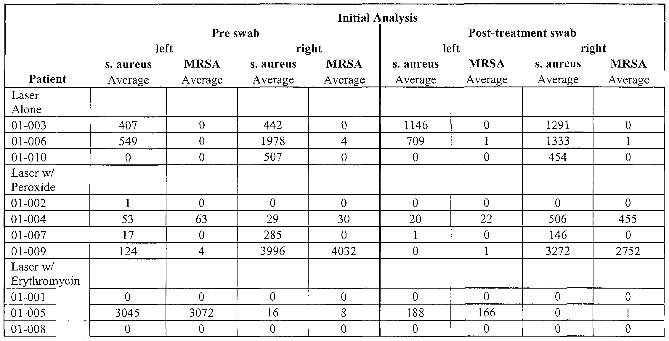
Table 43
 Table 44
Third Analysis
Swab left right s. aureus MRSA s. aureus MRSA
Patient Average Average Average Average
Laser Alone
01-003 193 0 359 0
01-006 387 0 645 5
01-010 22 0 387 0
Laser w/ Peroxide
01-002 1 0 1 0
01-004 868 827 586 563
01-007 28 0 52 0
01-009 0 0 3493 3648
Laser w/ Erythromycin
01-001 0 0 0 0
01-005 0 0 0 0
01-008 0 0 0 0
Results We treated performed 36 treatments of 10 patients (20 infection sites) with zero negative sequelae from the laser m identified MRSA earners based on a physician's evaluation of all the patients 2 days following the second laser therapy
Patients 1 and 8 (m the laser plus antibiotic arm) were not treated a second time, as there was no growth of S aureus or MRSA colonies present on the pre-test swabs These patients were dismissed from the study by the pπncipal investigator The Laser alone arm was showed mconsequential colony reduction in MRSA and MSSA colomes in the nares The Laser plus H2O2 arm may have had some transient benefit in some of the patients, but no obvious long-term efficacy The remaining patient (01-005) in the Laser/erythromycm that began the study with culturable S aureus and MRSA showed a remarkable reduction in culturable bacterial from the colonization site as the treatments progressed, to the point of MRSA and MSSA eradication m both nostπls In this patient, the combination of near infrared bactenal photodamage and topical antibiotics eradicated the MRSA infection The heavily colonized nostπl showed at least a 3 log reduction of bioburden, and resulted m no culturable bacteπa, and the moderately colonized nostπl showed at least a 2 log reduction of bioburden, and
resulted in no culturable bacteπa Notably, the MRSA colony in that patient was not sensitive to erythromycin pπor to phototherapy with the NOVEON™ laser system PART TWO
A second human study was conducted, to further evaluate the therapeutic potential of the NOVEON™ laser system, including its ability to reverse drug resistance m bacteπa The study was conducted in a similar manner as Part One, above Outcome measures assessed included both laboratory study and clinical observations
Positive anterior nares cultures were obtained in six patients (12 nostπls) having nasal colonization of MRSA or MSSA, before initiating bacterial photodamage through
10 doses of phototherapeutic near infrared radiation One patient had MRSA only, 3 had MSSA only, and 2 had both MRSA and MSSA All MRSA and MSSA were cultured and verified to be resistant to erythromycin
Application of Topical Antimicrobial Antimicrobial paste (generic 2% erythromycin) was 15 placed on a cotton tipped swab for application after phototherapeutic near mfrared radiation The swab was inserted approximately 1 cm in to the anteπor nares of the subject, rotated 360 degrees several times and removed The application of erythromycin was maintained for 3 times a day for the remainder of the study
The laser was calibrated before the first treatment of the day and between each 20 patient The NOVEON™ laser was used for four six-minute treatments of the nares at the following sets of dosimetries (Tables 45), which were evaluated for safety in previous studies Utilizing a 10 cm flat-top diffuser, each patient underwent exposure with the Noveon for 7 minutes (energy density - 207 J/cm2) to each anterior nostril on Day 1 and on Day 3 The treatment was divided into two parts, an approximately 3 -minute exposure using 25 a combination of 870 nm and 930 nm and an approximately 3-mmute exposure of 930 run alone Temperatures of the nares were recorded every 30 seconds with an IR temperature thermometer
Table 45
power set set IRB
Laser density Length Diameter Area Trans power Laser power Power nm W/cm2 mm mm cm2 percent W Amp W ratio
930 0 46 10 12 3 77 80 2 17 5 95 2 17 1 00
870 0 185 10 12 3 77 80 0 87 4 65 0 87 0 40
930 0 277 10 12 3 77 80 1 30 4 85 1 30 0 60
Bacteriology
Quantitative cultures from each nostril were obtained and plated in triplicate on chromogenic agar before and 20 minutes after exposure on day 1 and day 3. A final culture was taken on day 5. Anterior nares specimens were collected on rayon-tipped swabs, and stored in Amies liquid transport medium. The nasal swab was plated on Columbia colistin- nalidixic agar (CNA) with 5% sheep blood, then incubated 18 to 48 hours at 350C in 5% CO2. S. aureus was identified by colony morphology and Staphaurex™ latex agglutination test (Murex Biotech Limited, Dartford, Kent, UK). Samples were frozen and stored at - 200C. Results:
The Erythromycin resistant MRSA was completely cleared by culture in all 3 carriers, as was the E-mycin resistant MSSA in four of the five (5) carriers after the second laser treatment on day 3 and remained clear on day 5. In one patient the E-mycin resistant MSSA (baseline count > 1000 CFU's) showed a 3-log reduction in MSSA on the day 5 culture. No sequelae or adverse events were observed. The average maximum temperature of the nares reached in all patients was 99F.
Conclusions:
NOVEON™ laser exposure at a non-damaging energy density and approximately physiologic temperatures, re-sensitized erythromycin resistant MRSA and MSSA to 2% generic erythromycin paste. Photodamage to the organism results in sensitivity to antibiotics in otherwise drug resistant strains. The NOVEON™ laser system provides for local reduction of drug resistant microbes and a concomitant reduction of bio-burden in: e.g., wounds, mucosal or cutaneous tissues, and other colonized or infected areas such as surgical sites and tissue/medical device interfaces, which are prone to contamination particularly by nosocomial strains of microbes frequently having multidrug resistance phenotypes.
Exemplary NIMELS Systems
Figure 17 illustrates a schematic diagram of a therapeutic radiation treatment device according one embodiment of the present disclosure. The therapeutic system 110 includes an optical radiation generation device 112, a delivery assembly 114, an application region
116, and a controller 118.
According one aspect of the present disclosure, the optical radiation generation device (source) includes one or more suitable lasers, Ll and L2 A suitable laser may be selected based on a degree of coherence In exemplary embodiments, a therapeutic system can include at least one diode laser configured and arranged to produce an output m the near infrared region Suitable diode lasers can include a semiconductor mateπals for producing radiation in desired wavelength ranges, e g , 850nm-900nm and 905nm-945nm Suitable diode laser configurations can include cleave-coupled, distπbuted feedback, distπbuted Bragg reflector, vertical cavity surface emitting lasers (VCSELS), etc
With contmued reference to Figure 17, in certain embodiments the delivery assembly 114 can generate a "flat-top" energy profile for uniform distribution of energy over large areas For example, a diffuser tip 10, may be included which diffuses treatment light with a uniform cylindrical energy profile in an application region 116 (e g a nasal cavity as descπbed in the example above) As noted, the optical radiation generation device 1 12 can include one or more lasers, e g , laser oscillators Ll and L2 In exemplary embodiments, one laser oscillator can be configured to emit optical radiation m a first wavelength range of 850 nm to 900 nm, and the other laser oscillator can be configured to emit radiation m a second wavelength range of 905 nm to 945 nm In certain embodiments, one laser oscillator is configured to emit radiation m a first wavelength range of 865 nm to 875 nm, and the other laser oscillator 28 is configured to emit radiation m a second wavelength range of 925 nm to 935 nm The geometry or configuration of the individual laser oscillators may be selected as desired, and the selection may be based on the intensity distributions produced by a particular oscillator geometry or configuration
With continued reference to Figure 17, m certain embodiments, the delivery assembly 114 includes an elongated flexible optical fiber 118 adapted for delivery of the dual wavelength radiation from the oscillators 26 and 28 to diffuser tip 10 to illuminate the application region 116 The delivery assembly 14 may have different formats (e g , including safety features to prevent thermal damage) based on the application requirements For example, in one form, the delivery assembly 114 or a portion thereof (e g tip 10) may be constructed with a size and with a shape for inserting into a patient's body In alternate forms, the delivery assembly 114 may be constructed with a corneal shape for emitting radiation in a divergmg-conical manner to apply the radiation to a relatively large area Hollow waveguides may be used for the delivery assembly 114 m certain embodiments Other size and shapes of the delivery assembly 14 may also be employed based on the
requirements of the application site Lti exemplary embodiments, the delivery assembly 114 can be configured for free space or free beam application of the optical radiation, e g , making use of available transmission through tissue at NEVIELS wavelengths descπbed herein For example, at 930nm (and to a similar degree, 870nm), the applied optical radiation can penetrate patient tissue by up to 1 cm or more Such embodiments may be particularly well suited for use with in vivo medical devices as descnbed herein
In an exemplary embodiment, the controller 118 includes a power lirmter 124 connected to the laser oscillators Ll and L2 for controlling the dosage of the radiation transmitted through the application region 116, such that the time integral of the power density of the transmitted radiation per unit area is below a predetermined threshold, which is set up to prevent damages to the healthy tissue at the application site The controller 1 18 may further mclude a memory 126 for storing treatment information of patients The stored information of a particular patient may include, but not limited to, dosage of radiation, (for example, including which wavelength, power density, treatment tune, skin pigmentation parameters, etc ) and application site information (for example, including type of treatment site (lesion, cancer, etc ), size, depth, etc )
In an exemplary embodiment, the memory 126 may also be used to store information of different types of diseases and the treatment profile, for example, the pattern of the radiation and the dosage of the radiation, associated with a particular type of disease The controller 118 may further include a dosimetry calculator 128 to calculate the dosage needed for a particular patient based on the application type and other application site information input mto the controller by a physician In one form, the controller 118 further mcludes an imaging system for imaging the application site The imaging system gathers application site information based on the images of the application site and transfers the gathered information to the dosimetry calculator 128 for dosage calculation A physician also can manually calculate and input information gathered from the images to the controller 118
As shown in Figure 17, the controller may further include a control panel 130 through which, a physician can control the therapeutic system manually The therapeutic system 10 also can be controlled by a computer, which has a control platform, for example, a WINDOWS ™ based platform The parameters such as pulse intensity, pulse width, pulse
repetition rate of the optical radiation can be controlled through both the computer and the control panel 30
Figures 18a-l 8d show different temporal patterns of the optical radiation that can be delivered from the therapeutic system to the application site The optical radiation can be delivered m one wavelength range only, for example, in the first wavelength range of 850 nm to 900 run, or m the range of 865 nm to 875 nm, or m the second wavelength range of 905 nm to 945 nm, or m the range of 925 nm to 935 nm, as shown m Figure 18a The radiation in the first wavelength range and the radiation m the second wavelength range also can be multiplexed by a multiplex system installed in the optical radiation generation device 112 and delivered to the application site m a multiplexed form, as shown in Figure 18b In an alternative form, the radiation in the first wavelength range and the radiation m the second wavelength range can be applied to the application site simultaneously without passing through a multiplex system Figure 18c shows that the optical radiation can be delivered in an intermission-alternating manner, for example, a first pulse m the first wavelength range, a second pulse in the second wavelength range, a third pulse in the first wavelength range agam, and a fourth pulse in the second wavelength range again, and so on The interval can be CW (Continuous Wave), one pulse as shown m Figure 18c, or two or more pulses (not shown) Figure 18d shows another pattern m which the application site is first treated by radiation m one of the two wavelength ranges, for example, the first wavelength range, and then treated by radiation in the other wavelength range The treatment pattern can be determined by the physician based on the type, and other information of the application site
Fiber diffusion Tip
As noted above, in some embodiments, treatment system 110 employs a diffusion tip 10 to diffuse therapeutic treatment light delivered from a therapeutic light source by optical fiber 118 The tip operates to provide a desired illumination profile (i e emitted intensity profile) at the application region 116 For example, as descπbed above, m embodiments where treatment light is applied to the nares, a substantially uniform cylindrical illumination profile is desirable Other embodiments of tip 10 may be used to direct treatment light to other areas such as tissue spaces (e g the peπodontal pocket or within a joint e g m an orthopedic surgical procedure), interfaces between body tissue and
- I l l -
other surfaces (e g the surface of an implantable medical device), over a wide area such as a dermal surface, etc
The following describes several embodiments of diffuser tips suitable for use with the treatment techniques and devices descπbed herein
In one embodiment, diffuser tip 10 is an optically transmissive, light diffusing, fiber tip assembly having an entrance aperture through a proximal reflector, a radiation- scattering, transmissive mateπal surrounding an enclosed void (e g a cylindrical cavity), and a distal reflective surface is disclosed As radiation propagates through the fiber tip, a portion of the radiation is scattered in a cylindrical (or partly cylindrical) pattern along the distal portion of the fiber tip Radiation, which is not scattered during this initial pass through the tip, is reflected by at least one surface of the assembly and returned through the tip During this second pass, the remaining radiation, (or a portion of the returning radiation), is scattered and emitted from the proximal portion of the tube Multiple additional reflections off of the proximal and distal reflectors provide further homogemzation of the intensity profile Preferably the scattering medium has a prescribed inner diameter This inner diameter of the scatteπng material is designed such that the interaction with this mateπal and the multiple reflections off of the cavity reflectors mteract to provide a substantially proscπbed axial distribution of laser radiation over the length of the tip apparatus Various embodiments provide a diffusing tip with control over the emitted axial and azimuthal energy distributions
The diffusion techniques and devices disclosed herein are generally applicable for diffusing radiation from an optical fiber to provide a larger exposure area for photo- illummation Some embodiments are particularly useful as part of a fiber-optic based medical laser system in which a lower aspect ratio of length to diameter than typical diffuser designs is desirable Suitable laser systems include those descπbed herein along with those described in U S Patent Application No 11/930,941 filed 31 October 2007 and U S Patent Application Seπal No 11/981,486 filed 31 October 2007, the entire contents of both of which applications are incorporated herein by reference
Some embodiments provide substantially uniform energy distπbution to a major portion of the exposure area Some embodiments provide for constructing and implementing circumferential and/or sideways emitting diffusing tip assemblies for optical
fibers to direct laser radiation in a radially outward pattern relative to the fiber's longitudinal axis. As used herein the term "optical fiber" is intended to encompass optically transmissive waveguides of various shapes and sizes.
Some diffusing tip designs are intended for a higher aspect ratio of length to diameter. Typical aspect ratios for prior art diffusing tip technologies are usually from 20 to 1 and higher, (e.g. 1 mm diameter and 20 mm length). Some embodiments of the diffusion tips described herein allow for producing diffusing tip assemblies with aspect ratios of about 10 or less, about 1 or less, or about .1 or less. For example, one embodiment may be used to produce uniform emission from a diffusing tip with 10mm diameter and 10 mm length. The aspect ratio of this diffusing tip would be 1.
In one embodiment, a diffusive tip assembly is disclosed for diffusing radiation from an optical fiber. The tip assembly includes a light transmissive, tubular housing, alignable with, and adapted to receive, the distal end of the fiber and serve as a diffusive scattering medium for light that has been emitted by the optical fiber. The assembly further includes a reflective cavity formed by reflectors on each side of the diffusive tube, such that the light is scattered by the tube on it's first pass through the tube, and is emitted outward to the illumination site. The un-scattered portion of the illumination is reflected back to further interact with the scattering tube. This second pass illumination is then scattered outward by the scattering tube to complement the light emitted on the first pass to produce the desired illumination profile. Additional 2nd, 3rd and 4th reflections with subsequent scattering from the diffusing tube can be added to produce additional homogeneity of the emitted axial energy profiles.
The reflective surfaces of the apparatus can also be modified to effect non-planar forms. Reflective structures are disclosed which control the spatial distribution of the light emitted from the tip. These techniques and structures permit, for example, an evenly distributed orthogonal projection of the radiation.
In another aspect, the diameter of the tubular scattering material and/ or the length of the diffusing tip can be controlled such that the diffusion of the radiation during the initial and reflected paths are complementary. By proper choice of such parameters, the cumulative energy profile, or fluence, along at least a portion of the fiber tip can be rendered uniform. The term "substantially uniform" is commonly used in the field of phototherapy to
describe light diffusers that possess a uniformity of about +/-15% or less of the average intensity of light emitted from the diffusive tip assembly Thus embodiments of tip 10 provide a mechanism for substantially uniform cylindrical illumination of biological structures and other illumination applications
In some embodiments, the diffuser tips descπbed herem may be used to apply therapeutic light at NIMELS dosimetry and wavelengths (e g as descπbed above) without exhibiting heating to temperatures which are unwanted or intolerable at the treatment site (i e temperatures that would cause substantial thermal damage at the site, or discomfort to a patient undergoing treatment) For example, in some embodiments, the diffuser tip may absorb about 20% or less of the therapeutic light delivered from a therapeutic source at NIMELS dosimetry and wavelengths hi some embodiments, the diffuser tip may be operated to deliver therapeutic light at NIMELS dosimetry and wavelengths for treatment times on the order tens of seconds or on the order of minutes or more while remaining at an operatmg temperature of 110 F degrees or less, or IOOF degrees or less
In Fig 19 an optical fiber diffusive tip assembly 10 is shown including and optical fiber 12 having a light transmissive core 14, a cladding 16, a proximal first mirror 18, a diffusing tube 20, and a distal second mirror 22 The end face of fiber 12 is inserted through an aperture 24 m the first mirror 18
In Fig 20, the operation of diffuser tip 10 is shown where the radiation 28 from the fiber 12 expanding at angle defined by numerical aperture NA intersects the diffusing tube 20 As indicated, a distal portion 32 of diffusing tube 20 is illuminated by light which intersects the tub and is scattered radially outward
Fig 21 shows the operation of diffuser tip 10 where the radiation 28 from the fiber 12 which does not interact with the diffusing tube 20 reflects back into the void 30 from the distal mirror 22 The diffusing tube may be constructed from many mateπals with suitable optical and scatteπng properties, but preferably from those mateπals with low absorption (e g less than 20% of the incident intensity) and high scatteπng properties, of which expanded poly-tetrafluoroethylene (PTFE) is an example Radiation which propagates into the diffusing mateπal 20 is efficiently scattered m region 32 with a portion escaping the mateπal 20 and emergmg into external space A portion of the radiation 28 is returned into the void 30 where it is propagated into another region of the diffusing tube 20 or contmues
to be reflected by first or second mirrors 18, 22 The radiation scattered out of tube 20 sums to a cumulative illumination pattern which depends on properties of tip 10 (e g the distance between the reflectors, the size of the inteπor void, the curvature of the reflectors, etc )
For example, Fig 22 shows the diffusive tip assembly 10 Shown are the optical fiber 12, a proximal first mirror 18, a diffusing tube 20, and a distal second mirror 22 having a curved reflective face Fig 23 shows a graph of the tip's light intensity (ordmate) as a function of axial position (ordmate) for a planar distal mirror 22 (left graph) and for a curved distal second mirror 22 (πght graph) For this configuration the void in the diffusion tube 20 was larger than preferred to provide a uniform axial intensity distribution In this design regime a properly curved second mirror 22 can mcrease the uniformity of the tip's axial mtensity profile
Fig 24 shows a method of construction of the curved second mirror 22 where a small form or sphere 34 is placed between the flexible reflective films 36 and the distal backing of the second mirror assembly 22 An example of a flexible reflective film is 3M Vikuity enhanced specular reflective film
Fig 25 shows a cross section of the fiber tip 10 having a proximal first mirror 18 with diffuse reflective surface 40 An example of this kind of film is white Backlight reflector sheet made of a polyester film such as that produced by Kimoto This alternative construction enables an improved radiant uniformity when factors including, but not limited to, cost and simplicity of construction are considered In some embodiments, the distal mirror 36 may be specular or diffuse, preferably specular The proximal mirror may be specular or diffuse, preferably diffuse
Fig 26 shows an embodiment of diffuser tip 10 where the diffusion tube is enclosed by a disposable, sterile, test tube sized appropriately for the diffusion tip assembly A preferred disposable tube is made from Polypropylene, due to its high transmission of visible and near infrared light, non shattering nature and ability to withstand high temperatures Alternate mateπals could include polycarbonate or Pyrex glass In some embodiments diffuser tip 10 is autoclavable and reusable In other embodiments, diffuser tip 20 is detachbly connected to fiber 12, and is disposable
Fig 27 shows how the size of void 30 may be adjusted relative to the length of tip 10 m order to maintain a substantially uniform axial intensity distribution In general, as the void size is increased, the length of the tip that can be used to provide uniform illumination is increased up to the the point at which the wall thickness becomes extremely thm Above that pomt, other properties of tip 10 (e g the curvature of one or more of the reflectors) must be adjusted to maintain uniform illumination
Fig 28 shows an intensity profile of a diffusion tip 10 Axial intensity distribution uniformity of +/- 8% is achieved using the techniques taught herein Note also the lack of any light outside the cavity region This indicates that the cumulative illumination pattern for this tip is a substantially uniform cylindrical pattern characteπzed by uniform axial intensity distribution directed outward m the radial direction along diffusion tube 20 and substantially proscπbed illumination in the axial direction
Referring to Fig 29, m one embodiment, the piefeπed void size or preferred length for a given void size may be calculated as follows Light from fiber 12 propagates radial outward in a cone from aperture 24 as defined by the numerical aperture NA and the length L of tube 20 The radius of this cone at the distal end of tube 20 is defined as R The radius of void 30 is defined at RVOid In one embodiment, for uniform axial illumination, the area of the void is chosen to be chosen to be 40% of the area of the cone at the distal end of tube 20 such that
RV0ld = 4 ττRΛ2 = ( 4 NA L)Λ(l/2) = 0 63 NA L
Thus, for an embodiment where L= 10mm and NA=O 3, RV0ld is about 1 89mm In an embodiment employing a standard 5/16 inch tube having a 3/16 inch void, L is equal to about one half inch Thus, uniform intensity profiles may be obtained using easily obtainable components for diffusion tips have aspect rations on the order of 1 to 10
In some embodiments, the fiber assembly elements are held together m a transparent tube, such as a heat shrink tubmg In one embodiment, the heat shrink tubing is a clear thm walled polyester tubmg In some embodiments, aluminum disks make convenient backing plates for both mirror assemblies
Referring to Fig 30, a kit 300 includes a diffuser tip 10, e g of the types descπbed herein, suitable for use with a treatment system of the type descπbed above (e g system 110) to provide a desired distribution of treatment light at a target site Tip 10 may be may be detachably connected to the treatment system and may be configured to apply therapeutic light at NIMELS dosimetry and wavelengths without becoming subject to unwanted levels of heating at treatment site
Kit 300 also includes antimicrobial (e g antibiotic or antifungal) application 301 which is potentiated by treatment light delivered from the diffuser tip As described m detail above, antimicrobial application 301 may be potentiated by the treatment light to treat a resistant biological contaminate at a treatment site even though the antimicrobial application alone would be ineffective in treating the contaminate For example, in one embodiment, antimicrobial application 302 may be erythromycin potentiated by NIMELS treatment light to treat antibiotic resistant bacteπa such as MRSA or MSSA In various embodiments antimicrobial application 301 may be a topical application (e g a paste) or may be administered m other ways known m the art (e g ingested orally, administered intravenously, etc )
Kit 300 may also include instructions 302 instructing the use of antimicrobial application 301 in conjunction with potentiating treatment light diffused by tip 10 The instructions may provide guidance as to the types of contaminates which may be treated using the kit, along with information regarding suitable dosimetry for the treatment light (e g NIMELS dosimetry) The instructions may be provided m any media including pπnt or electronic formats
The above descπbed elements of Kit 300 may be contained in suitable packaging 303, e g a box or pouch In some embodiments the packaging may include steπle packaging For example, m some embodiments tip 10 may be steπhzed and packed in a sterile container
In various embodiments, diffusion tip 10 may include any number of sensors, e g , temperature sensors which may communicate (by wire or wirelessly) with therapeutic system 110 Information provided by these sensors may be used to control applied dosages of therapeutic light, e g for safety purposes Tip 10 may also include one or more cooling
devices (e.g. a thermoelectric cooler), or attachments suitable for engagement with external cooling devices (e.g. tubular plumbing for circulation of cooling fluids).
It is to be understood that, as used herein, the phrases "light", "optical", etc. are not limited to the visible spectrum, but may refer to electromagnetic radiation at any wavelength including, e.g., the infrared.
While this invention has been particularly shown and described with references to example embodiments thereof, it will be understood by those skilled in the art that various changes in form and details may be made therein without departing from the scope of the invention encompassed by the appended claims.
Claims
1. A diffuser tip assembly adapted to receive the distal end of optical fiber, the assembly comprising: a reflective cavity comprising: a first reflector positioned proximal the distal end of the received fiber and comprising an aperture adapted to admit light emitted from the fiber into the cavity; and a second reflector positioned distal the first reflector; and a diffuser tube positioned between the first and second reflectors about a cavity axis extending from the first reflector to the second reflector, the diffuser tube comprising an inner void surrounded by an outer portion comprising a diffusive scattering material; wherein the cavity and diffuser tube are arranged such that at least a portion of light admitted into the cavity is scattered by the diffusive scattering material out of the tip assembly through the outer portion in a direction transverse to the cavity axis.
2. The diffuser tip assembly of claim 1 , wherein the cavity and diffuser tube are configured such that: light admitted into the cavity is directed from the aperture towards the second reflector; a portion of the light directed towards the second reflector impinges upon the diffusive scattering material and is scattered out of the tip assembly in a direction transverse to the axis; at least a portion of unscattered light impinges upon the second reflector and is reflected back towards the first reflector; and a portion of the light directed back towards the first reflector impinges upon the diffusive scattering material and is scattered out of the tip assembly in a direction transverse to the axis.
3 The diffuser tip assembly of any preceding claim, wherein the cavity and diffuser tube are configured such that light admitted into the cavity travels multiple passes between the first and second reflectors, and on each pass, at least a portion of the light is scattered by the diffusive scattering material out of the tip assembly m a direction transverse to the axis
4 The diffuser tip assembly of claim 3, wherein the light scattered out of the tip assembly on each pass combine to produce a cumulative illumination pattern
5 The diffuser tip assembly of claim 4, wherein the cumulative illumination pattern is characteπzed by substantially uniform axial intensity profile along at least a portion of the diffuser tube
6 The diffuser tip assembly of claim 5, wherein the cumulative illumination pattern is characteπzed by a substantially uniform azimuthal illumination profile
7 The diffuser tip assembly of any of claims 4-6, wherein the cumulative illumination pattern is characterized by substantially proscπbed illumination in the direction parallel to the axis
8 The diffuser tip assembly of any of claims 4-7, wherem the cumulative illumination pattern is a substantially uniform cylindrical illumination pattern emitted radially from the outer surface of the diffuser tube
9 The diffuser tip assembly of any of claims 4-8, wherein the cumulative illumination pattern is determined by at least on at least one chosen from the list consisting of a length of the diffuser tube, the diameter of the inner void of the diffuser tube, a numerical aperture associate with the aperture m the first reflector
10 The diffuser tip assembly of any preceding claim, wherein the inner void is filled with a substantially transparent non-scatteπng material
11 The diffuser tip assembly of any preceding claim, wherein the at least one of the first and second reflectors comprises a curved reflector
12. The diffuser tip assembly of any preceding claim, wherein the first reflector is a diffuse reflector and the second reflector is a specular reflector.
13. The diffuser tip assembly of any preceding claim, wherein the ratio of the distance between the first reflector and the second reflector along the cavity axis to the outer diameter of the diffusion tube is about 10 or less.
14. The diffuser tip assembly of any preceding claim, wherein the ratio of the distance between the first reflector and the second reflector along the cavity axis to the outer diameter of the diffusion tube is about 1 or less.
15. The diffuser tip assembly of any preceding claim, wherein the ratio of the distance between the first reflector and the second reflector along the cavity axis to the outer diameter of the diffusion tube is about 0.1 or less.
16. The diffuser tip assembly of any preceding claim, wherein the diffusive scattering material comprises at least one selected from the list consisting of: a plastic, a glass, a polymer, and a fluid.
17. The diffuser tip assembly of any preceding claim, wherein the diffusive scattering material comprises PTFE.
18. The diffuser tip assembly of any preceding claim, wherein the reflective cavity and the diffuser tube are autoclavable.
19. The diffuser tip assembly of any preceding claim, further comprising a substantially transparent outer jacket adapted to contain the reflective cavity and the diffuser tube.
20. The diffuser tip assembly of any preceding claim, wherein the outer jacket is detachable from the reflective cavity and the diffuser tube.
21. The diffuser tip assembly of any preceding claim, wherein the diffusive scattering material is adapted to scatter light in the near infrared.
22 The diffuser tip assembly of any of the preceding claims, wherein the tip assembly is adapted to scatter about 80% or more of the light delivered from the fiber while absorbing about 20% or less of the light delivered from the fiber
23 A kit for treating an antimicrobial resistant biological contaminate at a treatment site comprising a diffuser tip adapted to receive near infrared therapeutic light from a light delivery system and diffuse the light to illuminate at least a portion of the treatment site, a quantity of an antimicrobial, agent, instructions to use the antimicrobial agent in conjunction with the therapeutic light to potentiate the antimicrobial agent to treat the biological contaminate, and suitable packagmg
24 The kit of claim 23, wherein the diffuser tip is the diffuser tip assembly of any of claims 1 through 22
25 The kit of any preceding claim, wherein the therapeutic light comprises optical radiation substantially in a first wavelength range from about 865 nm to about 875 nm or a second wavelength range having a wavelength from about 925 nm to about 935 nm, or both wavelength ranges
26 The kit of any preceding claim, wherein the treatment light compnses optical radiation at both wavelength ranges
27 The kit of any preceding claim, wherein the diffuser tip assembly is adapted to provide substantially uniform illumination of the illuminated portion of the treatment site
28 The kit of any preceding claim, wherein the diffuser tip assembly is adapted to illuminate the illuminated portion of the treatment site at a power density and an energy density which potentiates the antimicrobial agent at the treatment site without causing substantial photothermal or photomechanical damage to the treatment site
29. The kit of any preceding claim , wherein the diffuser tip assembly is adapted to illuminate the illuminated portion of the treatment site at a power density of about 0.2 W/cm2 to about 1 W/cm2 and an energy density from about 100 J/cm2 to about 400 J/cm2 at the illuminated target region.
30. The kit of any preceding claim , wherein the diffuser tip is adapted to operate for about 30 seconds or more at an operating temperature of 110° F or less.
31. The kit of any preceding claim, wherein the diffuser tip assembly is adapted for detachable connection to a distal end of an optical fiber which transmits the therapeutic light from a light source to the distal end of the fiber.
32. The kit of any preceding claim, wherein the antimicrobial agent is ineffective for treating the antimicrobial resistant biological contaminate in the absence of the therapeutic light.
33. The kit of any preceding claim, wherein the quantity of antimicrobial agent comprises a topical paste.
34. The kit of any preceding claim, wherein the antimicrobial agent comprises an antibiotic or a pharmacologically acceptable salt thereof, selected from the group consisting of: /3-lactams, glycopeptides, cyclic polypeptides, macrolides, ketolides, anilinouracils, lincosamides, chloramphenicols, tetracyclines, aminoglycosides, bacitracins, cefazolins, cephalosporins, mupirocins, nitroimidazoles, quinolones and fluoroquinolones, novobiocins, polymixins, cationic detergent antibiotics, oxazolidinones or other heterocyclic organic compounds, glycylcyclines, lipopeptides, cyclic lipopeptides, pleuromutilins, and gramicidins, daptomycins, linezolids, ansamycins, carbacephems, carbapenems, monobactams, platensimycins, streptogramins and tinidazoles.
35. The kit of any preceding claim, wherein the resistant biological contaminate comprises MRSA or MSSA.
36 The kit of any preceding claim, wherein the biological contaminate comprises an aberrant microbial colonization, and wherein the instructions comprise instruction to use the antimicrobial agent in conjunction with the therapeutic light to potentiate the antimicrobial agent to reduce the level of colonization at the treatment site
37 The kit of claim 36, wherem the an aberrant microbial colonization, prior to application of the treatment light, has a drug resistant phenotype with respect to the antimicrobial agent
38 The kit of any preceding claim, wherein the diffuser tip is sterilized
39 A therapeutic system for treatment of a biological contaminant at a treatment site compnsmg an optical radiation generation device configured and arranged to generate near infrared therapeutic light, a controller operatively connected to the optical radiation generation device for controlling dosage of the therapeutic light transmitted to the treatment site at a dosimetry sufficient to produce photodamage m the biological contaminant without causing substantial photothermal or photomechanical damage to biological tissue at the treatment site, a delivery assembly including an optical fiber which directs the therapeutic light to be transmitted to the treatment site, and a diffuser tip adapted to receive the therapeutic light from the delivery assembly and diffuse the therapeutic light to illuminate at least a portion of the treatment site with a prescπbed illumination pattern
40 The system of claim 39, wherein the diffuser tip is the diffuser tip assembly of any of claims 1 through 22
41 The system of any preceding claim, wherem the therapeutic light comprises optical radiation substantially in a first wavelength range from about 865 nm to about 875 nm or a second wavelength range having a wavelength from about 925 nm to about 935 nm, or both wavelength ranges
42 The system of claim 41, wherem the treatment light comprises optical radiation at both wavelength ranges
43 The system of any preceding claim, wherem the diffuser tip assembly is adapted to provide substantially uniform illumination of at least a portion of the illuminated portion of the treatment site
44 The system of any preceding claim, wherem the diffuser tip assembly is adapted to illuminate the portion of the treatment site at a power density and an energy density which potentiates an antimicrobial application at the treatment site
45 The system of any preceding claim , wherein the diffuser tip assembly is adapted to illuminate the portion of the treatment site at a power density of about 0 3 W/cm2 to about 7
W/cm2 and an energy density from about 100 J/cm2 to about 400 J/cm2 at the illuminated portion of the treatment site
46 The system of any precedmg claim, wherem the diffuser tip is adapted to operate for about 30 seconds or more at an operating temperature of 110° F or less
47 The system of any preceding claim, wherem the diffuser tip assembly is adapted for detachable connection to a distal end of the optical fiber
48 A method treatment of a biological contaminant at a treatment site comprising generatmg near mfrared therapeutic light, controlling dosage of the therapeutic light transmitted to the treatment site at a dosimetry sufficient to produce photodamage in the biological contaminant without causing substantial photothermal or photomechanical damage to biological tissue at the treatment site, directing the therapeutic light to be transmitted to the treatment site, and using a diffuser tip, diffusing the therapeutic light to illuminate at least a portion of the treatment site with a prescπbed illumination pattern
49 The method of claim 48, wherem the diffuser tip is the diffuser tip assembly of any of claims 1 through 22
50 The method of any preceding claim, wherem the therapeutic light comprises optical radiation substantially in a first wavelength range from about 865 nm to about 875 nm or a second wavelength range having a wavelength from about 925 nm to about 935 nm, or both wavelength ranges
51. The method of claim 50, wherein the treatment light comprises optical radiation at both wavelength ranges.
52. The method of any preceding claim, wherein the diffusing the therapeutic light comprises providing substantially uniform illumination of at least a portion of the illuminated portion of the treatment site.
53. The method of any preceding claim, further comprising: applying a quantity of antimicrobial agent the treatment site; and illuminating the portion of the treatment site at a power density and an energy density which potentiates the antimicrobial application.
54. The method of any preceding claim, wherein the antimicrobial agent is ineffective for treating the antimicrobial resistant biological contaminate in the absence of the therapeutic light.
55. The method of claim 54, wherein the antimicrobial agent is ineffective for treating the biological contaminate in the absence of the therapeutic light.
56. The method of any of claims 53-55, wherein the biological contaminate comprises an aberrant microbial colonization, and further comprising comprises using the antimicrobial agent in conjunction with the therapeutic light to potentiate the antimicrobial agent to reduce the level of colonization at the treatment site.
57. The method of claim 56, wherein the aberrant microbial colonization, prior to application of the treatment light, has a drug resistant phenotype with respect to the antimicrobial agent.
58. The method of any preceding claim , wherein controlling the dosage of the therapeutic light comprises controlling the illumination of the portion of the treatment site at a power density of about 0.2 W/cm2 to about 1 W/cm2 and an energy density from about 100 J/cm2 to about 400 J/cm2 at the illuminated region.
Priority Applications (2)
| Application Number | Priority Date | Filing Date | Title |
|---|---|---|---|
| US12/933,376 US20110208274A1 (en) | 2008-03-20 | 2009-03-20 | Low aspect ratio diffusing fiber tip |
| US14/478,556 US20150057598A1 (en) | 2007-10-31 | 2014-09-05 | Low aspect ratio diffusing fiber tip |
Applications Claiming Priority (4)
| Application Number | Priority Date | Filing Date | Title |
|---|---|---|---|
| US3826508P | 2008-03-20 | 2008-03-20 | |
| US61/038,265 | 2008-03-20 | ||
| US9779208P | 2008-09-17 | 2008-09-17 | |
| US61/097,792 | 2008-09-17 |
Related Parent Applications (1)
| Application Number | Title | Priority Date | Filing Date |
|---|---|---|---|
| US11/981,486 Continuation-In-Part US20090299263A1 (en) | 2002-08-28 | 2007-10-31 | Near-Infrared electromagnetic modification of cellular steady-state membrane potentials |
Related Child Applications (2)
| Application Number | Title | Priority Date | Filing Date |
|---|---|---|---|
| US12/933,376 A-371-Of-International US20110208274A1 (en) | 2008-03-20 | 2009-03-20 | Low aspect ratio diffusing fiber tip |
| US14/478,556 Continuation US20150057598A1 (en) | 2007-10-31 | 2014-09-05 | Low aspect ratio diffusing fiber tip |
Publications (1)
| Publication Number | Publication Date |
|---|---|
| WO2009117675A1 true WO2009117675A1 (en) | 2009-09-24 |
Family
ID=41091256
Family Applications (2)
| Application Number | Title | Priority Date | Filing Date |
|---|---|---|---|
| PCT/US2009/037834 Ceased WO2009117675A1 (en) | 2007-10-31 | 2009-03-20 | Low aspect ratio diffusing fiber tip |
| PCT/US2009/037830 Ceased WO2009117672A2 (en) | 2008-03-20 | 2009-03-20 | Modification of cellular steady-state membrane potentials in drug resistant bacteria |
Family Applications After (1)
| Application Number | Title | Priority Date | Filing Date |
|---|---|---|---|
| PCT/US2009/037830 Ceased WO2009117672A2 (en) | 2008-03-20 | 2009-03-20 | Modification of cellular steady-state membrane potentials in drug resistant bacteria |
Country Status (2)
| Country | Link |
|---|---|
| US (2) | US20110208274A1 (en) |
| WO (2) | WO2009117675A1 (en) |
Cited By (3)
| Publication number | Priority date | Publication date | Assignee | Title |
|---|---|---|---|---|
| EP2995346A1 (en) * | 2008-10-29 | 2016-03-16 | Nomir Medical Technologies, Inc | Near-infrared electromagnetic modification of cellular steady-state membrane potentials |
| WO2018170360A1 (en) * | 2017-03-16 | 2018-09-20 | Nomir Medical Technologies, Inc. | Specially designed kit to aid photo-biologic eradication of staphylococcus aureus and mrsa in the nares |
| US10702706B2 (en) | 2013-07-16 | 2020-07-07 | Nomir Medical Technologies, Inc. | Apparatus, system, and method for generating photo-biologic minimum biofilm inhibitory concentration of infrared light |
Families Citing this family (17)
| Publication number | Priority date | Publication date | Assignee | Title |
|---|---|---|---|---|
| US10543338B2 (en) * | 2016-02-05 | 2020-01-28 | Light Line Medical, Inc. | Method and apparatus for removable catheter visual light therapeutic system |
| US11229728B1 (en) | 2020-08-24 | 2022-01-25 | Light Line Medical, Inc. | Method and apparatus to deliver therapeutic, non-ultraviolet electromagnetic radiation in a dialysis system |
| US10307612B2 (en) | 2012-04-05 | 2019-06-04 | Light Line Medical, Inc. | Methods and apparatus to deliver therapeutic, non-ultraviolet electromagnetic radiation to inactivate infectious agents and/or to enhance healthy cell growth via a catheter residing in a body cavity |
| US11241585B2 (en) | 2016-02-05 | 2022-02-08 | Light Line Medical, Inc. | Method and apparatus for removable catheter visual light therapeutic system |
| US10894173B2 (en) | 2012-04-05 | 2021-01-19 | Light Line Medical, Inc. | Methods and apparatus to deliver therapeutic, non-ultraviolet electromagnetic radiation to inactivate infectious agents and/or to enhance healthy cell growth via a catheter residing in a body cavity |
| US9808647B2 (en) | 2012-04-05 | 2017-11-07 | Veritas Medical, L.L.C. | Methods and apparatus to inactivate infectious agents on a catheter residing in a body cavity |
| US11229808B2 (en) | 2012-04-05 | 2022-01-25 | Light Line Medical, Inc. | Methods and apparatus to deliver therapeutic, non-ultraviolet electromagnetic radiation versatilely via a catheter residing in a body cavity |
| US11497932B2 (en) | 2012-04-05 | 2022-11-15 | Light Line Medical, Inc. | Electromagnetic radiation delivery and monitoring system and methods for preventing, reducing and/or eliminating catheter-related infections during institutional or in-home use |
| US20140211473A1 (en) * | 2013-01-25 | 2014-07-31 | University Of Central Florida Research Foundation Inc. | Optical component, method and application |
| US11583462B2 (en) | 2013-03-12 | 2023-02-21 | Biolase, Inc. | Dental laser unit with communication link to assistance center |
| CA2935691C (en) * | 2013-12-31 | 2022-06-21 | Biolase, Inc. | Dual wavelength laser treatment device |
| US10226642B2 (en) | 2015-11-06 | 2019-03-12 | Illuminoss Medical, Inc. | Systems and methods for anti-microbial effect for bones |
| US20170340898A1 (en) * | 2016-05-27 | 2017-11-30 | Lasertel, Inc. | Source-insensitive cylindrical light diffuser and visual indicator system for phototherapy |
| CN109116468B (en) * | 2018-11-26 | 2019-02-22 | 中聚科技股份有限公司 | A kind of optical fiber and preparation method thereof of the end for laser therapy with optothermal material |
| WO2023028342A1 (en) * | 2021-08-27 | 2023-03-02 | Illuminoss Medical, Inc. | Anti-microbial blue light systems and methods |
| US12527945B2 (en) | 2022-11-13 | 2026-01-20 | Light Line Medical, Inc. | Disposable fiber optic introducer component of a light delivery system for preventing, reducing and/or eliminating infections during institutional or in-home use |
| US12533014B2 (en) | 2022-11-13 | 2026-01-27 | Light Line Medical, Inc. | Disposable introducer for advancing an elongate member into a tubular structure |
Citations (3)
| Publication number | Priority date | Publication date | Assignee | Title |
|---|---|---|---|---|
| US20050271346A1 (en) * | 2002-04-24 | 2005-12-08 | Wolfgang Beyer | Light applicator and method for producing a diffuser |
| US20060047329A1 (en) * | 2003-10-15 | 2006-03-02 | Yosef Krespi | Control of halitosis-generating and other microorganisms in the non-dental upper respiratory tract |
| US20060223729A1 (en) * | 2003-09-05 | 2006-10-05 | Hamblin Michael R | Photodynamic inactivation of bacterial spores |
Family Cites Families (9)
| Publication number | Priority date | Publication date | Assignee | Title |
|---|---|---|---|---|
| JP2001510773A (en) * | 1997-07-28 | 2001-08-07 | ダーマトレイザー テクノロジーズ リミテッド | Method of treating pathogens based on phototherapy and compositions for achieving the same |
| US6676655B2 (en) * | 1998-11-30 | 2004-01-13 | Light Bioscience L.L.C. | Low intensity light therapy for the manipulation of fibroblast, and fibroblast-derived mammalian cells and collagen |
| US7193706B2 (en) * | 2000-08-02 | 2007-03-20 | Arizona Board Of Regents, Acting On Behalf Of Arizona State University | Computer interfaced scanning fluorescence lifetime microscope applied to directed evolution |
| WO2002089750A2 (en) * | 2001-01-19 | 2002-11-14 | Advanced Photodynamic Technologies, Inc. | Apparatus and method of photodynamic eradication of organisms utilizing pyrrolnitrin |
| US6899706B2 (en) * | 2002-01-09 | 2005-05-31 | Inolase 2002 Ltd. | Isotopic laser for the oral cavity and use of the same |
| US6960201B2 (en) * | 2002-02-11 | 2005-11-01 | Quanticum, Llc | Method for the prevention and treatment of skin and nail infections |
| WO2007019305A2 (en) * | 2005-08-03 | 2007-02-15 | Nomir Medical Technologies, Inc. | Near infrared microbial elimination laser systems (nimels) for use with medical devices |
| US7470124B2 (en) * | 2003-05-08 | 2008-12-30 | Nomir Medical Technologies, Inc. | Instrument for delivery of optical energy to the dental root canal system for hidden bacterial and live biofilm thermolysis |
| US20070104664A1 (en) * | 2005-10-26 | 2007-05-10 | California Institute Of Technology | Treatment of toenail fungus |
-
2009
- 2009-03-20 US US12/933,376 patent/US20110208274A1/en not_active Abandoned
- 2009-03-20 WO PCT/US2009/037834 patent/WO2009117675A1/en not_active Ceased
- 2009-03-20 WO PCT/US2009/037830 patent/WO2009117672A2/en not_active Ceased
-
2014
- 2014-09-05 US US14/478,556 patent/US20150057598A1/en not_active Abandoned
Patent Citations (3)
| Publication number | Priority date | Publication date | Assignee | Title |
|---|---|---|---|---|
| US20050271346A1 (en) * | 2002-04-24 | 2005-12-08 | Wolfgang Beyer | Light applicator and method for producing a diffuser |
| US20060223729A1 (en) * | 2003-09-05 | 2006-10-05 | Hamblin Michael R | Photodynamic inactivation of bacterial spores |
| US20060047329A1 (en) * | 2003-10-15 | 2006-03-02 | Yosef Krespi | Control of halitosis-generating and other microorganisms in the non-dental upper respiratory tract |
Cited By (3)
| Publication number | Priority date | Publication date | Assignee | Title |
|---|---|---|---|---|
| EP2995346A1 (en) * | 2008-10-29 | 2016-03-16 | Nomir Medical Technologies, Inc | Near-infrared electromagnetic modification of cellular steady-state membrane potentials |
| US10702706B2 (en) | 2013-07-16 | 2020-07-07 | Nomir Medical Technologies, Inc. | Apparatus, system, and method for generating photo-biologic minimum biofilm inhibitory concentration of infrared light |
| WO2018170360A1 (en) * | 2017-03-16 | 2018-09-20 | Nomir Medical Technologies, Inc. | Specially designed kit to aid photo-biologic eradication of staphylococcus aureus and mrsa in the nares |
Also Published As
| Publication number | Publication date |
|---|---|
| WO2009117672A3 (en) | 2009-12-30 |
| US20110208274A1 (en) | 2011-08-25 |
| US20150057598A1 (en) | 2015-02-26 |
| WO2009117672A2 (en) | 2009-09-24 |
Similar Documents
| Publication | Publication Date | Title |
|---|---|---|
| US20150057598A1 (en) | Low aspect ratio diffusing fiber tip | |
| EP2995346B1 (en) | Near-infrared electromagnetic modification of cellular steady-state membrane potentials | |
| US8983257B2 (en) | Therapeutic light delivery apparatus, method, and system | |
| US8506979B2 (en) | Near-infrared electromagnetic modification of cellular steady-state membrane potentials | |
| US8535359B2 (en) | Near infrared microbial elimination laser systems (NIMELS) | |
| WO2010019800A1 (en) | Therapeutic light delivery apparatus, method, and system | |
| JP2009502439A (en) | Near-infrared microorganism removal laser system (NIMELS) | |
| Ahmed et al. | Recent patents on light-based anti-infective approaches | |
| Rosa et al. | Effectiveness of antimicrobial photodynamic therapy using a 660 nm laser and methyline blue dye for inactivating Staphylococcus aureus biofilms in compact and cancellous bones: An in vitro study | |
| US20080131968A1 (en) | Near-infrared electromagnetic modification of cellular steady-state membrane potentials | |
| Feng et al. | Dramatic destruction of methicillin-resistant Staphylococcus aureus infections with a simple combination of amoxicillin and light-activated methylene blue | |
| Bhargava et al. | Effect of blue light and photosensitizers on cutibacterium acnes on shoulder periprosthetic joint infection isolates | |
| AU2007333073A1 (en) | Near-infrared electromagnetic modification of cellular steady- state membrane potentials | |
| CN101663066A (en) | Near-infrared electromagnetic modification of cellular steady- state membrane potentials | |
| US20090299263A1 (en) | Near-Infrared electromagnetic modification of cellular steady-state membrane potentials | |
| JP2009502258A (en) | How to reduce the level of biological contaminants at a target site | |
| Paschoal et al. | Photodynamic and tissue tolerable plasma therapies as alternatives to antimicrobials to control pathogenic biofilms | |
| Awad et al. | In vitro photodynamic antimicrobial activity of protoporphyrin IX in the presence of hydrogen peroxide against Staphylococcus aureus and Pseudomonas aeruginosa | |
| Leanse et al. | Dual-wavelength photo-killing of methicillin resistant staphylococcus aureus | |
| Wainwright et al. | Embargo:[add date when known] |
Legal Events
| Date | Code | Title | Description |
|---|---|---|---|
| 121 | Ep: the epo has been informed by wipo that ep was designated in this application |
Ref document number: 09721557 Country of ref document: EP Kind code of ref document: A1 |
|
| NENP | Non-entry into the national phase |
Ref country code: DE |
|
| 122 | Ep: pct application non-entry in european phase |
Ref document number: 09721557 Country of ref document: EP Kind code of ref document: A1 |
|
| WWE | Wipo information: entry into national phase |
Ref document number: 12933376 Country of ref document: US |