WO2009136570A1 - 血液分析装置、血液分析方法および溶血剤 - Google Patents
血液分析装置、血液分析方法および溶血剤 Download PDFInfo
- Publication number
- WO2009136570A1 WO2009136570A1 PCT/JP2009/058316 JP2009058316W WO2009136570A1 WO 2009136570 A1 WO2009136570 A1 WO 2009136570A1 JP 2009058316 W JP2009058316 W JP 2009058316W WO 2009136570 A1 WO2009136570 A1 WO 2009136570A1
- Authority
- WO
- WIPO (PCT)
- Prior art keywords
- measurement sample
- sample
- blood
- information
- hemolytic agent
- Prior art date
- Legal status (The legal status is an assumption and is not a legal conclusion. Google has not performed a legal analysis and makes no representation as to the accuracy of the status listed.)
- Ceased
Links
Images







Classifications
-
- G—PHYSICS
- G01—MEASURING; TESTING
- G01N—INVESTIGATING OR ANALYSING MATERIALS BY DETERMINING THEIR CHEMICAL OR PHYSICAL PROPERTIES
- G01N33/00—Investigating or analysing materials by specific methods not covered by groups G01N1/00 - G01N31/00
- G01N33/48—Biological material, e.g. blood, urine; Haemocytometers
- G01N33/483—Physical analysis of biological material
- G01N33/487—Physical analysis of biological material of liquid biological material
- G01N33/49—Blood
- G01N33/4915—Blood using flow cells
-
- G—PHYSICS
- G01—MEASURING; TESTING
- G01N—INVESTIGATING OR ANALYSING MATERIALS BY DETERMINING THEIR CHEMICAL OR PHYSICAL PROPERTIES
- G01N21/00—Investigating or analysing materials by the use of optical means, i.e. using sub-millimetre waves, infrared, visible or ultraviolet light
- G01N21/17—Systems in which incident light is modified in accordance with the properties of the material investigated
- G01N21/47—Scattering, i.e. diffuse reflection
-
- G—PHYSICS
- G01—MEASURING; TESTING
- G01N—INVESTIGATING OR ANALYSING MATERIALS BY DETERMINING THEIR CHEMICAL OR PHYSICAL PROPERTIES
- G01N21/00—Investigating or analysing materials by the use of optical means, i.e. using sub-millimetre waves, infrared, visible or ultraviolet light
- G01N21/62—Systems in which the material investigated is excited whereby it emits light or causes a change in wavelength of the incident light
- G01N21/63—Systems in which the material investigated is excited whereby it emits light or causes a change in wavelength of the incident light optically excited
- G01N21/64—Fluorescence; Phosphorescence
- G01N21/6428—Measuring fluorescence of fluorescent products of reactions or of fluorochrome labelled reactive substances, e.g. measuring quenching effects, using measuring "optrodes"
-
- G—PHYSICS
- G01—MEASURING; TESTING
- G01N—INVESTIGATING OR ANALYSING MATERIALS BY DETERMINING THEIR CHEMICAL OR PHYSICAL PROPERTIES
- G01N21/00—Investigating or analysing materials by the use of optical means, i.e. using sub-millimetre waves, infrared, visible or ultraviolet light
- G01N21/62—Systems in which the material investigated is excited whereby it emits light or causes a change in wavelength of the incident light
- G01N21/63—Systems in which the material investigated is excited whereby it emits light or causes a change in wavelength of the incident light optically excited
- G01N21/64—Fluorescence; Phosphorescence
- G01N21/6486—Measuring fluorescence of biological material, e.g. DNA, RNA, cells
-
- G—PHYSICS
- G01—MEASURING; TESTING
- G01N—INVESTIGATING OR ANALYSING MATERIALS BY DETERMINING THEIR CHEMICAL OR PHYSICAL PROPERTIES
- G01N33/00—Investigating or analysing materials by specific methods not covered by groups G01N1/00 - G01N31/00
- G01N33/48—Biological material, e.g. blood, urine; Haemocytometers
- G01N33/483—Physical analysis of biological material
- G01N33/487—Physical analysis of biological material of liquid biological material
- G01N33/49—Blood
-
- G—PHYSICS
- G01—MEASURING; TESTING
- G01N—INVESTIGATING OR ANALYSING MATERIALS BY DETERMINING THEIR CHEMICAL OR PHYSICAL PROPERTIES
- G01N21/00—Investigating or analysing materials by the use of optical means, i.e. using sub-millimetre waves, infrared, visible or ultraviolet light
- G01N21/17—Systems in which incident light is modified in accordance with the properties of the material investigated
- G01N21/47—Scattering, i.e. diffuse reflection
- G01N2021/4704—Angular selective
- G01N2021/4726—Detecting scatter at 90°
-
- G—PHYSICS
- G01—MEASURING; TESTING
- G01N—INVESTIGATING OR ANALYSING MATERIALS BY DETERMINING THEIR CHEMICAL OR PHYSICAL PROPERTIES
- G01N21/00—Investigating or analysing materials by the use of optical means, i.e. using sub-millimetre waves, infrared, visible or ultraviolet light
- G01N21/62—Systems in which the material investigated is excited whereby it emits light or causes a change in wavelength of the incident light
- G01N21/63—Systems in which the material investigated is excited whereby it emits light or causes a change in wavelength of the incident light optically excited
- G01N21/64—Fluorescence; Phosphorescence
- G01N21/6428—Measuring fluorescence of fluorescent products of reactions or of fluorochrome labelled reactive substances, e.g. measuring quenching effects, using measuring "optrodes"
- G01N2021/6439—Measuring fluorescence of fluorescent products of reactions or of fluorochrome labelled reactive substances, e.g. measuring quenching effects, using measuring "optrodes" with indicators, stains, dyes, tags, labels, marks
-
- G—PHYSICS
- G01—MEASURING; TESTING
- G01N—INVESTIGATING OR ANALYSING MATERIALS BY DETERMINING THEIR CHEMICAL OR PHYSICAL PROPERTIES
- G01N21/00—Investigating or analysing materials by the use of optical means, i.e. using sub-millimetre waves, infrared, visible or ultraviolet light
- G01N21/62—Systems in which the material investigated is excited whereby it emits light or causes a change in wavelength of the incident light
- G01N21/63—Systems in which the material investigated is excited whereby it emits light or causes a change in wavelength of the incident light optically excited
- G01N21/64—Fluorescence; Phosphorescence
- G01N21/645—Specially adapted constructive features of fluorimeters
- G01N2021/6482—Sample cells, cuvettes
-
- G—PHYSICS
- G01—MEASURING; TESTING
- G01N—INVESTIGATING OR ANALYSING MATERIALS BY DETERMINING THEIR CHEMICAL OR PHYSICAL PROPERTIES
- G01N21/00—Investigating or analysing materials by the use of optical means, i.e. using sub-millimetre waves, infrared, visible or ultraviolet light
- G01N21/62—Systems in which the material investigated is excited whereby it emits light or causes a change in wavelength of the incident light
- G01N21/63—Systems in which the material investigated is excited whereby it emits light or causes a change in wavelength of the incident light optically excited
- G01N21/64—Fluorescence; Phosphorescence
- G01N2021/6491—Measuring fluorescence and transmission; Correcting inner filter effect
- G01N2021/6493—Measuring fluorescence and transmission; Correcting inner filter effect by alternating fluorescence/transmission or fluorescence/reflection
-
- G—PHYSICS
- G01—MEASURING; TESTING
- G01N—INVESTIGATING OR ANALYSING MATERIALS BY DETERMINING THEIR CHEMICAL OR PHYSICAL PROPERTIES
- G01N21/00—Investigating or analysing materials by the use of optical means, i.e. using sub-millimetre waves, infrared, visible or ultraviolet light
- G01N21/01—Arrangements or apparatus for facilitating the optical investigation
- G01N21/03—Cuvette constructions
- G01N21/05—Flow-through cuvettes
-
- G—PHYSICS
- G01—MEASURING; TESTING
- G01N—INVESTIGATING OR ANALYSING MATERIALS BY DETERMINING THEIR CHEMICAL OR PHYSICAL PROPERTIES
- G01N21/00—Investigating or analysing materials by the use of optical means, i.e. using sub-millimetre waves, infrared, visible or ultraviolet light
- G01N21/17—Systems in which incident light is modified in accordance with the properties of the material investigated
- G01N21/47—Scattering, i.e. diffuse reflection
- G01N21/49—Scattering, i.e. diffuse reflection within a body or fluid
- G01N21/51—Scattering, i.e. diffuse reflection within a body or fluid inside a container, e.g. in an ampoule
-
- Y—GENERAL TAGGING OF NEW TECHNOLOGICAL DEVELOPMENTS; GENERAL TAGGING OF CROSS-SECTIONAL TECHNOLOGIES SPANNING OVER SEVERAL SECTIONS OF THE IPC; TECHNICAL SUBJECTS COVERED BY FORMER USPC CROSS-REFERENCE ART COLLECTIONS [XRACs] AND DIGESTS
- Y10—TECHNICAL SUBJECTS COVERED BY FORMER USPC
- Y10T—TECHNICAL SUBJECTS COVERED BY FORMER US CLASSIFICATION
- Y10T436/00—Chemistry: analytical and immunological testing
- Y10T436/10—Composition for standardization, calibration, simulation, stabilization, preparation or preservation; processes of use in preparation for chemical testing
-
- Y—GENERAL TAGGING OF NEW TECHNOLOGICAL DEVELOPMENTS; GENERAL TAGGING OF CROSS-SECTIONAL TECHNOLOGIES SPANNING OVER SEVERAL SECTIONS OF THE IPC; TECHNICAL SUBJECTS COVERED BY FORMER USPC CROSS-REFERENCE ART COLLECTIONS [XRACs] AND DIGESTS
- Y10—TECHNICAL SUBJECTS COVERED BY FORMER USPC
- Y10T—TECHNICAL SUBJECTS COVERED BY FORMER US CLASSIFICATION
- Y10T436/00—Chemistry: analytical and immunological testing
- Y10T436/10—Composition for standardization, calibration, simulation, stabilization, preparation or preservation; processes of use in preparation for chemical testing
- Y10T436/101666—Particle count or volume standard or control [e.g., platelet count standards, etc.]
-
- Y—GENERAL TAGGING OF NEW TECHNOLOGICAL DEVELOPMENTS; GENERAL TAGGING OF CROSS-SECTIONAL TECHNOLOGIES SPANNING OVER SEVERAL SECTIONS OF THE IPC; TECHNICAL SUBJECTS COVERED BY FORMER USPC CROSS-REFERENCE ART COLLECTIONS [XRACs] AND DIGESTS
- Y10—TECHNICAL SUBJECTS COVERED BY FORMER USPC
- Y10T—TECHNICAL SUBJECTS COVERED BY FORMER US CLASSIFICATION
- Y10T436/00—Chemistry: analytical and immunological testing
- Y10T436/11—Automated chemical analysis
-
- Y—GENERAL TAGGING OF NEW TECHNOLOGICAL DEVELOPMENTS; GENERAL TAGGING OF CROSS-SECTIONAL TECHNOLOGIES SPANNING OVER SEVERAL SECTIONS OF THE IPC; TECHNICAL SUBJECTS COVERED BY FORMER USPC CROSS-REFERENCE ART COLLECTIONS [XRACs] AND DIGESTS
- Y10—TECHNICAL SUBJECTS COVERED BY FORMER USPC
- Y10T—TECHNICAL SUBJECTS COVERED BY FORMER US CLASSIFICATION
- Y10T436/00—Chemistry: analytical and immunological testing
- Y10T436/11—Automated chemical analysis
- Y10T436/117497—Automated chemical analysis with a continuously flowing sample or carrier stream
Definitions
- the present invention relates to a blood analyzer, a blood analysis method, and a hemolytic agent.
- blood analyzers that perform white blood cell classification are known. Such blood analyzers are disclosed in, for example, Japanese Patent Application Laid-Open Nos. 2003-83960 and 2005-257450.
- the blood analyzer described in JP-A-2003-83960 includes an electrical resistance detection unit that detects white blood cells in a measurement sample prepared from a blood sample and a reagent. Based on this, the white blood cells in the measurement sample are classified into three.
- the blood analyzer described in JP-A-2005-257450 includes a sample preparation unit for preparing a measurement sample from a blood sample and a reagent, an optical system detection unit for detecting forward scattered light, side scattered light, and fluorescence.
- the white blood cells in the measurement sample are classified into four based on the detection result by the optical system detection unit. Further, this optical system detection unit detects the fluorescence of the fluorescently labeled white blood cells.
- the blood analyzer disclosed in Japanese Patent Application Laid-Open No. 2003-83960 has a problem that the white blood cells in the measurement sample cannot be classified into four or more.
- white blood cells in a blood sample can be classified into four, while it is necessary to perform fluorescent labeling on white blood cells in a measurement sample. Inconveniently, it is necessary to use an expensive reagent containing a labeling substance. Therefore, a blood analyzer that can classify white blood cells into four or more and does not need to use a reagent containing a labeling substance is desired.
- the blood analyzer described in the above Japanese Patent Application Laid-Open No. 2006-292738 measures forward scattered light, side scattered light and fluorescence using a measurement sample containing a blood sample and a leukocyte classification reagent, and detects leukocytes in the measurement sample. Are classified into four.
- This blood analyzer detects the transmitted light and obtains the hemoglobin concentration in the measurement sample using a blood sample and a measurement sample containing a dedicated hemoglobin measurement reagent that has a composition different from that of the leukocyte classification reagent. Is configured to do.
- a blood analyzer includes a sample preparation unit that prepares a measurement sample that does not include a labeling substance from a blood sample and a hemolytic agent that does not include the labeling substance, and a measurement sample prepared by the sample preparation part. Based on the fluorescence information and the two types of scattered light information, the white blood cells in the measurement sample are converted into at least a monocyte and a favorable information based on the fluorescence information and the two types of scattered light information.
- a control unit that performs first classification into four types of neutrophils, eosinophils, and other groups is provided.
- the optical information generation unit is preferably configured to generate fluorescence information based on autofluorescence of eosinophils in the measurement sample.
- the sample preparation unit prepares a second measurement sample including a blood sample and a predetermined hemolytic agent, and the blood analysis device extracts the sample from the second measurement sample.
- An electrical information generation unit that generates electrical information is further provided, and the control unit converts the white blood cells in the second measurement sample into at least lymphocytes and a group other than lymphocytes based on the electrical information generated by the electrical information generation unit.
- the leukocytes in the measurement sample are at least lymphocytes, basophils, monocytes, neutrophils, and eosinophils. It is comprised so that it may classify
- the optical information generation unit receives fluorescence generated by irradiating the measurement sample with light from the light source, and generates fluorescence information according to the received light intensity.
- a scattered light receiving unit that receives scattered light generated by irradiating the measurement sample with light from the light source and generates scattered light information according to the received light intensity.
- the scattered light receiving unit receives forward scattered light generated along the traveling direction of the light irradiated from the light source when the measurement sample is irradiated with light, and forward scattered light corresponding to the received light intensity.
- a first light receiving unit that generates information as first information of scattered light information, and a side that is generated along a direction perpendicular to the traveling direction of the light emitted from the light source by irradiating the measurement sample with light.
- a second light receiving unit that receives the scattered light and generates side scattered light information corresponding to the received light intensity as the second information of the scattered light information.
- the control unit converts at least eosinic acid in the white blood cell in the measurement sample based on the forward scattered light information and the fluorescence information. It is configured to classify into spheres and groups other than eosinophils.
- the optical information generation unit includes a light source
- the light source is preferably configured to irradiate light having a wavelength of 350 nm to 500 nm.
- the light source preferably has a blue-violet semiconductor laser element.
- the blood analysis method includes a step of preparing a measurement sample that does not include a labeling substance from a blood sample and a hemolytic agent that does not include a labeling substance, fluorescence information from the prepared measurement sample, Based on the step of generating at least two types of scattered light information, the fluorescence information and the two types of scattered light information, the white blood cells in the measurement sample are at least monocytes, neutrophils, and eosinophils. And a step of classifying into four groups with other groups.
- the fluorescence information is generated based on autofluorescence of eosinophils in the measurement sample.
- the hemolytic agent according to the third aspect of the present invention includes a step of preparing a measurement sample that does not include a labeling substance from a blood sample and a hemolytic agent that does not include a labeling substance, fluorescence information from the prepared measurement sample, and at least Based on the step of generating the two types of scattered light information, the fluorescence information and the two types of scattered light information, the white blood cells in the measurement sample are at least monocytes, neutrophils, eosinophils, And a blood analysis method comprising a step of classifying into four groups with other populations.
- the fluorescence information is generated based on autofluorescence of eosinophils in the measurement sample.
- the hemolytic agent according to the fourth aspect of the present invention includes a cationic surfactant, does not include a labeling substance, and uses at least two types of scattered light information and fluorescence information to convert at least leukocytes, monocytes, It is used to classify into four categories: neutrophils, eosinophils, and other populations.
- the concentration of the cationic surfactant in the measurement sample containing the blood sample, the hemolytic agent and the diluent is 0.62 mM or more and 2.15 mM or less.
- the concentration of the cationic surfactant in the hemolytic agent is 15.5 mM or more and 53.75 mM or less.
- a blood analyzer is a sample preparation unit for preparing a first measurement sample including a blood sample and a hemolytic agent, and a second measurement sample including the blood sample and the same hemolytic agent as the hemolytic agent;
- the first optical information generation unit that generates fluorescence information and at least two types of scattered light information from the first measurement sample, and the first optical information generation unit that generates at least one of transmitted light information or scattered light information from the second measurement sample.
- the white blood cells in the first measurement sample are at least monocytes, neutrophils, , Eosinophils and other groups, which are first classified into four groups, and hemoglobin in the second measurement sample based on at least one of transmitted light information and scattered light information generated by the second light information generating unit And a control unit for acquiring the density.
- the blood analyzer preferably further includes an electrical information generation unit that generates electrical information of the sample from the second measurement sample, and the control unit applies the electrical information generated by the electrical information generation unit.
- the leukocytes in the measurement sample are at least , Lymphocytes, basophils, monocytes, neutrophils, and eosinophils.
- the sample preparation unit further prepares a third measurement sample from the blood sample
- the electrical information generation unit generates electrical information of the sample from the third measurement sample
- the control unit generates electrical information. Based on the electrical information generated from the third measurement sample by the unit, the red blood cell count and the platelet count in the third measurement sample are calculated.
- the dilution factor of the hemolytic agent in the second measurement sample is different from the dilution factor of the hemolytic agent in the first measurement sample.
- the dilution factor of the hemolytic agent in the second measurement sample is smaller than the dilution factor of the hemolytic agent in the first measurement sample.
- the sample preparation unit mixes the blood sample, the hemolytic agent contained in a predetermined reagent container, and a predetermined amount of the diluted solution to prepare the first measurement sample.
- the second measurement sample is prepared by mixing and mixing the blood sample, the hemolytic agent accommodated in the predetermined reagent container, and an amount of diluent smaller than the predetermined amount.
- the sample preparation unit is configured to prepare the second measurement sample by mixing the hemolytic agent in a state where the diluent and the blood sample are mixed.
- the sample preparation unit is in a second reagent container different from the first reagent container in which at least a blood sample and a hemolytic agent used for the first measurement sample are accommodated.
- a second measurement sample is prepared by mixing the stored hemolytic agent.
- the hemolytic agent accommodated in the second reagent container is preferably diluted approximately three times.
- the hemolytic agent includes a cationic surfactant.
- the hemolytic agent preferably does not contain a labeling substance.
- the first optical information generation unit generates fluorescence information based on autofluorescence of eosinophils in the measurement sample.
- the blood analysis method comprises a step of preparing a first measurement sample including a blood sample and a hemolytic agent, and a second measurement sample including the blood sample and the same hemolytic agent as the hemolytic agent; Generating fluorescence information and at least two types of scattered light information from one measurement sample; generating at least one of transmitted light information and scattered light information from the second measurement sample; and from the first measurement sample Based on the generated fluorescence information and two types of scattered light information, the white blood cells in the first measurement sample are divided into at least four of monocytes, neutrophils, eosinophils, and other populations.
- the hemolytic agent according to the seventh aspect of the present invention comprises a step of preparing a blood sample and a first measurement sample containing the same hemolytic agent as the hemolytic agent, and a second measurement sample containing the blood sample and the hemolytic agent; Generating fluorescence information and at least two types of scattered light information from the measurement sample; generating at least one of transmitted light information and scattered light information from the second measurement sample; and generating from the first measurement sample
- the white blood cells in the first measurement sample are classified into at least four types of monocytes, neutrophils, eosinophils, and other populations based on the fluorescence information and the two types of scattered light information.
- a step of acquiring a hemoglobin concentration in the second measurement sample based on at least one of transmitted light information and scattered light information generated from the second measurement sample.
- the hemolytic agent according to the eighth aspect of the present invention includes a cationic surfactant and is used for measuring the hemoglobin concentration, and at least leukocytes, lymphocytes, basophils, monocytes, and neutrophils. Used to classify into spheres and eosinophils.
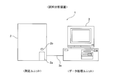
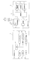
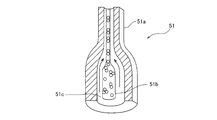
- FIG. 1 It is a front view which shows schematic structure of the blood analyzer by one Embodiment of this invention. It is a block diagram which shows the structure of the blood analyzer by one Embodiment shown in FIG. It is a perspective view which shows the measurement unit of the blood analyzer by one Embodiment shown in FIG. It is a perspective view which shows the internal structure of the measurement unit of the blood analyzer by one Embodiment shown in FIG. It is a side view which shows the internal structure of the measurement unit of the blood analyzer by one Embodiment shown in FIG. It is a perspective view which shows typically the structure of the flow cell provided in the measurement unit of the blood analyzer by one Embodiment shown in FIG.
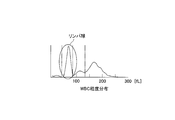
- FIG. 2 is a particle size distribution diagram of white blood cells created in the blood analyzer according to the embodiment shown in FIG. 1.
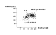
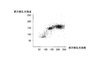
- 2 is a scattergram for white blood cell classification created in the blood analyzer according to the embodiment shown in FIG. 1.
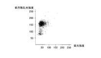
- 2 is a scattergram for white blood cell classification created in the blood analyzer according to the embodiment shown in FIG. 1.
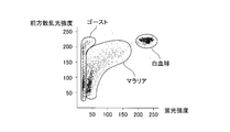
- 2 is a scattergram for classifying malaria created in the blood analyzer according to the embodiment shown in FIG. 1.
- FIG. 4 is a diagram showing experimental results when a hemolytic agent having a concentration of 2.15 mM in a WBC (for classification) measurement sample of a cationic surfactant is used in the blood analyzer according to the embodiment shown in FIG. 1. .
- FIG. 4 is a diagram showing experimental results when a hemolytic agent having a concentration of 2.15 mM in a WBC (for classification) measurement sample of a cationic surfactant is used in the blood analyzer according to the embodiment shown in FIG. 1.
- FIG. 3 is a diagram showing experimental results when a hemolytic agent having a concentration of 0.62 mM in a WBC (for classification) measurement sample of a cationic surfactant is used in the blood analyzer according to the embodiment shown in FIG. 1. .
- FIG. 4 is a diagram showing experimental results when a hemolytic agent having a concentration of 0.62 mM in a WBC (for classification) measurement sample of a cationic surfactant is used in the blood analyzer according to the embodiment shown
- FIG. 3 is a diagram showing experimental results when a hemolytic agent having a concentration of 0.62 mM in a WBC (for classification) measurement sample of a cationic surfactant is used in the blood analyzer according to the embodiment shown in FIG. 1. .
- FIG. 11 is a diagram showing an experimental result when the hemolytic agent shown in FIG. 10 is used in the blood analyzer according to the embodiment shown in FIG. 1.
- FIG. 11 is a diagram showing an experimental result when the hemolytic agent shown in FIG. 10 is used in the blood analyzer according to the embodiment shown in FIG. 1.
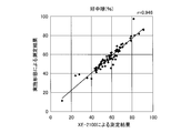
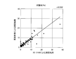
- FIG. 5 is a correlation diagram for a group of lymphocytes and basophils showing a correlation with measurement results obtained by an analyzer XE-2100 (Sysmex Corporation).
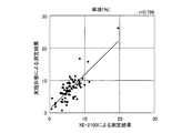
- FIG. 6 is a correlation diagram for monocytes showing a correlation with measurement results obtained by an analyzer XE-2100 (Sysmex Corporation).
- a multi-item automatic blood for the same specimen as the measurement result obtained by the method described in the embodiment described later (the reagent used is the same as the reagent described in the embodiment described later).
- FIG. 4 is a correlation diagram of neutrophils showing a correlation with measurement results obtained by an analyzer XE-2100 (Sysmex Corporation).
- FIG. 5 is a correlation diagram for eosinophils showing a correlation with measurement results obtained by an analyzer XE-2100 (Sysmex Corporation).
- the blood analyzer 1 is an apparatus used for blood tests, and is mainly composed of a measurement unit 2 and a data processing unit 3.
- the blood analyzer 1 is installed in a medical institution such as a hospital or a pathological examination facility, for example.
- the measurement unit 2 performs a predetermined measurement on the components contained in the blood sample
- the data processing unit 3 receives the measurement data to perform analysis processing.
- the measurement unit 2 and the data processing unit 3 are connected by a data transmission cable 3a so that data communication can be performed.
- the measurement unit 2 and the data processing unit 3 may be directly connected by a data transmission cable 3a.
- the measurement unit 2 and the data processing unit 3 may be connected via a dedicated line using a telephone line, a communication network such as a LAN or the Internet. It may be connected.
- the measurement unit 2 includes a sample supply unit 4, a WBC classification measurement unit 5, a DC measurement unit 6, an HGB measurement unit 7, a control unit 8, and a communication unit 9. .
- a blood collection tube setting portion 2 a configured to set a blood collection tube 20 containing a blood sample is provided in the lower right part of the front of the measurement unit 2.
- the blood collection tube setting unit 2a is configured to be pushed forward by the user pressing a button switch 2b provided in the vicinity thereof. The user can set the blood collection tube 20 in a state in which the blood collection tube setting unit 2a protrudes. Then, after setting the blood collection tube 20, the blood collection tube setting unit 2 a is configured to be returned to the inside of the measurement unit 2 when the user presses the button switch 2 b again.
- the measurement unit 2 includes a pipette 21 for aspirating the measurement sample, and chambers 22 and 23 (see FIG. 5) for mixing and preparing the blood sample and the reagent.
- the pipette 21 is formed in a tubular shape extending in the vertical direction, and its tip is sharply pointed.
- the pipette 21 is connected to a syringe pump (not shown), and is configured to suck and discharge a predetermined amount of liquid by the operation of the syringe pump.
- the pipette 21 is connected to a moving mechanism, and is configured to be movable in the vertical direction and the front-back direction.
- the pipette 21 is configured to suck a blood sample contained in the blood collection tube 20 by puncturing a sharp tip into a rubber cap 20 a that seals the blood collection tube 20.
- the pipette 21 is configured to suck the blood sample and then move to a predetermined position by the moving mechanism to supply the blood sample into the chambers 22 and 23.
- the sample supply unit 4 is a fluid unit having chambers 22 and 23, a plurality of solenoid valves, a diaphragm pump, and the like.
- the chamber 22 is provided for preparing a measurement sample used for measurement of red blood cells, platelets, and hemoglobin concentration.
- the chamber 23 is provided for preparing a measurement sample used for white blood cell measurement.
- a reagent container is connected to the fluid unit constituted by the sample supply unit 4. Specifically, the diluent container 24 for containing the diluent, the hemolyzer container 25 for containing the hemolyzing agent 100, and the staining liquid container 26 for containing the staining liquid used for the measurement sample for malaria detection. Is connected to the fluid unit. Accordingly, the diluent and hemolytic agent 100 can be supplied to the chamber 22, and the diluent, hemolytic agent 100 and staining solution can be supplied to the chamber 23.
- the WBC classification measurement unit 5 is an optical flow cytometer, and is provided for performing white blood cell classification detection and malaria detection by a flow cytometry method using semiconductor laser light.
- the WBC classification measurement unit 5 includes a flow cell 51 (see FIG. 6) that forms a liquid flow of the measurement sample.
- the flow cell 51 is formed in a tubular shape by a material such as translucent quartz, glass, and synthetic resin, and the inside thereof is a flow path through which a measurement sample and a sheath liquid (diluent) flow.
- the flow cell 51 is provided with an orifice 51a whose inner space is narrowed narrower than other portions.
- the vicinity of the inlet of the orifice 51a has a double tube structure, and the inner tube portion is a sample nozzle 51b.
- the space outside the sample nozzle 51b is a flow path 51c through which the sheath liquid (diluent) flows, and the sheath liquid (diluent) flows through the flow path 51c and is introduced into the orifice 51a.
- the sheath liquid (dilution liquid) supplied to the flow cell 51 flows so as to surround the measurement sample discharged from the sample nozzle 51b.
- the flow of the measurement sample is narrowed down by the orifice 51a, and the white blood cells and red blood cells contained in the measurement sample are surrounded by the sheath liquid (diluent) and pass through the orifice 51a one by one.
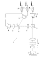
- a semiconductor laser light source 52 is arranged so as to emit laser light toward the orifice 51a of the flow cell 51.
- the semiconductor laser light source 52 has a blue-violet semiconductor laser element 52a and is configured to emit blue-violet laser light having a wavelength of about 405 nm.
- An irradiation lens system 53 including a plurality of lenses is disposed between the semiconductor laser light source 52 and the flow cell 51.
- a beam stopper 54a is provided on the optical axis linearly extending from the semiconductor laser light source 52 so as to face the irradiation lens system 53 with the flow cell 51 interposed therebetween.
- the beam stopper 54a is a semiconductor laser light source. The direct light from 52 is shielded.
- a photodiode 54 is disposed further downstream of the beam stopper 54a on the optical axis.
- the photodiode 54 is configured to receive the scattered light of the laser light generated by the measurement sample flowing through the flow cell 51. Specifically, among the light traveling along the optical axis extending linearly from the semiconductor laser light source 52, the direct light of the semiconductor laser light source 52 is blocked by the beam stopper 54a. Only scattered light (hereinafter referred to as forward scattered light) traveling along the axial direction is received.
- the photodiode 54 is configured to photoelectrically convert forward scattered light emitted from the flow cell 51 and transmit an electric signal (hereinafter referred to as forward scattered light signal) generated thereby to the amplifier 54b.
- the amplifier 54 b is configured to amplify the transmitted forward scattered light signal and output it to the control unit 8.
- a side condenser lens 55 is disposed on the side of the flow cell 51 and in a direction orthogonal to the optical axis extending linearly from the semiconductor laser light source 52 to the photodiode 54. Is configured to collect side light (light emitted in a direction intersecting the optical axis) generated when a blood cell passing through the flow cell 51 is irradiated with laser light.
- a dichroic mirror 56 is provided on the downstream side of the side condenser lens 55, and the dichroic mirror 56 is configured to divide the signal light transmitted from the side condenser lens 55 into a scattered light component and a fluorescent component.
- a photodiode 57 for receiving side scattered light is provided, and light from the dichroic mirror 56 is provided.
- An optical filter 58a and an avalanche photodiode 58 are provided on the downstream side of the shaft.
- the photodiode 57 is configured to photoelectrically convert the side scattered light component divided by the dichroic mirror 56 and transmit an electric signal (hereinafter referred to as a side scattered light signal) generated thereby to the amplifier 57a.
- the amplifier 57 a is configured to amplify the transmitted side scattered light signal and output the amplified side scattered light signal to the control unit 8.
- the avalanche photodiode 58 is configured to photoelectrically convert the side fluorescent component after wavelength selection by the optical filter 58a, and to transmit an electric signal (side fluorescent signal) generated thereby to the amplifier 58b. Yes.
- the amplifier 58b is configured to amplify the transmitted side fluorescent signal and output it to the control unit 8.
- the DC measurement unit 6 is configured to be able to measure the red blood cell count (RBC) and the platelet count (PLT) by the sheath flow DC detection method. Further, the DC measurement unit 6 is configured to be able to obtain measurement data for calculating a hematocrit value (HCT) by the red blood cell pulse peak value detection method. Furthermore, the DC measurement unit 6 is also used for white blood cell count (WBC) detection for calculating the lymphocyte ratio. In this way, the DC measuring unit 6 is used to measure the number of red blood cells and platelets, to acquire measurement data for calculating the hematocrit value (HCT), and to detect the number of white blood cells (WBC) for calculating the lymphocyte ratio.
- RBC red blood cell count
- PHT platelet count
- WBC white blood cell count
- the DC measurement unit 6 has a flow cell, and a measurement sample is transferred from the chamber 22 to the flow cell.
- a measurement sample prepared by mixing a blood sample and a diluent in the chamber 22 together with a sheath solution (diluent) 4 to the flow cell.
- a liquid flow is formed in a state where the measurement sample is surrounded by the sheath liquid (diluent).
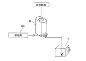
- the HGB measuring unit 7 is configured to measure the amount of hemoglobin (HGB) by the methemoglobin method. As shown in FIG. 9, the HGB measurement unit 7 has a cell for storing a diluted sample, and the measurement sample is transferred from the chamber 22 to this cell. And the HGB measurement part 7 has a light emitting diode which irradiates the light whose wavelength is about 555 nm, and measures the light absorbency by irradiating the light from a light emitting diode to the measurement sample in the said cell. It is configured.
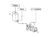
- a measurement sample is prepared by mixing the blood sample, the diluent, and the hemolytic agent 100 in the chamber 22.
- the control unit 8 includes a CPU, a ROM, a RAM, and the like, and is configured to perform operation control of each unit of the measurement unit 2.
- the communication unit 9 is, for example, an RS-232C interface, a USB interface, or an Ethernet (registered trademark) interface, and is configured to be able to transmit / receive data to / from the data processing unit 3.
- the data processing unit 3 includes a CPU 31, a ROM 32, a RAM 33, a hard disk 34, a communication interface 35, an input unit 36 such as a keyboard and a mouse, and a display device 37.
- the hard disk 34 of the data processing unit 3 is installed with an operating system and an application program for analyzing the measurement data received from the measurement unit 2.
- the CPU 31 of the data processing unit 3 executes the application program to analyze the measurement data, and the white blood cell count (WBC), red blood cell count (RBC), hemoglobin amount (HGB), Hematocrit value (HCT), average erythrocyte volume (MCV), average erythrocyte hemoglobin amount (MCH), average erythrocyte hemoglobin concentration (MCHC), and platelet count (PLT) are calculated. Further, the CPU 31 creates a scattergram using the forward scattered light signal, the side scattered light signal, and the side fluorescent signal, and converts the white blood cells into neutrophils (Neut), lymphocytes, monocytes (Mono), and eosinophils. It is configured to be classified into five types: sphere (EO) and basophil (BASO).
- EO sphere
- BASO basophil
- the communication interface 35 is, for example, an RS-232C interface, a USB interface, or an Ethernet (registered trademark) interface, and is configured to be able to transmit and receive data to and from the measurement unit 2.
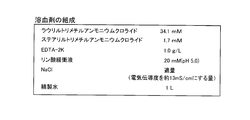
- the hemolytic agent 100 in this embodiment includes a cationic surfactant (lauryltrimethylammonium chloride; 34.1 mM, stearyltrimethylammonium chloride; 1.7 mM), and a labeling substance.
- the labeling substance is a substance that is selectively given to a cell to be measured and becomes a label for measurement, and corresponds to, for example, a fluorescent dye that stains the nucleic acid of the cell to be measured.
- the hemolytic agent 100 has a property of converting hemoglobin in blood into methemoglobin.
- each measurement sample used for each measurement has a different dilution ratio of the hemolytic agent 100 and a dilution ratio of the blood sample.
- a cationic surfactant in this way, the white blood cells in the measurement sample are classified into four by changing the dilution factor without using two or more hemolytic agents having different compositions, and in the measurement sample. It is possible to obtain the hemoglobin concentration.
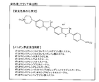
- the staining solution contains a fluorescent dye having a chemical formula structure shown in FIG. 11 (for example, Invitrogen's Hoechst 34580) and one of the nonionic surfactant groups shown in FIG. This fluorescent dye can be excited by blue-violet laser light (having a wavelength of about 405 nm) emitted from the semiconductor laser light source 52a.
- step S1 when the blood analyzer 1 is activated, after initialization of an application program and the like, it is determined in step S1 whether or not there is a measurement start instruction from the user by the CPU 31 of the data processing unit 3, This determination is repeated until instructed. If there is a measurement start instruction, a measurement start instruction signal is transmitted from the data processing unit 3 to the measurement unit 2 in step S2.
- step S21 the control unit 8 of the measurement unit 2 determines whether a measurement start instruction signal is received, and this determination is repeated until it is received.
- step S22 a blood sample is aspirated from the blood collection tube 20 set in the blood collection tube setting section 2a by the pipette 21.
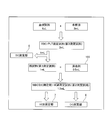
- step S23 the sample supply unit 4 prepares an RBC / PLT measurement sample (hereinafter referred to as a third measurement sample). Specifically, as shown in FIG. 13, a predetermined amount (for example, 2.0 mL) of diluent from the diluent container 24 and a predetermined amount (for example, 6 ⁇ L) sucked from the blood collection tube 20 by the pipette 21. A blood sample is supplied to the chamber 22 and agitated. Thereby, a predetermined amount (for example, 2.0 mL) of the third measurement sample is prepared.
- a predetermined amount for example, 2.0 mL
- step S24 a part of the third measurement sample (for example, 1 mL) in the chamber 22 is transferred to the DC measurement unit 6 together with the sheath liquid (diluent), and the third measurement is performed by the DC measurement unit 6. RBC and PLT detection of the sample is performed.
- the sample supply unit 4 prepares a WBC (for DC detection) / HGB measurement sample (hereinafter referred to as a second measurement sample). Specifically, as shown in FIG. 13, a predetermined amount (for example, 0.5 mL) of the hemolytic agent 100 is supplied from the hemolytic agent container 25 to the chamber 22 in which a predetermined amount (for example, 1 mL) of the third measurement sample remains. Supplied and stirred. That is, after the blood sample and the diluent are mixed in the chamber 22, the hemolytic agent 100 is mixed to prepare the second measurement sample.
- a predetermined amount for example, 0.5 mL
- a predetermined amount for example, 1 mL
- the hemolytic agent is mixed with the blood sample in a state diluted with a diluent, it is possible to prevent the blood sample from being mixed with a hemolytic agent having a concentration higher than a desired concentration.
- red blood cells are hemolyzed and hemoglobin is converted into methemoglobin.
- the red blood cell in the measurement sample is reliably obtained by making the dilution rate (3 times) of the hemolytic agent 100 in the second measurement sample smaller than the dilution rate (25 times) of the hemolysis agent 100 in the first measurement sample described later. Therefore, hemoglobin concentration can be obtained with high accuracy.
- step S26 the second measurement sample in the chamber 22 is transferred to the DC measurement unit 6, and the WBC measurement of the second measurement sample is performed.
- step S27 the second measurement sample is transferred to the HGB measurement unit 7, and HGB detection of the second measurement sample is performed.
- a predetermined amount for example, 1 mL
- a predetermined amount for example, 10 ⁇ L
- a predetermined amount for example, 10 ⁇ L
- staining solution from the staining solution container 26 are supplied to the chamber 23 and stirred.
- step S31 the fourth measurement sample in the chamber 23 is transferred to the WBC classification measurement unit 5 together with the sheath liquid (diluent), and the WBC classification measurement unit 5 detects malaria of the fourth measurement sample. Is called.
- step S ⁇ b> 32 the measurement data measured in each detection unit is transmitted from the measurement unit 2 to the data processing unit 3.
- step S3 it is determined whether or not the measurement data transmitted by the measurement unit 2 has been received, and this determination is repeated until it is received.
- the CPU 31 calculates the white blood cell count (WBC) based on the measurement data obtained by WBC detection measured in step S26.
- step S5 the CPU 31 creates a leukocyte particle size distribution map based on the measurement data obtained by WBC detection, and calculates the lymphocyte ratio relative to the white blood cell count (WBC), as shown in FIG.
- the lymphocytes appear as the first mountain (group) from the left in the particle size distribution diagram.
- the leukocytes are a group of lymphocytes and basophils, monocytes and granulocytes (neutrophils and eosinophils). (Group with sphere).
- the CPU 31 creates a scattergram as shown in FIG. 15 using the forward scattered light signal and the side scattered light signal, and from this scattergram, a group of lymphocytes and basophils, The ratio of monocytes and granulocytes (neutrophil and eosinophil population) to each white blood cell count (WBC) is calculated.
- WBC white blood cell count
- leukocytes are classified into 3 groups of lymphocytes and basophils, monocytes and granulocytes (a group of neutrophils and eosinophils) on the scattergram. It can be seen that it can be classified into two types.
- step S7 the CPU 31 classifies the leukocytes into two groups, eosinophils and a group other than eosinophils, based on the measurement data obtained by WBC classification detection. Specifically, the CPU 31 creates a scattergram using the forward scattered light signal and the side fluorescence signal, as shown in FIG. 16, and from this scattergram, the eosinophil ratio to the white blood cell count (WBC) is determined. calculate.
- This side fluorescent signal is based on autofluorescence of leukocytes excited by blue-violet laser light (wavelength of about 405 nm) emitted from the semiconductor laser light source 52. Eosinophils are other than leukocyte eosinophils.
- the CPU 31 can also calculate the ratio of eosinophils to the white blood cell count (WBC) from the scattergram based on the side scattered light signal and the side fluorescence signal.
- WBC white blood cell count
- FIG. 23 a blood sample actually collected from a subject is measured using the hemolytic agent 100 (see FIG. 10) in the present embodiment, and the forward scattered light signal and the side fluorescence signal obtained as a result are measured. Shows the scattergram by. As shown in FIG. 23, it can be seen that the actual measurement results can be classified into eosinophils and groups other than eosinophils on the scattergram.
- step S8 the CPU 31 subtracts the eosinophil ratio calculated in step S7 from the granulocyte (the group of neutrophils and eosinophils) ratio calculated in step S6, thereby obtaining the white blood cell count ( The neutrophil ratio relative to (WBC) is calculated.
- leukocytes are classified into four groups: lymphocytes and basophils, monocytes, neutrophils, and eosinophils.
- step S9 the CPU 31 calculates the basophil ratio relative to the white blood cell count (WBC) by subtracting the lymphocyte ratio calculated in step S5 from the ratio of the population of lymphocytes and basophils. .
- leukocytes are classified into five types: lymphocytes, basophils, monocytes, neutrophils and eosinophils.
- step S10 the CPU 31 classifies plasmodium-infected erythrocytes from a group other than the plasmodium-infected erythrocytes based on the measurement data obtained by malaria detection measured in step S31. Specifically, the CPU 31 creates a scattergram using the forward scattered light signal and the side fluorescence signal, as shown in FIG. 17, and from this scattergram, malaria parasite-infected erythrocytes other than malaria parasite-infected erythrocytes are produced. Classify from the population. This makes it possible to determine the presence or absence of malaria infection.
- step S11 the CPU 31 calculates the red blood cell count (RBC), platelet count (PLT), and hematocrit value (HCT) based on the measurement data by RBC / PLT detection measured in step S24.
- RBC red blood cell count
- PLT platelet count
- HCT hematocrit value
- step S12 the CPU 31 calculates the amount of hemoglobin (HGB) based on the measurement data obtained by the HGB detection measured in step S27. That is, the hemoglobin concentration is calculated based on the absorbance obtained by the HGB detection using the SLS hemoglobin method.
- HGB hemoglobin
- step S13 from the red blood cell count (RBC), hematocrit value (HCT) and hemoglobin amount (HGB) calculated as described above, the CPU 31 causes the average red blood cell volume (MCV), average red blood cell hemoglobin amount (MCH) and Average erythrocyte hemoglobin concentration (MCHC) is calculated.
- MCV average red blood cell volume
- MCH average red blood cell hemoglobin amount
- MCHC Average erythrocyte hemoglobin concentration
- MCV (HCT / RBC) ⁇ 1000 (1)
- MCV represents the mean red blood cell volume (fL)
- HCT represents the hematocrit value (%)
- RBC represents the number of red blood cells ( ⁇ 10 4 / ⁇ L).
- MCH (HGB / RBC) ⁇ 1000 (2)
- MCH represents the average amount of red blood cell pigment (pg)
- HGB represents the amount of hemoglobin (g / dL)
- RBC represents the number of red blood cells ( ⁇ 10 4 / ⁇ L).
- MCHC (HGB / HCT) ⁇ 100 (3)
- MCHC represents the average erythrocyte hemoglobin concentration (g / dL)
- HGB represents the hemoglobin amount (g / dL)
- HCT represents the hematocrit value (%).
- step S14 the white blood cell count (WBC), red blood cell count (RBC), hemoglobin amount (HGB), hematocrit value (HCT), average red blood cell volume (MCV), average red blood cell hemoglobin amount (calculated as described above) MCH), average erythrocyte hemoglobin concentration (MCHC), and platelet count (PLT) are output to the display device 37. Further, the neutrophil ratio, lymphocyte ratio, monocyte ratio, eosinophil ratio, and basophil ratio relative to the white blood cell count (WBC) are output to the display device 37, and the result of malaria detection is also output.
- the neutrophil count, lymphocyte count, monocyte count, eosinophil count and basophil count calculated based on the white blood cell count (WBC) and each blood cell ratio The number of balls is output.
- step S15 it is determined whether or not there is a shutdown instruction from the user. If there is no instruction, the process proceeds to step S1. When there is a shutdown instruction, the operation of the data processing unit 3 for the sample analysis process in the blood analyzer 1 is terminated. On the measurement unit 2 side, after transmitting the measurement data to the data processing unit 3 in step S32, it is determined in step S33 whether or not there has been a shutdown instruction from the user. If there is no instruction, the process proceeds to step S21. To be migrated. If there is a shutdown instruction, the operation of the measurement unit 2 of the sample analysis process in the blood analyzer 1 is ended.
- the WBC classification that generates the side fluorescence signal, the forward scattered light signal, and the side scattered light signal from the measurement sample prepared from the blood sample and the hemolytic agent 100 that does not include the labeling substance By providing the measurement unit 5, the side fluorescence signal, the forward scattered light signal, and the side scattered light signal are obtained using the autofluorescence of eosinophils in the measurement sample without labeling the measurement sample. Obtainable. Further, based on the side fluorescence signal, the forward scattered light signal, and the side scattered light signal, the leukocytes in the measurement sample are converted into monocytes, neutrophils, eosinophils, and a group of lymphocytes and basophils.
- the hemolytic agent 100 Since the white blood cells can be classified into four without using a labeling substance by providing the CPU 31 for classifying into four, the hemolytic agent 100 does not need to include the labeling substance. Thereby, the leukocytes in the measurement sample can be classified into four using the hemolytic agent 100 having a simple composition not containing the labeling substance.
- a DC measurement unit 6 that performs blood cell measurement by the sheath flow DC detection method is provided, and the CPU 31 determines that the white blood cells in the measurement sample other than lymphocytes and lymphocytes based on the measurement data obtained by the DC measurement unit 6
- White blood cells are classified into lymphocytes, basophils, monocytes, and neutrophils based on the classification results and the classification results based on the side fluorescence signal, the forward scattered light signal, and the side scattered light signal.
- the step of generating the side fluorescence signal, the forward scattered light signal, and the side scattered light signal from the measurement sample not containing the labeling substance is provided.
- a side fluorescence signal, a forward scattered light signal, and a side scattered light signal can be obtained using autofluorescence of eosinophils in a measurement sample without labeling the sample. Further, based on the side fluorescence signal, the forward scattered light signal, and the side scattered light signal, the leukocytes in the measurement sample are converted into monocytes, neutrophils, eosinophils, and a group of lymphocytes and basophils.
- the white blood cells can be classified into four without using a labeling substance, so that it is not necessary to include the labeling substance in the hemolytic agent 100.
- the leukocytes in the measurement sample can be classified into four using the hemolytic agent 100 having a simple composition not containing the labeling substance.
- the blood analysis method using the hemolytic agent 100 is provided with a step of generating a side fluorescence signal, a forward scattered light signal, and a side scattered light signal from a measurement sample that does not include a labeling substance.
- a side fluorescence signal, a forward scattered light signal, and a side scattered light signal can be obtained using autofluorescence of eosinophils in a measurement sample without labeling the sample.
- leukocytes in the measurement sample are converted into monocytes, neutrophils, eosinophils, based on the side fluorescence signal, the forward scattered light signal, and the side scattered light signal.
- the leukocytes in the measurement sample can be classified into four using the hemolytic agent 100 having a simple composition that does not contain the labeling substance.
- the hemolytic agent 100 containing no labeling substance is used to convert white blood cells into monocytes, neutrophils, and acid acids using side fluorescent signals, forward scattered light signals, and side scattered light signals. Since it is possible to classify leukocytes into four without using a labeling substance by using it to classify into four groups of spheres and lymphocytes and basophils, the labeling substance is added to the hemolytic agent 100. There is no need to include it. That is, the leukocytes in the measurement sample can be classified into four using the hemolytic agent 100 having a simple composition that does not contain the labeling substance.
- the WBC classification measurement unit 5 converts the side fluorescence signal, the forward scattered light signal, and the side scattered light signal generated from the first measurement sample including the blood sample and the hemolytic agent 100. Based on this, the white blood cells in the first measurement sample are classified into at least four types of monocytes, neutrophils, eosinophils, and other groups, and the blood sample and the hemolytic agent are analyzed by the HGB measurement unit 7.
- a hemolytic agent for classifying white blood cells into four by providing a CPU 31 that acquires the hemoglobin concentration in the second measurement sample based on the absorbance measured from the second measurement sample containing the same hemolytic agent 100 as 100
- hemolytic agent for obtaining hemoglobin concentration can be used in common, so two or more hemolytic agents (reagents) with different compositions are developed to obtain leukocyte classification and hemoglobin concentration acquisition. There is no need. Thereby, while reducing the burden on the user due to the development of the reagent, the white blood cells in the measurement sample can be classified into four, and the hemoglobin concentration in the measurement sample can be acquired.
- the white blood cells in the second measurement sample are classified into lymphocytes and a group other than lymphocytes based on the measurement data obtained by the DC measurement unit 6, and the classification result and the above-described white blood cells are classified.
- the white blood cells in the measurement sample are classified into at least five of lymphocytes, basophils, monocytes, neutrophils, and eosinophils.
- the DC measurement unit 6 can classify white blood cells into lymphocytes and a group other than lymphocytes using the same second measurement sample as the measurement sample used for acquiring the hemoglobin concentration. Therefore, white blood cells can be classified into five without separately preparing measurement samples having different compositions.
- the side fluorescent signal, the forward scattered light signal, and the side light generated from the first measurement sample including the blood sample and the hemolytic agent 100 by the WBC classification measurement unit 5 are used.
- the step of classifying the white blood cells in the first measurement sample into at least four of monocytes, neutrophils, eosinophils, and other populations, and the HGB measurement unit 7 Obtaining a hemoglobin concentration in the second measurement sample based on the absorbance measured from the blood sample and the second measurement sample containing the same hemolytic agent 100 as the hemolytic agent 100, thereby providing four leukocytes.
- the hemolytic agent for classification and the hemolytic agent for obtaining hemoglobin concentration can be shared, two types having different compositions for performing white blood cell classification and hemoglobin concentration acquisition There is no need to be developed on the hemolytic agent (reagent). Thereby, while reducing the burden on the user due to the development of the reagent, the white blood cells in the measurement sample can be classified into four, and the hemoglobin concentration in the measurement sample can be acquired.
- the hemolytic agent according to the present embodiment is converted from the side fluorescence signal, the forward scattered light signal, and the side scattering generated from the WBC classification measurement unit 5 from the first measurement sample including the blood sample and the hemolytic agent 100.
- the step of classifying the white blood cells in the first measurement sample into at least four of monocytes, neutrophils, eosinophils, and other populations, and blood by the HGB measurement unit 7 Obtaining a hemoglobin concentration in the second measurement sample based on the absorbance measured from the sample and the second measurement sample containing the same hemolyzing agent 100 as the hemolytic agent 100, thereby obtaining leukocytes Can be used in common with the hemolytic agent for classifying the blood cells into four and the hemolytic agent for obtaining the hemoglobin concentration.
- the white blood cells in the measurement sample can be classified into four, and the hemoglobin concentration in the measurement sample can be acquired.
- the hemolytic agent according to the present embodiment contains a cationic surfactant and is used for measuring hemoglobin concentration, and at least leukocytes, basophils, monocytes, By using it to classify into neutrophils and eosinophils, it is possible to share a hemolytic agent for classifying leukocytes into four and a hemolytic agent for obtaining hemoglobin concentration. There is no need to develop two or more hemolytic agents (reagents) with different compositions in order to perform classification and hemoglobin concentration acquisition. Thereby, while reducing the burden on the user due to the development of the reagent, the white blood cells in the measurement sample can be classified into four, and the hemoglobin concentration in the measurement sample can be acquired.
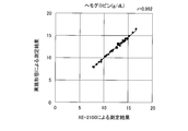
- FIG. 24 to FIG. 28 show correlation diagrams of measurement results of hemoglobin, a population of lymphocytes and basophils, monocytes, neutrophils, and eosinophils, respectively.
- the measurement results obtained by the method described in the above embodiment are shown on the vertical axis, and the measurement results obtained by the multi-item automatic blood cell analyzer XE-2100 are shown on the horizontal axis.
- the correlation coefficient r is 0.992 for hemoglobin, 0.968 for the population of lymphocytes and basophils, 0.796 for monocytes, and 0 for neutrophils. .946 and 0.892 for eosinophils, both showing high correlation.
- the hemolytic agent used for WBC detection, HGB detection, WBC classification detection, and malaria detection is accommodated in one hemolytic agent container, but the invention according to the first to fourth aspects
- the present invention is not limited to this, and a separate hemolytic agent may be prepared for each detection, and each hemolytic agent may be accommodated in a separate hemolytic agent container.
- the hemolytic agent used for each detection may be a different type of hemolytic agent.
- the hemolytic agent accommodated in each hemolytic agent container may be diluted in advance at a predetermined dilution rate.
- the semiconductor laser light source which has a blue-violet semiconductor laser element was shown as an example of a light source, this invention is not restricted to this, Blue-violet semiconductor laser elements, such as a blue semiconductor laser element or an argon laser element, A light source having a laser element other than the above may be used.
- a semiconductor laser light source that emits blue-violet laser light having a wavelength of about 405 nm is shown as an example of the light source.
- the invention according to the first to fourth aspects is not limited to this, and is 350 nm to 500 nm. Any light source other than the semiconductor laser light source may be used as long as it emits light having the following wavelengths. If comprised in this way, the light of a short wavelength range which can acquire the fluorescence information based on the autofluorescence of an eosinophil can be irradiated to a measurement sample.
- each detection process is performed in the order of RBC / PLT detection, WBC detection, HGB detection, WBC classification detection, and malaria detection from the earliest in the sample analysis process.
- each detection process may be performed in a sequence other than the above.
- the order of the white blood cell classification process, the malaria classification process, the red blood cell count / platelet count calculation process, and the hemoglobin amount calculation process in the sample analysis process can be appropriately changed.
- category detection, and malaria detection to the sample supply part was shown.
- the invention according to the fifth to eighth aspects is not limited to this, and four hemolytic agent containers may be connected to the sample supply unit so as to accommodate the hemolytic agents used for each detection separately.
- the hemolytic agent used for any of the two detections may be accommodated in a common hemolytic agent container, and two or three hemolytic agent containers may be connected to the sample supply unit. Further, five or more hemolytic agent containers may be connected to the sample supply unit.
- the hemolytic agent accommodated in each hemolytic agent container is previously diluted to a predetermined dilution rate, the hemolytic agent is diluted to a desired dilution rate when preparing a measurement sample used for each detection. There is no need to provide a separate process.
- a hemolytic agent that does not contain a labeling substance is shown as an example of a hemolytic agent.
- the invention according to the fifth to eighth aspects is not limited to this, and the hemolytic agent may contain a labeling substance. Good.
- the HGB measurement part which measures the light absorbency as transmitted light information was shown as an example of a 2nd light information generation part, this invention is not limited to this, HGB measurement which produces
- the CPU of the data processing unit acquires the hemoglobin concentration based on the scattered light information generated by the HGB measurement unit.
- a hemolytic agent it is an alkyl trimethyl ammonium salt
- Cationic surfactant (lauryl trimethyl ammonium chloride; 34.1 mM, stearyl trimethyl whose alkyl group has 12 to 18 carbon atoms)
- a hemolytic agent containing ammonium chloride (1.7 mM) was shown, but the present invention is not limited to this, and a WBC (a total of lauryltrimethylammonium chloride and stearyltrimethylammonium chloride in the above embodiment) is not limited thereto.
- the concentration in the measurement sample is 0.62 mM or more and 2.15 mM or less, it may be a hemolytic agent containing a cationic surfactant having a concentration other than the above.
- the concentration of the active agent in the hemolytic agent is 15.5 mM, and the concentration of the cationic surfactant in the hemolytic agent when the concentration of the cationic surfactant in the WBC (classification) measurement sample is 2.15 mM. 75 mM.
- the concentration of the cationic surfactant in the WBC (for classification) measurement sample is 2.15 mM. Even above, it can be measured.
- the concentration of the cationic surfactant in the hemolytic agent is varied in the blood analyzer according to one embodiment of the present invention.
- a plurality of experimental results were obtained in which the concentration of the cationic surfactant in the hemolytic agent varies by a small amount.
- the concentration of the cationic surfactant in the WBC (for classification) measurement sample is 2
- Two experimental results in the case of using a hemolytic agent having a concentration of 15 mM and in the case of using a hemolytic agent having a concentration of 0.62 mM in the WBC (for classification) measurement sample of the cationic surfactant will be described.
- leukocytes are classified into three groups: a lymphocyte and basophil group, a monocyte, and a granulocyte (neutrophil and eosinophil group). It is possible. Further, as shown in FIGS. 19 and 21, it is possible to classify into eosinophils and groups other than eosinophils on the scattergram. From these classification results, it is possible to classify leukocytes into four groups: a group of lymphocytes and basophils, monocytes, neutrophils and eosinophils. Therefore, it is considered that leukocytes can be classified into four categories when the concentration of the cationic surfactant in the WBC (for classification) measurement sample is in the range of 0.62 mM to 2.15 mM.
Landscapes
- Health & Medical Sciences (AREA)
- Life Sciences & Earth Sciences (AREA)
- Engineering & Computer Science (AREA)
- Chemical & Material Sciences (AREA)
- Physics & Mathematics (AREA)
- Biomedical Technology (AREA)
- Immunology (AREA)
- General Health & Medical Sciences (AREA)
- General Physics & Mathematics (AREA)
- Pathology (AREA)
- Biochemistry (AREA)
- Analytical Chemistry (AREA)
- Hematology (AREA)
- Molecular Biology (AREA)
- Ecology (AREA)
- Nuclear Medicine, Radiotherapy & Molecular Imaging (AREA)
- Biophysics (AREA)
- Urology & Nephrology (AREA)
- Food Science & Technology (AREA)
- Medicinal Chemistry (AREA)
- Optics & Photonics (AREA)
- Chemical Kinetics & Catalysis (AREA)
- Investigating Or Analysing Biological Materials (AREA)
- Investigating, Analyzing Materials By Fluorescence Or Luminescence (AREA)
- Investigating Or Analysing Materials By Optical Means (AREA)
Abstract
Description
上記式(1)において、MCVは平均赤血球容積(fL)、HCTはヘマトクリット値(%)、RBCは赤血球数(×104/μL)をそれぞれ表す。
上記式(2)において、MCHは平均赤血球血色素量(pg)、HGBは血色素量(g/dL)、RBCは赤血球数(×104/μL)をそれぞれ表す。
上記式(3)において、MCHCは平均赤血球血色素濃度(g/dL)、HGBは血色素量(g/dL)、HCTはヘマトクリット値(%)をそれぞれ表す。
次に、図24~図28を参照して、上記実施形態に記載の方法により得られた測定結果と、多項目自動血液分析装置XE-2100(シスメックス株式会社)により得られた測定結果との相関についての検証結果について説明する。上記実施形態に記載の方法による測定と多項目自動血液分析装置XE-2100による測定とは、複数の血液試料について、同じ検体について行った。なお、図24~図28は、それぞれ、ヘモグロビン、リンパ球と好塩基球との集団、単球、好中球、および好酸球の測定結果の相関図を示している。また、図24~図28では、それぞれ、縦軸に上記実施形態に記載の方法により得られた測定結果を、横軸に多項目自動血球分析装置XE-2100により得られた測定結果を示している。これらの図に示すように、相関係数rは、ヘモグロビンについては0.992、リンパ球と好塩基球との集団については0.968、単球については0.796、好中球については0.946、および、好酸球については0.892であり、いずれも高い相関を示す結果となった。
Claims (27)
- 血液試料と標識物質を含まない溶血剤とから、標識物質を含まない測定試料を調製する試料調製部と、
前記試料調製部により調製された前記測定試料から、蛍光情報と、少なくとも2種類の散乱光情報とを生成する光情報生成部と、
前記蛍光情報と前記2種類の散乱光情報とに基づいて、前記測定試料中の白血球を、少なくとも、単球と、好中球と、好酸球と、他の集団との4つに第1分類する制御部とを備える、血液分析装置。 - 前記光情報生成部は、前記測定試料中の好酸球の自家蛍光に基づいて、前記蛍光情報を生成するように構成されている、請求項1に記載の血液分析装置。
- 前記試料調製部は、前記血液試料と所定の溶血剤とを含む第2測定試料を調製し、
前記血液分析装置は、前記第2測定試料から、試料の電気情報を生成する電気情報生成部をさらに備え、
前記制御部は、前記電気情報生成部により生成された電気情報に基づいて、前記第2測定試料中の白血球を少なくともリンパ球とリンパ球以外の集団とに第2分類するとともに、前記第1分類および前記第2分類の分類結果に基づいて、前記測定試料中の白血球を、少なくとも、リンパ球と、好塩基球と、単球と、好中球と、好酸球との5つに分類するように構成されている、請求項1または2に記載の血液分析装置。 - 前記光情報生成部は、光源と、前記光源から前記測定試料に光が照射されることにより生じる蛍光を受光し、受光強度に応じた前記蛍光情報を生成する蛍光受光部と、前記光源から前記測定試料に光が照射されることにより生じる散乱光を受光し、受光強度に応じた前記散乱光情報を生成する散乱光受光部とを含む、請求項1に記載の血液分析装置。
- 前記散乱光受光部は、前記測定試料に光が照射されることにより前記光源から照射された光の進行方向に沿って生じる前方散乱光を受光し、受光強度に応じた前方散乱光情報を、前記散乱光情報のうちの第1情報として生成する第1受光部と、前記測定試料に光が照射されることにより前記光源から照射された光の進行方向に略垂直な方向に沿って生じる側方散乱光を受光し、受光強度に応じた側方散乱光情報を、前記散乱光情報のうちの第2情報として生成する第2受光部とを有する、請求項4に記載の血液分析装置。
- 前記制御部は、前記前方散乱光情報と前記蛍光情報とに基づいて、前記測定試料中の白血球を、少なくとも好酸球と好酸球以外の集団とに分類するように構成されている、請求項5に記載の血液分析装置。
- 前記光源は、350nm以上500nm以下の波長を有する光を照射するように構成されている、請求項4~6のいずれか1項に記載の血液分析装置。
- 前記光源は、青紫色半導体レーザ素子を有する、請求項7に記載の血液分析装置。
- 血液試料と標識物質を含まない溶血剤とから、標識物質を含まない測定試料を調製するステップと、
調製された前記測定試料から、蛍光情報と、少なくとも2種類の散乱光情報とを生成するステップと、
前記蛍光情報と前記2種類の散乱光情報とに基づいて、前記測定試料中の白血球を、少なくとも、単球と、好中球と、好酸球と、他の集団との4つに分類するステップとを備える、血液分析方法。 - 血液試料と標識物質を含まない溶血剤とから、標識物質を含まない測定試料を調製するステップと、調製された前記測定試料から、蛍光情報と、少なくとも2種類の散乱光情報とを生成するステップと、前記蛍光情報と前記2種類の散乱光情報とに基づいて、前記測定試料中の白血球を、少なくとも、単球と、好中球と、好酸球と、他の集団との4つに分類するステップとを備える血液分析方法に用いられる前記溶血剤。
- カチオン性界面活性剤を含み、標識物質を含まないとともに、少なくとも2種類の散乱光情報と蛍光情報とを用いて白血球を少なくとも、単球と、好中球と、好酸球と、他の集団との4つに分類するために用いられる溶血剤。
- 血液試料と前記溶血剤と希釈液とを含む測定試料における前記カチオン性界面活性剤の濃度は、0.62mM以上2.15mM以下である、請求項11に記載の溶血剤。
- 前記カチオン性界面活性剤の前記溶血剤における濃度は、15.5mM以上53.75mM以下である、請求項11に記載の溶血剤。
- 血液試料および溶血剤を含む第1測定試料と、前記血液試料および前記溶血剤と同じ溶血剤を含む第2測定試料とを調製する試料調製部と、
前記第1測定試料から、蛍光情報と、少なくとも2種類の散乱光情報とを生成する第1光情報生成部と、
前記第2測定試料から、透過光情報または散乱光情報の少なくとも一方を生成する第2光情報生成部と、
前記第1光情報生成部により生成された前記蛍光情報と前記2種類の散乱光情報とに基づいて、前記第1測定試料中の白血球を、少なくとも、単球と、好中球と、好酸球と、他の集団との4つに第1分類し、前記第2光情報生成部により生成された前記透過光情報または前記散乱光情報の少なくとも一方に基づいて、前記第2測定試料中のヘモグロビン濃度を取得する制御部とを備える、血液分析装置。 - 前記第2測定試料から、試料の電気情報を生成する電気情報生成部をさらに備え、
前記制御部は、前記電気情報生成部により生成された電気情報に基づいて、前記第2測定試料中の白血球を少なくともリンパ球とリンパ球以外の集団とに第2分類するとともに、前記第1分類および前記第2分類の分類結果に基づいて、測定試料中の白血球を、少なくとも、リンパ球と、好塩基球と、単球と、好中球と、好酸球との5つに分類するように構成されている、請求項14に記載の血液分析装置。 - 前記試料調製部は、前記血液試料から第3測定試料をさらに調製し、
前記電気情報生成部は、前記第3測定試料から、試料の電気情報を生成し、
前記制御部は、前記電気情報生成部により前記第3測定試料から生成された電気情報に基づいて、前記第3測定試料中の赤血球数および血小板数を算出するように構成されている、請求項15に記載の血液分析装置。 - 前記第2測定試料における前記溶血剤の希釈倍率は、前記第1測定試料における前記溶血剤の希釈倍率と異なる、請求項14~16のいずれか1項に記載の血液分析装置。
- 前記第2測定試料における前記溶血剤の希釈倍率は、前記第1測定試料における前記溶血剤の希釈倍率よりも小さい、請求項17に記載の血液分析装置。
- 前記試料調製部は、前記血液試料、所定の試薬容器に収容された前記溶血剤、および、所定量の希釈液を混合することにより前記第1測定試料を調製し、前記血液試料、前記所定の試薬容器に収容された前記溶血剤、および、前記所定量よりも少ない量の前記希釈液とを混合することにより前記第2測定試料を調製するように構成されている、請求項14に記載の血液分析装置。
- 前記試料調製部は、前記希釈液と前記血液試料とを混合した状態で、前記溶血剤を混合することにより前記第2測定試料を調製するように構成されている、請求項19に記載の血液分析装置。
- 前記試料調製部は、少なくとも前記血液試料、および、前記第1測定試料に用いられる前記溶血剤が収容された第1試薬容器とは異なる第2試薬容器に収容された前記溶血剤を混合することにより前記第2測定試料を調製するように構成されている、請求項14に記載の血液分析装置。
- 前記第2試薬容器に収容された前記溶血剤は、略3倍に希釈されている、請求項21に記載の血液分析装置。
- 前記溶血剤は、カチオン性界面活性剤を含む、請求項14に記載の血液分析装置。
- 前記溶血剤は、標識物質を含まない、請求項14に記載の血液分析装置。
- 血液試料および溶血剤を含む第1測定試料と、前記血液試料および前記溶血剤と同じ溶血剤を含む第2測定試料とを調製するステップと、
前記第1測定試料から、蛍光情報と、少なくとも2種類の散乱光情報とを生成するステップと、
前記第2測定試料から、透過光情報または散乱光情報の少なくとも一方を生成するステップと、
前記第1測定試料から生成された前記蛍光情報と前記2種類の散乱光情報とに基づいて、前記第1測定試料中の白血球を、少なくとも、単球と、好中球と、好酸球と、他の集団との4つに分類するステップと、
前記第2測定試料から生成された前記透過光情報または前記散乱光情報の少なくとも一方に基づいて、前記第2測定試料中のヘモグロビン濃度を取得するステップとを備える、血液分析方法。 - 血液試料および溶血剤を含む第1測定試料と、前記血液試料および前記溶血剤と同じ溶血剤を含む第2測定試料とを調製するステップと、前記第1測定試料から、蛍光情報と、少なくとも2種類の散乱光情報とを生成するステップと、前記第2測定試料から、透過光情報または散乱光情報の少なくとも一方を生成するステップと、前記第1測定試料から生成された前記蛍光情報と前記2種類の散乱光情報とに基づいて、前記第1測定試料中の白血球を、少なくとも、単球と、好中球と、好酸球と、他の集団との4つに分類するステップと、前記第2測定試料から生成された前記透過光情報または前記散乱光情報の少なくとも一方に基づいて、前記第2測定試料中のヘモグロビン濃度を取得するステップとを備える血液分析方法に用いられる前記溶血剤。
- カチオン性界面活性剤を含み、ヘモグロビン濃度を取得するために用いられるとともに、白血球を少なくとも、リンパ球と、好塩基球と、単球と、好中球と、好酸球とに分類するために用いられる溶血剤。
Priority Applications (5)
| Application Number | Priority Date | Filing Date | Title |
|---|---|---|---|
| JP2010511052A JP5627457B2 (ja) | 2008-05-09 | 2009-04-28 | 血液分析方法 |
| CN200980116627.0A CN102016577B (zh) | 2008-05-09 | 2009-04-28 | 血液分析装置,血液分析方法及溶血剂 |
| EP20090742694 EP2280278A4 (en) | 2008-05-09 | 2009-04-28 | Blood analyzer, blood analysis method and hemolysis |
| US12/942,561 US8808624B2 (en) | 2008-05-09 | 2010-11-09 | Blood analyzer, blood analysis method and hemolytic agent |
| US14/325,526 US9891210B2 (en) | 2008-05-09 | 2014-07-08 | Blood analyzer, blood analysis method and hemolytic agent |
Applications Claiming Priority (4)
| Application Number | Priority Date | Filing Date | Title |
|---|---|---|---|
| JP2008-123347 | 2008-05-09 | ||
| JP2008123347 | 2008-05-09 | ||
| JP2008-123388 | 2008-05-09 | ||
| JP2008123388 | 2008-05-09 |
Related Child Applications (1)
| Application Number | Title | Priority Date | Filing Date |
|---|---|---|---|
| US12/942,561 Continuation US8808624B2 (en) | 2008-05-09 | 2010-11-09 | Blood analyzer, blood analysis method and hemolytic agent |
Publications (1)
| Publication Number | Publication Date |
|---|---|
| WO2009136570A1 true WO2009136570A1 (ja) | 2009-11-12 |
Family
ID=41264625
Family Applications (1)
| Application Number | Title | Priority Date | Filing Date |
|---|---|---|---|
| PCT/JP2009/058316 Ceased WO2009136570A1 (ja) | 2008-05-09 | 2009-04-28 | 血液分析装置、血液分析方法および溶血剤 |
Country Status (5)
| Country | Link |
|---|---|
| US (2) | US8808624B2 (ja) |
| EP (1) | EP2280278A4 (ja) |
| JP (3) | JP5627457B2 (ja) |
| CN (2) | CN102016577B (ja) |
| WO (1) | WO2009136570A1 (ja) |
Cited By (14)
| Publication number | Priority date | Publication date | Assignee | Title |
|---|---|---|---|---|
| CN103091286A (zh) * | 2011-10-31 | 2013-05-08 | 深圳迈瑞生物医疗电子股份有限公司 | 疟原虫感染的红细胞的识别方法及装置 |
| JP2014506323A (ja) * | 2010-12-23 | 2014-03-13 | シーメンス アクチエンゲゼルシヤフト | プラスモジウム感染の検出方法 |
| JP2014106059A (ja) * | 2012-11-26 | 2014-06-09 | Sysmex Corp | 血球分析方法、血球分析装置およびプログラム |
| JP2016528506A (ja) * | 2013-08-19 | 2016-09-15 | シーメンス ヘルスケア ダイアグノスティクス プロダクツ ゲゼルシヤフト ミツト ベシユレンクテル ハフツング | 分類を支援するための分析法 |
| CN108387553A (zh) * | 2018-02-09 | 2018-08-10 | 重庆东渝中能实业有限公司 | 针对白细胞与血小板共存全息图的分块重建与分类计数方法 |
| US10107754B2 (en) | 2015-08-31 | 2018-10-23 | Sysmex Corporation | Blood analyzer and blood analyzing method |
| US10113966B2 (en) | 2014-08-28 | 2018-10-30 | Sysmex Corporation | Blood analyzer, diagnostic support method, and non-transitory storage medium |
| US10379120B2 (en) | 2015-03-27 | 2019-08-13 | Sysmex Corporation | Blood analyzer and blood analysis method |
| JP2021148651A (ja) * | 2020-03-19 | 2021-09-27 | シスメックス株式会社 | 細胞分類方法、分類装置及びプログラム。 |
| JP2024045407A (ja) * | 2015-09-17 | 2024-04-02 | エス.ディー.サイト ダイアグノスティクス リミテッド | 身体試料中の実体を検出する方法および装置 |
| US12393010B2 (en) | 2013-08-26 | 2025-08-19 | S.D. Sight Diagnostics Ltd. | Distinguishing between entities in a blood sample |
| US12436101B2 (en) | 2019-12-12 | 2025-10-07 | S.D. Sight Diagnostics Ltd. | Microscopy unit |
| US12498325B2 (en) | 2019-10-22 | 2025-12-16 | S.D. Sight Diagnostics Ltd. | Accounting for errors in optical measurements |
| US12523579B2 (en) | 2019-12-12 | 2026-01-13 | S.D. Sight Diagnostics Ltd. | Detecting platelets in a blood sample |
Families Citing this family (39)
| Publication number | Priority date | Publication date | Assignee | Title |
|---|---|---|---|---|
| CN102331411A (zh) * | 2011-07-08 | 2012-01-25 | 无锡荣兴科技有限公司 | 一种具有蓝色半导体激光器的血液细胞分析仪 |
| JP5865009B2 (ja) * | 2011-10-25 | 2016-02-17 | シスメックス株式会社 | 活性化好中球の検出方法 |
| US8741233B2 (en) * | 2011-12-27 | 2014-06-03 | Honeywell International Inc. | Disposable cartridge for fluid analysis |
| CN103383349B (zh) * | 2012-05-03 | 2016-06-08 | 陕西正源科技发展有限责任公司 | 能高质量收集血浆的监控系统与监控方法 |
| SE537208C2 (sv) | 2012-12-03 | 2015-03-03 | Tommy Forsell | Blodanalysapparat för malariaanalys |
| JP6364426B2 (ja) | 2012-12-31 | 2018-07-25 | ベックマン コールター, インコーポレイテッド | 未成熟血小板計算システム及び方法 |
| CN104297135B (zh) * | 2013-07-16 | 2017-02-15 | 成都深迈瑞医疗电子技术研究院有限公司 | 血液样本中粒子的识别方法、系统及血液细胞分析仪 |
| CN104297497B (zh) | 2013-07-16 | 2016-02-24 | 成都深迈瑞医疗电子技术研究院有限公司 | 血液分析方法、控制装置和血液细胞分析仪 |
| JP6092132B2 (ja) * | 2014-01-29 | 2017-03-08 | シスメックス株式会社 | 血球分析装置 |
| CN103940793B (zh) * | 2014-03-28 | 2016-05-04 | 北京理工大学 | 荧光检测系统及检测方法 |
| CN103913454B (zh) * | 2014-04-04 | 2017-01-25 | 重庆医科大学 | 一种快速检测高铁血红蛋白血含量比色卡的制备方法 |
| JP6383216B2 (ja) * | 2014-08-08 | 2018-08-29 | シスメックス株式会社 | 血液分析方法、血液分析装置およびプログラム |
| JP6352750B2 (ja) * | 2014-09-26 | 2018-07-04 | シスメックス株式会社 | 血液分析装置および血液分析方法 |
| US9835640B2 (en) | 2015-02-13 | 2017-12-05 | Abbott Laboratories | Automated storage modules for diagnostic analyzer liquids and related systems and methods |
| JP6542557B2 (ja) * | 2015-03-27 | 2019-07-10 | シスメックス株式会社 | 血液測定装置および血液測定装置の制御方法 |
| JP6609429B2 (ja) * | 2015-06-30 | 2019-11-20 | 日本光電工業株式会社 | 血液分析方法および血液分析装置 |
| CN105181649B (zh) * | 2015-10-09 | 2018-03-30 | 山东大学 | 一种新型免标记模式识别细胞仪方法 |
| CN105207299A (zh) * | 2015-10-12 | 2015-12-30 | 上海合既得动氢机器有限公司 | 一种水氢血液分析仪 |
| DE102016004051B3 (de) | 2016-04-04 | 2017-07-27 | Technische Universität Dresden | Verfahren und Vorrichtung zur optischen in-ovo Geschlechtsbestimmung von befruchteten und bebrüteten Vogeleiern |
| EP3455626B1 (en) * | 2016-05-11 | 2025-08-06 | S.D. Sight Diagnostics Ltd. | Performing optical measurements on a sample |
| CN107655865B (zh) | 2016-07-25 | 2021-10-19 | 希森美康株式会社 | 血液分析装置及血液分析方法 |
| CN106370627A (zh) * | 2016-09-26 | 2017-02-01 | 北京华为应运科技发展有限公司 | 血液多功能检测仪 |
| CN106520535B (zh) * | 2016-10-12 | 2019-01-01 | 山东大学 | 一种基于光片照明的免标记细胞检测装置及方法 |
| WO2018081880A1 (en) * | 2016-11-07 | 2018-05-11 | Kertesz Geza | Equipment for carrying out hemograms |
| WO2018148942A1 (zh) * | 2017-02-17 | 2018-08-23 | 深圳迈瑞生物医疗电子股份有限公司 | 血液细胞分析方法及血液细胞分析仪 |
| JP7185743B2 (ja) * | 2017-05-31 | 2022-12-07 | シスメックス株式会社 | 試料調製装置、及び試料調製方法 |
| EP3837543A4 (en) * | 2018-08-16 | 2022-11-16 | Essenlix Corporation | CELL ANALYSIS IN BODY FLUID, ESPECIALLY BLOOD |
| CN111366737A (zh) * | 2018-12-26 | 2020-07-03 | 深圳市帝迈生物技术有限公司 | 血细胞分析仪用质控物及其制备方法、血细胞分析仪 |
| WO2020252701A1 (zh) * | 2019-06-19 | 2020-12-24 | 深圳迈瑞生物医疗电子股份有限公司 | 白细胞分类和计数的方法、血液分析仪和计算机可读存储介质 |
| CN112305232A (zh) * | 2019-07-30 | 2021-02-02 | 深圳迈瑞生物医疗电子股份有限公司 | 血液与特定蛋白的混合质控物及其质控方法 |
| CN110579613A (zh) * | 2019-10-28 | 2019-12-17 | 深圳开立生物医疗科技股份有限公司 | 一种血液分析仪 |
| JP7660123B2 (ja) | 2019-12-27 | 2025-04-10 | ベックマン コールター, インコーポレイテッド | サンプル調製器具 |
| WO2021179177A1 (zh) * | 2020-03-10 | 2021-09-16 | 深圳迈瑞生物医疗电子股份有限公司 | 血液分析仪、血液分析方法和计算机可读存储介质 |
| WO2021183927A1 (en) * | 2020-03-13 | 2021-09-16 | Advanced Animal Diagnostics, Inc. | Rapid mobile screening and triage for infections and infection severity |
| CN115201153A (zh) * | 2021-04-08 | 2022-10-18 | 深圳迈瑞生物医疗电子股份有限公司 | 血液检测方法和血液分析系统 |
| CN113533287A (zh) * | 2021-07-29 | 2021-10-22 | 深圳中科芯海智能科技有限公司 | 一种白细胞检测方法 |
| WO2023106738A1 (ko) * | 2021-12-06 | 2023-06-15 | 재단법인 아산사회복지재단 | 호산구성 질병의 진단 방법 및 시스템 |
| US12584902B2 (en) | 2023-12-10 | 2026-03-24 | Instrumentation Laboratory Company | Acoustic separation of a test sample |
| CN121511399A (zh) * | 2023-12-29 | 2026-02-10 | 深圳迈瑞动物医疗科技股份有限公司 | 血液细胞分析仪和血液细胞分析方法 |
Citations (9)
| Publication number | Priority date | Publication date | Assignee | Title |
|---|---|---|---|---|
| JPH07294518A (ja) * | 1994-04-21 | 1995-11-10 | Toa Medical Electronics Co Ltd | 白血球分析用試薬及び白血球の分類方法 |
| JP2836865B2 (ja) * | 1989-10-23 | 1998-12-14 | 東亜医用電子株式会社 | 血液中の白血球およびヘモグロビンの測定用試薬 |
| JP2001509273A (ja) * | 1997-01-21 | 2001-07-10 | コールター インターナショナル コーポレイション | ヘモグロビン及び細胞分析のためのシアン化物不含溶解試薬組成物及び方法 |
| JP2003083960A (ja) | 2001-09-11 | 2003-03-19 | Sysmex Corp | 血液分析装置 |
| JP2005077398A (ja) * | 2003-08-29 | 2005-03-24 | Kiyoshi Nishikawa | フローサイトメトリー法を用いた微量細胞群の高感度(1×10⁻⁶レベル)検出方法 |
| JP2005257450A (ja) | 2004-03-11 | 2005-09-22 | Sysmex Corp | 試料分析装置、プログラムおよびそのプログラムを記録した記録媒体 |
| JP3717939B2 (ja) * | 1995-12-18 | 2005-11-16 | コールター インターナショナル コーポレイション | 血液中の白血球の鑑別式測定のための試薬及び方法 |
| JP2006091024A (ja) * | 2005-10-28 | 2006-04-06 | Sysmex Corp | 白血球分類計数方法及び白血球分類計数試薬キット |
| JP2006292738A (ja) | 2005-03-17 | 2006-10-26 | Sysmex Corp | 試料分析装置、試料分析方法、及び血液分析装置 |
Family Cites Families (20)
| Publication number | Priority date | Publication date | Assignee | Title |
|---|---|---|---|---|
| CA2016699C (en) | 1989-05-15 | 2003-11-18 | Paul N. Marshall | Lytic agents and uses thereof |
| JP3345135B2 (ja) | 1992-11-19 | 2002-11-18 | シスメックス株式会社 | 血液分析方法 |
| DE69327775T2 (de) * | 1992-11-19 | 2000-06-21 | Sysmex Corp., Kobe | Verfahren zur Vorbehandlung für Blutanalyse |
| JP3320869B2 (ja) * | 1993-12-22 | 2002-09-03 | シスメックス株式会社 | 白血球分析用試薬 |
| JP3555203B2 (ja) | 1994-03-30 | 2004-08-18 | 株式会社デンソー | 薄膜el表示装置 |
| JP3355038B2 (ja) * | 1994-08-03 | 2002-12-09 | シスメックス株式会社 | 白血球分類方法 |
| JP3375203B2 (ja) * | 1994-08-08 | 2003-02-10 | シスメックス株式会社 | 細胞分析装置 |
| US5882934A (en) * | 1997-01-21 | 1999-03-16 | Coulter International Corp. | Composition and method for hemoglobin and cell analysis |
| EP1291659A3 (en) | 2001-09-06 | 2008-05-21 | Sysmex Corporation | Automatic sample analyzer and its components |
| US7625730B2 (en) * | 2002-06-24 | 2009-12-01 | Sysmex Corporation | Method for classifying and counting leukocytes |
| CA2540260A1 (en) | 2003-09-26 | 2005-04-07 | Janssen Pharmaceutica N.V. | Analyzing histamine h4 receptor-mediated effects in whole blood |
| EP1758579A1 (en) | 2004-05-29 | 2007-03-07 | 7TM Pharma A/S | Crth2 receptor ligands for medicinal uses |
| CN1980664B (zh) * | 2004-05-29 | 2011-02-23 | 7Tm制药联合股份有限公司 | 用于医学用途的crth2受体配体 |
| US7465584B2 (en) * | 2005-01-24 | 2008-12-16 | Sysmex Corporation | Method and reagent for classifying leukocytes in animal blood |
| US9243993B2 (en) | 2005-03-17 | 2016-01-26 | Sysmex Corporation | Sample analyzer and sample analyzing method |
| ES2439728T3 (es) * | 2005-03-29 | 2014-01-24 | Sysmex Corporation | Un reactivo para lisar parcialmente una membrana celular de un glóbulo rojo, un reactivo para detectar glóbulos rojos infectados de malaria, y un método para analizar muestras para detectar glóbulos rojos infectados de malaria |
| US7580120B2 (en) * | 2005-04-07 | 2009-08-25 | Sysmex Corporation | Blood analyzer, sample analyzer, and flow cytometer |
| JP2007024844A (ja) * | 2005-07-21 | 2007-02-01 | Sysmex Corp | 血液分析方法及び血液分析装置 |
| JP4745030B2 (ja) * | 2005-11-15 | 2011-08-10 | シスメックス株式会社 | 血液分析装置 |
| US7804594B2 (en) * | 2006-12-29 | 2010-09-28 | Abbott Laboratories, Inc. | Method and apparatus for rapidly counting and identifying biological particles in a flow stream |
-
2009
- 2009-04-28 EP EP20090742694 patent/EP2280278A4/en not_active Withdrawn
- 2009-04-28 CN CN200980116627.0A patent/CN102016577B/zh not_active Expired - Fee Related
- 2009-04-28 JP JP2010511052A patent/JP5627457B2/ja not_active Expired - Fee Related
- 2009-04-28 WO PCT/JP2009/058316 patent/WO2009136570A1/ja not_active Ceased
- 2009-04-28 CN CN201510246790.6A patent/CN104965068B/zh not_active Expired - Fee Related
-
2010
- 2010-11-09 US US12/942,561 patent/US8808624B2/en not_active Expired - Fee Related
-
2012
- 2012-04-23 JP JP2012097863A patent/JP5627634B2/ja not_active Expired - Fee Related
-
2014
- 2014-02-21 JP JP2014031308A patent/JP5721874B2/ja active Active
- 2014-07-08 US US14/325,526 patent/US9891210B2/en not_active Expired - Fee Related
Patent Citations (9)
| Publication number | Priority date | Publication date | Assignee | Title |
|---|---|---|---|---|
| JP2836865B2 (ja) * | 1989-10-23 | 1998-12-14 | 東亜医用電子株式会社 | 血液中の白血球およびヘモグロビンの測定用試薬 |
| JPH07294518A (ja) * | 1994-04-21 | 1995-11-10 | Toa Medical Electronics Co Ltd | 白血球分析用試薬及び白血球の分類方法 |
| JP3717939B2 (ja) * | 1995-12-18 | 2005-11-16 | コールター インターナショナル コーポレイション | 血液中の白血球の鑑別式測定のための試薬及び方法 |
| JP2001509273A (ja) * | 1997-01-21 | 2001-07-10 | コールター インターナショナル コーポレイション | ヘモグロビン及び細胞分析のためのシアン化物不含溶解試薬組成物及び方法 |
| JP2003083960A (ja) | 2001-09-11 | 2003-03-19 | Sysmex Corp | 血液分析装置 |
| JP2005077398A (ja) * | 2003-08-29 | 2005-03-24 | Kiyoshi Nishikawa | フローサイトメトリー法を用いた微量細胞群の高感度(1×10⁻⁶レベル)検出方法 |
| JP2005257450A (ja) | 2004-03-11 | 2005-09-22 | Sysmex Corp | 試料分析装置、プログラムおよびそのプログラムを記録した記録媒体 |
| JP2006292738A (ja) | 2005-03-17 | 2006-10-26 | Sysmex Corp | 試料分析装置、試料分析方法、及び血液分析装置 |
| JP2006091024A (ja) * | 2005-10-28 | 2006-04-06 | Sysmex Corp | 白血球分類計数方法及び白血球分類計数試薬キット |
Non-Patent Citations (4)
| Title |
|---|
| DAY J P ET AL.: "Differentiation of Phytophthora infestans Sporangia from Other Airborne Biological Particles by Flow Cytometry", APPL ENVIRON MICROBIOL, vol. 68, no. L, 2002, pages 37 - 45, XP008145603 * |
| KOSUKE SASAKI ET AL.: "Flow Cytometry no Tameno Keiko Saibo Kagaku", THE CELL, vol. 17, no. 8, 1985, pages 357 - 360, XP008168965 * |
| See also references of EP2280278A4 * |
| VIKSMAN M Y ET AL.: "Application of a flow cytometric method using autofluoresecnce and a tandem fluorescent dye to analyze human alveolar macrophage surface markaers", J IMMUNOL METHODS, vol. 172, no. L, 1994, pages 17 - 24, XP023992825 * |
Cited By (21)
| Publication number | Priority date | Publication date | Assignee | Title |
|---|---|---|---|---|
| JP2014506323A (ja) * | 2010-12-23 | 2014-03-13 | シーメンス アクチエンゲゼルシヤフト | プラスモジウム感染の検出方法 |
| US9605293B2 (en) | 2010-12-23 | 2017-03-28 | Siemens Healthcare Diagnostics Products Gmbh | Method for detecting a plasmodium infection |
| CN103091286A (zh) * | 2011-10-31 | 2013-05-08 | 深圳迈瑞生物医疗电子股份有限公司 | 疟原虫感染的红细胞的识别方法及装置 |
| CN103091286B (zh) * | 2011-10-31 | 2016-08-17 | 深圳迈瑞生物医疗电子股份有限公司 | 疟原虫感染的红细胞的识别方法及装置 |
| US10656143B2 (en) | 2011-10-31 | 2020-05-19 | Shenzhen Mindray Bio-Medical Electronics Co., Ltd. | Methods and apparatuses for identifying red blood cells infected by plasmodium |
| JP2014106059A (ja) * | 2012-11-26 | 2014-06-09 | Sysmex Corp | 血球分析方法、血球分析装置およびプログラム |
| US10401275B2 (en) | 2013-08-19 | 2019-09-03 | Siemens Healthcare Diagnostics Products Gmbh | Analysis method for supporting classification |
| JP2016528506A (ja) * | 2013-08-19 | 2016-09-15 | シーメンス ヘルスケア ダイアグノスティクス プロダクツ ゲゼルシヤフト ミツト ベシユレンクテル ハフツング | 分類を支援するための分析法 |
| US12393010B2 (en) | 2013-08-26 | 2025-08-19 | S.D. Sight Diagnostics Ltd. | Distinguishing between entities in a blood sample |
| US10113966B2 (en) | 2014-08-28 | 2018-10-30 | Sysmex Corporation | Blood analyzer, diagnostic support method, and non-transitory storage medium |
| US10379120B2 (en) | 2015-03-27 | 2019-08-13 | Sysmex Corporation | Blood analyzer and blood analysis method |
| US10107754B2 (en) | 2015-08-31 | 2018-10-23 | Sysmex Corporation | Blood analyzer and blood analyzing method |
| JP2024045407A (ja) * | 2015-09-17 | 2024-04-02 | エス.ディー.サイト ダイアグノスティクス リミテッド | 身体試料中の実体を検出する方法および装置 |
| JP7737485B2 (ja) | 2015-09-17 | 2025-09-10 | エス.ディー.サイト ダイアグノスティクス リミテッド | 身体試料中の実体を検出する方法および装置 |
| CN108387553A (zh) * | 2018-02-09 | 2018-08-10 | 重庆东渝中能实业有限公司 | 针对白细胞与血小板共存全息图的分块重建与分类计数方法 |
| US12498325B2 (en) | 2019-10-22 | 2025-12-16 | S.D. Sight Diagnostics Ltd. | Accounting for errors in optical measurements |
| US12436101B2 (en) | 2019-12-12 | 2025-10-07 | S.D. Sight Diagnostics Ltd. | Microscopy unit |
| US12523579B2 (en) | 2019-12-12 | 2026-01-13 | S.D. Sight Diagnostics Ltd. | Detecting platelets in a blood sample |
| US11965817B2 (en) | 2020-03-19 | 2024-04-23 | Sysmex Corporation | Cell classification method, classification device, and program |
| JP7357425B2 (ja) | 2020-03-19 | 2023-10-06 | シスメックス株式会社 | 細胞分類方法、分類装置及びプログラム。 |
| JP2021148651A (ja) * | 2020-03-19 | 2021-09-27 | シスメックス株式会社 | 細胞分類方法、分類装置及びプログラム。 |
Also Published As
| Publication number | Publication date |
|---|---|
| CN102016577A (zh) | 2011-04-13 |
| JP5721874B2 (ja) | 2015-05-20 |
| CN104965068A (zh) | 2015-10-07 |
| CN104965068B (zh) | 2017-12-08 |
| US8808624B2 (en) | 2014-08-19 |
| US20140322750A1 (en) | 2014-10-30 |
| JPWO2009136570A1 (ja) | 2011-09-08 |
| CN102016577B (zh) | 2015-06-17 |
| JP2012168192A (ja) | 2012-09-06 |
| JP2014130155A (ja) | 2014-07-10 |
| US20110053210A1 (en) | 2011-03-03 |
| US9891210B2 (en) | 2018-02-13 |
| JP5627634B2 (ja) | 2014-11-19 |
| EP2280278A1 (en) | 2011-02-02 |
| EP2280278A4 (en) | 2015-04-08 |
| JP5627457B2 (ja) | 2014-11-19 |
Similar Documents
| Publication | Publication Date | Title |
|---|---|---|
| JP5627634B2 (ja) | 血液分析装置および血液分析方法 | |
| JP5619604B2 (ja) | 血液分析装置、血液分析方法、溶血剤および染色剤 | |
| CN103941026B (zh) | 标本分析仪 | |
| JP4468590B2 (ja) | 全血液サンプルにおけるセル分析方法および装置 | |
| CN105891090B (zh) | 血细胞分析仪、体液分析方法及其控制系统 | |
| JP5670052B2 (ja) | 血液分析装置、血液分析方法及びコンピュータプログラム | |
| JP4994920B2 (ja) | 検体分析装置 | |
| JP7340002B2 (ja) | 血液分析装置、血液分析方法およびプログラム | |
| JP2006313151A (ja) | 血液分析装置、試料分析装置及びフローサイトメータ | |
| JP5441466B2 (ja) | 動物用血球測定装置 | |
| WO2026070928A1 (ja) | 測定装置 | |
| JP2025010969A (ja) | 血小板凝集の検出方法 | |
| EP4729923A1 (en) | Sample measurement apparatus | |
| JP2026074826A (ja) | 検体測定装置 | |
| JP2021103109A (ja) | 血液分析方法 |
Legal Events
| Date | Code | Title | Description |
|---|---|---|---|
| WWE | Wipo information: entry into national phase |
Ref document number: 200980116627.0 Country of ref document: CN |
|
| 121 | Ep: the epo has been informed by wipo that ep was designated in this application |
Ref document number: 09742694 Country of ref document: EP Kind code of ref document: A1 |
|
| WWE | Wipo information: entry into national phase |
Ref document number: 2010511052 Country of ref document: JP |
|
| WWE | Wipo information: entry into national phase |
Ref document number: 4185/KOLNP/2010 Country of ref document: IN |
|
| NENP | Non-entry into the national phase |
Ref country code: DE |
|
| WWE | Wipo information: entry into national phase |
Ref document number: 2009742694 Country of ref document: EP |