WO2010099384A1 - HYPER IgE ANIMAL MODEL WITH ENHANCED IMMUNOGLOBULIN HEAVY CHAIN CLASS SWITCHING TO Cε - Google Patents

HYPER IgE ANIMAL MODEL WITH ENHANCED IMMUNOGLOBULIN HEAVY CHAIN CLASS SWITCHING TO Cε Download PDF

Info

Publication number
WO2010099384A1
WO2010099384A1 PCT/US2010/025507 US2010025507W WO2010099384A1 WO 2010099384 A1 WO2010099384 A1 WO 2010099384A1 US 2010025507 W US2010025507 W US 2010025507W WO 2010099384 A1 WO2010099384 A1 WO 2010099384A1
Authority
WO
WIPO (PCT)
Prior art keywords
region
ige
nucleotides
mouse
cells
Prior art date
Legal status (The legal status is an assumption and is not a legal conclusion. Google has not performed a legal analysis and makes no representation as to the accuracy of the status listed.)
Ceased
Application number
PCT/US2010/025507
Other languages
French (fr)
Inventor
Shahram Misaghi
Ali Zarrin
Current Assignee (The listed assignees may be inaccurate. Google has not performed a legal analysis and makes no representation or warranty as to the accuracy of the list.)
Genentech Inc
Original Assignee
Genentech Inc
Priority date (The priority date is an assumption and is not a legal conclusion. Google has not performed a legal analysis and makes no representation as to the accuracy of the date listed.)
Filing date
Publication date
Priority to AU2010217934A priority Critical patent/AU2010217934A1/en
Priority to US13/255,226 priority patent/US20120202985A1/en
Priority to BRPI1005959-8A priority patent/BRPI1005959A2/en
Priority to CN2010800093947A priority patent/CN102333874A/en
Priority to CA2752681A priority patent/CA2752681A1/en
Priority to RU2011139297/10A priority patent/RU2011139297A/en
Priority to EP10706872A priority patent/EP2401382A1/en
Priority to MX2011008977A priority patent/MX2011008977A/en
Priority to JP2011552173A priority patent/JP2012519001A/en
Application filed by Genentech Inc filed Critical Genentech Inc
Publication of WO2010099384A1 publication Critical patent/WO2010099384A1/en
Priority to IL214131A priority patent/IL214131A0/en
Anticipated expiration legal-status Critical
Ceased legal-status Critical Current

Links

Classifications

    • CCHEMISTRY; METALLURGY
    • C12BIOCHEMISTRY; BEER; SPIRITS; WINE; VINEGAR; MICROBIOLOGY; ENZYMOLOGY; MUTATION OR GENETIC ENGINEERING
    • C12NMICROORGANISMS OR ENZYMES; COMPOSITIONS THEREOF; PROPAGATING, PRESERVING, OR MAINTAINING MICROORGANISMS; MUTATION OR GENETIC ENGINEERING; CULTURE MEDIA
    • C12N15/00Mutation or genetic engineering; DNA or RNA concerning genetic engineering, vectors, e.g. plasmids, or their isolation, preparation or purification; Use of hosts therefor
    • C12N15/09Recombinant DNA-technology
    • C12N15/63Introduction of foreign genetic material using vectors; Vectors; Use of hosts therefor; Regulation of expression
    • C12N15/79Vectors or expression systems specially adapted for eukaryotic hosts
    • C12N15/85Vectors or expression systems specially adapted for eukaryotic hosts for animal cells
    • CCHEMISTRY; METALLURGY
    • C12BIOCHEMISTRY; BEER; SPIRITS; WINE; VINEGAR; MICROBIOLOGY; ENZYMOLOGY; MUTATION OR GENETIC ENGINEERING
    • C12NMICROORGANISMS OR ENZYMES; COMPOSITIONS THEREOF; PROPAGATING, PRESERVING, OR MAINTAINING MICROORGANISMS; MUTATION OR GENETIC ENGINEERING; CULTURE MEDIA
    • C12N15/00Mutation or genetic engineering; DNA or RNA concerning genetic engineering, vectors, e.g. plasmids, or their isolation, preparation or purification; Use of hosts therefor
    • C12N15/09Recombinant DNA-technology
    • C12N15/63Introduction of foreign genetic material using vectors; Vectors; Use of hosts therefor; Regulation of expression
    • C12N15/79Vectors or expression systems specially adapted for eukaryotic hosts
    • C12N15/85Vectors or expression systems specially adapted for eukaryotic hosts for animal cells
    • C12N15/8509Vectors or expression systems specially adapted for eukaryotic hosts for animal cells for producing genetically modified animals, e.g. transgenic
    • AHUMAN NECESSITIES
    • A01AGRICULTURE; FORESTRY; ANIMAL HUSBANDRY; HUNTING; TRAPPING; FISHING
    • A01KANIMAL HUSBANDRY; AVICULTURE; APICULTURE; PISCICULTURE; FISHING; REARING OR BREEDING ANIMALS, NOT OTHERWISE PROVIDED FOR; NEW BREEDS OF ANIMALS
    • A01K67/00Rearing or breeding animals, not otherwise provided for; New or modified breeds of animals
    • A01K67/027New or modified breeds of vertebrates
    • AHUMAN NECESSITIES
    • A01AGRICULTURE; FORESTRY; ANIMAL HUSBANDRY; HUNTING; TRAPPING; FISHING
    • A01KANIMAL HUSBANDRY; AVICULTURE; APICULTURE; PISCICULTURE; FISHING; REARING OR BREEDING ANIMALS, NOT OTHERWISE PROVIDED FOR; NEW BREEDS OF ANIMALS
    • A01K67/00Rearing or breeding animals, not otherwise provided for; New or modified breeds of animals
    • A01K67/027New or modified breeds of vertebrates
    • A01K67/0275Genetically modified vertebrates, e.g. transgenic
    • AHUMAN NECESSITIES
    • A61MEDICAL OR VETERINARY SCIENCE; HYGIENE
    • A61PSPECIFIC THERAPEUTIC ACTIVITY OF CHEMICAL COMPOUNDS OR MEDICINAL PREPARATIONS
    • A61P37/00Drugs for immunological or allergic disorders
    • A61P37/08Antiallergic agents
    • CCHEMISTRY; METALLURGY
    • C12BIOCHEMISTRY; BEER; SPIRITS; WINE; VINEGAR; MICROBIOLOGY; ENZYMOLOGY; MUTATION OR GENETIC ENGINEERING
    • C12NMICROORGANISMS OR ENZYMES; COMPOSITIONS THEREOF; PROPAGATING, PRESERVING, OR MAINTAINING MICROORGANISMS; MUTATION OR GENETIC ENGINEERING; CULTURE MEDIA
    • C12N5/00Undifferentiated human, animal or plant cells, e.g. cell lines; Tissues; Cultivation or maintenance thereof; Culture media therefor
    • C12N5/06Animal cells or tissues; Human cells or tissues
    • C12N5/0602Vertebrate cells
    • C12N5/0603Embryonic cells ; Embryoid bodies
    • C12N5/0606Pluripotent embryonic cells, e.g. embryonic stem cells [ES]
    • AHUMAN NECESSITIES
    • A01AGRICULTURE; FORESTRY; ANIMAL HUSBANDRY; HUNTING; TRAPPING; FISHING
    • A01KANIMAL HUSBANDRY; AVICULTURE; APICULTURE; PISCICULTURE; FISHING; REARING OR BREEDING ANIMALS, NOT OTHERWISE PROVIDED FOR; NEW BREEDS OF ANIMALS
    • A01K2217/00Genetically modified animals
    • A01K2217/07Animals genetically altered by homologous recombination
    • A01K2217/072Animals genetically altered by homologous recombination maintaining or altering function, i.e. knock in
    • AHUMAN NECESSITIES
    • A01AGRICULTURE; FORESTRY; ANIMAL HUSBANDRY; HUNTING; TRAPPING; FISHING
    • A01KANIMAL HUSBANDRY; AVICULTURE; APICULTURE; PISCICULTURE; FISHING; REARING OR BREEDING ANIMALS, NOT OTHERWISE PROVIDED FOR; NEW BREEDS OF ANIMALS
    • A01K2227/00Animals characterised by species
    • A01K2227/10Mammal
    • A01K2227/105Murine
    • AHUMAN NECESSITIES
    • A01AGRICULTURE; FORESTRY; ANIMAL HUSBANDRY; HUNTING; TRAPPING; FISHING
    • A01KANIMAL HUSBANDRY; AVICULTURE; APICULTURE; PISCICULTURE; FISHING; REARING OR BREEDING ANIMALS, NOT OTHERWISE PROVIDED FOR; NEW BREEDS OF ANIMALS
    • A01K2267/00Animals characterised by purpose
    • A01K2267/03Animal model, e.g. for test or diseases
    • A01K2267/035Animal model for multifactorial diseases
    • A01K2267/0387Animal model for diseases of the immune system

Definitions

  • This disclosure relates to a recombinant mouse and methods for testing allergy treatments.
  • Asthma is a debilitating disease affecting one fifth of the population of the developed world. Severe asthma is a major cause of hospitalization and health care costs. In clinical practice, asthma is classified as atopic or nonatopic, according to the presence or absence of circulating IgE directed against local aeroallergens detected by skin prick test (SPT) or in vitro techniques (RAST or ELISA). These IgE antibodies interact with the high-affinity IgE receptor (FceRI) on mast cells, which may result in immediate hypersensitivity on allergen provocation and acute exacerbation of disease.
  • SPT skin prick test
  • RAST or ELISA in vitro techniques
  • An allergy is an immunological reaction, generally of the immediate hypersensitivity type, to a particular type of antigen termed an allergen. Such reactions underlie attacks of anaphylaxis, allergic rhinitis (hay fever), hives, and allergic asthma, and may be triggered by common allergens such as ragweed, pollen, bee or wasp venom, animal dander, mold, or a component of house dust (such as mites).
  • IgE nonspecific and antigen-specific serum immunoglobulin E
  • IgE antibodies specific for a given allergen are produced and secreted by B lymphocytes upon contact with that allergen.
  • B lymphocytes or B cells express antibodies of the IgM isotype, with each B cell committed to producing antibody specific for a particular antigenic determinant.
  • contact with both an allergen bearing that antigenic determinant, and certain factors produced by T lymphocytes will induce the B cell to undergo what is termed an antibody heavy chain class switch, in which the antigen-specific portion of the antibody produced by the B cell remains the same, but it is attached to the ⁇ - heavy chain (to yield IgE antibody) rather than the ⁇ -heavy chain of the IgM isotype.
  • Such a class switch is apparently permanent for a given B cell, which thereafter secretes IgE antibody specific for the allergen whenever stimulated to do so.
  • Ovalbumin (OVA)-induced asthma in mice is one of the most commonly used models of human asthma. Th2 type cells are believed to be critical in pathogenesis of OVA-induced asthma. While we know that Th2 lymphocytes play an important role in the initiation, progression and persistence of allergic asthma, there is a lot to be understood about the immunoregulatory mechanisms. This model fails to mimic human disease associated with hyper-lgE.
  • Allergic asthma models have also been described in large animal models, e.g., cats, dogs, pigs, sheep, and monkeys. Among these species, the feline one is of particular interest because cats spontaneously develop idiopathic asthma. However, large animal models are expensive and time consuming and have limited availability of immunological and/or molecular tools.
  • transgenic mice which constitutively express an antibody-type molecule encoded by a transgene and which has an IgE heavy chain constant region and is specific for a pre-defined antigen (i.e., TNP). It does not provide a polyclonal response to an unknown or non-specific antigen.
  • TNP pre-defined antigen
  • a recombinant non-human animal and a method for using it, that is useful as a reliable model for the search for, and/or evaluation of, anti-allergic drugs.
  • An animal model that has a genomic structure within the immunoglobulin locus that is substantially similar to the wild-type (i.e., native or unmodified) immunoglobulin locus and retains the potential to provide a full repertoire of immunoglobulins in response to antigen challenge would allow the search for, and/or evaluation of, anti-allergic drugs that inhibit IgE isotype switching in B cells.
  • the present disclosure provides an animal model for testing allergy therapies.
  • the animal model provides a wide diversity of antibody production in response to antigenic challenge, producing the full diversity of antibody isotypes and full complement of specificities to epitopes on the antigen.
  • the animal model further provides a means to further understand the physiological importance of IgE to allergy and asthma.
  • a targeting vector comprising: a) a fragment of DNA homologous to the 5' end of the switch region to be altered (the 5' arm/acceptor) is selected from the group consisting of at least 1500 nucleotides, at least 1800 nucleotides, at least 2000 nucleotides, at least 2200 nucleotides and at least 2400 nucleotides corresponding to Nucleotides 25470628 to 25468161 of NCB!
  • accession number NTJ 66318 (Mus muscuius chromosome 12 genomic contig, strain C57BL/6J) (SEQ ID NO:5); b) a selectable gene marker; c) a desired/donor DNA sequence encoding a donor switch region; and d) a second fragment of DNA homologous to the 3' end of the switch region to be altered (the 3' arm/acceptor) is selected from the group consisting of at least 1500 nucleotides, at least 1800 nucleotides, at least 2000 nucleotides, at least 2200 nucleotides, at least 2400 nucleotides and at least 2800 nucleotides corresponding to Nucleotides 25470628 to 2546816 of NCBI accession number NT__166318 (Mus muscuius chromosome 12 genomic contig, strain C57BL/6J 1 ) (SEQ ID NO:8).
  • the targeting vector has a 5' arm comprising SEQ ID NO:4 or 5.
  • the 5' arm comprises residues 25-2471 , inclusive, of SEQ ID NO:4.
  • the 5' arm is homologous to a region 3' of the endogenous l ⁇ and 5' of the endogenous S ⁇ .
  • the targeting vector has a 3' arm comprising SEQ ID NO:7 or 8.
  • the 3' arm comprises residues 2-2495, inclusive, of SEQ ID NO:7.
  • the targeting vector has a selectable gene marker that is selected from the group consisting of Neomycin and tymidine kinase.
  • the selectable gene marker is Neomycin.
  • the targeting vector has the selectable gene marker flanked by loxp sites.
  • the targeting vector has a desired switch region that is selected from the group consisting of human and mouse.
  • the desired switch region is selected from S ⁇ , S ⁇ 1 , S ⁇ 2a, S ⁇ 2b and S ⁇ 3.
  • the desired switch region is the Hindlll/Nhel fragment containing most of mouse S ⁇ region.
  • the desired switch region comprises Nucleotides 25617172 to 25615761 of NCBI accession number NTjI 66318 (Mus muscuius chromosome 12 genomic contig, strain C57BL/6J) (SEQ ID NO:6).
  • the S ⁇ region comprises a 4.9 kb Nhel-Hindlll fragment was subcloned from a plasmid containing a genomic fragment isolated from BAC clone RP23-354L16.
  • a method for producing an altered embryonic stem cell in vitro comprising the steps of: a) Altering the genomic DNA in said cell to enhance the probability of class switch recombination (CSR) to express the C ⁇ selected from b) increasing the S ⁇ length by adding at least one additional S ⁇ copy in tandem with the endogenous S ⁇ region; c) S ⁇ region substitution; and d) Selecting the cell for correctly altered genomic DNA.
  • CSR class switch recombination
  • the alteration is a substitution of a switch region selected from S ⁇ , S ⁇ 1 , S ⁇ 2a, S ⁇ 2b and S ⁇ 3 for the S ⁇ region.
  • the alteration is a substitution of a S ⁇ region for the S ⁇ region.
  • the alteration is a substitution of any acceptor S region (S ⁇ 1 , S ⁇ 2a, S ⁇ 2b and S ⁇ 3, Sa) with Sm or vice versa.
  • a method for producing an altered embryonic stem cell (ESC) in vitro comprising the steps of: a) Using the vector according to Claim 1 to exchange the S ⁇ for the S ⁇ region b) Selecting the cell for correctly altered genomic DNA.
  • the method provides an alteration that is a substitution of a switch region selected from S ⁇ , S ⁇ 1 , S ⁇ 2a, S ⁇ 2b and S ⁇ 3 for the S ⁇ region.
  • the method provides an alteration that is a substitution of a S ⁇ region for the S ⁇ region.
  • the method provides that the ESC are from a mouse strain selected from BALB/c or C57BL/6.
  • a non-human animal wherein: a) At least one allele of the IgH locus has been altered to enhance the rate of IgE expression/production/secretion/ relative to a non-altered allele; and b) Has an IgE profile selected from the group consisting of: i.
  • the IgE fraction of all serum antibodies is greater than 0.04%; ii.
  • the IgE serum concentration is above 4,000 ng/ml; iii.
  • the IgG/lgE ratio is less than 10.
  • the non-human mammal has an IgE serum level greater than 4,000 ng/ml, greater than 10,000 ng/ml, greater than 15,000 ng/ml, greater than 30,000 ng/ml, greater than 90,000 ng/ml, greater than 10 ⁇ g/ml, greater than 20 ⁇ g/ml, greater than 30 ⁇ g/ml, greater than 40 ⁇ g/ml, greater than 50 ⁇ g/ml, greater than 60 ⁇ g/ml, greater than 70 ⁇ g/ml, greater than 80 ⁇ g/ml, greater than 90 ⁇ g/ml or greater than 100 ⁇ g/ml.
  • the non-human mammal has an IgG/lgE ratio that is between 0.1 and 10.
  • the non-human mammal having an unchallenged (i.e., resting) IgE serum concentration of between 100 ng/mL and 10000 ng/mL.
  • the non-human mammal has a challenged (i.e., activated or stimulated) IgE serum concentration of between 1000 ng/mL and 1000000 ng/mL.
  • the animal model is a nonhuman vertebrate.
  • the animal model is a mouse, rat, guinea pig, rabbit, or primate.
  • the genome of the non-animal described herein has had the
  • the non-human animal/mammal model has an alteration that is achieved by gene targeting.
  • the non-human mammal has an IgE fraction of at least 0.04%, 0.1%, 0.2%,
  • a method of testing an allergy therapy using the animal model comprising exposing said animal to an allergen prior to, simultaneous with or after the administration of said method of treatment for allergic disorders and evaluating the IgE response.
  • the IgE levels in response to antigen challenge is less than without the allergy therapy.
  • the test animal and the control animal are littermates.
  • a cell line obtainable from the animal model described herein.
  • a process for making a non-human animal model comprising: a) microinjecting linearized fragments of plasmids encoding SEQ ID NO:6 (S ⁇ ) into a fertilized egg of a mouse such that the fragment is incorporated in the genomic DNA upstream from and operably linked to the C ⁇ -encoding region, b) transferring said fertilized egg to the oviduct of a female mouse which has previously been treated to induce pseudopregnancy, and c) allowing said egg to develop in the uterus of the female mouse.
  • S ⁇ linearized fragments of plasmids encoding SEQ ID NO:6
  • a recombinant mouse comprising, in its germline, a modified genome wherein said modification comprises at least one allele of the IgH locus altered to enhance the rate of IgE production.
  • the recombinant mouse has an alteration that comprises replacing the S ⁇ with the S ⁇ region or a functional portion thereof.
  • the S ⁇ functional portion is between at least 1 kb and 10kb in length.
  • Figure 1 is a schematic of the genetic alterations of the mouse IgH locus.
  • V(D)J recombination assembles the functional coding variable region generating a large pool of low affinity IgM producing B cells.
  • C Activation of B cells accompanied by induction of AID and germline transcription results in SHM, where point mutations are introduced into assembled
  • V region (asterisks).
  • AID-mediated DSBs (lightning symbol) in Sm and a downstream S region (e.g., Sg1 ) are joined to generate new isotypes (e.g., IgGI ) transcript.
  • an excised circular fragment is generated by joining the intervening sequence.
  • Figure 2A illustrates a schematic of the gene targeting strategy and recombination sites for modification of the mouse S ⁇ region. The structure of the targeted allele after
  • CreJoxP recombination is illustrated at the bottom. Restriction enzyme cleavage sites are designated. R1 indicates the splice site for EcoRI. All other restriction enzymes have their full name.
  • FIG. 2B is a schematic of the targeting vector, pSW312. See Example 3.
  • FIG. 3 is a schematic of the overall mouse IgH locus before and after replacement of the S ⁇ region with a donor switch region.
  • the donor switch region is S ⁇ .
  • the unmodified IgH locus In the upper panel is the unmodified IgH locus; the lower panel illustrates a modified IgE locus as described herein.
  • Figure 4A is a schematic of the unmodified (i.e., wild-type) genomic locus and illustrates the relative locations of the restriction sites, probe and switch region.
  • the 5' homology arm is represented by the black box.
  • the 3' homology arm is represented by the gray box.
  • Figure 4B is a schematic of the modified genomic locus and illustrates the relative locations of the restriction sites, probe and switch region with Se replaced with Sm.
  • the 5' homology arm is represented by the black box.
  • the 3' homology arm is represented by the gray box.
  • Figure 4C is a Southern blot confirming the replacement of the S ⁇ with S ⁇ . While wild-type B6 samples show only one band of relevant size indicating existence of a single genomic l ⁇ region, targeted embryonic stem cell samples show the wild-type and the targeted S ⁇ sites (where S ⁇ is replaced with S ⁇ ) manifested as two bands with distinct size differences. This shows successful targeting and replacement of the intended switch region.
  • Figure 4D s a Southern blot confirming the replacement of the S ⁇ with S ⁇ .
  • wild-type B6 samples show only one band of relevant size indicating existence of a single genomic l ⁇ region
  • targeted embryonic stem cell samples show the wild-type and the targeted S ⁇ sites (where S ⁇ is replaced with S ⁇ ) manifested as two bands with distinct size differences. This shows successful targeting and replacement of the intended switch region.
  • Figures 5-11 show nucleotides described herein.
  • the nucleotide base codes are: A or a is adenine; C or c is cytosine; G or g is guanine; T or t is thymine; M or m is adenine or cytosine; S or s is cytosine or guanine; and N or n is adenine or cytosine or guanine or thymine.
  • Figure 5 shows two nucleotide sequences from the Mouse. The motif
  • GGGCTGGGCTG (SEQ ID NO:1 shown in Fig. 5A) is found in Sm and Se and a second motif GAGCTGACT is slightly modified in the Se region as GAGCTGAGCT (has an added G relative to the Sm motif) (SEQ ID NO:2 shown in Fig. 5B).
  • Figure 6 shows a 2055 bp (SEQ ID NO:3) deleted from the the BamHI/PVul fragment of the IgH locus.
  • Figure 7 shows A) SEQ ID NO:4, the 2471 bp 5'arm (for 129 mice) and B) SEQ ID NO:
  • Figure 8 shows SEQ ID NO:6 corresponding to nucleotides 25617172 to 25615761 of NCBI Accession Number NT_166318 (Mus musculus chromosome 12 genomic contig, strain C57BL/6J)(in caps) (1141 bp).
  • Figure 9 shows A) SEQ ID NO:7, the 3' arm (129 mouse sequence) and B) SEQ ID NO:
  • Figure 10 shows A) the 3.7 kb upstream of BamHI (used to design 5' probe) (SEQ ID NO:
  • Figure 11 shows probes used in Example 2.
  • Figure 12 is the retrieved IgE C57BL/6 genomic sequence (SE region to be deleted shown in bold, underlined font) from BAC RP23-135L12 (Invitrogen) (SEQ ID NO:19).
  • Figure 13 A-D summarizes the FACS data for intracellular levels of various immunoglobulins in wild-type (WT) and heterozygotes (HET) splenocytes following immune stimulation.
  • Figure 13A is a bar graph showing the IgM levels after lipopolysaccharide (LPS) stimulation is the same for both WT and HET animals.
  • Figure 13B is a bar graph showing the lgG3 levels after lipopolysaccharide (LPS) stimulation is the same for both WT and HET animals.
  • Figure 13C is a bar graph showing the IgGI levels after IL-4 in combination with anti-CD40 (4/40) stimulation is the decreased in HET animals compared to WT.
  • Figure 13D is a bar graph showing the IgE levels after IL-4 in combination with anti-CD40 (4/40) stimulation is the increased in HET animals compared to WT.
  • FIG 14 A-D summarizes the ELISA data for the same splenocytes as used to generate the data presented in Figure 13.
  • supernatants form the same stimulated splenocytes (three Het and three WT mice) that were used for FACS analysis were used in ELISA assay.
  • increase in levels of IgE expression and decrease in levels of IgGI expression in Het compared to WT when stimulated with IL4/anti-CD40 we also observed increase in levels of IgE expression and decrease in levels of IgGI expression in Het compared to WT when stimulated with IL4/anti-CD40. This suggests that there are more frequent breaks occurring in SmKI site that competes with switching to IgGI and increases levels of IgE switching.
  • LPS stimulation serves as control and shows that both WT and Het have similar levels of IgM and lgG3, suggesting that the locus is intact and functions normally when other switch sequences are accessible for class switching.
  • nucleic acids are written left to right in 5' to 3' orientation; amino acid sequences are written left to right in amino to carboxy orientation, respectively. Practitioners are particularly directed to Sambrook et al., 1989, and Ausubel FM et al., 1993, for definitions and terms of the art. It is to be understood that this invention is not limited to the particular methodology, protocols, and reagents described, as these may vary.
  • nucleic acids are written left to right in 5' to 3' orientation; amino acid sequences are written left to right in amino to carboxy orientation, respectively.
  • Novel recombinant non-human hosts particularly mammalian hosts, usually murine, are provided, wherein the host is capable of mounting an immune response to an immunogen (also called an antigen).
  • an immunogen also called an antigen.
  • the immune response produced is a full repertoire of antibodies albeit with an elevated IgE component or fraction of the total serum Ig concentration.
  • recombinant is meant that the DNA of an animal or cell contains a genetically engineered modification.
  • a “recombinant animal” would be one in which at least a portion of its cells contain a genetic modification as described herein.
  • a “recombinant animal” would be one in which at least a portion of its cells contain a genetic modification as described herein.
  • recombinant cell would be one in which its genome has a genetic modification as described herein.
  • Non-specific antigen means any substance (as an immunogen or a hapten) foreign to the body that evokes an immune response either alone or after forming a complex with a larger molecule (as a protein) and that is capable of binding with a product (as an antibody or T cell) of the immune response.
  • isotype refers to the antibody class (e.g., IgM or IgGi) that is encoded by heavy chain constant region genes.
  • isotype switching refers to the phenomenon by which the class, or isotype, of an antibody changes from one Ig class to one of the other Ig classes.
  • nonswitched isotype refers to the isotypic class of heavy chain that is produced when no isotype switching has taken place; the C H gene encoding the nonswitched isotype is typically the first C H gene immediately downstream from the functionally rearranged VDJ gene.
  • switch sequence refers to those DNA sequences responsible for switch recombination.
  • CSR class switch recombination
  • a "switch donor" sequence typically a ⁇ switch region, will be 5' (i.e., upstream) of the region to be deleted during the switch recombination.
  • the "switch acceptor” region will be between the region to be deleted and the replacement constant region (e.g., v, ⁇ , etc.).
  • the replacement constant region e.g., v, ⁇ , etc.
  • the switch acceptor region is modified to enhance CSR so that the serum IgE levels are elevated.
  • S regions are large, repetitive intronic sequences that vary greatly in length (repetitive regions range from 2.0 to 6.5 kb in mice). Mammalian S regions are unusually G- rich on the nontemplate strand and are composed primarily of tandem repetitive units within which certain motifs— such as TGGGG, GGGGT, GGGCT, GAGCT, and AGCT predominate.
  • rearranged refers to a configuration of a heavy chain or light chain immunoglobulin locus wherein a V segment is positioned immediately adjacent to a D-J or J segment in a conformation encoding essentially a complete V H or V L domain, respectively.
  • a rearranged immunoglobulin gene locus can be identified by comparison to germline DNA; a rearranged locus will have at least one recombined heptamer/nonamer homology element.
  • nucleic acids refers to the configuration wherein the V segment is not recombined so as to be immediately adjacent to a D or J segment. Reference is made to Figure 1.
  • nucleic acids the term “substantial homology” indicates that two nucleic acids, or designated sequences thereof, when optimally aligned and compared, are identical, with appropriate nucleotide insertions or deletions, in at least about 80% of the nucleotides, usually at least about 90% to 95%, and more preferably at least about 98 to 99.5% of the nucleotides.
  • nucleic acids may be present in whale cells, in a cell lysate, or in a partially purified or substantially pure form.
  • a nucleic acid is "isolated” or “rendered substantially pure” when purified away from other cellular components or other contaminants, e.g., other cellular nucleic acids or proteins, by standard techniques, including alkaline/SDS treatment, CsCI banding, column chromatography, agarose gel electrophoresis and others well known in the art. See, F. Ausubel, et al., ed. Current Protocols in Molecular Biology, Greene Publishing and Wiley- Interscience, New York (1987).
  • nucleic acid compositions of the present invention while often in a native sequence (except for modified restriction sites and the like), from either cDNA, genomic or mixtures may be mutated, thereof in accordance with standard techniques to provide gene sequences. For coding sequences, these mutations, may affect amino acid sequence as desired. In particular, DNA sequences substantially homologous to or derived from native V, D, J, constant, switches and other such sequences described herein are contemplated (where "derived” indicates that a sequence is identical or modified from another sequence). [72] A nucleic acid is "operably linked" when it is placed into a functional relationship with another nucleic acid sequence.
  • a promoter or enhancer is operably linked to a coding sequence if it affects the transcription of the sequence.
  • operably linked means that the DNA sequences being linked are contiguous and, where necessary to join two protein coding regions, contiguous and in reading frame.
  • operably linked indicates that the sequences are capable of effecting switch recombination.
  • the genes of the invention are constructed so as to produce isotype switching and one or more of the following: (1 ) high level and cell-type specific expression, (2) functional gene rearrangement, (3) activation of and response to allelic exclusion, (4) expression of a sufficient primary repertoire, (5) signal transduction, (6) somatic hypermutation, and (7) domination of the IgE antibody locus during the immune response.
  • CH genes are arranged in the order 5'-V(D)J-C ⁇ -C ⁇ -C ⁇ 3-C ⁇ 1-C ⁇ 2b- C ⁇ 2a-C ⁇ -C ⁇ -3'.
  • CSR occurs in switch (S) regions, which are 1- to 10-kilobase (kb) repetitive DNA elements 5' of individual CH genes.
  • Sm switch
  • kb 10-kilobase
  • the immune system responds to foreign invaders (antigens) by producing antibodies.
  • Antibodies are protein molecules that attach themselves to invading microorganisms and mark them for destruction or prevent them from infecting cells.
  • Antibodies are antigen specific. That is antibodies produced in response to antigen exposure are specific to that antigen.
  • Mammals produce four isotypes (or classes) of Ig: IgM, IgG, IgE, and IgA, encoded by the ⁇ , y, ⁇ , and a constant regions, respectively.
  • IgM the first isotype synthesized by a B cell, activates complement.
  • IgG the most abundant isotype in serum, binds receptors on phagocytic cells. IgG antibodies cross the placenta to provide maternal protection to the fetus.
  • IgA antibodies are abundant in secretions, such as tears and saliva; they coat invading pathogens to prevent proliferation.
  • IgE antibodies can provide protection against parasitic nematodes, but in developed countries they are the bad guys: They bind basophils and mast cells, activating histamine release and resulting in an allergic response.
  • lmmunogens or antigens can trigger an antibody response. Successful recognition and eradication of many different types of antigens requires diversity among antibodies; their amino acid composition varies allowing them to interact with many different antigens. It has been estimated that humans generate about 10 billion different antibodies, each capable of binding a distinct epitope of an antigen.
  • B cells undergo a series of differentiation checkpoints in the bone marrow and spleen before they become mature functional cells. Decisions as whether to continue differentiation or to undergo cell death occur at these checkpoints and revolve principally around the immunoglobulin B-cell receptor (BCR) and its ability to function as an antigen-binding and signal-transduction molecule.
  • BCR immunoglobulin B-cell receptor
  • the first two such checkpoints are in the bone marrow at the pro-B to pre-B transition, where the newly synthesized heavy (H) chains associate with surrogate light (L) chains to form a pre-BCR, and at the pre-B to immature B-cell stage, where the H chains associate with conventional L chains to form a BCR.
  • B cells that are unable to form a pre-BCR or BCR undergo apoptosis (programmed cell death), whereas those that can form a BCR continue differentiating.
  • the mature B cell that moves into the periphery can be activated by antigen and become an antibody-secreting plasma cell or memory B cell, which will respond more quickly to a second exposure to antigen.
  • antigen-activated B cells stop proliferating they can differentiate into mature plasma cells.
  • Plasma cells are essentially 'antibody factories'. (See Hardy & Hayakawa, B Cell Development Pathways, Annu Rev Immunol. (2001 ) 19:595-621. ) [80] Initially, all B cells produce IgM antibodies.
  • V, D and J elements encoding the variable-region domains of the m heavy chain are located adjacent to the Cm exons that encode the IgM C-regions at the 5' end of the immunoglobulin heavy-chain (IgH) locus.
  • B cells can alter the isotype of the antibodies they produce via class switching while retaining their antigenic specificity.
  • Class switching occurs in the heavy chain gene locus by a mechanism called class switch recombination (CSR). This mechanism relies on conserved nucleotide motifs, called switch (S) regions, found in DNA upstream of each constant region gene (except in the ⁇ -chain).
  • genomic DNA is spliced and rejoined to juxtapose the VDJ elements to the C-region exons that encode the v, ⁇ and ⁇ chains of IgG, IgE and IgA isotypes, respectively; these C-region exons are located further downstream in the IgH region.
  • This process results in an immunoglobulin gene that encodes an antibody of a different isotype.
  • the molecular basis of antibody class switching to the expression of C ⁇ , C ⁇ , and C ⁇ genes in activated B cells is a recombination which positions the new C H gene 3' next to the VDJ gene.
  • the apparent sites of Ig class switch recombination are located within the S regions, highly repetitive DNA sequences which are present 5' of each C H gene, except C 5 .
  • All murine and most humans S regions are sequenced at least partially. They are 1- 10 kb in length, highly repetitive and GC-rich. Murine and human S ⁇ are almost homogeneously composed of the two pentamer sequences GAGCT and GGGGT and the heptamer sequence (C/T)AGGTTG.
  • All other S regions also contain multiple copies of the pentameric sequences.
  • All murine S regions except S ⁇ are composed of tandem repeats that vary both in sequence and in length, with 49 bp repeats for S Y 1 , S Y 3 and S Y 2b, 52 bp repeats for S Y 2a, 80 bp repeats for S ⁇ and 40 bp repeats for S ⁇ .
  • Both human and murine S ⁇ are more homologous to S ⁇ and S ⁇ than to the S ⁇ regions, which have considerable homology among each other.
  • the S regions are sufficiently conserved between human and mouse to allow human S regions to be used as substrate for switch recombination in murine cells.
  • the S ⁇ , S ⁇ and S ⁇ regions are more homologous between the two species than the S ⁇ regions.
  • the Mouse Sm motif GGGCTGGGCTG (SEQ ID NO:1 ) is found in Se and a second motif GAGCTGACT is slightly modified in the Se region as GAGCTGAGCT (has an added G relative to the Sm motif) (SEQ ID NO:2).
  • the length of the S regions is subject to considerable allelic variation (length polymorphism) indicating that there is no functional requirement for a particular size of a given S region.
  • allelic variation length polymorphism
  • Immunoglobulin E is a class of antibody (or immunoglobulin "isotype") that has only been found in mammals. It plays an important role in allergy, and is especially associated with type 1 hypersensitivity. IgE has also been implicated in immune system responses to most parasitic worms like Schistosoma mansoni, Trichinella spiralis, and Fasciola hepatica, and may be important during immune defense against certain protozoan parasites such as Plasmodium falciparum.
  • IgE is typically the least abundant isotype - blood serum IgE levels in a normal (“non-atopic") individual are only 0.05% of the IgG concentration, compared to 10 mg/ml for the IgGs (the isotypes responsible for most of the classical adaptive immune response) - it is capable of triggering the most powerful immune reactions.
  • Atopic individuals can have up to 10 times the normal level of IgE in their blood (as do sufferers of hyper-lgE syndrome).
  • IgE that can specifically recognise an "allergen” (typically this is a protein, such as dust mite DerP1 , cat FeIDI , grass or ragweed pollen, etc.) has a unique long-lived interaction with its high affinity receptor, Fc ⁇ RI, so that basophils and mast cells, capable of mediating inflammatory reactions, become “primed", ready to release chemicals like histamine, leukotrienes and certain interleukins, which cause many of the symptoms we associate with allergy, such as airway constriction in asthma, local inflammation in eczema, increased mucus secretion in allergic rhinitis and increased vascular permeability, ostensibly to allow other immune cells to gain access to tissues, but which can lead to a potentially fatal drop in blood pressure as in anaphylaxis.
  • allergen typically this is a protein, such as dust mite DerP1 , cat FeIDI , grass or ragweed pollen, etc.
  • Fc ⁇ RI high affinity receptor
  • Total serum IgE concentration tests allows for measurement of the total IgE level in a serum sample. Elevated levels of IgE are associated with the presence of allergy.
  • PRIST paper radioimmunosorbent test
  • This test involves causing serum samples to react with IgE that has been tagged with radioactive iodine. Bound radioactive iodine, calculated upon completion of the test procedure, is proportional to the amount of total IgE in the serum sample.
  • the geometric mean IgE level of normal adults was 105 ng/ml with a 95% interval of 5 to 2045.
  • the normal level of IgE in adults has been reported to be approximately 100 to 400 ng/ml, 1/400,000 of that of IgG. (see Waldmann et al., The Journal of Immunology, 1972, 109: 304-310; see also Medical Immunology - 10th Ed. (2001 ) TG Parslow, DP Stites, Al Terr and JB Imboden, eds., Table 7-2 ).
  • Gene targeting is a technique utilizing homologous recombination between an engineered exogenous DNA fragment and the genome of the mouse embryonic stem (ES) cells. Recombination between identical regions contained within the introduced DNA fragment and the native chromosome will lead to the replacement of a portion of the chromosome with the engineered DNA.
  • ES cells can then be injected into blastocysts where they can incorporate and contribute to the fetal development along with the blastomeres from the ICM (inner cell mass).
  • gene targeting vectors are designed which, through homologous recombination, replace the wild-type allele of a given gene with a mutated form.
  • ES cells are then implanted into 2-4 day blastocysts and transferred to pseudopregnant mothers (see below).
  • the targeting vectors used herein have four components: a. a 5' arm (also referred to as a 5' flanking region); b. a selection marker; c. a DNA sequence encoding a donor switch region; and d. a 3' arm (also referred to as a 5' flanking region).
  • the 5' arm is a fragment of DNA homologous to the 5' end of the switch region to be replaced.
  • the selection marker confers a selectable phenotype upon homologous recombination.
  • the selection marker may be flanked by loxp sites.
  • the donor switch region may be either before or after the selection marker.
  • the 3' arm is a fragment of DNA homologous to the 3' end of the switch region to be replaced.
  • the 5' and 3' flanking regions may be any length but is dependent on the degree of the homology.
  • substantially homology between two DNA sequence portions means that the sequence portions are sufficiently homologous to facilitate detectable recombination when DNA fragments are co-introduced into a recombination competent cell.
  • Two sequence portions are substantially homologous if their nucleotide sequences are at least 40%, preferably at least 60%, more preferably at least 80% and most preferably, 100% identical with one another. This is because a decrease in the amount of homology results in a corresponding decrease in the frequency of successful homologous recombination.
  • a practical lower limit to sequence homology can be defined functionally as that amount of homology which if further reduced does not mediate detectable homologous recombination of the DNA fragments in a recombination competent mammalian cell.
  • the 5' and 3' flanking regions are preferably at least 500 bp, more preferably, 1000 bp, next most preferably about 1800 bp, and most preferably, greater than 1800 bp for each homologous sequence portion.
  • a marker gene is used in the targeting construct to replace the deleted sequences. Various markers may be employed, particularly those which allow for positive selection.
  • the donor switch region may be the S ⁇ , S ⁇ 1 , S ⁇ 2a, S ⁇ 2b or S ⁇ 3 region when the S ⁇ region is the region to be replaced.
  • the donor region should be one that under stimulated, non-recombinant conditions (i.e., the switch regions have not been altered) results in its associated heavy chain is expressed at a higher level than C ⁇ .
  • PCR may also be used with advantage in detecting the presence of homologous recombination. PCR primers may be used which are complementary to a sequence within the targeting construct and complementary to a sequence outside the construct and at the target locus. In this way, one can only obtain DNA molecules having both the primers present in the complementary strands if homologous recombination has occurred. By demonstrating the expected size fragments, e.g. using Southern blot analysis, the occurrence of homologous recombination is supported.
  • the construct may now be introduced into the target cell, for example an ES cell (see below).
  • ES cell any convenient technique for introducing the DNA into the target cells may be employed. Techniques include protoplast fusion, e.g. yeast spheroplast:cell fusion, lipofection, electroporation, calcium phosphate-mediated DNA transfer or direct microinjection.
  • target cells may be selected by means of positive and/or negative markers, as previously indicated, neomycin resistance and acyclovir or gancyclovir resistance. Those cells which show the desired phenotype may then be further analyzed by restriction analysis, electrophoresis, Southern analysis, PCR, or the like. By identifying fragments which show the presence of the desired alteration at the target locus, one can identify cells in which homologous recombination has occurred to alter the IgH in a manner that enhances switching to C ⁇ .
  • Transfection is carried out by one of several methods described in detail in Lovell-Badge, in Teratocarcinomas and embryonic stem cells, a practical approach, ed. EJ. Robertson, (IRL Press 1987) or in Potter et a/.. Proc. Natl. Acad. Sci. USA 81 , 7161 (1984). Calcium phosphate/DNA precipitation, direct injection, and electroporation are the preferred methods. In these procedures, a number of ES cells, for example, 0.5 X 10 6 , are plated into tissue culture dishes and transfected with a mixture of the linearized nucleic acid sequence and 1 mg of pSV2neo DNA (Southern and Berg, J. MoI. Appl. Gen.
  • DNA molecules introduced into ES cells can also be integrated into the chromosome through the process of homologous recombination, described by Capecchi, (1989) Science 244:1288-1292. Direct injection results in a high efficiency of integration. Desired clones are identified through PCR of DNA prepared from pools of injected ES cells. Positive cells within the pools are identified by PCR subsequent to cell cloning (Zimmer and Bruss, Nature 338, 150-153 (1989)). DNA introduction by electroporation is less efficient and requires a selection step.
  • mice Female animals are induced to superovulate using methodology adapted from the standard techniques used with mice, that is, with an injection of pregnant mare serum gonadotrophin (PMSG; Sigma) followed 48 hours later by an injection of human chorionic gonadotrophin (hCG; Sigma).
  • PMSG pregnant mare serum gonadotrophin
  • hCG human chorionic gonadotrophin
  • Females are placed with males immediately after hCG injection.
  • hCG human chorionic gonadotrophin
  • BSA bovine serum albumin
  • Surrounding cumulus cells are removed with hyaluronidase (1 mg/ml).
  • Pronuclear embryos are then washed and placed in Earle's balanced salt solution containing 0.5% BSA (EBSS) in a 37.5°C incubator with a humidified atmosphere at 5% CO 2 , 95% air until the time of injection.
  • EBSS Earle's balanced salt solution containing 0.5% BSA
  • Naturally cycling or superovulated females mated with males are used to harvest embryos for the injection of ES cells. Embryos of the appropriate age are recovered after successful mating. Embryos are flushed from the uterine horns of mated females and placed in Dulbecco's modified essential medium plus 10% calf serum for injection with ES cells. Approximately 10-20 ES cells are injected into blastocysts using a glass microneedle with an internal diameter of approximately 20 ⁇ m.
  • Samples (1-2 cm of mouse tails) are removed from young animals. For larger animals, blood or other tissue can be used. To test for chimeras in the homologous recombination experiments, i.e., to look for contribution of the targeted ES cells to the animals, coat color has been used in mice, although blood could be examined in larger animals. DNA is prepared and analyzed by both Southern blot and PCR to detect recombinant founder (F 0 ) animals and their progeny (F 1 and F 2 ).
  • DNA was obtained from cell lines by standard phenol extraction procedure or by cesium gradient centrifugation.
  • Flasks of cells are washed with HBSS buffer, then 2.5 ml/100cm 2 of lysing solution (1% sodium dodecyl sulfate/150 mM NaCI/10mM EDTA/10 mM Tris, pH 7.4) is added. After all cells are solubilized, they are transferred to a 50 ml conical tube and proteinase K to a final concentration of 0.4 mg/ml is added. The lysate is incubated at 65°C for 10 minutes to inactivate DNAse enzymes, then incubated overnight at 37°C.
  • lysing solution 1% sodium dodecyl sulfate/150 mM NaCI/10mM EDTA/10 mM Tris, pH 7.4
  • the supernatant is transferred to a fourth tube and the DNA precipitated by the addition of 1/10 volume of 3M NaAcetate, 2.5 vol of cold ethanol. After washing the resulting precipitate with 70% ethanol and air-drying the pellet, it is resuspended in TE buffer (10 mM Tris pH 7.4/1 mM EDTA) and RNase to a final concentration of 50% ⁇ g/ml is added (The RNase is prepared to be DNase-free by heating the freshly suspended enzyme at 70 0 C for 30 minutes. The solution is then extracted with an equal volume of 1 :1 SS- phenokchloroform.
  • the phases are separated by centrifugation, as above, and the supernatant extracted with an equal volume of chloroform. Following centrifugation, as above, and the supernatant extracted with an equal volume of chloroform. Following centrifugation, the DNA in the supernatant is precipitated with 12.5 ml of ethanol, then washed with 70% ethanol and air-dried. The pellet is then suspended in TE buffer, and the DNA yield determined by O. D. reading at 260 nM and the purity determined by 260/280 ratio. The DNA preparation is stored in TE at 4°C.
  • RNA and DNA from cultured cells were washed with HBS then 2.5 ml/100cm 2 of guanidine isothyocyanate (GIT) buffer was added.
  • the guanidine isothyocyanate buffer was 4M GIT/25 mM sodium acetate, pH6/0.8% beta-mercaptoethanol (v/v).
  • the cell lysates were layered on top of 4 ml of cesium chloride buffer in Beckman SW41 10 ml ultracentrifuge tubes. The tubes were filled to nearly the top with GIT buffer, then they were spun overnight at 32,000 rpm (174,000 x g) at 200C.
  • the GIT solution in the upper two-thirds of the tube was then removed and discarded, the CsCI solution in the lower one third of the tube that contains the DNA was transferred to a second tube.
  • the RNA pellet in the bottom of the rube was resuspended in 200 ⁇ l of 0.3 M sodium acetate, pH 6 and transferred to a 1.5 ml microfuge tube.
  • To this tube was added 750 ⁇ l of ethanol, and the tube was placed on dry ice for 10 minutes. After microcentrifugation for 10 min., the supernatant was discarded, 300 ⁇ l of 70% ethanol was added, and the tube was microfuged again. The supernatant was discarded, and the pellet was dried in a vacuum centrifuge.
  • the pellet was resuspended in 200 ⁇ l of dH 2 O.
  • the RNA preparation was stored as an ethanol precipitate of -70 0 C.
  • the 4 ml of CsCI containing the DNA was diluted with dH 2 O. To this was added 30 ml of cold ethanol.
  • the DNA precipitate was recovered, transferred to a new 50 ml tube, and rinsed with 70% ethanol, then air-dried.
  • the pellet was then resuspended in PK buffer, and 10mg of proteinase K was added. After incubation at 65°C for 15 minutes, the solution was incubated overnight at 37°C.
  • the hydrolysate was then extracted with 1 :1 SS-phenol:chloroform, followed by chloroform, ethanol precipitation, and quantitated as described above.
  • Restriction digestion, electrophoresis, and Southern transfer [113] Restriction endonuclease digest conditions were according to the recommendations of the suppliers. For genomic DNA, the restriction digestion was for 4-6 hrs. at 37°C. For simple DNA preparations (cloned or PCR amplified) the incubation was for 1-2 hours at 37°C. Generally, 10 ⁇ g of DNA was digested in a volume of 150 ⁇ l. The digest was precipitated by addition of 3 ⁇ l 5M NaCI and 375 ⁇ l (2.5 vol) of cold ethanol, microfuged for 10 minutes at 4°C, washed with 500 ⁇ l cold 70% ethanol and microfuged.
  • the pellet was air-dried in a vacuum microfuge and resuspended in 17 ⁇ l of electrophoresis running buffer (routinely TAE buffer) and 3 ⁇ l of gel loading buffer (TAE buffer containing 50% glycerol/1% saturated bromphenol blue), heated to 68°C for 10 minutes, and loaded into wells of an agarose gel, along with a lambda-Hindlll digest in a separate well to serve as a size marker. The concentration of agarose in the gel was 1.0%. Following electrophoresis for 8-16 hours, the gel was stained with ethidium bromide, the migration distance of the marker bands measured and recorded, and the gel photographed.
  • electrophoresis running buffer routinely TAE buffer
  • TAE buffer 50% glycerol/1% saturated bromphenol blue
  • the digested DNA was vacuum-transferred to a Nytran membrane.
  • the gel was laid on top of the Nytran membrane on the vacuum apparatus, covered with 500 ml of 0.4 M NaOH/0.8 M NaCI and a vacuum pressure of 50 cm of water applied for four minutes.
  • the NaCI-NaOH solution was removed, 500 ml of 10 X SSC added, and a pressure of 50 cm water applied for 30-60 minutes.
  • the Southern blot was then baked at 80 0 C for 2 hours and stored in a vegetable freezing bag. D. Southern hybridization
  • the Southern blot was placed in a heat sealable plastic bag and incubated with 10 ml of pre-hybridization buffer containing 1 M NaCI, 1% SDS, 10% dextran sulphate, and 200 ⁇ g/ml herring sperm DNA, and incubated for 15 minutes at 65°C. A corner of the bag was then cut off, and the radiolabeled oligonucleotide probe was added (approximately 10 7 dpm). The bag was resealed and placed at 65°C in an oven or water bath and gently rocked or shaken for 12-16 hours.
  • the membrane was then removed from the bag and washed in a series of increasingly dilute and higher temperature (increasing stringency) SSC buffers until the background radioactivity was low relative to the specifically bound probe.
  • the membrane was then placed in a plastic bag which was positioned in an X- ray film cassette equipped with intensifier screens, a sheet of Kodak XAR-5 film was added, and the sealed cassette was placed at -70 0 C for variable time depending on the intensity of signal. Usually, exposures after varying time periods are useful.
  • the film was developed in a Kodak X-OMAT automatic developer. Membranes may be re-hybridized several times. Nytran membranes may be stripped of labeled probe by heating in boiling 0.1 X SSC for 2 minutes. B-CeII culture
  • B cells may be purified from spleens by negative selection. Briefly, T cells in single cell suspensions are coated with antibodies and depleted by complement lysis. The remaining spleen cells were layered over a discontinuous Percoll (GE Healthcare) gradient. Resting B cells may be selected from the 66% to 70% interface and total B cells (50-70% Percoll interface) were used.
  • Percoll GE Healthcare
  • B cells may be cultured in B cell media consisting of RPMI 1640 media (Sigma Aldrich), supplemented with 10% heat inactivated FBS, 100 U/ml penicillin and streptomycin, 2 mM L-glutamine, 1 mM sodium pyruvate, 10 mM Hepes, 100 mM non-essential amino acids and 5 x10 "5 M 2-mercaptoethanol.
  • B cell media consisting of RPMI 1640 media (Sigma Aldrich), supplemented with 10% heat inactivated FBS, 100 U/ml penicillin and streptomycin, 2 mM L-glutamine, 1 mM sodium pyruvate, 10 mM Hepes, 100 mM non-essential amino acids and 5 x10 "5 M 2-mercaptoethanol.
  • Recombinant animals will be tested for elevated IgE serum levels using techniques known in the art.
  • the ImmunoCAP Specific IgE blood test (which the literature may also describe as: CAP RAST, CAP FEIA (fluorenzymeimmunoassay), and Pharmacia CAP) may be used.
  • Enzyme-Linked Immunosorbent Assay also called ELISA, Enzyme ImmunoAssay or EIA
  • FACS Fluorescence-activated cell sorting
  • splenocytes are washed and fused to myeloma cells under appropriate conditions.
  • the hybridomas are exposed to HAT or other selection agent 24 hours later, and the non-fused myeloma cells will die.
  • the non-fused splenocytes also have a finite lifetime, and the hybridomas are then the only proliferating cells left in the culture.
  • Assays are known in the art and are described in, for example, Shinkura, R. et al. Nat. Immunol. (2003) 4, 435-441 and Zarrin, et al., Nat. Immunol. (2004) 5, 1275-1281. Briefly, splenocytes were stimulated with anti-CD40 and IL-4 for 4 days to generate hybridomas or for 6 days to perform ELISA. A monoclonal anti-lgE antibody may be used to detect IgE (mutated alleles). Total IgE may be measured by polyclonal anti-lgE antibodies (Southern Biotechnology Associates).
  • RT-PCR may be used to examine ⁇ immunoglobulin heavy- chain germline gene transcripts (GLTs; ⁇ GLTs), ⁇ circle transcripts (CTs; I ⁇ -C ⁇ CT or l ⁇ C ⁇ CT), and mRNA encoding the heavy chain of IgE ( ⁇ mRNA) and activation-induced cytidine deaminase (AID) (see Takhar et al., J Allergy Clin Immunol (2007) 119(1 ):213-218).
  • GLTs immunoglobulin heavy- chain germline gene transcripts
  • CTs ⁇ circle transcripts
  • I ⁇ -C ⁇ CT or l ⁇ C ⁇ CT mRNA encoding the heavy chain of IgE
  • AID activation-induced cytidine deaminase
  • Targeting constructs were designed based on sequence information available in the NCBI for NT_166318. (See also, Waterston et al., Initial sequencing and comparative analysis of the mouse genome, Nature. (2002) 420(6915):520-62.) A BamHI/PVul fragment (7022 bp with Se region (129 mice); 7355 bp with Se region (B6 mice)) was isolated from 129 or C57B6 BAC clones and amplified.
  • a similar construct may be made to target 129SvEv ES cells to account for strain allelic differences that exist in the IgH locus.
  • the plasmid from above was linearized using Pvu ⁇ restriction enzyme. DNA was washed in 70% Ethanol and subsequently pelleted and resuspened in 50 ⁇ l TE. Using techniques known in the art (see, for example, Templeton et al., Efficient gene targeting in mouse embryonic stem cells, Gene Therapy (1997) 4:700-709), 10 6 ES cells were transfected with linearized vector by electroporation and selected using G418 (400 ⁇ g/ml). Subsequently the Neo gene was deleted using cre/loxP recombination system.
  • Correctly targeted clones were identified by Southern analyses with two probes of at least 300 base pairs and designed from SEQ ID NO:9 (for the 5' probe) and SEQ ID NO: 10 (for the 3' probe) of the construct (shown in Figure 2) identified recombinants.
  • Germline mice were generated to create a mouse with a modified IgH locus. ES cell clones showing homologous recombination of S ⁇ /S ⁇ were injected into C57B6 blastocysts, and the resulting male chimeras were mated with C57B6 females. Germline transmission in heterozygous and homozygous mutant mice was assessed by coat color. //. Specified Procedure
  • NCBI for NT_166318 See also, Waterston et al., Initial sequencing and comparative analysis of the mouse genome, Nature. (2002) 420(6915):520-62.).
  • DTA Diphtheria toxin A
  • Neo-lox-MCS using three-way ligation ligation of a 6.2 kb Xhol-Hindlll fragment and a 4.1 kb Xhol-Nhel fragment, both from pBlight-DTA-lgE-Neo-MCS, to the 4.9 kb Hindlll-Nhel
  • Germline mice were generated to create a mouse with a modified IgH locus. ES cell clones showing homologous recombination of S ⁇ /S ⁇ were injected into C57B6 blastocysts, and the resulting male chimeras were mated with C57B6 females. Germline transmission in heterozygous and homozygous mutant mice was assessed by coat color.
  • Example 2 In vitro stimulation (anti-CD40 /IL4; LPS) to induce isotype switching
  • FACS analyses was done at day four of stimulation. Samples were collected on a FACS Scan (Becton Dickinson) and analyzed using Flojo analyses software. [143] CSR was evaluated by Sothern blot analysis. Briefly, genomic DNA from hybridoma clones was used to assess DNA rearrangement and CSR in IgH locus. Hybridoma genomic DNA (about 150 ul) was digested overnight with EcoRI (NEB) restriction enzyme and the digest was resolved by applying the samples to a 0.7% agarose gel.
  • EcoRI EcoRI
  • the resolved samples were then transferred to Zeta-probe blotting membrane (Bio-Rad), fixed by UV cross-linking and/or baking in 80° Celsius vacuum oven (20 min), and probed with 32 P labeled l-mu, C-mu, l-epsilon, or C-epsilon probes following standard southern blotting protocols (Molecular Cloning, 3 rd Edition VoU , pages 6.33-6.64).
  • the labeled DNA was visualized by putting the membranes on X-ray films (Kodak).
  • the PCR cycles were 94°C for 2 min; (94°C for 10 sec,60°C for 30 sec, and 68 0 C for 150 sec) X 35 cycles, 68°C for 7 min.
  • the product from this PCR step was used as template (2 ul) for a second PCR cycle, using the following primers: E-mu Forward-2: ⁇ '-AGACCTGGGAATGTATGGTT-S' (SEQ ID NO: 13) and C-epsilon Reverse-2: ⁇ '-TAGGTTAGACTTATTTATATCACTGCATGC-S' (SEQ ID NO: 14).
  • the PCR program was the same as above except the annealing temperature was lowered to 55°C.
  • PCR products were then gel purified (Quigen) and directly sequenced using the following primers: Forward: SM5': 5'- GTTGAGAGCCCT AGTAAGCG- 3' (SEQ ID NO: 15); 9225F: ⁇ '-TTGAGAGCCCTAGTAAGCG-S' (SEQ ID NO: 16); 9518F: TGAGCTCAGCTATGCTACGCGTGTTG-3' (SEQ ID NO: 17); Reverse: 5'- GCCCGATTGGCTCTACCTACCCAGTCTGGC-3' (SEQ ID NO: 18).
  • This example demonstrates the intracellular staining of IgE and FACS anaylsis from tissue obtained from heterozygotic mice and wild-type mice after exposure to different stimuli.
  • Anti-lgE antibody (e-Bioscience, San Diego, CA; Cat. No. 14-5992-85) was added at 1 ⁇ g/sample to block surface IgE molecules. The cells were incubated for
  • This example demonstrates the use of ELISA (luminex or conventional ELISA) to measure the IgE in an unchallenged or challenged recombinant animal or in in vitro cell culture.
  • the cells may be stimulated with LPS or antiCD40/IL4.
  • This assay uses a multiplex assay kit (Millipore Beadlyte Mouse Immunoglobulin lsotyping Kit, Cat#48-300, ) for isotyping (heavy chain: IgGI , lgG2a, lgG2b, lgG3, IgA, IgE, IgM; and light chain: kappa or lambda) mouse monoclonal cell culture supernatants or serum samples (using the Millipore Mouse Isotyping Serum Diluent) in a single well with the Luminex® Instrument system.
  • a multiplex assay kit (Millipore Beadlyte Mouse Immunoglobulin lsotyping Kit, Cat#48-300, ) for isotyping (heavy chain: IgGI , lgG2a, lgG2b, lgG3, IgA, IgE, IgM; and light chain: kappa or lambda) mouse monoclo
  • the cell culture supernatants should be centrifuged at 14,000 ⁇ g to remove any particulates.
  • serum and plasma samples should be spun down (8000 ⁇ g) prior to assay to remove particulate and lipid layers. This will prevent the blocking of wash plate as well as sample needle.
  • Filter Plate Millipore multiscreen-HA 0.45um surfactant-free.
  • the assay may be performed according to the manufacturer's instructions.
  • Washing step Apply vacuum manifold to the bottom of filter plate to remove liquid and blot. Wash by adding 75 ⁇ l of assay diluent, vacuum and blot. Repeat washing twice.
  • This assay is run to quantitate mouse IgE serum levels in both na ⁇ ve and immunized animals.
  • Assay Controls are mouse IgE Klsotype control, BD Bioscience, Catalog #557079, Main Stock: 0.5 mg/ml, keep in 4 0 C ) at the following dilutions: 8ng/ml, 4ng/ml, 0.5ng/ml.
  • TMB peroxidase solutions A & B KPL, Gaithersburg, MD, cat # 50-76-02 and 50-65-02, respectively
  • TMB substrate 25 ⁇ l
  • TMB substrate 25 ⁇ l
  • Hybridomas are constructed using techniques known in the art. Using the assays of Example 4 the immunoglobulin isotypes are characterized. [175] The antibodies are then further characterized for binding affinity, epitope characterization and mode of action on a relevant pathway.
  • mice Eight-week-old sex-matched Balb/C mice aged 8 weeks, weighing approximately 25- 30 g are immunized i.p. with TNP-OVA 50ug/alum 2mg or TNP-Ficoll 50ug injected i.p. in 100 ul sterile PBS, boosted at day 28. 6OuI samples are collected on day -3, 7, 14, 21 , 28, 35 and 42 via tail vein for antibody isotype measurements using the assays described in Example 4.
  • Peritonitis is induced by the intraperitoneal (i.p.) injection of 0.4 ml of a solution containing 2.5 or 25 gm/ml ovalbumin diluted in sterile saline (1 or 10 ⁇ g of ovalbumin, as final doses injected per cavity).
  • Control animals receive the same volume of sterile saline.
  • animals are euthanized by an overdose of ether and the peritoneal cavity is opened and washed with 3 ml of heparinised saline (10 U per ml). Approximately 90% of the initial volume is recovered. In rare cases, when hemorrhages are noted in the peritoneal cavity, the animals are used.
  • Histamine levels are measured using methods known in the art.
  • Worm expulsion that is preceed by a sharp fall in fecal egg output occurs soon after patency (approx. day 9 and is virtually complete by the twelfth day.
  • the maintenance of N. brasiliensis under laboratory conditions, methods of infection, worm transfer and collection of worms for counting have been described previously (Love & Ogilvie, Nippostrongylus brasiiiensis in young rats. Lymphocytes expel larval infections but not adult worms. (1975) CHn. Exp. Immunol. 21 :155). Live worms are purified from a worm mount.
  • the purified worms are counted and resuspended at 2500 worms/ml in phosphate buffered saline (PBS).
  • Mice are infected with 500 worms/200ul via a subcutaneous injection as described by Ogilvie (Reagin-like antibodies in animals immune to helminth parasites Nature (1964) 204:91 ).
  • Ogilvie Reagin-like antibodies in animals immune to helminth parasites Nature (1964) 204:91 ).
  • the infected mice are kept on a normal diet and provided ad lib antibiotic water (0.5g polymyxin B and 10g neomycin sulfate in 5000 ml ddH 2 O) for 5 days.
  • the mice are checked for lung inflammation at day 9 and serum IgE levels checked at days 9 and 15 using the methods provided in Example 4.
  • Example 8 Sensitization with panel of allergens to induce IgE (airways, skin)
  • a panel of allergens (used in clinic) such as Dust mite D. farinae, D. pteronyssinus, American Cockroach, Alternaria tenuis, Aspergillus mix, Cladosporidium herbarum, Cladosporidium herbarum, Cat, Dog, Plantain-Sorrel mix, Short Ragweed, West Oak mix, Grass mix/Bermuda/Johnsonand fungus and other allergens are injected with varying doses and the serum immunoglobulin levels are assessed as described above.
  • allergens used in clinic
  • allergens such as Dust mite D. farinae, D. pteronyssinus, American Cockroach, Alternaria tenuis, Aspergillus mix, Cladosporidium herbarum, Cladosporidium herbarum, Cat, Dog, Plantain-Sorrel mix, Short Ragweed, West Oak mix, Grass mix/Bermuda/Johnsonand fungus and
  • the serum IgE concentration of na ⁇ ve animals is measured.
  • the animals are randomly assigned to one of seven group.
  • the first group will receive no therapeutic intervention or antigen challenge.
  • the second group receives vehicle only (i.e., no antigen) then the proposed therapeutic agent.
  • the third group receives antigen challenge then the proposed therapeutic agent.
  • the fourth group receives the proposed therapeutic agent then vehicle only.
  • the fifth group receives the proposed therapeutic agent then antigen challenge.
  • the sixth group group receives vehicle only (i.e., no proposed therapeutic agent).
  • the seventh group receives antigen challenge only (i.e., no proposed therapeutic agent).
  • the time between antigen challenge and the administration of the proposed therapeutic agent may be varied to determine optimal administration times.
  • IgE levels are measured over time to evaluate the proposed therapeutics ability to modulate IgE serum levels. 6OuI samples are collected on days 3, 7, 14, 21 , 28, 35 and 42 (post antigenic challenge) via tail vein for antibody isotype measurements using the assays described in Example 4.
  • the embryonic stem cells provided herein allow the generation of an in vivo model IgE response to non-specific allergens.
  • the in vivo animal model described herein provides a full repertoire IgE response to a non-specific allergen.
  • GCTCATTCCA GTTCATTACA GTCTACTTCA TTTTGGCTCA
  • CATCCAACTC TTCTCAGCTT GTAATCTTCT CAATATTTCC
  • CATCACTGAG CAGCAATGGA TGTCTGAAAG CACCTTCACC
  • CAGAGTCGAC CAGCACCTCA GCCTCCAGAC TAAATCCTTA

Landscapes

  • Life Sciences & Earth Sciences (AREA)
  • Health & Medical Sciences (AREA)
  • Engineering & Computer Science (AREA)
  • Genetics & Genomics (AREA)
  • Zoology (AREA)
  • Biotechnology (AREA)
  • Bioinformatics & Cheminformatics (AREA)
  • Chemical & Material Sciences (AREA)
  • Organic Chemistry (AREA)
  • Biomedical Technology (AREA)
  • Wood Science & Technology (AREA)
  • General Engineering & Computer Science (AREA)
  • General Health & Medical Sciences (AREA)
  • Environmental Sciences (AREA)
  • Veterinary Medicine (AREA)
  • Biochemistry (AREA)
  • Animal Behavior & Ethology (AREA)
  • Microbiology (AREA)
  • Biophysics (AREA)
  • Animal Husbandry (AREA)
  • Biodiversity & Conservation Biology (AREA)
  • Physics & Mathematics (AREA)
  • Plant Pathology (AREA)
  • Molecular Biology (AREA)
  • Developmental Biology & Embryology (AREA)
  • Reproductive Health (AREA)
  • Gynecology & Obstetrics (AREA)
  • Immunology (AREA)
  • Medicinal Chemistry (AREA)
  • Pharmacology & Pharmacy (AREA)
  • Public Health (AREA)
  • Pulmonology (AREA)
  • Chemical Kinetics & Catalysis (AREA)
  • General Chemical & Material Sciences (AREA)
  • Nuclear Medicine, Radiotherapy & Molecular Imaging (AREA)
  • Cell Biology (AREA)
  • Micro-Organisms Or Cultivation Processes Thereof (AREA)
  • Peptides Or Proteins (AREA)
  • Preparation Of Compounds By Using Micro-Organisms (AREA)
  • Medicines That Contain Protein Lipid Enzymes And Other Medicines (AREA)

Abstract

Animal model in which the immunoglobulin heavy chain gene has an enhanced probability of switching to c-epsilon

Description

Hyper IqE Animal Model with Enhanced Immunoglobulin Heavy Chain Class Switching to Cε
CROSS-REFERENCE TO RELATED APPLICATIONS [01] The present application claims priority to U.S. Provisional Patent Application Serial
No. 61/156,299, entitled "Hyper IgE Animal Model with Enhanced Immunoglobulin Heavy Chain Class Switching to Cε", filed February 27, 2009.
SEQUENCE LISTING
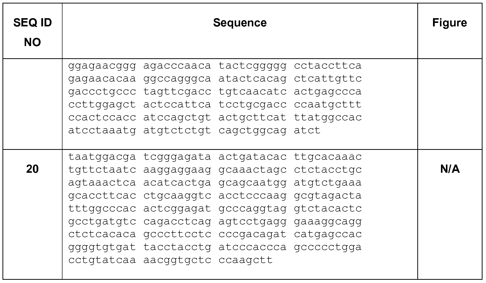
[02] A sequence listing comprising SEQ ID NOS: 1-20 is attached hereto as Table 1. Each sequence provided in the sequence listing is incorporated herein by reference, in its entirety, for all purposes.
TECHNICAL FIELD
[03] This disclosure relates to a recombinant mouse and methods for testing allergy treatments.
BACKGROUND
[04] Asthma is a debilitating disease affecting one fifth of the population of the developed world. Severe asthma is a major cause of hospitalization and health care costs. In clinical practice, asthma is classified as atopic or nonatopic, according to the presence or absence of circulating IgE directed against local aeroallergens detected by skin prick test (SPT) or in vitro techniques (RAST or ELISA). These IgE antibodies interact with the high-affinity IgE receptor (FceRI) on mast cells, which may result in immediate hypersensitivity on allergen provocation and acute exacerbation of disease.
[05] About one third of adult patients with asthma are classified as nonatopic. They tend to have more severe disease, often associated with chronic rhinosinusitis, but apart from their lack of acute reactivity to allergens, their disease is clinically similar. [06] An allergy is an immunological reaction, generally of the immediate hypersensitivity type, to a particular type of antigen termed an allergen. Such reactions underlie attacks of anaphylaxis, allergic rhinitis (hay fever), hives, and allergic asthma, and may be triggered by common allergens such as ragweed, pollen, bee or wasp venom, animal dander, mold, or a component of house dust (such as mites).
[07] There is a close concordance between asthma, allergic rhinitis and atopic dermatitis; the presence of one of these entities increases the relative risk of the other two by 3- to 30- fold over the lifetime of the subject. All three of these diseases are associated with high levels of nonspecific and antigen-specific serum immunoglobulin E (IgE). [08] In humans, immediate hypersensitivity (IH) is mediated by antibodies of the IgE isotype anchored to the surfaces of mast cells and basophils in the skin and elsewhere. Binding of antigen to these cell-bound IgE molecules triggers release of mediators such as histamine from the cells, which mediators induce the clinical phenomena such as tissue swelling, itching, or bronchial smooth muscle contraction that typify an allergic reaction. [09] IgE antibodies specific for a given allergen are produced and secreted by B lymphocytes upon contact with that allergen. Initially, B lymphocytes (or B cells) express antibodies of the IgM isotype, with each B cell committed to producing antibody specific for a particular antigenic determinant. Contact with both an allergen bearing that antigenic determinant, and certain factors produced by T lymphocytes, will induce the B cell to undergo what is termed an antibody heavy chain class switch, in which the antigen-specific portion of the antibody produced by the B cell remains the same, but it is attached to the ε- heavy chain (to yield IgE antibody) rather than the μ-heavy chain of the IgM isotype. Such a class switch is apparently permanent for a given B cell, which thereafter secretes IgE antibody specific for the allergen whenever stimulated to do so.
[10] Ovalbumin (OVA)-induced asthma in mice is one of the most commonly used models of human asthma. Th2 type cells are believed to be critical in pathogenesis of OVA-induced asthma. While we know that Th2 lymphocytes play an important role in the initiation, progression and persistence of allergic asthma, there is a lot to be understood about the immunoregulatory mechanisms. This model fails to mimic human disease associated with hyper-lgE.
[11] Allergic asthma models have also been described in large animal models, e.g., cats, dogs, pigs, sheep, and monkeys. Among these species, the feline one is of particular interest because cats spontaneously develop idiopathic asthma. However, large animal models are expensive and time consuming and have limited availability of immunological and/or molecular tools.
[12] US 6118044 (2000) provides transgenic mice which constitutively express an antibody-type molecule encoded by a transgene and which has an IgE heavy chain constant region and is specific for a pre-defined antigen (i.e., TNP). It does not provide a polyclonal response to an unknown or non-specific antigen.
[13] Currently there is no good model of chronic airway disease as they lack many key pathological features of human asthma such as mast cell infiltration of smooth muscle. In addition, almost all of them resolve spontaneously over time because mice don't get asthma. Thus, it would be desirable to have a non-human animal model that will allow the generation of an elevated IgE response relative to a native non-human animal wherein the antibody repertoire is polyclonal.
[14] Currently, common treatments for allergy include avoidance of the suspected allergen; injections of the allergen as immunotherapy to stimulate certain protective mechanisms and thereby eventually desensitize the host to the allergen; drugs such as corticosteroids, which interfere with the release of the mediators of allergy from mast cells; and drugs such as antihistamines, which block the biological action of the released mediators. However, there is no good animal model for testing allergy therapies, especially therapeutic agents that will block IgE isotype switching in B cells without adverse effects. [15] Thus, it is desirable to have cells and/or animals that have the ability to generate an elevated IgE response. In other words, selectively enhancing the rate of chain switch recombination for IgE production would be desirable.
BRIEF SUMMARY OF THE INVENTION
[16] Despite ongoing research, there still remains a need for an in vivo model for IgE involvement in asthma, allergies and other immunologic pathologies that provides a polyclonal response to a non-specific antigen, i.e., an antigen that is not predefined. Also lacking is an in vivo animal model for hyper-lgE generation wherein there is an elevated serum IgE response.
[17] Provided herein is a recombinant non-human animal, and a method for using it, that is useful as a reliable model for the search for, and/or evaluation of, anti-allergic drugs. [18] An animal model that has a genomic structure within the immunoglobulin locus that is substantially similar to the wild-type (i.e., native or unmodified) immunoglobulin locus and retains the potential to provide a full repertoire of immunoglobulins in response to antigen challenge would allow the search for, and/or evaluation of, anti-allergic drugs that inhibit IgE isotype switching in B cells.
[19] The present disclosure provides an animal model for testing allergy therapies. The animal model provides a wide diversity of antibody production in response to antigenic challenge, producing the full diversity of antibody isotypes and full complement of specificities to epitopes on the antigen. The animal model further provides a means to further understand the physiological importance of IgE to allergy and asthma. [20] In a first embodiment there is provided a targeting vector comprising: a) a fragment of DNA homologous to the 5' end of the switch region to be altered (the 5' arm/acceptor) is selected from the group consisting of at least 1500 nucleotides, at least 1800 nucleotides, at least 2000 nucleotides, at least 2200 nucleotides and at least 2400 nucleotides corresponding to Nucleotides 25470628 to 25468161 of NCB! accession number NTJ 66318 (Mus muscuius chromosome 12 genomic contig, strain C57BL/6J) (SEQ ID NO:5); b) a selectable gene marker; c) a desired/donor DNA sequence encoding a donor switch region; and d) a second fragment of DNA homologous to the 3' end of the switch region to be altered (the 3' arm/acceptor) is selected from the group consisting of at least 1500 nucleotides, at least 1800 nucleotides, at least 2000 nucleotides, at least 2200 nucleotides, at least 2400 nucleotides and at least 2800 nucleotides corresponding to Nucleotides 25470628 to 2546816 of NCBI accession number NT__166318 (Mus muscuius chromosome 12 genomic contig, strain C57BL/6J 1 ) (SEQ ID NO:8).
[21] In one aspect the targeting vector has a 5' arm comprising SEQ ID NO:4 or 5. In an embodiment the 5' arm comprises residues 25-2471 , inclusive, of SEQ ID NO:4. In a further aspect, the 5' arm is homologous to a region 3' of the endogenous lε and 5' of the endogenous Sε. In a second aspect, the targeting vector has a 3' arm comprising SEQ ID NO:7 or 8. In an embodiment the 3' arm comprises residues 2-2495, inclusive, of SEQ ID NO:7. In a third aspect, the targeting vector has a selectable gene marker that is selected from the group consisting of Neomycin and tymidine kinase. In a further aspect, the selectable gene marker is Neomycin. In a fourth aspect, the targeting vector has the selectable gene marker flanked by loxp sites. In a fifth aspect, the targeting vector has a desired switch region that is selected from the group consisting of human and mouse. In a sixth aspect, the desired switch region is selected from Sμ, Sγ1 , Sγ2a, Sγ2b and Sγ3. In a seventh aspect, the desired switch region is the Hindlll/Nhel fragment containing most of mouse Sμ region. In an eighth aspect, the desired switch region comprises Nucleotides 25617172 to 25615761 of NCBI accession number NTjI 66318 (Mus muscuius chromosome 12 genomic contig, strain C57BL/6J) (SEQ ID NO:6). In a ninth aspect the Sμ region comprises a 4.9 kb Nhel-Hindlll fragment was subcloned from a plasmid containing a genomic fragment isolated from BAC clone RP23-354L16. [22] In second embodiment there is provided a method for producing an altered embryonic stem cell in vitro, comprising the steps of: a) Altering the genomic DNA in said cell to enhance the probability of class switch recombination (CSR) to express the Cε selected from b) increasing the Sε length by adding at least one additional Sε copy in tandem with the endogenous Sε region; c) Sε region substitution; and d) Selecting the cell for correctly altered genomic DNA.
[23] In one aspect, the alteration is a substitution of a switch region selected from Sμ, Sγ1 , Sγ2a, Sγ2b and Sγ3 for the Sε region. In a further aspect, the alteration is a substitution of a Sμ region for the Sε region. In a further aspect, the alteration is a substitution of any acceptor S region (Sγ1 , Sγ2a, Sγ2b and Sγ3, Sa) with Sm or vice versa. [24] In a third embodiment there is provided a method for producing an altered embryonic stem cell (ESC) in vitro, comprising the steps of: a) Using the vector according to Claim 1 to exchange the Sμ for the Sε region b) Selecting the cell for correctly altered genomic DNA.
[25] In one aspect, the method provides an alteration that is a substitution of a switch region selected from Sμ, Sγ1 , Sγ2a, Sγ2b and Sγ3 for the Sε region. In a further aspect, the method provides an alteration that is a substitution of a Sμ region for the Sε region. In another aspect, the method provides that the ESC are from a mouse strain selected from BALB/c or C57BL/6. [26] In a fourth embodiment there is provided a non-human animal wherein: a) At least one allele of the IgH locus has been altered to enhance the rate of IgE expression/production/secretion/ relative to a non-altered allele; and b) Has an IgE profile selected from the group consisting of: i. The IgE fraction of all serum antibodies is greater than 0.04%; ii. The IgE serum concentration is above 4,000 ng/ml; iii. The IgG/lgE ratio is less than 10.
[27] In a first aspect, the non-human mammal has an IgE serum level greater than 4,000 ng/ml, greater than 10,000 ng/ml, greater than 15,000 ng/ml, greater than 30,000 ng/ml, greater than 90,000 ng/ml, greater than 10 μg/ml, greater than 20 μg/ml, greater than 30 μg/ml, greater than 40 μg/ml, greater than 50 μg/ml, greater than 60 μg/ml, greater than 70 μg/ml, greater than 80 μg/ml, greater than 90 μg/ml or greater than 100 μg/ml. In a second aspect, the non-human mammal has an IgG/lgE ratio that is between 0.1 and 10. In a third aspect, the non-human mammal having an unchallenged (i.e., resting) IgE serum concentration of between 100 ng/mL and 10000 ng/mL. In a fourth aspect, the non-human mammal has a challenged (i.e., activated or stimulated) IgE serum concentration of between 1000 ng/mL and 1000000 ng/mL. In a fifth aspect, the animal model is a nonhuman vertebrate. In a sixth aspect, the animal model is a mouse, rat, guinea pig, rabbit, or primate. In a seventh aspect, the genome of the non-animal described herein has had the
Sε region of the IgH locus altered to express/produce more IgE. In an eighth aspect, the non-human animal/mammal model has an alteration that is achieved by gene targeting. In a ninth aspect, the non-human mammal has an IgE fraction of at least 0.04%, 0.1%, 0.2%,
0.3%, 0.4%, 0.5%, 0.6%, 0.7%, 0.8%, 0.9% or 1.0%.
[28] In a fifth embodiment there is provided a method of testing an allergy therapy using the animal model comprising exposing said animal to an allergen prior to, simultaneous with or after the administration of said method of treatment for allergic disorders and evaluating the IgE response. In a first aspect of the method the IgE levels in response to antigen challenge is less than without the allergy therapy. In a second aspect, the test animal and the control animal are littermates.
[29] In a sixth embodiment there is provided a use of a compound identified by the method of testing an allergy therapy as a medicament for the treatment of an allergy.
[30] In a seventh embodiment there is provided a cell line obtainable from the animal model described herein.
[31] In a eighth embodiment there is provided a cell isolated from an animal model described herein.
[32] In a ninth embodiment there is provided a process for making a non-human animal model, said process comprising: a) microinjecting linearized fragments of plasmids encoding SEQ ID NO:6 (Sμ) into a fertilized egg of a mouse such that the fragment is incorporated in the genomic DNA upstream from and operably linked to the Cε-encoding region, b) transferring said fertilized egg to the oviduct of a female mouse which has previously been treated to induce pseudopregnancy, and c) allowing said egg to develop in the uterus of the female mouse.
[33] In a tenth embodiment there is provided a recombinant mouse comprising, in its germline, a modified genome wherein said modification comprises at least one allele of the IgH locus altered to enhance the rate of IgE production. In a first aspect, the recombinant mouse has an alteration that comprises replacing the Sε with the Sμ region or a functional portion thereof. In a second aspect, the Sμ functional portion is between at least 1 kb and 10kb in length.
[34] Other objects, features and advantages of the present invention will become apparent from the following detailed description. It should be understood, however, that the detailed description and specific examples, while indicating preferred embodiments of the invention, are given by way of illustration only, since various changes and modifications within the scope and spirit of the invention will become apparent to one skilled in the art from this detailed description.
BRIEF DESCRIPTION OF THE DRAWINGS
[35] Figure 1 is a schematic of the genetic alterations of the mouse IgH locus. (A)
Genomic organization of the variable region up to Cm in germ line configuration. (B) V(D)J recombination assembles the functional coding variable region generating a large pool of low affinity IgM producing B cells. (C) Activation of B cells accompanied by induction of AID and germline transcription results in SHM, where point mutations are introduced into assembled
V region (asterisks). AID-mediated DSBs (lightning symbol) in Sm and a downstream S region (e.g., Sg1 ) are joined to generate new isotypes (e.g., IgGI ) transcript. In addition, an excised circular fragment is generated by joining the intervening sequence.
[36] Figure 2A illustrates a schematic of the gene targeting strategy and recombination sites for modification of the mouse Sε region. The structure of the targeted allele after
CreJoxP recombination is illustrated at the bottom. Restriction enzyme cleavage sites are designated. R1 indicates the splice site for EcoRI. All other restriction enzymes have their full name.
[37] Figure 2B is a schematic of the targeting vector, pSW312. See Example 3.
[38] Figure 3 is a schematic of the overall mouse IgH locus before and after replacement of the Sε region with a donor switch region. In this diagram the donor switch region is Sμ. In the upper panel is the unmodified IgH locus; the lower panel illustrates a modified IgE locus as described herein.
[39] Figure 4A is a schematic of the unmodified (i.e., wild-type) genomic locus and illustrates the relative locations of the restriction sites, probe and switch region. The 5' homology arm is represented by the black box. The 3' homology arm is represented by the gray box.
[40] Figure 4B is a schematic of the modified genomic locus and illustrates the relative locations of the restriction sites, probe and switch region with Se replaced with Sm. The 5' homology arm is represented by the black box. The 3' homology arm is represented by the gray box.
[41] Figure 4C is a Southern blot confirming the replacement of the Sε with Sμ. While wild-type B6 samples show only one band of relevant size indicating existence of a single genomic lε region, targeted embryonic stem cell samples show the wild-type and the targeted Sε sites (where Sε is replaced with Sμ) manifested as two bands with distinct size differences. This shows successful targeting and replacement of the intended switch region. [42] Figure 4D s a Southern blot confirming the replacement of the Sε with Sμ. While wild-type B6 samples show only one band of relevant size indicating existence of a single genomic lε region, targeted embryonic stem cell samples show the wild-type and the targeted Sε sites (where Sε is replaced with Sμ) manifested as two bands with distinct size differences. This shows successful targeting and replacement of the intended switch region.
[43] Figures 5-11 show nucleotides described herein. The nucleotide base codes are: A or a is adenine; C or c is cytosine; G or g is guanine; T or t is thymine; M or m is adenine or cytosine; S or s is cytosine or guanine; and N or n is adenine or cytosine or guanine or thymine.
[44] Figure 5 shows two nucleotide sequences from the Mouse. The motif
GGGCTGGGCTG (SEQ ID NO:1 shown in Fig. 5A) is found in Sm and Se and a second motif GAGCTGACT is slightly modified in the Se region as GAGCTGAGCT (has an added G relative to the Sm motif) (SEQ ID NO:2 shown in Fig. 5B).
[45] Figure 6 shows a 2055 bp (SEQ ID NO:3) deleted from the the BamHI/PVul fragment of the IgH locus.
[46] Figure 7 shows A) SEQ ID NO:4, the 2471 bp 5'arm (for 129 mice) and B) SEQ ID
NO:5, the 2467 bp 5' arm (for C57BI/6J strain) corresponding to Nucleotides 25470628 to
25468161 of NCBI NT_166318.
[47] Figure 8 shows SEQ ID NO:6 corresponding to nucleotides 25617172 to 25615761 of NCBI Accession Number NT_166318 (Mus musculus chromosome 12 genomic contig, strain C57BL/6J)(in caps) (1141 bp).
[48] Figure 9 shows A) SEQ ID NO:7, the 3' arm (129 mouse sequence) and B) SEQ ID
NO:8, the 3' arm (C57BI/6J sequence) corresponding to 25466106 to 25463273 of NCBI
NT_166318.
[49] Figure 10 shows A) the 3.7 kb upstream of BamHI (used to design 5' probe) (SEQ ID
NO:9): and B) the PVUI to ECORI fragment (used for 3' probe design) (SEQ ID NO:10).
[50] Figure 11 shows probes used in Example 2. A) SEQ ID NO:11 , l-mu Forward-1 (21 bp); and B) SEQ ID NO:12, C-epsilon Reverse-1 (30 bp); C) SEQ ID NO:13, E-mu Forward-
2 (20 bp); D) SEQ ID NO:14, C-epsilon Reverse-2 (30 bp); E) SEQ ID NO:15, Forward: SM5'
(20 bp); F) SEQ ID NO:16, 9225F (19 bp); G) SEQ ID NO:17, 9518F (26 bp); and H) SEQ ID
NO: 18, Reverse (30 bp).
[51] Figure 12 is the retrieved IgE C57BL/6 genomic sequence (SE region to be deleted shown in bold, underlined font) from BAC RP23-135L12 (Invitrogen) (SEQ ID NO:19).
[52] Figure 13 A-D summarizes the FACS data for intracellular levels of various immunoglobulins in wild-type (WT) and heterozygotes (HET) splenocytes following immune stimulation. Figure 13A is a bar graph showing the IgM levels after lipopolysaccharide (LPS) stimulation is the same for both WT and HET animals. Figure 13B is a bar graph showing the lgG3 levels after lipopolysaccharide (LPS) stimulation is the same for both WT and HET animals. Figure 13C is a bar graph showing the IgGI levels after IL-4 in combination with anti-CD40 (4/40) stimulation is the decreased in HET animals compared to WT. Figure 13D is a bar graph showing the IgE levels after IL-4 in combination with anti-CD40 (4/40) stimulation is the increased in HET animals compared to WT.
[53] Figure 14 A-D summarizes the ELISA data for the same splenocytes as used to generate the data presented in Figure 13. At day 6 post stimulation, supernatants form the same stimulated splenocytes (three Het and three WT mice) that were used for FACS analysis were used in ELISA assay. In agreement to what we observed in FACS analysis, we also observed increase in levels of IgE expression and decrease in levels of IgGI expression in Het compared to WT when stimulated with IL4/anti-CD40. This suggests that there are more frequent breaks occurring in SmKI site that competes with switching to IgGI and increases levels of IgE switching. LPS stimulation serves as control and shows that both WT and Het have similar levels of IgM and lgG3, suggesting that the locus is intact and functions normally when other switch sequences are accessible for class switching.
DETAILED DESCRIPTION
[54] The invention will now be described in detail by way of reference only using the following definitions and examples. All patents and publications, including all sequences disclosed within such patents and publications, referred to herein are expressly incorporated by reference.
[55] Unless defined otherwise herein, all technical and scientific terms used herein have the same meaning as commonly understood by one of ordinary skill in the art to which this invention belongs. Singleton, et al., DICTIONARY OF MICROBIOLOGY AND MOLECULAR BIOLOGY, 2D ED., John Wiley and Sons, New York (1994), and Hale & Marham, THE HARPER COLLINS DICTIONARY OF BIOLOGY, Harper Perennial, NY (1991 ) provide one of skill with a general dictionary of many of the terms used in this invention. Although any methods and materials similar or equivalent to those described herein can be used in the practice or testing of the present invention, the preferred methods and materials are described. Numeric ranges are inclusive of the numbers defining the range. Unless otherwise indicated, nucleic acids are written left to right in 5' to 3' orientation; amino acid sequences are written left to right in amino to carboxy orientation, respectively. Practitioners are particularly directed to Sambrook et al., 1989, and Ausubel FM et al., 1993, for definitions and terms of the art. It is to be understood that this invention is not limited to the particular methodology, protocols, and reagents described, as these may vary.
[56] Numeric ranges are inclusive of the numbers defining the range.
[57] Unless otherwise indicated, nucleic acids are written left to right in 5' to 3' orientation; amino acid sequences are written left to right in amino to carboxy orientation, respectively.
[58] The headings provided herein are not limitations of the various aspects or embodiments of the invention which can be had by reference to the specification as a whole.
Accordingly, the terms defined immediately below are more fully defined by reference to the specification as a whole.
Definitions
[59] Novel recombinant non-human hosts, particularly mammalian hosts, usually murine, are provided, wherein the host is capable of mounting an immune response to an immunogen (also called an antigen). The immune response produced is a full repertoire of antibodies albeit with an elevated IgE component or fraction of the total serum Ig concentration.
[60] By "recombinant" is meant that the DNA of an animal or cell contains a genetically engineered modification. Thus, for example, a "recombinant animal" would be one in which at least a portion of its cells contain a genetic modification as described herein. Similarly, a
"recombinant cell" would be one in which its genome has a genetic modification as described herein.
[61] "Non-specific antigen" means any substance (as an immunogen or a hapten) foreign to the body that evokes an immune response either alone or after forming a complex with a larger molecule (as a protein) and that is capable of binding with a product (as an antibody or T cell) of the immune response.
[62] As used herein, "isotype" refers to the antibody class (e.g., IgM or IgGi) that is encoded by heavy chain constant region genes.
[63] As used herein, "isotype switching" refers to the phenomenon by which the class, or isotype, of an antibody changes from one Ig class to one of the other Ig classes.
[64] As used herein, "nonswitched isotype" refers to the isotypic class of heavy chain that is produced when no isotype switching has taken place; the CH gene encoding the nonswitched isotype is typically the first CH gene immediately downstream from the functionally rearranged VDJ gene.
[65] As used herein, the term "switch sequence" refers to those DNA sequences responsible for switch recombination. During class switch recombination (CSR) a "switch donor" sequence, typically a μ switch region, will be 5' (i.e., upstream) of the region to be deleted during the switch recombination. The "switch acceptor" region will be between the region to be deleted and the replacement constant region (e.g., v, ε, etc.). As there is no specific site where recombination always occurs, the final gene sequence will typically not be predictable. Switch sequence may be used interchangeably with switch region herein. [66] In the genetically modified (i.e., recombinant) animal described herein the switch acceptor region is modified to enhance CSR so that the serum IgE levels are elevated. [67] S regions are large, repetitive intronic sequences that vary greatly in length (repetitive regions range from 2.0 to 6.5 kb in mice). Mammalian S regions are unusually G- rich on the nontemplate strand and are composed primarily of tandem repetitive units within which certain motifs— such as TGGGG, GGGGT, GGGCT, GAGCT, and AGCT predominate.
[68] The term "rearranged" as used herein refers to a configuration of a heavy chain or light chain immunoglobulin locus wherein a V segment is positioned immediately adjacent to a D-J or J segment in a conformation encoding essentially a complete VH or VL domain, respectively. A rearranged immunoglobulin gene locus can be identified by comparison to germline DNA; a rearranged locus will have at least one recombined heptamer/nonamer homology element.
[69] The term "unrearranged" or "germline configuration" as used herein in reference to a V segment refers to the configuration wherein the V segment is not recombined so as to be immediately adjacent to a D or J segment. Reference is made to Figure 1. [70] For nucleic acids, the term "substantial homology" indicates that two nucleic acids, or designated sequences thereof, when optimally aligned and compared, are identical, with appropriate nucleotide insertions or deletions, in at least about 80% of the nucleotides, usually at least about 90% to 95%, and more preferably at least about 98 to 99.5% of the nucleotides. Alternatively, substantial homology exists when the segments will hybridize under selective hybridization conditions, to the complement of the strand. The nucleic acids may be present in whale cells, in a cell lysate, or in a partially purified or substantially pure form. A nucleic acid is "isolated" or "rendered substantially pure" when purified away from other cellular components or other contaminants, e.g., other cellular nucleic acids or proteins, by standard techniques, including alkaline/SDS treatment, CsCI banding, column chromatography, agarose gel electrophoresis and others well known in the art. See, F. Ausubel, et al., ed. Current Protocols in Molecular Biology, Greene Publishing and Wiley- Interscience, New York (1987).
[71] The nucleic acid compositions of the present invention, while often in a native sequence (except for modified restriction sites and the like), from either cDNA, genomic or mixtures may be mutated, thereof in accordance with standard techniques to provide gene sequences. For coding sequences, these mutations, may affect amino acid sequence as desired. In particular, DNA sequences substantially homologous to or derived from native V, D, J, constant, switches and other such sequences described herein are contemplated (where "derived" indicates that a sequence is identical or modified from another sequence). [72] A nucleic acid is "operably linked" when it is placed into a functional relationship with another nucleic acid sequence. For instance, a promoter or enhancer is operably linked to a coding sequence if it affects the transcription of the sequence. With respect to transcription regulatory sequences, operably linked means that the DNA sequences being linked are contiguous and, where necessary to join two protein coding regions, contiguous and in reading frame. For switch sequences, operably linked indicates that the sequences are capable of effecting switch recombination.
[73] The design of a non-human animal that responds to foreign antigen stimulation with an antibody repertoire requires that the immunoglobulin genes contained within the animal function correctly throughout the pathway of B-cell development. Correct function of a heavy chain gene includes isotype switching. Accordingly, the genes of the invention are constructed so as to produce isotype switching and one or more of the following: (1 ) high level and cell-type specific expression, (2) functional gene rearrangement, (3) activation of and response to allelic exclusion, (4) expression of a sufficient primary repertoire, (5) signal transduction, (6) somatic hypermutation, and (7) domination of the IgE antibody locus during the immune response.
[74] In the mouse, CH genes are arranged in the order 5'-V(D)J-Cμ-Cδ-Cγ3-Cγ1-Cγ2b- Cγ2a-Cε-Cα-3'. CSR occurs in switch (S) regions, which are 1- to 10-kilobase (kb) repetitive DNA elements 5' of individual CH genes. CSR results from recombination between the S region upstream of Cm (Sm) and a downstream S region, accompanied by deletion of intervening sequences. Immunoglobulins
[75] The immune system responds to foreign invaders (antigens) by producing antibodies. Antibodies are protein molecules that attach themselves to invading microorganisms and mark them for destruction or prevent them from infecting cells. Antibodies are antigen specific. That is antibodies produced in response to antigen exposure are specific to that antigen.
[76] Mammals produce four isotypes (or classes) of Ig: IgM, IgG, IgE, and IgA, encoded by the μ, y, ε, and a constant regions, respectively. [77] Related IgG subclasses are encoded by distinct C/ regions. Each Ig isotype is specialized for particular modes of antigen removal. IgM, the first isotype synthesized by a B cell, activates complement. IgG, the most abundant isotype in serum, binds receptors on phagocytic cells. IgG antibodies cross the placenta to provide maternal protection to the fetus. IgA antibodies are abundant in secretions, such as tears and saliva; they coat invading pathogens to prevent proliferation. IgE antibodies can provide protection against parasitic nematodes, but in developed countries they are the bad guys: They bind basophils and mast cells, activating histamine release and resulting in an allergic response. [78] lmmunogens (or antigens) can trigger an antibody response. Successful recognition and eradication of many different types of antigens requires diversity among antibodies; their amino acid composition varies allowing them to interact with many different antigens. It has been estimated that humans generate about 10 billion different antibodies, each capable of binding a distinct epitope of an antigen. Although a huge repertoire of different antibodies is generated in a single individual, the number of genes available to make these proteins is limited. Several complex genetic mechanisms have evolved that allow vertebrate B cells to generate a diverse pool of antibodies from a relatively small number of antibody genes. B-cell development
[79] B cells undergo a series of differentiation checkpoints in the bone marrow and spleen before they become mature functional cells. Decisions as whether to continue differentiation or to undergo cell death occur at these checkpoints and revolve principally around the immunoglobulin B-cell receptor (BCR) and its ability to function as an antigen-binding and signal-transduction molecule. The first two such checkpoints are in the bone marrow at the pro-B to pre-B transition, where the newly synthesized heavy (H) chains associate with surrogate light (L) chains to form a pre-BCR, and at the pre-B to immature B-cell stage, where the H chains associate with conventional L chains to form a BCR. Cells that are unable to form a pre-BCR or BCR undergo apoptosis (programmed cell death), whereas those that can form a BCR continue differentiating. The mature B cell that moves into the periphery can be activated by antigen and become an antibody-secreting plasma cell or memory B cell, which will respond more quickly to a second exposure to antigen. When antigen-activated B cells stop proliferating they can differentiate into mature plasma cells. Plasma cells are essentially 'antibody factories'. (See Hardy & Hayakawa, B Cell Development Pathways, Annu Rev Immunol. (2001 ) 19:595-621. ) [80] Initially, all B cells produce IgM antibodies. The V, D and J elements encoding the variable-region domains of the m heavy chain are located adjacent to the Cm exons that encode the IgM C-regions at the 5' end of the immunoglobulin heavy-chain (IgH) locus. Following appropriate stimulation, B cells can alter the isotype of the antibodies they produce via class switching while retaining their antigenic specificity. Class switching occurs in the heavy chain gene locus by a mechanism called class switch recombination (CSR). This mechanism relies on conserved nucleotide motifs, called switch (S) regions, found in DNA upstream of each constant region gene (except in the δ-chain). In this process, genomic DNA is spliced and rejoined to juxtapose the VDJ elements to the C-region exons that encode the v, ε and α chains of IgG, IgE and IgA isotypes, respectively; these C-region exons are located further downstream in the IgH region. This process results in an immunoglobulin gene that encodes an antibody of a different isotype. S Repions
[81] The molecular basis of antibody class switching to the expression of Cγ, Cε, and Cα genes in activated B cells is a recombination which positions the new CH gene 3' next to the VDJ gene. The apparent sites of Ig class switch recombination are located within the S regions, highly repetitive DNA sequences which are present 5' of each CH gene, except C5. [82] All murine and most humans S regions are sequenced at least partially. They are 1- 10 kb in length, highly repetitive and GC-rich. Murine and human Sμ are almost homogeneously composed of the two pentamer sequences GAGCT and GGGGT and the heptamer sequence (C/T)AGGTTG. All other S regions also contain multiple copies of the pentameric sequences. All murine S regions except Sμ are composed of tandem repeats that vary both in sequence and in length, with 49 bp repeats for SY1 , SY3 and SY2b, 52 bp repeats for SY2a, 80 bp repeats for Sα and 40 bp repeats for Sε. Both human and murine Sμ are more homologous to Sε and Sα than to the Sγ regions, which have considerable homology among each other. The S regions are sufficiently conserved between human and mouse to allow human S regions to be used as substrate for switch recombination in murine cells. The Sμ, Sε and Sα regions are more homologous between the two species than the Sγ regions. Indeed, the Mouse Sm motif GGGCTGGGCTG (SEQ ID NO:1 ) is found in Se and a second motif GAGCTGACT is slightly modified in the Se region as GAGCTGAGCT (has an added G relative to the Sm motif) (SEQ ID NO:2). The length of the S regions is subject to considerable allelic variation (length polymorphism) indicating that there is no functional requirement for a particular size of a given S region. IQE and Serum IQE Levels
[83] Immunoglobulin E (IgE) is a class of antibody (or immunoglobulin "isotype") that has only been found in mammals. It plays an important role in allergy, and is especially associated with type 1 hypersensitivity. IgE has also been implicated in immune system responses to most parasitic worms like Schistosoma mansoni, Trichinella spiralis, and Fasciola hepatica, and may be important during immune defense against certain protozoan parasites such as Plasmodium falciparum.
[84] Although IgE is typically the least abundant isotype - blood serum IgE levels in a normal ("non-atopic") individual are only 0.05% of the IgG concentration, compared to 10 mg/ml for the IgGs (the isotypes responsible for most of the classical adaptive immune response) - it is capable of triggering the most powerful immune reactions. [85] Atopic individuals can have up to 10 times the normal level of IgE in their blood (as do sufferers of hyper-lgE syndrome). IgE that can specifically recognise an "allergen" (typically this is a protein, such as dust mite DerP1 , cat FeIDI , grass or ragweed pollen, etc.) has a unique long-lived interaction with its high affinity receptor, FcεRI, so that basophils and mast cells, capable of mediating inflammatory reactions, become "primed", ready to release chemicals like histamine, leukotrienes and certain interleukins, which cause many of the symptoms we associate with allergy, such as airway constriction in asthma, local inflammation in eczema, increased mucus secretion in allergic rhinitis and increased vascular permeability, ostensibly to allow other immune cells to gain access to tissues, but which can lead to a potentially fatal drop in blood pressure as in anaphylaxis. Although the mechanisms of each response are fairly well understood, why some allergies develop such drastic sensitivities when others merely get a runny nose is still not well understood. [86] Total serum IgE concentration tests allows for measurement of the total IgE level in a serum sample. Elevated levels of IgE are associated with the presence of allergy. One method of testing for total serum IgE is the PRIST (paper radioimmunosorbent test). This test involves causing serum samples to react with IgE that has been tagged with radioactive iodine. Bound radioactive iodine, calculated upon completion of the test procedure, is proportional to the amount of total IgE in the serum sample. In clinical immunology, levels of individual classes of immunoglobulins are measured by nephelometry (or turbidimetry) to characterize the antibody profile of patient. Other methods of measuring IgE levels are ELISA, immunofluorescence, Western blot, immunodiffusion and immunoelectrophoresis. [87] Measurement of a total serum IgE concentration using a UniCAP 250® system (Pharmacia, Uppsala, Sweden) above 100 kU/L is considered elevated. In one study, using a sensitive double antibody radioimmunoassay to measure IgE, serum IgE from normal subjects free from evident allergic symptoms varied over a 130-fold range from 6 to 1000 ng/ml. In patients with allergic respiratory diseases the range of IgE concentrations overlapped that of normal subjects to a considerable extent, but approximately 35% of untreated allergic individuals had IgE concentrations above the 97th percentile for normals and 51% are above the 95th percentile. (See GJ. Gleich, A.K. Averbach and H.A. Swedlund, Measurement of IgE in normal and allergic serum by radioimmunoassay. J. Lab. CHn. Med. 77 (1971 ), p. 690.)
[88] In another study, the geometric mean IgE level of normal adults was 105 ng/ml with a 95% interval of 5 to 2045. The normal level of IgE in adults has been reported to be approximately 100 to 400 ng/ml, 1/400,000 of that of IgG. (see Waldmann et al., The Journal of Immunology, 1972, 109: 304-310; see also Medical Immunology - 10th Ed. (2001 ) TG Parslow, DP Stites, Al Terr and JB Imboden, eds., Table 7-2 ). [89] For a comparison of serum Ig levels in young (24-43 y.o.), old (66-96) and centenarians (99-108) see Listi et al., A Study of Serum Immunoglobulin Levels in Elderly Persons That Provides New Insights into B Cell Immunosenescence. Ann. N.Y. Acad. Sci (2006) 1089:487-495. In particular see Table 2 of Listi et al. {supra) for the age- and gender- related serum concentration of immunoglobulins for normal individuals [90] Although it normally represents only a minute fraction (0.004%) of all serum antibodies, immunoglobulin E (IgE) is extremely important from the clinical standpoint because of its central involvement in allergic disorders. Two specialized types of inflammatory cells involved in allergic responses, the mast cell and the basophil, carry a unique, high-affinity Fc receptor that is specific for IgE antibodies. Thus, despite the very low concentration of IgE (roughly 10"7 M) in blood and tissue fluids, the surfaces of these cells are constantly decorated with IgE antibodies, adsorbed from the blood, that serve as antigen receptors. When its passively bound IgE molecules contact an antigen, the mast cell or basophil releases inflammatory mediator substances that produce many of the acute manifestations of allergic disease. Elevated levels of serum IgE may also signify infection by helminths or certain other types of multicellular parasites. Like IgG and IgD, IgE exists only in monomeric form. Fc receptors appear to recognize primarily the CH3 domain of the ε chain. See Medical Immunology - 10th Ed. (2001 ; supra)
[91] Despite the variability in the serum concentrations of immunoglobulin E in humans it is clear that serum IgE levels of greater than about 2500 ng/ml are associated with a variety of diseases. Similar low levels of IgE is reported in mice (see Pinaud et al., Localization of the 3' IgH locus Elements that Effect Long-Distance Regulation of Class Switch Recombination, Immunity (2001 ) 15(2): 187-199). Gene Targeting and Plasmid
[92] Gene targeting is a technique utilizing homologous recombination between an engineered exogenous DNA fragment and the genome of the mouse embryonic stem (ES) cells. Recombination between identical regions contained within the introduced DNA fragment and the native chromosome will lead to the replacement of a portion of the chromosome with the engineered DNA. These modified ES cells can then be injected into blastocysts where they can incorporate and contribute to the fetal development along with the blastomeres from the ICM (inner cell mass).
[93] In brief, gene targeting vectors are designed which, through homologous recombination, replace the wild-type allele of a given gene with a mutated form. The targeted
ES cells are then implanted into 2-4 day blastocysts and transferred to pseudopregnant mothers (see below).
[94] The targeting vectors used herein have four components: a. a 5' arm (also referred to as a 5' flanking region); b. a selection marker; c. a DNA sequence encoding a donor switch region; and d. a 3' arm (also referred to as a 5' flanking region).
The 5' arm is a fragment of DNA homologous to the 5' end of the switch region to be replaced. The selection marker confers a selectable phenotype upon homologous recombination. The selection marker may be flanked by loxp sites. The donor switch region may be either before or after the selection marker. The 3' arm is a fragment of DNA homologous to the 3' end of the switch region to be replaced.
[95] The 5' and 3' flanking regions may be any length but is dependent on the degree of the homology. As used herein "substantial homology" between two DNA sequence portions means that the sequence portions are sufficiently homologous to facilitate detectable recombination when DNA fragments are co-introduced into a recombination competent cell. Two sequence portions are substantially homologous if their nucleotide sequences are at least 40%, preferably at least 60%, more preferably at least 80% and most preferably, 100% identical with one another. This is because a decrease in the amount of homology results in a corresponding decrease in the frequency of successful homologous recombination. A practical lower limit to sequence homology can be defined functionally as that amount of homology which if further reduced does not mediate detectable homologous recombination of the DNA fragments in a recombination competent mammalian cell. The 5' and 3' flanking regions are preferably at least 500 bp, more preferably, 1000 bp, next most preferably about 1800 bp, and most preferably, greater than 1800 bp for each homologous sequence portion. [96] Desirably, a marker gene is used in the targeting construct to replace the deleted sequences. Various markers may be employed, particularly those which allow for positive selection. Of particular interest is the use of G418 resistance, resulting from expression of the gene for neomycin phosphotransferase ("neo"). The presence of the marker gene in the genome will indicate that integration has occurred. [97] The donor switch region may be the Sμ, Sγ1 , Sγ2a, Sγ2b or Sγ3 region when the Sε region is the region to be replaced. The donor region should be one that under stimulated, non-recombinant conditions (i.e., the switch regions have not been altered) results in its associated heavy chain is expressed at a higher level than Cε.
[98] For the most part, DNA analysis by Southern blot hybridization will be employed to establish the location of the integration. By employing probes for the insert and the sequences at the 5' and 3' regions flanking the region where homologous integration would occur, one can demonstrate that homologous targeting has occurred. [99] PCR may also be used with advantage in detecting the presence of homologous recombination. PCR primers may be used which are complementary to a sequence within the targeting construct and complementary to a sequence outside the construct and at the target locus. In this way, one can only obtain DNA molecules having both the primers present in the complementary strands if homologous recombination has occurred. By demonstrating the expected size fragments, e.g. using Southern blot analysis, the occurrence of homologous recombination is supported.
[100] Once a targeting construct has been prepared and any undesirable sequences removed, e.g., procaryotic sequences, the construct may now be introduced into the target cell, for example an ES cell (see below). Any convenient technique for introducing the DNA into the target cells may be employed. Techniques include protoplast fusion, e.g. yeast spheroplast:cell fusion, lipofection, electroporation, calcium phosphate-mediated DNA transfer or direct microinjection.
[101] After transformation or transfection of the target cells, target cells may be selected by means of positive and/or negative markers, as previously indicated, neomycin resistance and acyclovir or gancyclovir resistance. Those cells which show the desired phenotype may then be further analyzed by restriction analysis, electrophoresis, Southern analysis, PCR, or the like. By identifying fragments which show the presence of the desired alteration at the target locus, one can identify cells in which homologous recombination has occurred to alter the IgH in a manner that enhances switching to Cε.
EMBRYONIC STEM (ES) CELL METHODS A. Introduction of cDNA into ES cells
[102] Methods for the culturing of ES cells and the subsequent production of recombinant animals, the introduction of DNA into ES cells by a variety of methods such as electroporation, calcium phosphate/DNA precipitation, and direct injection are described in detail in Teratocarcinomas and embryonic stem cells, a practical approach, ed. EJ. Robertson, (IRL Press 1987), the teachings of which are incorporated herein. Selection of the desired clone of recombinant ES cells is accomplished through one of several means. In cases involving sequence specific gene integration, a nucleic acid sequence for recombination with the gene of interest or sequences for controlling expression thereof is co- precipitated with a gene encoding a marker such as neomycin resistance. Transfection is carried out by one of several methods described in detail in Lovell-Badge, in Teratocarcinomas and embryonic stem cells, a practical approach, ed. EJ. Robertson, (IRL Press 1987) or in Potter et a/.. Proc. Natl. Acad. Sci. USA 81 , 7161 (1984). Calcium phosphate/DNA precipitation, direct injection, and electroporation are the preferred methods. In these procedures, a number of ES cells, for example, 0.5 X 10⁶, are plated into tissue culture dishes and transfected with a mixture of the linearized nucleic acid sequence and 1 mg of pSV2neo DNA (Southern and Berg, J. MoI. Appl. Gen. 1 :327-341 (1982)) precipitated in the presence of 50 mg lipofectin in a final volume of 100 μl. The cells are fed with selection medium containing 10% fetal bovine serum in DMEM supplemented with an antibiotic such as G418 (between 200 and 500 μg/ml). Colonies of cells resistant to G418 are isolated using cloning rings and expanded. DNA is extracted from drug resistant clones and Southern blotting experiments using the nucleic acid sequence as a probe are used to identify those clones carrying the desired nucleic acid sequences. In some experiments, PCR methods are used to identify the clones of interest.
[103] DNA molecules introduced into ES cells can also be integrated into the chromosome through the process of homologous recombination, described by Capecchi, (1989) Science 244:1288-1292. Direct injection results in a high efficiency of integration. Desired clones are identified through PCR of DNA prepared from pools of injected ES cells. Positive cells within the pools are identified by PCR subsequent to cell cloning (Zimmer and Bruss, Nature 338, 150-153 (1989)). DNA introduction by electroporation is less efficient and requires a selection step. Methods for positive selection of the recombination event (i.e., neo resistance) and dual positive-negative selection (i.e., neo resistance and ganciclovir resistance) and the subsequent identification of the desired clones by PCR have been described bv Jovner et a/.. Nature 338. 153-156 (1989) and Capecchi, (1989), the teachings of which are incorporated herein. B. Embryo Recovery and ES cell Injection
[104] Female animals are induced to superovulate using methodology adapted from the standard techniques used with mice, that is, with an injection of pregnant mare serum gonadotrophin (PMSG; Sigma) followed 48 hours later by an injection of human chorionic gonadotrophin (hCG; Sigma). Females are placed with males immediately after hCG injection. Approximately one day after hCG, the mated females are sacrificed and embryos are recovered from excised oviducts and placed in Dulbecco's phosphate buffered saline with 0.5% bovine serum albumin (BSA; Sigma). Surrounding cumulus cells are removed with hyaluronidase (1 mg/ml). Pronuclear embryos are then washed and placed in Earle's balanced salt solution containing 0.5% BSA (EBSS) in a 37.5°C incubator with a humidified atmosphere at 5% CO2, 95% air until the time of injection.
[105] Naturally cycling or superovulated females mated with males are used to harvest embryos for the injection of ES cells. Embryos of the appropriate age are recovered after successful mating. Embryos are flushed from the uterine horns of mated females and placed in Dulbecco's modified essential medium plus 10% calf serum for injection with ES cells. Approximately 10-20 ES cells are injected into blastocysts using a glass microneedle with an internal diameter of approximately 20 μm.
C. Transfer of Embryos to Pseudopreqnant Females
[106] Randomly cycling adult females are paired with vasectomized males. Recipient females are mated such that they will be at 2.5 to 3.5 days post-mating (for mice, or later for larger animals) when required for implantation with blastocysts containing ES cells. At the time of embryo transfer, the recipient females are anesthetized. The ovaries are exposed by making an incision in the body wall directly over the oviduct and the ovary and uterus are externalized. A hole is made in the uterine horn with a needle through which the blastocysts are transferred. After the transfer, the ovary and uterus are pushed back into the body and the incision is closed by suturing. This procedure is repeated on the opposite side if additional transfers are to be made.
[107] The procedures for manipulation of the embryo and for microinjection of DNA are described in detail in Hogan et al., Manipulating the mouse embryo, Cold Spring Harbor laboratory, Cold Spring Harbor, NY (1986), the teachings of which are incorporated herein. These techniques are readily applicable to embryos of other animal species, and, although the success rate is lower, it is considered to be a routing practice to those skilled in this art.
D. Identification of Recombinant Animals
[108] Samples (1-2 cm of mouse tails) are removed from young animals. For larger animals, blood or other tissue can be used. To test for chimeras in the homologous recombination experiments, i.e., to look for contribution of the targeted ES cells to the animals, coat color has been used in mice, although blood could be examined in larger animals. DNA is prepared and analyzed by both Southern blot and PCR to detect recombinant founder (F0) animals and their progeny (F1 and F2).
[109] Once the recombinant animals are identified, lines are established by conventional breeding. SOUTHERN ANALYSIS
[110] DNA was obtained from cell lines by standard phenol extraction procedure or by cesium gradient centrifugation.
A. Phenol extraction
[111] Flasks of cells are washed with HBSS buffer, then 2.5 ml/100cm2 of lysing solution (1% sodium dodecyl sulfate/150 mM NaCI/10mM EDTA/10 mM Tris, pH 7.4) is added. After all cells are solubilized, they are transferred to a 50 ml conical tube and proteinase K to a final concentration of 0.4 mg/ml is added. The lysate is incubated at 65°C for 10 minutes to inactivate DNAse enzymes, then incubated overnight at 37°C. To this lysate an equal volume of fresh phenol that has been equilibrated in 50 mM Tris, pH 8.0, is added, and the tube is gently inverted for 5 minutes at room temperature (about 22°-24°C) then centrifuged at 200Og for 5 minutes, and the top (aqueous) layer is transferred to a second tube. An equal volume of 50% phenol/50% chloroform (v/v) is then added, and the inversion centrifugation process repeated. The supernatant is then transferred to a third tube, and an equal volume of chloroform is added. After a third inversion, centrifugation cycle, the supernatant is transferred to a fourth tube and the DNA precipitated by the addition of 1/10 volume of 3M NaAcetate, 2.5 vol of cold ethanol. After washing the resulting precipitate with 70% ethanol and air-drying the pellet, it is resuspended in TE buffer (10 mM Tris pH 7.4/1 mM EDTA) and RNase to a final concentration of 50% μg/ml is added (The RNase is prepared to be DNase-free by heating the freshly suspended enzyme at 700C for 30 minutes. The solution is then extracted with an equal volume of 1 :1 SS- phenokchloroform. The phases are separated by centrifugation, as above, and the supernatant extracted with an equal volume of chloroform. Following centrifugation, as above, and the supernatant extracted with an equal volume of chloroform. Following centrifugation, the DNA in the supernatant is precipitated with 12.5 ml of ethanol, then washed with 70% ethanol and air-dried. The pellet is then suspended in TE buffer, and the DNA yield determined by O. D. reading at 260 nM and the purity determined by 260/280 ratio. The DNA preparation is stored in TE at 4°C.
B. Cesium chloride preparation of RNA and DNA from cultured cells [112] Flasks of cells were washed with HBS then 2.5 ml/100cm2 of guanidine isothyocyanate (GIT) buffer was added. The guanidine isothyocyanate buffer was 4M GIT/25 mM sodium acetate, pH6/0.8% beta-mercaptoethanol (v/v). After 3-5 minutes, with gentle rocking, the cell lysates were layered on top of 4 ml of cesium chloride buffer in Beckman SW41 10 ml ultracentrifuge tubes. The tubes were filled to nearly the top with GIT buffer, then they were spun overnight at 32,000 rpm (174,000 x g) at 200C. The GIT solution in the upper two-thirds of the tube was then removed and discarded, the CsCI solution in the lower one third of the tube that contains the DNA was transferred to a second tube. The RNA pellet in the bottom of the rube was resuspended in 200 μl of 0.3 M sodium acetate, pH 6 and transferred to a 1.5 ml microfuge tube. To this tube was added 750 μl of ethanol, and the tube was placed on dry ice for 10 minutes. After microcentrifugation for 10 min., the supernatant was discarded, 300 μl of 70% ethanol was added, and the tube was microfuged again. The supernatant was discarded, and the pellet was dried in a vacuum centrifuge. The pellet was resuspended in 200 μl of dH2O. The RNA preparation was stored as an ethanol precipitate of -700C. The 4 ml of CsCI containing the DNA was diluted with dH2O. To this was added 30 ml of cold ethanol. The DNA precipitate was recovered, transferred to a new 50 ml tube, and rinsed with 70% ethanol, then air-dried. The pellet was then resuspended in PK buffer, and 10mg of proteinase K was added. After incubation at 65°C for 15 minutes, the solution was incubated overnight at 37°C. The hydrolysate was then extracted with 1 :1 SS-phenol:chloroform, followed by chloroform, ethanol precipitation, and quantitated as described above.
C. Restriction digestion, electrophoresis, and Southern transfer [113] Restriction endonuclease digest conditions were according to the recommendations of the suppliers. For genomic DNA, the restriction digestion was for 4-6 hrs. at 37°C. For simple DNA preparations (cloned or PCR amplified) the incubation was for 1-2 hours at 37°C. Generally, 10 μg of DNA was digested in a volume of 150 μl. The digest was precipitated by addition of 3 μl 5M NaCI and 375 μl (2.5 vol) of cold ethanol, microfuged for 10 minutes at 4°C, washed with 500 μl cold 70% ethanol and microfuged. [114] The pellet was air-dried in a vacuum microfuge and resuspended in 17 μl of electrophoresis running buffer (routinely TAE buffer) and 3 μl of gel loading buffer (TAE buffer containing 50% glycerol/1% saturated bromphenol blue), heated to 68°C for 10 minutes, and loaded into wells of an agarose gel, along with a lambda-Hindlll digest in a separate well to serve as a size marker. The concentration of agarose in the gel was 1.0%. Following electrophoresis for 8-16 hours, the gel was stained with ethidium bromide, the migration distance of the marker bands measured and recorded, and the gel photographed. [115] The digested DNA was vacuum-transferred to a Nytran membrane. The gel was laid on top of the Nytran membrane on the vacuum apparatus, covered with 500 ml of 0.4 M NaOH/0.8 M NaCI and a vacuum pressure of 50 cm of water applied for four minutes. The NaCI-NaOH solution was removed, 500 ml of 10 X SSC added, and a pressure of 50 cm water applied for 30-60 minutes. The Southern blot was then baked at 800C for 2 hours and stored in a vegetable freezing bag. D. Southern hybridization
[116] The Southern blot was placed in a heat sealable plastic bag and incubated with 10 ml of pre-hybridization buffer containing 1 M NaCI, 1% SDS, 10% dextran sulphate, and 200 μg/ml herring sperm DNA, and incubated for 15 minutes at 65°C. A corner of the bag was then cut off, and the radiolabeled oligonucleotide probe was added (approximately 107 dpm). The bag was resealed and placed at 65°C in an oven or water bath and gently rocked or shaken for 12-16 hours. The membrane was then removed from the bag and washed in a series of increasingly dilute and higher temperature (increasing stringency) SSC buffers until the background radioactivity was low relative to the specifically bound probe. In a darkroom, the membrane was then placed in a plastic bag which was positioned in an X- ray film cassette equipped with intensifier screens, a sheet of Kodak XAR-5 film was added, and the sealed cassette was placed at -700C for variable time depending on the intensity of signal. Usually, exposures after varying time periods are useful. The film was developed in a Kodak X-OMAT automatic developer. Membranes may be re-hybridized several times. Nytran membranes may be stripped of labeled probe by heating in boiling 0.1 X SSC for 2 minutes. B-CeII culture
[117] B cells may be purified from spleens by negative selection. Briefly, T cells in single cell suspensions are coated with antibodies and depleted by complement lysis. The remaining spleen cells were layered over a discontinuous Percoll (GE Healthcare) gradient. Resting B cells may be selected from the 66% to 70% interface and total B cells (50-70% Percoll interface) were used. B cells may be cultured in B cell media consisting of RPMI 1640 media (Sigma Aldrich), supplemented with 10% heat inactivated FBS, 100 U/ml penicillin and streptomycin, 2 mM L-glutamine, 1 mM sodium pyruvate, 10 mM Hepes, 100 mM non-essential amino acids and 5 x10"5M 2-mercaptoethanol.
Confirming in vivo hvper-lqE production
[118] Recombinant animals will be tested for elevated IgE serum levels using techniques known in the art. For example, the ImmunoCAP Specific IgE blood test (which the literature may also describe as: CAP RAST, CAP FEIA (fluorenzymeimmunoassay), and Pharmacia CAP) may be used.
[119] Other methods are known in the art. Such methods include for example Enyzme- linked immunosorbent assay and potentially FACS using an anti-lgE antibody. Enzyme- Linked Immunosorbent Assay, also called ELISA, Enzyme ImmunoAssay or EIA, is a biochemical technique used mainly in immunology to detect the presence of an antibody or an antigen in a sample. Fluorescence-activated cell sorting (FACS) is a powerful technique for analyzing large mixed populations of single cells. A higher proportion of IgE positive cells would indicate an elevated serum IgE level.
Hvbridoma production
[120] Standard techniques known in the art are used to prepare hybridomas that produce
IgE. See, for example, Kohler & Milstein (1975) Continuous cultures of fused cells secreting antibody of predefined specificity, Nature 256:495, and Kohler & Milstein (1976) Derivation of specific antibody-producing tissue culture and tumor lines by cell fusion. Eur. J. Immunol.
6:511.
[121] Briefly, immunized splenocytes are washed and fused to myeloma cells under appropriate conditions. The hybridomas are exposed to HAT or other selection agent 24 hours later, and the non-fused myeloma cells will die. The non-fused splenocytes also have a finite lifetime, and the hybridomas are then the only proliferating cells left in the culture.
Assay for Class Switching
[122] Assays are known in the art and are described in, for example, Shinkura, R. et al. Nat. Immunol. (2003) 4, 435-441 and Zarrin, et al., Nat. Immunol. (2004) 5, 1275-1281. Briefly, splenocytes were stimulated with anti-CD40 and IL-4 for 4 days to generate hybridomas or for 6 days to perform ELISA. A monoclonal anti-lgE antibody may be used to detect IgE (mutated alleles). Total IgE may be measured by polyclonal anti-lgE antibodies (Southern Biotechnology Associates).
[123] See also, for example, Southern and Berg (Detection of specific sequences among DNA fragments separated by gel electrophoresis. J MoI Appl Genet (1982) 1 : 327-341 ) describes Southern blot analysis to assess DNA rearrangement and CSR in IgH locus. [124] ε Germline transcription marks the first step in the commitment of B cells to the synthesis of IgE. Therefore, RT-PCR may be used to examine ε immunoglobulin heavy- chain germline gene transcripts (GLTs; ε GLTs), ^circle transcripts (CTs; I ε -Cμ CT or lεCγ CT), and mRNA encoding the heavy chain of IgE (ε mRNA) and activation-induced cytidine deaminase (AID) (see Takhar et al., J Allergy Clin Immunol (2007) 119(1 ):213-218). [125] In the experimental disclosure which follows, the following abbreviations apply: eq (equivalents); M (Molar); μM (micromolar); N (Normal); mol (moles); mmol (millimoles); μmol (micromoles); nmol (nanomoles); g (grams); mg (milligrams); kg (kilograms); μg (micrograms); L (liters); ml (milliliters); μl (microliters); cm (centimeters); mm (millimeters); μm (micrometers); nm (nanometers); 0C. (degrees Centigrade); h (hours); min (minutes); sec (seconds); msec (milliseconds); Ci (Curies) mCi (milliCuries); μCi (microCuries); TLC (thin layer chromatography).
EXAMPLES
[126] The present invention is described in further detain in the following examples which are not in any way intended to limit the scope of the invention as claimed. The attached Figures are meant to be considered as integral parts of the specification and description of the invention. All references cited are herein specifically incorporated by reference for all that is described therein. The following examples are offered to illustrate, but not to limit the claimed invention.
Example 1 Gene Targeting/Generation of mutant mice
[127] This example illustrates the construction of the targeting vector, the transformation of embryonic stem cells (ES) and generation of mutant mice. /. Generalized Procedure
A. Transformation of embryonic stem cells (ES)
[128] Targeting constructs were designed based on sequence information available in the NCBI for NT_166318. (See also, Waterston et al., Initial sequencing and comparative analysis of the mouse genome, Nature. (2002) 420(6915):520-62.) A BamHI/PVul fragment (7022 bp with Se region (129 mice); 7355 bp with Se region (B6 mice)) was isolated from 129 or C57B6 BAC clones and amplified.
[129] The following sequence (5'→3'; SEQ ID NO:3) was deleted from the BamHI/PVul fragment and was replaced with a Neo cassette plus the Hindlll/Nhel containing most of mouse Sm region (see Figure 2 and SEQ ID NO:6).
tgggttaagc agagctgtgc tgggctggta tgagctggtc caagttgggc 50 taaacagagc tgggccaggc tagtatgagc tggtctgaac tacactaagc 100 aggactaggc tgggctgagc tgagctggac tggctggact tggctgagat 150 gtgttgagct gggttaagta tggctgggct gggctggcct gggctgggct 200 ggactggatt ggtatgagct ggtccaagtt gggctaagca gagctgggcc 250 aggctggtat gagctggtct aaactgaact aagtagggct gggctaagct 300 gagctggtct acactagcct gacctgagct agggtaggct ggactgggct 350 gagctaagtt gcactgggca ggggtgggct ggaccgagct gatttgagct 400 gggatgggct gagatgggtt cagcaggcct aagcaggcct agctgggttt 450 agctagattt agctaggcaa ggctgagcta ggctgggcgg ggcggggcta 500 ggctgggcag ggctggactg agctagcttt tgtatattcg gttgaaatgg 550 gttggtctgg tctggactga actgactgag ctgggctagc ctgagctcga 600 tggggggtat actcagctga gatgggctgg tctggctaga ctgaactgga 650 ttgggctagg ctgagctagg ctgacctgaa ctggcctggt ctgggctgga 700 ctgggcaggg ctggtctcag ctagactaca ctgagttaac ctgggctgga 750 ccatactggg ttaaactagg ttgcactggc tgggttagac ttggctgagc 800 tgggcttggc tgagctgagt caagatggtc tgagttgatt tgagttggct 850 aagctaagct gagctacact gaactaggca aggctgggct ggaaaggtct 900 gggttaagtt aggagggact tggcttggct tagctgggcc aagctaggct 950 gaactgggct gaactgagct gagctgggct gagctgggct gagctgggct 1000 gagctgggca aggctaaact ggaatggact gaattggcct aagatgggcc 1050 cggctaagct aagtaaggct gccctgaact gagcaggact ggcctggcct 1100 ggattgacct ggcatgagct taacttgact agactagtct atcttgggtg 1150 aactgggcta agcaggacta atctggcctg atctgagcta gactgaacta 1200 ggctaagctg agctgagttt agcttggctg aactgggctg ggctgcactg 1250 aactgtattg agctatgtag aactgagctg gtcttgtctg aggtgggttg 1300 ggctggtctg ggctgaacca gattgcacta gactgagctt agctggacct 1350 ggctgagctg gactgcattg tgctaaactg gctctcttta gaccgagctt 1400 agctggactg gactgagcta ggttgggtgg gctgatctaa gctgagctag 1450 gctggtctca cctgaggaat gctgtgctgt gctgagctga actaaactga 1500 gctcagctaa ggaagtgtga gctagactga gctgagctag gctgggttgg 1550 gctgaactga gctaccttgg gtggactagg ctgagctgag ctgggttgag 1600 ctgagctata gatttggttg gactggactg gattgggcta aactgaactg 1650 gtttggggta ggctgggatg agctggactg agctaggctg tactggtctg 1700 agctaaacta agttgagtgg ggctaagagg agctgagtga ggctgggctg 1750 gaatgagcta ggctagggtt gtgagctagg gttgtactgg tctaagctga 1800 gtttagctga gagaggctgg gctagacttc cataaggtgg ctgagtcata 1850 ctacagtgca ctgagctgtg ttgagcttaa cttggattaa gtggaatggg 1900 ttgagctggc tgaactgggc tgaactgaga taaactagac tgagctggga 1950 cacgctggga cgagctggaa cgagctagaa ttactgttct aatctgatct 2000 gggctgaggt aaactgggcc tggttgagct ctactaggct aagtagagtt 2050 gagct 2055
[130] A similar construct may be made to target 129SvEv ES cells to account for strain allelic differences that exist in the IgH locus.
B. Transformation of embryonic stem cells (ES)
[131] The plasmid from above was linearized using Pvu\ restriction enzyme. DNA was washed in 70% Ethanol and subsequently pelleted and resuspened in 50 μl TE. Using techniques known in the art (see, for example, Templeton et al., Efficient gene targeting in mouse embryonic stem cells, Gene Therapy (1997) 4:700-709), 106 ES cells were transfected with linearized vector by electroporation and selected using G418 (400 μg/ml). Subsequently the Neo gene was deleted using cre/loxP recombination system. Correctly targeted clones were identified by Southern analyses with two probes of at least 300 base pairs and designed from SEQ ID NO:9 (for the 5' probe) and SEQ ID NO: 10 (for the 3' probe) of the construct (shown in Figure 2) identified recombinants.
C. Genera tion of mutant mice
[132] Germline mice were generated to create a mouse with a modified IgH locus. ES cell clones showing homologous recombination of Sμ/Sε were injected into C57B6 blastocysts, and the resulting male chimeras were mated with C57B6 females. Germline transmission in heterozygous and homozygous mutant mice was assessed by coat color. //. Specified Procedure
A. Target Vector Construction
[133] The construct for targeting the C57BL/6 IgE locus in ES cells was made using recombineering and standard molecular cloning techniques. See, for example, Liu et al., A highly efficient recombineering-based method for generating conditional knockout mutations.
Genome Research (2003) vol. 13 (3) pp. 476-84.
[134] Targeting constructs were designed based on sequence information available in the
NCBI for NT_166318. (See also, Waterston et al., Initial sequencing and comparative analysis of the mouse genome, Nature. (2002) 420(6915):520-62.).
[135] First, a 6988 bp genomic fragment (SEQ ID NO: 19; Figure 13) from a mouse BAC
(RP23-135L12; Invitrogen, Carlsbad, CA) containing the C57BL/6 Switch epsilon
(designated as SE or Sε or Sepsilon, herein) region/sequence was isolated and introduced into a plasmid containing the negative selection marker Diphtheria toxin A (DTA) called
"pBlight-DTA" (see Warming et al., MoI. Cell. Biol. (2006) 26 (18):6913-22) for subsequent use in embryonic stem (ES) cell targeting, resulting in "pBlight-DT A-IgE".
[136] Second, a loxP-PGK-em7-Neo-BGHpA-loxP-Hindlll-Sall-Ascl-Nhel cassette was inserted into the IgE fragment using homologous recombination, replacing the endogenous
Sε region with a floxed Neo and a polylinker for subsequent insertion of switch mu region
(designated as SMu or Sμ or Sm, herein) ("pBlight-DTA-lgE-lox-Neo-lox-MCS").
[137] Finally, a 4.9 kb Hindlll-Nhel fragment containing C57BL/6 SMu was isolated from the BAC RP23-135L12 (Invitrogen) and this fragment was cloned into pBlight-DTA-lgE-lox-
Neo-lox-MCS using three-way ligation (ligation of a 6.2 kb Xhol-Hindlll fragment and a 4.1 kb Xhol-Nhel fragment, both from pBlight-DTA-lgE-Neo-MCS, to the 4.9 kb Hindlll-Nhel
Smu fragment). The resulting construct is called "pSW312" (pBlight-DTA-lgE-lox-neo-lox-
Smu). See Figure 2B.
B. Transformation of embryonic stem cells (ES)
[138] C57BL/6 ES cells were targeted using standard methods (G418 positive and DTA negative selection), and positive clones were identified using PCR and taqman analysis. Correctly targeted clones were confirmed by Southern blotting analysis using Hindi 11 digested genomic DNA and an external 3' probe (the sequence between the 3'end of the construct and an endogenous IgE Hindi 11 site) (SEQ ID NO:20): taatggacga tcgggagata actgatacac ttgcacaaac tgttctaatc 50 aaggaggaag gcaaactagc ctctacctgc agtaaactca acatcactga 100 gcagcaatgg atgtctgaaa gcaccttcac ctgcaaggtc acctcccaag 150 gcgtagacta tttggcccac actcggagat gcccaggtag gtctacactc 200 gcctgatgtc cagacctcag agtcctgagg gaaaggcagg ctctcacaca 250 gcccttcctc cccgacagat catgagccac ggggtgtgat tacctacctg 300 atcccaccca gccccctgga cctgtatcaa aacggtgctc ccaagctt 348 C. Generation ofmutant mice
[139] Germline mice were generated to create a mouse with a modified IgH locus. ES cell clones showing homologous recombination of Sμ/Sε were injected into C57B6 blastocysts, and the resulting male chimeras were mated with C57B6 females. Germline transmission in heterozygous and homozygous mutant mice was assessed by coat color.
Example 2 In vitro stimulation (anti-CD40 /IL4; LPS) to induce isotype switching
[140] The following example details how B-cells were collected and analyzed for class switch recombination (CSR).
[141] Spleen cells from 6-8 week old wild-type or heterozygotes animals were stimulated in vitro with anti-CD40 (1 μg/ml_; HM40-3, Pharmingen) plus IL-4 (25 ng/mL), or Lipopolysaccharide (LPS, 20 μg/mL). 1.5 x 10⁶ cells were seeded in one well of 6 well plates (0.5 x 1 O6VmL) in RPMI media supplemented with 10% Fetal Bovine Serum, 2mM glutamine, 100 units/mL of penicillin-streptomycin and 100 μM β-mercaptoethanol. After stimulation, activated B cell cultures were used to generate hybridomas (using standard methods; see, for example, Monoclonal Antibodies: Methods and Protocols in Methods in Molecular Biology (2007) vol. 378:1-13) at day 4-5 or to measure Ig levels by ELISA (day 6). Monoclonal anti- mouse antibodies (Pharmingen) were used to detect Ig levels followed with alkaline phosphotase-conjugated goat anti-mouse IgGI (Southern Biotechnology) as the detection antibody. Purified mouse Ig (Pharmingen) was used as the standard. [142] Surface Ig staining was performed using PE anti-mouse Ig (Pharmingen) antibodies on splenocytes. FACS analyses was done at day four of stimulation. Samples were collected on a FACS Scan (Becton Dickinson) and analyzed using Flojo analyses software. [143] CSR was evaluated by Sothern blot analysis. Briefly, genomic DNA from hybridoma clones was used to assess DNA rearrangement and CSR in IgH locus. Hybridoma genomic DNA (about 150 ul) was digested overnight with EcoRI (NEB) restriction enzyme and the digest was resolved by applying the samples to a 0.7% agarose gel. The resolved samples were then transferred to Zeta-probe blotting membrane (Bio-Rad), fixed by UV cross-linking and/or baking in 80° Celsius vacuum oven (20 min), and probed with 32P labeled l-mu, C-mu, l-epsilon, or C-epsilon probes following standard southern blotting protocols (Molecular Cloning, 3rd Edition VoU , pages 6.33-6.64). The labeled DNA was visualized by putting the membranes on X-ray films (Kodak).
[144] In order to sequence the junctions of S-mu and S-epsilon CSR in IgE positive hybridoma clones, nested PCR was used to amplify and sequence this region. First we used l-mu Forward-1 :5'-CTCTGGCCCTGCTTATTGTTG-3' (SEQ ID NO:11 ) and C-epsilon Reverse-1 !5'-CCTGATAGAGGCTGTGAGAAAGGAAGGACC-S' (SEQ ID NO: 12) primers to amplify this region from genomic hybridoma DNA with PCR. The PCR cycles were 94°C for 2 min; (94°C for 10 sec,60°C for 30 sec, and 68 0C for 150 sec) X 35 cycles, 68°C for 7 min. The product from this PCR step was used as template (2 ul) for a second PCR cycle, using the following primers: E-mu Forward-2: δ'-AGACCTGGGAATGTATGGTT-S' (SEQ ID NO: 13) and C-epsilon Reverse-2: δ'-TAGGTTAGACTTATTTATATCACTGCATGC-S' (SEQ ID NO: 14). The PCR program was the same as above except the annealing temperature was lowered to 55°C. The PCR products were then gel purified (Quigen) and directly sequenced using the following primers: Forward: SM5': 5'- GTTGAGAGCCCT AGTAAGCG- 3' (SEQ ID NO: 15); 9225F: δ'-TTGAGAGCCCTAGTAAGCG-S' (SEQ ID NO: 16); 9518F: TGAGCTCAGCTATGCTACGCGTGTTG-3' (SEQ ID NO: 17); Reverse: 5'- GCCCGATTGGCTCTACCTACCCAGTCTGGC-3' (SEQ ID NO: 18).
Example 3 Intracellular IgE Staining & FACS Analysis
[145] This example demonstrates the intracellular staining of IgE and FACS anaylsis from tissue obtained from heterozygotic mice and wild-type mice after exposure to different stimuli.
[146] 0.5x10⁶ splenocytes were spun down and resuspended in FACS buffer (PBS+0.5%
Fetal Bovine Serum). Anti-lgE antibody (e-Bioscience, San Diego, CA; Cat. No. 14-5992-85) was added at 1 μg/sample to block surface IgE molecules. The cells were incubated for
15min at 4°C then washed twice with FACS buffer and pelleted by centrifugation for 3 minutes at 1700 rpm. The pelleted cells were resuspended in 1% Fetal Bovine Serum in
PBS.
[147] Cells were vortexed and BD Cytofix/Cytoperm (BD Biosciences, San Jose, CA; Cat.
No. 554722) was added at 200 μl/sample to fix the cells. Incubated at 4°C for 20 min.
[148] Cells were washed twice with 1X BD Perm/Wash buffer (BD Biosciences, San Diego,
CA; Cat. No. 554723) and were resuspended in 250 μl of 1X BD Perm/Wash buffer with Fc - blocker. Cells aliquots were then used for staining with Biotin-lsotype Control (e-Bioscience, 13-4301-82) or anti-lgE-biotin (e-Bioscience, 13-5992-82) along with B220-FITC at 1 :200 final antibody dilution. Incubated for 30 min at 4°C.
[149] Cells were washed twice with 1X BD Perm/Wash buffer.
[150] Streptavidin-PE (Pharmingen) in 1X BD Perm/Wash was added at 1 :200 dilution.
Incubated 10 min at 4°C.
[151] Washed twice with BD Perm/Wash buffer and resuspended cells in 200 μl of FACS buffer and performed FACS analysis using a BD FACS Calibur equipment and CellQuest
Pro program to analyze the cells.
[152] Results are shown in Figure 13. In Figure 13, it can be seen that the number of cells that stain positive for IgE is increased relative to the WT. FACS results show that the percentage of IgE expressing B-cells increase (approximately twice) in the Het animal compared to WT animal while the levels of IgGI drops approximately by half in Het compared to WT. This demonstrates that having SmKI (in place of Se) increases switching to IgE and at the same time reducing the chance of switching to IgGI by competing with this locus.
Example 4 IgE Assays/Measurements
[153] This example demonstrates the use of ELISA (luminex or conventional ELISA) to measure the IgE in an unchallenged or challenged recombinant animal or in in vitro cell culture. For in vitro cell cultures, the cells may be stimulated with LPS or antiCD40/IL4.
Luminex Mouse 7-plex Immunoglobulin lsotyping Assay
[154] This assay uses a multiplex assay kit (Millipore Beadlyte Mouse Immunoglobulin lsotyping Kit, Cat#48-300, ) for isotyping (heavy chain: IgGI , lgG2a, lgG2b, lgG3, IgA, IgE, IgM; and light chain: kappa or lambda) mouse monoclonal cell culture supernatants or serum samples (using the Millipore Mouse Isotyping Serum Diluent) in a single well with the Luminex® Instrument system.
[155] Before use, the cell culture supernatants should be centrifuged at 14,000χg to remove any particulates. Similarly, serum and plasma samples should be spun down (8000χg) prior to assay to remove particulate and lipid layers. This will prevent the blocking of wash plate as well as sample needle. Materials used for Bead-Based Multiplex Assays
[156] Millipore Beadlyte Mouse Immunoglobulin Isotypinq Kit Cat#48-300 (Contains: Beadlyte Cytokine Assay Buffer, Cat#43-002, Beadlyte mouse multi-lmmunoglobulin Beads, Cat#42-045, Beadlyte mouse Immunoglobulin positive control, Cat#43-008, Beadlyte anti- mouse k light chain, PE (10OX), Cat#44-029, Beadlyte anti-mouse lambda light chain, PE
(10OX), Cat#44-029).
[157] Millipore Beadlyte mouse lsotyping Serum Diluent (5X), Cat#43-033, Phosphate
Buffered Saline with 1 % Bovine Serum Albumin.
[158] Ig Standard Curve Reagents: Millipore: Beadlyte Mouse Multi-lmmunoglobulin
Standard (IgGI , lgG2a, lgG2b, lgG3, IgA, IgE, IgM) Lyophilized, from balb/c mouse Cat#47-
300.
[159] Filter Plate: Millipore multiscreen-HA 0.45um surfactant-free.
[160] Millipore Filtration System (NOTE: Any system that provides ddH20 will work.)
[161] The assay may be performed according to the manufacturer's instructions.
General Protocol for Processing Bead-Based Multiplex Assays
[162] Centrifuge the sample (as appropriate) to precipitate any particulates before diluting into appropriate diluent. Resuspend the standard into appropriate diluent and prepare an eight-point standard curve using twofold serial dilutions. Wet filter plate with 50-100 μl assay diluent per well.
[163] Plate fitting: Add 50 μl of the standard or sample to each well. Sonicate the coupled beads for 15-20 s to yield a homogeneous suspension. Thoroughly vortex the beads for at least 10 s. Dilute the beads to 1500 beads per well, and add 25 μl of diluted bead suspension to each well. Incubate for 15 min in the dark at room temperature (Incubation time can be varied, typically between 15 min and 2 h. The primary incubation of the bead and sample can be performed overnight at 4°C for greater low-end sensitivity.).
[164] Washing step: Apply vacuum manifold to the bottom of filter plate to remove liquid and blot. Wash by adding 75 μl of assay diluent, vacuum and blot. Repeat washing twice.
Resuspend the beads in 75 μl of assay diluent. Add 25 μl of the detection antibody solution to each well. Incubate for 15 min in the dark at room temperature. Apply vacuum manifold to the bottom of filter plate to remove liquid. Wash by adding 125 μl of assay diluent, vacuum and blot. Repeat washing twice. Resuspend the beads in 125 μl of assay buffer.
Incubate on a plate shaker for 1 min. Read the results on Luminex® 100 instrument. Data evaluation: extrapolate the sample concentrations from a 4-PL or 5-PL curve.
[165] At day 6 post stimulation, supernatants from the same stimulated splenocytes
(three Het and three WT mice) that were used for FACS analysis were used in an ELISA assay as described above. In agreement to what we observed in FACS analysis, we also observed an increase in levels of IgE expression and decrease in levels of IgGI expression in Het compared to WT when stimulated with IL4/anti-CD40. This suggests that there are more frequent breaks occurring in SmKI site that competes with switching to IgGI and increases levels of IgE switching. LPS stimulation serves as control and shows that both WT and Het have similar levels of IgM and lgG3, suggesting that the locus is intact and functions normally when other switch regions are accessible for class switching. See Figure 14.
Total Mouse IgE Binding ELISA
[166] This assay is run to quantitate mouse IgE serum levels in both naϊve and immunized animals.
/. Coat with Capture Antibody:
[167] Dilute the purified anti-mouse IgE capture mAb (Rat Anti-mu IgE, Clone R35-92 BD Pharmingen, San Diego, CA, Stored at 40C. Cat. #553416 (0.5 mg/ml)) to 2 μg/mla in coating buffer (0.05 M Carbonate/bicarbonate, pH 9.6. Add 100 μl per well to an enhanced protein- binding ELISA plate (e.g., Nunc immunoplate Cat #464718, 384-Well). Shake plate to ensure all wells are covered by capture antibody solution. Cover the plate and incubate for overnight at 4°C. [Note: may be done at 1 hour at 37°C. Wash the plate 3X with PBS/Tween® (PBS + 0.05% Tween 20). For each wash, wells are filled with 200 μl PBS/Tween ® and allowed to stand at least 1 min prior to aspirating or dumping. As a final step, tap plate on paper towels to remove excess buffer.
//. Blocking:
[168] Block the plate with 50 μl blocking buffer (PBS + 0.5% BSA + 10 ppm Proclin pH 7.4) per well. Cover the plate and incubate at RT for 1 hour with gentle agitation. Wash the plate 3X with PBS/Tween ® , as above.
///. Apply Standards and Samples:
[169] Leave column 1 as blank wells (i.e., no antigen added, 25 μl per well blocking buffer only). Use columns 2 and 3 for duplicates of the standard, 25 μl per well: Prepare standard curve from 500 ug/ml Stock standard antibodies to starting standard concentration of 10ng/ml (1 :50,000). Make Serial 1 :2 dilutions (PBS + 0.5% BSA + 0.05% Tween 20 + 15ppm Proclin + 0.2% BgG + 0.25% CHAPS + 5 mM EDTA , pH 7.4) The mouse IgE standard curve: 10.0, 5.0, 2.50, 1.25, 0.625, 0.313, 0.156, 0 ng/ml. Assay Controls are mouse IgE Klsotype control, BD Bioscience, Catalog #557079, Main Stock: 0.5 mg/ml, keep in 40C ) at the following dilutions: 8ng/ml, 4ng/ml, 0.5ng/ml. Use the remaining columns to add samples at various dilutions in blocking buffer, 25 μl per well. Dilute the serum samples with Assay Diluent (PBS + 0.5% BSA + 0.05% Tween 20 + 15ppm Proclin) 1 :25 minimum initial dilution, serial 1 :3, using Hamilton Diluter. Cover the plate and incubate for 2 hours with gentle agitation. [Note: May be done for at least 1 hour at RT or overnight at 4°C] Wash the plate 6X with PBS/Tween® , as above, and incubate with agitation for one hour.
IV. Incubation with Detection Antibody:
[170] Add 25 μl biotinylated anti-mouse IgE (Rat Anti-mu IgE-Biotin, Clone R35-1 18, BD Pharmingen, San Diego, CA, 0.5ug/ml, 40C, Cat #553419) per well. Cover the plate and incubate at RT for 30 minutes with gentle agitation. Wash the plate 6X with PBS/Tween® , as above.
V. Add Streptavidin-Horseradish Peroxidase (SAv-HRP):
[171] Dilute Streptavidin-HRP (GE Healthcare, formerly Amersham Biosciences, Piscataway, NJ, 1 mg/ml, Stored at 40C, Cat.#RPN4401 ) 1 :20,000 in blocking buffer to a final concentration of 50 ng/ml. Add 25 μl per well and incubate with agitation for 30 minutes. [Note: Avidin-HRP may be used instead of Streptavidin-HRP with appropriate modification as noted in the art.] Cover the plate and incubate at RT for 30 min. Wash the plate 6X with PBS/Tween® , as above, of this protocol.
VI. Add Substrate and Develop:
[172] Mix 1 Part of TMB A to 1 Part of TMB B (TMB peroxidase solutions A & B (KPL, Gaithersburg, MD, cat # 50-76-02 and 50-65-02, respectively), store at 40C). Add 25 μl TMB substrate to each well and shake. Incubate 15 min. at room temperature for color to develop and add 25 μl of 1 M H3PO4 to quench the development. Read the plate at 450-650 nm. [173] Interpolate the serum sample IgE levels from the standard curve.
Example 5 Hybridoma to quantitate antibody isotypes
[174] Hybridomas are constructed using techniques known in the art. Using the assays of Example 4 the immunoglobulin isotypes are characterized. [175] The antibodies are then further characterized for binding affinity, epitope characterization and mode of action on a relevant pathway.
Example 6 Immunization: TNP-OVA; OVA; Ficol to induce isotype switching in vivo
[176] This example illustrates the in vivo immunization of recombinant animals as described herein.
[177] Eight-week-old sex-matched Balb/C mice aged 8 weeks, weighing approximately 25- 30 g are immunized i.p. with TNP-OVA 50ug/alum 2mg or TNP-Ficoll 50ug injected i.p. in 100 ul sterile PBS, boosted at day 28. 6OuI samples are collected on day -3, 7, 14, 21 , 28, 35 and 42 via tail vein for antibody isotype measurements using the assays described in Example 4.
//. Antigen-induced peritonitis model with measurement of histamine in the peritoneal fluid.
Animals and sensitization procedure
[178] Balb/C mice aged 8 weeks, weighing approximately 25-30 g raised at the Pasteur
Institute (Paris, France), are actively sensitized by a subcutaneous (s.c.) injection of 0.4 ml 0.9% w/v NaCI (saline) containing 100 gl_g ovalbumin adsorbed in 1.6 mg aluminium hydroxide (Andersson & Brattsand, 1982). Seven days later, the animals receive the same dose of ovalbumin in the presence of AI(OH)3 and are used 7 days thereafter.
Antigen-induced peritonitis
[179] Peritonitis is induced by the intraperitoneal (i.p.) injection of 0.4 ml of a solution containing 2.5 or 25 gm/ml ovalbumin diluted in sterile saline (1 or 10 μg of ovalbumin, as final doses injected per cavity). Control animals receive the same volume of sterile saline. At various time intervals after antigen challenge (30 min- 164 h), animals are euthanized by an overdose of ether and the peritoneal cavity is opened and washed with 3 ml of heparinised saline (10 U per ml). Approximately 90% of the initial volume is recovered. In rare cases, when hemorrhages are noted in the peritoneal cavity, the animals are used. [180] Histamine levels are measured using methods known in the art.
Example 7 Infection with Nippostrongylus brasiliensis to induce IgE
[181] This example illustrates the IgE response to infection with parasitic worms, Nippostrongylus brasiliensis.
[182] The course of development of N. brasiliensis has been well characterized in the mouse (Love, Nippostrongyius brasiliensis infections in mice: the immunological basis of worm expulstion, (1975) Parasitiology 70:1 1 ). In the mouse once infective larvae (L3) have penetrated the skin they are transported to the lungs via the lymph and blood vascular systems. After a tracheal-esophageal migration, fourth-stage larvae (approx. 15-35% of the original L3 dose) are carried to the lumen of the small intestine and mature. The infection is patent by the seventh day after larval inoculation. Worm expulsion that is preceed by a sharp fall in fecal egg output occurs soon after patency (approx. day 9 and is virtually complete by the twelfth day. [183] The maintenance of N. brasiliensis under laboratory conditions, methods of infection, worm transfer and collection of worms for counting have been described previously (Love & Ogilvie, Nippostrongylus brasiiiensis in young rats. Lymphocytes expel larval infections but not adult worms. (1975) CHn. Exp. Immunol. 21 :155). Live worms are purified from a worm mount. The purified worms are counted and resuspended at 2500 worms/ml in phosphate buffered saline (PBS). Mice are infected with 500 worms/200ul via a subcutaneous injection as described by Ogilvie (Reagin-like antibodies in animals immune to helminth parasites Nature (1964) 204:91 ). The infected mice are kept on a normal diet and provided ad lib antibiotic water (0.5g polymyxin B and 10g neomycin sulfate in 5000 ml ddH2O) for 5 days. The mice are checked for lung inflammation at day 9 and serum IgE levels checked at days 9 and 15 using the methods provided in Example 4.
Example 8 Sensitization with panel of allergens to induce IgE (airways, skin)
[184] This example illustrates the IgE response to various allergens. [185] A panel of allergens (used in clinic) such as Dust mite D. farinae, D. pteronyssinus, American Cockroach, Alternaria tenuis, Aspergillus mix, Cladosporidium herbarum, Cladosporidium herbarum, Cat, Dog, Plantain-Sorrel mix, Short Ragweed, West Oak mix, Grass mix/Bermuda/Johnsonand fungus and other allergens are injected with varying doses and the serum immunoglobulin levels are assessed as described above.
Example 9
Evaluation of serum IgE or memory IgE positive B cells following administration of desired therapeutics
[186] This example illustrates how various therapeutics influence the IgE response under various conditions. Both preventative and therapeutic interventions are evaluated in a similar manner. Reference to proposed therapeutic agents is intended to cover both preventative and therapeutic interventions.
[187] The serum IgE concentration of naϊve animals is measured. The animals are randomly assigned to one of seven group. The first group will receive no therapeutic intervention or antigen challenge. The second group receives vehicle only (i.e., no antigen) then the proposed therapeutic agent. The third group receives antigen challenge then the proposed therapeutic agent. The fourth group receives the proposed therapeutic agent then vehicle only. The fifth group receives the proposed therapeutic agent then antigen challenge. The sixth group group receives vehicle only (i.e., no proposed therapeutic agent). The seventh group receives antigen challenge only (i.e., no proposed therapeutic agent). [188] The time between antigen challenge and the administration of the proposed therapeutic agent (or vice versa) may be varied to determine optimal administration times. [189] The animals IgE levels are measured over time to evaluate the proposed therapeutics ability to modulate IgE serum levels. 6OuI samples are collected on days 3, 7, 14, 21 , 28, 35 and 42 (post antigenic challenge) via tail vein for antibody isotype measurements using the assays described in Example 4.
[190] It is understood that the examples and embodiments described herein are for illustrative purposes only and that various modifications or changes in light thereof will be suggested to persons skilled in the art and are to be included within the spirit and purview of this application and scope of the appended claims. All publications, patents, and patent applications cited herein are hereby incorporated by reference in their entirety for all purposes.
INDUSTRIAL APPLICABILITY
[191] The embryonic stem cells provided herein allow the generation of an in vivo model IgE response to non-specific allergens.
[192] The in vivo animal model described herein provides a full repertoire IgE response to a non-specific allergen.
Table 1 : Summary of Sequences
Figure imgf000037_0001
SEQID Sequence Figure
NO agctagcttt tgtatattcg gttgaaatgg gttggtctgg tctggactga actgactgag ctgggctagc ctgagctcga tggggggtat actcagctga gatgggctgg tctggctaga ctgaactgga ttgggctagg ctgagctagg ctgacctgaa ctggcctggt ctgggctgga ctgggcaggg ctggtctcag ctagactaca ctgagttaac ctgggctgga ccatactggg ttaaactagg ttgcactggc tgggttagac ttggctgagc tgggcttggc tgagctgagt caagatggtc tgagttgatt tgagttggct aagctaagct gagctacact gaactaggca aggctgggct ggaaaggtct gggttaagtt aggagggact tggcttggct tagctgggcc aagctaggct gaactgggct gaactgagct gagctgggct gagctgggct gagctgggct gagctgggca aggctaaact ggaatggact gaattggcct aagatgggcc cggctaagct aagtaaggct gccctgaact gagcaggact ggcctggcct ggattgacct ggcatgagct taacttgact agactagtct atcttgggtg aactgggcta agcaggacta atctggcctg atctgagcta gactgaacta ggctaagctg agctgagttt agcttggctg aactgggctg ggctgcactg aactgtattg agctatgtag aactgagctg gtcttgtctg aggtgggttg ggctggtctg ggctgaacca gattgcacta gactgagctt agctggacct ggctgagctg gactgcattg tgctaaactg gctctcttta gaccgagctt agctggactg gactgagcta ggttgggtgg gctgatctaa gctgagctag gctggtctca cctgaggaat gctgtgctgt gctgagctga actaaactga gctcagctaa ggaagtgtga gctagactga gctgagctag gctgggttgg gctgaactga gctaccttgg gtggactagg ctgagctgag ctgggttgag ctgagctata gatttggttg gactggactg gattgggcta aactgaactg gtttggggta ggctgggatg agctggactg agctaggctg tactggtctg agctaaacta agttgagtgg ggctaagagg agctgagtga ggctgggctg gaatgagcta ggctagggtt gtgagctagg gttgtactgg tctaagctga gtttagctga gagaggctgg gctagacttc cataaggtgg ctgagtcata ctacagtgca ctgagctgtg ttgagcttaa cttggattaa gtggaatggg ttgagctggc tgaactgggc tgaactgaga taaactagac tgagctggga cacgctggga cgagctggaa cgagctagaa ttactgttct aatctgatct gggctgaggt aaactgggcc tggttgagct ctactaggct aagtagagtt gagct
GATCCCTGTG AAGCCCTGGG CCATGGGAAG AGATAGAAGG
4 AAGGCTAGGT GGGGCAAGAC GAGGGAACTA AAGCCACTGT 7A
GCTGCTGGGG ACACTGTGGA CACtgatgga cagaaaggga gtgatcagtc tgtggacagg agggggaggg gCAAGGATGA
TGCTGACAGA GAGTCACAGT GGAGTCCGTA GCAGGAAAGA
GAGAGAGCGC CCAGTGTAGT CCTAAGGCTT AGGAAGTTGC
AACTGCCTCC TCTCCTTCCA GAGGATCACT CACTGCCACC
TAGCATAGAA CTCAGAGGAC CCAGAACCAG CAGCTCAGCC SEQID Sequence Figure
NO
CAACCTGTGT GTCACAGAAG AATCAGGCCC GGTCAGGCTA
GACACAAAGG CTCTTGGCCC TCATGCTGTG AGGGAGGTAC
ACACTGGAGG CACACCACAA ACAGTTGGAG CAGAGGCTTC
TCGCCCCTAT TTTTCCCTCT GAACAATAGT TGCTTCCAGG
GAACTCTGCA TTTACCCCTC AGGCTCCCAC CCATGTCTAT
TAGGCTGAAG GCCAAGCCTG TCACCTCAGA CAGACAGTGT
ATCTGAAAGA CAGAAGGCCG TGCAAGACCA CAATTCCCTT
GAATCTCACA CTCTGTCTTC CCAAAGTTCC TAACTGCATC
TGACCTTTCT GGGCCAGCCT CTCAGCCTGC CTGGCTCTGC
CACTATCAGG AAGATCTCTA ATATCTTCCA AATGCAATTA
AACACGCTCC TGTGAAAGTC AGACTTGGCA TAGCCTAAGT
CCCTTCGGTC CCTTTCACTG GGACCAACGA CCCTGAGCAG
CCAGGGTCCA AGGGATGGGG CTCTCATTTT CTTCCCCAAA
TCTCTGTGTG CCTCTCTCAA GACTCAAGAC TCACAAGCAA
AATTAGTGGC TCCTATAGTT TGTATGTATG TTTTCTTAGA
ACTCCTAGGA ACCATGGGCC TACAGAGACA TCAGAGTGTA
GAGGGAATCC CTGAACCCAG AAGATGACCT TGCTCTACAA
AGCTGCAGCT GAGACAGACA CTACTAGTAC CCCATGAAAG
CTGCTGAGCC AAAGCCCAGC CCTCACACCA TCTTTACCCT
CATCCCTCCC CTCAGTGCAG ACATAGACCA CAGGCCTGGA
AGAGACGTTA GCTGTTTCTA CACAGCTCCG TGAAACCCAG
TCACAACCCA GATGTGCTCT GTCCTTCTGG ACTCCTTGCC
AGAGTAGCAG GTAGAGGACC TCAAGCTGAA AGATAATCAC
TTGTGAGTGG GCACCAGGGA AGGCCACTGT CCCTCGCATG
CCAGCTCCAA AGCTGATACA GGAACTAGGG TGCCTCTATC
AGAGGCCCTG CAATGTCATA TCTGGCCCAC AGGCTGTTCC
TCTTTGTGCA CCATTAATAA CTTACAAAGT GACAGCCACA
CTCCCCTGAA GGGCTGCCAA AGGAACAGAA AAAGCAATGG
CGAGGGTCTA GTCCTGCCTC AGGGCAGTGA CACTCCAAAG
GGGCAGGCAT GGTGACTGCA CGCASNNCAC ACATGCAAGG
CTTTAATACG AGAGCTATGC AAGGAGACCT GGGATCAGAC
GATGGAGAAT AGAGAGCCTT GACCAGAGTG TGCAGGTGTG
TCTCCTAGAA AGAGGCCTCA CCTGAGACCC CACTGTGCCT
TAGTCAACTT CCCAAGAACA GAATCAAAAG GGAACTTCCA
AGGCTGCTAA GGCCGGGGGT TCCCACCCCA CTTTTAGCTG
AGGGCACTGA GGCAGAGCGG CCCCTAGGTA CTACCATCTG
GGCATGAATT AATGGTTACT AGAGATTCAC AACGCCTGGG
AGCCTGCACA GGGGGCAGAA GATGGCTTCG AATAAGAACA
GTCTGGCCAG CCACTCACTT ATCAGAGGAC CTCAGGTATT
ACAACCCATG GGACCCTGAG CAAAAGGGTT TGCCTAAGGA
GAAGGGACAA ACAGGTTACA GGGTCCTGGG TGGGGAAGGG
GACACCTGGG CTGCCTTCTA ATGTGGACAG TCTCTTGACC
ACCGAATGTC CTTCAGCTAT CACTTCCCTG CACTAAGGCA
CACAGGTATT AGAAACTGCT ATAGCTATTC ATGAAGACGG
GGGACTGTGG ATCTCAACCA GAGAGGGCTG AACCAAGATA
AACTGAATAT GTTGTGAGAA ACTCAAAAAC TGCAGGAGAG
GCTGGAGAGG AATCGGCCAG CAAGCCATCA GACAAGAATG
CAATGACAAA TGTCAGATCC AGCAAATGAC AGCAAGGAAT SEQID Sequence Figure
NO
TGCCCTGTGA TGAACTAACA ACCAAGAGGA CTGTCCACAG
CTGGGCTGAC CCAGGCAGCA CTGGGCTAAA TTGGGTGGGA
TCTGTGCTGC CCTGGGCTGG TATGAGCCAG GATGAGCCAA
GTGAAGTGGG CTGGACTAGT TTGGGCTGGA CTGGCCTGGA
GTAGGCTAAA CCAGTTTAAA CTAGAGTAGG CTGGGCTGAG
GTGAATCAGA CTAGGCTAGA CTAGTCTGAG C
GATCCCTGTG AAGCCCTGGG CCATGGGAAG AGATAGAAGG
5 AAGGCTAGGT GGGGCAAGAC GAGGGAACTA AAGCCACTGT 7B
GCTGCTGGGG ACACTGTGGA CACTGATGGA CAGAAAGGGA
GTGATCAGTC TGTGGACAGG AGGGGGAGGG GCAAGGATGA
TGCTGACAGA GAGTCACAGT GGAGTCCGTA GCAGGAAAGA
GAGAGAGCGC CCAGTGTAGT CCTAAGGCTT AGGAAGTTGC
AACTGCCTCC TCTCCTTCCA GAGGATCACT CACTGCCATC
TAGCATAGAA CTCAGATGAC CCAGAACCAG CAGCTCAGCC
CAACCTGTGT GTCACAGAAG AATCAGGCCC AGTCAGGCTA
GACACAAAGG CTCTTGGCCC TCATGCTGTG AGGGAGGTAC
ACACTGGGGG CACACCACAA ACAGTTGGAG CAGAGGCTTC
TCACCCCTAT TTTTCCCTCT GAACAATAGT TGCTTCCAGG
GAACTCTGCA TTTACCCCTC AGGCTCCCAC CCATGTCTGT
TAGGCTGAAG GCCAAGCCTG TCACCTCAGA CAGACAGTGG
ATCTGAAAGA CAGAAGGCCG TGCAAGACCA CAATTCCCTT
GAATCTCACA CTCTGTCTTC CCAAAGTTCC TAACTGCATC
TGACCTTTCT GGGCCAGCCT CTCAGCCTGC CTGGCTCTGC
CACTATCAGG AAGATCTCTA ATATCTTCCA AATGCAATTA
AACACGCTCC TGTGAAAGTC AGACTTGGCA AAGCCTAAGT
CCCTTCGGTC CCTTTCAGTG GGACCAACGA CTCTGAGCAG
CCAGGGTCCA AGGGATGGGG CTCTCATTTT CTTCCCCAAA
TCTCTGTGTG CCTCTCTCAA GACTCAAGAC TCACAAGCAA
AATTAGTGGC TCCTATAGCT TGTATGTATG TTTTCTTGGA
ACTCCTAGGA ACCATGGGCC TACAGAGACA TCAGAGTGTA
GAGGGAATCC CTGAACCCAG AAGATGACCT TGCTCTACAA
AGCTGCAGCT GAGACAGACA CTACTAGTAC CCCATGAAAG
CTGCTGAGCC AAAGCCCAGC CCTCACACCA TCTTTACCCT
CATCCCTCCC CTCAGTGCAG ACATAGACCA CAGGCCTGGA
AGAGACGTTA GCTGTTTCTA CACAGCTCCG TGAAACCCAG
TCACAACCCA GATGCGCTCT GTCCTTCTGG ACTCCTTGCC
AGAGTAGCAG GTAGAGGACC TCAAGCTGAA AGATAATCAC
TTGTGAGTGG GCACCAGGGA AGGCCACTGT CCCTCGCATG
CCAGCTCCAA AGCTGATACA GGAACTAGGG TGCCTCTATC
AGAGGCCCTG CAATGTCATA TCTGGCCCAC AGGCTGTTCC
TCTTTGTGCA CCATTAATAA CTTACAAAGT GACAGCCACA
CTCCCCTGAA GGGCTGCCAA AGGAACAGAA AAAGCAATGG
CTAGGGTCTA GTCCTGCTTC AGGGCAGTGA CACTCCAAAG
GGGCAGGCAT GGTGACTGCA CACA CGC ACATGCAAGG
CTTTAATACG AGAGCTATGC AAGGAGACCT GGGATCAGAC
GATGGAGAAT AGAGAGCCTT GACCAGAGTG TGCAGGTGTG
TCTCCTAGAA AGAGGCCTCA CCTGAGACCC CACTGTGCCT
Figure imgf000041_0001
SEQID Sequence Figure
NO
TCTCAGCTAC TCCAGAGTAT CTCATTTCAG ATCAGCTCAC
CCCAACACAG CGTAGCATAG CTGAGCTCAC CCCAGCTCAT
CTCAGCTCAG AACAGTCCAG TGTAGGCAGT AGAGTTTAGC
TCTATTCAAC CTAGATTAAT GAAGTTCATT CCAGTTTGGC
TCATCTCGGT TAAGCCAGCC TAGTTTAGCT TAGCGGCCCA
GCTCATTCCA GTTCATTACA GTCTACTTCA TTTTGGCTCA
AGCCCAGCTT TGCTTACCTC AGAGTAATCA CCTCAGTTTA
GCGCATTTTA GAAGCACTCA GAGAAGCCCA CCCATCTCAG
CTCAGCTGTG CTTTTTAGAG CCTCGCTTAC TAGGGCTCTC
AACCTTGTTC CCTTAATTTT GCTCAGCAAG CTTTATGAGT
TATGAGATGA AAGTAAGCTG AGTTGGGCAG TTCTAGACTA
7 TTCTAGGCTG TTCTGGGCTG ACCTGAACTG GGTGGGGTTG 9A
AGCTGAATGA AGTAGGCTGT GCTGAGCTCT GCCAGGCTGG
ATGAGTTTAT TGATCTGAGT TGGACTGGCC TGGGCTGCAC
TAAATGGGAC TGAGATGAGA TTGGCCAGGC TAGGAGGGAT
TGAGCAAGGC TAAGCTAAGT TGTTCTGGAC TAGGCCAAAC
TATTAGAGGT CTTTTGGTTT AGTTGAACTT TCTGGTACTG
AACTAAACTG TCTTTAAGAT AGAATGCTCA AAATTATTTG
TGGGTGTTTT AACTGTCCTC AAAGAAGATT GTCCTGTTGT
AGGATACAAC AACAGCTACT AGCCAGACTG GGTAGGTAGA
GCCAATCGGG CCTAGCAGGA ATATCCTGTG CTTTCTGAGG
ACCTGGCACA GAGCTGAGCT GAGCCCCTCT CTCAGGAGAA
TGTCCTGGGC ATGTGGACAC TCTAGAGCAT CAAGGTGGCT
TCTGAAGTGG TTGTATTCTC TATGTGCTTT CTGGGATCCA
CGGAGGTCAT CTTGGAGGCA GAACACTGTG CAGGTTAGCC
TATGGTAAAG CAGAGAGCCT CATGTATCTG AAACCCAAAG
ATCCGATTAA TTGCCATTGT AAGTTTGCCT CTTCATCCAA
ACTCGTGCCC AGCTCTCCTG GAAGCCCCTG TGCTAAGCCA
GCTAGGGGCA GTGAGTGAGC AAAGCCTGAT GGGGTGTAAG
GAATCAGGGG GATCCCTAGG TCTGTGTTTG GGTTTAGTGA
ATAAAGACAA GACCCAGAAG GAATCTATGA CCAACAGCCC
TAGGAAACAA GAATCTCACC ATTCTGTCCT CAATGTGTCC
CAAAACAGAT TTAATGTGTC TCACCAAGAA ACTGGTGGTC
CTGGGAAAGC TCTGAATCCC CAGGCCCCAA GAGTGGGGAC
AGAAGAGACA ATGGCAATTC ATCGGATCTC TGGGCCACCA
AGCCCTGTGG GGTACCTATG TCCTGGACAT AAAGGACAAC
CTAGTCCCTC TGTCAACATT ACATAGCCTA CCTTAAAGCT
ACTCCATTCA TCCTGAGACC ATAATGGCTT CCAGTCTGCC
ACCCAGCTCT CATGCTTCAT TTCTGGACAT TCCCTAGATG
GCGTCACTGT CACCTGGTCT AAAGGACAGA CAGGAGATAC
CTCACACATA TCCACAAAAT TTCCCAATCA AGAAAGAGGG
CAAGTTTGCC TCTTTATCCA AACTTGTGCC CAGCTCTCCT
GGAAGCCCCT GTGCTAAGCC AGCGAGGGGC AGTGAGTGAG
CAAAGCCTGG CGGGGTGTAA GGAATCAGGG AGCTCCCTAG
GTCTGTGTTT GGGTTTAGAG AATAAAGACA AGACCCAGAA
GGAATCTAAC CATCTGTCTC CTAGACTGGA ATGGGGTCCC
CAGAGCCCTG CTCCTGTCAC AGCTGCCCTT AATCAGTTCC SEQID Sequence Figure
NO
CCATGCTGCA GSNNGCATGC AGTGATATAA ATAAGTCTAA
CCTAGGTCCT TCCTTTCTCA CAGCCTCTAT CAGGAACCCT
CAGCTCTACC CCTTGAAGCC CTGTAAAGGC ACTGCTTCCA
TGACCCTGGG CTGCCTGGTA AAGGACTACT TCCCTGGTCC
TGTGACTGTG ACCTGGTATT CAGACTCCCT GAACATGAGC
ACTGTGAACT TCCCTGCCCT CGGTTCTGAA CTCAAGGTCA
CCACCAGCCA AGTGACCAGC TGGGGCAAGT CAGCCAAGAA
CTTCACATGC CACGTGACAC ATCCTCCATC ATTCAACGAA
AGTAGGACTA TCCTAGGTAA GTAGGGATGG GCTGacagtt acactgtgta ttctcccttg gagatggaac agtttctgtc taatcaggaa cttgtcacaa tttcctttca tagaggactt cataagagat ttttttt-ct acttctatca tgtttagtga tccaaataga ttttaaaaac tggttgagtg catattactt ttagcctcag aagacatcat gtatatttaa gaggcattta actattgtaa attattctga tgactttaaa aaatgttcat gctgagttgt atatttttaa ataaatttta ttagtttagt ttaaaaaaag aaaagaaaat tattaatttt attaaaaaat ctcctatatt taaaaaaaaa agagaaaaaG GCAGAGCTGG
GCTGGCTACA GTTACCACAA GAACATGGTC AGAGGAGGAA
GGGACTCTTA TACATACCTA TGACAGGAGA ACGGGAGACC
CAACATACTC GGGGGCCTAC CTTCAGAGAA CACAAGGCCA
GGGCAATACT CACAGSNNCT CATTGTTCGA CCCTGCCCTA
GTTCGACCTG TCAACATCAC TGAGCCCACC TTGGAGCTAC
TCCATTCATC CTGCGACCCC AATGCATTCC ACTCCACCAT
CCAGCTGTAC TGCTTCATTT ATGGCCACAT CCTAAATGAT
GTCTCTGTCA GCTGGCTAAT GGACGATCGG GAGATAACTG
ATACACTTGC ACAAACTGTT CTAATCAAGG AGGAAGGCAA
ACTAGCCTCT ACCTGCAGTA AACTCAACAT CACTGAGCAG
CAATGGATGT CTGAAAGCAC CTTCACCTGC AAGGTCACCT
CCCAAGGCGT AGACTATTTG GCCCACACTC GGAGATGCCC
AGGTAGGTCT ACACTCGCCT GATGCCCAGA CCTCAGAGTC
CTGAGGGAAA GGCAGGCTCT CACACAGCCC TTCCTCCCSN
NCGACAGATC ATGAGCCACG GGGTGTGATT ACCTACCTGA
TCCCACCCAG CCCCCTGGAC CTGTATCAAA ACGGTGCTCC CAA
TATGAGATGA AAGTAAGCTG AGTTGGGCAG TTCTAGACTA
8 TTCTAGGCTG TTCTGGGCTG ACCTGAACTG GGTGGGGTTG 9B
AGCTGAATGA AGTAGGCTGT GCTGAGCTCT GCCAGGCTGG
ATGAGTTTAT TGATCTGAGT TGGACTGGCC TGGGCTGCAC
TAAATGGGAC TGAGATGAGA TTGGCCAGGC TAGGAGGGGT
TGAGCAAGGC TAAGCTAAGT TGTTCTGGAC TAGGCCAAAC
TATTAGAGGT CTTTTGGTTT AGTTGAACTT TCTGGTACTG
AACTAAACTG TCTTTAAGAT AGAATGCTCA AAATTATTTG
TGGGTGTTTT AACTGTCCTC AAAGAAGATT GTCCTGTTGT
AGGATACAAC AACAGCTACT AGCCAGACTG GGTAGGTAGA
GCCAATCGGG CCTAGCAGGA ATATCCTGTG CTTTCTGAGG
ACCTGGCACA GAGCTGAGCT GAGCCCCTCT CTCAGGAGAA
TGTCCTGGGC ATGTGGACAC TCTAGAGCAT CAAGGTGGCT SEQID Sequence Figure
NO
TCTGAAGTGG TTGTATTCTC TATGTGCTTT CTGGGATCCA
CGGAGGTCAT CTTGGAGGCA GAACACTGTG CAGGTTAGCC
TATGGTAAAG CAGAGAGCCT CATGTATCTG AAACCCAAAG
ATCCGATGAA TTGCCATTGT AAGTTTGCCT CTTCATCCAA
ACTCGTGCCC AGCTCTCCTG GAAGCCCCTG TGCTAAGCCA
GCTAGGGGCA GTGAGTGAGC AAAGCCTGAT GGGGTGTAAG
GAATCAGGGG GATCCCTAGG TCTGTGTTTG GGTTTAGTGA
ATAAAGACAA GACCCAGAAG GAATCTATGA CCAACAGCCC
TAGGAAACAA GAATCTCACC ATTCTGTCCT CAATGTGTCC
CAAAACAGAT TTAATGTGTC TCACCAAGAA ACTGGTGGTC
CTGGGAAAGC TCTGAATCCC CAGGCCCCAA GAGTGGGGAC
AGAAGAGACA ATGGCAATTC ATCGGATCTC TGGGCCACCA
AGCCCTGTGG GGTACCTATG TCCTGGACAT AAAGGACAAC
CTAGTCCCTC TGTCAACATT ACATAGCCTA CCTTAAAGCT
ACTCCATTCA TCCTGAGACC ATAATGGCTT CCAGTCTGCC
ACCCAGCTCT CATGCTTCAT TTCTGGACAT TCCCTAGATG
GTGTCACTGT CACCTGGTCT AAAGGACAGA CAGGAGATAC
CTCACACATA TCCACAAAAT TTCCCAATCA AGAAAGAGGG
CAAGTTTGCC TCTTTATCCA AACTTGTGCC CAGCTCTCCT
GGAAGCCCCT GTGCTAAGCC AGCGAGGGGC AGTGAGTGAG
CAAAGCCTGG CGGGGTGTAA GGAATCAGGG AGCTCCCTAG
GTCTGTGTTT GGGTTTAGAG AATAAAGACA AGACCCAGAA
TGAATCTAAC CATCTGTCTC CTAGACTGGA ATGGGGTCCC
CAGAGCCCTG CTCCTGTCAC AGCTGCCCTT AATCAGTTCC
CCATGCTGCA G GCATGC AGTGATATAA ATAAGTCTAA
CCTAGGTCCT TCCTTTCTCA CAGCCTCTAT CAGGAACCCT
CAGCTCTACC CCTTGAAGCC CTGTAAAGGC ACTGCTTCCA
TGACCCTGGG CTGCCTGGTA AAGGACTACT TCCCTGGTCC
TGTGACTGTG ACCTGGTATT CAGACTCCCT GAACATGAGC
ACTGTGAACT TCCCTGCCCT TGGTTCTGAA CTCAAGGTCA
CCACCAGCCA AGTGACCAGC TGGGGCAAGT CAGCCAAGAA
CTTCACATGC CACGTGACAC ATCCTCCATC ATTCAACGAA
AGTAGGACTA TCCTAGGTAA GTAGGGATGG GCTGACAGTT
ACACTGTGTA TTCTCCCTTG GAGATGGAAC AGTTTCTGTC
TAATCAGGAA CTTGTCACAA TTTCCTTTCA TAGAGGACTT
CATAAGAGAT TTTTTTTTCT ACTTCTATCA TGTTTAGTGC
TCCAAATAGA TTTTTAAAAC TGGTTGAGTG CATATTACTT
TTAGCCTCAG AAGACATCAT GTATATTTAA GAGGCATTTA
ACTATTGTAA ATTATTCTGA TGACTTTAAA AAAAGTTAAT
GCTGAGTTGT ATATTTTTAA ATAAATTTTA TTAGTTTAGT
TTAAAAAAAG AAAAGAAAAT TATTAATTTT ATTTAAAAAT
CTCCTATATT TAAAAAAAAA AGAGAAAAAA GCAGAGCTGG
GCTGGCTACA GTTACCACAA GAACATGGTC AGAGGAGGAA
GGGACTCTTA TACATACCTA TGACAGGAGA ACGGGAGACC
CAACATACTC GGGGGCCTAC CTTCAGAGAA CACAAGGCCA
GGGCAATACT CACAG CT CATTGTTCGA CCCTGCCCTA
GTTCGACCTG TCAACATCAC TGAGCCCACC TTGGAGCTAC
TCCATTCATC CTGCGACCCC AATGCTTTCC ACTCCACCAT SEQID Sequence Figure
NO
CCAGCTGTAC TGCTTCATTT ATGGCCACAT CCTAAATGAT
GTCTCTGTCA GCTGGCTAAT GGACGATCGG GAGATAACTG
ATACACTTGC ACAAACTGTT CTAATCAAGG AGGAAGGCAA
ACTAGCCTCT ACCTGCAGTA AACTCAACAT CACTGAGCAG
CAATGGATGT CTGAAAGCAC CTTCACCTGC AAGGTCACCT
CCCAAGGCGT AGACTATTTG GCCCACACTC GGAGATGCCC
AGGTAGGTCT ACACTCGCCT GATGTCCAGA CCTCAGAGTC
CTGAGGGAAA GGCAGGCTCT CACACAGCCC TTCCTCCC--
-CGACAGATC ATGAGCCACG GGGTGTGATT ACCTACCTGA
TCCCACCCAG CCCCCTGGAC CTGTATCAAA ACGGTGCTCC CAA
CTCTCCCTGT GGACCACAAA AGTTTATATT CTTCCTACAT
9 ACAGTGCCCC TCCCCCCATG CCAACATCCC AAAGTCTCAT 1OA
CCTTCCAGCA TTCAGCTCTG CCCCAAGTCT CCTCCTAAGG
TCTGCTAGTC AAGTAAGGGT CAGATTGTGT GAGGTTTGTT
TTGAGATAGG ATTCAGTCCA CCTAGGCCAT AGCTGTCAGG
AGGAAGGGGA AGGAGAGAGG CACAGAAGGG AGAGGTATAC
CGTGATGAAC TGGGCAGACT GATAACATGC TGTAGAGCCA
AAAGCTGAGG GCAAGTGGGG TCCCCTCCCT CTCATGCTAA
GGTGACAGTT TCTAAGGGAG AGCAAGGGAT TTGGAGAAAG
AAGTGAAGGT TTGGTTCAGC ACTGGCCTTC CTGGTCCAGC
ACACTGCCCC TGCCTCAAAC TTTGCACATA CAGATCCCCC
ACTGTACTCC CCTCCTGCAT TTTCCCCACT ACCTTCAGCA
CACCACCAAC CTCTCTTCCA TGACTGCCTC CATCCCACCT
AGCATGGAGC CCCACTCCTG TGTGAGCCTA GTTCACTCAA
TGACCATGGG TGTCCATCTT CCAATGAAAC ATGAGCTCCA
TGGACAGGAA TATCCCTCCA GACCCATGTT CCTGCAGTTC
TATCTAACTG TTGGGCATTT ATGATGAAGT CACACCAGGT
CCCTCATTCC TAACTAACCT TCTAATCTGG CACAGTTATC
TGCTGGGAAC TAAGAGTGTG GTCAAAGTAA GATATGATGC
TGGCCGACCA GTAGGTCCAT GTGCTGGCTT TCAACTCTAA
ATCTTCCCCA CGCCTACCAT GTAGCCACAG GATCTTTCCC
CAAGGCAGGG TACAGATAGC ACATAGAGGA AGGAGGCCAA
GTGGATGGGG CTCTCCCGTA GCAGGTGTGG GAGAGGCAGG
TTGCACACAT GTGTGAATCC ATATGGTTCT GAGGTTTGGG
GGGTTTTTTT GTCCCAAAAT ATCATGTTCC AGAAGTACTT
GCTCCTTTGA CCTATACACC AGAGAAGAGA CCAAAAACTG
TGGTAGACAC AGGAGCAAGA ACAAAACCAT GCTGTTGTTT
TTCTACAGGC AAAACTTGTT GTCACCCTTA TTCCTGGCAA
AGCTATGTCG TCAAATTGCT AGTCCTGGTT GTAATTACAA
ATTTAATAAT ATATATAAAT ATATGTGCTA GACAACAGTT
AAAGAAATAA GGACAATGTT GGAGGGAGGA AATGAAAGTG
GGAAATGATA CAATAATTAT ATTTTCATCC CAAAGATAAA
GATATATGTT TTATTTCTGG TCCTGGGCTA AGGATATAGT
TCAATTGTTA AAGTACTTAT ATAGCATGCA GGAAGCCCTG
GATCTGATCT CCATAAATAA ACCAACTGTG GTCATTCATA
CCTGTAATTC CAGTACTCCA AAGGCAGACA CAGGAAGATG
GGGGCTTTGA AGTCATTCTT GGCTACATAG CAAATTAGAG SEQID Sequence Figure
NO
ACCAGCCTTA TCTTTAATAA TAATAATAAT CCTGACATTC
CACTTGAAGT AAATGTATCC AGTAATTGTA CCTGATACCA
TGCCATGTTA GATTTTATTT CAGGGTTTCA GAGAAAAGTA
CCCAGGGTTT TCCAGAGACG CACAAGTAGT GGAAGACAGC
AGACTGAGGA CTGGTAGATG AACGCATGAA CAACTTAGGA
CAGAGCTGGA GGACCATAAG GCTGATAGGT AGGTAGAAAC
AAGGATAACA CATAATAGGC TTACAGGTTT TATGGTTCAC
CTTGACTGGA CTAAGGGATG GAAGGCCTGT GGAGGTGTCT
GCAAAGGATT TTCCAGAAGT GTTAGGTCTT GGGGACTCTG
ATTTAACCGC ATGGTGCTGA ACAGTAGGAA GAAGGCCAGG
TGAAAGAAGC AGGTTACTGT GGGTAAGCCC GGGTAATTCT
ATCAGATCCT GGCACTGTAT TCCTGTTATC TCTACTTCCC
ATCAGTTGAG AAACAAACAG CCTCTTCCAC ACAACCCCGC
TGCCAAGATG GCCCTAAGCA CATAGGGCCA AACAAAAGTA
AATCTTTTCC CCCTTATGTT TCTTTGAGGT ATCAGGTCAT
CCTAGCTCCC GGCTCCAAGT CACACAGAGG GCTCCTATGA
GCAAGCATTT CCAACTCTAC CCTTTTTTTC TGTCACAAAC
CGCTGGCTAT TTGAGACATT TCAGACAGCT AAAAGATGGC
TCCCTTAGCA CTGATGGCTG AGTTCAAGAG GCTCACCTGC
TGGCTCGCAA ACCCAGAGGT AACCTGGGTT TGTGTTGCTT
TAGGCATAGA GAATGCTGAC ATCCAGCGGC TACGTGACTG
CTTCTGGGTT TTGGAACTTG TTCTTCAGTA CCATCCCCTG
CATCCAACTC TTCTCAGCTT GTAATCTTCT CAATATTTCC
ACCGTTCTTC ATCACCCTAG ATTCTTGCAG ATGCTGCCCT
CGATGGCTCA CTGGCTCCTC TACTCCCTGC CACTATCACC
CTCGGGGCCA GAAACCTGGA GCCATGTCTG CTCTTTGCAC
AGTAACACAG CCTACTCTAC CCATGACAAA GACCATAAGA
AGACTTGGAG ACCATTTAGG AAAAGCCTTT ATCATGGCCT
AATGCTGCAC ACGTGGATCA GGAGAAGCCT CAAAATATAG
TAGGGGGCAC ACTGTAGAGA CAGAATAGAG TCCATGATAC
GCTCATACAT GGATTATACT TCCAACAAGC ACTGCCCTGT
TTGTGCTCAT CTCCTGGTTC GACCAAGCAC AGTCTTCCCA
TATGAACCCA TCACAAGCCC TGCAGAATCA CAGATCACAG
GTCTTAGATA GGACCAGCTT TCTTTCTGAC AATAACCAGG
ATTTATTTGT TATTTCTTTT TATTGTATTT ATTTTTATTC
ATAATTTTAC ATCCCTCTCA CTGCCCCCTC CCAGTCATTC
CCTTCTGTTT TCCTCTCCCC ACTCCCCCTC CCTTTCTCCT
CTAAGCCAGT TAAGCCCCCT CTGTGTACCC CCCACCCTAA
TAATCAGGAG TTTTGAGCCA CCAGAGATGT TCTTCCTCCT
CTCTGACCTT GCTGAGAGCC TCTATGCCAA GGTCCTCTCG
AGCTGCATGT GAAGTCACTT GGAAGTCGTA GGTGAAGTGG
AGTTTTCCAG CTACAGTGCA GGCTGGAGCC CTGGTAACTA
GAACAAGGCT GTAGTTTCAG CAGCAGCCAT GATTGCAGGA
TACCTTGCAG CTCAAATATG GCCTCCTTGG GGCTCTGTGA
GGTATTCAAA GCATCTAGAA TCCCATGATG ACAGTTCTAC
CAGTCCCTAA AAGAAACCTA AGACGACTAG ATATAAGGAA
AGACCCACCT GAGTGCATCA AAAGGTCAAA TCAGCCTGGC
GCTCAACAGC TCATTTTACA TGAAGAAAAG GTGAACACTA SEQID Sequence Figure
NO
CCCTATTCCC AATAAAGACA TGTTGTTACA CTTACACTAA
CATCCTTGGC AGCCCTTAGC AGATGATCCT AGGGAGAGCT
GAGCAGTCTC ATCTACCTCA CCTCCACCCA GGCATCAAGT
TAACACTGTT CTAAGGTGCA CTTCTGAAAC TTACAGAGTT
GGGGTAGCAG TCAGACCTTT CCCTGACCCC CAAGATATGA
TCACACCCAC AACCACATAC ATGAGTTCGC AGACACTAAC
CGACACAGTG GATCTTAGAC CTGGCCCATT CCGGAATAGA
TCACTGTCAC AGTCACTTGA GTGAAGGAGC CACCCAAGGG
AATGGCTAAA GGACTG
CGGGAGATAA CTGATACACT TGCACAAACT GTTCTAATCA
10 AGGAGGAAGG CAAACTAGCC TCTACCTGCA GTAAACTCAA 10B
CATCACTGAG CAGCAATGGA TGTCTGAAAG CACCTTCACC
TGCAAGGTCA CCTCCCAAGG CGTAGACTAT TTGGCCCACA
CTCGGAGATG CCCAGGTAGG TCTACACTCG CCTGATGCCC
AGACCTCAGA GTCCTGAGGG AAAGGCAGGC TCTCACACAG
CCCTTCCTCC CSNNCGACAG ATCATGAGCC ACGGGGTGTG
ATTACCTACC TGATCCCACC CAGCCCCCTG GACCTGTATC
AAAACGGTGC TCCCAAGCTT ACCTGTCTGG TGGTGGACCT
GGAAAGCGAG AAGAATGTCA ATGTGACGTG GAACCAAGAG
AAGAAGACTT CAGTCTCAGC ATCCCAGTGG TACACTAAGC
ACCACAATAA CGCCACAACT AGTATCACCT CCATCCTGCC
TGTAGTTGCC AAGGACTGGA TTGAAGGCTA CGGCTATCAG
TGCATAGTGG ACCACCCTGA TTTTCCCAAG CCCATTGTGC
GTTCCATCAC CAAGACCCCA GGTGAGTACA GGAGGTGGAG
AGTGGGCCAG CCCTSNNSMT CTTCATGTTC AGAGAACATG
GTTAACTGGT TAAGTCATGT CTGCCCACAG GCCAGCGCTC
AGCCCCCGAG GTATATGTGT TCCCACCACC AGAGGAGGAG
AGCGAGGACA AACGCACACT CACCTGTTTG ATCCAGAACT
TCTTCCCTGA GGATATCTCT GTGCAGTGGC TGGGGGATGG
CAAACTGATC TCAAACAGCC AGCACAGTAC CACAACACCC
CTGAAATCCA ATGGCTCCAA TCAAGGCTTC TTCATCTTCA
GTCGCCTAGA GGTCGCCAAG ACACTCTGGA CACAGAGAAA
ACAGTTCACC TGCCAAGTGA TCCATGAGGC ACTTCAGAAA
CCCAGGAAAC TGGAGAAAAC AATATCCACA AGCCTTGGTA
ACACCTCCCT CCGTCCCTCC TAGGCCTCCA TGTAGCTGTG
GTGGGGAAGG TGGATGACAG ACATCCGCTC ACTGTTGTAA
CACCAGGAAG CTACCCCAAT AAACACTCAG TGCCTGATTA
GAGCCCTGGG TGCCTGTTCT TGGGGAAGGC AGGTTATGGG
CAGAAATATC TTGGCCTGAA AGAAGGGACA CCCCAAGAGA
AGGACAGGAG TGAAGCATGG CTCACCCATC TGTCTATGTG
TTGAATATTT AACAAATAGG ACATCACAGG ACTTCAGCAT
AGTCCTTCAG CATACCCCTG GTCCTTCCTG CTCTTCACTG
GATATCATGC ACCTGATCTC TAGAGATGCA GCTAAAATGA
GCCAGTCTGA GAAGCCTCAG CACCCACCTC TCGGTCTTGC
AAGCTCCTGC TCCCAGGCTT TCCTGGATAC TAAACCCCTT
CAGGTAGAGA AACAGCCAAA GTCAACATCT AGGACGCAGG
ACTCAACATG GTCCTGCTCC TTCCCTCTCT ACTCAACAGC SEQID Sequence Figure
NO
CATTGAGGCT GAGCCCACCG CCCCAACCGC CTGCCTTGCC
AAATGATCAC GCCAGGCCTG GTGCTCCTCG ACTTACTACC
TAGACTCACT CCAACCCAAA TTCATCCCAA GGACCAGAAT
GGGCTGCCAG CCTCATACAG TCAGGTTCCC CCATCTATGA
CATGTTTTCA CACACATGCA CACACACACA CACACACACA
CACACACACA GAGCTAGGCT TCATTGAGCT CTCTGGTTTA
GCAATAGCCC AAAGCAAGCC ATACATCCAT CCCAGTTCCA
GAAGGATAAG AAAACCAGAA CCAAGACACA CCCACACCTA
TTCCATACCC AACCACCAGC ACATATGGCT TACACACCTG
AGATCAGTGG CTCCCATCAT GTACACACAC ATGCACACAA
AGGAGACCAT ACATACCCAT CATTTCCAGA GGTAAGTATC
TAACCTTTGG ATCTGAGATA CCTCTGAGGA ACACCAATGG
CAGAGTCGAC CAGCACCTCA GCCTCCAGAC TAAATCCTTA
CATTTTGGCC CACCCCAAGC CATGAGAGAT GGAGGAGGGT
AGAGGCCTGA GCTGCGGGAA AGCAGAGACA GGAAGATGGG
CTGTTTGGTG AGAGTAGTAA ACCAGACAAT GGGGAGACTA
AGGCAGGAGT AGAGCCCCTA CAAGGCCCAG AGTCTGCTTT
AGAGTCCATG TGTCCTGACC TGCCCCTCAG ATGCCACAAC
CAAGATTTCT GGTTCCAGAG CATGCATGCA GGCCCTAGAA
ATGGACCTAT GAGCTCAGAG CCTTCCTAGA GAGCCCTGGG
TACTCTCTGA ACAAAAGGCA ATTCTGTGTA GAGGCATCCT
GTGGCCAAAG ACCCTAAGAC AGTCATACAC ACACACAACA
CACACAACAC AGGTAGGCTT TATCATGCTC TTTGATTTAG
CAATAGCCCT GTTGATGGTG GGGGATACTG GGTCACTGTG
GGCACCGGAG TAGAAAGAGG GAATGAACAG TCAGTGGGGA
AAGGACATCT GCCTCTAGGG CTGAACAGAG ACTGGAGCAG
TCTCAGAGCA GGTGGGATGG GGACCTCTGC CACTCTAGCT
TCATCAGAAC TGCATGAGAC AAATATGGGG CCTACCCCCT
CCCCACTGTC ACCTGGAGTS NNMCTGGGGA AGCTAACTGG
CTGGTCCCAC CCCATCCCAG AGCTAGACCT CCAGGACCTA
TGTATTGAAG AGGTGGAGGG CGAGGAGCTG GAAGAGCTGT
GGACCAGTAT TTGTGTCTTC ATCACCCTGT TCCTGCTCAG
TGTGAGCTAT GGGGCCACTG TCACCGTCCT CAAGGTGGGA
TCCTGCACCT CAGCGGGTGG GTCTGGGAGG GCTAGGCCAA
GCCGCAGAGC CATCCTCACA TACSNNMACC TTTCCCCCAG
GTGAAGTGGG TCTTCTCCAC ACCGATGCAG GATACACCCC
AGACCTTCCA AGACTATGCC AACATCCTCC AGACCAGGGC
ATAGGTGCGA TGCCAGCACC CATGCAGGCC TGCAGCCATG
TGTGCTTGAG CCTCCTGAGG TGCCTGTTTG CCCGGGTGAT
AGGAGGGAGC AGAGACCCCT AAAGGCACCA ACGTTGATGA
GATATCAGCA TCCCAGAAAG TTGCAGCTCA GAGCACCTAG
GTGGGCTGTC CTACACAGAT ACTTTGAGAC AAAGCTTAGA
AGAACATCTA TCCCTCATCG ATTTGACCTA CCAGATGCTA
GCCACCTGGG CTAATCCCAG GTCTATGGGC ATCAGGACCA
CTCCATTTTG ACTGAATAAC CACAAAAACA CAAGAACTCA
GAGTCTAGAG TTCCCACTAG ACCCCACCTA GAGCACAGAG
TCAAAGCTGG GACACTCAGA ATCAACCCTA AGTCCAGACG
CTGGCTCCTC AGAAGG
Figure imgf000049_0001
SEQID Sequence Figure NO tagtacccca tgaaagctgc tgagccaaag cccagccctc acaccatctt taccctcatc cctcccctca gtgcagacat agaccacagg cctggaagag acgttagctg tttctacaca gctccgtgaa acccagtcac aacccagatg cgctctgtcc ttctggactc cttgccagag tagcaggtag aggacctcaa gctgaaagat aatcacttgt gagtgggcac cagggaaggc cactgtccct cgcatgccag ctccaaagct gatacaggaa ctagggtgcc tctatcagag gccctgcaat gtcatatctg gcccacaggc tgttcctctt tgtgcaccat taataactta caaagtgaca gccacactcc cctgaagggc tgccaaagga acagaaaaag caatggctag ggtctagtcc tgcttcaggg cagtgacact ccaaaggggc aggcatggtg actgcacaca cgcacatgca aggctttaat acgagagcta tgcaaggaga cctgggatca gacgatggag aatagagagc cttgaccaga gtgtgcaggt gtgtctccta gaaagaggcc tcacctgaga ccccactgtg ccttagtcaa cttcccaaga acagaatcaa aagggaactt ccaaggctgc taaggccggg ggttcccacc ccacttttag ctgagggcac tgaggcagag cggcccctag gtactaccat ctgggcatga attaatggtt actagagatt cacaacgcct gggagcctgc acagggggca gaagatggct tcgaataaga acagtctggc cagccactca cttatcagag gacctcaggt attacaaccc atgggaccct gagcaaaagg gtttgcctaa ggagaaggga caaacaggtt acagggtcct gggtggggaa ggggacacct gggctgcctt ctaatgtgga cagtctcttg gtcaccgaat gtccttcagc tatcacttcc ctgcactaag gcacacaggt attagaaact gctatagcta tccatgaaga caggggactg tggatctcaa ccagagaggg ctgaaccaag ataaactgaa tatgttgtga gaaactcaaa aactgcagga gaggctggag aggaatcggc cagcaagcca tcagacaaaa atgcaatgac aaatgtcaga tccagcaaat gacagcaagg aattgccctg tgatgaacta acaaccaaga ggactgtcca cagctgggct gacccaggca gcactgggct aaattgggtg ggatctgtgc tgccctgggc tggtatgagc caggatgagc caagtgaagt gggctggact agtttgggct ggactggcct ggagtaggct aaaccagttt aaactagagt aggctgggct gaggtgaatc agactaggct agactagtct gagctgggtt aagcagagct gtgctgggct ggtatgagct ggtccaagtt gggctaaaca gagctgggcc aggctagtat gagctggtct gaactacact aagcaggact aggctgggct gagctgagct ggactggctg gacttggctg agatgtgttg agctgggtta agtatggctg ggctgggctg gcctgggctg ggctggactg gattggtatg agctggtcca agttgggcta agcagagctg ggccaggctg gtatgagctg gtctaaactg aactaagtag ggctgggcta agctgagctg gtctacacta gcctgacctg agctagggta ggctggactg ggctgagcta agttgcactg ggcaggggtg ggctggaccg agctgatttg agctgggatg ggctgagatg ggttcagcag gcctaagcag gcctagctgg gtttagctag atttagctag gcaaggctga SEQID Sequence Figure NO gctaggctgg gcggggcggg gctaggctgg gcagggctgg actgagctag cttttgtata ttcggttgaa atgggttggt ctggtctgga ctgaactgac tgagctgggc tagcctgagc tcgatggggg gtatactcag ctgagatggg ctggtctggc tagactgaac tggattgggc taggctgagc taggctgacc tgaactggcc tggtctgggc tggactgggc agggctggtc tcagctagac tacactgagt taacctgggc tggaccatac tgggttaaac taggttgcac tggctgggtt agacttggct gagctgggct tggctgagct gagtcaagat ggtctgagtt gatttgagtt ggctaagcta agctgagcta cactgaacta ggcaaggctg ggctggaaag gtctgggtta agttaggagg gacttggctt ggcttagctg ggccaagcta ggctgaactg ggctgaactg agctgagctg ggctgagctg ggctgagctg ggctgagctg ggcaaggcta aactggaatg gactgaattg gcctaagatg ggcccggcta agctaagtaa ggctgccctg aactgagcag gactggcctg gcctggattg acctggcatg agcttaactt gactagacta gtctatcttg ggtgaactgg gctaagcagg actaatctgg cctgatctga gctagactga actaggctaa gctgagctga gtttagcttg gctgaactgg gctgggctgc actgaactgt attgagctat gtagaactga gctggtcttg tctgaggtgg gttgggctgg tctgggctga accagattgc actagactga gcttagctgg acctggctga gctggactgc attgtgctaa actggctctc tttagaccga gcttagctgg actggactga gctaggttgg gtgggctgat ctaagctgag ctaggctggt ctcacctgag gaatgctgtg ctgtgctgag ctgaactaaa ctgagctcag ctaaggaagt gtgagctaga ctgagctgag ctaggctggg ttgggctgaa ctgagctacc ttgggtggac taggctgagc tgagctgggt tgagctgagc tatagatttg gttggactgg actggattgg gctaaactga actggtttgg ggtaggctgg gatgagctgg actgagctag gctgtactgg tctgagctaa actaagttga gtggggctaa gaggagctga gtgaggctgg gctggaatga gctaggctag ggttgtgagc tagggttgta ctggtctaag ctgagtttag ctgagagagg ctgggctaga cttccataag gtggctgagt catactacag tgcactgagc tgtgttgagc ttaacttgga ttaagtggaa tgggttgagc tggctgaact gggctgaact gagataaact agactgagct gggacacgct gggacgagct ggaacgagct agaattactg ttctaatctg atctgggctg aggtaaactg ggcctggttg agctctacta ggctaagtag agttgagcta tgagatgaaa gtaagctgag ttgggcagtt ctagactatt ctaggctgtt ctgggctgac ctgaactggg tggggttgag ctgaatgaag taggctgtgc tgagctctgc caggctggat gagtttattg atctgagttg gactggcctg ggctgcacta aatgggactg agatgagatt ggccaggcta ggaggggttg agcaaggcta agctaagttg ttctggacta ggccaaacta ttagaggtct tttggtttag ttgaactttc tggtactgaa ctaaactgtc tttaagatag aatgctcaaa attatttgtg ggtgttttaa ctgtcctcaa SEQID Sequence Figure NO agaagattgt cctgttgtag gatacaacaa cagctactag ccagactggg taggtagagc caatcgggcc tagcaggaat atcctgtgct ttctgaggac ctggcacaga gctgagctga gcccctctct caggagaatg tcctgggcat gtggacactc tagagcatca aggtggcttc tgaagtggtt gtattctcta tgtgctttct gggatccacg gaggtcatct tggaggcaga acactgtgca ggttagccta tggtaaagca gagagcctca tgtatctgaa acccaaagat ccgatgaatt gccattgtaa gtttgcctct tcatccaaac tcgtgcccag ctctcctgga agcccctgtg ctaagccagc taggggcagt gagtgagcaa agcctgatgg ggtgtaagga atcaggggga tccctaggtc tgtgtttggg tttagtgaat aaagacaaga cccagaagga atctatgacc aacagcccta ggaaacaaga atctcaccat tctgtcctca atgtgtccca aaacagattt aatgtgtctc accaagaaac tggtggtcct gggaaagctc tgaatcccca ggccccaaga gtggggacag aagagacaat ggcaattcat cggatctctg ggccaccaag ccctgtgggg tacctatgtc ctggacataa aggacaacct agtccctctg tcaacattac atagcctacc ttaaagctac tccattcatc ctgagaccat aatggcttcc agtctgccac ccagctctca tgcttcattt ctggacattc cctagatggt gtcactgtca cctggtctaa aggacagaca ggagatacct cacacatatc cacaaaattt cccaatcaag aaagagggca agtttgcctc tttatccaaa cttgtgccca gctctcctgg aagcccctgt gctaagccag cgaggggcag tgagtgagca aagcctggcg gggtgtaagg aatcagggag ctccctaggt ctgtgtttgg gtttagagaa taaagacaag acccagaatg aatctaacca tctgtctcct agactggaat ggggtcccca gagccctgct cctgtcacag ctgcccttaa tcagttcccc atgctgcagg catgcagtga tataaataag tctaacctag gtccttcctt tctcacagcc tctatcagga accctcagct ctaccccttg aagccctgta aaggcactgc ttccatgacc ctgggctgcc tggtaaagga ctacttccct ggtcctgtga ctgtgacctg gtattcagac tccctgaaca tgagcactgt gaacttccct gcccttggtt ctgaactcaa ggtcaccacc agccaagtga ccagctgggg caagtcagcc aagaacttca catgccacgt gacacatcct ccatcattca acgaaagtag gactatccta ggtaagtagg gatgggctga cagttacact gtgtattctc ccttggagat ggaacagttt ctgtctaatc aggaacttgt cacaatttcc tttcatagag gacttcataa gagatttttt tttctacttc tatcatgttt agtgctccaa atagattttt aaaactggtt gagtgcatat tacttttagc ctcagaagac atcatgtata tttaagaggc atttaactat tgtaaattat tctgatgact ttaaaaaaag ttaatgctga gttgtatatt tttaaataaa ttttattagt ttagtttaaa aaaagaaaag aaaattatta attttattta aaaatctcct atatttaaaa aaaaaagaga aaaaagcaga gctgggctgg ctacagttac cacaagaaca tggtcagagg aggaagggac tcttatacat acctatgaca
Figure imgf000053_0001
N/A = not applicable
CITATION LIST
Patent Literature
[193] Karasuyama et al., US 6118044 - September 12, 2000 - Transgenic non-human animal allergy models
Non-Patent Literature
[194] Gerstein et al., lsotype switching of an immunoglobulin heavy chain transgene occurs by DNA recombination between different chromosomes, Cell (1990) 63:537-548
[195] Liu et al., A highly efficient recombineering-based method for generating conditional knockout mutations. Genome Research (2003) vol. 13 (3) pp. 476-84.
[196] Pan et al., Characterization of Human γ4 Switch Region Polymorphisms Suggests a
Meiotic Recombinational Hot Spot Within the Ig Locus: Influence of S Region Length on lgG4 Production, J. Immunol. (1998) 161 :3520-3526.
[197] Schmidtz, J and Radbruch, A, Immunoglobulin Class Switching in Encyclopedia of
Immunology, Delves and Roitt (eds.), pages 1302-1306.
[198] Szurek et al., Complete nucleotide sequence of the murine gamma-3 switch region and analysis of switch recombination in two gamma-3 expressing hybridomas, J. Immunol.
135:620-626 (1985).
[199] Warming et al., MoI. Cell. Biol. (2006) 26 (18):6913-22) for subsequent use in embryonic stem (ES) cell targeting, resulting in "pBlight-DTA-lgE".
[200] Waterston et al., Initial sequencing and comparative analysis of the mouse genome,
Nature. (2002) 420(6915):520-62.
[201] Zarrin et al., Influence of switch region length on immunoglobulin class switch recombination, Proc Natl Acad Sci (2005) 102(7):2466-2470.
[202] Zarrin et al., Antibody Class Switching Mediated by Yeast Endonuclease-Generated
DNA Breaks, Science (2007) 315:377-381
[203] Zarrin et al., Sgamma3 switch sequences function in place of endogenous Sgammal to mediate antibody class switching, (2008) J. Exp. Med. 205, 1567

Claims

CLAIMSWhat is claimed is:
1. A targeting vector comprising: a. a fragment of DNA homologous to the 5' end of the switch region to be altered (the 5' arm/acceptor) is selected from the group consisting of at least 1500 nucleotides, at least 1800 nucleotides, at least 2000 nucleotides, at least 2200 nucleotides and at least 2400 nucleotides corresponding to Nucleotides 25470828 to 25468161 of NCBi Accession number NTJ 66318 (Mus mυscυlus chromosome 12 genomic contig, strain C57BL/6J); b. a selectable gene marker; c. a desired/donor DNA sequence encoding a donor switch region; and d. a second fragment of DNA homologous to the 3' end of the switch region to be altered (the 3' arm/acceptor) is selected from the group consisting of at least 1500 nucleotides, at least 1800 nucleotides, at least 2000 nucleotides, at least 2200 nucleotides, at least 2400 nucleotides and at least 2800 nucleotides corresponding to Nucleotides 25470628 to 25468161 of NCBI Accession Number NT__168318 (Mus musculus chromosome 12 genomic contig, strain C57BL/6J).
2. The targeting vector of Claim 1 wherein the 5' arm comprises SEQ ID NO:4 or 5.
3. The targeting vector of Claim 1 wherein the 5' arm is homologous to a region 3' of the endogenous lε and 5' of the endogenous Sε.
4. The targeting vector of Claim 1 wherein the 3' arm comprises SEQ ID NO:7 or 8.
5. The targeting vector of Claim 1 wherein the selectable gene marker is selected from the group consisting of Neomycin and thymidine kinase.
6. The targeting vector of Claim 1 wherein the selectable gene marker is Neomycin.
7. The targeting vector of Claim 1 wherein the selectable gene marker is flanked by loxp sites.
8. The targeting vector of Claim 1 wherein the desired switch region is from a mouse.
9. The targeting vector of Claim 1 wherein the desired switch region is selected from Sμ, Sγ1 , Sγ2a, Sγ2b and Sγ3.
10. The targeting vector of Claim 1 wherein the desired switch region is the Hindlll/Nhel fragment containing most of mouse Sm region.
11. The targeting vector of Claim 1 wherein the desired switch region comprises Nucleotides corresponding to 25817172 to 25615761 of NCBI Accession Number NT__166318 (Mus musculus chromosome 12 genomic contig, strain C57BL/6J).
12. A method for producing an altered embryonic stem cell in vitro, comprising the steps of: a. Altering the genomic DNA in said cell to enhance the probability of CSR to express the Cε selected from i. increasing the Sε length by adding at least one additional Sε copy in tandem with the endogenous Sε region; ii. Sε region substitution; and b. Selecting the cell for correctly altered genomic DNA.
13. The method according to Claim 12 wherein the alteration is a substitution of a switch region selected from Sμ, Sγ1 , Sγ2a, Sγ2b and Sγ3 for the Sε region.
14. The method according to Claim 12 wherein the alteration is a substitution of a Sμ region for the Sε region.
15. A method for producing an altered embryonic stem cell in vitro, comprising the steps of: a. Using the vector according to Claim 1 to exchange the Sμ for the Sε region b. Selecting the cell for correctly altered genomic DNA.
16. The method according to Claim 15 wherein the alteration is a substitution of a switch region selected from Sμ, Sγ1 , Sγ2a, Sγ2b and Sγ3 for the Sε region.
17. The method according to Claim 15 wherein the alteration is a substitution of a Sμ region for the Sε region.
18. The method of claim 15, wherein the ESC are from a mouse strain selected from BALB/c or C57BL/6.
19. A non-human animal wherein a. At least one allele of the IgH locus has been altered to enhance the rate of IgE expression/production/secretion/ relative to a non-altered allele; and b. Has an IgE profile selected from the group consisting of i. The IgE fraction of all serum antibodies is greater than 0.04%; ii.The IgE serum concentration is above 4,000 ng/ml Ni. The IgG/lgE ratio is less than 10.
20. A non-human mammal having a genome which has been altered to express an IgE molecule at a level greater than 4000 ng/ml.
21. A non-human mammal having an IgG/lgE ratio that is between 0.1 and 10.
22. A non-human mammal having an unchallenged (i.e., resting) IgE serum concentration of between 100 ng/mL and 10000 ng/mL.
23. A non-human mammal having a challenged (i.e., activated) IgE serum concentration of between 1000 ng/mL and 1000000 ng/mL.
24. The animal model of claim 19, wherein the animal model is a nonhuman vertebrate.
25. The animal model of claim 19, wherein the animal model is a mouse, rat, guinea pig, rabbit, or primate.
26. The non-human animal/mammal model of Claim 19, wherein the genome of said non-animal has had the Sε region of the IgH locus altered to express/produce more IgE.
27. The non-human animal/mammal model of claim19, wherein the alteration is by gene targeting.
28. A method of testing an allergy therapy using the animal model of Claim 19 comprising exposing said animal to an allergen prior to, simultaneous with or after the administration of said method of treatment for allergic disorders and evaluating the IgE response.
29. The method of claim 28 wherein the IgE response is less than without the allergy therapy.
30. The method of Claim 28, wherein the test animal and the control animal are littermates.
31. Use of a compound identified by the method of claim 28 as a medicament for the treatment of an allergy.
32. A cell line obtainable from the animal model of Claim 19.
33. A cell isolated from an animal model of Claim 19.
34. A process for making a non-human animal model, said process comprising: a. microinjecting linearized fragments of plasmids encoding SEQ ID NO:6 (Sμ) into a fertilized egg of a mouse such that the fragment is incorporated in the genomic DNA upstream from and operably linked to the Cε-encoding region, b. transferring said fertilized egg to the oviduct of a female mouse which has previously been treated to induce pseudopregnancy, and c. allowing said egg to develop in the uterus of the female mouse.
35. A recombinant mouse comprising in its germline a modified genome wherein said modification comprises at least one allele of the IgH locus altered to enhance the rate of IgE production.
36. The recombinant mouse of Claim 35 wherein the alteration comprises a replacing the Sε with the Sμ region or a functional portion thereof.
37. The recombinant mouse of Claim 35 wherein the Sμ functional portion is between at least 1 kb and 10kb in length.
PCT/US2010/025507 2009-02-27 2010-02-26 HYPER IgE ANIMAL MODEL WITH ENHANCED IMMUNOGLOBULIN HEAVY CHAIN CLASS SWITCHING TO Cε Ceased WO2010099384A1 (en)

Priority Applications (10)

Application Number Priority Date Filing Date Title
MX2011008977A MX2011008977A (en) 2009-02-27 2010-02-26 HYPER IgE ANIMAL MODEL WITH ENHANCED IMMUNOGLOBULIN HEAVY CHAIN CLASS SWITCHING TO Cε.
US13/255,226 US20120202985A1 (en) 2009-02-27 2010-02-26 Hyper IgE Animal Model with Enhanced Immunoglobulin Heavy Chain Class Switching to C-epsilon
BRPI1005959-8A BRPI1005959A2 (en) 2009-02-27 2010-02-26 "targeting vector, in vitro altered embryonic stem cell production method, nonhuman animal, nonhuman mammal, animal model, animal / nonhuman mammal model, allergy therapy test method, cell line, cell isolated from a animal model, process of making a non-human animal model and recombinant mouse
CN2010800093947A CN102333874A (en) 2009-02-27 2010-02-26 Hyper ige animal model with enhanced immunoglobulin heavy chain class switching to ce
CA2752681A CA2752681A1 (en) 2009-02-27 2010-02-26 Hyper ige animal model with enhanced immunoglobulin heavy chain class switching to c-epsilon
AU2010217934A AU2010217934A1 (en) 2009-02-27 2010-02-26 Hyper IgE animal model with enhanced immunoglobulin heavy chain class switching to c-epsilon
EP10706872A EP2401382A1 (en) 2009-02-27 2010-02-26 HYPER IgE ANIMAL MODEL WITH ENHANCED IMMUNOGLOBULIN HEAVY CHAIN CLASS SWITCHING TO C
RU2011139297/10A RU2011139297A (en) 2009-02-27 2010-02-26 HYPER-IGE MODEL ON ANIMALS WITH AN INCREASED SWITCHING OF THE CLASS OF HEAVY IMMUNOGLOBULIN CHAINS ON WITH Epsilon
JP2011552173A JP2012519001A (en) 2009-02-27 2010-02-26 High IgE animal model with enhanced immunoglobulin heavy chain class switching to Cε
IL214131A IL214131A0 (en) 2009-02-27 2011-07-18 HYPER IgE ANIMAL MODEL WITH ENHANCED IMMUNOGLOBULIN HEAVY CHAIN CLASS SWITCHING TO Ce

Applications Claiming Priority (2)

Application Number Priority Date Filing Date Title
US15629909P 2009-02-27 2009-02-27
US61/156,299 2009-02-27

Publications (1)

Publication Number Publication Date
WO2010099384A1 true WO2010099384A1 (en) 2010-09-02

Family

ID=42123051

Family Applications (1)

Application Number Title Priority Date Filing Date
PCT/US2010/025507 Ceased WO2010099384A1 (en) 2009-02-27 2010-02-26 HYPER IgE ANIMAL MODEL WITH ENHANCED IMMUNOGLOBULIN HEAVY CHAIN CLASS SWITCHING TO Cε

Country Status (12)

Country Link
US (1) US20120202985A1 (en)
EP (1) EP2401382A1 (en)
JP (1) JP2012519001A (en)
KR (1) KR20110119759A (en)
CN (1) CN102333874A (en)
AU (1) AU2010217934A1 (en)
BR (1) BRPI1005959A2 (en)
CA (1) CA2752681A1 (en)
IL (1) IL214131A0 (en)
MX (1) MX2011008977A (en)
RU (1) RU2011139297A (en)
WO (1) WO2010099384A1 (en)

Families Citing this family (3)

* Cited by examiner, † Cited by third party
Publication number Priority date Publication date Assignee Title
CN102660569B (en) * 2012-04-21 2013-11-06 大连理工大学 Method for preparing recombinant human IgE receptor protein and application of recombinant human IgE receptor protein
JP2020534024A (en) * 2017-08-24 2020-11-26 ボード オブ リージェンツ オブ ザ ユニバーシティ オブ ネブラスカ Methods and Compositions for In situ Germline Genome Engineering
WO2022192625A1 (en) * 2021-03-11 2022-09-15 IgGenix, Inc. Methods and systems for predicting allergic response

Citations (1)

* Cited by examiner, † Cited by third party
Publication number Priority date Publication date Assignee Title
US6118044A (en) 1997-11-14 2000-09-12 Sankyo Company, Limited Transgenic animal allergy models and methods for their use

Family Cites Families (2)

* Cited by examiner, † Cited by third party
Publication number Priority date Publication date Assignee Title
DE19828377A1 (en) * 1998-06-25 1999-12-30 Philipp Yu A transgenic non-human mammal expressing immunoglobulin E heavy chain, useful for testing of anti-human IgE antibodies
FR2861255B1 (en) * 2003-10-24 2006-02-17 Centre Nat Rech Scient NON-HUMAN TRANSGENIC MAMMAL FOR THE CONSTANT REGION OF THE HEAVY CHAIN OF HUMAN CLASS A IMMUNOGLOBULINS AND ITS APPLICATIONS

Patent Citations (1)

* Cited by examiner, † Cited by third party
Publication number Priority date Publication date Assignee Title
US6118044A (en) 1997-11-14 2000-09-12 Sankyo Company, Limited Transgenic animal allergy models and methods for their use

Non-Patent Citations (38)

* Cited by examiner, † Cited by third party
Title
"Molecular Cloning, 3d Edition", vol. 1, pages: 6.33 - 6.64
AAVV: "Mus musculus chromosome 12 genomic contig, strain C57BL/6J, nucleotides 25468161-25470628", 20 June 2007 (2007-06-20), XP002581014, Retrieved from the Internet <URL:http://www.ncbi.nlm.nih.gov/nuccore/NT_166318.1?report=fasta&log$=seqview&from=25468161&to=25470628> [retrieved on 20100504] *
AUSUBEL, ET AL.,: "Current Protocols in Molecular Biology", 1987, GREENE PUBLISHING AND WILEY-INTERSCIENCE
CAPECCHI, SCIENCE, vol. 244, 1989, pages 1288 - 1292
GERSTEIN ET AL.: "Isotype switching of an immunoglobulin heavy chain transgene occurs by DNA recombination between different chromosomes", CELL, vol. 63, 1990, pages 537 - 548
HALE; MARHAM: "THE HARPER COLLINS DICTIONARY OF BIOLOGY", 1991, HARPER PERENNIAL
HARDY; HAYAKAWA: "B Cell Development Pathways", ANNU REV IMMUNOL., vol. 19, 2001, pages 595 - 621
HOGAN ET AL.: "Manipulating the mouse embryo", 1986, COLD SPRING HARBOR LABORATORY
JOYNER ET AL., NATURE, vol. 338, 1989, pages 153 - 156
LAURENCIKIENE J., TAMOSIUNAS V. & SEVERINSON E.: "Regulation of [epsilon] germline transcription and switch region mutations by IgH locus 3' enhancers in transgenic mice", IMMUNOBIOL., vol. 109, no. 1, 1 January 2007 (2007-01-01), pages 159 - 167, XP002581016 *
LEE G.R. & FLAVELL R.A.: "Transgenic mice which overproduce Th2 cytokines develop spontaneous atopic dermatitis and asthma", INTL. IMMUNOL., vol. 16, no. 8, 28 June 2004 (2004-06-28), pages 1155 - 1160, XP002581017 *
LISTI ET AL.: "A Study of Serum Immunoglobulin Levels in Elderly Persons That Provides New Insights into B Cell Immunosenescence", ANN. N.Y. ACAD. SCI, vol. 1089, 2006, pages 487 - 495
LIU ET AL.: "A highly efficient recombineering-based method for generating conditional knockout mutations", GENOME RESEARCH, vol. 13, no. 3, 2003, pages 476 - 84
LOVE: "Nippostrongylus brasiliensis infections in mice: the immunological basis of worm expulstion", PARASITIOLOGY, vol. 70, 1975, pages 11
LOVE; OGILVIE: "Nippostrongylus brasiliensis in young rats. Lymphocytes expel larval infections but not adult worms", CLIN. EXP. IMMUNOL., vol. 21, 1975, pages 155
MANIS J P ET AL: "Mechanism and control of class-switch recombination", TRENDS IN IMMUNOLOGY, ELSEVIER, RAHWAY, NJ, US LNKD- DOI:10.1016/S1471-4906(01)02111-1, vol. 23, no. 1, 1 January 2002 (2002-01-01), pages 31 - 39, XP004332191, ISSN: 1471-4906 *
MONOCLONAL ANTIBODIES: METHODS AND PROTOCOLS IN METHODS IN MOLECULAR BIOLOQV, vol. 378, 2007, pages 1 - 13
OGILVIE: "Reagin-like antibodies in animals immune to helminth parasites", NATURE, vol. 204, 1964, pages 91
PAN ET AL.: "Characterization of Human y4 Switch Region Polymorphisms Suggests a Meiotic Recombinational Hot Spot Within the Ig Locus: Influence of S Region Length on gG4 Production", J. IMMUNOL., vol. 161, 1998, pages 3520 - 3526
SCHMIDTZ, J; RADBRUCH, A: "Encyclopedia of Immunology", article "Immunoglobulin Class Switching", pages: 1302 - 1306
SHINKURA, R. ET AL., NAT. IMMUNOL., vol. 4, 2003, pages 435 - 441
SINGER S.M., UMETSU D.T. & MCDEVITT H.O.: "High copy number I-Ab transgenes induce production of IgE through an interleukin 4-dependent mechanism", PROC. NATL. ACAD. SCI. USA, vol. 93, 1 April 1996 (1996-04-01), pages 2947 - 2952, XP002581018 *
SINGLETON ET AL.: "DICTIONARY OF MICROBIOLOGY AND MOLECULAR BIOLOGY,2D ED.,", 1994, JOHN WILEY AND SONS
SOUTHERN; BERG, J. MOL. APPL. GEN., vol. 1, 1982, pages 327 - 341
SOUTHERN; BERG: "Detection of specific sequences among DNA fragments separated by gel electrophoresis", J MOL APPL GENET, vol. 1, 1982, pages 327 - 341
SZUREK ET AL.: "Complete nucleotide sequence of the murine gamma-3 switch region and analysis of switch recombination in two gamma-3 expressing hybridomas", J. IMMUNOL., vol. 135, 1985, pages 620 - 626
TAKHAR ET AL., J ALLERGY CLIN IMMUNOL, vol. 119, no. 1, 2007, pages 213 - 218
TEMPLETON ET AL.: "Efficient gene targeting in mouse embryonic stem cells", GENE THERAPY, vol. 4, 1997, pages 700 - 709
WALDMANN ET AL., THE JOURNAL OF IMMUNOLOGY, vol. 109, 1972, pages 304 - 310
WARMING ET AL., MOL. CELL. BIOL., vol. 26, no. 18, 2006, pages 6913 - 22
WATERSTON ET AL.: "Initial sequencing and comparative analysis of the mouse genome", NATURE, vol. 420, no. 6915, 2002, pages 520 - 62
ZARRIN A.A. ET AL.: "Influence of switch region length on immunoglobulin class switch recombination", PROC. NATL. ACAD. SCI. USA, vol. 102, no. 7, 15 February 2005 (2005-02-15), pages 2466 - 2470, XP002581015 *
ZARRIN A.A. ET AL.: "S[gamma]3 switch sequences function in place of endogenous S[gamma]1 to mediate antibody class switching.", J. EXP. MED., vol. 205, no. 7, 7 July 2008 (2008-07-07), pages 1567 - 1572, XP002581013 *
ZARRIN ET AL., NAT. IMMUNOL., vol. 5, 2004, pages 1275 - 1281
ZARRIN ET AL.: "Antibody Class Switching Mediated by Yeast Endonuclease-Generated DNA Breaks", SCIENCE, vol. 315, 2007, pages 377 - 381
ZARRIN ET AL.: "Influence of switch region length on immunoglobulin class switch recombination", PROC NATL ACAD SCI, vol. 102, no. 7, 2005, pages 2466 - 2470
ZARRIN ET AL.: "Sgamma3 switch sequences function in place of endogenous Sgamma1 to mediate antibody class switching", J. EXP. MED., vol. 205, 2008, pages 1567
ZIMMER; BRUSS, NATURE, vol. 338, 1989, pages 150 - 153

Also Published As

Publication number Publication date
CN102333874A (en) 2012-01-25
IL214131A0 (en) 2011-08-31
BRPI1005959A2 (en) 2015-08-25
AU2010217934A1 (en) 2011-08-18
JP2012519001A (en) 2012-08-23
RU2011139297A (en) 2013-04-10
CA2752681A1 (en) 2010-09-02
EP2401382A1 (en) 2012-01-04
KR20110119759A (en) 2011-11-02
MX2011008977A (en) 2011-09-15
US20120202985A1 (en) 2012-08-09

Similar Documents

Publication Publication Date Title
Shlomchik et al. A rheumatoid factor transgenic mouse model of autoantibody regulation
JP2024159985A (en) Humanized IL-4 and IL-4Rα Animals
JP3732407B2 (en) Chromosome modification method
KR102462042B1 (en) Mice expressing a light chain with human lambda variable and mouse constant regions
KR101830020B1 (en) Genetic Engineering of non-human animals for the production of chimeric antibodies
JP4318736B2 (en) Non-human animals expressing human antibody genes and their use
US7435871B2 (en) Transgenic animals bearing human Igλ light chain genes
Ait-Azzouzene et al. An immunoglobulin Cκ-reactive single chain antibody fusion protein induces tolerance through receptor editing in a normal polyclonal immune system
JP4082740B2 (en) Pluripotent retaining cells with disrupted endogenous genes
US8629317B2 (en) Non-human transgenic mammal for the constant region of the class a human immunoglobulin heavy chain and applications thereof
CN116716345A (en) A method for preparing humanized RHO and or RHO mutant mice and its application
US20120202985A1 (en) Hyper IgE Animal Model with Enhanced Immunoglobulin Heavy Chain Class Switching to C-epsilon
WO2004040969A1 (en) Transgenic mammal carrying ganp and utilization thereof
WO2014178392A1 (en) Atopic dermatitis model animal and use thereof
WO1993019166A1 (en) Small animal model for studying cholesterol metabolism
CN117265006A (en) CHD1L flox/flox Construction method and application of mouse model
US6858773B2 (en) Transgenic mice comprising transcription control elements associated with mouse eosinophil peroxidase expression
JP2007312792A (en) Chimeric animal and method for producing the same
HK1162607A (en) HYPER IGE ANIMAL MODEL WITH ENHANCED IMMUNOGLOBULIN HEAVY CHAIN CLASS SWITCHING TO Cε
CN120519461B (en) Construction method and application of lupus susceptibility animal model
JP2005517410A (en) Mutant gene encoding LAT protein and biological application thereof
JPWO2003037081A1 (en) HCV gene transgenic animals
Yan Functional study of the 3'regulatory region of the immunoglobulin heavy chain locus
JP2006500063A (en) Methods for treating autoimmune diseases and methods for screening for therapeutic compounds for autoimmune diseases

Legal Events

Date Code Title Description
WWE Wipo information: entry into national phase

Ref document number: 201080009394.7

Country of ref document: CN

121 Ep: the epo has been informed by wipo that ep was designated in this application

Ref document number: 10706872

Country of ref document: EP

Kind code of ref document: A1

WWE Wipo information: entry into national phase

Ref document number: 2010706872

Country of ref document: EP

WWE Wipo information: entry into national phase

Ref document number: 1531/MUMNP/2011

Country of ref document: IN

ENP Entry into the national phase

Ref document number: 2010217934

Country of ref document: AU

Date of ref document: 20100226

Kind code of ref document: A

WWE Wipo information: entry into national phase

Ref document number: 2752681

Country of ref document: CA

WWE Wipo information: entry into national phase

Ref document number: 2011552173

Country of ref document: JP

WWE Wipo information: entry into national phase

Ref document number: MX/A/2011/008977

Country of ref document: MX

ENP Entry into the national phase

Ref document number: 20117019850

Country of ref document: KR

Kind code of ref document: A

NENP Non-entry into the national phase

Ref country code: DE

WWE Wipo information: entry into national phase

Ref document number: 2011139297

Country of ref document: RU

WWE Wipo information: entry into national phase

Ref document number: 13255226

Country of ref document: US

ENP Entry into the national phase

Ref document number: PI1005959

Country of ref document: BR

Kind code of ref document: A2

Effective date: 20110826