WO2011008502A2 - Isolation of unknown rearranged t-cell receptors from single cells - Google Patents
Isolation of unknown rearranged t-cell receptors from single cells Download PDFInfo
- Publication number
- WO2011008502A2 WO2011008502A2 PCT/US2010/040245 US2010040245W WO2011008502A2 WO 2011008502 A2 WO2011008502 A2 WO 2011008502A2 US 2010040245 W US2010040245 W US 2010040245W WO 2011008502 A2 WO2011008502 A2 WO 2011008502A2
- Authority
- WO
- WIPO (PCT)
- Prior art keywords
- tcr
- primers
- cloning
- alpha
- beta
- Prior art date
- Legal status (The legal status is an assumption and is not a legal conclusion. Google has not performed a legal analysis and makes no representation as to the accuracy of the status listed.)
- Ceased
Links
Classifications
-
- C—CHEMISTRY; METALLURGY
- C07—ORGANIC CHEMISTRY
- C07K—PEPTIDES
- C07K14/00—Peptides having more than 20 amino acids; Gastrins; Somatostatins; Melanotropins; Derivatives thereof
- C07K14/435—Peptides having more than 20 amino acids; Gastrins; Somatostatins; Melanotropins; Derivatives thereof from animals; from humans
- C07K14/705—Receptors; Cell surface antigens; Cell surface determinants
-
- C—CHEMISTRY; METALLURGY
- C07—ORGANIC CHEMISTRY
- C07K—PEPTIDES
- C07K14/00—Peptides having more than 20 amino acids; Gastrins; Somatostatins; Melanotropins; Derivatives thereof
- C07K14/195—Peptides having more than 20 amino acids; Gastrins; Somatostatins; Melanotropins; Derivatives thereof from bacteria
- C07K14/315—Peptides having more than 20 amino acids; Gastrins; Somatostatins; Melanotropins; Derivatives thereof from bacteria from Streptococcus (G), e.g. Enterococci
- C07K14/3156—Peptides having more than 20 amino acids; Gastrins; Somatostatins; Melanotropins; Derivatives thereof from bacteria from Streptococcus (G), e.g. Enterococci from Streptococcus pneumoniae (Pneumococcus)
Definitions
- the human immune system is comprised of innate and adaptive mechanisms that clear foreign particles from the body.
- the adaptive immune response includes the humoral response, which involves B lymphocytes and antibody secretion, and a cell- mediated response, which involves T lymphocytes.
- the cell-mediated immune response functions by activating macrophages, natural killer cells, and CD8+ cytotoxic T-lymphocytes (CTLs) that act to destroy pathogens, as well as CD4+ helper T-cells that activate the humoral immune response.
- CTLs cytotoxic T-lymphocytes
- the specificity of the cell-mediated response comes from the T- cell receptors (TCRs) found on the T-cell surface. These TCRs recognize a specific combination of antigen and major histocompatibility complex (MHC) molecules and trigger T- cell function. TCR recognition on CD8+ CTLs can lead to the induction of apoptosis of the target cell, and the initiation of the humoral immune response from CD4+ T-helper cells.
- a single TCR consists of two unique peptide chains (alpha and beta), each of which is produced by a genomic recombination of two segments known as variable and joining regions (V and J, respectively).
- a TCR includes one of 44 alpha variable (V) regions, 76 alpha junctional (J) regions, 54 beta V regions, and 14 beta J regions.
- V variable
- J alpha junctional
- J alpha junctional
- J alpha junctional
- T-cells do not express high transcript levels of TCR, making it difficult to isolate sufficient cDNA template for reliable amplification.
- TCR T cell receptor
- methods of isolating DNA encoding the variable regions of a T cell receptor (TCR) alpha or beta chain comprise isolating genomic DNA from a single T cell and then amplifying a gene segment encompassing the TCR alpha or beta chain variable region by an enrichment amplification reaction to produce a first enrichment product.
- the first enrichment product comprises the V and J regions of the TCR alpha or beta chain from the single cell.
- the amplification reaction comprises incubating the isolated genomic DNA with a set of outer primers comprising at least one primer complementary to each of the V and J regions of the TCR alpha chain or beta chain.
- the amplification reaction may be a PCR reaction, such as a touchdown PCR reaction.
- the first enrichment product is further amplified in an isolation amplification reaction to produce a second isolation product.
- the isolation amplification reaction may comprise incubating the first enrichment product with a set of inner primers comprising at least one primer complementary to each of the V and J regions of the TCR alpha chain or beta chain.
- the isolation product may be further amplified in a cloning amplification reaction to facilitate cloning into a vector. Further the product of the cloning amplification reaction may be further amplified in a homologous amplification reaction to increase yield of the desired product.
- methods of isolating the variable regions of a T-cell receptor (TCR) alpha or beta chain comprise: (a) isolating a single T cell; (b) performing whole genome amplification to amplify the genomic DNA of the T cell; (c) incubating the amplified genomic DNA with a set of outer primers in a genomic TCR alpha enrichment amplification reaction or genomic TCR beta enrichment amplification reaction to produce an enrichment product, wherein the set of primers comprises at least one outer primer complementary to substantially each V and J region of the TCR alpha chain or TCR beta chain; and (d) incubating the enrichment product with a set of inner primers in an TCR alpha isolation amplification reaction or TCR beta isolation amplification reaction to produce an isolation product, wherein the set of inner primers comprises at least one inner primer complementary to each of the V and J regions of the TCR alpha chain or TCR beta chain.
- the methods comprise the additional step of (e) incubating the isolation product with a set of cloning primers in a cloning amplification reaction to produce a cloning product, wherein the set of cloning primers comprises at least one cloning primer complementary to each of the V and J regions of the TCR alpha chain or TCR beta chain.
- a further homologous amplification reaction may also be used.
- kits are provided.
- a kit may comprise a T-cell receptor (TCR) alpha outer primer set, a TCR beta outer primer set, a TCR alpha inner primer set and a TCR beta inner primer set.
- TCR T-cell receptor
- each of the TCR alpha inner and outer primer sets comprises at least one primer complementary to each of the V and J regions of a TCR alpha chain and each of the TCR beta inner and outer primer sets comprises at least one primer complementary to each of the V and J regions of a TCR beta chain.
- Figure 1 is a representation of the structure of the genes encoding the TCR alpha and beta chains. The relative positions of outer, inner, cloning, and homologous primer annealing sites are also shown.
- FIG. 2 shows the results of Jurkat cell TCR-specific PCR with or without WGA. Genomic DNA was isolated from Jurkat cells diluted from 10,000 to single cells. A Jurkat TCR alpha chain-specific PCR was run on samples with and without WGA. Alpha chains were isolated from single genomes only when these genomes were WGA-amplified.
- Figure 3 shows the results of the design and testing of primers against all alpha and beta V and J regions in the genome.
- Figure 3 a is the relative positions of inner and outer primers.
- Figure 3b is a test of the ability of each of the oligonucleotides from 3a to function under identical PCR conditions.
- Figure 4 illustrates the results of Jurkat TCR PCR with increasing numbers of oligonuclotides in the pool.
- Figure 4a shows the number of Forward/Reverse primers in each pool for TCR alpha chain-specific PCR.
- Figure 4b shows the number of Forward/Reverse primers in each pool for TCR beta chain-specific PCR.
- Figure 5 shows the isolation of melanoma-specific T cells.
- Three alpha- beta pairs were isolated from a strip of eight NY-ESO+ T-cells. Cells were obtained from a patient sample and stained with an NY-ESO specific tetramer.
- Figure 6 shows the isolation of NA- 17 specific T cells.
- Four alpha chains and five beta chains were isolated from a strip of eight NA- 17+ T-cells. Cells were obtained from a patient sample and stained with an NA-17specific tetramer.
- Methods and compositions are provided for identifying the alpha and beta chains of specific TCRs from individual T-cells.
- a single T cells is isolated and the genomic DNA of that T cell is amplified using whole genome amplification (WGA).
- WGA whole genome amplification
- An optional screening assay for example a PCR-based assay, can be used to screen the product of the WGA to verify that genomic DNA encoding the T cell receptor is present. This can be accomplished, for example, by amplifying constant regions of the T cell receptor. If the appropriate genomic DNA is present, separate amplification reactions for the alpha and beta chain of the TCR are then performed.
- Figure 1 summarizes various sets of primers that can be used in each round of amplification.
- the first round of amplification is typically an enrichment round in which outer forward and reverse primers are used on amplified genomic DNA to enrich for DNA sequences encompassing the TCR alpha or beta chain.
- the second round of PCR is an isolation round. Inner forward and reverse primers are used on the enriched product of round 1 to isolate TCR sequences from non-specific product.
- An optional third round of PCR is a cloning round. Cloning primers that overlap the inner primers are used to generate TCR sequences that contain ends with homology to a cloning vector, which may also function as an expression vector.
- An optional fourth round of PCR is a homologous "clean-up" round. Homologous primers are used to isolate TCR sequences with vector homology on their ends from non-specific product.
- the amplified nucleic acid can be sequenced to determine the identity of the alpha and beta chains of the TCR from the single isolated T-cell. Furthermore, it can cloned into an expression vector for subsequent expression in a cell of interest.
- TCR sequences are isolated from a single T-cell that has itself been isolated from a population of T Cells.
- the population of T cells may be selected for a certain activity or set of activities, and TCRs isolated from one or more individual cells in the population.
- populations of T cells with specificity for disease antigens such as melanoma antigens (for example MARTl, NY-ESO, NA- 17, GP- 100, or Tyrosinase) can be isolated using FACs of patient T cells stained with tetramers for these antigens (see Example 3).
- the population of T cells is a subpopulation of T cells with a certain activity; the subpopulation may be selected using an activity, a marker, and/or a set of markers.
- T cells are isolated from the population and the TCR alpha and beta chain sequences of the individual T cells are separately isolated by PCR.
- a single cell may be isolated from tissue, from bodily fluid, or from cell culture.
- methods for isolating T Cells include Fluorescence Activated Cell Sorting (FACs), and serial dilutions. Other methods are known in the art.
- WGA whole genome amplification
- WGA has been used in a variety of applications, for example, large-scale genotyping (e.g. SNP typing, RFLP analysis), comparative genome hybridization (e.g. Southern blotting), and molecular cloning.
- WGA is used to amplify the genomic DNA of the single isolated T cell.
- WGA methods or kits can be used to perform the WGA step.
- a PCR-based method of WGA is used (See, e.g. Zhang et al (1992). Proc Natl Acad Sci USA 89: 5847-51, incorporated herein by reference in its entirety).
- an Omniplex method of WGA is used (Langmore, J.T. (2002). Genome Res. 14: 901-07, incorporated herein by reference in its entirety). Other methods of amplification of genomic DNA known in the art may be used.
- a multiple displacement amplification (MDA) method of WGA is used: Briefly, DNA synthesis is initiated by the addition of random hexamers to DNA, which prime the reaction. The PM29 polymerase is used to elongate from each hexamer and continues until it reaches a downstream synthesis reaction. The upstream reaction then displaces the downstream reaction and continues replication and displacement. Additional priming and synthesis is able to occur on the displaced strands in a branching pattern, allowing for mass amplification from low levels of template. PM29 is a highly processive enzyme, able to replicate long stretches of DNA (up to 100kb).
- the polymerase also contains a 3' to 5' proofreading mechanism, making it 100 times more accurate than conventional PCR enzymes such as Taq (Telenius, H et al (1992). Genomics 13: 718-25). Dean et al ((2002). Proc. Natl. Acad. Sci. USA 99: 5261-66, incorporated herein by reference in its entirety) disclose a method of MDA which comprises an isothermal strand-displacing reaction, that can amplify 1-10 copies of human genomic DNA to produce 20-30 ⁇ g of product. MDA may be used to amplify DNA from crude sources, for example whole blood cells or whole tissue culture cells (Id. at 5263).
- the invention can comprise a method of WGA other than PCR, MDA, or Omniplex.
- other known methods of amplifying at least the relevant portion of the genomic DNA of the cell are used.
- the cell is lysed.
- the lysis is performed using methods known in the art. For example, each cell can be sorted into 1.5ul of alkaline lysis buffer (ALB), incubated at -2O 0 C for 30 minutes, and then incubated at 65 0 C for 10 minutes to lyse that cell.
- ALB alkaline lysis buffer
- WGA is performed using a commercial kit, such as a Qiagen Repli-g Midi kit (catalog number 150043), which comprises an MDA method of WGA (See Qiagen "REPLI-g® Mini/Midi Handbook" Feb. 2008, incorporated herein by reference in its entirety).
- a screening assay is performed on the amplification products to verify that the appropriate genomic DNA was amplified and that the reaction was not contaminated.
- a screening assay can verify there was no bacterial contamination in the WGA reaction.
- the screening assay comprises a PCR reaction.
- the PCR primers are directed against a known un-rearranged human gene, for example ataxia telangiectasia mutated (ATM) (see Example 2).
- the screening PCR primers are directed to the constant regions of the alpha and beta chains of the TCR.
- the screening primers can comprise the primers described in Table 1.
- the sizes of the alpha and beta constant region products generated by these primers are 420bp and 530bp respectively.
- Other primer combinations can also be used to identify the presence of the TCR constant regions.
- WGA amplification products for example DNA comparative genome hybridization such as microarray screening or Southern blotting.
- the alpha and beta chains of the TCR are isolated using a series of amplification reactions, preferably PCR reactions, comprising two or more sets of nested primers.
- two series of amplifications may be performed in parallel— one for the alpha chain and one for the beta chain— to isolate the genes encoding the TCR alpha and beta chains of a single T cell.
- the TCR alpha or beta chain may be isolated individually or in sequence.
- each series of amplifications comprises two rounds of amplification reactions: First, parallel enrichment reactions are performed to enrich previously-amplified genomic DNA for sequences encompassing the variable regions of the TCR alpha and beta chains. If both the TCR alpha and beta regions are to be identified, one reaction is carried out using primers to enrich for sequences encoding the variable region of the TCR alpha chain and a separate reaction is carried out to enrich for sequences encoding the variable region of the TCR beta chain. These may be referred to as the TCR alpha enrichment reaction and the TCR beta enrichment reaction, respectively. In some embodiments these reactions are carried out in parallel.
- each of the enrichment reactions comprises forward and reverse primers that anneal to substantially all variable regions of the TCR alpha or beta chain. That is, in each enrichment reaction, the genomic DNA is incubated with a set of primers comprising at least one primer complementary to each of the V and J regions of the TCR alpha chain or TCR beta chain. In other embodiments a subset of primers is utilized.
- the primers for this step are referred to as outer primers (Fig. 1) and are described in more detail below.
- an isolation reaction is performed on the product of the enrichment reactions to isolate sequence encoding the variable region of the TCR alpha and beta chains. These reactions may be referred to as the TCR alpha isolation reaction and TCR beta isolation reaction, respectively.
- each of the isolation reactions may be performed using a set of primers that anneal to substantially all variable regions of the TCR alpha or beta chain.
- the product of the enrichment reaction may be incubated with a set of forward and reverse primers comprising at least one primer complementary to each of the V and J regions of the TCR alpha chain or TCR beta chain. In other embodiments a subset of primers is utilized.
- the primers for this step are referred to as inner primers (Fig. 1) and are described in more detail below.
- the amplification reaction series also comprises a third cloning amplification reaction, such as a PCR reaction, in which end sequences with homology to a cloning vector are added to the isolated TCR alpha and/or beta chain sequence.
- a third cloning amplification reaction such as a PCR reaction
- end sequences with homology to a cloning vector are added to the isolated TCR alpha and/or beta chain sequence.
- TCR alpha cloning reaction and TCR beta cloning reaction, respectively.
- each of the cloning reactions may be performed using a set of primers that anneal to substantially all variable regions of the TCR alpha or beta chain.
- the product of the isolation reaction may be incubated with a set of forward and reverse primers comprising at least one primer complementary to each of the V and J regions of the TCR alpha chain or TCR beta chain, where the primers additionally comprise sequences with homology to a cloning vector.
- a subset of primers is utilized.
- the primers for this step are referred to as cloning primers (Fig. 1) and are described in more detail below.
- the amplification reaction series also comprises a fourth homologous amplification reaction, such as a fourth PCR reaction, to further isolate and amplify DNA segments comprising end sequences homologous to a cloning vector.
- This reaction may be referred to as the homologous reaction.
- the primers used in the homologous reaction are preferably homologous to the cloning vector sequences.
- the product of the cloning reaction is incubated with these forward and reverse primers.
- the primers for this step are referred to as homologous primers (Fig. 1) and are described in more detail below.
- the product of the isolation round using the inner primers is sequenced directly. In some embodiments, the product of the isolation round using the inner primers is cloned into a vector, for example TOPO-TA or TOPO-Blunt.
- the product of the amplification using the cloning primers is sequenced.
- This product may be cloned into a vector, for example an expression vector.
- the product of the amplification using the homologous primers is sequenced. It may be cloned into a vector, for example an expression vector.
- Primers may be designed so that each set of inner and outer primers for the alpha chain contains at least one primer that anneals to each of the 44 possible alpha variable ("alpha V") regions and 76 possible alpha junctional ("alpha J") regions.
- each set of inner and outer primers for the beta chain preferably contains at least one primer that anneals to each of the 54 possible beta variable (“beta V") regions and the 14 possible beta junctional (“beta J”) regions.
- one primer may anneal to two or more different possible regions.
- the sequence of the primers is selected based on their predicted annealing temperatures to genomic sequences within the TCR gene.
- the primer sets for isolating sequence encoding the TCR alpha chain comprise outer primers and inner primers, as described herein; in other embodiments the primers sets comprise outer primers, inner primers and cloning primers, as described herein; in still other embodiments, the primer sets comprise outer primers, inner primers, cloning primers and homologous primers, as described herein:
- Outer primers comprise forward and reverse outer primers (Fig. 1), in order to amplify the V and J regions. Each outer forward primer anneals to sequence about 5-40 base pairs upstream of the signal sequence junction of the alpha V region. In these embodiments, the outer primer set comprises at least one forward primer that anneals to each of the 44 possible alpha V regions. Each outer reverse primer anneals to sequence about 5-40 base pairs downstream of the exon/intron junction of the alpha J region. In these embodiments, the outer primer set comprises at least one reverse primer that anneals to each of the 76 possible alpha J regions.
- one possible set of alpha chain outer primers is disclosed in Tables 3-1 and 3-2.
- Inner primers comprise forward and reverse inner primers (Fig. 1), in order to amplify the V and J regions.
- a set of inner primers is used comprising at least one primer complementary to each V and J region of the TCR alpha chain.
- Each inner forward primer anneals to sequence at the start of the first amino acid downstream of the signal sequence of the alpha V region.
- the forward inner primer set comprises at least one inner primer that anneals to each of the 44 possible alpha V regions.
- Each inner reverse primer anneals to sequence at or near the downstream end of the alpha J region.
- the inner reverse primer set comprises at least one reverse primer that anneals to each of the 76 possible alpha J regions.
- one possible set of alpha chain inner primers is disclosed in Tables 3-5 and 3-6.
- Cloning primers comprise forward and reverse cloning primers (Fig. 1), in order to amplify the V and J regions.
- Each cloning forward primer is designed to anneal to sequence that overlaps the sequence of an inner forward primer.
- a set of cloning primers that comprises at least one primer complementary to each V and J regions of the TCR alpha chain are used.
- the 5' end of each cloning forward primer also comprises about 10 to about 50, more preferably about 15 bases of homology to a desired vector, the "vector region,” in order to facilitate cloning into a desired vector.
- each cloning forward primer is designed to create a product that can be cloned directly into a vector.
- the primers may be designed such that the product may be cloned within the correct reading frame, starting with the amino acid directly after the signal sequence.
- the 5' end of each cloning forward primer instead comprises restriction sites that allow ligation into a desired vector, such as an expression vector.
- the cloning forward primer does not necessarily produce an in-frame product, and the vector need not be an expression vector.
- Each cloning reverse primer is designed to anneal to sequence that overlaps the sequence of an inner reverse primer.
- the 5' end of each cloning reverse primer also comprises about 10 to about 50, more preferably about 15 bases of homology to facilitate insertion into a vector, for example direct recombination into an expression vector, for example a lentiviral vector.
- the 5' end of each cloning reverse primer comprises restriction sites that allow ligation into a vector, such as an expression vector.
- alpha chain cloning primers is disclosed in Tables 3-9 and 3-10.
- Homologous primers are designed to anneal to the vector region of the product of the cloning reaction. That is, the homologous primers are designed to anneal to the 5' ends of the product of the cloning primers that are homologous to the vector of choice.
- the homologous forward primer anneals to the homologous region from the cloning forward (V) primer.
- the homologous reverse primer anneals to the homologous region from the cloning reverse (J) primer. See Fig. 1.
- the homologous primers are specific to only the 15 bases of homology at the end of the cloning product.
- each homologous primer anneals to the 5' sequence added by the cloning primers.
- one possible set of homologous primers is disclosed in Table 3-13.
- the primer sets for isolating sequence encoding the TCR beta chain comprise outer primers and inner primers as described herein; in other embodiments the primers sets comprise outer primers, inner primers and cloning primers as described herein; in other embodiments, the primer sets comprise outer primers, inner primers, cloning primers and homologous primers as described herein:
- Outer primers comprise forward and reverse outer primers (Fig. 1), in order to amplify the V and J regions. Each outer forward primer anneals to sequence about 5-40 base pairs upstream of the signal sequence junction of the beta V region. In these embodiments, the outer primer set comprises at least one forward primer that anneals to each of the 54 possible beta V regions. Each outer reverse primer anneals to sequence about 5-40 base pairs downstream of the exon/intron junction of the beta J region. In these embodiments, the outer primer set comprises at least one reverse primer that anneals to each of the 14 possible beta J regions.
- one possible set of beta chain outer primers is disclosed in Tables 3-3 and 3-4.
- Inner primers comprise forward and reverse inner primers (Fig. 1), in order to amplify the V and J regions.
- a set of inner primers is used comprising at least one primer complementary to each V and J region of the TCR beta chain.
- Each inner forward primer anneals to sequence at the start of the first amino acid downstream of the signal sequence of the beta V region.
- the forward primer set comprises at least one inner primer that anneals to each of the 54 possible beta V regions.
- Each inner reverse primer anneals to sequence at or near the downstream end of the beta J region.
- the inner primer set comprises at least one reverse primer that anneals to each of the 14 possible beta J regions.
- one possible set of beta chain inner primers is disclosed in Tables 3-7 and 3-8.
- Cloning primers comprise forward and reverse cloning primers (Fig. 1). Each cloning forward primer is designed to anneal to sequence that overlaps the sequence of an inner forward primer. Thus, a set of cloning primers that comprises at least one primer complementary to each V and J regions of the TCR beta chain are used. In the preferred embodiments, the 5' end of each cloning forward primer also comprises about 10 to about 50, more preferably about 15 bases of homology to a desired vector, the "vector region,” in order to facilitate cloning into a desired vector. The homologous region may facilitate direct recombination into an expression vector, for example a lentiviral vector.
- Each cloning forward primer is designed to create a product that can be cloned directly into a vector.
- the primers may be designed such that the product may be cloned within the correct reading frame, starting with the amino acid directly after the signal sequence.
- the 5' end of each cloning forward primer instead comprises restriction sites that allow ligation into a desired vector, such as an expression vector.
- the cloning forward primer does not necessarily produce an in-frame product, and the vector need not be an expression vector.
- Each cloning reverse primer is designed to anneal to sequence that overlaps the sequence of an inner reverse primer.
- each cloning reverse primer also comprises about 10 to about 50, more preferably about 15 bases of homology to facilitate insertion into a vector, for example direct recombination into an expression vector, for example a lentiviral vector.
- the 5' end of each cloning reverse primer comprises restriction sites that allow ligation into a vector, such as an expression vector.
- beta chain cloning primers is disclosed in Tables 3-11 and 3-12.
- Homologous primers Homologous "clean-up" primers are designed to anneal to the vector region of the product of the cloning reaction. That is the homologous primers are designed to anneal to the 5' ends of the product of the cloning primers that are homologous to the vector of choice.
- the homologous forward primer anneals to the homologous region from the cloning forward (V) primer.
- the homologous reverse primer anneals to the homologous region from the cloning reverse (J) primer. See Figure 1.
- each homologous primer anneals the 5' added by the cloning primers.
- one possible set of homologous primers is disclosed in Table 3-13. Amplification of the TCR gene:
- template genomic DNA undergoes two or more rounds of amplification. Two series of amplification reactions may be performed in parallel, one for the alpha chain and one for the beta chain. However, in some embodiments, it is possible to perform only the reactions for identifying the alpha chain or only the reactions for identifying the beta chain.
- two rounds of amplification reactions are performed, the first to enrich genomic template for sequence encompassing a TCR alpha or beta chain, and the second to isolate sequence encoding the TCR alpha or beta chain.
- a third round of amplification such as PCR, is also performed to add sequence homologous to a cloning vector to the ends of the DNA segments encoding the TCR alpha or beta chain.
- a fourth round of amplification such as PCR, is also performed to isolate DNA segments that contain end sequence homologous to a cloning vector.
- First round (genomic TCR enrichment reaction): In some embodiments, a first round of amplification is performed to enrich genomic template for all TCR loci. A first enrichment product is produced. All of the outer forward and outer reverse primers for a TCR chain (alpha or beta chain) are pooled, the product of WGA is added as template, and amplification is performed.
- amplification is performed using a touchdown PCR protocol (for a description of touchdown PCR, see Don et al (1991). Nucleic Acids Res. 19: 4008, incorporated herein by reference in its entirety).
- the touchdown PCR protocol utilizes multiple iterations of a three-step cycle, each cycle comprising: first melting the template DNA at a melting temperature (TM)— for example 95 0 C; second allowing primers to anneal at an annealing temperature; and third allowing polymerase to extend DNA at an extension temperature— for example, 70 0 C.
- TM melting temperature
- the annealing temperature may be the same as the extension temperature, or annealing temperature may be the different from the extension temperature.
- a top annealing temperature is used— for example 77 0 C.
- the annealing temperature is incrementally decreased in until a bottom annealing temperature at or slightly below the optimal annealing temperature is reached.
- the annealing temperature is decreased by an increment of less than 0.1, 0.1, 0.2, 0.3, 0.4, 0.5, 0.6, 0.7, 0.8, 0.9, 1.0, 1.1, 1.2, 1.3, 1.4, 1.5,
- a number of other amplification protocols can be used, and the protocol for each round may be optimized along different parameters including, but not limited to, annealing temperature(s), annealing time(s), extension temperature(s), extension time(s), number of cycles, type and amount of polymerase used, concentrations of deoxyribonucleic acid trisphosphates (dNTP's), buffer composition, magnesium concentration, and the addition or removal of additional reagents, for example betaine, bovine serum albumin (BSA), dimethyl sulfoxide (DMSO), or preCES (a cocktail of additives).
- BSA bovine serum albumin
- DMSO dimethyl sulfoxide
- preCES a cocktail of additives
- Second round (TCR isolation reaction): In some embodiments, a second round of amplification is performed to isolate specific alpha or beta chain sequence from the enriched genomic template. A second isolation product is produced. The inner forward and inner reverse primers for a TCR chain (alpha or beta) are pooled for the second round, and the enrichment product from the TCR enrichment reaction is added as a template. An amplification reaction, such as a touchdown PCR reaction, is then performed.
- a sample touchdown PCR protocol for the second round is described in Table 3-14. The skilled artisan will recognize that other amplification protocols can be used.
- products of the second TCR isolation round are used as template for DNA sequencing.
- products of the second round are cloned directly into a vector.
- products of the second round are used as template in a TCR cloning reaction, as described below.
- Third round (TCR cloning reaction) : In some embodiments, a third round of amplification is used to add sequence homologous to a cloning vector to the ends of DNA segments isolated in the second round (the isolation product).
- the cloning forward and reverse primers for a TCR chain (alpha or beta) are pooled for the third round, and isolation product from the second isolation round is added as a template.
- An amplification reaction such as a touchdown PCR protocol, is then performed and a third cloning product is produced.
- a sample touchdown PCR protocol for the third round is described in Table 3-14. The skilled artisan will recognize that other amplification protocols can be used.
- Products from the third round may be sequenced directly. They may also be cloned into a vector as desired, such as an expression vector.
- products from the third round are purified via gel electrophoresis, for example on an agarose gel using techniques known in the art.
- desired bands are about 300- 400bp in length.
- Fourth round (homologous reaction): In the preferred embodiments, a fourth amplification reaction is performed to isolate and amplify DNA segments that contain end sequence homologous to a cloning vector. The homologous forward and reverse primers are pooled for the fourth round. Cloning product from the third amplification round is used as a template for a fourth amplification round to produce a fourth homologous product. In some embodiments a PCR protocol is performed.
- the PCR protocol comprises multiple iterations of a three-step cycles: first, melting the template DNA at a melting temperature (TM)— for example 95 0 C; second, allowing primers to anneal at an annealing temperature— for example 65 0 C; and third, allowing polymerase to extend DNA at an extension temperature— for example, 70 0 C.
- TM melting temperature
- TM primers to anneal at an annealing temperature
- polymerase for example, 70 0 C.
- a sample PCR protocol for the fourth round is described in Table 3-14. In other embodiments, a touchdown PCR protocol may be performed.
- Homologous products from the fourth amplification round may be sequenced directly.
- products from the fourth round are purified via gel electrophoresis, for example on an agarose gel using techniques known in the art.
- desired bands are about 300-400bp in length.
- the isolated gene for each T cell receptor is subsequently cloned into a vector.
- the vector comprises an expression vector, for example a lentiviral expression vector.
- kits that comprise one or more of the following materials: oligonucleotide primers— for example forward outer primers for the TCR alpha chain, reverse outer primers for the TCR alpha chain, forward outer primers for the TCR beta chain, reverse outer primers for the TCR beta chain, forward inner primers for the TCR alpha chain, reverse inner primers for the TCR alpha chain, forward inner primers for the TCR beta chain, reverse inner primers for the TCR beta chain, forward cloning primers for the TCR alpha chain, reverse cloning primers for the TCR alpha chain, forward cloning primers for the TCR beta chain, reverse cloning primers for the TCR beta chain, or homologous primers (see also Tables 1-15 for examples of oligonucleotide primers); working solutions of oligonucleotide primers; and pooled oligonucleotide primers— for example pooled outer primers for the alpha
- kits may comprise buffers; PCR enzymes—for example Taq polymerase; PCR additives—for example Magnesium, betaine, bovine serum albumin (BSA), dimethyl sulfoxide (DMSO), or preCES; dNTPs; PCR master mix; genomic DNA; positive control DNA— for example, a sequence encoding the TCR alpha or beta chain; antibodies— for example anti-CD3, anti-CD4, anti-VB14 (in some embodiments, the antibodies may be conjugated to fluorophores or other detection molecules); tetramers— for example tetramers against MART or NY-ESO VB 14 (in some embodiments, the tetramers may be conjugated to fluorophores or other detection molecules); WGA reagents; lysis reagents; cloning vector; instructions; and/or reference materials.
- PCR enzymes for example Taq polymerase
- PCR additives for example Magnesium, betaine, bovine serum albumin (BSA), dimethyl sulfox
- kits comprise one or more pools of multiple primers.
- a kit comprises a TCR alpha outer primer set, the outer primer set comprising forward and reverse primers, wherein the outer primer set comprises at least one primer complementary to substantially each common variant of the V and J regions of the TCR alpha chain.
- a kit comprises a TCR beta outer primer set, the outer primer set comprising forward and reverse primers, wherein the outer primer set comprises at least one primer complementary to each common variant of the V and J regions of the TCR beta chain.
- kits comprises a TCR alpha inner primer set, the inner primer set comprising forward and reverse primers, wherein the inner primer set comprises at least one primer complementary to substantially each common variant of the V and J regions of the TCR alpha chain.
- a kit comprises a TCR beta inner primer set, the inner primer set comprising forward and reverse primers, wherein the inner primer set comprises at least one primer complementary to substantially each common variant of the V and J regions of the TCR beta chain.
- a kit comprises a TCR alpha outer primer set, a TCR beta outer primer set, a TCR alpha inner primer set and a TCR beta inner primer set.
- the pools of primers are as above, except that each primer set for the V region variants is in a separate pool from the corresponding primer set for the J region variants.
- kits also comprise a pool of cloning primers, each set of cloning primers comprising forward and reverse cloning primers with homology to a specific vector.
- a TCR alpha cloning primer set comprises at least one primer complementary to substantially each common variant of the V and J regions of the TCR alpha chain.
- a TCR beta cloning primer set comprises forward and reverse cloning primers with homology to a specific vector, wherein the TCR beta cloning primer set comprises at least one primer complementary to substantially each common variant of the V and J regions of the TCR beta chain.
- the kit may additionally comprise the vector to which the cloning primers have homology.
- the pools of primers are as above, except that each primer set for the V region variants is in a separate pool from the corresponding primer set for the J region variants.
- kits comprises a pool of homologous primers, including forward and reverse homologous primers that are homologous to the portion of the cloning primers that in turn is homologous to a specific vector.
- a kit comprises a TCR alpha outer primer set, a TCR beta outer primer set, a TCR alpha inner primer set, a TCR beta inner primer set, a TCR alpha cloning primer set, and a TCR beta cloning primer set.
- the kit may additionally comprise a homologous primer set.
- Alpha and beta forward primers were designed with BbvCl and Sbfl restriction sites respectively at the ends to facilitate cloning.
- corresponding reverse primers were designed for each V and J region. PCR was performed using unrearranged genomic DNA as template using each pair of oligonucleotides. All oligonucleotides were functional under the same PCR conditions ( Figure 3b).
- the amplification protocol was multiplexed by pooling oligonucleotides into groups of V and J inner primers. Tests were run to determine the minimum number of forward and reverse pools of inner oligonucleotides (maximum number of oligonucleotides in each reaction) that could still isolate a single TCR. Multiplexed PCRs with increasing number of inner primers were performed on template genomic DNA isolated from cultured Jurkat cells to determine the maximum number of primers that still permitted successful TCR amplification. ( Figures 4a, 4b).
- TCR sequences were isolated from a specific population of T cells, CD25+ regulatory T-cells (T-regs).
- T-regs were obtained from a patient blood sample from the University of California, Los Angeles and are diluted to single cells. These cells were lysed, amplified by WGA, and confirmed to contain human genetic material by a PCR for a known unrearranged human gene, ataxia telangiectasia mutated (ATM). The TCR isolation protocol was run on these samples. Four different alpha chains and eight different beta chains were identified. Amplified chains were cloned into a TOPO shuttle cloning vector, and sequenced using techniques known in the art.
- TCR sequences were isolated from a population of T cells with anti-melanoma activity.
- CD25+ regulatory T-cells (T-regs) were isolated Patient samples demonstrating clinical anti-melanoma activity were stained with antibodies against CD3, CD13, CD19, CD4, CD8.
- CD3+ patient T-cells were stained with melanoma antigen-specific tetramers, including NYSEO, MARTl, NA- 17, Tyrosinase and GPlOO. 7-AAD negative live cells were further gated to be CD13- CD19- CD3+ and tetramer positive ( Figures 5, 6).
- Cells meeting the gating criteria were individually sorted into 1.5ul of alkaline lysis buffer (ALB), each cell added to a single well of a 96-well plate, and frozen at -20 0 C for transport.
- ALB alkaline lysis buffer
- WGA kits were obtained from Qiagen (Repli-G) and GE Healthcare Life Sciences (Illustra GenomiPhi V2 DNA Amplification Kit) and amplification was done according to Spits, C. et al (2006). Nature Protocols 1 : 1965-70. Briefly, cells were sorted into 1.5ul of alkaline lysis buffer (ALB) and incubated at -2O 0 C for 30 minutes. Reactions were incubated at 65 0 C for 10 minutes to lyse cells prior to addition of 9ul of GenomiPhi sample buffer, 9ul of GenomiPhi reaction buffer and IuI of PM29 enzyme. Reactions were run isothermally at 3O 0 C for 2 hours and inactivated at 65 0 C for 10 minutes. The products were stored at 4 0 C.
- ALB alkaline lysis buffer
- the PCR amplification was multiplexed by pooling oligonucleotides into groups of V and J primers.
- the first round PCR reactions was performed in 20 ⁇ l reactions comprising: l ⁇ l of WGA product; lO ⁇ l of Novagen KOD Hot Start Master Mix; 4 ⁇ l of DEP-C treated H 2 O; 4 ⁇ l of a 5x solution of preCES additive; 0.5 ⁇ l of pooled forward primers; 0.5 ⁇ l of pooled reverse primers (pools of primers are created by combining 5 ⁇ l of lOO ⁇ M solution of each primer and creating 20 ⁇ l aliquots).
- the primers pairs used for amplifying the alpha chain were the alpha V outer primers (Table 3-1) and the alpha J outer primers (Table 3-2).
- the primer pairs used for amplifying the beta chain were the beta V outer primers (Table 3-3) and the beta J outer primers (Table 3-4).
- the PCR amplification protocol for the first round is summarized in Table 3-14.
- parameters of the PCR protocol for example annealing temperatures and incubation times, can readily be altered to optimize the protocol for a certain set of primers and/or template.
- PCR cocktail comprising all of the inner primers for either the alpha or the beta chain.
- the primer pairs used for amplifying the alpha chain are the alpha V inner primers (Table 3-5) and the alpha J inner primers (Table 3-6).
- the primer pairs used for amplifying the beta chain are the beta V inner primers (Table 3-7) and the beta J inner primers (Table 3- 8).
- the PCR cocktail is otherwise as described in the first round.
- the PCR amplification protocol for the second round is summarized in Table 3-14.
- PCR cocktail comprising all of the cloning primers for either the alpha or the beta chain.
- the primer pairs used for amplifying the alpha chain are the alpha V cloning primers (Table 3- 9) and the alpha J cloning primers (Table 3-10).
- the primer pairs used for amplifying the beta chain are the beta V cloning primers (Table 3-11) and the beta J cloning primers (Table 3-12).
- the PCR cocktail is otherwise as described in the first round.
- the PCR amplification protocol for the third round is summarized in Table 3-14.
- TRBV5-6J1-2 The receptor TRAV38-2DV8J53, TRBV5-6J1-2 was found to be a non-functional rearrangement with no open reading frames. It was found that two of the isolated TCRs have a common identical beta chain (TRBV5-6J1-1). Without limiting the invention to any one theory, this result could be explained by positive selection, where the beta chain first rearranges and is expressed on the surface with the pre-T-cell receptor alpha chain (pTalpha). If the beta chain is positively selected for, a subsequent alpha chain will undergo rearrangement.
- TCRaV9-2us TTCTTCATGTTAAGGATCAAGACCATTATTTGGGTAA;
- TCRaV8-6us 25 5' TCRaV8-6us; TGAGGCTCAGCGCCCTTGGCTTCTGTCCGCC;
- TCRaVl 7us TCATCTGTGACTGAGGAGCCTTGCTCC:
- 262 5' TRAJ49;/5PHOS/AT TTGGAATGACCGTCAAACTTGTCCCTGT; 263 5' TRAJ50;/5PHOS/AT TTGGAATGACTGATAAGCTTGTCCCTGGC;
- NA- 17 is a melanoma-specific antigen.
- T Cells were obtained from a patient sample and stained with an NY-ESO specific tetramer. Genomic DNA from eight NA- 17 + CD3 + cells was amplified and used as template for a TCR isolation protocol. These cells yielded four distinct alpha chains and five distinct beta chains: TRAV12-1J11, TRAV12-3J9, TRAV16J28, TRAV17J10, TRBV12-3J1-6, TRBV27J1-2, TRBV27J1-6, TRBV5-5J1-3, TRBV5-6J1-1. Some chains were found to be expressed in multiple cells. Without subscribing to any one theory, these results may indicate that the cells were clonal or that individual wells contained multiple cells, making pairing ambiguous.
Landscapes
- Health & Medical Sciences (AREA)
- Chemical & Material Sciences (AREA)
- Organic Chemistry (AREA)
- Life Sciences & Earth Sciences (AREA)
- Medicinal Chemistry (AREA)
- Proteomics, Peptides & Aminoacids (AREA)
- Biophysics (AREA)
- General Health & Medical Sciences (AREA)
- Genetics & Genomics (AREA)
- Gastroenterology & Hepatology (AREA)
- Molecular Biology (AREA)
- Biochemistry (AREA)
- Pulmonology (AREA)
- Cell Biology (AREA)
- Immunology (AREA)
- Toxicology (AREA)
- Zoology (AREA)
- Measuring Or Testing Involving Enzymes Or Micro-Organisms (AREA)
Abstract
Disclosed herein are methods and materials for isolating and identifying T cell receptors from single cells. In some embodiments, genomic DNA from a single T cell is isolated using whole genome amplification (WGA). A series of PCR reactions is carried out to enrich the genomic template for sequences encoding the TCR alpha and beta chains, and then to isolate the sequences encoding the TCR alpha and beta chains.
Description
ISOLATION OF UNKNOWN REARRANGED T-CELL RECEPTORS FROM
SINGLE CELLS
CROSS-REFERENCE TO RELATED APPLICATIONS
[0001] The present application claims priority to U.S. Provisional Application Number 61/221,505, filed June 29, 2009, which is incorporated by reference herein.
REFERENCE TO SEQUENCE LISTING, TABLE, OR COMPUTER PROGRAM
LISTING
[0002] The present application is being filed along with a Sequence Listing in electronic format. The Sequence Listing is provided as a file entitled Sequence Listing- CALTE062A.txt, created June 24, 2010, which is 85,981 bytes in size. The information in the electronic format of the Sequence Listing is incorporated herein by reference in its entirety.
BACKGROUND OF THE INVENTION
Field of the Invention
[0003] The human immune system is comprised of innate and adaptive mechanisms that clear foreign particles from the body. The adaptive immune response includes the humoral response, which involves B lymphocytes and antibody secretion, and a cell- mediated response, which involves T lymphocytes. The cell-mediated immune response functions by activating macrophages, natural killer cells, and CD8+ cytotoxic T-lymphocytes (CTLs) that act to destroy pathogens, as well as CD4+ helper T-cells that activate the humoral immune response. The specificity of the cell-mediated response comes from the T- cell receptors (TCRs) found on the T-cell surface. These TCRs recognize a specific combination of antigen and major histocompatibility complex (MHC) molecules and trigger T- cell function. TCR recognition on CD8+ CTLs can lead to the induction of apoptosis of the target cell, and the initiation of the humoral immune response from CD4+ T-helper cells.
[0004] A single TCR consists of two unique peptide chains (alpha and beta), each of which is produced by a genomic recombination of two segments known as variable and
joining regions (V and J, respectively). A TCR includes one of 44 alpha variable (V) regions, 76 alpha junctional (J) regions, 54 beta V regions, and 14 beta J regions. Thus, amplification of this region is complex, and requires at least 188 unique oligonucleotides. There are 3344 different alpha chain combinations and 216 beta combinations, leading to 722,304 possible chain pairings. This diversity is greatly amplified by additional mutagenesis occurring from DNA repair following the RAG1-RAG2 recombination process. As a consequence, simply identifying the different V and J regions is not sufficient information to isolate a functional TCR.
[0005] This complexity and variability make difficult the isolation of an unknown TCR from an inhomogeneous population of cells. Isolation of the TCR from a single cell has been suggested as a high throughput solution to the problem of alpha-beta mispairing. However, unlike plasma cells, T-cells do not express high transcript levels of TCR, making it difficult to isolate sufficient cDNA template for reliable amplification.
[0006] Because there are 722,304 possible chain pairings, previous protocols have not permitted the isolation of a TCR sequence using a single PCR reaction using all 188 oligonucleotides simultaneously. Instead, prior art methods have utilized an array of simultaneous PCR reactions, each using a different, restricted pool of primers, followed by additional iterations of PCR using progressively smaller pools of primers.
SUMMARY OF THE INVENTION
[0007] Methods and compositions for isolating and identifying the sequence of the recombinant region of the alpha and/or beta chains of a T cell receptor (TCR) from a single T cell are provided. In some embodiments, a series of amplification reactions are used to isolate the alpha or beta chain of the TCR.
[0008] In one aspect, methods of isolating DNA encoding the variable regions of a T cell receptor (TCR) alpha or beta chain are provided. In some embodiments the methods comprise isolating genomic DNA from a single T cell and then amplifying a gene segment encompassing the TCR alpha or beta chain variable region by an enrichment amplification reaction to produce a first enrichment product. The first enrichment product comprises the V and J regions of the TCR alpha or beta chain from the single cell. The amplification reaction
comprises incubating the isolated genomic DNA with a set of outer primers comprising at least one primer complementary to each of the V and J regions of the TCR alpha chain or beta chain. The amplification reaction may be a PCR reaction, such as a touchdown PCR reaction.
In some embodiments the first enrichment product is further amplified in an isolation amplification reaction to produce a second isolation product. The isolation amplification reaction may comprise incubating the first enrichment product with a set of inner primers comprising at least one primer complementary to each of the V and J regions of the TCR alpha chain or beta chain. The isolation product may be further amplified in a cloning amplification reaction to facilitate cloning into a vector. Further the product of the cloning amplification reaction may be further amplified in a homologous amplification reaction to increase yield of the desired product.
In other embodiments, methods of isolating the variable regions of a T-cell receptor (TCR) alpha or beta chain comprise: (a) isolating a single T cell; (b) performing whole genome amplification to amplify the genomic DNA of the T cell; (c) incubating the amplified genomic DNA with a set of outer primers in a genomic TCR alpha enrichment amplification reaction or genomic TCR beta enrichment amplification reaction to produce an enrichment product, wherein the set of primers comprises at least one outer primer complementary to substantially each V and J region of the TCR alpha chain or TCR beta chain; and (d) incubating the enrichment product with a set of inner primers in an TCR alpha isolation amplification reaction or TCR beta isolation amplification reaction to produce an isolation product, wherein the set of inner primers comprises at least one inner primer complementary to each of the V and J regions of the TCR alpha chain or TCR beta chain.
In some embodiments, the methods comprise the additional step of (e) incubating the isolation product with a set of cloning primers in a cloning amplification reaction to produce a cloning product, wherein the set of cloning primers comprises at least one cloning primer complementary to each of the V and J regions of the TCR alpha chain or TCR beta chain. A further homologous amplification reaction may also be used.
In other embodiments, kits are provided. For example, a kit may comprise a T-cell receptor (TCR) alpha outer primer set, a TCR beta outer primer set, a TCR alpha inner primer set and a TCR beta inner primer set. In some embodiments, each of the TCR alpha inner and
outer primer sets comprises at least one primer complementary to each of the V and J regions of a TCR alpha chain and each of the TCR beta inner and outer primer sets comprises at least one primer complementary to each of the V and J regions of a TCR beta chain.
BRIEF DESCRIPTION OF THE DRAWINGS
[0009] Figure 1 is a representation of the structure of the genes encoding the TCR alpha and beta chains. The relative positions of outer, inner, cloning, and homologous primer annealing sites are also shown.
[0010] Figure 2 shows the results of Jurkat cell TCR-specific PCR with or without WGA. Genomic DNA was isolated from Jurkat cells diluted from 10,000 to single cells. A Jurkat TCR alpha chain-specific PCR was run on samples with and without WGA. Alpha chains were isolated from single genomes only when these genomes were WGA-amplified.
[0011] Figure 3 shows the results of the design and testing of primers against all alpha and beta V and J regions in the genome. Figure 3 a is the relative positions of inner and outer primers. Figure 3b is a test of the ability of each of the oligonucleotides from 3a to function under identical PCR conditions.
[0012] Figure 4 illustrates the results of Jurkat TCR PCR with increasing numbers of oligonuclotides in the pool. Figure 4a shows the number of Forward/Reverse primers in each pool for TCR alpha chain-specific PCR. Figure 4b shows the number of Forward/Reverse primers in each pool for TCR beta chain-specific PCR.
[0013] Figure 5 shows the isolation of melanoma-specific T cells. Three alpha- beta pairs were isolated from a strip of eight NY-ESO+ T-cells. Cells were obtained from a patient sample and stained with an NY-ESO specific tetramer.
[0014] Figure 6 shows the isolation of NA- 17 specific T cells. Four alpha chains and five beta chains were isolated from a strip of eight NA- 17+ T-cells. Cells were obtained from a patient sample and stained with an NA-17specific tetramer.
DETAILED DESCRIPTION OF THE INVENTION
[0015] Methods and compositions are provided for identifying the alpha and beta chains of specific TCRs from individual T-cells. In some embodiments, a single T cells is
isolated and the genomic DNA of that T cell is amplified using whole genome amplification (WGA). An optional screening assay, for example a PCR-based assay, can be used to screen the product of the WGA to verify that genomic DNA encoding the T cell receptor is present. This can be accomplished, for example, by amplifying constant regions of the T cell receptor. If the appropriate genomic DNA is present, separate amplification reactions for the alpha and beta chain of the TCR are then performed. Several rounds of PCR may be carried out using different primer sets, as described in detail below, in order to isolate and amplify the DNA encoding the alpha and beta chains of the TCR. Generally, the product of each round of PCR is used as the template for the subsequent round of PCR.
[0016] Figure 1 summarizes various sets of primers that can be used in each round of amplification. The first round of amplification is typically an enrichment round in which outer forward and reverse primers are used on amplified genomic DNA to enrich for DNA sequences encompassing the TCR alpha or beta chain. The second round of PCR is an isolation round. Inner forward and reverse primers are used on the enriched product of round 1 to isolate TCR sequences from non-specific product. An optional third round of PCR is a cloning round. Cloning primers that overlap the inner primers are used to generate TCR sequences that contain ends with homology to a cloning vector, which may also function as an expression vector. An optional fourth round of PCR is a homologous "clean-up" round. Homologous primers are used to isolate TCR sequences with vector homology on their ends from non-specific product.
[0017] Following isolation, the amplified nucleic acid can be sequenced to determine the identity of the alpha and beta chains of the TCR from the single isolated T-cell. Furthermore, it can cloned into an expression vector for subsequent expression in a cell of interest.
Analysis of Populations of T Cells
[0018] In some embodiments, TCR sequences are isolated from a single T-cell that has itself been isolated from a population of T Cells. The population of T cells may be selected for a certain activity or set of activities, and TCRs isolated from one or more individual cells in the population. For example, populations of T cells with specificity for
disease antigens such as melanoma antigens (for example MARTl, NY-ESO, NA- 17, GP- 100, or Tyrosinase) can be isolated using FACs of patient T cells stained with tetramers for these antigens (see Example 3). In some embodiments, the population of T cells is a subpopulation of T cells with a certain activity; the subpopulation may be selected using an activity, a marker, and/or a set of markers.
[0019] Individual T cells are isolated from the population and the TCR alpha and beta chain sequences of the individual T cells are separately isolated by PCR. By way of example, a single cell may be isolated from tissue, from bodily fluid, or from cell culture. Examples of methods for isolating T Cells include Fluorescence Activated Cell Sorting (FACs), and serial dilutions. Other methods are known in the art.
Whole Genome Amplification:
[0020] Once an individual T-cell has been isolated, at least the portion of the genome encoding the TCR alpha and beta chains is amplified. For example, whole genome amplification (WGA) may be carried out. WGA has been used in a variety of applications, for example, large-scale genotyping (e.g. SNP typing, RFLP analysis), comparative genome hybridization (e.g. Southern blotting), and molecular cloning. In some embodiments, WGA is used to amplify the genomic DNA of the single isolated T cell.
[0021] One skilled in the art will appreciate that any number of WGA methods or kits can be used to perform the WGA step. In some embodiments, a PCR-based method of WGA is used (See, e.g. Zhang et al (1992). Proc Natl Acad Sci USA 89: 5847-51, incorporated herein by reference in its entirety). In other embodiments, an Omniplex method of WGA is used (Langmore, J.T. (2002). Genome Res. 14: 901-07, incorporated herein by reference in its entirety). Other methods of amplification of genomic DNA known in the art may be used.
[0022] In some embodiments, a multiple displacement amplification (MDA) method of WGA is used: Briefly, DNA synthesis is initiated by the addition of random hexamers to DNA, which prime the reaction. The PM29 polymerase is used to elongate from each hexamer and continues until it reaches a downstream synthesis reaction. The upstream reaction then displaces the downstream reaction and continues replication and displacement.
Additional priming and synthesis is able to occur on the displaced strands in a branching pattern, allowing for mass amplification from low levels of template. PM29 is a highly processive enzyme, able to replicate long stretches of DNA (up to 100kb). The polymerase also contains a 3' to 5' proofreading mechanism, making it 100 times more accurate than conventional PCR enzymes such as Taq (Telenius, H et al (1992). Genomics 13: 718-25). Dean et al ((2002). Proc. Natl. Acad. Sci. USA 99: 5261-66, incorporated herein by reference in its entirety) disclose a method of MDA which comprises an isothermal strand-displacing reaction, that can amplify 1-10 copies of human genomic DNA to produce 20-30 μg of product. MDA may be used to amplify DNA from crude sources, for example whole blood cells or whole tissue culture cells (Id. at 5263). Dean et al report that relative to PCR-based WGA methods, MDA increases genomic coverage, reduces amplification bias, and generates longer products (>10kb for MDA versus ~lkb for PCR-based amplification) (Id. at 5265-66).
[0023] One skilled in the art will appreciate that in some embodiments, the invention can comprise a method of WGA other than PCR, MDA, or Omniplex. In other embodiments, other known methods of amplifying at least the relevant portion of the genomic DNA of the cell are used.
[0024] In the some embodiments, after a single T cell is isolated, the cell is lysed. The lysis is performed using methods known in the art. For example, each cell can be sorted into 1.5ul of alkaline lysis buffer (ALB), incubated at -2O0C for 30 minutes, and then incubated at 650C for 10 minutes to lyse that cell. After lysis, WGA is performed using a commercial kit, such as a Qiagen Repli-g Midi kit (catalog number 150043), which comprises an MDA method of WGA (See Qiagen "REPLI-g® Mini/Midi Handbook" Feb. 2008, incorporated herein by reference in its entirety).
Screening of WGA products:
[0025] Following WGA, or other amplification of genomic DNA, a screening assay is performed on the amplification products to verify that the appropriate genomic DNA was amplified and that the reaction was not contaminated. For example, such a screening assay can verify there was no bacterial contamination in the WGA reaction. In some embodiments, the screening assay comprises a PCR reaction. In some embodiments, the PCR
primers are directed against a known un-rearranged human gene, for example ataxia telangiectasia mutated (ATM) (see Example 2). In other embodiments, the screening PCR primers are directed to the constant regions of the alpha and beta chains of the TCR. For example, the screening primers can comprise the primers described in Table 1. In the presence of amplified genomic DNA template, the sizes of the alpha and beta constant region products generated by these primers are 420bp and 530bp respectively. Other primer combinations can also be used to identify the presence of the TCR constant regions.

[0026] In some embodiments, other techniques may be used to perform the screening assay on WGA amplification products, for example DNA comparative genome hybridization such as microarray screening or Southern blotting.
Design of primers for isolating TCR alpha and beta chains:
[0027] The generalized structure of the gene encoding the TCR receptor alpha or beta chains is depicted in Figure 1. In some embodiments, the alpha and beta chains of the TCR are isolated using a series of amplification reactions, preferably PCR reactions, comprising two or more sets of nested primers. In order to identify both the alpha and beta chains, two series of amplifications may be performed in parallel— one for the alpha chain and one for the beta chain— to isolate the genes encoding the TCR alpha and beta chains of a single T cell. In other embodiments, the TCR alpha or beta chain may be isolated individually or in sequence.
[0028] In some embodiments, each series of amplifications comprises two rounds of amplification reactions: First, parallel enrichment reactions are performed to enrich previously-amplified genomic DNA for sequences encompassing the variable regions of the TCR alpha and beta chains. If both the TCR alpha and beta regions are to be identified, one reaction is carried out using primers to enrich for sequences encoding the variable region of the TCR alpha chain and a separate reaction is carried out to enrich for sequences encoding the variable region of the TCR beta chain. These may be referred to as the TCR alpha enrichment reaction and the TCR beta enrichment reaction, respectively. In some embodiments these reactions are carried out in parallel. In some embodiments, each of the enrichment reactions comprises forward and reverse primers that anneal to substantially all variable regions of the TCR alpha or beta chain. That is, in each enrichment reaction, the genomic DNA is incubated with a set of primers comprising at least one primer complementary to each of the V and J regions of the TCR alpha chain or TCR beta chain. In other embodiments a subset of primers is utilized. The primers for this step are referred to as outer primers (Fig. 1) and are described in more detail below.
[0029] Second, an isolation reaction is performed on the product of the enrichment reactions to isolate sequence encoding the variable region of the TCR alpha and beta chains. These reactions may be referred to as the TCR alpha isolation reaction and TCR beta isolation reaction, respectively. As with the enrichment reactions, each of the isolation reactions may be performed using a set of primers that anneal to substantially all variable regions of the TCR alpha or beta chain. Thus, the product of the enrichment reaction may be incubated with a set of forward and reverse primers comprising at least one primer complementary to each of the V and J regions of the TCR alpha chain or TCR beta chain. In other embodiments a subset of primers is utilized. The primers for this step are referred to as inner primers (Fig. 1) and are described in more detail below.
[0030] In some embodiments, the amplification reaction series also comprises a third cloning amplification reaction, such as a PCR reaction, in which end sequences with homology to a cloning vector are added to the isolated TCR alpha and/or beta chain sequence. These reactions may be referred to as the TCR alpha cloning reaction and TCR beta cloning reaction, respectively. As with the enrichment and isolation reactions, each of the cloning
reactions may be performed using a set of primers that anneal to substantially all variable regions of the TCR alpha or beta chain. Thus, the product of the isolation reaction may be incubated with a set of forward and reverse primers comprising at least one primer complementary to each of the V and J regions of the TCR alpha chain or TCR beta chain, where the primers additionally comprise sequences with homology to a cloning vector. In other embodiments a subset of primers is utilized. The primers for this step are referred to as cloning primers (Fig. 1) and are described in more detail below.
[0031] In some embodiments, the amplification reaction series also comprises a fourth homologous amplification reaction, such as a fourth PCR reaction, to further isolate and amplify DNA segments comprising end sequences homologous to a cloning vector. This reaction may be referred to as the homologous reaction. The primers used in the homologous reaction are preferably homologous to the cloning vector sequences. The product of the cloning reaction is incubated with these forward and reverse primers. The primers for this step are referred to as homologous primers (Fig. 1) and are described in more detail below.
[0032] In some embodiments, the product of the isolation round using the inner primers is sequenced directly. In some embodiments, the product of the isolation round using the inner primers is cloned into a vector, for example TOPO-TA or TOPO-Blunt.
[0033] In other embodiments, the product of the amplification using the cloning primers is sequenced. This product may be cloned into a vector, for example an expression vector.
[0034] In other embodiments, the product of the amplification using the homologous primers is sequenced. It may be cloned into a vector, for example an expression vector.
[0035] Primers may be designed so that each set of inner and outer primers for the alpha chain contains at least one primer that anneals to each of the 44 possible alpha variable ("alpha V") regions and 76 possible alpha junctional ("alpha J") regions. Similarly, each set of inner and outer primers for the beta chain preferably contains at least one primer that anneals to each of the 54 possible beta variable ("beta V") regions and the 14 possible beta junctional ("beta J") regions. In some of these embodiments, one primer may anneal to two or more
different possible regions. In some embodiments, the sequence of the primers is selected based on their predicted annealing temperatures to genomic sequences within the TCR gene.
[0036] Alpha chain primers. In some embodiments, the primer sets for isolating sequence encoding the TCR alpha chain comprise outer primers and inner primers, as described herein; in other embodiments the primers sets comprise outer primers, inner primers and cloning primers, as described herein; in still other embodiments, the primer sets comprise outer primers, inner primers, cloning primers and homologous primers, as described herein:
[0037] Outer primers: Outer primers comprise forward and reverse outer primers (Fig. 1), in order to amplify the V and J regions. Each outer forward primer anneals to sequence about 5-40 base pairs upstream of the signal sequence junction of the alpha V region. In these embodiments, the outer primer set comprises at least one forward primer that anneals to each of the 44 possible alpha V regions. Each outer reverse primer anneals to sequence about 5-40 base pairs downstream of the exon/intron junction of the alpha J region. In these embodiments, the outer primer set comprises at least one reverse primer that anneals to each of the 76 possible alpha J regions. By way of example, one possible set of alpha chain outer primers is disclosed in Tables 3-1 and 3-2.
[0038] Inner primers: Inner primers comprise forward and reverse inner primers (Fig. 1), in order to amplify the V and J regions. Preferably a set of inner primers is used comprising at least one primer complementary to each V and J region of the TCR alpha chain. Each inner forward primer anneals to sequence at the start of the first amino acid downstream of the signal sequence of the alpha V region. In these embodiments, the forward inner primer set comprises at least one inner primer that anneals to each of the 44 possible alpha V regions. Each inner reverse primer anneals to sequence at or near the downstream end of the alpha J region. In these embodiments, the inner reverse primer set comprises at least one reverse primer that anneals to each of the 76 possible alpha J regions. By way of example, one possible set of alpha chain inner primers is disclosed in Tables 3-5 and 3-6.
[0039] Cloning primers: Cloning primers comprise forward and reverse cloning primers (Fig. 1), in order to amplify the V and J regions. Each cloning forward primer is designed to anneal to sequence that overlaps the sequence of an inner forward primer. Thus, a set of cloning primers that comprises at least one primer complementary to each V and J
regions of the TCR alpha chain are used. In the preferred embodiments, the 5' end of each cloning forward primer also comprises about 10 to about 50, more preferably about 15 bases of homology to a desired vector, the "vector region," in order to facilitate cloning into a desired vector. For example, it may facilitate cloning by direct recombination into a vector, such as an expression vector, for example a lentiviral expression vector. Each cloning forward primer is designed to create a product that can be cloned directly into a vector. For an expression vector, the primers may be designed such that the product may be cloned within the correct reading frame, starting with the amino acid directly after the signal sequence. In other embodiments, the 5' end of each cloning forward primer instead comprises restriction sites that allow ligation into a desired vector, such as an expression vector. In some embodiments— for example embodiments in which TCR sequence is desired but functional TCR need not be expressed— the cloning forward primer does not necessarily produce an in-frame product, and the vector need not be an expression vector. Each cloning reverse primer is designed to anneal to sequence that overlaps the sequence of an inner reverse primer. In the preferred embodiments, the 5' end of each cloning reverse primer also comprises about 10 to about 50, more preferably about 15 bases of homology to facilitate insertion into a vector, for example direct recombination into an expression vector, for example a lentiviral vector. In other embodiments, the 5' end of each cloning reverse primer comprises restriction sites that allow ligation into a vector, such as an expression vector. By way of example, one possible set of alpha chain cloning primers is disclosed in Tables 3-9 and 3-10.
[0040] Homologous primers: Homologous "clean-up" primers are designed to anneal to the vector region of the product of the cloning reaction. That is, the homologous primers are designed to anneal to the 5' ends of the product of the cloning primers that are homologous to the vector of choice. The homologous forward primer anneals to the homologous region from the cloning forward (V) primer. The homologous reverse primer anneals to the homologous region from the cloning reverse (J) primer. See Fig. 1. Preferably, the homologous primers are specific to only the 15 bases of homology at the end of the cloning product. In other embodiments— for example, embodiments wherein the primers contain 5' restriction sites for cloning into a vector— each homologous primer anneals to the
5' sequence added by the cloning primers. By way of example, one possible set of homologous primers is disclosed in Table 3-13.
[0041] Beta chain primers. In some embodiments, the primer sets for isolating sequence encoding the TCR beta chain comprise outer primers and inner primers as described herein; in other embodiments the primers sets comprise outer primers, inner primers and cloning primers as described herein; in other embodiments, the primer sets comprise outer primers, inner primers, cloning primers and homologous primers as described herein:
[0042] Outer primers: Outer primers comprise forward and reverse outer primers (Fig. 1), in order to amplify the V and J regions. Each outer forward primer anneals to sequence about 5-40 base pairs upstream of the signal sequence junction of the beta V region. In these embodiments, the outer primer set comprises at least one forward primer that anneals to each of the 54 possible beta V regions. Each outer reverse primer anneals to sequence about 5-40 base pairs downstream of the exon/intron junction of the beta J region. In these embodiments, the outer primer set comprises at least one reverse primer that anneals to each of the 14 possible beta J regions. By way of example, one possible set of beta chain outer primers is disclosed in Tables 3-3 and 3-4.
[0043] Inner primers: Inner primers comprise forward and reverse inner primers (Fig. 1), in order to amplify the V and J regions. Preferably a set of inner primers is used comprising at least one primer complementary to each V and J region of the TCR beta chain. Each inner forward primer anneals to sequence at the start of the first amino acid downstream of the signal sequence of the beta V region. In these embodiments, the forward primer set comprises at least one inner primer that anneals to each of the 54 possible beta V regions. Each inner reverse primer anneals to sequence at or near the downstream end of the beta J region. In these embodiments, the inner primer set comprises at least one reverse primer that anneals to each of the 14 possible beta J regions. By way of example, one possible set of beta chain inner primers is disclosed in Tables 3-7 and 3-8.
[0044] Cloning primers: Cloning primers comprise forward and reverse cloning primers (Fig. 1). Each cloning forward primer is designed to anneal to sequence that overlaps the sequence of an inner forward primer. Thus, a set of cloning primers that comprises at least one primer complementary to each V and J regions of the TCR beta chain are used. In
the preferred embodiments, the 5' end of each cloning forward primer also comprises about 10 to about 50, more preferably about 15 bases of homology to a desired vector, the "vector region," in order to facilitate cloning into a desired vector. The homologous region may facilitate direct recombination into an expression vector, for example a lentiviral vector. Each cloning forward primer is designed to create a product that can be cloned directly into a vector. For an expression vector, the primers may be designed such that the product may be cloned within the correct reading frame, starting with the amino acid directly after the signal sequence. In other embodiments, the 5' end of each cloning forward primer instead comprises restriction sites that allow ligation into a desired vector, such as an expression vector. In some embodiments— for example embodiments in which TCR sequence is desired but functional TCR need not be expressed— the cloning forward primer does not necessarily produce an in-frame product, and the vector need not be an expression vector. Each cloning reverse primer is designed to anneal to sequence that overlaps the sequence of an inner reverse primer. In the preferred embodiments, the 5' end of each cloning reverse primer also comprises about 10 to about 50, more preferably about 15 bases of homology to facilitate insertion into a vector, for example direct recombination into an expression vector, for example a lentiviral vector. In other embodiments, the 5' end of each cloning reverse primer comprises restriction sites that allow ligation into a vector, such as an expression vector. By way of example, one possible set of beta chain cloning primers is disclosed in Tables 3-11 and 3-12.
[0045] Homologous primers. Homologous "clean-up" primers are designed to anneal to the vector region of the product of the cloning reaction. That is the homologous primers are designed to anneal to the 5' ends of the product of the cloning primers that are homologous to the vector of choice. The homologous forward primer anneals to the homologous region from the cloning forward (V) primer. The homologous reverse primer anneals to the homologous region from the cloning reverse (J) primer. See Figure 1. In other embodiments— for example, embodiments wherein the primers contain 5' restriction sites for cloning into an expression vector— each homologous primer anneals the 5' added by the cloning primers. By way of example, one possible set of homologous primers is disclosed in Table 3-13.
Amplification of the TCR gene:
[0046] In some embodiments, template genomic DNA undergoes two or more rounds of amplification. Two series of amplification reactions may be performed in parallel, one for the alpha chain and one for the beta chain. However, in some embodiments, it is possible to perform only the reactions for identifying the alpha chain or only the reactions for identifying the beta chain.
[0047] In some embodiments, two rounds of amplification reactions, such as PCR reactions, are performed, the first to enrich genomic template for sequence encompassing a TCR alpha or beta chain, and the second to isolate sequence encoding the TCR alpha or beta chain. In some embodiments, a third round of amplification, such as PCR, is also performed to add sequence homologous to a cloning vector to the ends of the DNA segments encoding the TCR alpha or beta chain. In some embodiments, a fourth round of amplification, such as PCR, is also performed to isolate DNA segments that contain end sequence homologous to a cloning vector.
[0048] First round (genomic TCR enrichment reaction): In some embodiments, a first round of amplification is performed to enrich genomic template for all TCR loci. A first enrichment product is produced. All of the outer forward and outer reverse primers for a TCR chain (alpha or beta chain) are pooled, the product of WGA is added as template, and amplification is performed.
[0049] In some embodiments amplification is performed using a touchdown PCR protocol (for a description of touchdown PCR, see Don et al (1991). Nucleic Acids Res. 19: 4008, incorporated herein by reference in its entirety). The touchdown PCR protocol utilizes multiple iterations of a three-step cycle, each cycle comprising: first melting the template DNA at a melting temperature (TM)— for example 95 0C; second allowing primers to anneal at an annealing temperature; and third allowing polymerase to extend DNA at an extension temperature— for example, 70 0C. The annealing temperature may be the same as the extension temperature, or annealing temperature may be the different from the extension temperature. In the first cycle, a top annealing temperature is used— for example 770C. In each subsequent cycle of touchdown PCR, the annealing temperature is incrementally decreased in until a bottom annealing temperature at or slightly below the optimal annealing
temperature is reached. In some embodiments, the annealing temperature is decreased by an increment of less than 0.1, 0.1, 0.2, 0.3, 0.4, 0.5, 0.6, 0.7, 0.8, 0.9, 1.0, 1.1, 1.2, 1.3, 1.4, 1.5,
1.6, 1.7, 1.8, 1.9, 2.0, 2.1, 2.2, 2.3, 2.4, 2.5, 2.6, 2.7, 2.8, 2.9, 3.0, 3.1, 3.2, 3.3, 3.4, 3.5, 3.6,
3.7, 3.8, 3.9, 4.0, 4.1, 4.2, 4.3, 4.4, 4.5, 4.5, 4.6, 4.7, 4.8, 4.9, 5.0, or more than 5.0 0C in each cycle until the bottom annealing temperature is reached— for example 6O0C. Once the bottom annealing temperature is reached, one or more cycles are performed using the bottom annealing temperature. By way of example only, a sample touchdown PCR protocol for the first round is described in Table 3-14.
[0050] One skilled in the art will appreciate that a number of other amplification protocols can be used, and the protocol for each round may be optimized along different parameters including, but not limited to, annealing temperature(s), annealing time(s), extension temperature(s), extension time(s), number of cycles, type and amount of polymerase used, concentrations of deoxyribonucleic acid trisphosphates (dNTP's), buffer composition, magnesium concentration, and the addition or removal of additional reagents, for example betaine, bovine serum albumin (BSA), dimethyl sulfoxide (DMSO), or preCES (a cocktail of additives).
[0051] Second round (TCR isolation reaction): In some embodiments, a second round of amplification is performed to isolate specific alpha or beta chain sequence from the enriched genomic template. A second isolation product is produced. The inner forward and inner reverse primers for a TCR chain (alpha or beta) are pooled for the second round, and the enrichment product from the TCR enrichment reaction is added as a template. An amplification reaction, such as a touchdown PCR reaction, is then performed. By way of example only, a sample touchdown PCR protocol for the second round is described in Table 3-14. The skilled artisan will recognize that other amplification protocols can be used.
[0052] In some embodiments, products of the second TCR isolation round are used as template for DNA sequencing. In other embodiments, products of the second round are cloned directly into a vector. In other embodiments, products of the second round are used as template in a TCR cloning reaction, as described below.
[0053] Third round (TCR cloning reaction) : In some embodiments, a third round of amplification is used to add sequence homologous to a cloning vector to the ends of DNA
segments isolated in the second round (the isolation product). The cloning forward and reverse primers for a TCR chain (alpha or beta) are pooled for the third round, and isolation product from the second isolation round is added as a template. An amplification reaction, such as a touchdown PCR protocol, is then performed and a third cloning product is produced. By way of example only, a sample touchdown PCR protocol for the third round is described in Table 3-14. The skilled artisan will recognize that other amplification protocols can be used.
[0054] Products from the third round may be sequenced directly. They may also be cloned into a vector as desired, such as an expression vector. In some embodiments, products from the third round are purified via gel electrophoresis, for example on an agarose gel using techniques known in the art. In some embodiments, desired bands are about 300- 400bp in length.
[0055] Fourth round (homologous reaction): In the preferred embodiments, a fourth amplification reaction is performed to isolate and amplify DNA segments that contain end sequence homologous to a cloning vector. The homologous forward and reverse primers are pooled for the fourth round. Cloning product from the third amplification round is used as a template for a fourth amplification round to produce a fourth homologous product. In some embodiments a PCR protocol is performed. The PCR protocol comprises multiple iterations of a three-step cycles: first, melting the template DNA at a melting temperature (TM)— for example 95 0C; second, allowing primers to anneal at an annealing temperature— for example 65 0C; and third, allowing polymerase to extend DNA at an extension temperature— for example, 70 0C. By way of example only, a sample PCR protocol for the fourth round is described in Table 3-14. In other embodiments, a touchdown PCR protocol may be performed.
[0056] Homologous products from the fourth amplification round may be sequenced directly. In some embodiments, products from the fourth round are purified via gel electrophoresis, for example on an agarose gel using techniques known in the art. In some embodiments, desired bands are about 300-400bp in length. In some embodiments, the isolated gene for each T cell receptor is subsequently cloned into a vector. In some
embodiments, the vector comprises an expression vector, for example a lentiviral expression vector.
Kits
[0057] In some embodiments, the present invention comprises kits that comprise one or more of the following materials: oligonucleotide primers— for example forward outer primers for the TCR alpha chain, reverse outer primers for the TCR alpha chain, forward outer primers for the TCR beta chain, reverse outer primers for the TCR beta chain, forward inner primers for the TCR alpha chain, reverse inner primers for the TCR alpha chain, forward inner primers for the TCR beta chain, reverse inner primers for the TCR beta chain, forward cloning primers for the TCR alpha chain, reverse cloning primers for the TCR alpha chain, forward cloning primers for the TCR beta chain, reverse cloning primers for the TCR beta chain, or homologous primers (see also Tables 1-15 for examples of oligonucleotide primers); working solutions of oligonucleotide primers; and pooled oligonucleotide primers— for example pooled outer primers for the alpha chain of the TCR.
[0058] In addition, the kits may comprise buffers; PCR enzymes— for example Taq polymerase; PCR additives— for example Magnesium, betaine, bovine serum albumin (BSA), dimethyl sulfoxide (DMSO), or preCES; dNTPs; PCR master mix; genomic DNA; positive control DNA— for example, a sequence encoding the TCR alpha or beta chain; antibodies— for example anti-CD3, anti-CD4, anti-VB14 (in some embodiments, the antibodies may be conjugated to fluorophores or other detection molecules); tetramers— for example tetramers against MART or NY-ESO VB 14 (in some embodiments, the tetramers may be conjugated to fluorophores or other detection molecules); WGA reagents; lysis reagents; cloning vector; instructions; and/or reference materials.
[0059] In some embodiments, the kits comprise one or more pools of multiple primers. In some embodiments a kit comprises a TCR alpha outer primer set, the outer primer set comprising forward and reverse primers, wherein the outer primer set comprises at least one primer complementary to substantially each common variant of the V and J regions of the TCR alpha chain. In other embodiments, a kit comprises a TCR beta outer primer set, the outer primer set comprising forward and reverse primers, wherein the outer primer set
comprises at least one primer complementary to each common variant of the V and J regions of the TCR beta chain.
[0060] In other embodiments a kit comprises a TCR alpha inner primer set, the inner primer set comprising forward and reverse primers, wherein the inner primer set comprises at least one primer complementary to substantially each common variant of the V and J regions of the TCR alpha chain. In other embodiments a kit comprises a TCR beta inner primer set, the inner primer set comprising forward and reverse primers, wherein the inner primer set comprises at least one primer complementary to substantially each common variant of the V and J regions of the TCR beta chain.
[0061] In some embodiments, a kit comprises a TCR alpha outer primer set, a TCR beta outer primer set, a TCR alpha inner primer set and a TCR beta inner primer set.
[0062] In some embodiments, the pools of primers are as above, except that each primer set for the V region variants is in a separate pool from the corresponding primer set for the J region variants.
[0063] In some embodiments the kits also comprise a pool of cloning primers, each set of cloning primers comprising forward and reverse cloning primers with homology to a specific vector. A TCR alpha cloning primer set comprises at least one primer complementary to substantially each common variant of the V and J regions of the TCR alpha chain. In other embodiments, a TCR beta cloning primer set comprises forward and reverse cloning primers with homology to a specific vector, wherein the TCR beta cloning primer set comprises at least one primer complementary to substantially each common variant of the V and J regions of the TCR beta chain. The kit may additionally comprise the vector to which the cloning primers have homology. In some embodiments, the pools of primers are as above, except that each primer set for the V region variants is in a separate pool from the corresponding primer set for the J region variants.
[0064] In other embodiments a kit comprises a pool of homologous primers, including forward and reverse homologous primers that are homologous to the portion of the cloning primers that in turn is homologous to a specific vector.
[0065] In some embodiments a kit comprises a TCR alpha outer primer set, a TCR beta outer primer set, a TCR alpha inner primer set, a TCR beta inner primer set, a TCR alpha
cloning primer set, and a TCR beta cloning primer set. The kit may additionally comprise a homologous primer set.
Example 1 :
[0066] Cells from the well-characterized Jurkat T cell liner were used to validate the primer set and PCR protocol for isolating TCR alpha and beta chain sequences. Cells were serially diluted into two sets of wells of PBS containing 10000, 1000, 100, 10, and single cells. Both sets of cells were lysed but only one was amplified using WGA. PCR using oligonucleotides designed against Jurkat-specific alpha chains was run on both sets. In the absence of WGA, PCR amplification the TCR alpha chain (target size ~350bp) produced very low yields for templates with fewer than 100 cells. PCR amplification of samples processed using WGA produced high yields (Figure 2).
[0067] In order to isolate all possible rearrangements of the alpha and beta chains by nested PCR, two sets of oligonucleotides (inner and outer) were designed against all 188 V and J regions of both chains. Forward inner oligonucleotides in the V region were designed downstream of the V region signal sequence and intron. Care was taken to ensure that the primer would create a template capable of cloning into an expression vector while maintaining the correct reading frame starting with the amino acid directly after the signal sequence. Reverse inner oligonucleotides in the J region were designed at the ends of the J regions while also maintaining reading frame (Figure 3 a). Outer oligonucleotides were designed 20-30 base pairs upstream of their corresponding inner oligonucleotides. Alpha and beta forward primers were designed with BbvCl and Sbfl restriction sites respectively at the ends to facilitate cloning. To confirm the ability of each of these oligonucleotides to function under identical PCR conditions, corresponding reverse primers were designed for each V and J region. PCR was performed using unrearranged genomic DNA as template using each pair of oligonucleotides. All oligonucleotides were functional under the same PCR conditions (Figure 3b).
[0068] To minimize the number of reactions necessary for TCR amplification, the amplification protocol was multiplexed by pooling oligonucleotides into groups of V and J inner primers. Tests were run to determine the minimum number of forward and reverse pools
of inner oligonucleotides (maximum number of oligonucleotides in each reaction) that could still isolate a single TCR. Multiplexed PCRs with increasing number of inner primers were performed on template genomic DNA isolated from cultured Jurkat cells to determine the maximum number of primers that still permitted successful TCR amplification. (Figures 4a, 4b). Pools of 10 forward and reverse alpha primers (fw/rv) were added to PCR reactions until all primers against alpha loci were present in a single reaction. For example, a pool of 50 forward and 70 reverse primers allowed isolation of a single alpha chain. The Jurkat alpha chain could be isolated with all primers against alpha loci present in a single reaction (Figure 4a). Pools of 10 forward and reverse beta primers (forward/reverse) were added to PCR reactions until all primers against beta loci were present in a single reaction. The Jurkat beta chain was isolated with all primers in a single reaction (Figure 4b). For example a pool of 60 forward and 14 reverse primers allowed isolation of a single beta chain.
Example 2:
[0069] In this example, TCR sequences were isolated from a specific population of T cells, CD25+ regulatory T-cells (T-regs). T-regs were obtained from a patient blood sample from the University of California, Los Angeles and are diluted to single cells. These cells were lysed, amplified by WGA, and confirmed to contain human genetic material by a PCR for a known unrearranged human gene, ataxia telangiectasia mutated (ATM). The TCR isolation protocol was run on these samples. Four different alpha chains and eight different beta chains were identified. Amplified chains were cloned into a TOPO shuttle cloning vector, and sequenced using techniques known in the art. They were found to be aV9-2J33, aV17J33, aV2J3, aV26-2J33, bV18J2-2, bV6-Ul-l, bV4-U2-5, bVl l-2J2-2, bV6-2J2-7, bV9J2-7, bV6-U2-2, bV6-8Jl-l. These sequences were aligned against the specific V and J regions of the genome and were 100% homologous except for the N region at the junction of the V and J regions.
Example 3: NY-ESO TCR Isolation
[0070] In this example, TCR sequences were isolated from a population of T cells with anti-melanoma activity. CD25+ regulatory T-cells (T-regs) were isolated Patient samples
demonstrating clinical anti-melanoma activity were stained with antibodies against CD3, CD13, CD19, CD4, CD8. CD3+ patient T-cells were stained with melanoma antigen-specific tetramers, including NYSEO, MARTl, NA- 17, Tyrosinase and GPlOO. 7-AAD negative live cells were further gated to be CD13- CD19- CD3+ and tetramer positive (Figures 5, 6). Cells meeting the gating criteria were individually sorted into 1.5ul of alkaline lysis buffer (ALB), each cell added to a single well of a 96-well plate, and frozen at -200C for transport.
[0071] WGA kits were obtained from Qiagen (Repli-G) and GE Healthcare Life Sciences (Illustra GenomiPhi V2 DNA Amplification Kit) and amplification was done according to Spits, C. et al (2006). Nature Protocols 1 : 1965-70. Briefly, cells were sorted into 1.5ul of alkaline lysis buffer (ALB) and incubated at -2O0C for 30 minutes. Reactions were incubated at 650C for 10 minutes to lyse cells prior to addition of 9ul of GenomiPhi sample buffer, 9ul of GenomiPhi reaction buffer and IuI of PM29 enzyme. Reactions were run isothermally at 3O0C for 2 hours and inactivated at 650C for 10 minutes. The products were stored at 4 0C.
[0072] To minimize the number of reactions necessary for TCR amplification, the PCR amplification was multiplexed by pooling oligonucleotides into groups of V and J primers. The first round PCR reactions was performed in 20μl reactions comprising: lμl of WGA product; lOμl of Novagen KOD Hot Start Master Mix; 4μl of DEP-C treated H2O; 4μl of a 5x solution of preCES additive; 0.5 μl of pooled forward primers; 0.5μl of pooled reverse primers (pools of primers are created by combining 5μl of lOOμM solution of each primer and creating 20μl aliquots). The primers pairs used for amplifying the alpha chain were the alpha V outer primers (Table 3-1) and the alpha J outer primers (Table 3-2). The primer pairs used for amplifying the beta chain were the beta V outer primers (Table 3-3) and the beta J outer primers (Table 3-4). The PCR amplification protocol for the first round is summarized in Table 3-14. One skilled in the art will appreciate that parameters of the PCR protocol, for example annealing temperatures and incubation times, can readily be altered to optimize the protocol for a certain set of primers and/or template.
[0073] In the second round of PCR, 1 μl of product from the first round was added to a PCR cocktail comprising all of the inner primers for either the alpha or the beta chain. The primer pairs used for amplifying the alpha chain are the alpha V inner primers
(Table 3-5) and the alpha J inner primers (Table 3-6). The primer pairs used for amplifying the beta chain are the beta V inner primers (Table 3-7) and the beta J inner primers (Table 3- 8). The PCR cocktail is otherwise as described in the first round. The PCR amplification protocol for the second round is summarized in Table 3-14.
[0074] In the third round of PCR, 1 μl of product from the second round is added to a PCR cocktail comprising all of the cloning primers for either the alpha or the beta chain. The primer pairs used for amplifying the alpha chain are the alpha V cloning primers (Table 3- 9) and the alpha J cloning primers (Table 3-10). The primer pairs used for amplifying the beta chain are the beta V cloning primers (Table 3-11) and the beta J cloning primers (Table 3-12). The PCR cocktail is otherwise as described in the first round. The PCR amplification protocol for the third round is summarized in Table 3-14.
[0075] Product from the third round was added to a cocktail comprising the homologous primers (Table 3-13), and amplified using the protocol disclosed in Table 3-14. Three cells yielded both alpha and beta chains, and one cell yielded an alpha chain. The pairs were TRAV12-1J1, TRBV5-6J1-1; TRAV38-2DV8J53, TRBV5-6J1-2; and TRAV8-3J44, TRBV5-6J1-1. The unpaired alpha chain was TRAV21J47. Upon sequence analysis, it was found that all chains aligned perfectly with their corresponding genomic regions and regions of junction diversity could be readily identified. The receptor TRAV38-2DV8J53, TRBV5-6J1-2 was found to be a non-functional rearrangement with no open reading frames. It was found that two of the isolated TCRs have a common identical beta chain (TRBV5-6J1-1). Without limiting the invention to any one theory, this result could be explained by positive selection, where the beta chain first rearranges and is expressed on the surface with the pre-T-cell receptor alpha chain (pTalpha). If the beta chain is positively selected for, a subsequent alpha chain will undergo rearrangement.
SEQ ID Table 3-1: Alpha V Outer Primers
NO:
5' TCRaVl -2us; CCAATGGCTCAGGAACTGGGAATGC;
5' TCRaV2us; GGAAAAAATGCAAAACAGGTAGTCTTAAATAAGCATTC;
5' TCRaV3us; GACCCCCCCAATCCCGCCC;
5' TCRaV4us; CAGACACAGCAAAAGAGCCTAGAACCTGG;
5' TCRaV5us; GATAATATAGCTCTCTTGGCTGGAGATTGCAGGT;
10 5' TCRaVόus; TAACACCTATCAAACTAAACAGAATGGCTTTTTGG;
11 5' TCRaV7us; AAGAAACAAACAATAAAAGCTTTGTTTGGCTACATAATT;
12 5' TCRaV8-lus; AAGACCTGGGTTCCAGCCACTTTCCTACT;
13 5' TCRaV8-2us; CTCCTAGCTCCTGAGGCTCAGGACCCCTGGCTTC;
14 5' TCRaV8-3us; ACACCTCTTGGTCTTGGTCTCTTCAGACACTT;
15 5' TCRaV8-4us; TGTCCGCTCTGCTCAGGGCCCT;
16 5' TCRaV9-lus; TTTTCCTCACACTAAGAAGACAAGACCCAAGG;
17 5' TCRaVl Ous: TAGTGTTAAAAAAAAAGAGAAGATGTTGAAT AC AC AAGTCAACT:
18 5' TCRaV12-lus; ATTTCTTTTTGGATTGAAAATTTTAATCCTCAGTGAAC;
19 5' TCRaV12-2us: AAATATCCATTCTAGGTGCATTTTTTAAGGGTTTAAAATTT:
20 5' TCRaV13-lus; ACAACCTGATGATAGAAGTAACTCTTATAACTGGAGGTTG;
21 5' TCRaV13-2us; CGATGATGGAAGTAGCTCTTATGGCTGGAGAT;
22 5' TCRaV14D4us; CACCTCACAGTACAGAGTCCTGAAAATAAAGAAGAAGA;
23 5' TCRaV9-2us; TTCTTCATGTTAAGGATCAAGACCATTATTTGGGTAA;
24 5' TCRaV12-3us; AAATGAGAAACGTTTGTTATTATTTTTTTTTCGTGTTTAA;
25 5' TCRaV8-6us; TGAGGCTCAGCGCCCTTGGCTTCTGTCCGCC;
26 5' TCRaVl 6us; CAGAGTGTCTATGTGGCTGAATCGTTTCCAG;
27 5' TCRaVl 7us: TCATCTGTGACTGAGGAGCCTTGCTCC:
28 5' TCRaVl 8us; ACCTTTCGGTTTGGATATCTCTCAAC AAAACC;
29 5' TCRaVl 9us: AGACGGAGC ACGGAAC ATTTCACTCAG:
30 5' TCRaV20us; AAGAAGGTTGGAATTATCGTAATTTGTTTCTAGGCTG;
5' TCRaV21us: CTTGTGAGCCATTCTCCAT ATTTCAGAT AT AAGATTTCAG:
32 5' TCRaV22us; CCAAGGTTTAGTTAAATATATCTTATGGTGAAAATGCCC;
1*3 5' TCRaV23D6us; GTTGGGAAGACTGGAAGACCACCTGG;
~34 5' TCRaV24us; TTCCACAGATTTTGGCTGAAAAACGTTTTTCT;
1*5 5' TCRaV25us; GTACCAGGCAACCCATTTAGGAGAAGTTGG;
~36 5' TCRaV26-lus; AACCTAGAATCAGACACAAAAACTGAACTCTGGG;
~37 5' TCRaV8-7us; CCCACTCAGGAGATCTTCTAGAATAGAGCTCTCA;
~38 5' TCRaV27us; AGGAGCAGCTAAAGTCAGGGGCCATGT;
~39 5' TCRaV29DV5us; TGCAGCTTTCTAGGCAGGAGACAAGACAAT;
"40 5' TCRaV30us; GTTAAGGAAGCCCATTCAGAAGCTGACTGG;
~4Ϊ 5' TCRaV26-2us; GACACAGAGTCTGAGTTCTGGGGCCTG;
"42 5' TCRaV34us; GCAAAGTAACTTCTGCTGGGGAAGCTCAT;
"43 5' TCRaV35us; GTGTCACTCTAAGCCCAAGAGAGTTTCTTGAAGC;
"44 5' TCRaV36DV7us; TTAAAGGTAGTGAATCACGTTTTGCCCAGG;
"45 5' TCRaV38-lus; AACCCATCAGAGCAGGAGACTTTTCACTCT;
"46 5' TCRaV38-2DV8us; ATACTCAAGGTTCAGATCAGAAGAGGAGGCTTCTC;
"47 5' TCRaV39us; CAACTTTCAAGGCTCCTAAATCTGAGTTTTCAGTG;
"48 5' TCRaV40us; AACTGTGAATCCTCACTTCAACAGTGATGCC;
"49 5' TCRaV41us; ATATATTCCGAAATCCTCCAACAGAGACCTGTG;


NO:
103 5' TCRb V2us; CCACAGGACCAGATGCCTGAGCTAGG;
104 5' TCRbV3-lus; CACTGCAGACCAGAATCCTGCCCTG;
105 5' TCRbV4-lus; CAGCACCTCGCCCAAAGGACCC;
106 5' TCRbV5-lus; GGAGGACCAAGCCCTGAGCACAGA;
107 5' TCRbV6-lus; TATCACCGATGCACAGACCCAGAAGACC;
108 5' TCRbV7-lus; TCCTACTCACAGTGACTCTGATCTGGTAAAGCTC;
109 5' TCRbV4-2us; TCACCCAGAGGACCCCAGTCAGAGG;
110 5' TCRbV6-2us; TCCCTTTTCACCAATGCACAGACCCA;
111 5' TCRbV4-3us; ACCTCACCCAGAGGACCCCAGTCAGA;
112 5' TCRbV6-3us; CCTTTTCACCAATGCACAGACCCAGAG;
113 5' TCRbV7-2us; CTCACAGTGATCCTGATCTGGTAAAGCTCCC;
114 5' TCRbV6-4us; GCCTTTCATCAACACACAGACCCAGAAGA;
115 5' TCRbV7-3us; TCCTGCTCACAGTGACCCTGATCTGGTA;
116 5' TCRbV5-3us; CCCAGGAGGACCAAGCCCTGAATC;
117 5' TCRb V9us; GGAGCTTAGGAACTTCAGAATGCTTACTACAGAGA;
118 5' TCRbVl 0-lus ; CTTCAGTCTGCCCACAGCAGGGCT;
119 5' TCRbVl 1-lus ; CTCCTCTGCTCCTGTTCACAAGGACCCT;
120 5' TCRbVl 0-2us ; AATTTGCCCACAGCAGGGCTGG;
121 5' TCRbVl l-2us ; TTTTGCTCACAGTGACCCTGATTGGG;
122 5' TCRbV6-5us; CTGCTCCCCTTTCATCAATGCACAGATA;
123 5' TCRbV7-4us; GCTCCTGCTCATAGTGACACTGACCTGGTA;
124 5' TCRbv5-4us; ( XCCAGGAGGACCAAGCCCTGAAT;
125 5' TCRbV6-6us; CCTTTCATCAATGCACAGATACAGAAGACCC;
126 5' TCRbV7-5us; GCTCCTGCTCACAGTGACACTGATCTGGTA;
127 5' TCRbV5-5us; CCCAGGAGGACCAAGCCCTGAATC;
128 5' TCRbV6-7us; CCCCTTTCATCAATGCACAGACCCAG;
129 5' TCRbV7-6us; TGCTGCTGCTCACAGTGACACTGATCTG;


209 5' TCRaVSODVTiGAAGACAAGGTGGTACAAAGCCCTCTATCTCTG;
210 5' TCRaV38-2DV8;CAGACAGTCACTCAGTCTCAACCAGAGATGTCT;
211 5' TCRaV39;GAGCTGAAAGTGGAACAAAACCCTCTGTTC;
212 5' TCRaV40;AATTCAGTCAAGCAGACGGGCCAAATAAC;
213 5' TCRaV41 ;GCCAAAAATGAAGTGGAGC AGAGTCCTC;
SEQ Table 3-6: Alpha J Inner Primers
ID
NO:
214 5' TRAJl ;/5PHOS/AT GGGGAGAAGTGGAAACTCTGGTTCC;
215 5' TRAJ2;/5PHOS/AT CAGATATAATGAATACATGGGTCCCTTTCCCA;
216 5' TRAJ3;/5PHOS/AT TTGGCCGGATGCTGAGTCTGGTC;
217 5' TRAJ4;/5PHOS/AT ATGGGTGTACAGCCAGCCTGGTCCC;
218 5' TRAJ5;/5PHOS/AT TTGGTTGCACTTGGAGTCTTGTTCCACTC;
219 5' TRAJ6;/5PHOS/AT ACGGATGAACAATAAGGCTGGTTCCTCTTC;
220 5' TRAJ7;/5PHOS/AT TTGGTATGACCACCACTTGGTTCCCCTT;
221 5' TRAJ8;/5PHOS/AT TTGGACTGACCAGAAGTCGGGTGCC;
222 5' TRAJ9;/5PHOS/AT TTGCTTTAACAAATAGTCTTGTTCCTGCTCCAAAG;
223 5' TRAJl 0;/5PHOS/AT TGAGTTCCACTTTTAGCTGAGTGCCTGTCC;
224 5' TRAJl 1;/5PHOS/AT CTGGAGAGACTAGAAGCATAGTCCCCTTCCC;
225 5' TRAJ12;/5PHOS/AT CAGGCCTGACCAGCAGTCTGGTCC;
226 5' TRAJ13;/5PHOS/AT TTGGGATGACTTGGAGCTTTGTTCCAAT;
227 5' TRAJ14;/5PHOS/AT CAGGTTTTACTGATAATCTTGTCCCACTCCCA;
228 5' TRAJl 5;/5PHOS/AT TGGAACTCACTGATAAGGTGGGTTCCCTTC;
229 5' TRAJl 6;/5PHOS/AT TAAGATCCACCTTTAACATGGTTCCCCTTG;
230 5' TRAJl 7;/5PHOS/AT TTGGTTTAACTAGCACCCTGGTTCCTCCTC;
231 5' TRAJl 8;/5PHOS/AT CAGGCCAGACAGTCAACTGAGTTCCTCTTC;
232 5' TRAJl 9;/5PHOS/AT TTGGAGTGACATTATGTTTGGATCCCTTTCC;
233 5' TRAJ20;/5PHOS/AT TTGCTCTTACAGTTACTGTGGTTCCGGCTC;
234 5' TRAJ21;/5PHOS/AT TTGGTTTTACATTGAGTTTGGTCCCAGATCC;
235 5' TRAJ22;/5PHOS/AT CAGGTAAAACAGTCAATTGTGTCCCAGATCC;
236 5' TRAJ23;/5PHOS/AT TGGGTTTCACAGATAACTCCGTTCCCTGT;
237 5' TRAJ24;/5PHOS/AT CTGGGGTGACCACAACCTGGGTC;
238 5' TRAJ25;/5PHOS/AT CTGGGGTGACCACAACCTGGGTC;
239 5' TRAJ26;/5PHOS/AT AGGGCAGCACGGACAATCTGGTTC;
240 5' TRAJ27;/5PHOS/AT TTGGCTTCACAGTGAGCGTAGTCCCATC;
241 5' TRAJ28;/5PHOS/AT TTGGTATGACCGAGAGTTTGGTCCCCTT;
242 5' TRAJ29;/5PHOS/AT TTGCAATCACAGAAAGTCTTGTGCCCTTTC;
243 5' TRAJ30;/5PHOS/AT TGGGGAGAATATGAAGTCGTGTCCCTTTTC;
244 5' TRAJ31;/5PHOS/AT TGGGCTTCACCACCAGCTGAGTTC;
245 5' TRAJ32;/5PHOS/AT TTGGCTGGACAGCAAGCAGAGTGC;
246 5' TRAJ33;/5PHOS/AT CTGGCTTTATAATTAGCTTGGTCCCAGCG;
247 5' TRAJ34;/5PHOS/AT TTGGAAAGACTTGTAATCTGGTCCCAGTCC;
248 5' TRAJ35;/5PHOS/AT GTGGTAAAACAATCACTTGAGTGCCGGAC;
249 5' TRAJ36;/5PHOS/AT AGGGGAATAACGGTGAGTCTCGTTCCAGT;
250 5' TRAJ37;/5PHOS/AT CTGGTTTTACTTGGTAAAGTTGTCCCTTGCC;
251 5' TRAJ38;/5PHOS/AT TCGGATTTACTGCCAGGCTTGTTCCC;
252 5' TRAJ39;/5PHOS/AT GGGGTTTGACCATTAACCTTGTTCCCC;
253 5' TRAJ40;/5PHOS/AT TTGCTAAAACCTTCAGCCTGGTGCCTG;
254 5' TRAJ41;/5PHOS/AT GGGGTGTGACCAACAGCGAGGTG;
255 5' TRAJ42;/5PHOS/AT TTGGTTTAACAGAGAGTTTAGTGCCTTTTCCAAAGA;
256 5' TRAJ43;/5PHOS/AT TTGGTTTTACTGTCAGTCTGGTCCCTGCTC;
257 5' TRAJ44;/5PHOS/AT CGAGCGTGACCTGAAGTCTTGTTCCAGT;
258 5' TRAJ45;/5PHOS/AT AGGGCTGGATGATTAGATGAGTCCCTTTG;
259 5' TRAJ46;/5PHOS/AT TGGGCCTAACTGCTAAACGAGTCCCG;
260 5' TRAJ47;/5PHOS/AT AGGACTTGACTCTCAGAATGGTTCCTGCG;
261 5' TRAJ48;/5PHOS/AT TGGGTATGATGGTGAGTCTTGTTCCAGTCC;
262 5' TRAJ49;/5PHOS/AT TTGGAATGACCGTCAAACTTGTCCCTGT;
263 5' TRAJ50;/5PHOS/AT TTGGAATGACTGATAAGCTTGTCCCTGGC;
264 5' TRAJ51 ;/5PHOS/AT TTGGCTTCACAGTTAGTCATGTCTCCTTTCC;
265 5' TRAJ52;/5PHOS/AT TTGGATGGACAGTCAAGATGGTCCCTTG;
266 5' TRAJ53;/5PHOS/AT TTGGATTCACGGTTAAGAGAGTTCCTTTTCC;


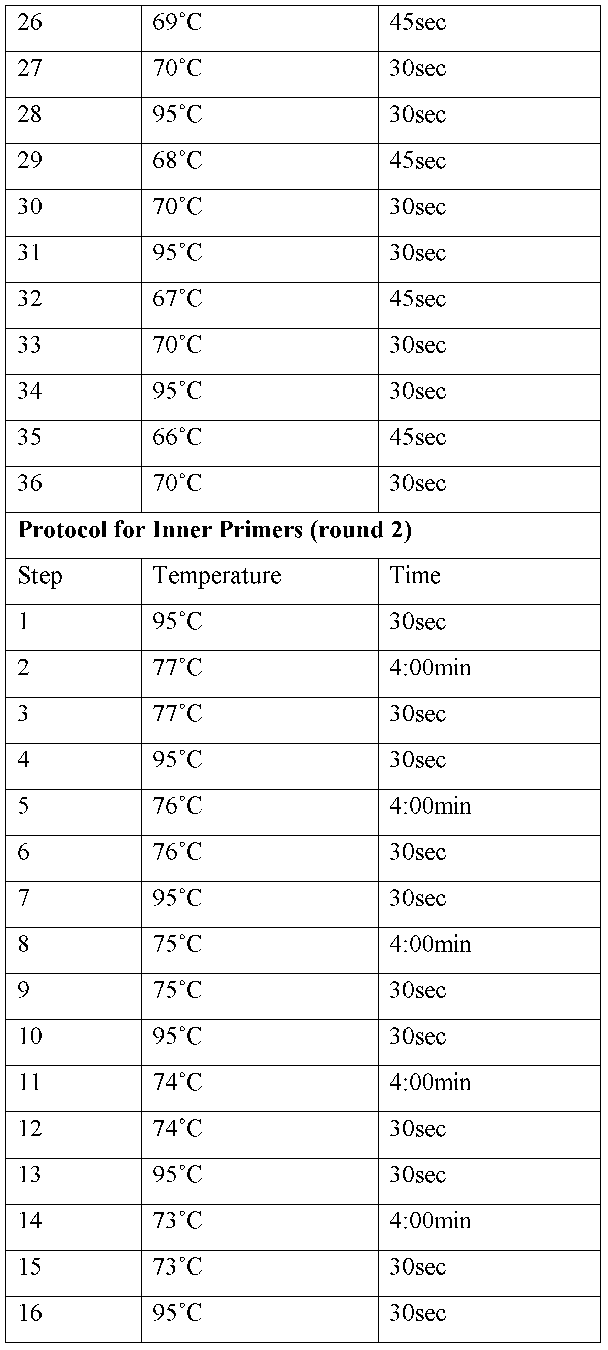


Example 4: NA- 17 TCR Isolation
NA- 17 is a melanoma-specific antigen. T Cells were obtained from a patient sample and stained with an NY-ESO specific tetramer. Genomic DNA from eight NA- 17+ CD3+ cells was amplified and used as template for a TCR isolation protocol. These cells yielded four distinct alpha chains and five distinct beta chains: TRAV12-1J11, TRAV12-3J9, TRAV16J28, TRAV17J10, TRBV12-3J1-6, TRBV27J1-2, TRBV27J1-6, TRBV5-5J1-3, TRBV5-6J1-1. Some chains were found to be expressed in multiple cells. Without subscribing to any one theory, these results may indicate that the cells were clonal or that individual wells contained multiple cells, making pairing ambiguous.
Claims
1. A method of isolating DNA encoding the variable regions of a T cell receptor (TCR) alpha or beta chain, said method comprising: (a) isolating genomic DNA from a single T cell; (b) amplifying a gene segment encompassing the TCR alpha or beta chain variable region by an enrichment amplification reaction to produce a first enrichment product comprising the V and J regions of the TCR alpha or beta chain from the single cell, the amplification reaction comprising incubating the isolated genomic DNA with a set of outer primers comprising at least one primer complementary to each of the V and J regions of the TCR alpha chain or TCR beta chain.
2. The method of claim 1 wherein the first enrichment product is further amplified in an isolation amplification reaction to produce a second isolation product, the isolation amplification reaction comprising incubating the first enrichment product with a set of inner primers comprising at least one primer complementary to each of the V and J regions of the TCR alpha chain or TCR beta chain.
3. The method of claim 2 wherein the second isolation product is further amplified in a cloning amplification reaction to produce a third cloning product, the cloning amplification reaction comprising incubating the second isolation product with a set of cloning primers comprising at least one primer complementary to each of the V and J regions of the TCR alpha chain or TCR beta chain, and wherein each cloning primer also comprises a vector region that is homologous to a vector.
4. The method of claim 3 wherein the third cloning product is further amplified in a homologous amplification reaction to produce a fourth homologous product, the homologous amplification reaction comprising incubating the third cloning product with a set of homologous primers that are homologous to the vector region of the cloning primers.
5. The method of claim 1 wherein the amplification reaction comprises a PCR reaction.
6. The method of claim 5, wherein the PCR reaction utilizes a touchdown PCR protocol.
7. The method of claim 1, wherein isolating genomic DNA from a single T cell comprises whole genome amplification
8. The method of claim 1 additionally comprising screening the isolated genomic DNA obtained in step (a) for the presence of T cell receptor constant regions prior to step (b).
9. The method of claim 1, wherein the outer primers anneal to a sequence about 5 to 40 base pairs upstream of the signal sequence junction of the alpha or beta V region or a sequence about 5 to 50 base pairs downstream of the exon/intron junction of the alpha or beta J region.
10. The method of claim 2, wherein the set of inner primers comprises forward and reverse inner primers, wherein each forward inner primer anneals to a sequence at the start of the first amino acid downstream of the signal sequence of the alpha or beta V region and each reverse inner primer anneals to a sequence at the downstream end of the alpha or beta J region.
11. The method of claim 3, wherein the vector region comprises about 15 bases of homology to the vector.
12. The method of claim 2, additionally comprising sequencing the isolation product.
13. The method of claim 3, additionally comprising inserting the cloning product into the vector.
14. The method of claim 13, wherein the vector is an expression vector.
15. A method of isolating the variable regions of a T-cell receptor (TCR) alpha or beta chain, said method comprising: (a) isolating a single T cell; (b) performing whole genome amplification to amplify the genomic DNA of the T cell; (c) incubating the amplified genomic DNA with a set of outer primers in a genomic TCR alpha enrichment amplification reaction or genomic TCR beta enrichment amplification reaction to produce an enrichment product, wherein the set of primers comprises at least one outer primer complementary to substantially each V and J region of the TCR alpha chain or TCR beta chain; and (d) incubating the enrichment product with a set of inner primers in an TCR alpha isolation amplification reaction or TCR beta isolation amplification reaction to produce an isolation product, wherein the set of inner primers comprises at least one inner primer complementary to each of the V and J regions of the TCR alpha chain or TCR beta chain.
16. The method of claim 15 comprising the additional step of (e) incubating the isolation product with a set of cloning primers in a cloning amplification reaction to produce a cloning product, wherein the set of cloning primers comprises at least one cloning primer complementary to each of the V and J regions of the TCR alpha chain or TCR beta chain.
17. The method of claim 9 comprising the additional step of (f) incubating the cloning product with a set of homologous primers in a homologous amplification reaction.
18. A kit comprising a T-cell receptor (TCR) alpha outer primer set, a TCR beta outer primer set, a TCR alpha inner primer set and a TCR beta inner primer set, wherein each of the TCR alpha inner and outer primer sets comprises at least one primer complementary to each of the V and J regions of a TCR alpha chain and each of the TCR beta inner and outer primer sets comprises at least one primer complementary to each of the V and J regions of a TCR beta chain.
19. The kit of claim 18, additionally comprising a TCR alpha cloning primer set and a TCR beta cloning primer set, wherein each of the primers in the TCR alpha and TCR beta cloning primer sets comprises a region of homology to a vector of interest.
20. The kit of claim 19, additionally comprising the vector of interest.
Priority Applications (2)
| Application Number | Priority Date | Filing Date | Title |
|---|---|---|---|
| JP2012517831A JP5829606B2 (en) | 2009-06-29 | 2010-06-28 | Isolation of unknown rearranged T cell receptors from single cells |
| EP10800274.2A EP2449103B1 (en) | 2009-06-29 | 2010-06-28 | Isolation of unknown rearranged t-cell receptors from single cells |
Applications Claiming Priority (2)
| Application Number | Priority Date | Filing Date | Title |
|---|---|---|---|
| US22150509P | 2009-06-29 | 2009-06-29 | |
| US61/221,505 | 2009-06-29 |
Publications (2)
| Publication Number | Publication Date |
|---|---|
| WO2011008502A2 true WO2011008502A2 (en) | 2011-01-20 |
| WO2011008502A3 WO2011008502A3 (en) | 2011-05-19 |
Family
ID=43450074
Family Applications (1)
| Application Number | Title | Priority Date | Filing Date |
|---|---|---|---|
| PCT/US2010/040245 Ceased WO2011008502A2 (en) | 2009-06-29 | 2010-06-28 | Isolation of unknown rearranged t-cell receptors from single cells |
Country Status (4)
| Country | Link |
|---|---|
| US (1) | US8497071B2 (en) |
| EP (1) | EP2449103B1 (en) |
| JP (1) | JP5829606B2 (en) |
| WO (1) | WO2011008502A2 (en) |
Cited By (56)
| Publication number | Priority date | Publication date | Assignee | Title |
|---|---|---|---|---|
| US20160009781A1 (en) | 2012-09-12 | 2016-01-14 | International Institute Of Cancer Immunology, Inc. | Antigen-specific helper t-cell receptor genes |
| WO2021247568A1 (en) | 2020-06-02 | 2021-12-09 | 10X Genomics, Inc. | Spatial trancriptomics for antigen-receptors |
| US11299774B2 (en) | 2015-04-10 | 2022-04-12 | Spatial Transcriptomics Ab | Spatially distinguished, multiplex nucleic acid analysis of biological specimens |
| US11352659B2 (en) | 2011-04-13 | 2022-06-07 | Spatial Transcriptomics Ab | Methods of detecting analytes |
| US11359228B2 (en) | 2013-06-25 | 2022-06-14 | Prognosys Biosciences, Inc. | Methods and systems for determining spatial patterns of biological targets in a sample |
| US11365442B2 (en) | 2010-04-05 | 2022-06-21 | Prognosys Biosciences, Inc. | Spatially encoded biological assays |
| US11407992B2 (en) | 2020-06-08 | 2022-08-09 | 10X Genomics, Inc. | Methods of determining a surgical margin and methods of use thereof |
| US11408029B2 (en) | 2020-06-25 | 2022-08-09 | 10X Genomics, Inc. | Spatial analysis of DNA methylation |
| US11434524B2 (en) | 2020-06-10 | 2022-09-06 | 10X Genomics, Inc. | Methods for determining a location of an analyte in a biological sample |
| US11505828B2 (en) | 2019-12-23 | 2022-11-22 | 10X Genomics, Inc. | Methods for spatial analysis using RNA-templated ligation |
| US11519033B2 (en) | 2018-08-28 | 2022-12-06 | 10X Genomics, Inc. | Method for transposase-mediated spatial tagging and analyzing genomic DNA in a biological sample |
| US11535887B2 (en) | 2020-04-22 | 2022-12-27 | 10X Genomics, Inc. | Methods for spatial analysis using targeted RNA depletion |
| US11560592B2 (en) | 2020-05-26 | 2023-01-24 | 10X Genomics, Inc. | Method for resetting an array |
| US11608498B2 (en) | 2020-06-02 | 2023-03-21 | 10X Genomics, Inc. | Nucleic acid library methods |
| US11608520B2 (en) | 2020-05-22 | 2023-03-21 | 10X Genomics, Inc. | Spatial analysis to detect sequence variants |
| US11618897B2 (en) | 2020-12-21 | 2023-04-04 | 10X Genomics, Inc. | Methods, compositions, and systems for capturing probes and/or barcodes |
| US11624086B2 (en) | 2020-05-22 | 2023-04-11 | 10X Genomics, Inc. | Simultaneous spatio-temporal measurement of gene expression and cellular activity |
| US11649485B2 (en) | 2019-01-06 | 2023-05-16 | 10X Genomics, Inc. | Generating capture probes for spatial analysis |
| US11702698B2 (en) | 2019-11-08 | 2023-07-18 | 10X Genomics, Inc. | Enhancing specificity of analyte binding |
| US11702693B2 (en) | 2020-01-21 | 2023-07-18 | 10X Genomics, Inc. | Methods for printing cells and generating arrays of barcoded cells |
| US11732300B2 (en) | 2020-02-05 | 2023-08-22 | 10X Genomics, Inc. | Increasing efficiency of spatial analysis in a biological sample |
| US11733238B2 (en) | 2010-04-05 | 2023-08-22 | Prognosys Biosciences, Inc. | Spatially encoded biological assays |
| US11732299B2 (en) | 2020-01-21 | 2023-08-22 | 10X Genomics, Inc. | Spatial assays with perturbed cells |
| US11753673B2 (en) | 2021-09-01 | 2023-09-12 | 10X Genomics, Inc. | Methods, compositions, and kits for blocking a capture probe on a spatial array |
| US11761038B1 (en) | 2020-07-06 | 2023-09-19 | 10X Genomics, Inc. | Methods for identifying a location of an RNA in a biological sample |
| US11827935B1 (en) | 2020-11-19 | 2023-11-28 | 10X Genomics, Inc. | Methods for spatial analysis using rolling circle amplification and detection probes |
| US11835462B2 (en) | 2020-02-11 | 2023-12-05 | 10X Genomics, Inc. | Methods and compositions for partitioning a biological sample |
| US11891654B2 (en) | 2020-02-24 | 2024-02-06 | 10X Genomics, Inc. | Methods of making gene expression libraries |
| US11898205B2 (en) | 2020-02-03 | 2024-02-13 | 10X Genomics, Inc. | Increasing capture efficiency of spatial assays |
| US11926822B1 (en) | 2020-09-23 | 2024-03-12 | 10X Genomics, Inc. | Three-dimensional spatial analysis |
| US11926867B2 (en) | 2019-01-06 | 2024-03-12 | 10X Genomics, Inc. | Generating capture probes for spatial analysis |
| US11926863B1 (en) | 2020-02-27 | 2024-03-12 | 10X Genomics, Inc. | Solid state single cell method for analyzing fixed biological cells |
| US11933957B1 (en) | 2018-12-10 | 2024-03-19 | 10X Genomics, Inc. | Imaging system hardware |
| US11965213B2 (en) | 2019-05-30 | 2024-04-23 | 10X Genomics, Inc. | Methods of detecting spatial heterogeneity of a biological sample |
| US11981960B1 (en) | 2020-07-06 | 2024-05-14 | 10X Genomics, Inc. | Spatial analysis utilizing degradable hydrogels |
| US11981958B1 (en) | 2020-08-20 | 2024-05-14 | 10X Genomics, Inc. | Methods for spatial analysis using DNA capture |
| US12031177B1 (en) | 2020-06-04 | 2024-07-09 | 10X Genomics, Inc. | Methods of enhancing spatial resolution of transcripts |
| USRE50065E1 (en) | 2012-10-17 | 2024-07-30 | 10X Genomics Sweden Ab | Methods and product for optimising localised or spatial detection of gene expression in a tissue sample |
| US12071655B2 (en) | 2021-06-03 | 2024-08-27 | 10X Genomics, Inc. | Methods, compositions, kits, and systems for enhancing analyte capture for spatial analysis |
| US12076701B2 (en) | 2020-01-31 | 2024-09-03 | 10X Genomics, Inc. | Capturing oligonucleotides in spatial transcriptomics |
| US12110541B2 (en) | 2020-02-03 | 2024-10-08 | 10X Genomics, Inc. | Methods for preparing high-resolution spatial arrays |
| US12117439B2 (en) | 2019-12-23 | 2024-10-15 | 10X Genomics, Inc. | Compositions and methods for using fixed biological samples |
| US12129516B2 (en) | 2020-02-07 | 2024-10-29 | 10X Genomics, Inc. | Quantitative and automated permeabilization performance evaluation for spatial transcriptomics |
| US12195790B2 (en) | 2021-12-01 | 2025-01-14 | 10X Genomics, Inc. | Methods for improved in situ detection of nucleic acids and spatial analysis |
| US12203134B2 (en) | 2021-04-14 | 2025-01-21 | 10X Genomics, Inc. | Methods of measuring mislocalization of an analyte |
| US12209280B1 (en) | 2020-07-06 | 2025-01-28 | 10X Genomics, Inc. | Methods of identifying abundance and location of an analyte in a biological sample using second strand synthesis |
| US12265079B1 (en) | 2020-06-02 | 2025-04-01 | 10X Genomics, Inc. | Systems and methods for detecting analytes from captured single biological particles |
| US12275988B2 (en) | 2021-11-10 | 2025-04-15 | 10X Genomics, Inc. | Methods, compositions, and kits for determining the location of an analyte in a biological sample |
| US12281357B1 (en) | 2020-02-14 | 2025-04-22 | 10X Genomics, Inc. | In situ spatial barcoding |
| US12297486B2 (en) | 2020-01-24 | 2025-05-13 | 10X Genomics, Inc. | Methods for spatial analysis using proximity ligation |
| US12365935B2 (en) | 2021-05-06 | 2025-07-22 | 10X Genomics, Inc. | Methods for increasing resolution of spatial analysis |
| US12365942B2 (en) | 2020-01-13 | 2025-07-22 | 10X Genomics, Inc. | Methods of decreasing background on a spatial array |
| US12405264B2 (en) | 2020-01-17 | 2025-09-02 | 10X Genomics, Inc. | Electrophoretic system and method for analyte capture |
| US12529094B2 (en) | 2018-12-10 | 2026-01-20 | 10X Genomics, Inc. | Imaging system hardware |
| US12566113B2 (en) | 2019-12-23 | 2026-03-03 | 10X Genomics, Inc. | Reversible fixing reagents and methods of use thereof |
| US12571029B1 (en) | 2022-11-09 | 2026-03-10 | 10X Genomics, Inc. | Methods, compositions, and kits for determining the location of multiple analytes in a biological sample |
Families Citing this family (39)
| Publication number | Priority date | Publication date | Assignee | Title |
|---|---|---|---|---|
| US9528160B2 (en) | 2008-11-07 | 2016-12-27 | Adaptive Biotechnolgies Corp. | Rare clonotypes and uses thereof |
| US8748103B2 (en) | 2008-11-07 | 2014-06-10 | Sequenta, Inc. | Monitoring health and disease status using clonotype profiles |
| US9506119B2 (en) | 2008-11-07 | 2016-11-29 | Adaptive Biotechnologies Corp. | Method of sequence determination using sequence tags |
| US8628927B2 (en) | 2008-11-07 | 2014-01-14 | Sequenta, Inc. | Monitoring health and disease status using clonotype profiles |
| EP2364368B1 (en) | 2008-11-07 | 2014-01-15 | Sequenta, Inc. | Methods of monitoring conditions by sequence analysis |
| US9365901B2 (en) | 2008-11-07 | 2016-06-14 | Adaptive Biotechnologies Corp. | Monitoring immunoglobulin heavy chain evolution in B-cell acute lymphoblastic leukemia |
| ES2726702T3 (en) | 2009-01-15 | 2019-10-08 | Adaptive Biotechnologies Corp | Adaptive immunity profiling and methods for the generation of monoclonal antibodies |
| WO2010151416A1 (en) | 2009-06-25 | 2010-12-29 | Fred Hutchinson Cancer Research Center | Method of measuring adaptive immunity |
| US9480241B2 (en) * | 2011-07-05 | 2016-11-01 | Eric James Holmstrom | Retractable leash system |
| US10385475B2 (en) | 2011-09-12 | 2019-08-20 | Adaptive Biotechnologies Corp. | Random array sequencing of low-complexity libraries |
| EP2768982A4 (en) * | 2011-10-21 | 2015-06-03 | Adaptive Biotechnologies Corp | QUANTIFICATION OF GENOMES OF ADAPTIVE IMMUNE CELLS IN A COMPLEX MIXTURE OF CELLS |
| US9824179B2 (en) | 2011-12-09 | 2017-11-21 | Adaptive Biotechnologies Corp. | Diagnosis of lymphoid malignancies and minimal residual disease detection |
| US9499865B2 (en) | 2011-12-13 | 2016-11-22 | Adaptive Biotechnologies Corp. | Detection and measurement of tissue-infiltrating lymphocytes |
| EP3372694A1 (en) | 2012-03-05 | 2018-09-12 | Adaptive Biotechnologies Corporation | Determining paired immune receptor chains from frequency matched subunits |
| HUE029357T2 (en) | 2012-05-08 | 2017-02-28 | Adaptive Biotechnologies Corp | Compositions and method for measuring and calibrating amplification bias in multiplexed pcr reactions |
| EP3330384B1 (en) | 2012-10-01 | 2019-09-25 | Adaptive Biotechnologies Corporation | Immunocompetence assessment by adaptive immune receptor diversity and clonality characterization |
| WO2015160439A2 (en) | 2014-04-17 | 2015-10-22 | Adaptive Biotechnologies Corporation | Quantification of adaptive immune cell genomes in a complex mixture of cells |
| GB2525568B (en) | 2013-03-15 | 2020-10-14 | Abvitro Llc | Single cell barcoding for antibody discovery |
| US9708657B2 (en) | 2013-07-01 | 2017-07-18 | Adaptive Biotechnologies Corp. | Method for generating clonotype profiles using sequence tags |
| US9884075B2 (en) | 2014-01-16 | 2018-02-06 | California Institute Of Technology | Domain-swap T cell receptors |
| WO2015134787A2 (en) | 2014-03-05 | 2015-09-11 | Adaptive Biotechnologies Corporation | Methods using randomer-containing synthetic molecules |
| US10066265B2 (en) | 2014-04-01 | 2018-09-04 | Adaptive Biotechnologies Corp. | Determining antigen-specific t-cells |
| US10202640B2 (en) | 2014-05-07 | 2019-02-12 | The Board Of Trustees Of The Leland Stanford Junior University | Single cell analysis of T cells using high-throughput multiplex amplification and deep sequencing |
| EP3950944A1 (en) | 2014-09-15 | 2022-02-09 | AbVitro LLC | High-throughput nucleotide library sequencing |
| CA2966201A1 (en) | 2014-10-29 | 2016-05-06 | Adaptive Biotechnologies Corp. | Highly-multiplexed simultaneous detection of nucleic acids encoding paired adaptive immune receptor heterodimers from many samples |
| US10246701B2 (en) | 2014-11-14 | 2019-04-02 | Adaptive Biotechnologies Corp. | Multiplexed digital quantitation of rearranged lymphoid receptors in a complex mixture |
| AU2015353581A1 (en) | 2014-11-25 | 2017-06-15 | Adaptive Biotechnologies Corporation | Characterization of adaptive immune response to vaccination or infection using immune repertoire sequencing |
| US20180266726A1 (en) * | 2015-01-23 | 2018-09-20 | Camus Hydronics Ltd. | Fire Tube Heater |
| CA2976580A1 (en) | 2015-02-24 | 2016-09-01 | Adaptive Biotechnologies Corp. | Methods for diagnosing infectious disease and determining hla status using immune repertoire sequencing |
| EP3277294B1 (en) | 2015-04-01 | 2024-05-15 | Adaptive Biotechnologies Corp. | Method of identifying human compatible t cell receptors specific for an antigenic target |
| US10428325B1 (en) | 2016-09-21 | 2019-10-01 | Adaptive Biotechnologies Corporation | Identification of antigen-specific B cell receptors |
| KR102702552B1 (en) | 2016-12-23 | 2024-09-03 | 비스테라, 인크. | Conjugated polypeptide and method for producing same |
| US11254980B1 (en) | 2017-11-29 | 2022-02-22 | Adaptive Biotechnologies Corporation | Methods of profiling targeted polynucleotides while mitigating sequencing depth requirements |
| US12247060B2 (en) | 2018-01-09 | 2025-03-11 | Marengo Therapeutics, Inc. | Calreticulin binding constructs and engineered T cells for the treatment of diseases |
| EP3765517A1 (en) | 2018-03-14 | 2021-01-20 | Elstar Therapeutics, Inc. | Multifunctional molecules that bind to calreticulin and uses thereof |
| CA3105448A1 (en) | 2018-07-03 | 2020-01-09 | Elstar Therapeutics, Inc. | Anti-tcr antibody molecules and uses thereof |
| GB2599228B (en) | 2019-02-21 | 2024-02-07 | Marengo Therapeutics Inc | Multifunctional molecules that bind to T cell related cancer cells and uses thereof |
| CN119039441A (en) | 2019-02-21 | 2024-11-29 | 马伦戈治疗公司 | Antibody molecules that bind to NKP30 and uses thereof |
| AU2020416273A1 (en) | 2020-01-03 | 2022-07-28 | Marengo Therapeutics, Inc. | Anti-TCR antibody molecules and uses thereof |
Family Cites Families (8)
| Publication number | Priority date | Publication date | Assignee | Title |
|---|---|---|---|---|
| EP0570027B1 (en) * | 1984-03-01 | 1999-12-29 | The Board Of Trustees Of The Leland Stanford Junior University | T cell receptor-specific for antigen polypeptides and related polynucleotides |
| US6221352B1 (en) * | 1989-03-21 | 2001-04-24 | The Immune Response Corporation | Method of preventing the proliferation of Vβ14 or Vβ17-Expressing T cells |
| US7294712B2 (en) * | 1990-06-04 | 2007-11-13 | Aventis Pharma S.A. | Nucleotide sequences coding for variable regions of β chains of human T lymphocyte receptors, corresponding peptide segments and the diagnostic and therapeutic uses |
| DK0529033T3 (en) * | 1991-02-08 | 2007-01-08 | Aventis Pharma Sa | Nucleotide sequences encoding variable regions of α-chains in human lymphocyte receptors and their applications |
| US6087096A (en) * | 1995-11-13 | 2000-07-11 | Dau; Peter C. | Method of intrafamily fragment analysis of the T cell receptor α and β chain CDR3 regions |
| US6416948B1 (en) * | 1996-06-03 | 2002-07-09 | The Governors Of The University Of Alberta | Methods for detection of rearranged DNA |
| WO2004015070A2 (en) * | 2002-08-08 | 2004-02-19 | Baylor College Of Medicine | Isolation and identification of t cells |
| US7375211B2 (en) * | 2005-11-18 | 2008-05-20 | Kou Zhong C | Method for detection and quantification of T-cell receptor Vβ repertoire |
-
2010
- 2010-06-28 US US12/824,744 patent/US8497071B2/en not_active Expired - Fee Related
- 2010-06-28 WO PCT/US2010/040245 patent/WO2011008502A2/en not_active Ceased
- 2010-06-28 EP EP10800274.2A patent/EP2449103B1/en not_active Not-in-force
- 2010-06-28 JP JP2012517831A patent/JP5829606B2/en not_active Expired - Fee Related
Non-Patent Citations (6)
| Title |
|---|
| DEAN ET AL., PROC. NATL. ACAD. SCI. USA, vol. 99, 2002, pages 5261 - 66 |
| LANGMORE, J.T, GENOME RES., vol. 14, 2002, pages 901 - 07 |
| See also references of EP2449103A4 |
| SPITS, C. ET AL., NATURE PROTOCOLS, vol. 1, 2006, pages 1965 - 70 |
| TELENIUS, H ET AL., GENOMICS, vol. 13, 1992, pages 718 - 25 |
| ZHANG ET AL., PROC NATL ACAD SCI USA, vol. 89, 1992, pages 5847 - 51 |
Cited By (119)
| Publication number | Priority date | Publication date | Assignee | Title |
|---|---|---|---|---|
| US11560587B2 (en) | 2010-04-05 | 2023-01-24 | Prognosys Biosciences, Inc. | Spatially encoded biological assays |
| US11767550B2 (en) | 2010-04-05 | 2023-09-26 | Prognosys Biosciences, Inc. | Spatially encoded biological assays |
| US12234505B2 (en) | 2010-04-05 | 2025-02-25 | Prognosys Biosciences, Inc. | Spatially encoded biological assays |
| US11634756B2 (en) | 2010-04-05 | 2023-04-25 | Prognosys Biosciences, Inc. | Spatially encoded biological assays |
| US12297487B2 (en) | 2010-04-05 | 2025-05-13 | Prognosys Biosciences, Inc. | Spatially encoded biological assays |
| US11519022B2 (en) | 2010-04-05 | 2022-12-06 | Prognosys Biosciences, Inc. | Spatially encoded biological assays |
| US11733238B2 (en) | 2010-04-05 | 2023-08-22 | Prognosys Biosciences, Inc. | Spatially encoded biological assays |
| US11866770B2 (en) | 2010-04-05 | 2024-01-09 | Prognosys Biosciences, Inc. | Spatially encoded biological assays |
| US11365442B2 (en) | 2010-04-05 | 2022-06-21 | Prognosys Biosciences, Inc. | Spatially encoded biological assays |
| US11371086B2 (en) | 2010-04-05 | 2022-06-28 | Prognosys Biosciences, Inc. | Spatially encoded biological assays |
| US11384386B2 (en) | 2010-04-05 | 2022-07-12 | Prognosys Biosciences, Inc. | Spatially encoded biological assays |
| US11732292B2 (en) | 2010-04-05 | 2023-08-22 | Prognosys Biosciences, Inc. | Spatially encoded biological assays correlating target nucleic acid to tissue section location |
| US11401545B2 (en) | 2010-04-05 | 2022-08-02 | Prognosys Biosciences, Inc. | Spatially encoded biological assays |
| US12297488B2 (en) | 2010-04-05 | 2025-05-13 | Prognosys Biosciences, Inc. | Spatially encoded biological assays |
| US12391980B2 (en) | 2010-04-05 | 2025-08-19 | Prognosys Biosciences, Inc. | Spatially encoded biological assays |
| US11549138B2 (en) | 2010-04-05 | 2023-01-10 | Prognosys Biosciences, Inc. | Spatially encoded biological assays |
| US11542543B2 (en) | 2010-04-05 | 2023-01-03 | Prognosys Biosciences, Inc. | System for analyzing targets of a tissue section |
| US11479810B1 (en) | 2010-04-05 | 2022-10-25 | Prognosys Biosciences, Inc. | Spatially encoded biological assays |
| US12391979B2 (en) | 2010-04-05 | 2025-08-19 | Prognosys Biosciences, Inc. | Spatially encoded biological assays |
| US11761030B2 (en) | 2010-04-05 | 2023-09-19 | Prognosys Biosciences, Inc. | Spatially encoded biological assays |
| US11352659B2 (en) | 2011-04-13 | 2022-06-07 | Spatial Transcriptomics Ab | Methods of detecting analytes |
| US11479809B2 (en) | 2011-04-13 | 2022-10-25 | Spatial Transcriptomics Ab | Methods of detecting analytes |
| US11788122B2 (en) | 2011-04-13 | 2023-10-17 | 10X Genomics Sweden Ab | Methods of detecting analytes |
| US11795498B2 (en) | 2011-04-13 | 2023-10-24 | 10X Genomics Sweden Ab | Methods of detecting analytes |
| US20160009781A1 (en) | 2012-09-12 | 2016-01-14 | International Institute Of Cancer Immunology, Inc. | Antigen-specific helper t-cell receptor genes |
| JP2018143247A (en) * | 2012-09-12 | 2018-09-20 | 株式会社癌免疫研究所 | Antigen-specific helper T cell receptor gene |
| US10815288B2 (en) | 2012-09-12 | 2020-10-27 | International Institute Of Cancer Immunology, Inc. | Antigen-specific helper T-cell receptor genes |
| US11091531B2 (en) | 2012-09-12 | 2021-08-17 | International Institute Of Cancer Immunology, Inc. | Antigen-specific helper T-cell receptor genes |
| USRE50065E1 (en) | 2012-10-17 | 2024-07-30 | 10X Genomics Sweden Ab | Methods and product for optimising localised or spatial detection of gene expression in a tissue sample |
| US11618918B2 (en) | 2013-06-25 | 2023-04-04 | Prognosys Biosciences, Inc. | Methods and systems for determining spatial patterns of biological targets in a sample |
| US11359228B2 (en) | 2013-06-25 | 2022-06-14 | Prognosys Biosciences, Inc. | Methods and systems for determining spatial patterns of biological targets in a sample |
| US11821024B2 (en) | 2013-06-25 | 2023-11-21 | Prognosys Biosciences, Inc. | Methods and systems for determining spatial patterns of biological targets in a sample |
| US11753674B2 (en) | 2013-06-25 | 2023-09-12 | Prognosys Biosciences, Inc. | Methods and systems for determining spatial patterns of biological targets in a sample |
| US11613773B2 (en) | 2015-04-10 | 2023-03-28 | Spatial Transcriptomics Ab | Spatially distinguished, multiplex nucleic acid analysis of biological specimens |
| US11739372B2 (en) | 2015-04-10 | 2023-08-29 | Spatial Transcriptomics Ab | Spatially distinguished, multiplex nucleic acid analysis of biological specimens |
| US11390912B2 (en) | 2015-04-10 | 2022-07-19 | Spatial Transcriptomics Ab | Spatially distinguished, multiplex nucleic acid analysis of biological specimens |
| US11299774B2 (en) | 2015-04-10 | 2022-04-12 | Spatial Transcriptomics Ab | Spatially distinguished, multiplex nucleic acid analysis of biological specimens |
| US12344892B2 (en) | 2018-08-28 | 2025-07-01 | 10X Genomics, Inc. | Method for transposase-mediated spatial tagging and analyzing genomic DNA in a biological sample |
| US12270077B2 (en) | 2018-08-28 | 2025-04-08 | 10X Genomics, Inc. | Method for transposase-mediated spatial tagging and analyzing genomic DNA in a biological sample |
| US11519033B2 (en) | 2018-08-28 | 2022-12-06 | 10X Genomics, Inc. | Method for transposase-mediated spatial tagging and analyzing genomic DNA in a biological sample |
| US12378607B2 (en) | 2018-08-28 | 2025-08-05 | 10X Genomics, Inc. | Method for transposase-mediated spatial tagging and analyzing genomic DNA in a biological sample |
| US11933957B1 (en) | 2018-12-10 | 2024-03-19 | 10X Genomics, Inc. | Imaging system hardware |
| US12529094B2 (en) | 2018-12-10 | 2026-01-20 | 10X Genomics, Inc. | Imaging system hardware |
| US12404544B2 (en) | 2018-12-10 | 2025-09-02 | 10X Genomics, Inc. | Methods of capturing multiple analytes on a spatial array |
| US12180543B2 (en) | 2018-12-10 | 2024-12-31 | 10X Genomics, Inc. | Imaging system hardware |
| US11753675B2 (en) | 2019-01-06 | 2023-09-12 | 10X Genomics, Inc. | Generating capture probes for spatial analysis |
| US11926867B2 (en) | 2019-01-06 | 2024-03-12 | 10X Genomics, Inc. | Generating capture probes for spatial analysis |
| US11649485B2 (en) | 2019-01-06 | 2023-05-16 | 10X Genomics, Inc. | Generating capture probes for spatial analysis |
| US12442045B2 (en) | 2019-05-30 | 2025-10-14 | 10X Genomics, Inc. | Methods of detecting spatial heterogeneity of a biological sample |
| US11965213B2 (en) | 2019-05-30 | 2024-04-23 | 10X Genomics, Inc. | Methods of detecting spatial heterogeneity of a biological sample |
| US11702698B2 (en) | 2019-11-08 | 2023-07-18 | 10X Genomics, Inc. | Enhancing specificity of analyte binding |
| US12566113B2 (en) | 2019-12-23 | 2026-03-03 | 10X Genomics, Inc. | Reversible fixing reagents and methods of use thereof |
| US12117439B2 (en) | 2019-12-23 | 2024-10-15 | 10X Genomics, Inc. | Compositions and methods for using fixed biological samples |
| US11505828B2 (en) | 2019-12-23 | 2022-11-22 | 10X Genomics, Inc. | Methods for spatial analysis using RNA-templated ligation |
| US12241890B2 (en) | 2019-12-23 | 2025-03-04 | 10X Genomics, Inc. | Methods for generating barcoded nucleic acid molecules using fixed cells |
| US11560593B2 (en) | 2019-12-23 | 2023-01-24 | 10X Genomics, Inc. | Methods for spatial analysis using RNA-templated ligation |
| US11795507B2 (en) | 2019-12-23 | 2023-10-24 | 10X Genomics, Inc. | Methods for spatial analysis using RNA-templated ligation |
| US11981965B2 (en) | 2019-12-23 | 2024-05-14 | 10X Genomics, Inc. | Methods for spatial analysis using RNA-templated ligation |
| US12365942B2 (en) | 2020-01-13 | 2025-07-22 | 10X Genomics, Inc. | Methods of decreasing background on a spatial array |
| US12405264B2 (en) | 2020-01-17 | 2025-09-02 | 10X Genomics, Inc. | Electrophoretic system and method for analyte capture |
| US11702693B2 (en) | 2020-01-21 | 2023-07-18 | 10X Genomics, Inc. | Methods for printing cells and generating arrays of barcoded cells |
| US11732299B2 (en) | 2020-01-21 | 2023-08-22 | 10X Genomics, Inc. | Spatial assays with perturbed cells |
| US12297486B2 (en) | 2020-01-24 | 2025-05-13 | 10X Genomics, Inc. | Methods for spatial analysis using proximity ligation |
| US12076701B2 (en) | 2020-01-31 | 2024-09-03 | 10X Genomics, Inc. | Capturing oligonucleotides in spatial transcriptomics |
| US12110541B2 (en) | 2020-02-03 | 2024-10-08 | 10X Genomics, Inc. | Methods for preparing high-resolution spatial arrays |
| US11898205B2 (en) | 2020-02-03 | 2024-02-13 | 10X Genomics, Inc. | Increasing capture efficiency of spatial assays |
| US12286673B2 (en) | 2020-02-05 | 2025-04-29 | 10X Genomics, Inc. | Increasing efficiency of spatial analysis in a biological sample |
| US11732300B2 (en) | 2020-02-05 | 2023-08-22 | 10X Genomics, Inc. | Increasing efficiency of spatial analysis in a biological sample |
| US12129516B2 (en) | 2020-02-07 | 2024-10-29 | 10X Genomics, Inc. | Quantitative and automated permeabilization performance evaluation for spatial transcriptomics |
| US11835462B2 (en) | 2020-02-11 | 2023-12-05 | 10X Genomics, Inc. | Methods and compositions for partitioning a biological sample |
| US12281357B1 (en) | 2020-02-14 | 2025-04-22 | 10X Genomics, Inc. | In situ spatial barcoding |
| US11891654B2 (en) | 2020-02-24 | 2024-02-06 | 10X Genomics, Inc. | Methods of making gene expression libraries |
| US11926863B1 (en) | 2020-02-27 | 2024-03-12 | 10X Genomics, Inc. | Solid state single cell method for analyzing fixed biological cells |
| US11535887B2 (en) | 2020-04-22 | 2022-12-27 | 10X Genomics, Inc. | Methods for spatial analysis using targeted RNA depletion |
| US11773433B2 (en) | 2020-04-22 | 2023-10-03 | 10X Genomics, Inc. | Methods for spatial analysis using targeted RNA depletion |
| US11624086B2 (en) | 2020-05-22 | 2023-04-11 | 10X Genomics, Inc. | Simultaneous spatio-temporal measurement of gene expression and cellular activity |
| US11959130B2 (en) | 2020-05-22 | 2024-04-16 | 10X Genomics, Inc. | Spatial analysis to detect sequence variants |
| US11608520B2 (en) | 2020-05-22 | 2023-03-21 | 10X Genomics, Inc. | Spatial analysis to detect sequence variants |
| US11866767B2 (en) | 2020-05-22 | 2024-01-09 | 10X Genomics, Inc. | Simultaneous spatio-temporal measurement of gene expression and cellular activity |
| US11560592B2 (en) | 2020-05-26 | 2023-01-24 | 10X Genomics, Inc. | Method for resetting an array |
| US11840687B2 (en) | 2020-06-02 | 2023-12-12 | 10X Genomics, Inc. | Nucleic acid library methods |
| WO2021247568A1 (en) | 2020-06-02 | 2021-12-09 | 10X Genomics, Inc. | Spatial trancriptomics for antigen-receptors |
| US11859178B2 (en) | 2020-06-02 | 2024-01-02 | 10X Genomics, Inc. | Nucleic acid library methods |
| US12265079B1 (en) | 2020-06-02 | 2025-04-01 | 10X Genomics, Inc. | Systems and methods for detecting analytes from captured single biological particles |
| EP4600376A2 (en) | 2020-06-02 | 2025-08-13 | 10x Genomics, Inc. | Spatial transcriptomics for antigen-receptors |
| US11692218B2 (en) | 2020-06-02 | 2023-07-04 | 10X Genomics, Inc. | Spatial transcriptomics for antigen-receptors |
| US12098417B2 (en) | 2020-06-02 | 2024-09-24 | 10X Genomics, Inc. | Spatial transcriptomics for antigen-receptors |
| US11845979B2 (en) | 2020-06-02 | 2023-12-19 | 10X Genomics, Inc. | Spatial transcriptomics for antigen-receptors |
| US11608498B2 (en) | 2020-06-02 | 2023-03-21 | 10X Genomics, Inc. | Nucleic acid library methods |
| US12031177B1 (en) | 2020-06-04 | 2024-07-09 | 10X Genomics, Inc. | Methods of enhancing spatial resolution of transcripts |
| US11407992B2 (en) | 2020-06-08 | 2022-08-09 | 10X Genomics, Inc. | Methods of determining a surgical margin and methods of use thereof |
| US11492612B1 (en) | 2020-06-08 | 2022-11-08 | 10X Genomics, Inc. | Methods of determining a surgical margin and methods of use thereof |
| US11624063B2 (en) | 2020-06-08 | 2023-04-11 | 10X Genomics, Inc. | Methods of determining a surgical margin and methods of use thereof |
| US11781130B2 (en) | 2020-06-08 | 2023-10-10 | 10X Genomics, Inc. | Methods of determining a surgical margin and methods of use thereof |
| US11434524B2 (en) | 2020-06-10 | 2022-09-06 | 10X Genomics, Inc. | Methods for determining a location of an analyte in a biological sample |
| US11661626B2 (en) | 2020-06-25 | 2023-05-30 | 10X Genomics, Inc. | Spatial analysis of DNA methylation |
| US11408029B2 (en) | 2020-06-25 | 2022-08-09 | 10X Genomics, Inc. | Spatial analysis of DNA methylation |
| US12060604B2 (en) | 2020-06-25 | 2024-08-13 | 10X Genomics, Inc. | Spatial analysis of epigenetic modifications |
| US11981960B1 (en) | 2020-07-06 | 2024-05-14 | 10X Genomics, Inc. | Spatial analysis utilizing degradable hydrogels |
| US11761038B1 (en) | 2020-07-06 | 2023-09-19 | 10X Genomics, Inc. | Methods for identifying a location of an RNA in a biological sample |
| US11952627B2 (en) | 2020-07-06 | 2024-04-09 | 10X Genomics, Inc. | Methods for identifying a location of an RNA in a biological sample |
| US12209280B1 (en) | 2020-07-06 | 2025-01-28 | 10X Genomics, Inc. | Methods of identifying abundance and location of an analyte in a biological sample using second strand synthesis |
| US11981958B1 (en) | 2020-08-20 | 2024-05-14 | 10X Genomics, Inc. | Methods for spatial analysis using DNA capture |
| US11926822B1 (en) | 2020-09-23 | 2024-03-12 | 10X Genomics, Inc. | Three-dimensional spatial analysis |
| US11827935B1 (en) | 2020-11-19 | 2023-11-28 | 10X Genomics, Inc. | Methods for spatial analysis using rolling circle amplification and detection probes |
| US11959076B2 (en) | 2020-12-21 | 2024-04-16 | 10X Genomics, Inc. | Methods, compositions, and systems for capturing probes and/or barcodes |
| US12241060B2 (en) | 2020-12-21 | 2025-03-04 | 10X Genomics, Inc. | Methods, compositions, and systems for capturing probes and/or barcodes |
| US11873482B2 (en) | 2020-12-21 | 2024-01-16 | 10X Genomics, Inc. | Methods, compositions, and systems for spatial analysis of analytes in a biological sample |
| US12371688B2 (en) | 2020-12-21 | 2025-07-29 | 10X Genomics, Inc. | Methods, compositions, and systems for spatial analysis of analytes in a biological sample |
| US11618897B2 (en) | 2020-12-21 | 2023-04-04 | 10X Genomics, Inc. | Methods, compositions, and systems for capturing probes and/or barcodes |
| US11680260B2 (en) | 2020-12-21 | 2023-06-20 | 10X Genomics, Inc. | Methods, compositions, and systems for spatial analysis of analytes in a biological sample |
| US12203134B2 (en) | 2021-04-14 | 2025-01-21 | 10X Genomics, Inc. | Methods of measuring mislocalization of an analyte |
| US12365935B2 (en) | 2021-05-06 | 2025-07-22 | 10X Genomics, Inc. | Methods for increasing resolution of spatial analysis |
| US12071655B2 (en) | 2021-06-03 | 2024-08-27 | 10X Genomics, Inc. | Methods, compositions, kits, and systems for enhancing analyte capture for spatial analysis |
| US11840724B2 (en) | 2021-09-01 | 2023-12-12 | 10X Genomics, Inc. | Methods, compositions, and kits for blocking a capture probe on a spatial array |
| US11753673B2 (en) | 2021-09-01 | 2023-09-12 | 10X Genomics, Inc. | Methods, compositions, and kits for blocking a capture probe on a spatial array |
| US12275988B2 (en) | 2021-11-10 | 2025-04-15 | 10X Genomics, Inc. | Methods, compositions, and kits for determining the location of an analyte in a biological sample |
| US12195790B2 (en) | 2021-12-01 | 2025-01-14 | 10X Genomics, Inc. | Methods for improved in situ detection of nucleic acids and spatial analysis |
| US12571029B1 (en) | 2022-11-09 | 2026-03-10 | 10X Genomics, Inc. | Methods, compositions, and kits for determining the location of multiple analytes in a biological sample |
Also Published As
| Publication number | Publication date |
|---|---|
| JP5829606B2 (en) | 2015-12-09 |
| US8497071B2 (en) | 2013-07-30 |
| US20110014659A1 (en) | 2011-01-20 |
| EP2449103B1 (en) | 2016-08-03 |
| EP2449103A2 (en) | 2012-05-09 |
| EP2449103A4 (en) | 2013-07-24 |
| WO2011008502A3 (en) | 2011-05-19 |
| JP2012531895A (en) | 2012-12-13 |
Similar Documents
| Publication | Publication Date | Title |
|---|---|---|
| US8497071B2 (en) | Isolation of unknown rearranged T-cell receptors from single cells | |
| Lim et al. | Combination of MHC–peptide multimer-based T cell sorting with the Immunoscope permits sensitive ex vivo quantitation and follow-up of human CD8+ T cell immune responses | |
| EP3277294B1 (en) | Method of identifying human compatible t cell receptors specific for an antigenic target | |
| KR101773690B1 (en) | Cancer antigen-specific t-cell receptor gene, peptide encoded by the gene, and use of them | |
| JP6302847B2 (en) | Determination of paired immunoreceptor chains from frequency matched subunits | |
| EP3577234B1 (en) | Enhanced immune cell receptor sequencing methods | |
| WO2015059277A1 (en) | Method and kit for determining whether a subject shows an immune response | |
| EP3060679A1 (en) | Method and kit for determining whether a subject shows an immune response | |
| CN119923271A (en) | Vaccine constructs comprising tuberculosis antigens | |
| JP2023058626A (en) | Methods and materials for cloning functional T cell receptors from single T cells | |
| Bettinotti et al. | New HLA-A,-B, and-C locus-specific primers for PCR amplification from cDNA: application in clinical immunology | |
| Du et al. | Combined megaplex TCR isolation and SMART-based real-time quantitation methods for quantitating antigen-specific T cell clones in mycobacterial infection | |
| Ling et al. | A Comparison of Internal Eliminated Sequences in the Genes that Encode Two K+‐Channel Isoforms in Paramecium tetraurelia | |
| WO2015182749A1 (en) | TCR cDNA AMPLIFICATION METHOD | |
| JP2022521707A (en) | Reverse immunosuppression | |
| Johnston et al. | Spectratyping of TCRs expressed by CTL-infiltrating minor histocompatibility antigen-disparate allografts | |
| CA2979726C (en) | Method of identifying human compatible t cell receptors specific for an antigenic target | |
| HK40062498A (en) | Methods for cloning functional t cell receptors from single t cells | |
| HK40013267B (en) | Enhanced immune cell receptor sequencing methods | |
| CN116323898A (en) | Primers for more than one HLA gene | |
| HND | Gene expression profiling during early stages of infection of bovine lymphocytes by the parasite Theileria parva | |
| HK40013267A (en) | Enhanced immune cell receptor sequencing methods |
Legal Events
| Date | Code | Title | Description |
|---|---|---|---|
| 121 | Ep: the epo has been informed by wipo that ep was designated in this application |
Ref document number: 10800274 Country of ref document: EP Kind code of ref document: A2 |
|
| WWE | Wipo information: entry into national phase |
Ref document number: 2012517831 Country of ref document: JP |
|
| NENP | Non-entry into the national phase |
Ref country code: DE |
|
| REEP | Request for entry into the european phase |
Ref document number: 2010800274 Country of ref document: EP |
|
| WWE | Wipo information: entry into national phase |
Ref document number: 2010800274 Country of ref document: EP |