WO2011024592A1 - 肝細胞の培養方法 - Google Patents
肝細胞の培養方法 Download PDFInfo
- Publication number
- WO2011024592A1 WO2011024592A1 PCT/JP2010/062707 JP2010062707W WO2011024592A1 WO 2011024592 A1 WO2011024592 A1 WO 2011024592A1 JP 2010062707 W JP2010062707 W JP 2010062707W WO 2011024592 A1 WO2011024592 A1 WO 2011024592A1
- Authority
- WO
- WIPO (PCT)
- Prior art keywords
- hepatocytes
- collagen
- permeable membrane
- gas permeable
- extracellular matrix
- Prior art date
- Legal status (The legal status is an assumption and is not a legal conclusion. Google has not performed a legal analysis and makes no representation as to the accuracy of the status listed.)
- Ceased
Links
Images







Classifications
-
- C—CHEMISTRY; METALLURGY
- C12—BIOCHEMISTRY; BEER; SPIRITS; WINE; VINEGAR; MICROBIOLOGY; ENZYMOLOGY; MUTATION OR GENETIC ENGINEERING
- C12M—APPARATUS FOR ENZYMOLOGY OR MICROBIOLOGY; APPARATUS FOR CULTURING MICROORGANISMS FOR PRODUCING BIOMASS, FOR GROWING CELLS OR FOR OBTAINING FERMENTATION OR METABOLIC PRODUCTS, i.e. BIOREACTORS OR FERMENTERS
- C12M23/00—Constructional details, e.g. recesses, hinges
- C12M23/20—Material Coatings
-
- C—CHEMISTRY; METALLURGY
- C12—BIOCHEMISTRY; BEER; SPIRITS; WINE; VINEGAR; MICROBIOLOGY; ENZYMOLOGY; MUTATION OR GENETIC ENGINEERING
- C12M—APPARATUS FOR ENZYMOLOGY OR MICROBIOLOGY; APPARATUS FOR CULTURING MICROORGANISMS FOR PRODUCING BIOMASS, FOR GROWING CELLS OR FOR OBTAINING FERMENTATION OR METABOLIC PRODUCTS, i.e. BIOREACTORS OR FERMENTERS
- C12M23/00—Constructional details, e.g. recesses, hinges
- C12M23/24—Gas permeable parts
-
- C—CHEMISTRY; METALLURGY
- C12—BIOCHEMISTRY; BEER; SPIRITS; WINE; VINEGAR; MICROBIOLOGY; ENZYMOLOGY; MUTATION OR GENETIC ENGINEERING
- C12M—APPARATUS FOR ENZYMOLOGY OR MICROBIOLOGY; APPARATUS FOR CULTURING MICROORGANISMS FOR PRODUCING BIOMASS, FOR GROWING CELLS OR FOR OBTAINING FERMENTATION OR METABOLIC PRODUCTS, i.e. BIOREACTORS OR FERMENTERS
- C12M25/00—Means for supporting, enclosing or fixing the microorganisms, e.g. immunocoatings
- C12M25/14—Scaffolds; Matrices
-
- C—CHEMISTRY; METALLURGY
- C12—BIOCHEMISTRY; BEER; SPIRITS; WINE; VINEGAR; MICROBIOLOGY; ENZYMOLOGY; MUTATION OR GENETIC ENGINEERING
- C12M—APPARATUS FOR ENZYMOLOGY OR MICROBIOLOGY; APPARATUS FOR CULTURING MICROORGANISMS FOR PRODUCING BIOMASS, FOR GROWING CELLS OR FOR OBTAINING FERMENTATION OR METABOLIC PRODUCTS, i.e. BIOREACTORS OR FERMENTERS
- C12M41/00—Means for regulation, monitoring, measurement or control, e.g. flow regulation
- C12M41/46—Means for regulation, monitoring, measurement or control, e.g. flow regulation of cellular or enzymatic activity or functionality, e.g. cell viability
-
- C—CHEMISTRY; METALLURGY
- C12—BIOCHEMISTRY; BEER; SPIRITS; WINE; VINEGAR; MICROBIOLOGY; ENZYMOLOGY; MUTATION OR GENETIC ENGINEERING
- C12N—MICROORGANISMS OR ENZYMES; COMPOSITIONS THEREOF; PROPAGATING, PRESERVING, OR MAINTAINING MICROORGANISMS; MUTATION OR GENETIC ENGINEERING; CULTURE MEDIA
- C12N5/00—Undifferentiated human, animal or plant cells, e.g. cell lines; Tissues; Cultivation or maintenance thereof; Culture media therefor
- C12N5/06—Animal cells or tissues; Human cells or tissues
- C12N5/0602—Vertebrate cells
- C12N5/067—Hepatocytes
- C12N5/0671—Three-dimensional culture, tissue culture or organ culture; Encapsulated cells
-
- G—PHYSICS
- G01—MEASURING; TESTING
- G01N—INVESTIGATING OR ANALYSING MATERIALS BY DETERMINING THEIR CHEMICAL OR PHYSICAL PROPERTIES
- G01N33/00—Investigating or analysing materials by specific methods not covered by groups G01N1/00 - G01N31/00
- G01N33/48—Biological material, e.g. blood, urine; Haemocytometers
- G01N33/50—Chemical analysis of biological material, e.g. blood, urine; Testing involving biospecific ligand binding methods; Immunological testing
- G01N33/5005—Chemical analysis of biological material, e.g. blood, urine; Testing involving biospecific ligand binding methods; Immunological testing involving human or animal cells
- G01N33/5008—Chemical analysis of biological material, e.g. blood, urine; Testing involving biospecific ligand binding methods; Immunological testing involving human or animal cells for testing or evaluating the effect of chemical or biological compounds, e.g. drugs, cosmetics
- G01N33/5044—Chemical analysis of biological material, e.g. blood, urine; Testing involving biospecific ligand binding methods; Immunological testing involving human or animal cells for testing or evaluating the effect of chemical or biological compounds, e.g. drugs, cosmetics involving specific cell types
-
- C—CHEMISTRY; METALLURGY
- C12—BIOCHEMISTRY; BEER; SPIRITS; WINE; VINEGAR; MICROBIOLOGY; ENZYMOLOGY; MUTATION OR GENETIC ENGINEERING
- C12N—MICROORGANISMS OR ENZYMES; COMPOSITIONS THEREOF; PROPAGATING, PRESERVING, OR MAINTAINING MICROORGANISMS; MUTATION OR GENETIC ENGINEERING; CULTURE MEDIA
- C12N2503/00—Use of cells in diagnostics
-
- C—CHEMISTRY; METALLURGY
- C12—BIOCHEMISTRY; BEER; SPIRITS; WINE; VINEGAR; MICROBIOLOGY; ENZYMOLOGY; MUTATION OR GENETIC ENGINEERING
- C12N—MICROORGANISMS OR ENZYMES; COMPOSITIONS THEREOF; PROPAGATING, PRESERVING, OR MAINTAINING MICROORGANISMS; MUTATION OR GENETIC ENGINEERING; CULTURE MEDIA
- C12N2533/00—Supports or coatings for cell culture, characterised by material
- C12N2533/30—Synthetic polymers
-
- C—CHEMISTRY; METALLURGY
- C12—BIOCHEMISTRY; BEER; SPIRITS; WINE; VINEGAR; MICROBIOLOGY; ENZYMOLOGY; MUTATION OR GENETIC ENGINEERING
- C12N—MICROORGANISMS OR ENZYMES; COMPOSITIONS THEREOF; PROPAGATING, PRESERVING, OR MAINTAINING MICROORGANISMS; MUTATION OR GENETIC ENGINEERING; CULTURE MEDIA
- C12N2533/00—Supports or coatings for cell culture, characterised by material
- C12N2533/50—Proteins
- C12N2533/54—Collagen; Gelatin
Definitions
- the present invention is a hepatocyte culture method capable of efficiently inducing polarity in hepatocytes and forming capillary bile ducts, a method for producing hepatocyte culture cells that have formed capillary bile ducts by the method, a method of using the liver culture cells, And a device using the liver cultured cells.
- Patent Document 1 describes that the formation of a capillary bile duct is considered to be promoted by binding a binding protein or an adhesion protein to a microscale substrate and then culturing hepatocytes thereon.
- Patent Document 2 describes that when small hepatocytes were cultured on a polycarbonate porous sheet coated with collagen, a capillary-like structure was formed on the 30th day of culture. In the above three-dimensional culture method, it takes several weeks to several months to produce polar hepatocytes from primary cells.
- Non-patent Document 1 LeCluyse et al., Am J Physiol Cell Physiol, 1994, vol.266, pp.1764-1774
- the collagen gel sandwich method it takes days until the bile component excretion activity is obtained.
- metabolites cannot be continuously analyzed by a bile duct having an exit, as in a living body.
- the number of capillaries formed is small and the activity is low.
- Non-Patent Document 2 Tissue Engineering vol.13 Number 8 2007 2105-2117
- Non-Patent Document 3 Acta Biomaterialia 5 2009 613-620
- PDMS polydimetylsiloxane
- the present invention has been made in view of the above problems, and a cell culture method for inducing the polarity of hepatocytes at an early stage to form a capillary bile duct, and a liver capable of prolonging the polarity (capillary bile duct) formation period.
- the object is to provide a method for culturing cells.
- a method for producing liver culture cells in which capillary bile ducts are formed by the cell culture a method for using the liver culture cells, a device using the liver culture cells and the like, a drug transport assay, etc. stably and highly sensitively. It is another object of the present invention to provide means capable of performing the above.
- the present invention provides the following.
- a method for culturing hepatocytes comprising placing hepatocytes embedded in an extracellular matrix on a gas permeable membrane and culturing the hepatocytes while supplying oxygen from the gas permeable membrane side.
- the gas permeable membrane is arranged in a cylindrical shape with the collagen-coated surface inside, and hepatocytes embedded with an extracellular matrix are arranged inside the cylindrical shape, Method.
- a method for producing hepatocytes (11) The method according to (10), wherein the surface of the gas permeable membrane is coated with collagen, and hepatocytes embedded with an extracellular matrix are disposed on the collagen-coated surface of the gas permeable membrane.
- a method for assaying compound metabolism comprising producing cultured hepatocytes in which capillary bile ducts are formed by any one of methods (10) to (12), and evaluating the metabolism of the compound using the obtained cultured hepatocytes.
- a compound transport assay method wherein cultured hepatocytes in which capillary bile ducts are formed by any one of the methods (10) to (12) are produced, and the transport of the compounds is evaluated using the obtained cultured hepatocytes.
- a compound test apparatus using cultured hepatocytes comprising a main body part containing cultured hepatocytes, a compound supply part for supplying a compound to the main body part, and a recovery part for recovering the compound or its metabolite from the main body part
- the main body is A gas permeable membrane; An extracellular matrix disposed on the gas permeable membrane; Hepatocytes embedded in the extracellular matrix;
- a device characterized by comprising: (16) The device according to (15), wherein the surface of the gas permeable membrane is coated with collagen, and hepatocytes embedded with an extracellular matrix are disposed on the collagen-coated surface of the gas permeable membrane.
- the gas permeable membrane forms a cylindrical body having a collagen-coated surface inside, and encapsulates hepatocytes adhered to the collagen-coated surface inside the cylindrical body.
- the device of (16) comprising an extracellular matrix to be buried.
- the body portion is a flow path that forms a space defined by a semipermeable membrane in the axial direction of the cylindrical body so that a supply from the compound supply portion flows, the semipermeable membrane (17)
- the recovery unit has a tubular body in which a gas-permeable membrane coated with a collagen surface is formed into a cylindrical shape with the collagen coating surface on the inside, and a liver adhered to the collagen coated surface on the inner side of the cylindrical body (18)
- the apparatus according to (18), comprising a cell, an extracellular matrix embedding the hepatocyte, and a flow path formed by a capillary bile duct of the hepatocyte.
- the gas permeable membrane is a polydimethylsiloxane membrane.
- the present invention is characterized in that hepatocytes embedded with an extracellular matrix are arranged on a gas permeable membrane, and the hepatocytes are cultured while supplying oxygen from the gas permeable membrane side.
- the hepatocytes can be efficiently induced in polarity by adhering hepatocytes on a highly gas-permeable material coated with collagen and then embedding them in an extracellular matrix.
- a highly active polarity-inducing signal can be stably transmitted and received from surrounding cells and extracellular matrix. It leads to bile duct induction and long-term polarity maintenance.
- the “embedded” state means that the periphery of the hepatocytes is surrounded by at least one extracellular matrix.
- the extracellular matrix may be continuous or discontinuous as long as the capillary bile duct is efficiently formed.
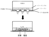
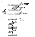
- mode of the hepatocyte culture method of this invention The schematic diagram (side view) of the one aspect
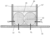
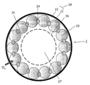
- 1 is a schematic diagram (aa sectional view) of one embodiment of a hepatocyte culture device of the present invention.
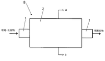
- 1 is a schematic diagram (aa cross-sectional view) of a first embodiment of a compound assay device of the present invention.
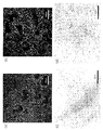
- the figure (photograph) which shows the metabolism of 5- (and-6) -carboxy-2 ', 7'-dichlorofluorescein (CDCF) in the hepatocyte cultured with the incubator using PDMS membrane.
- the figure (photograph) which shows the metabolism of 5- (and-6) -carboxy-2 ', 7'-dichlorofluorescein (CDCF) in the hepatocytes cultured by the incubator using a polystyrene incubator.
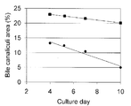
- the graph which shows the time-dependent change of the area of a capillary bile duct.
- ⁇ is the incubator using PDMS membrane
- ⁇ is the polystyrene incubator.
- the figure (photograph) which shows the result of having observed the formation of the capillary bile duct using fluorescein diacetate.
- (a), (c) is an incubator in which collagen is adsorbed and bound, (a) is the fourth day of culture, (c) is the seventh day of culture; (b), (d) is the covalently bound collagen.
- (B) is the fourth day of culture, and (d) is the seventh day of culture.
- the figure (photograph) which shows the result of having observed the formation of the capillary bile duct using fluorescein diacetate. Collagen gel sandwich (a), Matrigel sandwich group (b), untreated group (c).
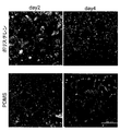
- Fluorescence micrographs of cells after 2 and 4 days in a collagen-adsorbed PDMS membrane incubator and collagen-coated polystyrene plate Fluorescence micrographs showing the localization of MRP2 and CD147 in a collagen adsorptive PDMS membrane incubator (48 hours) and collagen-coated polystyrene plates (48 hours and 96 hours).
- (B) is an enlarged photograph of a tight binding part
- (C) is a photograph of a capillary bile duct having microvilli (MV) on the cavity wall.
- the scale bars (A) and (C) are 2 ⁇ m, and (B) is 1 ⁇ m.
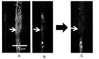
- FIG. 1 A is a bright field image of the tissue
- B is a fluorescence image before opening
- C is a fluorescence image after opening about 60 seconds.
- White arrows indicate the opening locations.
- the hepatocyte culture method of the present invention is characterized in that hepatocytes embedded with an extracellular matrix are arranged on a gas permeable membrane, and the hepatocytes are cultured while supplying oxygen from the gas permeable membrane side.
- hepatocytes are adhered to the collagen-coated surface of the gas-permeable membrane whose surface is coated with collagen, and oxygen is supplied from the gas-permeable membrane side while the hepatocytes are embedded in an extracellular matrix. It is characterized by culturing.
- the material of the gas permeable membrane used in the present invention may be any material that can permeate oxygen gas, but a porous and highly hydrophobic material is suitable. Examples include polydimethylsiloxane (PDMS), fluorocarbon, polytetrafluoroethylene (tetrafluoride), and polyurethane, and derivatives and similar substances thereof are also included.
- PDMS polydimethylsiloxane
- fluorocarbon fluorocarbon
- polytetrafluoroethylene (tetrafluoride) polytetrafluoroethylene
- polyurethane polyurethane
- a method for producing a PDMS film is described in a non-patent document (M Nishikawa et al. Biotechnology and Bioengineering, vol.99, pp.1472-1481).
- the manufacturing method of the PDMS film is not limited to this, and the PDMS film can be manufactured using a generally known film manufacturing method.
- it can be produced by a method of applying with a bar coater (bar coating method) or a method of applying with a gap coater (gap coating method).
- a film can be produced in the same manner.
- a culture plate Lumox manufactured by In vitro systems and services
- a fluorocarbon membrane is arranged on the culture surface
- the thickness of the gas permeable membrane is preferably as thin as possible from the viewpoint of gas permeability, but 50 ⁇ m to 2.0 mm is suitable. However, the optimum film thickness is not limited to the above range because it varies depending on the strength of the material and the intended use.
- the entire incubator may be composed of the gas permeable membrane, but at least a site where hepatocytes are seeded is composed of the gas permeable membrane. It ’s fine. It can change suitably according to the aspect of an incubator.
- Collagen that covers the gas permeable membrane can be prepared by a known method, but is coated with a commercially available collagen solution (for example, rat tail collagen manufactured by Becton Dickinson) to a thickness that allows oxygen to permeate. be able to.
- a well-known method can be used for the method of covering the gas permeable membrane with the collagen.
- a method of performing oxygen plasma treatment to adsorb the collagen on the gas permeable membrane or a method of covalently bonding using a chemically reactive functional group can be mentioned.
- the method of binding collagen to the PDMS membrane using a covalent bond should be performed, for example, according to the method described in non-patent literature (M. Nishikawa et al.
- hepatocytes in which capillaries are formed can be prepared efficiently, stably and in the long term, it is preferable to cover the gas permeable membrane with collagen by a covalent bond.
- the short-term capillary bile duct formation efficiency is the same for both covalent bonds and adsorptive bonds, both can be used for short-term measurements. As long as the capillary bile duct necessary for the test can be formed, an optimal binding can be appropriately selected according to the culture conditions of hepatocytes.
- the culturable hepatocytes may be derived from any animal, and examples include humans, monkeys, dogs, cats, cows, pigs, minipigs, hamsters, ferrets, rabbits, rats, and mice.
- the method for isolating hepatocytes from the animal can be performed according to a known method.
- the origin of hepatocytes may be any of fetus, newborn, and adult.
- embryonic stem (ES) cells and induced pluripotent (iPS) stem cells, or hepatocytes induced to differentiate from cord blood, bone marrow, fat, or blood-derived tissue stem cells can also be used.
- a method for inducing hepatocytes from these cells can be performed according to a known method.
- the cell density to be seeded may be any cell as long as the cells can normally grow. Usually, it is preferable to seed at a cell density of about 0.1 to 12.0 ⁇ 10 5 cells / cm 2 , and the cells may be seeded so as to form 2 to 3 layers. A preferable cell density can be appropriately set according to the culture conditions and the culture instrument to be used.
- Examples of the extracellular matrix for embedding hepatocytes include those that can be used in the known collagen gel sandwich method.
- collagen I collagen I, fibronectin, laminin, vitronectin, gelatin, elastin, proteoglycan, glucosaminoglycan, chondroitin sulfate, dermatan sulfate, heparan sulfate, heparin, keratan sulfate, Matrigel (trademark: Becton Dickinson), growth factor (basic FGF , EGF, IGF-1, PDGF, NGF, TGF beta, etc.) and mixtures thereof can be selected as appropriate and used as an extracellular matrix gel, but efficiently prepare hepatocytes that have formed capillary bile ducts Collagen gel and Matrigel are preferable, and collagen cells are more preferable because the cells can be prepared in a stable and long-term manner.
- the method for producing the extracellular matrix gel can be carried out by the method described in (Non-patent Document 1: LeCluyse et al., Am J Physiol Cell Physiol, 1994, vol.266, pp.1764-1774).
- the thickness of the extracellular matrix layer embedding the hepatocytes can be appropriately determined from the viewpoint of the nutrient and permeability of the test compound, but 10 to 100 micrometers is preferable.
- hepatocytes may be covered using the extracellular matrix sheet.
- extracellular matrix sheet Such an embodiment is also included in “embedding”.
- the same effect as a collagen gel can be derived by overlaying a commercially available collagen membrane (trade name Vitrigel, manufactured by Asahi Techno Glass Co., Ltd.) on hepatocytes.
- the thickness of the sheet can be appropriately determined from the viewpoint of nutrients and permeability of the test compound, but 10 to 100 micrometers is preferable.
- hepatocytes may be covered with an extracellular matrix-like substance composed of non-biological components.
- an extracellular matrix-like substance composed of non-biological components.
- Such an embodiment is also included in “embedding”.
- the collagen-coated cellulose membrane described in Non-Patent Document 4 TISSUE ENGINEERING, Volume 12, 2006, 2181-2191
- the porous coating described in Non-Patent Document 5 Biomaterials, volume 29,2008, 3993-4002
- a semi-permeable membrane such as a silicon nitride membrane, a polyethylene terephthalate membrane described in Non-Patent Document 6 (Biomaterials, volume 29,2008, 290-301) induces the formation of capillaries with physical signals, and The same effect as that of the collagen gel can be induced by overlaying a hepatocyte with a substance capable of supplying a medium component or the like.
- semipermeable membranes made of non-biological components include porous membranes of regenerated cellulose (cellophane), acetylcellulose, polyacrylonitrile, Teflon (registered trademark), polyester polymer alloy or polysulfone.
- the extracellular matrix-like substance of the non-biological component can be used in combination with the extracellular matrix.
- a simple method for culturing hepatocytes according to the present invention will be described with reference to FIG. Cover the gas permeable membrane with collagen, seed the hepatocytes on it, embed the hepatocytes with advanced adhesion in an extracellular matrix gel such as collagen gel, and culture while supplying oxygen from the gas permeable membrane side Do.
- the side of the gas permeable membrane not covered with cells is preferably in a gas phase so that oxygen can be supplied.
- the gas phase through the gas permeable membrane can be air having an oxygen concentration of 1 to 20% for supplying oxygen to the cells.
- the optimal oxygen concentration is between 5% and 13% following the liver in the living body.
- the oxygen concentration can be easily adjusted by setting a commercially available multi-gas incubator (for example, MCO-5M manufactured by Sanyo Electric Co., Ltd.).
- the oxygen supply may be performed by an artificial blood vessel made of an artificial material or a blood vessel cell disposed over a gas permeable membrane.
- the culture conditions may be in accordance with a known method for culturing hepatocytes.
- the medium for example, a medium in which serum, insulin / transferrin / selenium salt, dexamethasone is added to Dulbecco's modified Eagle medium or Williams E medium is used. Can do.
- the culture is usually performed under conditions of 37 ° C. and 5% CO 2 .
- the temperature and CO 2 concentration may be appropriately changed.
- the hepatocyte membrane is polarized, and a capillary bile duct membrane is formed along the gap between the hepatocytes, and a basement membrane is formed in other portions.
- the capillary bile duct formed by this capillary bile duct membrane is usually connected with a width of 1 to 2 micrometers and a length of 100 micrometers or more. In the middle of this capillary network, a hangout with a width of about 5 micrometers may be formed.
- an organic anion transporter group for taking in the compound and a sodium / taurocholic acid cotransporter group are expressed.
- Representative examples are OATP (Organic anion transporting polypeptide) 1a1, OATP1b2, OATP1b3, OAT2, OATP4, and OATP8, and the presence can be confirmed by cell antibody staining using antibodies specific to these.
- the main ATP-binding cassette (ABC) transporter protein is expressed in the capillary bile duct network.
- Representative examples are MRP2 (Multidrug-Resistance Protein 2), MDR1 (Multidrug-Resistance 1), and BCRP (breast cancer resistance protein). Can be determined. It can also be confirmed by cell antibody staining using antibodies specific for MRP2, MDR1, and BCRP.
- the compound test apparatus of the present invention includes a culture liver having a main body, a compound supply unit that supplies the compound to the main body, and a compound or metabolite recovery unit that recovers the compound or its metabolite from the main body.
- a compound testing apparatus using cells wherein the main body includes a gas-permeable membrane coated with a collagen surface, hepatocytes adhered to a collagen-coated surface of the permeable membrane, and the hepatocytes embedded therein Having an extracellular matrix.
- the gas permeable membrane may be finely processed.
- the gas permeable membrane may be finely processed, and grooves, depressions, or partition walls may be formed on the surface of the gas permeable membrane.
- the cell adhesion range and direction can be limited by partition walls applied by microfabrication to control the cell arrangement direction and adhesion range, and the capillary bile duct arrangement direction and range formed thereby.
- the metabolites excreted in the capillaries can be continuously collected.
- the placement of hepatocytes in the grooves of the extracellular matrix having grooves is also included in “embedding”.
- the formation of capillaries can be promoted by covering the hepatocytes arranged in the groove with an extracellular matrix.
- the shape of the groove or the depression is not particularly limited as long as hepatocytes in which capillaries are formed, but a width that allows the hepatocytes to be arranged in about two rows is preferable in order to reproduce the arrangement of hepatocytes in the living body.
- the width is 20 to 70 ⁇ m, preferably 30 to 50 ⁇ m, more preferably about 30 ⁇ m.
- the distance from the bottom surface of the groove or dent to the gas permeable membrane is preferably narrow, but any distance that can sufficiently supply oxygen from the gas permeable membrane may be used, and the height of the side surface is 10 ⁇ m or more and 1 mm or less, preferably Is preferably 50 ⁇ m or more and 500 ⁇ m or less, more preferably about 100 ⁇ m.
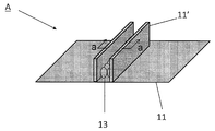
- FIG. 2A in the incubator A, two partition walls 11 ′ made of the same gas permeable membrane are provided on the gas permeable membrane 11, and the gas permeable membrane 11 and the two partition walls 11 ′ are provided. Hepatocytes are cultured in the enclosed space.
- FIG. 2B which is a cross-sectional view taken along the line aa of FIG. 2A, the gas permeable membrane 11 has a collagen coat layer 12 on which hepatocytes 13 are adhered and embedded with a collagen matrix 15. Then, oxygen is supplied using an oxygen supply device (not shown) installed outside the gas permeable membrane 11 and the two partition walls 11 ′, and the culture medium 16 is added to perform culture.
- an oxygen supply device not shown
- a capillary bile duct 14 is formed on the surface where the hepatocytes 13 contact each other.
- the partition wall may have a size that prevents adjacent hepatocytes from adhering to each other.
- the partition wall is preferably 1 micrometer or more.
- FIG. 18 demonstrates an example of the method of culture
- a collagen solution before gelation (concentration is 0.1-30 mg / mL, preferably 0.3-3 mg / mL, more preferably about 2.0 mg / mL) is spread on the gas permeable membrane 11.
- a PDMS mold having a convex portion is placed and left standing to gel, thereby producing a collagen gel having a concave portion.
- the width of the formed groove is 20 ⁇ m or more and 70 ⁇ m or less, preferably 30 ⁇ m or more and 50 ⁇ m or less, more preferably about 30 ⁇ m, and the height is 10 ⁇ m or more and 1 mm or less, preferably The length is 50 ⁇ m or more and 500 ⁇ m or less, more preferably about 100 ⁇ m, and the length can be appropriately changed and used.
- the material having the convex portions may be any material that does not break the shape of the gelatinized collagen. For example, a material made of PDMS is preferable because of its weight, ease of processing and handling. Subsequently, as shown in FIG.
- the hepatocytes dissolved in the culture medium are seeded on the gel having the recesses prepared above, and washed with the medium several times, so that the hepatocytes are only in the recesses. Arrange and culture. Subsequently, after about 24 hours of incubation, the collagen solution before gelation (concentration is 0.1-30 mg / mL, preferably 0.3-3 mg / mL, more preferably about 2.0 mg / mL) or matrigel solution (concentration is 5 mg).
- -5000 ⁇ g / mL, preferably 50-500 ⁇ g / mL, more preferably about 150 ⁇ g / mL is layered on the cells and further cultured for 2-9 days. Capillary duct formation is observed from the 2nd day, but longer capillaries can be made by culturing for a long time. It is possible to appropriately select an optimal state for drug evaluation tests and the like.
- the gas permeable membrane may be shaped like a cylindrical channel such as a cylinder or a cube. That is, the culture method of the present invention also includes an embodiment in which the gas permeable membrane is arranged in a cylindrical shape with the collagen-coated surface inside, and hepatocytes are embedded in the extracellular matrix and cultured in the extracellular matrix. .
- the compound assay device B of the present invention is a device having a main body part 2, a compound supply part 1 for supplying a compound to the main body part, and a recovery part 3 for recovering the compound or its metabolite from the main body part,
- the main body 2 is bonded to a cylindrical body 28 in which a gas-permeable membrane 21 whose surface is coated with collagen is formed in a cylindrical shape with the collagen coated surface 22 inside, and a collagen coated surface 22 inside the cylindrical body 28.
- the main body 2 has hepatocytes 23 and an extracellular matrix 25 in which hepatocytes 23 are embedded.
- the main body 2 further has a flow path 27 formed by forming a space defined by the semipermeable membrane 26 in the axial direction of the cylindrical body 28. From this channel, the supply can be supplied to the hepatocytes embedded in the extracellular matrix through the semipermeable membrane 26.
- the material of the semipermeable membrane is not particularly limited as long as it can permeate compounds and medium components.
- regenerated cellulose cellophane
- acetylcellulose collagen-coated cellulose
- polyacrylonitrile Teflon (registered trademark)
- porous examples thereof include porous silicon nitride, polyethylene terephthalate, polyester polymer alloy or polysulfone porous membrane.
- these materials can be used as a non-biological component cell matrix-like substance, by arranging a semipermeable membrane made of these materials so as to cover hepatocytes, there is no need to add an extra extracellular matrix. Can also induce polarity and form capillaries.
- a medium for culturing and maintaining hepatocytes is also supplied from the compound supply unit 1, and the medium components are supplied to the hepatocytes via the semipermeable membrane 26 when passing through the flow path 27.
- the medium supply unit for supplying the medium may be provided separately from the compound supply unit.
- the shape of the flow path 27 is not limited to a cylindrical shape, and may be other shapes or may be branched. The diameter is not particularly limited. Also, a plurality of flow paths may be provided, and the flow path through which the medium passes and the flow path through which the compound passes may be separate.
- the flow path 27 and the semipermeable membrane 26 are not essential, and the entire inside of the cylindrical body formed by the gas permeable membrane may be a hepatocyte and an extracellular matrix that embeds it. In this case, the culture medium and the compound may pass through the extracellular matrix enclosed in the cylindrical body.
- hepatocytes 23 are arranged along the inner wall (collagen coated surface 22) of the gas permeable membrane 21, and are partitioned by an extracellular matrix 25 on the inner side and a semipermeable membrane 26 on the inner side.
- a space (flow path 27) for adding a medium and a compound is provided, and by supplying oxygen from the outside of the gas permeable membrane 21 and culturing, the capillary bile duct 24 is induced by the extracellular matrix. Yes.
- the outside of the gas permeable membrane 21 is in a gas phase, and the oxygen concentration touching hepatocytes can be adjusted.
- the metabolic characteristics of a compound can be evaluated. That is, by supplying the test compound from the medium supply unit 1 to the flow path 27, hepatocytes are exposed to the compound via the semipermeable membrane 26. The compound metabolized by the hepatocytes is collected and analyzed in the collection unit 3.
- the recovery part for recovering the compound or its metabolite may be any member that can separate a liquid such as a medium containing the compound or metabolite from the flow path or extracellular matrix, but is a cylindrical body connected to the main body.
- a cylindrical body having a collagen-coated gas permeable membrane with a collagen-coated surface inside, a hepatocyte adhered to a collagen-coated surface inside the cylindrical body, and the hepatocyte May have a flow path formed by an extracellular matrix that embeds the liver and the capillary bile duct of the hepatocytes.
- the bile ducts formed by the hepatocyte population cultured by the method of the present invention may gather to form a flow path (cavity) so that metabolites can flow.
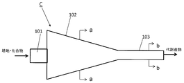
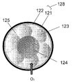
- the hepatocyte culture apparatus of the present invention having such an embodiment will be described with reference to FIG. 4A and 4B which is a bb cross-sectional view of FIG. The aa cross-sectional view is the same as FIG. 3B.
- the main body 102 is formed so that its diameter gradually decreases, and is connected to the recovery unit 103.
- recovery part 103 hepatocytes are introduce
- the formed capillary bile duct is provided with a flow path composed of ultrathin tubes and bile duct epithelial cells, and the metabolite discharged into the capillary bile duct is excreted without delay and collected in a reservoir for analysis.
- a compound assay device can be suitably used for a drug (compound) transport assay as described later.
- Hepatocytes formed with capillaries produced by the method of the present invention can be used for drug transport assays and high-throughput screening of drug candidates.
- An example of a drug transport assay is an assay of how much and how much drug is taken up by hepatocytes and excreted into bile.
- an assay of whether a certain compound A inhibits or promotes the transport of compound B is exemplified.
- Examples of the method for drug transport assay include the method of non-patent literature (Liu X et al., Am J Physiol, 1999, vol. 277, pp. G12-21).
- Specific methods for high-throughput screening include, for example, the following method that can analyze a very small amount of compound with a fine flow path and a highly sensitive detector, and further performs this automatically or semi-automatically.
- capillaries are embedded in an extracellular matrix in very small compartments on a gas-permeable membrane or in ultrafine channels of gas-permeable channels as shown in FIG.
- Each part of the culture where hepatocytes that have formed cells were cultured was provided with an inlet for exposure to the test compound and a mouth for collecting metabolites that were metabolized by hepatocytes in each compartment and excreted into the capillary bile duct This is possible by constructing a microchannel device.
- the method of preparing a 24-well incubator made of PDMS membrane covalently bound with collagen is the method described in non-patent literature (M. Nishikawa et al. Biotechnology and Bioengineering, 2008, vol.99, pp.1472-1481). I followed.
- a 30g mixture of Silpot 184 (manufactured by Toray Dow Corning Co., Ltd.) and a curing agent mixed at 10: 2 is thinly stretched into a plastic container of 258mm x 174mm x 45mm and cured by heating at 80 ° C for 2 hours to thicken it.
- a PDMS film with a thickness of about 1 mm was produced.
- the ratio of the main agent and the curing agent is not limited to 10: 2, but is usually made between 10: 1 and 10: 2.
- a PDMS membrane 24-well incubator is sandwiched between a polycarbonate frame with 24 holes and a 1.5 mm thick SUS plate with holes in the same position as the frame, and fixed with screws.
- oxygen plasma treatment 5 seconds
- 1% acetic acid and 2% aminosilane manufactured by Shin-Etsu Silicon Co., Ltd. were added to the well, reacted at room temperature for 45 minutes, and then heated at 80 ° C. for 90 minutes.
- Non-Patent Document 1 (LeCluyse et al., Am J Physiol, 1994, vol.266, pp.1764-1774). Inoculate 2x10 5 hepatocytes per well into the incubator prepared above, and after 4 hours, after changing the medium, 24 hours after seeding, 1.7 mg / ml collagen solution (Becton Dickinson) 20 ⁇ L overlay Then, after gelling at 37 ° C. for 1 hour, 500 ⁇ L of serum-free medium was added and cultured at 37 ° C./5% CO 2 . Moreover, the culture solution was changed once every two days.
- hepatocytes cultured on a conventional polystyrene incubator (product name: BioCoat collagen I-coated 24-well, manufactured by Becton Dickinson) almost have a tubular structure on the second day. In addition, a large number of cells were stained inside because they did not have excretion activity.
- the area of the capillary bile was compared over time, the area on which the capillary bile duct was formed was larger on the PDMS membrane from day 2 to day 10 than on the conventional culture on polystyrene.
- PDMS membrane 24-well incubator covalently bound with collagen was produced in the same manner as in Example 1.
- the PDMS membrane 24-well incubator to which collagen was adsorbed was compared with the 1.7 mg / mL collagen solution (Becton Dickinson) compared to the PDMS membrane 24-well incubator treated with oxygen plasma after the aminosilane treatment step of Example 1. (Supplied) was added to the well and covered, and left at room temperature for 18 hours, and then washed with PBS. Both were used for the following experiments on the same day.
- Collagen gel sandwich hepatocyte culture was prepared in the same manner as in Example 1. Capillary bile duct formation was observed using fluorescein diacetate (Fig. 8). On the 4th day of culture (3 days after the collagen gel overlay), an incubator with collagen adsorbed (a) and a covalently incubator (b) Both showed formation of capillaries. However, on the 7th day of culture, the culturing vessel (c) to which collagen was adsorbed and bound was more prominent than the culturing vessel (d) to which the collagen was covalently bound, and the capillaries were also lost. From this, it is possible to prepare hepatocytes in which capillary bile ducts are formed stably and long-term by covalently binding collagen.
- Example 1 Except for the overlying extracellular matrix components, the procedure of Example 1 was followed.
- the incubator prepared above was seeded with 2 ⁇ 10 5 hepatocytes per well, and 4 hours later, the medium was changed.
- a collagen gel sandwich group was obtained by overlaying 20 ⁇ L of a 1.7 mg / ml collagen solution (Becton Dickinson) 24 hours after seeding and allowing gelation at 37 ° C. for 1 hour.
- Matrigel sandwich group was obtained by adding Matrigel (manufactured by Becton Dickinson) diluted 50 times (corresponding to 150 ⁇ g / mL) in serum-free medium.
- a group to which nothing was added was defined as an untreated group.
- capillary bile ducts using fluorescein diacetate were formed (Fig. 9).
- both collagen gel sandwich (a) and Matrigel sandwich group (b) were capillary bile ducts. Formation was observed and maintained until day 7 of culture.
- capillaries were not formed at all, cell detachment was remarkable from the third day of culture, and most cells were detached from the fifth day (c). From the above, it was found that capillary bile ducts can be formed on a PDMS membrane with either a collagen overlay or a Matrigel overlay.
- Ca / Mg (+) HBSS was prepared by adding 50 mL of HBSS (Invitrogen, 14175-079), 500 ⁇ L of 14 g / L CaCl 2 and 500 ⁇ L of 10 g / L MgCl 2 / 6H 2 O.
- As the CDCF solution 1 mM CDCF (in dimethyl sulfoxide: Molecular Probes, C-369) was used to prepare a 5 ⁇ M CDCF solution with Ca / Mg (+) HBSS and kept at 37 ° C. in a water bath.
- Ca / Mg ( ⁇ ) HBSS was prepared by adding 500 ⁇ L of 100 mM EGTA to 50 mL of HBSS (Invitrogen, 14175-079). 0.5% Triton X-100 / PBS was prepared by adding Triton X-100 to a PBS buffer so that the concentration was 0.5%.
- the amount of CDCF was quantified by measuring excitation 492 nm and fluorescence 530 nm with a fluorescence microplate reader. In addition, 25 ⁇ L of the stock solution was subjected to protein measurement using a BCA protein assay kit (Thermo).
- the Bile Excretion Index (BEI) was obtained from the following calculation formula from the fluorescence intensity value (Accumulation) per protein amount.
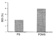
- the BEI value was about 40%
- a polystyrene (PS) substrate was used, the BEI value was about 20%. From the above, it can be seen that the gas permeable membrane allows hepatocytes to build a capillary bile duct structure with high MRP excretion activity, rather than using a polystyrene substrate, and evaluates compounds with high sensitivity (accurately with a small amount of compounds). I found out that
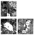
- MRP2 protein is expressed more widely in the PDMS incubator (FIG. 11). Since MRP2 is a major transporter involved in bile excretion, it can be inferred that capillaries produced in PDMS incubators have higher bile excretion activity.
- hepatocytes seeding density of PDMS film made 24-well culture vessel 1 well Atari bound collagen adsorbed 2x10 5 cells, 4x10 5 cells, and a 6x10 5 cells, 48 hours 150 [mu] g / mL Matrigel After culturing, BEI measurement was performed as described in Example 4. As a result, as shown in FIG. 13, 2x10 5 cells are most BEI high, 4x10 5 cells, 6x10 5 cells of BEI sequentially decreased.
- the optimum point of the culture period was examined.
- the Matrigel concentration (150 ⁇ g / mL) and the cell seeding density (2 ⁇ 10 5 cells) determined as described above BEI measurement was performed as described in Example 4 at 24, 48, 72, and 96 hours after hepatocyte seeding.
- the PDMS membrane showed BEI equivalent to that of hepatocytes cultured on polystyrene for 24 hours and 48 hours after seeding.
- 14B is a photograph of the site where CDCF is accumulated 48 hours and 96 hours after seeding of hepatocytes on a 24-well incubator made of PDMS membrane and a 24-well plate made of collagen-coated polystyrene with a fluorescence microscope. From this image, it can be confirmed that hepatocytes on the PDMS membrane formed capillary bile ducts having activity earlier than that of the conventional culture method. From the above results, when polarity is induced by overlaying Matrigel using a 24-well incubator made of PDMS membrane, polarity is formed 24 hours after sowing, and functional capillaries appear. This was found to be 72 hours earlier than that for the conventional method reaching the equivalent BEI value.
- the localization of MRP2 and basement membrane marker CD147 was examined by cell antibody staining according to a conventional method. In addition, it was compared with hepatocytes cultured in a collagen-coated polystyrene 24-well plate. As shown in FIG. 15, when cultured on a PDMS membrane, MRP2 protein and CD147 protein are detected between cells after 48 hours of culture. On the other hand, the expression of CD147 is seen on conventional polystyrene, but the localization of MRP2 protein is hardly seen.
- MRP2 protein is expressed more extensively in the hepatocytes on the PDMS membrane than after 120 hours of culture where polarity is formed on polystyrene.
- the localization pattern of CD147 is almost the same in both cases. From these results, it was shown that MRP2 molecules were localized earlier in hepatocytes cultured on PDMS membrane than on polystyrene, and that MRP2 expression was enhanced more than on polystyrene.
- FIG. 16A In the hepatocytes in which the polarity is induced on the PDMS membrane, capillary bile ducts (BC) and tight junctions (TJ) that are typically seen in the hepatocytes with polarity are seen.
- FIG. 16B shows an enlarged photograph of a tight junction
- FIG. 16C shows a capillary bile duct having microvilli (MV) on the cavity wall.
- BEI was measured according to the method described in Example 4, and the degree of polarity formation was analyzed.
- Cells cultured in a collagen-coated polystyrene 24-well plate were used as comparative controls. As shown in FIG. 17, on Lumox, BEI higher than the polystyrene (PS) 24-well plate was shown.
- the cell seeding density of 1 hole Atari 1.0x10 5 cells and 2.0x10 5 cells gave 1.0x10 5 pieces of more high BEI.
- Collagen gels with recesses were prepared by allowing them to gel for 60 minutes at 37 ° C.
- rat hepatocytes suspended in a culture medium are seeded on the prepared gel having recesses, and washed twice with the medium, so that hepatocytes are arranged only in the recesses.
- the culture was performed.
- a collagen solution (concentration 2.1 mg / mL) was overlaid on the cells, and further cultured for 9 days.
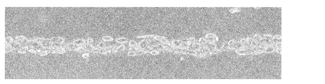
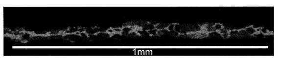
- a photograph of the hepatocytes on day 9 arranged in this manner is shown in FIG. It was observed that capillaries were continuously formed.
- CDCF is added to the medium as described in Example 1 to metabolize the continuous capillary bile duct prepared above.
- the product fluorescein was accumulated and its fluorescence was observed.
- On the 2nd day it was possible to observe the formation of capillaries in a part, and on the 6th day, it was possible to observe the continuous formation of these capillaries.
- On the 9th day we could observe a longer continuous state.
- the results on day 9 are shown in FIG. Since the fluorescence was continuously observed, it was confirmed that a continuous capillary bile duct exceeding 1 mm could be produced.
- fluorescein was accumulated in the produced continuous capillary bile duct, and one end of the capillary bile duct (FIG. 21A) was opened with a thin glass tube. As a result, it was found that fluorescein accumulated in the capillary bile duct before opening (FIG. 21B) disappeared about 60 seconds after opening (FIG. 21C). As a result, the continuity of the capillary bile duct was demonstrated, and it was further demonstrated that it can be used for analysis of continuous collection of bile. In the above, the presence or absence of the collagen coating treatment of the fluorocarbon oxygen permeable membrane did not affect the results.
- the cultured hepatocytes obtained by the culturing method of the present invention can be used for testing drug candidate compound screening using hepatocytes. This contributes to an increase in the accuracy and efficiency of analysis of uptake, metabolism, and excretion of drug candidate compounds in hepatocytes, leading to higher efficiency of the drug discovery process.
Landscapes
- Health & Medical Sciences (AREA)
- Life Sciences & Earth Sciences (AREA)
- Engineering & Computer Science (AREA)
- Chemical & Material Sciences (AREA)
- Bioinformatics & Cheminformatics (AREA)
- Biomedical Technology (AREA)
- Organic Chemistry (AREA)
- Wood Science & Technology (AREA)
- Zoology (AREA)
- Biotechnology (AREA)
- Microbiology (AREA)
- Biochemistry (AREA)
- General Health & Medical Sciences (AREA)
- Genetics & Genomics (AREA)
- Immunology (AREA)
- General Engineering & Computer Science (AREA)
- Sustainable Development (AREA)
- Cell Biology (AREA)
- Hematology (AREA)
- Analytical Chemistry (AREA)
- Clinical Laboratory Science (AREA)
- Urology & Nephrology (AREA)
- Molecular Biology (AREA)
- Toxicology (AREA)
- Tropical Medicine & Parasitology (AREA)
- Gastroenterology & Hepatology (AREA)
- Food Science & Technology (AREA)
- Medicinal Chemistry (AREA)
- Physics & Mathematics (AREA)
- General Physics & Mathematics (AREA)
- Pathology (AREA)
- Apparatus Associated With Microorganisms And Enzymes (AREA)
- Micro-Organisms Or Cultivation Processes Thereof (AREA)
- Measuring Or Testing Involving Enzymes Or Micro-Organisms (AREA)
Abstract
Description
上記の3次元培養方法では極性を持った肝細胞を初代細胞から作製するためには数週間から数カ月の時間を要する。
それに対して、コラーゲンゲルサンドイッチ法(非特許文献1:LeCluyse et al., Am J Physiol Cell Physiol, 1994, vol.266, pp.1764-1774)を用いるとコラーゲンゲル重層後3~4日程度で毛細胆管の形成と胆汁成分排出活性が検出され始める。しかし、コラーゲンゲルサンドイッチ法を用いても、胆汁成分排出活性が得られるまでには日数を要する。また、生体と同じように出口のある胆管によって連続的に代謝物を解析することはできない。薬物スクリーニングに使用するためには、形成される毛細胆管の数が少なく、活性が低いことも問題となる。
(1)ガス透過膜上に細胞外マトリクスで包埋された肝細胞を配置し、ガス透過性膜側から酸素を供給しつつ肝細胞を培養することを特徴とする、肝細胞培養方法。
(2)ガス透過性膜の表面がコラーゲンコーティングされており、該ガス透過性膜のコラーゲンコート面に細胞外マトリクスで包埋された肝細胞が配置される、(1)に記載の方法。
(3)ガス透過性膜がコラーゲンコート面を内側にして筒状に配置されており、該筒状の内部で細胞外マトリクスで包埋された肝細胞が配置される、(2)に記載の方法。
(4)前記細胞外マトリクスが溝を構成し、該溝に肝細胞が配置された、(1)~(3)のいずれかに記載の方法。
(5)前記ガス透過性膜がポリジメチルシロキサン膜である、(1)~(4)のいずれかの方法。
(6)前記ガス透過性膜がフルオロカーボン膜である、(1)~(4)のいずれかの方法。
(7)コラーゲンコーティングが共有結合によるものである、(1)~(6)のいずれかの方法。
(8)細胞外マトリクスがコラーゲンゲルまたはマトリゲル(商標)である、(1)~(7)のいずれかの方法。
(9)細胞外マトリクスが非生体成分からなる、(1)~(7)のいずれかの方法。
(10)ガス透過膜上に細胞外マトリクスで包埋された肝細胞を配置し、ガス透過性膜側から酸素を供給しつつ肝細胞を培養することを特徴とする、毛細胆管を形成した培養肝細胞の製造方法。
(11)ガス透過性膜の表面がコラーゲンコーティングされており、該ガス透過性膜のコラーゲンコート面に細胞外マトリクスで包埋された肝細胞が配置される、(10)に記載の方法。
(12)ガス透過性膜上に溝を有する細胞外マトリクス層が配置され、該細胞外マトリクス層の溝に肝細胞が配列された、(10)または(11)に記載の方法。
(13)(10)~(12)のいずれかの方法により毛細胆管を形成した培養肝細胞を製造し、得られた培養肝細胞を用いて化合物の代謝を評価する、化合物の代謝検定方法。
(14)(10)~(12)のいずれかの方法により毛細胆管を形成した培養肝細胞を製造し、得られた培養肝細胞を用いて化合物の輸送を評価する、化合物の輸送検定方法。
(15)培養肝細胞を含む本体部と、本体部に化合物を供給する化合物供給部と、本体部から化合物またはその代謝物を回収する回収部とを有する培養肝細胞を用いた化合物検定装置であって、前記本体部は、
ガス透過性膜と、
該ガス透過膜上に配置された細胞外マトリクスと、
該細胞外マトリクスに包埋された肝細胞と、
を有することを特徴とする、装置。
(16)ガス透過性膜の表面がコラーゲンコーティングされており、該ガス透過性膜のコラーゲンコート面に細胞外マトリクスで包埋された肝細胞が配置された、(15)に記載の装置。
(17)前記ガス透過性膜は、コラーゲンコート面を内側にして筒状にした筒状体を形成しており、該筒状体の内部にコラーゲンコート面に接着された肝細胞とそれを包埋する細胞外マトリクスを含む、(16)の装置。
(18)前記本体部は、前記化合物供給部からの供給物が流れるように半透膜で画定された空間を前記筒状体の軸方向に形成する流路であって、該半透膜を介して該供給物を前記細胞外マトリクスに包埋された肝細胞に供給可能な供給物用流路を、更に有する(17)の装置。
(19)前記回収部が、表面がコラーゲンコーティングされたガス透過性膜をコラーゲンコート面を内側にして筒状にした筒状体と、該筒状体の内側のコラーゲンコート面に接着された肝細胞と、該肝細胞を包埋する細胞外マトリクスと、該肝細胞の毛細胆管によって形成される流路を有する、(18)の装置。
(20)前記細胞外マトリクスが溝を構成し、該溝に肝細胞が配置された、(15)~(19)のいずれかに記載の方法。
(21)前記ガス透過性膜がポリジメチルシロキサン膜である、(15)~(20)のいずれかの装置。
(22)前記ガス透過性膜がフルオロカーボン膜である、(15)~(20)のいずれかの装置。
(23)コラーゲンコーティングが共有結合によるものである、(15)~(22)のいずれかの装置。
(24)細胞外マトリクスがコラーゲンゲルまたはマトリゲル(商標)である、(15)~(23)のいずれかの装置。
(25)細胞外マトリクスが非生体成分からなる、(15)~(23)のいずれかの装置。
(26)化合物代謝検定装置である、(15)~(25)のいずれかの装置。
(27)化合物輸送検定装置である、(15)~(25)のいずれかの装置。
また、「包埋」されている状態とは、該肝細胞の周辺が少なくとも1層の細胞外マトリクスで囲まれていることを言う。毛細胆管が効率的に形成される限り、該細胞外マトリクスは連続的でも非連続的でも構わない。
また、フルオロカーボン膜が培養面に配置された培養プレートLumox (In vitro systems and services社製)等、を適宜購入して使用することもできる。
一方、短期的な毛細胆管の形成効率は共有結合でも吸着結合でも同様であることから、短期的な測定に使用する場合には、いずれも使用することができる。試験に必要な毛細胆管を形成できるかぎり、肝細胞の培養条件に合わせて、適宜、最適な結合を選択することができる。
播種する細胞密度は、該細胞が正常に増殖可能であれば良い。通常、細胞密度は約0.1~12.0 x 10⁵cells/cm2の間で播種することが好ましく、細胞が2~3層になるように播種してもよい。培養条件や使用する培養器具などに合わせて、適宜、好ましい細胞密度を設定することができる。
また、具体的な非生体成分からなる半透膜の例として、再生セルロース(セロファン)、アセチルセルロース、ポリアクリロニトリル、テフロン(登録商標)、ポリエステル系ポリマーアロイあるいはポリスルホンの多孔質膜が挙げられる。
また、上記の非生体成分の細胞外マトリクス様の物質に、該細胞外マトリクスを組み合わせて使用することもできる。
この場合、ガス透過性膜越しの気相は細胞への酸素供給のために酸素濃度1~20%の空気とすることができる。生体内の肝臓に倣って酸素濃度5~13%の間が最適である。この酸素濃度調節は市販のマルチガスインキュベーター(例えば、サンヨー電機社製MCO-5M)の設定によって容易に行うことができる。
また、上記の酸素供給の別の態様としては、ガス透過性膜越しに配置された人工素材や血管細胞からなる人工血管により供給されてもよい。
そして、一般的な細胞培養と同じく、通常37℃、5%CO2の条件で培養を行う。ただし、特殊な細胞や条件で培養を行う場合は、温度やCO2濃度は適宜変更すればよい。培養条件を調節することによって、肝細胞を2次元あるいは3次元に培養することや、毛細胆管の数を調節することができる。
すなわち、本発明の培養方法によれば、肝細胞の膜が分極し、肝細胞同士の間隙に沿って毛細胆管膜が、それ以外の部分には基底膜が形成される。この毛細胆管膜によって形成される毛細胆管は通常幅1~2マイクロメートル、長さは100マイクロメートル以上連結している。この毛細胆管ネットワークの途中には幅が5マイクロメートル程度に広くなったたまり場が形成されることがある。
すなわち、本発明の化合物検定装置は、本体部と、本体部に化合物を供給する化合物供給部と、本体部から化合物またはその代謝物を回収する化合物またはその代謝物の回収部とを有する培養肝細胞を用いた化合物検定装置であって、前記本体部は、表面がコラーゲンコーティングされたガス透過性膜と、該透過性膜のコラーゲンコート面に接着された肝細胞と、該肝細胞を包埋する細胞外マトリクスを有する。
例えば、ガス透過性膜に微細加工を施し、ガス透過性膜の表面に溝や窪みや隔壁を形成してもよい。例えば、微細加工で施される隔壁により細胞の接着範囲と方向を限定して、細胞の配列方向および接着範囲、そしてそれにより形成される毛細胆管の配列方向および範囲を制御することができる。
また、ガス透過性膜上にコラーゲンゲルなどの細胞外マトリクスで溝や窪み(凹部)を形成し、その溝や窪みに細胞を配置することで、細胞の配列方向および接着範囲と方向を限定することができ、それにより形成される毛細胆管の配列方向および範囲を制御することで、毛細胆管内に排出された代謝物を連続的に回収することができる。このように、溝を有する細胞外マトリクスの溝に肝細胞を配置することも、「包埋」に含まれる。更に、前記の溝に配置された肝細胞を、細胞外マトリクスで覆うことで毛細胆管の形成を促進することができる。
溝や窪みの形状は、毛細胆管が形成された肝細胞が作製できれば、何でも良いが、生体内の肝細胞の配置を再現するため、肝細胞が約2列で配列できる幅が好ましい。すなわち、幅20μm 以上70μm以下、好ましくは 30μm 以上50μm以下、さらに好ましくは30μm程度が望ましい。また、溝や窪みの底面からガス透過性膜までの距離は狭い方が好ましいが、ガス透過性膜から十分に酸素を供給できる距離であれば良く、側面の高さは10μm以上1mm以下、好ましくは50μm以上500μm以下、更に好ましくは100μm程度が望ましい。
この隔壁は隣接する肝細胞同士が接着を妨げられる大きさであればよく、具体的には1マイクロメートル以上であることが好ましい。
また、図18で、溝や窪みで毛細胆管が形成された肝細胞を培養する方法の一例を説明する。図2のコラーゲンコート層12を有するガス透過性膜11の上に、図18(A)で示した溝が形成されたコラーゲンゲルなどの細胞外マトリクスを配置する。まず、前記ガス透過性膜11の上に、ゲル化前のコラーゲン溶液(濃度は0.1-30mg/mL、好ましくは0.3-3mg/mL、更に好ましくは約2.0mg/mL)を撒く。次に、該コラーゲン溶液が完全にゲル化する前に、凸部を持ったPDMS製の型を配置して、放置することでゲル化させることで凹部をもったコラーゲンゲルを作製する。前記凸部をもった型の形状としては、形成された溝の幅が、20μm 以上70μm以下、好ましくは 30μm 以上50μm以下、さらに好ましくは30μm程度であり、高さが10μm以上1mm以下、好ましくは50μm以上500μm以下、更に好ましくは100μm程度であり、また、長さは適宜変更して使用できる。また前記凸部をもった型の素材としては、ゲル化したコラーゲンの形を壊さないようなものであれば良く、例えば、重さや加工や取り扱いのし易さからPDMS製のものが良い。
続いて、図18(B)で示すように、上記で作製した凹部を持ったゲルに、培養培地に溶かした肝細胞を播き、何度か培地で洗浄することで、凹部のみに肝細胞を配列させて培養を行う。
続いて、約24時間後培養後、ゲル化前のコラーゲン溶液(濃度は0.1-30mg/mL、好ましくは0.3-3mg/mL、更に好ましくは約2.0mg/mL)もしくはマトリゲル溶液(濃度は、5-5000μg/mL、好ましくは50-500μg/mL、更に好ましくは約150μg/mL)を細胞上に重層し、さらに2-9日培養する。2日から毛細胆管の形成が認められるが、長期間培養する方がより長い毛細胆管を作らせることができる。薬物の評価試験等に最適な状態を適宜選択することができる。
すなわち、ガス透過性膜がコラーゲンコート面を内側にして筒状に配置され、該筒状空間の内部で肝細胞を細胞外マトリクスに包埋して培養する態様も本発明の培養方法に含まれる。
本発明の化合物検定装置Bは、本体部2と、本体部に化合物を供給する化合物供給部1と、本体部から化合物またはその代謝物を回収する回収部3とを有する装置であって、前記本体部2は、表面がコラーゲンコーティングされたガス透過性膜21をコラーゲンコート面22を内側にして筒状にした筒状体28と、筒状体28の内側のコラーゲンコート面22に接着された肝細胞23と、肝細胞23を包埋する細胞外マトリクス25とを有する。
そして、本体部2は、さらに、半透膜26で画定された空間を筒状体28の軸方向に形成してなる流路27を有する。この流路から半透膜26を介して該供給物を細胞外マトリクスに包埋された肝細胞に供給することができる。
ここで、半透膜の素材は化合物や培地成分を透過できるものであれば特に制限されないが、例えば、再生セルロース(セロファン)、アセチルセルロース、コラーゲンコーティングセルロース、ポリアクリロニトリル、テフロン(登録商標)、多孔質の窒化ケイ素、ポリエチレンテレフタレート、ポリエステル系ポリマーアロイあるいはポリスルホンの多孔質膜などが例示される。
なお、これらの素材は非生体成分の細胞マトリクス様物質として使用できるものであるため、これらの素材からなる半透膜を肝細胞を覆うように配置することにより、別途細胞外マトリクスを加えなくても極性を誘導させ、毛細胆管を形成させることができる。
化合物検定装置Bにおいては、肝細胞を培養・維持するための培地も化合物供給部1から供給され、流路27を通過する際に、培地成分が半透膜26を介して肝細胞に供給される。ただし、本発明の化合物検定装置においては、培地を供給する培地供給部は化合物供給部とは別に設けられていてもよい。
なお、流路27の形状は円筒形状に限定されず、その他の形状であってもよく、枝分かれしていてもよい。また、その径も特に制限されない。また、流路は複数設けられていてもよく、培地が通過する流路と化合物が通過する流路が別々になっていてもよい。
さらに、流路27および半透膜26は必須ではなく、ガス透過性膜によって形成される筒状体の内部全てが肝細胞とそれを包埋する細胞外マトリクスであってもよい。この場合は、培地や化合物が筒状体の内部に封入された細胞外マトリクスの中を通過するようにすればよい。
すなわち、本発明の方法で培養された肝細胞集団によって形成された胆管が集合して流路(腔)を形成し、代謝物が流れるようにしたものであってもよい。
このような態様の本発明の肝細胞培養装置について図4Aと図4Aのb-b断面図である4Bを参照して説明する。なお、a-a断面図は図3Bと同様である。
図4Aに示すように、本体部102はその径が徐々に小さくなるように形成され、回収部103に接続されている。そして、回収部103では、ガス透過性膜の内部に、毛細胆管腔を中心に形成するように肝細胞が導入されている。
形成させた毛細胆管からは極細の管や胆管上皮細胞からなる流路が設けられており、毛細胆管に排出された代謝物が滞りなく排泄され、分析のためのリザーバーに回収される。
このような化合物検定装置は後述するような薬物(化合物)輸送検定に好適に用いることができる。
薬物輸送検定としては、薬物がどのくらいの量と速度で肝細胞に取り込まれ、胆汁に排出されるかどうかの検定が例示される。または、ある化合物Aが化合物Bの輸送を阻害・促進するかどうかの検定が例示される。
薬物輸送検定の方法としては、例えば、非特許文献(Liu X et al., Am J Physiol, 1999, vol.277, pp.G12-21)の方法が挙げられる。
ハイスループットなスクリーニングの具体的な方法としては、例えば、微細な流路と高感度な検出器でごく微量の化合物を解析でき、さらにこれを自動あるいは半自動で行うような以下の方法などが挙げられる。
ハイスループットなスクリーニングのためには、ガス透過性膜上の区切られたごく微小な区画、または図2のようなガス透過性流路の極細流路の中で細胞外マトリクスに包埋され毛細胆管を形成した肝細胞が培養されている培養部分それぞれに、試験化合物暴露させる注入口と、それぞれの区画の肝細胞で代謝され毛細胆管に排出される代謝物を回収するための口が設けられたマイクロ流路デバイスを構築することで可能となる。
肝細胞の単離
5週齢のラット(購入先:三共ラボサービス)の肝臓から非特許文献(Seglen PO, 1976, in Methods in cell biology (Prescott DM ed) 13th ed, pp29-83, Academic press, New York)に従って肝臓由来の細胞を分離した。
方法は非特許文献(M. Nishikawa et al. Biotechnology and Bioengineering, 2008, vol.99, pp.1472-1481)に述べられている方法に従った。Silpot 184(東レ・ダウコーニング社製)の主剤と硬化剤を10:2で混合した混合液30gを258mm×174mm×45mmのプラスチック製容器に薄く伸ばし、80℃で2時間加熱し硬化させ、厚さ約1mmのPDMS膜を作製した。主剤と硬化剤の割合は10:2に限らず、10:1~10:2の間で作製されるのが通常である。このPDMS膜を24個の穴が開いたポリカーボネート製の枠と、枠と同じ位置に穴の開いた厚さ1.5mmのSUS製板の間に挟み、ねじで固定することでPDMS膜製24穴培養器を作製した。これに酸素プラズマ処理(5秒)を行った後、ウェルに1%酢酸,2%アミノシラン(信越シリコン社製)を加えて、室温で45分反応させた後、80℃で90分間加熱した。次いで、このように処理されたウェルに0.5mM Sulfosuccinimidyl 2-(m-azido-o-nitrobenzamido)-ethyl-1, 3'-dithiopropionate溶液(Thermo Fisher Scientific社製)を加えて紫外線照射(1分を2回)したのち、0.3mg/mLコラーゲン溶液(新田ゼラチン社製)を添加して18時間室温で放置し、PBSで洗浄して当日のうちに以下の実験に使用した。
コラーゲンゲルサンドイッチ法は非特許文献1(LeCluyse et al., Am J Physiol, 1994, vol.266, pp.1764-1774)に準じた。上記で調製した培養器に1穴あたり肝実質細胞を2x10⁵個播種し、その4時間後、培地を交換した後、播種24時間後に1.7mg/mlコラーゲン溶液(ベクトンディッキンソン社製)を20μL重層して、37℃で1時間ゲル化を行わせたあと、血清不含培地を500μL加え、37℃/5%CO2で培養を行った。また、培養液は2日に1度の割合で交換した。
毛細胆管の面積を経時的に比較したところ、2日目から10日目までPDMS膜上のほうが従来のポリスチレン上での培養より、毛細胆管形成面積が広かった。一方、従来のポリスチレン上での培養では毛細胆管の経時的な漸減が見られたが、PDMS膜上の培養では顕著な毛細胆管の減少は見られなかった(図7)。
以上より、PDMS膜上で培養することにより、効率よく、安定かつ長期的に、毛細胆管を形成した肝細胞を調製できることがわかった。
実施例1で作製したコラーゲンを共有結合させたPDMS膜培養器と、コラーゲンを吸着結合させたPDMS膜培養器との、毛細胆管形成の効率を比較した。
コラーゲンを共有結合させたPDMS膜24穴培養器は実施例1と同様に作製した。コラーゲンを吸着結合させたPDMS膜24穴培養器は、実施例1のアミノシラン処理工程の後、酸素プラズマ処理したPDMS膜製24穴培養器に対して、1.7mg/mLのコラーゲン溶液(ベクトンディッキンソン社製)をウェルに少量添加して覆い、18時間室温で置いた後、PBSで洗浄して作製した。いずれも、当日のうちに以下の実験に用いた。
実施例1と同様の方法でコラーゲンゲルサンドイッチ肝細胞カルチャーを作製した。フルオレセインジアセテートを用いて毛細胆管の形成を観察したところ(図8)、培養4日目(コラーゲンゲル重層3日後)にはコラーゲンを吸着結合させた培養器(a)、共有結合させた培養器(b)どちらとも毛細胆管の形成が見られた。しかしながら、培養7日目では、コラーゲンを吸着結合させた培養器(c)のほうが共有結合させた培養器(d)より細胞の剥離が顕著となり、毛細胆管も消失してしまった。
このことから、コラーゲンを共有結合させた方が、安定かつ長期的に毛細胆管を形成した肝細胞を調製できる。
実施例1で作製したコラーゲンを共有結合させたPDMS膜培養器において、重層する細胞外マトリクス成分による毛細胆管形成の効率を比較した。
(細胞外マトリクス成分添加3日後)にはコラーゲンゲルサンドイッチ(a)、マトリゲルサンドイッチ群(b)とも毛細胆管の形成が観察され、培養7日目まで維持された。無処理群では毛細胆管が全く形成されず、培養3日目から細胞の剥離が顕著になり、5日目にはほとんどの細胞が剥がれてしまった(c)。
以上より、PDMS膜上ではコラーゲンの重層でもマトリゲルの重層でも毛細胆管の形成が可能なことがわかった。
実施例1で作製したPDMS膜培養器で培養された毛細胆管を形成したコラーゲンゲルサンドイッチ肝細胞を使用して、5-(and-6)-carboxy-2',7'-dichlorofluorescein (CDCF)の取り込みによる薬物トランスポーター(MRP2)の活性を非特許文献(Liu X et al., Am J Physiol, 1999, vol.277, pp.12-21)に従って以下のように測定した。また、従来のポリスチレン培養器で培養された肝細胞を比較として使用した。
Ca/Mg(+) HBSSは、HBSS (Invitrogen, 14175-079) を50mL、14g/L CaCl2を500μL、10g/L MgCl2/6H2Oを500μL加え用時調製した。CDCF溶液は、用時に1mM CDCF (in dimethyl sulfoxide: Molecular Probes, C-369)を用いて、Ca/Mg(+) HBSSで5μM CDCF溶液を作成し、37℃ウォーターバスで保温した。Ca/Mg(-) HBSSは、HBSS (Invitrogen, 14175-079) 50mLに100mM EGTAを500μL加えて調製した。0.5% Triton X-100/PBSは、PBSバッファーに0.5%になるようにTriton X-100を加えて調製した。
培養開始4日後(ゲル重層3日後)の肝細胞培養を二つ用意した。続いて、それぞれの培養を別々に温Ca/Mg(+) HBSS または 温Ca/Mg(-) HBSSバッファ0.5mLで2回洗浄した。続いて、それぞれの培養を別々に温Ca/Mg(+) HBSS または 温Ca/Mg(-) HBSSバッファ0.5mL中で37℃に10分間放置したのち、液を除いた。続いて、両培養に5μM CDCFを含んだ温Ca/Mg(+) HBSSバッファ0.5mLを加え、5分間インキュベートののち、CDCF溶液を除去した。続いて、両培養を0.5mLの冷Ca/Mg(+) HBSSバッファで3回洗浄し、液を除いた。続いて、0.5% Triton X-100を含むPBS を500μL加えて、室温で20分間浸透したのち、回収した液を13000 x g, 15分、4℃で遠心して、上澄みを回収し、100μLを取って、励起492nm、蛍光530nmを蛍光マイクロプレートリーダーで計測して、CDCF量を定量した。また、原液25μLをBCAプロテインアッセイキット(Thermo社製)によりタンパク量を計測した。胆汁排出インデックス(Bile Excretion Index: BEI)はタンパク量当たりの蛍光輝度値(Accumulation)からの以下の計算式より求めた。

以上より、ガス透過性膜のほうがポリスチレン製基板を用いるより、肝細胞にMRP排出活性が高い毛細胆管構造を構築させることができることがわかり、高感度(少量の化合物で精度良く)に化合物の評価を行えることがわかった。
実施例1で作製したPDMS膜培養器で培養された毛細胆管を形成した肝細胞を使用して、MRP2タンパク質の局在を、常法に従い細胞抗体染色により調べた。また、従来のポリスチレン培養器で培養された肝細胞を比較として使用した。
PDMS培養器と従来のポリスチレン培養器ともにMRP2タンパクが細胞間に検出されるが、PDMS培養器のほうがより広範囲にMRP2タンパクが発現している(図11)。MRP2は胆汁排出に関わる主要なトランスポーターであることから、PDMS培養器で作製した毛細胆管のほうがより高い胆汁排出活性を持っていると推測できる。
実施例2で作製したコラーゲン吸着結合したPDMS膜製24穴培養器およびコラーゲンコートポリスチレン製24穴プレート(Beckton Dickinson)に、ラットから調製した肝細胞を播種した。播種2時間後に培地を除いてマトリゲルを含んだWilliam's Medium E(含5μg/mLインスリン、5μg/mLトランスフェリン、5μg/mL亜セレン酸,1μMデキサメタゾン)に交換した。
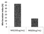
マトリゲル(MG)濃度の検討のため、コラーゲン吸着結合したPDMS膜製24穴培養器1穴辺り2x10⁵個の細胞を播種し、播種2時間後に交換する培地中のマトリゲル濃度を50および150μg/mLにして48時間培養した後に実施例4に記載の方法の通りBEI測定した。その結果、図12に示すように、150μg/mLマトリゲルの方が50μg/mLより高いBEIを示した。
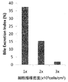
また、細胞播種密度の検討のため、肝細胞播種密度をコラーゲン吸着結合したPDMS膜製24穴培養器1穴辺り2x10⁵個、4x10⁵個、6x10⁵個にして、150μg/mLマトリゲルで48時間培養した後に実施例4に記載の方法の通りBEI測定した。その結果、図13に示すように、2x105個がもっともBEIが高く、4x105個、6x105個の順にBEIは低下した。
図14Aに示すように、PDMS膜上では播種後24時間および48時間後に、ポリスチレン上で96時間培養した肝細胞と同等のBEIを示した。図14BはPDMS膜製24穴培養器およびコラーゲンコートポリスチレン製24穴プレート上に肝細胞を播種後48時間および96時間後にCDCFが蓄積している部位を蛍光顕微鏡で撮影したものである。この像からもPDMS膜上の肝細胞のほうが従来の培養方法のそれより早期に活性をもった毛細胆管が形成されていることが確認できる。以上の結果から、PDMS膜製24穴培養器を用いてマトリゲルを重層して極性を誘導すると、播種後24時間後には極性が形成され、機能的な毛細胆管が現れる。これは、従来法によるものが同等のBEI値に到達するより72時間も早いことがわかった。
実施例6で作製した毛細胆管を形成した肝細胞を使用して、MRP2および基底膜マーカーCD147の局在を、常法に従い細胞抗体染色により調べた。また、コラーゲンコートポリスチレン製24穴プレートで培養した肝細胞と比較した。
図15に示すように、PDMS膜上で培養すると、培養48時間後にはMRP2タンパク質とCD147タンパク質が細胞間に検出される。一方、従来のポリスチレン上ではCD147の発現は見られるがMRP2タンパク質の局在はほとんど見られない。ポリスチレン上でも極性が形成されている培養120時間後と比較してもPDMS膜上の肝細胞のほうが広範囲にMRP2タンパクが発現している。CD147の局在様式は両者でほとんど同様である。これらの結果から、PDMS膜上で培養された肝細胞はポリスチレン上によるものに比べてより早くMRP2分子の局在が生じるとともに、MRP2の発現はポリスチレン上より増強されることが示された。
実施例6で作製した培養48時間後の肝細胞の微細構造を透過型電子顕微鏡(JEM1400 JEOL社製)で観察した。
図16Aで示すように、PDMS膜上で極性を誘導した肝細胞において、極性をもった肝細胞に典型的に見られる、毛細胆管腔(BC)、タイト結合(TJ)が見られる。図16Bはタイト結合部分の拡大写真、図16Cは腔壁に微小絨毛(MV)をもつ毛細胆管を示した。
この結果から、本法によって培養される肝細胞には構造的に生体と同等の極性が形成されることが示された。
ガス透過性を持つフルオロカーボン膜が培養面に配置された24穴培養プレートLumox (In vitro systems and services社製)にラットから調製した肝細胞を1穴辺り1.0x10⁵個または2.0x10⁵個播種した。播種2時間後に培地を除いて150μg/mLマトリゲルを含んだWilliam's Medium E(含5μg/mLインスリン、5μg/mLトランスフェリン、5μg/mL亜セレン酸,1μM デキサメタゾン)に交換した。肝細胞播種後48時間に実施例4に記載の方法の通りBEI測定し、極性形成の程度を解析した。コラーゲンコートポリスチレン製24穴プレートで培養した細胞を比較対照とした。
図17に示すように、Lumox上ではポリスチレン(PS)製24穴プレートより高いBEIを示した。細胞播種密度1穴辺り1.0x10⁵個と2.0x10⁵個では1.0x10⁵個のほうが高いBEIを与えた。
実施例1に記載の要領で作製したフルオロカーボン製の膜に100μg/mL コラーゲンI-P(新田ゼラチン社製)を含む0.001N HCl溶液を塗布したのち乾燥させ、コラーゲンコート処理を行った。
次に、前記で作製した基板の上に肝細胞を規則正しく並べた。図18Aに示すように、ゲル化前のコラーゲン溶液(濃度2.1mg/mL)に、幅が30μmで高さが100μmで長さが10mmの凸部を持ったPDMS製の型を配置して、37℃で60分間放置することでゲル化させることで凹部をもったコラーゲンゲルを作製した。
次に、図18Bに示したように、作製した凹部を持ったゲルに、培養培地に懸濁したラットの肝細胞を播き、2回培地で洗浄することで、凹部のみに肝細胞を配列させて培養を行った。
24時間後に、コラーゲン溶液(濃度2.1mg/mL)を細胞上に重層し、さらに9日間培養した。このようにして配列させた9日目の肝細胞の写真を図19に示す。連続的に毛細胆管が形成しているように観察された。
また、上記において、フルオロカーボン製酸素透過膜のコラーゲンコート処理の有無は結果に影響しなかった。
1、101:化合物供給部;2、102:本体部;3、103:回収部;11:ガス透過性膜;11’:隔壁;12:コラーゲンコート層;13:肝細胞;14:毛細胆管;15:コラーゲンマトリクス;16:培地;21:ガス透過性膜;22:コラーゲン;23:肝細胞;24:毛細胆管;25:細胞外マトリクス;26:半透膜;27:流路;28:筒状体。
Claims (27)
- ガス透過膜上に細胞外マトリクスで包埋された肝細胞を配置し、ガス透過性膜側から酸素を供給しつつ肝細胞を培養することを特徴とする、肝細胞培養方法。
- ガス透過性膜の表面がコラーゲンコーティングされており、該ガス透過性膜のコラーゲンコート面に細胞外マトリクスで包埋された肝細胞が配置される、請求項1に記載の肝細胞培養方法。
- ガス透過性膜がコラーゲンコート面を内側にして筒状に配置されており、該筒状の内部で細胞外マトリクスで包埋された肝細胞が配置される、請求項2に記載の方法。
- 前記細胞外マトリクスが溝を構成し、該溝に肝細胞が配置された、請求項1~3のいずれか一項に記載の方法。
- 前記ガス透過性膜がポリジメチルシロキサン膜である、請求項1~4のいずれか一項に記載の方法。
- 前記ガス透過性膜がフルオロカーボン膜である、請求項1~4のいずれか一項に記載の方法。
- コラーゲンコーティングが共有結合によるものである、請求項1~6のいずれか一項に記載の方法。
- 細胞外マトリクスがコラーゲンゲルまたはマトリゲル(商標)である、請求項1~7のいずれか一項に記載の方法。
- 細胞外マトリクスが非生体成分からなる、請求項1~7のいずれか一項に記載の方法。
- ガス透過膜上に細胞外マトリクスで包埋された肝細胞を配置し、ガス透過性膜側から酸素を供給しつつ肝細胞を培養することを特徴とする、毛細胆管を形成した培養肝細胞の製造方法。
- ガス透過性膜の表面がコラーゲンコーティングされており、該ガス透過性膜のコラーゲンコート面に細胞外マトリクスで包埋された肝細胞が配置される、請求項10に記載の方法。
- ガス透過性膜上に溝を有する細胞外マトリクス層が配置され、該細胞外マトリクス層の溝に肝細胞が配列された、請求項10または11に記載の方法。
- 請求項10~12のいずれか一項記載の方法により毛細胆管を形成した培養肝細胞を製造し、得られた培養肝細胞を用いて化合物の代謝を評価する、化合物の代謝検定方法。
- 請求項10~12のいずれか一項に記載の方法により毛細胆管を形成した培養肝細胞を製造し、得られた培養肝細胞を用いて化合物の輸送を評価する、化合物の輸送検定方法。
- 培養肝細胞を含む本体部と、本体部に化合物を供給する化合物供給部と、本体部から化合物またはその代謝物を回収する回収部とを有する培養肝細胞を用いた化合物検定装置であって、前記本体部は、
ガス透過性膜と、
該ガス透過膜上に配置された細胞外マトリクスと、
該細胞外マトリクスに包埋された肝細胞と、
を有することを特徴とする、装置。 - ガス透過性膜の表面がコラーゲンコーティングされており、該ガス透過性膜のコラーゲンコート面に細胞外マトリクスで包埋された肝細胞が配置された、請求項15に記載の装置。
- 前記ガス透過性膜は、コラーゲンコート面を内側にして筒状にした筒状体を形成しており、該筒状体の内部にコラーゲンコート面に接着された肝細胞とそれを包埋する細胞外マトリクスを含む、請求項16に記載の装置。
- 前記本体部は、前記化合物供給部からの供給物が流れるように半透膜で画定された空間を前記筒状体の軸方向に形成する流路であって、該半透膜を介して該供給物を前記細胞外マトリクスに包埋された肝細胞に供給可能な供給物用流路を、更に有する請求項17に記載の装置。
- 前記回収部が、表面がコラーゲンコーティングされたガス透過性膜をコラーゲンコート面を内側にして筒状にした筒状体と、該筒状体の内側のコラーゲンコート面に接着された肝細胞と、該肝細胞を包埋する細胞外マトリクスと、該肝細胞の毛細胆管によって形成される流路を有する、請求項18に記載の装置。
- 前記細胞外マトリクスが溝を構成し、該溝に肝細胞が配置された、請求項15~19のいずれか一項に記載の方法。
- 前記ガス透過性膜がポリジメチルシロキサン膜である、請求項15~20のいずれか一項に記載の装置。
- 前記ガス透過性膜がフルオロカーボン膜である、請求項15~20のいずれか一項に記載の装置。
- コラーゲンコーティングが共有結合によるものである、請求項15~22のいずれか一項に記載の装置。
- 細胞外マトリクスがコラーゲンゲルまたはマトリゲル(商標)である、請求項15~23のいずれか一項に記載の装置。
- 細胞外マトリクスが非生体成分からなる、請求項15~23のいずれか一項に記載の装置。
- 化合物代謝検定装置である、請求項15~25のいずれか一項に記載の装置。
- 化合物輸送検定装置である、請求項15~25のいずれか一項に記載の装置。
Priority Applications (3)
| Application Number | Priority Date | Filing Date | Title |
|---|---|---|---|
| JP2011528714A JP5818001B2 (ja) | 2009-08-26 | 2010-07-28 | 肝細胞の培養方法 |
| EP10811643.5A EP2471908B1 (en) | 2009-08-26 | 2010-07-28 | Method for culture of hepatocytes |
| US13/392,226 US8906644B2 (en) | 2009-08-26 | 2010-07-28 | Method for culture of hepatocytes |
Applications Claiming Priority (4)
| Application Number | Priority Date | Filing Date | Title |
|---|---|---|---|
| JP2009-195869 | 2009-08-26 | ||
| JP2009195869 | 2009-08-26 | ||
| JP2010073486 | 2010-03-26 | ||
| JP2010-073486 | 2010-03-26 |
Publications (1)
| Publication Number | Publication Date |
|---|---|
| WO2011024592A1 true WO2011024592A1 (ja) | 2011-03-03 |
Family
ID=43627702
Family Applications (1)
| Application Number | Title | Priority Date | Filing Date |
|---|---|---|---|
| PCT/JP2010/062707 Ceased WO2011024592A1 (ja) | 2009-08-26 | 2010-07-28 | 肝細胞の培養方法 |
Country Status (4)
| Country | Link |
|---|---|
| US (1) | US8906644B2 (ja) |
| EP (1) | EP2471908B1 (ja) |
| JP (1) | JP5818001B2 (ja) |
| WO (1) | WO2011024592A1 (ja) |
Cited By (14)
| Publication number | Priority date | Publication date | Assignee | Title |
|---|---|---|---|---|
| JP2013215152A (ja) * | 2012-04-11 | 2013-10-24 | Vessel Inc | 3次元細胞培養プレートとその製造方法 |
| JP2014187971A (ja) * | 2013-03-28 | 2014-10-06 | Lsi Medience Corp | 肝細胞培養用基板及び肝細胞培養方法 |
| KR101448377B1 (ko) | 2013-03-19 | 2014-10-07 | 경희대학교 산학협력단 | 세포 기능 평가용 양방향 동시측정 시스템 |
| JP2014223061A (ja) * | 2013-04-24 | 2014-12-04 | 株式会社Lsiメディエンス | 肝細胞培養方法 |
| WO2015080106A1 (ja) * | 2013-11-29 | 2015-06-04 | 株式会社日立ハイテクノロジーズ | 成分分析装置、薬効分析装置、及び分析方法 |
| WO2016147975A1 (ja) * | 2015-03-13 | 2016-09-22 | 国立研究開発法人医薬基盤・健康・栄養研究所 | 小腸上皮様細胞 |
| WO2016158417A1 (ja) * | 2015-03-27 | 2016-10-06 | 国立研究開発法人農業生物資源研究所 | 肝代謝物の毛細胆管様構造への蓄積と排泄を促進する肝細胞培養装置、並びに該肝細胞培養装置を用いた胆汁中又は血液中排泄感受性の候補化合物及び該候補化合物の肝代謝物の評価方法 |
| JP2017104117A (ja) * | 2017-01-31 | 2017-06-15 | 株式会社日立ハイテクノロジーズ | 成分分析装置、薬効分析装置、及び分析方法 |
| JP2018099123A (ja) * | 2018-01-22 | 2018-06-28 | 株式会社日立ハイテクノロジーズ | 分析方法 |
| WO2019021528A1 (ja) * | 2017-07-28 | 2019-01-31 | 株式会社日立製作所 | 酸素供給機構 |
| WO2021132586A1 (ja) * | 2019-12-27 | 2021-07-01 | 学校法人高崎健康福祉大学 | 肝細胞培養膜、それを備えた薬物輸送能評価キット、及び薬物輸送能評価方法 |
| WO2022158609A1 (ja) * | 2021-01-25 | 2022-07-28 | ニッタ株式会社 | 細胞培養容器 |
| JP7348997B1 (ja) | 2022-07-27 | 2023-09-21 | 浜松ホトニクス株式会社 | 毛細胆管領域の特定又は評価の方法、装置及びプログラム |
| WO2024080363A1 (ja) * | 2022-10-13 | 2024-04-18 | 国立大学法人東京大学 | デバイス、システム、方法、および薬剤評価方法 |
Families Citing this family (1)
| Publication number | Priority date | Publication date | Assignee | Title |
|---|---|---|---|---|
| US9512406B2 (en) | 2013-12-20 | 2016-12-06 | The J. David Gladstone Institute, a testamentary trust established under the Will of J. David Gladstone | Generating hepatocytes |
Family Cites Families (6)
| Publication number | Priority date | Publication date | Assignee | Title |
|---|---|---|---|---|
| EP1200110B1 (en) * | 1999-07-22 | 2014-04-23 | Organogenesis Inc. | In vivo induction for enhanced function of isolated hepatocytes |
| JPWO2005047496A1 (ja) | 2003-11-14 | 2007-05-31 | 学校法人慶應義塾 | 細胞培養法、細胞の三次元培養法、三次元組織、人工臓器、及び組織移植方法 |
| US7393687B2 (en) * | 2004-07-16 | 2008-07-01 | William Marsh Rice University | Biomimetic 3-dimensional scaffold with metabolic stream separation for bioartificial liver device |
| JP2006254722A (ja) * | 2005-03-15 | 2006-09-28 | Toray Ind Inc | 細胞足場材料 |
| CN101223268A (zh) | 2005-05-18 | 2008-07-16 | 康奈尔研究基金会(有限公司) | 具有生物屏障的基于药代动力学的培养系统 |
| JP4863655B2 (ja) * | 2005-06-17 | 2012-01-25 | 国立大学法人 東京医科歯科大学 | 細胞含有シート |
-
2010
- 2010-07-28 US US13/392,226 patent/US8906644B2/en not_active Expired - Fee Related
- 2010-07-28 WO PCT/JP2010/062707 patent/WO2011024592A1/ja not_active Ceased
- 2010-07-28 JP JP2011528714A patent/JP5818001B2/ja not_active Expired - Fee Related
- 2010-07-28 EP EP10811643.5A patent/EP2471908B1/en not_active Not-in-force
Non-Patent Citations (3)
| Title |
|---|
| LECLUYSE, E L. ET AL.: "Formation of extensive canalicular networks by rat hepatocytes cultured in collagen-sandwich configuration", AMERICAN JOURNAL OF PHYSIOLOGY - CELL PHYSIOLOGY, vol. 266, 1994, pages C1764 - C1774, XP008151208 * |
| MASAKI NISHIKAWA ET AL.: "Development of highly functional hepatic tissue through mimicking spatio-temporal environment in the liver (<Featured Theme 2>Engineering and Biotechnology)", MONTHLY JOURNAL OF INSTITUTE OF INDUSTRIAL SCIENCE, vol. 60, no. 2, March 2008 (2008-03-01), pages 52 - 59, XP008151215 * |
| MATSUI, H. ET AL.: "ENHANCED DEVELOPMENT OF A BILE CANALICULI NETWORK IN HEPATOCYTE SANDWICH CULTURE WITH DIRECT OXYGEN SUPPLY THROUGH POLYDIMETHYLSILOXANE MEMBRANES", 7TH WORLD CONGRESS ON ALTERNATIVES AND ANIMAL USE IN THE LIFE SCIENCE(2009) FINAL PROGRAMME,, pages 47, XP008158420 * |
Cited By (25)
| Publication number | Priority date | Publication date | Assignee | Title |
|---|---|---|---|---|
| JP2013215152A (ja) * | 2012-04-11 | 2013-10-24 | Vessel Inc | 3次元細胞培養プレートとその製造方法 |
| KR101448377B1 (ko) | 2013-03-19 | 2014-10-07 | 경희대학교 산학협력단 | 세포 기능 평가용 양방향 동시측정 시스템 |
| JP2014187971A (ja) * | 2013-03-28 | 2014-10-06 | Lsi Medience Corp | 肝細胞培養用基板及び肝細胞培養方法 |
| JP2014223061A (ja) * | 2013-04-24 | 2014-12-04 | 株式会社Lsiメディエンス | 肝細胞培養方法 |
| WO2015080106A1 (ja) * | 2013-11-29 | 2015-06-04 | 株式会社日立ハイテクノロジーズ | 成分分析装置、薬効分析装置、及び分析方法 |
| US9880153B2 (en) | 2013-11-29 | 2018-01-30 | Hitachi High-Technologies Corporation | Componential analyzer, drug efficacy analyzer, and analysis method |
| CN107677779A (zh) * | 2013-11-29 | 2018-02-09 | 株式会社日立高新技术 | 成分分析装置、药效分析装置和分析方法 |
| US11789013B2 (en) | 2013-11-29 | 2023-10-17 | Hitachi High-Tech Corporation | Componential analyzer, drug efficacy analyzer, and analysis method |
| CN107677779B (zh) * | 2013-11-29 | 2020-06-12 | 株式会社日立高新技术 | 成分分析装置、药效分析装置和分析方法 |
| US10684276B2 (en) | 2013-11-29 | 2020-06-16 | Hitachi High-Tech Corporation | Componential analyzer, drug efficacy analyzer, and analysis method |
| WO2016147975A1 (ja) * | 2015-03-13 | 2016-09-22 | 国立研究開発法人医薬基盤・健康・栄養研究所 | 小腸上皮様細胞 |
| JPWO2016147975A1 (ja) * | 2015-03-13 | 2018-03-08 | 国立研究開発法人医薬基盤・健康・栄養研究所 | 小腸上皮様細胞 |
| US10889805B2 (en) | 2015-03-13 | 2021-01-12 | National Institutes Of Biomedical Innovation, Health And Nutrition | Intestinal epithelioid cells |
| WO2016158417A1 (ja) * | 2015-03-27 | 2016-10-06 | 国立研究開発法人農業生物資源研究所 | 肝代謝物の毛細胆管様構造への蓄積と排泄を促進する肝細胞培養装置、並びに該肝細胞培養装置を用いた胆汁中又は血液中排泄感受性の候補化合物及び該候補化合物の肝代謝物の評価方法 |
| JPWO2016158417A1 (ja) * | 2015-03-27 | 2018-01-18 | 国立研究開発法人農業・食品産業技術総合研究機構 | 肝代謝物の毛細胆管様構造への蓄積と排泄を促進する肝細胞培養装置、並びに該肝細胞培養装置を用いた胆汁中又は血液中排泄感受性の候補化合物及び該候補化合物の肝代謝物の評価方法 |
| JP2017104117A (ja) * | 2017-01-31 | 2017-06-15 | 株式会社日立ハイテクノロジーズ | 成分分析装置、薬効分析装置、及び分析方法 |
| WO2019021528A1 (ja) * | 2017-07-28 | 2019-01-31 | 株式会社日立製作所 | 酸素供給機構 |
| JP2018099123A (ja) * | 2018-01-22 | 2018-06-28 | 株式会社日立ハイテクノロジーズ | 分析方法 |
| WO2021132586A1 (ja) * | 2019-12-27 | 2021-07-01 | 学校法人高崎健康福祉大学 | 肝細胞培養膜、それを備えた薬物輸送能評価キット、及び薬物輸送能評価方法 |
| WO2022158609A1 (ja) * | 2021-01-25 | 2022-07-28 | ニッタ株式会社 | 細胞培養容器 |
| JP7348997B1 (ja) | 2022-07-27 | 2023-09-21 | 浜松ホトニクス株式会社 | 毛細胆管領域の特定又は評価の方法、装置及びプログラム |
| WO2024024147A1 (ja) | 2022-07-27 | 2024-02-01 | 浜松ホトニクス株式会社 | 毛細胆管領域の特定又は評価の方法、装置及びプログラム |
| JP2024016956A (ja) * | 2022-07-27 | 2024-02-08 | 浜松ホトニクス株式会社 | 毛細胆管領域の特定又は評価の方法、装置及びプログラム |
| KR20250040954A (ko) | 2022-07-27 | 2025-03-25 | 하마마츠 포토닉스 가부시키가이샤 | 모세담관 영역의 특정 또는 평가의 방법, 장치 및 프로그램 |
| WO2024080363A1 (ja) * | 2022-10-13 | 2024-04-18 | 国立大学法人東京大学 | デバイス、システム、方法、および薬剤評価方法 |
Also Published As
| Publication number | Publication date |
|---|---|
| EP2471908A1 (en) | 2012-07-04 |
| EP2471908A4 (en) | 2013-11-06 |
| EP2471908B1 (en) | 2016-10-05 |
| JP5818001B2 (ja) | 2015-11-18 |
| US20120183989A1 (en) | 2012-07-19 |
| US8906644B2 (en) | 2014-12-09 |
| JPWO2011024592A1 (ja) | 2013-01-24 |
Similar Documents
| Publication | Publication Date | Title |
|---|---|---|
| JP5818001B2 (ja) | 肝細胞の培養方法 | |
| Grant et al. | Establishment of physiologically relevant oxygen gradients in microfluidic organ chips | |
| Guo et al. | A biomimetic human gut‐on‐a‐chip for modeling drug metabolism in intestine | |
| Nieskens et al. | Kidney-on-a-chip technology for renal proximal tubule tissue reconstruction | |
| Costello et al. | Microscale bioreactors for in situ characterization of GI epithelial cell physiology | |
| EP3383413B1 (en) | Devices and methods for simulating a function of a liver tissue | |
| De Gregorio et al. | Intestine‐on‐chip device increases ECM remodeling inducing faster epithelial cell differentiation | |
| JP7799729B2 (ja) | 幹細胞/増殖細胞区画及び分化細胞ゾーンを有する平面腸陰窩のアレイの形成 | |
| KR102621919B1 (ko) | iPSC-유래 세포의 장기 등가물로의 분화를 확립하는 신규한 다중 장기 칩 | |
| JP2019037237A (ja) | 操作した肝臓組織、そのアレイ、およびそれを製造する方法 | |
| US20200224136A1 (en) | Intestine-chip: differential gene expression model | |
| Zhang et al. | Microphysiological constructs and systems: biofabrication tactics, biomimetic evaluation approaches, and biomedical applications | |
| Sridhar et al. | Microstamped petri dishes for scanning electrochemical microscopy analysis of arrays of microtissues | |
| Wang et al. | Paper supported long-term 3D liver co-culture model for the assessment of hepatotoxic drugs | |
| Van Ness et al. | Microphysiological systems in absorption, distribution, metabolism, and elimination sciences | |
| Kim et al. | Effect of shear stress on the proximal tubule-on-a-chip for multi-organ microphysiological system | |
| Mughal et al. | Organs‐on‐chips: Trends and challenges in advanced systems integration | |
| Perin et al. | Generation of a glomerular filtration barrier on a glomerulus-on-a-chip platform | |
| JP2013226112A (ja) | 肝細胞の培養方法 | |
| JP2014223061A (ja) | 肝細胞培養方法 | |
| EP3000869A1 (en) | Cell culturing method, particulate culture carrier, and particle-encompassing cell aggregate | |
| Liang et al. | A microfluidic tool for real-time impedance monitoring of in vitro renal tubular epithelial cell barrier | |
| Becher et al. | A cost-effective vessel-on-a-chip for high shear stress applications in vascular biology | |
| Lei et al. | The utilisation of biliary organoids for biomedical applications | |
| WO2021132586A1 (ja) | 肝細胞培養膜、それを備えた薬物輸送能評価キット、及び薬物輸送能評価方法 |
Legal Events
| Date | Code | Title | Description |
|---|---|---|---|
| 121 | Ep: the epo has been informed by wipo that ep was designated in this application |
Ref document number: 10811643 Country of ref document: EP Kind code of ref document: A1 |
|
| WWE | Wipo information: entry into national phase |
Ref document number: 2011528714 Country of ref document: JP |
|
| NENP | Non-entry into the national phase |
Ref country code: DE |
|
| WWE | Wipo information: entry into national phase |
Ref document number: 13392226 Country of ref document: US |
|
| REEP | Request for entry into the european phase |
Ref document number: 2010811643 Country of ref document: EP |
|
| WWE | Wipo information: entry into national phase |
Ref document number: 2010811643 Country of ref document: EP |