WO2012132230A1 - 電子顕微鏡 - Google Patents
電子顕微鏡 Download PDFInfo
- Publication number
- WO2012132230A1 WO2012132230A1 PCT/JP2012/001363 JP2012001363W WO2012132230A1 WO 2012132230 A1 WO2012132230 A1 WO 2012132230A1 JP 2012001363 W JP2012001363 W JP 2012001363W WO 2012132230 A1 WO2012132230 A1 WO 2012132230A1
- Authority
- WO
- WIPO (PCT)
- Prior art keywords
- electron microscope
- imaging device
- attached
- detection device
- observation
- Prior art date
- Legal status (The legal status is an assumption and is not a legal conclusion. Google has not performed a legal analysis and makes no representation as to the accuracy of the status listed.)
- Ceased
Links
Images


Classifications
-
- H—ELECTRICITY
- H01—ELECTRIC ELEMENTS
- H01J—ELECTRIC DISCHARGE TUBES OR DISCHARGE LAMPS
- H01J37/00—Discharge tubes with provision for introducing objects or material to be exposed to the discharge, e.g. for the purpose of examination or processing thereof
- H01J37/02—Details
- H01J37/22—Optical, image processing or photographic arrangements associated with the tube
- H01J37/224—Luminescent screens or photographic plates for imaging; Apparatus specially adapted therefor, e. g. cameras, TV-cameras, photographic equipment or exposure control; Optical subsystems specially adapted therefor, e. g. microscopes for observing image on luminescent screen
-
- H—ELECTRICITY
- H01—ELECTRIC ELEMENTS
- H01J—ELECTRIC DISCHARGE TUBES OR DISCHARGE LAMPS
- H01J37/00—Discharge tubes with provision for introducing objects or material to be exposed to the discharge, e.g. for the purpose of examination or processing thereof
- H01J37/02—Details
- H01J37/16—Vessels; Containers
-
- H—ELECTRICITY
- H01—ELECTRIC ELEMENTS
- H01J—ELECTRIC DISCHARGE TUBES OR DISCHARGE LAMPS
- H01J37/00—Discharge tubes with provision for introducing objects or material to be exposed to the discharge, e.g. for the purpose of examination or processing thereof
- H01J37/02—Details
- H01J37/244—Detectors; Associated components or circuits therefor
-
- H—ELECTRICITY
- H01—ELECTRIC ELEMENTS
- H01J—ELECTRIC DISCHARGE TUBES OR DISCHARGE LAMPS
- H01J37/00—Discharge tubes with provision for introducing objects or material to be exposed to the discharge, e.g. for the purpose of examination or processing thereof
- H01J37/26—Electron or ion microscopes; Electron or ion diffraction tubes
Definitions
- the present invention relates to an electron microscope in which an observation room is provided in the lower part of the lens barrel and the second photographing device is provided in the observation room.
- An electron microscope is a type of electron microscope that observes an interference image created by irradiating an electron beam to a sample to be observed and transmitting or reflecting the electron beam. Widely used in science and medicine.
- the electron microscope can be broadly divided into a transmission electron microscope (TEM), which applies an electron beam to the sample to be observed, and magnifies and observes the electrons transmitted through the sample, and an electron on the sample to be observed.
- TEM transmission electron microscope
- a scanning electron microscope (Scanning Electron Microscope; SEM) that observes an image obtained from electrons (or secondary electrons) reflected from a line is known.
- SEM scanning Electron Microscope
- STEM scanning transmission electron microscope
- the main body of the electron microscope is composed of the lens barrel and the gantry housing that supports the lens barrel, and the main body of the electron microscope is controlled by a monitor device, and images acquired by the main body of the electron microscope are monitored. It is structured to be confirmed on the display device of the device.
- an electron beam generated at the upper part of the lens barrel is irradiated on the sample within the lens barrel, and this transmitted beam is projected onto a first fluorescent plate provided at the lower end of the lens barrel.
- a projection object is photographed by a first photographing means (CCD camera or the like), and confirmed and recorded by a display device of a monitor device.
- an observation room is provided above the first fluorescent plate, and in this observation room, a scintillator for observation with a second photographing means (CCD camera or the like), a dark field image or a bright field image is obtained. It has a structure to which other detection devices can be attached. These scintillators and other detection devices have a structure that can be retracted with respect to the electron beam applied to the first fluorescent screen so as not to hinder the acquisition of an image by the first imaging means.
- the lower end portion of the lens barrel portion on which the first fluorescent plate and the first photographing means are provided is attached in the gantry housing, and the observation chamber formed on the upper portion of the first fluorescent plate is mounted on the gantry housing.
- the lens barrel is attached to the gantry housing so as to be exposed from the table surface covering the upper surface.
- the scintillator and other detection devices attached to the observation room are attached so as to be retractable in a horizontal direction perpendicular to the beam axis of the vertical electron beam.
- the scintillator includes a second fluorescent plate, a mirror for refracting a beam transmitted through the second fluorescent plate in a horizontal direction, a second imaging unit for imaging the beam reflected by the mirror, and the second A drive mechanism for retracting the fluorescent plate and the mirror is included.
- a second fluorescent plate protruding in an oblique posture is provided on the beam axis in the observation chamber, and an image projected on the second fluorescent plate is obtained by the second photographing means.
- a structure for photographing is also proposed.
- the space of the scintillator occupying the observation chamber is large, and therefore, in order to attach another detection device, it must be provided at a position below the table surface avoiding the operating range of the scintillator. I had to.
- the position below the table surface has a problem in maintenance because of poor workability.
- the other detectors attached so as to be retractable from the lateral direction of the beam axis of the electron beam are: Retraction is not possible unless the position below the observation room is moved larger than the position below. In other words, if the other detectors are not moved in the horizontal direction, the first fluorescent screen will not be photographed, resulting in an increase in the size of the other detectors including the drive mechanism.
- an object of the present invention is to mount the second imaging means and other detectors compactly in an observation room provided on the table surface of the gantry housing, so that the workability of these devices and the effective use of the table surface can be achieved.
- an electron microscope according to the present invention is provided with a second imaging device provided in an observation room so as to be retractable with respect to a transmitted beam, and is on the same plane as a mounting position of the second imaging device.
- An attachment part of another detection device is provided at the position.
- the second imaging device and the other detection device can be mounted in the same plane in the observation room, the second imaging means and other detectors are provided in the observation room provided on the table surface of the gantry housing.
- a compact including a side camera
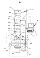
- FIG. 1 is a schematic structural diagram of an electron microscope system according to Embodiment 1.
- FIG. 1 is an apparatus configuration diagram of an electron microscope system according to Embodiment 1.
- FIG. 1 is a cross-sectional view of the vicinity of an observation room of an electron microscope system according to Example 1.
- FIG. 6 is an external view of a shield cover of another detection device of the electron microscope system according to Embodiment 1.
- FIG. 6 is a structural diagram in the vicinity of an observation room of an electron microscope system according to Example 2.
- FIG. 6 is a structural diagram in the vicinity of an observation room of an electron microscope system according to Example 2.
- FIGS. 1 to 4 show the first embodiment
- FIGS. 5 and 6 show another embodiment of the detector cover.
- part, an arrow, etc. are shown with the same code
- FIG. 1 is a schematic structural diagram of the electron microscope system according to this embodiment.
- FIG. 2 is an apparatus configuration diagram of the electron microscope system according to this embodiment.
- FIG. 3 is a cross-sectional view of the vicinity of the observation chamber of the electron microscope system according to this embodiment.
- FIG. 4 is an external view of a shield cover of another detection apparatus of the electron microscope system according to this embodiment.
- an electron microscope system generally indicated by reference numeral 1 is a transmission type electron microscope main body 20 that applies an electron beam to a sample 221 to be observed and magnifies and observes the electrons that have passed through the electron beam. And a monitor device 50 for operating and controlling the microscope main body 20 to monitor the acquired enlarged image.
- the electron microscope main body 20 includes a lens barrel portion 200 supported on the top of the gantry housing 100 and a peripheral device housing 40 with a built-in vacuum device.
- the electron microscope main body 20 includes a sample insertion portion in which an electron beam generated by an electron gun 210 disposed at the top of a lens barrel portion 200 that constitutes an electron optical system is disposed in the middle stage of the lens barrel portion 200.
- the sample 221 to be observed set in 220 is transmitted, and an image of the transmitted electron beam is formed on a first fluorescent plate portion 290 disposed below the lens barrel portion 200, and the formed image is displayed on the first fluorescent plate. It is possible to take an image with the first camera unit 291 serving as a first imaging unit disposed below the unit 290 and observe the image with the monitor device 50.
- the table surface 110 is provided on the upper portion of the gantry housing 100, and the lower end portion of the lens barrel portion 200 is mounted on the gantry so that the observation chamber 230 formed on the fluorescent plate 290 is exposed from the table surface 110. It is attached to the housing 100.
- the observation chamber 230 includes a second imaging device 240 including a second camera unit 250 serving as a second imaging means for directly observing an image that has passed through the sample 221, and other detection devices 260 can be attached and detached. Can be attached to.
- examples of the other detection device 260 include a dark field detector for acquiring a dark field image and a bright field detector for acquiring a bright field image, but are not limited thereto. .
- the monitor device 50 includes a table housing 51 that can be arranged side by side with the table surface 110 of the gantry housing 100, a display unit 52 for displaying various images and observation images, and inputs such as a keyboard and a mouse.
- the apparatus 53 and the control apparatus 54 which controls the electron microscope system 1 collectively are comprised.
- the monitor device 50 has a general personal computer device configuration, and can include a storage device (not shown) for storing image information, various programs, and the like, and a communication device for connecting to another computer system. According to the monitor device 50, the electron microscope main body 20 can be smoothly operated and controlled, and an enlarged image acquired by the electron microscope main body 20 can be observed or analyzed, or another system can be connected via a network. Can be sent to.
- the second imaging device 240 provided in the observation room 230 is retractable with respect to the beam axis (optical axis) P of the electron beam.
- the other detection device 260 can be attached to a position on the same plane.
- another detector 260 is attached to the second imaging device 240 at a position rotated by 90 degrees in the horizontal direction.
- the other detector 260 includes a side camera.
- the second photographing device 240 is provided so as to be retractable with respect to the beam axis P of the electron beam via the horizontal driving device 242.
- 241 and a second camera unit 250 attached to the side surface of the observation room 230.
- the second camera unit 250 and the horizontal driving device 242 are disposed on the side surface of the observation chamber 230 so as to sandwich the optical axis P of the electron beam in the front and rear directions, and operate the horizontal driving device 242 to operate the electron beam on the beam axis P.
- a structure is adopted in which the second fluorescent screen 241 disposed in the rear is retracted backward.
- the observation chamber 230 is configured such that another detection device 260 can be attached to and detached from the side wall in the left-right direction at a position rotated 90 degrees with respect to the arrangement of the second camera unit 250 and the horizontal driving device 242.
- a detector mounting portion 231 for mounting is provided.
- the other detection device 260 attached to the detector attachment portion 231 includes another detector 261 for acquiring a dark field image and a bright field image, and the other detector 261 with respect to the beam axis P of the electron beam.
- a horizontal driving device 262 that retracts in the horizontal direction.
- the detector mounting portion 231 of this embodiment is mounted with an electron beam stopper 263 provided with a horizontal driving device 262 that retracts in the horizontal direction with respect to the beam axis P of the electron beam, similarly to the other detectors 261. Can do.
- FIG. 1 shows an example in which another detection device 260 is attached to the left side of the observation room 230 and the electron beam stopper 263 is attached to the right side.
- the other detection device 260 is provided so as to be attached at a position rotated by 90 degrees with respect to the second imaging device 240. It can be set as the operation area of the detection device 260. That is, in this embodiment, the height at which the observation chamber 230 of the other detection device 260 is attached can be attached to the same height (on the same plane) as the second imaging device 240. Therefore, it has become possible to attach another detection device 260, which has conventionally been attached only below the table surface 110, on the table surface 110, which is easy to maintain. In addition, there is a problem that data acquired by another detection device 260 such as a side camera becomes data rotated by 90 degrees. However, rotation of 90 degrees is easy in terms of data conversion of image data by an image processing unit (not shown). Therefore, this problem can be solved easily. However, it goes without saying that an image can be acquired by image processing by the image processing unit regardless of the position in the same plane.
- the height at which the other detection device 260 is attached can be attached to the upper position of the observation chamber 230 from the lower position of the observation chamber 230 close to the first fluorescent plate portion 290.
- the device 260 can be miniaturized.
- the electron beam transmitted through the sample 221 is irradiated radially from the upper part of the observation chamber 230 and forms an image on the first fluorescent plate part 290 as shown by a broken line in FIG.
- the electron beam irradiated radially is retracted, it is necessary to increase the retracting amount of the other detection device 260 as it is closer to the first fluorescent plate portion 290. That is, it is necessary to increase the size of the horizontal drive device 262 as the other detection device 260 is attached to a position closer to the first fluorescent plate portion 230.
- the horizontal driving device 262 can be reduced in size.
- the second camera unit 250 is fixedly provided at the front portion of the observation room 230, and a CCD camera having a compact structure is employed.
- the amount of projection of the camera unit 250 to the front can be reduced.
- the horizontal driving device 262 it is possible to arrange another detection device 260 having a large outward protrusion amount in the left-right direction, so that the other detection device 260 is exposed on the table surface 110. Even if it is provided, the influence can be reduced, so that workability and designability on the table surface 110 can be improved.
- a structure in which the outside of another detection device 260 is covered with a detector cover 270 is employed.
- the detector cover 270 is formed of a metal plate material such as permalloy, iron plate, or aluminum, and is detachably attached to the observation chamber 230. According to this detector cover 270, it is possible to easily obtain high resolution by shielding heat, airflow, radio waves, dust and the like received from the outside with respect to the other detection devices 260.
- Another major feature of the electron microscope main body 20 according to this embodiment is that the second fluorescent plate 241 of the second imaging device 240 is fixed in an inclined posture, and the fixed second fluorescent plate 241 is horizontally driven.
- the device 242 is provided so as to be retractable.
- the conventional structure employs a structure in which the second fluorescent plate 241 and the mirror constituting the second imaging device 240 are rotatably provided in the observation chamber 230, and therefore occupies the observation chamber 230 of the second imaging device 240.
- the operating range was large. For this reason, there is a problem that it is difficult to provide another detection device 260 in the vicinity of the second imaging device 240.
- the second fluorescent plate 241 is fixed at a preset angle, and the fixed angle is maintained and the retraction operation is performed. Thereby, the operation range which occupies in the observation room 230 of the 2nd imaging device 240 can be made small.
- the inclination angle of the second fluorescent screen 241 is set upward from the angle of 45, and the second camera unit 250 is attached in an oblique posture according to the upward angle, so that the operating range (height) occupied in the observation room 230 is increased. Direction) can be reduced.
- the second camera unit 250 at an angle, the amount of the second camera unit 250 protruding forward can be reduced.
- FIG. 1 a schematic structure of a transmission electron microscope 20 according to this embodiment is shown in FIG.
- the electron beam from the electron gun 210 disposed on the uppermost part of the lens barrel part 200 passes through a condenser lens 215 constituting an irradiation system, and the sample 221 set on the sample part 220 provided in the middle stage of the lens barrel part 200. Is irradiated.
- the transmission electron beam that has passed through the sample 221 reaches the first fluorescent plate section 290 via the objective lens 225, the intermediate lens 226, and the projection lens 227 that form the imaging system.
- An image of the sample 221 based on the transmission electron beam is formed on the fluorescent plate unit 230.
- a transmission image or a diffraction image is formed depending on the set observation conditions.
- an objective aperture means 228 is installed to be detachable with respect to the beam axis P of the transmission electron beam.
- the objective aperture means 228 is formed with an opening 229.
- An image is formed on the first fluorescent screen 290 based on the transmitted electron beam that has passed through the opening 229.
- the image formed on the first fluorescent screen 290 in this way is acquired by the first camera 291.
- the first camera unit 291 includes an image sensor such as a CCD, and an image is captured by the image sensor.
- the image data of the image acquired by the first camera unit 291 is sent to the monitor device 50.
- the control device 54 processes the image data and causes the display unit 51 to display the image data.
- an observation chamber 230 is provided between the projection lens 227 and the first fluorescent screen 290.
- the thin electron beam irradiated from the projection lens 227 is irradiated radially toward the first fluorescent plate portion 290, and is formed on the first fluorescent plate portion 290 as an image having a predetermined size.
- the observation chamber 230 is provided with a second imaging device 240 that can irradiate the electron beam radially and refract the electron beam, and directly observe the refracted image, and a plurality of other detection devices.
- 260 and the electron beam stopper 263 can be detachably attached.
- the second imaging device 240 includes a second fluorescent plate 241 fixed at an inclination angle ⁇ 1, a horizontal driving device 242 attached to the wall surface on the back side of the observation chamber 230, and a first wall surface attached to the wall surface on the front side of the observation chamber 230. 2 camera unit 250.
- the horizontal driving device 242 can retract the second fluorescent plate 241 on the beam axis P of the electron beam backward, and can move the retracted second fluorescent plate 241 onto the beam axis P.
- the second fluorescent plate 241 is attached to the tip of the horizontal driving device 242 in a posture in which the rear side is raised at an inclination angle ⁇ 1 from the horizontal position.
- the conventional second fluorescent screen 241 is mounted with an inclination of 45 degrees, and has a structure that refracts the vertical beam axis P at a right angle.
- the height of the operation region Q1 having a height h1 is required.
- the height h1 of the operation region Q1 is reduced by setting the inclination angle ⁇ 1 of the second fluorescent plate 241 to an angle smaller than 45 degrees.
- the range of the operation region Q1 occupying the height H of the observation chamber 230 exposed on the table surface 110 of the gantry housing 100 is reduced, and the installation space for the other detection devices 260 is ensured in other ranges. It can be made easy.
- the second camera unit 250 that captures an image of the second fluorescent plate 241 has a problem that the image is distorted unless its optical axis P2 is set at a right angle to the second fluorescent plate 241. Therefore, in the conventional example, the second camera unit 250 is attached to the observation room 230 in a posture in which the optical axis P2 of the second camera unit 250 is horizontal. However, there is a problem that the horizontally mounted second camera unit 250 protrudes greatly in the horizontal direction. In response to this problem, the second camera unit 250 of this embodiment is attached to the observation chamber 230 in an oblique posture because the second fluorescent plate 241 is installed at an angle close to the horizontal direction, thus reducing the problem. can do.
- control device 54 controls the electron gun 210, controls an irradiation system including a condenser lens 215 disposed in the lens barrel 200, and forms an imaging system including an objective lens 225, an intermediate lens 226, and a projection lens 227.
- the control device 54 can control the objective aperture means 228 that moves the objective aperture, the first camera unit 291, the second imaging device 240, another detection device 260, the electron beam stopper 263, and the like. These control operations can be performed by operating the monitoring image displayed on the display unit 52 via the input device 53.
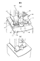
- observation chamber 230 which is a characteristic structure of this embodiment will be further described.
- the structure of the observation chamber 230 shown in FIG. 3 shows a cross section viewed from the front of the lens barrel portion 200.
- a second fluorescent plate 241 attached to the horizontal driving device 242 is provided behind the observation chamber 230 exposed on the table surface 110, and a second camera unit 250 (not shown) is attached to the front thereof.
- two detector mounting portions 231 (231a, 231b, 231c, 231d) are formed vertically on both sides of the observation chamber 230 exposed on the table surface 110, respectively.
- a dark field image detection device 260a is attached to the detector attachment portion 231a on the upper left side of the observation room 230, and a bright field image is detected on the detector attachment portion 231b on the lower left side of the observation room 230.
- a device 260b is attached.
- an electron beam stopper 263 is attached to the detector attachment portion 231c on the upper right side of the observation chamber 230, and the detector attachment portion 231d on the lower right portion of the observation chamber 230 is a spare attachment space.
- detector mounting portions 231 (231e, 231f) are provided on both sides of the observation chamber 230 concealed below the table surface 110.
- FIG. 3 a state in which another detection device 260 is attached to the left detector attachment portion 231e is shown.
- this embodiment uses the observation chamber 230 exposed on the table surface 110 in two upper and lower stages, and uses the lower operation area Q1 and the upper operation area Q2 to perform the second imaging.
- the device 240 and the other detection device 260 and the electron beam stopper 263 can be removably attached to the electron beam.
- the detector mounting portion 231a and the electron beam stopper 263 are disposed opposite to each other on both sides of the beam axis P of the electron beam, and one operation region Q2 can be used by two apparatuses. it can.
- the second imaging device 240 and two other detection devices 260 can be attached. That is, the second camera unit 250 and the second fluorescent plate 241 attached to the horizontal driving device 242 are provided before and after the operation region Q1, and two other detection devices 260 are attached to the left and right of the operation region Q1. it can. Therefore, the lower operation area Q1 of this embodiment can be used as an operation area of a device (second fluorescent plate 241 and two other detectors 261) mounted in three directions shifted by 90 degrees.
- the upper front part and the lower front part can be used as a mounting space for the second camera part 250 in an inclined posture.
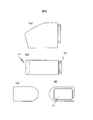
- FIG. 4 (a) is a plan view of the detector cover, (b) is a front view, (c) is a left side view, and (d) is a right side view.
- the embodiment of the detector cover 270 shown in FIG. 4 has a structure for individually shielding each other detection device 260. That is, the detector cover 270 includes a mounting portion 272 having an opening 271 on the right side surface, and a structure other than the opening 271 is covered with a metal material. The appearance is rounded with rounded corners so that the size is not felt from the front.
- the other detection device 260 is attached to the detector attachment portion 231 alone, and then the detector cover 270 is covered with the other detection device 260 through the opening 271. Wear as follows. Then, the attachment portion 272 is attached to the detector attachment portion 231 via a screw (not shown).
- the other detection devices 260 covered with the detector cover 270 are attached to both sides of the observation chamber 230 exposed on the table surface 110. Even if the detection device 260 protrudes greatly, it is reduced that the workability on the table surface 110 is hindered. In addition, since the other detection devices 260 covered with the detector cover 270 are attached to be exposed on the table surface 110, workability in attaching and detaching these devices and maintenance is significantly improved.
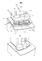
- FIGS. 5 and 6 show another embodiment of the detector cover.
- (a) is a development view of parts in the vicinity of the observation room
- (b) is an external view in the vicinity of the observation room.
- a plurality of other detection devices 260 can be attached to the observation chamber 230 to which the second imaging device 240 is attached.
- these other detection devices 260 need to shield heat, airflow, radio waves, dust and the like received from the outside.
- the number of the other detection devices 260 is one, it is effective to attach the detector cover 270 shown in FIG. 4, but when there are a plurality of other detection devices 260, the trouble of installation and maintenance becomes a problem. .
- FIGS. 5 and 6 employs a structure that can efficiently cover and shield a plurality of other detection devices 260.
- the shielding cover 300 pays attention to the fact that up to two other detection devices 260 are attached on both sides of the observation chamber 230.
- the cover is covered with one shielding cover 300.
- the shielding covers 300 to which other detection devices 260 attached in the vertical direction are collectively attached are attached symmetrically on both sides of the camera cover 301 of the second camera unit 250 attached to the front part of the observation room 230. .
- Shield cover 300 that covers a plurality of other detection devices 260 forms an open portion 302 that is continuous to one side surface and the bottom surface that are attached to detector attachment portion 231 in order to facilitate attachment.
- the table surface 110a of the gantry casing 100 is constituted by a front table surface 111 and a pair of side table surfaces 112 separated on both sides.
- the side table surface 112 is provided with a notch 113 connected to the opening 302 on the bottom surface of the shielding cover 300.
- a sufficient working space can be taken on both sides of the observation chamber 230 by the notch portions 113, so that the other detection device 260 can be easily attached to and detached from the detector mounting portion 231. Moreover, after attaching the other detection apparatus 260 to the detector attachment part 231, it can shield by only attaching the shielding cover 300 to this attachment part.
- the shielding cover 310 shown in FIG. 6 pays attention to the fact that the detector mounting portions 231 are provided on both sides of the second camera unit 250, and the camera cover 301 that covers the second camera unit 250 and the other detection device 260.
- the shielding cover 300 to be covered is formed as an integral structure.
- the shielding cover 310 is formed with an opening 311 that is continuous between a portion that contacts the observation chamber 230 and a bottom surface.
- the table surface 110b includes a front table surface 111 and a rear table surface 114.
- the rear table surface 114 is formed so as to surround both the front part and both sides of the lens barrel part 200, and a notch part 115 that opens both sides of the observation chamber 230 is formed in the front part.
- both sides of the observation chamber 230 are largely opened by the notch 115, the detachability and maintenance workability of the other detection devices 260 can be greatly improved.
- the second camera unit 250 and the other detection devices 260 on both sides of the observation room 230 can be collectively covered only by attaching the detector cover 270. Can be significantly improved.
- Electron microscope system 20 ... Electron microscope main body, 40 ... Peripheral device housing
Landscapes
- Chemical & Material Sciences (AREA)
- Analytical Chemistry (AREA)
- Analysing Materials By The Use Of Radiation (AREA)
Abstract
Description
また、好ましくは第2撮影装置240に対して、水平方向に90度回転させた位置に他の検出器260を取り付け可能に設けるようにする。ここで他の検出器260には、サイドカメラも含まれる。
但し、同一平面内のどの位置に配置したとしても、画像処理部による画像処理によって画像を取得できることは云うまでも無い。
Claims (10)
- 電子線を鏡筒部内で試料に照射し、この透過ビームを鏡筒部の下端部に設けた第1蛍光板に投影し、その投影物を撮影する第1撮影装置と、前記第1蛍光板の上部に形成される観察室に設けられる第2撮影装置とを備えた電子顕微鏡において、
前記第2撮像装置を前記透過ビームに対して退避可能に設け、
当該第2撮像装置の取付位置と同一平面上にある位置に他の検出装置の取付部を設けたことを特徴とする電子顕微鏡。 - 請求項1記載の電子顕微鏡において、
前記第2撮像装置は、前記観察室の前部壁面に取り付けられる第2カメラ部と、前記観察室の背面側の壁面に取り付けられる水平駆動装置を介して、前記透過ビームに対して退避可能に設けられる第2蛍光板とを含んで構成し、
前記他の検出装置の取付部は、前記観察部の少なくとも一方の側壁に形成されていることを特徴とする電子顕微鏡。 - 請求項2記載の電子顕微鏡において、
前記他の検出装置の取付部は、前記第2撮像装置の前記水平駆動装置と同じ高さに取り付けられていることを特徴とする電子顕微鏡。 - 請求項3記載の電子顕微鏡において、
前記第2蛍光板は、水平に対して45度より小さい角度で固定され、
前記第2カメラ部は、その光軸を第2蛍光板の面に対して直交する姿勢で取り付けられていることを特徴とする電子顕微鏡。 - 請求項4記載の電子顕微鏡において、
前記他の検出装置の取付部は、前記観察室の側壁に上下2段に設けられ、
前記第2撮像装置は、下段に対応する位置に取り付けられていることを特徴とする電子顕微鏡。 - 請求項1記載の電子顕微鏡において、
前記他の検出装置からの信号に基づき画像を形成する画像処理部を備えることを特徴とする電子顕微鏡。 - 請求項1記載の電子顕微鏡において、
前記他の検出器は暗視野検出器もしくは明視野検出器であることを特徴とする電子顕微鏡。 - 請求項1記載の電子顕微鏡において、
前記取付部に前記他の検出装置が着脱可能に取り付けられることを特徴とする電子顕微鏡。 - 電子線を鏡筒部内で試料に照射し、この透過ビームを鏡筒部の下端部に設けた第1蛍光板に投影し、その投影物を撮影する第1撮影装置と、前記第1蛍光板の上部に形成される観察室に設けられる第2撮影装置とを備えた電子顕微鏡において、
前記第2撮像装置を前記透過ビームに対して退避可能に設け、
この第2撮像装置の取付位置と水平方向に90度回転させた位置に他の検出装置の取付部を設けたことを特徴とする電子顕微鏡。 - 試料に電子線を照射する照射光学系と、
前記試料を透過した電子線を投影する第1蛍光板と、
前記第1蛍光板の上部に形成された観察室に設けられる撮像装置と、
当該撮像装置は前記試料を透過した電子線の光軸に対して退避可能に設けられ、
当該撮像装置と同一平面上にある位置に、着脱可能に設けられた検出装置を備えることを特徴とする電子顕微鏡。
Priority Applications (3)
| Application Number | Priority Date | Filing Date | Title |
|---|---|---|---|
| EP20120764716 EP2696363A4 (en) | 2011-03-31 | 2012-02-29 | ELECTRON MICROSCOPE |
| US14/002,141 US20140103208A1 (en) | 2011-03-31 | 2012-02-29 | Electron microscope |
| JP2013507110A JP5771685B2 (ja) | 2011-03-31 | 2012-02-29 | 電子顕微鏡 |
Applications Claiming Priority (2)
| Application Number | Priority Date | Filing Date | Title |
|---|---|---|---|
| JP2011-077103 | 2011-03-31 | ||
| JP2011077103 | 2011-03-31 |
Publications (1)
| Publication Number | Publication Date |
|---|---|
| WO2012132230A1 true WO2012132230A1 (ja) | 2012-10-04 |
Family
ID=46930026
Family Applications (1)
| Application Number | Title | Priority Date | Filing Date |
|---|---|---|---|
| PCT/JP2012/001363 Ceased WO2012132230A1 (ja) | 2011-03-31 | 2012-02-29 | 電子顕微鏡 |
Country Status (4)
| Country | Link |
|---|---|
| US (1) | US20140103208A1 (ja) |
| EP (1) | EP2696363A4 (ja) |
| JP (1) | JP5771685B2 (ja) |
| WO (1) | WO2012132230A1 (ja) |
Cited By (1)
| Publication number | Priority date | Publication date | Assignee | Title |
|---|---|---|---|---|
| CN107622934A (zh) * | 2017-09-22 | 2018-01-23 | 中国科学院生物物理研究所 | 一种用于透射电镜成像的相位板更换转移装置 |
Families Citing this family (6)
| Publication number | Priority date | Publication date | Assignee | Title |
|---|---|---|---|---|
| WO2016056096A1 (ja) * | 2014-10-09 | 2016-04-14 | 株式会社日立ハイテクノロジーズ | 荷電粒子線装置、電子顕微鏡、試料の観察方法 |
| CN108279247B (zh) * | 2016-12-30 | 2019-07-26 | 北京金竟科技有限责任公司 | 一种电子束激发荧光大范围直接探测成像装置及其方法 |
| KR101900603B1 (ko) * | 2017-05-08 | 2018-09-19 | 한국기초과학지원연구원 | 하전 입자 빔 정렬 장치 및 방법 |
| JP7092619B2 (ja) * | 2018-08-30 | 2022-06-28 | 株式会社キーエンス | 拡大観察装置 |
| JP7092618B2 (ja) * | 2018-08-30 | 2022-06-28 | 株式会社キーエンス | 拡大観察装置 |
| EP4546394B1 (en) * | 2023-10-26 | 2026-03-11 | The Provost, Fellows, Scholars and other Members Of the Board of Trinity College Dublin | Electron microscopy detector |
Citations (5)
| Publication number | Priority date | Publication date | Assignee | Title |
|---|---|---|---|---|
| JPH06231719A (ja) * | 1993-02-03 | 1994-08-19 | Seiko Instr Inc | 断面加工観察用荷電ビーム装置および加工方法 |
| JPH08138609A (ja) * | 1994-11-04 | 1996-05-31 | Hitachi Ltd | 電子顕微鏡における電子線検出器 |
| JPH0982263A (ja) | 1995-09-14 | 1997-03-28 | Hitachi Ltd | 電子顕微鏡 |
| JPH09223478A (ja) | 1996-02-16 | 1997-08-26 | Hitachi Ltd | 透過型電子顕微鏡 |
| JP2003331773A (ja) | 2002-05-13 | 2003-11-21 | Jeol Ltd | 電子顕微鏡 |
Family Cites Families (3)
| Publication number | Priority date | Publication date | Assignee | Title |
|---|---|---|---|---|
| JPS63292554A (ja) * | 1987-05-26 | 1988-11-29 | Hitachi Ltd | 電子顕微鏡 |
| US7755043B1 (en) * | 2007-03-21 | 2010-07-13 | Kla-Tencor Technologies Corporation | Bright-field/dark-field detector with integrated electron energy spectrometer |
| US7745786B2 (en) * | 2008-03-19 | 2010-06-29 | Fama Leo A | Method and apparatus allowing simultaneous direct observation and electronic capture of scintillation images in an electron microscope |
-
2012
- 2012-02-29 US US14/002,141 patent/US20140103208A1/en not_active Abandoned
- 2012-02-29 JP JP2013507110A patent/JP5771685B2/ja active Active
- 2012-02-29 EP EP20120764716 patent/EP2696363A4/en not_active Withdrawn
- 2012-02-29 WO PCT/JP2012/001363 patent/WO2012132230A1/ja not_active Ceased
Patent Citations (5)
| Publication number | Priority date | Publication date | Assignee | Title |
|---|---|---|---|---|
| JPH06231719A (ja) * | 1993-02-03 | 1994-08-19 | Seiko Instr Inc | 断面加工観察用荷電ビーム装置および加工方法 |
| JPH08138609A (ja) * | 1994-11-04 | 1996-05-31 | Hitachi Ltd | 電子顕微鏡における電子線検出器 |
| JPH0982263A (ja) | 1995-09-14 | 1997-03-28 | Hitachi Ltd | 電子顕微鏡 |
| JPH09223478A (ja) | 1996-02-16 | 1997-08-26 | Hitachi Ltd | 透過型電子顕微鏡 |
| JP2003331773A (ja) | 2002-05-13 | 2003-11-21 | Jeol Ltd | 電子顕微鏡 |
Non-Patent Citations (1)
| Title |
|---|
| See also references of EP2696363A4 * |
Cited By (2)
| Publication number | Priority date | Publication date | Assignee | Title |
|---|---|---|---|---|
| CN107622934A (zh) * | 2017-09-22 | 2018-01-23 | 中国科学院生物物理研究所 | 一种用于透射电镜成像的相位板更换转移装置 |
| CN107622934B (zh) * | 2017-09-22 | 2024-04-12 | 中国科学院生物物理研究所 | 一种用于透射电镜成像的相位板更换转移装置 |
Also Published As
| Publication number | Publication date |
|---|---|
| EP2696363A4 (en) | 2014-09-03 |
| JPWO2012132230A1 (ja) | 2014-07-24 |
| JP5771685B2 (ja) | 2015-09-02 |
| US20140103208A1 (en) | 2014-04-17 |
| EP2696363A1 (en) | 2014-02-12 |
Similar Documents
| Publication | Publication Date | Title |
|---|---|---|
| JP5771685B2 (ja) | 電子顕微鏡 | |
| JP5921462B2 (ja) | 放射線遮蔽ユニット、放射線画像撮影装置及び放射線画像撮影方法 | |
| US9848840B2 (en) | X-ray diagnostic apparatus comprising an X-ray filter movable along an imaging axis of X-rays | |
| JP5690086B2 (ja) | 拡大観察装置 | |
| JP5619535B2 (ja) | X線診断装置 | |
| US6804049B2 (en) | Microscope device and microscope system | |
| JP2009077759A (ja) | X線診断装置 | |
| JP2017080042A (ja) | X線画像診断装置 | |
| WO2015011975A1 (ja) | Ebsd検出器で所望箇所を容易に分析できる荷電粒子線装置およびその制御方法 | |
| JP2014021015A (ja) | 放射線画像取得装置 | |
| JP6680653B2 (ja) | 顕微鏡装置、顕微鏡システムおよび撮像方法 | |
| US20070189447A1 (en) | Medical imaging equipment | |
| US9113821B2 (en) | Radiation imaging apparatus and method | |
| EP3177009A1 (en) | Camera system with a full view monitoring function | |
| US11399786B2 (en) | X-ray diagnosis apparatus | |
| JP2003116845A (ja) | 医療用x線装置 | |
| JP5333279B2 (ja) | 放射線撮影装置 | |
| US10823858B2 (en) | Hybrid X-ray detector structure | |
| JP4456962B2 (ja) | 試料表示装置、試料表示装置の操作方法、試料表示装置操作プログラムおよびコンピュータで読み取り可能な記録媒体又は記録した機器 | |
| JP6274501B2 (ja) | X線診断装置 | |
| JP6258833B2 (ja) | 電子顕微鏡観察窓用の遮光器および電子顕微鏡 | |
| JP2007322384A (ja) | X線断層撮像装置及びx線断層撮像方法 | |
| CN206136073U (zh) | 摄像机的操作面板 | |
| JP2011146215A (ja) | 電子顕微鏡 | |
| JP5544909B2 (ja) | X線検査装置 |
Legal Events
| Date | Code | Title | Description |
|---|---|---|---|
| 121 | Ep: the epo has been informed by wipo that ep was designated in this application |
Ref document number: 12764716 Country of ref document: EP Kind code of ref document: A1 |
|
| ENP | Entry into the national phase |
Ref document number: 2013507110 Country of ref document: JP Kind code of ref document: A |
|
| WWE | Wipo information: entry into national phase |
Ref document number: 2012764716 Country of ref document: EP |
|
| NENP | Non-entry into the national phase |
Ref country code: DE |
|
| WWE | Wipo information: entry into national phase |
Ref document number: 14002141 Country of ref document: US |