WO2012137830A1 - 動物細胞培養キット、動物細胞の培養方法、動物細胞の選択培養方法及び細胞分化方法 - Google Patents
動物細胞培養キット、動物細胞の培養方法、動物細胞の選択培養方法及び細胞分化方法 Download PDFInfo
- Publication number
- WO2012137830A1 WO2012137830A1 PCT/JP2012/059219 JP2012059219W WO2012137830A1 WO 2012137830 A1 WO2012137830 A1 WO 2012137830A1 JP 2012059219 W JP2012059219 W JP 2012059219W WO 2012137830 A1 WO2012137830 A1 WO 2012137830A1
- Authority
- WO
- WIPO (PCT)
- Prior art keywords
- group
- sam
- culture
- cells
- stem cells
- Prior art date
- Legal status (The legal status is an assumption and is not a legal conclusion. Google has not performed a legal analysis and makes no representation as to the accuracy of the status listed.)
- Ceased
Links
Images























Classifications
-
- C—CHEMISTRY; METALLURGY
- C12—BIOCHEMISTRY; BEER; SPIRITS; WINE; VINEGAR; MICROBIOLOGY; ENZYMOLOGY; MUTATION OR GENETIC ENGINEERING
- C12N—MICROORGANISMS OR ENZYMES; COMPOSITIONS THEREOF; PROPAGATING, PRESERVING, OR MAINTAINING MICROORGANISMS; MUTATION OR GENETIC ENGINEERING; CULTURE MEDIA
- C12N5/00—Undifferentiated human, animal or plant cells, e.g. cell lines; Tissues; Cultivation or maintenance thereof; Culture media therefor
- C12N5/06—Animal cells or tissues; Human cells or tissues
- C12N5/0602—Vertebrate cells
- C12N5/0652—Cells of skeletal and connective tissues; Mesenchyme
- C12N5/0662—Stem cells
- C12N5/0663—Bone marrow mesenchymal stem cells (BM-MSC)
-
- C—CHEMISTRY; METALLURGY
- C12—BIOCHEMISTRY; BEER; SPIRITS; WINE; VINEGAR; MICROBIOLOGY; ENZYMOLOGY; MUTATION OR GENETIC ENGINEERING
- C12M—APPARATUS FOR ENZYMOLOGY OR MICROBIOLOGY; APPARATUS FOR CULTURING MICROORGANISMS FOR PRODUCING BIOMASS, FOR GROWING CELLS OR FOR OBTAINING FERMENTATION OR METABOLIC PRODUCTS, i.e. BIOREACTORS OR FERMENTERS
- C12M23/00—Constructional details, e.g. recesses, hinges
- C12M23/20—Material Coatings
-
- C—CHEMISTRY; METALLURGY
- C12—BIOCHEMISTRY; BEER; SPIRITS; WINE; VINEGAR; MICROBIOLOGY; ENZYMOLOGY; MUTATION OR GENETIC ENGINEERING
- C12M—APPARATUS FOR ENZYMOLOGY OR MICROBIOLOGY; APPARATUS FOR CULTURING MICROORGANISMS FOR PRODUCING BIOMASS, FOR GROWING CELLS OR FOR OBTAINING FERMENTATION OR METABOLIC PRODUCTS, i.e. BIOREACTORS OR FERMENTERS
- C12M25/00—Means for supporting, enclosing or fixing the microorganisms, e.g. immunocoatings
- C12M25/02—Membranes; Filters
-
- C—CHEMISTRY; METALLURGY
- C12—BIOCHEMISTRY; BEER; SPIRITS; WINE; VINEGAR; MICROBIOLOGY; ENZYMOLOGY; MUTATION OR GENETIC ENGINEERING
- C12N—MICROORGANISMS OR ENZYMES; COMPOSITIONS THEREOF; PROPAGATING, PRESERVING, OR MAINTAINING MICROORGANISMS; MUTATION OR GENETIC ENGINEERING; CULTURE MEDIA
- C12N5/00—Undifferentiated human, animal or plant cells, e.g. cell lines; Tissues; Cultivation or maintenance thereof; Culture media therefor
- C12N5/0068—General culture methods using substrates
-
- C—CHEMISTRY; METALLURGY
- C12—BIOCHEMISTRY; BEER; SPIRITS; WINE; VINEGAR; MICROBIOLOGY; ENZYMOLOGY; MUTATION OR GENETIC ENGINEERING
- C12N—MICROORGANISMS OR ENZYMES; COMPOSITIONS THEREOF; PROPAGATING, PRESERVING, OR MAINTAINING MICROORGANISMS; MUTATION OR GENETIC ENGINEERING; CULTURE MEDIA
- C12N2533/00—Supports or coatings for cell culture, characterised by material
- C12N2533/20—Small organic molecules
Definitions
- the present invention relates to an animal cell culture kit, an animal cell culture method, an animal cell selective culture method, and a cell differentiation method.
- a polystyrene culture dish for tissue culture into which various ionic groups have been introduced by plasma irradiation or the like is widely used.
- the degree of ionization by introducing ionic groups is not constant, and the surface composition is not chemically defined.
- the immortal cell lines often proliferate well.
- adhesion factors such as fibronectin and collagen, partial peptides of adhesion factors, extracellular matrix such as polylysine and basement membrane are used as coating agents. These coating agents are expensive and the effect of culture is limited to limited cells.
- a serum-containing culture solution to which high-concentration serum is added is used for culturing animal cells.
- the serum-containing culture solution since serum is a natural component, the serum-containing culture solution has disadvantages such as component indefiniteness, lot-to-lot variation, risk of pathogen contamination, and immune reaction risk. For this reason, when a serum-containing culture solution is used, cell proliferation (differentiation) tends to vary.
- cell proliferation tends to vary.
- serum-free cultures are being developed.
- a serum-free culture solution does not contain an adhesion factor in serum, there is a problem that cells are difficult to adhere / grow when attempting to culture in a normal tissue culture polystyrene culture dish.
- Non-patent Document 1 Non-patent Document 1
- Non-patent Document 2 the effect of a combination of two types of functional groups on the short-term proliferation of cancer cells and established (immortalized) cells has been studied.
- Non-patent Document 3 adhesion of vascular endothelial cells to SAM of RGD peptide has been reported.
- Non-Patent Document 1 and Non-Patent Document 2 there is no mention of cells (primary cells, stem cells, etc.) other than those containing a functional group that promotes growth and immortalized cell lines more than normal polystyrene culture dishes for tissue culture. .
- the presence of functional groups that significantly suppress cell adhesion is within the expected range.
- Non-Patent Document 3 it is natural that the RGD peptide promotes cell adhesion.
- functional group blended SAMs can bring cells to a higher level than normal culture dishes. There was no result.
- the present invention has been made in view of the above-mentioned matters.
- the object of the present invention is to provide an animal cell culture kit, an animal cell culture method, an animal cell selective culture method, and a cell that can be suitably used for animal cell culture. It is to provide a differentiation method.
- the animal cell culture kit according to the first aspect of the present invention comprises: An incubator having a predetermined ratio of one or more functional groups from the group consisting of a hydrophilic functional group and a hydrophobic functional group on a surface thereof, and a serum-free medium. It is characterized by that.
- the incubator may include a self-assembled monolayer having the functional groups on the surface at a predetermined ratio.
- the hydrophilic functional group is a hydroxyl group, an amino group or a carboxyl group
- the hydrophobic functional group is an alkyl group
- it may have a hydroxyl group: carboxyl group ratio of 100: 0 to 20:80 to promote adhesion and proliferation of human bone marrow mesenchymal stem cells.
- amino group: hydroxyl group is 40:60
- carboxyl group: methyl group is 100: 0-80: 20
- amino group: methyl group is 20: 80-80: 20
- amino group: carboxyl group is 40: 60-20 : 80 or hydroxyl group: methyl group in a ratio of 100: 0 to 80:20, and may promote the adhesion and proliferation of primary rat mesenchymal stem cells in a heterogeneous cell population.
- it may have a hydroxyl group: carboxyl group ratio of 100: 0 to 40:60 and promote adhesion and proliferation of human synovial stem cell derived mesenchymal stem cells.
- it may have a hydroxyl group: carboxyl group ratio of 100: 0 to 60:40 and promote adhesion and proliferation of human dental pulp-derived mesenchymal stem cells.
- the hydroxyl group: carboxyl group has a ratio of 100: 0 to 40:60 or the hydroxyl group: methyl group has a ratio of 80:20 to 20:80 to promote adhesion and proliferation of human fibroblasts.
- the hydroxyl group: carboxyl group may have a ratio of 100: 0 to 0: 100, or the hydroxyl group: methyl group may have a ratio of 80:20 to 20:80 to promote adhesion and proliferation of rat osteoblasts.
- it may have a hydroxyl group: carboxyl group ratio of 100: 0 to 40:60 and promote adhesion and proliferation of human primary mesenchymal stem cells.
- it may have a hydroxyl group: carboxyl group ratio of 100: 0 to 40:60 and promote adhesion and proliferation of human mesenchymal stem cells.
- the animal cell culture method according to the second aspect of the present invention comprises: Using an incubator having a predetermined ratio of one or more functional groups from the group consisting of hydrophilic functional groups and hydrophobic functional groups on the surface, and a serum-free medium, Promote the adhesion and proliferation of certain animal cells, It is characterized by that.
- the animal cell selective culture method comprises: Using an incubator having a predetermined ratio of one or more functional groups from the group consisting of hydrophilic functional groups and hydrophobic functional groups on the surface, and a serum-free medium, Promote adhesion and proliferation of certain animal cells selectively from heterogeneous cell populations; It is characterized by that.
- the animal cell culture method comprises: Serum-containing medium, amino group: hydroxyl group 100: 0-20: 80, hydroxyl group: carboxyl group 80: 20-20: 80, carboxyl group: methyl group 100: 0-60: 40, amino group: methyl group
- Serum-containing medium amino group: hydroxyl group 100: 0-20: 80, hydroxyl group: carboxyl group 80: 20-20: 80, carboxyl group: methyl group 100: 0-60: 40, amino group: methyl group
- a culture vessel having 80:20 to 20:80 or amino group: carboxyl group on the surface at a ratio of 80:20 to 20:80. Promote the proliferation of leaf stem cells, It is characterized by that.
- the cell differentiation method comprises: Human bone marrow mesenchymal stem cells are cultured using a culture vessel having a predetermined ratio of one or more functional groups from the group consisting of hydrophilic functional groups and hydrophobic functional groups on the surface and a serum-free differentiation induction medium.
- the human bone marrow mesenchymal stem cells are differentiated into chondrocytes or bone cells, It is characterized by that.
- human bone marrow mesenchymal stem cells may be differentiated into chondrocytes using the incubator having a hydroxyl group: carboxyl group in a ratio of 100: 0 to 20:80 and a serum-free cartilage differentiation inducing medium. .
- the human bone marrow mesenchymal stem cells may be differentiated into bone cells using the incubator having a hydroxyl group: carboxyl group ratio of 100: 0 to 20:80 and a serum-free osteogenic differentiation induction medium. Good.
- the cell differentiation method comprises: Human bone marrow mesenchymal stem cells are cultured using an incubator having a predetermined ratio of one or more functional groups from the group consisting of a hydrophilic functional group and a hydrophobic functional group on a surface and an adipogenic differentiation medium, and the human Differentiating bone marrow mesenchymal stem cells into adipocytes, It is characterized by that.
- the animal cell culture kit according to the present invention includes an incubator having a functional group suitable for adhesion / proliferation of specific animal cells on its surface, so that it is possible to promote the growth of animal cells even in a serum-free medium.
- (A) is a schematic diagram of alkanethiol
- (B) is a schematic diagram of SAM.
- (A) is a graph showing the composition of functional groups on the prepared SAM surface with respect to the mixing ratio of alkanethiols (NH 2 —SH and OH—SH)
- (B) is alkanethiols (OH—SH and COOH—
- (C) is a graph showing the composition of functional groups on the prepared SAM surface with respect to the mixing ratio of (SH)
- (C) shows the composition of functional groups on the prepared SAM surface with respect to the mixing ratio of alkanethiols (COOH-SH and CH 3 -SH).
- the graph (D) is a graph showing the composition of the functional group on the surface of the prepared SAM with respect to the mixing ratio of alkanethiol (CH 3 —SH and NH 2 —SH).
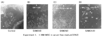
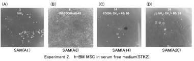
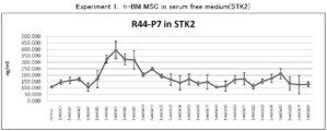
- (A) is a diagram showing the MTT test results after 4-day culture in Experiment 1
- (B) is a diagram showing the MTT test results after 4-day culture in Experiment 2
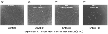
- (C) is the result after 4-day culture in Experiment 3. It is a figure which shows a MTT test result. It is the photograph of the cell after culture
- (A) is SAM (A1)
- (B) is SAM (A8)
- (C) is SAM (A14)
- (D) is SAM (A20).
- (A) is SAM (A1)
- (B) is SAM (A7)
- (C) is a photograph of SAM (A16).
- (A) is Control
- (B) is SAM (B5)
- (C) is SAM (B8)
- D) is a photograph of SAM (B13).
- (A) is a figure which shows the MTT test result after 4-day culture in Experiment 5
- (B) is a figure which shows the MTT test result after 4-day culture in Experiment 6. It is the photograph of the cell after culture
- (A) is a figure which shows the MTT test result after 4 days culture
- (B) is the normal culture in Experiment 1. It is a figure which shows the DNA fixed_quantity
- (A) shows the MTT test results after 7 days culture in Experiment 11-1
- (B) shows the MTT test results after 8 days culture in Experiment 11-2
- (C) shows 8 results in Experiment 12
- (D) is a figure which shows the MTT test result after 7-day culture
- (A) is Control
- (B) is SAM (A6)
- (C) is SAM (A11), (D) is SAM (A15), (D) is a photograph of cells after 7 days culture in Experiment 11-1.
- E) is a photograph of SAM (A25).
- (A) is Control
- (B) is SAM (A4)
- (C) is SAM (A11)
- (D) is SAM (A18)
- (D) E) is a photograph of SAM (A25). It is the photograph of the cell after 8 days culture
- (A) is a figure which shows the MTT test result after 4-day culture in Experiment 21
- (B) is a figure which shows the MTT test result after 14-day culture in Experiment 22. It is the photograph of the cell after culture
- (A) is a figure which shows the MTT test result after 4-day culture
- (B) is a figure which shows the MTT test result after 4-day culture
- Experiment 62 it is the photograph of the cell cultured by SAM (A6), (A) is a 10 times magnified photograph, (B) is a 40 times magnified photograph.
- Experiment 63 it is the photograph of the cell cultured by SAM (A8), (A) is a 10 times magnified photograph, (B) is a 40 times magnified photograph.
- Experiment 64 it is the photograph of the cell cultured by SAM (A11), (A) is a 10 times magnified photograph, (B) is a 40 times magnified photograph. It is a photograph of cells cultured in a normal culture dish stained with toluidine blue. It is a photograph of the cells cultured with SAM (A8) stained with toluidine blue.
- the animal cell culture kit according to the present embodiment includes an incubator and a serum-free medium.
- the incubator has at least one functional group from the group consisting of a hydrophilic functional group and a hydrophobic functional group at a predetermined ratio on the surface. More specifically, the incubator has self-assembled monolayers (hereinafter referred to as SAM) on the surface.
- SAM is a form in which one or more functional groups from the group consisting of hydrophilic functional groups and hydrophobic functional groups are exposed on the surface at a predetermined ratio. In this case, one SAM may have two or more different hydrophilic functional groups or hydrophobic functional groups, or may have both of them.
- the hydrophilic functional group include a hydroxyl group, an amino group, a carboxyl group, and a nitro group.
- hydrophobic functional group examples include an alkyl group such as a methyl group, an ethyl group, and a propyl group, a phenyl group, a cyclohexyl group, a cyclopentyl group, and a fluoro group.
- alkyl group such as a methyl group, an ethyl group, and a propyl group, a phenyl group, a cyclohexyl group, a cyclopentyl group, and a fluoro group.
- the hydrophilic functional group may be a positively charged functional group or a negatively charged functional group.
- the amino group may be NH 3 + by a proton and the carboxyl group may be COO ⁇ by deprotonation.
- the SAM can be made as follows. Alkanethiol is dissolved in a solvent such as ethanol to prepare an alkanethiol solution. As shown in the schematic diagram of FIG. 1A, alkanethiol has a thiol group (—SH) at one end of a linear alkyl chain and a methyl group (—CH 3 ) at the other end. It has a hydroxyl group (—OH), an amino group (—NH 2 ), or a carboxyl group (—COOH). The number of carbon atoms in the alkyl chain is not particularly limited, but is 5 to 20.
- the alkanethiol is bonded to the gold vapor deposition substrate by immersing the gold vapor deposition substrate or the like in the prepared alkanethiol solution.
- a SAM is formed on a gold vapor deposition substrate. Since the thiol group of alkanethiol specifically binds to gold, and the van der Waals force between the alkyl chains creates a high-density and highly-oriented structure of molecules, a monomolecular film with exposed functional groups on the surface is gold. It is formed on a vapor deposition substrate. An incubator is obtained by installing this gold vapor deposition substrate on a polystyrene culture dish or the like. Alternatively, alkanethiol may be directly bonded using a gold-modified culture dish or the like.
- SAMs having various functional groups on the surface can be obtained by preparing and using an alkanethiol solution by combining one or more of the above-mentioned various alkanethiols.
- the surface composition of the SAM is almost the same as the composition of the alkanethiol solution used. Therefore, according to the animal cell to be grown, a culture suitable for the growth of the animal cell is prepared by preparing alkanethiol in which alkanethiol having the above four types of functional groups is dissolved in a solvent at a predetermined ratio to form SAM. A vessel is obtained.
- Serum-free medium is a medium that does not contain serum and has few unknown factors. For this reason, compared with a serum-containing medium, there are advantages such as that certain performance can be obtained, purification from the medium and subsequent processes are easy, accurate evaluation of cell function is easy, and physiological reactivity is easily controlled.
- the serum-free medium does not contain serum, there is almost no adhesion factor (substrate adhesive cells) in the serum.
- adhesion factor substrate adhesive cells
- the animal cell culture kit according to the present embodiment includes an incubator having a SAM in which various functional groups are exposed at a predetermined ratio, and the surface composition of the incubator is bonded to a predetermined animal cell. It is possible to obtain a surface composition suitable for extension and proliferation. Therefore, even in a serum-free medium, adhesion, extension, and proliferation of predetermined animal cells can be promoted. Furthermore, cell differentiation can also be promoted in stem cells such as mesenchymal stem cells that have the ability to differentiate into cells of multiple lineages.
- a serum-free medium a commonly used basic medium, for example, an isotonic pH equilibrium solution mixed with salts, amino acids, sugars, vitamins and other trace essential nutrients can be combined. It may be a medium containing an additive.
- a suitable serum-free medium STK1 (trade name), STK2 (trade name), STK3 (trade name) (all manufactured by DS Pharma Biomedical Co., Ltd.) and the like can be mentioned.
- SAM and serum-free medium suitable for adhesion / proliferation of specific target animal cells from heterogeneous cell populations for example, cell populations in bone marrow fluid
- heterogeneous cell populations for example, cell populations in bone marrow fluid
- the SAM is formed on the gold vapor deposition substrate and the incubator in which the SAM is placed on the culture dish has been described.
- the SAM is formed using gold dormant or gold colloid.
- it may be a substrate made of metal such as aluminum, titanium, platinum, or silver.
- SAM may be formed on the surface using an inorganic metal oxide such as aluminum oxide, silicon oxide or titanium oxide, or a ceramic substrate such as glass.
- a SAM-formed substrate obtained by forming SAM on the surface thereof can be laminated on the culture dish to obtain an incubator.
- the substrate is not provided with a gold vapor deposition substrate or the like, and the culture dish itself is formed from the above-mentioned material, or the culture dish surface is modified with the above-mentioned material, and the SAM is directly formed on the culture dish surface.
- An incubator may be used.
- the culture dish is described as an example of the incubator.
- the culture dish is not limited to the culture dish as long as animal cells can be cultured.
- the form which the functional group mentioned above was directly added to the surface may be sufficient, without forming SAM using alkanethiol.
- the above functional group may be added to the surface of a biomaterial for transplantation.
- it may be in a form in which a functional group is directly added to a silane coupling method to glass or the like, processing of a titanium surface, or various biomaterial polymers.
- SAMs having various surface compositions were prepared, and various animal cells and culture media were combined, and the effects of the surface composition of SAMs acting on animal cell adhesion and proliferation were examined.
- SAM was produced on a gold vapor deposition substrate as follows.
- 1-dodecanethiol (hereinafter referred to as CH 3 —SH) manufactured by Wako Pure Chemical Industries, Ltd. was used.
- 11-mercapto-1-undecanol (hereinafter referred to as OH-SH)
- 11-mercapto-1-undecanoic acid hereinafter referred to as COOH-SH
- 11-amino-1-undecanethiol (hereinafter referred to as NH 2 -SH) (Note) used was made by Sigma-Aldrich Corporation.
- Alkanethiol (CH 3 —SH, OH—SH, COOH—SH, NH 2 —SH) is added to ethanol deoxygenated with nitrogen gas to a total of 1 mmol / L in various combinations and proportions as shown in Table 1. Then, an alkanethiol solution was prepared.
- the gold vapor deposition substrate was immersed in the prepared alkanethiol solution for 24 hours, and various SAMs (hereinafter referred to as SAM (A1) to SAM (A28), respectively) were produced on the gold vapor deposition substrate.
- SAM SAM
- A28 SAM
- the prepared SAM was stored in 2-propanol until used for experiments.
- SAM surface composition analysis The surface elemental composition of the prepared SAMs (A1) to (A20) was determined using X-ray photoelectron spectroscopy (XPS AXIS-HS, Kratos, Manchester, UK). The measurement was performed under a pressure of 10 ⁇ 7 Pa or less.
- XPS AXIS-HS X-ray photoelectron spectroscopy
- the surface of the sample was irradiated under conditions of an acceleration voltage of 15 kV and a filament current of 10 mA, and the elemental composition ratio of carbon and oxygen was measured under the condition of a photoelectron desorption angle of 90 degrees.
- FIG. 2 (A) shows the ratio of functional groups on the prepared SAM surface to the mixing ratio of NH 2 —SH and OH—SH in the alkanethiol solution
- FIG. 2 (B) shows OH—SH and COOH in the alkanethiol solution.
- FIG. 2C shows the ratio of the functional group on the prepared SAM surface to the blending ratio of COOH—SH and CH 3 —SH in the alkanethiol solution
- FIG. 2D shows the ratio of the functional group on the prepared SAM surface to the blending ratio of CH 3 —SH and NH 2 —SH in the alkanethiol solution.
- FIG. 2A As the ratio of amino groups to hydroxyl groups in the alkanethiol solution increases, a SAM having a surface outermost layer with a high ratio of amino groups to hydroxyl groups is obtained.
- FIG. 2B, FIG. 2C, and FIG. 2D show the same tendency when an alkanethiol solution having another functional group at the terminal is mixed.
- the composition of the functional group on the SAM surface can be adjusted according to the mixing ratio of the alkanethiol to be blended.
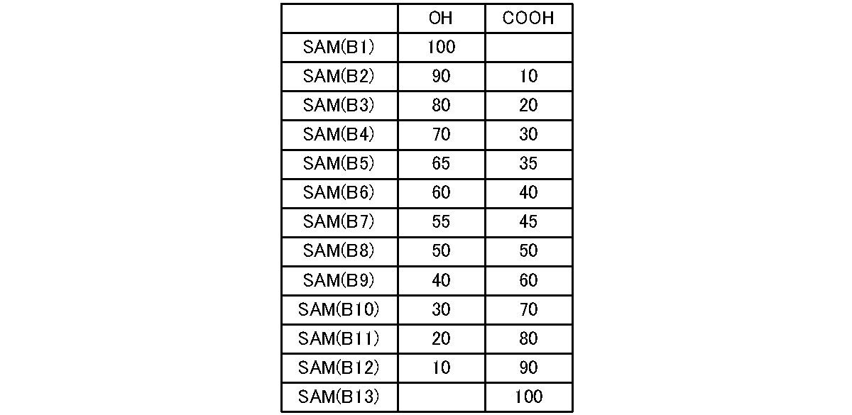
- alkanethiol solutions having the mixing ratios shown in Table 2 were prepared, and various SAMs were prepared in the same manner as described above (hereinafter referred to as SAM (B1) to (B13)) and used in the experiments.
- STK1 and STK2 are serum-free culture solutions containing human serum albumin, transferrin, and several types of growth factor proteins, but no unknown substances.
- FBS 10% fetal bovine serum
- DMEM Dulbecco'S modified Eagle's medium
- a 24-well microplate 24 Well Cell Culture Microplate diameter 16 mm, Corning Life Sciences, Inc.
- a normal culture dish or Control is a tissue culture culture dish (with polystyrene plasma treatment).
- Human bone marrow mesenchymal stem cells (2 strains: R-44, R-81) were isolated from the iliac bone by the method of Igarashi et al. (Igarashi A et al. Tissue Eng, 2007).
- serum medium A the medium was changed at 37 ° C. under 5% CO 2 while changing the medium every three days.
- 4th to 7th generation cells were used.
- the cells were dispersed by incubating with trypsin and EDTA for 5 minutes. Then, the enzyme reaction was stopped with a culture solution containing 10% FBS, and the cells were washed three times with serum-free DMEM. Thereafter, the cells were dispersed in a serum-free culture solution (STK1 or STK2) or serum culture solution A and seeded on SAM.
- STK1 or STK2 serum-free culture solution
- Table 3 shows combinations of human bone marrow mesenchymal stem cells (h-BM MSC), culture solution and SAM used in the experiment.
- the cell proliferation was evaluated by the MTT test by the WST assay (dye: WST-8 (2- (2-methoxy-4-nitrophenyl) -3- (4-nitrophenyl) -5- (2,4-disulfophenyl) -2H- tetrazolium), measured absorbance: 450 nm).
- the number of cells was measured with Cell Counting Kit-8 (Wako) using WST-8 or Coulter counter (Coulter).
- DNA was measured with a PicoGreenDsDNA Quantitation kit (Molecular Probe).
- FIGS. 9A and 9B are MTT test results after 4-day culture in Experiments 5 and 6 performed using serum culture medium A
- FIG. 10 is a photograph after 4-day culture in Experiment 5.
- human bone marrow mesenchymal stem cells strain R44
- strain R44 adhered to all SAM compositions (A1) to (A28). Due to the presence of the adhesion factor in the serum, the cells are hardly affected by the SAM composition, and are considered to adhere to all SAMs.
- the degree of proliferation differs depending on the SAM composition, and the SAMs (A1) to (A5), SAMs (A10) to (A12), and SAMs (A17) to (A24) proliferate remarkably.
- SAMs (A6) to (A9) that promoted growth in serum-free culture medium suppressed growth in serum culture medium A. It is considered that some serum protein adhered to SAMs (A6) to (A8) and hindered proliferation.
- FIG. 11 and FIG. 12 show the results of quantification of DNA after 4 days of culture in Experiment 1 and Experiment 5, respectively. MTT test results and DNA quantification results were in agreement.
- FIG. 13A shows the MTT test results after 4 days of culture of the normal culture dish and SAM (A8) of Experiment 1 and the normal culture dish and SAM (A5) of Experiment 5, and FIG. As shown in B), in serum-free medium, the number of cells and DNA increased 2-3 times on day 4 of culture on SAM (A8) compared to normal culture dishes. I understand.
- the surface hydroxyl group: carboxyl group ratio is 100: 0 to 0: 100, preferably 80:20 to 20:80, more preferably 70:30 to 60:40, and serum-free. It can be seen that by combining with the culture medium (STK2), adhesion and proliferation of human bone marrow mesenchymal stem cells can be promoted even in a serum-free culture medium.
- STK2 culture medium
- Bone marrow fluid contains hematopoietic cells, vascular cells, stromal cells, mesenchymal stem cells, and the like. That is, a heterogeneous cell population. Bone marrow mesenchymal stem cells are isolated as fibroblast-like cells that adhere to and propagate in culture dishes in the bone marrow. However, it is difficult to obtain homogeneous bone marrow mesenchymal stem cells in a normal culture dish. Therefore, in this experiment, rat bone marrow fluid was directly seeded on various SAM culture dishes, and the influence of the SAM composition on the adhesion and proliferation of mesenchymal stem cells (fibroblast-like cells) was examined.
- Bone marrow fluid separated from the left and right femurs and tibias of five male Wistar rats aged 5 weeks was diluted with serum-free alpha MEM to a total of 25 ml (bone marrow dilution).
- a bone marrow dilution is seeded at 0.06 mL / 16 mm well, and further, STMEM, serum culture medium A, or ⁇ MEM containing 10% fetal bovine serum (FBS) ⁇ MEM + 10% FBS) was added to make the total culture volume 0.5 ml / well.
- the culture solution was changed every 3 days from 3 days later and cultured for 7 to 8 days.
- rat bone marrow mesenchymal stem cells 3 ml of bone marrow dilution is seeded on a 100 mm tissue culture culture dish (Corning), cultured for 8 days, and then subconfluent. Cells dispersed with trypsin and EDTA from each culture system were seeded on various SAM substrates at a cell density of 2000 / cm 2 .
- the cell morphology observation and cell number evaluation method are the same as in Experiments 1-6.
- Table 4 shows combinations of cells, culture solution, and SAM used.
- fibroblast-like cells including mesenchymal stem cells
- SAM serum-free medium
- A6 fibroblast-like cells (including mesenchymal stem cells) of rat bone marrow
- A11 fibroblast-like cells
- A12 mesenchymal stem cells
- A17 fibroblast-like cells
- the surface amino group: hydroxyl group is 40:60
- carboxyl group: methyl group is 100: 0-80: 20
- amino group: methyl group is 20: 80-80: 20
- amino group: carboxyl group 40:60 to 20:80 or a SAM having a hydroxyl group: methyl group of 100: 0 to 80:20 and a serum-free culture solution (STK1), so that even in serum-free culture solution, the primary rat bone marrow It can be seen that adhesion and proliferation of mesenchymal stem cells can be promoted.
- STK1 serum-free culture solution
- Human synovial mesenchymal stem cells human osteoarthritis-derived mesenchymal stem cells, also called synovial fibroblasts (2 strains) (h-synovial MSC (S6-P2), h-synovial MSC)) Is available from Cell Applications Inc. Obtained from
- Human dental pulp mesenchymal stem cells (1 strain) (h-Dental pulp MSC) were separated from the extracted dental pulp tissue fragment of the extracted tooth as a population of cells that migrated and grew (Odontblast Differentiation of Human DulpCults in Explant. .-L. Couple et al., Calcif Tissue Int. 2000).
- the cell morphology observation and cell number evaluation method are the same as in Experiments 1-6.
- Table 5 shows combinations of cells, culture medium, and SAM used.
- serum-free by combining a SAM having a surface hydroxyl group: carboxyl group ratio of 100: 0 to 40:60, preferably 80:20 to 60:40, and a serum-free culture solution (STK2 or STK1). It can be seen that adhesion and proliferation of human synovial-derived mesenchymal stem cells can be promoted even in the culture medium.
- FIG. 22 shows the MTT test results after 4 days of culture in Experiment 23, and FIG.
- the SAM composition that promotes the proliferation of human dental pulp-derived mesenchymal stem cells (5th generation) was also almost in common with human bone marrow and human synovial stem cells. Therefore, by combining a SAM having a hydroxyl group: carboxyl group ratio of 100: 0 to 60:40 on the surface and a serum-free culture solution (STK2), adhesion of human dental pulp-derived mesenchymal stem cells can be achieved even in a serum-free medium. It turns out that proliferation can be promoted.
- STK2 serum-free culture solution
- Human skin fibroblasts were purchased from Kurabo Corporation (Osaka) and maintained in serum culture medium A as described above.
- the rat skull-derived osteoblasts, Yoshiko et al's method (A subset of osteoblasts expressing high endogenous levels of PPARgamma switches fate to adipocytes in the rat calvaria cell culture model.
- male Wistar rat calvaria of embryonic day 21 was digested with collagenase over time, the cells were fractionated into 5 fractions, and the 4 fractions excluding the first fraction (fibroblast fraction) were separately separated with 10% FBS-containing alpha. Cultured in MEM. The next day, these fractions were pooled and cultured in the same culture solution supplemented with ascorbic acid (50 ⁇ g / ml). These cells were grown to near confluence and then passaged with trypsin and EDTA as described above.
- SAM (A8) and SAM (A27) to (A28) also grew to a medium level, but other compositions did not.
- the SAM composition in which human fibroblasts proliferate in serum-free cultures partially overlapped the SAM composition in which human mesenchymal stem cells proliferate, but the pattern was different.
- the ratio of hydroxyl group to carboxyl group on the surface is 100: 0 to 40:60, or the ratio of hydroxyl group: methyl group is 80:20 to 20:80, preferably the ratio of hydroxyl group: carboxyl group is 100. : 0 to 80:20, or a combination of a SAM having a hydroxyl group: methyl group ratio of 80:20 to 60:40 and a serum-free medium (STK2), so that human fibroblasts can be used even in a serum-free medium. It can be seen that the adhesion and proliferation of can be promoted.
- STK2 serum-free medium
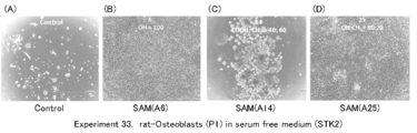
- FIG. 26 shows the MTT test results after 4 days of culture in Experiment 33 using rat osteoblasts
- FIG. 27 shows the photographs.
- Rat osteoblasts had good adhesion to SAM (A6) to (A12) and SAM (A25) to (A28) in the presence of serum-free culture medium (STK2).
- serum-free culture medium STK2
- the SAM composition in which osteoblasts proliferate in serum-free culture was different from the pattern of mesenchymal stem cells or fibroblasts.
- the surface hydroxyl group: carboxyl group ratio is 100: 0 to 0: 100
- the carboxyl group: methyl group ratio is 80:20
- the hydroxyl group: methyl group ratio is 80: 20-20: 80.
- Rat bone marrow mesenchymal stem cells were used after being separated and cultured in the same manner as in Experiments 11-13.
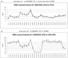
- FIGS. 28A and 28B show the MTT test results after the 4-day culture in Experiment 41 and 42
- FIGS. 29A and 29B show the DNA quantification results after the 4-day culture in Experiment 41 and 42.
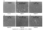
- photographs after 4 days of cultivation in Experiment 41 and Experiment 42 are shown in FIGS. 30 and 31, respectively.
- 2h-4days shows the state of cell adhesion 2 hours after seeding and 4 days after seeding (A) with many adhesions and (F) with many non-adherent cells. That is, some SAMs promoted initial adhesion but did not maintain long-term adhesion or proliferation, and others promoted proliferation as well as initial adhesion. SAM has been shown to affect proliferation as well as adhesion.
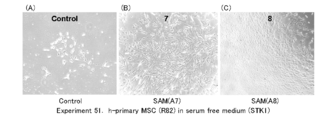
- FIG. 32 shows the MTT test results of Experiment 51, and FIG.
- serum-free medium STK1
- SAM serum-free medium
- FIG. 32 shows the MTT test results of Experiment 51, and FIG.
- serum-free medium STK1
- SAM serum-free medium
- STK1 serum-free medium
- the results of human bone marrow mesenchymal stem cells in Experiments 1 to 4 are all the same.
- the cell proliferation is observed in SAM (A6) to (A8). Therefore, if the SAM composition is a ratio of hydroxyl group: carboxyl group of 100: 0 to 60:40, human mesenchyme from other tissues such as fat origin, umbilical cord origin, placenta origin, amnion origin, periosteum, etc. It can also be applied to the adhesion and proliferation of stem cells.
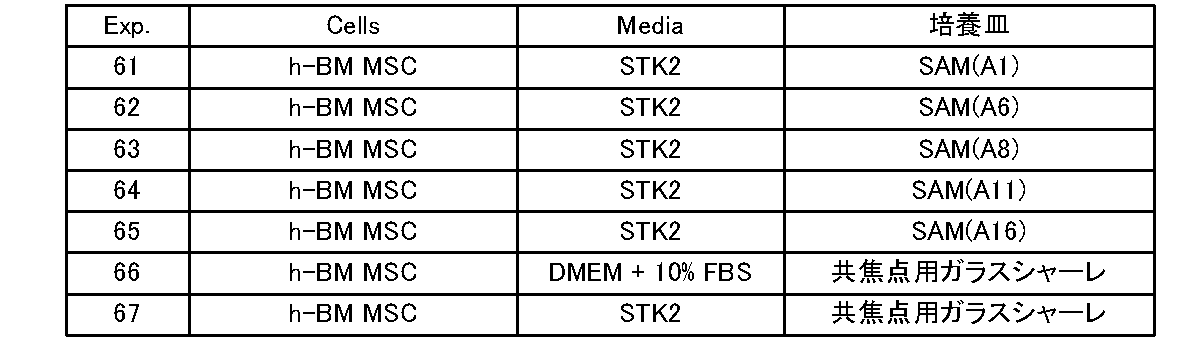
- Pre-cultured human bone marrow mesenchymal stem cells were dispersed in STK2, seeded at a density of 4000 cells / well in a 16 mm culture dish, and cultured. Further, as a reference example, the culture was performed in the same manner as described above using a combination of a confocal glass petri dish and DMEM + 10% FBS or STK2. Table 9 shows combinations of the culture medium and the culture dish used.
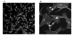
- the primary antibody and the secondary antibody were added and stained for each, and observed with a confocal laser microscope (FV-1000D, manufactured by Olympus Corporation). The adhesion and extension of mesenchymal stem cells to the culture dish were evaluated.
- the primary antibody used is Actin Cytoskeleton / Focal Adhesion Staining Kit (Merck MILLIPORE), and the secondary antibody is GOAT ANTI-MOUSE IgG (H & L) FLUORESCEIN CONJUGATED (Takara Bio Inc.).
- the FBS medium containing serum (Experiment 66) has an adhesion factor, so that the presence of filamentous protrusions and the like is observed, and the cells expand and become large.
- STK2 is a serum-free medium.
- the cells did not adhere to the glass petri dish, the presence of filamentous protrusions, etc., and the extension of the cells was not observed.
- the culture medium was changed to STK2, adipose differentiation induction medium, and STK3 to be differentiated into chondrocytes, adipocytes, and bone cells.
- a fat differentiation induction medium 1% Antibiotic-Antimycotic, 10% FBS, 2 mM L-glutamine, 10-6 MDex, 0.2 mM Mindomethacin (Wako Pure Chemical Industries, Ltd), 0.01 mg / ml insulin (WakoPure Chemical Industries, Ltd), 0.5 DMEM supplemented with mM 3-isobutyl-1-methylxanthine (Wako PureChemical Industries, Ltd) was used.
- the cell was cultured like the above using the normal culture dish (Control) instead of SAM (A8) as a reference example. In each experiment, three samples were performed.
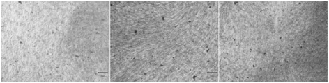
- FIG. 39 shows a photograph in which cells cultured in a normal culture dish are stained
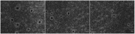
- FIG. 40 shows a photograph in which cells cultured in SAM (A8) are stained.
- Table 11 shows the adhesion state and the amount of cells stained with toluidine blue staining.
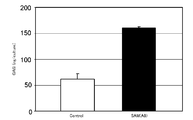
- glycosaminoglycan (GAG) produced when cartilage was formed was measured to verify whether differentiation into chondrocytes was promoted. The result is shown in FIG. Note that, for both SAM (A8) and the normal culture dish, the GAG production amount per culture dish was measured on the 28th day after the medium was changed.
- GAG was about 60 ng / culture, but in SAM (A8), it was about 160 ng / culture, and GAG was produced nearly three times as compared with the normal culture dish.
- FIG. 42 shows a photograph in which cells cultured in a normal culture dish are stained
- FIG. 43 shows a photograph in which cells cultured in SAM (A8) are stained. Table 12 shows the results of visual observation of the amount of stained cells.
- GPDH Glycerol 3-phosphate dehydrogenase
- human bone marrow mesenchymal stem cells can be differentiated from the incubator on the surface at a ratio. That is, if human bone marrow mesenchymal stem cells are cultured using a cartilage differentiation induction medium, they can be differentiated into chondrocytes.
- human bone marrow mesenchymal stem cells can be differentiated into bone cells.
- human bone marrow mesenchymal stem cells can be differentiated into adipocytes.
- cell differentiation can be performed even in a serum-free medium.
- the serum-free cartilage differentiation-inducing medium and osteogenesis-inducing medium are not limited, and examples include STK2 and STK3 described above.
- the culture method is excellent not only for cell proliferation but also for cell differentiation.
- the animal cell culture kit according to the present invention can promote adhesion and proliferation of specific animal cells even in a serum-free medium containing no adhesion factor, and thus is useful for research and development such as regenerative medicine and biopharmaceuticals.
Landscapes
- Health & Medical Sciences (AREA)
- Life Sciences & Earth Sciences (AREA)
- Engineering & Computer Science (AREA)
- Chemical & Material Sciences (AREA)
- Organic Chemistry (AREA)
- Bioinformatics & Cheminformatics (AREA)
- Wood Science & Technology (AREA)
- Zoology (AREA)
- Biomedical Technology (AREA)
- Biotechnology (AREA)
- Genetics & Genomics (AREA)
- Biochemistry (AREA)
- Microbiology (AREA)
- General Engineering & Computer Science (AREA)
- General Health & Medical Sciences (AREA)
- Immunology (AREA)
- Cell Biology (AREA)
- Sustainable Development (AREA)
- Developmental Biology & Embryology (AREA)
- Clinical Laboratory Science (AREA)
- Hematology (AREA)
- Rheumatology (AREA)
- Micro-Organisms Or Cultivation Processes Thereof (AREA)
- Apparatus Associated With Microorganisms And Enzymes (AREA)
Abstract
Description
親水性官能基及び疎水性官能基からなる群から1種以上の官能基を所定の割合で表面に有する培養器と、無血清培地と、を備える、
ことを特徴とする。
親水性官能基及び疎水性官能基からなる群から1種以上の官能基を所定の割合で表面に有する培養器と、無血清培地とを用い、
所定の動物細胞の接着及び増殖を促進させる、
ことを特徴とする。
親水性官能基及び疎水性官能基からなる群から1種以上の官能基を所定の割合で表面に有する培養器と、無血清培地とを用い、
不均質な細胞集団から選択的に所定の動物細胞の接着及び増殖を促進させる、
ことを特徴とする。
血清含有培地と、アミノ基:水酸基を100:0~20:80、水酸基:カルボキシル基を80:20~20:80、カルボキシル基:メチル基を100:0~60:40、アミノ基:メチル基を80:20~20:80、或いは、アミノ基:カルボキシル基を80:20~20:80の割合で表面に有する培養器とを用いて、ヒト間葉系幹細胞、ヒト線維芽細胞又はラット間葉系幹細胞の増殖を促進させる、
ことを特徴とする。
親水性官能基及び疎水性官能基からなる群から1種以上の官能基を所定の割合で表面に有する培養器と、無血清の分化誘導培地とを用い、ヒト骨髄間葉系幹細胞を培養して前記ヒト骨髄間葉系幹細胞を軟骨細胞又は骨細胞に分化させる、
ことを特徴とする。
親水性官能基及び疎水性官能基からなる群から1種以上の官能基を所定の割合で表面に有する培養器と脂肪分化誘導培地とを用いてヒト骨髄間葉系幹細胞を培養し、前記ヒト骨髄間葉系幹細胞を脂肪細胞に分化させる、
ことを特徴とする。
(SAMの作製)
まず、以下のようにして金蒸着基板上にSAMを作製した。

作製したSAM(A1)~(A20)の表面元素組成をX-ray photoelectoron spectroscopy(XPS AXIS-HS,Kratos,Manchester,UK)を用いて求めた。測定は10-7Pa以下の圧力下で行った。Al-Kapha単色X線は、加速電圧が15kV,フィラメント電流が10mAの条件で試料表面に照射し、光電子脱離角度が90度の条件における炭素と酸素の元素組成比を測定した。
無血清培養液としてSTK1(商品名)及びSTK2(商品名)(いずれもDSファーマバイオメディカル社製)を用いた。なお、STK1及びSTK2には、ヒト血清アルブミン、トランスフェリン、数種類の成長因子の蛋白質が含有されているが、未知物質は一切含有されていない無血清培養液である。
SAM表面の官能基の組み合わせが、ヒト骨髄間葉系幹細胞(h-BM MSC)の接着・増殖に及ぼす影響について検証した。間葉系幹細胞(mesenchymal stem cells)を適宜MSCと称する。
ヒト骨髄間葉系幹細胞(2株:R-44,R-81)は五十嵐らの方法(Igarashi A et al. Tissue Eng,2007)で腸骨から分離した。血清培養液Aにて、3日毎に培養液交換しながら、37℃で5%CO2条件下にて培養した。そして、実験には4-7代目の細胞を使用した。細胞がほぼコンフルエントになるまで増殖した後、トリプシンとEDTAと5分間インキュベートすることにより細胞を分散した。
そして10%FBS含有培養液にて酵素反応を停止して、細胞を無血清のDMEMで3回洗浄した。その後、無血清培養液(STK1又はSTK2)あるいは血清培養液Aに細胞を分散させ、SAM上に播種した。
保存しておいたSAM基板(直径約15mm)をPBS(リン酸緩衝生理的食塩水)で3回洗浄後、24Well Suspension Culture Plate(浮遊培養用培養皿、プラズマ処置なし、直径約16mm、Greiner bio-one社)に設置した。

細胞を培養後、細胞の状態/形態を位相顕微鏡下で観察して、接着/進展細胞(Adherent cells,A)と浮遊細胞あるいは弱く接着した球形の細胞(Floating cells,F)に区分した。
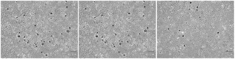
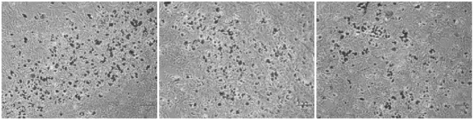
実験1,2,3の4日間培養後のMTT試験結果をそれぞれ図3(A)、(B)、(C)に、4日間培養後の写真をそれぞれ図4、図5、図6に示している。
図9(A)、(B)は、血清培養液Aを用いて行った実験5,6の4日間培養後のMTT試験結果であり、また、図10は実験5の4日間培養後の写真であるが、ヒト骨髄間葉系幹細胞(R44株)はすべてのSAM組成(A1)~(A28)に接着した。血清中の接着因子の存在により、細胞がSAM組成の影響を受けにくく、全てのSAMに接着したものと考えられる。なお、SAM組成によって増殖の程度は異なっており、SAM(A1)から(A5),SAM(A10)~(A12),SAM(A17)~(A24)上で顕著に増殖している。
ラット初代間葉系幹細胞(rat-primary MSC)の接着・増殖に及ぼすSAM表面の官能基配合の影響について検証した。

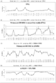
実験11では7日間培養、及び、8日間培養を行いそれぞれについて評価を行った。以下では、実験11の7日間培養について実験11-1、8日間培養について実験11-2と記す。実験11-1の7日間培養後のMTT試験結果を図14(A)に、実験11-2の8日間培養後のMTT試験結果を図14(B)に、実験12の8日間培養後のMTT試験結果を図14(C)に、実験13の7日間培養後のMTT試験結果を図14(D)に示す。また、実験11-1の7日間培養後の写真を図15に、実験11-2の8日間培養後の写真を図16に、実験12の8日間培養後の写真を図17に、実験13の7日間培養後のMTT試験結果を図18に示す。
各種組織由来の間葉系幹細胞の接着と増殖に及ぼすSAM表面の官能基配合の影響について検証した。

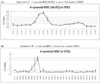
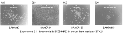
実験21のMTT試験結果を図19(A)に、実験22のMTT試験結果を図19(B)にそれぞれ示すとともに、実験21の4日間培養後の写真を図20に、実験22の14日間培養後の写真を図21にそれぞれ示す。
ヒト線維芽細胞(Fibroblast)およびラット骨芽細胞(rat-Osteoblast)の接着と増殖に及ぼす培養皿表面の官能基配合の影響について検証した。

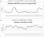
実験31のMTT試験結果を図24(A)に、実験32のMTT試験結果を図24(B)にそれぞれ示すとともに、実験31の4日間培養後の写真を図25に示す。
ラット骨髄間葉系幹細胞(rat-BM MSC)の接着と増殖に及ぼす培養皿表面の官能基配合の影響について検証した。

実験41及び実験42の4日間培養後のMTT試験結果を図28(A)、(B)に、実験41及び実験42の4日間培養後のDNA定量結果を図29(A)、(B)に、実験41及び実験42の4日間培養後の写真を図30、図31にそれぞれ示す。
ヒト初代間葉系幹細胞(h-primary MSC)の接着と増殖に及ぼす培養皿表面の官能基配合の影響について検証した。

実験51のMTT試験結果を図32に、培養後の写真を図33にそれぞれ示す。無血清培養液(STK1)存在下では、SAM(A6)~(A11)でヒト初代間葉系幹細胞の顕著な増殖が見られる。表面の水酸基:カルボキシル基の割合が100:0~40:60のSAMと、無血清培養液(STK1)とを組み合わせることで、無血清培養液中でも、ヒト初代間葉系幹細胞の接着・増殖を促進させ得ることがわかる。
続いて、ヒト骨髄間葉系幹細胞(h-BM MSC)の培養皿表面への接着、伸展性等について検証した。
d:脱着或いは浮遊細胞
S:小細胞
L:大細胞
R:丸まって伸展していない形状
F:扁平し伸展した形状
-:存在していない
±:存否の判断不能
+:存在している
++:多く存在している

続いて、SAM(A8)を用い、ヒト骨髄間葉系幹細胞(h-BM MSC)の軟骨細胞、脂肪細胞および骨細胞への分化について検証した
培地をSTK2に交換後、顕微鏡で経時的に観察し、培養皿への細胞の接着状態を観察した。また、トルイジンブルー染色(Toluidine blue staining)を行い、軟骨細胞に分化しているか否かを検証した。

脂肪分化誘導培地に交換した後、共焦点レーザー顕微鏡で観察し、培養皿への細胞の接着状態を観察するとともに、オイルレッドO(Oil red O staining)染色により、軟骨細胞に分化しているか否かを検証した。図42に、通常培養皿にて培養した細胞を染色した写真、図43に、SAM(A8)にて培養した細胞を染色した写真をそれぞれ示す。また、表12に染色された細胞の量を目視で観察した結果を示す。

STK3に交換した後、骨細胞に発現するALP(Alkaline Phosphatase)活性を測定した。なお、本実験では、通常培養皿とSTK3とを組み合わせた参考例のほか、他の参考例として、通常培養皿とDMEM+10%FBSとの組み合わせ(Negative Control)についても行った。
Claims (19)
- 親水性官能基及び疎水性官能基からなる群から1種以上の官能基を所定の割合で表面に有する培養器と、無血清培地と、を備える、
ことを特徴とする動物細胞培養キット。 - 前記培養器が前記官能基を所定の割合で表面に有する自己組織化単分子膜を備える、
ことを特徴とする請求項1に記載の動物細胞培養キット。 - 前記親水性官能基が水酸基、アミノ基又はカルボキシル基であり、前記疎水性官能基がアルキル基である、
ことを特徴とする請求項1又は2に記載の動物細胞培養キット。 - 水酸基:カルボキシル基を100:0~20:80の割合で有し、ヒト骨髄間葉系幹細胞の接着及び増殖を促進する、
ことを特徴とする請求項3に記載の動物細胞培養キット。 - アミノ基:水酸基を40:60、カルボキシル基:メチル基を100:0~80:20、アミノ基:メチル基を20:80~80:20、アミノ基:カルボキシル基を40:60~20:80、或いは、水酸基:メチル基を100:0~80:20の割合で有し、不均質な細胞集団において初代のラット間葉系幹細胞の接着及び増殖を促進する、
ことを特徴とする請求項3に記載の動物細胞培養キット。 - 水酸基:カルボキシル基を100:0~40:60の割合で有し、ヒト滑膜由来間葉系幹細胞の接着及び増殖を促進する、
ことを特徴とする請求項3に記載の動物細胞培養キット。 - 水酸基:カルボキシル基を100:0~60:40の割合で有し、ヒト歯髄由来間葉系幹細胞の接着及び増殖を促進する、
ことを特徴とする請求項3に記載の動物細胞培養キット。 - 水酸基:カルボキシル基を100:0~40:60、或いは、水酸基:メチル基を80:20~20:80の割合で有し、ヒト線維芽細胞の接着及び増殖を促進する、
ことを特徴とする請求項3に記載の動物細胞培養キット。 - 水酸基:カルボキシル基を100:0~0:100、或いは、水酸基:メチル基を80:20~20:80の割合で有し、ラット骨芽細胞の接着及び増殖を促進する、
ことを特徴とする請求項3に記載の動物細胞培養キット。 - 水酸基:カルボキシル基を100:0~0:100、或いは、カルボキシル基:メチル基を100:0~80:20の割合で有し、ラット骨髄間葉系幹細胞の接着及び増殖を促進する、
ことを特徴とする請求項3に記載の動物細胞培養キット。 - 水酸基:カルボキシル基を100:0~40:60の割合で有し、ヒト初代間葉系幹細胞の接着及び増殖を促進する、
ことを特徴とする請求項3に記載の動物細胞培養キット。 - 水酸基:カルボキシル基を100:0~40:60の割合で有し、ヒト間葉系幹細胞の接着及び増殖を促進する、
ことを特徴とする請求項3に記載の動物細胞培養キット。 - 親水性官能基及び疎水性官能基からなる群から1種以上の官能基を所定の割合で表面に有する培養器と、無血清培地とを用い、
所定の動物細胞の接着及び増殖を促進させる、
ことを特徴とする動物細胞の培養方法。 - 親水性官能基及び疎水性官能基からなる群から1種以上の官能基を所定の割合で表面に有する培養器と、無血清培地とを用い、
不均質な細胞集団から選択的に所定の動物細胞の接着及び増殖を促進させる、
ことを特徴とする動物細胞の選択培養方法。 - 血清含有培地と、アミノ基:水酸基を100:0~20:80、水酸基:カルボキシル基を80:20~20:80、カルボキシル基:メチル基を100:0~60:40、アミノ基:メチル基を80:20~20:80、或いは、アミノ基:カルボキシル基を80:20~20:80の割合で表面に有する培養器とを用いて、ヒト間葉系幹細胞、ヒト線維芽細胞又はラット間葉系幹細胞の増殖を促進させる、
ことを特徴とする動物細胞の培養方法。 - 親水性官能基及び疎水性官能基からなる群から1種以上の官能基を所定の割合で表面に有する培養器と、無血清の分化誘導培地とを用い、ヒト骨髄間葉系幹細胞を培養して前記ヒト骨髄間葉系幹細胞を軟骨細胞又は骨細胞に分化させる、
ことを特徴とする細胞分化方法。 - 水酸基:カルボキシル基を100:0~20:80の割合で有する前記培養器と、無血清の軟骨分化誘導培地とを用い、前記ヒト骨髄間葉系幹細胞を軟骨細胞に分化させる、
ことを特徴とする請求項16に記載の細胞分化方法。 - 水酸基:カルボキシル基を100:0~20:80の割合で有する前記培養器と、無血清の骨形成分化誘導培地とを用い、前記ヒト骨髄間葉系幹細胞を骨細胞に分化させる、
ことを特徴とする請求項16に記載の細胞分化方法。 - 親水性官能基及び疎水性官能基からなる群から1種以上の官能基を所定の割合で表面に有する培養器と脂肪分化誘導培地とを用いてヒト骨髄間葉系幹細胞を培養し、前記ヒト骨髄間葉系幹細胞を脂肪細胞に分化させる、
ことを特徴とする細胞分化方法。
Priority Applications (3)
| Application Number | Priority Date | Filing Date | Title |
|---|---|---|---|
| JP2013508904A JP6011879B2 (ja) | 2011-04-05 | 2012-04-04 | 動物細胞培養キット、動物細胞の培養方法、動物細胞の選択培養方法及び細胞分化方法 |
| EP12768652.5A EP2695934B1 (en) | 2011-04-05 | 2012-04-04 | Animal cell culture kit, method for culturing animal cells, method for selective culture of animal cells and cell differentiation method |
| US14/009,874 US10196610B2 (en) | 2011-04-05 | 2012-04-04 | Animal cell culture kit, method for culturing animal cells, method for selective culture of animal cells and cell differentiation method |
Applications Claiming Priority (2)
| Application Number | Priority Date | Filing Date | Title |
|---|---|---|---|
| JP2011084119 | 2011-04-05 | ||
| JP2011-084119 | 2011-04-05 |
Publications (1)
| Publication Number | Publication Date |
|---|---|
| WO2012137830A1 true WO2012137830A1 (ja) | 2012-10-11 |
Family
ID=46969224
Family Applications (1)
| Application Number | Title | Priority Date | Filing Date |
|---|---|---|---|
| PCT/JP2012/059219 Ceased WO2012137830A1 (ja) | 2011-04-05 | 2012-04-04 | 動物細胞培養キット、動物細胞の培養方法、動物細胞の選択培養方法及び細胞分化方法 |
Country Status (4)
| Country | Link |
|---|---|
| US (1) | US10196610B2 (ja) |
| EP (1) | EP2695934B1 (ja) |
| JP (1) | JP6011879B2 (ja) |
| WO (1) | WO2012137830A1 (ja) |
Cited By (3)
| Publication number | Priority date | Publication date | Assignee | Title |
|---|---|---|---|---|
| WO2015082908A1 (en) * | 2013-12-02 | 2015-06-11 | Medway School Of Pharmacy | Self-assembled monolayer |
| JP2020500588A (ja) * | 2016-12-12 | 2020-01-16 | カール クリストフ | 関節での新規組織形成のための関節インプラント |
| JP2021003084A (ja) * | 2019-06-27 | 2021-01-14 | 国立大学法人山口大学 | 骨髄由来間葉系幹細胞の培養方法 |
Citations (5)
| Publication number | Priority date | Publication date | Assignee | Title |
|---|---|---|---|---|
| WO2006093207A1 (ja) * | 2005-03-02 | 2006-09-08 | National University Corporation Hokkaido University | 細胞の分化/増殖を制御するための基材 |
| JP2009216483A (ja) * | 2008-03-10 | 2009-09-24 | Kyushu Univ | 混合samの作製方法 |
| JP2010029186A (ja) * | 2008-07-25 | 2010-02-12 | Becton Dickinson & Co | 規定細胞培養表面及び使用方法 |
| WO2010033925A2 (en) * | 2008-09-19 | 2010-03-25 | Wisconsin Alumni Research Foundation | Peptide-presenting surfaces for long-term culture of pluripotent cells |
| JP2010166901A (ja) * | 2008-12-24 | 2010-08-05 | Institute Of Physical & Chemical Research | 卵膜由来細胞の細胞外マトリクスを用いた多能性幹細胞の培養方法 |
-
2012
- 2012-04-04 EP EP12768652.5A patent/EP2695934B1/en not_active Not-in-force
- 2012-04-04 JP JP2013508904A patent/JP6011879B2/ja not_active Expired - Fee Related
- 2012-04-04 US US14/009,874 patent/US10196610B2/en not_active Expired - Fee Related
- 2012-04-04 WO PCT/JP2012/059219 patent/WO2012137830A1/ja not_active Ceased
Patent Citations (5)
| Publication number | Priority date | Publication date | Assignee | Title |
|---|---|---|---|---|
| WO2006093207A1 (ja) * | 2005-03-02 | 2006-09-08 | National University Corporation Hokkaido University | 細胞の分化/増殖を制御するための基材 |
| JP2009216483A (ja) * | 2008-03-10 | 2009-09-24 | Kyushu Univ | 混合samの作製方法 |
| JP2010029186A (ja) * | 2008-07-25 | 2010-02-12 | Becton Dickinson & Co | 規定細胞培養表面及び使用方法 |
| WO2010033925A2 (en) * | 2008-09-19 | 2010-03-25 | Wisconsin Alumni Research Foundation | Peptide-presenting surfaces for long-term culture of pluripotent cells |
| JP2010166901A (ja) * | 2008-12-24 | 2010-08-05 | Institute Of Physical & Chemical Research | 卵膜由来細胞の細胞外マトリクスを用いた多能性幹細胞の培養方法 |
Non-Patent Citations (11)
| Title |
|---|
| ARIMA Y; IWATA H., BIOMATERIALS, vol. 28, 2007, pages 3074 - 3082 |
| FAUCHEUX, N ET AL.: "The dependence of fibrillar adhesions in human fibroblasts on substratum chemistry", BIOMATERIALS, vol. 27, no. 2, 2006, pages 234 - 245, XP005071169 * |
| FUSE Y; HIRATA I; KURIHARA H; OKAZAKI M: "Cell adhesion and proliferation patterns on mixed self-assembled monolayers carrying various ratios of hydroxyl and methyl groups", DENT MATER J., vol. 26, no. 6, 2007, pages 814 - 819 |
| IGARASHI A ET AL., TISSUE ENG., 2007 |
| J AM CHEM SOC, vol. 120, 1998, pages 6548 - 6555 |
| KUBO H; SHIMIZU M; TAYA Y ET AL., GENE TO CELLS, vol. 14, 2009, pages 407 - 424 |
| M. L. COUBLE ET AL.: "Odontoblast Differentiation of Human Dental Pulp Cells in Explant Cultures", CALCIF. TISSUE INT., 2000 |
| ROBERTS, C. ET AL.: "Using mixed self-assembled monolayers presenting RGD and (EG)30H groups to characterize long-term attachment of bovine capillary endothelial cells to surfaces", J. AM. CHEM. SOC., vol. 120, no. 26, 8 July 1998 (1998-07-08), pages 6548 - 6555, XP002928821 * |
| See also references of EP2695934A4 * |
| YOKOTA, S. ET AL.: "Biofunctionality of self- assembled nanolayers composed of cellulosic polymers.", CARBOHYDR. POLYM, vol. 74, no. 3, 4 November 2008 (2008-11-04), pages 666 - 672, XP023904821 * |
| YOSHIKO Y; OIZUMI K; HASEGAWA T; MINAMIZAKI T; TANNE K; MAEDA N; AUBIN JE: "A subset of osteoblasts expressing high endogenous levels of PPAR gamma switches fate to adipocytes in the rat calvaria cell culture model", PLOS ONE, vol. 5, no. 7, 2010, pages 1782 |
Cited By (4)
| Publication number | Priority date | Publication date | Assignee | Title |
|---|---|---|---|---|
| WO2015082908A1 (en) * | 2013-12-02 | 2015-06-11 | Medway School Of Pharmacy | Self-assembled monolayer |
| JP2020500588A (ja) * | 2016-12-12 | 2020-01-16 | カール クリストフ | 関節での新規組織形成のための関節インプラント |
| JP2021003084A (ja) * | 2019-06-27 | 2021-01-14 | 国立大学法人山口大学 | 骨髄由来間葉系幹細胞の培養方法 |
| JP7372518B2 (ja) | 2019-06-27 | 2023-11-01 | 国立大学法人山口大学 | 骨髄由来間葉系幹細胞の培養方法 |
Also Published As
| Publication number | Publication date |
|---|---|
| EP2695934B1 (en) | 2019-01-09 |
| EP2695934A1 (en) | 2014-02-12 |
| JPWO2012137830A1 (ja) | 2014-07-28 |
| EP2695934A4 (en) | 2014-12-31 |
| JP6011879B2 (ja) | 2016-10-19 |
| US20140030804A1 (en) | 2014-01-30 |
| US10196610B2 (en) | 2019-02-05 |
Similar Documents
| Publication | Publication Date | Title |
|---|---|---|
| Liu et al. | One-step derivation of mesenchymal stem cell (MSC)-like cells from human pluripotent stem cells on a fibrillar collagen coating | |
| Villa-Diaz et al. | Derivation of mesenchymal stem cells from human induced pluripotent stem cells cultured on synthetic substrates | |
| Karp et al. | Cultivation of human embryonic stem cells without the embryoid body step enhances osteogenesis in vitro | |
| Lavenus et al. | Adhesion and osteogenic differentiation of human mesenchymal stem cells on titanium nanopores | |
| Luo et al. | Three-dimensional hydrogel culture conditions promote the differentiation of human induced pluripotent stem cells into hepatocytes | |
| Li et al. | Agarose-based spheroid culture enhanced stemness and promoted odontogenic differentiation potential of human dental follicle cells in vitro: Li et al. | |
| WO2011114237A2 (en) | Composition and method for enabling proliferation of pluripotent human stem cells | |
| JP2016528898A (ja) | 人工心筋(ehm)を産生するための方法 | |
| JP5138367B2 (ja) | 胚性幹細胞から間葉性幹細胞を形成する方法 | |
| WO2011110886A1 (en) | Composition and method for enabling proliferation of pluripotent human stem cells | |
| Zhou et al. | Effects of human fibroblast-derived extracellular matrix on mesenchymal stem cells | |
| CN110475855A (zh) | 用于从牙髓组织衍生的细胞制备牙髓干细胞的方法 | |
| Rao et al. | Adenosine signaling mediates osteogenic differentiation of human embryonic stem cells on mineralized matrices | |
| Viale-Bouroncle et al. | Soft matrix supports osteogenic differentiation of human dental follicle cells | |
| Gorska et al. | Growth inhibition of osteosarcoma cell lines in 3D cultures: role of nitrosative and oxidative stress | |
| JP6011879B2 (ja) | 動物細胞培養キット、動物細胞の培養方法、動物細胞の選択培養方法及び細胞分化方法 | |
| Tragoonlugkana et al. | The use of human platelet lysate as a coating substance for adipose-derived stem cell expansion | |
| CN110951686A (zh) | 一种造血干细胞体外扩增培养体系和方法 | |
| Wang et al. | Pluripotency maintenance of amniotic fluid-derived stem cells cultured on biomaterials | |
| Ahmed et al. | Comparing the Stemness Properties of Dental Pulp Cells (DPSCs), Collected, Processed, and Cryopreserved under Different Conditions. | |
| WO2020072791A1 (en) | Amniotic fluid cell-derived extracellular matrix and uses thereof | |
| SENAFI | Proliferation and osteoblast differentiation mice dental pulp stem cells between enzyme digestion and outgrowth method | |
| CN105854084A (zh) | 一种抗细胞早衰的细胞外基质生物材料及其制备方法和应用 | |
| Omer et al. | Biological responses of dental pulp cells to surfaces modified by collagen 1 and fibronectin | |
| WO2008070258A2 (en) | Directed differentiation of human embryonic stem cells into mesenchymal/stromal cells |
Legal Events
| Date | Code | Title | Description |
|---|---|---|---|
| 121 | Ep: the epo has been informed by wipo that ep was designated in this application |
Ref document number: 12768652 Country of ref document: EP Kind code of ref document: A1 |
|
| DPE1 | Request for preliminary examination filed after expiration of 19th month from priority date (pct application filed from 20040101) | ||
| ENP | Entry into the national phase |
Ref document number: 2013508904 Country of ref document: JP Kind code of ref document: A |
|
| WWE | Wipo information: entry into national phase |
Ref document number: 14009874 Country of ref document: US |
|
| NENP | Non-entry into the national phase |
Ref country code: DE |
|
| WWE | Wipo information: entry into national phase |
Ref document number: 2012768652 Country of ref document: EP |