WO2012154306A1 - Bio-nano-chips for on-site drug screening - Google Patents
Bio-nano-chips for on-site drug screening Download PDFInfo
- Publication number
- WO2012154306A1 WO2012154306A1 PCT/US2012/030012 US2012030012W WO2012154306A1 WO 2012154306 A1 WO2012154306 A1 WO 2012154306A1 US 2012030012 W US2012030012 W US 2012030012W WO 2012154306 A1 WO2012154306 A1 WO 2012154306A1
- Authority
- WO
- WIPO (PCT)
- Prior art keywords
- bead
- drug
- cartridge
- drug testing
- assay
- Prior art date
- Legal status (The legal status is an assumption and is not a legal conclusion. Google has not performed a legal analysis and makes no representation as to the accuracy of the status listed.)
- Ceased
Links
Classifications
-
- G—PHYSICS
- G01—MEASURING; TESTING
- G01N—INVESTIGATING OR ANALYSING MATERIALS BY DETERMINING THEIR CHEMICAL OR PHYSICAL PROPERTIES
- G01N33/00—Investigating or analysing materials by specific methods not covered by groups G01N1/00 - G01N31/00
- G01N33/48—Biological material, e.g. blood, urine; Haemocytometers
- G01N33/50—Chemical analysis of biological material, e.g. blood, urine; Testing involving biospecific ligand binding methods; Immunological testing
- G01N33/94—Chemical analysis of biological material, e.g. blood, urine; Testing involving biospecific ligand binding methods; Immunological testing involving narcotics or drugs or pharmaceuticals, neurotransmitters or associated receptors
- G01N33/948—Sedatives, e.g. cannabinoids, barbiturates
-
- B—PERFORMING OPERATIONS; TRANSPORTING
- B01—PHYSICAL OR CHEMICAL PROCESSES OR APPARATUS IN GENERAL
- B01L—CHEMICAL OR PHYSICAL LABORATORY APPARATUS FOR GENERAL USE
- B01L3/00—Containers or dishes for laboratory use, e.g. laboratory glassware; Droppers
- B01L3/50—Containers for the purpose of retaining a material to be analysed, e.g. test tubes
- B01L3/502—Containers for the purpose of retaining a material to be analysed, e.g. test tubes with fluid transport, e.g. in multi-compartment structures
- B01L3/5027—Containers for the purpose of retaining a material to be analysed, e.g. test tubes with fluid transport, e.g. in multi-compartment structures by integrated microfluidic structures, i.e. dimensions of channels and chambers are such that surface tension forces are important, e.g. lab-on-a-chip
-
- A—HUMAN NECESSITIES
- A61—MEDICAL OR VETERINARY SCIENCE; HYGIENE
- A61B—DIAGNOSIS; SURGERY; IDENTIFICATION
- A61B10/00—Instruments for taking body samples for diagnostic purposes; Other methods or instruments for diagnosis, e.g. for vaccination diagnosis, sex determination or ovulation-period determination; Throat striking implements
- A61B10/0045—Devices for taking samples of body liquids
- A61B10/0051—Devices for taking samples of body liquids for taking saliva or sputum samples
-
- A—HUMAN NECESSITIES
- A61—MEDICAL OR VETERINARY SCIENCE; HYGIENE
- A61B—DIAGNOSIS; SURGERY; IDENTIFICATION
- A61B10/00—Instruments for taking body samples for diagnostic purposes; Other methods or instruments for diagnosis, e.g. for vaccination diagnosis, sex determination or ovulation-period determination; Throat striking implements
- A61B10/0045—Devices for taking samples of body liquids
- A61B10/007—Devices for taking samples of body liquids for taking urine samples
-
- A—HUMAN NECESSITIES
- A61—MEDICAL OR VETERINARY SCIENCE; HYGIENE
- A61B—DIAGNOSIS; SURGERY; IDENTIFICATION
- A61B5/00—Measuring for diagnostic purposes; Identification of persons
- A61B5/15—Devices for taking samples of blood
- A61B5/151—Devices specially adapted for taking samples of capillary blood, e.g. by lancets, needles or blades
-
- A—HUMAN NECESSITIES
- A61—MEDICAL OR VETERINARY SCIENCE; HYGIENE
- A61B—DIAGNOSIS; SURGERY; IDENTIFICATION
- A61B50/00—Containers, covers, furniture or holders specially adapted for surgical or diagnostic appliances or instruments, e.g. sterile covers
- A61B50/30—Containers specially adapted for packaging, protecting, dispensing, collecting or disposing of surgical or diagnostic appliances or instruments
-
- A—HUMAN NECESSITIES
- A61—MEDICAL OR VETERINARY SCIENCE; HYGIENE
- A61F—FILTERS IMPLANTABLE INTO BLOOD VESSELS; PROSTHESES; DEVICES PROVIDING PATENCY TO, OR PREVENTING COLLAPSING OF, TUBULAR STRUCTURES OF THE BODY, e.g. STENTS; ORTHOPAEDIC, NURSING OR CONTRACEPTIVE DEVICES; FOMENTATION; TREATMENT OR PROTECTION OF EYES OR EARS; BANDAGES, DRESSINGS OR ABSORBENT PADS; FIRST-AID KITS
- A61F13/00—Bandages or dressings; Absorbent pads
- A61F13/15—Absorbent pads, e.g. sanitary towels, swabs or tampons for external or internal application to the body; Supporting or fastening means therefor; Tampon applicators
- A61F13/36—Surgical swabs, e.g. for absorbency or packing body cavities during surgery
-
- G—PHYSICS
- G01—MEASURING; TESTING
- G01N—INVESTIGATING OR ANALYSING MATERIALS BY DETERMINING THEIR CHEMICAL OR PHYSICAL PROPERTIES
- G01N33/00—Investigating or analysing materials by specific methods not covered by groups G01N1/00 - G01N31/00
- G01N33/48—Biological material, e.g. blood, urine; Haemocytometers
- G01N33/50—Chemical analysis of biological material, e.g. blood, urine; Testing involving biospecific ligand binding methods; Immunological testing
- G01N33/94—Chemical analysis of biological material, e.g. blood, urine; Testing involving biospecific ligand binding methods; Immunological testing involving narcotics or drugs or pharmaceuticals, neurotransmitters or associated receptors
- G01N33/946—CNS-stimulants, e.g. cocaine, amphetamines
-
- G—PHYSICS
- G01—MEASURING; TESTING
- G01N—INVESTIGATING OR ANALYSING MATERIALS BY DETERMINING THEIR CHEMICAL OR PHYSICAL PROPERTIES
- G01N33/00—Investigating or analysing materials by specific methods not covered by groups G01N1/00 - G01N31/00
- G01N33/48—Biological material, e.g. blood, urine; Haemocytometers
- G01N33/50—Chemical analysis of biological material, e.g. blood, urine; Testing involving biospecific ligand binding methods; Immunological testing
- G01N33/94—Chemical analysis of biological material, e.g. blood, urine; Testing involving biospecific ligand binding methods; Immunological testing involving narcotics or drugs or pharmaceuticals, neurotransmitters or associated receptors
- G01N33/9486—Analgesics, e.g. opiates, aspirine
-
- B—PERFORMING OPERATIONS; TRANSPORTING
- B01—PHYSICAL OR CHEMICAL PROCESSES OR APPARATUS IN GENERAL
- B01L—CHEMICAL OR PHYSICAL LABORATORY APPARATUS FOR GENERAL USE
- B01L2200/00—Solutions for specific problems relating to chemical or physical laboratory apparatus
- B01L2200/02—Adapting objects or devices to another
- B01L2200/026—Fluid interfacing between devices or objects, e.g. connectors, inlet details
- B01L2200/027—Fluid interfacing between devices or objects, e.g. connectors, inlet details for microfluidic devices
-
- B—PERFORMING OPERATIONS; TRANSPORTING
- B01—PHYSICAL OR CHEMICAL PROCESSES OR APPARATUS IN GENERAL
- B01L—CHEMICAL OR PHYSICAL LABORATORY APPARATUS FOR GENERAL USE
- B01L2300/00—Additional constructional details
- B01L2300/08—Geometry, shape and general structure
- B01L2300/0809—Geometry, shape and general structure rectangular shaped
- B01L2300/0816—Cards, e.g. flat sample carriers usually with flow in two horizontal directions
-
- B—PERFORMING OPERATIONS; TRANSPORTING
- B01—PHYSICAL OR CHEMICAL PROCESSES OR APPARATUS IN GENERAL
- B01L—CHEMICAL OR PHYSICAL LABORATORY APPARATUS FOR GENERAL USE
- B01L2300/00—Additional constructional details
- B01L2300/08—Geometry, shape and general structure
- B01L2300/0861—Configuration of multiple channels and/or chambers in a single devices
- B01L2300/0867—Multiple inlets and one sample wells, e.g. mixing, dilution
-
- B—PERFORMING OPERATIONS; TRANSPORTING
- B01—PHYSICAL OR CHEMICAL PROCESSES OR APPARATUS IN GENERAL
- B01L—CHEMICAL OR PHYSICAL LABORATORY APPARATUS FOR GENERAL USE
- B01L2400/00—Moving or stopping fluids
- B01L2400/04—Moving fluids with specific forces or mechanical means
- B01L2400/0475—Moving fluids with specific forces or mechanical means specific mechanical means and fluid pressure
- B01L2400/0481—Moving fluids with specific forces or mechanical means specific mechanical means and fluid pressure squeezing of channels or chambers
-
- B—PERFORMING OPERATIONS; TRANSPORTING
- B01—PHYSICAL OR CHEMICAL PROCESSES OR APPARATUS IN GENERAL
- B01L—CHEMICAL OR PHYSICAL LABORATORY APPARATUS FOR GENERAL USE
- B01L3/00—Containers or dishes for laboratory use, e.g. laboratory glassware; Droppers
- B01L3/50—Containers for the purpose of retaining a material to be analysed, e.g. test tubes
- B01L3/502—Containers for the purpose of retaining a material to be analysed, e.g. test tubes with fluid transport, e.g. in multi-compartment structures
- B01L3/5027—Containers for the purpose of retaining a material to be analysed, e.g. test tubes with fluid transport, e.g. in multi-compartment structures by integrated microfluidic structures, i.e. dimensions of channels and chambers are such that surface tension forces are important, e.g. lab-on-a-chip
- B01L3/502715—Containers for the purpose of retaining a material to be analysed, e.g. test tubes with fluid transport, e.g. in multi-compartment structures by integrated microfluidic structures, i.e. dimensions of channels and chambers are such that surface tension forces are important, e.g. lab-on-a-chip characterised by interfacing components, e.g. fluidic, electrical, optical or mechanical interfaces
-
- B—PERFORMING OPERATIONS; TRANSPORTING
- B01—PHYSICAL OR CHEMICAL PROCESSES OR APPARATUS IN GENERAL
- B01L—CHEMICAL OR PHYSICAL LABORATORY APPARATUS FOR GENERAL USE
- B01L3/00—Containers or dishes for laboratory use, e.g. laboratory glassware; Droppers
- B01L3/50—Containers for the purpose of retaining a material to be analysed, e.g. test tubes
- B01L3/502—Containers for the purpose of retaining a material to be analysed, e.g. test tubes with fluid transport, e.g. in multi-compartment structures
- B01L3/5027—Containers for the purpose of retaining a material to be analysed, e.g. test tubes with fluid transport, e.g. in multi-compartment structures by integrated microfluidic structures, i.e. dimensions of channels and chambers are such that surface tension forces are important, e.g. lab-on-a-chip
- B01L3/50273—Containers for the purpose of retaining a material to be analysed, e.g. test tubes with fluid transport, e.g. in multi-compartment structures by integrated microfluidic structures, i.e. dimensions of channels and chambers are such that surface tension forces are important, e.g. lab-on-a-chip characterised by the means or forces applied to move the fluids
-
- G—PHYSICS
- G01—MEASURING; TESTING
- G01N—INVESTIGATING OR ANALYSING MATERIALS BY DETERMINING THEIR CHEMICAL OR PHYSICAL PROPERTIES
- G01N21/00—Investigating or analysing materials by the use of optical means, i.e. using sub-millimetre waves, infrared, visible or ultraviolet light
- G01N21/62—Systems in which the material investigated is excited whereby it emits light or causes a change in wavelength of the incident light
- G01N21/63—Systems in which the material investigated is excited whereby it emits light or causes a change in wavelength of the incident light optically excited
- G01N21/64—Fluorescence; Phosphorescence
Definitions
- the invention relates to microfluidic devices, systems, methods and kits for drug testing, that is reliable, fast and simple enough to use even in a security or employment settings.
- the technology here described employs bio-nano-chip (BNC) technology that works in connection with non-invasive samples, such as saliva, cheek swab or urine samples that can be easily performed by non-specialists, such as security personnel and police officers.
- BNC bio-nano-chip
- the mini-sensor ensemble here described is unique in its capacity to provide lab quality data at the point of need without extensive sample processing.
- the tests have a lower limit of detection of less than 10 ng/ml, and in some cases less than 1 ng/ml, and a detection range covering at least four orders of magnitude and in some cases five orders.
- the BNC device employs optical bead sensor technology to analyze biological samples, such as saliva, for the presence of drugs of abuse.
- the BNC-based tests are applied for the detection and measurement of drugs of abuse in biological specimens collected by non-invasive (i.e. saliva or urine) or minimally-invasive (i.e. finger-stick blood) sampling approaches.
- the drug screening device or 'drug-ometer' is programmed to execute a number of 2-10 minute tests, which will reveal if there are any drugs, and at what levels, present in the body.
- the invention is a microfluidic system for drug testing comprising an analyzer or reader having a housing containing a slot for receiving a cartridge, a drug testing cartridge, a processor having a user interface, an optical or energy sensing means, and a means for moving fluid.
- the housing also contains heating and cooling means, such as piezoelectric heater/cooler, radiant heater and fan, peltier, and the like.
- the optical sensing means is configured to receive a signal from said bead sensors, and the microfluidics are configured so as to allow fluid movement past said bead sensors.
- the processor and user interface control the system and the processor records data from said optical sensing means.
- device includes a display means operably connected to said processor for displaying said data, but the display means is optional, and a data-port can instead connect to independent processors and/or display means.
- any commercially available reader can be used, or the reader can be manufactured specially for the test.
- Commercially available readers to date include the AgilentTM 2100 bioanalyser; LabChip® EZ Reader; VerelDTM Biosystem; Micro Total Analysis System ( ⁇ S); AnalyzerTM, and the like, but we envision that a dedicated device will be manufactured to be specific for this application, thus minimizing the size and complexity of the device, while maximizing ease of use.
- the invention is a cartridge comprising a substrate having inlets and microfluidics for moving fluid and a plurality of individual bead sensors wherein each bead sensor is a porous polymeric bead having a competitor drug bound thereto (either covalently bound or just absorbed, adsorbed, or adhered thereto).
- the cartridge or card can also include blisters containing reagent fluids for use in said system.
- the reagents include wash buffers, reaction buffers, and the like, and can also include an anti-drug antibody coupled to a signaling reagent (i.e. tracer).
- the signaling reagent can be any reagent capable of providing a signal to the optical or energy sensing means, and preferably are fluorescent dyes, radioactive reagents, phosphorescent, chemi-luminescent or other energy emitting reagents.
- the analyzer can thus include mechanical actuators that apply pressure for the bursting of the blisters in a controlled fashion for the delivery of the said buffers and reagents.
- the invention is the cartridge as described above, which can also include internal microfluidics on said substrate for carrying fluid to and from said bead sensors, as well as sample and/or fluid entry/exit port(s), together with a valve or access port, e.g., a pinch valve or elastomeric stopper for accessing said internal microfluidics.
- a valve or access port e.g., a pinch valve or elastomeric stopper for accessing said internal microfluidics.
- the invention also include drug testing methods, using the cartridge and device of the invention.
- a sample is provided by using a simple swab taken from the mouth, which is then inserted in the lab card/analyzer for drug measurement.
- the same method may be used in conjunction with needle-prick derived whole blood or urine.
- the sample is applied to the cartridge, which is then inserted into the slot, fluids are applied, signal is generated and the data is read and displayed either on the device or an independent display means.
- one embodiment of the invention disposable drug testing cartridge comprising a generally flat substrate having thereon individual bead sensors arranged in an array, wherein each bead sensor is a porous polymeric bead having a drug bound thereto, wherein said drug is selected from 3, or more of tetrahydrocannabinol (THC), diazepam, oxazepan, nordiazepam, temazepam, D -amphetamine, methamphetamine, methadone, morphine, cocaine, 3,4-Methylenedioxyamphetamine (MDA) 3,4-Methylenedioxymethamphetamine (MDMA) and biological metabolites of same.
- THC tetrahydrocannabinol
- diazepam diazepam
- oxazepan nordiazepam
- temazepam a drug bound thereto
- D -amphetamine methamphetamine
- methadone methadone
- morphine
- the cartridge also has internal microfluidics on said substrate for carrying fluid to and from said bead sensors, and a sample entry port.
- the drug testing cartridge can include at least one reagent blister fluidly connected to said bead sensors, and/or at least one waste fluid chamber fluidly connected to and downstream of said bead sensors. However, in other embodiments, these can be provided by the analyzer.
- the drug testing cartridge has positive and negative control bead sensors and calibrator bead sensors, and every drug bead sensor is present in said array in at least duplicate or triplicate or more.
- each bead sensor is a porous polymeric bead of size between 50-300 nm ⁇ 10% having a drug conjugated thereto.
- the drug is conjugated to said bead sensor via a linker, but this can vary depending on the bead sensor chemistry.
- the bead sensor comprises crosslinked agarose, and the linker is a peptide or protein, such as BSA.
- the disposable drug testing cartridge can include reagent chambers, one of which contains an absorbent pad containing dried antibodies for said drugs, each antibody conjugated to a fluorophore.
- Another reagent chamber can contain wash buffer.
- the invention is a lab-on-chip system for drug testing comprising a analyzer having a housing containing i) a slot for receiving a cartridge, ii) a processor having a user interface, iii) an optical or energy sensing means, and iv) a means for moving fluid.
- the analyzer is used with a cartridge as described above, wherein said cartridge fits into said slot such that said inlets are fiuidly connected to said means for moving fluid, the optical sensing means is configured to receive a signal from said bead sensors, the microfluidics are configured so as to allow fluid movement past said bead sensors, and the processor and user interface controls said lab on chip system and said processor records data from said optical sensing means.
- the processor uses line profile and/or circular area of interest to analyze said data.
- the invention can also be a kit comprising the cartridge as described above, wrapped in an airtight package, together with a vial of wash buffer, and a swab or other sample collection device, instructions and the like.
- a sample collection and solubilization device can comprise a container closed with a cap, a lower portion of said container having a buffer separated from an upper portion of said container by a piercable membrane, and the cap comprising a flexible bulb passing through said cap and fiuidly connected to a hollow stem ending in a point, a lower portion of said stem being coated with an absorbent or bristled material for collecting a biological sample, wherein said device is proportioned to store said cap with said stem and said swab in said upper portion of said container, but can reach said buffer when said point pierces said piercable membrane, and said flexible bulb can be used to draw up and deliver said buffer.
- embedded channel or chamber what is meant is that the channel or chamber is enclosed inside the substrate, rather than being an open top channel or chamber on the surface of the substrate.
- Embedded channels and chambers can be made in lab cards, as described in US20040132059, US20050233440 and US7635454, or can be made by welding patterned layers together.
- reader or “detector” or “analyzer” what is meant is a device that contains the optics, optic sensing means, processor, user interface, and fluidics and is the device that runs the assays described herein and thus “analyzes” the sample and “reads” or “detects” the results.
- card or “cartridge” what is meant is a generally planar substrate having microfiuidic channels and chambers therein, as well as one or more access ports, and houses the bead array specific for the drug testing assays described herein.
- reverse competitive assay what is meant is a competitive immunoassay, wherein the target is immobilized, rather than the antibody.
- the targets are bead bound, and the labeled antibody is in the assay solution.
- Drug presence in the sample reduces the signal as in the typical competitive assay.
- label any detectable chemical, but preferably including a bioluminescent, chemi-luminescent or fluorescent molecule.
- FIG. 1A-B (A) Components and (B) reverse competitive immunoassay scheme and typical result of competitive-type BNC drug test immunoassay demonstrating a dose-dependent (i- control 0, ii- 10 and iii- 100 ng/mL amphetamine, respectively) decrease in the fluorescent signal gathered on beads sensitized for the target drug. Note the dose-dependent decrease in signal acquired on the drug sensors (D sensor), the absence of signal on negative control beads (-ve CTL) and the consistency in signal intensity on the calibrator beads after the 3 assay runs.
- FIG. 2 Bead image analysis methods: Line profile (LP), circular area of interest (cAOI), integrated density (ID), circular profile (CP) and fixed AOI, for the generation of dose response curves for NBC-based assay for THC. Each bead from the array was probed with these different data analysis strategies. Dose response curves generated by each method were compared and the method, or combination of methods, that provided the most sensitive and wide detection capabilities were selected as the optimal image analysis approach for subsequent experiments. Line profile and circular area of interest were the most informative data analysis method, and exemplary data for the LP analysis is shown in graphic form on the bottom. 422 ms is the exposure time of the CCD (camera) of the optical sensor.
- LP Line profile
- cAOI circular area of interest
- ID integrated density
- CP circular profile
- Fixed AOI fixed AOI
- the dotted line on the left graph is the threshold signal intensity that defines the LOD for this assay.
- the threshold signal intensity (179.58) is defined by the signal intensity of the zero drug condition (i.e. + 0 CONTROL) minus 3x times the standard deviation of the same zero drug standard run.
- the threshold signal intensity is then applied on the graph on the right. It is plotted as SI (large dot) and interpolated from the graph on the right to provide the LOD of 4.95 ng/mL for this assay (X-axis, see dashed line).
- the other dotted lines are results from the testing of unknown samples.
- FIG. 3A-D show top or cap 1 in FIG. 3A and vial 21 in FIG. 3B.
- FIG. 4A-B shows a top plan view 4A of the cartridge, and a perspective view 4B showing details of a preferred access hatch construction.
- Reverse Competitive Immunoassay Bead-based drug-specific tests were developed as reverse competitive immuno-assays (FIG. 1).
- the assay sequence included addition of the sample to an anti-drug specific tracer antibody solution and delivering the mixture to an array of addressable beads.
- the array included beads coupled to BSA-drug conjugates (drug sensors), beads coated with BSA alone (that serve as negative controls and indicators of the specificity of the reactions that take place within the Sab card), as well as beads loaded with fixed amounts of photo-stable fluor that serve as calibrators of the LOC system.
- the array also offers a bead redundancy that contributes to higher accuracy and precision of LOC drug measurements.
- the tracer antibody (a labeled antibody) specifically bound the drug sensors and produced a strong signal on the surface as well as within the interior of the porous bead.
- the binding of the tracer was reduced in a drag-specific, dose-dependent manner.
- Use of drag standards allowed for the generation of dose response curves, which were then he used to interpolate the concentration of the drug in unknown samples.
- representative images of the bead array exposed to 0, 10 and 100 ng/mL of amphetamine are shown.
- the reverse competitive immunoassay has at least three advantages.
- the assay needs only a single labeled antibody per drug. Lastly, the tests are more sensitive or can detect lower levels of drugs when set up this way.
- Bead sensor development Macro beads were used in the proof of concept studies as easy to make and handle. Beads were developed as described previously (Christodoulides, Ann. N. Y. Acad. Sciences 1098:411-428 (2007)).
- an integral component of the bead production protocol included a sieving step where beads within a 280 ⁇ 10 ⁇ diameter distribution were selected. Thus, beads should be sorted to obtain a narrow size range, preferably ⁇ 10% or more preferred ⁇ 5% or less.
- BSA-drug conjugates were coupled to the agarose bead sensors using reductive amination. Briefly, 500 volume of beads were rotate -incubated overnight with BSA-drug conjugates in the presence of a fixed concentration of sodium cyanoborohydride in a round bottom eppendorf tube hosting a total reaction volume of 1500 ⁇ L. The following day, the beads were allowed to settle, the supernatant was aspirated and beads were washed two times with PBS. This was followed by 1 hour blocking step with 50 mM Tris buffer and freshly prepared solution of sodium cyanoborohydride, followed by a final two washes with PBS.
- Fluor® 488 dye to anti-drug antibodies to create the tracer reagents for the competitive immunoassays was completed as described in the manufacturer's manual (Cat.# A-10235, Alexa Fluor® 488 Protein Labeling Kit, Invitrogen).
- the Alexa Fluor® 488 dye which is spectrally similar to fluorescein, produces protein conjugates that are brighter and more photostable than fluorescein conjugates.
- the fluorescence of the Alexa Fluor®488 dye is insensitive to pH between pH 4 and 10.
- Alexa Fluor®488 dye- labeled proteins have absorption and fluorescence emission maxima of approximately 494 nm and 519 nm, respectively meeting the optical specifications of the fluorescence-based analyzer.
- BNC based assays were performed at room temperature under continuous fluid flow conditions using a prototype lab-on-chip system.
- the system uses a commercial card reader called AnalyzerTM and lab assembled cartridges containing an array of bead sensors, with two reagent blisters containing buffer with microfluidic channels connecting same.
- the bead sensors were arrayed by placing the beads onto a bead holder with forceps (tweezer).
- the bead holder includes an array of wells, each of which hosts a single bead in addressable position within the array; the array dropped into a slot or recess in the cartridge for same.
- the total assay time was 10-12 minutes. This included the sequential priming of the microfluidic lines, delivery of the tracer/sample mixture to the array of bead sensors and a final wash with PBS.
- the images were saved as 24-bit colorized TIFF files and analyzed via NIH ImageJ software (Bethesda, MD) with bead fluorescence signal intensity inversely correlating to the concentration of drug/analyte in the sample.
- Customized macros were developed and optimized for the automated analysis of drug-specific bead-based assays serve to determine the exact bead location, followed by their respective bead-specific assignments and to extract bead data using 5 different "regional pixel extraction-analysis" strategies that can be automatically applied for the generation of dose response curves as well as used for the measurement of the various drug levels in unknown samples.
- FIG. 2 The assays benefited from automated image and data analysis macros developed specifically for this application (FIG. 2).
- Five dedicated image analysis "probing" strategies are shown in FIG. 2, including Line profile (LP), circular area of interest (cAOI), integrated density (ID), circular profile (CP) and fixed AOL
- Line Profile (LP) and circular Area of Interest (cAOI) were the two image analysis methods that consistently provided the best results. Hence, these two methods were selected and used extensively for the validation of the drug tests with respect to assay performance studies.
- Assay performance The LOD, LOQ, detection range and dynamic range (range of quantitative data) for each drug-specific assay on the LOC system were established as follows: The assay dilution buffer was processed in the absence of antigen to establish the mean signal intensity on the drug sensor beads for the zero-analyte condition (baseline) in response to the tracer. The standard deviation from bead to bead of the zero- analyte condition was recorded and used to derive a threshold signal intensity (SI) value using the signal intensity of the drug-sensitized beads minus 3x standard deviations for the "zero" drug condition. The assay was then repeated with increasing concentrations of drug standard antigen added in each run and signal intensities for the drug sensor beads within the array for each concentration were averaged and recorded.
- SI threshold signal intensity
- the LOD for this competitive type of assay was defined as the lowest concentration of antigen standard that yields an average bead signal lower than the threshold SI value.
- the detection range of the assay was defined by its LOD at the low end of analyte concentrations and by the drug concentration that caused the ultimate level of decrease in the signal. The mean signal intensity from the analyte-specific beads was then plotted against the analyte concentration to establish the dose-response curve for the given assay.
- the LOQ was determined as the lowest drug standard concentration on the linear portion of the dose-response curve (-10 sd below the SI from the zero condition); the LOQ together with the lower end of the linear portion of the curve were used to establish the quantitative range (also known as useful range or dynamic range) of the assay.
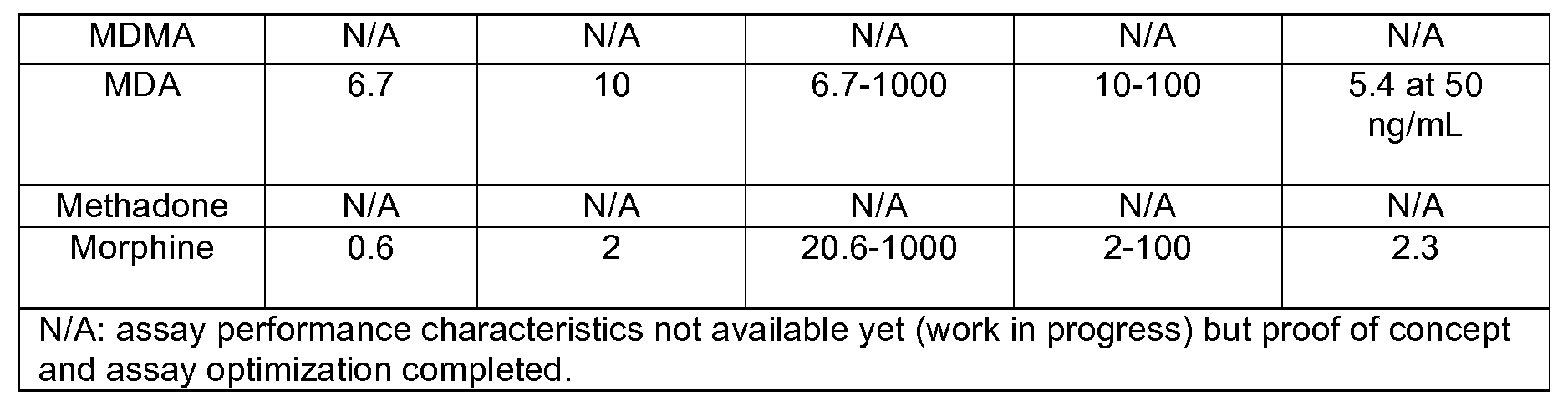
- FIG. 2 provides a typical dose response for the drug methamphetamine, but the other dose response curves are omitted for brevity. Instead, the complete assay performance characteristics of the 5 drug tests developed are summarized in Table 2.
- the LOQ i.e. the lower drug concentration on the linear portion of the dose curve, was about 1 decade of concentration higher than the LOD and one decade of concentration lower than the upper limit of the assay, suggesting that the most accurate measurements of the test should be expected at 10-1,000, 1-100, 1-100 and 1-10,000 ng/niL of cocaine, diazepam, amphetamine and methamphetamine concentrations, respectively.
- drugs could be detected in a simple yes/no assay at levels ranging from at least four orders of magnitude to as high as five orders.
- Intra-assay precision within the context of the parameters of the LOC system was derived from the degree of agreement of the signals obtained from at least 4 redundant beads within the array, 3.2-4.6 %CV for the drugs tests for THC, cocaine/BZE, diazepam, amphetamine and methamphetamine. Inter-assay precision was evaluated using drug standards tested in successive assay runs. This effort revealed inter-assay precision to vary between 4.4-8.0 % for the drug based LOC tests for the mid- to high-range drug standard concentrations and 7-15% for the low end of drug standard concentrations.
- the portable BNC tests exhibit exceptional assay performance characteristics with limits of detection (LODs) comparable to the laboratory-confined reference method of LC-MS/MS and vastly superior (significantly lower) than their ICS counterparts. Similar to LC-MS/MS and unlike ICS tests, which are qualitative (Yes/No) type of tests, BNC-based drug tests are fully quantitative. Furthermore, unlike LC-MS/MS which requires tedious sample processing and is limited to testing for one drug at a time, BNC tests are amenable to the point of need, require no extensive sample processing and offer the capacity to multiplex, or test more than one anaiyte (dmg) concurrently, using microliters of sample.
- LODs limits of detection
- THC-Alexa Fluor® 488 was mixed with the sample and the mixture was delivered over a period of 7.5 minutes to the array of beads in the LOC flow cell. Strong and specific binding of the tracer to the anti-THC sensitized beads was quickly demonstrated (not shown). Most importantly, the signal was now more uniform around each bead as well as more consistent with that achieved with other reverse competitive immunoassay format.
- IFB IFB was an improvement, it was limited to detecting only high concentrations of the drug.
- concentration that resulted in competition also exhibited a distortion of the bead integrity when bound to the bead sensors (not shown).
- the inability to detect lower concentrations of THC was attributed to a sub-optimal tracer, the generation of which is limited to the fact that BSA-THC conjugate (and not THC alone) was used to create the Alexa Fluor® 488-coupled reagent. Noted here, there is currently no known method to couple the Alexa Fluor® 488 directly to the drug, and the BSA or some linker is necessary.
- THC represents a challenging target for assay development.
- THC has been in our experience the most difficult to optimize.
- the above described "detective" work has resulted in the important observation that THC, when bound at significantly high concentrations on the beads, significantly affects the structural integrity and functional capacity of agarose beads to serve as a sensor for the same drug.
- THC-BSA should be loaded at significantly lower levels, as least when coupled to agarose beads.
- Further optimization steps include exploring functionality of bead sensors loaded with even lower THC-BSA (i.e. 0.01 and O.OOlmg/mL) for IF A, as well as identifying optimal tracer dilution (1 : 100, 1 :500, 1 : 1000) for the IFA using bead sensors as identified from previous step. It is expected that the LOD can be further improved beyond the 10 ng/ml LOD already demonstrated, to ⁇ 1 ng/ml.
- a field environment is likely to be quite different from a lab environment with trained technicians, complex machinery, and optimal working conditions. As one example, in the field, it is likely that the beads may be opened to air, and not used for some time. Therefore, methods of stabilizing the 500 ⁇ beads against drying out were undertaken and glycerol was tested as a preservative or anti-drying agent.
- PBS served as an effective method to maintain the moisture around the beads, while likewise maintaining the structural capacity of the beads.
- the next objective was to determine its effects on their functional capacity as immune sensors on the BNC platform.
- agarose beads coupled to BSA-THC conjugate were distributed in 5 eppendorf tubes and suspended in 1%, 2%, 10% and 30%> glycerol in PBS. Beads were then manually loaded onto the microchip, which was left uncovered and exposed to ambient air, overnight at RT. During that same time period, a fresh bead control (i.e. beads in PBS) was stored at 4°C, until the following day, upon which time beads from this aliquot were also manually loaded on the microchip to complete the array.
- a fresh bead control i.e. beads in PBS
- the lab-based assay must be further optimized for field use, especially as regards sample collection and test procedures. To that end we are testing a variety of field formats, including use of commercially available swab tests for sample collection. These commercially available cheek swab kits have tested as better and more reproducable than just collecting expectorant. Thus, we anticipate that standard swabs will be packaged with the kit, and including a vial of PBS or other suitable sample extraction buffer for collecting the sample off the swab and solubilizing it.
- the tracer can be applied in various ways. It can added by the user to the sample buffer or be kept in dried form in a separate cap to be added to the sample buffer once the swab is washed in the buffer. Alternatively, it can be contained in a reagent blister or in a dried reagent pad in the fluidic pathway. It is expected that this third alternative will be the most user friendly, however, all methods will be tested. Of course, the stability of dried reagent will have to be assessed, but dried antibodies are already extensively used in home testing kits and are known to be reliable and long lasting.
- Two capped vials is one option— one vial holding the sterile swab, and the other containing wash buffer.
- the swab is pulled from the first vial, swished in the second buffer-containing vial and the resulting sample is then applied to the cartridge.
- the swab end can also comprise an inexpensive plastic dropper (like an eye dropper), as is already known in the art, thus facilitating transfer from the buffer vial to the cartridge.
- FIG. 3A-D shows this embodiment comprising a top 1 in FIG. 3A and vial 21 in 3B, shown in a storage position in 3C and in wash position in 3D.
- the top is a combination of eye dropper, cap and swab, preferably made of lightweight flexible plastic, wherein the bulb portion and cap ridges have more flexibility (higher durometer or thinner) than the remaining portions.
- the vial has a sheet of flexible foil or plastic separating wash buffer in the lower wash portion of the vial from the upper storage portion of the vial.
- the sizes shown in FIG. 3 are approximate only, and the exact proportions will have to be calibrated to allow washing of the swab without overflowing or contaminating the buffer.
- top 1 comprises a bulb 11, and cap 12, having optional annular flexible ridges or protrusions 13 for sealing the cap inside the vial 21.
- Bulb 11 is fluidly connected via hollow stem 14 to open point 16 and has an absorbent or bristled swab 15 on the outer surface of stem 14.
- Vial 21 consists of a container 22 having a foil or plastic separator 24 to separate buffer 25 from the upper portion of the container 22.
- Optional ledge 23 can be included to prevent the top 1 from being pushed too far into the vial 1 , but this may not be needed, since the length of top 1 can effectively control this as well.
- Ledge 23 can be annular, that is circumnavigate the container, or can just be two or more smaller steps roughly equi-spaced, as desired.
- container have a flat bottom (not shown) to allow it to stand on its own during assemble and use.
- the cap 12 seals the container via flexible ridges 13, keeping swab 15 sterile and above separator 24 in the upper storage portion of the vial.
- the top is removed, and the swab used to collect saliva samples from a suspect.
- the top 1 is then returned to the vial 21 in 3D, pushing point 16 past the separator 24, thus rupturing it, and allowing the user to wash the swab 15 in buffer 25.
- the bulb 11 is squeezed and released, thus drawing buffer 25 up into the bulb 11 via open point 16 and hollow stem 14. This wash and sample buffer can then be applied to the cartridge.
- the device can be recapped and saved, or thrown away, according to standard protocols.
- the BNC cartridge itself will also be contained inside a sterile, airtight wrapper, probably foil (not shown). It is opened when needed, sample applied either via the cartridge or via a fluidly connected port on the reader, dropped into a portable detector, and the test run.
- FIG. 4 shows an exemplary cartridge, which is disposable and fits into a standard reader.
- 101 is the sample entry port, which is fluidly connected via micro fluidics 111 to the bead support chip chamber 117.
- a small array of bead sensors fits inside this chamber, which has a transparent lid 118.
- Pinch valve 102 functions to allow controlled delivery of micro fluidic elements.
- Buffer entry ports 103 are fluidly connected 112 to micro fluidic channel 111.
- One, two or more blister packs 104 can contain liquid reagents, such as wash buffers. Alternatively, the device could be connected directly to an external fluid source via buffer entry ports 103, but the blister packs are preferred as being more self-contained and providing a smaller footprint.
- the blisters are accessed via pressure actuation, a function provided by the analyzer/reader and embedded software, and thus are preferably foil blisters.
- Bubble trap 105 allows for pressure relief, otherwise the fluid would not flow in the tiny channels.
- waste chambers 110 can be closed under negative pressure and thus pull fluid in their direction when a valve is opened.
- Reagent port 106 can contain an absorbent pad 120 (see perspective inset 4B) having dried reagents (labeled antibody-tracer) thereon.
- reagent port 6 can consist of an access hatch or affixed cover 121 and recess 122, into which reagent pad 120 can be placed.
- reagent port 106 could be another blister pack or again just an inlet allowing connection to external fluids.
- Waste reservoir 107 and waste reservoir external vent 108 are also fluidly connected via micro fluidic channel 113 to assay chamber 117 having a transparent access hatch or affixed cover 118 allowing visual access to the bead array, but keeping the beads airtight.
- Optional port to waste chamber 110 is also shown, although the chamber can be made sufficiently large to hold all waste and this port omitted.
- disposable plastic chip containing the microfluidics is made by injection molding and/or etching of parts and adhering layers together.
- Access hatch 121 shown open
- access hatch 118 shown closed
- recess 117 allow the insertion of the bead array 109 and reagent pad 120, and can then be closed and tightly sealed, e.g., with heat or adhesive.
- Blisters are added via adhesive strip.
- Preferred materials for constructing the cartridge are plastics of durometer 34-40 Shore D for the substrate and microfluidics, such as polymers and copolymers of styrene, acrylic, carbonate, butadiene, propylene, vinyl, acrylonitrile, and foil for the blisters.
- a detector will be designed and manufactured specifically for this assay, as this will allow simplification of the device and its software, and minimization of the footprint. Ideally, the device will be reduced to a hand held size, and thus be easy for police officers to use in road-side testing environments.
- the analyzer (aka reader or detector) serves as a universal interface, providing the user with access to a fully embedded software, and components needed to run the assay, read the results, and convert the data to a user friendly output.
- the analyzer is composed of i) lab card loading deck, ii) optics, iii) charged coupled device (CCD camera) or other light measuring means, iv) software, vi) mechanical actuators for movement of microfluidics (e.g., needle for piercing blister packs and means for moving/actuating same), vii) pump, and viii) data output (e.g., paper and printer), and/or USB port and/or display means and ix) data input means.
- microfluidics e.g., needle for piercing blister packs and means for moving/actuating same
- pump e.g., pump
- data output e.g., paper and printer
- the officer inputs the suspects name and any other pertinent information, collects and applies the sample, presses a start button, and the device runs the tests and outputs the answers.
Landscapes
- Health & Medical Sciences (AREA)
- Life Sciences & Earth Sciences (AREA)
- Engineering & Computer Science (AREA)
- Molecular Biology (AREA)
- Biomedical Technology (AREA)
- Hematology (AREA)
- General Health & Medical Sciences (AREA)
- Chemical & Material Sciences (AREA)
- Surgery (AREA)
- Pathology (AREA)
- Animal Behavior & Ethology (AREA)
- Public Health (AREA)
- Veterinary Medicine (AREA)
- Heart & Thoracic Surgery (AREA)
- Medical Informatics (AREA)
- Urology & Nephrology (AREA)
- Immunology (AREA)
- Analytical Chemistry (AREA)
- Physics & Mathematics (AREA)
- Microbiology (AREA)
- General Physics & Mathematics (AREA)
- Biochemistry (AREA)
- Medicinal Chemistry (AREA)
- Food Science & Technology (AREA)
- Cell Biology (AREA)
- Bioinformatics & Cheminformatics (AREA)
- Pharmacology & Pharmacy (AREA)
- Biotechnology (AREA)
- Nuclear Medicine, Radiotherapy & Molecular Imaging (AREA)
- Pulmonology (AREA)
- Chemical Kinetics & Catalysis (AREA)
- Dispersion Chemistry (AREA)
- Clinical Laboratory Science (AREA)
- Emergency Medicine (AREA)
- Biophysics (AREA)
- Pain & Pain Management (AREA)
- Epidemiology (AREA)
- Vascular Medicine (AREA)
- Anesthesiology (AREA)
- Investigating Or Analysing Biological Materials (AREA)
Abstract
Description


Claims
Priority Applications (3)
| Application Number | Priority Date | Filing Date | Title |
|---|---|---|---|
| US14/114,925 US9709580B2 (en) | 2011-05-12 | 2012-03-21 | Bio-nano-chips for on-site drug screening |
| SG2013083381A SG194892A1 (en) | 2011-05-12 | 2012-03-21 | Bio-nano-chips for on-site drug screening |
| CA2836061A CA2836061A1 (en) | 2011-05-12 | 2012-03-21 | Bio-nano-chips for on-site drug screening |
Applications Claiming Priority (2)
| Application Number | Priority Date | Filing Date | Title |
|---|---|---|---|
| US201161485189P | 2011-05-12 | 2011-05-12 | |
| US61/485,189 | 2011-05-12 |
Publications (1)
| Publication Number | Publication Date |
|---|---|
| WO2012154306A1 true WO2012154306A1 (en) | 2012-11-15 |
Family
ID=47139496
Family Applications (1)
| Application Number | Title | Priority Date | Filing Date |
|---|---|---|---|
| PCT/US2012/030012 Ceased WO2012154306A1 (en) | 2011-05-12 | 2012-03-21 | Bio-nano-chips for on-site drug screening |
Country Status (4)
| Country | Link |
|---|---|
| US (1) | US9709580B2 (en) |
| CA (1) | CA2836061A1 (en) |
| SG (1) | SG194892A1 (en) |
| WO (1) | WO2012154306A1 (en) |
Cited By (11)
| Publication number | Priority date | Publication date | Assignee | Title |
|---|---|---|---|---|
| EP2821138A1 (en) * | 2013-07-05 | 2015-01-07 | Thinxxs Microtechnology Ag | Flow cell with integrated dry substance |
| US20170333894A1 (en) * | 2016-05-19 | 2017-11-23 | Plasmotica, LLC | Stand alone microfluidic analytical chip device |
| US10060937B2 (en) | 2013-06-28 | 2018-08-28 | William Marsh Rice University | Integrated instrumentation for the analysis of biofluids at the point-of-care |
| US10458963B2 (en) | 2016-06-08 | 2019-10-29 | Kathleen Stitzlein | Quantitative HPTLC cannabinoid field testing device and method |
| US10466159B2 (en) | 2014-11-28 | 2019-11-05 | Chipcare Corporation | Multiplex bead array assay |
| WO2020143862A1 (en) * | 2019-01-07 | 2020-07-16 | Nuuvera Deutschland GmbH | Method for qualitatively and/or quantitatively detecting substances contained in a hemp plant and kit for use therein |
| US10724069B2 (en) | 2014-09-29 | 2020-07-28 | Chipcare Corporation | Methods and devices for cell detection |
| US10946376B2 (en) | 2013-07-05 | 2021-03-16 | Thinxxs Microtechnology Ag | Carrier element for introducing a dry substance into a flow cell |
| US11717820B2 (en) | 2020-03-17 | 2023-08-08 | Nordetect Aps | Microfluidic device, production of a microfluidic device and method and system for performing inorganic determinations |
| US12235272B2 (en) | 2020-01-13 | 2025-02-25 | New York University | Screening and assessment of potentially malignant oral lesions |
| US12493038B2 (en) | 2018-06-05 | 2025-12-09 | New York University | Systems and methods of oral cancer assessment using cellular phenotype data |
Families Citing this family (21)
| Publication number | Priority date | Publication date | Assignee | Title |
|---|---|---|---|---|
| CA2850998A1 (en) | 2014-04-18 | 2015-10-18 | Thc Breathalyzer, Inc. | Cannabis drug detection device |
| EP3705881A1 (en) * | 2014-10-24 | 2020-09-09 | Abbott Laboratories | Paper substrate diagnostic apparatus and related methods and systems |
| US20160154015A1 (en) * | 2014-12-02 | 2016-06-02 | Kathleen Stitzlein | Microfluidic device to detect cannabis in body fluids |
| EP3331445B1 (en) * | 2015-08-05 | 2019-10-23 | ART Healthcare Ltd. | Point of care urine analyzer |
| GB201515964D0 (en) * | 2015-09-09 | 2015-10-21 | Global Icle Test Ltd | Disposable urine test device |
| US9933445B1 (en) | 2016-05-16 | 2018-04-03 | Hound Labs, Inc. | System and method for target substance identification |
| USD812242S1 (en) * | 2016-07-13 | 2018-03-06 | Precision Nanosystems Inc | Microfluidic cartridge |
| USD800336S1 (en) * | 2016-07-13 | 2017-10-17 | Precision Nanosystems Inc | Microfluidic cartridge |
| US11026596B1 (en) | 2017-05-19 | 2021-06-08 | Hound Labs, Inc. | Detection and measurement of target substance in exhaled breath |
| US10660619B2 (en) | 2017-07-19 | 2020-05-26 | Evanostics Llc | Cartridges for oral fluid analysis and methods of use |
| US11187711B1 (en) * | 2017-09-11 | 2021-11-30 | Hound Labs, Inc. | Analyte detection from breath samples |
| WO2019118989A1 (en) | 2017-12-15 | 2019-06-20 | Evanostics Llc | Optical reader for analyte testing |
| USD878622S1 (en) * | 2018-04-07 | 2020-03-17 | Precision Nanosystems Inc. | Microfluidic chip |
| US11287432B2 (en) * | 2018-06-05 | 2022-03-29 | New York University | System and method for detection of trauma |
| US11426097B1 (en) * | 2018-10-17 | 2022-08-30 | Hound Labs, Inc. | Rotary valve assemblies and methods of use for breath sample cartridge systems |
| US12066383B2 (en) | 2018-12-18 | 2024-08-20 | Aspida Dx Inc. | Optical analyte detection |
| EP3969143A4 (en) * | 2019-05-13 | 2023-09-06 | Buzzkill Labs, Inc. | TREATMENT CARTRIDGE FOR PORTABLE DRUG TESTING SYSTEM |
| KR102361251B1 (en) * | 2020-02-11 | 2022-02-11 | 한국전자기술연구원 | Multiple times available drug concentration measuring apparatus and method |
| US11933731B1 (en) | 2020-05-13 | 2024-03-19 | Hound Labs, Inc. | Systems and methods using Surface-Enhanced Raman Spectroscopy for detecting tetrahydrocannabinol |
| US12392769B1 (en) | 2021-01-12 | 2025-08-19 | Hound Labs, Inc. | Ambient contamination in breath analyte detection and measurement |
| US11992318B2 (en) | 2022-07-13 | 2024-05-28 | Celestin B. Bitjonck | Diagnostic lab-on-a-chip device |
Citations (6)
| Publication number | Priority date | Publication date | Assignee | Title |
|---|---|---|---|---|
| US5879635A (en) * | 1997-03-31 | 1999-03-09 | Nason; Frederic L. | Reagent dispenser and related test kit for biological specimens |
| WO2000004381A1 (en) * | 1998-07-14 | 2000-01-27 | Cozart Bioscience Limited | Screening device and method of screening an immunoassay test |
| US20060046310A1 (en) * | 2004-08-25 | 2006-03-02 | Zong-Li Xia | Amplification method for solid phase immunoassays |
| US20080286816A1 (en) * | 2004-06-14 | 2008-11-20 | Cozart Bioscience Limited | Detection of Methamphetamine Group Drugs |
| US20090017555A1 (en) * | 2004-07-02 | 2009-01-15 | Cozart Bioscience Limited | Delta-9-tetrahydrocannabinol detection method |
| US20100285490A1 (en) * | 2006-12-29 | 2010-11-11 | Invitrogen Corporation | Detection apparatus |
Family Cites Families (26)
| Publication number | Priority date | Publication date | Assignee | Title |
|---|---|---|---|---|
| US6908770B1 (en) | 1998-07-16 | 2005-06-21 | Board Of Regents, The University Of Texas System | Fluid based analysis of multiple analytes by a sensor array |
| CA2379130A1 (en) | 1999-07-16 | 2001-01-25 | Board Of Regents, The University Of Texas System | Method and apparatus for the delivery of samples to a chemical sensor array |
| US7022517B1 (en) | 1999-07-16 | 2006-04-04 | Board Of Regents, The University Of Texas System | Method and apparatus for the delivery of samples to a chemical sensor array |
| ATE403145T1 (en) | 2000-01-31 | 2008-08-15 | Univ Texas | PORTABLE DEVICE HAVING A SENSOR ARRAY ARRANGEMENT |
| WO2003090605A2 (en) | 2002-04-26 | 2003-11-06 | Board Of Regents, The University Of Texas System | Method and system for the detection of cardiac risk factors |
| AU2003256742A1 (en) | 2002-07-24 | 2004-02-09 | Board Of Regents, The University Of Texas System | Capture and detection of microbes by membrane methods |
| ITTO20020808A1 (en) | 2002-09-17 | 2004-03-18 | St Microelectronics Srl | INTEGRATED DNA ANALYSIS DEVICE. |
| EP1590659A4 (en) | 2003-02-07 | 2010-04-21 | Univ Texas | MULTICHIECH MICROSPHERES WITH INTEGRATED CHROMATOGRAPHIC AND DETECTION LAYERS USED IN MOSAIC SENSORS |
| EP1535665A1 (en) | 2003-11-28 | 2005-06-01 | STMicroelectronics S.r.l. | Integrated chemical microreactor with separated channels for confining liquids inside the channels and manufacturing process thereof |
| EP1695082A2 (en) | 2003-12-11 | 2006-08-30 | Board of Regents, The University of Texas System | Method and system for the analysis of saliva using a sensor array |
| US8101431B2 (en) | 2004-02-27 | 2012-01-24 | Board Of Regents, The University Of Texas System | Integration of fluids and reagents into self-contained cartridges containing sensor elements and reagent delivery systems |
| US20060257941A1 (en) | 2004-02-27 | 2006-11-16 | Mcdevitt John T | Integration of fluids and reagents into self-contained cartridges containing particle and membrane sensor elements |
| US8105849B2 (en) | 2004-02-27 | 2012-01-31 | Board Of Regents, The University Of Texas System | Integration of fluids and reagents into self-contained cartridges containing sensor elements |
| US20060257854A1 (en) | 2004-02-27 | 2006-11-16 | Mcdevitt John T | Membrane assay system including preloaded particles |
| US20060257991A1 (en) | 2004-02-27 | 2006-11-16 | Mcdevitt John T | Integration of fluids and reagents into self-contained cartridges containing particle-based sensor elements and membrane-based sensor elements |
| US7781226B2 (en) | 2004-02-27 | 2010-08-24 | The Board Of Regents Of The University Of Texas System | Particle on membrane assay system |
| CA2557549A1 (en) | 2004-02-27 | 2005-09-09 | Board Of Regents, The University Of Texas System | System and method for integrating fluids and reagents in self-contained cartridges containing particle and membrane sensor elements |
| US20060211059A1 (en) | 2005-03-18 | 2006-09-21 | Taneja Samir S | Methods of improving screening, diagnosis and staging of prostate cancer using serum testosterone |
| US8377398B2 (en) | 2005-05-31 | 2013-02-19 | The Board Of Regents Of The University Of Texas System | Methods and compositions related to determination and use of white blood cell counts |
| AU2006261953B2 (en) | 2005-06-24 | 2012-02-23 | Board Of Regents, The University Of Texas System | Systems and methods including self-contained cartridges with detection systems and fluid delivery systems |
| EP2021491A4 (en) | 2006-05-10 | 2010-02-24 | Univ Texas | DETECTION OF TUMOR BIOMARKERS IN MOUTH CANCER |
| US20080050830A1 (en) | 2006-05-10 | 2008-02-28 | The Board Of Regents Of The University Of Texas System | Detecting multiple types of leukocytes |
| CA2697357A1 (en) | 2007-04-16 | 2008-10-30 | John T. Mcdevitt | Cardibioindex/cardibioscore and utility of salivary proteome in cardiovascular diagnostics |
| US7947465B2 (en) * | 2007-08-20 | 2011-05-24 | Bio-Rad Laboratories, Inc. | Simultaneous assay for determining drugs |
| US20120208715A1 (en) | 2009-08-20 | 2012-08-16 | Mcdevitt John T | Methods and compositions for diagnosis of acute myocardial infarction (ami) |
| WO2012021714A2 (en) | 2010-08-12 | 2012-02-16 | William Marsh Rice University | Biomarker signatures for wellness testing |
-
2012
- 2012-03-21 SG SG2013083381A patent/SG194892A1/en unknown
- 2012-03-21 WO PCT/US2012/030012 patent/WO2012154306A1/en not_active Ceased
- 2012-03-21 US US14/114,925 patent/US9709580B2/en active Active
- 2012-03-21 CA CA2836061A patent/CA2836061A1/en not_active Abandoned
Patent Citations (6)
| Publication number | Priority date | Publication date | Assignee | Title |
|---|---|---|---|---|
| US5879635A (en) * | 1997-03-31 | 1999-03-09 | Nason; Frederic L. | Reagent dispenser and related test kit for biological specimens |
| WO2000004381A1 (en) * | 1998-07-14 | 2000-01-27 | Cozart Bioscience Limited | Screening device and method of screening an immunoassay test |
| US20080286816A1 (en) * | 2004-06-14 | 2008-11-20 | Cozart Bioscience Limited | Detection of Methamphetamine Group Drugs |
| US20090017555A1 (en) * | 2004-07-02 | 2009-01-15 | Cozart Bioscience Limited | Delta-9-tetrahydrocannabinol detection method |
| US20060046310A1 (en) * | 2004-08-25 | 2006-03-02 | Zong-Li Xia | Amplification method for solid phase immunoassays |
| US20100285490A1 (en) * | 2006-12-29 | 2010-11-11 | Invitrogen Corporation | Detection apparatus |
Non-Patent Citations (3)
| Title |
|---|
| CHRISTODOULIDES ET AL.: "Lab-on-a-Chip Methods for Point-of-Care Measurements of Salivary Biomarkers of Periodontitis.", ANNALS OF THE NEW YORK ACADEMY OF SCIENCES, vol. 1098, 2007, pages 411 - 428 * |
| JOKERST ET AL.: "Location of Biomarkers and Reagents within Agarose Beads of a Programmable Bio-nano-chip", SMALL, vol. 7, no. IS. 5, 7 March 2011 (2011-03-07), pages 613 - 624 * |
| JOKERST ET AL.: "Programmable Nano-Bio-Chip Sensors: Analytical Meets Clinical", ANAL. CHEM., vol. 82, 2010, pages 1571 - 1579 * |
Cited By (18)
| Publication number | Priority date | Publication date | Assignee | Title |
|---|---|---|---|---|
| US10060937B2 (en) | 2013-06-28 | 2018-08-28 | William Marsh Rice University | Integrated instrumentation for the analysis of biofluids at the point-of-care |
| WO2015001070A1 (en) * | 2013-07-05 | 2015-01-08 | Thinxxs Microtechnology Ag | Flow cell with an integrated dry substance |
| CN105517710A (en) * | 2013-07-05 | 2016-04-20 | 极小微技术股份公司 | Flow battery with integrated dry matter |
| CN105517710B (en) * | 2013-07-05 | 2017-04-05 | 极小微技术股份公司 | Flow battery with integrated dry matter |
| EP2821138A1 (en) * | 2013-07-05 | 2015-01-07 | Thinxxs Microtechnology Ag | Flow cell with integrated dry substance |
| US10232367B2 (en) | 2013-07-05 | 2019-03-19 | Thinxxs Microtechnology Ag | Flow cell with an integrated dry substance |
| US10946376B2 (en) | 2013-07-05 | 2021-03-16 | Thinxxs Microtechnology Ag | Carrier element for introducing a dry substance into a flow cell |
| US10724069B2 (en) | 2014-09-29 | 2020-07-28 | Chipcare Corporation | Methods and devices for cell detection |
| US10466159B2 (en) | 2014-11-28 | 2019-11-05 | Chipcare Corporation | Multiplex bead array assay |
| US20170333894A1 (en) * | 2016-05-19 | 2017-11-23 | Plasmotica, LLC | Stand alone microfluidic analytical chip device |
| US10497541B2 (en) | 2016-05-19 | 2019-12-03 | Nedal Saleh | Apparatus and method for programmable spatially selective nanoscale surface functionalization |
| US10832895B2 (en) | 2016-05-19 | 2020-11-10 | Plasmotica, LLC | Stand alone microfluidic analytical chip device |
| US10458963B2 (en) | 2016-06-08 | 2019-10-29 | Kathleen Stitzlein | Quantitative HPTLC cannabinoid field testing device and method |
| US12493038B2 (en) | 2018-06-05 | 2025-12-09 | New York University | Systems and methods of oral cancer assessment using cellular phenotype data |
| WO2020143862A1 (en) * | 2019-01-07 | 2020-07-16 | Nuuvera Deutschland GmbH | Method for qualitatively and/or quantitatively detecting substances contained in a hemp plant and kit for use therein |
| CN113873903A (en) * | 2019-01-07 | 2021-12-31 | 弗里茨·施密特 | Method for qualitatively and/or quantitatively detecting substance contained in cannabis plant and kit used therein |
| US12235272B2 (en) | 2020-01-13 | 2025-02-25 | New York University | Screening and assessment of potentially malignant oral lesions |
| US11717820B2 (en) | 2020-03-17 | 2023-08-08 | Nordetect Aps | Microfluidic device, production of a microfluidic device and method and system for performing inorganic determinations |
Also Published As
| Publication number | Publication date |
|---|---|
| US9709580B2 (en) | 2017-07-18 |
| CA2836061A1 (en) | 2012-11-15 |
| US20140094391A1 (en) | 2014-04-03 |
| SG194892A1 (en) | 2013-12-30 |
Similar Documents
| Publication | Publication Date | Title |
|---|---|---|
| US9709580B2 (en) | Bio-nano-chips for on-site drug screening | |
| US10660619B2 (en) | Cartridges for oral fluid analysis and methods of use | |
| TWI335430B (en) | Directed-flow assay device | |
| US11026611B2 (en) | Rotatable disk-shaped fluid sample collection device | |
| EP2874545B1 (en) | Disposable test device | |
| ES2949146T3 (en) | Lateral testing device | |
| JP4233686B2 (en) | Immunochromatography equipment housing | |
| US8470608B2 (en) | Combined visual/fluorescence analyte detection test | |
| US20120322682A1 (en) | Brain injury biomarker panel | |
| US20200345286A1 (en) | Cassette device for quick test of diagnosis, method for detecting a ligand in a biological sample and kit | |
| US20090087925A1 (en) | Devices and methods for analysis of samples with depletion of analyte content | |
| CN104042252A (en) | Rotatable fluid sample collection device | |
| AU2007205745A1 (en) | Immunoassay test device and method of use | |
| US20150111778A1 (en) | Bio-nano-chip for anticonvulsant drug salivary assay | |
| ES2857735T3 (en) | Rotating cartridge with multiple metering chambers | |
| WO2009152209A2 (en) | Combined visual/fluorescence analyte detection test | |
| US9535061B1 (en) | Multi-functional rapid diagnostic test device | |
| US20150017656A1 (en) | Rapid Lateral Flow Assay Method for Detecting Low Quantity Liquid or Dry Samples | |
| WO2020140185A1 (en) | Assay device | |
| US9784733B1 (en) | Rapid diagnostic test device by driven flow technology | |
| WO2008156491A2 (en) | Devices and methods for analysis of samples with depletion of analyte content | |
| CA2889492C (en) | Self-contained assay device | |
| US9360479B2 (en) | Rapid lateral flow assay method for low quantity liquid or dry samples | |
| RU2398235C2 (en) | Test-system with application of elements of non-enzymatic recognition of analysed substance | |
| McDevitt et al. | Bio-nano-chips for on-site drug screening |
Legal Events
| Date | Code | Title | Description |
|---|---|---|---|
| 121 | Ep: the epo has been informed by wipo that ep was designated in this application |
Ref document number: 12782849 Country of ref document: EP Kind code of ref document: A1 |
|
| ENP | Entry into the national phase |
Ref document number: 2014510311 Country of ref document: JP Kind code of ref document: A |
|
| ENP | Entry into the national phase |
Ref document number: 2836061 Country of ref document: CA |
|
| NENP | Non-entry into the national phase |
Ref country code: DE |
|
| WWE | Wipo information: entry into national phase |
Ref document number: 14114925 Country of ref document: US |
|
| NENP | Non-entry into the national phase |
Ref country code: JP |
|
| 122 | Ep: pct application non-entry in european phase |
Ref document number: 12782849 Country of ref document: EP Kind code of ref document: A1 |