WO2014036861A1 - 一种防雾内窥镜系统的装置和方法 - Google Patents
一种防雾内窥镜系统的装置和方法 Download PDFInfo
- Publication number
- WO2014036861A1 WO2014036861A1 PCT/CN2013/079898 CN2013079898W WO2014036861A1 WO 2014036861 A1 WO2014036861 A1 WO 2014036861A1 CN 2013079898 W CN2013079898 W CN 2013079898W WO 2014036861 A1 WO2014036861 A1 WO 2014036861A1
- Authority
- WO
- WIPO (PCT)
- Prior art keywords
- fog
- illumination source
- light
- endoscope
- infrared
- Prior art date
- Legal status (The legal status is an assumption and is not a legal conclusion. Google has not performed a legal analysis and makes no representation as to the accuracy of the status listed.)
- Ceased
Links
Images
Classifications
-
- A—HUMAN NECESSITIES
- A61—MEDICAL OR VETERINARY SCIENCE; HYGIENE
- A61B—DIAGNOSIS; SURGERY; IDENTIFICATION
- A61B1/00—Instruments for performing medical examinations of the interior of cavities or tubes of the body by visual or photographical inspection, e.g. endoscopes; Illuminating arrangements therefor
- A61B1/00064—Constructional details of the endoscope body
- A61B1/00071—Insertion part of the endoscope body
- A61B1/0008—Insertion part of the endoscope body characterised by distal tip features
- A61B1/00096—Optical elements
-
- A—HUMAN NECESSITIES
- A61—MEDICAL OR VETERINARY SCIENCE; HYGIENE
- A61B—DIAGNOSIS; SURGERY; IDENTIFICATION
- A61B1/00—Instruments for performing medical examinations of the interior of cavities or tubes of the body by visual or photographical inspection, e.g. endoscopes; Illuminating arrangements therefor
- A61B1/06—Instruments for performing medical examinations of the interior of cavities or tubes of the body by visual or photographical inspection, e.g. endoscopes; Illuminating arrangements therefor with illuminating arrangements
- A61B1/0638—Instruments for performing medical examinations of the interior of cavities or tubes of the body by visual or photographical inspection, e.g. endoscopes; Illuminating arrangements therefor with illuminating arrangements providing two or more wavelengths
-
- A—HUMAN NECESSITIES
- A61—MEDICAL OR VETERINARY SCIENCE; HYGIENE
- A61B—DIAGNOSIS; SURGERY; IDENTIFICATION
- A61B1/00—Instruments for performing medical examinations of the interior of cavities or tubes of the body by visual or photographical inspection, e.g. endoscopes; Illuminating arrangements therefor
- A61B1/06—Instruments for performing medical examinations of the interior of cavities or tubes of the body by visual or photographical inspection, e.g. endoscopes; Illuminating arrangements therefor with illuminating arrangements
- A61B1/0661—Endoscope light sources
- A61B1/0684—Endoscope light sources using light emitting diodes [LED]
-
- A—HUMAN NECESSITIES
- A61—MEDICAL OR VETERINARY SCIENCE; HYGIENE
- A61B—DIAGNOSIS; SURGERY; IDENTIFICATION
- A61B1/00—Instruments for performing medical examinations of the interior of cavities or tubes of the body by visual or photographical inspection, e.g. endoscopes; Illuminating arrangements therefor
- A61B1/06—Instruments for performing medical examinations of the interior of cavities or tubes of the body by visual or photographical inspection, e.g. endoscopes; Illuminating arrangements therefor with illuminating arrangements
- A61B1/07—Instruments for performing medical examinations of the interior of cavities or tubes of the body by visual or photographical inspection, e.g. endoscopes; Illuminating arrangements therefor with illuminating arrangements using light-conductive means, e.g. optical fibres
-
- A—HUMAN NECESSITIES
- A61—MEDICAL OR VETERINARY SCIENCE; HYGIENE
- A61B—DIAGNOSIS; SURGERY; IDENTIFICATION
- A61B1/00—Instruments for performing medical examinations of the interior of cavities or tubes of the body by visual or photographical inspection, e.g. endoscopes; Illuminating arrangements therefor
- A61B1/12—Instruments for performing medical examinations of the interior of cavities or tubes of the body by visual or photographical inspection, e.g. endoscopes; Illuminating arrangements therefor with cooling or rinsing arrangements
- A61B1/127—Instruments for performing medical examinations of the interior of cavities or tubes of the body by visual or photographical inspection, e.g. endoscopes; Illuminating arrangements therefor with cooling or rinsing arrangements with means for preventing fogging
-
- G—PHYSICS
- G02—OPTICS
- G02B—OPTICAL ELEMENTS, SYSTEMS OR APPARATUS
- G02B23/00—Telescopes, e.g. binoculars; Periscopes; Instruments for viewing the inside of hollow bodies; Viewfinders; Optical aiming or sighting devices
- G02B23/24—Instruments or systems for viewing the inside of hollow bodies, e.g. fibrescopes
- G02B23/2407—Optical details
- G02B23/2461—Illumination
- G02B23/2469—Illumination using optical fibres
-
- G—PHYSICS
- G02—OPTICS
- G02B—OPTICAL ELEMENTS, SYSTEMS OR APPARATUS
- G02B27/00—Optical systems or apparatus not provided for by any of the groups G02B1/00 - G02B26/00, G02B30/00
- G02B27/0006—Optical systems or apparatus not provided for by any of the groups G02B1/00 - G02B26/00, G02B30/00 with means to keep optical surfaces clean, e.g. by preventing or removing dirt, stains, contamination, condensation
Definitions
- the invention relates to an anti-fog endoscope system device and method, and belongs to the technical field of minimally invasive medical treatment.
- the doctor mainly uses the saline preheating endoscope and the anti-fog oil to relieve the fogging of the endoscope to a certain extent, but the disadvantage is that the surgical preparation process is complicated. There are hidden dangers of sterilization and will increase the time required for surgery, so it is the best way to provide an anti-fog or defogging solution directly from the endoscope system.
- the technical problem to be solved by the present invention is to provide an apparatus and method for an anti-fog endoscope system.
- a near-infrared anti-fog illumination source is added, and the color combination is coupled into the illumination transmission channel.
- changing the material properties of the front optical window It transmits visible light, ensures the absorption of near-infrared light energy while the white light is illuminated by the field, and raises the temperature of the front optical window to reduce the temperature difference between the optical window and the human body before the endoscope, thereby achieving the purpose of anti-fog. .
- An anti-fog endoscope system device comprising an illumination source 1 , a transmission fiber bundle 2 , an endoscope 3 , an illumination source 1 and a white light illumination source 11 , coupled optical lens 14 , white light illumination source 11 is a xenon lamp, white LED or red, green and blue three primary color mixed white LED, providing light output in the visible light band, using xenon lamp, power in In the range of 250W ⁇ 350W, white LED is used, the power is in the range of 50W ⁇ 100W, and the red, green and blue mixed color white LEDs are used. The power is 150W ⁇ 200W.
- the coupling optical lens 14 is an optical lens or lens group
- the transmission fiber bundle 2 is bound by a plurality of optical fibers having a diameter of several tens of micrometers, and the surface of the incident surface and the exit surface are polished, and the optical lens is coupled.
- the focal plane coincides with the incident surface of the transmission fiber bundle 2, and the endoscope 3 includes an endoscope optical lens system 31, a front optical window 32, a light guiding fiber bundle 33, an endoscope housing 34, and an optical fiber connection interface.
- endoscope optical lens system 31 consists of dozens of optical lenses, imaging the field
- the endoscope housing 34 is mainly made of stainless steel, polished surface, front optical window 32 It is a transparent optical material that transmits visible light and is placed in front of the endoscope optical lens system 31, soldered or glued to the endoscope housing 34, and guided to the optical fiber bundle 33
- the optical fibers are bound by a plurality of optical fibers having a diameter of several tens of micrometers, and the incident surface and the exit surface are polished.
- the incident surface of the light guiding fiber bundle 33 is placed in the optical connection interface 35, and the incident surface of the light guiding optical fiber bundle 33 is connected through the optical fiber.
- the illumination source 1 further includes a near-infrared anti-fog illumination source 12, a color-matching color filter 13 , and an image-collecting filter 13 is an optical plate to be coupled with the coupling optical lens 14 The angle of the angle is between the white light illumination source 11 and the coupling optical lens 14.
- the color filter 13 is coated with an optical film that transmits visible light to reflect near-infrared light, and the near-infrared anti-fog illumination source 12 It is a light source that emits only near-infrared light, such as a semiconductor laser source or a pump laser source.
- the wavelength of the emitted light ranges from 780 nm to 1100 nm, and the total power of the emitted light is between 5 watts and 10 watts.
- the near-infrared anti-fog illumination source 12 is placed perpendicular to the white illumination source 11 and at 45° to the color filter 13 , white illumination source 11 and near-infrared anti-fog illumination source 12
- the switch and brightness can be independently controlled.
- the front optical window 32 of the endoscope 3 is made of an optical material that improves the spectral characteristics of the material. It can transmit not only visible light but also near-infrared light, and the average visible light transmittance is greater than 80%, the average absorption rate of near-infrared light is greater than 80%.
- the anti-fog method of the anti-fog endoscope system includes the following steps:
- the near-infrared anti-fog illumination source 12 is turned on at full power, and the near-infrared light passes through the coupling optical lens in turn.
- the transmission fiber bundle 2 is transmitted, the light guiding fiber bundle 33 is irradiated onto the front optical window 32, and the front optical window 32 absorbs the near-infrared light energy, and the temperature starts to rise.
- the doctor inserts the endoscope into the human body, turns off the near-infrared anti-fog illumination light source 12, and turns on the white light illumination source. Then, the endoscope is inserted into the human body, and the temperature of the front optical window 32 is substantially the same as the temperature of the human body, and no fog is formed, and the doctor can perform normal observation and surgery to achieve the purpose of anti-fog.
- the positive progress of the invention is that the main endoscope system is not changed too much, only one near-infrared anti-fog illumination source is added, and the material of the front optical window is changed to realize the system-level anti-fog function.
- anti-fog such as pre-heating with saline and anti-fog oil, it has the advantages of more convenient and simpler operation for doctors, and there is no hidden danger of sterilization caused by physiological saline and anti-fog oil.
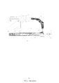
- FIG. 1 is a schematic view showing the structure of an apparatus of an anti-fog endoscope system of the present invention.
- White light illumination source 11 is a white LED with a total power of 80W.
- the near-infrared anti-fog illumination source 12 is a radiation wavelength of 808nm.
- the semiconductor laser has a full power output of 5 W and the front optical window 32 has an absorbance of 80% at 808 nm and an average visible light transmittance of 90%.
- Near-infrared anti-fog illumination source 12 When the power is turned on for 1 minute, the temperature of the front optical window 32 rises from about 25 °C to about 37 °C, the power is reduced to 30% of the full power, and the temperature is maintained at about 37 °C.
- the working principle is: 2 ⁇ 3 minutes before the operation, the 808nm semiconductor laser is turned on, and the 5W 808nm laser is output.
- the 808 nm laser passes through the color filter 13 and is reflected back into the coupling optical lens 14.
- the coupling optical lens 14 couples the 808 nm near-infrared light to the transmission fiber bundle 2 And exiting from the exit surface of the transmission fiber bundle 2, the outgoing 808 nm near-infrared light is coupled into the light guiding fiber bundle 33 through the fiber connection interface 35, and from the light guiding fiber bundle 33 The exit surface exits, and the 808 nm near-infrared light is emitted onto the front optical window 32.
- the front optical window 32 About 80% of the near-infrared light is absorbed by the front optical window, and the front optical window 32 The temperature starts to rise from room temperature. After 1 minute, the temperature of the front optical window rises to about 37 °C. At this time, the power of the near-infrared anti-fog illumination source 12 is reduced to 30% of the full power. The temperature of the front optical window will be maintained at about 37 °C.
- the endoscope 3 Before inserting the endoscope 3 into the human body, turn off the near-infrared anti-fog illumination light source 12, turn on the white light illumination source 11 and white light illumination source 11
- the emitted white light is incident on the color filter 13 and passes through the color filter 13 to the coupling optical lens 14, which couples the white light to the transmission fiber bundle 2 And then exiting from the exit face of the transport fiber bundle 2, the exiting white light is coupled through the fiber optic connection interface 35 into the light guide fiber bundle 33 and from the light guide fiber bundle 33
- the exit surface is emitted, and the emitted white light is irradiated onto the front optical window 32, and about 90% of the white light passes through the front optical window 32 to illuminate the field.
- the temperature is basically the same as the temperature in the human body, so the front optical window 32 does not fog, and the white light illuminates the field to ensure normal observation and operation of the field.
- White light illumination source 11 is a red, green and blue three-color mixed white LED with a total power of 170W and a near-infrared anti-fog illumination source. It is a semiconductor laser with a radiation wavelength of 940nm. The full-power output is 10W. The front optical window 32 has an absorption rate of 90% at 940nm and an average visible light transmittance of 85%. .
- Near-infrared anti-fog illumination source 12 Full power on for 30 seconds, the temperature of the front optical window 32 rises from around 25 °C to about 37 °C, the power is reduced to 10% of the full power, and the temperature is maintained at Around 37 °C.
- the white light illumination source 11 is a xenon lamp with a total power of 350 W
- the near-infrared anti-fog illumination source 12 is a radiation wavelength of 1064 nm.
- the Nd:YAG laser has a full power output of 7 W and the front optical window 32 has an absorbance of 95% at 1064 nm and an average visible light transmittance of 80%.
- Near-infrared anti-fog illumination source 12 When the power is turned on for 45 seconds, the temperature of the front optical window 32 rises from about 25 °C to about 37 °C, the power is reduced to 20% of the full power, and the temperature is maintained at about 37 °C.
- Embodiment 2 and Embodiment 3 are similar to those of Embodiment 1, and are not described here.
- the device and method of the anti-fog endoscope system of the present invention are suitably applied to various endoscopic operations, and are particularly suitable for use in endoscopic surgery in which an endoscope is repeatedly inserted and pulled out of a human body to provide an anti-fog effect.
Landscapes
- Health & Medical Sciences (AREA)
- Life Sciences & Earth Sciences (AREA)
- Physics & Mathematics (AREA)
- Surgery (AREA)
- Optics & Photonics (AREA)
- Engineering & Computer Science (AREA)
- Biomedical Technology (AREA)
- Molecular Biology (AREA)
- Pathology (AREA)
- Nuclear Medicine, Radiotherapy & Molecular Imaging (AREA)
- Biophysics (AREA)
- Heart & Thoracic Surgery (AREA)
- Medical Informatics (AREA)
- Radiology & Medical Imaging (AREA)
- Animal Behavior & Ethology (AREA)
- General Health & Medical Sciences (AREA)
- Public Health (AREA)
- Veterinary Medicine (AREA)
- General Physics & Mathematics (AREA)
- Microelectronics & Electronic Packaging (AREA)
- Astronomy & Astrophysics (AREA)
- Endoscopes (AREA)
Description
技术领域
本发明涉及一种防雾内窥镜系统的装置和方法,属于微创医疗的技术领域。
背景技术
医疗内窥镜已经广泛应用于微创手术中,当前采用内窥镜系统的微创手术一个操作上的缺陷是:当内窥镜进入人体操作时,由于人体内外的温度差异,部分水蒸汽遇到温度较低的内窥镜时,容易在内窥镜前光学窗口片上形成凝雾,影响术野的成像清晰度,使显示器上显示的术野图像变得模糊,影响医生的观察和操作。为了克服内窥镜起雾,医生主要采用生理盐水预热内窥镜,涂抹防雾油等方式,能在一定程度上缓解内窥镜起雾的问题,但缺点是复杂化了手术准备过程,
存在消毒不尽的隐患, 并会增加手术准备时间,因此,直接从内窥镜系统上提供防雾或除雾的解决方案是最佳的方式。
发明内容
本发明所要解决的技术问题是提供一种防雾内窥镜系统的装置和方法,其在传统内窥镜系统的基础上,增加一路近红外防雾照明光源,合色耦合进入照明传输通道,并改变前光学窗口片的材料特性,
使其透射可见光,保证术野白光照明的同时吸收近红外光能量,而使前光学窗口片的温度升高,减小内窥镜前光学窗口片与人体的温度差,从而达到防雾的目的。
现结合附图详细说明本发明的技术方案:
一种防雾内窥镜系统的装置,含照明光源 1 ,传输光纤束 2 ,内窥镜 3 ,照明光源 1 含白光照明光源
11 ,耦合光学透镜 14 ,白光照明光源 11 是氙灯,白光 LED 或红绿蓝三基色混合白光 LED ,提供可见光波段的光输出,采用氙灯,功率在
250W~350W 范围内,采用白光 LED ,功率在 50W~100W 范围内,采用红绿蓝三基色混合白光 LED ,功率在 150W~200W
范围内,耦合光学透镜 14 是光学透镜或透镜组,传输光纤束 2 由若干直径数十微米的光纤绑定在一起,入射面和出射面表面抛光,耦合光学透镜 14
的聚焦面与传输光纤束 2 的入射面重合,内窥镜 3 含内窥镜光学透镜系统 31 ,前光学窗口片 32 ,导光光纤束 33 ,内窥镜壳体 34 ,光纤连接接口
35 ,内窥镜光学透镜系统 31 由数十片光学透镜组成,对术野进行成像,内窥镜壳体 34 主要由不锈钢材料制成,表面抛光,前光学窗口片 32
是一种透明光学材料,可透射可见光,置于内窥镜光学透镜系统 31 前面,焊接或胶接在内窥镜壳体 34 上, 导光光纤束 33
由若干直径数十微米的光纤绑定在一起,入射面和出射面表面抛光,导光光纤束 33 的入射面置于光学连接接口 35 内,导光光纤束 33 的入射面通过光纤连接接口
35 与传输光纤束 2 的出射面耦合连接,导光光纤束 33 置于光学透镜系统 31 和内窥镜壳体 34 之间的环带内,出射面紧贴前光学窗口片 32 ,
其特征在于:所述的照明光源 1 还含近红外防雾照明光源 12 ,合色滤色片 13 ,合色滤色片 13 是一种光学平板,以与耦合光学透镜 14 成 45
°夹角的方式置于白光照明光源 11 和耦合光学透镜 14 之间,合色滤色片 13 上镀制透射可见光反射近红外光的光学薄膜,近红外防雾照明光源 12
是一种只发射近红外光的光源如半导体激光光源或泵浦激光器光源等,发射光的波长范围在 780nm~1100nm 之间,全功率发射光功率介于 5 瓦 ~10
瓦之间,近红外防雾照明光源 12 以与白光照明光源 11 垂直,与合色滤色片 13 呈 45 °角的方式放置,白光照明光源 11 和近红外防雾照明光源 12
可独立控制开关和亮度,内窥镜 3 的前光学窗口片 32 由一种改进了材料光谱特性的光学材料制成,它不仅能透射可见光同时能吸收近红外光,可见光平均透过率大于
80% ,近红外光平均吸收率大于 80% 。
根据上述防雾内窥镜系统的装置,所述的防雾内窥镜系统的防雾方法包括以下步骤:
( 1 )手术前 2~3 分钟,全功率开启近红外防雾照明光源 12 ,近红外光依次通过耦合光学透镜 14
,传输光纤束 2 ,导光光纤束 33 传输后,照射到前光学窗口片 32 上,前光学窗口片 32 吸收近红外光能量,温度开始上升。
( 2 ) 30 秒 ~1 分钟后,前光学窗口片温度从室温 25 ℃左右上升到 37 ℃
左右,此时降低近红外防雾照明光源 12 功率至全功率的 10%~30% ,以保持前光学窗口片温度维持在 37 ℃ 左右。
( 3 )医生将内窥镜插入人体前,关闭近红外防雾照明光源 12 ,开启白光照明光源 11
,然后将内窥镜插入人体,前光学窗口片 32 温度与人体温度基本一致,不会起雾,医生可以进行正常的观察和手术,达到防雾的目的。
本发明的积极进步效果在于:在典型内窥镜系统基础上不做太大的改变,只增加一路近红外防雾照明光源,同时改变前光学窗口片的材料就实现了系统级的防雾功能,与采用生理盐水预热和涂抹防雾油等防雾相比,具有医生手术操作更方便,更简单的优点,且不存在生理盐水,防雾油等存在的消毒不尽的隐患。
附图说明
图 1 为本发明的防雾内窥镜系统的装置结构示意图。
具体实施方式
现结合附图和实施例详细说明本发明的技术方案和工作原理。
所有实施例都具有与发明内容所述装置的结构完全相同的结构和方法所述的相同的方法。为避免重复,以下实施例仅罗列关键的技术数据。
实施例 1 :
白光照明光源 11 是白光 LED ,总功率 80W ,近红外防雾照明光源 12 是辐射波长为 808nm
的半导体激光器,全功率输出 5W ,前光学窗口片 32 在 808nm 的吸收率为 80% ,可见光平均透过率为 90% 。近红外防雾照明光源 12
全功率开启 1 分钟,前光学窗口片 32 的温度从室温 25 ℃左右上升到 37 ℃左右,功率降至全功率的 30% ,温度维持在 37 ℃左右。
工作原理为:手术前 2~3 分钟,开启 808nm 的半导体激光器,输出 5W 的 808nm 的激光,
808nm 的激光通过合色滤色片 13 后反射进入耦合光学透镜 14 ,耦合光学透镜 14 将 808nm 的近红外光聚焦耦合到传输光纤束 2
中,并从传输光纤束 2 的出射面出射,出射的 808nm 的近红外光通过光纤连接接口 35 耦合进入导光光纤束 33 ,并从导光光纤束 33
的出射面出射,出射的 808nm 的近红外光照射到前光学窗口片 32 上,约 80% 的近红外光被前光学窗口片吸收,前光学窗口片 32
的温度开始由室温开始上升, 1 分钟后,前光学窗口片温度上升到 37 ℃左右,这时,降低近红外防雾照明光源 12 的功率至全功率的 30%
,前光学窗口片温度将维持在 37 ℃左右,在将内窥镜 3 插入人体前,关闭近红外防雾照明光源 12 ,开启白光照明光源 11 ,白光照明光源 11
发出的白光入射到合色滤色片 13 上并透过合色滤色片 13 进入到耦合光学透镜 14 ,耦合光学透镜 14 将白光聚焦耦合到传输光纤束 2
中,然后从传输光纤束 2 的出射面出射,出射的白光通过光纤连接接口 35 耦合进入导光光纤束 33 并从导光光纤束 33
的出射面出射,出射的白光照射到前光学窗口片 32 上,约 90% 的白光透过前光学窗口片 32 照明术野。这样,由于前光学窗口片 32
的温度与人体内温度基本一致,因此前光学窗口片 32 上不会起雾,同时,白光照明术野,保证术野正常的观察和手术操作。
实施例 2 :
白光照明光源 11 是红绿蓝三基色混合白光 LED ,总功率 170W ,近红外防雾照明光源 12
是辐射波长微 940nm 的半导体激光器,全功率输出 10W ,前光学窗口片 32 在 940nm 吸收率为 90% ,可见光平均透过率为 85%
。近红外防雾照明光源 12 全功率开启 30 秒,前光学窗口片 32 的温度从室温 25 ℃左右上升到 37 ℃左右,功率降至全功率的 10% ,温度维持在
37 ℃左右。
实施例 3 :
白光照明光源 11 是氙灯,总功率 350W ,近红外防雾照明光源 12 是辐射波长为 1064nm 的
Nd:YAG 激光器,全功率输出 7W ,前光学窗口片 32 在 1064nm 吸收率为 95% ,可见光平均透过率为 80% 。近红外防雾照明光源 12
全功率开启 45 秒,前光学窗口片 32 的温度从室温 25 ℃左右上升到 37 ℃左右,功率降至全功率的 20% ,温度维持在 37 ℃左右。
实施例 2 和实施例 3 的工作原理与实施例 1 工作原理类似,这里不在赘述。
本发明的防雾内窥镜系统的装置和方法,适宜应用于各种内窥手术中,特别适宜应用于需将内窥镜反复插入和拔出人体的内窥手术中,起防雾作用。
Claims (1)
- 1 、一 种防雾内窥镜系统的装置,含照明光源( 1 ),传输光纤束( 2 ),内窥镜( 3 ),照明光源( 1 )含白光照明光源( 11 ),耦合光学透镜( 14 ),白光照明光源( 11 )是氙灯,白光 LED 或红绿蓝三基色混合白 光 LED ,提供可见光波段的光输出,采用氙灯,功率在 250W~350W 范围内,采用白光 LED ,功率在 50W~100W 范围内,采用红绿蓝三基色混合白光 LED ,功率在 150W~200W 范围内,耦合光学透镜( 14 )是光学透镜或透镜组,传输光纤束( 2 )由若干直径数十微米的光纤绑定在一起,入射面和出射面表面抛光,耦合光学透镜( 14 ) 的聚焦面与传输光纤束( 2 ) 的入射面重合,内窥镜( 3 ) 含内窥镜光学透镜系统( 31 ) ,前前光学窗口片( 32 ) ,导光光纤束( 33 ) ,内窥镜壳体( 34 ) ,光纤连接接口( 35 ) ,内窥镜光学透镜系统( 31 ) 由数十片光学透镜组成,对术野进行成像,内窥镜壳体( 34 ) 主要由不锈钢材料制成,表面抛光,前光学窗口片( 32 ) 是一种透明光学材料,可透过可见光,置于内窥镜光学透镜系统( 31 ) 前面,焊接或胶接在内窥镜壳体( 34 ) 上, 导光光纤束( 33 ) 由若干直径数十微米的光纤绑定在一起,入射面和出射面表面抛光,导光光纤束( 33 ) 的入射面置于光学连接接口( 35 ) 内,导光光纤束( 33 ) 的入射面通过光纤连接接口( 35 ) 与传输光纤束( 2 ) 的出射面耦合连接,导光光纤束( 33 ) 置于光学透镜系统( 31 ) 和内窥镜壳体( 34 ) 之间的环带内,出射面紧贴前光学窗口片( 32 ) , 其特征在于,所述的照明光源( 1 ) 还含近红外防雾照明光源( 12 ) ,合色滤色片( 13 ) ,合色滤色片( 13 ) 是一种光学平板,以与耦合光学透镜( 14 ) 成 45° 夹角的方式置于白光照明光源( 11 ) 和耦合光学透镜( 14 ) 之间,合色滤色片( 13 ) 上镀制透过可见光反射近红外光的光学薄膜,近红外防雾照明光源( 12 ) 是一种只发射近红外光的光源如半导体激光光源或泵浦激光器光源等,发射光的波长范围在 780nm~1100nm 之间, 全 功率发射光功率介于 5 瓦 ~10 瓦之间,近红外防雾照明光源( 12 ) 以与白光照明光源( 11 ) 垂直,与合色滤色片( 13 ) 呈 45° 角的方式放置,白光照明光源( 11 ) 和近红外防雾照明光源( 12 ) 可独立控制开关和亮度, 内窥镜( 3 )的前光学窗口片( 32 )由一种改进了材料光谱特性的光学材料制成,它不仅能透射可见光同时能吸收近红外光 , 可见光平均透过率大于 80% ,近红外光平均吸收率大于 80% 。2 、根据权利要求 1 所述的一种防雾内窥镜系统的装置,其特征在于,所述的防雾内窥镜系统的防雾方法包括以下步骤:( 1 )手术前 2~3 分钟,全功率开启近红外防雾照明光源( 12 ) ,近红外光依次通过耦合光学透镜( 14 ) ,传输光纤束( 2 ) ,导光光纤束( 33 )传输后, 照射到前光学窗口片( 32 ) 上,前光学窗口片( 32 ) 吸收近红外光能量,温度开始上升;( 2 ) 30 秒 ~1 分钟后,前光学窗口片温度从室温 25 ℃ 左右上升到 37 ℃ 左右,此时降低近红外防雾照明光源( 12 )功率至全功率的 10%~30% ,以保持前光学窗口片温度维持在 37 ℃ 左右;( 3 )医生将内窥镜插入人体前,关闭近红外防雾照明光源( 12 ) ,开启白光照明光源( 11 ) ,然后将内窥镜插入人体,前光学窗口片( 32 ) 温度与人体温度基本一致,不会起雾,医生进行正常的观察和手术,达到防雾的目的。3 、根据权利要求 1 和权利要求 2 所述防雾内窥镜系统的装置和方法,其特征在于,白光照明光源( 11 ) 是白光 LED ,功率 80W ,近红外防雾照明光源( 12 ) 是辐射波长为 808nm 的半导体激光器,全功率输出 5W ,前光学窗口片( 32 ) 在 808nm 的吸收率为 80% ,可见光平均透过率为 90% ,近红外防雾照明光源( 12 ) 全功率开启 1 分钟,前光学窗口片( 32 ) 的温度从室温 25 ℃ 左右上升到 37 ℃ 左右,功率降至全功率的 30% ,温度维持在 37 ℃ 左右。4 、根据权利要求 1 和权利要求 2 所述防雾内窥镜系统的装置和方法,其特征在于,白光照明光源( 11 ) 是红绿蓝三基色混合白光 LED ,总功率 170W ,近红外防雾照明光源( 12 ) 是辐射波长微 940nm 的半导体激光器,全功率输出 10W ,前光学窗口片( 32 ) 在 940nm 吸收率为 90% ,可见光平均透过率为 85% ,近红外防雾照明光源( 12 ) 全功率开启 30 秒,前光学窗口片( 32 ) 的温度从室温 25 ℃ 左右上升到 37 ℃ 左右,功率降至全功率的 10% ,温度维持在 37 ℃ 左右。5 、根据权利要求 1 和权利要求 2 所述防雾内窥镜系统的装置和方法,其特征在于,白光照明光源( 11 ) 是氙灯,总功率 350W ,近红外防雾照明光源( 12 ) 是辐射波长为 1064nm 的 Nd:YAG 激光器,全功率输出 7W ,前光学窗口片( 32 ) 在 1064nm 吸收率为 95% ,可见光平均透过率为 80% ,近红外防雾照明光源( 12 ) 全功率开启 45 秒,前光学窗口片( 32 ) 的温度从室温 25 ℃ 左右上升到 37 ℃ 左右,功率降至全功率的 20% ,温度维持在 37 ℃ 左右。
Priority Applications (2)
| Application Number | Priority Date | Filing Date | Title |
|---|---|---|---|
| EP13835074.9A EP2932887B1 (en) | 2012-09-05 | 2013-07-23 | Anti-fog endoscope system device and method |
| US14/379,269 US20150173591A1 (en) | 2012-09-05 | 2013-07-23 | Device of anti-fogging endoscope system and its method |
Applications Claiming Priority (2)
| Application Number | Priority Date | Filing Date | Title |
|---|---|---|---|
| CN201210324982.0A CN103654701B (zh) | 2012-09-05 | 2012-09-05 | 一种防雾内窥镜系统的装置和方法 |
| CN201210324982.0 | 2012-09-05 |
Publications (1)
| Publication Number | Publication Date |
|---|---|
| WO2014036861A1 true WO2014036861A1 (zh) | 2014-03-13 |
Family
ID=50236510
Family Applications (1)
| Application Number | Title | Priority Date | Filing Date |
|---|---|---|---|
| PCT/CN2013/079898 Ceased WO2014036861A1 (zh) | 2012-09-05 | 2013-07-23 | 一种防雾内窥镜系统的装置和方法 |
Country Status (4)
| Country | Link |
|---|---|
| US (1) | US20150173591A1 (zh) |
| EP (1) | EP2932887B1 (zh) |
| CN (1) | CN103654701B (zh) |
| WO (1) | WO2014036861A1 (zh) |
Families Citing this family (18)
| Publication number | Priority date | Publication date | Assignee | Title |
|---|---|---|---|---|
| US10537236B2 (en) | 2013-01-17 | 2020-01-21 | Stryker Corporation | Anti-fogging device for endoscope |
| US10582832B2 (en) | 2013-01-17 | 2020-03-10 | Stryker Corporation | System for altering functions of at least one surgical device dependent upon information saved in an endoscope related to the endoscope |
| DK3228254T3 (da) | 2014-02-21 | 2020-03-23 | 3Dintegrated Aps | Sæt omfattende et kirurgisk instrument |
| CN103948364B (zh) * | 2014-04-28 | 2016-06-15 | 中国科学院深圳先进技术研究院 | 内窥镜及检查方法 |
| US11020144B2 (en) | 2015-07-21 | 2021-06-01 | 3Dintegrated Aps | Minimally invasive surgery system |
| JP6776327B2 (ja) | 2015-07-21 | 2020-10-28 | スリーディインテグレイテッド アーペーエス3Dintegrated Aps | カニューレアセンブリキット、套管針アセンブリキット、スリーブアセンブリ、低侵襲性手術システム及び方法 |
| DK178899B1 (en) | 2015-10-09 | 2017-05-08 | 3Dintegrated Aps | A depiction system |
| KR101713684B1 (ko) * | 2016-09-21 | 2017-03-08 | 주식회사 에스카 | 시정상태의 변화에 강인한 복합 필터링 방식의 감시카메라 및 그것이 적용된 영상감시시스템 |
| CN107831557B (zh) * | 2017-11-07 | 2019-05-03 | 王一凡 | 一种防雾眼镜的制备方法 |
| CN110389438A (zh) * | 2018-04-23 | 2019-10-29 | 新视电科技有限公司 | 内视镜系统及其光源机 |
| US11510561B2 (en) * | 2018-08-21 | 2022-11-29 | Verily Life Sciences Llc | Endoscope defogging |
| US11457154B2 (en) * | 2019-06-20 | 2022-09-27 | Cilag Gmbh International | Speckle removal in a pulsed hyperspectral, fluorescence, and laser mapping imaging system |
| DE102019009010A1 (de) * | 2019-12-23 | 2021-06-24 | Karl Storz Se & Co. Kg | Endoskop, Trokarhülle, endoskopisches System und Antibeschlagverfahren für ein Endoskop |
| CN111053523B (zh) * | 2020-02-17 | 2022-04-22 | 青岛奥美克医疗科技有限公司 | 一种防雾内窥镜系统的装置 |
| WO2022077032A1 (en) * | 2020-10-09 | 2022-04-14 | Stryker Corporation | Systems and methods for mitigating fogging in endoscopic imaging |
| CN112617721B (zh) * | 2020-12-31 | 2022-12-30 | 青岛海泰新光科技股份有限公司 | 一种内窥镜、内窥镜冷光源的连接反馈装置 |
| CN115412718A (zh) * | 2022-08-17 | 2022-11-29 | 华伦医疗用品(深圳)有限公司 | 一种内窥镜摄像系统、图像处理方法及可读存储介质 |
| CN115657292B (zh) * | 2022-10-17 | 2024-10-22 | 青岛海信医疗设备股份有限公司 | 内窥镜光源系统和内窥镜 |
Citations (4)
| Publication number | Priority date | Publication date | Assignee | Title |
|---|---|---|---|---|
| US5605532A (en) * | 1995-10-20 | 1997-02-25 | Vista Medical Technologies, Inc. | Fog-free endoscope |
| US5647840A (en) * | 1994-09-14 | 1997-07-15 | Circon Corporation | Endoscope having a distally heated distal lens |
| JP2003334157A (ja) * | 2002-05-21 | 2003-11-25 | Pentax Corp | 硬性鏡の曇り止め装置 |
| CN102292014A (zh) * | 2009-07-23 | 2011-12-21 | 奥林巴斯医疗株式会社 | 内窥镜装置 |
Family Cites Families (7)
| Publication number | Priority date | Publication date | Assignee | Title |
|---|---|---|---|---|
| JPS5826641Y2 (ja) * | 1978-06-19 | 1983-06-09 | 株式会社町田製作所 | 内視鏡における観察窓の露結防止装置 |
| US5009655A (en) * | 1989-05-24 | 1991-04-23 | C. R. Bard, Inc. | Hot tip device with optical diagnostic capability |
| DE19743431B4 (de) * | 1997-10-01 | 2011-02-17 | Karl Storz Gmbh & Co. Kg | Endoskop mit Verbundfenster |
| US5951543A (en) * | 1997-06-30 | 1999-09-14 | Clinicon Corporation | Delivery system and method for surgical laser |
| US20040019636A1 (en) * | 2002-07-24 | 2004-01-29 | Sun Microsystems, Inc. | System and method for dynamically routing web procedure calls |
| US9386914B2 (en) * | 2007-04-04 | 2016-07-12 | Karl Storz Endovision, Inc. | Video endoscopic device with detachable control circuit |
| US8098375B2 (en) * | 2007-08-06 | 2012-01-17 | Lumencor, Inc. | Light emitting diode illumination system |
-
2012
- 2012-09-05 CN CN201210324982.0A patent/CN103654701B/zh active Active
-
2013
- 2013-07-23 EP EP13835074.9A patent/EP2932887B1/en active Active
- 2013-07-23 US US14/379,269 patent/US20150173591A1/en not_active Abandoned
- 2013-07-23 WO PCT/CN2013/079898 patent/WO2014036861A1/zh not_active Ceased
Patent Citations (4)
| Publication number | Priority date | Publication date | Assignee | Title |
|---|---|---|---|---|
| US5647840A (en) * | 1994-09-14 | 1997-07-15 | Circon Corporation | Endoscope having a distally heated distal lens |
| US5605532A (en) * | 1995-10-20 | 1997-02-25 | Vista Medical Technologies, Inc. | Fog-free endoscope |
| JP2003334157A (ja) * | 2002-05-21 | 2003-11-25 | Pentax Corp | 硬性鏡の曇り止め装置 |
| CN102292014A (zh) * | 2009-07-23 | 2011-12-21 | 奥林巴斯医疗株式会社 | 内窥镜装置 |
Also Published As
| Publication number | Publication date |
|---|---|
| EP2932887A4 (en) | 2016-09-21 |
| EP2932887B1 (en) | 2017-09-06 |
| US20150173591A1 (en) | 2015-06-25 |
| EP2932887A1 (en) | 2015-10-21 |
| CN103654701B (zh) | 2016-08-10 |
| CN103654701A (zh) | 2014-03-26 |
Similar Documents
| Publication | Publication Date | Title |
|---|---|---|
| EP2932887B1 (en) | Anti-fog endoscope system device and method | |
| CA2144209C (en) | Method and apparatus for an efficient photodynamic treatment | |
| RU2526423C2 (ru) | Офтальмологическая эндоиллюминация с использованием света, генерируемого волокном | |
| JP7850668B2 (ja) | 曇り止め内視鏡システム用装置 | |
| JP5945104B2 (ja) | 蛍光手術用実体顕微鏡 | |
| CN102553084B (zh) | 一种光治疗装置 | |
| KR100798486B1 (ko) | 형광 진단 및 광역동치료를 위한 광원장치 | |
| JP2017533074A (ja) | 眼の疾患を手当てするシステムおよび方法 | |
| CN106308727A (zh) | 具有杀菌功能的内窥镜及其控制方法 | |
| CA2595587A1 (en) | Optical therapy devices, systems, kits and methods for providing therapy to a body cavity | |
| WO2011007461A1 (ja) | 開口絞り | |
| CN105664372A (zh) | 黄疸检测蓝光治疗仪 | |
| EP3599983B1 (en) | Endoscopes | |
| CN103389267A (zh) | 一种激光物证勘查仪 | |
| CN206151443U (zh) | 一种实现窄带或宽带光谱复合照明的装置 | |
| CN222068341U (zh) | 一种人体体内手术辅助照明预警光纤 | |
| Epstein | Endoscopy: developments in optical instrumentation | |
| CN107875522B (zh) | 用于理疗系统的柔性导管、复合能量源及多功能理疗系统 | |
| CN207693552U (zh) | 一种内窥式荧光观察装置 | |
| JP2016021978A (ja) | Pdt用内視鏡システム | |
| CN116138717A (zh) | 出光角度可调的可视化光热治疗内窥镜装置 | |
| JP2002253600A (ja) | 医療用レーザ治療機器 | |
| CN208990076U (zh) | 一种光纤宽光谱治疗仪 | |
| US20250242168A1 (en) | Light Based Therapy Mouthpiece | |
| US20040254619A1 (en) | Apparatus and method for photothermal and photochemical medical treatments with incoherent light |
Legal Events
| Date | Code | Title | Description |
|---|---|---|---|
| 121 | Ep: the epo has been informed by wipo that ep was designated in this application |
Ref document number: 13835074 Country of ref document: EP Kind code of ref document: A1 |
|
| WWE | Wipo information: entry into national phase |
Ref document number: 14379269 Country of ref document: US |
|
| NENP | Non-entry into the national phase |
Ref country code: DE |
|
| REEP | Request for entry into the european phase |
Ref document number: 2013835074 Country of ref document: EP |
|
| WWE | Wipo information: entry into national phase |
Ref document number: 2013835074 Country of ref document: EP |