WO2014098211A1 - 組織再生促進剤 - Google Patents
組織再生促進剤 Download PDFInfo
- Publication number
- WO2014098211A1 WO2014098211A1 PCT/JP2013/084228 JP2013084228W WO2014098211A1 WO 2014098211 A1 WO2014098211 A1 WO 2014098211A1 JP 2013084228 W JP2013084228 W JP 2013084228W WO 2014098211 A1 WO2014098211 A1 WO 2014098211A1
- Authority
- WO
- WIPO (PCT)
- Prior art keywords
- mmp14
- cells
- cell
- tissue
- activated stellate
- Prior art date
- Legal status (The legal status is an assumption and is not a legal conclusion. Google has not performed a legal analysis and makes no representation as to the accuracy of the status listed.)
- Ceased
Links
Images













Classifications
-
- A—HUMAN NECESSITIES
- A61—MEDICAL OR VETERINARY SCIENCE; HYGIENE
- A61K—PREPARATIONS FOR MEDICAL, DENTAL OR TOILETRY PURPOSES
- A61K38/00—Medicinal preparations containing peptides
- A61K38/16—Peptides having more than 20 amino acids; Gastrins; Somatostatins; Melanotropins; Derivatives thereof
- A61K38/43—Enzymes; Proenzymes; Derivatives thereof
- A61K38/46—Hydrolases (3)
- A61K38/48—Hydrolases (3) acting on peptide bonds (3.4)
- A61K38/4886—Metalloendopeptidases (3.4.24), e.g. collagenase
-
- A—HUMAN NECESSITIES
- A61—MEDICAL OR VETERINARY SCIENCE; HYGIENE
- A61K—PREPARATIONS FOR MEDICAL, DENTAL OR TOILETRY PURPOSES
- A61K35/00—Medicinal preparations containing materials or reaction products thereof with undetermined constitution
- A61K35/12—Materials from mammals; Compositions comprising non-specified tissues or cells; Compositions comprising non-embryonic stem cells; Genetically modified cells
-
- A—HUMAN NECESSITIES
- A61—MEDICAL OR VETERINARY SCIENCE; HYGIENE
- A61K—PREPARATIONS FOR MEDICAL, DENTAL OR TOILETRY PURPOSES
- A61K35/00—Medicinal preparations containing materials or reaction products thereof with undetermined constitution
- A61K35/12—Materials from mammals; Compositions comprising non-specified tissues or cells; Compositions comprising non-embryonic stem cells; Genetically modified cells
- A61K35/37—Digestive system
- A61K35/407—Liver; Hepatocytes
-
- A—HUMAN NECESSITIES
- A61—MEDICAL OR VETERINARY SCIENCE; HYGIENE
- A61K—PREPARATIONS FOR MEDICAL, DENTAL OR TOILETRY PURPOSES
- A61K38/00—Medicinal preparations containing peptides
- A61K38/01—Hydrolysed proteins; Derivatives thereof
- A61K38/012—Hydrolysed proteins; Derivatives thereof from animals
- A61K38/014—Hydrolysed proteins; Derivatives thereof from animals from connective tissue peptides, e.g. gelatin, collagen
-
- A—HUMAN NECESSITIES
- A61—MEDICAL OR VETERINARY SCIENCE; HYGIENE
- A61K—PREPARATIONS FOR MEDICAL, DENTAL OR TOILETRY PURPOSES
- A61K38/00—Medicinal preparations containing peptides
- A61K38/16—Peptides having more than 20 amino acids; Gastrins; Somatostatins; Melanotropins; Derivatives thereof
- A61K38/17—Peptides having more than 20 amino acids; Gastrins; Somatostatins; Melanotropins; Derivatives thereof from animals; from humans
- A61K38/39—Connective tissue peptides, e.g. collagen, elastin, laminin, fibronectin, vitronectin, cold insoluble globulin [CIG]
-
- A—HUMAN NECESSITIES
- A61—MEDICAL OR VETERINARY SCIENCE; HYGIENE
- A61P—SPECIFIC THERAPEUTIC ACTIVITY OF CHEMICAL COMPOUNDS OR MEDICINAL PREPARATIONS
- A61P1/00—Drugs for disorders of the alimentary tract or the digestive system
- A61P1/16—Drugs for disorders of the alimentary tract or the digestive system for liver or gallbladder disorders, e.g. hepatoprotective agents, cholagogues, litholytics
-
- A—HUMAN NECESSITIES
- A61—MEDICAL OR VETERINARY SCIENCE; HYGIENE
- A61P—SPECIFIC THERAPEUTIC ACTIVITY OF CHEMICAL COMPOUNDS OR MEDICINAL PREPARATIONS
- A61P43/00—Drugs for specific purposes, not provided for in groups A61P1/00-A61P41/00
-
- C—CHEMISTRY; METALLURGY
- C12—BIOCHEMISTRY; BEER; SPIRITS; WINE; VINEGAR; MICROBIOLOGY; ENZYMOLOGY; MUTATION OR GENETIC ENGINEERING
- C12M—APPARATUS FOR ENZYMOLOGY OR MICROBIOLOGY; APPARATUS FOR CULTURING MICROORGANISMS FOR PRODUCING BIOMASS, FOR GROWING CELLS OR FOR OBTAINING FERMENTATION OR METABOLIC PRODUCTS, i.e. BIOREACTORS OR FERMENTERS
- C12M23/00—Constructional details, e.g. recesses, hinges
- C12M23/20—Material Coatings
-
- C—CHEMISTRY; METALLURGY
- C12—BIOCHEMISTRY; BEER; SPIRITS; WINE; VINEGAR; MICROBIOLOGY; ENZYMOLOGY; MUTATION OR GENETIC ENGINEERING
- C12M—APPARATUS FOR ENZYMOLOGY OR MICROBIOLOGY; APPARATUS FOR CULTURING MICROORGANISMS FOR PRODUCING BIOMASS, FOR GROWING CELLS OR FOR OBTAINING FERMENTATION OR METABOLIC PRODUCTS, i.e. BIOREACTORS OR FERMENTERS
- C12M25/00—Means for supporting, enclosing or fixing the microorganisms, e.g. immunocoatings
- C12M25/14—Scaffolds; Matrices
-
- C—CHEMISTRY; METALLURGY
- C12—BIOCHEMISTRY; BEER; SPIRITS; WINE; VINEGAR; MICROBIOLOGY; ENZYMOLOGY; MUTATION OR GENETIC ENGINEERING
- C12Y—ENZYMES
- C12Y304/00—Hydrolases acting on peptide bonds, i.e. peptidases (3.4)
- C12Y304/24—Metalloendopeptidases (3.4.24)
- C12Y304/2408—Membrane-type matrix metalloproteinase-1 (3.4.24.80)
Definitions
- the present invention comprises activated stellate cells, activated stellate cell degradation products, MMP14, collagen treated with MMP14, and a component selected from the group consisting of activated stellate secretions, promoting tissue regeneration, promoting cell differentiation and / or
- the present invention relates to a composition for promoting cell growth, a composition for inhibiting cell growth containing an MMP14 inhibitor, a cell culture substrate containing collagen treated with MMP14, and the like.
- tissue regeneration When a living tissue is damaged, it regenerates to make up for the damaged portion and tries to recover its function. For example, it is known that the liver regenerates in a short period of time even if more than half of it is excised, and almost restores its original size and function. Research on tissue regeneration has been conducted for a long time, mainly in the liver, and various findings have been reported, but the detailed mechanism of tissue regeneration is still unclear. Multiple types of cells are thought to be involved in complex tissue regeneration, but there is no definitive view even of which of these cells plays a major role .
- Non-Patent Document 1 discloses that in a mouse partial hepatectomy model, a specific phenotype of hepatic sinusoidal endothelial cell is an endothelial cell-specific transcription factor mediated by VEGFR2 (vascular endothelial growth factor-A receptor-2).
- VEGFR2 vascular endothelial growth factor-A receptor-2
- the present invention aims to provide a novel tissue regeneration promoting agent and tissue regeneration promoting method.
- a composition for promoting tissue regeneration comprising a component selected from the group consisting of activated stellate cells, activated stellate cell degradation products, MMP14, collagen treated with MMP14, and activated stellate cell secretions.
- composition according to (1) wherein the tissue regeneration occurs in a damaged tissue or a transplanted tissue.
- the composition according to (2) which is administered within 4 days from the day of receiving the disorder or transplantation.
- the composition according to (2) which is administered within one day from the day of receiving the disorder or transplantation.
- the composition according to any one of (2) to (4), wherein the disorder is selected from the group consisting of tissue destruction, inflammation, necrosis, fibrosis, surgical invasion, and organ failure.
- the composition according to (9), wherein the state in which tissue regeneration is suppressed is selected from the group consisting of inflammation, necrosis, fibrosis, organ failure, decreased platelet count, genetic abnormality, and decreased noradrenaline.
- a method for promoting tissue regeneration in a subject comprising the step of administering the composition according to any one of (1) to (10) to a subject in need thereof.
- stem cells comprising a component selected from the group consisting of activated stellate cells, activated stellate degradation products, MMP14, collagen treated with MMP14, and activated stellate secretions Composition.
- a tissue stem cell comprising a step of contacting a stem cell with a component selected from the group consisting of activated stellate cell, activated stellate cell degradation product, MMP14, collagen treated with MMP14, and activated stellate secretion To differentiate and / or proliferate.
- a composition for promoting cell proliferation comprising a component selected from the group consisting of activated stellate cells, activated stellate cell degradation products, MMP14, collagen treated with MMP14, and activated stellate secretions.
- Cell proliferation including the step of contacting cells with a component selected from the group consisting of activated stellate cells, activated stellate degradation products, MMP14, collagen treated with MMP14, and activated stellate secretions How to promote.
- a composition for suppressing cell proliferation comprising a substance that inhibits MMP14.
- the composition according to (16), wherein the cell on which the substance that inhibits MMP14 acts is CAF and / or tumor cell.
- a cell culture substrate containing collagen treated with MMP14 is CAF and / or tumor cell.
- tissue regeneration not only in-vivo tissue regeneration but also in-vitro tissue formation can be promoted, and thus a great contribution can be expected from biological and medical aspects. Furthermore, the promotion of tissue regeneration according to the present invention is particularly useful in situations where tissue regeneration is suppressed, for example, in situations involving diseases such as fibrosis.
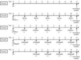
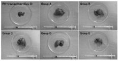
- FIG. 10 is a diagram showing an outline of treatment applied to each group in Example 3. It is the figure which showed the external appearance of the extract
- One aspect of the present invention is to promote tissue regeneration comprising a component selected from the group consisting of activated stellate cells, activated stellate cell degradation products, MMP14, collagen treated with MMP14, and activated stellate secretions. Of the composition.
- Activated stellate cells can be obtained by subculturing stellate cells isolated from living organisms. Astrocytes are known to exist in various tissues such as the liver, pancreas, kidney, intestinal tract, and lung (Zhao and Burt, J Mol Histol. 2007 Mar; 38 (1): 53-64). Either of these can be used. Isolation of stellate cells can be performed using any known method. A specific example of the method for isolating hepatic stellate cells is exemplified in Example 5 (1) described later. Activated stellate cells are characterized by the expression of ⁇ SMA, which can be selected as a marker.
- the activated stellate cell may be subjected to a treatment for increasing the expression of a protein selected from the group consisting of HGF, EGF, and MMP14.
- a treatment for increasing the expression of a protein selected from the group consisting of HGF, EGF, and MMP14 examples include, but are not limited to, introduction of a gene encoding the protein into activated stellate cells.
- Genes encoding HGF, EGF and MMP14 are known, and gene introduction methods are also well known in the art.
- the activated stellate cell degradation product can be obtained by decomposing activated stellate cells by various methods including physical and / or chemical methods.
- any known method for degrading cells such as osmotic shock method, freeze-thaw method, use of surfactant, enzymatic digestion method, ultrasonic treatment, French press, mortar grinding, homogenizer disruption, For example, crushing with glass beads can be used.
- the decomposition method does not denature the protein or has a light denaturation. By using this method, MMP14 expressed on the cell membrane can be obtained without impairing its function.
- the activated stellate cell degradation product contains a cell membrane component.
- MMP14 (also called MT1-MMP) is an MMP expressed on the cell membrane. MMP14 can act on collagen I. The present inventors have revealed that the action of MMP14 on collagen I is deeply involved in cell proliferation. MMP14 in the present invention includes not only those expressed on the cell membrane but also those released from the cell membrane. MMP14 may be naturally occurring or artificially produced. Thus, MMP14 includes recombinant MMP14. Free MMP14 is known (eg, Jo et al., Biochem J. 2000 Feb 1; 345 Pt 3: 511-9) and is commercially available (eg, R & D Systems, Cat No. 918-MP). -010, 918-MPN-010, etc.).
- the amino acid of MMP14 and the base sequence encoding it are known (for example, the base sequence of human MMP14 is registered as GenBank accession number NM_004995, and the amino acid sequence is registered as GenBank accession number NP_004986).
- MMP14 in the present invention includes functional mutants thereof.
- a functional variant of MMP14 is not limited, and includes, for example, (i) one or more, typically one or several mutations in the amino acid sequence of the protein.
- a variant having the same function as (ii) one or more nucleotide sequences of nucleic acids having the nucleotide sequence of the gene encoding the protein or the same polypeptide as the nucleic acid typically Is encoded by a nucleic acid having one or several mutations and has a function equivalent to that of the protein, (iii) a nucleic acid having a base sequence of a gene encoding the protein, and the same polypeptide as the nucleic acid To a complementary strand of a nucleic acid encoding a nucleic acid encoding (ii) or a variant thereof, or a fragment thereof under stringent conditions
- a variant having an amino acid sequence having a homology of 95% or more and having a function equivalent to that of the protein, (v) 60% or more with the base sequence of the gene encoding the protein preferably examples include mutants encoded by nucleic acids having homology of 70% or more, more preferably 80% or more, more preferably 90% or more, particularly preferably 95% or more, and having functions equivalent to the protein. .
- a person skilled in the art can convert the above functional mutant into any known method such as chemical synthesis, cleavage or insertion of a nucleic acid with a restriction enzyme, site-directed mutagenesis, radiation or ultraviolet irradiation based on the sequence information of MMP14. It can be suitably produced by such as. Whether or not a certain mutant has a function equivalent to that of MMP14 is determined by analyzing the mutant by any known method with respect to the known function of MMP14, for example, collagen resolution, etc. It can be evaluated by comparing with a negative control or MMP14 as a positive control.
- this mutant is a functional mutant of MMP14.
- the above function when the above function is superior to the negative control, for example, 10% or more, 25% or more, 50% or more, 75% or more, or even 100% or more, and / or If this function is 1/100 or more, 1/50 or more, 1/25 or more, 1/10 or more, 1/5 or more, or even 1/2 or more of CSABP, this mutant is a functional mutant of MMP14.
- stringent conditions is a well-known parameter in the art, and standard protocol collections such as Sambrook et al.,. Molecular Cloning: A Laboratory Manual, 3d ed., Cold Spring. Harbor Press (2001), Ausubel et al., Current Protocols in Molecular Biology, Greene Publishing Associates (1992), and the like.
- the stringent conditions in the present invention are, for example, 3.5 ⁇ SSC (0.15 M sodium chloride / 0.15 M sodium citrate, pH 7) at 65 ° C., Ficoll 0.02%, polyvinylpyrrolidone 0.02% Hybridization with a hybridization buffer consisting of 0.02% bovine serum albumin, 425 mM NaH2PO (pH 7), 0.05% SDS, and 2 mM EDTA. After hybridization, the membrane to which the DNA has been transferred is washed with 2 ⁇ SSC at room temperature and then with 0.1 to 0.5 ⁇ SSC / 0.1 ⁇ SDS at a temperature up to 68 ° C.
- stringent hybridization may be performed using a commercially available hybridization buffer such as ExpressHyb® Hybridization Solution (Clontech) under the hybridization and washing conditions described by the manufacturer.
- the MMP14 and / or functional variant thereof in the present invention includes not only the protein itself or a functional variant thereof, but also a nucleic acid encoding the protein or the functional variant thereof.
- the collagen treated with MMP14 can be obtained by treating collagen with MMP14.
- collagen for example, collagen I can be used.
- MMP14 used for the treatment may be the above-described MMP14 (including a functional mutant of MMP14), a cell expressing MMP14 itself, or a degradation product of the cell containing MMP14.
- the treatment time by the MMP 14 may be, for example, 1 to 120 hours, 3 to 60 hours, 6 to 48 hours, 12 to 36 hours, etc. at a temperature at which the MMP 14 can act.
- the collagen treated with MMP14 is preferably in a state in which the RGD sequence can interact with cells.
- Activated stellate secretion can be obtained from the culture supernatant of activated stellate cells.
- the culture supernatant may be used as it is or after being concentrated by dialysis or lyophilization. Since activated stellate secretions contain various proteins, it is preferable to handle them so that the proteins are not denatured.
- the tissue that promotes regeneration by the composition of the present invention is not particularly limited, and includes various tissues throughout the body.
- a tissue include, but are not limited to, a tissue in which stellate cells are present, a tissue in which fibrosis occurs, and a tissue in which stem cells are present.
- Specific examples include, but are not limited to, liver, pancreas, kidney, intestine, lung, spleen, heart, bone marrow, vocal cord, skin, peritoneum, eye, and vascular.
- the composition of the present invention is useful for tissue regeneration that occurs in damaged tissues or transplanted tissues.
- Damaged tissue includes tissue that has undergone tissue destruction, inflammation, necrosis, fibrosis, surgical invasion, organ failure, and the like.
- the administration time of the composition of the present invention is not particularly limited, but it is preferably administered within 4 days from the day of receiving the disorder or transplant, or within 1 day of receiving the disorder or transplant.
- Tissue regeneration by the composition of the present invention may involve stem cell differentiation and / or proliferation.
- tissue regeneration by the composition of the present invention may be accompanied by proliferation of tissue parenchymal cells.
- Stem cell differentiation can be evaluated, for example, by detecting a cell marker specific to the differentiated cell, detecting a function of the differentiated cell, or the like.
- Cell proliferation can be determined by various known methods such as counting the number of living cells over time, measuring the size, volume or weight of tissues, measuring the amount of DNA synthesis, WST-1 method, BrdU (bromodeoxyuridine) method. , 3 H thymidine incorporation method, and the like.
- composition of the present invention can be used for a subject having a state in which tissue regeneration is suppressed.
- the state in which tissue regeneration is suppressed includes, but is not limited to, for example, inflammation, necrosis, fibrosis, organ failure, decreased platelet count, genetic abnormality, decreased noradrenaline, and the like.
- the compounding amount of the active ingredient in the composition of the present invention may be an amount that promotes tissue regeneration when the composition is administered. In addition, an amount that does not cause adverse effects exceeding the benefits of administration is preferred. Such an amount is known or can be appropriately determined by an in vitro test using cultured cells, or a test in a model animal such as a mouse, rat, dog or pig. Such a test method is well known to those skilled in the art. Are known.
- the promotion of tissue regeneration can be evaluated by restoring the function, weight, size, etc. of the tissue by biochemical examination, X-ray, ultrasound, MRI, CT, endoscopic image diagnosis, or the like.
- the compounding amount of the active ingredient may vary depending on the dosage form of the composition.
- the amount of the active ingredient blended in one unit of the composition may be a plurality of the amount of the active ingredient necessary for one administration. it can. Those skilled in the art can appropriately adjust the blending amount.
- the present invention also promotes tissue regeneration, including blending a component selected from the group consisting of activated stellate cells, activated stellate cell degradation products, MMP14, collagen treated with MMP14, and activated stellate secretions
- the present invention relates to a method for producing a composition for use, the use of the component for producing a composition for promoting tissue regeneration, and the component used for promoting tissue regeneration.
- Each component can be blended according to any known technique.
- the present invention also relates to a method for promoting tissue regeneration in a subject, comprising the step of administering the above composition to a subject in need thereof.
- the subject in the method may have a disorder in the tissue or have undergone a tissue transplant. Disorders include, but are not limited to, for example, tissue destruction, inflammation, necrosis, fibrosis, surgical invasion, organ failure, and the like. Administration of the composition may be, for example, within 4 days from the date of receiving the disorder or transplant, or within one day of receiving the disorder or transplant.
- the present invention also provides for the differentiation and / or proliferation of stem cells comprising a component selected from the group consisting of activated stellate cells, activated stellate cell lysates, MMP14, collagen treated with MMP14 and activated stellate cell secretions.
- a composition for the preparation a method for producing a composition for stem cell differentiation and / or proliferation, comprising combining the components, and a composition for producing the composition for stem cell differentiation and / or proliferation.
- the component used for stem cell differentiation and / or proliferation, and a method for differentiating and / or proliferating the stem cell comprising the step of contacting the component with the stem cell.
- Stem cells are not particularly limited and include, for example, tissue stem cells (somatic stem cells, adult stem cells), embryonic stem cells, iPS cells, and the like. Stem cells may be totipotent, pluripotent, pluripotent or unipotent. Examples of tissue stem cells include, but are not limited to, neural stem cells, hematopoietic stem cells, mesenchymal stem cells, hepatic stem cells, pancreatic stem cells, skin stem cells, muscle stem cells, and reproductive stem cells. Stem cells may be autologous or may be from other individuals of the same species or from different species. Differentiated and / or expanded stem cells can be transplanted into a subject in need thereof.
- the above method may be performed in in vitro, in vivo or ex vivo.
- Each component can be blended according to any known technique.
- collagen particularly collagen I
- the present invention also includes a composition for promoting cell proliferation, comprising a component selected from the group consisting of activated stellate cells, activated stellate cell degradation products, MMP14, collagen treated with MMP14, and activated stellate secretions.
- a method for producing a composition for promoting cell growth comprising combining the components, use of the component for producing a composition for promoting cell growth, and use for promoting cell growth
- the present invention relates to a method for promoting cell proliferation, comprising the step of bringing the component into contact with the cell.
- the cell that promotes proliferation is not limited, and examples thereof include cells such as liver, pancreas, kidney, intestine, lung, spleen, heart, bone marrow, vocal cord, skin, peritoneum, eye, and blood vessel.
- the cells may be autologous, other individuals of the same species, or of different species. Differentiated and / or expanded cells can be transplanted into a subject in need thereof.
- the above method may be performed in in vitro, in vivo or ex vivo.
- Each component can be blended according to any known technique.
- collagen particularly collagen I
- the present invention also includes a composition for inhibiting cell proliferation, comprising a substance that inhibits MMP14, a method for producing a composition for inhibiting cell proliferation, comprising combining the substance,
- the present invention relates to the use of a composition for inhibiting cell proliferation, the substance used for inhibiting cell proliferation, and a method for inhibiting cell proliferation comprising a step of bringing the substance into contact with cells.
- the present invention also includes a composition for treating a cell proliferative disease comprising a substance that suppresses MMP14, a method for producing a composition for treating a cell proliferative disorder, comprising combining the substance, Use of the substance in the manufacture of a composition for treating a cell proliferative disorder, the substance used for the treatment of a cell proliferative disorder, and a therapeutically effective amount of the substance administered to a subject in need thereof And a method for treating a cell proliferative disorder.
- substances that inhibit MMP14 include, but are not limited to, drugs that inhibit production and / or activity of MMP14, drugs that promote degradation and / or inactivation of MMP14, and the like.
- examples of the drug that inhibits the production of MMP14 include, but are not limited to, RNAi molecules, ribozymes, antisense nucleic acids, DNA / RNA chimera polynucleotides for DNA encoding MMP14, and vectors that express these.
- MMP14 inhibition can be determined by the fact that the expression and activity of MMP14 are inhibited in the cells compared to the case where no MMP14 inhibitor was allowed to act.
- the expression of MMP14 is not limited to any known method, for example, immunoprecipitation using an anti-MMP14 antibody, EIA, ELISA, IRA, IRMA, Western blotting, immunohistochemistry, immunocytochemistry, Various hybridization methods, Northern blotting methods using nucleic acids that specifically hybridize to a nucleic acid that specifically encodes a nucleic acid encoding MMP14 or a unique fragment thereof or a transcription product (eg, mRNA) or a splicing product of the nucleic acid. , Southern blotting, various PCR methods and the like.
- an RNAi molecule refers to any molecule that causes RNA interference, including but not limited to siRNA (small interfering RNA), miRNA (micro RNA), shRNA (short hairpin RNA), ddRNA (DNA- directed RNA), piRNA (Piwi-interacting RNA), double-stranded RNA such as rasiRNA (repeat associated siRNA), and variants thereof.
- siRNA small interfering RNA
- miRNA miRNA
- micro RNA miRNA
- shRNA short hairpin RNA
- ddRNA DNA- directed RNA
- piRNA piRNA
- double-stranded RNA such as rasiRNA (repeat associated siRNA)
- Antisense nucleic acid as used herein includes RNA, DNA, PNA, or a composite thereof.
- a DNA / RNA chimeric polynucleotide is not limited, for example, a double-stranded polynucleotide comprising DNA and RNA that inhibits expression of a target gene described in JP-A-2003-219893. Including.
- Examples of the cells that suppress the growth include, but are not limited to, cells that are adversely affected by the growth, cells that are involved in the disease. Specific examples include tumor cells, cancer cells, activated stellate cells, and the like. A substance that suppresses MMP14 may be administered to these cells, or may be administered to MMP14-expressing cells that assist the growth of these cells, such as CAF (cancer-associated fibroblasts).
- Examples of cell proliferative diseases include, but are not limited to, benign or malignant tumors, hyperplasias, keloids, Cushing syndrome, primary aldosteronism, erythema, polycythemia vera, leukoplakia, hyperplastic scars, flatness Examples include lichen and melanosis.
- the active ingredient in the various compositions, methods, etc. of the invention described herein is a nucleic acid, such as an RNAi molecule, ribozyme, antisense nucleic acid, DNA / RNA chimeric polynucleotide, etc.
- a nucleic acid such as an RNAi molecule, ribozyme, antisense nucleic acid, DNA / RNA chimeric polynucleotide, etc.
- the vector any known vector such as a plasmid vector, a phage vector, a phagemid vector, a cosmid vector, or a virus vector can be used.
- the vector preferably includes at least a promoter that enhances the expression of the nucleic acid carried, and in this case, the nucleic acid is preferably operably linked to such a promoter.
- Nucleic acid operably linked to a promoter means that the nucleic acid and the promoter are arranged so that the protein encoded by the nucleic acid is appropriately produced by the action of the promoter.
- the vector may or may not be replicable in the host cell, and transcription of the gene may be performed outside or inside the nucleus of the host cell. In the latter case, the nucleic acid may be integrated into the genome of the host cell.
- the active ingredient can also be supported on various non-viral lipids or protein carriers.
- examples of such carriers include, but are not limited to, cholesterol, liposomes, antibody protomers, cyclodextrin nanoparticles, fusion peptides, aptamers, biodegradable polylactic acid copolymers, polymers, and the like, and increase the efficiency of incorporation into cells.
- cationic liposomes and polymers for example, polyethyleneimine
- polymers useful as such carriers include those described in US ⁇ ⁇ ⁇ 2008/0207553, US 2008/0312174, and the like.
- compositions of the present invention described in the present specification can be used for medical applications such as tissue regeneration in the living body and treatment of diseases. Therefore, the various compositions of the present invention can be made into pharmaceutical compositions.
- the active ingredient may be combined with other optional ingredients as long as the effect of the active ingredient is not hindered.
- optional components include other chemotherapeutic agents, pharmacologically acceptable carriers, excipients, diluents and the like.
- the composition may be coated with an appropriate material such as an enteric coating or a time-disintegrating material. It may be incorporated.
- compositions (including various pharmaceutical compositions) of the present invention described herein can be produced by a variety of routes including both oral and parenteral, including, but not limited to, oral, intravenous, intramuscular. , Subcutaneous, topical, intratumoral, rectal, intraarterial, intraportal, intraventricular, transmucosal, transdermal, intranasal, intraperitoneal, intrapulmonary, and intrauterine routes. It may be formulated into a dosage form suitable for the above. Any known dosage form and formulation method can be adopted as appropriate (see, for example, Standard Pharmaceutical Sciences, Yoshiaki Watanabe, Nankodo, 2003, etc.).
- dosage forms suitable for oral administration include, but are not limited to, powders, granules, tablets, capsules, solutions, suspensions, emulsions, gels, syrups, etc.
- Suitable dosage forms include injections such as solution injections, suspension injections, emulsion injections, and injections prepared at the time of use.
- Formulations for parenteral administration can be in the form of grafts, aqueous or non-aqueous isotonic sterile solutions or suspensions.
- compositions (including various pharmaceutical compositions) of the present invention described in the present specification may be targeted to specific tissues or cells. Targeting can be accomplished by any known technique. When intended for delivery to cancer, it is not limited, for example, passive by making the formulation 50 to 200 ⁇ m in diameter suitable for the expression of EPR (enhanced permeability and retention) effect, particularly 75 to 150 ⁇ m. Targeting, CD19, HER2, transferrin receptor, folate receptor, VIP receptor, EGFR (Torchilin, AAPS J.
- RAAG10 (Special Table 2005-532050), PIPA (Special Table 2006-506071), ligands such as KID3 (special table 2007-529197), peptides having RGD motif or NGR motif, F3, LyP-1 (Ruoslahti et al., J Cell Biol. 2010; 188 (6): 759-68) can be used as a targeting agent.
- ligands such as KID3 (special table 2007-529197), peptides having RGD motif or NGR motif, F3, LyP-1 (Ruoslahti et al., J Cell Biol. 2010; 188 (6): 759-68) can be used as a targeting agent.
- a carrier containing retinoids as targeting agents can also be used.
- such carriers are described in WO 2009/036368, WO ⁇ ⁇ 2010/014117, WO ⁇ ⁇ 2012/170952, and the like.
- compositions (including various pharmaceutical compositions) of the present invention described in the present specification may be supplied in any form, but from the viewpoint of storage stability, a form that can be prepared at the time of use, for example, medical It may be provided in a form that can be prepared by a doctor and / or pharmacist, nurse, or other paramedical at or near the site. Such a form is particularly useful when the composition of the present invention contains components that are difficult to stably store, such as lipids, proteins, and nucleic acids.
- the composition of the present invention is provided as one or more containers comprising at least one of the essential components thereof, and is used before use, for example, within 24 hours, preferably 3 hours before. Within, and more preferably just before use. In the preparation, reagents, solvents, dispensing devices and the like that are usually available at the place of preparation can be appropriately used.
- the present invention provides a preparation kit for a composition
- a preparation kit for a composition comprising one or more containers, alone or in combination, containing the active ingredients that can be included in the various compositions of the present invention, as well as the form of such a kit. And the necessary constituents of the various compositions provided in.
- the kit of the present invention may contain instructions describing how to prepare and administer the various compositions of the present invention, such as instructions, electronic recording media such as CDs and DVDs, and the like.
- the kit of this invention may contain all the components for completing the various compositions of this invention, it does not necessarily need to contain all the components. Therefore, the kit of the present invention may not contain reagents and solvents that are usually available at medical sites, experimental facilities, etc., such as sterile water, physiological saline, and glucose solution.
- the effective amount in the various methods of the present invention described in the present specification may be an amount that promotes tissue regeneration or eliminates delay in tissue regeneration, for example, with respect to tissue regeneration. With respect to the treatment of, it may be an amount that reduces the symptoms of the disease or delays or stops the progression of the disease, preferably an amount that suppresses or cures the disease. In addition, an amount that does not cause adverse effects exceeding the benefits of administration is preferred. Such an amount can be appropriately determined by an in vitro test using cultured cells or the like and a test in a model animal such as a mouse, rat, dog or pig, and such a test method is well known to those skilled in the art. . Moreover, the dose of the drug used in the treatment method of the present invention is known to those skilled in the art, or can be appropriately determined by the above-described test or the like.
- the specific dose of the active ingredient administered in the treatment method of the present invention described herein depends on various conditions relating to the subject requiring treatment, such as the presence or absence of a state that inhibits regeneration, the severity of symptoms, the subject General health status, age, weight, subject sex, diet, timing and frequency of administration, medications used in combination, responsiveness to treatment, dosage form, compliance with treatment, and the like.
- Administration routes include various routes including both oral and parenteral, such as oral, intravenous, intramuscular, subcutaneous, topical, intratumoral, rectal, intraarterial, intraportal, intraventricular, transmucosal, Routes such as transdermal, intranasal, intraperitoneal, intrapulmonary and intrauterine are included.
- the frequency of administration varies depending on the properties of the agent and composition used and the conditions of the subject including the above, but for example, many times a day (ie, 2, 3, 4 or 5 times a day), 1 day a day Times, every few days (ie every 2, 3, 4, 5, 6, 7 days, etc.), every week, every few weeks (ie every 2, 3, 4 weeks, etc.).
- the term “subject” means any living individual, preferably an animal, more preferably a mammal, more preferably a human individual.
- a subject may be healthy or suffer from some disease, but when treatment of a specific disease is intended, typically the subject is suffering from such disease.
- treatment as used herein also encompasses all types of medically acceptable prophylactic and / or therapeutic interventions intended to cure, temporarily ameliorate, or prevent disease. Shall.
- the term “treatment” encompasses medically acceptable interventions for various purposes, including delaying or stopping the progression of a disease, regression or disappearance of a lesion, prevention of onset or prevention of recurrence, and the like.
- the present invention also relates to a cell culture substrate comprising collagen treated with MMP14.
- the collagen treated with MMP14 is as described above for various compositions.
- the cell culture substrate of the present invention serves as a scaffold for cell growth, and may take various forms such as membranes and gels.
- the cell culture substrate is preferably sterile, but may be provided in a non-sterile state and sterilized at the time of use.
- the cell culture substrate of the present invention may be used for coating a cell culture container.
- the coating concentration is not limited.
- the collagen concentration may be 0.01 to 1000 ⁇ g / cm 2 , 0.1 to 100 ⁇ g / cm 2 , 1 to 50 ⁇ g / cm 2 , or 5 to 25 ⁇ g / cm 2. Good.
- the present invention also relates to a cell culture substrate preparation kit containing collagen treated with MMP14 or MMP14 and collagen.
- the kit includes instructions for using collagen treated with MMP14 as a cell culture substrate, instructions for treating collagen with MMP14, such as instructions, electronic recording media such as CDs and DVDs, etc. Etc. may be included.
- the invention also relates to a cell culture vessel coated with collagen treated with MMP14.
- Any known cell culture vessel can be used.
- the cell culture container of the present invention may be produced by coating a cell culture container with collagen treated with MMP14, or may be produced by treating collagen previously coated on a cell culture container with MMP14.
- MMP14 used for the treatment may be attached to the cell membrane or may be released from the cell membrane.
- the treatment of collagen may include culturing cells that express MMP14, such as activated stellate cells, in a cell culture vessel coated with collagen.
- the cells expressing MMP14 used for the collagen treatment may be removed after the treatment, or may be left in the cell culture vessel as it is, and cells to be proliferated may be added thereto.
- This cell culture container is excellent in cell growth ability and can be used to promote cell culture.
- a cell culture vessel further containing stellate cells can be used for stem cell differentiation induction.
- the collagen coating concentration is not limited, and may be, for example, 0.01 to 1000 ⁇ g / cm 2 , 0.1 to 100 ⁇ g / cm 2 , 1 to 50 ⁇ g / cm 2 , and 5 to 25 ⁇ g / cm 2 .
- the present invention also relates to a kit for producing a cell culture container coated with collagen treated with MMP14 or collagen treated with MMP14, which contains MMP14 and collagen.
- the kit includes information including information for coating the cell culture container with collagen treated with MMP14, information for treating collagen coated on the cell culture container with MMP14, such as instructions, CD, An electronic recording medium such as a DVD may be included.
- the kit of the present invention may include a cell culture container, but a commercially available cell culture container may be separately prepared and used.
- the present invention further includes a step of coating a cell culture vessel with collagen treated with MMP14, or a step of treating collagen coated on the cell culture vessel with MMP14, and a cell culture vessel coated with collagen treated with MMP14 It relates to the manufacturing method.
- MMP14 used for the treatment may be attached to the cell membrane or may be released from the cell membrane.
- the step of treating the collagen coated on the cell culture container with MMP14 involves culturing cells expressing MMP14, such as activated stellate cells, in the cell culture container coated with collagen, Contacting the coated collagen with a degradation product of cells expressing MMP14.
- Example 1 Preparation of VA-lip siRNA
- siRNA Targeting the base sequence of gp46 GenBank Accession No. M69246, SEQ ID NO: 1
- a rat homologue of human HSP47 a common molecular chaperone of collagen (types I to IV)
- sense strand and antisense strand of siRNA were used.
- siRNArandom (sometimes referred to as siRNA scramble).
- C CGAUUCCGCUAGACCGGCUUCAUUGCAG (sense strand siRNA, SEQ ID NO: 4)
- D GCAAUGAAGCCGGUCUAGCGAAUCGAU (antisense strand siRNA, SEQ ID NO: 5)
- a sense strand with 6'-carboxyfluorescein (6-FAM) or fluorescein isothiocyanate (FITC) bound to the 5 'end was used. These sequences were confirmed to have no homology with other known rat mRNAs in BLAST searches.
- VA-lip siRNA As cationic lipids, O, O′-ditetradecanoyl-N- ( ⁇ -trimethylammonioacetyl) diethanolamine chloride (DC-6-14), cholesterol and dioleylphosphatidylethanol Cationic liposomes (Lipotrust) containing amine (DOPE) in a molar ratio of 4: 3: 3 were purchased from Hokkaido System Science (Sapporo, Japan). Liposomes were prepared at a concentration of 1 mM (DC-6-14) by adding double-distilled water (DDW) under stirring conditions to the lyophilized lipid mixture prior to use.
- DC-6-14 O, O′-ditetradecanoyl-N- ( ⁇ -trimethylammonioacetyl) diethanolamine chloride (DC-6-14), cholesterol and dioleylphosphatidylethanol Cationic liposomes (Lipotrust) containing amine (DOPE) in a molar ratio of 4: 3: 3 were purchased from Hokkaido
- VA-bound liposomes 200 nmol of vitamin A (retinol, Sigma, USA) dissolved in DMSO and liposome suspension (100 nmol as DC-6-14) were stirred at 25 ° C. in a 1.5 ml tube. Mixed.
- siRNAgp46 siRNAgp46 solution
- the molar ratio of siRNA to DC-6-14 was 1: 11.5, and the molar ratio of vitamin A, DC-6-14 and siRNA was 11.5: 11.5: 1.
- PBS phosphate buffered saline
- Example 2 Effects of VA-lip siRNAgp46 administration on liver regeneration in partially hepatectomized rats (1) Preparation and treatment of partially hepatectomized rats The partially hepatectomized rats were male SD rats (150-200 g) (Slc Japan, Shizuoka, Japan) was prepared by excising the left lobe and middle lobe of about 70% of the whole liver.
- VA-lip siRNAgp46 group I
- VA-lip siRNA scramble mouse control, group II
- glucose siRNA-free control, group III
- Each siRNA was used at 0.75 mg / kg relative to the rat body weight.
- the liver of the partially hepatectomized rat was collected 24 hours after the completion of the third and fifth administrations (that is, on the fifth and tenth days after partial hepatectomy).
- the collected liver was observed for appearance and weighed and then embedded using an OCT compound to prepare a frozen section.
- the obtained sections were fixed with 4% paraformaldehyde (PFA), then blocked with PBS containing 5% goat serum, washed with PBS, and then Cy3-labeled anti- ⁇ smooth muscle actin ( ⁇ -SMA) antibody (Sigma).
- PFA paraformaldehyde
- ⁇ -SMA Cy3-labeled anti- ⁇ smooth muscle actin
- the reaction was carried out overnight at 4 ° C. using anti-Ki-67 antibody (Dako) or anti-CD133 antibody.
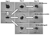
- FIG. 2 shows the appearance of the liver of each group collected on the 5th and 10th days after partial hepatectomy in comparison with the liver before and after partial hepatectomy.
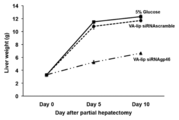
- FIG. 3 shows changes in liver weight of each group collected. As can be seen from both figures, the liver already recovered in size and weight before excision on day 5 in groups II and III, but in group I administered VA-lip siRNAgp46, even after 10 days after excision. Liver size and weight did not recover.
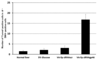
- Ki67 positive cells in group I were more prominent than other groups (group II and group III) that had also undergone partial hepatectomy. ( Figures 6 and 7).
- the number of Ki67 positive cells in group II and group III is significantly larger than that of normal liver tissue that has not undergone partial hepatectomy, and Ki67 is a marker for cell proliferation. It shows that vigorous cell growth occurs.
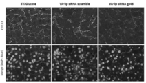
- CD133 positive cells in group I were more prominent than other groups (group II and group III) that had also undergone partial hepatectomy. It has become clear that the number is low (Fig. 8). Since CD133 is a stem cell marker, this result indicates that proliferation of stem cells was remarkably suppressed by VA-lip siRNAgp46. In consideration of the above-mentioned staining results, the proliferation of cells including stem cells in liver tissue was suppressed by apoptosis of ⁇ -SMA positive cells. In other words, ⁇ -SMA positive cells were partially excised from liver. It shows that it is indispensable for cell proliferation in liver tissue for subsequent tissue regeneration, especially for stem cell proliferation.
- Example 3 Effects of VA-lip siRNAgp46 administration time on liver regeneration in partially hepatectomized rats
- Group A no treatment (no treatment control group)
- Group B 5% glucose (solvent control group)
- Group C VA-lip siRNA scramble (mock control group)
- Group D VA-lip siRNAgp46 (test group)
- VA-lip siRNAgp46 and VA-lip siRNA scramble were the same as those prepared in Example 1.
- Groups B to D were administered each dose in a volume of 300 ⁇ l / dose for a total of 6 times every other day (ie, on days 0, 2, 4, 6, 8, and 10 after partial hepatectomy), , Total 4 times (ie 4, 6, 8 and 10 days after partial hepatectomy).
- Each siRNA was used at 0.75 mg / kg relative to the rat body weight.
- the liver of the partially hepatectomized rat was collected 24 hours after the sixth administration (fourth for the E group) was completed (that is, 11 days after the partial hepatectomy).
- the collected liver was observed for appearance and weighed and then embedded using an OCT compound to prepare a frozen section.
- the obtained sections were fixed with 4% paraformaldehyde, then blocked with PBS containing 5% goat serum, washed with PBS, and then washed with Cy3-labeled ⁇ -smooth muscle actin ( ⁇ -SMA) antibody (Sigma).
- the reaction was allowed to proceed overnight at 0 ° C. After washing with PBS, sealed with ProLong (R) Gold with DAPI ( Invitrogen), it was observed by the fluorescence microscope.
- FIGS. 10 and 11 show the appearance and weight of each group collected in (2) in comparison with the liver immediately after partial hepatectomy (day 0), respectively.
- the liver recovered size and weight before excision in groups AC and E, but in group D, which received VA-lip siRNAgp46 from day 0, 11 days after excision.
- liver size and weight did not recover.
- VA-lip siRNAgp46 was administered at an early stage of tissue regeneration. It became clear that it should be.
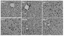
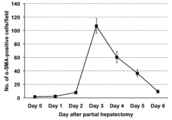
- Example 4 Temporal Change in Number of ⁇ -SMA Positive Cells in Liver Tissue after Partial Hepatectomy
- Livers were collected from partially hepatectomized rats at 0, 1, 2, 3, 4, 5 or 6 days after partial hepatectomy.
- the collected liver tissue was embedded using an OCT compound to prepare a frozen section.
- the obtained sections were fixed with 4% paraformaldehyde, then blocked with PBS containing 5% goat serum, washed with PBS, and then washed with Cy3-labeled ⁇ -smooth muscle actin ( ⁇ -SMA) antibody (Sigma).
- the reaction was allowed to proceed overnight at 0 ° C. After washing with PBS, sealed with ProLong (R) Gold with DAPI ( Invitrogen), it was observed by the fluorescence microscope.
- Example 5 Involvement of stellate cells in stem cell differentiation (1) Preparation of cells As stellate cells, hepatic stellate cells collected from SD rat liver were used. That is, first, SD rats were perfused with an EGTA solution and a collagenase solution, and then the liver was collected, and the collected liver was cut into small pieces and filtered with a cell strainer (pore diameter: 100 ⁇ m). HBSS + 0.25% BSA (Bovine Serum Albumin) solution was added to the resulting cell suspension, and the mixture was centrifuged at 4 ° C. and 500 rpm for 2 minutes. The supernatant was collected and centrifuged at 4 ° C. and 1300 rpm for 5 minutes.
- BSA Bovine Serum Albumin
- HBSS + 0.25% BSA solution was added, and 28.7% Nycodenz solution (Axis Shield, Oslo, Norway) was added and mixed so that the concentration of Nycodenz was 13.2%.
- Nycodenz solution Analog to Crystal
- DMEM Dulbecco's Modified Eagle's medium
- FBS fetal bovine serum
- the cells on the first day of culture were quiescent hepatic stellate cells (qHSC), and the cells that had been subcultured on the fifth day of culture and further cultured for two days were called activated hepatic stellate cells (aHSC) (FIG. 14). .
- qHSC quiescent hepatic stellate cells
- aHSC activated hepatic stellate cells
- hepatic stem cells collected from the liver of a 4-week-old GFP gene-introduced rat were used.
- an EGTA solution and a collagenase solution were perfused into a GFP gene-introduced rat, and then the liver was collected, and the collected liver was cut into pieces and then filtered with a cell strainer (pore size: 100 ⁇ m).
- Hank's balanced salt solution (HBSS) + 0.25% bovine serum albumin (BSA) solution was added to the obtained cell suspension and centrifuged at 4 ° C. and 500 rpm for 2 minutes. The supernatant was collected and centrifuged at 4 ° C. and 1300 rpm for 5 minutes.
- HBSS Hank's balanced salt solution
- BSA bovine serum albumin
- MACS® Magnetic Activating Cell Sorting buffer (Miltenyi Biotec, Auburn, CA, USA) was added to the precipitate and mixed. After counting the number of cells, MACS (R) was performed using FITC-conjugated mouse anti-CD45 antibody (BD Pharmingen), rabbit polyclonal anti-CD133 antibody (Abcam) and mouse monoclonal anti-EpCAM antibody (Santa Cruz), and CD133 positive, EpCAM positive CD45 negative cells were collected and used as rat hepatic stem cells in this experiment.
- hepatic stem cells were cultured in the same manner in the absence of aHSC.
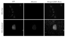
- immunostaining was performed with an antibody against albumin which is a hepatocyte marker (rabbit polyclonal, MP Biomedicals), and a GFP / albumin positive colony was photographed at a magnification of 100 using an inverted microscope (Nikon).
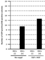
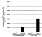
- the number of GFP / albumin positive colonies was counted based on the obtained images, and the area was calculated using NIS-Elements software (Nikon) (FIGS. 15 to 18).
- the cell culture insert containing aHSC was inserted into a well of a 24-well plate and co-cultured in an incubator at 37 ° C. and 5% CO 2 for 10 days (as a medium, DME / F12 (Dulbecco's Modified Eagle's Medium / Nutrient F-12 Ham) + 10% FBS + ITS (using 10 mg / l insulin, 5.5 mg / l transferrin, 0.67 ⁇ g / l selenium) +0.1 ⁇ M dexamethasone + 10 mM nicotinamide + 50 ⁇ g / ml ⁇ -mercaptoethanol + 2 mM L-glutamine + 5 mM Hepes) .
- a control only hepatic stem cells were cultured in the same manner in the absence of aHSC.
- Example 6 Change in DNA synthesis over time in liver tissue after partial hepatectomy
- rats with partial hepatectomy were prepared.
- rats were injected intraperitoneally with BrdU at a concentration of 100 ⁇ g / g body weight.
- Liver tissue was collected 3 hours after BrdU administration.
- the collected liver tissue was fixed with 10% formalin and embedded in paraffin.
- the paraffin-embedded liver tissue was cut into 5 ⁇ m-thick sections, activated with 10 mM citrate buffer at 120 ° C. for 20 minutes, and then blocked with 5% goat serum.
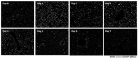
- mouse monoclonal anti-BrdU antibody (MBL) was reacted at 37 ° C. for 60 minutes. After the section was washed with PBS, Alexa488-labeled goat anti-mouse IgG (Invitrogen) was reacted at 37 ° C. for 60 minutes. After washing with PBS, it was encapsulated using an encapsulant containing DAPI, and BrdU incorporation in the liver tissue was observed. The results are shown in FIG.
- BrdU was injected intraperitoneally at a concentration of 100 ⁇ g / g body weight into the rats subjected to partial hepatectomy. .
- the cell suspension was adjusted with Hanks solution (Invitrogen) containing 0.25% BSA so that the cell concentration was 1 ⁇ 10 7 cells / 100 ⁇ l, and APC-labeled mouse monoclonal anti-BrdU antibody (BioLegends) was added. , Reacted at 37 ° C.
- BrdU was injected intraperitoneally at a concentration of 100 ⁇ g / g body weight into rats subjected to partial hepatectomy. did.
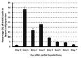
- FIGS. 23 and 24 show that the proliferation of hepatocytes gradually decreases after peaking one day after partial hepatectomy.
- the proliferation of whole cells present in the liver including hepatocytes has two peaks after 1 day and 3 days after partial hepatectomy, and the first peak after 1 day is the proliferation of hepatocytes.
- the second peak after 3 days is considered to be due to proliferation of cells other than hepatocytes, such as stem cells.
- Example 7 Involvement of stellate cells in hepatocyte proliferation (1) Co-culture of hepatocytes and stellate cells To evaluate the effect of stellate cells on hepatocyte proliferation, co-culture of hepatocytes and stellate cells was performed. It was. The aHSC obtained in Example 5 was seeded in a 60 mm dish at a concentration of 1 ⁇ 10 5 cells, and cultured at 37 ° C., 5% CO 2 for 24 hours using 10% FBS-added DMEM. At 24 hours of culture, the medium was removed, and Opti-MEM I reducing medium (Invitrogen) was added, and precultured until ready for siRNA transfection.
- Opti-MEM I reducing medium Invitrogen
- siRNA was mixed with RNAiMAX (Invitrogen) to a final concentration of 10 nM and allowed to stand at room temperature for 15 minutes to obtain a siRNA complex.
- the siRNA used is as follows. GFP siRNA (Ambion, Silencer GFP siRNA, Cat. No. AM4626) Scramble siRNA: as described in Example 1 gp46 siRNA: as described in Example 1
- siRNA complex was added to the pre-cultured aHSC and cultured at 37 ° C., 5% CO 2 for 5 hours. At 5 hours of culture, the medium was replaced with DMEM supplemented with 10% FBS, and cultured at 37 ° C., 5% CO 2 for 48 hours. At 48 hours of culture, the medium was removed and Opti-MEM I reducing medium was added and pre-cultured until ready for siRNA transfection.
- SiRNA (GFP siRNA, Scramble siRNA, gp46 siRNA) was mixed with RNAiMAX to a final concentration of 10 nM and allowed to stand at room temperature for 15 minutes.
- siRNA complex was added to the pre-cultured aHSC and cultured at 37 ° C., 5% CO 2 for 5 hours. After 5 hours of culture, the medium was removed, washed with PBS, and HSCs were collected using a trypsin-EDTA solution.
- the collected HSCs were collected from the liver of a GFP gene-introduced rat in advance and seeded at a concentration of 5 ⁇ 10 4 on a 35 mm dish coated with collagen I at a concentration of 5 ⁇ 10 4 / dish.
- the cells were cultured at 37 ° C. and 5% CO 2 for 48 hours.
- the medium was replaced with DME / F12 medium containing 10 mM BrdU and 10% FBS, and the cells were further cultured at 37 ° C. and 5% CO 2 for 24 hours.
- the medium was removed, washed with PBS, 4% PFA was added, and the mixture was fixed at room temperature for 30 minutes. After fixation, the plate was washed with PBS, and permeabilized by adding PBS containing 0.2N HCl and 0.1% Triton X-100.
- Example 8 Effect of variously treated collagen on proliferation of hepatocytes
- Rat tail-derived collagen I (Sigma) at a concentration of 10 ⁇ g / cm 2 A 60 mm dish was coated.
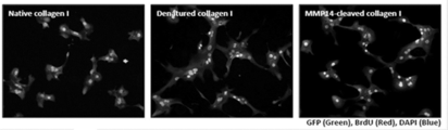
- denatured collagen I rat tail-derived collagen I was treated at 60 ° C. for 30 minutes and coated on a 60 mm dish at a concentration of 10 ⁇ g / cm 2 .
- MMP14-treated collagen I comprises rat tail-derived collagen I containing active MMP14 (R & D System) (0.1 ⁇ g / ml trypsin 3 (Recombinant Human Active Trypsin 3, R & D systems, Cat. No. 3714-SE), 50 mM.
- Tris 0.15 M NaCl, 10 mM CaCl 2 , 5 ⁇ M ZnCl 2 , 0.05% (v / v) Brij 35, pH 7.5) was used for reaction at room temperature for 20 hours.
- MMP14-treated collagen I was coated on a 60 mm dish at a concentration of 10 ⁇ g / cm 2 .
- Hepatocytes were collected from rat liver, seeded at a concentration of 2 ⁇ 10 5 cells / dish, and cultured at 37 ° C. and 5% CO 2 for 48 hours. At 48 hours of culture, the medium was replaced with DME / F12 medium containing 10 ⁇ M BrdU and 10% FBS, and the cells were further cultured at 37 ° C.
- the aHSC obtained in Example 5 was seeded in a 60 mm dish at a concentration of 1 ⁇ 10 5 cells, and cultured at 37 ° C., 5% CO 2 for 24 hours using 10% FBS-added DMEM. At 24 hours of culture, the medium was removed, and Opti-MEM I reducing medium (Invitrogen) was added, and precultured until ready for siRNA transfection.
- siRNA was mixed with RNAiMAX (Invitrogen) to a final concentration of 10 nM and allowed to stand at room temperature for 15 minutes to obtain a siRNA complex.
- the siRNA used is as follows. GFP siRNA (Ambion, Silencer GFP siRNA, Cat. No.
- MMP14 siRNA Sense strand: 5′-GUCAUUCAUGGGUAGCGATT-3 ′ (SEQ ID NO: 6) Antisense strand: 5′-UCGCUACCCAUGAAUGAGCCT-3 ′ (SEQ ID NO: 7) HGF siRNA: Sense strand: 5′-AUAUCUUUCCGGCAAGAAUUUGUGC-3 ′ (SEQ ID NO: 8) Antisense strand: 5′-GCACAAAUUCUGCCCGGAAAGAUAU-3 ′ (SEQ ID NO: 9)
- the siRNA complex was added to the pre-cultured aHSC and cultured at 37 ° C., 5% CO 2 for 5 hours. At 5 hours of culture, the medium was replaced with DMEM supplemented with 10% FBS, and cultured at 37 ° C., 5% CO 2 for 48 hours. At 48 hours of culture, the medium was removed and Opti-MEM I reducing medium was added and pre-cultured until ready for siRNA transfection.
- SiRNA GFP siRNA, MMP14 siRNA, HGF siRNA
- RNAiMAX RNAiMAX
- the siRNA complex was added to the pre-cultured aHSC and cultured at 37 ° C., 5% CO 2 for 5 hours. After 5 hours of culture, the medium was removed, washed with PBS, and HSCs were collected using a trypsin-EDTA solution.
- the collected HSCs were collected from the liver of a GFP gene-introduced rat in advance and seeded at a concentration of 5 ⁇ 10 4 on a 35 mm dish coated with collagen I at a concentration of 5 ⁇ 10 4 / dish.
- the cells were cultured at 37 ° C. and 5% CO 2 for 48 hours.
- the medium was replaced with DME / F12 medium containing 10 mM BrdU and 10% FBS, and the cells were further cultured at 37 ° C. and 5% CO 2 for 24 hours.
- the medium was removed, washed with PBS, 4% PFA was added, and the mixture was fixed at room temperature for 30 minutes. After fixation, the plate was washed with PBS, and permeabilized by adding PBS containing 0.2N HCl and 0.1% Triton X-100.
- MMP14 expressed by aHSC is involved in promoting the proliferation of hepatocytes by aHSC, and the contribution exceeds that of HGF, which is known as a key factor for promoting proliferation of hepatocytes. Recognize.
- Example 10 Effect of RGD sequence on MMP14-treated collagen on hepatocyte proliferation
- Rat hepatocytes collected from GFP gene-introduced rat liver were seeded in DME / F12 medium at a concentration of 2 ⁇ 10 5 cells / ml.
- Peptide control peptide (H-Gly-Arg-Gly-Glu-Glu-Ser-OH, Peptides International, Cat. No. PFA-3907-PI): 500 ⁇ g / ml
- GRGDS peptide Peptide Institute, Cat. No. 4189-v
- 100 ⁇ g / ml, 200 ⁇ g / ml, and 500 ⁇ g / ml were added and reacted at 37 ° C.
- rat hepatocytes were seeded at a concentration of 5 ⁇ 10 4 cells / dish in a 35 mm dish coated with collagen I treated with MMP14, and in DME / F12 medium containing 10 mM BrdU and 10% fetal bovine serum. The cells were cultured at 37 ° C. and 5% CO 2 for 72 hours. After completion of the culture, the medium was removed, washed with PBS, 4% PFA was added, and the mixture was fixed at room temperature for 30 minutes. After fixation, the plate was washed with PBS, and permeabilized by adding PBS containing 0.2N HCl and 0.1% Triton X-100.
Landscapes
- Health & Medical Sciences (AREA)
- Life Sciences & Earth Sciences (AREA)
- Engineering & Computer Science (AREA)
- Chemical & Material Sciences (AREA)
- Bioinformatics & Cheminformatics (AREA)
- General Health & Medical Sciences (AREA)
- Zoology (AREA)
- Organic Chemistry (AREA)
- Immunology (AREA)
- Pharmacology & Pharmacy (AREA)
- Animal Behavior & Ethology (AREA)
- Medicinal Chemistry (AREA)
- Public Health (AREA)
- Veterinary Medicine (AREA)
- Biomedical Technology (AREA)
- Wood Science & Technology (AREA)
- Epidemiology (AREA)
- Biotechnology (AREA)
- General Engineering & Computer Science (AREA)
- Genetics & Genomics (AREA)
- Biochemistry (AREA)
- Proteomics, Peptides & Aminoacids (AREA)
- Gastroenterology & Hepatology (AREA)
- Microbiology (AREA)
- Sustainable Development (AREA)
- Cell Biology (AREA)
- Developmental Biology & Embryology (AREA)
- Virology (AREA)
- Clinical Laboratory Science (AREA)
- Nutrition Science (AREA)
- Physiology (AREA)
- Nuclear Medicine, Radiotherapy & Molecular Imaging (AREA)
- Chemical Kinetics & Catalysis (AREA)
- General Chemical & Material Sciences (AREA)
- Medicines That Contain Protein Lipid Enzymes And Other Medicines (AREA)
- Micro-Organisms Or Cultivation Processes Thereof (AREA)
- Medicines Containing Material From Animals Or Micro-Organisms (AREA)
- Apparatus Associated With Microorganisms And Enzymes (AREA)
Abstract
Description
しかしながら、精力的な研究にも拘らず、組織再生に係る機構は未だそのごく一部が明らかにされているにすぎず、さらなる研究努力が求められている。
すなわち、本発明は以下に関する。
(1)活性化星細胞、活性化星細胞分解物、MMP14、MMP14で処理したコラーゲンおよび活性化星細胞分泌物からなる群から選択される成分を含む、組織再生を促進するための組成物。
(2)組織再生が、障害組織または移植組織において生じる、(1)に記載の組成物。
(3)障害または移植を受けた日から4日以内に投与される、(2)に記載の組成物。
(4)障害または移植を受けた日から1日以内に投与される、(2)に記載の組成物。
(5)障害が、組織破壊、炎症、壊死、線維化、手術的侵襲、臓器不全からなる群から選択される、(2)~(4)のいずれかに記載の組成物。
(7)組織再生が組織実質細胞の増殖を伴う、(1)~(6)のいずれかに記載の組成物。
(8)活性化星細胞が、HGF、EGF、MMP14からなる群から選択されるタンパク質の発現を増大させる処置を施されたものである、(1)~(7)のいずれかに記載の組成物。
(9)組織再生が抑制された状態を有する対象に対して用いる、(1)~(8)のいずれかに記載の組成物。
(10)組織再生が抑制された状態が、炎症、壊死、線維化、臓器不全、血小板数の減少、遺伝子異常、ノルアドレナリンの減少からなる群から選択される、(9)に記載の組成物。
(12)活性化星細胞、活性化星細胞分解物、MMP14、MMP14で処理したコラーゲンおよび活性化星細胞分泌物からなる群から選択される成分を含む、幹細胞の分化および/または増殖のための組成物。
(13)活性化星細胞、活性化星細胞分解物、MMP14、MMP14で処理したコラーゲンおよび活性化星細胞分泌物からなる群から選択される成分と、幹細胞とを接触させる工程を含む、組織幹細胞を分化および/または増殖させる方法。
(14)活性化星細胞、活性化星細胞分解物、MMP14、MMP14で処理したコラーゲンおよび活性化星細胞分泌物からなる群から選択される成分を含む、細胞増殖を促進するための組成物。
(16)MMP14を阻害する物質を含む、細胞増殖を抑制するための組成物。
(17)MMP14を阻害する物質が作用する細胞が、CAFおよび/または腫瘍細胞である、(16)に記載の組成物。
(18)MMP14で処理したコラーゲンを含む、細胞培養基材。
(19)MMP14で処理したコラーゲンで被覆された細胞培養容器。
また、本発明による組織再生の促進は、組織再生が抑制されている状況、例えば、線維症などの疾患を伴う状況などにおいて特に有用である。
本発明におけるMMP14は、細胞膜上に発現しているもののみならず、細胞膜から遊離したものも含む。MMP14は天然に存在するものであっても、人工的に作製したものであってもよい。したがって、MMP14は組換えMMP14を含む。遊離型のMMP14は既知であり(例えば、Jo et al., Biochem J. 2000 Feb 1;345 Pt 3:511-9など)、市販もされている(例えば、R&D Systems, Cat No.918-MP-010、918-MPN-010など)。MMP14のアミノ酸およびこれをコードする塩基配列は既知である(例えば、ヒトMMP14の塩基配列はGenBankアクセッション番号NM_004995、アミノ酸配列はGenBankアクセッション番号NP_004986として登録されている)。
ある変異体がMMP14と同等の機能を有するか否かは、当該変異体を、MMP14の既知の機能、限定されずに、例えば、コラーゲン分解能などについて、既知の任意の方法により解析し、適切な陰性対照や、陽性対照としてのMMP14と比較することによって評価することができる。例えば、ある変異体において、上記機能が陰性対照より優れている場合、例えば、10%以上、25%以上、50%以上、75%以上、さらには100%以上優れている場合、および/または、同機能がCSABPの1/100以上、1/50以上、1/25以上、1/10以上、1/5以上、さらには1/2以上である場合、この変異体をMMP14の機能的変異体に含める。
本発明におけるMMP14および/またはその機能的変異体は、当該タンパク質自体やその機能的変異体のほか、当該タンパク質やその機能的変異体をコードする核酸をも含む。
上記製造方法または使用における各成分やその配合量については、既に上述したとおりである。各成分の配合は、既知の任意の手法に従って行うことができる。
増殖を促進する細胞は、限定されずに、例えば、肝臓、膵臓、腎臓、腸管、肺、脾臓、心臓、骨髄、声帯、皮膚、腹膜、眼、脈管などの細胞が挙げられる。細胞は自己由来のものであっても、同種の他の個体または異種の個体のものであってもよい。分化および/または増殖させた細胞は、それを必要とする対象に移植することができる。
本発明はまた、MMP14を抑制する物質を含む、細胞増殖性疾患を処置するための組成物、前記物質を配合することを含む、細胞増殖性疾患を処置するための組成物を製造する方法、前記物質の、細胞増殖性疾患を処置するための組成物の製造への使用、細胞増殖性疾患の処置に用いる前記物質、ならびに、前記物質の治療有効量を、それを必要とする対象に投与することを含む、細胞増殖性疾患を処置するための方法に関する。
また、本明細書で用いる場合アンチセンス核酸は、RNA、DNA、PNA、またはこれらの複合物を含む。
本明細書で用いる場合、DNA/RNAキメラポリヌクレオチドは、限定されずに、例えば、特開2003-219893に記載の、標的遺伝子の発現を阻害するDNAとRNAとからなる2本鎖ポリヌクレオチドを含む。
細胞増殖性疾患としては、限定されずに、例えば、良性または悪性腫瘍、過形成症、ケロイド、クッシング症候群、原発性アルドステロン症、紅板症、真性多血症、白板症、過形成瘢痕、扁平苔癬および黒子症などが挙げられる。
投与頻度は、用いる剤や組成物の性状や、上記のものを含む対象の条件によって異なるが、例えば、1日多数回(すなわち1日2、3、4回または5回以上)、1日1回、数日毎(すなわち2、3、4、5、6、7日毎など)、1週間毎、数週間毎(すなわち2、3、4週間毎など)であってもよい。
また、用語「処置」は、本明細書で用いる場合、疾患の治癒、一時的寛解または予防などを目的とする医学的に許容される全ての種類の予防的および/または治療的介入を包含するものとする。例えば、「処置」の用語は、疾患の進行の遅延または停止、病変の退縮または消失、発症の予防または再発の防止などを含む、種々の目的の医学的に許容される介入を包含する。
本発明の細胞培養基材は、細胞生育の足場となるものであり、膜状、ゲル状など種々の形態をとってもよい。細胞培養基材は無菌であることが好ましいが、非無菌状態で提供され、使用時に滅菌してもよい。本発明の細胞培養基材は、細胞培養容器を被覆するために用いてもよい。被覆濃度は、限定されずに、例えば、コラーゲンの濃度として0.01~1000μg/cm2、0.1~100μg/cm2、1~50μg/cm2、5~25μg/cm2であってもよい。
(1)siRNAの調製
コラーゲン(I~IV型)の共通分子シャペロンであるヒトHSP47のラットホモログ、gp46(GenBank Accession No. M69246、配列番号1)の塩基配列を標的とするsiRNA(北海道システム・サイエンス, Sapporo, Japan)のセンス鎖およびアンチセンス鎖は以下のものを用いた。
A:GUUCCACCAUAAGAUGGUAGACAACAG(gp46の塩基配列上の第757塩基から始まるセンス鎖siRNA、配列番号2)
B:GUUGUCUACCAUCUUAUGGUGGAACAU(アンチセンス鎖siRNA、配列番号3)
C:CGAUUCGCUAGACCGGCUUCAUUGCAG(センス鎖siRNA、配列番号4)
D:GCAAUGAAGCCGGUCUAGCGAAUCGAU(アンチセンス鎖siRNA、配列番号5)
カチオン性脂質として、O,O’-ジテトラデカノイル-N-(α-トリメチルアンモニオアセチル)ジエタノールアミンクロライド(DC-6-14)、コレステロールおよびジオレイルホスファチジルエタノールアミン(DOPE)を4:3:3のモル比で含むカチオン性リポソーム(Lipotrust)を北海道システム・サイエンス(Sapporo, Japan)から購入した。リポソームは、使用前に、凍結乾燥した脂質混合物に攪拌条件下で再蒸留水(DDW)を添加することによって、1mM(DC-6-14)の濃度で調製した。VA結合リポソームを調製するため、DMSOに溶解した200nmolのビタミンA(レチノール、Sigma, USA)をリポソーム懸濁液(DC-6-14として100nmol)と、1.5mlチューブ中で攪拌しながら25℃で混合した。siRNAgp46を担持するVA結合リポソーム(VA-lip-siRNAgp46)を調製するため、siRNAgp46溶液(DDW中に580pmol/μl)を、レチノール結合リポソーム溶液に攪拌しながら室温下で添加した。siRNAとDC-6-14とのモル比は1:11.5、また、ビタミンA、DC-6-14およびsiRNAのモル比は11.5:11.5:1であった。in vitroでの使用に望ましい用量を得るため、VA-lip siRNAをリン酸緩衝生理食塩水(PBS)で再構成した。
(1)肝部分切除ラットの作製および処置
肝部分切除ラットは、雄SDラット(150~200g)(Slc Japan, Shizuoka, Japan)の肝臓全体の約70%にあたる肝左葉および中葉を切除することにより作製した。この肝部分切除ラットに、例1で作製したVA-lip siRNAgp46(群I)、VA-lip siRNAscramble(mockコントロール、群II)、または5%グルコース(siRNA非含有コントロール、群III)を、300μl/回の容量で、1日おきに合計5回(すなわち肝部分切除後1、3、5、7および9日目に)尾静脈投与した(図1、n=6)。なお、各siRNAは、ラット体重に対して0.75mg/kgで使用した。
3回目および5回目の投与が終了した24時間後(すなわち肝部分切除後5日目および10日目)に、肝部分切除ラットの肝臓を採取した。採取した肝臓は、外観を観察し、重量を測定した後、OCTコンパウンドを用いて封埋し、凍結切片を作製した。得られた切片を4%パラホルムアルデヒド(PFA)で固定し、次いで5%ヤギ血清含有PBSでブロッキングを施し、PBSで洗浄した後、Cy3標識抗α平滑筋アクチン(α-SMA)抗体(Sigma)、抗Ki-67抗体(Dako)または抗CD133抗体を用いて、4℃で一晩反応させた。PBSで洗浄した後、Alexa488標識ヤギ抗マウス抗体(Invitrogen)を用いて室温で60分間反応させた。PBSで洗浄した後、ProLong(R) Gold with DAPI(Invitrogen)で封入し、蛍光顕微鏡で観察を行った。また、採取した肝凍結切片のTunel染色は、In situ Apoptosis Detection Kit (Takara Bio, Japan)を用い、製造者の指示に従って行った。α-SMAとTunelとの共染色には、FITC-TUNEL抗体およびCy3標識抗α-SMA抗体を用いた。陽性細胞数は、5視野(倍率200倍)の平均値を算出した。
図2に肝部分切除後5日目および10日目に採取した各群の肝臓の外観を、肝部分切除前および切除直後の肝臓との比較で示す。また、図3に、採取した各群の肝重量の推移を示す。両図から分るとおり、肝臓は、群IIおよびIIIでは5日目にすでに切除前の大きさおよび重量を回復したが、VA-lip siRNAgp46を投与した群Iでは、切除後10日経っても肝臓の大きさおよび重量は回復しなかった。
上記の各染色結果は、合わせ考慮すると、α-SMA陽性細胞のアポトーシスにより、肝組織における幹細胞を含む細胞の増殖が抑制されたこと、逆に言えば、α-SMA陽性細胞が、肝部分切除後の組織再生に向けた肝組織における細胞増殖、特に幹細胞の増殖に不可欠であることを示すものである。
(1)肝部分切除ラットの作製および処置
例2と同様にして肝部分切除ラットを作製した。この肝部分切除ラットに、以下の処置を施した(図9)。
A群:無処置(無処置コントロール群)
B群:5%グルコース(溶媒コントロール群)
C群:VA-lip siRNAscramble(mockコントロール群)
D群:VA-lip siRNAgp46(試験群)
E群:VA-lip siRNAgp46(遅延投与群)
(各群ともn=4)
6回目(E群については4回目)の投与が終了した24時間後(すなわち肝部分切除後11日目)に、肝部分切除ラットの肝臓を採取した。採取した肝臓は、外観を観察し、重量を測定した後、OCTコンパウンドを用いて封埋し、凍結切片を作製した。得られた切片を4%パラホルムアルデヒドで固定し、次いで5%ヤギ血清含有PBSでブロッキングを施し、PBSで洗浄した後、Cy3標識α平滑筋アクチン(α-SMA)抗体(Sigma)を用いて4℃で一晩反応させた。PBSで洗浄した後、ProLong(R) Gold with DAPI(Invitrogen)で封入し、蛍光顕微鏡で観察を行った。
図10および11に(2)で採取した各群の肝臓の外観および重量を、肝部分切除直後(0日目)の肝臓との比較でそれぞれ示す。両図から分るとおり、肝臓は、A~CおよびE群で切除前の大きさおよび重量を回復したが、VA-lip siRNAgp46を第0日目から投与したD群では、切除後11日経っても肝臓の大きさおよび重量は回復しなかった。また、同じ用量のVA-lip siRNAgp46を第4日目から投与したE群で肝臓の大きさおよび重量の回復が認められたことから、VA-lip siRNAgp46は、組織再生の早期の段階で投与すべきことが明らかとなった。
(1)肝部分切除ラットの作製および採取組織の評価
例2と同様にして肝部分切除ラットを作製した。肝部分切除ラットから、肝部分切除後0、1、2、3、4、5または6日目に肝臓を採取した。採取した肝組織をOCTコンパウンドを用いて封埋し、凍結切片を作製した。得られた切片を4%パラホルムアルデヒドで固定し、次いで5%ヤギ血清含有PBSでブロッキングを施し、PBSで洗浄した後、Cy3標識α平滑筋アクチン(α-SMA)抗体(Sigma)を用いて4℃で一晩反応させた。PBSで洗浄した後、ProLong(R) Gold with DAPI(Invitrogen)で封入し、蛍光顕微鏡で観察を行った。
図12および13に示す結果から、α-SMA陽性細胞の数が肝部分切除後3日目に急激に増加し、その後なだらかに減少することが分る。この結果は、例2~3の結果および星細胞は活性化するとα-SMAを発現するようになることを考慮すると、組織再生の早期の段階で組織に存在する星細胞が活性化し、組織再生に向けた細胞増殖を引き起すことを示唆しするものである。
(1)細胞の調製
星細胞としては、SDラット肝臓から採取した肝星細胞を用いた。すなわち、まず、SDラットにEGTA溶液およびコラゲナーゼ溶液を灌流した後、肝臓を採取し、採取した肝臓を細切後、セルストレーナー(孔径100μm)で濾過した。得られた細胞懸濁液にHBSS+0.25%BSA(Bovine Serum Albumin)溶液を加えて、4℃、500rpmで2分間遠心した。上清を採取して、4℃、1300rpmで5分間遠心を行った。上清を除去した後、HBSS+0.25%BSA溶液を加え、Nycodenzの濃度が13.2%になるように28.7% Nycodenz溶液(Axis Shield, Oslo, Norway)を加えて混合した。HBSS+0.25%BSA溶液を重層した後、4℃、1400×gで20分間遠心した。遠心終了後、中間層を採取して、DMEM(Dulbecco's Modified Eagle's medium)+10%ウシ胎児血清(FBS)培地を用いて、培養を行った。培養1日目の細胞を静止型肝星細胞(qHSC)とし、また、培養5日目に継代を行い、さらに2日間培養したものを活性型肝星細胞(aHSC)とした(図14)。
I型コラーゲンでコートされたカバーガラス(IWAKI, Tokyo, Japan)を入れた6穴プレートに、上記(1)で得たaHSCを5×10⁴個/ウェルの密度で播種し、インキュベーター内で37℃、5%CO2にて48時間培養した。aHSC播種の2日後に、上記(1)で得た肝幹細胞を3×104個/ウェルの密度でウェル内のaHSC上に播種し、インキュベーター内で37℃、5%CO2にて9日間共培養した(培地として、DME/F12(Dulbecco's Modified Eagle's Medium/Nutrient F-12 Ham)+10%FBS+ITS(10mg/lインスリン、5.5mg/lトランスフェリン、0.67μg/lセレン)+0.1μMデキサメサゾン+10mMニコチンアミド+50μg/mlβ-メルカプトエタノール+2mM L-グルタミン+5mM Hepesを使用)。条件によっては、20ng/mlのEGFおよび/または50ng/mlのHGFを共培養開始時に添加した。また、コントロールとして、肝幹細胞のみをaHSCが存在しない状態で同様に培養した。
共培養9日目に、肝細胞マーカーであるアルブミンに対する抗体(ウサギポリクローナル、MP Biomedicals)で免疫染色を行い、倒立顕微鏡(Nikon)を用いて100倍の倍率でGFP/アルブミン両陽性コロニーを撮影し、得られた画像をもとにGFP/アルブミン両陽性コロニーの数をカウントするとともに、面積をNIS-Elements software(Nikon)を用いて算出した(図15~18)。
セルカルチャーインサート(孔径0.4μm、BD Falcon, Franklin Lakes, NJ, USA)上に上記(1)で得たaHSCを5×10⁴個/ウェルの密度で播種し、インキュベーター内で37℃、5%CO2にて、DMEM+10%FBSを用いて48時間培養した。aHSC播種の2日後に、上記(1)で得た肝幹細胞をI型コラーゲンでコートされたカバーガラス(IWAKI, Tokyo, Japan)を入れた24穴プレート(BD Falcon)に1×10⁴個/ウェルの密度で播種した。次いで、aHSCを含む上記セルカルチャーインサートを24穴プレートのウェルに挿入し、インキュベーター内で37℃、5%CO2にて10日間共培養した(培地として、DME/F12(Dulbecco's Modified Eagle's Medium/Nutrient F-12 Ham)+10%FBS+ITS(10mg/lインスリン、5.5mg/lトランスフェリン、0.67μg/lセレン)+0.1μMデキサメサゾン+10mMニコチンアミド+50μg/mlβ-メルカプトエタノール+2mM L-グルタミン+5mM Hepesを使用)。また、コントロールとして、肝幹細胞のみをaHSCが存在しない状態で同様に培養した。
別な実験では、共培養10日目に、Premix WST-1 Cell Proliferation Assay System (Takara, Tokyo, Japan)を使用して、マイクロプレートリーダー(Bio-Rad Laboratories, Hercules, CA, USA)を用いて細胞増殖の測定を行った(図20)。
図15~18に示す接触共培養実験の結果から、肝幹細胞を活性型肝星細胞と共培養することで、肝幹細胞の肝細胞への分化および増殖が生じることが明らかとなった。また、肝幹細胞の肝細胞への分化・増殖は、EGFおよびHGFの添加により顕著に促進されることが明らかとなった。
図19および20に示す非接触共培養実験の結果からは、活性型肝星細胞が、肝幹細胞と接触しない状態であっても肝幹細胞の肝細胞への分化・増殖を誘導し得ることが明らかとなった。
これらの結果は、活性型肝星細胞から分泌される液性因子が肝幹細胞の肝細胞への分化・増殖を誘導することを示すものである。
(1)BrdU陽性細胞の経時変化
例2と同様にして肝部分切除ラットを作製した。肝部分切除後の肝臓内のDNA合成の経時変化を調べるために、ラットに対して、BrdUを100μg/g体重の濃度で腹腔内注射した。BrdU投与後3時間目に肝臓組織を採取した。採取した肝臓組織は、10%ホルマリンで固定し、パラフィン包埋した。パラフィン包埋した肝臓組織は5μm厚の切片にし、10mMクエン酸バッファーを用いて、120℃で20分間賦活化を行った後、5%ヤギ血清を用いてブロッキングした。ブロッキング後、マウスモノクローナル抗BrdU抗体(MBL)を37℃で60分間反応させた。切片をPBSで洗浄後、Alexa488標識ヤギ抗マウスIgG(Invitrogen)を37℃で60分間反応させた。PBSで洗浄後、DAPIを含む封入剤を用いて封入し、肝臓組織内でのBrdUの取り込みを観察した。結果を図21に示す。
図21および22に示す結果から、肝部分切除後、肝組織内でのDNA合成が二峰性に増加することがわかる。すなわち、切除の1日後に最初のピークがあり、2日後にはやや沈静化するが、3日後に再び増加し、4日後以降は徐々に低下していく。
例2と同様にして肝部分切除ラットを作製した。肝部分切除後の肝臓内の肝細胞におけるDNA合成の経時変化を調べるために、ラットに対して、BrdUを100μg/g体重の濃度で腹腔内注射した。BrdU投与後3時間目に肝臓組織を採取した。採取した肝臓組織は、10%ホルマリンで固定し、パラフィン包埋した。パラフィン包埋した肝臓組織は5μm厚の切片にし、10mMクエン酸バッファーを用いて、120℃で20分間賦活化を行った後、5%ヤギ血清を用いてブロッキングした。ブロッキング後、APC標識マウスモノクローナル抗BrdU抗体(BioLegends)およびヤギ抗HNF4α抗体(Santa Cruz)を37℃で60分間反応させた。切片をPBSで洗浄後、Alexa488標識ヒツジ抗ヤギIgG(Invitrogen)を37℃で60分間反応させた。PBSで洗浄後、DAPIを含む封入剤を用いて封入し、肝臓組織内での肝細胞によるBrdUの取り込みを観察した。結果を図23に示す。
(1)肝細胞と星細胞との共培養
星細胞が肝細胞の増殖に与える影響を評価するために、肝細胞と星細胞との共培養を行った。例5で得たaHSCを60mmディッシュに1×10⁵個の濃度で播種し、10%FBS加DMEMを用いて、37℃、5%CO2で24時間培養した。培養24時間目に培地を除去し、Opti-MEM I reducing medium(Invitrogen)を加えて、siRNAのトランスフェクションの準備ができるまで前培養した。siRNAを最終濃度が10nMとなるようにRNAiMAX(Invitrogen)と混合し、室温で15分間静置してsiRNA複合体を得た。使用したsiRNAは以下のとおりである。
GFP siRNA(Ambion、Silencer GFP siRNA、Cat. No.AM4626)
Scramble siRNA:例1に記載のもの
gp46 siRNA:例1に記載のもの
上記(1)で透過処理を行った細胞をPBSで洗浄後、5%ヤギ血清でブロッキングを行い、マウスモノクローナル抗BrdU抗体(MBL)およびウサギポリクローナル抗GFP抗体(Invitrogen)を加えて一次抗体反応を37℃で60分間行った。一次抗体反応後、PBSで洗浄し、Alexa488標識ヤギ抗ウサギIgG(Invitrogen)およびAlexa555標識ヤギ抗マウスIgG(Invitrogen)を加えて二次抗体反応を37℃で60分間行った。二次抗体反応後、PBSで洗浄し、DAPIを含む封入剤を用いて細胞を封入した。蛍光顕微鏡像を図25に示す。
BrdU陽性GFP陽性肝細胞の割合を測定するために、上記(1)で透過処理を行った細胞をPBSで洗浄した後、APC標識マウスモノクローナル抗BrdU抗体およびFITC標識ウサギポリクローナル抗GFP抗体を加えて抗体反応を37℃で60分間行った。抗体反応終了後、PBSで細胞を洗浄して、FACS解析によりBrdU陽性GFP陽性肝細胞の割合を測定した。結果を図26に示す。BrdU陽性細胞の割合は、BrdU陽性細胞数をフローサイトメトリーで解析した全細胞数で割ったもので示した。
(1)種々の処理を施したコラーゲン上での肝細胞の培養
ラット尾由来コラーゲンI(Sigma)を10μg/cm2の濃度で60mmディッシュにコートした。変性コラーゲンIは、ラット尾由来コラーゲンIを60℃で30分間処理し、10μg/cm2の濃度で60mmディッシュにコートした。MMP14処理コラーゲンIは、ラット尾由来コラーゲンIを活性型MMP14(R&D System)を含む反応液(0.1μg/mlトリプシン3(Recombinant Human Active Trypsin3、R&D systems、Cat. No. 3714-SE)、50mM Tris、0.15M NaCl、10mM CaCl2、5μM ZnCl2、0.05%(v/v)Brij35、pH7.5)を用いて、室温で20時間反応させた。反応終了後、MMP14処理コラーゲンIを10μg/cm2の濃度で60mmディッシュにコートした。肝細胞はラット肝臓から採取し、2×105個/ディッシュの濃度で播種し、37℃、5%CO2で48時間培養した。培養48時間目に、培地を10μM BrdUおよび10%FBSを含むDME/F12培地に置換して、37℃、5%CO2でさらに24時間培養した。培養24時間目に、培地を除去し、PBSで洗浄した後、4%PFAを加え、室温で30分間固定した。固定後、PBSで洗浄し、0.2N HClおよび0.1%Triton X-100を含むPBSを加えて透過処理を行った。
上記(1)で透過処理を行った細胞をPBSで洗浄後、5%ヤギ血清でブロッキングを行い、マウスモノクローナル抗BrdU抗体(MBL)およびウサギポリクローナル抗GFP抗体(Invitrogen)を加えて一次抗体反応を37℃で60分間行った。一次抗体反応後、PBSで洗浄し、Alexa488標識ヤギ抗ウサギIgG(Invitrogen)およびAlexa555標識ヤギ抗マウスIgG(Invitrogen)を加えて二次抗体反応を37℃で60分間行った。二次抗体反応後、PBSで洗浄し、DAPIを含む封入剤を用いて細胞を封入した。蛍光顕微鏡像を図27に示す。
BrdU陽性肝細胞の割合を測定するために、上記(1)で透過処理を行った細胞をPBSで洗浄した後、APC標識マウスモノクローナル抗BrdU抗体を加えて抗体反応を4℃で30分間行った。抗体反応終了後、PBSで細胞を洗浄して、FACS解析によりBrdU陽性肝細胞の割合を測定した。結果を図28に示す。BrdU陽性細胞の割合は、BrdU陽性細胞数をフローサイトメトリーで解析した全細胞数(3×10⁴個)で割ったもので示した。
例9 aHSCにおけるMMP14の発現が肝細胞の増殖に与える影響
(1)肝細胞と星細胞との共培養
星細胞が肝細胞の増殖に与える影響を評価するために、肝細胞と星細胞との共培養を行った。例5で得たaHSCを60mmディッシュに1×10⁵個の濃度で播種し、10%FBS加DMEMを用いて、37℃、5%CO2で24時間培養した。培養24時間目に培地を除去し、Opti-MEM I reducing medium(Invitrogen)を加えて、siRNAのトランスフェクションの準備ができるまで前培養した。siRNAを最終濃度が10nMとなるようにRNAiMAX(Invitrogen)と混合し、室温で15分間静置してsiRNA複合体を得た。使用したsiRNAは以下のとおりである。
GFP siRNA(Ambion、Silencer GFP siRNA、Cat. No.AM4626)
MMP14 siRNA:
センス鎖:5’-GCUCAUUCAUGGGUAGCGATT-3’(配列番号6)
アンチセンス鎖:5’-UCGCUACCCAUGAAUGAGCCT-3’(配列番号7)
HGF siRNA:
センス鎖:5’-AUAUCUUUCCGGCAAGAAUUUGUGC-3’(配列番号8)
アンチセンス鎖:5’-GCACAAAUUCUUGCCGGAAAGAUAU-3’(配列番号9)
上記(1)で透過処理を行った細胞をPBSで洗浄後、5%ヤギ血清でブロッキングを行い、マウスモノクローナル抗BrdU抗体(MBL)およびウサギポリクローナル抗GFP抗体(Invitrogen)を加えて一次抗体反応を37℃で60分間行った。一次抗体反応後、PBSで洗浄し、Alexa488標識ヤギ抗ウサギIgG(Invitrogen)およびAlexa555標識ヤギ抗マウスIgG(Invitrogen)を加えて二次抗体反応を37℃で60分間行った。二次抗体反応後、PBSで洗浄し、DAPIを含む封入剤を用いて細胞を封入した。蛍光顕微鏡像を図29に示す。
BrdU陽性GFP陽性肝細胞の割合を測定するために、上記(1)で透過処理を行った細胞をPBSで洗浄した後、APC標識マウスモノクローナル抗BrdU抗体およびFITC標識ウサギポリクローナル抗GFP抗体を加えて抗体反応を4℃で30分間行った。抗体反応終了後、PBSで細胞を洗浄して、FACS解析によりBrdU陽性GFP陽性肝細胞の割合を測定した。結果を図30に示す。BrdU陽性細胞の割合は、BrdU陽性細胞数をフローサイトメトリーで解析した全細胞数で割ったもので示した。
GFP遺伝子導入ラット肝臓から採取したラット肝細胞を2×10⁵個/mlの濃度でDME/F12培地に播種し、異なる濃度のペプチド(コントロールペプチド(H-Gly-Arg-Gly-Glu-Glu-Ser-OH、Peptides International、Cat. No. PFA-3907-PI):500μg/ml、GRGDSペプチド(ペプチド研究所、Cat. No. 4189-v):100μg/ml、200μg/ml、500μg/ml)を加え、37℃で30分間反応させた。反応終了後のラット肝細胞を、MMP14で処理したコラーゲンIをコートした35mmディッシュに5×104個/ディッシュの濃度で播種し、10mM BrdUおよび10%ウシ胎児血清を含むDME/F12培地中、37℃、5%CO2で72時間培養した。培養終了後、培地を除去し、PBSで洗浄した後、4%PFAを加えて室温で30分間固定した。固定後、PBSで洗浄し、0.2N HClおよび0.1%Triton X-100を含むPBSを加えて透過処理を行った。
Claims (19)
- 活性化星細胞、活性化星細胞分解物、MMP14、MMP14で処理したコラーゲンおよび活性化星細胞分泌物からなる群から選択される成分を含む、組織再生を促進するための組成物。
- 組織再生が、障害組織または移植組織において生じる、請求項1に記載の組成物。
- 障害または移植を受けた日から4日以内に投与される、請求項2に記載の組成物。
- 障害または移植を受けた日から1日以内に投与される、請求項2に記載の組成物。
- 障害が、組織破壊、炎症、壊死、線維化、手術的侵襲、臓器不全からなる群から選択される、請求項2~4のいずれか一項に記載の組成物。
- 組織再生が組織幹細胞の分化および/または増殖を伴う、請求項1~5のいずれか一項に記載の組成物。
- 組織再生が組織実質細胞の増殖を伴う、請求項1~6のいずれか一項に記載の組成物。
- 活性化星細胞が、HGF、EGF、MMP14からなる群から選択されるタンパク質の発現を増大させる処置を施されたものである、請求項1~7のいずれか一項に記載の組成物。
- 組織再生が抑制された状態を有する対象に対して用いる、請求項1~8のいずれか一項に記載の組成物。
- 組織再生が抑制された状態が、炎症、壊死、線維化、臓器不全、血小板数の減少、遺伝子異常、ノルアドレナリンの減少からなる群から選択される、請求項9に記載の組成物。
- 対象において組織再生を促進する方法であって、請求項1~10のいずれか一項に記載の組成物を、これを必要とする対象に投与する工程を含む、前記方法。
- 活性化星細胞、活性化星細胞分解物、MMP14、MMP14で処理したコラーゲンおよび活性化星細胞分泌物からなる群から選択される成分を含む、幹細胞の分化および/または増殖のための組成物。
- 活性化星細胞、活性化星細胞分解物、MMP14、MMP14で処理したコラーゲンおよび活性化星細胞分泌物からなる群から選択される成分と、幹細胞とを接触させる工程を含む、組織幹細胞を分化および/または増殖させる方法。
- 活性化星細胞、活性化星細胞分解物、MMP14、MMP14で処理したコラーゲンおよび活性化星細胞分泌物からなる群から選択される成分を含む、細胞増殖を促進するための組成物。
- 活性化星細胞、活性化星細胞分解物、MMP14、MMP14で処理したコラーゲンおよび活性化星細胞分泌物からなる群から選択される成分と、細胞とを接触させる工程を含む、細胞増殖を促進させる方法。
- MMP14を阻害する物質を含む、細胞増殖を抑制するための組成物。
- MMP14を阻害する物質が作用する細胞が、CAFおよび/または腫瘍細胞である、請求項16に記載の組成物。
- MMP14で処理したコラーゲンを含む、細胞培養基材。
- MMP14で処理したコラーゲンで被覆された細胞培養容器。
Priority Applications (7)
| Application Number | Priority Date | Filing Date | Title |
|---|---|---|---|
| CA2895483A CA2895483A1 (en) | 2012-12-21 | 2013-12-20 | Tissue regeneration promoting agent |
| RU2015129592A RU2015129592A (ru) | 2012-12-21 | 2013-12-20 | Ускоритель регенерации ткани |
| US14/653,667 US20150297686A1 (en) | 2012-12-21 | 2013-12-20 | Tissue regeneration promoting agent |
| KR1020157016839A KR102276237B1 (ko) | 2012-12-21 | 2013-12-20 | 조직 재생 촉진제 |
| EP13866361.2A EP2937090B1 (en) | 2012-12-21 | 2013-12-20 | Tissue regeneration promoting agent |
| AU2013364894A AU2013364894A1 (en) | 2012-12-21 | 2013-12-20 | Tissue regeneration accelerator |
| CN201380066850.5A CN104884072B (zh) | 2012-12-21 | 2013-12-20 | 组织再生促进剂 |
Applications Claiming Priority (2)
| Application Number | Priority Date | Filing Date | Title |
|---|---|---|---|
| JP2012-280261 | 2012-12-21 | ||
| JP2012280261A JP6076076B2 (ja) | 2012-12-21 | 2012-12-21 | 組織再生促進剤 |
Publications (1)
| Publication Number | Publication Date |
|---|---|
| WO2014098211A1 true WO2014098211A1 (ja) | 2014-06-26 |
Family
ID=50978527
Family Applications (1)
| Application Number | Title | Priority Date | Filing Date |
|---|---|---|---|
| PCT/JP2013/084228 Ceased WO2014098211A1 (ja) | 2012-12-21 | 2013-12-20 | 組織再生促進剤 |
Country Status (10)
| Country | Link |
|---|---|
| US (1) | US20150297686A1 (ja) |
| EP (1) | EP2937090B1 (ja) |
| JP (1) | JP6076076B2 (ja) |
| KR (1) | KR102276237B1 (ja) |
| CN (1) | CN104884072B (ja) |
| AU (1) | AU2013364894A1 (ja) |
| CA (1) | CA2895483A1 (ja) |
| RU (1) | RU2015129592A (ja) |
| TW (1) | TW201440785A (ja) |
| WO (1) | WO2014098211A1 (ja) |
Families Citing this family (2)
| Publication number | Priority date | Publication date | Assignee | Title |
|---|---|---|---|---|
| RU2590859C1 (ru) * | 2015-06-30 | 2016-07-10 | государственное бюджетное образовательное учреждение высшего профессионального образования "Сибирский государственный медицинский университет" Министерства здравоохранения Российской Федерации (ГБОУ ВПО СибГМУ Минздрава России) | Способ дистантной стимуляции регенерации гепатоцитов |
| CN105213230A (zh) * | 2015-09-22 | 2016-01-06 | 薇碧生物科技(上海)有限公司 | 一种基于不可见光和酶抑制物来促进胶原蛋白再生的方法 |
Citations (12)
| Publication number | Priority date | Publication date | Assignee | Title |
|---|---|---|---|---|
| JP2003219893A (ja) | 2001-11-21 | 2003-08-05 | Mitsubishi Chemicals Corp | 遺伝子発現阻害方法 |
| JP2005532050A (ja) | 2002-06-19 | 2005-10-27 | レイヴェン バイオテクノロジーズ, インコーポレイテッド | 新規のraag10細胞表面標的および当該標的を認識する抗体ファミリー |
| JP2006506071A (ja) | 2002-11-13 | 2006-02-23 | レイヴェン バイオテクノロジーズ, インコーポレイテッド | 抗原pipaおよびそれに結合する抗体 |
| JP2007529197A (ja) | 2003-09-18 | 2007-10-25 | レイヴェン バイオテクノロジーズ, インコーポレイテッド | Kid3およびkid3に結合するkid3抗体 |
| JP2008054276A (ja) * | 2006-07-27 | 2008-03-06 | Koike Co Ltd | 圧電基板及びその製造方法 |
| US20080207553A1 (en) | 2004-10-04 | 2008-08-28 | Nitto Denko Corporation | Biodegradable Cationic Polymers |
| WO2008120815A1 (ja) | 2007-03-30 | 2008-10-09 | Nitto Denko Corporation | がん細胞および癌随伴線維芽細胞への標的化剤 |
| US20080312174A1 (en) | 2007-06-05 | 2008-12-18 | Nitto Denko Corporation | Water soluble crosslinked polymers |
| WO2009036368A2 (en) | 2007-09-14 | 2009-03-19 | Nitto Denko Corporation | Drug carriers |
| WO2010014117A1 (en) | 2008-07-30 | 2010-02-04 | Nitto Denko Corporation | Drug carriers |
| WO2012064834A2 (en) * | 2010-11-09 | 2012-05-18 | Cornell University | Methods for organ regeneration |
| WO2012170952A2 (en) | 2011-06-08 | 2012-12-13 | Nitto Denko Corporation | Compounds for targeting drug delivery and enhancing sirna activity |
Family Cites Families (9)
| Publication number | Priority date | Publication date | Assignee | Title |
|---|---|---|---|---|
| JP2006510586A (ja) * | 2002-08-29 | 2006-03-30 | ユニバーシティー オブ サザンプトン | 肝疾患治療のための薬剤の調製におけるアポトーシス誘導物質の使用方法 |
| CN1303206C (zh) * | 2003-10-22 | 2007-03-07 | 中国人民解放军军事医学科学院野战输血研究所 | 一种诱导干细胞向肝细胞定向分化的方法及肝细胞的用途 |
| US7662945B2 (en) * | 2005-02-01 | 2010-02-16 | Massachusetts Eye & Ear Infirmary | Neostatins |
| JP2008543276A (ja) * | 2005-03-30 | 2008-12-04 | ミネルバ バイオテクノロジーズ コーポレーション | Muc1発現細胞の増殖 |
| WO2007092899A2 (en) * | 2006-02-09 | 2007-08-16 | The Burnham Institute | Inhibitors of membrane type-1 matrix metalloproteinase for the treatment of insulin-dependent diabetes mellitus |
| CN102300987A (zh) * | 2008-12-17 | 2011-12-28 | 安吉奥开米公司 | 膜型-1基质金属蛋白抑制剂及其用途 |
| WO2010079340A2 (en) * | 2009-01-08 | 2010-07-15 | Eisai R & D Management Co., Ltd. | Assay |
| WO2011109767A2 (en) * | 2010-03-04 | 2011-09-09 | University Of Notre Dame Du Lac | Gelatinase inhibitors and prodrugs |
| US20140199324A1 (en) * | 2011-03-24 | 2014-07-17 | Daniel T. Dransfield | Evaluating protein expression in patient stratification and other therapeutic, diagnostic and prognostic methods for cancer |
-
2012
- 2012-12-21 JP JP2012280261A patent/JP6076076B2/ja not_active Expired - Fee Related
-
2013
- 2013-12-20 US US14/653,667 patent/US20150297686A1/en not_active Abandoned
- 2013-12-20 KR KR1020157016839A patent/KR102276237B1/ko not_active Expired - Fee Related
- 2013-12-20 RU RU2015129592A patent/RU2015129592A/ru not_active Application Discontinuation
- 2013-12-20 CN CN201380066850.5A patent/CN104884072B/zh not_active Expired - Fee Related
- 2013-12-20 WO PCT/JP2013/084228 patent/WO2014098211A1/ja not_active Ceased
- 2013-12-20 AU AU2013364894A patent/AU2013364894A1/en not_active Abandoned
- 2013-12-20 EP EP13866361.2A patent/EP2937090B1/en active Active
- 2013-12-20 CA CA2895483A patent/CA2895483A1/en active Pending
- 2013-12-20 TW TW102147570A patent/TW201440785A/zh unknown
Patent Citations (12)
| Publication number | Priority date | Publication date | Assignee | Title |
|---|---|---|---|---|
| JP2003219893A (ja) | 2001-11-21 | 2003-08-05 | Mitsubishi Chemicals Corp | 遺伝子発現阻害方法 |
| JP2005532050A (ja) | 2002-06-19 | 2005-10-27 | レイヴェン バイオテクノロジーズ, インコーポレイテッド | 新規のraag10細胞表面標的および当該標的を認識する抗体ファミリー |
| JP2006506071A (ja) | 2002-11-13 | 2006-02-23 | レイヴェン バイオテクノロジーズ, インコーポレイテッド | 抗原pipaおよびそれに結合する抗体 |
| JP2007529197A (ja) | 2003-09-18 | 2007-10-25 | レイヴェン バイオテクノロジーズ, インコーポレイテッド | Kid3およびkid3に結合するkid3抗体 |
| US20080207553A1 (en) | 2004-10-04 | 2008-08-28 | Nitto Denko Corporation | Biodegradable Cationic Polymers |
| JP2008054276A (ja) * | 2006-07-27 | 2008-03-06 | Koike Co Ltd | 圧電基板及びその製造方法 |
| WO2008120815A1 (ja) | 2007-03-30 | 2008-10-09 | Nitto Denko Corporation | がん細胞および癌随伴線維芽細胞への標的化剤 |
| US20080312174A1 (en) | 2007-06-05 | 2008-12-18 | Nitto Denko Corporation | Water soluble crosslinked polymers |
| WO2009036368A2 (en) | 2007-09-14 | 2009-03-19 | Nitto Denko Corporation | Drug carriers |
| WO2010014117A1 (en) | 2008-07-30 | 2010-02-04 | Nitto Denko Corporation | Drug carriers |
| WO2012064834A2 (en) * | 2010-11-09 | 2012-05-18 | Cornell University | Methods for organ regeneration |
| WO2012170952A2 (en) | 2011-06-08 | 2012-12-13 | Nitto Denko Corporation | Compounds for targeting drug delivery and enhancing sirna activity |
Non-Patent Citations (12)
| Title |
|---|
| "Hyojun Yakuzaigaku", 2003, NANKODO |
| "Hyoteki to shita Kankohen no Chiryo Senryaku", EXPERIMENTAL MEDICINE, vol. 29, no. 13, 2011, pages 2114 - 2122, XP008172815 * |
| AUSUBEL ET AL.: "Current Protocols in Molecular Biology", 1992, GREENE PUBLISHING ASSOCIATES |
| DING ET AL., NATURE, vol. 468, no. 7321, 11 November 2010 (2010-11-11), pages 310 - 5 |
| JO ET AL., BIOCHEM J., vol. 345, 1 February 2000 (2000-02-01), pages 511 - 9 |
| PIROLLO; CHANG, CANCER RES., vol. 68, no. 5, 2008, pages 1247 - 50 |
| RUOSLAHTI ET AL., J CELL BIOL., vol. 188, no. 6, 2010, pages 759 - 68 |
| SAMBROOK ET AL.: "Molecular Cloning: A Laboratory Manual", 2001, COLD SPRING HARBOR PRESS |
| See also references of EP2937090A4 |
| TORCHILIN, AAPS J., vol. 9, no. 2, 2007, pages E128 - 47 |
| YOSHIRO NIITSU ET AL.: "Bunshi Saibo kara Kaimei suru Kanzo Bioloqy Kansei Saibo o Hyoteki to shita Kankohen no Chiryo Senryaku", EXPERIMENTAL MEDICINE, vol. 29, no. 13, 2011, pages 2114 - 2122, XP008172815 * |
| ZHAO; BURT, J MOL HISTOL., vol. 38, no. 1, March 2007 (2007-03-01), pages 53 - 64 |
Also Published As
| Publication number | Publication date |
|---|---|
| KR102276237B1 (ko) | 2021-07-12 |
| KR20150095705A (ko) | 2015-08-21 |
| TW201440785A (zh) | 2014-11-01 |
| EP2937090B1 (en) | 2020-06-10 |
| US20150297686A1 (en) | 2015-10-22 |
| CA2895483A1 (en) | 2014-06-26 |
| CN104884072A (zh) | 2015-09-02 |
| CN104884072B (zh) | 2019-11-08 |
| AU2013364894A1 (en) | 2015-07-09 |
| JP6076076B2 (ja) | 2017-02-08 |
| EP2937090A1 (en) | 2015-10-28 |
| EP2937090A4 (en) | 2016-08-03 |
| RU2015129592A (ru) | 2017-01-27 |
| JP2014122190A (ja) | 2014-07-03 |
Similar Documents
| Publication | Publication Date | Title |
|---|---|---|
| JP7275193B2 (ja) | 筋ジストロフィーの処置における心筋球由来細胞およびこのような細胞によって分泌されたエキソソーム | |
| Cao et al. | Cell-free osteoarthritis treatment with sustained-release of chondrocyte-targeting exosomes from umbilical cord-derived mesenchymal stem cells to rejuvenate aging chondrocytes | |
| Liao et al. | Engineering extracellular vesicles restore the impaired cellular uptake and attenuate intervertebral disc degeneration | |
| Qiao et al. | microRNA-21-5p dysregulation in exosomes derived from heart failure patients impairs regenerative potential | |
| US20220218757A1 (en) | Therapeutically active cells and exosomes | |
| KR102059054B1 (ko) | 장 섬유증 치료제 | |
| RU2742308C2 (ru) | Стромальные стволовые клетки жировой ткани для использования в лечении рефракторных сложных перианальных фистул при болезни крона | |
| CN101272798A (zh) | 基因修饰以表达自杀基因的骨髓间充质干细胞用于治疗癌症的用途 | |
| Zheng et al. | circPTP4A2‐miR‐330‐5p‐PDK2 Signaling facilitates in vivo survival of HuMSCs on SF‐SIS scaffolds and improves the repair of damaged endometrium | |
| Liu et al. | Human umbilical cord-derived mesenchymal stem cells-exosomes-delivered miR-375 targets HDAC4 to promote autophagy and suppress T cell apoptosis in sepsis-associated acute kidney injury | |
| US10780107B2 (en) | Agent for inducing cell death, agent for suppressing cell proliferation, and pharmaceutical composition used for treatment of disease resulting from abnormal cell proliferation | |
| Tirgar et al. | Preclinical gene therapy in glioblastoma multiforme: Using olfactory ensheathing cells containing a suicide gene | |
| JP6076076B2 (ja) | 組織再生促進剤 | |
| CN107326027A (zh) | 一种rna嵌合体及其用途 | |
| CN106459902B (zh) | 组织再生促进剂 | |
| Zhang et al. | Mst1 Inhibition As A Cellular Mediator: Prevention Of Anoikis In mBMSCs Through Activating ITGα5β1/FAK Signaling Pathway | |
| CN116615211A (zh) | 尿源性上皮细胞培养物、由此得到的肾球状体以及其制备和使用方法 | |
| Luo et al. | combined with dental pulp stem cells on the injured fallopian tube | |
| JPWO2018164141A1 (ja) | Lypd1阻害剤及びそれを用いた生体組織の製造方法 | |
| Zhao et al. | CAR-CD34 (+) hematopoietic stem/progenitor cells produced in vivo protect against thoracic aortic aneurysm and dissection | |
| Wang | Studies of Novel Factors from Fetal Mesenchymal Stem Cell and Their Applications in Bone and Cartilage Repair | |
| CN119970616A (zh) | 凝胶组合物及其在制备治疗肢体缺血的药物中的应用 | |
| WO2021146566A1 (en) | Engineered fibroblasts as cell therapy to treat cancer via tumor stroma stabilization | |
| Przystal | Combination of anti-cancer drug therapy with targeted gene therapy against glioblastoma | |
| McGinn | Bioreducible Polymer-mediated Gene Therapy for the Treatment of Ischemic Heart Disease |
Legal Events
| Date | Code | Title | Description |
|---|---|---|---|
| 121 | Ep: the epo has been informed by wipo that ep was designated in this application |
Ref document number: 13866361 Country of ref document: EP Kind code of ref document: A1 |
|
| ENP | Entry into the national phase |
Ref document number: 2895483 Country of ref document: CA |
|
| WWE | Wipo information: entry into national phase |
Ref document number: 14653667 Country of ref document: US |
|
| NENP | Non-entry into the national phase |
Ref country code: DE |
|
| ENP | Entry into the national phase |
Ref document number: 20157016839 Country of ref document: KR Kind code of ref document: A |
|
| ENP | Entry into the national phase |
Ref document number: 2013364894 Country of ref document: AU Date of ref document: 20131220 Kind code of ref document: A |
|
| ENP | Entry into the national phase |
Ref document number: 2015129592 Country of ref document: RU Kind code of ref document: A |
|
| WWE | Wipo information: entry into national phase |
Ref document number: 2013866361 Country of ref document: EP |