WO2014115776A1 - 癒着防止材 - Google Patents
癒着防止材 Download PDFInfo
- Publication number
- WO2014115776A1 WO2014115776A1 PCT/JP2014/051280 JP2014051280W WO2014115776A1 WO 2014115776 A1 WO2014115776 A1 WO 2014115776A1 JP 2014051280 W JP2014051280 W JP 2014051280W WO 2014115776 A1 WO2014115776 A1 WO 2014115776A1
- Authority
- WO
- WIPO (PCT)
- Prior art keywords
- adhesion
- cells
- liver
- mesothelial
- cell sheet
- Prior art date
- Legal status (The legal status is an assumption and is not a legal conclusion. Google has not performed a legal analysis and makes no representation as to the accuracy of the status listed.)
- Ceased
Links
Images



Classifications
-
- A—HUMAN NECESSITIES
- A61—MEDICAL OR VETERINARY SCIENCE; HYGIENE
- A61L—METHODS OR APPARATUS FOR STERILISING MATERIALS OR OBJECTS IN GENERAL; DISINFECTION, STERILISATION OR DEODORISATION OF AIR; CHEMICAL ASPECTS OF BANDAGES, DRESSINGS, ABSORBENT PADS OR SURGICAL ARTICLES; MATERIALS FOR BANDAGES, DRESSINGS, ABSORBENT PADS OR SURGICAL ARTICLES
- A61L31/00—Materials for other surgical articles, e.g. stents, stent-grafts, shunts, surgical drapes, guide wires, materials for adhesion prevention, occluding devices, surgical gloves, tissue fixation devices
- A61L31/005—Ingredients of undetermined constitution or reaction products thereof
-
- A—HUMAN NECESSITIES
- A61—MEDICAL OR VETERINARY SCIENCE; HYGIENE
- A61K—PREPARATIONS FOR MEDICAL, DENTAL OR TOILETRY PURPOSES
- A61K35/00—Medicinal preparations containing materials or reaction products thereof with undetermined constitution
- A61K35/12—Materials from mammals; Compositions comprising non-specified tissues or cells; Compositions comprising non-embryonic stem cells; Genetically modified cells
- A61K35/37—Digestive system
- A61K35/407—Liver; Hepatocytes
-
- A—HUMAN NECESSITIES
- A61—MEDICAL OR VETERINARY SCIENCE; HYGIENE
- A61L—METHODS OR APPARATUS FOR STERILISING MATERIALS OR OBJECTS IN GENERAL; DISINFECTION, STERILISATION OR DEODORISATION OF AIR; CHEMICAL ASPECTS OF BANDAGES, DRESSINGS, ABSORBENT PADS OR SURGICAL ARTICLES; MATERIALS FOR BANDAGES, DRESSINGS, ABSORBENT PADS OR SURGICAL ARTICLES
- A61L27/00—Materials for grafts or prostheses or for coating grafts or prostheses
- A61L27/36—Materials for grafts or prostheses or for coating grafts or prostheses containing ingredients of undetermined constitution or reaction products thereof, e.g. transplant tissue, natural bone, extracellular matrix
- A61L27/3604—Materials for grafts or prostheses or for coating grafts or prostheses containing ingredients of undetermined constitution or reaction products thereof, e.g. transplant tissue, natural bone, extracellular matrix characterised by the human or animal origin of the biological material, e.g. hair, fascia, fish scales, silk, shellac, pericardium, pleura, renal tissue, amniotic membrane, parenchymal tissue, fetal tissue, muscle tissue, fat tissue, enamel
-
- A—HUMAN NECESSITIES
- A61—MEDICAL OR VETERINARY SCIENCE; HYGIENE
- A61L—METHODS OR APPARATUS FOR STERILISING MATERIALS OR OBJECTS IN GENERAL; DISINFECTION, STERILISATION OR DEODORISATION OF AIR; CHEMICAL ASPECTS OF BANDAGES, DRESSINGS, ABSORBENT PADS OR SURGICAL ARTICLES; MATERIALS FOR BANDAGES, DRESSINGS, ABSORBENT PADS OR SURGICAL ARTICLES
- A61L27/00—Materials for grafts or prostheses or for coating grafts or prostheses
- A61L27/36—Materials for grafts or prostheses or for coating grafts or prostheses containing ingredients of undetermined constitution or reaction products thereof, e.g. transplant tissue, natural bone, extracellular matrix
- A61L27/3641—Materials for grafts or prostheses or for coating grafts or prostheses containing ingredients of undetermined constitution or reaction products thereof, e.g. transplant tissue, natural bone, extracellular matrix characterised by the site of application in the body
-
- A—HUMAN NECESSITIES
- A61—MEDICAL OR VETERINARY SCIENCE; HYGIENE
- A61L—METHODS OR APPARATUS FOR STERILISING MATERIALS OR OBJECTS IN GENERAL; DISINFECTION, STERILISATION OR DEODORISATION OF AIR; CHEMICAL ASPECTS OF BANDAGES, DRESSINGS, ABSORBENT PADS OR SURGICAL ARTICLES; MATERIALS FOR BANDAGES, DRESSINGS, ABSORBENT PADS OR SURGICAL ARTICLES
- A61L27/00—Materials for grafts or prostheses or for coating grafts or prostheses
- A61L27/50—Materials characterised by their function or physical properties, e.g. injectable or lubricating compositions, shape-memory materials, surface modified materials
-
- A—HUMAN NECESSITIES
- A61—MEDICAL OR VETERINARY SCIENCE; HYGIENE
- A61L—METHODS OR APPARATUS FOR STERILISING MATERIALS OR OBJECTS IN GENERAL; DISINFECTION, STERILISATION OR DEODORISATION OF AIR; CHEMICAL ASPECTS OF BANDAGES, DRESSINGS, ABSORBENT PADS OR SURGICAL ARTICLES; MATERIALS FOR BANDAGES, DRESSINGS, ABSORBENT PADS OR SURGICAL ARTICLES
- A61L31/00—Materials for other surgical articles, e.g. stents, stent-grafts, shunts, surgical drapes, guide wires, materials for adhesion prevention, occluding devices, surgical gloves, tissue fixation devices
- A61L31/14—Materials characterised by their function or physical properties, e.g. injectable or lubricating compositions, shape-memory materials, surface modified materials
-
- A—HUMAN NECESSITIES
- A61—MEDICAL OR VETERINARY SCIENCE; HYGIENE
- A61P—SPECIFIC THERAPEUTIC ACTIVITY OF CHEMICAL COMPOUNDS OR MEDICINAL PREPARATIONS
- A61P1/00—Drugs for disorders of the alimentary tract or the digestive system
- A61P1/16—Drugs for disorders of the alimentary tract or the digestive system for liver or gallbladder disorders, e.g. hepatoprotective agents, cholagogues, litholytics
-
- A—HUMAN NECESSITIES
- A61—MEDICAL OR VETERINARY SCIENCE; HYGIENE
- A61P—SPECIFIC THERAPEUTIC ACTIVITY OF CHEMICAL COMPOUNDS OR MEDICINAL PREPARATIONS
- A61P41/00—Drugs used in surgical methods, e.g. surgery adjuvants for preventing adhesion or for vitreum substitution
-
- A—HUMAN NECESSITIES
- A61—MEDICAL OR VETERINARY SCIENCE; HYGIENE
- A61P—SPECIFIC THERAPEUTIC ACTIVITY OF CHEMICAL COMPOUNDS OR MEDICINAL PREPARATIONS
- A61P43/00—Drugs for specific purposes, not provided for in groups A61P1/00-A61P41/00
-
- A—HUMAN NECESSITIES
- A61—MEDICAL OR VETERINARY SCIENCE; HYGIENE
- A61L—METHODS OR APPARATUS FOR STERILISING MATERIALS OR OBJECTS IN GENERAL; DISINFECTION, STERILISATION OR DEODORISATION OF AIR; CHEMICAL ASPECTS OF BANDAGES, DRESSINGS, ABSORBENT PADS OR SURGICAL ARTICLES; MATERIALS FOR BANDAGES, DRESSINGS, ABSORBENT PADS OR SURGICAL ARTICLES
- A61L2430/00—Materials or treatment for tissue regeneration
- A61L2430/28—Materials or treatment for tissue regeneration for liver reconstruction
Definitions
- the present invention relates to an adhesion preventing material composed of a mesothelial cell sheet.
- NASH non-alcoholic steatohepatitis
- “repeated hepatectomy” is considered to be the most effective method that can be expected to prolong survival or be cured, and is widely used.
- repeated hepatectomy has problems with postoperative adhesions. When postoperative adhesions occur, it is necessary to first remove the adhesions when hepatectomy is performed again, but this increases the risk of prolonged operation time, increased bleeding, and increased complications such as infections. .
- Adhesion is a phenomenon that always occurs after abdominal surgery regardless of age, sex, or race, and occurs not only in the liver but also in various places, and may cause intestinal obstruction, chronic pelvic pain, abdominal pain, infertility, and the like.
- Non-patent Document 1 It has been reported that the degree of adhesion is reduced by applying an absorbable adhesion preventing material (Non-patent Document 1).
- Non-patent Document 2 recent meta-analysis has revealed that the frequency of intestinal obstruction itself does not decrease, and that the frequency of intraabdominal abscess and suture failure increases (Non-Patent Documents 2 and 3).
- the absorbable anti-adhesive agent may inhibit wound healing and may increase the risk of postoperative bile leakage when used in the liver.
- An object of the present invention is to provide an adhesion preventing material that can prevent adhesion safely and efficiently.
- the present inventors have found that adhesion can be safely prevented by using a cell sheet prepared from mesothelial cells as an adhesion preventing material. Furthermore, it has been found that when an adhesion preventing material prepared using hepatic mesothelial cells is applied to the separated surface after partial excision of the liver, in addition to preventing adhesion, liver regeneration ability is also promoted. Moreover, even if the mesothelial cell sheet was produced using cells derived from individuals other than the transplantation target, it was confirmed that it functions and the present invention has been completed.
- the present invention [1] Anti-adhesion material comprising a cell sheet containing mesothelial cells; [2] The adhesion preventing material according to [1], wherein the mesothelial cells are cells derived from the same individual as the adhesion prevention target; [3] The adhesion preventing material according to the above [1], wherein the mesothelial cells are cells derived from an individual different from the adhesion prevention target; [4] The adhesion-preventing material according to any one of [1] to [3], wherein the mesothelial cells are cells derived from a fetus to an infant individual; [5] The adhesion preventing material according to any one of [1] to [4], wherein the mesothelial cells are liver mesothelial cells; [6] The adhesion-preventing material according to [5], which is used for preventing adhesion on a separated surface after partial hepatectomy or preventing peritoneal adhesion; [7
- the adhesion preventing material of the present invention is composed of mesothelial cells that are biomaterials, it can be safely affixed to organs and tissues, and adhesion can be prevented without inhibiting wound healing. In addition, it is possible to prevent adhesion of a detached surface after partial hepatectomy, which has not been conventionally provided with means for preventing adhesion.
- the cell sheet using hepatic mesothelial cells can promote liver regeneration after partial hepatectomy in addition to preventing adhesions, thus suppressing postoperative liver failure and increasing the number of repeated liver transplants There is sex.
- the adhesion preventing material of the present invention functions even if it is produced using cells derived from individuals other than the transplant target, it is also useful for clinical application.
- FIG. 1 shows an outline of an adhesion preventing method using the adhesion preventing material according to the present invention.
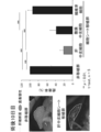
- the left side of FIG. 2 shows photographs on the 10th day after the operation when the hepatic mesothelial cell sheet was autotransplanted into the PPHx mouse model and when nothing was transplanted.
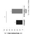
- the right side of FIG. 2 shows the result of calculating the adhesion rate 10 days after the operation after autotransplantation of the liver mesothelial cell sheet, peritoneal mesothelial cell sheet, and fibroblast cell sheet into the hepatic section of the PPHx mouse model. .
- FIG. 1 shows an outline of an adhesion preventing method using the adhesion preventing material according to the present invention.
- the left side of FIG. 2 shows photographs on the 10th day after the operation when the hepatic mesothelial cell sheet was autotransplanted into the PPHx mouse model and when nothing was transplanted.
- the right side of FIG. 2 shows the result
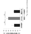
- FIG. 3 shows the results of evaluation of post-operative liver regeneration in the liver mesothelial cell transplant group, peritoneal mesothelial cell transplant group, and non-transplant group using the ratio of Ki67 positive hepatocytes as an index.
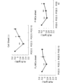
- FIG. 4 shows the results of evaluation of postoperative liver regeneration in the liver mesothelial cell transplant group, peritoneal mesothelial cell transplant group, and non-transplant group using serum albumin as an index.
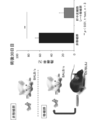
- FIG. 5 shows the results of measuring the adhesion rate after transplanting a cell sheet using hepatic mesothelial cells derived from C57BL / 6J mice into a BALB / c PPHx mouse model.
- FIG. 6 shows the results of evaluation of postoperative liver regeneration using a cell sheet using hepatic mesothelial cells derived from C57BL / 6J mice as transplanted to a BALB / c PPHx mouse model and the ratio of Ki67 positive hepatocytes as an index. Show.
- FIG. 7 shows a state in which a cell sheet using hepatic mesothelial cells was transplanted into a peritoneal adhesion mouse model, opened after 10 days, and observed.
- FIG. 8 shows a state in which a peritoneal adhesion mouse model was closed without transplanting a cell sheet and opened after 10 days for monitoring.
- the anti-adhesion material according to the present invention comprises a cell sheet containing mesothelial cells.
- the term “adhesion prevention material” refers to the possibility of adhesion occurring for the purpose of preventing “adhesion”, which is a phenomenon in which organs and tissues that should originally be separated from each other after surgery. A cell sheet to be transplanted into an organ or tissue. As used herein, “preventing” not only completely prevents adhesions but also significantly suppresses adhesions.
- the “cell sheet” means a sheet mainly composed of cells. Although it is not particularly limited as long as it can be applied to an organ or tissue, it may be a single-layer cell sheet, a sheet formed of two or more layers of cells, or a sheet formed of three-dimensionally cultured cells. . Typically a monolayer cell sheet. The cell sheet may contain an extracellular matrix.
- mesothelial cell refers to a single-layer cell that covers the outermost surface of the mesothelium.
- the mesothelium refers to a patterned tissue covering the surface of a body cavity such as the thoracic cavity, pericardium, abdominal cavity.
- Mesothelial cells may be isolated from tissues or organs such as the peritoneum, mesentery, testicular sheath membrane, pleura, pericardium, liver, heart, lung, large intestine, etc.
- Stem cells ( For example, it may be obtained by differentiating iPS cells or ES cells) or obtained by direct reprogramming from fibroblasts.
- mesothelial cells When isolated from tissues or organs, mesothelial cells may be derived from any fetus to adult, for example, from an individual between the fetus and the infant, that is, from an individual less than 6 years old from the fetus. Obtainable. Fetal to infant cells are rich in growth factors such as stem cell growth factor (HGF) and epidermal growth factor (EGF), which is useful for preventing adhesions and promoting the regeneration of organs and tissues.
- HGF stem cell growth factor
- EGF epidermal growth factor
- promotion of regeneration of organs and tissues refers to a configuration or function originally possessed by organs or tissues so that all or part of those damaged by surgery or adhesion may be brought back to the original state. It means to recover.
- the mesothelial cells may be derived from the same individual as the transplantation target of the adhesion preventing material, or may be derived from an individual different from the transplantation target.
- the “adhesion prevention material” may include a support necessary for maintaining the shape of the cell culture or cell sheet.
- the "adhesion prevention material” may consist only of a cell sheet substantially. In the case where the cell sheet is substantially composed only of a cell sheet, a state closer to nature in a living body can be reproduced, so that there is an advantage that it is easy to obtain a function for promoting regeneration of an organ or tissue.
- the “cell sheet containing mesothelial cells” may contain other cells as long as they contain mesothelial cells.
- the percentage of mesothelial cells in the total number of cells contained in the cell sheet is, for example, 50% or more, 60% or more, 70% or more, 80% or more, 90% or more, 95% or more, 98% or more, 99% or more can do.
- Mesothelial cells may include mesothelial progenitor cells that differentiate into mesothelial cells.
- the mesothelial cell is a liver mesothelial cell.
- Liver mesothelial cells are mesothelial cells that cover the liver surface.
- An adhesion-preventing material comprising a cell sheet containing hepatic mesothelial cells can be used to prevent adhesion and / or regeneration of all organs and tissues. For example, it is useful for preventing adhesion of a detached section after partial hepatectomy. is there. Adipose tissue in the abdominal cavity adheres to the separated surface after partial hepatectomy, and adhesion is likely to occur.
- the anti-adhesion material consisting of a cell sheet containing hepatic mesothelial cells, it is possible to significantly prevent adhesion at the section after partial hepatectomy and promote liver regeneration to prevent a decrease in liver reserve capacity.
- the “separation plane” refers to a surface that appears when a part of an organ or tissue is excised or removed.
- liver reserve ability means the function of the liver itself. Liver reserve ability can be evaluated by a known method. For example, liver damage classification and Child-Pugh classification are often used. The degree of liver damage is a method of measuring and evaluating ascites, serum bilirubin level, serum albumin level, ICG R15, and prothrombin activity level. It is a method of measuring and evaluating.
- liver regeneration is promoted means that at least one of ascites, serum bilirubin level, serum albumin level, ICG R15, prothrombin activity value, and encephalopathy is improved; -Improved assessment by Pugh classification; increased liver; increased liver weight; promoted hepatocyte proliferation; and at least one state of increased hepatocyte size achieved That means.
- the adhesion-preventing material according to the present invention does not require suturing, particularly when an extracellular matrix is held, and can be engrafted simply by being disposed so as to be attached to the surface of an organ or tissue or a cross section.
- the adhesion preventing material according to the present invention can be used for preventing adhesion of any organ or tissue in which adhesion can occur.
- an adhesion-preventing material prepared from hepatic mesothelial cells can be used to prevent adhesions of the intestine and peritoneum
- an adhesion-preventing material prepared from peritoneal mesothelial cells can be used to prevent adhesions from the liver cross section.
- the adhesion preventing material according to the present invention can be produced by a known method for producing cultured cell sheets or a method analogous thereto.
- mesothelial cells are cultured in a culture dish coated with a temperature-responsive polymer, and when the mesothelial cells become a sheet, they can be recovered from the culture dish by changing the temperature to obtain a cell sheet.
- the temperature-responsive polymer include those whose hydrophilicity changes to hydrophobic when the temperature changes.
- a known one or a similar one can be used.
- Commercially available culture dishes that are pre-coated with a temperature-responsive polymer may be used. By using a culture dish coated with a temperature-responsive polymer, it is possible to obtain an adhesion-preventing material that does not contain a support and consists essentially of a mesothelial cell sheet.
- the adhesion prevention method according to the present invention includes a step of placing the above-described cell sheet containing mesothelial cells on the surface or separation surface of a tissue or organ in which adhesion may occur during surgery.
- the mesothelial cell sheet can be engrafted in a tissue or an organ and prevented from adhering simply by placing it without requiring suturing or the like, but it may be sewn.
- the method for promoting liver regeneration according to the present invention includes a step of placing a cell sheet containing hepatic mesothelial cells on a separated section after partial hepatectomy. According to this method, it is possible to prevent postoperative liver failure and increase the number of repetitions as a result of promoting the recovery of the liver's reserve ability as well as preventing adhesions that become problematic during repeated hepatectomy It becomes.
- the method for producing a partial hepatectomy adhesion rodent model includes a step of ligating and separating the middle lobe and a step of separating the middle of the left lobe.
- the rodent liver differs from the human liver in that the left lobe, middle lobe, right lobe, and caudate lobe are divided into tufts, so in the conventional mouse hepatectomy model, each lobe is completely removed. As a result, no hepatic section was produced and no adhesion model was obtained.
- a hepatic section can be formed in the left lobe, so that a rodent model in which adhesion has occurred can be obtained.
- the method for producing a partial hepatectomy adhesion rodent model according to the present invention may further include a step of placing epididymal adipose tissue near the cross section of the left lobe. By disposing the adipose tissue in the vicinity, adhesion of the adipose tissue to the separated surface is promoted, and adhesion is likely to occur.
- the partial hepatectomy rodent model according to the present invention has a cross section in the left lobe of the liver.
- FIG. 1 shows an outline of a method for preventing adhesion in Examples.
- GFP-positive hepatic mesothelial cells were separated from a recombinant mouse expressing green fluorescent protein (GFP) using a cell sorter and cultured on a temperature-responsive culture dish.
- the cell sheet obtained by changing the temperature was collected from the culture dish and transplanted to the hepatic section.
- the application to the hepatic section was confirmed by detecting GFP. Detailed materials and methods are shown below.
- Wild animal male C57BL / 6J mice (10 weeks old) were purchased from Clea Japan.
- Recombinant C57BL / 6J mice expressing green fluorescent protein (GFP) were provided by Dr. Okabe of Osaka University.
- the age of the mouse embryo was defined as the number of days from the date of appearance of the vaginal plug (noon of this day was E0.5).
- the epididymal adipose tissue was lifted to the upper abdominal cavity and placed on the left cross section of the left lobe. When the abdomen was closed, the epididymal adipose tissue was sewn to the abdominal wall to prevent it from shifting. Mice were sacrificed 10 or 30 days after surgery. The value obtained by dividing the length of adhesion by the length of the hepatic section was taken as the adhesion rate and measured for each mouse.
- Samples were sorted with a cell sorter Moflo XDP (Beckman Coulter). After sorting, in culture dishes coated with type IV collagen, MEM ⁇ medium (10% fetal bovine serum (FBS), 50 nmol / L mercaptoethanol, penicillin, streptomycin, and glutamine, 10 ng / mL basic fibroblast growth In vitro culture was performed using a factor (bFGF), 10 ng / mL Oncostatin M (OSM), and insulin-transferrin-selenium-X) at 37 ° C. in a 5% CO 2 incubator.
- FBS fetal bovine serum
- OSM Oncostatin M
- insulin-transferrin-selenium-X insulin-transferrin-selenium-X
- peritoneal mesothelial cells were resuspended in RPMI 1640 (20% FBS, penicillin, streptomycin, glutamine and hydrocortisone (0.4 ⁇ g / mL) and seeded in culture dishes coated with type I collagen.
- Plasma concentrations of AST and ALT test aspartate aminotransferase (AST) and alanine aminotransferase (ALT) were measured with a commercially available kit (Transaminase C2-Test Wako, Wako Pure Chemical Industries, Ltd.).
- Serum albumin test The serum albumin concentration was outsourced to Oriental Yeast.
- PFA paraformaldehyde
- HE hematoxylin and eosin
- Quantitative RT-PCR Total RNA was extracted from each cell sheet using Trizol reagent (Invitrogen), and cDNA was synthesized using High Capacity cDNA Reverse Transcription Kit (Takara Bio). Quantitative RT-PCR was performed using LightCycler 480 (Roche) and the following primers.
- Mouse PCLP1 TCCTTGTTGCTGCCCTCT (SEQ ID NO: 1) CTCTGTGAGCCGTTGCTG (SEQ ID NO: 2)
- Mouse HGF CACCCCTTGGGAGTATTGTG (SEQ ID NO: 3) GGGACATCAGTCTCATTCACAG (SEQ ID NO: 4)
- Mouse IL6 GCTACCAAACTGGATATAATCAGGA (SEQ ID NO: 5) CCAGGTAGCTATGGTACTCCAGAA (SEQ ID NO: 6)
- Mouse Mdk CGCACTGGTAAAACCGAACT (SEQ ID NO: 7) GAAGAAGCCTCGGTGCTG (SEQ ID NO: 8)
- Mouse Ptn AGGACCTCTGCAAGCCAAA (SEQ ID NO: 9) CACAGCTGCCAAGATGAAAAT (SEQ ID NO: 10)
- Mouse ⁇ -actin CTAAGGCCAACCGTGAAAAG (SEQ ID NO: 11) ACCAGAGGCATACAGGGACA (SEQ ID NO: 12) Universal Probe
- the cell size and the ratio of dividing cells were measured according to the method of Miyaoka et al. Using IN Cell Analyzer 2000 (GE Healthcare) (Miyaoka Y. et al., Curr Biol. 22, 1166-1175 (2012)). . Briefly, cell size was measured using phalloidin-stained actin surrounding the nucleus stained with Hoechst 33342 as the periphery of hepatocytes. The percentage of dividing cells was counted by detecting Ki67 positive cells in addition to Hoechst 33342 and phalloidin staining. Four photos were taken for each mouse. Since each photograph contained 500-700 hepatocytes, a total of 2000-2800 cells were examined for a single mouse.
- Peritoneal mesothelial cell defect site was prepared by peritoneal mesothelial cell detachment of C57BL / 6J mice, and peritoneal adhesion model was established. Subsequently, the above 7.
- the hepatic mesothelial cell sheet prepared using the GFP recombinant C57BL / 6J mouse-derived hepatic mesothelial cells by the above method was transplanted to the peritoneal mesothelial defect site.
- the control group was closed without cell sheet transplantation. On the 10th day after the operation, the abdomen was opened, the state of adhesion was observed, and the GFP signal at the peritoneal defect site was observed.
- PPHx mouse model A mouse model that produced adhesions was prepared by preparing a hepatic section using the method described above. The transition of liver injury after hepatectomy was similar to the postoperative course of humans, confirming that the animal model mimics human hepatectomy (data not shown).
- FIG. 2 shows the results of calculating the adhesion rate on the 10th day after the operation after autotransplanting the liver mesothelial cell sheet, peritoneal mesothelial cell sheet, and fibroblast cell sheet to the liver section of the PPHx mouse model. .
- adhesion was greatly suppressed, which was about 20% and about 40%, respectively.
- the adhesion rate was almost 100% as in the non-transplanted group.
- FIG. 3 shows the results of evaluation of postoperative liver regeneration in the liver mesothelial cell transplant group, peritoneal mesothelial cell transplant group, and non-transplant group using the ratio of Ki67 positive hepatocytes as an index. Show. In the liver mesothelial cell sheet transplant group, the proportion of Ki67 positive hepatocytes was nearly double that in the non-transplant group, confirming that cell division was promoted.
- FIG. 4 shows the results of evaluation of postoperative liver regeneration in the liver mesothelial cell transplant group, peritoneal mesothelial cell transplant group, and non-transplant group using serum albumin as an index. In the liver mesothelial cell sheet transplanted group, it was confirmed that recovery of serum albumin after surgery was promoted. Early recovery of liver function is known to contribute significantly to avoiding postoperative liver failure and is highly desirable.
- FIG. 5 shows the cell sheet using C57BL / 6J mouse-derived hepatic mesothelial cells and transplanted to the BALB / c PPHx mouse model. The result of having measured is shown. As in the case of autotransplantation, the adhesion rate was significantly reduced in the liver mesothelial cell sheet transplant group.
- FIG. 6 shows the results of evaluation of postoperative liver regeneration using the ratio of Ki67 positive hepatocytes as an index. Similar to the case of autotransplantation, in the liver mesothelial cell sheet transplant group, the proportion of Ki67 positive hepatocytes was nearly twice that in the non-transplant group, confirming that cell division was promoted.
- FIG. 7 shows a state in which a hepatic mesothelial cell sheet was transplanted to the peritoneal adhesion model, opened and observed 10 days later.
- FIG. 8 shows a state observed after laparotomy 10 days later without transplanting the liver mesothelial cell sheet.
- the intestines in the abdominal cavity were firmly adhered to the abdominal wall defect site, whereas in the liver mesothelial cell transplanted group, the adhesion was remarkably suppressed.
- SEQ ID NOs: 1 and 2 are primers for mouse PCLP1.
- SEQ ID NOs: 3 and 4 are mouse HGF primers.
- SEQ ID NOs: 5 and 6 are primers for mouse IL6.
- SEQ ID NOs: 7 and 8 are mouse Mdk primers.
- SEQ ID NOs: 9 and 10 are primers for mouse Ptn.
- SEQ ID NOs: 11 and 12 are primers for mouse ⁇ -actin.
Landscapes
- Health & Medical Sciences (AREA)
- Life Sciences & Earth Sciences (AREA)
- Chemical & Material Sciences (AREA)
- Animal Behavior & Ethology (AREA)
- Public Health (AREA)
- General Health & Medical Sciences (AREA)
- Veterinary Medicine (AREA)
- Epidemiology (AREA)
- Engineering & Computer Science (AREA)
- Medicinal Chemistry (AREA)
- Chemical Kinetics & Catalysis (AREA)
- Biomedical Technology (AREA)
- Pharmacology & Pharmacy (AREA)
- Vascular Medicine (AREA)
- Surgery (AREA)
- Heart & Thoracic Surgery (AREA)
- Dermatology (AREA)
- Oral & Maxillofacial Surgery (AREA)
- Transplantation (AREA)
- Zoology (AREA)
- Organic Chemistry (AREA)
- Nuclear Medicine, Radiotherapy & Molecular Imaging (AREA)
- General Chemical & Material Sciences (AREA)
- Bioinformatics & Cheminformatics (AREA)
- Botany (AREA)
- Immunology (AREA)
- Virology (AREA)
- Developmental Biology & Embryology (AREA)
- Nutrition Science (AREA)
- Physiology (AREA)
- Biotechnology (AREA)
- Cell Biology (AREA)
- Molecular Biology (AREA)
- Urology & Nephrology (AREA)
- Gastroenterology & Hepatology (AREA)
- Materials For Medical Uses (AREA)
- Micro-Organisms Or Cultivation Processes Thereof (AREA)
- Environmental Sciences (AREA)
- Medicines Containing Material From Animals Or Micro-Organisms (AREA)
- Animal Husbandry (AREA)
Abstract
Description
癒着は、年齢・性別・人種を問わず腹部手術後には必ず発生する現象であり、肝臓に限らず様々な場所で生じ、腸閉塞、慢性骨盤痛、腹痛、不妊などの原因にもなり得る。
投薬によって術後癒着を軽減する方法も試みられているが、現時点では効果はほとんど認められていない。
現在、術後癒着に対する最も一般的な対処法として、高分子材料を用いた吸収性癒着防止材の貼付がある。吸収性癒着防止材の貼付によって癒着の程度が軽減されることが報告されている(非特許文献1)。しかしながら、近年のメタアナリシスの結果により、腸閉塞の頻度そのものは減少しないことが判明し、腹腔内膿瘍及び縫合不全の頻度が増加することが明らかとなった(非特許文献2、3)。さらに、吸収性癒着防止剤は、創傷治癒を阻害する可能性や、肝臓に使用した場合、術後胆汁漏のリスクが高くなる可能性も指摘されている。
さらに、肝中皮細胞を用いて作製した癒着防止材を肝臓の部分切除後の離断面に貼付すると、癒着が防止されることに加えて、肝再生能も促進することを見出した。
また、中皮細胞シートは、移植対象以外の個体に由来する細胞を用いて作製しても、機能することを確認し、本発明を完成するに至った。
すなわち、本発明は、
〔1〕中皮細胞を含む細胞シートからなる癒着防止材;
〔2〕前記中皮細胞が、癒着防止の対象と同一個体に由来する細胞である、上記〔1〕に記載の癒着防止材;
〔3〕前記中皮細胞が、癒着防止の対象と異なる個体に由来する細胞である、上記〔1〕に記載の癒着防止材;
〔4〕前記中皮細胞が、胎児から幼児の個体に由来する細胞である、上記〔1〕から〔3〕のいずれか1つに記載の癒着防止材;
〔5〕前記中皮細胞が、肝中皮細胞である、上記〔1〕から〔4〕のいずれか1つに記載の癒着防止材;
〔6〕肝部分切除後の離断面の癒着防止、又は腹膜癒着の防止に用いられる、上記〔5〕に記載の癒着防止材;
〔7〕前記中皮細胞が、腹膜中皮細胞である、上記〔1〕から〔4〕のいずれか1つに記載の癒着防止材;
〔8〕腹膜の癒着防止に用いられる、上記〔7〕に記載の癒着防止材;
〔9〕支持体を含まず、細胞シートのみからなる、上記〔1〕から〔8〕のいずれか1項に記載の癒着防止材;
〔10〕中皮細胞を含む細胞シートを、組織又は臓器の表面若しくは離断面に配置する工程を含む、癒着防止方法;
〔11〕肝中皮細胞を含む細胞シートを、肝部分切除後の離断面に配置する肝再生促進方法;
〔12〕肝部分切除癒着げっ歯類モデルの製造方法であって、
中葉を結紮切離する工程と、
左葉の中程を切離する工程と、
を含む方法;及び
〔13〕さらに、精巣上体脂肪組織を、左葉の離断面付近に配置する工程を含む、上記〔12〕に記載の方法
に関する。
また、従来癒着防止の手段がなかった肝部分切除後の離断面の癒着も防止することが可能である。肝中皮細胞を用いた細胞シートによれば、癒着の防止に加えて肝部分切除後の肝再生も促進することができるので、術後肝不全を抑制し、繰り返し肝移植の回数を増やせる可能性がある。
また、本発明の癒着防止材は、移植対象以外の個体に由来する細胞を用いて作製しても機能するので、臨床応用にも有用である。
本明細書において「癒着防止材」とは、手術後などに、本来は離れているべき臓器や組織同士がくっついてしまう現象である「癒着」を防ぐことを目的として、癒着が生じる可能性のある臓器や組織等に移植する細胞シートをいう。
本明細書において「防止」は、完全に癒着を防ぐことだけでなく、癒着を有意に抑制することも含む。
また、中皮細胞は、癒着防止材の移植対象と同一個体に由来するものであってもよく、移植対象と異なる個体に由来するものであってもよい。
現在、原発性肝がんや大腸癌肝転移に対しては、肝切除後の残肝再発に対して「繰り返し肝切除」を積極的に試みることが根治や延命を期待しうる唯一の治療法であるところ、癒着が生じると再手術や再々手術の際にこれを剥離する必要が生じ、大量出血や感染のリスクが高くなる。また、癒着により肝予備能も低下するため、繰り返し回数が制限されるとともに、術後肝不全に陥りやすい。
肝中皮細胞を含む細胞シートからなる癒着防止材によれば、肝部分切除後の離断面の癒着を有意に防止することができるとともに、肝再生を促して、肝予備能の低下を防止することもできる。
なお、本明細書において「離断面」とは、臓器や組織の一部を切除又は剥離することによって現れた面をいう。
肝障害度は、腹水、血清ビリルビン値、血清アルブミン値、ICG R15、およびプロトロンビン活性値を測定して評価する方法であり、Child-Pughは、脳症、腹水、血清ビリルビン、血清アルブミン、プロトロンビン活性値を測定して評価する方法である。
また、本発明に係る癒着防止材は、癒着が生じ得るあらゆる臓器や組織の癒着防止に用いることができる。例えば、肝中皮細胞で作製した癒着防止材を、腸や腹膜の癒着防止に用いることもできるし、腹膜中皮細胞で作製した癒着防止材を肝離断面の癒着防止に用いることもできる。
一例として、中皮細胞を、温度応答性高分子でコーティングした培養皿で培養し、中皮細胞がシート状になったら、温度を変えることによって培養皿から回収して細胞シートを得ることができる。温度応答性高分子は、例えば、温度が変わると親水性が疎水性に変化するものが挙げられる。温度応答性高分子は、公知のもの又はそれに準ずるものを用いることができる。温度応答性高分子で予めコーティングされた市販の培養皿を用いてもよい。温度応答性高分子でコーティングした培養皿を用いることにより、支持体を含まず、実質的に中皮細胞シートのみからなる癒着防止材を得ることができる。
げっ歯類の肝臓は、ヒトの肝臓と異なり、左葉、中葉、右葉、尾状葉の各葉が房状に分かれているため、従来のマウス肝切除モデルでは各葉がまるごと切除されており、肝離断面が生じず、癒着モデルが得られなかった。
本発明に係る方法によれば、左葉に肝離断面を形成することができるので、そこに癒着を生じたげっ歯類モデルを得ることができる。
1.方法の概略
図1に、実施例における癒着防止方法の概略を示す。まず、緑色蛍光タンパク質(GFP)を発現する組換えマウスから、GFP陽性肝中皮細胞をセルソーターにより分離し、温度応答性培養皿上で培養した。温度を変化させることにより得られた細胞シートを培養皿から回収して、肝離断面に移植した。肝離断面への貼付は、GFPを検出して確認した。以下に、詳細な材料と方法を示す。
野生型雄のC57BL/6Jマウス(10週齢)を日本クレア社より購入した。緑色蛍光タンパク質(GFP)を発現する組換えC57BL/6Jマウスは、大阪大学のDr. Okabeより供与を受けた。マウス胚の日齢は、膣栓の出現日(この日の正午をE0.5とした。)からの日数とした。
全身麻酔下で、野生型C57BL/6Jマウスをspin positionに配置した。Xyphoid inferiorlyから2センチ上を正中線に沿って切開し、腹腔内の臓器を傷付けないように、腹膜を持ち上げて切開した。開腹後、鎌状靭帯を上大静脈まで分離した。まず、中葉の根部を4-0シルクで結紮して切離し、中葉を切除した。次に、電気メスを用いて左葉の周囲の肝臓被膜を焼灼し、左葉を中程で切離した。途中、左門脈と左肝静脈をまとめて結紮し、分離した。
止血を確認した後、精巣上体脂肪組織を腹腔上部まで持ち上げて左葉の離断面に配置した。閉腹する際、精巣上体脂肪組織を腹壁に一針縫いつけてずれないようにした。
マウスは手術から10日後又は30日後に犠牲死させた。癒着した長さを肝離断面の長さで除した値を癒着率とし、各マウスについて測定した。
胎児肝中皮細胞は、E12.5のGFP組換えマウスから、Onitsukaらの方法(Onitsuka, I. et al., Gastroenterology. 138, 1525-1535(2010))に従って単離した。簡単に説明すると、胎児肝組織を刻み、肝消化培地(Life technologies社)で分離し、低張緩衝液で溶血させた。細胞を、抗FcR抗体でブロッキングし、抗マウスPodocalyxin-ビオチン(MBL社)共染した後、APC結合ストレプトアビジン(Invitrogen社)と共にインキュベートした。サンプルをセルソーターMoflo XDP(Beckman Coulter社)でソーティングした。ソーティングの後、IV型コラーゲンでコーティングした培養皿で、MEMα培地(10%ウシ胎児血清(FBS)、50nmol/Lのメルカプトエタノール、ペニシリン、ストレプトマイシン、及びグルタミン、10ng/mLの塩基性線維芽細胞成長因子(bFGF)、10ng/mLのオンコスタチンM(OSM)、及びインスリン-トランスフェリン-セレニウム-X)を用いて、37℃、5%CO2インキュベーターで、in vitro培養した。
成人線維芽細胞は、GFP組換えマウスの皮膚組織から採取した。皮膚組織を刻み、0.05%トリプシンと0.5mMのEDTAにより、37℃で20分処理した。細胞懸濁液を遠心処理し、培地に再懸濁した。
成人腹膜中皮細胞は、GFP組換えマウスの精巣上体脂肪から、Loureiroらの方法(Loureiro, J. et al., Nephrol Dial Transplant. 25, 1098-1108(2010))に従って採取した。精巣上体脂肪を、0.05%トリプシンと0.5mMのEDTAにより、37℃で30分処理した。細胞懸濁液を1500rpmで5分間遠心処理し、ペレットを溶血バッファー中、氷上で5分処理した。PBSで数回洗浄後、腹膜中皮細胞は、RPMI1640(20%FBS、ペニシリン、ストレプトマイシン、グルタミン及びハイドロコルチゾン(0.4μg/mL)に再懸濁し、I型コラーゲンでコーティングした培養皿に播種した。
それぞれの初代培養細胞を、UpCell(登録商標)プレート(セルシード社)に移して培養した。プレートを室温に置くことにより、細胞を培養皿から回収し、細胞シートを得た。
PPHxを施した直後のマウスの肝離断面に細胞シートを移植し、閉腹した。細胞シートは、肝離断面に置くだけで生着した。130S-GFP(Science eye社)のGFPシグナルにより、手術をモニターした。
アスパラギン酸アミノトランスフェラーゼ(AST)と、アラニンアミノトランスフェラーゼ(ALT)の血漿中濃度を、市販のキット(Transaminase C2-Test Wako、和光純薬社)で測定した。
血清アルブミン濃度は、オリエンタル酵母社に委託した。
左葉の凍結切片(8μm)を、Microm HM505Eクライオスタット(Microm International GmbH)で調製し、APSでコーティングしたスライドガラス(松浪硝子社)に配置した。切片を、4%パラホルムアルデヒド(PFA)を含むPBSで固定し、ヘマトキシリン・エオジン(HE)染色を行った。
各細胞シートから、Trizol試薬(Invitrogen社)を用いて全RNAを抽出し、High Capacity cDNA Reverse Transcription Kit(タカラバイオ社)でcDNAを合成した。LightCycler 480(Roche社)と以下のプライマーを用いて定量RT-PCRを行った。
マウスPCLP1:
TCCTTGTTGCTGCCCTCT(配列番号:1)
CTCTGTGAGCCGTTGCTG(配列番号:2)
マウスHGF:
CACCCCTTGGGAGTATTGTG(配列番号:3)
GGGACATCAGTCTCATTCACAG(配列番号:4)
マウスIL6:
GCTACCAAACTGGATATAATCAGGA(配列番号:5)
CCAGGTAGCTATGGTACTCCAGAA(配列番号:6)
マウスMdk:
CGCACTGGTAAAACCGAACT(配列番号:7)
GAAGAAGCCTCGGTGCTG(配列番号:8)
マウスPtn:
AGGACCTCTGCAAGCCAAA(配列番号:9)
CACAGCTGCCAAGATGAAAAT(配列番号:10)
マウスβアクチン:
CTAAGGCCAACCGTGAAAAG(配列番号:11)
ACCAGAGGCATACAGGGACA(配列番号:12)
Universal ProbeLibrary Mouse ACTB Gene Assay(Roche社)
4%PFAを含むPBSで固定した後、肝離断面を含む左葉の切片(8mm)をブロッキング試薬により、室温で1時間ブロッキングし、抗体溶液中に4℃で一晩静置した。一次抗体として、1:300希釈のウサギ抗マウスKi67抗体(Leica biosystems社)を用い、二次抗体として、Alexa Fluor 555ヤギ抗ウサギIgG(H+L)(Invitrogen社)を用いた。核は、Hoechst 33342(Sigma-Aldrich社)で対比染色した。細胞の外周は、Alexa Fluor 488ファロイジン(Invitrogen社)で染色した。
細胞サイズと分裂細胞の割合の測定は、IN Cell Analyzer 2000(GE Healthcare社)を用い、Miyaokaらの方法に従って行った(Miyaoka Y. et al., Curr Biol. 22, 1166-1175(2012))。簡単に説明すると、細胞サイズは、Hoechst 33342で染色した核の周りを囲むファロイジン染色されたアクチンを肝細胞の外周として測定した。分裂細胞の割合は、Hoechst 33342とファロイジンの染色に加え、Ki67陽性細胞を検出して数えた。
マウスごとに4枚の写真を撮った。各写真は500~700の肝細胞を含んでいたので、合計で1匹のマウスについて、2000~2800の細胞を調べたことになる。
上記3.の方法と同様に、BALB/cマウスを用いてPPHxマウスモデルを作製した。
一方、GFP組換えC57BL/6Jマウスから、上記4.と同様に胎児肝中皮細胞を得て、上記7.の方法で細胞シートを作製し、PPHxマウスモデルに移植した。
C57BL/6Jマウスの腹膜中皮細胞剥離により腹膜中皮細胞欠損部位を作製し、腹膜癒着モデルを確立した。続いて、上記7.の方法でGFP組換えC57BL/6Jマウス由来肝中皮細胞を用いて作製した肝中皮細胞シートを、腹膜中皮欠損部位に移植した。対照群は、細胞シートの移植をせずに閉腹した。
術後10日目に開腹し、癒着の様子を観察し、腹膜欠損部位のGFPシグナルを観察した。
結果は、平均±標準偏差で示した。統計にはStudent-t検定で行った。p値<0.05の場合に有意差ありと判断した。
1.PPHxマウスモデル
上述の方法で肝離断面を作製することで癒着を生じたマウスモデルを作製することができた。肝切除手術後の肝障害の推移がヒトの術後経過と類似しており、よりヒトの肝切除を模倣した動物モデルであることが確認された(データ示さず)。
(1)癒着率の測定
図2左に、肝中皮細胞シートを、PPHxマウスモデルに自家移植した場合と、何も移植しなかった場合の術後10日目の写真を示す。写真に示されるように、非移植マウスでは肝離断面の全体に癒着が観察されたが、肝中皮細胞シート移植マウスでは、肝離断面にはほとんど癒着が見られなかった。術後30日でも同様の結果が得られた。
図2右に、肝中皮細胞シート、腹膜中皮細胞シート、及び線維芽細胞シートを、PPHxマウスモデルの肝離断面に自家移植し、術後10日目に癒着率を計算した結果を示す。
肝中皮細胞シート移植群及び腹膜中皮細胞シート移植群では、癒着は大きく抑制され、それぞれ約20%と約40%であった。一方、線維芽細胞シート移植群では、非移植群で同様に、癒着率はほぼ100%であった。
図3に、Ki67陽性肝細胞の割合を指標として、肝中皮細胞移植群、腹膜中皮細胞移植群、及び非移植群における術後の肝再生を評価した結果を示す。肝中皮細胞シート移植群では、Ki67陽性肝細胞の割合が非移植群の2倍近く、細胞分裂が促進されていることが確認された。
図4に、血清アルブミンを指標として、肝中皮細胞移植群、腹膜中皮細胞移植群、及び非移植群における術後の肝再生を評価した結果を示す。肝中皮細胞シート移植群では、血清アルブミンの術後の回復が促進されていることが確認された。肝機能の早期回復は、術後肝不全の回避に大きく貢献することが知られており、非常に望ましい。
(1)癒着率の測定
図5に、C57BL/6Jマウス由来の肝中皮細胞を用いた細胞シートをBALB/c PPHxマウスモデルに移植し、癒着率を測定した結果を示す。自家移植したときと同様に、肝中皮細胞シート移植群では癒着率が大幅に低下した。
(2)肝再生の評価
図6に、Ki67陽性肝細胞の割合を指標として、術後の肝再生を評価した結果を示す。自家移植したときと同様に、肝中皮細胞シート移植群では、Ki67陽性肝細胞の割合が非移植群の2倍近く、細胞分裂が促進されていることが確認された。
図7に、腹膜癒着モデルに肝中皮細胞シートを移植し、10日後に開腹して観察した様子を示す。図8に、肝中皮細胞シートを移植せずに、10日後に開腹して観察した様子を示す。非移植群では、腹壁欠損部位に腹腔内の腸などが強固に癒着していたのに対し、肝中皮細胞移植群では、癒着が顕著に抑制された。また、腹壁欠損部位には肝中皮細胞シート由来のGFPシグナルが観察されたことから、肝中皮細胞が生着し、腹腔内の癒着を防止したと考えられる。
配列番号:3及び4は、マウスHGF用プライマーである。
配列番号:5及び6は、マウスIL6用プライマーである。
配列番号:7及び8は、マウスMdk用プライマーである。
配列番号:9及び10は、マウスPtn用プライマーである。
配列番号:11及び12は、マウスβアクチン用プライマーである。
Claims (13)
- 中皮細胞を含む細胞シートからなる癒着防止材。
- 前記中皮細胞が、癒着防止の対象と同一個体に由来する細胞である、請求項1に記載の癒着防止材。
- 前記中皮細胞が、癒着防止の対象と異なる個体に由来する細胞である、請求項1に記載の癒着防止材。
- 前記中皮細胞が、胎児から幼児の個体に由来する細胞である、請求項1から3のいずれか1項に記載の癒着防止材。
- 前記中皮細胞が、肝中皮細胞である、請求項1から4のいずれか1項に記載の癒着防止材。
- 肝部分切除後の離断面の癒着防止、又は腹膜癒着の防止に用いられる、請求項5に記載の癒着防止材。
- 前記中皮細胞が、腹膜中皮細胞である、請求項1から4のいずれか1項に記載の癒着防止材。
- 腹膜の癒着防止に用いられる、請求項7に記載の癒着防止材。
- 支持体を含まず、細胞シートのみからなる、請求項1から8のいずれか1項に記載の癒着防止材。
- 中皮細胞を含む細胞シートを、組織又は臓器の表面若しくは離断面に配置する工程を含む、癒着防止方法。
- 肝中皮細胞を含む細胞シートを、肝部分切除後の離断面に配置する肝再生促進方法。
- 肝部分切除癒着げっ歯類モデルの製造方法であって、
中葉を結紮切離する工程と、
左葉の中程を切離する工程と、
を含む方法。 - さらに、精巣上体脂肪組織を、左葉の離断面付近に配置する工程を含む、請求項12に記載の方法。
Priority Applications (5)
| Application Number | Priority Date | Filing Date | Title |
|---|---|---|---|
| KR1020157022387A KR20150109425A (ko) | 2013-01-22 | 2014-01-22 | 유착 방지재 |
| EP14743521.8A EP2949348B1 (en) | 2013-01-22 | 2014-01-22 | Adhesion preventing material |
| US14/762,189 US20160058917A1 (en) | 2013-01-22 | 2014-01-22 | Adhesion Preventing Material |
| JP2014558597A JP6168666B2 (ja) | 2013-01-22 | 2014-01-22 | 癒着防止材 |
| US15/710,507 US20180008749A1 (en) | 2013-01-22 | 2017-09-20 | Adhesion Preventing Material |
Applications Claiming Priority (2)
| Application Number | Priority Date | Filing Date | Title |
|---|---|---|---|
| US201361755163P | 2013-01-22 | 2013-01-22 | |
| US61/755,163 | 2013-01-22 |
Related Child Applications (2)
| Application Number | Title | Priority Date | Filing Date |
|---|---|---|---|
| US14/762,189 A-371-Of-International US20160058917A1 (en) | 2013-01-22 | 2014-01-22 | Adhesion Preventing Material |
| US15/710,507 Division US20180008749A1 (en) | 2013-01-22 | 2017-09-20 | Adhesion Preventing Material |
Publications (1)
| Publication Number | Publication Date |
|---|---|
| WO2014115776A1 true WO2014115776A1 (ja) | 2014-07-31 |
Family
ID=51227567
Family Applications (1)
| Application Number | Title | Priority Date | Filing Date |
|---|---|---|---|
| PCT/JP2014/051280 Ceased WO2014115776A1 (ja) | 2013-01-22 | 2014-01-22 | 癒着防止材 |
Country Status (5)
| Country | Link |
|---|---|
| US (2) | US20160058917A1 (ja) |
| EP (1) | EP2949348B1 (ja) |
| JP (1) | JP6168666B2 (ja) |
| KR (1) | KR20150109425A (ja) |
| WO (1) | WO2014115776A1 (ja) |
Cited By (5)
| Publication number | Priority date | Publication date | Assignee | Title |
|---|---|---|---|---|
| WO2015178498A1 (ja) * | 2014-05-23 | 2015-11-26 | 国立大学法人東京大学 | 中皮細胞の製造方法、及び癒着防止用細胞シート |
| WO2016031824A1 (ja) * | 2014-08-28 | 2016-03-03 | 国立大学法人弘前大学 | 人工腹膜組織及びその製造方法 |
| WO2016114355A1 (ja) * | 2015-01-15 | 2016-07-21 | 国立大学法人東京大学 | 癒着防止用組成物 |
| CN113637631A (zh) * | 2021-08-09 | 2021-11-12 | 南方医科大学南方医院 | 一种大鼠腹膜间皮细胞的提取与培养方法 |
| WO2022181776A1 (ja) * | 2021-02-25 | 2022-09-01 | 学校法人関西医科大学 | 多能性幹細胞から腹膜中皮細胞を分化誘導する方法 |
Families Citing this family (1)
| Publication number | Priority date | Publication date | Assignee | Title |
|---|---|---|---|---|
| CN108795867A (zh) * | 2018-06-05 | 2018-11-13 | 华东理工大学 | 用于构建结肠癌细胞腹膜转移体外三维模型的方法 |
Citations (2)
| Publication number | Priority date | Publication date | Assignee | Title |
|---|---|---|---|---|
| JP2005000608A (ja) * | 2003-06-11 | 2005-01-06 | Mitsuo Okano | 高生着性培養細胞シート、製造方法及びその利用方法 |
| WO2005047496A1 (ja) * | 2003-11-14 | 2005-05-26 | Keio University | 細胞培養法、細胞の三次元培養法、三次元組織、人工臓器、及び組織移植方法 |
Family Cites Families (2)
| Publication number | Priority date | Publication date | Assignee | Title |
|---|---|---|---|---|
| US6987024B1 (en) * | 2000-04-10 | 2006-01-17 | Raven Biotechnologies, Inc. | Human ovarian mesothelial cells and methods of isolation and uses thereof |
| US9045734B2 (en) * | 2012-07-31 | 2015-06-02 | The Board Of Trustees Of The Leland Stanford Junior University | Isolation and characterization of progenitor cells from mesothelium |
-
2014
- 2014-01-22 US US14/762,189 patent/US20160058917A1/en not_active Abandoned
- 2014-01-22 JP JP2014558597A patent/JP6168666B2/ja not_active Expired - Fee Related
- 2014-01-22 KR KR1020157022387A patent/KR20150109425A/ko not_active Ceased
- 2014-01-22 EP EP14743521.8A patent/EP2949348B1/en not_active Not-in-force
- 2014-01-22 WO PCT/JP2014/051280 patent/WO2014115776A1/ja not_active Ceased
-
2017
- 2017-09-20 US US15/710,507 patent/US20180008749A1/en not_active Abandoned
Patent Citations (2)
| Publication number | Priority date | Publication date | Assignee | Title |
|---|---|---|---|---|
| JP2005000608A (ja) * | 2003-06-11 | 2005-01-06 | Mitsuo Okano | 高生着性培養細胞シート、製造方法及びその利用方法 |
| WO2005047496A1 (ja) * | 2003-11-14 | 2005-05-26 | Keio University | 細胞培養法、細胞の三次元培養法、三次元組織、人工臓器、及び組織移植方法 |
Non-Patent Citations (16)
| Title |
|---|
| FAZIO, VW. ET AL., DIS COLON RECTUM, vol. 49, 2006, pages 1 - 11 |
| KUNIO KAWANISHI ET AL., FUKUMAKU SAIBO SHEET NO KAIHATSU TO JUTSUGO CHOKAN YUCHAKU MODEL NI OKERU YUCHAKU BOSHI KOKA NO KENTO, vol. 32, 2012, TOKYO, pages 72 - 73, XP008180253 * |
| KUNIO KAWANISHI ET AL.: "Fukumaku Saibo Sheet no Kaihatsu to Jutsugo Chokan Yuchaku Model ni Okeru Yuchaku Boshi Koka no Kento", SAISEI IRYO, vol. 11, 2012, pages 199, XP008180249 * |
| LOUREIRO, J. ET AL., NEPHROL DIAL TRANSPLANT, vol. 25, 2010, pages 1098 - 1108 |
| MIYAOKA Y. ET AL., CURR BIOL., vol. 22, 2012, pages 1166 - 1175 |
| NORIKO AJIRO: "Saibo Sheet Kaishuyo Ondo Otosei Saibo Baiyo Kizai 'UpCell", BIO CLINICA, vol. 23, no. 7, 2008, pages 649 - 650, XP008180248 * |
| ONITSUKA, 1. ET AL., GASTROENTEROLOGY, vol. 138, 2010, pages 1525 - 1535 |
| ONITSUKA, I. ET AL.: "Characterization and Functional Analyses of Hepatic Mesothelial Cells in Mouse Liver Development.", GASTROENTEROLOGY, vol. 138, no. 4, 2010, pages 1525 - 1535, XP026985010 * |
| RYOICHI SAKIYAMA ET AL.: "Chuhi Saibo Sheet ni yoru Fukumaku Shogai Kaizen eno Kokoromi", THERAPEUTICS & ENGINEERING, vol. 24, no. 1, 2012, pages 45 - 48, XP008180259 * |
| RYOICHI SAKIYAMA ET AL.: "Fukumaku Sen'isho Kaizen no Tameno Hito Chuhi Saibokabu Saibo Sheet no Kaihatsu", JOURNAL OF JAPANESE SOCIETY FOR DIALYSIS THERAPY, vol. 40, no. 1, 2007, pages 307, XP008180252 * |
| RYOICHI SAKIYAMA ET AL.: "Hito Chuhi Saibo Sheet ni yoru Shogai Fukumaku Kaizen no Koka", JOURNAL OF JAPANESE SOCIETY FOR DIALYSIS THERAPY, vol. 44, no. 1, 2011, pages 406, XP008180338 * |
| RYOICHI SAKIYAMA: "Manufacture of Mesothelial Cell Sheet Using Temperature Responsive Culture Dish", MEMBRANE, vol. 37, no. 3, 2012, pages 108 - 112, XP008180226 * |
| TAKASHI SUHARA ET AL.: "Kanzo Setsujo ni Okeru in situ Kakyo Hyaluronic Acid Hydrogel Yuchaku Boshizai no Kaihatsu", JAPANESE SOCIETY FOR BIOMATERIALS SYMPOSIUM 2012 YOKOSHU, 2012, pages 221, XP008180250 * |
| TANG, CL. ET AL., ANN SURG., vol. 243, 2006, pages 449 - 455 |
| TARO UYAMA: "Kokino Saibo Baiyo Kizai ' Cell ' Series", GEKKAN MEDICAL SCIENCE DIGEST, vol. 33, no. 9, 2007, pages 1029 - 1033, XP008180257 * |
| ZENG, Q. ET AL., WORLD J SURG., vol. 31, 2007, pages 2125 - 2131 |
Cited By (9)
| Publication number | Priority date | Publication date | Assignee | Title |
|---|---|---|---|---|
| WO2015178498A1 (ja) * | 2014-05-23 | 2015-11-26 | 国立大学法人東京大学 | 中皮細胞の製造方法、及び癒着防止用細胞シート |
| WO2016031824A1 (ja) * | 2014-08-28 | 2016-03-03 | 国立大学法人弘前大学 | 人工腹膜組織及びその製造方法 |
| JPWO2016031824A1 (ja) * | 2014-08-28 | 2017-06-08 | 国立大学法人弘前大学 | 人工腹膜組織及びその製造方法 |
| WO2016114355A1 (ja) * | 2015-01-15 | 2016-07-21 | 国立大学法人東京大学 | 癒着防止用組成物 |
| JPWO2016114355A1 (ja) * | 2015-01-15 | 2017-04-27 | 国立大学法人 東京大学 | 癒着防止用組成物 |
| WO2022181776A1 (ja) * | 2021-02-25 | 2022-09-01 | 学校法人関西医科大学 | 多能性幹細胞から腹膜中皮細胞を分化誘導する方法 |
| JPWO2022181776A1 (ja) * | 2021-02-25 | 2022-09-01 | ||
| CN113637631A (zh) * | 2021-08-09 | 2021-11-12 | 南方医科大学南方医院 | 一种大鼠腹膜间皮细胞的提取与培养方法 |
| CN113637631B (zh) * | 2021-08-09 | 2023-06-16 | 南方医科大学南方医院 | 一种大鼠腹膜间皮细胞的提取与培养方法 |
Also Published As
| Publication number | Publication date |
|---|---|
| JP6168666B2 (ja) | 2017-07-26 |
| EP2949348A1 (en) | 2015-12-02 |
| US20180008749A1 (en) | 2018-01-11 |
| US20160058917A1 (en) | 2016-03-03 |
| KR20150109425A (ko) | 2015-10-01 |
| EP2949348A4 (en) | 2016-11-02 |
| JPWO2014115776A1 (ja) | 2017-01-26 |
| EP2949348B1 (en) | 2017-11-15 |
Similar Documents
| Publication | Publication Date | Title |
|---|---|---|
| JP6168666B2 (ja) | 癒着防止材 | |
| Zhong et al. | S100A8 and S100A9 are induced by decreased hydration in the epidermis and promote fibroblast activation and fibrosis in the dermis | |
| CN101242862B (zh) | 片状组合物 | |
| CN101940801B (zh) | 增强血管通路的方法和组合物 | |
| Takagi et al. | Fabrication of human oral mucosal epithelial cell sheets for treatment of esophageal ulceration by endoscopic submucosal dissection | |
| AU2018286846B2 (en) | Methods of expanding cholangiocytes | |
| BR112015003839B1 (pt) | Método para produzir lâmina de células do epitélio pigmentar da retina | |
| CN105769910B (zh) | 一种人羊膜间充质干细胞的应用 | |
| Morino et al. | Explant culture of oral mucosal epithelial cells for fabricating transplantable epithelial cell sheet | |
| Inagaki et al. | Use of mouse liver mesothelial cells to prevent postoperative adhesion and promote liver regeneration after hepatectomy | |
| CN107427535A (zh) | 经修饰的血凝块 | |
| Rosso et al. | Pleural tissue repair with cord blood platelet gel | |
| Sugino et al. | Cell-deposited matrix improves retinal pigment epithelium survival on aged submacular human Bruch's membrane | |
| KR20180093396A (ko) | 섬유증 치료제의 개발에 유용한 간엽줄기세포주 | |
| JP7024974B2 (ja) | 培養上皮シートの製造方法 | |
| JP5979571B2 (ja) | 中皮細胞の製造方法、及び癒着防止用細胞シート | |
| JP2016030755A (ja) | 中皮細胞を含む肝細胞増殖促進剤 | |
| KR101832133B1 (ko) | 폐암세포의 분리 및 증식 방법 | |
| CN116218771B (zh) | 一种hUC-MSCs制剂及其制备方法和应用 | |
| CN119746079B (zh) | 组蛋白乳酸化修饰作为靶点在制备预防或治疗慢性阻塞性肺疾病药物中的应用 | |
| Kutten | Role of Thrombospondin-1 (TSP1)-CD47 Signaling in Tracheal Repair and Regeneration | |
| Murakami et al. | Pilot study of autologous multilayered fibroblast sheet transplantation for reinforcing bronchial stump healing after pulmonary lobectomy in a canine model | |
| Guo et al. | METTL3‐Mediated m6A Modification Enhances the Function of Adipose‐Derived Stem Cells Under Hypoxic Conditions Thereby Improving Fat Graft Retention | |
| CN118525083A (zh) | 用于处置肝功能障碍的片状细胞培养物 | |
| Wongvisavavit | The effect of human posterior corneal stromal cells on corneal endothelial cell maintenance |
Legal Events
| Date | Code | Title | Description |
|---|---|---|---|
| 121 | Ep: the epo has been informed by wipo that ep was designated in this application |
Ref document number: 14743521 Country of ref document: EP Kind code of ref document: A1 |
|
| ENP | Entry into the national phase |
Ref document number: 2014558597 Country of ref document: JP Kind code of ref document: A |
|
| NENP | Non-entry into the national phase |
Ref country code: DE |
|
| ENP | Entry into the national phase |
Ref document number: 20157022387 Country of ref document: KR Kind code of ref document: A |
|
| REEP | Request for entry into the european phase |
Ref document number: 2014743521 Country of ref document: EP |
|
| WWE | Wipo information: entry into national phase |
Ref document number: 2014743521 Country of ref document: EP |
|
| WWE | Wipo information: entry into national phase |
Ref document number: 14762189 Country of ref document: US |