WO2014142084A1 - モノクローナルIgA抗体の製造方法 - Google Patents
モノクローナルIgA抗体の製造方法 Download PDFInfo
- Publication number
- WO2014142084A1 WO2014142084A1 PCT/JP2014/056216 JP2014056216W WO2014142084A1 WO 2014142084 A1 WO2014142084 A1 WO 2014142084A1 JP 2014056216 W JP2014056216 W JP 2014056216W WO 2014142084 A1 WO2014142084 A1 WO 2014142084A1
- Authority
- WO
- WIPO (PCT)
- Prior art keywords
- iga antibody
- intestinal
- genus
- monoclonal iga
- bacteria
- Prior art date
- Legal status (The legal status is an assumption and is not a legal conclusion. Google has not performed a legal analysis and makes no representation as to the accuracy of the status listed.)
- Ceased
Links
Images




Classifications
-
- C—CHEMISTRY; METALLURGY
- C07—ORGANIC CHEMISTRY
- C07K—PEPTIDES
- C07K16/00—Immunoglobulins [IG], e.g. monoclonal or polyclonal antibodies
- C07K16/40—Immunoglobulins [IG], e.g. monoclonal or polyclonal antibodies against enzymes
-
- A—HUMAN NECESSITIES
- A61—MEDICAL OR VETERINARY SCIENCE; HYGIENE
- A61P—SPECIFIC THERAPEUTIC ACTIVITY OF CHEMICAL COMPOUNDS OR MEDICINAL PREPARATIONS
- A61P1/00—Drugs for disorders of the alimentary tract or the digestive system
-
- A—HUMAN NECESSITIES
- A61—MEDICAL OR VETERINARY SCIENCE; HYGIENE
- A61P—SPECIFIC THERAPEUTIC ACTIVITY OF CHEMICAL COMPOUNDS OR MEDICINAL PREPARATIONS
- A61P1/00—Drugs for disorders of the alimentary tract or the digestive system
- A61P1/04—Drugs for disorders of the alimentary tract or the digestive system for ulcers, gastritis or reflux esophagitis, e.g. antacids, inhibitors of acid secretion, mucosal protectants
-
- A—HUMAN NECESSITIES
- A61—MEDICAL OR VETERINARY SCIENCE; HYGIENE
- A61P—SPECIFIC THERAPEUTIC ACTIVITY OF CHEMICAL COMPOUNDS OR MEDICINAL PREPARATIONS
- A61P1/00—Drugs for disorders of the alimentary tract or the digestive system
- A61P1/14—Prodigestives, e.g. acids, enzymes, appetite stimulants, antidyspeptics, tonics, antiflatulents
-
- A—HUMAN NECESSITIES
- A61—MEDICAL OR VETERINARY SCIENCE; HYGIENE
- A61P—SPECIFIC THERAPEUTIC ACTIVITY OF CHEMICAL COMPOUNDS OR MEDICINAL PREPARATIONS
- A61P11/00—Drugs for disorders of the respiratory system
- A61P11/06—Antiasthmatics
-
- A—HUMAN NECESSITIES
- A61—MEDICAL OR VETERINARY SCIENCE; HYGIENE
- A61P—SPECIFIC THERAPEUTIC ACTIVITY OF CHEMICAL COMPOUNDS OR MEDICINAL PREPARATIONS
- A61P3/00—Drugs for disorders of the metabolism
- A61P3/04—Anorexiants; Antiobesity agents
-
- A—HUMAN NECESSITIES
- A61—MEDICAL OR VETERINARY SCIENCE; HYGIENE
- A61P—SPECIFIC THERAPEUTIC ACTIVITY OF CHEMICAL COMPOUNDS OR MEDICINAL PREPARATIONS
- A61P31/00—Antiinfectives, i.e. antibiotics, antiseptics, chemotherapeutics
- A61P31/04—Antibacterial agents
-
- A—HUMAN NECESSITIES
- A61—MEDICAL OR VETERINARY SCIENCE; HYGIENE
- A61P—SPECIFIC THERAPEUTIC ACTIVITY OF CHEMICAL COMPOUNDS OR MEDICINAL PREPARATIONS
- A61P37/00—Drugs for immunological or allergic disorders
- A61P37/02—Immunomodulators
- A61P37/06—Immunosuppressants, e.g. drugs for graft rejection
-
- A—HUMAN NECESSITIES
- A61—MEDICAL OR VETERINARY SCIENCE; HYGIENE
- A61P—SPECIFIC THERAPEUTIC ACTIVITY OF CHEMICAL COMPOUNDS OR MEDICINAL PREPARATIONS
- A61P37/00—Drugs for immunological or allergic disorders
- A61P37/08—Antiallergic agents
-
- C—CHEMISTRY; METALLURGY
- C07—ORGANIC CHEMISTRY
- C07K—PEPTIDES
- C07K16/00—Immunoglobulins [IG], e.g. monoclonal or polyclonal antibodies
- C07K16/12—Immunoglobulins [IG], e.g. monoclonal or polyclonal antibodies against material from bacteria
- C07K16/1203—Gram-negative bacteria
- C07K16/1228—Enterobacterales (O), e.g. Citrobacter (G), Serratia (G), Proteus (G), Providencia (G), Morganella (G) or Yersinia (G)
-
- A—HUMAN NECESSITIES
- A61—MEDICAL OR VETERINARY SCIENCE; HYGIENE
- A61K—PREPARATIONS FOR MEDICAL, DENTAL OR TOILETRY PURPOSES
- A61K39/00—Medicinal preparations containing antigens or antibodies
- A61K2039/505—Medicinal preparations containing antigens or antibodies comprising antibodies
-
- C—CHEMISTRY; METALLURGY
- C07—ORGANIC CHEMISTRY
- C07K—PEPTIDES
- C07K2317/00—Immunoglobulins specific features
- C07K2317/30—Immunoglobulins specific features characterized by aspects of specificity or valency
- C07K2317/34—Identification of a linear epitope shorter than 20 amino acid residues or of a conformational epitope defined by amino acid residues
Definitions
- the present invention relates to a monoclonal IgA antibody, an oral or enteral composition containing the same, and a pharmaceutical composition containing the same.
- intestinal tract is “external” for the living body, so it is always exposed to a great variety of antigens such as bacteria in the intestines, viruses, foods, and the like.
- Such intestinal resident bacteria form an intestinal flora (hereinafter referred to as “intestinal bacterial flora” in the present specification).
- Non-Patent Documents 4 to 7 If the gut microbiota changes and the symbiotic relationship with the host is disrupted, the intestinal immune system is overstimulated and its homeostasis breaks down, resulting in inflammatory bowel disease, colon cancer, asthma, allergies, Many diseases such as obesity are induced. For this reason, it is known that the intestinal tract immune system not only eliminates pathogens and the like, but also plays an important role in maintaining homeostasis of the entire immune system (Non-Patent Documents 4 to 7).
- the mucosal tissue surface of the gastrointestinal tract is one of the entry routes of antigens such as resident bacteria, pathogenic microorganisms, and allergens into the living body, and is constantly attacked as the invasion site of these many pathogenic microorganisms from the outside world. And have been exposed to various antigens.
- a recognition and / or response system for foreign antigens including these pathogenic microorganisms is called a mucosal immune system.
- a mucosal immune system Immediately below the surface of the mucosal tissue of the digestive tract, there are many cells responsible for the mucosal immune system, and these cells make a dynamic response that causes immune responses against pathogenic microorganisms, food antigens, allergen foreign foreign antigens, etc. It constitutes the immune mechanism.
- the mucosal immune system constitutes a unique immune system centered on intestinal tract IgA antibodies against foreign foreign antigens taken in through the mucosal surface composed of intestinal epithelial cells.
- Intestinal-derived IgA antibodies are not only produced in a special tissue as a so-called immune reaction site such as Peyer's patches, mesenteric lymph nodes, isolated lymphoid follicles, but also in the intestinal mucosa lamina intestinal. It is also produced in large quantities by scattered antibody-producing cells (Non-patent Documents 8 and 9).
- the intestinal tract-derived IgA antibody secreted to the luminal side binds to and neutralizes pathogenic bacteria and toxins in the intestinal tract. Alternatively, it binds and neutralizes pathogenic bacteria and toxins that have invaded the intestinal epithelial cells and the lamina basement, and when the intestinal IgA antibody itself is secreted to the luminal side, it also eliminates such pathogenic bacteria. .
- a characteristic feature of the function of an intestinal tract-derived IgA antibody is that it does not cause an inflammatory reaction, unlike a general immune system reaction involving general antibodies (Non-Patent Documents 10 to 12).
- the intestinal tract-derived IgA antibody recognizes not only pathogenic bacteria but also resident bacteria and binds to them to prevent the resident bacteria from excessively entering the mucosa.
- the intestinal tract-derived IgA antibody bound to the resident bacteria is carried on the mucin layer on the epithelial cells, forming a biofilm by the resident bacteria and the intestinal tract-derived IgA antibody. It is thought that it prevents the pathogenic bacteria from contacting and invading epithelial cells (Non-patent Documents 13 and 14).
- Somatic mutation is a mechanism in vivo that diversifies the immunoglobulin genes of B cells that produce antibodies to produce high-affinity antibodies.
- the antibody class switch is the same as the IgM antibody in a phenomenon that changes the structure of the heavy chain constant region without changing the variable region of the antibody, that is, in a cell producing an IgM antibody selected in vivo.
- This is a phenomenon in which the class is changed to produce other classes of immunoglobulins, such as IgG antibodies having variable regions, IgE antibodies, IgA antibodies, and the like.
- Inflammatory bowel disease is often difficult to treat and is designated as an intractable disease.
- This disease is one of the diseases that develops due to the loss of the balance between the host immune system and intestinal bacteria, that is, abnormal growth of intestinal bacteria and pathological changes in intestinal flora.
- systemic administration of immunosuppressive agents such as steroids and anti-TNF antibodies is performed in clinical settings.
- these drugs require long-term administration, and their side effects are a major problem. Therefore, there is a demand for the development of a medicine that has few side effects and is effective in the treatment or prevention of intestinal diseases such as inflammatory bowel diseases.
- An object of the present invention is to improve or optimize the intestinal environment in the living body, suppress intestinal rot, or inhibit the abnormal growth of intestinal bacteria and / or suppress the pathological changes in the intestinal microflora. Is to provide. It is also an object of the present invention to provide an active ingredient that is suitably used for the treatment of intestinal diseases.
- a monoclonal IgA antibody that specifically binds to a specific site of a protein having a specific amino acid sequence has an excessive response in the intestinal immune system. It was found to have an action of suppressing and reverting from a decrease in gland duct in the lamina intestinal of the large intestine. The action of such monoclonal IgA antibody was confirmed by oral administration to an individual.
- ⁇ Section 1> A monoclonal IgA antibody that binds to amino acids 11 to 333 of serine hydroxymethyltransferase.
- the invention of the embodiment shown in item 1 includes a wide range of inventions related to monoclonal IgA antibodies of the embodiments shown in the following items 1-1 to 1-17.
- Item 10 The monoclonal IgA antibody according to Item 1, which binds to amino acids 25 to 28 of serine hydroxymethyltransferase.
- Monoclonal IgA antibody (1) heavy chain CDR1, consisting of the amino acid sequence shown in SEQ ID NO: 3, 13, 23, 33, or 43, (2) heavy chain CDR2 consisting of the amino acid sequence shown in SEQ ID NO: 4, 14, 24, 34, or 44, and (3) heavy chain CDR3 consisting of the amino acid sequence shown in SEQ ID NO: 5, 15, 25, 35, or 45
- a heavy chain variable region comprising: and / or (I) a light chain CDR1, comprising the amino acid sequence set forth in SEQ ID NO: 8, 18, 28, 38, or 48, (II) light chain CDR2 consisting of the amino acid sequence shown in SEQ ID NO: 9, 19, 29, 39, or 49, and (III) light chain CDR3 consisting of the amino acid sequence shown in SEQ ID NO: 10, 20, 30, 40, or 50 Item 3.
- the monoclonal IgA antibody according to Item 1, and Item 1-1, or Item 1-2, comprising a light chain variable region.
- the monoclonal IgA antibody is a heavy chain variable region consisting of the amino acid sequence shown in SEQ ID NO: 2, 12, 22, 32, or 42; and / or a light chain consisting of the amino acid sequence shown in SEQ ID NO: 7, 17, 27, 37, or 47.
- Item 14 The monoclonal IgA antibody according to any one of Items 2 and 1-1 to 1-3, comprising a chain variable region.
- a monoclonal IgA antibody comprising a heavy chain consisting of the amino acid sequence shown in SEQ ID NO: 1, 11, 21, 31, or 41; and / or a light chain consisting of the amino acid sequence shown in SEQ ID NO: 6, 16, 26, 36, or 46.
- Item 5 The monoclonal IgA antibody according to any one of Items 1 and 1-1 to 1-4.
- Item 6 The monoclonal IgA antibody according to any one of Items 1 and 1-1 to 1-5, which inhibits the growth of at least two types of enteric bacteria.
- ⁇ Section 1-8> Intestinal bacteria Prevotella melaninogenica, Bacteroidetes vulgatus, Megamonas funiformis, Megamonas hypermegale, Bifidobacterium bifidum, Faecalibacterium pranititzii, Coprococcus euactus, Ruminococcus obeuum, Blautia productus, Brautia coccoides, Eubacterium render, Roseburia intestinalis, Clostridium difficile, Escherichia coli, Staphylococcus aureus, Lactobacillus murinus, Lactobacillus casei, Enterococcus faecalis, Pseudomonas fulva and Enterorhabdus mucosicola Item 8.
- the monoclonal IgA antibody according to any one of Items 1 and 1-1 to 1-7, which are at least two types of bacteria selected from the group consisting of:
- Item 9 The monoclonal IgA antibody according to any one of Items 1 and 1-1 to 1-8, wherein the monoclonal IgA antibody is IgA 1 or IgA 2 .
- Item 10 The monoclonal IgA antibody according to any one of Items 1, 1-1 to 1-9, wherein the monoclonal IgA antibody is a multimer containing a J chain.
- Item 11 The monoclonal IgA antibody according to any one of Items 1, 1-1 to 1-10, wherein the monoclonal IgA antibody contains a secretory factor.
- Item 13 The monoclonal IgA antibody according to any one of Items 1-1 to 1-12.
- the monoclonal IgA antibody has an amino acid sequence derived from two or more selected from the group consisting of mouse, rat, hamster, rabbit, goat, sheep, donkey, pig, cow, horse, monkey, chicken, chimpanzee, and human.
- Item 15 The monoclonal IgA antibody according to any one of Items 1, 1-1 to 1-11, and 1-14.
- Item 16 The monoclonal IgA antibody according to any one of Items 1, 1-1 to 1-11, 1-14, and 1-15, wherein the monoclonal IgA antibody is a humanized antibody.
- Item 16 The monoclonal IgA antibody according to any one of Items 1, 1-1 to 1-11, 1-14, and 1-15, wherein the monoclonal IgA antibody is a chimeric antibody.
- the invention of the embodiment shown in item 2 broadly encompasses the invention relating to the method for producing a monoclonal IgA antibody of the embodiments shown in the following items 2-1 to 2-18.
- the intestinal lamina basement membrane is mouse-derived, rat-derived, hamster-derived, rabbit-derived, goat-derived, sheep-derived, donkey-derived, bovine-derived, horse-derived, chicken-derived, monkey, chimpanzee or human-derived. Or the production method according to item 2-1.
- ⁇ Section 2-4> Other cells are derived from mouse, rat, hamster, rabbit, goat, sheep, donkey, pig, cow, horse, chicken, monkey, chimpanzee, or human.
- ⁇ Section 2-6> Intestinal bacteria Prevotella melaninogenica, Bacteroidetes vulgatus, Megamonas funiformis, Megamonas hypermegale, Bifidobacterium bifidum, Faecalibacterium pranititzii, Coprococcus euactus, Ruminococcus obeuum, Blautia productus, Brautia coccoides, Eubacterium render, Roseburia intestinalis, Lactobacillus murinus, Lactobacillus casei, Clostridium difficile, Escherichia coli, Staphylococcus aureus, Enterococcus faecalis, Pseudomonas fulva and Enterorhabdus mucosicola Item 6.
- the production method according to any one of Items 2 and 2 to 2-5, which are at least two kinds of bacteria selected from the group consisting of:
- Item 7 The production method according to any one of Items 2 and 2-1 to 2-6, wherein the monoclonal IgA antibody is IgA 1 or IgA 2 .
- Item 8 The production method according to any one of Items 2 and 2-1 to 2-7, wherein the monoclonal IgA antibody is a multimer containing a J chain.
- Item 9 The production method according to any one of Items 2 and 2-1 to 2-8, wherein the monoclonal IgA antibody contains a secretory factor.
- Item 10 The production method according to any one of Items 2 and 2 to 2-9, wherein the monoclonal IgA antibody has an amino acid sequence derived from the same species.
- Item 10 The production method according to any one of Items 2 and 2-1 to 2-9, wherein the monoclonal IgA antibody has an amino acid sequence derived from a different species.
- the monoclonal IgA antibody has an amino acid sequence derived from two or more selected from the group consisting of mouse, rat, hamster, rabbit, goat, sheep, donkey, pig, cow, horse, chicken, monkey, chimpanzee, and human.
- Item 14 The production method according to any one of Items 2, 2-1, 2-9, 2-12, and 2-13, wherein the monoclonal IgA antibody is a chimeric antibody.
- the monoclonal IgA antibody is a heavy chain variable region consisting of the amino acid sequence shown in SEQ ID NO: 2, 12, 22, 32, or 42; and / or a light chain consisting of the amino acid sequence shown in SEQ ID NO: 7, 17, 27, 37, or 47.
- Item 17. The production method according to any one of Items 2 and 2-1, which comprises a chain variable region.
- ⁇ Section 2-18> A monoclonal IgA antibody comprising a heavy chain consisting of the amino acid sequence shown in SEQ ID NO: 1, 11, 21, 31, or 41; and / or a light chain consisting of the amino acid sequence shown in SEQ ID NO: 6, 16, 26, 36, or 46.
- Item 18 The production method according to any one of Item 2 and Item 2-1, 2 to 17.
- ⁇ Section 3> A pharmaceutical composition comprising the monoclonal IgA antibody according to Item 1.
- Item 3 The pharmaceutical composition according to Item 3 or Item 3-1, wherein the intestinal disease is a disease that develops due to abnormal growth of intestinal bacteria and / or pathological changes in the intestinal bacterial flora.
- the disease that develops due to abnormal growth of enterobacteria and / or pathological changes in the intestinal flora is from the group consisting of inflammatory bowel disease, ulcerative colitis, Crohn's disease, allergy, asthma, obesity, and autoimmune disease Item 3.
- ⁇ Section 3-5> Intestinal bacteria Prevotella melaninogenica, Bacteroidetes vulgatus, Megamonas funiformis, Megamonas hypermegale, Bifidobacterium bifidum, Faecalibacterium pranititzii, Coprococcus euactus, Ruminococcus obeuum, Blautia productus, Brautia coccoides, Eubacterium render, Roseburia intestinalis, Lactobacillus murinus, Lactobacillus casei, Clostridium difficile, Escherichia coli, Staphylococcus aureus, Enterococcus faecalis, Pseudomonas fulva and Enterorhabdus mucosicola Item 5.
- the pharmaceutical composition according to any one of Items 3 and 3-1 to 3-4, which are at least two types of bacteria selected from the group consisting of:
- ⁇ Section 4> An oral or enteral composition comprising the monoclonal IgA antibody according to Item 1.
- ⁇ Section 4-3> Intestinal bacteria Prevotella melaninogenica, Bacteroidetes vulgatus, Megamonas funiformis, Megamonas hypermegale, Bifidobacterium bifidum, Faecalibacterium pranititzii, Coprococcus euactus, Ruminococcus obeuum, Blautia productus, Brautia coccoides, Eubacterium render, Roseburia intestinalis, Lactobacillus murinus, Lactobacillus casei, Clostridium difficile, Escherichia coli, Staphylococcus aureus, Enterococcus faecalis, Pseudomonas fulva and Enterorhabdus mucosicola Item 4.
- Item 5 The oral or enteral composition according to any one of Items 4 and 4-1 to 4-4, wherein the oral or enteral composition is a food or drink composition.
- ⁇ Section 5> A nucleic acid encoding the monoclonal IgA antibody according to Item 1.
- ⁇ Section 6> A hybridoma that produces the monoclonal IgA antibody according to Item 1.
- ⁇ Section 7> A method for inhibiting the growth of at least two types of enteric bacteria, comprising the step of bringing the antibody according to item 1 into contact with at least two types of enteric bacteria.
- Item 8 The method according to Item 7, wherein contact is made in the intestine.
- ⁇ Section 7-3> Intestinal bacteria Prevotella melaninogenica, Bacteroidetes vulgatus, Megamonas funiformis, Megamonas hypermegale, Bifidobacterium bifidum, Faecalibacterium pranititzii, Coprococcus euactus, Ruminococcus obeuum, Blautia productus, Brautia coccoides, Eubacterium render, Roseburia intestinalis, Lactobacillus murinus, Lactobacillus casei, Clostridium difficile, Escherichia coli, Staphylococcus aureus, Enterococcus faecalis, Pseudomonas fulva and Enterorhabdus mucosicola Item 7.
- ⁇ Section 8> A method for treating an intestinal disease, comprising a step of administering an effective amount of the antibody according to Item 1 to a human suffering from an intestinal disease.
- the invention of the aspect shown in item 8 includes a wide range of inventions relating to disease treatment methods of the aspects shown in the following items 8-1 to 8-5.
- Item 9 The treatment method according to Item 8, wherein the intestinal disease is a disease that develops due to abnormal growth of intestinal bacteria and / or pathological changes in the intestinal bacterial flora.
- ⁇ Section 8-2> The disease that develops due to abnormal growth of intestinal bacteria or pathological changes in the intestinal microflora is selected from the group consisting of inflammatory bowel disease, ulcerative colitis, Crohn's disease, allergy, asthma, obesity, and autoimmune disease The treatment method according to Item 8 or 8-1.
- ⁇ Section 8-4> Intestinal bacteria Prevotella melaninogenica, Bacteroidetes vulgatus, Megamonas funiformis, Megamonas hypermegale, Bifidobacterium bifidum, Faecalibacterium pranititzii, Coprococcus euactus, Ruminococcus obeuum, Blautia productus, Brautia coccoides, Eubacterium render, Roseburia intestinalis, Lactobacillus murinus, Lactobacillus casei, Clostridium difficile, Escherichia coli, Staphylococcus aureus, Enterococcus faecalis, Pseudomonas fulva and Enterorhabdus mucosicola Item 8.
- Item 8 The method for treating a disease according to any one of Items 8, 8-1 to 8-4, wherein the administration is oral administration or enteral administration.
- Item 10 The monoclonal IgA antibody according to Item 1, for use in a method for treating an intestinal disease.
- the invention of the embodiment shown in item 9 includes a wide range of inventions related to monoclonal IgA antibodies of the embodiments shown in items 9-1 to 9-5 below.
- Item 9 The monoclonal IgA antibody according to Item 8, wherein the intestinal disease is a method for treating a disease that develops due to abnormal growth of intestinal bacteria and / or pathological changes in the intestinal bacterial flora.
- ⁇ Section 9-2> The disease that develops due to abnormal growth of intestinal bacteria or pathological changes in the intestinal microflora is selected from the group consisting of inflammatory bowel disease, ulcerative colitis, Crohn's disease, allergy, asthma, obesity, and autoimmune disease Item 8.
- ⁇ Section 9-4> Intestinal bacteria Prevotella melaninogenica, Bacteroidetes vulgatus, Megamonas funiformis, Megamonas hypermegale, Bifidobacterium bifidum, Faecalibacterium pranititzii, Coprococcus euactus, Ruminococcus obeuum, Blautia productus, Brautia coccoides, Eubacterium render, Roseburia intestinalis, Lactobacillus murinus, Lactobacillus casei, Clostridium difficile, Escherichia coli, Staphylococcus aureus, Enterococcus faecalis, Pseudomonas fulva and Enterorhabdus mucosicola Item 10.
- the monoclonal IgA antibody according to any one of Items 9 and 9-1 to 9-3, which are at least two types of bacteria selected from the group consisting of:
- Item 9 The monoclonal IgA antibody for diseases according to any one of Items 9, 9-1 to 9-4, which is used by oral administration or enteral administration.
- ⁇ Section 10> Use of the monoclonal IgA antibody according to Item 1 for the manufacture of a medicament for the treatment of intestinal diseases.
- the invention of the embodiment shown in item 10 includes a wide range of inventions related to the use of monoclonal IgA antibodies of the embodiments shown in the following items 10-1 to 10-5.
- Item 11 The use according to Item 10, wherein the intestinal disease is a method for treating a disease that develops due to abnormal growth of intestinal bacteria and / or pathological changes in the intestinal bacterial flora.
- ⁇ Section 10-2> The disease that develops due to abnormal growth of intestinal bacteria or pathological changes in the intestinal microflora is selected from the group consisting of inflammatory bowel disease, ulcerative colitis, Crohn's disease, allergy, asthma, obesity, and autoimmune disease The use according to Item 10 or 10-1.
- ⁇ Section 10-4> Intestinal bacteria Prevotella melaninogenica, Bacteroidetes vulgatus, Megamonas funiformis, Megamonas hypermegale, Bifidobacterium bifidum, Faecalibacterium pranititzii, Coprococcus euactus, Ruminococcus obeuum, Blautia productus, Brautia coccoides, Eubacterium render, Roseburia intestinalis, Lactobacillus murinus, Lactobacillus casei, Clostridium difficile, Escherichia coli, Staphylococcus aureus, Enterococcus faecalis, Pseudomonas fulva and Enterorhabdus mucosicola Item 10.
- Items 10 and 10-1 to 10-3 which are at least two kinds of bacteria selected from the group consisting of:
- the vertical items in the table indicate various cells, and in order from the top, Escherichia coli (DH5 ⁇ strain), Escherichia coli (cloning strain from mouse intestinal bacteria), Staphylococcus aureus, Enterococcus faecalis, Fusaculus, Pseudomolus, L. Mucosicola, Lactobacillus casei, Coprococcus euactus, Brautia coccoides, Megamonas hypermegale, Eubacterium medium, and Bifidobacterium bifidium.
- the horizontal items in the table indicate the names of various clones.
- the vertical axis OD (405 nm) of the graph is shown, and the horizontal axis is the concentration of various IgA antibodies used (0.017, 0.05, 0.17, 0.5, 1.7, 5.0, 17). , And 50 ⁇ g / ml).
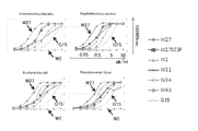
- the experimental result which evaluates the concentration-dependent binding power of monoclonal IgA with respect to various intestinal bacteria.
- the graph shows the result of binding to Enterococcus faecalis in the upper left, Staphylococcus aureus in the upper right, Escherichia coli in the lower left, and Pseudomonas fulva in the lower right.
- the vertical axis OD (405 nm) of the graph is shown, and the horizontal axis is the concentration of IgA antibody used (0.017, 0.05, 0.17, 0.5, 1.7, 5.0, 17, And 50 ⁇ g / ml).
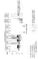
- Western blotting analysis results of various bacterial cell extracts using various monoclonal IgA antibodies (A) shows the experimental results of the W27 monoclonal IgA antibody, (B) shows the experimental results of the W30 monoclonal IgA antibody, and (C) shows the experimental results of the W45 monoclonal IgA antibody.
- Escherichia coli DH5 ⁇ strain; commercially available
- Escherichia coli cloning strain from mouse intestinal bacteria
- Pseudomonas fulva and Staphylococcus aureus are sequentially extracted from the left of the lane.
- (D) shows the experimental results using the W27 monoclonal IgA antibody, which are, in order from the left of the lane, the extracts of Eubacterium collectale, Brautia coccoides, Coprococcus eutactus, Megamonas hypermegale, lactobacillus, and bacilliids.
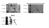
- the figure which shows the result of two-dimensional electrophoresis.
- the upper left shows Western blotting
- the upper right shows MemCode Reverseable Protein Stain
- the lower shows silver staining.
- a spot (arrow) was confirmed at the same position, and the gel was cut out and subjected to MS analysis.
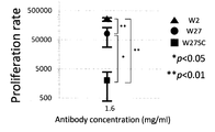
- the experimental result which measured the number of B cells in the germinal center of Peyer's board at the time of orally administering W27 monoclonal IgA antibody.
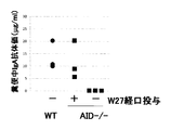
- the graph which shows the experimental result which confirmed the pharmacokinetics at the time of orally administering a W27 monoclonal IgA antibody.
- the vertical axis of the graph indicates the antibody titer ( ⁇ g / ml) of IgA antibody in stool.
- Many Poly-reactive IgA antibodies recognize serineshydroxymethylmetransferase (SHMT).
- SHMT serineshydroxymethylmetransferase
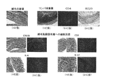
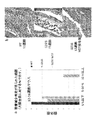
- the state of the gland duct when the W27 monoclonal IgA antibody is administered is shown. Gland duct atrophy observed in the large intestine of G23S mice was normalized by W27 antibody administration.
- the monoclonal IgA antibody according to the present invention specifically binds to the 11th to 333rd amino acid sequences of serine hydroxymethyltransferase.
- Serine hydroxymethyltransferase has a molecular weight of about 45 to 50 kDa, and its isoelectric point is about 4.3 to 4.7.
- ACCESSION No. which is listed on the NCBI website.
- This protein is an enzyme having the function of reversibly converting L-serine to glycine and simultaneously tetrahydrofolic acid to 5,10-methylenetetrahydrofolic acid, and its three-dimensional structure was highly conserved from E. coli to mammals. It is an enzyme.
- the region containing the 25th to 26th amino acids and the 31st to 44th amino acids of the amino acid sequence shown in SEQ ID NO: 74 is included together with the 25th to 28th amino acid sequences as the epitope of the monoclonal IgA antibody according to the present invention.
- the origin of the serine hydroxymethyltransferase to which the monoclonal IgA antibody according to the present invention binds is not only Escherichia coli but also Pseudomonas genus bacteria such as Pseudomonas fulva; Salmonella paratyphi A, Salmonella typhimurium, Shigella flexneri, Yersinia pestis bv. Examples include Antique, Burkholderia mallei, and the like.
- the monoclonal IgA antibody according to the present invention does not bind to serine hydroxymethyltransferase derived from other than the above or tends to dissociate immediately even if bound.
- such derived is not particularly limited, for example Lactobacillus casei, Lactobacillus such as Bifidobacterium bifidum, Blautia coccoides, Coprococcus eutactus, Megamonas hypermegale, Bordetella parapertussis; Saccharomyces cerevisiae, Schizosaccharomyces pompe, Arabidopsis thaliana, rabbits, mice, true human like Examples include serine hydroxymethyltransferase derived from nuclei. The presence or absence of these bindings and their tendency are confirmed by subjecting the cultivated lysates of various bacterial cells to Western blotting using the monoclonal IgA antibody according to the present invention.
- the specific site (epitope) in the serine hydroxymethyltransferase to which the monoclonal IgA antibody according to the present invention specifically binds is, for example, the serine hydroxymethyltransferase truncation as shown in the Examples of the present specification.
- a mutant may be prepared, and whether or not it binds to the monoclonal IgA according to the present invention may be confirmed by a known immunochemical means such as Western blotting.
- a partial amino acid sequence in a wild-type serine hydroxymethyltransferase derived from E. coli can be confirmed to bind to the monoclonal IgA antibody according to the present invention as described above.
- a monoclonal IgA antibody does not bind or is dissociated immediately after binding, for example, a mutant substituted with an amino acid sequence corresponding to the partial amino acid sequence of serine hydroxymethyltransferase derived from lactic acid bacteria is prepared, When this mutant cannot bind to the monoclonal IgA antibody according to the present invention or the binding power is reduced as compared with the wild-type serine hydroxymethyltransferase, the monoclonal IgA antibody according to the present invention is specific for the partial amino acid sequence.
- Binding serine hydroxymethyl The amino acid sequence and may be determined in the transferase.
- the term “monoclonal” is a modifier indicating the characteristics of an antibody or the like obtained from a substantially homogeneous antibody population.
- the individual antibodies contained in such a population of antibodies are identical except for possible naturally occurring mutations that may be present in trace amounts.
- the monoclonal IgA antibody according to the present invention is a kind of immunoglobulin and includes an embodiment having a heavy chain variable region and / or a light chain variable region.
- an embodiment having a heavy chain and / or a light chain, each of which further includes a constant region, is more preferable. More preferably, it is an embodiment having a heavy chain and a light chain.
- Such a heavy chain and light chain are mainly composed of a polypeptide, and each include a site called a variable region that recognizes an antigen (generally called a heavy chain variable region and a light chain variable region, respectively).
- Each of the above-mentioned variable regions has a part called CDR1-3, which is specified as a part that recognizes more antigens, in order from the amino terminus.
- CDR1 to 3 are also referred to as heavy chain CDR1, heavy chain CDR2, heavy chain CDR3, light chain CDR1, light chain CDR2, light chain CDR3 and the like in more detail.
- regions other than the heavy chain and light chain CDRs 1 to 3 are called heavy chain FR1 to 4 and light chain FR1 to 4 in order from the amino terminus.
- variable region in the monoclonal IgA antibody according to the present invention does not necessarily need to include all of CDR1 to CDR3 in the heavy chain variable region and the light chain variable region as described above, but at least one of CDR1 to CDR3. It only needs to include one CDR. Of these, CDR3 is preferably included.
- the amino acid sequences of the polypeptides constituting the heavy chain CDR1 to 3 and the light chain CDR1 to 3 are not particularly limited.
- As the light chain CDR1 the amino acid sequence shown in any of SEQ ID NOs: 8, 18, 28, 38, or 48;
- Examples of the light chain CDR3 include the amino acid sequence shown in any one of SEQ ID NOs: 10, 20, 30, 40, and 50.
- the heavy chain variable region of the monoclonal IgA antibody according to the present invention is not particularly limited.
- the heavy chain variable region includes CDRs 1 to 3, the above (1) SEQ ID NOs: 3, 13, 23, 33 are used.
- heavy chain CDR1 consisting of the amino acid sequence shown in any one of 43
- heavy chain CDR2 consisting of the amino acid sequence shown in any one of SEQ ID NOs: 4, 14, 24, 34, or 44
- Heavy chain CDR3 consisting of the amino acid sequence shown in any one of SEQ ID NOs: 5, 15, 25, 35, or 45
- the heavy chain variable region is included in order from the amino terminus.
- the light chain variable region including CDRs 1 to 3 is not particularly limited.
- the light chain variable region comprising the amino acid sequence shown in any of (I) SEQ ID NOs: 8, 18, 28, 38, or 48 described above, Chain CDR1;
- (III) Light chain CDR3 comprising the amino acid sequence shown in any one of SEQ ID NOs: 10, 20, 30, 40, or 50 Are light chain variable regions that are included in order from the amino terminus.
- More preferred heavy chain variable regions comprising CDRs 1-3 are as follows: A heavy chain variable region comprising, in order from the amino terminus, a heavy chain CDR1 consisting of the amino acid sequence shown in SEQ ID NO: 3, a heavy chain CDR2 consisting of the amino acid sequence shown in SEQ ID NO: 4, and a heavy chain CDR3 consisting of the amino acid sequence shown in SEQ ID NO: 5; A heavy chain variable region comprising, in order from the amino terminus, a heavy chain CDR1 consisting of the amino acid sequence shown in SEQ ID NO: 13, a heavy chain CDR2 consisting of the amino acid sequence shown in SEQ ID NO: 14, and a heavy chain CDR3 consisting of the amino acid sequence shown in SEQ ID NO: 15; A heavy chain variable region comprising a heavy chain CDR1 consisting of the amino acid sequence shown in SEQ ID NO: 23, a heavy chain CDR2 consisting of the amino acid sequence shown in SEQ ID NO: 24, and a heavy chain CDR3 consisting of the amino acid sequence shown in S
- a heavy chain CDR1 consisting of the amino acid sequence shown in SEQ ID NO: 3
- a heavy chain CDR2 consisting of the amino acid sequence shown in SEQ ID NO: 4
- a heavy chain CDR3 consisting of the amino acid sequence shown in SEQ ID NO: 5 in this order from the amino terminus. It is a chain variable region.
- light chain variable region comprising CDRs 1 to 3
- light chain CDR1 consisting of the amino acid sequence shown in SEQ ID NO: 8
- heavy chain CDR2 consisting of the amino acid sequence shown in SEQ ID NO: 9
- SEQ ID NO: 10 More preferred examples of the light chain variable region comprising CDRs 1 to 3
- a light chain variable region comprising a light chain CDR3 consisting of the amino acid sequence shown in this order from the amino N-terminus;
- a light chain variable region comprising a light chain CDR1 consisting of the amino acid sequence shown in SEQ ID NO: 18, a light chain CDR2 consisting of the amino acid sequence shown in SEQ ID NO: 19, and a light chain CDR3 consisting of the amino acid sequence shown in SEQ ID NO: 20 in this order from the amino terminus;
- a light chain variable region comprising a light chain CDR1 consisting of the amino acid sequence shown in SEQ ID NO: 28, a light chain CDR2 consisting of the amino acid sequence shown in SEQ ID NO: 29, and a light chain CDR3 consisting of the amino acid sequence shown in SEQ ID NO: 30 in this order from the amino terminus;
- a light chain variable region comprising a light chain CDR1 consisting of the amino acid sequence shown in SEQ ID NO: 38, a light chain CDR2 consisting of the amino acid sequence shown in SEQ ID
- a light chain CDR1 consisting of the amino acid sequence shown in SEQ ID NO: 8
- a heavy chain CDR2 consisting of the amino acid sequence shown in SEQ ID NO: 9
- a light chain CDR3 consisting of the amino acid sequence shown in SEQ ID NO: 10 in this order from the amino terminus. It is a chain variable region.
- heavy chain variable region consisting of the amino acid sequence shown in SEQ ID NO: 2, 12, 22, 32, or 42, most preferably consisting of the amino acid sequence shown in SEQ ID NO: 2. Heavy chain variable region.
- a light chain variable region consisting of the amino acid sequence shown in SEQ ID NO: 7, 17, 27, 37, or 47 is more preferable, and the amino acid sequence shown in SEQ ID NO: 7 is most preferable.
- the monoclonal IgA antibody according to the present invention may have a configuration in which the heavy chain variable region and / or the light chain variable region are appropriately combined as described above.
- Such a monoclonal IgA antibody of another embodiment has a configuration called, for example, F (ab ′) 2 , Fab, Fv, scFv, scFv-Fc, minibody, and the like.
- a monoclonal IgA antibody comprising a heavy chain and / or a light chain further comprising a constant region in the heavy chain and / or light chain variable region as described above. Can be mentioned.
- Such monoclonal IgA is not particularly limited.
- a heavy chain comprising the amino acid sequence shown in SEQ ID NO: 1, 11, 21, 31, or 41 and / or SEQ ID NO: 6, 16, 26, 36
- a monoclonal IgA antibody having a light chain comprising the amino acid sequence shown in 46.
- a monoclonal IgA antibody having a heavy chain comprising the amino acid sequence set forth in SEQ ID NO: 1, 11, 21, 31, or 41 and a light chain comprising the amino acid sequence set forth in SEQ ID NO: 6, 16, 26, 36, or 46 It is.
- the above monoclonal IgA antibody may be a monomer or a multimer. Preferably, it is a dimer. In addition, when said monoclonal antibody is a multimer, J chain mentioned later is included.
- the J chain is a peptide having an amino acid sequence having no antigen recognition site, and having a different amino acid sequence from the above heavy chain and light chain.
- amino acid sequence specifically, if it is derived from a mouse, ACCESSION No. is listed on the National Center of Biotechnology Information (NCBI) website (http://www.ncbi.nlm.nih.gov/). AAA38673; VERSION AAA38673.1 GI: 196379, and the like. If it is derived from humans, ACCESSION No. is also listed on the NCBI website.
- Such a J chain is disulfide bonded to the heavy chain of the above monoclonal IgA antibody monomer.
- amino acid sequence that defines the above monoclonal IgA antibody may be appropriately mutated as long as the functions and effects of the monoclonal IgA antibody described in the present specification are not attenuated.
- the specific number of mutagenesis is not particularly limited, but is usually 85% or more, preferably 90% or more, more preferably 95% or more, and most preferably 99% or more identity with the amino acid sequence before mutation.
- the number of mutation introductions may be such that a mutant having
- identity refers to two or more comparable amino acid sequences or base sequences that are identical in degree to each other. Therefore, the higher the identity of a certain two amino acid sequences or base sequences, the higher the identity or similarity of those sequences.
- the level of amino acid sequence or base sequence identity is usually determined using FASTA, a sequence analysis tool, using default parameters.
- the above-mentioned mutation introduction includes substitution, deletion, insertion and the like.
- a known method can be employed, and is not particularly limited.
- a conservative substitution technique may be employed for substitution.
- the term “conservative substitution technique” means a technique in which an amino acid residue is substituted with an amino acid residue having a similar side chain.
- amino acid residues having basic side chains such as lysine, arginine, and histidine
- amino acid residues having acidic side chains such as aspartic acid and glutamic acid
- amino acid residues having non-charged polar side chains such as glycine, asparagine, glutamine, serine, threonine, tyrosine, and cysteine
- Amino acid residues with non-polar side chains such as proline, phenylalanine, methionine and tryptophan
- amino acid residues with ⁇ -branched side chains such as threonine, valine and isoleucine
- aromatic side chains such as tyrosine, phenylalanine, tryptophan and histidine
- substitution between amino acid residues is a conservative substitution technique.
- the subclass of the above monoclonal IgA antibody is not particularly limited, and may be, for example, IgA 1 or IgA 2 .
- the above monoclonal IgA antibody may contain a secretory factor.
- a secretory factor is a peptide containing an amino acid sequence that does not have an antigen recognition site as in the case of the above-mentioned J chain, and is usually a peptide having a modified amino acid sequence that differs from the above heavy chain and light chain. It is.
- amino acid sequence specifically, if it is derived from a mouse, the ACCESSION No.
- amino acid sequences of the polyimmunoglobulin receptor shown in O70570; VERSION O705070.1 GI: 6225856 there may be mentioned amino acid sequences corresponding to the extracellular domain.
- the 19th to 638th amino acid sequence corresponds to a secretory factor.
- ACCESSION No. is also listed on the NCBI website. P01833; VERSION P01833.4 GI: Amino acid sequence corresponding to the extracellular domain among the amino acid sequences of the polyimmunoglobulin receptor shown in 150421625. In the above example, the 19th to 645th amino acid sequence corresponds to a secretory factor.
- a step of further introducing a nucleic acid having a base sequence encoding the secretory component region of the polyimmunoglobulin receptor described above into the hybridoma obtained in step 1 above may be included.
- a specific gene introduction method may be appropriately modified from known methods, and is not particularly limited.
- the above monoclonal IgA antibody may have an amino acid sequence derived from the same species or an amino acid derived from a different species.
- examples include human origin, mouse origin, rat origin, hamster origin, rabbit origin, goat origin, donkey origin, pig origin, cow origin, horse origin, chicken origin, monkey origin, chimpanzee origin, etc. .
- IgA monoclonal IgA derived from any two or more of human, mouse, rat, hamster, rabbit, goat, donkey, pig, cow, horse, chicken, monkey, chimpanzee, etc. An antibody is mentioned.
- a monoclonal IgA antibody having a specific amino acid sequence derived from a different species a monoclonal IgA antibody having an amino acid sequence derived from a human and an amino acid sequence derived from an animal species other than human
- examples include monoclonal IgA antibodies in which all light chain variable regions are amino acid sequences derived from animal species other than humans, and other regions have amino acid sequences derived from humans. This is sometimes called a chimeric antibody.
- the monoclonal IgA antibody according to the present invention inhibits the growth of at least two types of enteric bacteria.
- intestinal bacterium used in the present specification is not particularly limited as long as it is a bacterium that is resident in the intestine in a living body.
- enteric bacteria include bacteria that form intestinal flora in vivo and are involved in maintaining the homeostasis of the intestinal immune system.
- Specific intestinal bacteria are not particularly limited, for example, Bacteria belonging to the genus Prevotella, Bacteria belonging to the genus Bacteroides, Bacteria belonging to the genus Megamonas, Bacteria belonging to the genus Bifidobacterium A bacterium belonging to the genus Faecalibacterium, Bacteria belonging to the genus Coprococcus, Bacteria belonging to the genus Ruminococcus, Bacteria belonging to the genus Brautia, A bacterium belonging to the genus Eubacterium, Bacteria belonging to the genus Roseburia, Bacteria belonging to the genus Lactobacillus, Bacteria belonging to the genus Clostridium, Bacteria belonging to the genus Escherichia, Bacteria belonging to the genus Staphylococcus, Bacteria belonging to the genus Enterococcus, Bacteria belonging to the genus Pseudomonas, Examples include bacteria belonging
- Prevotella melaninogenica Bacteroidetes vulgatus, Megamonas funiformis, Megamonas hypermegale, Bifidobacterium bifidum, Faecalibacterium pranititzii, Coprococcus euactus, Ruminococcus obeuum, Blautia productus, Brautia coccoides, Eubacterium render, Roseburia intestinalis, Lactobacillus murinus, Lactobacillus casei, Clostridium difficile, Escherichia coli, Staphylococcus aureus, Enterococcus faecalis, Pseudomonas fulva and Enterorhabdus mucosicola And the like.
- the monoclonal antibody according to the present invention has an action of restoring gland duct reduction or atrophy in the lamina intestinal of the large intestine as shown in the following examples.
- Reduction of glandular ducts in the lamina intestinal of the large intestine is a histopathological finding found in intestinal diseases, particularly intestinal diseases that develop due to abnormal growth of intestinal bacteria and / or pathological changes in the intestinal microflora. It is also a lesion that is commonly seen as a chronic lesion of ulcerative colitis.
- the monoclonal IgA antibody according to the present invention is expected to exert an effect of suppressing abnormal growth of intestinal bacteria and / or pathological changes in the intestinal bacterial flora, and further, inflammatory bowel disease, ulcerative colitis It is effective for the treatment of intestinal diseases such as Crohn's disease, allergy, asthma, obesity, autoimmune diseases, etc. that develop due to abnormal growth of intestinal bacteria and / or pathological changes in intestinal microflora. Be expected.
- the above-mentioned abnormal growth of intestinal bacteria is not particularly limited, but represents, for example, a state in which the number of bacteria living in the intestinal tract abnormally increases.
- the pathological change of the intestinal bacterial flora described above is not particularly limited.
- the type of intestinal bacteria constituting the intestinal bacterial flora and the ratio of each intestinal bacterial flora change with the host Represents a condition that causes anomalies in maintaining the homeostasis of symbiotic relationships.
- Such abnormal growth of intestinal diseases and pathological changes in the intestinal microflora are not necessarily limited to the above-mentioned conditions of intestinal diseases, but induce factors that cause intestinal diseases. It may mean a state to do.
- treatment means obtaining a desired pharmacological and / or physiological effect.
- This effect includes partial or complete cure of the disease and / or adverse effects (pathology, symptoms, etc.) resulting from the disease.
- the above effects include the effect of preventing or delaying the progression of the disease and / or adverse effects (pathology, symptoms, etc.) caused by the disease, and the relief of the disease state, symptoms (reversal of the disease, symptoms, etc., or reversal of progression) Effects), effects to prevent recurrence, etc.
- the above effects may have a predisposition to a disease and / or an adverse effect due to the disease (such as a disease state or symptom), but in an individual who has not yet been diagnosed as having an adverse effect due to the disease and / or the disease. It includes the effect of partially or completely preventing (pathology, symptoms, etc.) from occurring. Therefore, the term “treatment” includes meanings such as “remission”, “prevention of recurrence”, and “prevention”.
- the monoclonal IgA antibody according to the present invention is expected to exhibit excellent effects when used for the treatment of intestinal diseases. Such an effect is an effect exhibited when such a monoclonal antibody is orally or enterally administered to an individual. Therefore, the monoclonal IgA antibody according to the present invention is useful as an active ingredient for oral or enteral compositions and pharmaceutical compositions.
- the nucleic acid according to the present invention is a nucleic acid encoding the monoclonal IgA antibody according to the present invention. More specifically, it is a nucleic acid having a base sequence encoding the amino acid sequence shown in any one of SEQ ID NOs: 1 to 50.
- the nucleic acid may be a ribonucleotide or a deoxynucleotide.
- the nucleic acid may be in a single-stranded form or a double-stranded form, and is not particularly limited.
- the base sequence encoding the above-mentioned amino acid sequence is not limited to a nucleic acid consisting of one kind of base sequence for one amino acid sequence, and various codons encoding the amino acid are appropriately selected according to the intended use of the nucleic acid. As long as it has been determined. Examples of the purpose of use include expression of the monoclonal IgA antibody according to the present invention using the base sequence and production thereof. In particular, various codons may be selected in consideration of the codon frequency corresponding to the type of host cell employed during production.
- the nucleic acid according to the present invention has a base sequence encoding an amino acid sequence that has been mutated in detail for the above-mentioned [Monoclonal IgA antibody of the present invention] with respect to SEQ ID NOs: 1 to 50. May be.
- Such a specific base sequence is not particularly limited, and examples thereof include the base sequences shown in SEQ ID NOs: 51 to 60.
- the correspondence between these nucleotide sequences and the amino acid sequences shown in SEQ ID NOs: 1 to 50 detailed in [Monoclonal IgA antibody of the present invention] can be explained in Table 2 below.
- the method for producing a monoclonal IgA antibody according to the present invention is the above-mentioned method for producing [monoclonal IgA], which comprises the following steps 1 and 2.
- (Process 1) A step of producing a hybridoma by mixing and fusing B cells collected from the lamina intestinal tract and other types of cells.
- (Process 2) Culturing the hybridoma prepared in the above step 1, determining a cell producing an IgA antibody that binds to at least two types of intestinal bacteria, and recovering the IgA antibody from the cell.
- a step of treating with a protease specific to IgA a step of introducing a functional group so that a chemical bond such as a disulfide bond is possible, and then forming the chemical bond via the functional group
- a step of introducing a functional group so that a chemical bond such as a disulfide bond is possible a step of introducing a functional group so that a chemical bond such as a disulfide bond is possible, and then forming the chemical bond via the functional group The process etc. to make may be included.
- step 1 and step 2 in the method for producing a monoclonal IgA antibody of the present invention will be described in detail below.
- Step 1 in the method for producing a monoclonal IgA antibody according to the present invention is a step of preparing a hybridoma by mixing and fusing B cells collected from the lamina intestinal tract and other types of cells.
- B cells used in Step 1 are B cells localized in the intestinal mucosa lamina intestinal.
- the B cell may be any cell that exhibits a function of producing an IgA antibody, and is not necessarily limited to a cell that contains an IgA antibody-encoding DNA or mRNA and that the mRNA is translated to produce an IgA antibody.
- an immunoglobulin such as an IgG antibody or an IgM antibody of a subtype different from the IgA antibody in the B cell is class-switched to become an IgA antibody, and a cell that produces the IgA antibody in the cell is also included in the B cell. It is.
- the lamina basement membrane is not particularly limited, but is present in, for example, the esophagus, stomach, small intestine (including duodenum, jejunum, ileum, etc.), large intestine (including cecum, colon, rectum, etc.). It is one of the layers constituting the mucous membrane, and may be any layer located between the layer containing epithelial cells and the mucosal substrate (Muscularis mucosae). Of these, the intestinal mucosa lamina basement present in the small intestine, including lymphoid tissues, capillaries, lymphatic vessels and the like, is preferable.
- the method for collecting B cells from the lamina intestinal tract is not particularly limited.
- the intestinal tract is collected, and the obtained intestinal tract is washed and incised to expose the mucosal layer.
- the epithelial cells are then released and removed by shaking in physiological saline containing an appropriate concentration of EDTA.
- concentration is mentioned.
- Other known methods may be employed, and commercially available B cells derived from the intestinal mucosa laminalitis may be purchased and obtained.
- the origin of the lamina intestinal mucosa that is, the origin of the B cell is not particularly limited.
- the other cell types are cell types other than the B cells, which are fused by contacting with the B cells. Any hybridoma may be used as long as it does not lose the function of producing the IgA antibody exhibited by the B cells described above.
- Such other cells are preferably cells that fuse with the B cells to form hybridomas, and the hybridomas can acquire an immortalizing function, specifically cancer cells, especially myeloid species such as myeloma. Examples include derived cells. Another preferred cell is myeloma.
- the origin of other cells is not particularly limited, and examples include mouse, rat, hamster, rabbit, goat, sheep, donkey, pig, cow, horse, chicken, monkey, chimpanzee, human and the like.
- Fusion means that the B cell and other cells are inextricably united.
- integrated inseparable excludes cell division, which is a phenomenon that occurs when cells after fusion proliferate.
- the fused cell is mentioned as an example of the hybridoma in this specification.
- the conditions at the time of mixing and fusion are not particularly limited, and conditions usually used in the method of fusing cells may be appropriately adopted.
- the conditions used when culturing B cells or other cells may be appropriately modified.
- a method for example, a method of mixing B cells and other types of cells in an appropriate medium and bringing them into contact with each other in the presence of polyethylene glycol; a method of applying electrical stimulation after the mixing A method using a virus such as Sendai virus, followed by a method of incubating the solution at 37 ° C. under 5% carbon dioxide.
- the time required for the fusion is not particularly limited, and the time until the fusion itself is completed may be set as appropriate.
- a method for confirming the completion of the fusion may be carried out by appropriately modifying a known method used for normal cell fusion. Examples of such a method include a method of observing the progress of fusion under a microscope.
- a known kit may be selected as appropriate, and cells may be fused according to the use conditions.
- Step 2 in the method for producing a monoclonal IgA antibody according to the present invention comprises culturing the hybridoma prepared in Step 1 above, determining cells that produce IgA antibodies that bind to at least two types of enteric bacteria, and , A step of recovering the antibody.
- the hybridoma obtained in Step 1 may be subjected to a subcloning step such as a limiting dilution method or the like that is usually used when producing a monoclonal antibody.
- a subcloning step such as a limiting dilution method or the like that is usually used when producing a monoclonal antibody.
- a hybridoma that produces an IgA antibody that binds to at least two types of intestinal bacteria can recognize the IgA antibody by, for example, an anti-IgA antibody that can distinguish the IgA antibody, as described in Examples below (if necessary, It may be determined by means of ELISA, EIA, RIA, FLISA, FIA, etc., FACS, etc. using radioisotopes, fluorescence, color development, etc.).
- the method for determining the above-mentioned specific “vibridooma producing an IgA antibody that binds to at least two types of intestinal bacteria” is not particularly limited.
- the following two types using the above-mentioned anti-IgA antibody can be used.
- the method etc. are mentioned.
- the following two types of methods may be used in appropriate combination.
- the first way to determine is (A) a step of determining a hybridoma that produces an IgA antibody that binds to a certain intestinal bacterium from the hybridomas obtained in step 1 above; and (B) a hybridoma determined in step A. Confirming that the IgA antibody that it produces also binds to other intestinal bacteria different from the intestinal bacterium, and determining a hybridoma that produces an IgA antibody that binds to both intestinal bacteria, The method containing is mentioned.
- the culture supernatant of the hybridoma obtained in the above-mentioned step 1 is sampled as the step A, and such a sample and a certain first enteric bacterium are obtained.
- a solid phase eg, a multi-well plate
- the IgA antibody that binds to the enteric bacteria is present in the sample. What is necessary is just to determine the hybridoma which produces an IgA antibody.
- the sample of the culture supernatant of the hybridoma producing the IgA antibody determined in the step A is used as it is, or the sample of the hybridoma culture supernatant determined above is sampled again as necessary.
- a sample is different from the above-mentioned enterobacteria, and the second (second) enterobacteria are immobilized on, for example, one well of a multiwell plate.
- the process following the process B may be repeatedly included using the third or more enteric bacteria different from the first and second.
- Intestinal bacteria may be solid-phased.
- the anti-IgA antibody is further contacted, and various intestinal bacteria, an IgA antibody that binds to the intestinal bacteria, and an anti-IgA antibody complex
- Step A in the first method two different types of enteric bacteria (three or more types as required) are used, for example, in a multiwell plate.
- a sample of the hybridoma culture supernatant obtained in Step 1 is solid-phased in two different wells (three or more depending on the number of types of enteric bacteria).
- a method of simultaneously identifying hybridomas producing IgA that binds to at least two types of enteric bacteria are simultaneously identifying hybridomas producing IgA that binds to at least two types of enteric bacteria.
- the first and second (and third or more if necessary) are used as wells or similar laboratory instruments.
- Each of the intestinal bacteria may be solid-phased.
- the limiting dilution method used in the method for producing a monoclonal antibody as described above is used.
- a step of further cloning a hybridoma that produces an IgA antibody that binds to the at least two types of intestinal bacteria may be included.
- the specific recovery method in step 2 is not particularly limited, and examples thereof include a method of recovering the supernatant of the culture medium of cells producing IgA antibody and a method of recovering the cell lysate.
- the cell lysate is lysed by appropriately combining known mechanical means such as ultrasonic disruption, French press, etc. and / or known chemical treatment methods using surfactants, cell wall digestive enzymes, and cell membrane digestive enzymes. Then, the liquid phase fraction after being subjected to the solid-liquid separation step can be recovered to produce the monoclonal IgA antibody of the present invention.
- the supernatant is recovered as described above, or the method is recovered from a cell lysate. May be provided.
- the above-described hybridoma may be intraperitoneally administered to an animal individual adapted to the origin of the hybridoma such as an immunodeficient mouse, and recovered from the ascites of such an individual.
- the monoclonal IgA antibody recovered by the above-described method may be appropriately subjected to a known purification step.
- Specific purification means are not particularly limited, but for example, purification means by precipitation using acetone, ammonium sulfate, etc .; purification means using column chromatography such as affinity, anion exchange, cation exchange, size exclusion, reverse phase, etc.
- Such known protein purification means may be used in appropriate combination.
- the production method of the monoclonal IgA antibody of the present invention includes the production method of the following embodiment.
- the nucleic acid detailed in [Nucleic acid] described above is introduced into a cell capable of producing a monoclonal IgA antibody, the cell is cultured, and the monoclonal IgA antibody is recovered from the cell extract or culture supernatant.
- purifying as needed is mentioned.
- Such a cell capable of producing a monoclonal IgA antibody is not particularly limited as long as it can produce various proteins that normally function by forming a higher-order structure derived from a mammal or the like.
- known cells such as cells derived from mammals such as COS cells, HEK cells, HELA cells, and CHO cells, and cells derived from insects such as Sf9 may be appropriately selected.
- the specific nucleic acid introduction method, the culture conditions, the recovery method, and the purification method of the cell into which the nucleic acid has been introduced vary depending on the type of cell used and the like, and are not particularly limited.
- the monoclonal IgA antibody according to the present invention can be produced.
- the nucleic acid sequence described above includes a nucleotide sequence encoding a heavy chain variable region and a light chain variable region, and a heterologous heavy chain constant region and light chain constant region.
- a nucleic acid having a base sequence encoding is prepared, a base sequence encoding the prepared heavy chain variable region is combined with a nucleic acid having a base sequence encoding the heavy chain constant region, and a base encoding the light chain variable region prepared
- a nucleic acid having a base sequence encoding the sequence and the light chain constant region is bound, and the bound nucleic acid may be introduced into a cell capable of producing a monoclonal IgA antibody as described above.
- the monoclonal IgA antibody according to the present invention is an antibody in such a manner that the heavy chain and / or light chain CDR1 to CDR3 have an amino acid sequence derived from mouse and the other region has an amino acid sequence derived from human.
- this may also be referred to as a humanized antibody
- the nucleotide sequence encoding heavy chain and / or light chain CDR1-3, and other regions are also produced in the same manner as the chimeric antibody described above.
- a nucleic acid having a base sequence that encodes a nucleic acid is prepared, the nucleic acids are recombined so as to constitute a heavy chain and a light chain, and these are introduced into cells capable of producing a monoclonal IgA antibody as described above. In order to maintain the binding ability of the antibody, it may be necessary to replace some bases with other bases.
- the monoclonal IgA antibody according to the present invention contains a J chain
- a cell capable of producing the monoclonal IgA antibody as described above is mixed with a nucleic acid having a base sequence encoding the monoclonal IgA antibody together with the J chain. What is necessary is just to introduce the nucleic acid containing the base sequence to encode.
- the base sequence encoding the J chain can be appropriately determined by adopting a known method based on the amino acid sequence of the J chain described on the NCBI website described in the above [Monoclonal IgA antibody]. . Moreover, such a nucleic acid can be produced using a known method based on the determined base sequence.
- the monoclonal IgA antibody according to the present invention contains a secretory factor
- a nucleic acid having a base sequence encoding the monoclonal IgA antibody and a J chain are added to a cell capable of producing the monoclonal IgA antibody as described above. What is necessary is just to introduce
- Such a method is described in, for example, Li C, et al. Sheng Wu Gong Cheng Xue Bao. 2011 Feb; 27 (2): 219-25. It can be manufactured by appropriately modifying such description and if necessary.
- nucleic acid containing the base sequence encoding the secretory factor examples include known methods based on the amino acid sequence of the polyimmunoglobulin receptor described in the NCBI website described in [Monoclonal IgA antibody] described above. And can be determined as appropriate. Moreover, such a nucleic acid can be produced using a known method based on the determined base sequence.
- the hybridoma according to the present invention is a hybridoma that produces the above-described monoclonal IgA antibody according to the present invention.
- the specific hybridoma is obtained in step 1 in the above-mentioned [Method for producing monoclonal IgA antibody]. That is, it is a cell obtained by mixing and fusing the B cell described in step 1 in the above-described method for producing a monoclonal IgA antibody according to the present invention and other types, that is, cells of a type other than the B cell.
- the hybridoma may be a hybridoma derived from the same species or a hybridoma derived from different species.
- the hybridoma derived from the same species may be the same as the above-mentioned [Method for producing monoclonal IgA antibody], wherein the B cells and the other species of cells other than the B cells are the same. What is necessary is just to make the origin seed
- hybridoma derived from the same species for example, among the origins described in Step 1 in the above-mentioned [Method for producing monoclonal IgA antibody], it may be appropriately selected from those overlapping in the origins of cells other than B cells and B cells. . Examples thereof include hybridomas derived from the same species such as human origin, mouse origin, rat origin, hamster origin, rabbit origin, goat origin, donkey origin, pig origin, bovine origin, horse origin, chicken origin, monkey origin, chimpanzee origin and the like.
- hybridoma derived from different species for example, among the origins described in Step 1 in the above-mentioned [Method for producing monoclonal IgA antibody], the origins described in the origins of cells other than B cells and B cells were appropriately selected. What is necessary is just to combine things. For example, hybridomas derived from different species obtained by appropriately combining the origins of humans, mice, rats, hamsters, rabbits, goats, donkeys, pigs, cows, horses, chickens, monkeys, chimpanzees and the like.
- the pharmaceutical composition according to the present invention comprises the monoclonal IgA antibody according to the present invention.
- the pharmaceutical composition according to the present invention is not particularly limited, but can be suitably used, for example, for the treatment of intestinal diseases. More preferably, it is used for the treatment of intestinal diseases that develop due to abnormal growth of intestinal bacteria and / or pathological changes in the intestinal flora.
- the abnormal growth of intestinal bacteria and the pathological changes in the intestinal bacterial flora are as described above for the “monoclonal IgA antibody of the present invention”.
- Intestinal diseases that develop due to abnormal growth of intestinal bacteria and / or pathological changes in the intestinal microflora are not particularly limited.
- inflammatory bowel disease ulcerative colitis, Crohn's disease, allergy, asthma , Obesity, autoimmune diseases and the like, among which inflammatory bowel disease is preferable.
- Such a pharmaceutical composition according to the present invention only needs to contain an effective amount of the monoclonal IgA antibody according to the present invention.
- the content of the antibody according to the present invention in 100% by weight of the pharmaceutical composition is 0.001.
- the target intestinal disease type, dosage form, administration method, administration subject, degree of symptom of administration subject, degree of effect exhibited by administration, etc. so as to be in the range of ⁇ 99.99% by weight. In consideration, it can be set appropriately.
- the term “effective amount” refers to an amount by which the monoclonal IgA antibody according to the present invention can exert a therapeutic effect on intestinal diseases, or the desired pharmacological and / or physiological effects described above ( It refers to the amount that exhibits a therapeutic effect on intestinal diseases.
- the pharmaceutical composition according to the present invention may contain a pharmaceutically acceptable carrier or additive together with the monoclonal IgA antibody according to the present invention.
- Pharmaceutically acceptable carriers or additives include any carrier, diluent, excipient, suspending agent, lubricant, adjuvant, vehicle, delivery system, emulsifier, tablet disintegrant, absorbent, preservative, It means a surfactant, a colorant, a fragrance, or a sweetener and may be a known one.
- the pharmaceutical composition according to the present invention has applicability in a method for treating intestinal diseases including a step of administering to an individual suffering from the above intestinal diseases. Moreover, although it does not develop the pathological condition or symptom of intestinal diseases as described above, it has applicability in a method for preventing intestinal diseases including a step of administering to an individual who may have a predisposition to intestinal diseases. . These individuals may be subjects of administration of the pharmaceutical composition according to the present invention.
- Such individuals to be administered are not particularly limited, and examples thereof include humans, mice, rats, guinea pigs, rabbits, hamsters, dogs, cats, weasels and the like.
- the dosage and administration method of the pharmaceutical composition vary depending on the type, gender, species, age, general condition, severity of the disease, degree of desired effect, etc. of the intestinal disease affected by the individual to be administered. It is.
- the dose is usually set appropriately in the range of 0.001 to 100 mg / kg / day.
- the administration method is not particularly limited, but it is preferably administered directly to the digestive tract.
- Examples of such administration methods include oral administration, nasal administration, transmucosal administration, and enteral administration.
- enteral administration is not limited to administration via the anus, but also includes administration via a tube inserted into the digestive tract from outside the individual, such as a gastrostomy.
- the position where the digestive tract is inserted is not limited to the intestine, but includes the esophagus, stomach, small intestine (including duodenum, jejunum, ileum, etc.), large intestine (including cecum, colon, rectum, etc.).
- the administration of the pharmaceutical composition according to the present invention may be performed once a day or may be divided into several times.
- the administration interval may be daily, every other day, every week, every other week, every two to three weeks, every month, every other month or every two to three months.
- the oral or enteral composition according to the present invention comprises the monoclonal IgA antibody according to the present invention.
- an effect of suppressing abnormal growth of intestinal bacteria and / or pathological changes in the intestinal bacterial flora is exhibited.
- the abnormal growth of intestinal bacteria and the pathological changes in the intestinal bacterial flora are as described above for the “monoclonal IgA antibody of the present invention”.
- the blending ratio of the monoclonal IgA antibody in such an oral or enteral composition is not particularly limited and may be appropriately adjusted depending on the form or use of the oral or enteral composition.
- the amount may be about 0.001 to 99% by weight based on the total amount of the enteral composition.
- the individuals for which the oral or enteral composition according to the present invention is used are not particularly limited, and examples thereof include humans, mice, rats, guinea pigs, rabbits, hamsters, dogs, cats, weasels and the like.
- the above-described monoclonal IgA antibody contained in the oral or enteral composition according to the present invention exhibits an effect of suppressing abnormal growth of intestinal bacteria and / or pathological changes in the intestinal flora. Therefore, it is particularly useful as an intestinal regulating composition, an intestinal environment improving composition, an intestinal environment optimizing composition, or an intestinal anticorrosion composition.
- the use amount of the oral or enteral composition according to the present invention is not particularly limited as long as the effect of the oral or enteral composition as described above is exhibited. What is necessary is just to set according to the kind of individual to ingest, the target effect, the target degree, and other various conditions. Specifically, in terms of the amount of the monoclonal IgA antibody according to the present invention, it is usually about 0.001 to 100 mg / kg / day, and this is taken once to several times a day. Also good.
- enteral is not limited to administration via the anus.
- the enteral composition of the present invention can be blended with known ingredients and used as an intestinal cleaning solution.
- the oral or enteral composition according to the present invention comprises the monoclonal IgA antibody according to the present invention, which exhibits an effect of suppressing abnormal growth of intestinal bacteria and / or pathological changes in the intestinal bacterial flora. It can be suitably used in the fields of food and feed in the hope of exerting such effects. Therefore, the oral or enteral composition of the present invention can be a food composition or a feed composition.
- the above-mentioned food composition is a composition that suitably uses the oral or enteral composition of the present invention exclusively in the field of food.
- a food composition can be provided as a food composition labeled as intestinal adjustment, intestinal environment improvement, intestinal environment optimization, intestinal rot prevention or the like.
- the specific form of the above-mentioned food composition is not particularly limited, for example, beverages such as soft drinks, carbonated drinks, nutrition drinks, fruit drinks, lactic acid drinks, milk drinks; frozen confectionery such as ice cream, ice sherbet, shaved ice, etc.
- Confectionery such as candy, candy, gum, chocolate, tablet confectionery, snack confectionery, biscuits, jelly, jam, cream, baked confectionery
- dairy products such as processed milk and fermented milk
- processed oils and fats such as salad oil, tempura oil, margarine, mayonnaise, shortening, whipped cream, and dressings
- seasonings such as sauces and sauces
- soup Examples include stew, salad, side dish, sprinkles, pickles, bread, and cereal.
- forms such as powder, granule, capsule, troche, tablet, syrup and the like can be mentioned.
- the above-mentioned feed composition is a composition used exclusively in the field of feed for the feed composition of the present invention.
- a feed composition can be provided as a food composition labeled as intestinal adjustment, intestinal environment improvement, intestinal environment optimization, intestinal rot prevention or the like.
- the specific form of the above-mentioned feed composition is not particularly limited, for example, as long as the effect exerted by the above-described feed composition according to the present invention is not impaired, it is usually mixed with normal feed or usually as necessary. What is necessary is just to mix with the component which can be mix
- the method for treating a disease according to the present invention is a method for treating an intestinal disease, comprising an effective amount of the above-described monoclonal IgA antibody according to the present invention, the pharmaceutical composition according to the present invention, or the oral composition according to the present invention.
- a method comprising administering to a human suffering from an intestinal disease.
- the above-mentioned human suffering from an intestinal disease may be the administration subject described in the above [Pharmaceutical composition according to the present invention].
- the specific administration method and dose may be those described in the above [Pharmaceutical composition of the present invention].
- AID G23S mouse a mouse expressing AID G23S in which a mutation was introduced into the N-terminal side of the AID protein.
- AID G23S mouse can be created using, for example, a known method shown in Non-Patent Document 18.
- 50 ml digestive enzyme solution 500 ml RPMI 1640, 25 ml FCS (final concentration 5%), 2 ⁇ l 2-mercaptoethanol (final concentration 55 ⁇ M), 0.75 g collagenase warmed to 37 ° C. in a 50 ml tube.
- 5 ml dispase prepared with a final concentration of 1 U / ml
- add small pieces of small intestine cut with scissors shake this at 37 ° C. for 60 minutes, and then leave the flask for 10 seconds to remove small intestine tissue.
- the supernatant was transferred to a new 50 ml tube.
- the digestion process using this digestive enzyme solution was repeated twice.
- the supernatant separated in the above digestion step was centrifuged at 1,500 rpm and 4 ° C. for 5 minutes, and the supernatant was discarded. Further, the obtained precipitate was suspended in RPMI 1640 containing 1 ml of 2% FCS. The suspension was passed through a filter to remove tissue debris and the cell suspension was stored on ice.
- the cell suspension was similarly collected. Two cell suspensions, the first cell suspension and the second cell suspension, were combined and used as cells in the intestinal mucosa lamina propria.
- a hybridoma was prepared by fusing the cells of the intestinal mucosa lamina basement obtained in [Separation of intestinal mucosa lamina basement cells] and NS1 cells, which are mouse myeloma cells. NS1 cells were cultured and maintained prior to cell fusion. Cell fusion was performed according to the reagents and procedures according to ClonCell®-HY Hybridoma Cloning Kit of STEMCELL Technologies.
- the obtained hybridoma was propagated and cloned in a methylcellulose-containing medium according to the above kit.
- the supernatant was then separated from the growing hybridoma.
- the antibody produced was IgA by sandwich ELISA, and the IgA antibody titer in the supernatant was measured to isolate an IgA-producing hybridoma clone. As a result, it was confirmed that a total of 37 clones were established.
- VDJ usage and mutation was analyzed by directly sequencing the amplified VDJ region PCR product. This analysis revealed that 17 types of IgA-producing hybridomas were obtained as independent clones. These are referred to as W1, W2, W3, W4, W6, W7, W11, W14, W24, W27, W28, W30, W32, W34, W37, W43, and W47, respectively.
- IgA-producing hybridoma clones were obtained from G23S mice by the same experiment. These are referred to as G1, G8, G9, G10, G12, G14, G15, G16, G18, and G19, respectively.
- [1-2: Preparation of IgA antibody] The 17 clones of IgA antibody-producing hybridoma obtained in [Preparation of IgA-producing hybridoma] were cultured in large quantities in RPMI 1640 containing 10% FCS, and the culture supernatant was collected by centrifugation. Next, the obtained culture supernatant was passed through a 0.22 ⁇ m filter, and then dialyzed against ammonium sulfate precipitation and PBS to give 27 types of monoclonal IgA antibodies (hereinafter referred to as [clone name] monoclonal IgA in this Example). Crudely purified).
- the 27 clone hybridoma obtained in [Preparation of IgA-producing hybridoma] was intraperitoneally injected into an immunodeficient mouse, and ascites was collected from the mouse after breeding for 10 to 21 days. The collected ascites was precipitated with ammonium sulfate, and the buffer was replaced with PBS using a PD-10 column. This also roughly purified 27 types of monoclonal IgA antibodies.
- the concentrations of 27 types of monoclonal IgA antibodies prepared by these methods were measured using an ELISA method.
- the antibodies used here were goat anti-mouse IgA (Southern Biotech) 2 ⁇ g / ml for coating on the solid layer, and ALP-labeled goat anti-mouse IgA (Southern Biotech) 0.5 ⁇ g / ml for detection. Used in. Mouse IgA ⁇ (Immunology Consultants Laboratory) was used as a standard inspection.
- the cloned mouse intestinal resident bacteria are specifically the following 6 types: Enterorhabdus mucosicola; Escherichia coli; Staphylococcus aureus; Lactobacillus murinus; Enterococcus faecalis; Pseudomonas fulva.
- the bacteria were suspended in 0.05 M Na 2 CO 3 buffer and coated on a plate for ELISA. After blocking by a known method, the above 27 types of monoclonal IgA antibodies (concentration: 1.4 ⁇ g / ml) were added to each of them to determine the binding power and specificity of these clonal IgA antibodies against the 13 types of enteric bacteria. Each was measured using the ELISA method described above. The above-described antibodies were used for detection.
- a specific experimental method is a measurement method employing the above-described ELISA method, and the monoclonal IgA antibodies used are W27, G15, and G19 monoclonal IgA antibodies.
- W2 clone monoclonal IgA antibody was used as a negative control.
- intestinal resident bacteria used are Staphylococcus aureus, Escherichia coli, and Enterococcus faecalis.
- pcDNA3.1 (+) / mSC3F Plasmid DNA of pcDNA3.1 (+) / mSC3F was prepared from E. coli, and the gene was introduced into a hybridoma producing a W27 monoclonal antibody by Nucleofector (AMAXA). Then, selection by G418 was performed.
- the W27SC3F monoclonal IgA antibody was obtained from the hybridoma selected by this method according to the method for recovering the monoclonal IgA antibody from the hybridoma as described above.
- intestinal resident bacteria used are Enterococcus faecalis, Staphylococcus aureus, Escherichia coli, and Pseudomonas fulva.
- Escherichia coli DH5 ⁇ ; commercially available
- Escherichia coli cloning strain from mouse intestinal bacteria
- Pseudomonas fulva Pseudomonas fulva
- Staphylococcus aureus and Eubacterium render was shake-cultured in 10 ml of LB or in an optimal culture solution under conditions suitable for each cell. After collection by centrifugation, the cells were suspended in PBS containing 1% NP-40 and sonicated on ice. After standing for 30 minutes on ice, a supernatant was obtained by centrifugation.
- SDS-buffer (containing 2-ME) was added thereto, and the protein was denatured by heat treatment at 95 ° C. for 10 minutes. After 8% SDS-PAGE, transferred to a filter, and after blocking, W27, W30 and W45 monoclonal IgA antibodies (2 ⁇ g / ml) followed by Goat anti-mouse IgA (1 ⁇ g / ml) (Southern Biotechnology), IR800 Anti-goat IgG (0.2 ⁇ g / ml) (Rockland), and further subjected to Western blotting reaction using anti-Goat IgG labeled as necessary, and the signal is expressed by Odyssey (LI-COR). Detected.
- the monoclonal IgA antibody used is W27.
- the results are shown in FIG. It was revealed that the W27 monoclonal IgA antibody did not bind to the protein in the bacterial cell extract other than the Eubacterium spectrum.
- the amino acid sequence was analyzed by a known method. Specifically, 5 'and 3' RACE reactions were performed to obtain the full length base sequence.
- the RACE method was in accordance with the method of In-Fusion (registered trademark) SMARTer TM Directional cDNA library Construction Kit (Clontech).
- a sequence analysis was performed on the heavy chain by combining the primer attached to Kit and the above-mentioned primer. For the light chain, the following primers were combined for sequence analysis.
- the nucleic acid sequences encoding these amino acids are as follows: W27 monoclonal IgA antibody; heavy chain: SEQ ID NO: 51, light chain: SEQ ID NO: 52; W30 monoclonal IgA antibody; heavy chain: SEQ ID NO: 53, light chain: SEQ ID NO: 54; W34 monoclonal IgA antibody; heavy chain: SEQ ID NO: 55, light chain: SEQ ID NO: 56; W43 monoclonal IgA antibody; heavy chain: SEQ ID NO: 57, light chain: SEQ ID NO: 58; W11 monoclonal IgA antibody; heavy chain: SEQ ID NO: 59, light chain: SEQ ID NO: 60.
- Example 2 Antigen specific experiment of monoclonal IgA antibody [2-1: immunoprecipitation experiment]
- cell extract components were obtained from each of three types of bacteria, Escherichia coli (DH5 ⁇ ), Pseudomonas fulva, and Staphylococcus aureus by the method described above, and then 2-ME (final concentration 300 mM) and SDS were obtained. (Final concentration 1%) was added and heat denaturation was performed at 95 ° C. for 10 minutes. Thereafter, the 2-ME concentration in the solution was adjusted to 60 mM, and the SDS concentration was adjusted to 0.2%.
- washed Protein G Sepharose 4 Fast Flow 100 ⁇ l of washed Protein G Sepharose 4 Fast Flow (GE) was added to the cell extract components, mixed by inverting at 30 ° C. for 30 minutes, and nonspecific binding components to Protein G Sepharose were eliminated by centrifugation.
- 5 ⁇ g of W27 monoclonal IgA antibody was added and allowed to stand on ice for 30 minutes, and then 5 ⁇ g of Goat anti-mouse IgA was further added and allowed to stand on ice for 30 minutes.
- 30 ⁇ l of washed Protein G Sepharose 4 Fast Flow (GE) was added and mixed by inverting at 4 ° C. for 15 hours.
- the above-described normal Western blotting was performed to obtain an image for confirming the position of the target protein (FIG. 5). From the two images, the target protein was identified as a spot of the protein developed in two dimensions. The second set of gels was directly subjected to silver staining (Sylbeststein; Nacalai tesque) to visualize all proteins. The target protein was identified based on the positional information obtained from Western blotting and cut out from the gel.
- the cut gel was subjected to in-gel enzymatic digestion with trypsin at 37 ° C. for 15 hours. MS analysis was performed by LCMS-IT-TOF (Shimazu). As a result of analysis, it was clarified that the protein of the spot uses serine hydroxymethyltransferase as an antigen.
- the various mutants prepared were (1) the amino acid sequence from 1 to 417 of the amino acid sequence shown in SEQ ID NO: 74 (full length: wild type), (2) 11th to 417th amino acid sequence ( ⁇ 2-10), (3) 26th to 417th amino acid sequence ( ⁇ 2-25), (4) 29th to 417th amino acid sequence ( ⁇ 2-28), (5) the 1st to 333rd amino acid sequence ( ⁇ 334-417) and (6) the 1st to 250th amino acid sequence ( ⁇ 251-417), A Myc tag (40aa) was provided on the C-terminal side.
- the results shown in FIG. 10 revealed that the W27 monoclonal IgA antibody binds to the ⁇ 2-25 mutant but does not bind to the ⁇ 2-28 mutant. This is apparent from experiments using the 293T system, which is considered not to be affected by endogenous wild-type serine hydroxymethyltransferase derived from E. coli. It was also revealed that the W27 monoclonal IgA antibody binds to the ⁇ 251-417 mutant and the ⁇ 334-417 mutant.
- Example 3 Oral administration experiment of W27 monoclonal IgA antibody [3-1: pathological model analysis of G23S mouse]
- Large intestine tissue sections of G23S mice were prepared, and the degree of inflammation was examined by HE staining.
- a decrease or atrophy of gland ducts was observed more at 13-16 weeks of age than in wild type mice (Fig. 6).
- a decrease or atrophy of gland ducts and infiltration of inflammatory cells into the colonic mucosa were frequently observed at 40 weeks of age or older.
- the number of inflammatory cells such as CD11b + cells, IL-6 positive cells, IL-6, and IL-17 was larger than that in the wild type mice.
- mice 10 to 12-week-old male G23S mice were used (background is Balb / c).
- W27 monoclonal IgA antibody was added to the drinking water for breeding to a concentration of 25 ⁇ g / ml, and this was orally ingested for 28 days.
- As a negative control an experiment in which only water was ingested, and an experiment in which wild type mice were ingested with water alone were performed. All mice were fed with food in the same manner as in normal breeding.
- mice were euthanized, and Peyer's patches were separated from the small intestine.
- Peyer's patch tissue was separated into single cells using a glass slide, and surface antigens were stained with the following antibodies.
- the ratio of the number of cells in the germinal center B cell fraction showing B220 + PNA high was determined by flow cytometry analysis, and it was examined whether the germinal center itself was regressed with or without antibody administration.
- the antibodies used in flow cytometry are shown below: PE-Cy7-labeled anti-mouse B220 (eBioscience); Biotinylated penant agglutinin (VECTOR laboratories); Streptavidin APC (eBioscience).
- FIG. 7 shows the results obtained by analyzing the specific ratio of B cells and the number of B cells in the Peyer's patch germinal center.
- the G23S mouse is suitable for use as a model mouse for inflammatory colitis, ulcerative colitis and the like.
- B cells at the germinal center of Peyer's patches were excessively proliferated.
- Peyer's patch germinal center B cells are the site of IgA antibody production in the intestinal immune system.
- an excessive increase in B cells indicates that the IgA antibody is excessively produced in the intestinal immunity system, which means that the intestinal mucosa cannot fully cope with the invasion of the intestinal mucosa.
- T cells are complementarily activated in the intestinal immune system, which enhances the production of inflammatory cytokines as described above, and causes inflammation.
- the W27 monoclonal IgA antibody has an effect of reducing B cells at the germinal center of Peyer's patch, so that the intestinal immune system is normalized and it is possible to suppress the action of T cells and the like. , Inflammation is resolved. Therefore, the W27 monoclonal IgA antibody is effective against diseases such as inflammatory colitis, ulcerative colitis, etc. that develop due to abnormal growth of intestinal bacteria and / or pathological changes in the intestinal bacterial flora, and thus enteric diseases. It was clearly suggested that it was effective.
- FIG. 11 shows the results of analysis showing decrease or contraction of gland ducts in the colonic mucosal tissue of 13-15 weeks old wild-type mice and G23S mice that had been orally treated with untreated G23S mice and W27 monoclonal IgA antibody for 4 weeks. Indicates. After removing the contents of the entire large intestine and washing with physiological saline, a roll was made around the toothpick, enclosed in an OCT compound, and frozen. Thereafter, a frozen section having a thickness of 6 microns was prepared and stained with hematoxylin-eosin. The lesion part where the gland duct decreased under the microscope was specified, and the spread of the lesion part occupying the entire length of the large intestine was quantified and summarized in the graph of FIG. 11a. A typical tissue image is shown in FIG.
- W27 monoclonal IgA antibody was orally administered to AID knockout mice using the same antibody dosage and method as in the above oral administration experiment. Specifically, W27 monoclonal IgA antibody was prepared to be 25 ⁇ g / ml in drinking water, and was administered by oral ingestion continuously for 28 days. On the second day, feces were collected. 0.9 ml of PBS was added to and suspended in 100 mg of the collected stool, centrifuged, and the supernatant was collected. The IgA concentration in the supernatant was measured by ELISA.
- E. coli strain DH5 ⁇ was added to the LB liquid medium, and shaking culture was performed overnight at 37 ° C. On the next day, the E. coli solution was appropriately diluted, seeded on an LB plate, and cultured at 37 ° C. overnight. On the next day, the number of colonies on the plate was counted to calculate the number of E. coli in the above E. coli solution. This was taken as the number of E. coli at the start of the growth inhibition experiment.
- E. coli solution on the LB plate 50 ⁇ L of E. coli diluted with LB liquid medium is added to the 96-well plate, and the W27 monoclonal IgA antibody and W27SC3F monoclonal IgA antibody are added to a final concentration of 1.6 mg / mL. , And W2 monoclonal IgA antibody, respectively.
- the 96-well plate was subjected to static culture at 37 ° C. for 7 hours. Seven hours later, the E. coli solution was appropriately diluted, seeded on an LB plate, and cultured overnight at 37 ° C. On the next day, the number of colonies on the LB plate was counted to count the number of E. coli, and the number of bacteria calculated when the number of E. coli at the start was set to 1 was plotted on a graph as a Proliferation rate (vertical axis in FIG. 12). The results are shown in FIG.
- the W27SC3F monoclonal IgA antibody which is a secretory IgA
- the secretory IgA dimer that is actually secreted into the intestinal tract of the living body recognizes and binds to bacteria and thus has a unique effector. It is suggested that the function is exerted, and as a result, the growth of E. coli is suppressed. Therefore, it is suggested that it is preferable to use a secretory IgA as a dosage form when using a monoclonal IgA antibody for intestinal bacteria control.
Landscapes
- Health & Medical Sciences (AREA)
- Chemical & Material Sciences (AREA)
- Organic Chemistry (AREA)
- Medicinal Chemistry (AREA)
- General Health & Medical Sciences (AREA)
- Life Sciences & Earth Sciences (AREA)
- Veterinary Medicine (AREA)
- General Chemical & Material Sciences (AREA)
- Chemical Kinetics & Catalysis (AREA)
- Nuclear Medicine, Radiotherapy & Molecular Imaging (AREA)
- Pharmacology & Pharmacy (AREA)
- Bioinformatics & Cheminformatics (AREA)
- Animal Behavior & Ethology (AREA)
- Engineering & Computer Science (AREA)
- Public Health (AREA)
- Immunology (AREA)
- Biophysics (AREA)
- Biochemistry (AREA)
- Proteomics, Peptides & Aminoacids (AREA)
- Molecular Biology (AREA)
- Genetics & Genomics (AREA)
- Pulmonology (AREA)
- Obesity (AREA)
- Oncology (AREA)
- Communicable Diseases (AREA)
- Transplantation (AREA)
- Hematology (AREA)
- Diabetes (AREA)
- Child & Adolescent Psychology (AREA)
- Nutrition Science (AREA)
- Preparation Of Compounds By Using Micro-Organisms (AREA)
- Peptides Or Proteins (AREA)
- Medicines Containing Antibodies Or Antigens For Use As Internal Diagnostic Agents (AREA)
- Micro-Organisms Or Cultivation Processes Thereof (AREA)
Abstract
Description
セリンヒドロキシメチルトランスフェラーゼの11~333番目のアミノ酸に結合する、モノクローナルIgA抗体。
セリンヒドロキシメチルトランスフェラーゼの25~44番目のアミノ酸に結合する、項1に記載のモノクローナルIgA抗体。
セリンヒドロキシメチルトランスフェラーゼの25~28番目のアミノ酸に結合する、項1に記載のモノクローナルIgA抗体。
モノクローナルIgA抗体が、
(1)配列番号3、13、23、33、又は43に示すアミノ酸配列からなる重鎖CDR1、
(2)配列番号4、14、24、34、又は44に示すアミノ酸配列からなる重鎖CDR2、及び
(3)配列番号5、15、25、35、又は45に示すアミノ酸配列からなる重鎖CDR3
を含む重鎖可変領域;及び/又は
(I)配列番号8、18、28、38、又は48に示すアミノ酸配列からなる軽鎖CDR1、
(II)配列番号9、19、29、39、又は49に示すアミノ酸配列からなる軽鎖CDR2、及び
(III)配列番号10、20、30、40、又は50に示すアミノ酸配列からなる軽鎖CDR3
を含む軽鎖可変領域を含む、項1及び項1-1、又は項1ー2に記載のモノクローナルIgA抗体。
モノクローナルIgA抗体が、配列番号2、12、22、32、又は42に示すアミノ酸配列からなる重鎖可変領域;及び/又は配列番号7、17、27、37、又は47に示すアミノ酸配列からなる軽鎖可変領域を含む、項2及び項1-1~項1ー3の何れか1項に記載のモノクローナルIgA抗体。
モノクローナルIgA抗体が、配列番号1、11、21、31、又は41に示すアミノ酸配列からなる重鎖;及び/又は配列番号6、16、26、36、又は46に示すアミノ酸配列からなる軽鎖を含む、上記項1及び項1-1~項1ー4の何れか1項に記載のモノクローナルIgA抗体。
少なくとも2種類の腸内細菌の成育を阻害する、上記項1及び項1-1~項1-5の何れか1項に記載のモノクローナルIgA抗体。
腸内細菌が、
Prevotella属に属する細菌、
Bacteroides属に属する細菌、
Megamonas属に属する細菌、
Bifidobacterium属に属する細菌、
Faecalibacterium属に属する細菌、
Coprococcus属に属する細菌、
Ruminococcus属に属する細菌、
Blautia属に属する細菌、
Eubacterium属に属する細菌、
Roseburia属に属する細菌、
Lactobacillus属に属する細菌、
Clostridium属に属する細菌、
Escherichia属に属する細菌、
Staphylococcus属に属する細菌、
Enterococcus属に属する細菌、
Pseudomonas属に属する細菌、及び
Enterorhabdus属に属する細菌
からなる群に属する細菌より選択される少なくとも2種の細菌である、項1、及び項1-1~項1-6の何れか1項に記載のモノクローナルIgA抗体。
腸内細菌が、
Prevotella melaninogenica、
Bacteroidetes vulgatus、
Megamonas funiformis、
Megamonas hypermegale、
Bifidobacterium bifidum、
Faecalibacterium prausnitzii、
Coprococcus eutactus、
Ruminococcus obeum、
Blautia productus、
Blautia coccoides、
Eubacterium rectale、
Roseburia intestinalis、
Clostridium difficile、
Escherichia coli、
Staphylococcus aureus、
Lactobacillus murinus、
Lactobacillus casei、
Enterococcus faecalis、
Pseudomonas fulva、及び
Enterorhabdus mucosicola
からなる群より選択される少なくとも2種の細菌である、項1、及び項1-1~項1-7の何れか1項に記載のモノクローナルIgA抗体。
モノクローナルIgA抗体が、IgA1又はIgA2である、項1、及び項1-1~項1-8の何れか1項に記載のモノクローナルIgA抗体。
モノクローナルIgA抗体が、J鎖を含む多量体である、項1、項1-1~項1-9の何れか1項に記載のモノクローナルIgA抗体。
モノクローナルIgA抗体が、分泌因子を含む、項1、項1-1~項1-10の何れか1項に記載のモノクローナルIgA抗体。
モノクローナルIgA抗体が、同一種に由来するアミノ酸配列を有する、項1、項1-1~項1-11の何れか1項に記載のモノクローナルIgA抗体。
モノクローナルIgA抗体が、マウス、ラット、ハムスター、ウサギ、ヤギ、ヒツジ、ロバ、ブタ、ウシ、ウマ、ニワトリ、サル、チンパンジー、又はヒトの何れかのアミノ酸に由来するアミノ酸配列を有する、項1、及び項1-1~項1-12の何れか1項に記載のモノクローナルIgA抗体。
モノクローナルIgA抗体が、異種に由来するアミノ酸配列を有する、項1、及び項1-1~項1ー11の何れか1項に記載のモノクローナルIgA抗体。
モノクローナルIgA抗体が、マウス、ラット、ハムスター、ウサギ、ヤギ、ヒツジ、ロバ、ブタ、ウシ、ウマ、サル、ニワトリ、チンパンジー、及びヒトからなる群より選択される2種以上に由来するアミノ酸配列を有する、項1、項1-1~項1-11、及び項1-14の何れか1項に記載のモノクローナルIgA抗体。
モノクローナルIgA抗体が、ヒト化抗体である、項1、項1-1~項1ー11、1-14、及び項1-15の何れか1項に記載のモノクローナルIgA抗体。
モノクローナルIgA抗体が、キメラ抗体である、項1、項1-1~項1ー11、1-14、及び項1-15の何れか1項に記載のモノクローナルIgA抗体。
以下の工程1及び2を含む、項1に記載のモノクローナルIgA抗体の製造方法:
(1)腸管粘膜固有層から採取されたB細胞と、それ以外の種類の細胞を混合して融合させ、ハイブリドーマを作製する工程1、
(2)工程1にて作製した該ハイブリドーマを培養し、少なくとも2種類の腸内細菌と結合するモノクローナルIgA抗体を産生する細胞を決定し、該細胞から、該IgA抗体を回収する工程2。
B細胞がIgA抗体を産生する細胞である、項2に記載の製造方法。
腸管粘膜固有層が、マウス由来、ラット由来、ハムスター由来、ウサギ由来、ヤギ由来、ヒツジ由来、ロバ由来、ブタ由来、ウシ由来、ウマ由来、ニワトリ由来、サル、チンパンジー又はヒト由来である、項2又は項2-1に記載の製造方法
他の細胞が、ミエローマ細胞である項2、項2-1、及び項2-2に記載の製造方法。
他の細胞が、マウス由来、ラット由来、ハムスター由来、ウサギ由来、ヤギ由来、ヒツジ由来、ロバ由来、ブタ由来、ウシ由来、ウマ由来、ニワトリ由来、サル由来、チンパンジー由来、又はヒト由来である、項2、及び項2-1~項2-3の何れか1項に記載の製造方法。
腸内細菌が、
Prevotella属に属する細菌、
Bacteroides属に属する細菌、
Megamonas属に属する細菌、
Bifidobacterium属に属する細菌、
Faecalibacterium属に属する細菌、
Coprococcus属に属する細菌、
Ruminococcus属に属する細菌、
Blautia属に属する細菌、
Eubacterium属に属する細菌、
Roseburia属に属する細菌、
Lactobacillus属に属する細菌、
Clostridium属に属する細菌、
Escherichia属に属する細菌、
Staphylococcus属に属する細菌、
Enterococcus属に属する細菌、
Pseudomonas属に属する細菌、及び
Enterorhabdus属に属する細菌
からなる群に属する細菌より選択される少なくとも2種の細菌である、項2、及び項2-1~項2-4の何れか1項に記載の製造方法。
腸内細菌が、
Prevotella melaninogenica、
Bacteroidetes vulgatus、
Megamonas funiformis、
Megamonas hypermegale、
Bifidobacterium bifidum、
Faecalibacterium prausnitzii、
Coprococcus eutactus、
Ruminococcus obeum、
Blautia productus、
Blautia coccoides、
Eubacterium rectale、
Roseburia intestinalis、
Lactobacillus murinus、
Lactobacillus casei、
Clostridium difficile、
Escherichia coli、
Staphylococcus aureus、
Enterococcus faecalis、
Pseudomonas fulva、及び
Enterorhabdus mucosicola
からなる群より選択される少なくとも2種の細菌である、項2、及び項2-1~項2-5の何れか1項に記載の製造方法。
モノクローナルIgA抗体が、IgA1又はIgA2である、項2、及び項2-1~項2ー6の何れか1項に記載の製造方法。
モノクローナルIgA抗体が、J鎖を含む多量体である、項2、及び項2-1~項2ー7の何れか1項に記載の製造方法。
モノクローナルIgA抗体が、分泌因子を含む、項2、及び項2-1~項2ー8の何れか1項に記載の製造方法。
モノクローナルIgA抗体が、同一種に由来するアミノ酸配列を有する、項2、及び項2-1~項2-9の何れか1項に記載の製造方法。
モノクローナルIgA抗体が、マウス、ラット、ハムスター、ウサギ、ヤギ、ヒツジ、ロバ、ブタ、ウシ、ウマ、ニワトリ、サル、チンパンジー、又はヒトの何れかに由来するアミノ酸配列を有する、項2、及び項2-1~項2ー10の何れか1項に記載の製造方法。
モノクローナルIgA抗体が、異種に由来するアミノ酸配列を有する、項2、及び項2-1~項2ー9の何れか1項に記載の製造方法。
モノクローナルIgA抗体が、マウス、ラット、ハムスター、ウサギ、ヤギ、ヒツジ、ロバ、ブタ、ウシ、ウマ、ニワトリ、サル、チンパンジー、及びヒトからなる群より選択される2種以上に由来するアミノ酸配列を有する、項2、項2-1~項2ー9、及び項2-12の何れか1項に記載の製造方法。
モノクローナルIgA抗体が、ヒト化抗体である、項2、項2-1~項2ー9、項2-12、及び項2-13の何れか1項に記載の製造方法。
モノクローナルIgA抗体が、キメラ抗体である、項2、項2-1~項2ー9、項2-12、及び項2-13の何れか1項に記載の製造方法。
モノクローナルIgA抗体が、
(1)配列番号3、13、23、33、又は43に示すアミノ酸配列からなる重鎖CDR1、
(2)配列番号4、14、24、34、又は44に示すアミノ酸配列からなる重鎖CDR2、及び
(3)配列番号5、15、25、35、又は45に示すアミノ酸配列からなる重鎖CDR3
を含む重鎖可変領域;及び/又は
(I)配列番号8、18、28、38、又は48に示すアミノ酸配列からなる軽鎖CDR1、
(II)配列番号9、19、29、39、又は49に示すアミノ酸配列からなる軽鎖CDR2、及び
(III)配列番号10、20、30、40、又は50に示すアミノ酸配列からなる軽鎖CDR3
を含む軽鎖可変領域を含む、項2及び項2-1~項2~15の何れか1項に記載の製造方法。
モノクローナルIgA抗体が、配列番号2、12、22、32、又は42に示すアミノ酸配列からなる重鎖可変領域;及び/又は配列番号7、17、27、37、又は47に示すアミノ酸配列からなる軽鎖可変領域を含む、項2及び項2-1~項2ー16の何れか1項に記載の製造方法。
モノクローナルIgA抗体が、配列番号1、11、21、31、又は41に示すアミノ酸配列からなる重鎖;及び/又は配列番号6、16、26、36、又は46に示すアミノ酸配列からなる軽鎖を含む、項2及び項2-1~項2~17の何れか1項に記載の製造方法。
項1に記載のモノクローナルIgA抗体を含む、医薬組成物。
医薬組成物が、腸内疾患の治療用である、項3に記載の医薬組成物。
腸内疾患が、腸内細菌の異常増殖及び/又は腸内細菌叢の病的変化により発症する疾患である、項3又は項3-1に記載の医薬組成物。
腸内細菌の異常増殖及び/又は腸内細菌叢の病的変化により発症する疾患が、炎症性腸疾患、潰瘍性大腸炎、クローン病、アレルギー、喘息、肥満、及び自己免疫疾患からなる群から選択される、項3、項3-1、及び項3-2に記載の医薬組成物。
腸内細菌が、
Prevotella属に属する細菌、
Bacteroides属に属する細菌、
Megamonas属に属する細菌、
Bifidobacterium属に属する細菌、
Faecalibacterium属に属する細菌、
Coprococcus属に属する細菌、
Ruminococcus属に属する細菌、
Blautia属に属する細菌、
Eubacterium属に属する細菌、
Roseburia属に属する細菌、
Lactobacillus属に属する細菌、
Clostridium属に属する細菌、
Escherichia属に属する細菌、
Staphylococcus属に属する細菌、
Enterococcus属に属する細菌、
Pseudomonas属に属する細菌、及び
Enterorhabdus属に属する細菌
からなる群に属する細菌より選択される少なくとも2種の細菌である、項3、及び項3-1~項3-3の何れか1項に記載の医薬組成物。
腸内細菌が、
Prevotella melaninogenica、
Bacteroidetes vulgatus、
Megamonas funiformis、
Megamonas hypermegale、
Bifidobacterium bifidum、
Faecalibacterium prausnitzii、
Coprococcus eutactus、
Ruminococcus obeum、
Blautia productus、
Blautia coccoides、
Eubacterium rectale、
Roseburia intestinalis、
Lactobacillus murinus、
Lactobacillus casei、
Clostridium difficile、
Escherichia coli、
Staphylococcus aureus、
Enterococcus faecalis、
Pseudomonas fulva、及び
Enterorhabdus mucosicola
からなる群より選択される少なくとも2種の細菌である、項3、及び項3-1~項3-4の何れか1項に記載の医薬組成物。
医薬組成物が、経口又は経腸にて投与される事を特徴とする、項3、項3-1~項3-5の何れか1項に記載の医薬組成物。
項1に記載のモノクローナルIgA抗体を含む経口又は経腸組成物。
経口又は経腸組成物が、腸内細菌の異常増殖及び/又は腸内細菌叢の病的変化を抑制する、項4に記載の経口又は経腸組成物。
腸内細菌が、
Prevotella属に属する細菌、
Bacteroides属に属する細菌、
Megamonas属に属する細菌、
Bifidobacterium属に属する細菌、
Faecalibacterium属に属する細菌、
Coprococcus属に属する細菌、
Ruminococcus属に属する細菌、
Blautia属に属する細菌、
Eubacterium属に属する細菌、
Roseburia属に属する細菌、
Lactobacillus属に属する細菌、
Clostridium属に属する細菌、
Escherichia属に属する細菌、
Staphylococcus属に属する細菌、
Enterococcus属に属する細菌、
Pseudomonas属に属する細菌、及び
Enterorhabdus属に属する細菌
からなる群に属する細菌より選択される少なくとも2種の細菌である、項4又は項4-1に記載の経口又は経腸組成物。
腸内細菌が、
Prevotella melaninogenica、
Bacteroidetes vulgatus、
Megamonas funiformis、
Megamonas hypermegale、
Bifidobacterium bifidum、
Faecalibacterium prausnitzii、
Coprococcus eutactus、
Ruminococcus obeum、
Blautia productus、
Blautia coccoides、
Eubacterium rectale、
Roseburia intestinalis、
Lactobacillus murinus、
Lactobacillus casei、
Clostridium difficile、
Escherichia coli、
Staphylococcus aureus、
Enterococcus faecalis、
Pseudomonas fulva、及び
Enterorhabdus mucosicola
からなる群より選択される少なくとも2種の細菌である、項4、項4-1、又は項4-2に記載の経口又は経腸組成物。
経口又は経腸組成物が、腸内環境改善組成物、腸内環境最適化組成物、又は腸内腐敗防止組成物である、項4及び項4-1~4-3の何れか1項に記載の経口又は経腸組成物。
経口又は経腸組成物が、飲食品組成物である、項4、及び項4-1~項4-4の何れか1項に記載の経口又は経腸組成物。
経口又は経腸組成物が、飼料組成物である、項4、及び項4-1~項4-5の何れか1項に記載の経口又は経腸組成物。
項1に記載のモノクローナルIgA抗体をコードする核酸。
項1に記載のモノクローナルIgA抗体を産生するハイブリドーマ。
ハイブリドーマが、同一種由来である、項6に記載のハイブリドーマ。
ハイブリドーマが、マウス、ラット、ハムスター、ウサギ、ヤギ、ヒツジ、ロバ、ブタ、ウシ、ウマ、ニワトリ、サル、チンパンジー、又はヒトの何れか由来する、項6又は項6-1に記載のハイブリドーマ。
ハイブリドーマが、異種由来である、項6に記載のハイブリドーマ。
ハイブリドーマが、マウス、ラット、ハムスター、ウサギ、ヤギ、ヒツジ、ロバ、ブタ、ウシ、ウマ、ニワトリ、サル、チンパンジー、及びヒトからなる群より選択される2種以上を由来とする、項6又は項6-2に記載のハイブリドーマ。
少なくとも2種類の腸内細菌に項1に記載の抗体を接触させる工程を含む、少なくとも2種類の腸内細菌の増殖を抑制する方法。
腸内で接触させる、項7に記載の方法。
腸内細菌が、
Prevotella属に属する細菌、
Bacteroides属に属する細菌、
Megamonas属に属する細菌、
Bifidobacterium属に属する細菌、
Faecalibacterium属に属する細菌、
Coprococcus属に属する細菌、
Ruminococcus属に属する細菌、
Blautia属に属する細菌、
Eubacterium属に属する細菌、
Roseburia属に属する細菌、
Lactobacillus属に属する細菌、
Clostridium属に属する細菌、
Escherichia属に属する細菌、
Staphylococcus属に属する細菌、
Enterococcus属に属する細菌、
Pseudomonas属に属する細菌、及び
Enterorhabdus属に属する細菌
からなる群に属する細菌より選択される少なくとも2種の細菌である、項7又は項7-1に記載の治療方法。
腸内細菌が、
Prevotella melaninogenica、
Bacteroidetes vulgatus、
Megamonas funiformis、
Megamonas hypermegale、
Bifidobacterium bifidum、
Faecalibacterium prausnitzii、
Coprococcus eutactus、
Ruminococcus obeum、
Blautia productus、
Blautia coccoides、
Eubacterium rectale、
Roseburia intestinalis、
Lactobacillus murinus、
Lactobacillus casei、
Clostridium difficile、
Escherichia coli、
Staphylococcus aureus、
Enterococcus faecalis、
Pseudomonas fulva、及び
Enterorhabdus mucosicola
からなる群より選択される少なくとも2種の細菌である、項7、項7-1、又は項7-2に記載の治療方法。
腸内疾患の治療方法であって、項1に記載の抗体の有効量を、腸内疾患に罹患したヒトに投与する工程を含む、治療方法。
腸内疾患が、腸内細菌の異常増殖及び/又は腸内細菌叢の病的変化により発症する疾患である、項8に記載の治療方法。
腸内細菌の異常増殖や腸内細菌叢の病的変化により発症する疾患が、炎症性腸疾患、潰瘍性大腸炎、クローン病、アレルギー、喘息、肥満、及び自己免疫疾患からなる群より選択される、項8又は項8-1に記載の治療方法。
腸内細菌が、
Prevotella属に属する細菌、
Bacteroides属に属する細菌、
Megamonas属に属する細菌、
Bifidobacterium属に属する細菌、
Faecalibacterium属に属する細菌、
Coprococcus属に属する細菌、
Ruminococcus属に属する細菌、
Blautia属に属する細菌、
Eubacterium属に属する細菌、
Roseburia属に属する細菌、
Lactobacillus属に属する細菌、
Clostridium属に属する細菌、
Escherichia属に属する細菌、
Staphylococcus属に属する細菌、
Enterococcus属に属する細菌、
Pseudomonas属に属する細菌、及び
Enterorhabdus属に属する細菌
からなる群に属する細菌より選択される少なくとも2種の細菌である、項8、項8-1、又は項8-2の何れかに記載の治療方法。
腸内細菌が、
Prevotella melaninogenica、
Bacteroidetes vulgatus、
Megamonas funiformis、
Megamonas hypermegale、
Bifidobacterium bifidum、
Faecalibacterium prausnitzii、
Coprococcus eutactus、
Ruminococcus obeum、
Blautia productus、
Blautia coccoides、
Eubacterium rectale、
Roseburia intestinalis、
Lactobacillus murinus、
Lactobacillus casei、
Clostridium difficile、
Escherichia coli、
Staphylococcus aureus、
Enterococcus faecalis、
Pseudomonas fulva、及び
Enterorhabdus mucosicola
からなる群より選択される少なくとも2種の細菌である、項8、及び項8-1~項8-3の何れか1項に記載の治療方法。
投与が経口投与又は経腸投与である、項8、項8-1~項8-4の何れか1項に記載の疾患の治療方法。
腸内疾患の治療方法における使用のための、項1に記載のモノクローナルIgA抗体。
腸内疾患が、腸内細菌の異常増殖及び/又は腸内細菌叢の病的変化により発症する疾患の治療方法である、項8に記載のモノクローナルIgA抗体。
腸内細菌の異常増殖や腸内細菌叢の病的変化により発症する疾患が、炎症性腸疾患、潰瘍性大腸炎、クローン病、アレルギー、喘息、肥満、及び自己免疫疾患からなる群より選択される、項8又は項8-1に記載のモノクローナルIgA抗体。
腸内細菌が、
Prevotella属に属する細菌、
Bacteroides属に属する細菌、
Megamonas属に属する細菌、
Bifidobacterium属に属する細菌、
Faecalibacterium属に属する細菌、
Coprococcus属に属する細菌、
Ruminococcus属に属する細菌、
Blautia属に属する細菌、
Eubacterium属に属する細菌、
Roseburia属に属する細菌、
Lactobacillus属に属する細菌、
Clostridium属に属する細菌、
Escherichia属に属する細菌、
Staphylococcus属に属する細菌、
Enterococcus属に属する細菌、
Pseudomonas属に属する細菌、及び
Enterorhabdus属に属する細菌
からなる群に属する細菌より選択される少なくとも2種の細菌である、項9、項9-1、又は項9-2に記載のモノクローナルIgA抗体。
腸内細菌が、
Prevotella melaninogenica、
Bacteroidetes vulgatus、
Megamonas funiformis、
Megamonas hypermegale、
Bifidobacterium bifidum、
Faecalibacterium prausnitzii、
Coprococcus eutactus、
Ruminococcus obeum、
Blautia productus、
Blautia coccoides、
Eubacterium rectale、
Roseburia intestinalis、
Lactobacillus murinus、
Lactobacillus casei、
Clostridium difficile、
Escherichia coli、
Staphylococcus aureus、
Enterococcus faecalis、
Pseudomonas fulva、及び
Enterorhabdus mucosicola
からなる群より選択される少なくとも2種の細菌である、項9、及び項9-1~項9-3の何れか1項に記載のモノクローナルIgA抗体。
経口投与又は経腸投与による使用である、項9、項9-1~項9-4の何れか1項に記載の疾患のモノクローナルIgA抗体。
腸内疾患の治療するための、医薬の製造のための、項1に記載のモノクローナルIgA抗体の使用。
腸内疾患が、腸内細菌の異常増殖及び/又は腸内細菌叢の病的変化により発症する疾患の治療方法である、項10に記載の使用。
腸内細菌の異常増殖や腸内細菌叢の病的変化により発症する疾患が、炎症性腸疾患、潰瘍性大腸炎、クローン病、アレルギー、喘息、肥満、及び自己免疫疾患からなる群より選択される、項10又は項10-1に記載の使用。
腸内細菌が、
Prevotella属に属する細菌、
Bacteroides属に属する細菌、
Megamonas属に属する細菌、
Bifidobacterium属に属する細菌、
Faecalibacterium属に属する細菌、
Coprococcus属に属する細菌、
Ruminococcus属に属する細菌、
Blautia属に属する細菌、
Eubacterium属に属する細菌、
Roseburia属に属する細菌、
Lactobacillus属に属する細菌、
Clostridium属に属する細菌、
Escherichia属に属する細菌、
Staphylococcus属に属する細菌、
Enterococcus属に属する細菌、
Pseudomonas属に属する細菌、及び
Enterorhabdus属に属する細菌
からなる群に属する細菌より選択される少なくとも2種の細菌である、項10、項10-1、又は項10-2に記載の使用。
腸内細菌が、
Prevotella melaninogenica、
Bacteroidetes vulgatus、
Megamonas funiformis、
Megamonas hypermegale、
Bifidobacterium bifidum、
Faecalibacterium prausnitzii、
Coprococcus eutactus、
Ruminococcus obeum、
Blautia productus、
Blautia coccoides、
Eubacterium rectale、
Roseburia intestinalis、
Lactobacillus murinus、
Lactobacillus casei、
Clostridium difficile、
Escherichia coli、
Staphylococcus aureus、
Enterococcus faecalis、
Pseudomonas fulva、及び
Enterorhabdus mucosicola
からなる群より選択される少なくとも2種の細菌である、項10、及び項10-1~項10-3の何れか1項に記載の使用。
経口投与又は経腸投与による使用である、項10、項10-1~項10-4の何れか1項に記載の使用。
本発明に係るモノクローナルIgA抗体は、セリンヒドロキシメチルトランスフェラーゼの11~333番目のアミノ酸配列に特異的に結合する。好ましくは、11番目又は25番目の何れかから28番目、37番目、44番目、250番目、333番目の何れかのアミノ酸配列(11~250、11~44、11~37、11~28、25~333、25~250、25~44、25~37、又は25~28番目)であり、さらに好ましくは25~44番目のアミノ酸配列であり、最も好ましくは25~28番目のアミノ酸配列である。
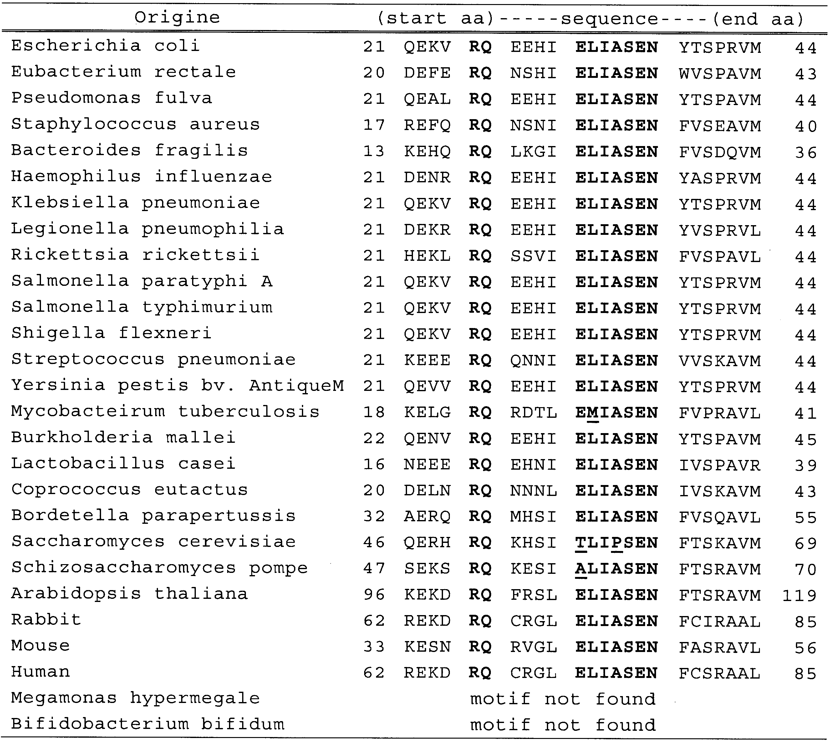
重鎖CDR1としては配列番号3、13、23、33、又は43の何れかに示すアミノ酸配列等;
重鎖CDR2としては配列番号4、14、24、34、又は44の何れかに示すアミノ酸配列等;
重鎖CDR3としては配列番号5、15、25、35、又は45の何れかに示すアミノ酸配列等;
軽鎖CDR1としては配列番号8、18、28、38、又は48の何れかに示すアミノ酸配列等;
軽鎖CDR2としては配列番号9、19、29、39、又は49の何れかに示すアミノ酸配列等;
軽鎖CDR3としては配列番号10、20、30、40、又は50の何れかに示すアミノ酸配列等が挙げられる。
(1)配列番号3、13、23、33、又は43の何れかに示すアミノ酸配列からなる、重鎖CDR1;
(2)配列番号4、14、24、34、又は44の何れかに示すアミノ酸配列からなる、重鎖CDR2;
(3)配列番号5、15、25、35、又は45の何れかに示すアミノ酸配列からなる、重鎖CDR3
が、アミノ末端から順に含まれている重鎖可変領域が挙げられる。
(I)配列番号8、18、28、38、又は48の何れかに示すアミノ酸配列からなる、軽鎖CDR1;
(II)配列番号9、19、29、39、又は49の何れかに示すアミノ酸配列からなる、軽鎖CDR2;
(III)配列番号10、20、30、40、又は50の何れかに示すアミノ酸配列からなる、軽鎖CDR3
が、アミノ末端から順に含まれている軽鎖可変領域が挙げられる。
配列番号3に示すアミノ酸配列からなる重鎖CDR1、配列番号4に示すアミノ酸配列からなる重鎖CDR2、及び配列番号5に示すアミノ酸配列からなる重鎖CDR3をアミノ末端から順に含む重鎖可変領域;
配列番号13に示すアミノ酸配列からなる重鎖CDR1、配列番号14に示すアミノ酸配列からなる重鎖CDR2、及び配列番号15に示すアミノ酸配列からなる重鎖CDR3をアミノ末端から順に含む重鎖可変領域;
配列番号23に示すアミノ酸配列からなる重鎖CDR1、配列番号24に示すアミノ酸配列からなる重鎖CDR2、及び配列番号25に示すアミノ酸配列からなる重鎖CDR3をアミノ末端から順に含む重鎖可変領域;配列番号33に示すアミノ酸配列からなる重鎖CDR1、配列番号34に示すアミノ酸配列からなる重鎖CDR2、及び配列番号35に示すアミノ酸配列からなる重鎖CDR3をアミノ末端から順に含む重鎖可変領域;又は
配列番号43に示すアミノ酸配列からなる重鎖CDR1、配列番号44に示すアミノ酸配列からなる重鎖CDR2、及び配列番号45に示すアミノ酸配列からなる重鎖CDR3をアミノ末端から順に含む重鎖可変領域
が挙げられる。
配列番号18に示すアミノ酸配列からなる軽鎖CDR1、配列番号19に示すアミノ酸配列からなる軽鎖CDR2、及び配列番号20に示すアミノ酸配列からなる軽鎖CDR3をアミノ末端から順に含む軽鎖可変領域;
配列番号28に示すアミノ酸配列からなる軽鎖CDR1、配列番号29に示すアミノ酸配列からなる軽鎖CDR2、及び配列番号30に示すアミノ酸配列からなる軽鎖CDR3をアミノ末端から順に含む軽鎖可変領域;
配列番号38に示すアミノ酸配列からなる軽鎖CDR1、配列番号39に示すアミノ酸配列からなる軽鎖CDR2、及び配列番号40に示すアミノ酸配列からなる軽鎖CDR3をアミノ末端から順に含む軽鎖可変領域;又は
配列番号48に示すアミノ酸配列からなる軽鎖CDR1、配列番号49に示すアミノ酸配列からなる軽鎖CDR2、及び配列番号50に示すアミノ酸配列からなる軽鎖CDR3をアミノ末端から順に含む軽鎖可変領域
が挙げられる。
配列番号11に示すアミノ酸配列を含む重鎖及び配列番号16に示すアミノ酸配列を含む軽鎖を有するモノクローナルIgA抗体;
配列番号21に示すアミノ酸配列を含む重鎖及び配列番号26に示すアミノ酸配列を含む軽鎖を有するモノクローナルIgA抗体;
配列番号31に示すアミノ酸配列を含む重鎖及び配列番号36に示すアミノ酸配列を含む軽鎖を有するモノクローナルIgA抗体;
配列番号41に示すアミノ酸配列を含む重鎖及び配列番号46に示すアミノ酸配列を含む軽鎖を有するモノクローナルIgA抗体が挙げられ、より好ましくは、
配列番号1に示すアミノ酸配列を含む重鎖及び配列番号6に示すアミノ酸配列を含む軽鎖を有するモノクローナルIgA抗体である。
Prevotella属に属する細菌、
Bacteroides属に属する細菌、
Megamonas属に属する細菌、
Bifidobacterium属に属する細菌、
Faecalibacterium属に属する細菌、
Coprococcus属に属する細菌、
Ruminococcus属に属する細菌、
Blautia属に属する細菌、
Eubacterium属に属する細菌、
Roseburia属に属する細菌、
Lactobacillus属に属する細菌、
Clostridium属に属する細菌、
Escherichia属に属する細菌、
Staphylococcus属に属する細菌、
Enterococcus属に属する細菌、
Pseudomonas属に属する細菌、
Enterorhabdus属に属する細菌
等が挙げられる。
Prevotella melaninogenica、
Bacteroidetes vulgatus、
Megamonas funiformis、
Megamonas hypermegale、
Bifidobacterium bifidum、
Faecalibacterium prausnitzii、
Coprococcus eutactus、
Ruminococcus obeum、
Blautia productus、
Blautia coccoides、
Eubacterium rectale、
Roseburia intestinalis、
Lactobacillus murinus、
Lactobacillus casei、
Clostridium difficile、
Escherichia coli、
Staphylococcus aureus、
Enterococcus faecalis、
Pseudomonas fulva、及び
Enterorhabdus mucosicola
等の細菌が挙げられる。
本発明に係る核酸は、本発明に係るモノクローナルIgA抗体をコードする核酸である。より具体的には、配列番号1~50のいずれか1つに示すアミノ酸配列をコードする塩基配列を有する核酸である。当該核酸は、リボヌクレオチドであっても、デオキシヌクレオチドであってもよい。また、当該核酸は1本鎖の形態であっても、2本鎖の形態であってもよく特に限定はされない。

本発明に係るモノクローナルIgA抗体の製造方法は、上述の〔モノクローナルIgA〕の製造方法であって、下記の工程1及び2を含むことを特徴とする。
(工程1)
腸管粘膜固有層から採取されたB細胞と、それ以外の種類の細胞を混合して融合させ、ハイブリドーマを作製する工程。
(工程2)
上記工程1にて作製したハイブリドーマを培養し、少なくとも2種類の腸内細菌と結合するIgA抗体を産生する細胞を決定し、該細胞から、前記IgA抗体を回収する工程。
本発明に係るモノクローナルIgA抗体の製造方法における工程1は、腸管粘膜固有層から採取されたB細胞と、それ以外の種類の細胞を混合して融合させ、ハイブリドーマを作製する工程である。
本発明に係るモノクローナルIgA抗体の製造方法における工程2は、上記工程1にて作製したハイブリドーマを培養し、少なくとも2種類の腸内細菌と結合するIgA抗体を産生する細胞を決定し、該細胞から、前記抗体を回収する工程である。
(A)上述の工程1にて得られるハイブリドーマの中から、ある1つの腸内細菌に結合するIgA抗体を産生するハイブリドーマを決定する工程;及び
(B)工程Aにて決定されたハイブリドーマの中から、それが産生するIgA抗体が、前記腸内細菌とは異なるその他の腸内細菌とも結合することを確認し、両方の腸内細菌と結合するIgA抗体を産生するハイブリドーマを決定する工程、
を含む方法が挙げられる。
本発明に係るハイブリドーマは、上述の本発明に係るモノクローナルIgA抗体を産生するハイブリドーマである。
本発明に係る医薬組成物は、本発明に係るモノクローナルIgA抗体を含む。
本発明に係る経口又は経腸組成物は、本発明に係るモノクローナルIgA抗体を含む。
この様な経口又は経腸組成物を使用することによって、腸内細菌の異常増殖及び/又は腸内細菌叢の病的変化を抑制する効果を発揮する。なお、腸内細菌の異常増殖及び腸内細菌叢の病的変化とは、上述の〔本発明のモノクローナルIgA抗体〕にて説明した通りである。
本発明に係る疾患の治療方法は、腸内疾患の治療方法であって、上述の本発明に係るモノクローナルIgA抗体、本発明に係る医薬組成物、又は本発明に係る経口組成物の有効量を、腸内疾患に罹患したヒトに投与する工程を含む方法である。
〔1-1:腸管粘膜固有層細胞の分離〕
8週齢の野生型C57BL/6マウス(日本クレア)を、自由飲食条件下で長浜バイオ大学付属実験施設にて20週齢まで飼育した。次いで飼育したマウスを、炭酸ガスにより安楽死させた後、開腹して小腸全長を摘出した。この摘出した小腸サンプルから、結合組織とパイエル板を取り除いた後、小腸を縦切開し、腸内容物をPBSで満たした10cmのdish内で洗い流した。そしてPBSをさらに3~4回交換して、小腸を十分洗浄した。
上記〔腸管粘膜固有層細胞の分離〕において得られた腸管粘膜固有層の細胞と、マウスミエローマ細胞であるNS1細胞を融合してハイブリドーマを作成した。NS1細胞は細胞融合に先立って培養及び維持を行った。細胞融合は、STEMCELL Technologies社のClonaCell(登録商標)-HY Hybridoma Cloning Kitに従い、その試薬と手順に従って行った。

上述の〔IgA産生ハイブリドーマの作製〕において得られた17クローンのIgA抗体産生ハイブリドーマを10%のFCSを含有するRPMI 1640にて大量培養し、その培養上清を遠心分離によって採取した。次いで得られた培養上清を0.22μmフィルターに通した後に、硫安沈殿とPBSに対して透析することによって、それぞれ27種類のモノクローナルIgA抗体(以下、本実施例において、〔クローン名〕モノクローナルIgA抗体と呼ぶことがある。)を粗精製した。
長浜バイオ大学付属実験施設SPF室で飼育中の野生型マウス糞便をPBSに懸濁後、血液寒天培地に希釈懸濁液をぬり拡げ、48~72時間37℃で培養した。その後、コロニーから直接16S rRNA遺伝子のPCRを行った。PCR産物をシークエンスして、各コロニーの細菌の種類を同定した。この方法により、6種類のマウス腸内細細菌をクローニングした。
Enterorhabdus mucosicola;
Escherichia coli;
Staphylococcus aureus;
Lactobacillus murinus;
Enterococcus faecalis;
Pseudomonas fulva。
上述のクローニングした6種類の腸内常在菌及び市販の
Escherichia coli(DH5α株:TOYOBO社から入手);Lactobacillus casei(ATCCより入手)、
Coprococcus eutactus(ATCCより入手)、
Blautia coccoides(ATCCより入手)、
Megamonas hypermegale(ATCCより入手)、
Eubacterium rectale(ATCCより入手)、
Bifidobacterium bifidium(ATCCより入手)
の合計13種類の腸内細菌を至適条件で培養し遠心により集菌した。
Staphylococcus aureus、Escherichia coli、及びEnterococcus faecalisである。
Enterococcus faecalis、Staphylococcus aureus、Escherichia coli、及びPseudomonas fulvaである。
腸内細菌として、
Escherichia coli(DH5α;市販品)、
Escherichia coli(マウス腸内細菌からのクローニング株)、
Pseudomonas fulva、
Staphylococcus aureus及び
Eubacterium rectale
を10mlのLB又は至適培養液中で、それぞれの菌体に適した条件下で振蕩培養を行った。遠心して集菌後、1%のNP-40を含むPBSに懸濁し氷上で超音波破砕を行った。氷上で30分間静置後、遠心分離によって上清を得た。これにSDS-buffer(2-ME含有)を加え、95℃で10分間の熱処理によりタンパク質を変性させた。8%のSDS-PAGE後、フィルターに転写し、ブロッキング後、W27、W30、及びW45モノクローナルIgA抗体(2μg/ml)、続いてGoat anti-mouse IgA(1μg/ml)(Southern Biotechnology社)、IR800 anti-goat IgG(0.2μg/ml)(Rockland社)、更に必要に応じでラベル化されたanti-Goat IgGにより反応を行うウエスタンブロッティング法に供し、Odyssey(LI-COR)により、そのシグナルを検出した。
Bifidobacterium bifidumも上記方法と同様の実験を行った。使用したモノクローナルIgA抗体は、W27である。結果を図4に合わせて示す。Eubacterium rectale以外の菌体の抽出物中のタンパク質に、W27モノクローナルIgA抗体は結合しないことが明らかとなった。
上述の方法によって得られたモノクローナルIgA抗体のうち、2種類以上の腸内細菌に結合することが確認されたW27クローン、W30クローン、W34クローン、W43クローン、及びW11モノクローナルIgA抗体の5つのモノクローナルIgA抗体のアミノ酸配列を、公知の方法によって解析した。
deg Vk:5’-GGCTGCAGSTTCAGTGGCAGTGGRTCWGGRAC-3’(配列番号73)
結果を表4~表8に示す。なお、これらの表中の「全長配列」の下線部は可変領域を示し、「全長配列」及び「可変領域」の太字部は、前から(N末端から)順に、CDR1、CDR2及びCDR3を示す。
・W27モノクローナルIgA抗体;重鎖:配列番号51、軽鎖:配列番号52;
・W30モノクローナルIgA抗体;重鎖:配列番号53、軽鎖:配列番号54;
・W34モノクローナルIgA抗体;重鎖:配列番号55、軽鎖:配列番号56;
・W43モノクローナルIgA抗体;重鎖:配列番号57、軽鎖:配列番号58;
・W11モノクローナルIgA抗体;重鎖:配列番号59、軽鎖:配列番号60。
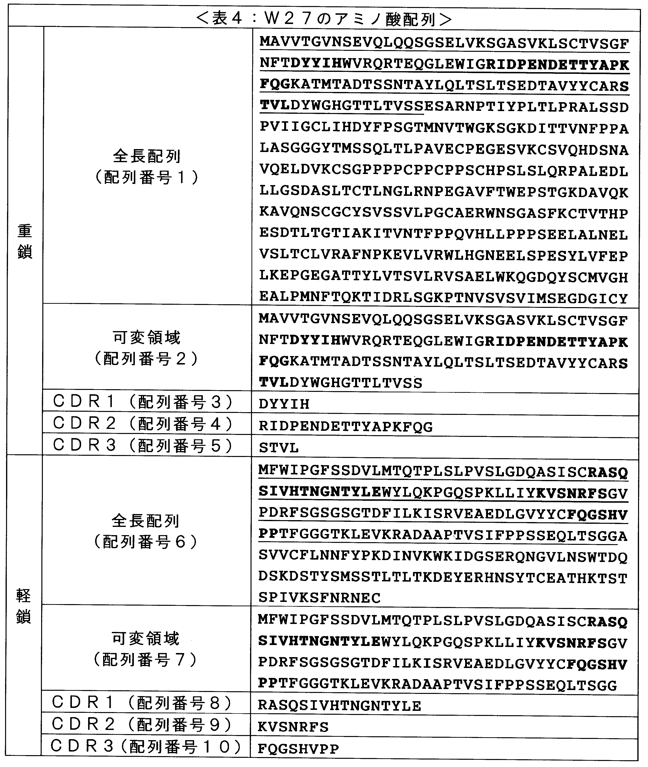
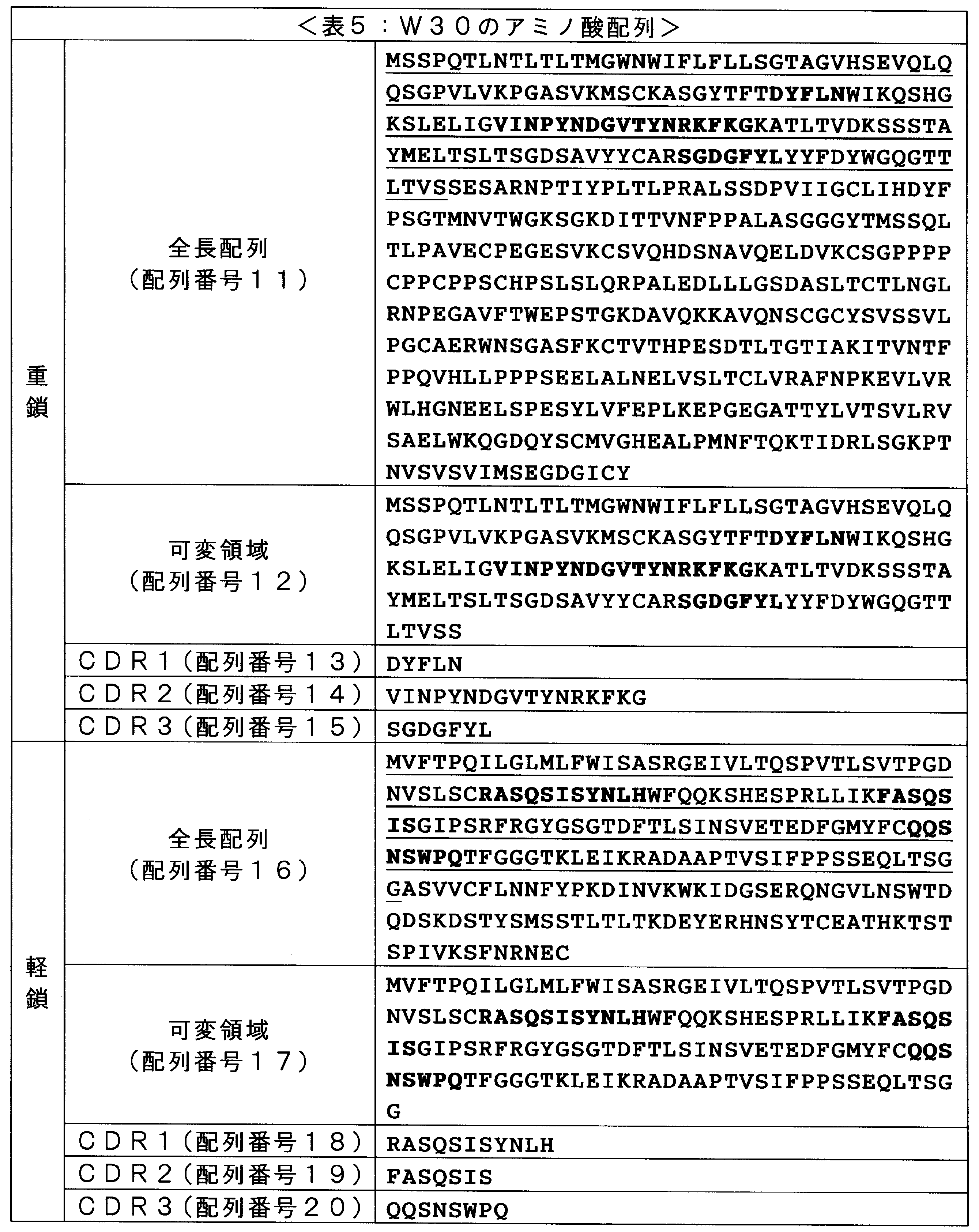
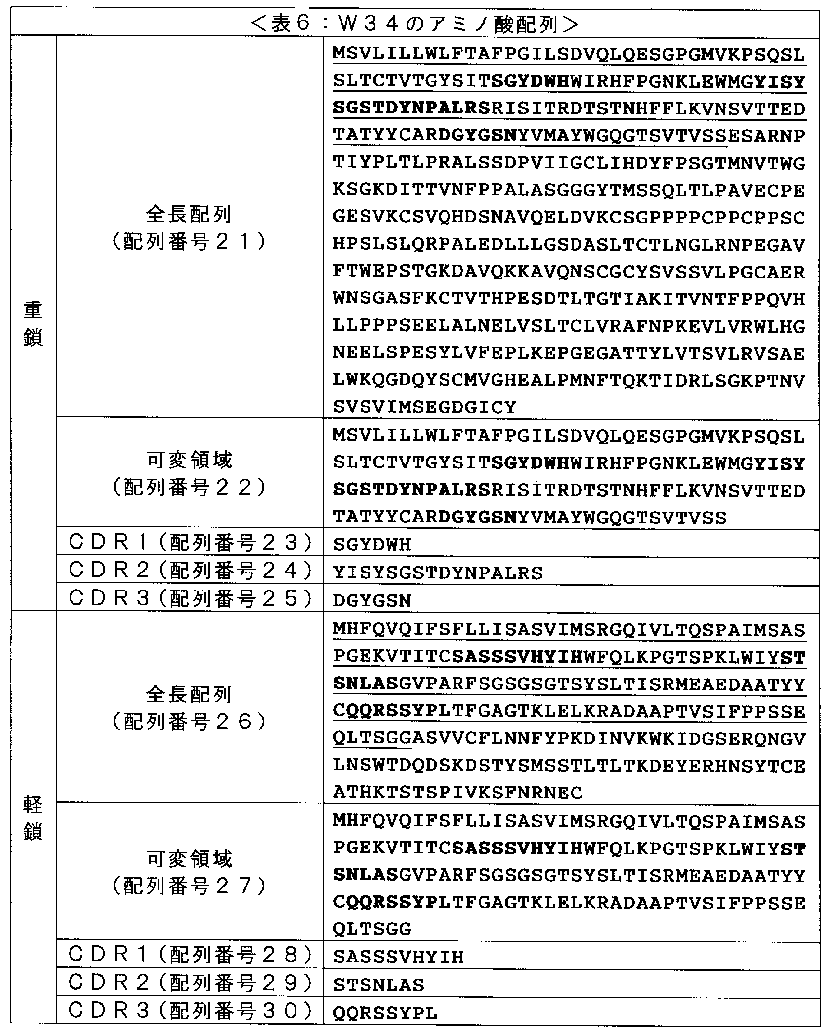
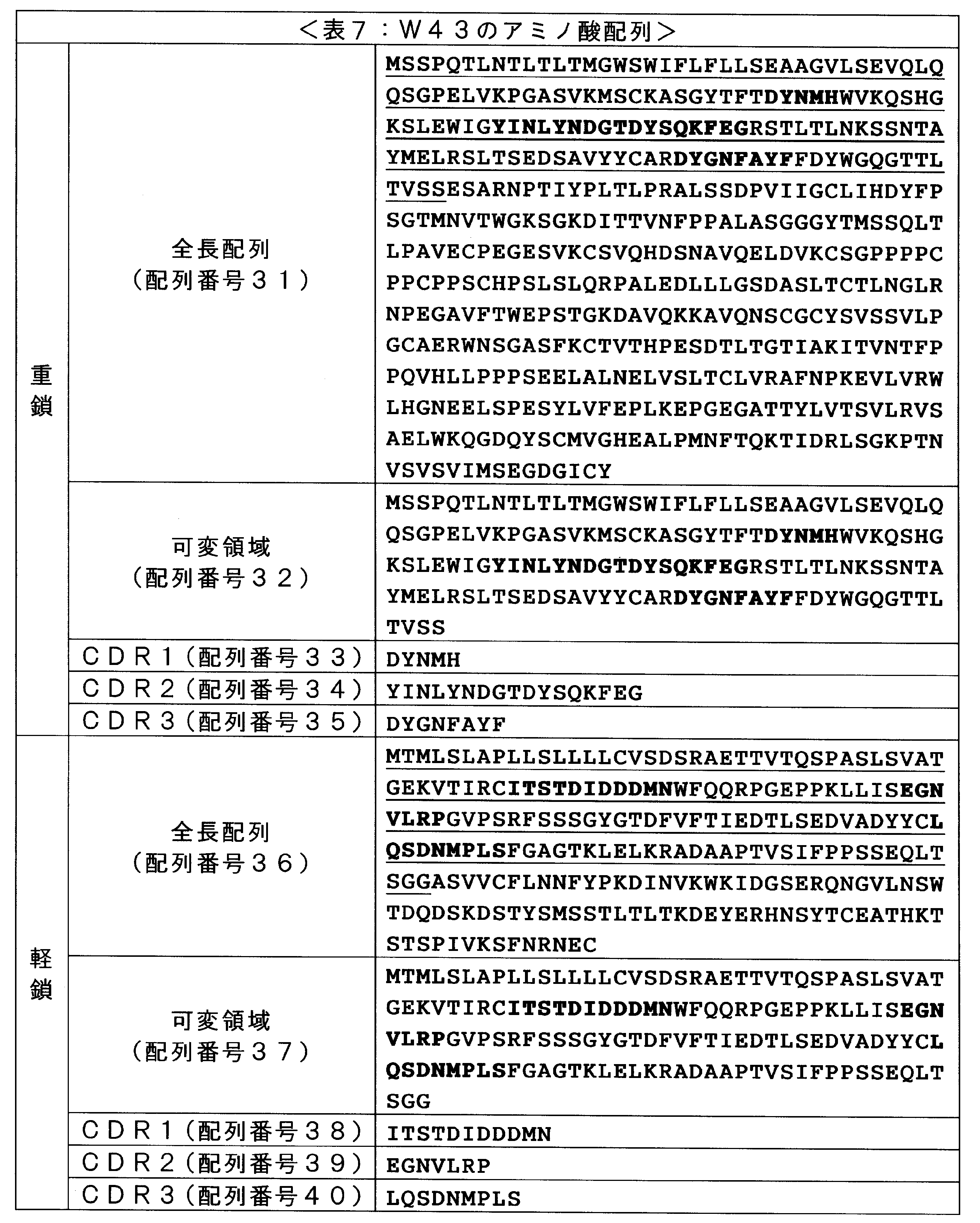
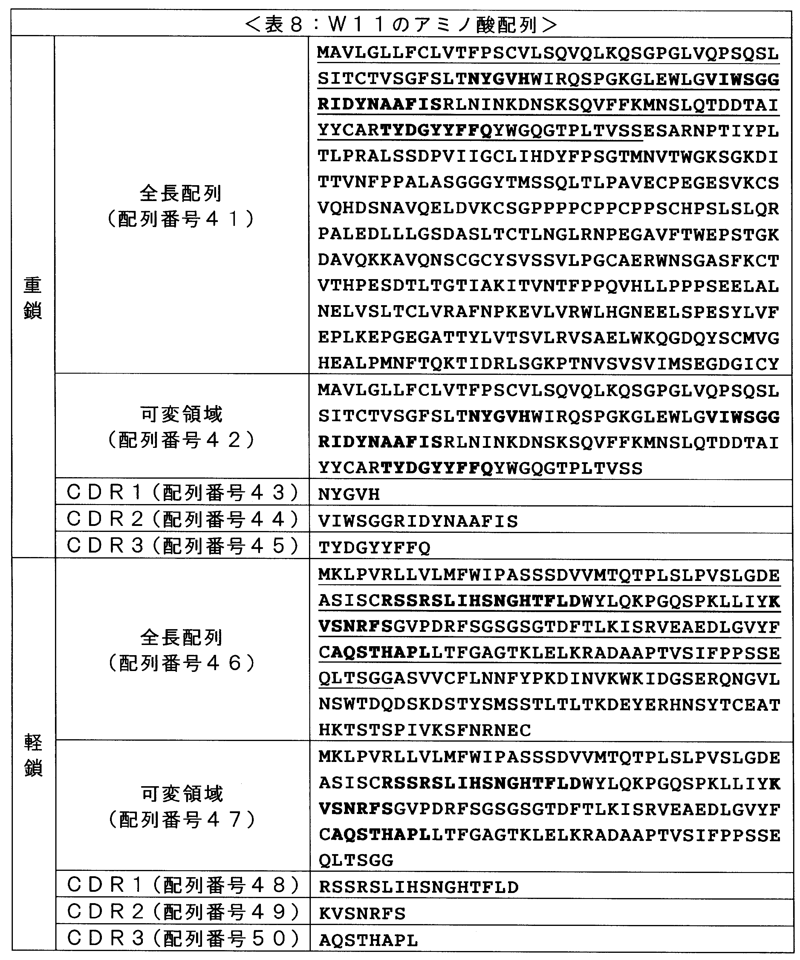
〔2-1:免疫沈降実験〕
上記〔1-5〕の方法で上述の方法でEscehrichia coli(DH5α)、Pseudomonas fulva、及びStaphylococcus aureusのそれぞれ3種類の細菌より細胞抽出成分を得た後、2-ME(終濃度300mM)とSDS(終濃度1%)を加え95℃10分間で熱変性を行った。その後、溶液中の2-ME濃度を60mMにSDS濃度を0.2%に調整した。まず細胞抽出成分に洗浄済みProtein G Sepharose 4 Fast Flow(GE社)を100μl加えて、4℃で30分間の転倒混和を行い、Protein G Sepharoseへの非特異的結合成分を遠心操作により排除した。このPre-clear後の細胞抽出成分に、5μgのW27モノクローナルIgA抗体を加え氷上30分間静置、その後Goat anti-mouse IgAを5μg加えさらに氷上30分間静置した。続いて洗浄済みProtein G Sepharose 4 Fast Flow(GE社)を30μl加えて4℃で15時間の転倒混和を行った。1%のNP-40を含むPBSによる洗浄操作を4回行い、洗浄液を排除後のProtein G SepharoseにSDS-buffer(2-MEを含む)を加え、95℃10分間の熱処理によりタンパク質を変性させた(免疫沈降サンプル)。これを8%のSDS-PAGE、続いて上述のウエスタンブロッティングに供して上述の〔1-5〕にて確認したタンパク質が濃縮されたことを確認した(図5)。
上述の免疫沈降サンプルに対してアセトン沈殿を行い、2次元電気泳動用のバッファーに溶解した。1次元目のゲルはAgar Gel(pH3~8;ATTO社)を使用した。2次元目は6%のSDS-PAGEを行った。毎回、サンプルを2組作成し同時に電気泳動した後は、1組目のサンプルは上述のウエスタンブロッティングを行い、標的タンパク質のゲル内の位置の確認を行った。具体的にはフィルターに転写後、まず転写タンパク質をMemCodeTM Reversible Protein Stain Kit(Thermo Scientific社)を用いて染色してその染色画像を得た(図5)。
MS解析はLCMS-IT-TOF(Shimazu社)により実施した。解析の結果、スポットのタンパク質は、セリンヒドロキシメチルトランスフェラーゼを抗原とすることが明確となった。
次いで、セリンヒドロキシメチルトランスフェラーゼの各種変異体及び野生型を、大腸菌発現系及び293T細胞を用いて作製し、上記各種モノクローナル抗体のエピトープを検討する実験を行った。
(1)1~417番目のアミノ酸配列(full length:野生型)、
(2)11~417番目のアミノ酸配列(△2-10)、
(3)26~417番目のアミノ酸配列(△2-25)、
(4)29~417番目のアミノ酸配列(△2-28)、
(5)1~333番目のアミノ酸配列(△334-417)及び
(6)1~250番目のアミノ酸配列(△251-417)であり、
C末端側にMycタグ(40aa)を設けた。
〔3-1:G23Sマウスの病態モデル解析〕
G23Sマウスの、大腸組織切片を作成し、これをHE染色によって炎症の度合いを調べたところ、13-16週齢においても野生型マウスに比べて腺管の減少または萎縮が多く観察された(図6)。とくに40週齢以上では腺管の減少または萎縮、炎症性細胞の大腸粘膜内への浸潤が多く観察された。免疫染色ではCD11b+細胞やIL-6陽性細胞、IL-6、IL-17等の炎症性細胞の数が野生型マウスに比べて多く存在していた。
上述のW27モノクローナルIgA抗体を、腸管免疫系過剰反応状態、即ちパイエル板胚中心過形成を示し、腸組織において炎症性サイトカインの産生が亢進しているマウスに対して経口投与を行った。
なお、全マウス個体に対して、通常の飼育時と同様に飼料も与えた。
・PE-Cy7-labelled anti-mouse B220(eBioscience社);
・Biotinylated peanut agglutinin(VECTOR laboratories社);
・Streptavidin APC(eBioscience社)。
上述のW27モノクローナルIgA抗体を経口投与して、それが消化管を経由して腸にまで到達するかどうかを確認する実験を、IgAが産生されないAIDノックアウトマウスを用いて行った。
大腸菌株DH5αをLB液体培地に加え、37℃で一晩震盪培養を行った。翌日、大腸菌液を適宜希釈し、LBプレートに播種し、37℃で一晩培養を行った。翌日にプレート上のコロニー数を数えることで、上記の大腸菌液中の大腸菌数を算定した。これを増殖抑制実験開始時の大腸菌数とした。
Claims (26)
- セリンヒドロキシメチルトランスフェラーゼの11~333番目のアミノ酸に結合する、モノクローナルIgA抗体。
- モノクローナルIgA抗体が、
(1)配列番号3、13、23、33、又は43に示すアミノ酸配列からなる重鎖CDR1、
(2)配列番号4、14、24、34、又は44に示すアミノ酸配列からなる重鎖CDR2、及び
(3)配列番号5、15、25、35、又は45に示すアミノ酸配列からなる重鎖CDR3
を含む重鎖可変領域;及び/又は
(I)配列番号8、18、28、38、又は48に示すアミノ酸配列からなる軽鎖CDR1、
(II)配列番号9、19、29、39、又は49に示すアミノ酸配列からなる軽鎖CDR2、及び
(III)配列番号10、20、30、40、又は50に示すアミノ酸配列からなる軽鎖CDR3
を含む軽鎖可変領域を含む、請求項1に記載のモノクローナルIgA抗体。 - モノクローナルIgA抗体が、配列番号2、12、22、32、又は42に示すアミノ酸配列からなる重鎖可変領域;及び/又は配列番号7、17、27、37、又は47に示すアミノ酸配列からなる軽鎖可変領域を含む、請求項1又は2に記載のモノクローナルIgA抗体。
- モノクローナルIgA抗体が、配列番号1、11、21、31、又は41に示すアミノ酸配列からなる重鎖;及び/又は配列番号6、16、26、36、又は46に示すアミノ酸配列からなる軽鎖を含む、請求項1~3の何れか1項に記載のモノクローナルIgA抗体。
- 少なくとも2種類の腸内細菌の成育を阻害する、請求項1~4の何れか1項に記載のモノクローナルIgA抗体。
- 腸内細菌が、
Prevotella属に属する細菌、
Bacteroides属に属する細菌、
Megamonas属に属する細菌、
Bifidobacterium属に属する細菌、
Faecalibacterium属に属する細菌、
Coprococcus属に属する細菌、
Ruminococcus属に属する細菌、
Blautia属に属する細菌、
Eubacterium属に属する細菌、
Roseburia属に属する細菌、
Lactobacillus属に属する細菌、
Clostridium属に属する細菌、
Escherichia属に属する細菌、
Staphylococcus属に属する細菌、
Enterococcus属に属する細菌、
Pseudomonas属に属する細菌、及び
Enterorhabdus属に属する細菌
からなる群に属する細菌より選択される少なくとも2種の細菌である、請求項1~5の何れか1項に記載の製造方法。 - 腸内細菌が、
Prevotella melaninogenica、
Bacteroidetes vulgatus、
Megamonas funiformis、
Megamonas hypermegale、
Bifidobacterium bifidum、
Faecalibacterium prausnitzii、
Coprococcus eutactus、
Ruminococcus obeum、
Blautia productus、
Blautia coccoides、
Eubacterium rectale、
Roseburia intestinalis、
Lactobacillus murinus、
Lactobacillus casei、
Clostridium difficile、
Escherichia coli、
Staphylococcus aureus、
Enterococcus faecalis、
Pseudomonas fulva、及び
Enterorhabdus mucosicola
からなる群より選択される少なくとも2種の細菌である、請求項1~6の何れか1項に記載の製造方法。 - モノクローナルIgA抗体が、IgA1又はIgA2である、請求項1~7の何れか1項に記載のモノクローナルIgA抗体。
- モノクローナルIgA抗体が、J鎖を含む多量体である、請求項1~8の何れか1項に記載のモノクローナルIgA抗体。
- モノクローナルIgA抗体が、分泌因子を含む、請求項1~9の何れか1項に記載のモノクローナルIgA抗体。
- モノクローナルIgA抗体が、ヒト化抗体である、請求項1~10の何れか1項に記載の製造方法。
- モノクローナルIgA抗体が、キメラ抗体である、請求項1~11の何れか1項に記載の製造方法。
- 以下の工程1及び2を含む、請求項1~12の何れか1項に記載のモノクローナルIgA抗体の製造方法:
(1)腸管粘膜固有層から採取されたB細胞と、それ以外の種類の細胞を混合して融合させ、ハイブリドーマを作製する工程1、
(2)工程1にて作製した該ハイブリドーマを培養し、少なくとも2種類の腸内細菌と結合するモノクローナルIgA抗体を産生する細胞を決定し、該細胞から、該IgA抗体を回収する工程2。 - 請求項1~12の何れか1項に記載のモノクローナルIgA抗体を含む、医薬組成物。
- 医薬組成物が、腸内疾患の治療用である、請求項14に記載の医薬組成物。
- 腸内疾患が、腸内細菌の異常増殖及び/又は腸内細菌叢の病的変化により発症する疾患である、請求項14又は15に記載の医薬組成物。
- 腸腸内細菌の異常増殖及び/又は腸内細菌叢の病的変化により発症する疾患が、炎症性腸疾患、潰瘍性大腸炎、クローン病、アレルギー、喘息、肥満、及び自己免疫疾患からなる群から選択される、請求項14~16の何れか1項に記載の医薬組成物。
- 請求項1~12の何れか1項に記載のモノクローナルIgA抗体を含む経口又は経腸組成物。
- 経口又は経腸組成物が、腸内環境改善組成物、腸内環境最適化組成物、又は腸内腐敗防止組成物である、請求項18に記載の経口又は経腸組成物。
- 経口又は経腸組成物が、飲食品組成物又は飼料組成物である、請求項18又は19に記載の経口又は経腸組成物。
- 請求項1~12の何れか1項に記載のモノクローナルIgA抗体をコードする核酸。
- 請求項1~12の何れか1項に記載のモノクローナルIgA抗体を産生するハイブリドーマ。
- 少なくとも2種類の腸内細菌に請求項1~12の何れか1項に記載の抗体を接触させる工程を含む、少なくとも2種類の腸内細菌の増殖を抑制する方法。
- 腸内疾患の治療方法であって、請求項1~12の何れか1項に記載の抗体の有効量を、腸内疾患に罹患したヒトに投与する工程を含む、治療方法。
- 腸内疾患の治療方法における使用のための、請求項1~12の何れか1項に記載のモノクローナルIgA抗体。
- 腸内疾患の治療するための、医薬の製造のための、請求項1~12の何れか1項に記載のモノクローナルIgA抗体の使用。
Priority Applications (5)
| Application Number | Priority Date | Filing Date | Title |
|---|---|---|---|
| CN201480026684.0A CN105246917A (zh) | 2013-03-11 | 2014-03-10 | 生产单克隆IgA抗体的方法 |
| JP2015505472A JP5916946B2 (ja) | 2013-03-11 | 2014-03-10 | モノクローナルIgA抗体の製造方法 |
| EP14762432.4A EP2995679A4 (en) | 2013-03-11 | 2014-03-10 | PROCESS FOR PREPARING MONOCLONAL IGA ANTIBODIES |
| US14/773,923 US9765151B2 (en) | 2013-03-11 | 2014-03-10 | Method for producing monoclonal IgA antibody |
| US15/676,641 US20180009906A1 (en) | 2013-03-11 | 2017-08-14 | METHOD FOR PRODUCING MONOCLONAL IgA ANTIBODY |
Applications Claiming Priority (4)
| Application Number | Priority Date | Filing Date | Title |
|---|---|---|---|
| JP2013-048365 | 2013-03-11 | ||
| JP2013048365 | 2013-03-11 | ||
| JP2013167120 | 2013-08-09 | ||
| JP2013-167120 | 2013-08-09 |
Related Child Applications (2)
| Application Number | Title | Priority Date | Filing Date |
|---|---|---|---|
| US14/773,923 A-371-Of-International US9765151B2 (en) | 2013-03-11 | 2014-03-10 | Method for producing monoclonal IgA antibody |
| US15/676,641 Division US20180009906A1 (en) | 2013-03-11 | 2017-08-14 | METHOD FOR PRODUCING MONOCLONAL IgA ANTIBODY |
Publications (1)
| Publication Number | Publication Date |
|---|---|
| WO2014142084A1 true WO2014142084A1 (ja) | 2014-09-18 |
Family
ID=51536740
Family Applications (1)
| Application Number | Title | Priority Date | Filing Date |
|---|---|---|---|
| PCT/JP2014/056216 Ceased WO2014142084A1 (ja) | 2013-03-11 | 2014-03-10 | モノクローナルIgA抗体の製造方法 |
Country Status (5)
| Country | Link |
|---|---|
| US (2) | US9765151B2 (ja) |
| EP (1) | EP2995679A4 (ja) |
| JP (2) | JP5916946B2 (ja) |
| CN (1) | CN105246917A (ja) |
| WO (1) | WO2014142084A1 (ja) |
Cited By (9)
| Publication number | Priority date | Publication date | Assignee | Title |
|---|---|---|---|---|
| CN113122507A (zh) * | 2021-04-27 | 2021-07-16 | 江南大学 | 一种快速检测铜绿假单胞菌的双抗体夹心elisa方法 |
| JP2021530237A (ja) * | 2018-07-17 | 2021-11-11 | ウーマブス ビオメド エセアー | Campylobacterの種に対する抗体 |
| WO2023277166A1 (ja) | 2021-07-02 | 2023-01-05 | 国立大学法人 東京大学 | クロストリジウム・ディフィシル菌の増殖を抑制するためのモノクローナル抗体含有組成物 |
| JPWO2023277142A1 (ja) * | 2021-07-01 | 2023-01-05 | ||
| JPWO2023054726A1 (ja) * | 2021-09-30 | 2023-04-06 | ||
| WO2024038839A1 (ja) | 2022-08-15 | 2024-02-22 | 国立大学法人 東京大学 | 糸状菌を用いた免疫グロブリンまたは多量体免疫グロブリンの製造方法 |
| WO2024185747A1 (ja) | 2023-03-03 | 2024-09-12 | 国立大学法人 東京大学 | 免疫グロブリン |
| WO2024237217A1 (ja) * | 2023-05-12 | 2024-11-21 | 国立大学法人 東京大学 | クロストリジオイデス・ディフィシル菌に結合する抗体 |
| WO2025220732A1 (ja) * | 2024-04-17 | 2025-10-23 | 国立研究開発法人国立循環器病研究センター | 肺高血圧症の予防又は治療薬 |
Families Citing this family (10)
| Publication number | Priority date | Publication date | Assignee | Title |
|---|---|---|---|---|
| EP2995679A4 (en) * | 2013-03-11 | 2016-12-14 | Kansai Bunri Sogo Gakuen | PROCESS FOR PREPARING MONOCLONAL IGA ANTIBODIES |
| WO2014144292A2 (en) | 2013-03-15 | 2014-09-18 | Sanofi Pasteur Biologics , Llc | Antibodies against clostridium difficile toxins and methods of using the same |
| WO2018181621A1 (ja) * | 2017-03-28 | 2018-10-04 | 味の素株式会社 | 腸内環境改善用飼料組成物 |
| US11633433B2 (en) * | 2017-07-18 | 2023-04-25 | Keio University | Anti-bacterial composition against TH1 cell-inducing bacteria |
| WO2019051790A1 (zh) * | 2017-09-15 | 2019-03-21 | 深圳华大生命科学研究院 | 单形巨单胞菌(Megamonas funiformis)及其应用 |
| CN112888448B (zh) * | 2018-12-07 | 2023-07-25 | 深圳华大生命科学研究院 | 单形巨单胞菌在预防和/或治疗代谢性疾病中的用途 |
| WO2022011264A2 (en) * | 2020-07-10 | 2022-01-13 | The Wistar Institute Of Anatomy And Biology | Dna antibody constructs for use against rotavirus |
| US20240228626A1 (en) * | 2021-05-04 | 2024-07-11 | Immunorizon Ltd. | Anti-nkg2d antibodies and uses thereof |
| TWI795150B (zh) * | 2021-12-28 | 2023-03-01 | 林知儀 | 以生物污泥製成農業用劑的方法 |
| CN117467016B (zh) * | 2023-12-27 | 2024-03-12 | 北京索莱宝科技有限公司 | 人IgA的抗体、抗体组合及其应用 |
Family Cites Families (4)
| Publication number | Priority date | Publication date | Assignee | Title |
|---|---|---|---|---|
| CN1381567A (zh) * | 2001-04-18 | 2002-11-27 | 上海博德基因开发有限公司 | 一种多肽——丝氨酸羟甲基转移酶-10.56和编码这种多肽的多核苷酸 |
| PL1976877T5 (pl) * | 2005-11-30 | 2017-09-29 | Abbvie Inc | Przeciwciała monoklonalne przeciwko białku amyloidu beta oraz ich zastosowania |
| NZ589297A (en) * | 2008-05-15 | 2011-12-22 | Health L P W | Process for producing milk fractions rich in secretory immunoglobulins |
| EP2995679A4 (en) * | 2013-03-11 | 2016-12-14 | Kansai Bunri Sogo Gakuen | PROCESS FOR PREPARING MONOCLONAL IGA ANTIBODIES |
-
2014
- 2014-03-10 EP EP14762432.4A patent/EP2995679A4/en not_active Withdrawn
- 2014-03-10 CN CN201480026684.0A patent/CN105246917A/zh active Pending
- 2014-03-10 US US14/773,923 patent/US9765151B2/en not_active Expired - Fee Related
- 2014-03-10 JP JP2015505472A patent/JP5916946B2/ja not_active Expired - Fee Related
- 2014-03-10 WO PCT/JP2014/056216 patent/WO2014142084A1/ja not_active Ceased
-
2016
- 2016-04-01 JP JP2016073877A patent/JP2016183153A/ja not_active Ceased
-
2017
- 2017-08-14 US US15/676,641 patent/US20180009906A1/en not_active Abandoned
Non-Patent Citations (26)
| Title |
|---|
| ALTSCHUL SF; GISH W; MILLER W; MYERS EW; LIPMAN DJ, J MOL BIOL, vol. 215, 1990, pages 403 - 10 |
| BOULLIER S ET AL., J IMMUNOL, vol. 183, 2009, pages 5879 |
| BRY L ET AL., SCIENCE, vol. 273, 1996, pages 1380 |
| CERF-BENSUSSAN N; GABORIAU-ROUTHIAU V, NAT REV IMMUNOL, vol. 10, 2010, pages 735 |
| CERUTTI A ET AL., ANN NY ACAD SCI, vol. 1238, 2011, pages 132 |
| CHAUDHURI J ET AL., NATURE, vol. 422, 2003, pages 726 |
| FAGARASAN S ET AL., ANNU REV IMMUNOL, vol. 28, 2010, pages 243 |
| FERNANDEZ MI ET AL., IMMUNITY, vol. 18, 2003, pages 739 |
| HOOPER LV; MACPHERSON AJ, NAT REV IMMUNOL, vol. 10, 2010, pages 159 |
| KARLIN S; ALTSCHUL SF, NATL ACAD SCI USA, vol. 90, 1993, pages 5873 - 7 |
| KARLIN S; ALTSCHUL SF, PROC NATL ACAD SCI USA., vol. 87, 1990, pages 2264 - 2268 |
| LI C ET AL., SHENG WU GONG CHENG XUE BAO., vol. 27, no. 2, February 2011 (2011-02-01), pages 219 - 25 |
| MACPHERSON AJ; UHR T, SCIENCE, vol. 303, 2004, pages 1662 |
| MARTINOLI C ET AL., IMMUNITY, vol. 27, 2007, pages 975 |
| MIN WEI ET AL.: "Mice carrying a knock-in mutation of Aicda resulting in a defect in somatic hypermutation have impaired gut homeostasis and compromised mucosal defense", NATURE IMMUNOLOGY, vol. 12, no. 3, March 2011 (2011-03-01), pages 264 - 270, XP055290921 * |
| PHALIPON A ET AL., IMMUNITY, vol. 17, 2002, pages 107 |
| REIKO SHINKURA ET AL.: "Chonai Flora Seigyo ni Yuko na Chokan IgA Kotai no Bunri", YAKULT BIO- SCIENCE FOUNDATION NENPO, 31 January 2013 (2013-01-31), pages 50 - 57, XP008182704 * |
| REIKO SHINKURA: "Chonai Flora Seigyo ni Yuko na Chokan IgA Kotai no Bunri to sono Oyo", YAKULT BIO-SCIENCE FOUNDATION NENPO, 30 September 2013 (2013-09-30), pages 32 - 40, XP008182703 * |
| ROUND JL; MAZMANIAN SK, PROC NATL ACAD SCI USA, vol. 107, 2010, pages 12204 |
| See also references of EP2995679A4 |
| SHINKURA R ET AL., NAT IMMUNOL, vol. 5, 2004, pages 707 |
| SHULZHENKO N ET AL., NAT MED, vol. 17, 2011, pages 1585 |
| TA VT ET AL., NAT IMMUNOL, vol. 4, 2003, pages 843 |
| TURNBAUGH PJ ET AL., NATURE, vol. 444, 2006, pages 1027 |
| VIJAY-KUMAR M ET AL., SCIENCE, vol. 328, 2010, pages 228 |
| WEI M ET AL., NATURE IMMUNOLOGY, vol. 12, no. 3, 2011, pages 264 - 270 |
Cited By (21)
| Publication number | Priority date | Publication date | Assignee | Title |
|---|---|---|---|---|
| JP2021530237A (ja) * | 2018-07-17 | 2021-11-11 | ウーマブス ビオメド エセアー | Campylobacterの種に対する抗体 |
| US12297258B2 (en) | 2018-07-17 | 2025-05-13 | Humabs Biomed Sa | Antibodies against Campylobacter species |
| CN113122507A (zh) * | 2021-04-27 | 2021-07-16 | 江南大学 | 一种快速检测铜绿假单胞菌的双抗体夹心elisa方法 |
| JP7425452B2 (ja) | 2021-07-01 | 2024-01-31 | 国立大学法人 東京大学 | クロストリジウム・ディフィシル菌に結合する抗体 |
| JP2024041901A (ja) * | 2021-07-01 | 2024-03-27 | 国立大学法人 東京大学 | クロストリジウム・ディフィシル菌に結合する抗体 |
| WO2023277142A1 (ja) | 2021-07-01 | 2023-01-05 | 国立大学法人 東京大学 | クロストリジウム・ディフィシル菌に結合する抗体 |
| EP4365293A4 (en) * | 2021-07-01 | 2026-03-11 | Univ Tokyo | ANTIBODIES BINDING TO CLOSTRIDIUM DIFFICILE |
| JPWO2023277142A1 (ja) * | 2021-07-01 | 2023-01-05 | ||
| JP2023123746A (ja) * | 2021-07-01 | 2023-09-05 | 国立大学法人 東京大学 | クロストリジウム・ディフィシル菌に結合する抗体 |
| JP7405386B2 (ja) | 2021-07-01 | 2023-12-26 | 国立大学法人 東京大学 | クロストリジウム・ディフィシル菌に結合する抗体 |
| WO2023277166A1 (ja) | 2021-07-02 | 2023-01-05 | 国立大学法人 東京大学 | クロストリジウム・ディフィシル菌の増殖を抑制するためのモノクローナル抗体含有組成物 |
| JP7430946B2 (ja) | 2021-07-02 | 2024-02-14 | 国立大学法人 東京大学 | クロストリジウム・ディフィシル菌の増殖を抑制するためのモノクローナル抗体含有組成物 |
| JPWO2023277166A1 (ja) * | 2021-07-02 | 2023-01-05 | ||
| EP4364755A4 (en) * | 2021-07-02 | 2026-02-18 | Univ Tokyo | COMPOSITION CONTAINING A MONOCLONAL ANTIBODY TO SUPPRESS CLOSTRIDIUM DIFFICILE BACTERIA |
| WO2023054726A1 (ja) | 2021-09-30 | 2023-04-06 | 礼子 新藏 | モノクローナル抗体含有組成物 |
| JPWO2023054726A1 (ja) * | 2021-09-30 | 2023-04-06 | ||
| WO2024038839A1 (ja) | 2022-08-15 | 2024-02-22 | 国立大学法人 東京大学 | 糸状菌を用いた免疫グロブリンまたは多量体免疫グロブリンの製造方法 |
| WO2024185747A1 (ja) | 2023-03-03 | 2024-09-12 | 国立大学法人 東京大学 | 免疫グロブリン |
| EP4678662A1 (en) | 2023-03-03 | 2026-01-14 | The University of Tokyo | Immunoglobulin |
| WO2024237217A1 (ja) * | 2023-05-12 | 2024-11-21 | 国立大学法人 東京大学 | クロストリジオイデス・ディフィシル菌に結合する抗体 |
| WO2025220732A1 (ja) * | 2024-04-17 | 2025-10-23 | 国立研究開発法人国立循環器病研究センター | 肺高血圧症の予防又は治療薬 |
Also Published As
| Publication number | Publication date |
|---|---|
| EP2995679A1 (en) | 2016-03-16 |
| JP2016183153A (ja) | 2016-10-20 |
| US9765151B2 (en) | 2017-09-19 |
| JP5916946B2 (ja) | 2016-05-11 |
| JPWO2014142084A1 (ja) | 2017-02-16 |
| US20160039944A1 (en) | 2016-02-11 |
| US20180009906A1 (en) | 2018-01-11 |
| CN105246917A (zh) | 2016-01-13 |
| EP2995679A4 (en) | 2016-12-14 |
Similar Documents
| Publication | Publication Date | Title |
|---|---|---|
| JP5916946B2 (ja) | モノクローナルIgA抗体の製造方法 | |
| JP7405386B2 (ja) | クロストリジウム・ディフィシル菌に結合する抗体 | |
| JP7430946B2 (ja) | クロストリジウム・ディフィシル菌の増殖を抑制するためのモノクローナル抗体含有組成物 | |
| KR20120128687A (ko) | 녹농균의 혈청형 g 지질다당류에 대한 항체 | |
| Pearson et al. | IgM-associated gut bacteria in obesity and type 2 diabetes in C57BL/6 mice and humans | |
| JP7862032B2 (ja) | 炎症性腸疾患(ibd)を治療または診断する方法および組成物 | |
| EP4410309A1 (en) | Composition containing monoclonal antibody | |
| WO2024237217A1 (ja) | クロストリジオイデス・ディフィシル菌に結合する抗体 | |
| WO2025134928A1 (ja) | 結腸直腸がんを治療するための組成物 | |
| WO2012029792A1 (ja) | TGF-β受容体の活性化を抑制する活性を有する化合物、そのスクリーニング方法、並びにC型肝炎ウィルスに起因する疾患の予防又は治療のための組成物 | |
| TW202608478A (zh) | 微生物與抗體之組合及用途 | |
| Pranger | Generation and characterisation of PIPE-cloned IgE, IgG1 and IgG4 antibodies against Beta-lactoglobulin to investigate the mechanisms underlying cow milk allergy | |
| Adherent-Invasive | Methyl‑donor supplementation prevents intestinal colonization by Adherent‑invasive E. coli in a mouse model of crohn’s | |
| WO2025220732A1 (ja) | 肺高血圧症の予防又は治療薬 | |
| Lin | Novel Insights in Ankylosing Spondylitis Biology: Mouse and Human Studies |
Legal Events
| Date | Code | Title | Description |
|---|---|---|---|
| 121 | Ep: the epo has been informed by wipo that ep was designated in this application |
Ref document number: 14762432 Country of ref document: EP Kind code of ref document: A1 |
|
| ENP | Entry into the national phase |
Ref document number: 2015505472 Country of ref document: JP Kind code of ref document: A |
|
| NENP | Non-entry into the national phase |
Ref country code: DE |
|
| WWE | Wipo information: entry into national phase |
Ref document number: 2014762432 Country of ref document: EP |
|
| WWE | Wipo information: entry into national phase |
Ref document number: 14773923 Country of ref document: US |