WO2014156584A1 - 微生物検出用センサー、その製造方法、およびポリマー層 - Google Patents
微生物検出用センサー、その製造方法、およびポリマー層 Download PDFInfo
- Publication number
- WO2014156584A1 WO2014156584A1 PCT/JP2014/056143 JP2014056143W WO2014156584A1 WO 2014156584 A1 WO2014156584 A1 WO 2014156584A1 JP 2014056143 W JP2014056143 W JP 2014056143W WO 2014156584 A1 WO2014156584 A1 WO 2014156584A1
- Authority
- WO
- WIPO (PCT)
- Prior art keywords
- microorganism
- polymer layer
- detection electrode
- sensor
- microorganisms
- Prior art date
- Legal status (The legal status is an assumption and is not a legal conclusion. Google has not performed a legal analysis and makes no representation as to the accuracy of the status listed.)
- Ceased
Links
Images


Classifications
-
- G—PHYSICS
- G01—MEASURING; TESTING
- G01N—INVESTIGATING OR ANALYSING MATERIALS BY DETERMINING THEIR CHEMICAL OR PHYSICAL PROPERTIES
- G01N33/00—Investigating or analysing materials by specific methods not covered by groups G01N1/00 - G01N31/00
- G01N33/48—Biological material, e.g. blood, urine; Haemocytometers
- G01N33/50—Chemical analysis of biological material, e.g. blood, urine; Testing involving biospecific ligand binding methods; Immunological testing
- G01N33/53—Immunoassay; Biospecific binding assay; Materials therefor
- G01N33/543—Immunoassay; Biospecific binding assay; Materials therefor with an insoluble carrier for immobilising immunochemicals
- G01N33/54366—Apparatus specially adapted for solid-phase testing
- G01N33/54373—Apparatus specially adapted for solid-phase testing involving physiochemical end-point determination, e.g. wave-guides, FETS, gratings
- G01N33/5438—Electrodes
-
- C—CHEMISTRY; METALLURGY
- C12—BIOCHEMISTRY; BEER; SPIRITS; WINE; VINEGAR; MICROBIOLOGY; ENZYMOLOGY; MUTATION OR GENETIC ENGINEERING
- C12Q—MEASURING OR TESTING PROCESSES INVOLVING ENZYMES, NUCLEIC ACIDS OR MICROORGANISMS; COMPOSITIONS OR TEST PAPERS THEREFOR; PROCESSES OF PREPARING SUCH COMPOSITIONS; CONDITION-RESPONSIVE CONTROL IN MICROBIOLOGICAL OR ENZYMOLOGICAL PROCESSES
- C12Q1/00—Measuring or testing processes involving enzymes, nucleic acids or microorganisms; Compositions therefor; Processes of preparing such compositions
- C12Q1/02—Measuring or testing processes involving enzymes, nucleic acids or microorganisms; Compositions therefor; Processes of preparing such compositions involving viable microorganisms
- C12Q1/04—Determining presence or kind of microorganism; Use of selective media for testing antibiotics or bacteriocides; Compositions containing a chemical indicator therefor
-
- C—CHEMISTRY; METALLURGY
- C12—BIOCHEMISTRY; BEER; SPIRITS; WINE; VINEGAR; MICROBIOLOGY; ENZYMOLOGY; MUTATION OR GENETIC ENGINEERING
- C12M—APPARATUS FOR ENZYMOLOGY OR MICROBIOLOGY; APPARATUS FOR CULTURING MICROORGANISMS FOR PRODUCING BIOMASS, FOR GROWING CELLS OR FOR OBTAINING FERMENTATION OR METABOLIC PRODUCTS, i.e. BIOREACTORS OR FERMENTERS
- C12M41/00—Means for regulation, monitoring, measurement or control, e.g. flow regulation
- C12M41/30—Means for regulation, monitoring, measurement or control, e.g. flow regulation of concentration
- C12M41/36—Means for regulation, monitoring, measurement or control, e.g. flow regulation of concentration of biomass, e.g. colony counters or by turbidity measurements
-
- C—CHEMISTRY; METALLURGY
- C25—ELECTROLYTIC OR ELECTROPHORETIC PROCESSES; APPARATUS THEREFOR
- C25D—PROCESSES FOR THE ELECTROLYTIC OR ELECTROPHORETIC PRODUCTION OF COATINGS; ELECTROFORMING; APPARATUS THEREFOR
- C25D5/00—Electroplating characterised by the process; Pretreatment or after-treatment of workpieces
- C25D5/34—Pretreatment of metallic surfaces to be electroplated
-
- C—CHEMISTRY; METALLURGY
- C25—ELECTROLYTIC OR ELECTROPHORETIC PROCESSES; APPARATUS THEREFOR
- C25D—PROCESSES FOR THE ELECTROLYTIC OR ELECTROPHORETIC PRODUCTION OF COATINGS; ELECTROFORMING; APPARATUS THEREFOR
- C25D5/00—Electroplating characterised by the process; Pretreatment or after-treatment of workpieces
- C25D5/48—After-treatment of electroplated surfaces
-
- G—PHYSICS
- G01—MEASURING; TESTING
- G01N—INVESTIGATING OR ANALYSING MATERIALS BY DETERMINING THEIR CHEMICAL OR PHYSICAL PROPERTIES
- G01N33/00—Investigating or analysing materials by specific methods not covered by groups G01N1/00 - G01N31/00
- G01N33/48—Biological material, e.g. blood, urine; Haemocytometers
- G01N33/50—Chemical analysis of biological material, e.g. blood, urine; Testing involving biospecific ligand binding methods; Immunological testing
- G01N33/53—Immunoassay; Biospecific binding assay; Materials therefor
- G01N33/569—Immunoassay; Biospecific binding assay; Materials therefor for microorganisms, e.g. protozoa, bacteria, viruses
-
- G—PHYSICS
- G01—MEASURING; TESTING
- G01N—INVESTIGATING OR ANALYSING MATERIALS BY DETERMINING THEIR CHEMICAL OR PHYSICAL PROPERTIES
- G01N2600/00—Assays involving molecular imprinted polymers/polymers created around a molecular template
Definitions
- the present invention relates to a sensor for detecting microorganisms, a method for producing the same, and a polymer layer.
- a technique capable of detecting the pathogenic bacteria quickly and with high sensitivity is required.
- methods for detecting and identifying microorganisms include ELISA methods and Western blotting methods. These include, for example, an antigen-antibody reaction between an antibody (primary antibody) and a microorganism-specific protein, and then a labeled secondary antibody is reacted with the antibody (primary antibody) to cause chemiluminescence of the secondary antibody or ATP. This is a method of detecting by monitoring the hydrolysis reaction.
- Patent Document 1 JP 2009-58232 A (Patent Document 1) describes a method for detecting anionic molecules (ATP, amino acids, etc.) derived from microorganisms using the electrochemical properties of a polymer provided with a molecular template. Has been.
- none of the above methods is a method for detecting microorganisms themselves.
- the ELISA method is not easy because it is necessary to prepare an antibody against a protein unique to a microorganism.
- An object of the present invention is to provide a novel sensor for detecting microorganisms, a method for producing the same, and a polymer layer that can detect microorganisms quickly and easily with high sensitivity.
- the present invention includes a detection unit having a detection electrode and a polymer layer that is arranged on the detection electrode and includes a three-dimensional structure template complementary to the three-dimensional structure of the microorganism to be detected. It is a sensor that detects microorganisms based on the capture state of microorganisms.
- the polymer layer is formed by polymerizing a monomer in the presence of a microorganism to be detected to form a polymer layer in which the microorganism is incorporated on the detection electrode, and at least a part of the microorganism incorporated in the polymer layer. Is formed by a production method having a destruction step of bringing the solution into contact with a solution containing a lytic enzyme and destroying the solution. In a preferred form of the sensor, the solution used in the destruction step further contains a chelating agent.
- a preferred form of the sensor further includes a crystal resonator having the detection electrode of the detection unit as one electrode, and measures the change in the mass of the polymer layer from the change in the resonance frequency of the crystal resonator to determine the capture state of the microorganism. To detect.
- the monomer is preferably selected from the group consisting of pyrrole, aniline, thiophene and derivatives thereof, more preferably pyrrole or derivatives thereof.
- the surface on which the polymer layer of the detection electrode is formed is preferably a rough surface.
- the microorganism As the microorganism, a microorganism having a negative or excessive charge on the entire surface or the surface is preferable.
- the present invention also relates to a sensor for detecting a microorganism having a detection portion having a detection electrode and a polymer layer provided on the detection electrode and having a three-dimensional template complementary to the three-dimensional structure of the microorganism.
- a polymerization step of polymerizing monomers in the presence of microorganisms to be detected to form a polymer layer in which the microorganisms are incorporated on the detection electrode, and at least of the microorganisms incorporated in the polymer layer And a destruction step in which a part is brought into contact with a solution containing a lytic enzyme and destroyed.
- the present invention also provides a polymer layer having a template having a three-dimensional structure complementary to the three-dimensional structure of a microorganism, the polymerization step of polymerizing monomers in the presence of the microorganism to form the polymer layer, And a destruction step of bringing the incorporated microorganisms into contact with a solution containing a lytic enzyme and destroying the microorganisms.
- the senor of the present invention it is possible to detect microorganisms quickly and easily with high sensitivity.
- a sensor capable of detecting microorganisms quickly and easily with high sensitivity is provided.
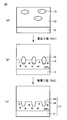
- the preferable preparation process of the polymer layer of the sensor concerning this invention is shown typically, (a) is before a polymerization process, (b) After a polymerization process, (c) is sectional drawing after a destruction process.
- the sensor of this invention it is a schematic diagram which shows the outline of a mode that the target microorganism is capture
- FIG. 2 is an electron micrograph of the polypyrrole layer surface after the polymerization step in Example 1.
- FIG. 2 is an electron micrograph of the surface of a polypyrrole layer after washing with sterilized water after the breaking step of Example 1.
- FIG. 2 is an electron micrograph of the surface of a polypyrrole layer after washing with sterilized water after the breaking step of Example 2.
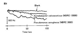
- FIG. 6 is a graph showing a change in resonance frequency of a crystal resonator during microorganism detection using the sensor of Example 1.
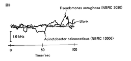
- 6 is a graph showing a change in resonance frequency of a crystal resonator when microorganisms are detected using the sensor of Example 2.
- the sensor of the present invention includes a detection part having a detection electrode and a polymer layer that is arranged on the detection electrode and has a three-dimensional template complementary to the three-dimensional structure of the microorganism, Microorganisms are detected based on the captured state.
- the polymer layer of the sensor of the present invention is a polymer that forms a polymer layer on the detection electrode by polymerizing monomers in the presence of microorganisms to be detected (hereinafter also referred to as “target microorganisms”). It is formed by the manufacturing method which has a process and the destruction process which contacts and destroys at least one part of the microorganisms taken in by the polymer layer with the solution containing a lytic enzyme.
- FIG. 1 is a cross-sectional view schematically showing a preferred production process of a polymer layer of a sensor according to the present invention.
- FIG. 1 shows an embodiment in which pyrrole is used as a monomer.
- a solution 12 containing a microorganism 13 and pyrrole is prepared in an environment in contact with the detection electrode 11.
- the concentration of the monomer constituting the polymer layer contained in the solution 12 can be 1 mM to 100 M, and the concentration of the microorganism 13 can be 1 to 1 ⁇ 10 10 cfu / mL.
- polypyrrole In the polymerization step (St1), electrolysis using the detection electrode 11 as an anode and a counter electrode (not shown) as a cathode is performed, and polypyrrole (FIG. 1B) is formed on the detection electrode 11 by an oxidative polymerization reaction of pyrrole. ) “PPy” is an abbreviation for polypyrrole).
- Microorganisms 13 are taken into the formed polymer layer 14.
- the pyrrole itself has a positive charge in order to emit electrons to the detection electrode 11 in the polymerization process, and in order to compensate for this positive charge, the microorganism 13 whose whole or surface charge is in a state of excessive negative charge. Is believed to be incorporated into the polymer layer 14.
- a destruction step for destroying the microorganisms 13 taken in the polymer layer 14 is performed.
- the destruction step can be performed by bringing the microorganism 13 into contact with a solution containing a lytic enzyme such as lysozyme.
- a lytic enzyme such as lysozyme.
- the solution used in the destruction step preferably further contains a chelating agent.
- the chelating agent used in the destruction step include EDTA (ethylenediaminetetraacetic acid), EGTA, NTA, DTPA, HEDTA and the like.
- the lytic enzyme used in the destruction step include lysozyme, N-acetylmuramitase, and Achromobacter endopeptidase.
- the microorganisms 13 taken into the polymer layer 14 can be destroyed, and the destroyed microorganisms 13 are released from the polymer layer 14 to form the template 15.
- the chelating agent contained in the solution facilitates the expression of the lytic action by the lytic enzyme. Therefore, the degree of destruction of the microorganism 13 can be increased by including the chelating agent, and the microorganism is associated with the destruction. It is understood that 13 facilitates release from the polymer layer 14.
- the efficiency of the destruction step can be easily improved, and as described later, the detection sensitivity of the sensor can be improved as compared with those not using a chelating agent.
- the concentration of the lytic enzyme in the solution used in the destruction step is preferably 1 to 1000 mg / mL.
- the concentration of the chelating agent in the solution is preferably 10 to 1000 ⁇ g / mL.
- the time for bringing the solution containing the lytic enzyme into contact with the microorganisms 13 incorporated in the polymer layer 14 is preferably 12 to 48 hours.
- the region where the microorganisms 13 exist in the polymer layer 14 becomes a template 15 having a three-dimensional structure complementary to the three-dimensional structure of the microorganisms 13.
- the laminate of the polymer layer 14 having the template 15 formed in this way and the detection electrode 11 constitutes the detection unit 17 in the sensor of the present invention.
- the thickness of the polymer layer 14 in the detection unit 17 can be set to 0.1 to 10 ⁇ m, for example.
- the microorganism 13 to be detected is not particularly limited as long as the whole or surface of the microorganism is in a negative charge excess state.
- Acinetobacter, Serratia, Klebsiella, Enterobacter, Citrobacter, Burkholderia, Sphingomonadase, Chromobacterium, Salmonella, Vibrio, Legionella, Campylobacter, Yersinia, Proteus, Nephy Examples include Streptococcus genus, Enterococcus genus, Clostridium genus, Corynebacterium genus, Listeria genus, Bacillus genus, Mycobacterium genus, Chlamydia genus, Rickettsia genus and Haemophilus genus bacteria.
- viruses examples include hepatitis A virus, adenovirus, rotavirus, and norovirus.
- examples of fungi examples include candida.
- Examples of protozoa include cryptosporidium.
- the total or surface charge of the microorganism varies depending on the water quality of the solution 12 such as pH. For example, there are various functional groups such as a carboxyl group, an amino group, and a phosphate group on the surface of the microorganism, and the surface containing these functional groups becomes negatively charged when the pH increases. Therefore, for example, the solution 12 may be made alkaline in order to make the negative charge excessive when forming the template or measuring.
- the monomer used as the raw material for the polymer layer is not limited to pyrrole, and other examples include aniline, thiophene. And derivatives thereof.
- the material of the detection electrode 11 is not particularly limited, and is a gold electrode, a multilayer electrode of gold and chromium, a multilayer electrode of gold and titanium, a silver electrode, a multilayer electrode of silver and chromium, and a silver and titanium layer. Examples include multilayer electrodes, lead electrodes, platinum electrodes, carbon electrodes, and the like.
- the surface on which the polymer layer 14 of the detection electrode 11 is formed is preferably subjected to a roughening treatment. Since the surface on which the polymer layer 14 of the detection electrode 11 is formed is a rough surface, the adhesion to the polymer layer 14 is improved, and the surface area of the electrode is increased.
- the detection electrode 11 when a gold electrode is used as the detection electrode 11, it is possible to perform a roughening process in which the gold electrode surface is subjected to plasma etching and then gold nanoparticles are fixed to roughen the surface.
- the surface roughness of the detection electrode surface 11 may be a center line average roughness, for example, 0.4 to 50 ⁇ m.
- FIG. 2 is a schematic diagram showing an outline of how a target microorganism is captured by a template.
- FIG. 2A shows a case where the microorganism 13a in the sample solution is a target microorganism
- FIG. 2B shows a case where the microorganism 13b in the sample solution is not a target microorganism.
- a sample solution is prepared in an environment in contact with the detection unit 17 including the polymer layer 14 and the detection electrode 11.
- the three-dimensional structure of the microorganism 13a complementary to the three-dimensional structure of the template 15 becomes the template.
- Microorganisms 13b that are captured within 15 (FIG. 2 (a)) but not complementary to template 15 are not captured within template 15 (FIG. 2 (b)).
- the movement of the microorganism may be an active movement of the microorganism, or may be moved by electrophoresis, dielectrophoresis or water flow, or simply precipitated or diffused.
- Detection of target microorganisms When the microorganisms 13a are trapped in the template 15, for example, a change in mass, a change in conductive characteristics, a change in capacitance, a change in light reflectance, a change in temperature, and the like occur in the laminate composed of the polymer layer 14 and the detection electrode 11.
- a change in mass, a change in conductive characteristics, a change in capacitance, a change in light reflectance, a change in temperature, and the like occur in the laminate composed of the polymer layer 14 and the detection electrode 11.
- the capture state of the microorganism in the template 15 is detected.
- the target microorganism can be detected based on the captured state. With such detection, rapid and sensitive detection of the target microorganism can be achieved.
- the mass change detection method there is a detection method for detecting a change in the resonance frequency of the crystal resonator.
- a crystal resonator microbalance (QCM) sensor which is a preferred example of the sensor of
- FIG. 3 is a schematic diagram showing a schematic configuration of the QCM sensor.
- the QCM sensor 33 includes a cell 27 for holding a solution, a crystal resonator 32 disposed at the bottom of the cell 27, an oscillation circuit 22, and a controller 21 having a frequency counter.
- the crystal unit 32 is formed by sequentially stacking the detection unit 17, the crystal piece 24, and the counter electrode (second counter electrode) 23 manufactured by the process illustrated in FIG. 1.
- the QCM sensor 33 further includes a counter electrode (first counter electrode) 16 immersed in the sample solution 31 and a reference electrode 30, and a DC power source between the detection electrode 11 and the counter electrode 16 of the detection unit 17. Can be connected.
- the sample solution 31 is added into the cell 27. Then, an alternating voltage is applied between the detection electrode 11 and the counter electrode 23 by the oscillation circuit 22 to vibrate the crystal piece 24.
- the mass of the detection unit 17 changes, and the resonance frequency of the crystal piece 24 changes.
- the frequency counter in the controller 21 receives the signal from the oscillation circuit 22 and measures the resonance frequency value. The capture state of the microorganism is detected from the change in the resonance frequency value.
- a polymer layer can be formed on the detection electrode 11 in accordance with the roughening treatment of the surface of the detection electrode 11 and the process shown in FIG.
- a crystal resonator in which the detection electrode 11, the crystal piece 24, and the counter electrode 23 are stacked in this order is disposed at the bottom of the cell 27, and a DC power source is provided between the detection electrode 11 and the counter electrode 16.
- the progress of the formation of the polymer layer can be confirmed by monitoring the change in the resonance frequency of the crystal resonator together with the formation of the polymer layer.
- the templates according to the present invention are individually formed and combined, or a template corresponding to a plurality of microorganisms is contained in a single template. By forming simultaneously, it is also possible to detect a plurality of types of microorganisms simultaneously.
- bacteria can be detected in several minutes to several tens of minutes, and can be detected much more quickly than in the culture method.
- it is a device such as a water purifier, a water server or an automatic ice making device. Easy to incorporate and automate.
- it can be used in water purification plants and beverage / food factories as a bacteria inspection tool for water quality testing and food testing. More specifically, it is possible to automatically detect bacteria in the apparatus such as the water storage tank and the piping path and notify the user, or take measures such as sterilization and washing automatically.
- the polymer layer in the above-mentioned sensor has a three-dimensional structure template complementary to the three-dimensional structure of the microorganism, a microorganism capturing device, a microorganism shape recognition device, a microorganism tracking device, It can also be used for a catalyst carrier utilizing the porous material.
- the polymer layer is produced using an electrochemical measurement system (Model 842B, manufactured by ALS), and the reference electrode is Ag / AgCl (saturated KCl), the counter electrode (first pair).
- the electrode a Pt rod (diameter 1 mm, length 4 cm, manufactured by Niraco Co., Ltd.) was used.
- the potential is a value relative to the potential of the reference electrode.
- a quartz crystal resonator (electrode area 0.196 cm 2 , fundamental vibration frequency 9 MHz, AT cut, square type, Seiko EG & G Co., Ltd.) provided with gold electrodes (detection electrode and second counter electrode) on both sides. Made).
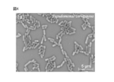
- FIG. 4 shows an electron micrograph of Pseudomonas aeruginosa.
- Example 1 (Roughening process of gold electrode)
- the surface of the gold electrode was roughened according to the following procedure. 1. Etching was performed for 30 seconds on the surface of the gold electrode (trade name: QA-A9M-AU, manufactured by Seiko EG & G Co., Ltd.) using a plasma etching apparatus (SEDE / meiwa fossis). 2. A crystal resonator was placed on the bottom of a cell (well type cell, trade name: QA-CL4, manufactured by Seiko EG & G Co.) 27 of a QCM sensor 33 as shown in FIG.
- a polypyrrole layer was produced on the gold electrode according to the following procedure. 5.
- a 0.1M pyrrole aqueous solution containing 1 ⁇ 10 9 cfu / mL of Pseudomonas aeruginosa was prepared as a modification solution. 6).
- the modification solution was added into the cell 27 of the QCM sensor 33 where the gold electrode subjected to the roughening treatment was disposed, and the first counter electrode and the reference electrode were inserted into the modification solution. 7).
- Polypyrrole was deposited on the gold electrode by performing constant potential electrolysis (+0.975 V, 90 seconds) in the modification solution to produce a polypyrrole layer (polymerization step), and then the polypyrrole layer was washed with sterilized water. In the polymerization process, the resonance frequency of the crystal resonator was also monitored. The surface of the polypyrrole layer after the polymerization step was observed with a scanning microscope (SEM). The thickness of the polypyrrole layer was about 0.6 ⁇ m. 8).
- EDTA solution 400 ⁇ g / mL, pH: 8.07, Tris buffer
- lysozyme was added to the EDTA solution and dissolved to prepare a lysozyme solution (20 mg / mL).
- a Triton solution (20 wt%, pH 8.03, Tris buffer) containing a nonionic surfactant (trade name: Triton) was also prepared.
- Triton solution 20 wt%, pH 8.03, Tris buffer
- Triton solution 250 ⁇ L was added to the cell 27 and allowed to stand at room temperature for 1 day.
- 250 ⁇ L of Triton solution was added dropwise and allowed to stand at room temperature for 1 day, thereby lysing the microorganisms incorporated into the polypyrrole layer. Done (destructive process).
- SEM scanning electron microscope
- Example 2 In “8.” of Example 1 described above, a lysozyme solution (20 mg / mL) containing no EDTA was prepared. In “9.”, polypyrrole was used in the same manner as in Example 1 except that this lysozyme solution was used. A layer was made.
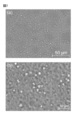
- FIG. 5 shows an electron micrograph of the surface of the polypyrrole layer after the polymerization step in Example 1.
- FIG. 5B is an electron micrograph showing an enlarged part of FIG. In FIG. 5, it was observed that Pseudomonas aeruginosa was taken up on the surface of the polypyrrole layer.
- FIG. 6 (a) and 6 (b) are electron micrographs of the surface of the polypyrrole layer after washing with sterilized water after the destruction step of Example 1, and FIG. 6 (b) is a part of FIG. 6 (a). It is the electron micrograph which expanded and showed.
- 7 (a) and 7 (b) are electron micrographs of the surface of the polypyrrole layer after washing with sterilized water after the destruction step of Example 2, and FIG. 7 (b) is a part of FIG. 7 (a). It is the electron micrograph which expanded and showed.
- FIG. 6 it can be seen that almost no incorporated P. aeruginosa exist and that a Pseudomonas aeruginosa template is formed in the polypyrrole layer as compared to FIG.
- FIGS. 7A and 7B it can be seen that although Pseudomonas aeruginosa is present on a part of the surface of the polypyrrole layer, a template is simultaneously formed.
- Detection experiment Microorganisms were detected using a QCM sensor prepared as described above and having a crystal resonator having a polypyrrole layer formed on the surface thereof and provided at the bottom of the cell. A sample solution containing microorganisms was added to the cell. Then, the resonance frequency of the crystal resonator was monitored.
- FIG. 8 is a graph showing changes in the resonance frequency of the crystal resonator in the sensor of Example 1. From the result shown in FIG. 8, in the sensor of Example 1, when a sample solution containing Pseudomonas aeruginosa was added, the resonance frequency was higher than that of a sample or blank containing Acinetobacter calcoaceticus. It was found that it decreased greatly. The decrease in the resonance frequency means an increase in the mass of the surface of the crystal unit, and it is considered that the mass of the surface of the crystal unit is increased by the incorporation of Pseudomonas aeruginosa into the mold of the polypyrrole layer. Therefore, it can be seen that the Pseudomonas aeruginosa can be detected by the sensor of Example 1.
- FIG. 9 is a graph showing changes in the resonance frequency of the crystal resonator in the sensor of Example 2. From the results shown in FIG. 9, in the sensor of Example 2, the resonance frequency was not decreased even when the sample solution containing Pseudomonas aeruginosa was added. However, from FIGS. 7A and 7B, it can be seen that a template is formed on the surface of the polypyrrole layer. Therefore, when a detection method with higher sensitivity than the detection method used here is adopted, It is understood that Pseudomonas aeruginosa can also be detected by the sensor of Example 2.
- microorganisms can be detected quickly and easily, which not only helps to reduce the defective product rate and storage cost, but also provides a desired microorganism template. By forming and detecting, it becomes possible to easily grasp the mixing route of microorganisms and plan countermeasures.
Landscapes
- Health & Medical Sciences (AREA)
- Chemical & Material Sciences (AREA)
- Life Sciences & Earth Sciences (AREA)
- Engineering & Computer Science (AREA)
- Organic Chemistry (AREA)
- Immunology (AREA)
- Wood Science & Technology (AREA)
- Zoology (AREA)
- Molecular Biology (AREA)
- Biomedical Technology (AREA)
- Biotechnology (AREA)
- Microbiology (AREA)
- Analytical Chemistry (AREA)
- General Health & Medical Sciences (AREA)
- Biochemistry (AREA)
- Bioinformatics & Cheminformatics (AREA)
- Hematology (AREA)
- Urology & Nephrology (AREA)
- Physics & Mathematics (AREA)
- General Engineering & Computer Science (AREA)
- Proteomics, Peptides & Aminoacids (AREA)
- Genetics & Genomics (AREA)
- Medicinal Chemistry (AREA)
- Electrochemistry (AREA)
- Cell Biology (AREA)
- Metallurgy (AREA)
- Materials Engineering (AREA)
- Food Science & Technology (AREA)
- Chemical Kinetics & Catalysis (AREA)
- General Physics & Mathematics (AREA)
- Pathology (AREA)
- Toxicology (AREA)
- Biophysics (AREA)
- Sustainable Development (AREA)
- Tropical Medicine & Parasitology (AREA)
- Virology (AREA)
- Apparatus Associated With Microorganisms And Enzymes (AREA)
- Measuring Or Testing Involving Enzymes Or Micro-Organisms (AREA)
Abstract
Description
[センサーにおけるポリマー層の作製]
図1は、本発明にかかるセンサーのポリマー層の好ましい作製工程を模式的に示す断面図である。図1では、モノマーとしてピロールを用いる場合の実施形態を示す。まず、図1(a)に示すように、検出用電極11に接触する環境下に、微生物13およびピロールを含む溶液12を準備する。溶液12中に含まれるポリマー層を構成するモノマーの濃度としては1mM~100Mとすることができ、微生物13の濃度は1~1×10¹⁰cfu/mLとすることができる。
図2は、鋳型へ標的微生物が捕捉される様子の概略を示す模式図である。図2(a)は試料溶液中の微生物13aが標的微生物である場合を示し、図2(b)は試料溶液中の微生物13bが標的微生物でない場合を示す。図2(a)、(b)に示すように、まず、ポリマー層14と検出用電極11からなる検出部17に接触する環境下に、試料溶液を準備する。負に帯電している微生物が、正に帯電したPPy膜との静電相互作用などにより、検出部17の方向に移動すると、鋳型15の三次元構造と相補的な立体構造の微生物13aは鋳型15内に捕捉されるが(図2(a))、鋳型15と相補的でない微生物13bは鋳型15内に捕捉されない(図2(b))。なお、微生物の移動は、微生物の能動的な移動であっても良いし、電気泳動、誘電泳動や水流などによって動かしたり、あるいは単に沈殿したり、拡散したりするものであっても良い。また、微生物以外の、たとえば、泥、鉄さびといった濁質が水に含まれていた場合であっても、それらも鋳型15と三次元的形状、荷電状態等が異なり相補的でないため、捕捉されない。そのため、標的微生物と他の濁質の識別が可能である。
微生物13aが鋳型15内に捕捉されると、ポリマー層14および検出用電極11からなる積層体に、たとえば、質量変化、導電特性変化、電気容量変化、光反射率変化、温度変化等が生じる。本発明のセンサーにおいては、このような変化を検出して、微生物の鋳型15への捕捉状態を検出する。そして、捕捉状態に基づいて標的微生物の検出が可能となる。このような検出により、標的微生物の迅速かつ高感度の検出が達成され得る。質量変化の検出方法の具体例として、水晶振動子の共振周波数の変化を検出する検出方法が挙げられる。以下、本発明のセンサーの好ましい一例である、水晶振動子マイクロバランス(QCM)センサーについて説明する。
図3は、QCMセンサーの概略構成を示す模式図である。QCMセンサー33は、溶液を保持するセル27と、セル27の底部に配置された水晶振動子32と、発振回路22と、周波数カウンタを有するコントローラ21とを備える。水晶振動子32は、図1に示した工程により作製された検出部17と、水晶片24と、対電極(第2対電極)23とが順に積層されてなる。QCMセンサー33は、さらに、試料溶液31内に浸漬される対電極(第1対電極)16と、参照電極30とを備え、検出部17の検出用電極11と対電極16の間に直流電源を接続することができる。
[実施例1]
(金電極の粗面化工程)
金電極の表面に、ポリピロール層との密着性向上のため以下の手順にしたがい水晶振動子積層体の金電極表面の粗面化処理を行なった。
1. プラズマエッチング装置(SEDE/meiwa fosis)により、金電極(商品名:QA-A9M-AU、(株)セイコー・イージーアンドジー製)表面に30秒間エッチングを行なった。
2. 水晶振動子を、図3に示すようなQCMセンサー33のセル(ウェル型セル、商品名:QA-CL4、(株)セイコー・イージーアンドジー製)27の底部に設置した。その後、30nmのクエン酸保護金ナノ粒子(0.0574wt%)を含んだ溶液500μLをセル27に添加し、室温で24時間放置した。
3. 金電極を純水で洗浄後、臭化ヘキサデシルトリメチルアンモニウム溶液(0.1M)9mL、塩化金(III)酸四塩化物(0.01M)250μL、NaOH(0.1M)
50μL、アスコルビン酸(0.1M)50μLを混合して調製した溶液(Auナノ粒子成長溶液)500μLをセル27に添加し、室温で24時間静置した。
4. セル27内の溶液を除去し、金電極を超純水で洗浄した。
以下の手順に従って金電極上にポリピロール層を作製した。
5. 1×10⁹cfu/mLの緑膿菌を含む0.1Mのピロール水溶液を調製して修飾溶液とした。
6. 上記で粗面化処理を施した金電極が配置されているQCMセンサー33のセル27内に、修飾溶液を添加し、修飾溶液内に第1対電極および参照電極を差し込んだ。
7. 修飾溶液中において定電位電解(+0.975V、90秒間)することで金電極上にポリピロールを析出させ、ポリピロール層を作製し(重合工程)、その後滅菌水でポリピロール層を洗浄した。重合工程においては、水晶振動子の共振周波数のモニターも行なった。重合工程後のポリピロール層について、走査型顕微鏡(SEM)により表面観察を行なった。ポリピロール層の厚みは約0.6μmであった。
8. EDTA溶液(400μg/mL、pH:8.07、Trisバッファー)を作製し、このEDTA溶液にリゾチームを加えて溶解することでリゾチーム溶液(20mg/mL)を調製した。また、非イオン界面活性剤(商品名:Triton)を含むTriton溶液(20wt%、pH8.03、Trisバッファー)も併せて調製した。
9. 作製したリゾチーム溶液250μLをセル27内に添加して室温で1日間静置し、さらにTriton溶液を250μL滴下して室温で1日間静置することで、ポリピロール層に取り込まれた微生物の溶菌処理を行った(破壊工程)。
10. セル27内の溶液を除去し、滅菌水でポリピロール層を洗浄した後、走査型電子顕微鏡(SEM)により表面観察を行った。
上記した実施例1の「8.」において、EDTAを含まないリゾチーム溶液(20mg/mL)を調製し、「9.」において、このリゾチーム溶液を用いた点以外は、実施例1と同様にポリピロール層を作製した。
図5は、実施例1における重合工程後のポリピロール層表面の電子顕微鏡写真を示す。図5(b)は、図5(a)の一部を拡大して示した電子顕微鏡写真である。図5において、ポリピロール層の表面に緑膿菌が取り込まれた様子が観察された。
(検出実験)
上述のようにして作製した、ポリピロール層が表面に形成された水晶振動子をセルの底部に備えたQCMセンサーを用いて微生物の検出を行なった。セル内に微生物を含む試料溶液を添加した。そして、水晶振動子の共振周波数をモニタリングした。
図8は、実施例1のセンサーにおける水晶振動子の共振周波数変化を示すグラフである。図8に示す結果から、実施例1のセンサーにおいては、緑膿菌(Pseudomonas aeruginosa)を含む試料溶液を添加した場合に、アシネトバクター(Acinetobacter calcoaceticus)を含む試料またはブランクと比較して、共振周波数が大きく減少することが分かった。共振周波数の減少は水晶振動子表面の質量の増加を意味しており、緑膿菌がポリピロール層の鋳型に取り込まれることで水晶振動子表面の質量が増加したものと考えられる。したがって、実施例1のセンサーでは緑膿菌を検出できることが分かる。
対電極(第1対電極)、17 検出部、21 コントローラ、22 発振回路、23 対電極(第2対電極)、24 水晶片、27 セル、30 参照電極、31 試料溶液、32 水晶振動子、33 QCMセンサー。
Claims (9)
- 検出用電極と、前記検出用電極上に配置され、検出対象の微生物の立体構造に相補的な三次元構造の鋳型を備えたポリマー層とを有する検出部を備え、
前記鋳型への前記微生物の捕捉状態に基づいて前記微生物を検出するセンサーであって、
前記ポリマー層は、検出対象とする微生物の存在下でモノマーを重合して前記微生物を取り込んだ状態の前記ポリマー層を前記検出用電極上に形成する重合工程と、前記ポリマー層に取り込まれた微生物の少なくとも一部を、溶菌酵素を含む溶液に接触させて破壊する破壊工程と、を有する製造方法により形成される、センサー。 - 前記破壊工程に用いる溶液がさらにキレート剤を含む、請求項1に記載のセンサー。
- 前記検出部の前記検出用電極を一方の電極とする水晶振動子をさらに備え、
前記水晶振動子の共振周波数の変化から前記ポリマー層の質量の変化を測定して前記微生物の捕捉状態を検出する、請求項1または2に記載のセンサー。 - 前記モノマーが、ピロール、アニリン、チオフェンおよびそれらの誘導体からなる群から選択される、請求項1~3のいずれか一項に記載のセンサー。
- 前記モノマーが、ピロールまたはその誘導体からなる、請求項4に記載のセンサー。
- 前記検出用電極の前記ポリマー層の形成面が粗面である、請求項1~5のいずれか一項に記載のセンサー。
- 前記微生物は、全体または表面の電荷が負電荷過剰の状態にある、請求項1~6のいずれか一項に記載のセンサー。
- 検出用電極と、前記検出用電極上に配置され、微生物の立体構造に相補的な三次元構造の鋳型を備えたポリマー層とを有する検出部を備えた微生物を検出するセンサーの製造方法であって、
検出対象とする微生物の存在下でモノマーを重合して前記微生物を取り込んだ状態の前記ポリマー層を前記検出用電極上に形成する重合工程と、
前記ポリマー層に取り込まれた微生物の少なくとも一部を、溶菌酵素を含む溶液に接触させて破壊する破壊工程と、を有する製造方法。 - 微生物の立体構造に相補的な三次元構造の鋳型を備えたポリマー層であって、
前記微生物の存在下でモノマーを重合して前記ポリマー層を形成する重合工程と、
前記ポリマー層に取り込まれた微生物の少なくとも一部を、溶菌酵素を含む溶液に接触させて破壊する破壊工程と、を有する製造方法により製造される、ポリマー層。
Priority Applications (4)
| Application Number | Priority Date | Filing Date | Title |
|---|---|---|---|
| US14/774,002 US20160018391A1 (en) | 2013-03-28 | 2014-03-10 | Microorganism detection sensor, method for manufacturing same, and polymer layer |
| CN201480018305.3A CN105229137A (zh) | 2013-03-28 | 2014-03-10 | 微生物检测用传感器、其制造方法以及聚合物层 |
| EP14774943.6A EP2980204A4 (en) | 2013-03-28 | 2014-03-10 | SENSOR FOR DETECTING MICROORGANISMS, METHOD FOR THE PRODUCTION THEREOF AND POLYMER LAYER |
| JP2015508246A JP6077106B2 (ja) | 2013-03-28 | 2014-03-10 | 微生物検出用センサー、その製造方法、およびポリマー層 |
Applications Claiming Priority (2)
| Application Number | Priority Date | Filing Date | Title |
|---|---|---|---|
| JP2013-069739 | 2013-03-28 | ||
| JP2013069739 | 2013-03-28 |
Publications (1)
| Publication Number | Publication Date |
|---|---|
| WO2014156584A1 true WO2014156584A1 (ja) | 2014-10-02 |
Family
ID=51623568
Family Applications (1)
| Application Number | Title | Priority Date | Filing Date |
|---|---|---|---|
| PCT/JP2014/056143 Ceased WO2014156584A1 (ja) | 2013-03-28 | 2014-03-10 | 微生物検出用センサー、その製造方法、およびポリマー層 |
Country Status (5)
| Country | Link |
|---|---|
| US (1) | US20160018391A1 (ja) |
| EP (1) | EP2980204A4 (ja) |
| JP (1) | JP6077106B2 (ja) |
| CN (1) | CN105229137A (ja) |
| WO (1) | WO2014156584A1 (ja) |
Cited By (1)
| Publication number | Priority date | Publication date | Assignee | Title |
|---|---|---|---|---|
| JP2017077197A (ja) * | 2015-10-20 | 2017-04-27 | 株式会社リコー | 液滴形成装置 |
Families Citing this family (4)
| Publication number | Priority date | Publication date | Assignee | Title |
|---|---|---|---|---|
| EP3139166A4 (en) * | 2014-05-02 | 2017-04-19 | Osaka Prefecture University Public Corporation | Polymer film for detection of cancer cells and manufacturing method thereof, and cancer cell detection device using such polymer film |
| CN106645348B (zh) * | 2016-12-23 | 2019-03-05 | 南开大学 | 一种高稳定微生物电化学传感器的制备方法 |
| GB2634255A (en) * | 2023-10-03 | 2025-04-09 | Univ Durham | An electrochemical system for detecting bacterial growth in a duct |
| CN118655199B (zh) * | 2024-08-19 | 2024-12-24 | 四川菊乐食品股份有限公司 | 一种细胞印迹电化学传感器的制备方法及利用其快速定量检测乳制品中乳酸菌的方法 |
Citations (2)
| Publication number | Priority date | Publication date | Assignee | Title |
|---|---|---|---|---|
| JP2009058232A (ja) | 2007-08-29 | 2009-03-19 | Atect Corp | 分子鋳型を有するポリマーを備えたセンサー |
| WO2012121229A1 (ja) * | 2011-03-08 | 2012-09-13 | 公立大学法人大阪府立大学 | 微生物検出用センサーおよびその製造方法 |
Family Cites Families (2)
| Publication number | Priority date | Publication date | Assignee | Title |
|---|---|---|---|---|
| US20030138845A1 (en) * | 2001-08-23 | 2003-07-24 | Changming Li | Protein and peptide sensors using electrical detection methods |
| JP2007319103A (ja) * | 2006-06-02 | 2007-12-13 | Hitachi Plant Technologies Ltd | 微生物分離システム |
-
2014
- 2014-03-10 EP EP14774943.6A patent/EP2980204A4/en not_active Withdrawn
- 2014-03-10 JP JP2015508246A patent/JP6077106B2/ja active Active
- 2014-03-10 CN CN201480018305.3A patent/CN105229137A/zh active Pending
- 2014-03-10 US US14/774,002 patent/US20160018391A1/en not_active Abandoned
- 2014-03-10 WO PCT/JP2014/056143 patent/WO2014156584A1/ja not_active Ceased
Patent Citations (2)
| Publication number | Priority date | Publication date | Assignee | Title |
|---|---|---|---|---|
| JP2009058232A (ja) | 2007-08-29 | 2009-03-19 | Atect Corp | 分子鋳型を有するポリマーを備えたセンサー |
| WO2012121229A1 (ja) * | 2011-03-08 | 2012-09-13 | 公立大学法人大阪府立大学 | 微生物検出用センサーおよびその製造方法 |
Non-Patent Citations (2)
| Title |
|---|
| See also references of EP2980204A4 |
| SHIHO TOKONAMI ET AL.: "DEVELOPMENT OF BACTERIAL SENSORS FABRICATED BY MOLECULARLY IMPRINTED POLYMER TECHNIQUES", ABSTRACTS OF THE 79TH ANNUAL MEETING OF THE ELECTROCHEMICAL SOCIETY OF JAPAN, 2012, pages 19, XP008183983 * |
Cited By (1)
| Publication number | Priority date | Publication date | Assignee | Title |
|---|---|---|---|---|
| JP2017077197A (ja) * | 2015-10-20 | 2017-04-27 | 株式会社リコー | 液滴形成装置 |
Also Published As
| Publication number | Publication date |
|---|---|
| EP2980204A1 (en) | 2016-02-03 |
| US20160018391A1 (en) | 2016-01-21 |
| EP2980204A4 (en) | 2016-10-19 |
| JP6077106B2 (ja) | 2017-02-08 |
| JPWO2014156584A1 (ja) | 2017-02-16 |
| CN105229137A (zh) | 2016-01-06 |
Similar Documents
| Publication | Publication Date | Title |
|---|---|---|
| JP6014582B2 (ja) | 微生物検出用センサーの製造方法 | |
| JP6077106B2 (ja) | 微生物検出用センサー、その製造方法、およびポリマー層 | |
| González‐Rivas et al. | Biofilms in the spotlight: Detection, quantification, and removal methods | |
| Nguyen et al. | Biofouling of water treatment membranes: a review of the underlying causes, monitoring techniques and control measures | |
| Gall et al. | The effect of electric fields on bacterial attachment to conductive surfaces | |
| Gutman et al. | Bacterial attachment and viscoelasticity: physicochemical and motility effects analyzed using quartz crystal microbalance with dissipation (QCM-D) | |
| Oulahal‐Lagsir et al. | “Escherichia coli‐milk” biofilm removal from stainless steel surfaces: synergism between ultrasonic waves and enzymes | |
| Luo et al. | Chlorine-resistant bacteria (CRB) in the reverse osmosis system for wastewater reclamation: Isolation, identification and membrane fouling mechanisms | |
| KR20100130218A (ko) | 예정된 세균 숙주 균주를 감염시킬 수 있는 박테리오파지를 탐지 및/정량화시키는 방법 및 시스템, 상기 박테리오파지 탐지 그리고 상기 방법을 실행하기 위한 마이크로전자공학 센서 장치 | |
| Zhu et al. | Cooperation mode of outer surface and inner space of nanochannel: separation-detection system based on integrated nanochannel electrode for rapid and facile detection of Salmonella | |
| Oulahal‐Lagsir et al. | Ultrasonic methodology coupled to ATP bioluminescence for the non‐invasive detection of fouling in food processing equipment—validation and application to a dairy factory | |
| Taskin et al. | Combined cell culture-biosensing platform using vertically aligned patterned peptide nanofibers for cellular studies | |
| Escosura-Muñiz et al. | Electrical evaluation of bacterial virulence factors using nanopores | |
| WO2012167218A2 (en) | Device and method for identifying microbes and counting microbes and determining antimicrobial sensitivity | |
| Chen et al. | Immobilized reporter phage on electrospun polymer fibers for improved capture and detection of Escherichia coli O157: H7 | |
| Patel et al. | Impedance based biosensor for agricultural pathogen detection | |
| Liu et al. | Bacterial surface appendages modulate the antimicrobial activity induced by nanoflake surfaces on titanium | |
| Jashrapuria et al. | Biofilm Inhibition by Laser-Induced Graphene: Impact of Surface Texture on Rod-Shaped E. coli and Coccus-Shaped Staphylococcus | |
| Starosvetsky et al. | Effects of physical parameters on bacterial cell adsorption onto pre-imprinted sol-gel films | |
| Maliszewska et al. | Biofouling removal from membranes using nonthermal plasma | |
| Vera-Villalobos et al. | Vibrio neptunius-ULV11 cell-free supernatant as a promising antifouling approach in reverse osmosis systems | |
| WO2017046313A1 (en) | Enzyme-functionalised nanobeads for anti-biofouling purposes | |
| WO2015166977A1 (ja) | がん細胞検出用高分子膜及びその製造方法、並びにそれを用いたがん細胞検出装置 | |
| JP6406875B2 (ja) | 抗菌性検査方法 | |
| Lin | Cell-surface interactions between bacteria and zinc oxide nanopillars |
Legal Events
| Date | Code | Title | Description |
|---|---|---|---|
| WWE | Wipo information: entry into national phase |
Ref document number: 201480018305.3 Country of ref document: CN |
|
| 121 | Ep: the epo has been informed by wipo that ep was designated in this application |
Ref document number: 14774943 Country of ref document: EP Kind code of ref document: A1 |
|
| WWE | Wipo information: entry into national phase |
Ref document number: 2014774943 Country of ref document: EP |
|
| ENP | Entry into the national phase |
Ref document number: 2015508246 Country of ref document: JP Kind code of ref document: A |
|
| WWE | Wipo information: entry into national phase |
Ref document number: 14774002 Country of ref document: US |
|
| NENP | Non-entry into the national phase |
Ref country code: DE |