WO2015093472A1 - 動物細胞用構造体、動物細胞の分離方法及び動物細胞用構造体表面の弾性調節方法 - Google Patents
動物細胞用構造体、動物細胞の分離方法及び動物細胞用構造体表面の弾性調節方法 Download PDFInfo
- Publication number
- WO2015093472A1 WO2015093472A1 PCT/JP2014/083247 JP2014083247W WO2015093472A1 WO 2015093472 A1 WO2015093472 A1 WO 2015093472A1 JP 2014083247 W JP2014083247 W JP 2014083247W WO 2015093472 A1 WO2015093472 A1 WO 2015093472A1
- Authority
- WO
- WIPO (PCT)
- Prior art keywords
- layer
- elastic
- base material
- elastic body
- elasticity
- Prior art date
- Legal status (The legal status is an assumption and is not a legal conclusion. Google has not performed a legal analysis and makes no representation as to the accuracy of the status listed.)
- Ceased
Links
Images







Classifications
-
- C—CHEMISTRY; METALLURGY
- C12—BIOCHEMISTRY; BEER; SPIRITS; WINE; VINEGAR; MICROBIOLOGY; ENZYMOLOGY; MUTATION OR GENETIC ENGINEERING
- C12M—APPARATUS FOR ENZYMOLOGY OR MICROBIOLOGY; APPARATUS FOR CULTURING MICROORGANISMS FOR PRODUCING BIOMASS, FOR GROWING CELLS OR FOR OBTAINING FERMENTATION OR METABOLIC PRODUCTS, i.e. BIOREACTORS OR FERMENTERS
- C12M23/00—Constructional details, e.g. recesses, hinges
-
- C—CHEMISTRY; METALLURGY
- C12—BIOCHEMISTRY; BEER; SPIRITS; WINE; VINEGAR; MICROBIOLOGY; ENZYMOLOGY; MUTATION OR GENETIC ENGINEERING
- C12N—MICROORGANISMS OR ENZYMES; COMPOSITIONS THEREOF; PROPAGATING, PRESERVING, OR MAINTAINING MICROORGANISMS; MUTATION OR GENETIC ENGINEERING; CULTURE MEDIA
- C12N5/00—Undifferentiated human, animal or plant cells, e.g. cell lines; Tissues; Cultivation or maintenance thereof; Culture media therefor
- C12N5/0068—General culture methods using substrates
-
- C—CHEMISTRY; METALLURGY
- C12—BIOCHEMISTRY; BEER; SPIRITS; WINE; VINEGAR; MICROBIOLOGY; ENZYMOLOGY; MUTATION OR GENETIC ENGINEERING
- C12M—APPARATUS FOR ENZYMOLOGY OR MICROBIOLOGY; APPARATUS FOR CULTURING MICROORGANISMS FOR PRODUCING BIOMASS, FOR GROWING CELLS OR FOR OBTAINING FERMENTATION OR METABOLIC PRODUCTS, i.e. BIOREACTORS OR FERMENTERS
- C12M23/00—Constructional details, e.g. recesses, hinges
- C12M23/26—Constructional details, e.g. recesses, hinges flexible
-
- C—CHEMISTRY; METALLURGY
- C12—BIOCHEMISTRY; BEER; SPIRITS; WINE; VINEGAR; MICROBIOLOGY; ENZYMOLOGY; MUTATION OR GENETIC ENGINEERING
- C12M—APPARATUS FOR ENZYMOLOGY OR MICROBIOLOGY; APPARATUS FOR CULTURING MICROORGANISMS FOR PRODUCING BIOMASS, FOR GROWING CELLS OR FOR OBTAINING FERMENTATION OR METABOLIC PRODUCTS, i.e. BIOREACTORS OR FERMENTERS
- C12M41/00—Means for regulation, monitoring, measurement or control, e.g. flow regulation
- C12M41/46—Means for regulation, monitoring, measurement or control, e.g. flow regulation of cellular or enzymatic activity or functionality, e.g. cell viability
-
- C—CHEMISTRY; METALLURGY
- C12—BIOCHEMISTRY; BEER; SPIRITS; WINE; VINEGAR; MICROBIOLOGY; ENZYMOLOGY; MUTATION OR GENETIC ENGINEERING
- C12M—APPARATUS FOR ENZYMOLOGY OR MICROBIOLOGY; APPARATUS FOR CULTURING MICROORGANISMS FOR PRODUCING BIOMASS, FOR GROWING CELLS OR FOR OBTAINING FERMENTATION OR METABOLIC PRODUCTS, i.e. BIOREACTORS OR FERMENTERS
- C12M47/00—Means for after-treatment of the produced biomass or of the fermentation or metabolic products, e.g. storage of biomass
- C12M47/02—Separating microorganisms from the culture medium; Concentration of biomass
-
- C—CHEMISTRY; METALLURGY
- C12—BIOCHEMISTRY; BEER; SPIRITS; WINE; VINEGAR; MICROBIOLOGY; ENZYMOLOGY; MUTATION OR GENETIC ENGINEERING
- C12M—APPARATUS FOR ENZYMOLOGY OR MICROBIOLOGY; APPARATUS FOR CULTURING MICROORGANISMS FOR PRODUCING BIOMASS, FOR GROWING CELLS OR FOR OBTAINING FERMENTATION OR METABOLIC PRODUCTS, i.e. BIOREACTORS OR FERMENTERS
- C12M47/00—Means for after-treatment of the produced biomass or of the fermentation or metabolic products, e.g. storage of biomass
- C12M47/04—Cell isolation or sorting
-
- C—CHEMISTRY; METALLURGY
- C12—BIOCHEMISTRY; BEER; SPIRITS; WINE; VINEGAR; MICROBIOLOGY; ENZYMOLOGY; MUTATION OR GENETIC ENGINEERING
- C12N—MICROORGANISMS OR ENZYMES; COMPOSITIONS THEREOF; PROPAGATING, PRESERVING, OR MAINTAINING MICROORGANISMS; MUTATION OR GENETIC ENGINEERING; CULTURE MEDIA
- C12N2535/00—Supports or coatings for cell culture characterised by topography
-
- C—CHEMISTRY; METALLURGY
- C12—BIOCHEMISTRY; BEER; SPIRITS; WINE; VINEGAR; MICROBIOLOGY; ENZYMOLOGY; MUTATION OR GENETIC ENGINEERING
- C12N—MICROORGANISMS OR ENZYMES; COMPOSITIONS THEREOF; PROPAGATING, PRESERVING, OR MAINTAINING MICROORGANISMS; MUTATION OR GENETIC ENGINEERING; CULTURE MEDIA
- C12N2539/00—Supports and/or coatings for cell culture characterised by properties
Definitions
- the present invention is used to control the migration, proliferation, and differentiation of animal cells in the fields of medical materials, medical devices, artificial organs, regenerative medicine, cell therapy, regenerative medical engineering, malignant neoplasm diagnosis, cell separation, and pathological diagnosis.
- the present invention relates to a structure for animal cells using a suitable three-dimensional shape of unevenness, a method for separating animal cells, and a method for adjusting the elasticity of the structure for animal cells.
- Non-Patent Documents 1-8 As a prior art for selectively adjusting elasticity on the surface of a structure for animal cells, a method in which regions having different concentrations of cross-linking agents are arranged at the time of preparation of the structure for animal cells (see, for example, Non-Patent Documents 1-7) ) And a method using local photocrosslinking (see, for example, Non-Patent Document 8).
- Non-Patent Documents 1 and 9-13 a technique for controlling the migration rate of cells
- a technique for controlling the cell growth rate for example, see Non-Patent Document 14
- a cell by changing the elastic modulus of the surface for example, see Non-Patent Documents 2, 9, and 14-16
- a technique that uses changes in cell differentiation and differentiation direction due to differences in surface elasticity see Non-Patent Documents 17-22).
- the temperature control method, various cross-linking methods, and the method of adjusting the elasticity of an elastic body by adding or dispersing an inorganic substance, etc. are elastic that can be heated, cross-linked, or added / dispersed by an inorganic substance. This method cannot be applied to other than the body, and cannot be applied to all elastic bodies.
- the elastic modulus of the elastic body surface can be changed, but physical properties other than the elasticity of the elastic body surface change as the crosslink density changes. sell.
- surface properties other than elasticity such as hydrophilicity / hydrophobicity, charge, or roughness, will also change, and the compatibility as a structure for animal cells such as soft materials may be reduced. (Problem 1).
- the surface flatness may be lost because the volume change due to the crosslink is accompanied.
- the photocrosslinking method is the most microscopic method for locally changing the crosslink density on the elastic surface of the structure for animal cells.
- the flatness is lost and the resolution is several. It is only about ⁇ m to tens of ⁇ m. In these methods, it is difficult to make the resolution smaller than 1 ⁇ m due to the problem of light diffraction limit. Also, it is difficult to adapt to an elastic body that cannot transmit light (Problem 2).
- Non-Patent Document 1-22 it is known that the control of the migration, proliferation, differentiation and metabolism of animal cells can be controlled by adjusting the elasticity of the surface of the structure for animal cells. It has also been shown that by adjusting the elasticity, cells can be collected into the desired shape. However, at present, the adjustment of the elasticity of the surface of the animal cell structure is limited to that which can be cross-linked, and even if the photo-crosslinking method is used, the resolution is only several ⁇ m to several tens ⁇ m. Collecting cells in the desired shape has not been achieved (Problem 3).
- the present invention has been made in view of the above circumstances, and can provide a structure for animal cells, a method for separating animal cells, and the elasticity of the surface of the structure for animal cells that can solve the above-described problems 1 to 3.
- the purpose is to provide an adjustment method.
- the migration, proliferation and differentiation functions of animal cells are also affected by nanometer-scale roughness, so the effects of microscopic changes in the surface shape due to cross-linking on the functions of these animal cells must also be considered.
- the surface of the scaffold material is desirably flat. Therefore, the present invention provides an animal cell structure that can adjust the surface elasticity with high resolution (about 1 ⁇ m) without changing the surface shape of any animal cell structure and can collect cells in a desired shape. It is an object to provide a method for adjusting the elasticity of a surface.
- the object of the present invention is suitable for controlling the migration, proliferation and differentiation of animal cells, can be applied to any elastic body, and does not involve physical property changes other than elasticity such as flatness or shape changes, and has high resolution. It is an object of the present invention to provide an animal cell structure using a three-dimensional shape that can be easily subjected to fine elastic adjustment, a method for separating animal cells, and an elastic adjustment method for the surface of an animal cell structure.
- the present inventors have intensively studied to solve the above-mentioned problems 1 to 3 and the like, and the elasticity of the surface of the elastic body layer provided in the animal cell structure is determined partially from the back side. By being supported or by changing the thickness of the elastic layer, it is found that it can be applied to any elastic body, that surface properties other than elasticity do not change, and that it can be easily manufactured, The present invention has been completed.
- the present inventors can easily adjust the elasticity of the surface of the elastic layer by changing the elasticity of the surface of the elastic layer as described above, and even when the elasticity is changed. It has been found that the flatness of the surface is not lost, and that the elasticity can be adjusted with high resolution (about 1 ⁇ m).
- the present inventors changed the surface elasticity of the elastic body according to the three-dimensional shape of the base material layer or the elastic body layer that supports the elastic body layer from the back surface side, so that the surface elasticity of all animal cell structures can be changed. Has been found to be able to accurately control the migration, proliferation and differentiation of animal cells on the surface of the animal cell structure thus obtained.
- the present invention provides an animal cell structure using a three-dimensional shape suitable for the control of animal cell migration, proliferation and differentiation, an animal cell separation method, and elastic regulation of the surface of the animal cell structure described below. Regarding the method.
- An elastic body layer having a flat surface on which animal cells are allowed to stand is provided, and the elastic body layer is supported by the elastic body layer being partially supported from the back side, or the elastic body layer A structure for animal cells, wherein the elasticity of the surface is changed by the change of the thickness of the structure.
- a base material layer that partially supports the elastic body layer from the back surface side is provided, and the base material layer includes a first base material layer portion that supports the elastic body layer, and the elastic body layer.
- the animal cell structure according to (1) wherein the back surface side of the elastic layer has an uneven three-dimensional shape, and the thickness of the elastic layer is changed by the uneven three-dimensional shape.
- the animal cell structure according to (3) further comprising a base material layer that supports the elastic body layer from the back side.
- the structure for animal cells according to (4) wherein the base material layer has an uneven three-dimensional shape corresponding to the uneven three-dimensional shape of the elastic body layer, and supports the elastic body layer as a whole. body.
- the animal cell structure according to any one of (1) to (5) which is a substrate for controlling migration, proliferation and differentiation of animal cells.
- a method for separating animal cells comprising using the animal cell structure according to any one of (1) to (6) above.
- a method for regulating the elasticity of the animal cell structure characterized by using the animal cell structure according to any one of (1) to (6).
- the structure for animal cells and the method for adjusting the elasticity of the surface of the structure for animal cells of the present invention change the elasticity of the surface of the structure for animal cells depending on the three-dimensional shape of the base material layer or the elastic layer, any elasticity can be obtained. Since this is a simple elastic adjustment method that can be applied to the body and the surface physical properties other than the elasticity do not change even if the elasticity of the elastic layer is changed, the above problem 1 can be solved.
- Control of migration, proliferation and differentiation of animal cells on the elastic body of the present invention is performed by changing the elasticity of the surface of the structure for animal cells according to the three-dimensional shape of the base material layer or the elastic body layer. That is, due to the unevenness of the base material layer or the unevenness of the elastic body layer, the surface elasticity of the elastic body layer does not continuously change from the portion supported by the base material to the portion not supported by the base material. Since the elasticity changes abruptly, the boundary is clear and the difference in elastic modulus can be adjusted precisely. Therefore, the surface elasticity of any animal cell structure can be adjusted with high resolution (about 1 ⁇ m), and can also be applied to an elastic body that cannot transmit light.
- the animal cell structure of the present invention does not require cross-linking, and the surface elasticity can be adjusted with high resolution (about 1 ⁇ m) without changing the surface shape of any animal cell structure, and the animal cells are collected in the desired form.
- the above problem 3 can be solved. That is, by making the shape of the surface having a high elastic modulus (hard) part into a desired shape for forming a cell aggregate (cell mass), for example, a quadrangle or the like, a cell tissue of the desired shape (for example, a square cell) Sheet).
- FIG. 2 is a schematic diagram illustrating the principle of elastic adjustment in the first embodiment.
- FIG. 3 is a conceptual schematic diagram showing an example of using the animal cell structure in the first embodiment.
- FIG. 10 is a diagram showing an example of using the animal cell structure in the second embodiment. It is a figure which shows the localization of the animal cell on the flat film
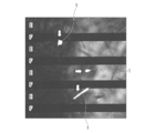
- Example 2 it is the image which has measured the shape image and the phase image using the AFM resonance mode on the polycaprolactone film
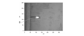
- FIG. 6 is a photographic diagram showing cell clusters formed on a flat film in Example 4. It is a schematic diagram which shows an example of the structure for animal cells of this invention. It is a schematic diagram which shows an example of the structure for animal cells of this invention. It is a schematic diagram which shows an example of the structure for animal cells of this invention. It is a schematic diagram which shows an example of the structure for animal cells of this invention.
- FIG. 1A is a photograph shown in the above-mentioned document, and the animal cells 1 placed on the elastic body layer 2 are exposed to the surface of the soft region (A ) To the surface (B) of the hard region. And since animal cells have the property of “stiffness” that moves to a harder surface, as shown in FIG. 1B, it can be said that animal cells 1 are minute sensors that distinguish “difference in elastic modulus” (FIG. 1). 1B), it has been reported that "difference in elastic modulus” regulates not only cell adhesion, movement and proliferation but also differentiation (Engler et al., Cell, 2006, Gilbert et al., SCIENCE). , 2010 etc.).
- the animal cell structure of the present invention includes an elastic body layer having a flat surface on which animal cells are allowed to stand, and the elastic body layer is supported by partially supporting the elastic body layer from the back side.
- the elasticity of the surface is changed by changing the thickness of the elastic layer.
- first aspect an aspect in which the elasticity of the surface is changed by partially supporting the elastic body layer from the back side
- second aspect an aspect in which the elasticity of the surface is changed by a change in the thickness of the elastic body layer.
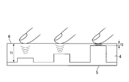
- the elasticity of the elastic body layer surface is changed by partially supporting the elastic body layer from the back side.
- the hardness of the surface can change depending on the presence or absence of support from the lower surface (back surface). That is, by being supported from the back surface, the surface has a relatively high elastic modulus and is relatively hard.
- the elasticity of the surface on which the animal cells are allowed to stand can be changed by changing the presence or absence of support from the back surface.
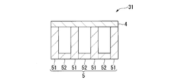
- FIG. 8A shows a more specific configuration of the animal cell structure 31 according to the first aspect of the present invention.
- the animal cell structure 31 includes a base material layer 5 that partially supports the elastic body layer 4 from the back surface side.
- the base material layer 5 has an uneven three-dimensional shape having a first base material layer portion 51 that supports the elastic body layer 4 and a second base material layer portion 52 that does not support the elastic body layer 4.
- the surface elasticity of the elastic body layer 4 can be changed by the uneven three-dimensional shape.
- the first base material layer portion 51 and the second base material layer portion 52 are described separately. However, the first base material layer portion 51 and the second base material layer portion 52 are the same member. It is preferable to be integrally formed with.
- the elasticity of the elastic layer surface changes due to the change in the thickness of the elastic layer.
- the hardness of the surface can be easily changed by changing the thickness of the elastic layer itself. That is, by providing an uneven three-dimensional shape on the elastic layer, the elastic modulus of the surface is relatively low on the concave portion with a small thickness, and the elastic modulus of the surface is relatively high on the convex portion with a large thickness.
- the elasticity of the surface on which animal cells are allowed to stand can be changed.
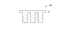
- FIGS. 8B to 8C A more specific configuration of the animal cell structures 61 and 62 according to the second aspect of the present invention is shown in FIGS. 8B to 8C.
- the animal cell structures 61 and 62 have the three-dimensional shape of the back surface of the elastic layer 4 and the thickness of the elastic layer 4 is changed by the three-dimensional shape of the unevenness. Elasticity can be changed.
- the elastic body layer 4 of the animal cell structure 61 may be supported by the base material layer 5 from the back side, and as shown in FIG. 8C, the elastic body of the animal cell structure 62
- the layer 4 may not be supported by the base material layer 5 or the like.
- the substrate having an uneven three-dimensional shape corresponding to the uneven three-dimensional shape formed on the back surface of the elastic body layer 4.
- the material layer 5 may be used, and the entire elastic body layer 4 (the entire back surface) may be supported by the base material layer 5 (FIG. 8B); It may be supported (not shown).
- a single elastic body layer 4 as shown in FIG. 8C is supported by a flat base material layer 5 (for example, a glass substrate) (placed on a flat substrate). Can be mentioned.
- the present invention relates to a structure for animal cells capable of strictly adjusting the “difference in elastic modulus”, a method for separating animal cells, and a method for adjusting the elastic modulus of the surface of the structure for animal cells.
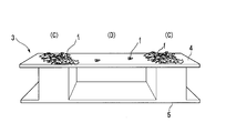
- the animal cell structure 3 includes an elastic body layer 4 and a base material layer 5.
- the first embodiment corresponds to the first aspect.
- the elastic body layer 4 is formed of a synthetic resin or natural fiber, or a thin film or fiber made of these.
- the elastic body layer 4 has an elastic body layer part (C) supported by the base material layer 5 and an elastic body layer part (D) not supported by the base material layer 5.
- the uneven three-dimensional shape of the base material layer 5 is formed by the base material layer portion that supports the elastic body layer portion (C) and the base material layer portion that does not support the elastic body layer portion (D).
- the elastic body layer part (C) supported by the base material layer 5 has a hard surface with a locally high elastic modulus
- the elastic body layer part (D) not supported by the base material layer 5 is It has a low elastic modulus and a soft surface.
- FIG. 2B shows the animal cell 1 on the elastic body layer portion (C) supported by the base material layer 5 when animal cells are cultured on the animal cell structure 3 formed according to Embodiment 1 of the present invention. It is a conceptual diagram which shows that many gather.
- FIG. 3A shows Embodiment 2 of the present invention as a structure for animal cells, a method for classifying animal cells, and a method for adjusting the elastic modulus of the surface of the structure for animal cells capable of strictly adjusting the “difference in elastic modulus”.
- the second embodiment corresponds to the second aspect.
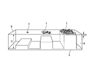
- the animal cell structure 6 similarly to Embodiment 1, in Embodiment 2 of the present invention, includes an elastic body layer 4 and a base material layer 5. And in Embodiment 2 of this invention, the elastic body layer 4 is supported by the base material layer 5, and changes the thickness of the elastic body layer 4 according to the uneven
- the surface of the structure 6 for animal cells becomes a hard surface where the elastic layer 4 is thin, and the elastic layer 4 is locally high in elastic modulus. A soft surface with a low rate.
- FIG. 3B shows that when animal cells are cultured on the animal cell structure 6 formed according to Embodiment 2 of the present invention, many animal cells 1 gather at a location where the elastic body layer 4 is thin. It is a conceptual diagram. FIG. 3B shows that by changing the thickness of the elastic body layer 4 stepwise, the number of animal cells and cell types collected can be changed accordingly.
- the animal cell structures 3 and 6 include the elastic body layer part (C) supported by the base material layer 5 and the elastic body layer part (D) not supported by the base material layer 5. Further, if necessary, the thickness of the elastic layer 4 is changed corresponding to the three-dimensional shape of the base material layer 5 to form the three-dimensional shape of the base material layer 4 on the back surface of the elastic layer 4. The way the body layer 4 is supported can be locally changed to form a hard surface having a high elastic modulus locally on the surfaces of the animal cell structures 3 and 6. That is, according to the present invention, the elastic modulus of the surfaces of the animal cell structures 3 and 6 can be changed only by locally changing the thickness of the elastic layer 4 and the presence or absence of the support of the base layer 5.
- a method for producing the animal cell structure of the present invention a method of forming an unevenness by etching or the like on the surface of a substrate and placing an elastic sheet of an elastic layer (the structure 31 of the first aspect)
- the method for forming the lower part of the elastic body layer with irregularities is not particularly limited, but an elastic material is applied to the substrate after the irregularities are formed on the surface in the same manner as described above, and the concave and convex top surfaces are made of an elastic material. After embedding and drying the elastic material, only the elastic material (elastic body layer) is peeled off from the base material, whereby an elastic body layer having irregularities on the back surface (lower part) can be obtained.
- the elastic material forming the elastic layer is not particularly limited, and any material having elasticity such as a polymer material or a rubber material can be used.
- the base material which comprises a base material layer can use not only a rigid material, such as glass, a silicon
- the animal cell structure of the present embodiment was prepared by attaching a thin film of an elastic layer on a base material layer formed in an uneven three-dimensional shape.
- a method for manufacturing a thin film of an elastic layer is described below.
- An aqueous solution of polyvinyl alcohol (hereinafter abbreviated as “PVA”) having a concentration of 70.6 g / l was spin-coated on a round cover glass blown with nitrogen gas (N 2 ) and thoroughly dusted (4000 rpm, 50 sec).
- PCL polycaprolactone
- EUKITT product name, O Kindler GmbH & Co (Okindler), Germany, encapsulating material
- radial petrolatum registered trademark
- a weight was placed right above the pattern substrate and allowed to stand overnight.
- the uneven three-dimensional pattern substrate was sandwiched between two cover glasses.
- EUKITT drying the uneven
- the elasticity was adjusted by the crosslinking method, so that fine shape change and chemical property change caused by crosslinking occur, and only the elasticity is strictly adjusted in one polymer film. Was very difficult.
- the uneven three-dimensional shape of the elastic body layer 4 or the base material layer 5 is formed, and the thickness of the elastic body layer 4 and the support of the elastic body layer 4 by the base material layer 5 are determined locally. It is possible to change the elastic modulus of the surfaces of the animal cell structures 3 and 6 simply by changing to.
- the shape of the base material layer 5 for supporting / unsupporting the elastic body layer 4 and the thickness of the elastic body layer 4 are merely changed, there is no need for crosslinking, and the type of the elastic body layer 4
- the elasticity can be adjusted without changing the material of the elastic layer 4, and in particular, the elasticity can be precisely adjusted with a resolution of nano to several micrometers on the surface of the elastic layer 4.
- the elastic modulus of the surface of the structure for animal cells of the present invention is not only the elastic modulus of the elastic layer itself, the thickness of the elastic layer, or the presence or absence of the support of the elastic layer by the base material layer, but also the uneven three-dimensional shape. Since it also changes depending on the shape of the top part of the above, these factors can be combined and adjusted. In addition, due to the uneven three-dimensional shape, the elastic modulus of the central surface of the elastic body layer is made higher and harder than other surfaces to collect animal cells in the center of the surface of the structure for animal cells.
- the elastic modulus of the surface of the peripheral part By making the elastic modulus of the surface of the peripheral part higher and harder than other surfaces, animal cells can be collected on the peripheral part of the surface of the structure for animal cells. This makes it possible to form a tissue cell sheet having a desired shape and to separate animal cells into a desired location.
- the uneven three-dimensional shape can be an arbitrary pattern such as a radial pattern, a scale-like pattern, a checkered pattern, a honeycomb-like pattern, as well as a stripe pattern or a square columnar pattern.
- the shape of the top of the three-dimensional uneven shape can be not only a planar shape but also a triangular pyramid shape, a conical shape, or the like.
- Example 1 The animal cell structure of the first aspect was prepared in the same manner as in the above-described embodiment of the animal cell structure. Specifically, a film made of a synthetic polymer PCL, which is often used as a biomaterial, is pasted as an elastic layer on a base material layer having a three-dimensional shape of a striped pattern, as shown in the schematic diagram of FIG. 8A. The animal cell structure of the first embodiment was constructed. The surface elasticity of this rod structure for animal cells could be precisely adjusted with a resolution of nano to several micrometers. When NIH3T3 cells were cultured on the obtained structure for animal cells, as shown in FIG.
- the base material layer was a recess, and the polymer layer (PCL film) as an elastic body layer was supported by the base material layer.
- the animal cell 1 does not collect in the part (F) which is not.
- the part (E) where the base material layer is a convex portion and the polymer layer (PCL film) which is an elastic body layer is in contact with the base material layer that is, the place where the elastic body layer is supported by the base material layer (E) It can be seen that animal cells 1 are gathered. From this, the elastic modulus of the surface of the animal cell structure 3 can be changed with a resolution of nano to several micrometers by simply changing the support / non-support of the elastic body layer 4 locally. It was confirmed that the change can be used to control the migration and proliferation of animal cells (NIH3T3 cells) and to control their localization.
- Example 2 A structure for animal cells in which a polymer film having a thickness of about 50 ⁇ m constituting an elastic body layer 4 having a flat surface is attached to a base material layer 5 on which a stripe-like groove 7 having a depth of 20 ⁇ m and a width of 25 ⁇ m is formed. 3 was obtained.
- FIG. 5A measurement was performed in a resonance mode of an AFM (Atomic Force Microscope).
- FIG. 5B and FIG. 5C are photographic diagrams showing a shape image and a phase image of the film surface of the animal cell structure 3 obtained using the AFM resonance mode. 5B shows that the phase image in FIG. 5C shows a clear contrast even though the polymer film constituting the elastic layer 4 is almost flat.
- FIG. 5A AFM (Atomic Force Microscope).
- FIG. 5B and FIG. 5C are photographic diagrams showing a shape image and a phase image of the film surface of the animal cell structure 3 obtained using the AFM resonance mode. 5B shows that the phase image in FIG. 5C shows
- Example 3 A structure for animal cells in which a polymer film having a thickness of about 50 ⁇ m constituting an elastic body layer 4 having a flat surface is attached to a base material layer 5 in which a square columnar groove 7 having a depth of 20 ⁇ m and a width of 10 ⁇ m is formed. Body 3 was obtained. As shown in FIG. 6A, measurement was performed in an AFM resonance mode. 6B and 6C are photographic diagrams showing a shape image and a phase image of the film surface of the animal cell structure 3 obtained by using the resonance mode of AFM. FIG. 6B shows that the phase image of FIG. 6C shows a clear contrast even though the polymer film constituting the elastic layer 4 is almost flat.
- the animal cell 1 is a location where the base material layer 5 is a convex part, ie, an elastic modulus is high and hard like Example 1 grade
- Example 4 A base material layer having an upright columnar structure having a diameter of 300 ⁇ m on a plane was prepared.
- the PCL-containing solution was dropped from above the base material layer, and was allowed to stand and dried so that the dropped liquid level was horizontal.
- an animal cell structure 61 having the elastic layer 4 having a flat surface was obtained.
- the thickness of the elastic layer 4 was 10 to 20 ⁇ m on the cylindrical structure, and 30 to 40 ⁇ m in the other portions.
- NIH3T3 cells were cultured on the flat surface of the obtained structure 61 for animal cells and the morphology of the cells was observed, it was confirmed that a circular cell mass as shown in FIG. 7 was formed. Since the position and size of the cell mass were almost the same as the position of the cylindrical structure and the horizontal cross section (circular shape with a diameter of 300 ⁇ m), the cells were aggregated on a hard surface portion supported by the base material layer. I understood.
- the present invention includes the following technical ideas.
- a structure composed of an elastic body layer and a base material layer from above comprising a base material layer portion that supports the elastic material layer and a base material layer portion that does not support the base material layer or the elastic material.
- a structure for animal cells wherein the layer changes the surface elasticity of the elastic layer according to the three-dimensional shape of the irregularities.
- a structure composed of an elastic body layer and a base material layer from above and has a base material layer portion that supports the elastic body layer, and the elasticity corresponding to the three-dimensional shape of the base material layer
- a structure for animal cells wherein the elasticity of the surface of the elastic body layer is changed by changing the thickness of the body layer.
- the animal cell structure according to (1) or (2) which is a base material for controlling migration, proliferation and differentiation of animal cells.
- a method for separating animal cells comprising using a base material using the structure of (1) or (2).
- An elastic body layer, a base material layer, and a base material layer portion that supports the elastic material layer are provided, and the thickness of the elastic material layer is changed corresponding to the three-dimensional shape of the unevenness of the base material layer.
- the elasticity adjustment method of the structure body for animal cells characterized by the above-mentioned.
- the surface elasticity of the elastic layer is adjusted by stepwise changing the elasticity of the surface of the elastic layer by the three-dimensional shape of the unevenness of the base material layer or the elastic layer. The method for adjusting the elasticity of the surface of a structure for animal cells according to (5) or (6) above.
Landscapes
- Life Sciences & Earth Sciences (AREA)
- Health & Medical Sciences (AREA)
- Engineering & Computer Science (AREA)
- Chemical & Material Sciences (AREA)
- Wood Science & Technology (AREA)
- Organic Chemistry (AREA)
- Bioinformatics & Cheminformatics (AREA)
- Zoology (AREA)
- Biotechnology (AREA)
- Biomedical Technology (AREA)
- Genetics & Genomics (AREA)
- Microbiology (AREA)
- Biochemistry (AREA)
- General Engineering & Computer Science (AREA)
- General Health & Medical Sciences (AREA)
- Sustainable Development (AREA)
- Cell Biology (AREA)
- Clinical Laboratory Science (AREA)
- Molecular Biology (AREA)
- Analytical Chemistry (AREA)
- Apparatus Associated With Microorganisms And Enzymes (AREA)
- Micro-Organisms Or Cultivation Processes Thereof (AREA)
Abstract
Description
本発明は、医用材料、医療用デバイス、人工臓器、再生医療、細胞治療、再生医工学、悪性新生物診断、細胞分離、病理診断の分野における、動物細胞の遊走、増殖および分化等の制御に適した凹凸三次元形状を用いた動物細胞用構造体、動物細胞の分離方法及び動物細胞用構造体表面の弾性調節方法に関する。
本願は、2013年12月18日に、日本に出願された特願2013-261677号に基づき優先権を主張し、その内容をここに援用する。
近年、動物細胞用構造体などのソフトマテリアルの弾性を調節する技術が注目されており、ソフトマテリアルの弾性を調節する技術として、以下に述べる先行技術が知られている。
(1)温度制御により弾性を調節する技術として、例えば、ゴム弾性を温度によって任意に変化させる方法(例えば特許文献1参照)や、分離膜の弾性を加熱によって調節する方法(例えば特許文献2参照)。
(2)架橋密度や架橋方法によって弾性を調節する技術として、コラーゲン、プロテオグリカン及びヒアルロン酸からなる成形体を熱脱水架橋することで、一定以上の弾性率を維持させる方法(例えば、特許文献3参照)、化学的方法や物理的方法による架橋により、足場材料として用いる高い弾性のコラーゲンゲルを得る方法(例えば特許文献4参照)、コラーゲンの架橋条件を変えることにより、細胞導出基材として用いるコラーゲンゲルの弾性率を調節する方法(例えば特許文献5参照)、ヒアルロン酸ゲルの架橋(光架橋)密度を変えることにより、ヒアルロン酸ゲルの弾性率を調節する方法(例えば特許文献6参照)。
(3)無機物を添加・分散させることによって弾性を調節する技術として、アクリルアミド系のモノマーに粘土鉱物を分散させ、重合後の高分子ヒドロゲルの弾性率を調節する方法(例えば、特許文献7参照)。
(4)三次元的な構造制御を行うことによって弾性を調節する技術として、コラーゲンを多孔質化することで、弾性を調節する方法(例えば、特許文献8参照)。
また、細胞は低弾性率の表面から高弾性率の表面に向かって遊走する傾向があることが知られている(例えば、非特許文献1-8参照)。動物細胞用構造体の表面において選択的に弾性を調節するための先行技術として、架橋剤の濃度が異なる領域を動物細胞用構造体の作製時に配置する方法(例えば、非特許文献1-7参照)や、局所的な光架橋を利用した方法(例えば、非特許文献8参照)が知られている。
更に、細胞の遊走速度を制御する技術(例えば、非特許文献1,9-13参照)、細胞の増殖速度を制御する技術(例えば、非特許文献14参照)、表面の弾性率の変化による細胞の形態変化を利用した技術(非特許文献2,9,14-16参照)、表面の弾性率の差異による細胞の分化度変化や分化方向変化利用した技術(非特許文献17-22参照)が知られている。
C. M. Lo, H. B. Wang, M. Dembo and Y. L. Wang, Biophys J, 2000, 79, 144-152. G. Jiang, A. H. Huang, Y. Cai, M. Tanase and M. P. Sheetz, Biophys J, 2006, 90, 1804-1809. A. Welle, A. Chiumiento and R. Barbucci, Biomol Eng, 2007, 24, 87-91. K. A. Lazopoulos and D. Stamenovic, J Biomech, 2008, 41, 1289-1294. P. C. Fung, Chin Med, 2009, 4, 10. B. C. Isenberg, P. A. Dimilla, M. Walker, S. Kim and J. Y. Wong, Biophys J, 2009, 97, 1313-1322. B. Harland, S. Walcott and S. X. Sun, Phys Biol, 2011, 8, 015011. T. Kawano and S. Kidoaki, Biomaterials, 2011, 32, 2725-2733. B. Ananthanarayanan, Y. Kim and S. Kumar, Biomaterials, 2011, 32, 7913-7923. J. L. MacKay, A. J. Keung and S. Kumar, Biophys J, 2012, 102, 434-442. R. A. Jannat, G. P. Robbins, B. G. Ricart, M. Dembo and D. A. Hammer, J Phys Condens Matter, 2010, 22, 194117. S. R. Peyton and A. J. Putnam, J Cell Physiol, 2005, 204, 198-209. C. B. Khatiwala, S. R. Peyton and A. J. Putnam, Am J Physiol Cell Physiol, 2006, 290, C1640-1650. A. M. Quinlan and K. L. Billiar, J Biomed Mater Res A, 2012, 100, 2474-2482. T. Yeung, P. C. Georges, L. A. Flanagan, B. Marg, M. Ortiz, M. Funaki, N. Zahir, W. Ming, V. Weaver and P. A. Janmey, Cell Motil Cytoskeleton, 2005, 60, 24-34. M. J. Paszek, N. Zahir, K. R. Johnson, J. N. Lakins, G. I. Rozenberg, A. Gefen, C. A. Reinhart-King, S. S. Margulies, M. Dembo, D. Boettiger, D. A. Hammer and V. M. Weaver, Cancer Cell, 2005, 8, 241-254. J. Du, X. Chen, X. Liang, G. Zhang, J. Xu, L. He, Q. Zhan, X. Q. Feng, S. Chien and C. Yang, Proc Natl Acad Sci U S A, 2011, 108, 9466-9471. M. D. Treiser, E. H. Yang, S. Gordonov, D. M. Cohen, I. P. Androulakis, J. Kohn, C. S. Chen and P. V. Moghe, Proc Natl Acad Sci U S A, 2010, 107, 610-615. A. M. Kloxin, J. A. Benton and K. S. Anseth, Biomaterials, 2010, 31, 1-8. P. M. Gilbert, K. L. Havenstrite, K. E. Magnusson, A. Sacco, N. A. Leonardi, P. Kraft, N. K. Nguyen, S. Thrun, M. P. Lutolf and H. M. Blau, Science, 2010, 329, 1078-1081. A. I. Teixeira, S. Ilkhanizadeh, J. A. Wigenius, J. K. Duckworth, O. Inganas and O. Hermanson, Biomaterials, 2009, 30, 4567-4572. A. J. Engler, S. Sen, H. L. Sweeney and D. E. Discher, Cell, 2006, 126, 677-689.
しかしながら、特許文献1-8における、温度制御による方法や各種架橋法、無機物等の添加・分散により弾性体の弾性を調節する方法は、加温や架橋または無機物等の添加・分散が可能な弾性体以外には適用できない方法であり、全ての弾性体に適用できるものではない。また、例えば、弾性体の架橋密度に差異を設けることによって、弾性体表面の弾性率を変化させることはできるが、架橋密度の変化に伴って弾性体表面の弾性以外の物性が変化してしまいうる。すなわち、弾性以外の表面物性である親水性・疎水性、電荷、またはラフネスなども変化してしまうことになり、ソフトマテリアルなどの動物細胞用構造体としての適合性が低下してしまうおそれがあるという問題がある(問題点1)。
また、架橋密度や架橋構造を局所的に変えて弾性体の表面弾性を調節する方法では、架橋による体積変化を伴うため、表面の平坦さが失われうる。現在、動物細胞用構造体の弾性体表面において、局所的に架橋密度を変える方法として、最も微細形成が可能な方法が光架橋法であるが、この場合でも平坦さが失われ、分解能も数μm~数十μm程度しかない。そしてこれらの方法では、光の回折限界の問題で分解能を1μmよりも小さくすることは難しい。また、光を透過できない弾性体などへの適応も難しい(問題点2)。
また、生体組織は様々な種類の細胞が共存している。こういった生体内の細胞の共存状態を、人工的に作製すべく様々な動物細胞用構造体が作製されてきた。上述した非特許文献1-22にみられるように、動物細胞用構造体表面の弾性を調節すれば、動物細胞の遊走や増殖、分化及び代謝といった制御ができることが知られており、局所的に弾性を調節すれば、細胞を望む形に集めることもできる可能性も示されている。
しかしながら、現状では、動物細胞用構造体表面の弾性の調節は、架橋が可能なものに限られており、また、光架橋法を用いても、数μm~数十μm程度の分解能しかないため、細胞を望む形に集めることは達成されていない(問題点3)。
更に、動物細胞の遊走、増殖および分化の機能は、ナノメートルスケールのラフネスの影響も受けるので、架橋することによる表面の微細な形状の変化がこれら動物細胞の機能に及ぼす影響も考慮しなくてはならず、弾性調節による細胞機能の制御のし易さから考えれば、足場材料の表面は平坦であることが望ましい。そこで、本発明は、あらゆる動物細胞用構造体の表面形状を変えず、これらの表面弾性を高い分解能(1μm程度)で調節でき、細胞を望む形に集めることを可能にする動物細胞用構造体表面の弾性調節方法を提供することを目的とする。
すなわち、本発明の目的は、動物細胞の遊走、増殖および分化の制御に適し、あらゆる弾性体に適用が可能であって、平坦性等の弾性以外の物性変化や形状変化を伴わず、高分解能を有する微細な弾性調節が簡便に可能な三次元形状を用いた動物細胞用構造体、動物細胞の分離方法及び動物細胞用構造体表面の弾性調節方法を提供することにある。
本発明者らは、上記した問題点1~3等を解決するために鋭意研究を行い、動物細胞用構造体が備える弾性体層表面の弾性を、該弾性体層が部分的に裏面側から支持されることによって、又は、該弾性体層の厚みの変化によって変化させることで、あらゆる弾性体に適用できること、弾性以外の表面物性が変化しないこと、及び簡便に製造可能であることを見出し、本発明を完成させた。
具体的には、本発明者らは、上記のように弾性体層の表面の弾性を変化させることで、弾性体層表面の弾性を容易に調節可能であること、弾性を変化させたときでも表面の平坦さが失われないこと、及び、高い分解能(1μm程度)で弾性の調節が可能となることを見出した。
また、本発明者らは、弾性体層を裏面側から支持する基材層又は弾性体層の三次元形状により弾性体の表面の弾性を変化させることで、あらゆる動物細胞用構造体の表面弾性を高い分解能(1μm程度)で調節できるので、それにより得た動物細胞用構造体の表面上で動物細胞の遊走、増殖および分化の制御を的確にできることを見出した。
すなわち、本発明は、以下に示す、動物細胞の遊走、増殖および分化の制御に適した三次元形状を用いた動物細胞用構造体、動物細胞の分離方法及び動物細胞用構造体表面の弾性調節方法に関する。
(1)動物細胞が静置される平坦な表面を有する弾性体層を備え、前記弾性体層は、該弾性体層が部分的に裏面側から支持されることによって、又は、該弾性体層の厚みの変化によって、表面の弾性が変化していることを特徴とする、動物細胞用構造体。
(2)さらに、前記弾性体層を裏面側から部分的に支持する基材層を備え、前記基材層は、前記弾性体層を支持する第1基材層部と、前記弾性体層を支持しない第2基材層部とを有する凹凸三次元形状とされ、前記凹凸三次元形状により、前記弾性体層の表面弾性が変化している、上記(1)に記載の動物細胞用構造体。
(3)前記弾性体層の裏面側が凹凸三次元形状とされ、該凹凸三次元形状によって前記弾性体層の厚みが変化している、上記(1)に記載の動物細胞用構造体。(4)さらに、前記弾性体層を裏面側から支持する基材層を備える、上記(3)に記載の動物細胞用構造体。
(5)前記基材層が、前記弾性体層の凹凸三次元形状に対応した凹凸三次元形状とされ、前記弾性体層を全体的に支持する、上記(4)に記載の動物細胞用構造体。
(6)動物細胞の遊走、増殖および分化制御用基材である上記(1)~(5)のいずれか一つに記載の動物細胞用構造体。
(7)上記(1)~(6)のいずれか一つに記載の動物細胞用構造体を用いることを特徴とする動物細胞の分離方法。
(8)(1)~(6)のいずれか一つに記載の動物細胞用構造体を用いることを特徴とする、動物細胞用構造体表面の弾性調節方法。
本発明の動物細胞用構造体、及び動物細胞用構造体表面の弾性調整方法は、基材層又は弾性体層の三次元形状によって動物細胞用構造体の表面の弾性を変化させるので、あらゆる弾性体に適用でき、弾性体層の弾性を変えても弾性以外の表面物性が変化しない簡便な弾性調節方法であるので、上記問題点1を解決できる。
本発明の弾性体上での動物細胞の遊走、増殖および分化の制御は、基材層又は弾性体層の三次元形状により動物細胞用構造体の表面の弾性を変化させることでなされる。即ち、基材層の凹凸又は弾性体層の凹凸により、弾性体層の表面弾性が基材に支持された部分から基材に支持されていない部分へ連続して変化するのではなく、該表面弾性が急激に変化することから境界が明瞭であり、弾性率の差を厳密に調整できる。そのため、あらゆる動物細胞用構造体の表面弾性を高い分解能(1μm程度)で調節でき、また光を透過できない弾性体などへの適応も可能であることから、上記問題点2を解決できる。
本発明の動物細胞用構造体は、架橋が不要であり、あらゆる動物細胞用構造体の表面形状を変えずに、高分解能(1μm程度)をもって表面弾性を調節でき、動物細胞を望む形に集めて分離することが可能であり、上記問題点3を解決できる。即ち、表面の弾性率の高い(硬い)部分の形状を、細胞集合体(細胞塊)を形成させる所望の形状、例えば、四角形等にすることにより、当該所望形状の細胞組織(例えば四角形の細胞シート等)が得られる。
本発明に係る動物細胞の遊走、増殖および分化の制御に適した凹凸三次元形状を用いた動物細胞用構造体及び動物細胞用構造体表面の弾性調節方法を実施するための形態を説明する。以下、本発明を実施するための実施形態に基づき、本発明の方法及び利用例を具体的に説明するが、本発明はこれら実施形態に限定されるものではない。
本願発明の基材層又は弾性体層の凹凸三次元形状を用いた動物細胞用構造体、および該動物細胞用構造体表面の弾性調節方法につき図を参照して説明する。
動物細胞は、より硬い表面へ運動する「走硬性」の性質を持つことが、(Lo et al., Biophys. J., 2000)に発見されて以降、細胞生物学、生物物理学だけではなく再生医工学の分野などで、動物細胞用構造体の弾性について活発に議論されるようになっている。また、図1Aは上記文献に示された写真図であって、左図から右図への時間の経過により、弾性体層2上に静置された動物細胞1が、軟らかい領域の表面(A)から硬い領域の表面(B)に移動していることを示している。そして、動物細胞がより硬い表面へ運動する「走硬性」の性質を有することから、図1Bに示すように、動物細胞1は、「弾性率の差」を見分ける微小センサーであるとも云え(図1B)、「弾性率の差」が、細胞の接着や運動、増殖のみならず分化まで調節しているとする報告もなされている(Engler et al., Cell, 2006、Gilbert et al.,SCIENCE, 2010など)。
本発明の動物細胞用構造体は、動物細胞が静置される平坦な表面を有する弾性体層を備え、前記弾性体層は、該弾性体層が部分的に裏面側から支持されることによって、又は、該弾性体層の厚みの変化によって、表面の弾性が変化していることを特徴とする。
以下、弾性体層が部分的に裏面側から支持されることにより表面の弾性を変化させる態様を「第一の態様」と言い、弾性体層の厚みの変化により表面の弾性を変化させる態様を「第二の態様」と言う。
本発明の第一の態様では、弾性体層が部分的に裏面側から支持されることにより、弾性体層表面の弾性が変化する。
弾性を有する弾性体層の場合、下面(裏面)からの支持の有無によりその表面の硬さが変化し得る。すなわち、裏面から支持されることにより、その表面は弾性率が比較的高くなり、且つ、比較的硬くなる。この事象を用いて、裏面からの支持の有無を変化させることにより、動物細胞が静置される表面の弾性を変化させることができる。
本発明の第二の態様では、弾性体層の厚みの変化により、弾性体層表面の弾性が変化する。
弾性を有する弾性体層の場合、該弾性体層自身の厚みの変化によりその表面の硬さが容易に変化し得る。すなわち、弾性体層に凹凸三次元形状を設けることにより、厚みが薄い凹部上では表面の弾性率が比較的低くなり、厚みが厚い凸部上では表面の弾性率が比較的高くなる。また、裏面に凹凸三次元形状を設けることで、表面自体は平坦とした場合であっても、動物細胞が静置される表面の弾性を変化させることができる。
図8Bに示すように、動物細胞用構造体61の弾性体層4は裏面側から基材層5に支持されていてもよく、図8Cに示すように、動物細胞用構造体62の弾性体層4は基材層5等により支持されていなくてもよい。
また、第二の態様の動物細胞用構造体61、62が基材層5に支持される場合、弾性体層4の裏面に形成された凹凸三次元形状に対応した凹凸三次元形状を有する基材層5が用いられて、弾性体層4の全体(裏面の全面)が基材層5により支持されていてもよく(図8B);弾性体層4の一部のみが基材層5に支持されていてもよい(図示せず)。後者の例としては、図8Cに示すような単独の弾性体層4が、平坦な基材層5(例えばガラス基板等)に支持されたもの(平坦な基板上に載置されたもの)が挙げられる。
[実施形態1]
本発明は、「弾性率の差」を厳密に調整することができる動物細胞用構造体、動物細胞分離方法及び該動物細胞用構造体表面の弾性率調節方法に関する。本発明の実施形態1は、図2Aに示すように、動物細胞用構造体3を、弾性体層4と基材層5とから構成する。本実施形態1は、上記第一の態様に相当する。
弾性体層4は、合成樹脂や天然繊維、あるいはこれらで作製された薄膜や繊維で形成する。そして、弾性体層4は、基材層5によって支持されている弾性体層部(C)と基材層5によって支持されていない弾性体層部(D)とを有する。弾性体層部(C)を支持する基材層部と、弾性体層部(D)を支持していない基材層部とによって、基材層5の凹凸三次元形状が形成されている。これにより、基材層5によって支持されている弾性体層部(C)は、局所的に弾性率が高く硬い表面となり、基材層5によって支持されていない弾性体層部(D)は、弾性率が低く軟らかい表面となる。
図2Bは、本発明の実施形態1より形成された動物細胞用構造体3上で動物細胞を培養した場合に、基材層5によって支持されている弾性体層部(C)に動物細胞1が多く集まることを示す概念図である。
[実施形態2]
上記「弾性率の差」を厳密に調整することができる動物細胞用構造体、動物細胞分類方法及び該動物細胞用構造体表面の弾性率調節方法として本発明の実施形態2を図3Aに示す。本実施形態2は、上記第二の態様に相当する。
図3Aに示すように、実施形態1と同様に、本発明の実施形態2において、動物細胞用構造体6は、弾性体層4と基材層5とからなる。そして、本発明の実施形態2において、弾性体層4は、基材層5によって支持され、基材層5の表面の凹凸三次元形状に対応して弾性体層4の厚さを変化させることにより、弾性体層4の裏面に凹凸三次元形状が形成されている。これにより、動物細胞用構造体6の表面は、弾性体層4の厚さが薄い箇所は、局所的に弾性率が高く硬い表面となり、該弾性体層4の厚さが厚い箇所は、弾性率が低い軟らかい表面となる。
図3Bは、本発明の実施形態2により形成された動物細胞用構造体6上で動物細胞を培養した場合に、弾性体層4の厚さが薄い箇所に動物細胞1が多く集まることを示す概念図である。
また、図3Bは、弾性体層4の厚さを段階的に変化させることにより、それに応じて集まる動物細胞数や細胞種別などが異なるものとできることを示している。
上述したように、動物細胞用構造体3、6を、基材層5によって支持されている弾性体層部(C)と、基材層5によって支持されていない弾性体層部(D)とによって、また、必要に応じて基材層5の凹凸三次元形状に対応して弾性体層4の厚さを変化させて弾性体層4の裏面に凹凸三次元形状を形成することで、弾性体層4の支持のされ方を局所的に変化させ、動物細胞用構造体3、6の表面に局所的に弾性率が高く硬い表面を形成することができる。
すなわち、本発明は、弾性体層4の厚さや基材層5の支持の有無を局所的に変化させるだけで、動物細胞用構造体3、6表面の弾性率を変化させることができる。
本発明の動物細胞用構造体の作製方法としては、基材表面に、エッチング等による凹凸を形成して弾性体層の弾性シートを載せ作製する方法(上記第一の態様の構造体31等);上記同様に表面に凹凸を形成した後の基材に、弾性材料を塗布して凹部及び凸部頂面を弾性材料で包埋することで作製する方法(第二の態様の構造体61等);弾性体層下部を凹凸に形成し、必要に応じて平坦な基板に載せて、作製する方法等が挙げられる(第二の態様の構造体62等)。また、弾性体層下部を凹凸に形成する方法としては特に限定されないが、上記同様に表面に凹凸を形成した後の基材に、弾性材料を塗布して凹部及び凸部頂面を弾性材料で包埋し、該弾性材料を乾燥させた後、基材から弾性材料(弾性体層)のみを剥離することにより、裏面(下部)に凹凸を有する弾性体層を得ることができる。
弾性体層を形成する弾性材料は、特に限定されるものではなく、高分子材料、ゴム材料等弾性のあるものなら全て使用できる。また、基材層を構成する基材は、ガラス、シリコン、プラスチック、金属など剛性のある材料だけでなく、弾性体層よりも硬度が高い柔軟材料を使用することもできる。
次に、本発明の動物細胞用構造体の作製について一実施形態を説明する。
本実施形態の動物細胞用構造体は、凹凸三次元形状に形成された基材層上に弾性体層の薄膜を貼り付けて作製した。
まず、弾性体層の薄膜の作製方法を以下に示す。窒素ガス(N2)をブローして十分に埃を飛ばした丸カバーガラス上に、濃度70.6g/lのポリビニールアルコール(以下「PVA」と略記する)水溶液を全面スピンコートした(4000rpm,50sec)。次いで、PVAにより被覆された丸カバーガラス上に、13.7g/lのポリカプロラクトン(以下、「PCL」と略記する)クロロホルム溶液を、丸カバーガラスの面積の8~9割程度に広がるように載せ、スピンコートすることにより(4000rpm,50sec)、丸カバーガラス上に均一な厚さの(約50μm)のPCL膜を作製した。
更に弾性体薄膜を、凹凸三次元形状を有する基材に貼り付ける方法を詳述する。
まず、新品の丸カバーガラスの中心に、1センチ角の凹凸三次元パターン基板をマニキュアによって接着・固定する。そして、スポンジ綿棒とパスツールピペットを使用して、基板の周りを円形になるようにワセリン(登録商標)で囲み、そこから放射状に外側にむけてワセリン(登録商標)を伸ばした。その放射状のワセリン(登録商標)の間に、EUKITT(製品名、O Kindler GmbH & Co(オーキンドラー社)、独国、封入材)を適量垂らし、乾かないうちに上からPCL膜を製膜済みのカバーガラスを、PCL膜面を下に向けて貼り付けた。密着性を向上させるために、パターン基板の真上に重りを置き、一晩静置した。以上の工程により、凹凸三次元パターン基板が二枚のカバーガラスによって挟まれた状態になった。そして、EUKITT乾燥後、100mmディッシュに、作製途中の二枚のカバーガラスによって挟まれた凹凸三次元パターン基板を入れ、脱気した水に一晩浸漬させた。PVAの溶解により、一枚のカバーガラスが剥離し、凹凸三次元パターン基板の上にはPCL膜のみが残った。カバーガラスを取り除いた後、ディッシュ内を排水し、キムワイプで軽く水分を取り除いた後、乾燥させた。次に、ワッシャーの片面にVECTA SHIELD(製品名、ヴェクターラボラトリー社、封入材)を適量乗せ、ワッシャーの穴の中心と基板の中心部が重なるようにワッシャーを載せた。そして、密着性を上げるために、ワッシャーの上に重りを置き一晩静置した。以上より、凹凸三次元パターン上に貼られたPCL膜がワッシャーによって保護されるとともに、密着性や平坦性が向上した。凹凸三次元パターン基板上へのPCL膜の貼り付けを行うことができた。
従来、弾性の調節を架橋法によって行っていたので、架橋することによる微細な形状変化や化学的な性質変化が生じてしまい、一枚の高分子膜の中で弾性のみを厳密に調節することは非常に困難であった。一方、本発明においては、弾性体層4または基材層5の凹凸三次元形状を形成して、弾性体層4の厚さや、弾性体層4の基材層5による支持の有無を局所的に変化させるだけで、動物細胞用構造体3、6表面の弾性率を変化させることができる。そして、本発明は、単に弾性体層4の支持・不支持のための基材層5の形状や弾性体層4の厚さを変えるだけなので、架橋の必要がなく、弾性体層4の種類を選ぶ必要はなく、また弾性体層4の素材を変化させずに弾性を調節でき、特に、弾性体層4表面においてナノ~数マイクロメートルスケールの分解能で弾性を厳密に調節できる。
[その他の実施形態]
以上、本発明の実施形態について説明したが、本発明は、上記実施形態に限定されるものではなく、種々の態様を取ることができる。
すなわち、本発明の動物細胞用構造体表面の弾性率は、弾性体層自体の弾性率、弾性体層の厚み、又は基材層による弾性体層の支持の有無ばかりでなく、凹凸三次元形状の頂部の形状によっても変化するものであるから、これらの因子を組み合わせて調節することができる。
また、凹凸三次元形状により、弾性体層の中央部表面の弾性率をそれ以外の表面よりも高くして硬くし、動物細胞を動物細胞用構造体表面の中央に集めることや、弾性体層の周辺部表面の弾性率をそれ以外の表面よりも高くして硬くすることにより、動物細胞を動物細胞用構造体表面の周辺部に集めることができる。それにより、所望の形状の組織細胞シートの形成や、所望箇所への動物細胞の分離が可能となる。
また、凹凸三次元形状は、ストライプパターンや四角柱状パターンだけでなく、放射状パターン、鱗状パターン、市松状パターン、蜂の巣状パターンなどの任意のパターンとすることができる。
更に、凹凸三次元形状の頂部の形状は、平面形状だけでなく、三角錐状、円錐状などとすることができる。
上述した動物細胞用構造体の作製実施形態と同様にして、第一の態様の動物細胞用構造体を作製した。具体的には、ストライプ状パターンの凹凸三次元形状を有する基材層上に、生体材料として良く用いられる合成高分子PCLからなる膜を弾性体層として貼り、図8Aの模式図に示すような第一の態様の動物細胞用構造体を構成した。この動物細胞用棒構造体の表面弾性は、ナノ~数マイクロメートルスケールの分解能で厳密に調節できていた。
得られた動物細胞用構造体上でNIH3T3細胞を培養したところ、図4に示すように、基材層が凹部で、弾性体層である高分子層(PCL膜)が基材層により支持されていない箇所(F)には、動物細胞1は集まらない。一方、基材層が凸部で、弾性体層である高分子層(PCL膜)が当該基材層と接する箇所、すなわち弾性体層が基材層により支持されている箇所(E)には、動物細胞1が集まっていることが分かる。
このことから、弾性体層4の支持・不支持を局所的に変化させるだけで、動物細胞用構造体3表面の弾性率をナノ~数マイクロメートルスケールの分解能で変化させることができ、その弾性変化を利用して動物細胞(NIH3T3細胞)の遊走や増殖を制御し、その局在を制御できることが確認できた。
深さ20μmで幅が25μmのストライプ状溝7が形成された基材層5に、表面が平坦な弾性体層4を構成する厚さ約50μmの高分子膜を貼り付けた動物細胞用構造体3を得た。
図5Aに示すようにAFM(Atomic Force Microscope;原子間力顕微鏡)の共振モードによる測定を行った。図5B及び図5Cは、AFM共振モードを用いて得た、動物細胞用構造体3の膜表面の形状像及び位相像を示す写真図である。図5Bを見ると、弾性体層4を構成する高分子膜はほとんど平坦であるにも関わらず、図5Cの位相像には、明確なコントラストが示されていることが分かる。また、実施例1の図4と同様に、弾性体層4を構成する高分子膜上で動物細胞1を培養すると、動物細胞1は、基材層5が凹部である箇所(H)ではなく、基材層5が凸部である箇所(G)、すなわち、弾性体層4が基材層5に支持されており、弾性率が高く硬い表面に集まることが確認された。
深さ20μmで幅が10μmの四角柱状の溝7が形成された基材層5に、表面が平坦な弾性体層4を構成する厚さ約50μmの高分子膜を貼り付けた動物細胞用構造体3を得た。
図6Aに示すようにAFM共振モードによる測定を行った。図6B及び図6Cは、AFMの共振モードを用いて得た、動物細胞用構造体3の膜表面の形状像及び位相像を示す写真図である。図6Bを見ると、弾性体層4を構成する高分子膜はほとんど平坦であるにも関わらず、図6Cの位相像には、明確なコントラストが示されていることが分かる。また、基材層5の凹凸三次元形状が異なるパターンになっていても、実施例1等と同様に、動物細胞1は基材層5が凸部である箇所、すなわち、弾性率が高く硬い表面に集まることが確認された(図示せず)。
直径300μmの立設した円柱状構造物を平面上に有する基材層を用意した。基材層上からPCL含有溶液を滴下し、滴下した液面が水平となるように静置して乾燥させた。その結果、表面が平坦な弾性体層4を有する動物細胞用構造体61を得た。弾性体層4の厚さは円柱状構造物の上で10~20μm、それ以外の部分では30~40μmであった。得られた動物細胞用構造体61の平坦な表面上でNIH3T3細胞を培養し、細胞の形態を観察したところ、図7に示すような円形の細胞塊を形成していることが確認できた。細胞塊の位置及び大きさは、円柱状構造物の位置及び水平方向断面(直径300μmの円形)とほぼ等しかったことから、細胞が基材層に支持された硬い表面部分に凝集していることが分かった。
(1)上から弾性体層、基材層からなる構造体であり、該弾性体層を支持する基材層部及び支持しない基材層部とを有し、前記基材層又は前記弾性体層が、その凹凸三次元形状により、前記弾性体層の表面弾性を変化させることを特徴とする動物細胞用構造体。
(2)上から弾性体層、基材層とからなる構造体であり、前記弾性体層を支持する基材層部を有し、前記基材層の凹凸三次元形状に対応して前記弾性体層の厚さを変化させることにより、前記弾性体層の表面の弾性を変化させることを特徴とする動物細胞用構造体。
(3)動物細胞の遊走、増殖および分化制御用基材である上記(1)または(2)に記載の動物細胞用構造体。
(4)上記(1)または(2)の構造体を用いた基材を用いることを特徴とする動物細胞の分離方法。
(5)弾性体層と基材層と、該弾性体層を支持する基材層部と支持しない基材層部とを備え、前記基材層又は前記弾性体層の凹凸三次元形状により、前記弾性体層の表面弾性を調節することを特徴とする動物細胞用構造体表面の弾性調節方法。
(6)弾性体層と基材層と、前記弾性体層を支持する基材層部を有し、前記基材層の凹凸三次元形状に対応して前記弾性体層の厚さを変化させることにより、前記弾性体層の表面弾性を調節することを特徴とする動物細胞用構造体表面の弾性調節方法。
(7)前記基材層又は前記弾性体層の凹凸三次元形状により前記弾性体層の表面の弾性を段階的に変化させることにより、前記弾性体層の表面弾性を調節することを特徴とする上記(5)または(6)に記載の動物細胞用構造体表面の弾性調節方法。
1 動物細胞
2 高分子膜表面
3、6、31、61、62 動物細胞用構造体
4 弾性体層
5 基材層
51 第1基材層部
52 第2基材層部
7 溝
8 AFM探針
(A) 硬い領域の表面
(B) 軟らかい領域の表面
(C)、(E)、(G) 支持されている弾性体層部
(D)、(F)、(H) 支持されていない弾性体層部
Claims (8)
- 動物細胞が静置される平坦な表面を有する弾性体層を備え、
前記弾性体層は、該弾性体層が部分的に裏面側から支持されることによって、又は、該弾性体層の厚みの変化によって、表面の弾性が変化していることを特徴とする、動物細胞用構造体。 - さらに、前記弾性体層を裏面側から部分的に支持する基材層を備え、
前記基材層は、前記弾性体層を支持する第1基材層部と、前記弾性体層を支持しない第2基材層部とを有する凹凸三次元形状とされ、
前記凹凸三次元形状により、前記弾性体層の表面弾性が変化している、請求項1に記載の動物細胞用構造体。 - 前記弾性体層の裏面側が凹凸三次元形状とされ、該凹凸三次元形状によって前記弾性体層の厚みが変化している、請求項1に記載の動物細胞用構造体。
- さらに、前記弾性体層を裏面側から支持する基材層を備える、請求項3に記載の動物細胞用構造体。
- 前記基材層が、前記弾性体層の凹凸三次元形状に対応した凹凸三次元形状とされ、前記弾性体層を全体的に支持する、請求項4に記載の動物細胞用構造体。
- 動物細胞の遊走、増殖および分化制御用である請求項1~5のいずれか一項に記載の動物細胞用構造体。
- 請求項1~6のいずれか一項に記載の動物細胞用構造体を用いることを特徴とする動物細胞の分離方法。
- 請求項1~6のいずれか一項に記載の動物細胞用構造体を用いることを特徴とする、動物細胞用構造体表面の弾性調節方法。
Priority Applications (3)
| Application Number | Priority Date | Filing Date | Title |
|---|---|---|---|
| JP2015553550A JP6226338B2 (ja) | 2013-12-18 | 2014-12-16 | 動物細胞用構造体、動物細胞の分離方法及び動物細胞用構造体表面の弾性調節方法 |
| US15/104,733 US10513683B2 (en) | 2013-12-18 | 2014-12-16 | Structure used for animal cells, method of separating animal cells, and method of adjusting elasticity of surface of structure used for animal cells |
| EP14872640.9A EP3085768A4 (en) | 2013-12-18 | 2014-12-16 | Structure for animal cell, method for separating animal cell, and method for adjusting elasticity of surface of structure for animal cell |
Applications Claiming Priority (2)
| Application Number | Priority Date | Filing Date | Title |
|---|---|---|---|
| JP2013261677 | 2013-12-18 | ||
| JP2013-261677 | 2013-12-18 |
Publications (1)
| Publication Number | Publication Date |
|---|---|
| WO2015093472A1 true WO2015093472A1 (ja) | 2015-06-25 |
Family
ID=53402817
Family Applications (1)
| Application Number | Title | Priority Date | Filing Date |
|---|---|---|---|
| PCT/JP2014/083247 Ceased WO2015093472A1 (ja) | 2013-12-18 | 2014-12-16 | 動物細胞用構造体、動物細胞の分離方法及び動物細胞用構造体表面の弾性調節方法 |
Country Status (5)
| Country | Link |
|---|---|
| US (1) | US10513683B2 (ja) |
| EP (1) | EP3085768A4 (ja) |
| JP (1) | JP6226338B2 (ja) |
| TW (1) | TW201527526A (ja) |
| WO (1) | WO2015093472A1 (ja) |
Cited By (1)
| Publication number | Priority date | Publication date | Assignee | Title |
|---|---|---|---|---|
| JP2022015233A (ja) * | 2020-07-08 | 2022-01-21 | 王子ホールディングス株式会社 | 細胞培養部材の製造方法、細胞培養部材、細胞培養部材の製造用原型の製造方法、および細胞培養部材の製造用原型 |
Citations (11)
| Publication number | Priority date | Publication date | Assignee | Title |
|---|---|---|---|---|
| JPH11512778A (ja) | 1995-11-15 | 1999-11-02 | 生化学工業株式会社 | 光架橋ヒアルロン酸ゲルおよびその製造法 |
| JP2001065617A (ja) | 1999-08-30 | 2001-03-16 | Nok Corp | ゴム弾性制御方法 |
| JP2005229914A (ja) * | 2004-02-19 | 2005-09-02 | Dainippon Printing Co Ltd | 細胞培養用パターニング基板 |
| JP2005232402A (ja) | 2004-02-23 | 2005-09-02 | Kawamura Inst Of Chem Res | 高分子複合体、その延伸物及び高分子複合体の製造方法 |
| WO2005085414A1 (ja) * | 2004-03-10 | 2005-09-15 | Dai Nippon Printing Co., Ltd. | 血管細胞培養用パターニング基板 |
| JP2011225462A (ja) | 2010-04-16 | 2011-11-10 | Ihara Suisan Kk | 多孔質コラーゲン、その製造方法およびその用途 |
| JP2012105609A (ja) * | 2010-11-18 | 2012-06-07 | Dainippon Printing Co Ltd | 糸状浮遊細胞パターニング基材 |
| JP2012126681A (ja) | 2010-12-16 | 2012-07-05 | Tokyo Metropolitan Industrial Technology Research Institute | コラーゲン線維ゲルおよびその用途 |
| JP2012130386A (ja) | 2010-12-20 | 2012-07-12 | Osaka Prefecture Univ | コラーゲンゲルおよびその製造方法 |
| JP2013099272A (ja) * | 2011-11-08 | 2013-05-23 | Dainippon Printing Co Ltd | 細胞培養基材の製造方法、細胞培養基材、およびそれを用いた細胞シートの製造方法 |
| JP2013150648A (ja) | 2012-01-24 | 2013-08-08 | Hoya Corp | 人工軟骨の製造方法 |
Family Cites Families (3)
| Publication number | Priority date | Publication date | Assignee | Title |
|---|---|---|---|---|
| JPH11347382A (ja) | 1998-06-04 | 1999-12-21 | Nitto Denko Corp | 形状記憶分離膜システムモジュール |
| JP2004200178A (ja) | 2004-02-19 | 2004-07-15 | Sumitomo Electric Ind Ltd | 酸化物超電導導体およびその製造方法 |
| US20100015709A1 (en) | 2006-02-10 | 2010-01-21 | Trustees Of The University Of Pennsylvania | Regulating Stem Cell Differentiation By Controlling 2D and 3D Matrix Elasticity |
-
2014
- 2014-12-16 EP EP14872640.9A patent/EP3085768A4/en active Pending
- 2014-12-16 WO PCT/JP2014/083247 patent/WO2015093472A1/ja not_active Ceased
- 2014-12-16 JP JP2015553550A patent/JP6226338B2/ja active Active
- 2014-12-16 US US15/104,733 patent/US10513683B2/en active Active
- 2014-12-17 TW TW103144046A patent/TW201527526A/zh unknown
Patent Citations (11)
| Publication number | Priority date | Publication date | Assignee | Title |
|---|---|---|---|---|
| JPH11512778A (ja) | 1995-11-15 | 1999-11-02 | 生化学工業株式会社 | 光架橋ヒアルロン酸ゲルおよびその製造法 |
| JP2001065617A (ja) | 1999-08-30 | 2001-03-16 | Nok Corp | ゴム弾性制御方法 |
| JP2005229914A (ja) * | 2004-02-19 | 2005-09-02 | Dainippon Printing Co Ltd | 細胞培養用パターニング基板 |
| JP2005232402A (ja) | 2004-02-23 | 2005-09-02 | Kawamura Inst Of Chem Res | 高分子複合体、その延伸物及び高分子複合体の製造方法 |
| WO2005085414A1 (ja) * | 2004-03-10 | 2005-09-15 | Dai Nippon Printing Co., Ltd. | 血管細胞培養用パターニング基板 |
| JP2011225462A (ja) | 2010-04-16 | 2011-11-10 | Ihara Suisan Kk | 多孔質コラーゲン、その製造方法およびその用途 |
| JP2012105609A (ja) * | 2010-11-18 | 2012-06-07 | Dainippon Printing Co Ltd | 糸状浮遊細胞パターニング基材 |
| JP2012126681A (ja) | 2010-12-16 | 2012-07-05 | Tokyo Metropolitan Industrial Technology Research Institute | コラーゲン線維ゲルおよびその用途 |
| JP2012130386A (ja) | 2010-12-20 | 2012-07-12 | Osaka Prefecture Univ | コラーゲンゲルおよびその製造方法 |
| JP2013099272A (ja) * | 2011-11-08 | 2013-05-23 | Dainippon Printing Co Ltd | 細胞培養基材の製造方法、細胞培養基材、およびそれを用いた細胞シートの製造方法 |
| JP2013150648A (ja) | 2012-01-24 | 2013-08-08 | Hoya Corp | 人工軟骨の製造方法 |
Non-Patent Citations (32)
| Title |
|---|
| A. I. TEIXEIRA; S. ILKHANIZADEH; J. A. WIGENIUS; J. K. DUCKWORTH; O. INGANAS; O. HERMANSON, BIOMATERIALS, vol. 30, 2009, pages 4567 - 4572 |
| A. J. ENGLER; S. SEN; H. L. SWEENEY; D. E. DISCHER, CELL, vol. 126, 2006, pages 677 - 689 |
| A. M. KLOXIN; J. A. BENTON; K. S. ANSETH, BIOMATERIALS, vol. 31, 2010, pages 1 - 8 |
| A. M. QUINLAN; K. L. BILLIAR, J BIOMED MATER RES A, vol. 100, 2012, pages 2474 - 2482 |
| A. WELLE; A. CHIUMIENTO; R. BARBUCCI, BIOMOL ENG, vol. 24, 2007, pages 87 - 91 |
| B. ANANTHANARAYANAN; Y. KIM, BIOMATERIALS, vol. 32, 2011, pages 7913 - 7923 |
| B. C. ISENBERG; P. A. DIMILLA; M. WALKER; S. KIM; J. Y. WONG, BIOPHYS J, vol. 97, 2009, pages 1313 - 1322 |
| B. HARLAND; S. WALCOTT; S. X. SUN, PHYS BIOL, vol. 8, 2011, pages 015011 |
| C. B. KHATIWALA; S. R. PEYTON; A. J. PUTNAM, AM J PHYSIOL CELL PHYSIOL, vol. 290, 2006, pages C1640 - 1650 |
| C. M. LO; H. B. WANG; M. DEMBO; Y. L. WANG, BIOPHYS J, vol. 79, 2000, pages 144 - 152 |
| ENGLER ET AL., CELL, 2006 |
| G. JIANG; A. H. HUANG; Y. CAI; M. TANASE; M. P. SHEETZ, BIOPHYS J, vol. 90, 2006, pages 1804 - 1809 |
| GILBERT ET AL., SCIENCE, 2010 |
| ISENBERG B.C. ET AL.: "Vascular smooth muscle cell durotaxis depends on substrate stiffness gradient strength.", BIOPHYS.J., vol. 97, no. 5, 2009, pages 1313 - 1322, XP055221184 * |
| J. DU; X. CHEN; X. LIANG; G. ZHANG; J. XU; L. HE; Q. ZHAN; X. Q. FENG; S. CHIEN; C. YANG, PROC NATL ACAD SCI U S A, vol. 108, 2011, pages 9466 - 9471 |
| J. L. MACKAY; A. J. KEUNG; S. KUMAR, BIOPHYS J, vol. 102, 2012, pages 434 - 442 |
| K. A. LAZOPOULOS; D. STAMENOVIC, J BIOMECH, vol. 41, 2008, pages 1289 - 1294 |
| KAWANO T ET AL.: "Elasticity boundary conditions required for cell mechanotaxis on microelastically-patterned gels.", BIOMATERIALS, vol. 32, no. 11, 2011, pages 2725 - 2733, XP028145159 * |
| LO ET AL., BIOPHYS. J., 2000 |
| M. D. TREISER; E. H. YANG; S. GORDONOV; D. M. COHEN; I. P. ANDROULAKIS; J. KOHN; C. S. CHEN; P. V. MOGHE, PROC NATL ACAD SCI USA, vol. 107, 2010, pages 610 - 615 |
| M. J. PASZEK; N. ZAHIR; K. R. JOHNSON; J. N. LAKINS; G. I. ROZENBERG; A. GEFEN; C. A. REINHART-KING; S. S. MARGULIES; M. DEMBO; D., CANCER CELL, vol. 8, 2005, pages 241 - 254 |
| P. C. FUNG, CHIN MED, vol. 4, 2009, pages 10 |
| P. M. GILBERT; K. L. HAVENSTRITE; K. E. MAGNUSSON; A. SACCO; N. A. LEONARDI; P. KRAFT; N. K. NGUYEN; S. THRUN; M. P. LUTOLF; H. M., SCIENCE, vol. 329, 2010, pages 1078 - 1081 |
| R. A. JANNAT; G. P. ROBBINS; B. G. RICART; M. DEMBO; D. A. HAMMER, J PHYS CONDENS MATTER, vol. 22, 2010, pages 194117 |
| S. R. PEYTON; A. J. PUTNAM, J CELL PHYSIOL, vol. 204, 2005, pages 198 - 209 |
| SATORU KIDOAKI ET AL.: "Mechanotaxis: Seibutsu Kihan Saibo Sosa Zairyo no Sekkei", CSJ: THE CHEMICAL SOCIETY OF JAPAN DAI 91 SHUNKI NENKAI (2011) KOEN YOKOSHU I, 2011, pages 38, 3H2 - 25, XP008183940 * |
| SATORU KIDOAKI: "Elastic interface to manipulate cell functions: Bishiteki Zairyo Rikigaku Ryoshiba Sekkei ni yoru Saibo Kino Seigyo", JOURNAL OF JAPANESE SOCIETY FOR BIOMATERIALS, vol. 27, no. 3, 2009, pages 136 - 144, XP008183944 * |
| See also references of EP3085768A4 |
| STYLE R.W. ET AL.: "Patterning droplets with durotaxis.", PROC.NATL.ACAD.SCI.USA, vol. 110, no. 31, July 2013 (2013-07-01), pages 12541 - 12544, XP055353466 * |
| T. KAWANO; S. KIDOAKI, BIOMATERIALS, vol. 32, 2011, pages 2725 - 2733 |
| T. YEUNG; P. C. GEORGES; L. A. FLANAGAN; B. MARG; M. ORTIZ; M. FUNAKI; N. ZAHIR; W. MING; V. WEAVER; P. A. JANMEY, CELL MOTIL CYTOSKELETON, vol. 60, 2005, pages 24 - 34 |
| UTSUMI A. ET AL.: "Efficient manipulation of cell mechanotaxis: effect of the of micro- elasticity boundary", BIOPHYSICS, vol. 52, no. SUPPL., 2012, pages S108, 1 PT 232, XP008183934 * |
Cited By (2)
| Publication number | Priority date | Publication date | Assignee | Title |
|---|---|---|---|---|
| JP2022015233A (ja) * | 2020-07-08 | 2022-01-21 | 王子ホールディングス株式会社 | 細胞培養部材の製造方法、細胞培養部材、細胞培養部材の製造用原型の製造方法、および細胞培養部材の製造用原型 |
| JP7472689B2 (ja) | 2020-07-08 | 2024-04-23 | 王子ホールディングス株式会社 | 細胞培養部材の製造方法、細胞培養部材、細胞培養部材の製造用原型の製造方法、および細胞培養部材の製造用原型 |
Also Published As
| Publication number | Publication date |
|---|---|
| US20160340641A1 (en) | 2016-11-24 |
| TW201527526A (zh) | 2015-07-16 |
| EP3085768A4 (en) | 2017-08-09 |
| JPWO2015093472A1 (ja) | 2017-03-16 |
| EP3085768A1 (en) | 2016-10-26 |
| US10513683B2 (en) | 2019-12-24 |
| JP6226338B2 (ja) | 2017-11-08 |
Similar Documents
| Publication | Publication Date | Title |
|---|---|---|
| Losic et al. | AFM nanoindentations of diatom biosilica surfaces | |
| Ku et al. | Spatial control of cell adhesion and patterning through mussel-inspired surface modification by polydopamine | |
| Han et al. | Mechanical properties of materials for stem cell differentiation | |
| Deng et al. | Nanopatterned adhesive, stretchable hydrogel to control ligand spacing and regulate cell spreading and migration | |
| Rianna et al. | Reversible holographic patterns on azopolymers for guiding cell adhesion and orientation | |
| Xue et al. | Reversible adhesion switching of porous fibrillar adhesive pads by humidity | |
| Li et al. | Quantifying the traction force of a single cell by aligned silicon nanowire array | |
| Ventre et al. | Engineering cell instructive materials to control cell fate and functions through material cues and surface patterning | |
| Monge et al. | Rigidity‐patterned polyelectrolyte films to control myoblast cell adhesion and spatial organization | |
| US20120029416A1 (en) | Porous electroactive hydrogels and uses thereof | |
| Kawano et al. | Mechanical regulation of cellular adhesion onto honeycomb-patterned porous scaffolds by altering the elasticity of material surfaces | |
| Lin et al. | Aligned 3D porous polyurethane scaffolds for biological anisotropic tissue regeneration | |
| Visalakshan et al. | Creating nano-engineered biomaterials with well-defined surface descriptors | |
| Jia et al. | Multifunctional biomimetic microstructured surfaces for healthcare applications | |
| US20100015709A1 (en) | Regulating Stem Cell Differentiation By Controlling 2D and 3D Matrix Elasticity | |
| Lee et al. | Micro glass ball embedded gels to study cell mechanobiological responses to substrate curvatures | |
| Gungordu et al. | Effect of mechanical loading and substrate elasticity on the osteogenic and adipogenic differentiation of mesenchymal stem cells | |
| Kundu et al. | Biomimetic designing of functional silk nanotopography using self-assembly | |
| Mehrotra et al. | Cell adhesive behavior on thin polyelectrolyte multilayers: cells attempt to achieve homeostasis of its adhesion energy | |
| Kim et al. | Artificial slanted nanocilia array as a mechanotransducer for controlling cell polarity | |
| JP6226338B2 (ja) | 動物細胞用構造体、動物細胞の分離方法及び動物細胞用構造体表面の弾性調節方法 | |
| do Nascimento et al. | Surface wettability of a natural rubber composite under stretching: A model to predict cell survival | |
| Xia et al. | Self-assembled two-dimensional thermoresponsive microgel arrays for cell growth/detachment control | |
| Pahapale et al. | Hierarchically curved gelatin for 3D biomimetic cell culture | |
| US9512397B2 (en) | Micro and nano scale structures disposed in a material so as to present micrometer and nanometer scale curvature and stiffness patterns for use in cell and tissue culturing and in other surface and interface applications |
Legal Events
| Date | Code | Title | Description |
|---|---|---|---|
| 121 | Ep: the epo has been informed by wipo that ep was designated in this application |
Ref document number: 14872640 Country of ref document: EP Kind code of ref document: A1 |
|
| DPE1 | Request for preliminary examination filed after expiration of 19th month from priority date (pct application filed from 20040101) | ||
| ENP | Entry into the national phase |
Ref document number: 2015553550 Country of ref document: JP Kind code of ref document: A |
|
| NENP | Non-entry into the national phase |
Ref country code: DE |
|
| REEP | Request for entry into the european phase |
Ref document number: 2014872640 Country of ref document: EP |
|
| WWE | Wipo information: entry into national phase |
Ref document number: 2014872640 Country of ref document: EP |
|
| WWE | Wipo information: entry into national phase |
Ref document number: 15104733 Country of ref document: US |