WO2015115656A1 - 抗Tissue Factorモノクローナル抗体 - Google Patents
抗Tissue Factorモノクローナル抗体 Download PDFInfo
- Publication number
- WO2015115656A1 WO2015115656A1 PCT/JP2015/052918 JP2015052918W WO2015115656A1 WO 2015115656 A1 WO2015115656 A1 WO 2015115656A1 JP 2015052918 W JP2015052918 W JP 2015052918W WO 2015115656 A1 WO2015115656 A1 WO 2015115656A1
- Authority
- WO
- WIPO (PCT)
- Prior art keywords
- amino acid
- monoclonal antibody
- seq
- antibody
- variable region
- Prior art date
- Legal status (The legal status is an assumption and is not a legal conclusion. Google has not performed a legal analysis and makes no representation as to the accuracy of the status listed.)
- Ceased
Links
Images


Classifications
-
- C—CHEMISTRY; METALLURGY
- C07—ORGANIC CHEMISTRY
- C07K—PEPTIDES
- C07K16/00—Immunoglobulins [IG], e.g. monoclonal or polyclonal antibodies
- C07K16/18—Immunoglobulins [IG], e.g. monoclonal or polyclonal antibodies against material from animals or humans
- C07K16/36—Immunoglobulins [IG], e.g. monoclonal or polyclonal antibodies against material from animals or humans against blood coagulation factors
-
- A—HUMAN NECESSITIES
- A61—MEDICAL OR VETERINARY SCIENCE; HYGIENE
- A61K—PREPARATIONS FOR MEDICAL, DENTAL OR TOILETRY PURPOSES
- A61K39/00—Medicinal preparations containing antigens or antibodies
- A61K39/395—Antibodies; Immunoglobulins; Immune serum, e.g. antilymphocytic serum
- A61K39/39533—Antibodies; Immunoglobulins; Immune serum, e.g. antilymphocytic serum against materials from animals
- A61K39/3955—Antibodies; Immunoglobulins; Immune serum, e.g. antilymphocytic serum against materials from animals against proteinaceous materials, e.g. enzymes, hormones, lymphokines
-
- A—HUMAN NECESSITIES
- A61—MEDICAL OR VETERINARY SCIENCE; HYGIENE
- A61K—PREPARATIONS FOR MEDICAL, DENTAL OR TOILETRY PURPOSES
- A61K47/00—Medicinal preparations characterised by the non-active ingredients used, e.g. carriers or inert additives; Targeting or modifying agents chemically bound to the active ingredient
- A61K47/50—Medicinal preparations characterised by the non-active ingredients used, e.g. carriers or inert additives; Targeting or modifying agents chemically bound to the active ingredient the non-active ingredient being chemically bound to the active ingredient, e.g. polymer-drug conjugates
- A61K47/51—Medicinal preparations characterised by the non-active ingredients used, e.g. carriers or inert additives; Targeting or modifying agents chemically bound to the active ingredient the non-active ingredient being chemically bound to the active ingredient, e.g. polymer-drug conjugates the non-active ingredient being a modifying agent
- A61K47/68—Medicinal preparations characterised by the non-active ingredients used, e.g. carriers or inert additives; Targeting or modifying agents chemically bound to the active ingredient the non-active ingredient being chemically bound to the active ingredient, e.g. polymer-drug conjugates the non-active ingredient being a modifying agent the modifying agent being an antibody, an immunoglobulin or a fragment thereof, e.g. an Fc-fragment
- A61K47/6801—Drug-antibody or immunoglobulin conjugates defined by the pharmacologically or therapeutically active agent
- A61K47/6803—Drugs conjugated to an antibody or immunoglobulin, e.g. cisplatin-antibody conjugates
- A61K47/6811—Drugs conjugated to an antibody or immunoglobulin, e.g. cisplatin-antibody conjugates the drug being a protein or peptide, e.g. transferrin or bleomycin
- A61K47/6813—Drugs conjugated to an antibody or immunoglobulin, e.g. cisplatin-antibody conjugates the drug being a protein or peptide, e.g. transferrin or bleomycin the drug being a peptidic cytokine, e.g. an interleukin or interferon
-
- A—HUMAN NECESSITIES
- A61—MEDICAL OR VETERINARY SCIENCE; HYGIENE
- A61K—PREPARATIONS FOR MEDICAL, DENTAL OR TOILETRY PURPOSES
- A61K47/00—Medicinal preparations characterised by the non-active ingredients used, e.g. carriers or inert additives; Targeting or modifying agents chemically bound to the active ingredient
- A61K47/50—Medicinal preparations characterised by the non-active ingredients used, e.g. carriers or inert additives; Targeting or modifying agents chemically bound to the active ingredient the non-active ingredient being chemically bound to the active ingredient, e.g. polymer-drug conjugates
- A61K47/51—Medicinal preparations characterised by the non-active ingredients used, e.g. carriers or inert additives; Targeting or modifying agents chemically bound to the active ingredient the non-active ingredient being chemically bound to the active ingredient, e.g. polymer-drug conjugates the non-active ingredient being a modifying agent
- A61K47/68—Medicinal preparations characterised by the non-active ingredients used, e.g. carriers or inert additives; Targeting or modifying agents chemically bound to the active ingredient the non-active ingredient being chemically bound to the active ingredient, e.g. polymer-drug conjugates the non-active ingredient being a modifying agent the modifying agent being an antibody, an immunoglobulin or a fragment thereof, e.g. an Fc-fragment
- A61K47/6801—Drug-antibody or immunoglobulin conjugates defined by the pharmacologically or therapeutically active agent
- A61K47/6803—Drugs conjugated to an antibody or immunoglobulin, e.g. cisplatin-antibody conjugates
- A61K47/6811—Drugs conjugated to an antibody or immunoglobulin, e.g. cisplatin-antibody conjugates the drug being a protein or peptide, e.g. transferrin or bleomycin
- A61K47/6817—Toxins
-
- A—HUMAN NECESSITIES
- A61—MEDICAL OR VETERINARY SCIENCE; HYGIENE
- A61K—PREPARATIONS FOR MEDICAL, DENTAL OR TOILETRY PURPOSES
- A61K47/00—Medicinal preparations characterised by the non-active ingredients used, e.g. carriers or inert additives; Targeting or modifying agents chemically bound to the active ingredient
- A61K47/50—Medicinal preparations characterised by the non-active ingredients used, e.g. carriers or inert additives; Targeting or modifying agents chemically bound to the active ingredient the non-active ingredient being chemically bound to the active ingredient, e.g. polymer-drug conjugates
- A61K47/51—Medicinal preparations characterised by the non-active ingredients used, e.g. carriers or inert additives; Targeting or modifying agents chemically bound to the active ingredient the non-active ingredient being chemically bound to the active ingredient, e.g. polymer-drug conjugates the non-active ingredient being a modifying agent
- A61K47/68—Medicinal preparations characterised by the non-active ingredients used, e.g. carriers or inert additives; Targeting or modifying agents chemically bound to the active ingredient the non-active ingredient being chemically bound to the active ingredient, e.g. polymer-drug conjugates the non-active ingredient being a modifying agent the modifying agent being an antibody, an immunoglobulin or a fragment thereof, e.g. an Fc-fragment
- A61K47/6835—Medicinal preparations characterised by the non-active ingredients used, e.g. carriers or inert additives; Targeting or modifying agents chemically bound to the active ingredient the non-active ingredient being chemically bound to the active ingredient, e.g. polymer-drug conjugates the non-active ingredient being a modifying agent the modifying agent being an antibody, an immunoglobulin or a fragment thereof, e.g. an Fc-fragment the modifying agent being an antibody or an immunoglobulin bearing at least one antigen-binding site
- A61K47/6849—Medicinal preparations characterised by the non-active ingredients used, e.g. carriers or inert additives; Targeting or modifying agents chemically bound to the active ingredient the non-active ingredient being chemically bound to the active ingredient, e.g. polymer-drug conjugates the non-active ingredient being a modifying agent the modifying agent being an antibody, an immunoglobulin or a fragment thereof, e.g. an Fc-fragment the modifying agent being an antibody or an immunoglobulin bearing at least one antigen-binding site the antibody targeting a receptor, a cell surface antigen or a cell surface determinant
-
- A—HUMAN NECESSITIES
- A61—MEDICAL OR VETERINARY SCIENCE; HYGIENE
- A61P—SPECIFIC THERAPEUTIC ACTIVITY OF CHEMICAL COMPOUNDS OR MEDICINAL PREPARATIONS
- A61P29/00—Non-central analgesic, antipyretic or antiinflammatory agents, e.g. antirheumatic agents; Non-steroidal antiinflammatory drugs [NSAID]
-
- A—HUMAN NECESSITIES
- A61—MEDICAL OR VETERINARY SCIENCE; HYGIENE
- A61P—SPECIFIC THERAPEUTIC ACTIVITY OF CHEMICAL COMPOUNDS OR MEDICINAL PREPARATIONS
- A61P35/00—Antineoplastic agents
-
- A—HUMAN NECESSITIES
- A61—MEDICAL OR VETERINARY SCIENCE; HYGIENE
- A61P—SPECIFIC THERAPEUTIC ACTIVITY OF CHEMICAL COMPOUNDS OR MEDICINAL PREPARATIONS
- A61P43/00—Drugs for specific purposes, not provided for in groups A61P1/00-A61P41/00
-
- A—HUMAN NECESSITIES
- A61—MEDICAL OR VETERINARY SCIENCE; HYGIENE
- A61P—SPECIFIC THERAPEUTIC ACTIVITY OF CHEMICAL COMPOUNDS OR MEDICINAL PREPARATIONS
- A61P7/00—Drugs for disorders of the blood or the extracellular fluid
- A61P7/02—Antithrombotic agents; Anticoagulants; Platelet aggregation inhibitors
-
- A—HUMAN NECESSITIES
- A61—MEDICAL OR VETERINARY SCIENCE; HYGIENE
- A61K—PREPARATIONS FOR MEDICAL, DENTAL OR TOILETRY PURPOSES
- A61K39/00—Medicinal preparations containing antigens or antibodies
- A61K2039/505—Medicinal preparations containing antigens or antibodies comprising antibodies
-
- C—CHEMISTRY; METALLURGY
- C07—ORGANIC CHEMISTRY
- C07K—PEPTIDES
- C07K2317/00—Immunoglobulins specific features
- C07K2317/20—Immunoglobulins specific features characterized by taxonomic origin
- C07K2317/24—Immunoglobulins specific features characterized by taxonomic origin containing regions, domains or residues from different species, e.g. chimeric, humanized or veneered
-
- C—CHEMISTRY; METALLURGY
- C07—ORGANIC CHEMISTRY
- C07K—PEPTIDES
- C07K2317/00—Immunoglobulins specific features
- C07K2317/50—Immunoglobulins specific features characterized by immunoglobulin fragments
- C07K2317/56—Immunoglobulins specific features characterized by immunoglobulin fragments variable (Fv) region, i.e. VH and/or VL
- C07K2317/565—Complementarity determining region [CDR]
-
- C—CHEMISTRY; METALLURGY
- C07—ORGANIC CHEMISTRY
- C07K—PEPTIDES
- C07K2317/00—Immunoglobulins specific features
- C07K2317/70—Immunoglobulins specific features characterized by effect upon binding to a cell or to an antigen
- C07K2317/77—Internalization into the cell
-
- C—CHEMISTRY; METALLURGY
- C07—ORGANIC CHEMISTRY
- C07K—PEPTIDES
- C07K2317/00—Immunoglobulins specific features
- C07K2317/90—Immunoglobulins specific features characterized by (pharmaco)kinetic aspects or by stability of the immunoglobulin
- C07K2317/92—Affinity (KD), association rate (Ka), dissociation rate (Kd) or EC50 value
Definitions
- the present invention relates to an anti-Tissue Factor monoclonal antibody and a pharmaceutical composition using the antibody.
- Tissue Factor (hereinafter sometimes referred to as “TF”) is an initiation factor of extrinsic coagulation, and its production is promoted by vascular injury or the like. TF expression in normal responses is local and transient. However, in many solid cancers such as pancreatic cancer and gastric cancer, it is known that TF is constantly up-regulated on the surface of cancer cells, vascular endothelial cells, monocytes, macrophages and the like in tumor tissues. Yes.
- One of the objects of the present invention is to provide a novel antibody against TF.
- Another object of the present invention is to provide a pharmaceutical composition using the antibody as a target binding factor.
- the inventors of the present application have discovered a novel anti-TF monoclonal antibody having the ability to internalize cells, and by using the antibody as a target binding factor, the drug has high selectivity for cells expressing TF on the surface. And the present invention has been completed.
- the following monoclonal antibody that binds to Tissue Factor is provided.
- a heavy chain variable region having complementarity determining regions 1, 2 and 3 comprising the amino acid sequences set forth in SEQ ID NOs: 3, 4 and 5, respectively, and complementation comprising amino acid sequences set forth in SEQ ID NOs: 6, 7 and 8, respectively.
- An anti-human Tissue Factor monoclonal antibody comprising a light chain variable region having sex determining regions 1, 2 and 3;
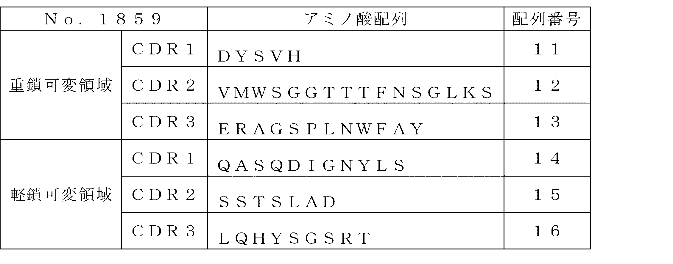
- a heavy chain variable region having complementarity determining regions 1, 2 and 3 comprising the amino acid sequences set forth in SEQ ID NOs: 11, 12 and 13, respectively, and complementation comprising the amino acid sequences set forth in SEQ ID NOs: 14, 15 and 16, respectively.
- a monoclonal antibody that binds to the same epitope as the Tissue Factor epitope to which the monoclonal antibody binds.
- a fragment of an antibody comprising a part of the above monoclonal antibody and capable of binding to Tissue Factor.
- a pharmaceutical composition comprising the monoclonal antibody or antibody fragment as a target binding agent and a drug.
- a composition for drug delivery comprising the above monoclonal antibody or antibody fragment as a target binding agent.
- the monoclonal antibody of the present invention can recognize a cell expressing TF and have an internalization ability for the cell. Therefore, the drug can be efficiently delivered to the cells by using the antibody as a target binding factor.
- a monoclonal antibody that binds to TF is provided.
- the monoclonal antibody of the present invention typically can bind to TF and has the ability to internalize cells expressing TF.
- TF is a factor III of blood coagulation and is expressed on the cell surface as a transmembrane glycoprotein.
- TF is preferably human TF (hTF).
- the full length amino acid sequence of hTF is already known as GenBank ACCESSION_AAA61152 (SEQ ID NO: 1).
- the present invention also provides a monoclonal antibody against mouse TF (mTF). Utilizing an anti-mTF monoclonal antibody as a drug target binding factor can be effective in tests and studies using mice.
- the full length amino acid sequence of mTF is already known as GenBank ACCESSION_AAA63400 (SEQ ID NO: 2).
- internalization means a phenomenon in which an antibody forms an immune complex with an antigen on the cell surface and then is taken into the cell. Whether or not the anti-TF monoclonal antibody has an internalization ability is determined by, for example, contacting an antibody bound with a labeling substance with a cell expressing TF on the surface and transferring the labeling substance into the cell. To determine whether cell death or cell growth inhibition is induced when an antibody bound with a cytotoxic substance is brought into contact with a cell expressing TF on the surface. Can do. More specifically, the presence or absence of the antibody's internalization ability can be confirmed by the internalization assay described in the Examples.
- any appropriate cell can be used as the cell expressing TF on the surface, and examples thereof include cells in tumor tissue.
- the expression of TF in normal tissue is usually local and transient expression, whereas the expression of TF is on the cell surface of cancer cells, vascular endothelial cells, monocytes, macrophages and the like in the tumor tissue. Constantly increased.
- cancer cells include pancreatic cancer cells and gastric cancer cells.
- a monoclonal antibody is an antibody produced by a single clone of antibody-producing cells. Monoclonal antibodies have a uniform primary structure and recognize the same epitope.
- the monoclonal antibody of the present invention has a basic structure consisting of a tetramer in which two homologous heavy chains and two homologous light chains are linked by a disulfide bond.
- the anti-TF monoclonal antibody of the present invention may be any isotype of IgG, IgA, IgM, IgD or IgE, and is preferably IgG.
- the epitope recognized by the anti-TF monoclonal antibody of the present invention is preferably present in the extracellular region of TF.
- the dissociation constant (KD) for TF of the anti-TF monoclonal antibody of the present invention reaches, for example, 5 ⁇ 10 ⁇ 9 M or less, further 1 ⁇ 10 ⁇ 9 M or less, particularly 2 ⁇ 10 ⁇ 10 M or less.
- the dissociation constant can be measured using, for example, a surface plasmon resonance method.
- the anti-TF monoclonal antibody of the present invention may or may not have an anticoagulant action.
- the presence or absence of anticoagulant action or its degree can be determined by prothrombin time (PT).
- PT prothrombin time
- the coagulation extension time ratio (PBS relative ratio) in the state in which the antigen-antibody complex of the anti-TF monoclonal antibody of the present invention is formed is preferably 3 or less, more preferably 2 or less, and further preferably 1 to 1.5.
- the coagulation extension time ratio in the state in which the antigen-antibody complex is formed can be determined by the method described in the Examples.
- the anti-hTF monoclonal antibody of the present invention comprises a heavy chain variable region having complementarity determining regions (CDRs) 1, 2 and 3 comprising the amino acid sequences set forth in SEQ ID NOs: 3, 4 and 5, respectively. And a light chain variable region having CDRs 1, 2 and 3 comprising the amino acid sequences set forth in SEQ ID NOs: 6, 7 and 8, respectively.
- CDRs complementarity determining regions
- a specific example thereof is preferably an anti-hTF monoclonal antibody comprising a heavy chain variable region comprising the amino acid sequence set forth in SEQ ID NO: 9 and a light chain variable region comprising the amino acid sequence set forth in SEQ ID NO: 10. .
- an anti-hTF monoclonal antibody of the invention comprises a heavy chain variable region having CDRs 1, 2 and 3 comprising the amino acid sequences set forth in SEQ ID NOs: 11, 12, and 13, respectively, and SEQ ID NO: 14, Light chain variable regions having CDRs 1, 2 and 3 comprising the amino acid sequences set forth in 15 and 16.
- a specific example thereof is preferably an anti-hTF monoclonal antibody comprising a heavy chain variable region comprising the amino acid sequence set forth in SEQ ID NO: 17 and a light chain variable region comprising the amino acid sequence set forth in SEQ ID NO: 18. .
- a variant of each monoclonal antibody exemplified in the first or second embodiment may also be included in the anti-hTF monoclonal antibody of the present invention.
- examples of such variants include substitution, insertion, addition and / or substitution of one or several (for example, 1 to 10, preferably 1 to 5) amino acids in the heavy chain variable region and / or the light chain variable region.
- a monoclonal antibody containing a deletion can be mentioned.
- Such a variant can also suitably bind to hTF and have the ability to internalize cells expressing hTF.
- the modified monoclonal antibody include a heavy chain comprising an amino acid sequence which is preferably 90% or more, more preferably 95% or more, and further preferably 98% or more identical to the amino acid sequence shown in SEQ ID NO: 9.
- a monoclonal antibody comprising a variable region and a light chain variable region comprising an amino acid sequence that is preferably 90% or more, more preferably 95% or more, and even more preferably 98% or more identical to the amino acid sequence set forth in SEQ ID NO: 10 Is mentioned.
- a heavy chain comprising an amino acid sequence which is preferably 90% or more, more preferably 95% or more, and further preferably 98% or more identical to the amino acid sequence shown in SEQ ID NO: 17.
- a monoclonal antibody comprising a variable region and a light chain variable region comprising an amino acid sequence that is preferably 90% or more, more preferably 95% or more, and even more preferably 98% or more identical to the amino acid sequence set forth in SEQ ID NO: 18 Is mentioned. These variants are preferably capable of binding to hTF and have the ability to internalize cells expressing hTF.
- the variant of the monoclonal antibody is one or several, for example, 1, 2 or 3, preferably 1, or at least one CDR of the heavy chain variable region and / or light chain variable region of the corresponding monoclonal antibody. Two, more preferably one amino acid substitution, insertion, addition and / or deletion may be included.
- Each CDR of the variant preferably has 90-100% homology with each CDR of the corresponding monoclonal antibody, more preferably 95-100%, even more preferably 98-100%, most preferably 100%. Have homology.
- the entire CDR1-3 of the heavy chain and light chain of the variant preferably has 90-100% homology with the entire CDR1-3 of heavy chain and light chain of the corresponding monoclonal antibody, more preferably 95 ⁇ 100%, more preferably 98-100%, most preferably 100% homology.
- the anti-hTF monoclonal antibody of the present invention may be a monoclonal antibody that binds to the same epitope as that of hTF to which the monoclonal antibody exemplified in the first or second embodiment binds.
- Antibodies that bind to the same epitope can be obtained by known methods such as competitive ELISA.
- the test antibody contains 30% or more compared to the binding activity of the control antibody (that is, the monoclonal antibody exemplified in the first or second embodiment) in the absence of the test antibody.
- the test antibody can be said to be an antibody that binds to substantially the same epitope as the control antibody.
- the antibody that binds to the same epitope is preferably capable of binding to hTF and has the ability to internalize cells expressing hTF.
- the antibody that binds to the same epitope may be a variant of the monoclonal antibody exemplified in the first or second embodiment.
- the anti-hTF monoclonal antibody of the present invention described above can be a human chimeric antibody or a humanized antibody.
- a human chimeric antibody is an antibody in which a variable region of an antibody derived from a non-human mammal and a constant region of an antibody derived from a human are linked. Therefore, the human chimeric antibody of the present invention comprises the heavy chain variable region and the light chain variable region of the monoclonal antibody exemplified in the first, second or third embodiment, respectively, as the human heavy chain constant region and human light chain constant region, respectively. It may be a chimeric antibody obtained by linking to a normal region.
- examples of the human chimeric antibody of the present invention include a heavy chain variable region and a light chain CDR1 comprising the amino acid sequences set forth in SEQ ID NOs: 3, 4, and 5 as heavy chain CDRs 1, 2, and 3, respectively.
- examples of 2 and 3 include chimeric antibodies in which a light chain variable region comprising the amino acid sequences set forth in SEQ ID NOs: 6, 7 and 8 are linked to a human heavy chain constant region and a human light chain constant region, respectively.
- Specific examples of the chimeric antibody include a heavy chain variable region comprising the amino acid sequence set forth in SEQ ID NO: 9 and a light chain variable region comprising the amino acid sequence set forth in SEQ ID NO: 10, respectively, as a human heavy chain constant region and human.
- human chimeric antibody of the present invention includes a heavy chain variable region comprising the amino acid sequences set forth in SEQ ID NOs: 11, 12, and 13 as light chain CDRs 1, 2 and 3, and light chain CDRs 1, 2 and 3, respectively.
- 3 includes chimeric antibodies in which a light chain variable region comprising the amino acid sequences set forth in SEQ ID NOs: 14, 15, and 16 are linked to a human heavy chain constant region and a human light chain constant region, respectively.
- Specific examples of the chimeric antibody include a heavy chain variable region comprising the amino acid sequence set forth in SEQ ID NO: 17 and a light chain variable region comprising the amino acid sequence set forth in SEQ ID NO: 18, respectively.
- the heavy chain constant region of the human chimeric antibody only needs to belong to human immunoglobulin (hereinafter referred to as hIg), and preferably belongs to the hIgG class.
- hIg human immunoglobulin
- the light chain constant region of the human chimeric antibody only needs to belong to hIg, and may be either ⁇ class or ⁇ class.
- a humanized antibody is an antibody obtained by grafting a CDR of an antibody derived from a non-human mammal into an appropriate position of a variable region of a human-derived antibody. Therefore, the humanized antibody of the present invention has the heavy chain and light chain CDR1 to 3 of the monoclonal antibody exemplified in the first, second or third embodiment as the heavy chain and light chain CDR1 to 3.
- the other region may be a human antibody derived from a human antibody.
- humanized antibodies of the present invention include heavy chain CDRs 1, 2 and 3, respectively, comprising the amino acid sequences set forth in SEQ ID NOs: 3, 4, and 5, respectively, and light chain CDRs 1, 2, and 3, respectively.
- the humanized antibody consisting of the amino acid sequences set forth in SEQ ID NOs: 6, 7 and 8 and other regions derived from human antibodies, and CDRs 1, 2 and 3 of the heavy chain are described in SEQ ID NOs: 11, 12 and 13, respectively.
- humanized antibodies in which the light chain CDRs 1, 2 and 3 have the amino acid sequences set forth in SEQ ID NOs: 14, 15 and 16, respectively, and other regions are derived from human antibodies.
- the heavy chain of a humanized antibody only needs to belong to hIg, and preferably belongs to the hIgG class.
- the light chain of the humanized antibody only needs to belong to hIg, and may be either ⁇ class or ⁇ class.
- an anti-mTF monoclonal antibody of the invention comprises a heavy chain variable region having CDRs 1, 2, and 3 comprising the amino acid sequences set forth in SEQ ID NOs: 19, 20, and 21, respectively, and SEQ ID NOs: 22, 23, respectively. And a light chain variable region having CDRs 1, 2 and 3 comprising the amino acid sequence described in 24.
- a specific example thereof is preferably an anti-mTF monoclonal antibody comprising a heavy chain variable region comprising the amino acid sequence set forth in SEQ ID NO: 25 and a light chain variable region comprising the amino acid sequence set forth in SEQ ID NO: 26. .
- the modified monoclonal antibody exemplified above can also be included in the anti-mTF monoclonal antibody of the present invention.
- examples of such variants include substitution, insertion, addition and / or substitution of one or several (for example, 1 to 10, preferably 1 to 5) amino acids in the heavy chain variable region and / or the light chain variable region.
- a monoclonal antibody containing a deletion can be mentioned.
- Such a variant can also suitably bind to mTF and have the ability to internalize cells expressing mTF.
- modified monoclonal antibody examples include a heavy chain comprising an amino acid sequence which is preferably 90% or more, more preferably 95% or more, and still more preferably 98% or more identical to the amino acid sequence set forth in SEQ ID NO: 25.
- a monoclonal antibody comprising a variable region and a light chain variable region comprising an amino acid sequence that is preferably 90% or more, more preferably 95% or more, and even more preferably 98% or more identical to the amino acid sequence set forth in SEQ ID NO: 26 Is mentioned.
- Such a variant preferably binds to mTF and has the ability to internalize cells expressing mTF.
- the variant of the monoclonal antibody is one or several, for example, 1, 2 or 3, preferably 1, or at least one CDR of the heavy chain variable region and / or light chain variable region of the corresponding monoclonal antibody. Two, more preferably one amino acid substitution, insertion, addition and / or deletion may be included.
- Each CDR of the variant preferably has 90-100% homology with each CDR of the corresponding monoclonal antibody, more preferably 95-100%, even more preferably 98-100%, most preferably 100%. Have homology.
- the entire CDR1-3 of the heavy chain and light chain of the variant preferably has 90-100% homology with the entire CDR1-3 of heavy chain and light chain of the corresponding monoclonal antibody, more preferably 95 ⁇ 100%, more preferably 98-100%, most preferably 100% homology.
- the anti-mTF monoclonal antibody of the present invention may be a monoclonal antibody that binds to the same epitope as that of mTF to which the monoclonal antibody exemplified above binds.
- Antibodies that bind to the same epitope are preferably capable of binding to mTF and have the ability to internalize cells expressing mTF.
- the antibody that binds to the same epitope may be a variant of the monoclonal antibody exemplified above.
- bonded with this same epitope is as above-mentioned.
- B-1 Preparation of monoclonal antibody using hybridoma
- the monoclonal antibody of the present invention is prepared, for example, by preparing a hybridoma by cell fusion between an antibody-producing cell obtained from an animal immunized with an antigen and a myeloma cell, and producing the target antibody from the resulting hybridoma. Can be obtained by selecting those to be made and causing the selected hybridomas to produce antibodies.
- TF full-length TF
- a partial peptide thereof or cells expressing TF on the surface can be used.
- hTF can be obtained, for example, by purifying human placenta-derived hTF according to the method described in JP-A-9-302000. Further, for example, TF or a partial peptide thereof can be obtained by a genetic engineering method or a chemical synthesis method. The partial peptide can be used in combination with any appropriate carrier protein as required.
- the mRNA sequence of hTF is known in GenBank NM_001993.4 (SEQ ID NO: 27). Also, the mRNA sequence of mTF is known in GenBank M57896.1 (SEQ ID NO: 28).
- B-1-2 Preparation of antibody-producing cells
- the antigen obtained as described above is mixed with any appropriate adjuvant and administered to non-human mammals such as mice, rats, horses, monkeys, rabbits, goats and sheep for immunization. .
- the antibody titer against the antigen of the immunized animal is measured, and the final immunization is given to the animal having a high antibody titer.
- antibody-producing cells such as spleen cells and lymph node cells are collected. Details of the immunization method and the method of collecting antibody-producing cells are well known to those skilled in the art, and thus detailed description thereof is omitted.
- the antibody titer can be measured by, for example, enzyme immunoassay (EIA) such as ELISA, radioimmunoassay (RIA) or the like using blood collected from an animal.
- EIA enzyme immunoassay
- RIA radioimmunoassay
- any appropriate cell line derived from an animal such as a mouse or a rat and generally available to those skilled in the art can be used.
- myeloma that has drug resistance and cannot survive in a selective medium eg, hypoxanthine, aminopterin, thymidine-containing medium (HAT medium)
- HAT medium thymidine-containing medium
- Cells are used.
- the cell fusion can be performed using any appropriate method such as PEG method or electrofusion method.
- the cells after cell fusion treatment are suspended and diluted in a selective medium (for example, HAT medium), and cultured in each well of the culture plate.
- a selective medium for example, HAT medium
- Hybridoma Screening and Cloning Cells that have formed colonies as a result of the culture after cell fusion are selected as hybridomas.
- the selected hybridoma is cultured in, for example, a microtiter plate, and the obtained culture supernatant is collected to measure the reactivity to the antigen.
- the reactivity to an antigen can be measured by EIA, RIA or the like.
- EIA EIA
- RIA a monoclonal antibody-producing hybridoma is isolated by a limiting dilution method or the like.
- Monoclonal antibody can be obtained by culturing the above hybridoma in any appropriate medium and purifying it from the obtained culture supernatant. Injecting the hybridoma into the abdominal cavity of a non-human mammal such as a mouse or rat. And it can prepare by the method etc. which culture
- Antibody purification can be achieved, for example, by using an ammonium sulfate salting-out method, a gel filtration chromatography method, an ion exchange chromatography method, an affinity column chromatography method such as an anti-immunoglobulin column, a protein A column, etc., in combination as necessary. It can be carried out.
- the monoclonal antibody of the present invention can be produced by genetic engineering technique using an antibody gene cloned from an antibody producing cell such as the above hybridoma.
- B-2-1 Cloning of the gene encoding the variable region Extracting the total mRNA from the hybridoma producing the target antibody, using reverse transcriptase from the obtained total mRNA, cDNA encoding the antibody variable region using a sequence common to the antibody gene as a primer Is synthesized.
- the cDNA can be synthesized and amplified using, for example, the 5′-RACE method, and any appropriate restriction enzyme sites can be introduced at both ends of the cDNA.
- a desired DNA fragment is purified from the obtained PCR product, ligated with vector DNA, and introduced into Escherichia coli or the like to prepare a desired recombinant vector.
- the base sequence of the target antibody gene is confirmed by a known method such as the deoxy method.
- DNA encoding the variable region cloned as described above is ligated with the DNA encoding the desired antibody constant region and incorporated into an expression vector.
- DNA encoding a variable region may be incorporated into an expression vector containing DNA encoding a desired constant region.
- the expression vector thus obtained can be introduced into any appropriate host cell, whereby the antibody can be expressed.
- the heavy chain or light chain may be separately incorporated into an expression vector, and these two expression vectors may be introduced simultaneously into the same host cell, or the DNA encoding the heavy chain or light chain may be introduced into a single expression vector. May be incorporated into a host cell.
- the DNA encoding the variable region can also be obtained by performing total synthesis using an artificial gene synthesis service or the like based on the base sequence determined in Section B-2-1. Examples of host cells include animal cells, plant cells, insect cells, yeasts, bacteria, and the like.
- Monoclonal antibodies can be obtained by culturing host cells into which the expression vector is incorporated in any appropriate medium and purifying the resulting culture supernatant. The method for purifying the antibody is as described above.
- a human chimeric antibody is obtained by, for example, ligating a DNA encoding a variable region of a monoclonal antibody obtained in the same manner as described above with a DNA encoding a constant region of a human antibody, and incorporating this into an expression vector. It can obtain by introduce
- Humanized antibodies can be obtained by grafting CDRs of non-human mammalian antibodies to human antibodies so as to link them to the framework regions of human antibodies using so-called CDR grafting techniques. Methods for transplanting CDRs of antibodies from non-human mammals (eg, mice) into human framework regions are known, and examples include overlap extension PCR.
- CDR grafting it is advantageous in maintaining CDR functions to select a human framework region having high homology with the framework region of an antibody of a non-human mammal. Therefore, it is preferable to use a human framework region consisting of an amino acid sequence having high homology with the amino acid sequence of the framework region adjacent to the CDR to be transplanted.
- the present invention also provides a fragment of an antibody comprising a portion of the monoclonal antibody described in Section A and capable of binding to Tissue Factor.
- the antibody fragment of the present invention typically has the ability to internalize cells expressing Tissue Factor. Examples of the antibody fragment include Fab, F (ab ′) 2 , Fab ′, single chain antibody (scFv), disulfide stabilized antibody (dsFv), dimerized V region fragment (Diabody), and peptide containing CDR Etc.
- Fab can be obtained by papain treatment of the above monoclonal antibody. It can also be obtained by inserting the DNA encoding the Fab of the above monoclonal antibody into any appropriate expression vector, introducing the vector into a host cell, and expressing it.
- F (ab ′) 2 can be obtained by treating the monoclonal antibody with pepsin. It can also be obtained by inserting the DNA encoding F (ab ′) 2 of the monoclonal antibody into any appropriate expression vector, introducing the vector into a host cell, and expressing it.
- Fab ′ is an antibody fragment obtained by cleaving the SS bond between the hinges of F (ab ′) 2 .
- Fab ′ can be obtained by treating the F (ab ′) 2 with a reducing agent dithiothreitol. It can also be obtained by inserting DNA encoding Fab ′ into any appropriate expression vector, introducing the vector into a host cell, and expressing it.
- ScFv is obtained by connecting only the variable regions of the heavy chain and the light chain with an appropriate peptide linker.
- the scFv can be obtained by constructing an scFv expression vector based on the DNA encoding the heavy chain variable region and the light chain variable region of the monoclonal antibody, introducing the vector into a host cell, and expressing the vector.
- DsFv is a polypeptide in which one amino acid residue in each of the heavy chain variable region and the light chain variable region is replaced with a cysteine residue via an SS bond.
- the position of introduction of a cysteine residue in each region can be determined based on the three-dimensional structure predicted by molecular modeling.
- the dsFv can be obtained by constructing a dsFv expression vector based on DNA encoding the heavy chain variable region and the light chain variable region of the monoclonal antibody, introducing the vector into a host cell, and expressing the vector.
- Diabody is a dimer of scFv linked by a short peptide linker of 8 amino acid residues or less, and has a bivalent antigen binding activity.
- the bivalent antigen binding activity may be the same or different.
- Diabody constructs an expression vector for scFv linked with a peptide linker of 8 amino acid residues or less based on DNA encoding the heavy chain variable region and the light chain variable region of the above monoclonal antibody, and introduces the vector into a host cell. And can be obtained by expression.
- the peptide containing CDR contains at least one CDR of heavy chain variable region or light chain variable region.
- the peptide containing CDR may be one in which a plurality of CDRs are bound directly or via an appropriate peptide linker.
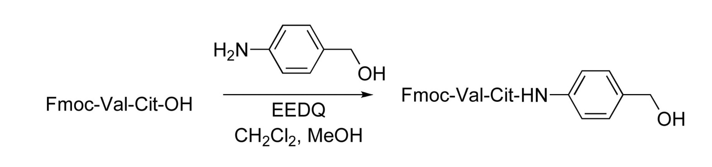
- a peptide containing CDR is obtained by inserting a DNA encoding CDR in the heavy chain variable region and the light chain variable region of the monoclonal antibody into any appropriate expression vector, introducing the vector into a host cell, and expressing the vector. be able to. It can also be obtained by chemical synthesis methods such as Fmoc method and tBoc method.
- the pharmaceutical composition of the present invention comprises the monoclonal antibody described in Section A as a target binding agent or the antibody fragment described in Section C and a drug.
- a monoclonal antibody or a fragment of the antibody hereinafter sometimes referred to as “monoclonal antibody”
- the monoclonal antibody or the like may be in a state of being bound to a drug.
- a polymer compound may be further bound to a monoclonal antibody or the like.
- any appropriate drug is selected according to the disease to be treated.
- biopharmaceuticals such as nucleic acid drugs, antibody drugs, gene therapy drugs, and cytotoxic molecules such as cytotoxins and cytotoxic drugs.
- nucleic acid drug examples include plasmid DNA, siRNA, micro RNA, shRNA, antisense nucleic acid, decoy nucleic acid, aptamer, and ribozyme site.
- cytoxins examples include taxol, cytochalasin B, gramicidin D, ethidium bromide, emetine, mitomycin, etoposide, tenoposide, vincristine, vinblastine, colchicine, doxorubicin, daunorubicin, dihydroxyanthracindione, mitoxantrone, mitramomycin, Actinomycin D, 1-dehydrotestosterone, glucocorticoid, procaine, tetracaine, lidocaine, propranolol, puromycin, duocarmycin, calicheamicin, maytansine, auristatin or derivatives thereof.
- cytotoxic drugs include methotrexate, 6-mercaptopurine, 6-thioguanine, cytarabine, 5-fluorouracil decarbazine and other antimetabolites, mechloretamine, thioepachloram butyl, melphalan, carmustine (BSNU), lomustine (CCNU), cyclososfamide, busulfan, dibromomannitol, streptozotocin, mitomycin C, anthracyclines such as cis-dichlorodiamineplatinum (II), anthracyclines such as daunorubicin and doxorubicin, dactinomycin, bleomycin, mitramycin, Antibiotics such as anthramycin (AMC) and mitotic agents such as vincristine and vinblastine can be mentioned.
- methotrexate 6-mercaptopurine, 6-thioguanine, cytarabine, 5-fluorouracil decarbazin
- the binding between a drug or a polymer compound and a monoclonal antibody or the like can be performed by a method known in the art. For example, it can be performed by reacting a functional group that each has or a functional group introduced as necessary. Examples of combinations of the functional groups include amino groups and carboxyl groups, carboxyl groups and hydroxyl groups, maleimide groups and thiol groups, thiol groups and thiol groups, hydrazide groups and ketone groups, hydrazide groups and aldehyde groups, and amino groups and aldehydes.
- the drug when it is a protein or peptide, it may be a fusion protein with a monoclonal antibody or the like by a genetic engineering technique. Furthermore, when the drug has a charge, it may be bound via an ionic bond.
- any appropriate polymer compound is selected as the polymer compound.
- Specific examples include polyethylene glycol, albumin, dextran, polyvinyl pyrrolidone, polyvinyl alcohol and the like. By binding such a polymer compound, stability in blood can be improved.
- the binding site of the polymer compound can be any appropriate site as long as it does not impair the antigen binding activity and internalization ability of the monoclonal antibody or the like.
- the monoclonal antibody or the like may be in a state of being bound to a DDS carrier material.
- DDS carrier material those capable of forming nanoparticles encapsulating a drug (for example, particles having an average particle diameter of preferably 10 nm to 400 nm, more preferably 20 nm to 300 nm, still more preferably 30 nm to 150 nm) are preferably used.
- a DDS carrier material nanoparticles encapsulating a drug can be formed, so that the stability of the drug in the living body can be improved and sustained release can be achieved. Furthermore, reliable drug delivery into the target cell by a monoclonal antibody or the like becomes possible.
- Monoclonal antibodies and the like are bound to any appropriate position of the DDS carrier material.
- the monoclonal antibody or the like is bound so as to be exposed on the outer surface of the nanoparticles formed by the DDS carrier material.
- the binding between the monoclonal antibody or the like and the DDS carrier material can be performed, for example, by reacting a functional group that each has or a functional group introduced as necessary. Preferred combinations of functional groups are as described above.
- DDS carrier material examples include polymer micelle-forming materials such as block copolymers having hydrophilic segments and hydrophobic segments, liposome-forming materials such as phospholipids, natural polymers such as gelatin, collagen, hyaluronic acid, and alginic acid, Examples include nanohydrogel capsule-forming materials such as synthetic polymers such as polyethylene glycol and polyvinyl alcohol, and nanosphere-forming materials such as polyglycolic acid, polylactic acid, and copolymers thereof. Among these, it is preferable to use a block copolymer having a hydrophilic segment and a hydrophobic segment.
- a polymer micelle capable of delivering a drug into a target cell with high certainty by binding a monoclonal antibody or the like to the hydrophilic segment of a block copolymer having a hydrophilic segment and a hydrophobic segment to form a target-binding block copolymer. Can be obtained.
- the drug the same drug as in the first embodiment can be used.
- the drug may be used as it is, or may be used in the form of a drug-binding block copolymer bound to the hydrophobic segment of a block copolymer having a hydrophilic segment and a hydrophobic segment. .
- the composition for drug delivery of the present invention comprises the monoclonal antibody described in Section A or the antibody fragment described in Section C.
- the monoclonal antibody or a fragment of the antibody can be used as a target binding factor to cells expressing TF, and can further exhibit the ability to internalize the cells. Therefore, by administering a drug using the drug delivery composition of the present invention, the drug can be efficiently delivered into cells that express TF on the surface.
- the anti-TF monoclonal antibody of the present invention is typically used for the treatment of a disease involving Tissue Factor (for example, a disease accompanied by increased expression of Tissue Factor on the tissue or cell surface).
- diseases involving Tissue Factor include cancer, inflammation, and thrombosis. Since TF is constantly increased in expression in various cancer tissues, it can be used for the treatment regardless of the type of cancer. In addition, for example, in pancreatic cancer, it has been reported that the prognosis of a patient who highly expresses TF is poor, and it can be effectively used in treatment for such a patient.
- the administration route of the pharmaceutical composition of the present invention is preferably parenteral administration such as subcutaneous, intravenous, arterial, local, etc., and intravenous injection is particularly preferable.
- the dosage can be appropriately determined according to the type of drug, usage, patient age, sex, patient health and the like.
- Parts and % mean “parts by weight” and “% by weight”.
- Recombinant hTF 50 ⁇ g was administered intraperitoneally to 3 6-week-old female female rats together with Freund's complete adjuvant (Difco) for initial immunization. 14 days later, 50 ⁇ g of recombinant hTF was administered together with Sigma Adjuvant System (Sigma) for booster immunization. Thereafter, the same booster was performed 5 times every 21 days. Further, 126 days later, 10 ⁇ g of recombinant hTF diluted with PBS was intraperitoneally administered, and 40 ⁇ g of recombinant hTF was administered to the tail vein for final immunization.
- each well was washed 3 times with 300 ⁇ L of a washing solution, and HRP-labeled anti-mouse IgG antibody (Bethyl) diluted 5,000 times with a blocking buffer was added at 50 ⁇ L and reacted for 30 minutes. After the reaction, each well was washed with 300 ⁇ L of washing solution three times, and 3.7 mM o-phenylenediamine / 25 mM citric acid / 130 mM Na 2 HPO 4 /0.006% H 2 O 2 (pH 5.0) was added at 100 ⁇ L. And colored. 10-15 minutes later, 2N sulfuric acid was added at 30 ⁇ L / well to stop the reaction, and the absorbance (490 nM) was measured with an absorbance plate reader.
- HRP-labeled anti-mouse IgG antibody Bethyl
- immunoprecipitation ELISA was performed by mixing recombinant hTF and hybridoma culture supernatant and measuring the amount of unbound hTF in the mixed solution by hTF quantitative sandwich ELISA. Furthermore, flow cytometry was performed according to a standard method, and the reactivity of the hybridoma culture supernatant to hTF-expressing cells was measured.
- hybridomas that showed strong affinity with hTF were selected, and hybridoma clones producing monoclonal antibodies that bind to hTF were established by performing the limiting dilution method twice for these clones.
- each of the established hybridomas was cultured in a large amount in an RPMI 1640 medium containing 5% of bovine serum from which cow-derived IgG was removed, and a culture supernatant was obtained.
- each hybridoma was cultured in large quantities in the abdominal cavity of ICR nude mice, and ascites was collected. The obtained culture supernatant or ascites was subjected to Protein G affinity column chromatography to purify the IgG monoclonal antibody.
- Lysotracker RED-DND99 Invitrogen was added to the culture solution to a final concentration of 75 nM after 2 hours from the start of the culture, and then further cultured for 1 hour.
- Lysotracker RED-DND99 Invitrogen was added to the culture solution to a final concentration of 75 nM after 2 hours from the start of the culture, and then further cultured for 1 hour.
- PBS 4% paraformaldehyde
- nuclear stained with DAPI (4 ′, 6-diamidino-2-phenylindole
- Fluoromount G Southern Biotech
- a DNA fragment encoding the heavy chain variable region and light chain variable region of the anti-hTF monoclonal antibody was obtained by PCR using the synthesized cDNA as a template. Specifically, from the following list of primers, the light chain variable region cloning was performed using the following primer 1-19 mixed primer and primer 20 as the sense primer and antisense primer for heavy chain variable region cloning, respectively. PCR was performed using the following mixed primer of primer 22 to 38 and primer 39 as a sense primer and an antisense primer, respectively.
- Primer 1 NNCCATGGCCGAGGTRMAGCTTCAGGAGTC Primer 2 (SEQ ID NO: 31) NNCCATGGCCGAGGGTBCAGCTBCAGCAGTC Primer 3 (SEQ ID NO: 32) NNCCATGGCCGAGGGTGCAGCTGAAGSASTC Primer 4 (SEQ ID NO: 33) NNCCATGGCCGAGGTCCARCTGCAACARTC Primer 5 (SEQ ID NO: 34) NNCCATGGCCGAGGTYCAGCTBCAGCARTC Primer 6 (SEQ ID NO: 35) NNCCATGGCCGAGGTYCARCTGCAGCAGTC Primer 7 (SEQ ID NO: 36) NNCCATGGCCGAGGTCCCACGTGAAGCAGTC Primer 8 (SEQ ID NO: 37) NNCCATGGCCGAGGGTGAASSTGGGGAATC Primer 9 (SEQ ID NO: 38) NNCCATGGCCGAGGGTGAWGYTGGTGGAGTC Primer 10 (SEQ ID NO: 39) N
- Each PCR product obtained above was cloned into a vector according to a conventional method, and the base sequence was determined.
- the base sequences of the heavy chain variable region and the light chain variable region of the anti-hTF monoclonal antibody (No. 1849) determined as described above are as shown in SEQ ID NOs: 69 and 70, respectively.
- the amino acid sequences of the heavy chain variable region and the light chain variable region are as described in SEQ ID NOs: 9 and 10, respectively. Further, by comparing these amino acid sequences with a database of amino acid sequences of known antibodies (IMGT website: http://www.imgt.org/), by examining the homology, the amino acid sequences of CDR It was decided as follows.
- Anti-hTF monoclonal antibody No. 1859
- Anti-hTF was obtained in the same manner as above except that total RNA was extracted from the hybridoma producing the anti-hTF monoclonal antibody (No. 1859) and that primer 21 was used as the antisense primer for cloning the heavy chain variable region.
- the base sequences of the heavy chain variable region and the light chain variable region of the monoclonal antibody (No. 1859) were determined.
- the determined base sequences of the heavy chain variable region and the light chain variable region of the anti-hTF monoclonal antibody are as shown in SEQ ID NOs: 71 and 72, respectively.
- the amino acid sequences of the heavy chain variable region and the light chain variable region are as described in SEQ ID NOs: 17 and 18, respectively.
- the amino acid sequences of CDRs in these variable regions were determined as follows.
- Anti-hTF monoclonal antibodies (No. 1849, No. 1859) were immobilized on the surface of a Biacore CM5 chip.
- a CM5 chip on which each antibody was immobilized was set in an SPR device, and an antigen-containing buffer containing purified hTF was passed through the flow path.
- the dissociation constant between each antibody and hTF was determined by this measurement system.
- the measurement conditions are as follows.
- SPR device Biacore 2000 (Biacore)
- Antigen-containing buffer 10 mM acetate buffer (pH 5.0) containing hTF at a final concentration of 25 ⁇ g / ml
- Running buffer HBS-EP buffer (10 mM HEPES, pH 7.5, 0.15 M NaCl, 3 mM EDTA, 0.005% surfactant P20 (Tween 20), pH 7.4).
- the dissociation constant (KD) for hTF of each antibody was No. No. 1849 is 9.139 ⁇ 10 ⁇ 11 , no. 1859 was 1.894 ⁇ 10 ⁇ 10 .
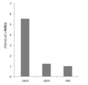
- the prothrombin time of the anti-hTF monoclonal antibodies was measured as follows. 3 ⁇ g of antibody was added to 350 ng of recombinant hTF antigen, and PBS was added until the total volume became 5 ⁇ l. The antigen-antibody reaction was allowed to proceed for 15 minutes while shaking at 37 ° C. and 600 rpm. 50 ⁇ l of human plasma anticoagulated with 3.8% sodium citrate and 100 ⁇ l of 25 mM CaCl 2 were added to the reaction solution, and the cells were cultured while shaking at 37 ° C. and 600 rpm, and the time until a fibrin clot was formed ( Prothrombin time) was measured. Table 3 and FIG. 2 show the coagulation extension time ratio (prothrombin time ratio) of each antibody when the prothrombin time in the control using PBS instead of the reaction solution is 1.
- Example 2 pharmaceutical composition [Binding of monoclonal antibody to drug]
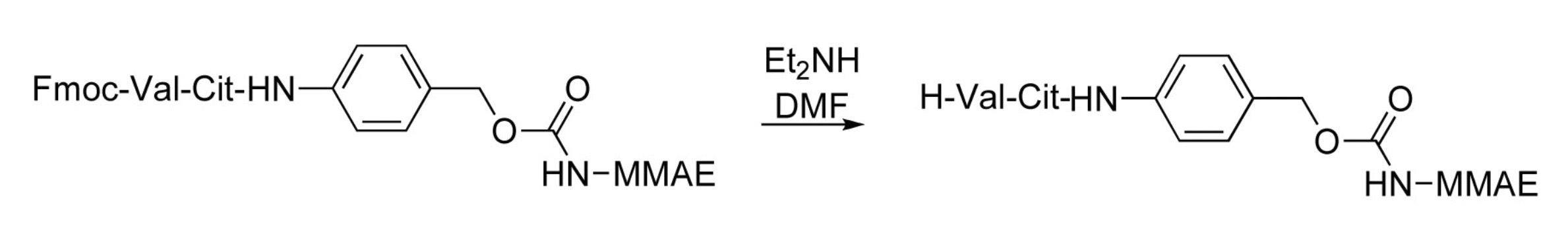
- An anti-hTF monoclonal antibody (No. 1849) conjugated with monomethyl auristatin E (MMAE) (hereinafter referred to as “hTF-MMAE”) was obtained as follows.
- a pH 6.4 buffer solution is prepared using 5 mM EDTA-containing PBS, 150 mM NaCl, and 5 mM EDTA-containing 100 mM phosphate buffer (pH 6.0), and the obtained buffer solution is used to produce an antibody concentration of 1.0 mg /
- the antibody solution was prepared to be ml.
- Dithiothreitol (DTT) was added to the antibody solution to a final concentration of 1 to 10 mM and reacted at 26 to 37 ° C. for 30 to 45 minutes. Subsequently, the reaction reagent was removed from the reaction solution with Amicon Ultra (MWCO: 30,000). Absorption measurement showed that the antibody recovery was 80-99%.
- DTT Dithiothreitol
- Amicon Ultra MWCO: 30,000
- a buffer solution of pH 6.4 is prepared using 5 mM EDTA-containing PBS and 150 mM NaCl and 5 mM EDTA-containing 100 mM phosphate buffer (pH 6.0), and the reaction solution is prepared using the obtained buffer solution.
- the protein concentration was diluted to 0.5 mg / ml.
- the maleimide MMAE compound was mixed with the diluted solution at a molar ratio of 1: 4 (antibody: maleimide MMAE compound), and then reacted at room temperature for 1 hour and then at 4 ° C. overnight.
- reaction reagent was removed from the reaction solution with Amicon Ultra (MWCO: 30,000), and the solvent was replaced with PBS.
- Amicon Ultra MWCO: 30,000
- the recovery rate was 60 to 90%.
- FIG. 3 shows the percentage (%) of each cell viability when the cell viability of the negative control (no drug sample added) is 100%.
- hTF-MMAE showed a remarkably superior cell killing effect compared to mTF-MMAE. From this, it can be seen that when the drug is bound to the anti-hTF monoclonal antibody (No. 1849), the migration into the cell expressing hTF on the surface is improved, and high drug efficacy can be exhibited.
- hTF-MMAE showed a remarkably excellent antitumor effect. The results are consistent with the cell killing effect confirmed in vitro. Further, as shown in FIG. 5, no decrease in body weight was confirmed in the group administered with hTF-MMAE.
- Recombinant mTF (50 ⁇ g) was administered intraperitoneally to three 6-week-old female female rats together with Freund's complete adjuvant (Difco) for initial immunization. 14 days later, 50 ⁇ g of recombinant mTF was administered together with Sigma Adjuvant System (Sigma) for booster immunization. Thereafter, the same boosting was performed 7 times every 21 days. Further, 207 days later, 10 ⁇ g of recombinant mTF diluted with PBS was intraperitoneally administered, and 40 ⁇ g of recombinant mTF was administered to the tail vein for final immunization.
- each well was washed 3 times with 300 ⁇ L of a washing solution, and HRP-labeled anti-mouse IgG antibody (Bethyl) diluted 5,000 times with a blocking buffer was added at 50 ⁇ L and reacted for 30 minutes. After the reaction, each well was washed with 300 ⁇ L of washing solution three times, and 3.7 mM o-phenylenediamine / 25 mM citric acid / 130 mM Na 2 HPO 4 /0.006% H 2 O 2 (pH 5.0) was added at 100 ⁇ L. And colored. 10-15 minutes later, 2N sulfuric acid was added at 30 ⁇ L / well to stop the reaction, and the absorbance (490 nM) was measured with an absorbance plate reader.
- HRP-labeled anti-mouse IgG antibody Bethyl
- the immunoprecipitation ELISA method was performed by mixing recombinant mTF and hybridoma culture supernatant, and measuring the amount of unbound mTF in the mixed solution by mTF quantitative sandwich ELISA. Furthermore, flow cytometry was performed according to a standard method, and the reactivity of the hybridoma culture supernatant to hTF-expressing cells was measured.
- hybridomas showing strong affinity with mTF were selected, and hybridoma clones producing monoclonal antibodies that bind to mTF were established by performing the limiting dilution method twice for these clones.
- each of the established hybridomas was cultured in a large amount in an RPMI 1640 medium containing 5% of bovine serum from which cow-derived IgG was removed, and a culture supernatant was obtained.
- each hybridoma was cultured in large quantities in the abdominal cavity of ICR nude mice, and ascites was collected. The obtained culture supernatant or ascites was subjected to Protein G affinity column chromatography to purify the IgG monoclonal antibody.
- Lysotracker RED-DND99 Invitrogen was added to the culture solution to a final concentration of 75 nM after 2 hours from the start of the culture, and then further cultured for 1 hour.
- Lysotracker RED-DND99 Invitrogen was added to the culture solution to a final concentration of 75 nM after 2 hours from the start of the culture, and then further cultured for 1 hour.
- PBS 4% paraformaldehyde
- nuclear stained with DAPI (4 ′, 6-diamidino-2-phenylindole
- Fluoromount G Southern Biotech
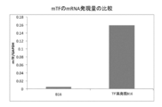
- Fig. 7 shows the level of enhancement of TF expression level in TF forced expression mouse B16 melanoma cells as a reference.
- Fig. 7 shows the expression of GAPDH mRNA in mouse B16 melanoma cells and their TF forced expression cells. It is a graph which shows the ratio of the mRNA expression level of TF with respect to the quantity).
- the determined nucleotide sequences of the heavy chain variable region and the light chain variable region of the anti-mTF monoclonal antibody are as shown in SEQ ID NOs: 74 and 75, respectively.
- the amino acid sequences of the heavy chain variable region and the light chain variable region are as described in SEQ ID NOs: 25 and 26, respectively.
- the amino acid sequences of CDRs in these variable regions were determined as follows.
- Example 4 Production of Chimeric Antibody
- the DNA fragment encoding the heavy chain variable region and the light chain variable region of the anti-hTF monoclonal antibody (No. 1849) cloned in Example 1 was amplified by PCR.
- pFUSEss_CHIg-hG1e2 in vivoGen
- the obtained expression vector was transfected into CHO-K1 cells using Lipofectamine LTX Reagent (Invitrogen). Subsequently, drug selection was performed using 10 ⁇ g / mL Blastcidin S (Kaken Pharmaceutical) and 300 ⁇ g / mL Zeocin (Invitrogen) to obtain both resistant cell lines.
- the obtained cell line was maintained and cultured in a medium containing Ham's F12K (wako), 10% FBS, 1% penicillin, streptomycin (Invitrogen), 10 ⁇ g / mL Blastcidin S and 300 ⁇ g / mL Zeocin. Subsequently, a constant expression cell line of an anti-hTF human chimeric antibody (No. 1849 chimeric clone) was cloned by a limiting dilution method using a 96-well plate.
- the monoclonal antibody or fragment thereof of the present invention can be suitably used in the field of DDS.
Landscapes
- Health & Medical Sciences (AREA)
- Chemical & Material Sciences (AREA)
- Life Sciences & Earth Sciences (AREA)
- Engineering & Computer Science (AREA)
- Bioinformatics & Cheminformatics (AREA)
- Medicinal Chemistry (AREA)
- General Health & Medical Sciences (AREA)
- Pharmacology & Pharmacy (AREA)
- Animal Behavior & Ethology (AREA)
- Public Health (AREA)
- Veterinary Medicine (AREA)
- Organic Chemistry (AREA)
- Immunology (AREA)
- Epidemiology (AREA)
- Molecular Biology (AREA)
- Hematology (AREA)
- Nuclear Medicine, Radiotherapy & Molecular Imaging (AREA)
- General Chemical & Material Sciences (AREA)
- Chemical Kinetics & Catalysis (AREA)
- Biochemistry (AREA)
- Proteomics, Peptides & Aminoacids (AREA)
- Genetics & Genomics (AREA)
- Biophysics (AREA)
- Cell Biology (AREA)
- Toxicology (AREA)
- Endocrinology (AREA)
- Mycology (AREA)
- Microbiology (AREA)
- Rheumatology (AREA)
- Pain & Pain Management (AREA)
- Diabetes (AREA)
- Medicines Containing Antibodies Or Antigens For Use As Internal Diagnostic Agents (AREA)
- Peptides Or Proteins (AREA)
- Preparation Of Compounds By Using Micro-Organisms (AREA)
- Micro-Organisms Or Cultivation Processes Thereof (AREA)
Abstract
Description
それぞれ配列番号3、4および5に記載されるアミノ酸配列を含む相補性決定領域1、2および3を有する重鎖可変領域と、それぞれ配列番号6、7および8に記載されるアミノ酸配列を含む相補性決定領域1、2および3を有する軽鎖可変領域と、を含む、抗ヒトTissue Factorモノクローナル抗体;
それぞれ配列番号11、12および13に記載されるアミノ酸配列を含む相補性決定領域1、2および3を有する重鎖可変領域と、それぞれ配列番号14、15および16に記載されるアミノ酸配列を含む相補性決定領域1、2および3を有する軽鎖可変領域と、を含む、抗ヒトTissue Factorモノクローナル抗体;または
それぞれ配列番号19、20および21に記載されるアミノ酸配列を含む相補性決定領域1、2および3を有する重鎖可変領域と、それぞれ配列番号22、23および24に記載されるアミノ酸配列を含む相補性決定領域1、2および3を有する軽鎖可変領域と、を含む、抗マウスTissue Factorモノクローナル抗体。
本発明の別の局面によれば、上記モノクローナル抗体が結合するTissue Factorのエピトープと同じエピトープに結合するモノクローナル抗体が提供される。
本発明のさらに別の局面によれば、上記モノクローナル抗体の一部を含み、Tissue Factorと結合できる、抗体の断片が提供される。
本発明のさらに別の局面によれば、標的結合因子としての上記モノクローナル抗体または抗体の断片と、薬物とを含む医薬組成物が提供される。
本発明のさらに別の局面によれば、標的結合因子としての上記モノクローナル抗体または抗体の断片を含む、薬物送達用組成物が提供される。
本発明によれば、TFに結合するモノクローナル抗体が提供される。本発明のモノクローナル抗体は、代表的には、TFに結合でき、かつ、TFを発現する細胞へのインターナリゼーション能を有する。TFは、血液凝固の第III因子であり、膜貫通型の糖タンパクとして細胞表面に発現している。本発明において、TFは、好ましくはヒトTF(hTF)である。hTFの全長アミノ酸配列は、GenBankACCESSION_AAA61152(配列番号1)として既に知られている。本発明においてはまた、マウスTF(mTF)に対するモノクローナル抗体も提供される。抗mTFモノクローナル抗体を薬物の標的結合因子として利用することは、マウスを用いた試験や研究において有効であり得る。mTFの全長アミノ酸配列は、GenBankACCESSION_AAA63400(配列番号2)として既に知られている。
第1の実施形態において、本発明の抗hTFモノクローナル抗体は、それぞれ配列番号3、4および5に記載されるアミノ酸配列を含む相補性決定領域(CDR)1、2および3を有する重鎖可変領域と、それぞれ配列番号6、7および8に記載されるアミノ酸配列を含むCDR1、2および3を有する軽鎖可変領域と、を含む。その具体例としては、配列番号9に記載されるアミノ酸配列を含む重鎖可変領域と、配列番号10に記載されるアミノ酸配列を含む軽鎖可変領域と、を含む抗hTFモノクローナル抗体が好ましく例示できる。
1つの実施形態において、本発明の抗mTFモノクローナル抗体は、それぞれ配列番号19、20および21に記載されるアミノ酸配列を含むCDR1、2および3を有する重鎖可変領域と、それぞれ配列番号22、23および24に記載されるアミノ酸配列を含むCDR1、2および3を有する軽鎖可変領域とを含む。その具体例としては、配列番号25に記載されるアミノ酸配列を含む重鎖可変領域と、配列番号26に記載されるアミノ酸配列を含む軽鎖可変領域と、を含む抗mTFモノクローナル抗体が好ましく例示できる。
B-1.ハイブリドーマを用いたモノクローナル抗体の作製
本発明のモノクローナル抗体は、例えば、抗原で免疫した動物から得られる抗体産生細胞とミエローマ細胞との細胞融合によりハイブリドーマを調製し、得られるハイブリドーマから目的の抗体を産生するものを選択し、選択されたハイブリドーマに抗体を産生させることによって得られ得る。
動物の免疫に用いる抗原としては、例えば、TF(全長TF)またはその部分ペプチドあるいはTFを表面に発現する細胞が用いられ得る。hTFは、例えば、特開平9-302000号に記載の方法等に準じてヒト胎盤由来のhTFを精製して得ることができる。また、例えば、遺伝子工学的方法または化学合成法によってTFまたはその部分ペプチドを得ることができる。部分ペプチドは、必要に応じて任意の適切なキャリアタンパク質と結合させて用いられ得る。なお、hTFのmRNA配列は、GenBank NM_001993.4(配列番号27)において公知である。また、mTFのmRNA配列は、GenBank M57896.1(配列番号28)において公知である。
上記のようにして得られた抗原を任意の適切なアジュバントと混合して、マウス、ラット、ウマ、サル、ウサギ、ヤギ、ヒツジ等の非ヒト哺乳動物に投与して免疫する。免疫した動物の抗原に対する抗体価を測定し、抗体価の高い動物に対して最終免疫を施す。最終免疫日から数日後に脾臓細胞、リンパ節細胞等の抗体産生細胞を採取する。免疫方法および抗体産生細胞の採取方法の詳細は、当業者に周知であるのでその詳細な説明は省略する。抗体価は、例えば、動物から採取した血液を用いて、ELISA法等の酵素免疫測定法(EIA)、放射性免疫測定法(RIA)等によって測定することができる。
抗体産生細胞と融合させるミエローマ細胞としては、マウス、ラット等の動物に由来し、当業者が一般に入手可能な任意の適切な細胞株を用いることができる。好ましくは、薬剤抵抗性を有し、未融合の状態では選択培地(例えば、ヒポキサンチン、アミノプテリン、チミジン含有培地(HAT培地))で生存できず、融合した状態でのみ生存できる性質を有するミエローマ細胞が用いられる。細胞融合は、PEG法、電気融合法等の任意の適切な方法を用いて行われ得る。次いで、細胞融合処理後の細胞を選択培地(例えば、HAT培地)に懸濁および希釈し、培養プレートの各ウェルにて培養を行う。
上記細胞融合後の培養の結果、コロニーを形成した細胞をハイブリドーマとして選択する。次いで、選択したハイブリドーマを、例えばマイクロタイタープレート中で培養し、得られた培養上清を採取して抗原に対する反応性を測定する。抗原に対する反応性は、EIA、RIA等によって測定することができる。該測定の結果、抗原に対して反応性を示すものを選択し、限界希釈法等によってモノクローナル抗体産生ハイブリドーマを単離する。
モノクローナル抗体は、上記ハイブリドーマを任意の適切な培地で培養し、得られた培養上清から精製する方法、マウス、ラット等の非ヒト哺乳動物の腹腔内にハイブリドーマを注入して腹水中で培養し、得られた腹水から精製する方法等によって調製され得る。抗体の精製は、例えば、硫安塩析法、ゲルろ過クロマトグラフィー法、イオン交換クロマトグラフィー法、抗イムノグロブリンカラム、プロテインAカラム等のアフィニティカラムクロマトグラフィー法等を必要に応じて組み合わせて用いることによって行うことができる。
本発明のモノクローナル抗体は、例えば、上記ハイブリドーマ等の抗体産生細胞からクローニングされた抗体遺伝子を利用して、遺伝子工学的手法によって作製され得る。
目的の抗体を産生するハイブリドーマから全mRNAを抽出し、得られた全mRNAから逆転写酵素を用い、抗体遺伝子に共通な配列をプライマーとして抗体可変領域をコードするcDNAを合成する。当該cDNAの合成および増幅は、例えば、5’-RACE法を用いて行うことができ、その際、cDNAの両末端に任意の適切な制限酵素サイトを導入することができる。得られたPCR産物から目的とするDNA断片を精製し、ベクターDNAと連結して大腸菌等に導入することによって所望の組換えベクターを調製する。次いで、目的とする抗体遺伝子の塩基配列をデオキシ法等の公知の方法によって確認する。
上記のようにしてクローニングした可変領域をコードするDNAを所望の抗体定常領域をコードするDNAと連結し、これを発現ベクターへ組み込む。あるいは、可変領域をコードするDNAを所望の定常領域をコードするDNAを含む発現ベクターへ組み込んでもよい。このようにして得られた発現ベクターを任意の適切な宿主細胞に導入し、これにより、抗体を発現させることができる。このとき、重鎖または軽鎖を別々に発現ベクターに組み込み、これら2つの発現ベクターを同じ宿主細胞に同時に導入してもよいし、あるいは重鎖または軽鎖をコードするDNAを単一の発現ベクターに組み込んで宿主細胞に導入してもよい。なお、可変領域をコードするDNAは、B-2-1項で決定した塩基配列に基づいて人工遺伝子合成サービス等を利用して全合成を行うことによっても得られ得る。宿主細胞としては、例えば、動物細胞、植物細胞、昆虫細胞、酵母、細菌等が挙げられる。
モノクローナル抗体は、上記発現ベクターを組み込まれた宿主細胞を任意の適切な培地で培養し、得られた培養上清から精製することによって得られ得る。抗体の精製方法は、上述のとおりである。
ヒト型キメラ抗体は、例えば、上記と同様にして得たモノクローナル抗体の可変領域をコードするDNAをヒト抗体の定常領域をコードするDNAと連結し、これを発現ベクターに組み込んで宿主に導入し、発現させることにより得られ得る(例えば、WO95/14041)。
ヒト化抗体は、いわゆるCDRグラフティング技術を用いて、非ヒト哺乳動物の抗体のCDRをヒト抗体のフレームワーク領域に連結するようにヒト抗体に移植することによって得られ得る。非ヒト哺乳動物(例えば、マウス)の抗体のCDRをヒトのフレームワーク領域に移植する方法は公知であり、例えば、オーバーラップエクステンションPCR法が挙げられる。
本発明はまた、A項に記載のモノクローナル抗体の一部を含み、Tissue Factorと結合できる、抗体の断片を提供する。本発明の抗体の断片は、代表的には、Tissue Factorを発現する細胞へのインターナリゼーション能を有する。当該抗体断片としては、例えば、Fab、F(ab’)2、Fab’、一本鎖抗体(scFv)、ジスルフィド安定化抗体(dsFv)、2量化体V領域断片(Diabody)、CDRを含むペプチド等が挙げられる。
本発明の医薬組成物は、標的結合因子としてのA項に記載のモノクローナル抗体またはC項に記載の抗体の断片と、薬物とを含む。上記モノクローナル抗体または該抗体の断片(以下、「モノクローナル抗体等」と称する場合がある)を標的結合因子として利用することにより、表面にTFを発現する細胞内に効率よく薬物を送達することができる。
本発明の薬物送達用組成物は、A項に記載のモノクローナル抗体またはC項に記載の抗体の断片を含む。上記モノクローナル抗体または該抗体の断片はTFを発現する細胞への標的結合因子として利用され得、さらには、該細胞へのインターナリゼーション能を発揮し得る。よって、本発明の薬物送達用組成物を用いて薬物を投与することにより、表面にTFを発現する細胞内に効率よく薬物を送達することができる。
本発明の抗TFモノクローナル抗体は、代表的には、Tissue Factorが関与する疾患(例えば、組織や細胞表面にてTissue Factorの発現の亢進を伴う疾患)の治療に用いられる。Tissue Factorが関与する疾患としては、例えば、癌、炎症、血栓症等が挙げられる。TFは、種々の癌組織において発現が恒常的に亢進していることから、癌の種類に関わらずその治療に用いることができる。また、例えば、膵臓癌においては、TFを高発現する患者の予後が悪いことが報告されており、このような患者を対象とする治療においても有効に利用され得る。
[抗原の調製]
hTF全長アミノ酸配列の33位~251位までのアミノ酸配列を含む組み換え蛋白質を大腸菌で発現させ、ニッケルカラムで精製して得られたレコンビナントhTF(配列番号29)を抗原として用いた。
レコンビナントhTF50μgをフロイント完全アジュバント(Difco)と共に6週令のWister雌ラット3匹に腹腔内投与し、初回免疫とした。その14日後にレコンビナントhTF50μgをSigma Adjuvant System(Sigma)と共に投与し、追加免疫とした。以降、21日毎に同様の追加免疫を5回行った。さらに126日後にPBSで希釈したレコンビナントhTF10μgを腹腔投与し、レコンビナントhTF40μgを尾静脈投与することで最終免疫とした。
最終免疫の3日後に脾臓を摘出し、脾臓細胞を回収した。脾臓細胞とマウスミエローマ細胞(p3X63Ag8.653)を50%濃度のポリエチレングリコール4000(Merck)を用いて融合させ、HAT培地で選択した。
細胞融合8日後に抗体産生ハイブリドーマのスクリーニングを行った。スクリーニングに用いたイムノアッセイは以下のとおりである。ELISA法を行うにあたり、96穴マイクロタイタープレート(ヌンク社製)の各ウェルにレコンビナントhTFを1μg/mL含む50mM炭酸緩衝液(pH8)を50μL/wellで添加し、4℃で一晩もしくは室温で2時間固定した。これらのウェルを300μLの洗浄液(0.05% Tween20/25mM Tris/140mM NaCl/2.5mM KCl、pH7.4)で3回洗浄した後、ブロッキングバッファー(0.05% Tween20/1% BSA/100 mM NaH2PO4/140mM NaCl、pH5)を200μLで加えて4℃で一晩もしくは室温で1時間静置してブロッキングを行った。このようにして得たhTF固相化プレートの各ウェルに50μLのハイブリドーマ培養上清を添加して室温で1時間反応させた。各ウェルを300μLの洗浄液で3回洗浄後、ブロッキングバッファーで5,000倍に希釈したHRP標識抗マウスIgG抗体(Bethyl)を50μLで加えて30分反応させた。反応後、各ウェルを300μLの洗浄液で3回洗浄し、3.7mM o-フェニレンジアミン/25mM クエン酸/130mM Na2HPO4/0.006% H2O2(pH5.0)を100μLで添加し、呈色させた。10~15分後に2規定硫酸を30μL/wellで添加して反応を停止し、吸光度(490nM)を吸光プレートリーダーで測定した。また免疫沈降ELISA法はレコンビナントhTFとハイブリドーマ培養上清とを混合し、混合液中の未結合hTFの量をhTF定量サンドイッチELISAで測定することで実施した。さらに、フローサイトメトリー法を定法に従って実施し、hTF発現細胞に対するハイブリドーマ培養上清の反応性を測定した。
樹立した各ハイブリドーマを牛由来IgGを除去した牛血清を5%量含むRPMI1640培地等で大量培養し、培養上清を得た。もしくは、各ハイブリドーマをICRヌードマウスの腹腔内で大量培養し、腹水を採取した。得られた培養上清もしくは腹水をProtein Gアフィニティカラムクロマトグラフィーに供してIgGモノクローナル抗体を精製した。
上記のようにして得られた各モノクローナル抗体を以下のインターナリゼーションアッセイに供した。
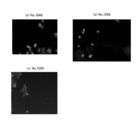
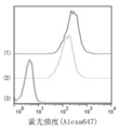
カルチャースライド4チャンバー(BD)にTF高発現のヒト膵臓癌細胞BxPC3を5×10⁴cells/チャンバーで播種し、RPMI 1640培地で37℃、5%CO2環境下、12時間培養した。PBSで3回洗浄した後にAlexa647蛍光ラベルkit(Invitrogen)で標識した30μgの抗体を1mlのRPMI 1640培地で希釈し各チャンバーに投与し、3時間培養した。当該3時間の培養においては、培養開始から2時間経過後に、終濃度75nMとなるようにLysotracker RED-DND99(Invitrogen)を培養液に加え、その後さらに1時間培養した。次いで、PBSで3回洗浄後、4%パラホルムアルデヒドで固定し、DAPI(4’,6-diamidino-2-phenylindole)で核染色後、FluoromountG(Southern Biotech)で封入した。次いで、蛍光顕微鏡(Keyence)を用いて各細胞を観察した。観察の結果を図1に示す。
1.抗hTFモノクローナル抗体(No.1849)
抗hTFモノクローナル抗体(No.1849)を産生するハイブリドーマから常法に従って全RNAを抽出し、次いで、得られた全RNAからcDNAを合成した。
[プライマー一覧]
プライマー1(配列番号30)
NNCCATGGCCGAGGTRMAGCTTCAGGAGTC
プライマー2(配列番号31)
NNCCATGGCCGAGGTBCAGCTBCAGCAGTC
プライマー3(配列番号32)
NNCCATGGCCGAGGTGCAGCTGAAGSASTC
プライマー4(配列番号33)
NNCCATGGCCGAGGTCCARCTGCAACARTC
プライマー5(配列番号34)
NNCCATGGCCGAGGTYCAGCTBCAGCARTC
プライマー6(配列番号35)
NNCCATGGCCGAGGTYCARCTGCAGCAGTC
プライマー7(配列番号36)
NNCCATGGCCGAGGTCCACGTGAAGCAGTC
プライマー8(配列番号37)
NNCCATGGCCGAGGTGAASSTGGTGGAATC
プライマー9(配列番号38)
NNCCATGGCCGAGGTGAWGYTGGTGGAGTC
プライマー10(配列番号39)
NNCCATGGCCGAGGTGCAGSKGGTGGAGTC
プライマー11(配列番号40)
NNCCATGGCCGAGGTGCAMCTGGTGGAGTC
プライマー12(配列番号41)
NNCCATGGCCGAGGTGAAGCTGATGGARTC
プライマー13(配列番号42)
NNCCATGGCCGAGGTGCARCTTGTTGAGTC
プライマー14(配列番号43)
NNCCATGGCCGAGGTRAAGCTTCTCGAGTC
プライマー15(配列番号44)
NNCCATGGCCGAGGTGAARSTTGAGGAGTC
プライマー16(配列番号45)
NNCCATGGCCGAGGTTACTCTRAAAGWGTSTG
プライマー17(配列番号46)
NNCCATGGCCGAGGTCCAACTVCAGCARCC
プライマー18(配列番号47)
NNCCATGGCCGAGGTGAACTTGGAAGTGTC
プライマー19(配列番号48)
NNCCATGGCCGAGGTGAAGGTCATCGAGTC
プライマー20(配列番号49)
TGTGCAGACCCTCGTGGACCACGGAGCA
プライマー21(配列番号50)
GGACTCTGGGRTCATTTACCMGGAGAGT
プライマー22(配列番号51)
NNNNGTCGACGCTCGAYATCCAGCTGACTCAGCC
プライマー23(配列番号52)
NNNNGTCGACGCTCGAYATTGTTCTCWCCCAGTC
プライマー24(配列番号53)
NNNNGTCGACGCTCGAYATTGTGMTMACTCAGTC
プライマー25(配列番号54)
NNNNGTCGACGCTCGAYATTGTGYTRACACAGTC
プライマー26(配列番号55)
NNNNGTCGACGCTCGAYATTGTRATGACMCAGTC
プライマー27(配列番号56)
NNNNGTCGACGCTCGAYATTMAGATRAMCCAGTC
プライマー28(配列番号57)
NNNNGTCGACGCTCGAYATTCAGATGAYDCAGTC
プライマー29(配列番号58)
NNNNGTCGACGCTCGAYATYCAGATGACACAGAC
プライマー30(配列番号59)
NNNNGTCGACGCTCGAYATTGTTCTCAWCCAGTC
プライマー31(配列番号60)
NNNNGTCGACGCTCGAYATTGWGCTSACCCAATC
プライマー32(配列番号61)
NNNNGTCGACGCTCGAYATTSTRATGACCCARTC
プライマー33(配列番号62)
NNNNGTCGACGCTCGAYRTTKTGATGACCCARAC
プライマー34(配列番号63)
NNNNGTCGACGCTCGAYATTGTGATGACBCAGKC
プライマー35(配列番号64)
NNNNGTCGACGCTCGAYATTGTGATAACYCAGGA
プライマー36(配列番号65)
NNNNGTCGACGCTCGAYATTGTGATGACCCAGWT
プライマー37(配列番号66)
NNNNGTCGACGCTCGAYATTGTGATGACACAACC
プライマー38(配列番号67)
NNNNGTCGACGCTCGAYATTTTGCTGACTCAGTC
プライマー39(配列番号68)
CCTTAGGAGGGAAGATTGGAAGGAGCT
なお、上記塩基配列中、RはGまたはA、YはTまたはC、MはAまたはC、KはGまたはT、SはGまたはC、WはAまたはT、BはG、CまたはT、DはA、GまたはT、VはA、GまたはC、NはA、T、GまたはCを意味する。

抗hTFモノクローナル抗体(No.1859)を産生するハイブリドーマから全RNAを抽出したこと、および、重鎖可変領域クローニング用のアンチセンスプライマーとしてプライマー21を用いたこと以外は、上記と同様にして抗hTFモノクローナル抗体(No.1859)の重鎖可変領域および軽鎖可変領域の塩基配列を決定した。
Biacore社のCM5チップ表面に抗hTFモノクローナル抗体(No.1849、No.1859)を固相化した。各抗体を固相化したCM5チップをSPR装置にセットし、その流路に精製hTFを含有した抗原含有bufferを流した。この測定系により各抗体とhTFとの解離定数を求めた。なお、測定条件は以下のとおりである。
SPR装置:Biacore 2000(ビアコア社)
抗原含有buffer:hTFを最終濃度25μg/mlで含む10mM酢酸緩衝液(pH5.0)
Running buffer:HBS-EP buffer (10mM HEPES, pH7.5, 0.15M NaCl, 3mM EDTA, 0.005% surfactant P20(Tween 20), pH7.4)。
以下のようにして、抗hTFモノクローナル抗体(No.1849およびNo.1859)のプロトロンビン時間を測定した。
リコンビナントhTF抗原350ngに抗体3μgを添加し、全量が5μlになるまでPBSを加えた。37℃、600rpmで振とうさせながら15分抗原抗体反応をさせた。反応液に3.8%クエン酸ナトリウムで抗凝固処理をしたヒト血漿 50μl、25mM CaCl2 100μlを添加すると同時に37℃、600rpmで振とうさせながら培養し、フィブリン塊が形成されるまでの時間(プロトロンビン時間)を計測した。反応液の代わりにPBSを用いた対照におけるプロトロンビン時間を1としたときの各抗体の凝固延長時間比(プロトロンビン時間比)を表3および図2に示す。

[モノクローナル抗体と薬物との結合]
以下のようにしてモノメチルオーリスタチンE(MMAE)が結合された抗hTFモノクローナル抗体(No.1849)(以下、「hTF-MMAE」と称する)を得た。


96well plateにヒト膵臓癌細胞(BxPC3)を3×10³個ずつ加え、10%FCS、ペニシリン(100U/ml)、およびストレプトマイシン(100μg/ml)を含むRPMI培地で培養した。各ウェルに種々の薬物濃度となるようにhTF-MMAEを添加し、添加後72時間における細胞生存率を算出した。
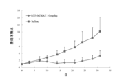
4週齢ヌードマウス(BALBc nu/nu、メス)の背部皮下に1×107個/100μLのヒト膵臓癌細胞(BxPC3)を移植し、腫瘍体積(腫瘍径に基づいて算出)が約200mm3となった時点から治療を開始した。治療開始当日をDay0とし、Day0、4および8に1回ずつ薬物を尾静脈投与した(計3回投与)。薬物としては、hTF-MMAEを用い、対照として生理食塩水を投与した(各群:N=7)。薬物の投与量は、1回あたり10mg/kg(抗体量:約200μg/匹)であった。以後、Day30まで腫瘍径および体重を週2回計測した。Day0における腫瘍体積および体重に対する腫瘍体積および体重の比をそれぞれ図4および5に示す。
[抗原の調製]
mTF全長アミノ酸配列の30位~251位までのアミノ酸配列を含む組み換え蛋白質を大腸菌で発現させ、ニッケルカラムで精製して得られたレコンビナントmTF(配列番号73)を抗原として用いた。
レコンビナントmTF50μgをフロイント完全アジュバント(Difco)と共に6週令のWister雌ラット3匹に腹腔内投与し、初回免疫とした。その14日後にレコンビナントmTF50μgをSigma Adjuvant System(Sigma)と共に投与し、追加免疫とした。以降、21日毎に同様の追加免疫を7回行った。さらに207日後にPBSで希釈したレコンビナントmTF10μgを腹腔投与し、レコンビナントmTF40μgを尾静脈投与することで最終免疫とした。
最終免疫の3日後に脾臓を摘出し、脾臓細胞を回収した。脾臓細胞とマウスミエローマ細胞(p3X63Ag8.653)を50%濃度のポリエチレングリコール4000(Merck)を用いて融合させ、HAT培地で選択した。
細胞融合8日後に抗体産生ハイブリドーマのスクリーニングを行った。スクリーニングに用いたイムノアッセイは以下のとおりである。ELISA法を行うにあたり、96穴マイクロタイタープレート(ヌンク社製)の各ウェルにレコンビナントmTFを1μg/mL含む50mM炭酸緩衝液(pH8)を50μL/wellで添加し、4℃で一晩もしくは室温で2時間固定した。これらのウェルを300μLの洗浄液(0.05% Tween20/25mM Tris/140mM NaCl/2.5mM KCl、pH7.4)で3回洗浄した後、ブロッキングバッファー(0.05% Tween20/1% BSA/100 mM NaH2PO4/140mM NaCl、pH5)を200μLで加えて4℃で一晩もしくは室温で1時間静置してブロッキングを行った。このようにして得たmTF固相化プレートの各ウェルに50μLのハイブリドーマ培養上清を添加して室温で1時間反応させた。各ウェルを300μLの洗浄液で3回洗浄後、ブロッキングバッファーで5,000倍に希釈したHRP標識抗マウスIgG抗体(Bethyl)を50μLで加えて30分反応させた。反応後、各ウェルを300μLの洗浄液で3回洗浄し、3.7mM o-フェニレンジアミン/25mM クエン酸/130mM Na2HPO4/0.006% H2O2(pH5.0)を100μLで添加し、呈色させた。10~15分後に2規定硫酸を30μL/wellで添加して反応を停止し、吸光度(490nM)を吸光プレートリーダーで測定した。また免疫沈降ELISA法はレコンビナントmTFとハイブリドーマ培養上清とを混合し、混合液中の未結合mTFの量をmTF定量サンドイッチELISAで測定することで実施した。さらに、フローサイトメトリー法を定法に従って実施し、hTF発現細胞に対するハイブリドーマ培養上清の反応性を測定した。
樹立した各ハイブリドーマを牛由来IgGを除去した牛血清を5%量含むRPMI1640培地等で大量培養し、培養上清を得た。もしくは、各ハイブリドーマをICRヌードマウスの腹腔内で大量培養し、腹水を採取した。得られた培養上清もしくは腹水をProtein Gアフィニティカラムクロマトグラフィーに供してIgGモノクローナル抗体を精製した。
上記のようにして得られた各モノクローナル抗体を以下のインターナリゼーションアッセイに供した。
カルチャースライド4チャンバー(BD)にマウスB16メラノーマ細胞とそのTF強制発現細胞を5×10⁴cells/チャンバーで播種し、RPMI 1640培地で37℃、5%CO2環境下、12時間培養した。PBSで3回洗浄した後にAlexa647蛍光ラベルkit(Invitrogen)で標識した30μgの抗体を1mlのRPMI 1640培地で希釈し各チャンバーに投与し、3時間培養した。当該3時間の培養においては、培養開始から2時間経過後に、終濃度75nMとなるようにLysotracker RED-DND99(Invitrogen)を培養液に加え、その後さらに1時間培養した。次いで、PBSで3回洗浄後、4%パラホルムアルデヒドで固定し、DAPI(4’,6-diamidino-2-phenylindole)で核染色後、FluoromountG(Southern Biotech)で封入した。次いで、蛍光顕微鏡(Keyence)を用いて各細胞を観察した。観察の結果を図6に示す。
抗mTFモノクローナル抗体(No.1157)を産生するハイブリドーマから全RNAを抽出したこと以外は、抗hTFモノクローナル抗体(No.1849)の重鎖可変領域および軽鎖可変領域の塩基配列の決定方法と同様にして、抗mTFモノクローナル抗体(No.1157)の重鎖可変領域および軽鎖可変領域の塩基配列を決定した。

実施例1でクローニングした抗hTFモノクローナル抗体(No.1849)の重鎖可変領域および軽鎖可変領域をコードするDNA断片をPCRで増幅した。重鎖の可変領域DNA断片をヒトIgG1重鎖定常領域を発現するクローニングベクター(pFUSEss_CHIg-hG1e2(invivoGen))に挿入し、軽鎖の可変領域DNA断片をヒトkappa軽鎖定常領域を発現するクローニングベクター(pFUSE2ss_CLIg_hk(invivoGen))に挿入して、発現ベクターを得た。得られた発現ベクターをLipofectamine LTX Reagent(Invitrogen)を用いてCHO-K1細胞にトランスフェクションした。次いで、10μg/mL Blastcidin S(科研製薬)および300μg/mL Zeocin(Invitrogen)を用いて薬剤選択を行い、両耐性細胞株を得た。
No.1849キメラクローンからベクターを抽出し、常法に従って該ベクターにコードされている抗hTFヒト型キメラ抗体(No.1849)の重鎖および軽鎖の塩基配列ならびにアミノ酸配列を決定した。決定されたキメラ抗体の重鎖および軽鎖の塩基配列はそれぞれ配列番号76および77に記載のとおりである。また、決定されたキメラ抗体の重鎖および軽鎖のアミノ酸配列はそれぞれ配列番号78および79に記載のとおりである。
Claims (9)
- Tissue Factorに結合するモノクローナル抗体であって、
それぞれ配列番号3、4および5に記載されるアミノ酸配列を含む相補性決定領域1、2および3を有する重鎖可変領域と、それぞれ配列番号6、7および8に記載されるアミノ酸配列を含む相補性決定領域1、2および3を有する軽鎖可変領域と、を含む、抗ヒトTissue Factorモノクローナル抗体;
それぞれ配列番号11、12および13に記載されるアミノ酸配列を含む相補性決定領域1、2および3を有する重鎖可変領域と、それぞれ配列番号14、15および16に記載されるアミノ酸配列を含む相補性決定領域1、2および3を有する軽鎖可変領域と、を含む、抗ヒトTissue Factorモノクローナル抗体;または
それぞれ配列番号19、20および21に記載されるアミノ酸配列を含む相補性決定領域1、2および3を有する重鎖可変領域と、それぞれ配列番号22、23および24に記載されるアミノ酸配列を含む相補性決定領域1、2および3を有する軽鎖可変領域と、を含む、抗マウスTissue Factorモノクローナル抗体;
である、モノクローナル抗体。 - 配列番号9に記載されるアミノ酸配列または該アミノ酸配列と90%以上同一であるアミノ酸配列を含む重鎖可変領域と、配列番号10に記載されるアミノ酸配列または該アミノ酸配列と90%以上同一であるアミノ酸配列を含む軽鎖可変領域と、を含む抗ヒトTissue Factorモノクローナル抗体;
配列番号17に記載されるアミノ酸配列または該アミノ酸配列と90%以上同一であるアミノ酸配列を含む重鎖可変領域と、配列番号18に記載されるアミノ酸配列または該アミノ酸配列と90%以上同一であるアミノ酸配列を含む軽鎖可変領域と、を含む抗ヒトTissue Factorモノクローナル抗体;または
配列番号25に記載されるアミノ酸配列または該アミノ酸配列と90%以上同一であるアミノ酸配列を含む重鎖可変領域と、配列番号26に記載されるアミノ酸配列または該アミノ酸配列と90%以上同一であるアミノ酸配列を含む軽鎖可変領域と、を含む抗マウスTissue Factorモノクローナル抗体;
である、請求項1に記載のモノクローナル抗体。 - 請求項1に記載のモノクローナル抗体が結合するTissue Factorのエピトープと同じエピトープに結合する、モノクローナル抗体。
- Tissue Factorを発現する細胞へのインターナリゼーション能を有する、請求項1から3のいずれかに記載のモノクローナル抗体。
- ヒト型キメラ抗体またはヒト化抗体である、請求項1から4のいずれかに記載のモノクローナル抗体。
- 請求項1から5のいずれかに記載のモノクローナル抗体の一部を含み、Tissue Factorと結合できる、抗体の断片。
- Tissue Factorを発現する細胞へのインターナリゼーション能を有する、請求項6に記載の抗体の断片。
- 標的結合因子としての請求項1から5のいずれかに記載のモノクローナル抗体あるいは請求項6または7に記載の抗体の断片と、薬物とを含む医薬組成物。
- 標的結合因子としての請求項1から5のいずれかに記載のモノクローナル抗体あるいは請求項6または7に記載の抗体の断片を含む、薬物送達用組成物。
Priority Applications (9)
| Application Number | Priority Date | Filing Date | Title |
|---|---|---|---|
| JP2015555482A JP5957637B2 (ja) | 2014-02-03 | 2015-02-03 | 抗TissueFactorモノクローナル抗体 |
| KR1020167021291A KR20160136279A (ko) | 2014-02-03 | 2015-02-03 | 항-조직 인자 모노클로널 항체 |
| EP15743456.4A EP3103811A4 (en) | 2014-02-03 | 2015-02-03 | Anti-tissue factor monoclonal antibody |
| MX2016009690A MX2016009690A (es) | 2014-02-03 | 2015-02-03 | Anticuerpos monoclonal anti-factor de tejido. |
| BR112016017010A BR112016017010A2 (pt) | 2014-02-03 | 2015-02-03 | Anticorpo monoclonal de fator antitecido |
| CN201580006870.2A CN106255703A (zh) | 2014-02-03 | 2015-02-03 | 抗组织因子单克隆抗体 |
| US15/111,875 US9920133B2 (en) | 2014-02-03 | 2015-02-03 | Anti-tissue factor monoclonal antibody |
| AU2015211674A AU2015211674A1 (en) | 2014-02-03 | 2015-02-03 | Anti-tissue factor monoclonal antibody |
| CA2937034A CA2937034A1 (en) | 2014-02-03 | 2015-02-03 | Anti-tissue factor monoclonal antibody |
Applications Claiming Priority (2)
| Application Number | Priority Date | Filing Date | Title |
|---|---|---|---|
| JP2014018586 | 2014-02-03 | ||
| JP2014-018586 | 2014-02-03 |
Publications (1)
| Publication Number | Publication Date |
|---|---|
| WO2015115656A1 true WO2015115656A1 (ja) | 2015-08-06 |
Family
ID=53757221
Family Applications (1)
| Application Number | Title | Priority Date | Filing Date |
|---|---|---|---|
| PCT/JP2015/052918 Ceased WO2015115656A1 (ja) | 2014-02-03 | 2015-02-03 | 抗Tissue Factorモノクローナル抗体 |
Country Status (11)
| Country | Link |
|---|---|
| US (1) | US9920133B2 (ja) |
| EP (1) | EP3103811A4 (ja) |
| JP (1) | JP5957637B2 (ja) |
| KR (1) | KR20160136279A (ja) |
| CN (1) | CN106255703A (ja) |
| AU (1) | AU2015211674A1 (ja) |
| BR (1) | BR112016017010A2 (ja) |
| CA (1) | CA2937034A1 (ja) |
| MX (1) | MX2016009690A (ja) |
| TW (1) | TWI655211B (ja) |
| WO (1) | WO2015115656A1 (ja) |
Cited By (6)
| Publication number | Priority date | Publication date | Assignee | Title |
|---|---|---|---|---|
| CN108883198A (zh) * | 2016-03-02 | 2018-11-23 | 卫材研究发展管理有限公司 | 基于艾日布林的抗体-药物偶联物和使用方法 |
| WO2019087994A1 (ja) | 2017-10-30 | 2019-05-09 | 国立研究開発法人国立がん研究センター | 固形腫瘍の治療に有益な抗体およびその抗体-薬物コンジュゲート並びにこれらを含む抗がん剤 |
| JP2019526256A (ja) * | 2015-08-20 | 2019-09-19 | フータン ユニバーシティ | 組織因子を標的とする抗体、その調製方法及びその使用 |
| EP3502141A4 (en) * | 2016-08-22 | 2020-04-08 | Fudan University | ANTIBODIES TAKEN AGAINST THE TISSUE FACTOR, MANUFACTURING METHODS THEREFOR AND USE THEREOF |
| WO2021200131A1 (ja) | 2020-03-30 | 2021-10-07 | 国立研究開発法人国立がん研究センター | 抗体薬物複合体 |
| US12290505B2 (en) | 2016-08-22 | 2025-05-06 | Fudan University | Tissue factor-targeted antibody-drug conjugate |
Families Citing this family (13)
| Publication number | Priority date | Publication date | Assignee | Title |
|---|---|---|---|---|
| CN107446047B (zh) * | 2017-09-04 | 2020-08-11 | 山西省生物研究院有限公司 | 一种双功能性抗组织因子人源化抗体及其制备方法 |
| EP3735271A4 (en) * | 2018-01-04 | 2022-06-15 | Iconic Therapeutics, Inc. | ANTI-TISSUE FACTOR ANTIBODIES, ANTIBODY DRUG CONJUGATES AND RELATED METHODS |
| US11401324B2 (en) | 2018-08-30 | 2022-08-02 | HCW Biologics, Inc. | Single-chain chimeric polypeptides and uses thereof |
| US11518792B2 (en) | 2018-08-30 | 2022-12-06 | HCW Biologics, Inc. | Multi-chain chimeric polypeptides and uses thereof |
| WO2020171020A1 (ja) * | 2019-02-18 | 2020-08-27 | 株式会社エヌビィー健康研究所 | 細胞の選抜方法、核酸の製造方法、組換え細胞の製造方法、目的物質の製造方法、医薬組成物の製造方法、及び試薬 |
| CN120924491A (zh) | 2019-06-21 | 2025-11-11 | 免疫生物公司 | 多链嵌合多肽和其用途 |
| CA3169243A1 (en) | 2020-02-11 | 2021-08-19 | HCW Biologics, Inc. | Methods of activating regulatory t cells |
| AU2021220870A1 (en) | 2020-02-11 | 2022-08-04 | Immunitybio, Inc. | Methods of treating age-related and inflammatory diseases |
| WO2021163299A1 (en) | 2020-02-11 | 2021-08-19 | HCW Biologics, Inc. | Chromatography resin and uses thereof |
| CN115836087A (zh) | 2020-04-29 | 2023-03-21 | Hcw生物科技公司 | 抗cd26蛋白及其用途 |
| US12024545B2 (en) | 2020-06-01 | 2024-07-02 | HCW Biologics, Inc. | Methods of treating aging-related disorders |
| WO2023201311A1 (en) | 2022-04-13 | 2023-10-19 | HCW Biologics, Inc. | Multi-chain chimeric polypeptide for use in the treatment of treating neuroinflammatory disorder |
| CN121358751A (zh) * | 2023-10-19 | 2026-01-16 | 优洛生物(上海)有限公司 | 多肽、生物活性偶联物及其制备方法和应用 |
Citations (5)
| Publication number | Priority date | Publication date | Assignee | Title |
|---|---|---|---|---|
| WO1995014041A1 (en) | 1993-11-19 | 1995-05-26 | Chugai Seiyaku Kabushiki Kaisha | Reconstituted human antibody against human medulloblastomatous cell |
| JPH09302000A (ja) | 1996-05-16 | 1997-11-25 | Chemo Sero Therapeut Res Inst | 抗組織因子モノクローナル抗体及び当該モノクローナル抗体を用いた組織因子凝固活性の測定法 |
| JP2003527861A (ja) * | 2000-03-16 | 2003-09-24 | ジェネンテック・インコーポレーテッド | 増強した抗血液凝固能を持つ抗組織因子抗体 |
| US20120039989A1 (en) | 2010-08-10 | 2012-02-16 | Hubbell Jeffrey A | Erythrocyte-binding therapeutics |
| JP2014018586A (ja) | 2012-07-23 | 2014-02-03 | Newgin Co Ltd | 遊技機 |
Family Cites Families (12)
| Publication number | Priority date | Publication date | Assignee | Title |
|---|---|---|---|---|
| WO1994005328A1 (en) * | 1992-08-28 | 1994-03-17 | The Scripps Research Institute | Inhibition of tumor metastasis via neutralization of tissue factor function |
| US20060235209A9 (en) | 1997-03-10 | 2006-10-19 | Jin-An Jiao | Use of anti-tissue factor antibodies for treating thromboses |
| US20030109680A1 (en) | 2001-11-21 | 2003-06-12 | Sunol Molecular Corporation | Antibodies for inhibiting blood coagulation and methods of use thereof |
| US5986065A (en) | 1997-03-10 | 1999-11-16 | Sunol Molecular Corporation | Antibodies for inhibiting blood coagulation and methods of use thereof |
| US6849425B1 (en) | 1999-10-14 | 2005-02-01 | Ixsys, Inc. | Methods of optimizing antibody variable region binding affinity |
| US20030124117A1 (en) | 2000-03-16 | 2003-07-03 | Refino Canio J. | Combinations of anti-tissue factor antibodies and anticoagulant and/or antiplatelet agents |
| US6703494B2 (en) | 2000-03-16 | 2004-03-09 | Genentech, Inc. | Anti-tissue factor antibodies with enhanced anticoagulant potency |
| US20030143225A1 (en) | 2001-03-08 | 2003-07-31 | Genentech, Inc. | Combinations of anti-tissue factor antibodies and anticoagulant and/or antiplatelet agents |
| TWI338009B (en) * | 2001-10-29 | 2011-03-01 | Genentech Inc | Antibodies for inhibiting blood coagulation and methods of use thereof |
| US7425328B2 (en) * | 2003-04-22 | 2008-09-16 | Purdue Pharma L.P. | Tissue factor antibodies and uses thereof |
| JP6055404B2 (ja) * | 2010-06-15 | 2016-12-27 | ゲンマブ エー/エス | 組織因子に対するヒト抗体薬物結合体 |
| US8722044B2 (en) | 2011-03-15 | 2014-05-13 | Janssen Biotech, Inc. | Human tissue factor antibody and uses thereof |
-
2015
- 2015-02-03 CA CA2937034A patent/CA2937034A1/en not_active Abandoned
- 2015-02-03 EP EP15743456.4A patent/EP3103811A4/en not_active Withdrawn
- 2015-02-03 CN CN201580006870.2A patent/CN106255703A/zh active Pending
- 2015-02-03 BR BR112016017010A patent/BR112016017010A2/pt not_active Application Discontinuation
- 2015-02-03 KR KR1020167021291A patent/KR20160136279A/ko not_active Withdrawn
- 2015-02-03 JP JP2015555482A patent/JP5957637B2/ja active Active
- 2015-02-03 WO PCT/JP2015/052918 patent/WO2015115656A1/ja not_active Ceased
- 2015-02-03 US US15/111,875 patent/US9920133B2/en not_active Expired - Fee Related
- 2015-02-03 AU AU2015211674A patent/AU2015211674A1/en not_active Abandoned
- 2015-02-03 MX MX2016009690A patent/MX2016009690A/es unknown
- 2015-02-03 TW TW104103660A patent/TWI655211B/zh not_active IP Right Cessation
Patent Citations (5)
| Publication number | Priority date | Publication date | Assignee | Title |
|---|---|---|---|---|
| WO1995014041A1 (en) | 1993-11-19 | 1995-05-26 | Chugai Seiyaku Kabushiki Kaisha | Reconstituted human antibody against human medulloblastomatous cell |
| JPH09302000A (ja) | 1996-05-16 | 1997-11-25 | Chemo Sero Therapeut Res Inst | 抗組織因子モノクローナル抗体及び当該モノクローナル抗体を用いた組織因子凝固活性の測定法 |
| JP2003527861A (ja) * | 2000-03-16 | 2003-09-24 | ジェネンテック・インコーポレーテッド | 増強した抗血液凝固能を持つ抗組織因子抗体 |
| US20120039989A1 (en) | 2010-08-10 | 2012-02-16 | Hubbell Jeffrey A | Erythrocyte-binding therapeutics |
| JP2014018586A (ja) | 2012-07-23 | 2014-02-03 | Newgin Co Ltd | 遊技機 |
Non-Patent Citations (2)
| Title |
|---|
| BREIJ E. C. W. ET AL.: "An Antibody-Drug Conjugate That Targets Tissue Factor Exhibits Potent Therapeutic Activity against a Broad Range of Solid Tumors", CANCER RESEARCH, vol. 74, no. 4, December 2013 (2013-12-01), pages 1214 - 1226, XP055170885 * |
| See also references of EP3103811A4 |
Cited By (16)
| Publication number | Priority date | Publication date | Assignee | Title |
|---|---|---|---|---|
| JP7020656B2 (ja) | 2015-08-20 | 2022-02-16 | フータン ユニバーシティ | 組織因子を標的とする抗体、その調製方法及びその使用 |
| JP2022031296A (ja) * | 2015-08-20 | 2022-02-18 | フータン ユニバーシティ | 組織因子を標的とする抗体、その調製方法及びその使用 |
| JP2019526256A (ja) * | 2015-08-20 | 2019-09-19 | フータン ユニバーシティ | 組織因子を標的とする抗体、その調製方法及びその使用 |
| JP2023082096A (ja) * | 2016-03-02 | 2023-06-13 | エーザイ・アール・アンド・ディー・マネジメント株式会社 | エリブリンをベースとする抗体-薬物コンジュゲート及び使用方法 |
| JP2020128413A (ja) * | 2016-03-02 | 2020-08-27 | エーザイ・アール・アンド・ディー・マネジメント株式会社 | エリブリンをベースとする抗体−薬物コンジュゲート及び使用方法 |
| JP2021185176A (ja) * | 2016-03-02 | 2021-12-09 | エーザイ・アール・アンド・ディー・マネジメント株式会社 | エリブリンをベースとする抗体−薬物コンジュゲート及び使用方法 |
| CN108883198B (zh) * | 2016-03-02 | 2022-08-09 | 卫材研究发展管理有限公司 | 基于艾日布林的抗体-药物偶联物和使用方法 |
| JP7254861B2 (ja) | 2016-03-02 | 2023-04-10 | エーザイ・アール・アンド・ディー・マネジメント株式会社 | エリブリンをベースとする抗体-薬物コンジュゲート及び使用方法 |
| CN108883198A (zh) * | 2016-03-02 | 2018-11-23 | 卫材研究发展管理有限公司 | 基于艾日布林的抗体-药物偶联物和使用方法 |
| JP7556086B2 (ja) | 2016-03-02 | 2024-09-25 | エーザイ・アール・アンド・ディー・マネジメント株式会社 | エリブリンをベースとする抗体-薬物コンジュゲート及び使用方法 |
| TWI871094B (zh) * | 2016-03-02 | 2025-01-21 | 日商衛材R&D企管股份有限公司 | 基於艾日布林之抗體-藥物結合物及使用方法 |
| US10676537B2 (en) | 2016-08-22 | 2020-06-09 | Fudan University | Antibody targeted to tissue factor, preparation method therefor, and use thereof |
| EP3502141A4 (en) * | 2016-08-22 | 2020-04-08 | Fudan University | ANTIBODIES TAKEN AGAINST THE TISSUE FACTOR, MANUFACTURING METHODS THEREFOR AND USE THEREOF |
| US12290505B2 (en) | 2016-08-22 | 2025-05-06 | Fudan University | Tissue factor-targeted antibody-drug conjugate |
| WO2019087994A1 (ja) | 2017-10-30 | 2019-05-09 | 国立研究開発法人国立がん研究センター | 固形腫瘍の治療に有益な抗体およびその抗体-薬物コンジュゲート並びにこれらを含む抗がん剤 |
| WO2021200131A1 (ja) | 2020-03-30 | 2021-10-07 | 国立研究開発法人国立がん研究センター | 抗体薬物複合体 |
Also Published As
| Publication number | Publication date |
|---|---|
| CA2937034A1 (en) | 2015-08-06 |
| EP3103811A1 (en) | 2016-12-14 |
| CN106255703A (zh) | 2016-12-21 |
| EP3103811A4 (en) | 2017-11-29 |
| TWI655211B (zh) | 2019-04-01 |
| AU2015211674A1 (en) | 2016-08-04 |
| US20160333113A1 (en) | 2016-11-17 |
| US9920133B2 (en) | 2018-03-20 |
| TW201613980A (en) | 2016-04-16 |
| JPWO2015115656A1 (ja) | 2017-03-23 |
| BR112016017010A2 (pt) | 2017-10-03 |
| KR20160136279A (ko) | 2016-11-29 |
| JP5957637B2 (ja) | 2016-07-27 |
| MX2016009690A (es) | 2017-03-03 |
Similar Documents
| Publication | Publication Date | Title |
|---|---|---|
| JP5957637B2 (ja) | 抗TissueFactorモノクローナル抗体 | |
| AU2018393424B2 (en) | Triabody, preparation method and use thereof | |
| TWI564306B (zh) | 雙特異性抗體 | |
| WO2021032078A1 (en) | Novel anti-sirpa antibodies | |
| US20240239878A1 (en) | Binding molecule against dll3 and use thereof | |
| US20240174760A1 (en) | Nanobody targeting bcma and application thereof | |
| TWI883215B (zh) | 用於治療密連蛋白相關疾病之抗體及方法 | |
| CN114685655A (zh) | Pd-1结合分子及其应用 | |
| CN114641503B (zh) | 抗cd371抗体及其用途 | |
| CN117836324A (zh) | 新型tnfr2结合分子 | |
| CN116685604A (zh) | 抗人msln的抗体及其用途 | |
| US20240190986A1 (en) | Mesothelin binding molecule and application thereof | |
| JP2023544140A (ja) | 新規の抗クローディン18抗体 | |
| WO2022206753A1 (zh) | GARP/TGFβ1抗体及其应用 | |
| JP2019050737A (ja) | Cd44陽性tmem−180陽性のがん細胞由来微粒子、これを用いた抗tmem−180抗体療法が有効ながん患者の選別方法、選別された患者に対する抗tmem−180抗体を含む抗がん剤、および前記方法に用いるキット | |
| US20250313625A1 (en) | Novel anti-lag3 antibodies | |
| HK1227424A1 (en) | Anti-tissue factor monoclonal antibody | |
| WO2025214438A1 (zh) | 结合cd19和/或cd22的抗原结合分子及其用途 | |
| WO2023190465A1 (ja) | 抗ヒトsema7a抗体 | |
| KR20240113488A (ko) | 신규 항-il-36r 항체 | |
| WO2025201493A1 (zh) | Dll3结合分子及其用途 | |
| HK40097196A (zh) | 新型抗cldn18抗体 | |
| HK40092400A (zh) | 用於治疗claudin相关疾病的抗体和方法 | |
| CN120813379A (zh) | 靶向l1cam的抗体及其用途 |
Legal Events
| Date | Code | Title | Description |
|---|---|---|---|
| 121 | Ep: the epo has been informed by wipo that ep was designated in this application |
Ref document number: 15743456 Country of ref document: EP Kind code of ref document: A1 |
|
| ENP | Entry into the national phase |
Ref document number: 2015555482 Country of ref document: JP Kind code of ref document: A |
|
| ENP | Entry into the national phase |
Ref document number: 2937034 Country of ref document: CA |
|
| WWE | Wipo information: entry into national phase |
Ref document number: 15111875 Country of ref document: US |
|
| WWE | Wipo information: entry into national phase |
Ref document number: MX/A/2016/009690 Country of ref document: MX |
|
| REEP | Request for entry into the european phase |
Ref document number: 2015743456 Country of ref document: EP |
|
| WWE | Wipo information: entry into national phase |
Ref document number: 2015743456 Country of ref document: EP |
|
| REG | Reference to national code |
Ref country code: BR Ref legal event code: B01A Ref document number: 112016017010 Country of ref document: BR |
|
| ENP | Entry into the national phase |
Ref document number: 20167021291 Country of ref document: KR Kind code of ref document: A |
|
| NENP | Non-entry into the national phase |
Ref country code: DE |
|
| ENP | Entry into the national phase |
Ref document number: 2015211674 Country of ref document: AU Date of ref document: 20150203 Kind code of ref document: A |
|
| ENP | Entry into the national phase |
Ref document number: 2016135648 Country of ref document: RU Kind code of ref document: A |
|
| ENP | Entry into the national phase |
Ref document number: 112016017010 Country of ref document: BR Kind code of ref document: A2 Effective date: 20160722 |