WO2015122429A1 - 人工血管 - Google Patents
人工血管 Download PDFInfo
- Publication number
- WO2015122429A1 WO2015122429A1 PCT/JP2015/053749 JP2015053749W WO2015122429A1 WO 2015122429 A1 WO2015122429 A1 WO 2015122429A1 JP 2015053749 W JP2015053749 W JP 2015053749W WO 2015122429 A1 WO2015122429 A1 WO 2015122429A1
- Authority
- WO
- WIPO (PCT)
- Prior art keywords
- blood vessel
- artificial blood
- acid
- group
- woven fabric
- Prior art date
- Legal status (The legal status is an assumption and is not a legal conclusion. Google has not performed a legal analysis and makes no representation as to the accuracy of the status listed.)
- Ceased
Links
- 0 C*(CCNC(C[C@@](C(N(CCC1)[C@]1C(NCc(cc1)ccc1C(N)=N)=O)=O)NS(c(cc1)ccc1OC)(=O)=O)=O)O[Si](C)(C)*(C)CCN Chemical compound C*(CCNC(C[C@@](C(N(CCC1)[C@]1C(NCc(cc1)ccc1C(N)=N)=O)=O)NS(c(cc1)ccc1OC)(=O)=O)=O)O[Si](C)(C)*(C)CCN 0.000 description 1
Classifications
-
- A—HUMAN NECESSITIES
- A61—MEDICAL OR VETERINARY SCIENCE; HYGIENE
- A61F—FILTERS IMPLANTABLE INTO BLOOD VESSELS; PROSTHESES; DEVICES PROVIDING PATENCY TO, OR PREVENTING COLLAPSING OF, TUBULAR STRUCTURES OF THE BODY, e.g. STENTS; ORTHOPAEDIC, NURSING OR CONTRACEPTIVE DEVICES; FOMENTATION; TREATMENT OR PROTECTION OF EYES OR EARS; BANDAGES, DRESSINGS OR ABSORBENT PADS; FIRST-AID KITS
- A61F2/00—Filters implantable into blood vessels; Prostheses, i.e. artificial substitutes or replacements for parts of the body; Appliances for connecting them with the body; Devices providing patency to, or preventing collapsing of, tubular structures of the body, e.g. stents
- A61F2/02—Prostheses implantable into the body
- A61F2/04—Hollow or tubular parts of organs, e.g. bladders, tracheae, bronchi or bile ducts
- A61F2/06—Blood vessels
-
- A—HUMAN NECESSITIES
- A61—MEDICAL OR VETERINARY SCIENCE; HYGIENE
- A61L—METHODS OR APPARATUS FOR STERILISING MATERIALS OR OBJECTS IN GENERAL; DISINFECTION, STERILISATION OR DEODORISATION OF AIR; CHEMICAL ASPECTS OF BANDAGES, DRESSINGS, ABSORBENT PADS OR SURGICAL ARTICLES; MATERIALS FOR BANDAGES, DRESSINGS, ABSORBENT PADS OR SURGICAL ARTICLES
- A61L27/00—Materials for grafts or prostheses or for coating grafts or prostheses
- A61L27/14—Macromolecular materials
- A61L27/18—Macromolecular materials obtained otherwise than by reactions only involving carbon-to-carbon unsaturated bonds
-
- A—HUMAN NECESSITIES
- A61—MEDICAL OR VETERINARY SCIENCE; HYGIENE
- A61L—METHODS OR APPARATUS FOR STERILISING MATERIALS OR OBJECTS IN GENERAL; DISINFECTION, STERILISATION OR DEODORISATION OF AIR; CHEMICAL ASPECTS OF BANDAGES, DRESSINGS, ABSORBENT PADS OR SURGICAL ARTICLES; MATERIALS FOR BANDAGES, DRESSINGS, ABSORBENT PADS OR SURGICAL ARTICLES
- A61L27/00—Materials for grafts or prostheses or for coating grafts or prostheses
- A61L27/50—Materials characterised by their function or physical properties, e.g. injectable or lubricating compositions, shape-memory materials, surface modified materials
- A61L27/507—Materials characterised by their function or physical properties, e.g. injectable or lubricating compositions, shape-memory materials, surface modified materials for artificial blood vessels
-
- A—HUMAN NECESSITIES
- A61—MEDICAL OR VETERINARY SCIENCE; HYGIENE
- A61L—METHODS OR APPARATUS FOR STERILISING MATERIALS OR OBJECTS IN GENERAL; DISINFECTION, STERILISATION OR DEODORISATION OF AIR; CHEMICAL ASPECTS OF BANDAGES, DRESSINGS, ABSORBENT PADS OR SURGICAL ARTICLES; MATERIALS FOR BANDAGES, DRESSINGS, ABSORBENT PADS OR SURGICAL ARTICLES
- A61L33/00—Antithrombogenic treatment of surgical articles, e.g. sutures, catheters, prostheses, or of articles for the manipulation or conditioning of blood; Materials for such treatment
- A61L33/0076—Chemical modification of the substrate
-
- A—HUMAN NECESSITIES
- A61—MEDICAL OR VETERINARY SCIENCE; HYGIENE
- A61L—METHODS OR APPARATUS FOR STERILISING MATERIALS OR OBJECTS IN GENERAL; DISINFECTION, STERILISATION OR DEODORISATION OF AIR; CHEMICAL ASPECTS OF BANDAGES, DRESSINGS, ABSORBENT PADS OR SURGICAL ARTICLES; MATERIALS FOR BANDAGES, DRESSINGS, ABSORBENT PADS OR SURGICAL ARTICLES
- A61L33/00—Antithrombogenic treatment of surgical articles, e.g. sutures, catheters, prostheses, or of articles for the manipulation or conditioning of blood; Materials for such treatment
- A61L33/06—Use of macromolecular materials
- A61L33/068—Use of macromolecular materials obtained otherwise than by reactions only involving carbon-to-carbon unsaturated bonds
-
- B—PERFORMING OPERATIONS; TRANSPORTING
- B82—NANOTECHNOLOGY
- B82B—NANOSTRUCTURES FORMED BY MANIPULATION OF INDIVIDUAL ATOMS, MOLECULES, OR LIMITED COLLECTIONS OF ATOMS OR MOLECULES AS DISCRETE UNITS; MANUFACTURE OR TREATMENT THEREOF
- B82B1/00—Nanostructures formed by manipulation of individual atoms or molecules, or limited collections of atoms or molecules as discrete units
-
- D—TEXTILES; PAPER
- D03—WEAVING
- D03D—WOVEN FABRICS; METHODS OF WEAVING; LOOMS
- D03D15/00—Woven fabrics characterised by the material, structure or properties of the fibres, filaments, yarns, threads or other warp or weft elements used
- D03D15/30—Woven fabrics characterised by the material, structure or properties of the fibres, filaments, yarns, threads or other warp or weft elements used characterised by the structure of the fibres or filaments
- D03D15/33—Ultrafine fibres, e.g. microfibres or nanofibres
-
- D—TEXTILES; PAPER
- D03—WEAVING
- D03D—WOVEN FABRICS; METHODS OF WEAVING; LOOMS
- D03D3/00—Woven fabrics characterised by their shape
- D03D3/02—Tubular fabrics
-
- A—HUMAN NECESSITIES
- A61—MEDICAL OR VETERINARY SCIENCE; HYGIENE
- A61F—FILTERS IMPLANTABLE INTO BLOOD VESSELS; PROSTHESES; DEVICES PROVIDING PATENCY TO, OR PREVENTING COLLAPSING OF, TUBULAR STRUCTURES OF THE BODY, e.g. STENTS; ORTHOPAEDIC, NURSING OR CONTRACEPTIVE DEVICES; FOMENTATION; TREATMENT OR PROTECTION OF EYES OR EARS; BANDAGES, DRESSINGS OR ABSORBENT PADS; FIRST-AID KITS
- A61F2210/00—Particular material properties of prostheses classified in groups A61F2/00 - A61F2/26 or A61F2/82 or A61F9/00 or A61F11/00 or subgroups thereof
- A61F2210/0076—Particular material properties of prostheses classified in groups A61F2/00 - A61F2/26 or A61F2/82 or A61F9/00 or A61F11/00 or subgroups thereof multilayered, e.g. laminated structures
-
- A—HUMAN NECESSITIES
- A61—MEDICAL OR VETERINARY SCIENCE; HYGIENE
- A61F—FILTERS IMPLANTABLE INTO BLOOD VESSELS; PROSTHESES; DEVICES PROVIDING PATENCY TO, OR PREVENTING COLLAPSING OF, TUBULAR STRUCTURES OF THE BODY, e.g. STENTS; ORTHOPAEDIC, NURSING OR CONTRACEPTIVE DEVICES; FOMENTATION; TREATMENT OR PROTECTION OF EYES OR EARS; BANDAGES, DRESSINGS OR ABSORBENT PADS; FIRST-AID KITS
- A61F2230/00—Geometry of prostheses classified in groups A61F2/00 - A61F2/26 or A61F2/82 or A61F9/00 or A61F11/00 or subgroups thereof
- A61F2230/0063—Three-dimensional shapes
- A61F2230/0069—Three-dimensional shapes cylindrical
-
- A—HUMAN NECESSITIES
- A61—MEDICAL OR VETERINARY SCIENCE; HYGIENE
- A61L—METHODS OR APPARATUS FOR STERILISING MATERIALS OR OBJECTS IN GENERAL; DISINFECTION, STERILISATION OR DEODORISATION OF AIR; CHEMICAL ASPECTS OF BANDAGES, DRESSINGS, ABSORBENT PADS OR SURGICAL ARTICLES; MATERIALS FOR BANDAGES, DRESSINGS, ABSORBENT PADS OR SURGICAL ARTICLES
- A61L2300/00—Biologically active materials used in bandages, wound dressings, absorbent pads or medical devices
- A61L2300/40—Biologically active materials used in bandages, wound dressings, absorbent pads or medical devices characterised by a specific therapeutic activity or mode of action
- A61L2300/42—Anti-thrombotic agents, anticoagulants, anti-platelet agents
-
- D—TEXTILES; PAPER
- D10—INDEXING SCHEME ASSOCIATED WITH SUBLASSES OF SECTION D, RELATING TO TEXTILES
- D10B—INDEXING SCHEME ASSOCIATED WITH SUBLASSES OF SECTION D, RELATING TO TEXTILES
- D10B2509/00—Medical; Hygiene
- D10B2509/06—Vascular grafts; stents
Definitions
- the present invention relates to a cloth artificial blood vessel that has a small amount of blood leakage and enables both antithrombogenicity and cell affinity.
- An artificial blood vessel is a medical device used to replace a pathological biological blood vessel such as arteriosclerosis, or to form a bypass or a shunt.
- a pathological biological blood vessel such as arteriosclerosis
- a shunt When classifying conventional artificial blood vessels by materials, they are roughly classified into 1) made of cloth, 2) made of polytetrafluoroethylene, 3) made of biomaterial, and 4) made of synthetic polymer material.
- artificial blood vessels made of fabric made of fiber woven fabric, knitted fabric or non-woven fabric have high flexibility, but have the drawback of easily leaking blood from the fiber gap due to blood pressure under actual use conditions. .
- the artificial blood vessel made of a knitted fabric has a simple manufacturing process and is flexible, but it tends to have a porous structure in addition to its weak shape maintenance force, and leaks from the fiber gap. Easy to blood. Artificial blood vessels made of non-woven fabrics are not preferable because they have a non-uniform structure and weak shape retention.
- fabric artificial blood vessels made of woven fabrics can be used for vascular surgical operations such as aorta, which are in high demand, because the fiber gap can be reduced and the amount of blood leakage can be reduced compared to that made of knitted fabrics.
- a method of reducing the amount of blood leakage a method of densifying the fiber gap is common, but the resulting artificial blood vessel becomes hard due to an increase in fiber density.
- an alternative pathological biological blood vessel that is, a biological blood vessel that is anastomosed with the artificial blood vessel is also affected by arteriosclerosis and the like, and thus surgery is often difficult.
- Patent Document 1 A method for filling and preventing blood leakage has been reported.
- the living body recognizes this as a foreign substance, and in particular, the blood coagulation reaction proceeds on the contact surface with the blood of the artificial blood vessel, that is, the inner surface, thereby forming a thrombus. is needed.
- heparin or a heparin derivative cannot be directly applied to a medical material made of cloth such as polyester fiber constituting an artificial blood vessel or a medical material made of expanded porous polytetrafluoroethylene (hereinafter, “ePTFE”).
- ePTFE expanded porous polytetrafluoroethylene
- Patent Documents 4 to 6 a method of imparting heparin or a heparin derivative to the surface of the material by a covalent bond
- Patent Documents 7 to 10 a method of imparting heparin or a heparin derivative to the surface of the material by an ionic bond
- Patent Documents 1 and 11 As a method of imparting antithrombogenicity to a cloth artificial blood vessel, a method in which heparin or a heparin derivative is included in a gel such as collagen or gelatin absorbed in vivo used to prevent blood leakage and imparted to the surface of the material.
- Patent Documents 1 and 11 a method in which segmented polyurethane dissolved in an organic solvent is impregnated and applied to the surface of the material have been reported.
- in vivo blood vessels have an intima on the inner surface, and having vascular endothelial cells can inhibit thrombus formation.
- conventional artificial blood vessels have low cytophilicity, and vascular endothelial cells are established. Not only is it difficult, it takes time to establish vascular endothelial cells and to form the intima. Therefore, not only the antithrombotic properties immediately after transplantation but also a function that exhibits cell affinity over time has been required.
- Patent Documents 16 to 19 As a method of imparting cell affinity to a cloth artificial blood vessel, there is a method of making the fiber structure a structure that promotes cell proliferation and infiltration, a method of optimizing the fiber diameter, napped, fluff and / or loop-like fibers (Patent Documents 16 to 19) have been reported.
- Patent Document 1 when the method disclosed in Patent Document 1 is used for an artificial blood vessel made of cloth, a gel such as collagen or gelatin containing heparin or a heparin derivative imparted to the surface of the fiber is used to promote the cell growth or the like. Since the fine structure such as the fiber gap is lost, the cell affinity is lowered. Another problem is that platelets adhere to gels such as gelatin that are absorbed in the body, which promotes thrombus formation.
- Patent Document 2 in order to promptly establish vascular endothelial cells on the inner surface of an artificial blood vessel and promote the formation of the intima, Patent Document 3 describes a biocompatible technique by reducing in vitro foreign matter.
- a method of making an artificial blood vessel into a high-porosity structure that is, a highly permeable woven structure is disclosed, but pre-clotting is indispensable, and the thrombus formed by the operation has a fiber diameter or fiber. Since the fine structure such as the gap is lost, the cell affinity is lowered.
- anticoagulants eg, heparin, argatroban, etc.
- heparin heparin, argatroban, etc.
- the fiber gap may not be sufficiently filled by pre-crotting.
- the blood fibrinolysis system in the blood after the operation may dissolve the thrombus generated by preclotting, leading to blood leakage.
- Patent Documents 4 to 10 describe a method in which a surface modifier and heparin or a heparin derivative are covalently bonded or ionically bonded to the surface of a medical material. However, in order to promote cell growth and the like. No suitable thickness of the anti-thrombotic material layer made of a surface modifier, heparin or a heparin derivative is described in use for a cloth artificial blood vessel having a fine structure such as fiber diameter or fiber gap.
- Patent Documents 11 and 12 describe a method of physically imparting an antithrombotic material dissolved in a gel or organic solvent containing heparin or a heparin derivative to the surface of a medical material.
- Patent Documents 11 and 12 describe a method of physically imparting an antithrombotic material dissolved in a gel or organic solvent containing heparin or a heparin derivative to the surface of a medical material.
- the thickness of the antithrombogenic material layer is large, fine structures such as fiber diameter and fiber gap for promoting cell proliferation and the like are lost.
- Patent Documents 13 to 15 describe two compounds having both antiplatelet adhesion performance and antithrombin activation performance on the surface of a medical material, and antiplatelet adhesion performance and antithrombin activation performance.
- a method for immobilizing a single molecule compound is described, an antithrombosis comprising such a compound when used for a cloth artificial blood vessel having a fine structure such as a fiber diameter or a fiber gap for promoting cell proliferation or the like
- the appropriate thickness of the conductive material layer is not described.
- Patent Documents 16 to 19 disclose a cytophilic artificial blood vessel using a fiber having a denier of 0.5 denier, that is, 0.56 dtex or less for at least a part of the inner surface. Since antithrombogenicity is not imparted, thrombus formation cannot be suppressed. Moreover, although the method of improving cell affinity is provided by providing a napping, a fluff, and / or a loop-like fiber, the additional process for forming a napping, a fluff, and / or a loop-like fiber is required However, there is a problem that fiber waste is generated by the additional process and can be eluted in the blood. Furthermore, since the warp and weft yarns are more disturbed in the fiber direction, there is a problem that the vascular endothelial cells are hardly fixed and the cell affinity is lowered.
- an artificial blood vessel composed of a tubular woven fabric structure that has a small amount of blood leakage and achieves both antithrombogenicity and cell affinity.
- a blood flow volume is small, so that a thrombus is easily formed, and even a small amount of blood clots has a size corresponding to the inner diameter of the blood vessel.
- Cheap for this reason, small-diameter artificial blood vessels do not have long-term results, and there is no one that can withstand clinical use at present.
- an object of the present invention is to provide a cloth artificial blood vessel that has a small amount of blood leakage and enables both antithrombogenicity and cell affinity.
- the present invention has found the inventions (1) to (12).
- the warp and weft constituting the tubular woven fabric whose inner side is in contact with blood include a multifilament yarn having a single yarn fineness of 0.50 dtex or less, and is bonded to an antithrombogenic material.
- the antithrombogenic material forms a water-permeable material under a condition in which an antithrombogenic material layer having a thickness of 1 to 600 nm is formed on the inner side of the tubular fabric in which the inner side is in contact with blood and a pressure of 16 kPa is applied to the inner surface.
- the tubular woven fabric structure is a multiple tubular woven fabric structure in which an outer tubular woven fabric is disposed on the outer side of the tubular woven fabric, the inner side of which contacts blood.
- the artificial blood vessel according to (1) which is a woven fabric in which warps and wefts are crossed into a tubular shape.
- the multifilament yarn having a single yarn fineness of 1.0 dtex or more has an exposure to the inner surface of 20% or less.
- a cover factor of the warp and the weft constituting the tubular woven fabric whose inner side is in contact with blood is 1800 to 4000.
- the antithrombotic material includes an anionic anticoagulant compound containing a sulfur atom.
- the antithrombotic material includes a cationic polymer including, as a constituent monomer, a compound selected from the group consisting of alkyleneimine, vinylamine, allylamine, lysine, protamine, and diallyldimethylammonium chloride, and the cationic polymer includes The artificial blood vessel according to any one of (1) to (8), wherein the inner side is covalently bonded to a warp and a weft constituting a tubular fabric in contact with blood.
- the antithrombogenic material is composed of a compound selected from the group consisting of ethylene glycol, propylene glycol, vinyl pyrrolidone, vinyl alcohol, vinyl caprolactam, vinyl acetate, styrene, methyl methacrylate, hydroxyethyl methacrylate and siloxane as a constituent monomer.
- a compound containing three kinds of skeleton structures comprising a skeleton structure of a hydrophilic polymer, a skeleton structure of 4- (aminomethyl) benzenecarboximidamide or benzeneamidine, and a skeleton structure of methoxybenzenesulfonic acid amide,
- the artificial blood vessel according to (10), wherein the compound containing the three types of skeleton structures is any one of compounds represented by the following general formulas (I) to (IV).
- n and o represent an integer of 0 to 4
- n represents an integer of 3 to 1000
- n ′ represents an integer of 3 to 1000
- X represents a hydroxyl group
- a functional group selected from the group consisting of a thiol group, an amino group, a carboxyl group, an aldehyde group, an isocyanate group, and a thioisocyanate group.
- the antithrombotic material is an anionic polymer containing a compound selected from the group consisting of acrylic acid, methacrylic acid, ⁇ -glutamic acid, ⁇ -glutamic acid and aspartic acid as a constituent monomer, or oxalic acid, malonic acid
- An anionic compound selected from the group consisting of succinic acid, fumaric acid, glutaric acid, adipic acid, pimelic acid, suberic acid, azelaic acid, sebacic acid, malic acid, tartaric acid and citric acid,
- the artificial blood vessel according to any one of (11).
- (13) The artificial blood vessel according to any one of (1) to (12), wherein the multifilament yarn is made of polyester.
- the present invention also provides an artificial blood vessel described in the following (13) to (24).
- (13) In the multiple tubular woven fabric structure in which the inner layer tubular fabric is arranged inside the outer layer tubular fabric, the outer layer tubular fabric and the inner layer tubular fabric have a plurality of warps and a plurality of wefts crossed.
- the warp and weft constituting the inner layer tubular fabric include a multifilament yarn having a single yarn fineness of 0.30 dtex or less, and is combined with an antithrombogenic material.
- the antithrombogenic material forms an antithrombogenic material layer having a thickness of 1 to 600 nm on the inner side of the inner layer tubular fabric, and has a water permeability of 300 mL / cm 2 under a condition in which a pressure of 16 kPa is applied to the inner surface.
- An artificial blood vessel that is less than / min.
- the multifilament yarn having a single yarn fineness of 1.0 dtex or more has an exposure to the inner surface of 20% or less.
- the antithrombotic material includes a compound containing a sulfur atom having anionic property and anticoagulant activity.
- the artificial blood vessel according to (17), wherein the abundance ratio of sulfur atoms to the abundance of all atoms on the inner surface measured by X-ray photoelectron spectroscopy (XPS) is 3.0 to 6.0 atomic% .
- the antithrombotic material includes a cationic polymer including, as a constituent monomer, a compound selected from the group consisting of alkyleneimine, vinylamine, allylamine, lysine, protamine, and diallyldimethylammonium chloride, and the cationic polymer includes The artificial blood vessel according to any one of (1) to (18), which is covalently bonded to the warp and the weft constituting the inner layer tubular fabric. (20) The artificial blood vessel according to (19), wherein the abundance ratio of nitrogen atoms to the abundance of all atoms on the inner surface measured by X-ray photoelectron spectroscopy (XPS) is 7.0 to 12.0 atomic% .
- XPS X-ray photoelectron spectroscopy
- the antithrombotic material includes, as a constituent monomer, a compound selected from the group consisting of ethylene glycol, propylene glycol, vinyl pyrrolidone, vinyl alcohol, vinyl caprolactam, vinyl acetate, styrene, methyl methacrylate, hydroxyethyl methacrylate, and siloxane.
- a compound containing three kinds of skeleton structures comprising a skeleton structure of a hydrophilic polymer, a skeleton structure of 4- (aminomethyl) benzenecarboximidamide or benzeneamidine, and a skeleton structure of methoxybenzenesulfonic acid amide,
- the artificial blood vessel according to (21), wherein the compound containing the three types of skeleton structures is any one of compounds represented by the following general formulas (I) to (IV).
- n and o represent an integer of 0 to 4
- n represents an integer of 3 to 1000
- n ′ represents an integer of 3 to 1000
- X represents a hydroxyl group
- a functional group selected from the group consisting of a thiol group, an amino group, a carboxyl group, an aldehyde group, an isocyanate group, and a thioisocyanate group.
- the antithrombotic material is an anionic polymer containing a compound selected from the group consisting of acrylic acid, methacrylic acid, ⁇ -glutamic acid, ⁇ -glutamic acid and aspartic acid as a constituent monomer, or oxalic acid, malonic acid
- An anionic compound selected from the group consisting of succinic acid, fumaric acid, glutaric acid, adipic acid, pimelic acid, suberic acid, azelaic acid, sebacic acid, malic acid, tartaric acid and citric acid,
- the artificial blood vessel according to any one of (22).
- the artificial blood vessel according to any one of (13) to (23), wherein the multifilament yarn is made of polyester.
- the amount of blood leakage is small, and it is possible to achieve both antithrombogenicity and cell affinity.
- the artificial blood vessel of the present invention is a tubular woven fabric structure in which a tubular woven fabric whose inner side is in contact with blood is arranged, and the cylindrical woven fabric whose inner side is in contact with blood has a plurality of warps and a plurality of wefts interlaced.
- the warp and weft constituting the tubular woven fabric in which the inner side is in contact with blood contain multifilament yarns having a single yarn fineness of 0.50 dtex or less and are bonded to an antithrombogenic material.
- the antithrombogenic material is formed by forming an antithrombogenic material layer having a thickness of 1 to 600 nm on the inner side of the tubular fabric in which the inner side is in contact with blood, and applying a pressure of 16 kPa to the inner surface.
- the water permeability below is less than 300 mL / cm 2 / min.
- the cylindrical woven fabric is a woven fabric obtained by crossing a plurality of warp yarns and a plurality of weft yarns.
- a cylindrical woven fabric whose inner side is in contact with blood is used.
- positioned structure be a cylindrical textile structure.
- the cylindrical fabric that is in contact with blood on the inner side is the inner-layer cylindrical fabric
- the cylindrical fabric that forms the outer layer of the artificial blood vessel is the outer-layer cylindrical fabric
- the structure in which the woven fabrics are overlapped is referred to as a multi-tubular woven fabric structure.
- the multiple tubular woven fabric structure constituting the artificial blood vessel of the present invention may include a tubular fabric layer other than the inner layer tubular fabric and the outer layer tubular fabric, but the number of the tubular fabric layers is too large. Since the thickness of the artificial blood vessel is increased, the deviation from the thickness of the biological blood vessel is increased, which makes it difficult to perform anastomosis during transplantation. For this reason, the number of the cylindrical fabric layers is preferably 2 to 4, more preferably 2 to 3. Double woven artificial blood vessels having two tubular fabric layers can be woven together by known means such as inner layer warp fastening, inner layer weft fastening, double weft fastening, or the like. Double weaving eliminates the need to laminate two fabrics together by laminating or sewing, as well as flexible and mechanical strength because the two layers are integrated with warp or weft. A high artificial blood vessel can be obtained.
- the single yarn fineness is a value obtained by measuring the fineness fineness at a predetermined load of 0.045 cN / dtex according to JIS L 1013 (2010) 8.3.1 A method as the total fineness. A numerical value calculated by dividing by a number.
- the obtained artificial not only is the blood vessel hardened, but the vascular endothelial cells are less likely to settle and the cell affinity is lowered.
- the single yarn fineness is too small, the mechanical strength of the artificial blood vessel becomes difficult to be maintained, and vascular endothelial cells are hardly fixed.
- the warp and weft constituting the tubular fabric in which the inner side is in contact with blood on the inner surface of the artificial blood vessel in contact with blood has a single yarn fineness of 0.05 to 0.00. It is preferable to include 50 dtex multifilament yarn, more preferably 0.05 to 0.30 dtex multifilament yarn, more preferably 0.06 to 0.28 dtex multifilament yarn, and 0.08 It has been found that it is even more preferable to include a multifilament yarn of ⁇ 0.25 dtex.
- the inner surface refers to an antithrombogenic material layer formed by bonding an antithrombogenic material to the inside of a tubular fabric that contacts the blood on the inside and the inside of the tubular fabric that contacts the blood on the inside. It refers to the inner surface of the artificial blood vessel.
- the multifilament yarn having a single yarn fineness of 0.50 dtex or less a so-called direct spinning type may be used as it is, but a split type such as sea-island composite fiber may also be used.
- the split fiber type can be made ultrafine after forming an artificial blood vessel using fibers that can be made ultrafine by chemical or physical means. For example, as shown in US Pat. No. 3,531,368 and US Pat. No. 3,350,488, one component of a multicomponent fiber is removed or exfoliated as a method of ultrafinening by chemical or physical means. There is a method of fibrillation or ultrathinning by means such as making them.
- the single yarn fineness can be reduced to 0.50 dtex or less after the formation of the artificial blood vessel.
- the occurrence of yarn breakage and fluff generation when the various yarn processing means are taken can be minimized.
- the bond refers to a chemical bond such as a covalent bond, a hydrogen bond, an ionic bond, and a coordination bond.
- a covalent bond refers to the chemical bond produced by sharing an electron between atoms.
- the kind of covalent bond is not limited, For example, an amine bond, an azide bond, an amide bond, an imine bond etc. are mentioned. Among these, an amide bond is more preferable from the viewpoint of easy formation of a covalent bond and stability after bonding.
- the confirmation of the covalent bond can be determined from the fact that even if the artificial blood vessel is washed with a solvent that dissolves the antithrombotic material, it does not elute.
- the water permeability under a condition where a pressure of 16 kPa is applied to the inner surface is preferably less than 300 mL / cm 2 / min, and less than 200 mL / cm 2 / min. Is more preferable, and it is still more preferable that it is less than 150 mL / cm ⁇ 2 > / min.
- tubular woven fabric in order to lower the water permeability, an operation for imparting a gel such as collagen or gelatin that can not be achieved only by the fiber gap but is absorbed in vivo as in a known example. Necessary. In this case, since the fine structure such as fiber diameter and fiber gap for promoting cell growth is lost, cell affinity is lowered. Furthermore, platelets adhere to gels such as gelatin that are absorbed in the body, which promotes thrombus formation.
- the water permeability under a condition in which a pressure of 16 kPa is applied to the inner surface is preferably more than 0.1 mL / cm 2 / min, and 0.5 mL / cm 2 / min Is more preferable, and exceeding 1 mL / cm 2 / min is even more preferable.
- the water permeability under the condition where a pressure of 16 kPa is applied to the inner surface is the inside of an artificial blood vessel composed of an inner side of a cylindrical fabric and an antithrombotic material layer in contact with blood in accordance with the guidance of ISO 7198.
- the amount (mL) of water flowing out to the outer layer of the artificial blood vessel when a pressure of 16 kPa (hydrostatic pressure) is applied to the surface is divided by the unit area (cm 2 ) and the unit time (min).
- This water permeability can be used as an index representing the size and amount of the fiber gap of the artificial blood vessel.
- the water permeability can be adjusted by the composition ratio of the warp and weft constituting the tubular woven fabric whose inner side is in contact with blood, the single yarn diameter, the packing density, the thickness of the antithrombogenic material layer, the hydrophilicity, and the like.
- the ratio of the multifilament yarn having a single yarn fineness of 0.50 dtex or less of the warp and weft constituting the tubular fabric in contact with blood on the inside is too small. Since the size and amount of the fiber gap increase, not only the amount of blood leakage increases, but also the vascular endothelial cells are less likely to settle and the cell affinity tends to be lowered. Therefore, as a result of intensive studies by the inventors of the present application, the ratio of multifilament yarns having a single yarn fineness of warp and weft constituting 0.5% dtex or less constituting a tubular woven fabric in contact with blood is 50% or more. It has been found that it is preferably 60% or more, more preferably 80% or more.
- the single yarn diameter of the warp and weft constituting the tubular woven fabric in contact with blood is 300 nm to 10 ⁇ m, It is more preferably 1 to 5 ⁇ m, and even more preferably 3 ⁇ m.
- the single yarn diameters of the warp and the weft constituting the tubular woven fabric in contact with blood were measured based on a photograph magnified 400 times with a microscope.
- the cover factor of the inner surface of the artificial blood vessel represents the degree of the fiber gap of the tubular woven fabric in contact with blood, that is, the filling density, and the smaller the smaller, the wider the fiber gap. If the cover factor of the inner surface of the artificial blood vessel is too large, not only will the flexibility of the biological blood vessel be impaired due to an increase in the filling density, but the weaving property becomes unstable. On the other hand, if the cover factor of the inner surface of the artificial blood vessel is too small, the size and amount of the fiber gap increase, and the water permeability and the amount of blood leakage increase. That is, the cover factor of the inner surface of the artificial blood vessel is preferably 1800 to 4000, and more preferably 2000 to 3000.
- the cover factor of the inner surface of the artificial blood vessel is a value calculated from the total fineness and base fabric density of the multifilament yarn used for the warp and the weft, and is calculated by the following formula 1.
- CF (Dw ⁇ 0.9) 1/2 ⁇ Nw + (Df ⁇ 0.9) 1/2 ⁇ Nf ...
- Formula 1 CF Cover factor of the inner surface of the artificial blood vessel
- Dw Total fineness of warp (dtex)
- Nw density of warp base fabric (line / 2.54cm)
- Df total weft fineness (dtex)
- Nf Weft base fabric density (line / 2.54cm)
- the base fabric density was measured based on a photograph in which the produced cylindrical woven fabric was cut in the longitudinal direction and the inner surface was magnified 50 times with a microscope VHX-2000 manufactured by Keyence Corporation.
- a multifilament yarn of 1.0 dtex or more is included as a warp constituting at least one layer of layers other than the tubular woven fabric whose inner side is in contact with blood.
- the single yarn fineness of the warp constituting at least one of the layers other than the tubular woven fabric whose inner side is in contact with blood is too small, the mechanical strength of the artificial blood vessel is insufficient, and artificial grafting is not possible during long-term transplantation. This is not preferable because strength deterioration due to hydrolysis of the fibers constituting the blood vessels is concerned.
- the single yarn fineness of the warp constituting at least one of the layers other than the cylindrical fabric in contact with blood is too large, kinks are generated because the warp is stiff against bending of the artificial blood vessel. That is, it is preferable to include a multifilament yarn having a single yarn fineness of 1.0 to 10.0 dtex as a warp constituting at least one of the layers other than the cylindrical woven fabric that comes into contact with blood. 0.0 dtex is more preferred. Moreover, it is preferable that at least one layer other than the cylindrical fabric in contact with blood is an outer cylindrical fabric.
- the degree of exposure of the multifilament yarn of 1.0 dtex or more to the inner surface is preferably 20% or less, more preferably 5% or less, and even more preferably 1% or less.
- the degree of exposure (%) on the inner surface of a multifilament yarn of 1.0 dtex or more is that the multifilament yarn of 1.0 dtex or more for 100 warp ridges arbitrarily selected by visually observing the inner surface of the artificial blood vessel. It is the ratio of the number of peaks that can be confirmed.
- monofilament yarn is included as the weft constituting at least one of the layers other than the cylindrical fabric in contact with blood on the inside.
- the single yarn fineness of the weft constituting at least one of the layers other than the cylindrical fabric in contact with blood is too small, the shape maintenance and stretchability of the artificial blood vessel cannot be maintained, and kink resistance Does not lead to improvement.
- the single yarn fineness of the weft constituting at least one of the layers other than the tubular woven fabric in contact with blood is too large, not only the weft becomes hard but also the weaving property becomes unstable.
- the single yarn fineness is preferably 15.0 to 1000.0 dtex, and more preferably 20.0 to 100.0 dtex.
- at least one layer other than the cylindrical fabric in contact with blood is an outer cylindrical fabric.
- antithrombogenicity is a property that blood does not coagulate on the surface in contact with blood, and inhibits blood coagulation that progresses by, for example, platelet aggregation or activation of blood coagulation factors typified by thrombin.
- the cell affinity refers to the property that vascular endothelial cells that are present on the inner surface of a living blood vessel and can inhibit the formation of a thrombus are settled and an intima is easily formed.
- the antithrombotic material is a material having antithrombogenicity.
- an anionic anticoagulant compound containing a sulfur atom and Antithrombotic material A containing a cationic polymer specifically, an anionic anticoagulant compound containing a sulfur atom and Antithrombotic material A containing a cationic polymer, and three types of skeletons: a skeleton structure of a hydrophilic polymer, a skeleton structure of 4- (aminomethyl) benzenecarboxyimidamide or benzeneamidine, and a skeleton structure of methoxybenzenesulfonic acid amide
- An antithrombotic material B containing a structure is used.
- an antithrombotic material layer is formed by bonding an antithrombotic material to the inside of a cylindrical fabric whose inside contacts with blood, and the thickness range of the antithrombogenic material layer
- the inner structure of the vascular endothelial cells occurs by destroying the fine structure consisting of the warp and weft that make up the tubular fabric that comes into contact with blood on the inside. It becomes difficult.
- the thickness range is too thin, the binding amount of the antithrombotic material is small, so that the desired antithrombogenicity required immediately after transplantation cannot be obtained.
- the thickness range of the antithrombogenic material layer bonded to the cylindrical fabric that contacts the blood on the inside is preferably 1 to 600 nm, more preferably 5 to 500 nm, and more preferably 10 to 400 nm. Is even more preferred.
- the thickness of the antithrombogenic material layer can be determined using, for example, a scanning transmission electron microscope (hereinafter referred to as “STEM”) described later.
- STEM scanning transmission electron microscope
- the inner surface measured by the STEM is an inner surface made of, for example, an acrylic resin to be embedded at the time of sample preparation before the measurement by the STEM, an inner side of the cylindrical fabric in contact with blood, and the antithrombotic material layer. It refers to the boundary with the surface.
- the artificial blood vessel of the present invention since it is necessary to reduce the amount of blood leakage, it is preferably made of non-porous fibers. Furthermore, it is preferable that an antithrombogenic material is also present on the outer layer side of the tubular fabric in which the inner side is in contact with blood, that is, in the depth direction. Specifically, instead of the inner surface measured by STEM, the cylindrical fabric in which the inner side is in contact with blood, starting from the point of presence of atoms originating from the warp and weft that constitute the cylindrical fabric in which the inner side is in contact with blood.
- the distance from the start point to the end point of the point where the atoms derived from the antithrombotic material layer are observed is 15 nm or more, that is, the antithrombotic material It is more preferable that the atoms derived from are present in a depth direction of 15 nm or more from the surface of the cylindrical woven fabric in contact with blood.
- the distance from the start point to the end point of the point where the atoms derived from the antithrombotic material layer are observed is less than 15 nm, the binding amount of the antithrombotic material is small and the desired antithrombogenicity required immediately after transplantation is satisfied. Absent.
- the distance from the start point to the end point of the point where the atoms derived from the antithrombogenic material layer are observed may exceed 200 nm, but the inner side of the tubular fabric in contact with blood, that is, the depth
- the fiber constituting the artificial blood vessel is appropriately hydrolyzed and oxidized with an acid or alkali and an oxidizing agent, so that the deterioration occurs, for example, the tensile strength of the artificial blood vessel There is a risk that the mechanical properties of the material will deteriorate.
- the present invention provides a warp and a weft constituting an anti-thrombotic material that forms a tubular woven fabric whose inner surface is in contact with blood so that the end point of atoms derived from the anti-thrombotic material is in the range of 15 to 200 nm in the depth direction. It is preferable to combine with.
- the anti-thrombotic material layer having an appropriate thickness is formed by bonding the antithrombotic material to the warp and weft constituting the tubular woven fabric that contacts the blood inside, thereby fixing the vascular endothelial cells and the intima Formation can be promoted.
- the thickness of the anti-thrombogenic material layer and the inner side of the tubular woven fabric that contacts the blood from the existing point of the warp and weft constituting the cylindrical woven fabric that contacts the blood on the inner side are directed to the outer layer side.
- the distance from the starting point to the end point of the point where the atoms derived from the antithrombotic material layer were observed when the atomic distribution was observed in the vertical direction STEM and X-ray photoelectron spectroscopy (hereinafter referred to as “XPS”) ) And the like.
- the STEM includes detectors such as an energy dispersive X-ray spectrometer (hereinafter referred to as “EDX”) and an electron energy loss spectrometer (hereinafter referred to as “EELS”).
- EDX energy dispersive X-ray spectrometer
- EELS electron energy loss spectrometer
- Apparatus Field emission type transmission electron microscope JEM-2100F (manufactured by JEOL)
- EELS detector GIF Tridiem (manufactured by GATAN)
- EDX detector JED-2300T (manufactured by JEOL)
- Image acquisition Digital Micrograph (manufactured by GATAN)
- Sample preparation Ultra-thin section method (suspended on a copper microgrid, embedding resin is acrylic resin) Acceleration voltage: 200 kV Beam diameter: Diameter 0.7nm Energy resolution: about 1.0 eVFFWHM
- the presence of atoms is determined by whether or not the peak intensity derived from each atom is recognized by subtracting the background in the spectrum obtained from the STEM measurement.
- the antithrombotic material A of the present invention is preferably a compound having an anionic anticoagulant activity containing a sulfur atom.
- a cationic polymer is included.
- the cationic monomer includes a compound selected from the group consisting of alkyleneimine, vinylamine, allylamine, lysine, protamine, and diallyldimethylammonium chloride as a constituent monomer A. More preferably, it contains a polymer.
- the compound containing a sulfur atom having anticoagulant activity is anionic. be able to.
- the anionic anticoagulant compound containing a sulfur atom include heparin or heparin derivatives, dextran sulfate, polyvinyl sulfonic acid and polystyrene sulfonic acid, and heparin or heparin derivatives are more preferable. Heparin or a heparin derivative may be purified or not, and is not particularly limited as long as it can inhibit the blood coagulation reaction.
- Heparin generally used in clinical practice In addition to unfractionated heparin and low molecular weight heparin, anti-thrombin III has high affinity heparin.
- Specific examples of heparin include “heparin sodium” (Organon API).
- the cationic polymer Since the cationic polymer has a cationic property and may develop hemolytic toxicity, it is not preferable to elute it in the blood. Therefore, it is preferable that the cationic polymer is bonded to the warp and the weft constituting the tubular woven fabric whose inner side is in contact with blood, and more preferably covalently bonded.
- the cationic polymer may be a homopolymer or a copolymer.
- the cationic polymer when it is a copolymer, it may be a random copolymer, a block copolymer, a graft copolymer or an alternating copolymer, but the repeating unit containing a nitrogen atom is continuous.
- a block copolymer is more preferable because the block portion and a compound having an anionic anticoagulant activity containing a sulfur atom interact to form a strong ionic bond.
- the homopolymer means a polymer compound obtained by polymerizing one kind of constituent monomer
- the copolymer means a polymer compound obtained by copolymerization of two or more kinds of monomers.
- the block copolymer is a copolymer having a molecular structure in which at least two kinds of polymers having different repeating units are connected by covalent bonds to form a long chain.
- a block constitutes a block copolymer. Each of at least two kinds of polymers having different repeating units.
- the structure of the cationic polymer may be linear or branched.
- branched compounds are more preferred because they can form more stable ionic bonds at multiple points with an anionic anticoagulant compound containing a sulfur atom.
- the cationic polymer has at least one functional group among primary to tertiary amino groups and quaternary ammonium groups.
- the quaternary ammonium groups are A compound having a strong ionic interaction with an anionic anticoagulant activity containing a sulfur atom rather than a primary to tertiary amino group, and an anionic anticoagulant activity containing a sulfur atom, This is preferable because the elution rate is easy to control.
- the number of carbon atoms of the three alkyl groups constituting the quaternary ammonium group is not particularly limited. However, if the number is too large, the hydrophobicity is high and the steric hindrance increases. Thus, an anionic anticoagulant compound containing a sulfur atom can not be ionically bonded. Further, since too much hemolytic toxicity is likely to occur, the number of carbon atoms per alkyl group bonded to the nitrogen atom constituting the quaternary ammonium group is preferably 1 to 12, and more preferably 2 to 6 Is preferred.
- the three alkyl groups bonded to the nitrogen atom constituting the quaternary ammonium group may all have the same carbon number or may be different.
- polyalkyleneimine is preferably used as the cationic polymer because of the large amount of adsorption based on the ionic interaction with the anionic anticoagulant compound containing a sulfur atom.
- the polyalkyleneimine include polyethyleneimine (hereinafter “PEI”), polypropyleneimine and polybutyleneimine, and further alkoxylated polyalkyleneimine. Among them, PEI is more preferable.
- PEI examples include “LUPASOL” (registered trademark) (manufactured by BASF) and “EPOMIN” (registered trademark) (manufactured by Nippon Shokubai Co., Ltd.). It may be a copolymer with another monomer or a modified product.
- the modified body is the same as the repeating unit of the monomer A constituting the cationic polymer, but for example, a part of which has undergone radical decomposition, recombination, etc. due to radiation irradiation described later. Point to.
- the constituent monomer for forming the copolymer used in addition to alkyleneimine, vinylamine, allylamine, lysine, protamine and diallyldimethylammonium chloride is not particularly limited, but for example, ethylene
- the constituent monomer B include glycol, propylene glycol, vinyl pyrrolidone, vinyl alcohol, vinyl caprolactam, vinyl acetate, styrene, methyl methacrylate, hydroxyethyl methacrylate, and siloxane.
- the weight of the constituent monomer B is too large, the ionic bond between the cationic polymer and the anionic anticoagulant compound containing a sulfur atom is weakened, so the weight of the constituent monomer B with respect to the total weight of the cationic polymer. Is preferably 10% by weight or less.
- the weight average molecular weight of the cationic polymer is preferably from 600 to 2,000,000, more preferably from 1,000 to 1500,000, and even more preferably from 10,000 to 1,000,000.
- the weight average molecular weight of the cationic polymer can be measured by, for example, a gel permeation chromatography method or a light scattering method.
- an anionic substance containing a sulfur atom is maintained while maintaining a fine structure composed of a warp and a weft containing a multifilament yarn having a single yarn fineness of 0.50 dtex or less, which constitutes a tubular woven fabric in contact with blood inside.
- the present inventors have intensively studied, and as a result, sulfur relative to the abundance of all atoms on the inner surface measured by XPS. It has been found that there is a preferable value for the abundance ratio of atoms.
- the abundance ratio of atoms is represented by “number of atoms%”.
- the number of atoms% indicates the abundance ratio of specific atoms in terms of the number of atoms when the abundance of all atoms is 100.
- the abundance ratio of sulfur atoms to the abundance of all atoms on the inner surface measured by XPS is preferably 3.0 to 6.0 atomic%, and 3.2 to 5.5 atomic%. More preferred is 3.5 to 5.0 atomic%.
- the abundance ratio of sulfur atoms with respect to the abundance of all atoms is less than 3.0 atomic%, the binding amount of an anionic anticoagulant compound containing sulfur atoms is reduced. It is difficult to obtain the required antithrombogenicity.
- the abundance ratio of sulfur atoms with respect to the abundance of all atoms is 6.0 atom number% or less, the binding amount of an anionic anticoagulant compound containing sulfur atoms will be an appropriate amount. Endothelial cell colonization is promoted.
- the abundance ratio of sulfur atoms to the abundance of all atoms on the inner surface measured by XPS can be determined by XPS.
- the inner surface measured by X-ray photoelectron spectroscopy refers to the X-electron escape angle under the XPS measurement conditions, that is, a detector for the inner surface of an artificial blood vessel in which an antithrombotic material and a tubular fabric are combined. It means that the depth from the measurement surface is 10 nm, which is detected when the inclination is measured at 90 °.
- the cylindrical woven fabric may or may not contain sulfur atoms.
- the atomic energy of the inner surface measured by XPS can be obtained from the binding energy value of the bound electrons in the substance.
- Information on the valence and bonding state can be obtained from the energy shift of the peak of the energy value. Furthermore, quantitative determination using the area ratio of each peak, that is, the existence ratio of each atom, valence, and bonding state can be calculated.
- the S2p peak indicating the presence of a sulfur atom is observed at a binding energy value near 161 eV to 170 eV, and in the present invention, the area ratio of the S2p peak to the total peak is 3.0 to 6.0 atomic%. It was found that it is preferable.
- the abundance ratio of sulfur atoms to the abundance of all atoms is calculated by rounding off the second decimal place.
- the ratio of nitrogen atoms to the total amount of atoms on the inner surface measured by XPS is preferably 6.0 to 12.0 atomic percent, more preferably 7.0 to 12.0 atomic percent, More preferred is 0.5 to 11.0 atomic percent, and even more preferred is 8.0 to 10.0 atomic percent.
- the ratio of nitrogen atoms to the total amount of atoms is less than 6.0% by number, the amount of the cationic polymer that binds to the tubular fabric that contacts the blood on the inside decreases, so the inside contacts with the blood.
- An anionic anticoagulant containing a sulfur atom that ion-bonds with a cationic polymer while retaining the microstructure of warp and weft containing multifilament yarns with a single yarn fineness of 0.50 dtex or less constituting the cylindrical fabric Since the binding amount of the active compound is reduced, it is difficult to obtain the desired antithrombogenicity required immediately after transplantation of the artificial blood vessel. On the other hand, when the abundance ratio of nitrogen atoms with respect to the abundance of all atoms exceeds 12.0% by number, the amount of the cationic polymer that binds to the tubular fabric in contact with blood on the inside increases.
- the abundance ratio of nitrogen atoms with respect to the abundance of all atoms is 12.0 atomic% or less, the binding amount of an anionic anticoagulant compound containing a sulfur atom becomes an appropriate amount. Endothelial cell colonization is promoted.
- the abundance ratio of nitrogen atoms to the total abundance of atoms is preferably 6.0 to 12.0 atomic%, and 6.0 to 9.5 atomic%. Is more preferable, and 8.0 to 9.5 atomic% is even more preferable.
- the N1s peak indicating the presence of a nitrogen atom is observed in the vicinity of a binding energy value of 396 eV to 403 eV.
- the area ratio of the N1s peak to the entire peak is 7.0 to 12.0 atomic%. It was found that it is preferable.
- the N1s peak is mainly composed of an n1 component (near 399 eV) attributed to a carbon-nitrogen (hereinafter “CN”) bond, an ammonium salt, CN (a structure different from n1), or a nitrogen oxide.
- the peak can be divided into n2 components (near 401 to 402 eV) belonging to (hereinafter “NO”).
- the existence ratio of each divided peak component is calculated by the following equation 2.
- the abundance ratio of nitrogen atoms to the abundance of all atoms and the abundance ratio of each divided peak component are calculated by rounding off the first decimal place.
- the n2 component attributed to NO obtained by the division of the N1s peak indicates the presence of a quaternary ammonium group, and the abundance ratio of the n2 component to the total components of the N1s peak, that is, the divided percent (n2 ) Is preferably 20 to 70 atomic%, more preferably 25 to 65 atomic%, and further preferably 30 to 60 atomic%.
- the divided percent (n2) is less than 20 atomic%, the amount of quaternary ammonium groups is small, so that the ionic interaction with an anionic anticoagulant compound containing a sulfur atom is weak, and the dissolution rate The target antithrombogenicity required immediately after transplantation of an artificial blood vessel is difficult to obtain.
- the divided percent (n2) exceeds 70 atomic%, the ionic interaction with the anionic anticoagulant compound containing a sulfur atom is too strong, so the degree of freedom due to the formation of the ion complex is reduced. Due to the decrease, not only the anticoagulant activity cannot be expressed for a long time, but also the elution rate tends to be slow. Further, since the abundance ratio of the n2 component, that is, the division ratio (n2) is calculated by the equation 2, it is preferably 1.4 to 8.4 atomic% for the above reason, and is preferably 1.8 to 7.2 atoms. Several percent is more preferred, and 2.4 to 6.0 atomic percent is even more preferred.
- the C1s peak indicating the presence of carbon atoms is seen in the vicinity of a binding energy value of 282 to 292 eV, and the C1s peak is mainly a carbon-hydrogen (hereinafter “CHx”) bond that suggests the presence of saturated hydrocarbons or the like.
- CHx carbon-hydrogen
- Division ratio C1s ratio ⁇ (division percentage / 100) Equation 3
- Split ratio Abundance ratio of each split peak component (%)
- C1s ratio abundance ratio of carbon atoms to abundance of all atoms (%)
- Divided percent Abundance ratio of each divided peak component in C1s peak (%)
- the c3 component attributed to the C ⁇ O bond obtained by splitting the C1s peak indicates the presence of an amide group in the present invention.
- the abundance ratio of the c3 component with respect to all components of the C1s peak ie, In the present invention, it has been found that the abundance ratio of the amide group is preferably 2.0 atomic% or more, more preferably 3.0 atomic% or more.
- the abundance ratio of the amide group is less than 2.0 atomic%, the covalent bond due to the amide bond is reduced between the cationic polymer and the tubular woven fabric in contact with blood on the inside, so the amount of the cationic polymer bound In addition, the ionic bond state between the cationic polymer and the anionic anticoagulant compound containing a sulfur atom is deteriorated, so that it is difficult to obtain the desired antithrombogenicity.
- the antithrombotic material B of the present invention has three types of skeletons: a skeleton structure of a hydrophilic polymer, a skeleton structure of 4- (aminomethyl) benzenecarboxyimidamide or benzeneamidine, and a skeleton structure of methoxybenzenesulfonic acid amide. It preferably includes a structure.
- the hydrophilic polymer include monomers selected from the group consisting of ethylene glycol, propylene glycol, vinyl pyrrolidone, vinyl alcohol, vinyl caprolactam, vinyl acetate, styrene, methyl methacrylate, hydroxyethyl methacrylate, and siloxane. More preferably, B is included.
- skeleton structures may be included in separate compounds, or may be compounds in which at least two skeleton structures are bonded by a covalent bond or an ionic bond.
- antithrombogenic material B a compound containing all the structures of a hydrophilic polymer skeleton structure, a 4- (aminomethyl) benzenecarboxyimidamide or benzeneamidine skeleton structure, and a methoxybenzenesulfonic acid amide skeleton structure is used. It is more preferable for the artificial blood vessel of the present invention to achieve both antithrombogenicity and cell affinity.
- any one of the three kinds of skeleton structures has a functional group selected from the group consisting of, for example, a hydroxyl group, a thiol group, an amino group, a carboxyl group, an aldehyde group, an isocyanate group, and a thioisocyanate group.
- a functional group selected from the group consisting of, for example, a hydroxyl group, a thiol group, an amino group, a carboxyl group, an aldehyde group, an isocyanate group, and a thioisocyanate group.
- it has an amino group or a carboxyl group, more preferably has an amino group.
- the functional group is preferably contained in the skeleton structure of the hydrophilic polymer, and more preferably present at the end of the skeleton structure of the hydrophilic polymer.
- a method such as radiation irradiation is used by providing the antithrombotic material with a reactive functional group. It can be covalently bonded without As in Patent Documents 14 and 15, when a covalent bond is formed by irradiation or the like, the skeleton structure of 4- (aminomethyl) benzenecarboxyimidamide or benzeneamidine and the skeleton structure of methoxybenzenesulfonic acid amide absorb high energy by radiation. Thus, radicals with very high reactivity are generated, and the skeletal structure changes due to the reaction between the unspecified sites of the compound and the radicals, and the antithrombin activation performance mainly decreases.
- the antiplatelet adhesion performance in antithrombogenicity is further enhanced, while achieving both antithrombogenicity and cell affinity. Therefore, as a result of intensive studies by the present inventors, it was found that the skeleton structure of the hydrophilic polymer is important.
- the skeleton structure of the hydrophilic polymer refers to a skeleton structure of a polymer having a hydrophilic functional group and solubility in water, and the hydrophilic polymer is different from other monomers within a range not impeding the effects of the present invention.
- the copolymer may be a modified product.
- the skeleton structure of the hydrophilic polymer may be a homopolymer or a copolymer as long as the constituent monomer B is used.
- the hydrophilic polymer when it is a copolymer, it may be any of a random copolymer, a block copolymer, a graft copolymer, or an alternating copolymer.
- the skeleton structure of the hydrophilic polymer may be linear or branched.
- the antithrombotic material B using an antithrombotic compound other than heparin or a heparin derivative provides antithrombin activation particularly in antithrombogenicity while achieving both antithrombogenicity and cell affinity.
- an antithrombotic compound other than heparin or a heparin derivative provides antithrombin activation particularly in antithrombogenicity while achieving both antithrombogenicity and cell affinity.
- the skeleton structure of 4- (aminomethyl) benzenecarboximidamide is a skeleton structure represented by the following general formula (V)
- the skeleton structure of benzeneamidine is a skeleton structure represented by the following general formula (VI).
- the skeleton structure of the methoxybenzenesulfonic acid amide is a skeleton structure represented by the following general formula (VII). [Wherein, R1 is a moiety linked to another skeleton structure. ] [Wherein R2 is a moiety linked to other skeleton structure. ] [Wherein, R3 and R4 are moieties linked to other skeleton structures. ]
- a compound containing all three types of structures such as a skeleton structure of a hydrophilic polymer, a skeleton structure of 4- (aminomethyl) benzenecarboxyimidamide or benzeneamidine and a skeleton structure of methoxybenzenesulfonic acid amide
- a compound represented by the formulas (I) to (IV) it is preferable that X is an amino group or a carboxyl group, and it is still more preferable that X is an amino group.
- n and o represent an integer of 0 to 4
- n represents an integer of 3 to 1000
- n ′ represents an integer of 3 to 1000
- X represents a hydroxyl group
- a functional group selected from the group consisting of a thiol group, an amino group, a carboxyl group, an aldehyde group, an isocyanate group, and a thioisocyanate group.
- X in the above formula may be contained in any of three types of skeletal structures, but the inner side to be bonded has hydrophilic properties that have antiplatelet adhesion performance on the side close to the tubular fabric in contact with blood.
- There is a skeletal structure of a functional polymer and there is a skeletal structure of 4- (aminomethyl) benzenecarboxyimidamide or benzeneamidine and a methoxybenzenesulfonic acid amide having antithrombin activation performance on the side in contact with blood
- the latter skeletal structure has a higher ability to capture thrombin and can exhibit high antithrombogenicity.
- the reactive functional group (X in the above formula) to be covalently bonded to the warp and the weft constituting the tubular woven fabric in contact with blood is contained in the skeleton structure of the hydrophilic polymer. It is more preferable that it exists at the terminal of the skeleton structure of the conductive polymer.
- the antithrombotic material B contains a betaine compound, and the warp and weft constituting the tubular fabric in which the inside contacts with blood or the above It has been found that it is more preferably covalently bonded to the antithrombotic material B.
- a betaine compound has a positive charge and a negative charge at positions that are not adjacent to each other in the same molecule, and a positively charged atom is not bonded with a dissociable hydrogen atom, and the molecule as a whole has a charge.
- it is not particularly limited as long as it is a compound partially containing a betaine compound, but carboxybetaine, sulfobetaine, and phosphobetaine are preferable. More preferred is carboxybetaine or sulfobetaine represented by the general formula (VIII) or (IX).
- X in the formula of general formula (VIII) or (IX) is preferably an amino group or a carboxyl group, and X is more preferably an amino group.
- n any one of 1 to 4
- m represents an integer of 2 to 4
- n ′ represents an integer of 2 to 4
- m ′ represents an integer of 2 to 4
- X represents It represents a functional group selected from the group consisting of a hydroxyl group, a thiol group, an amino group, a carboxyl group, an aldehyde group, an isocyanate group, and a thioisocyanate group.
- time-of-flight secondary ion mass spectrometry (hereinafter referred to as “TOF-SIMS”)
- TOF-SIMS time-of-flight secondary ion mass spectrometry
- the innermost surface measured by TOF-SIMS refers to a depth from 1 to 3 nm from the measurement surface under the TOF-SIMS measurement conditions.
- a pulsed primary ion is irradiated on the innermost surface in an ultra-high vacuum, and the secondary ion emitted from the innermost surface measured by TOF-SIMS obtains a constant kinetic energy to obtain a time-of-flight mass. Guided to the analyzer. Since a mass spectrum is obtained according to the mass of the secondary ion, information on the abundance can be obtained from the identification of the organic and inorganic substances present on the innermost surface measured by TOF-SIMS and the peak intensity.
- the skeleton structure of ethylene glycol or propylene glycol on the innermost surface measured by TOF-SIMS is a 45 C 2 H 5 O + peak of a positive secondary ion observed by TOF-SIMS, 59 C 3 H 7. It is confirmed by at least one peak selected from the group consisting of O + peak, 73 C 3 H 5 O 2 + peak, and 87 C 4 H 7 O 2 + peak.
- the skeleton structure of 4- (aminomethyl) benzenecarboximidamide on the innermost surface measured by TOF-SIMS is a 106 C 7 H 8 N + peak of a positive secondary ion observed by TOF-SIMS, 117 C At least selected from the group consisting of : 7 H 5 N 2 + peak, 134 C 8 H 10 N 2 + peak, 148 C 8 H 10 N 3 + peak, 119 C 7 H 7 N 2- peak of negative secondary ion confirmed a kind of peak skeletal structure of benzene amidine is 119 negative secondary ions observed by TOF-SIMS C 7 H 7 N 2 - confirmed by a peak of a skeleton methoxy benzenesulfonic acid amide, positive Secondary ion 117 C 7 H 7 SO 3 + peak, negative secondary ion 64 SO 2 - peak, 171 C 7 H 7 SO 3 - peak, 186 C 7 H 8 SNO 3 - peak, 212 C 9 H 10 SNO 3 - is
- the presence of betaine compound in the innermost surface measured by TOF-SIMS is, 94 CH 2 SO negative secondary ions observed by TOF-SIMS 3 - peak, 150 C 4 H 8 NSO 3 - peak, 166 C 5 H 12 NSO 3 - is confirmed in at least one peak selected from the group consisting of peaks.
- the presence of PEI on the innermost surface indicates that 18 NH 4 + peaks of positive secondary ions, 28 CH 2 N + peaks, 43 CH observed by TOF-SIMS. This is confirmed by at least one peak selected from the group consisting of 3 N 2 + peak, 70 C 4 H 8 N + peak, negative secondary ion 26 CN ⁇ peak, and 42 CNO ⁇ peak.
- PAA polyacrylic acid
- the presence of polyethylene terephthalate is the positive secondary ion 76 C 6 H 4 + peak, 104 C 7 H 4 NO + peak observed by TOF-SIMS. , 105 C 7 H 5 O + peak, 149 C 8 H 5 O 3 + peak, negative secondary ions 76 C 6 N 4 - peak, 120 C 7 H 4 O 2 - peak, 121 C 7 H 5 O 2 - peak, 147 C 9 H 7 O 2 - peak, 165 C 8 H 5 O 4 - is confirmed in at least one peak selected from the group consisting of peaks.
- the abundance ratio of the skeleton structure of 4- (aminomethyl) benzenecarboxyimidamide or benzeneamidine and the skeleton structure of methoxybenzenesulfonic acid amide to PAA on the innermost surface is There are preferred ranges.
- Negative second ion 71 C 3 H 3 O 2 for the presence of PAA is observed by TOF-SIMS - peak, 4- presence of (aminomethyl) benzene carboximidamide or benzene amidine skeleton structure observed by TOF-SIMS when a peak, the peak ratio, 119 C 7 H 7 N 2 - - is 119 negative secondary ions C 7 H 7 N 2 peaks / 71 C 3 H 3 O 2 - peak, 0.05 The above is preferable.
- the 2 - peak is preferably 0.5 or more.
- the present inventors when the antithrombotic material B is used for an artificial blood vessel, the present inventors have made extensive studies in order to achieve both antithrombogenicity and cell affinity while suppressing the elution of the compound. It was found that there is a preferable value for the abundance ratio of the split peak component of the c3 component attributed to the C ⁇ O bond, which suggests the presence of a carbonyl group with respect to the C1s peak indicating the presence of carbon atoms on the inner surface, as measured in (1).
- the abundance ratio of the c3 component split peak component to the total component of the C1s peak on the inner surface measured by XPS is preferably 1.0 atomic percent or more, more preferably 2.0 atomic percent or more, and 3.0 We have found that more than a few percent of atoms is even more preferable.
- the antithrombotic material that binds to the tubular fabric in which the inside contacts with blood when the ratio of the c3 component split peak component to the total component of the C1s peak on the inner surface measured by XPS is 1.0 atomic% or more Since a sufficient amount of B is present, as shown in Patent Documents 14 and 15, anti-thrombogenicity can be expressed higher and longer than when anti-thrombogenic material is covalently bonded by irradiation.
- the abundance ratio of the c3 component split peak component to the total component of the C1s peak on the inner surface measured by XPS is less than 1.0 atomic%, the warp and weft yarns that form a tubular fabric whose inner surface is in contact with blood, Since the covalent bond due to the amide bond derived from the carbonyl group is small with the thrombotic material B, and the amount of the antithrombotic material B is reduced, the target antithrombogenicity is hardly obtained.
- the abundance ratio of nitrogen atoms to the abundance in all atoms is 1.0 to 12.0. It has been found that the atomic percentage is preferably 2.0 to 11.0 atomic%, more preferably 3.0 to 10.0 atomic%.
- the number average molecular weight of the skeleton structure of the hydrophilic polymer in the antithrombotic material B is preferably 1500 to 20000, and more preferably 2000 to 10,000.
- the antithrombotic material B containing three kinds of skeleton structures, ie, a skeleton structure of a polymer, a skeleton structure of 4- (aminomethyl) benzenecarboxyimidamide or benzeneamidine, and a skeleton structure of methoxybenzenesulfonic acid amide, A functional polymer may be included.
- the antithrombotic material of the present invention is additionally selected from the group consisting of acrylic acid, methacrylic acid, ⁇ -glutamic acid, ⁇ -glutamic acid and aspartic acid.
- an anionic compound selected from the group consisting of dicarboxylic acids such as diacids and citric acid.
- the anionic polymer is not particularly limited, but the higher the weight ratio of the anionic functional group, the warp and weft constituting the tubular fabric in which the inside contacts with blood, or other antithrombotic materials Therefore, PAA, polymethacrylic acid, poly ⁇ -glutamic acid, poly ⁇ -glutamic acid, and polyaspartic acid are preferably used, and PAA is more preferable.
- PAA examples include “polyacrylic acid” (manufactured by Wako Pure Chemical Industries, Ltd.) and the like, but may be a copolymer with other monomers as long as the effects of the present invention are not hindered. It may be a body.
- the anionic polymer is not particularly limited, but may form a copolymer with a monomer other than the anionic monomer, such as ethylene glycol, propylene glycol, vinyl pyrrolidone, vinyl alcohol, vinyl caprolactam. And constituent monomers B such as vinyl acetate, styrene, methyl methacrylate, hydroxyethyl methacrylate, and siloxane. Since the constituent monomer B other than the anionic monomer that forms a copolymer with the anionic polymer is too much, the amount of binding with the tubular woven fabric or other antithrombotic material whose inside contacts with blood decreases, It is preferable that it is 10 weight% or less.
- a monomer other than the anionic monomer such as ethylene glycol, propylene glycol, vinyl pyrrolidone, vinyl alcohol, vinyl caprolactam.
- constituent monomers B such as vinyl acetate, styrene, methyl methacrylate, hydroxyethyl meth
- the anionic polymer is eluted in the blood from the viewpoint of safety and the like. Therefore, the anionic polymer is preferably bonded to the warp and weft constituting the tubular woven fabric whose inner side is in contact with blood, and more preferably covalently bonded.
- the cationic polymer may be a homopolymer or a copolymer.
- the anionic polymer when it is a copolymer, it may be a random copolymer, a block copolymer, a graft copolymer, or an alternating copolymer.
- the constituent monomer for forming the copolymer used in addition to acrylic acid, methacrylic acid, ⁇ -glutamic acid, ⁇ -glutamic acid and aspartic acid is not particularly limited.
- the constituent monomer B include ethylene glycol, propylene glycol, vinyl pyrrolidone, vinyl alcohol, vinyl caprolactam, vinyl acetate, styrene, methyl methacrylate, hydroxyethyl methacrylate, and siloxane. If the weight of the constituent monomer B is too large, there will be less reactive points for binding to the warp and weft, or other antithrombotic material constituting the tubular fabric whose inner side is in contact with blood.
- the weight of the constituent monomer B relative to the total weight of is preferably 10% by weight or less.
- the anionic compound is not particularly limited, but the higher the weight ratio of the anionic functional group, the greater the amount of binding with the tubular woven fabric or other antithrombotic material that comes into contact with blood inside.
- oxalic acid, malonic acid, succinic acid, fumaric acid, glutaric acid, adipic acid, pimelic acid, suberic acid, azelaic acid, sebacic acid, malic acid, tartaric acid and citric acid are used, and succinic acid is more preferred.
- the weight average molecular weight of the anionic polymer is preferably from 600 to 2,000,000, more preferably from 10,000 to 1,000,000.
- various organic fibers can be used as the fibers used for the tubular woven fabric, particularly the multifilament yarn, but polyester is preferable from the viewpoint of water absorption and deterioration resistance.
- the polyester include fibers made of polyethylene terephthalate, polybutylene terephthalate, and the like. Further, it may be a fiber made of a copolyester obtained by copolymerizing polyethylene terephthalate, polybutylene terephthalate or the like with an aliphatic dicarboxylic acid such as isophthalic acid, 5-sodium sulfoisophthalic acid or adipic acid as an acid component.
- the combination of the fibers constituting the multifilament yarn and the fibers constituting the warp and weft may be the same or different, and can be combined as appropriate.
- a water jet room, an air jet room, a rapier room, a shuttle room, and the like can be used.
- a shuttle room which is excellent in weaving property in a cylindrical shape and can obtain a uniform cylindrical structure.
- the woven structure of the double woven artificial blood vessel having two fiber layers plain woven fabric, twill woven fabric, satin woven fabric, and woven fabrics such as modified woven fabric and multiple woven fabric can be used.
- a basic weaving method known means can be adopted.
- the artificial blood vessel of the present invention is characterized in that it has a small amount of blood leakage and can achieve both antithrombogenicity and cell affinity. Therefore, although it can be applied to general artificial blood vessels, it is particularly suitable for artificial blood vessels having a small inner diameter, which do not have long-term results, and are not capable of withstanding clinical use at present. That is, the inner diameter is preferably 1 to 10 mm, and more preferably 1 to 6 mm.
- Example 1 Polyethylene terephthalate multifilament yarn having a single yarn fineness of 0.23 dtex and a total fineness of 33 dtex, 144 filaments was used as the warp and weft constituting the inner layer tubular fabric. Further, as the warp constituting the outer layer tubular woven fabric, the single yarn fineness is 2.33 dtex, the total fineness is 56 dtex, the 24-filament polyethylene terephthalate multifilament yarn, and the weft constituting the outer layer tubular woven fabric is the single yarn fineness of 33 dtex. Polyethylene terephthalate monofilament yarn was used.
- the warp yarn was placed on a narrow dobby shuttle 2-cutter loom, the weft yarn was driven in and woven so as to have an inner diameter of 3 mm. Refining was performed at 98 ° C. and heat setting was performed at 170 ° C. to obtain a multi-tubular woven fabric structure 1.
- tubular woven fabric 1 was immersed in an aqueous solution of 5.0% by weight potassium permanganate (manufactured by Wako Pure Chemical Industries, Ltd.) and 0.6 mol / L sulfuric acid (manufactured by Wako Pure Chemical Industries, Ltd.) at 60 ° C. The reaction was carried out for 3 hours to hydrolyze and oxidize the multi-tubular fabric structure 1.
- the obtained artificial blood vessel (sample 1) was evaluated for blood leakage, antithrombogenicity evaluation and cell affinity evaluation by an artificial blood vessel transplantation test to a canine carotid artery.
- the results are shown in Table 1.
- Table 1 in the blood leakage evaluation, the amount of blood leakage was small and “ ⁇ ”, in the antithrombogenicity evaluation, no complete occlusion was observed 28 days after transplantation, and “ ⁇ ”, and in the cell affinity evaluation, blood vessels Endothelial cell transfer was long and was “ ⁇ ”.
- sea components are completely leached with an aqueous solution of 4% by weight sodium hydroxide (manufactured by Wako Pure Chemical Industries, Ltd.) at 98 ° C., and further heat setting is performed.
- Example 2 Except that a polyethylene terephthalate multifilament yarn having a single yarn fineness of 0.084 dtex, a total fineness of 53 dtex, and 630 filaments was used, the same operation as in Example 1 was performed, and weaving to obtain a multi-tubular woven fabric structure 2.
- the island component polymer is composed of polyethylene terephthalate multifilament yarn
- the sea component polymer is composed of polyethylene terephthalate copolymerized with 5-sodium sulfoisophthalic acid.
- the obtained multi-tubular woven structure 2 was subjected to the same operation as in Example 1 to obtain an artificial blood vessel (sample 2) on which an antithrombotic material layer was formed.
- the obtained artificial blood vessel (Sample 2) was evaluated for blood leakage, antithrombogenicity evaluation and cell affinity evaluation by an artificial blood vessel transplantation test to a canine carotid artery.
- the results are shown in Table 1.
- Table 1 in the blood leakage evaluation, the amount of blood leakage was small and “ ⁇ ”, in the antithrombogenicity evaluation, no complete occlusion was observed 28 days after transplantation, and “ ⁇ ”, and in the cell affinity evaluation, blood vessels Endothelial cell transfer was long and was “ ⁇ ”.
- Example 3 As the warp and weft constituting the inner layer tubular woven fabric, the multifilament yarn, which is the sea-island composite fiber used in Example 2, was used for refining and then 98% by weight 4% sodium hydroxide (Wako Pure Chemical Industries, Ltd.) ) In the same manner as Example 1 except that the sea components were completely leached with an aqueous solution and heat-set, and a polyethylene terephthalate multifilament yarn having a single yarn fineness of 0.084 dtex, a total fineness of 53 dtex, and 630 filaments was obtained. Thus, weaving was performed to obtain a multi-tubular woven fabric structure 3.
- the sea components were completely leached with an aqueous solution and heat-set, and a polyethylene terephthalate multifilament yarn having a single yarn fineness of 0.084 dtex, a total fineness of 53 dtex, and 630 filaments was obtained.
- weaving was performed to obtain a multi-tubular
- the obtained multi-tubular woven structure 3 was subjected to the same operation as in Example 1 to obtain an artificial blood vessel (sample 3) on which an antithrombotic material layer was formed.
- the obtained artificial blood vessel (sample 3) was evaluated for blood leakage, antithrombogenicity evaluation and cell affinity evaluation by an artificial blood vessel transplantation test to a canine carotid artery.
- the results are shown in Table 1.
- Table 1 in the blood leakage evaluation, the amount of blood leakage was small and “ ⁇ ”, in the antithrombogenicity evaluation, no complete occlusion was observed 28 days after transplantation, and “ ⁇ ”, and in the cell affinity evaluation, blood vessels Endothelial cell transfer was long and was “ ⁇ ”.
- Example 4 The multi-tubular woven fabric structure 1 was subjected to the same operation as in Example 1, hydrolyzed and oxidized, and PEI was covalently bonded by a condensation reaction, and then 0.5 wt% DMT-MM, 40 wt% anhydrous succinate. It was immersed in dimethylacetamide of acid (manufactured by Wako Pure Chemical Industries, Ltd.) and reacted at 50 ° C. for 17 hours.
- dimethylacetamide of acid manufactured by Wako Pure Chemical Industries, Ltd.
- Example 4 An artificial blood vessel (sample 4) having an antithrombotic material layer formed with heparin sodium was obtained.
- the obtained artificial blood vessel (sample 4) was evaluated for blood leakage, antithrombogenicity evaluation and cell affinity evaluation by an artificial blood vessel transplantation test to a canine carotid artery.
- the results are shown in Table 1.
- Table 1 in the blood leakage evaluation, the amount of blood leakage was small and “ ⁇ ”, in the antithrombogenicity evaluation, no complete occlusion was observed 28 days after transplantation, and “ ⁇ ”, and in the cell affinity evaluation, blood vessels Endothelial cell transfer was long and was “ ⁇ ”.
- Example 5 The multi-tubular woven structure 1 was subjected to the same operation as in Example 1, hydrolyzed and oxidized, and PEI was covalently bonded by a condensation reaction, and then 0.5 wt% DMT-MM, 0.5 wt% It was immersed in an aqueous solution of PAA (weight average molecular weight 1 million; manufactured by Wako Pure Chemical Industries, Ltd.) and reacted at 30 ° C. for 2 hours.
- PAA weight average molecular weight 1 million
- Example 5 An artificial blood vessel (sample 5) having an antithrombogenic material layer formed with heparin sodium was obtained.
- the obtained artificial blood vessel (sample 5) was evaluated for blood leakage, antithrombogenicity evaluation and cell affinity evaluation by an artificial blood vessel transplantation test to a canine carotid artery.
- the results are shown in Table 1.
- Table 1 in the blood leakage evaluation, the amount of blood leakage was small and “ ⁇ ”, in the antithrombogenicity evaluation, no complete occlusion was observed 28 days after transplantation, and “ ⁇ ”, and in the cell affinity evaluation, blood vessels Endothelial cell transfer was long and was “ ⁇ ”.
- Example 6 The same procedure as in Example 5 was performed to obtain an artificial blood vessel in which PEI was changed to polyallylamine hydrochloride (hereinafter referred to as “PAH”) (weight average molecular weight 900,000; manufactured by Sigma-Aldrich) and poly-L-lysine An artificial blood vessel was obtained which was changed to hydrobromide (hereinafter PLys) (weight average molecular weight 30,000 to 70,000; manufactured by Sigma-Aldrich).
- PAH polyallylamine hydrochloride
- PLys hydrobromide
- sample 6 the artificial blood vessel in which the antithrombogenic material layer was formed by changing to PAH was designated as sample 6
- the artificial blood vessel in which the antithrombogenic material layer was formed by changing to PLys was designated as sample 7.
- the obtained artificial blood vessels (samples 6 and 7) were evaluated for blood leakage, antithrombogenicity evaluation and cell affinity evaluation by an artificial blood vessel transplantation test to a canine carotid artery.
- the results are shown in Table 1.
- Table 1 in the blood leakage evaluation, the amount of blood leakage was small and “ ⁇ ”, in the antithrombogenicity evaluation, no complete occlusion was observed 28 days after transplantation, and “ ⁇ ”, and in the cell affinity evaluation, blood vessels Endothelial cell transfer was long and was “ ⁇ ”.
- Example 7 The same operation as in Example 1 was performed, and heparin sodium was changed to dextran sulfate sodium (manufactured by Wako Pure Chemical Industries, Ltd.) to obtain an artificial blood vessel (sample 8) on which an antithrombotic material layer was formed.
- the obtained artificial blood vessel (sample 8) was evaluated for blood leakage, antithrombogenicity evaluation and cell affinity evaluation by an artificial blood vessel transplantation test to a canine carotid artery.
- the results are shown in Table 1.
- Table 1 in the blood leakage evaluation, the amount of blood leakage was small and “ ⁇ ”, in the antithrombogenicity evaluation, no complete occlusion was observed 28 days after transplantation, and “ ⁇ ”, and in the cell affinity evaluation, blood vessels Endothelial cell transfer was long and was “ ⁇ ”.
- Example 8 The multi-tubular woven structure 1 was subjected to the same operation as in Example 1, hydrolyzed and oxidized, and then 1.0 mol% of compound A (the following general formula (X)), 2 mol with respect to compound A It is immersed in an aqueous solution in which an equal amount of sodium hydroxide and 3 molar equivalents of DMT-MM are dissolved, and reacted at 30 ° C. for 2 hours to covalently bond Compound A to the tubular fabric 1 by a condensation reaction, thereby providing antithrombogenicity. An artificial blood vessel (sample 9) on which a material layer was formed was obtained.
- compound A the following general formula (X)
- the obtained artificial blood vessel (sample 9) was evaluated for blood leakage, antithrombogenicity evaluation and cell affinity evaluation by an artificial blood vessel transplantation test to a canine carotid artery.
- the results are shown in Table 1.
- Table 1 in the blood leakage evaluation, the amount of blood leakage was small and “ ⁇ ”, in the antithrombogenicity evaluation, no complete occlusion was observed 28 days after transplantation, and “ ⁇ ”, and in the cell affinity evaluation, blood vessels Endothelial cell transfer was long and was “ ⁇ ”.
- the artificial blood vessel in which the anti-thrombogenic material layer was formed by changing to Compound B was changed to Sample 10
- the artificial blood vessel in which the anti-thrombogenic material layer was changed to Compound C was changed to Sample 11
- the anti-thrombogenic material layer was changed to Compound D.
- Sample 12 was an artificial blood vessel in which a thrombotic material layer was formed.
- the obtained artificial blood vessels (samples 10 to 12) were evaluated for blood leakage, antithrombogenicity evaluation and cell affinity evaluation by artificial blood vessel transplantation test to canine carotid artery.
- the results are shown in Table 1.
- Table 1 in the blood leakage evaluation, the amount of blood leakage was small and “ ⁇ ”, in the antithrombogenicity evaluation, no complete occlusion was observed 28 days after transplantation, and “ ⁇ ”, and in the cell affinity evaluation, blood vessels Endothelial cell transfer was long and was “ ⁇ ”.
- Example 10 The multi-tubular woven structure 1 was subjected to the same operation as in Example 1, hydrolyzed and oxidized, and PEI was covalently bonded by a condensation reaction, and then 0.5 wt% DMT-MM, 0.5 wt% It was immersed in an aqueous solution of PAA (weight average molecular weight 1 million; manufactured by Wako Pure Chemical Industries, Ltd.) and reacted at 30 ° C. for 2 hours.
- PAA weight average molecular weight 1 million
- Example 13 An artificial blood vessel (sample 13) in which A was covalently bonded by a condensation reaction to form an antithrombotic material layer was obtained.
- the obtained artificial blood vessel (sample 13) was evaluated for blood leakage, antithrombogenicity evaluation and cell affinity evaluation by an artificial blood vessel transplantation test to a canine carotid artery.
- the results are shown in Table 1.
- Table 1 in the blood leakage evaluation, the amount of blood leakage was small and “ ⁇ ”, in the antithrombogenicity evaluation, no complete occlusion was observed 28 days after transplantation, and “ ⁇ ”, and in the cell affinity evaluation, blood vessels Endothelial cell transfer was long and was “ ⁇ ”.
- Example 11 The same operation as in Example 1 was performed, and the wefts constituting the outer layer tubular fabric were polyethylene terephthalate monofilament yarn having a single yarn fineness of 13 dtex, polyethylene terephthalate monofilament yarn having a single yarn fineness of 108 dtex, and polyethylene terephthalate having a single yarn fineness of 333 dtex. A multi-tubular woven fabric structure changed to a monofilament yarn was obtained.
- a woven fabric woven by changing to a polyethylene terephthalate monofilament yarn having a single yarn fineness of 13 dtex is a multi-tubular fabric structure 4
- a woven fabric changing to a polyethylene terephthalate monofilament yarn having a single yarn fineness of 108 dtex is a multi-tubular shape.
- the multi-tubular woven fabric structure 6 was formed by weaving the woven fabric structure 5 by changing to a polyethylene terephthalate monofilament yarn having a single yarn fineness of 333 dtex.
- the obtained multi-tubular woven structures 4 to 6 were subjected to the same operation as in Example 1 to obtain artificial blood vessels (samples 14 to 16) on which an antithrombotic material layer was formed.
- the obtained artificial blood vessels were evaluated for blood leakage, antithrombogenicity evaluation and cell affinity evaluation by an artificial blood vessel transplantation test to a canine carotid artery.
- the results are shown in Table 1.
- Table 1 in the blood leakage evaluation, the amount of blood leakage was small and “ ⁇ ”, in the antithrombogenicity evaluation, no complete occlusion was observed 28 days after transplantation, and “ ⁇ ”, and in the cell affinity evaluation, blood vessels Endothelial cell transfer was long and was “ ⁇ ”.
- Example 1 As the warp yarn constituting the inner layer tubular fabric, the same operation as in Example 1 was carried out except that a single filament fineness of 1.38 dtex, a total fineness of 33 dtex, and a 24 filament polyethylene terephthalate multifilament yarn was used. A multi-tubular fabric structure 7 was obtained.
- the obtained multi-tubular woven fabric structure 7 is subjected to the same operation as in Example 1 to obtain an artificial blood vessel (sample 17) in which an antithrombotic material layer is formed, and the same operation as in Example 10 is performed.
- An artificial blood vessel (sample 18) on which an antithrombogenic material layer was formed was obtained.
- the obtained artificial blood vessels (samples 17 and 18) were evaluated for blood leakage, antithrombogenicity evaluation and cell affinity evaluation by an artificial blood vessel transplantation test to a canine carotid artery.
- the results are shown in Table 1.
- Table 1 in the evaluation of blood leakage, the amount of blood leakage was large and was “x”, and it was not an artificial blood vessel that could withstand the performance of an artificial blood vessel transplantation test to a canine carotid artery. That is, the antithrombogenicity evaluation and the cell affinity evaluation were “ ⁇ ”.
- Example 2 As the weft constituting the inner layer tubular fabric, the same operation as in Example 1 was performed except that a single filament fineness of 1.38 dtex, a total fineness of 33 dtex, and a 24 filament polyethylene terephthalate multifilament yarn was used. A multi-tubular fabric structure 8 was obtained.
- the obtained multi-tubular woven structure 8 was subjected to the same operation as in Example 1, and an artificial blood vessel (sample 19) on which an antithrombotic material layer was formed and the same operation as in Example 10 were carried out.
- An artificial blood vessel (sample 20) in which a material layer was formed was obtained.
- the obtained artificial blood vessels (samples 19 and 20) were evaluated for blood leakage, antithrombogenicity evaluation and cell affinity evaluation by an artificial blood vessel transplantation test to a canine carotid artery.
- the results are shown in Table 1.
- Table 1 in the evaluation of blood leakage, the amount of blood leakage was large and was “x”, and it was not an artificial blood vessel that could withstand the performance of an artificial blood vessel transplantation test to a canine carotid artery. That is, the antithrombogenicity evaluation and the cell affinity evaluation were “ ⁇ ”.
- Example 3 The same operation as in Example 1 was performed, and weaving was performed while adjusting the base fabric density so that the cover factor was 1600, thereby obtaining a multi-tubular woven fabric structure 9.
- the obtained multi-tubular woven fabric structure 9 was subjected to the same operation as in Example 1, and an artificial blood vessel (sample 21) on which an antithrombotic material layer was formed and the same operation as in Example 10 were carried out.
- An artificial blood vessel (sample 22) in which a material layer was formed was obtained.
- the obtained artificial blood vessels (samples 21 and 22) were evaluated for blood leakage, antithrombogenicity evaluation and cell affinity evaluation by an artificial blood vessel transplantation test to a canine carotid artery.
- the results are shown in Table 1.
- Table 1 in the evaluation of blood leakage, the amount of blood leakage was large and was “x”, and it was not an artificial blood vessel that could withstand the performance of an artificial blood vessel transplantation test to a canine carotid artery. That is, the antithrombogenicity evaluation and the cell affinity evaluation were “ ⁇ ”.
- the obtained artificial blood vessel (sample 23) was evaluated for blood leakage, antithrombogenicity evaluation and cell affinity evaluation by an artificial blood vessel transplantation test to a canine carotid artery.
- the results are shown in Table 1.
- Table 1 in the blood leakage evaluation, the amount of blood leakage was small and “ ⁇ ”, but in the antithrombotic evaluation, complete occlusion was observed by the 28th day of transplantation. In the evaluation, no transfer of vascular endothelial cells was observed, and the evaluation was “x”.
- the obtained artificial blood vessel (sample 24) was evaluated for blood leakage, antithrombogenicity evaluation and cell affinity evaluation by an artificial blood vessel transplantation test to a canine carotid artery.
- the results are shown in Table 1.
- Table 1 the amount of blood leakage was small in the blood leakage evaluation and was “ ⁇ ”.
- the antithrombotic evaluation complete occlusion was not observed 28 days after transplantation, and “ ⁇ ” was obtained. Then, the transfer of vascular endothelial cells was short and “x”.
- Example 12 Weaving is carried out in the same manner as in Example 1 except that polyethylene warp and weft constituting the inner layer tubular woven fabric use polyethylene terephthalate multifilament yarn having a single yarn fineness of 0.30 dtex, a total fineness of 44 dtex, and 144 filaments. Thus, a multi-tubular fabric structure 10 was obtained.
- the obtained multi-tubular woven structure 10 was subjected to the same operation as in Example 1 to obtain an artificial blood vessel (sample 25) on which an antithrombotic material layer was formed.
- the obtained artificial blood vessel (sample 25) was evaluated for blood leakage, antithrombogenicity evaluation and cell affinity evaluation by an artificial blood vessel transplantation test to a canine carotid artery.
- the results are shown in Table 1.
- Table 1 in the blood leakage evaluation, the amount of blood leakage was small and “ ⁇ ”, in the antithrombogenicity evaluation, no complete occlusion was observed 28 days after transplantation, and “ ⁇ ”, and in the cell affinity evaluation, blood vessels Endothelial cell transfer was long and was “ ⁇ ”.
- Example 13 The same operation as in Example 1 was performed, and weaving was performed while adjusting the base fabric density so that the cover factor was 1700, whereby a multi-tubular woven fabric structure 11 was obtained.
- the obtained multi-tubular woven structure 11 was subjected to the same operation as in Example 1 to obtain an artificial blood vessel (sample 26) on which an antithrombotic material layer was formed.
- the obtained artificial blood vessel (sample 26) was evaluated for blood leakage, antithrombogenicity evaluation and cell affinity evaluation by an artificial blood vessel transplantation test to a canine carotid artery.
- the results are shown in Table 1.
- Table 1 in the blood leakage evaluation, the amount of blood leakage was small and “ ⁇ ”, in the antithrombogenicity evaluation, no complete occlusion was observed 28 days after transplantation, and “ ⁇ ”, and in the cell affinity evaluation, blood vessels Endothelial cell transfer was long and was “ ⁇ ”.
- the evaluation method for blood leakage, antithrombogenicity, and cell affinity of the artificial blood vessel of the present invention is shown below.
- Bovine blood has a hematocrit of 25-30%, a red blood cell count of 5.0-6.2 ⁇ 10 6 / ⁇ L, a white blood cell count of 30-110 ⁇ 10 2 / ⁇ L, and a platelet count of 6.0-20.0 ⁇ 10 4 / ⁇ L, and the total protein was adjusted to 6.5 g / dL. If the blood leakage is less than 2.0 mL / cm 2 / min, the amount of blood leakage is small and no operation such as pre-clotting is required. “ ⁇ ”, if the blood leakage is 2.0 mL / cm 2 / min or more In addition, it was determined as “ ⁇ ” because the amount of blood leakage was large and an operation such as pre-clotting was necessary.
- the artificial blood vessel of the present invention can be suitably used in the medical field as a fabric artificial blood vessel that has a small amount of blood leakage and enables both antithrombogenicity and cell affinity.
Landscapes
- Health & Medical Sciences (AREA)
- Chemical & Material Sciences (AREA)
- Engineering & Computer Science (AREA)
- Life Sciences & Earth Sciences (AREA)
- Veterinary Medicine (AREA)
- Public Health (AREA)
- General Health & Medical Sciences (AREA)
- Animal Behavior & Ethology (AREA)
- Epidemiology (AREA)
- Transplantation (AREA)
- Oral & Maxillofacial Surgery (AREA)
- Vascular Medicine (AREA)
- Medicinal Chemistry (AREA)
- Dermatology (AREA)
- Surgery (AREA)
- Hematology (AREA)
- Textile Engineering (AREA)
- Nanotechnology (AREA)
- Biomedical Technology (AREA)
- Gastroenterology & Hepatology (AREA)
- Pulmonology (AREA)
- Cardiology (AREA)
- Heart & Thoracic Surgery (AREA)
- Chemical Kinetics & Catalysis (AREA)
- Materials Engineering (AREA)
- Crystallography & Structural Chemistry (AREA)
- Prostheses (AREA)
- Materials For Medical Uses (AREA)
Abstract
Description
(1) 内側が血液と接触する筒状織物が配置された筒状織物構造体において、上記内側が血液と接触する筒状織物は、複数の経糸と複数の緯糸とを交錯させて筒状にされた織物であり、上記内側が血液と接触する筒状織物を構成する経糸及び緯糸は、単糸繊度が0.50dtex以下のマルチフィラメント糸を含むとともに、抗血栓性材料と結合しており、上記抗血栓性材料は、上記内側が血液と接触する筒状織物の内側で1~600nmの厚さの抗血栓性材料層を形成し、内表面に16kPaの圧力をかけた条件下での透水性が300mL/cm2/min未満である、人工血管。
(2) 上記筒状織物構造体は、上記内側が血液と接触する筒状織物の外側に外層筒状織物が配置された多重筒状織物構造体であり、上記外層筒状織物は、複数の経糸と緯糸とを交錯させて筒状にされた織物である、(1)記載の人工血管。
(3) 上記外層筒状織物は、経糸に単糸繊度が1.0dtex以上のマルチフィラメント糸を含む、(2)記載の人工血管。
(4) 上記単糸繊度が1.0dtex以上のマルチフィラメント糸は、上記内表面への露出度が20%以下である、(3)記載の人工血管。
(5) 上記内側が血液と接触する筒状織物を構成する経糸及び緯糸によるカバーファクターは、1800~4000である、(1)~(4)のいずれか記載の人工血管。
(6) 上記外層筒状織物は、緯糸に単糸繊度が15.0dtex以上のモノフィラメント糸を含む、(2)~(5)のいずれか記載の人工血管。
(7)上記抗血栓性材料は、硫黄原子を含むアニオン性の抗凝固活性を有する化合物、を含む、(1)~(6)のいずれか記載の人工血管。
(8)X線光電子分光法(XPS)で測定した内表面の全原子の存在量に対する硫黄原子の存在比率が、3.0~6.0原子数%である、(7)記載の人工血管。
(9)上記抗血栓性材料は、アルキレンイミン、ビニルアミン、アリルアミン、リジン、プロタミン及びジアリルジメチルアンモニウムクロライドからなる群から選択される化合物を構成モノマーとして含むカチオン性ポリマー、を含み、上記カチオン性ポリマーは上記内側が血液と接触する筒状織物を構成する経糸及び緯糸と共有結合している、(1)~(8)のいずれか記載の人工血管。
(10)上記抗血栓性材料は、エチレングリコール、プロピレングリコール、ビニルピロリドン、ビニルアルコール、ビニルカプロラクタム、酢酸ビニル、スチレン、メチルメタクリレート、ヒドロキシエチルメタクリレート及びシロキサンからなる群から選択される化合物を構成モノマーとして含む親水性ポリマーの骨格構造、4-(アミノメチル)ベンゼンカルボキシイミダミド又はベンゼンアミジンの骨格構造及びメトキシベンゼンスルホン酸アミドの骨格構造からなる3種類の骨格構造を含む化合物であり、上記3種類の骨格構造を含む化合物は上記内側が血液と接触する筒状織物を構成する経糸及び緯糸と共有結合している、(1)~(6)のいずれか記載の人工血管。
(11)上記3種類の骨格構造を含む化合物は、以下の一般式(I)~(IV)で示されるいずれかの化合物である、(10)記載の人工血管。
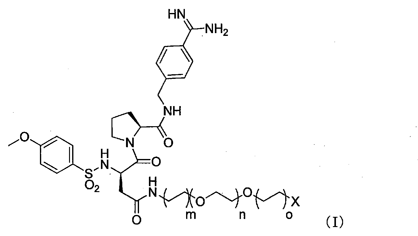

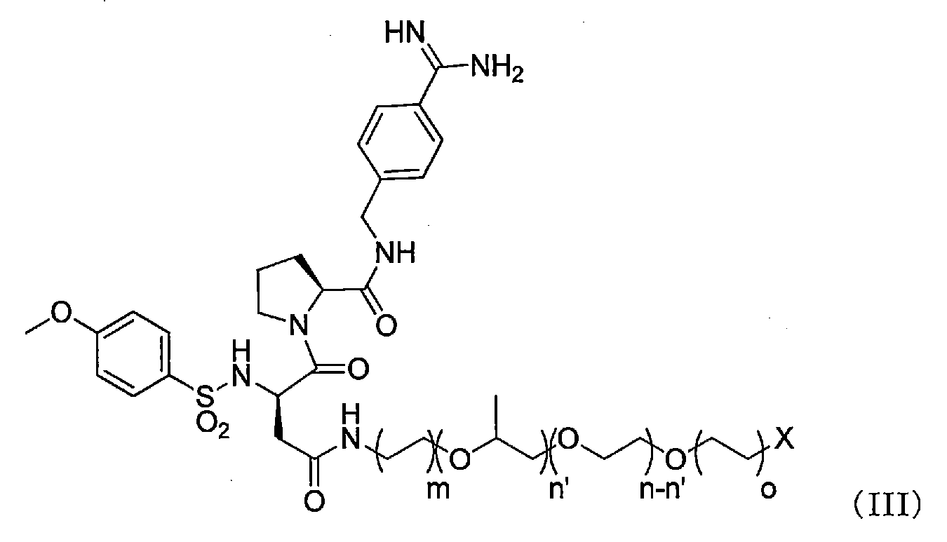

(12)上記抗血栓性材料は、アクリル酸、メタクリル酸、α-グルタミン酸、γ-グルタミン酸及びアスパラギン酸からなる群から選択される化合物を構成モノマーとして含むアニオン性ポリマー、又は、シュウ酸、マロン酸、コハク酸、フマル酸、グルタル酸、アジピン酸、ピメリン酸、スベリン酸、アゼライン酸、セバシン酸、リンゴ酸、酒石酸及びクエン酸からなる群から選択されるアニオン性化合物、を含む、(1)~(11)のいずれか記載の人工血管。
(13)上記マルチフィラメント糸は、ポリエステルからなる、(1)~(12)のいずれか記載の人工血管。
(13)外層筒状織物の内側に内層筒状織物が配置された多重筒状織物構造体において、上記外層筒状織物及び上記内層筒状織物は、複数の経糸と複数の緯糸とを交錯させて筒状にされた織物であり、上記内層筒状織物を構成する経糸及び緯糸は、単糸繊度が0.30dtex以下のマルチフィラメント糸を含むとともに、抗血栓性材料と結合しており、上記抗血栓性材料は、上記内層筒状織物の内側で1~600nmの厚さの抗血栓性材料層を形成し、内表面に16kPaの圧力をかけた条件下での透水性が300mL/cm2/min未満である、人工血管。
(14)上記外層筒状織物は、経糸に単糸繊度が1.0dtex以上のマルチフィラメント糸を含む、(13)記載の人工血管。
(15)上記単糸繊度が1.0dtex以上のマルチフィラメント糸は、上記内表面への露出度が20%以下である、(14)記載の人工血管。
(16)上記外層筒状織物は、緯糸に単糸繊度が15.0dtex以上のモノフィラメント糸を含む、(13)~(15)のいずれか記載の人工血管。
(17)上記抗血栓性材料は、アニオン性、かつ、抗凝固活性を有する硫黄原子を含んだ化合物、を含む、(13)~(16)のいずれか記載の人工血管。
(18)X線光電子分光法(XPS)で測定した内表面の全原子の存在量に対する硫黄原子の存在比率が、3.0~6.0原子数%である、(17)記載の人工血管。
(19)上記抗血栓性材料は、アルキレンイミン、ビニルアミン、アリルアミン、リジン、プロタミン及びジアリルジメチルアンモニウムクロライドからなる群から選択される化合物を構成モノマーとして含むカチオン性ポリマー、を含み、上記カチオン性ポリマーは上記内層筒状織物を構成する経糸及び緯糸と共有結合している、(1)~(18)のいずれか記載の人工血管。
(20)X線光電子分光法(XPS)で測定した内表面の全原子の存在量に対する窒素原子の存在比率が、7.0~12.0原子数%である、(19)記載の人工血管。
(21)上記抗血栓性材料は、エチレングリコール、プロピレングリコール、ビニルピロリドン、ビニルアルコール、ビニルカプロラクタム、酢酸ビニル、スチレン、メチルメタクリレート、ヒドロキシエチルメタクリレート及びシロキサンからなる群から選択される化合物を構成モノマーとして含む親水性ポリマーの骨格構造、4-(アミノメチル)ベンゼンカルボキシイミダミド又はベンゼンアミジンの骨格構造及びメトキシベンゼンスルホン酸アミドの骨格構造からなる3種類の骨格構造を含む化合物であり、上記3種類の骨格構造を含む化合物は上記内層筒状織物を構成する経糸及び緯糸と共有結合している、(13)~(16)のいずれか記載の人工血管。
(22)上記3種類の骨格構造を含む化合物は、以下の一般式(I)~(IV)で示されるいずれかの化合物である、(21)記載の人工血管。


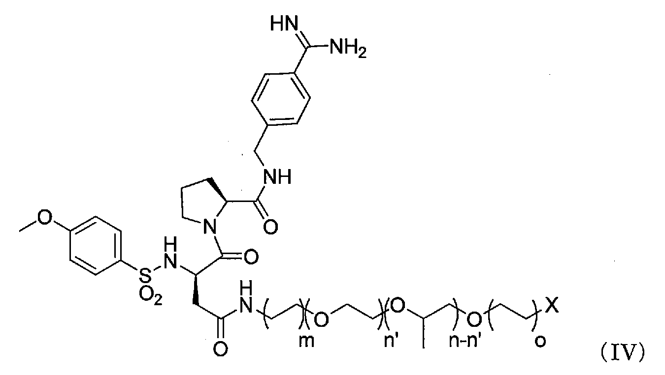
(23)上記抗血栓性材料は、アクリル酸、メタクリル酸、α-グルタミン酸、γ-グルタミン酸及びアスパラギン酸からなる群から選択される化合物を構成モノマーとして含むアニオン性ポリマー、又は、シュウ酸、マロン酸、コハク酸、フマル酸、グルタル酸、アジピン酸、ピメリン酸、スベリン酸、アゼライン酸、セバシン酸、リンゴ酸、酒石酸及びクエン酸からなる群から選択されるアニオン性化合物、を含む、(13)~(22)のいずれか記載の人工血管。
(24)上記マルチフィラメント糸は、ポリエステルからなる、(13)~(23)のいずれか記載の人工血管。
・・・式1
CF : 人工血管の内表面のカバーファクター
Dw : 経糸の総繊度(dtex)
Nw : 経糸の基布密度(本/2.54cm)
Df : 緯糸の総繊度(dtex)
Nf : 緯糸の基布密度(本/2.54cm)
なお、基布密度は、作製した筒状織物を長手方向に切開して、その内表面をキーエンス社製マイクロスコープVHX-2000にて50倍に拡大した写真をもとに計測した。
[測定条件]
装置 :電界放出型透過電子顕微鏡JEM-2100F(JEOL社製)
EELS検出器 :GIF Tridiem(GATAN社製)
EDX検出器 :JED-2300T(JEOL社製)
画像取得 :Digital Micrograph(GATAN社製)
試料調整 :超薄切片法(銅製マイクログリッドに懸架し、包埋樹脂はアクリル系樹脂を使用。)
加速電圧 :200kV
ビーム径 :直径0.7nm
エネルギー分解能 :約1.0eVFWHM
[測定条件]
装置 :ESCALAB220iXL(VG Scientific社製)
励起X線 :monochromaticAlKα1,2線(1486.6eV)
X線径 :1mm
X電子脱出角度 :90°(人工血管の表面に対する検出器の傾き)
分割ratio : 各分割ピーク成分の存在比率(%)
N1sratio : 全原子の存在量に対する窒素原子の存在比率(%)
分割percent : N1sピークにおける各分割ピーク成分の存在比率(%)
分割ratio : 各分割ピーク成分の存在比率(%)
C1sratio : 全原子の存在量に対する炭素原子の存在比率(%)
分割percent : C1sピークにおける各分割ピーク成分の存在比率(%)
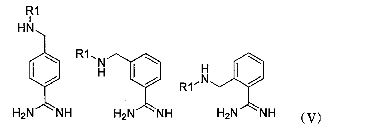


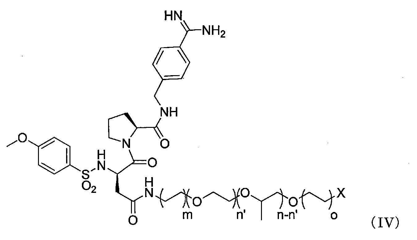
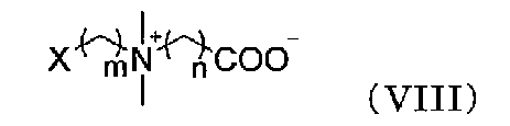
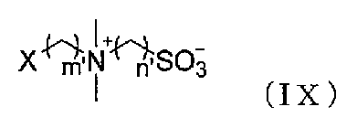
[測定条件]
装置 :TOF.SIMS5(ION-TOF社製)
1次イオン種 :Bi3 ++
2次イオン極性 :正および負
質量範囲(m/z) :0~1500
ラスターサイズ :300μm四方
ピクセル数(1辺) :256ピクセル
後段加速 :10kV
測定真空度(試料導入前) :4×10⁻⁷Mpa
1次イオン加速電圧 :25kV
パルス幅 :10.5ns
バンチング :あり(高質量分解能測定)
帯電中和 :あり
内層筒状織物を構成する経糸及び緯糸として、単糸繊度が0.23dtex、総繊度が33dtex、144フィラメントのポリエチレンテレフタレートマルチフィラメント糸を使用した。また、外層筒状織物を構成する経糸として、単糸繊度が2.33dtex、総繊度が56dtex、24フィラメントのポリエチレンテレフタレートマルチフィラメント糸、外層筒状織物を構成する緯糸として、単糸繊度が33dtexのポリエチレンテレフタレートモノフィラメント糸を使用した。経糸を細幅ドビー・シャトル二丁織機に仕掛け、緯糸を打ち込み、内径3mmとなるように製織した。98℃で精錬、170℃で熱セットを実施し、多重筒状織物構造体1を得た。
内層筒状織物を構成する緯糸として、単糸繊度が7.3dtex、総繊度66dtexで無撚りの海島複合繊維(海/島(重量比)=20/80の複合比率にて、島成分数が70)であるマルチフィラメント糸を用いて、精錬後、98℃の4重量%水酸化ナトリウム(和光純薬工業株式会社製)の水溶液で海成分を完全に溶脱させて、さらに熱セットを実施し、単糸繊度が0.084dtex、総繊度53dtex、630フィラメントのポリエチレンテレフタレートマルチフィラメント糸とした以外は、実施例1と同様の操作を行い、製織して多重筒状織物構造体2を得た。
内層筒状織物を構成する経糸及び緯糸として、実施例2で用いた海島複合繊維であるマルチフィラメント糸を用いて、精錬後、98℃の4重量%水酸化ナトリウム(和光純薬工業株式会社製)の水溶液で海成分を完全に溶脱させて、さらに熱セットを実施し、単糸繊度が0.084dtex、総繊度53dtex、630フィラメントのポリエチレンテレフタレートマルチフィラメント糸とした以外は、実施例1と同様の操作を行い、製織して多重筒状織物構造体3を得た。
多重筒状織物構造体1を、実施例1と同様の操作を行い、加水分解及び酸化し、PEIを縮合反応により共有結合させた後、0.5重量%DMT-MM、40重量%無水コハク酸(和光純薬工業株式会社製)のジメチルアセトアミドに浸漬し、50℃で17時間反応させた。
多重筒状織物構造体1を、実施例1と同様の操作を行い、加水分解及び酸化し、PEIを縮合反応により共有結合させた後、0.5重量%DMT-MM、0.5重量%PAA(重量平均分子量100万;和光純薬工業株式会社製)の水溶液に浸漬し、30℃で2時間反応させた。
実施例5と同様の操作を行い、PEIをポリアリルアミン塩酸塩(以下、「PAH」)(重量平均分子量90万;シグマ-アルドリッチ社製)に変更した人工血管を得るとともに、ポリ-L-リシン臭化水素酸塩(以下、PLys)(重量平均分子量3~7万;シグマ-アルドリッチ社製)に変更した人工血管を得た。
実施例1と同様の操作を行い、ヘパリンナトリウムをデキストラン硫酸ナトリウム(和光純薬工業株式会社製)に変更して、抗血栓性材料層を形成した人工血管(サンプル8)を得た。
多重筒状織物構造体1を、実施例1と同様の操作を行い、加水分解及び酸化した後、1.0重量%化合物A(以下の一般式(X))、化合物Aに対して2モル等量の水酸化ナトリウム及び3モル等量のDMT-MMを溶解させた水溶液に浸漬し、30℃で2時間反応させて筒状織物1に化合物Aを縮合反応により共有結合させて抗血栓性材料層を形成した人工血管(サンプル9)を得た。

実施例8と同様の操作を行い、化合物Aを化合物B(以下の一般式(XI))、化合物C(以下の一般式(XII))、及び化合物D(以下の一般式(XIII))に変更した人工血管をそれぞれ得た。

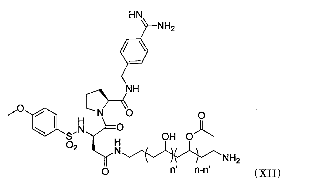
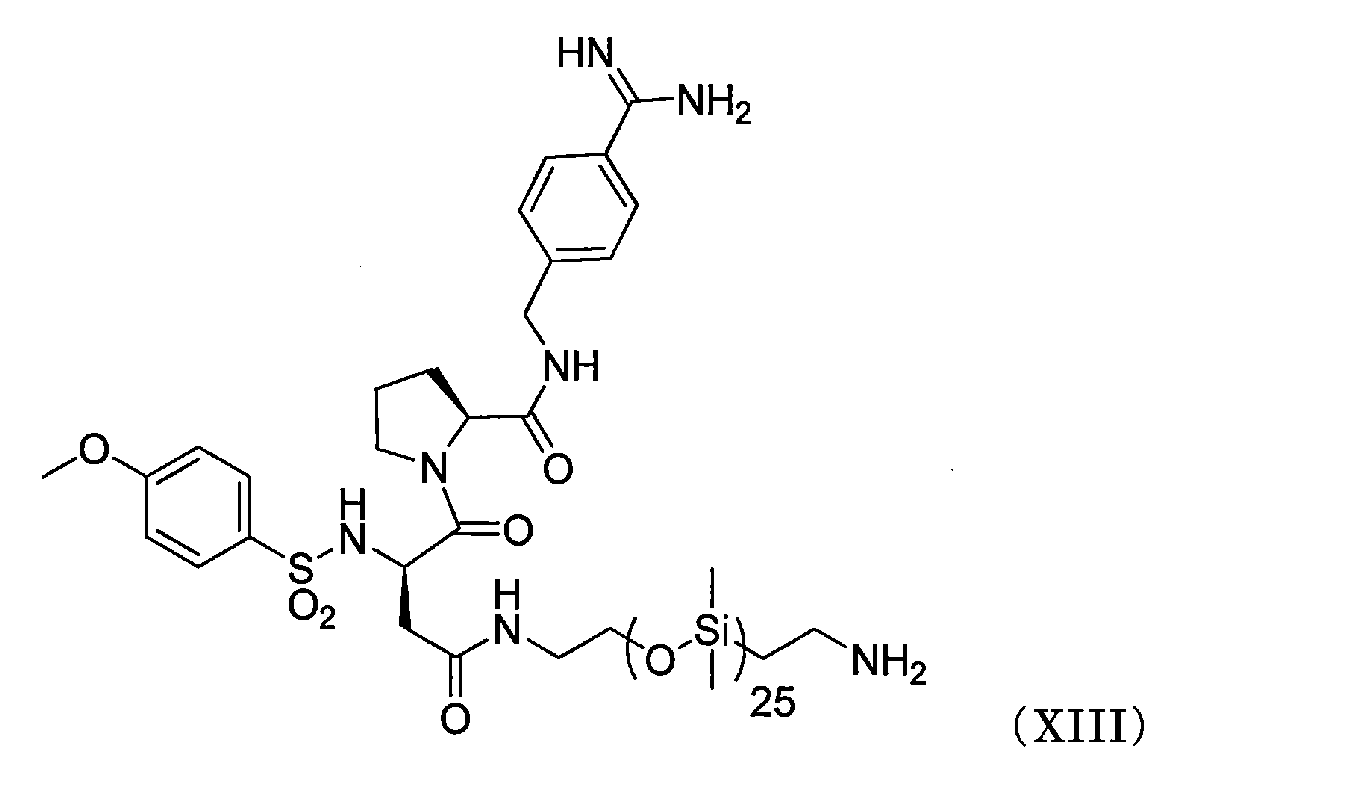
多重筒状織物構造体1を、実施例1と同様の操作を行い、加水分解及び酸化し、PEIを縮合反応により共有結合させた後、0.5重量%DMT-MM、0.5重量%PAA(重量平均分子量100万;和光純薬工業株式会社製)の水溶液に浸漬し、30℃で2時間反応させた。
実施例1と同様の操作を行い、外層筒状織物を構成する緯糸として、単糸繊度が13dtexのポリエチレンテレフタレートモノフィラメント糸、単糸繊度が108dtexのポリエチレンテレフタレートモノフィラメント糸及び単糸繊度が333dtexのポリエチレンテレフタレートモノフィラメント糸に変更した多重筒状織物構造体をそれぞれ得た。
内層筒状織物を構成する経糸として、単糸繊度が1.38dtex、総繊度が33dtex、24フィラメントのポリエチレンテレフタレートマルチフィラメント糸を使用した以外は、実施例1と同様の操作を行い、製織して多重筒状織物構造体7を得た。
内層筒状織物を構成する緯糸として、単糸繊度が1.38dtex、総繊度が33dtex、24フィラメントのポリエチレンテレフタレートマルチフィラメント糸を使用した以外は、実施例1と同様の操作を行い、製織して多重筒状織物構造体8を得た。
実施例1と同様の操作を行い、カバーファクターが1600となるよう基布密度を調整して製織することにより、多重筒状織物構造体9を得た。
多重筒状織物構造体1に、何も抗血栓性材料層を形成させない人工血管(サンプル23)を得た。
多重筒状織物構造体1を、PEI及び臭化エチル、ヘパリンナトリウムを溶解させた、メタノールやテトラヒドロフラン等の有機溶媒に浸漬し、抗血栓性材料層を形成した人工血管(サンプル24)を得た。
内層筒状織物を構成する経糸及び緯糸として、単糸繊度が0.30dtex、総繊度が44dtex、144フィラメントのポリエチレンテレフタレートマルチフィラメント糸を使用した以外は、実施例1と同様の操作を行い、製織して多重筒状織物構造体10を得た。
実施例1と同様の操作を行い、カバーファクターが1700となるよう基布密度を調整して製織することにより、多重筒状織物構造体11を得た。
ISO7198のガイダンスに記載の透水性(Water permeability)と同様にして、人工血管の内表面に16kPaの圧力(静水圧)をかけ、30秒経過後の5分間で人工血管の外層に流れ出てくる牛血液の量(mL)を量り、単位時間(min)及び単位面積(cm2)で除した値を、16kPa条件下における漏血性とした。牛血液は、ヘマトクリットが25~30%、赤血球数が5.0~6.2×10⁶/μL、白血球数が30~110×102/μL、血小板数が6.0~20.0×104/μL、総蛋白が6.5g/dLとなるように調整した。漏血性が2.0mL/cm2/min未満であれば、漏血量が少なくプレクロッティング等の操作が不要、として「○」、漏血性が2.0mL/cm2/min以上であれば、漏血量が多くプレクロッティング等の操作が必要、として「×」と判定した。
P.C.Begovacらの文献(Eur Vasc Endovasc Surg 25, 432-437 2003)等を参考にして、イヌ頚動脈に人工血管を移植した。定期的に、移植した人工血管及び両側の生体血管の超音波エコーや血管造影を実施し、血栓の有無や閉塞の有無を確認した。移植28日後に完全閉塞が見られなければ、抗血栓性が高いとして「○」、完全閉塞していれば、抗血栓性が不足しているとして「×」と判定した。
評価2と同様にして、イヌ頚動脈に人工血管を移植した。移植28日後に人工血管を摘出し、H.E.染色した標本を作成した。得られた標本を顕微鏡で観察し、人工血管と生体血管の縫合部から血管内皮細胞が移入して定着した部分までの長さを測定した。血管内皮細胞が移入した長さが4.0mm以上であれば、細胞親和性が高いとして「○」、移入した長さが4.0mm未満であれば、細胞親和性が不足しているとして「×」と判定した。

Claims (13)
- 内側が血液と接触する筒状織物が配置された筒状織物構造体において、
前記内側が血液と接触する筒状織物は、複数の経糸と複数の緯糸とを交錯させて筒状にされた織物であり、
前記内側が血液と接触する筒状織物を構成する経糸及び緯糸は、単糸繊度が0.50dtex以下のマルチフィラメント糸を含むとともに、抗血栓性材料と結合しており、
前記抗血栓性材料は、前記内側が血液と接触する筒状織物の内側で1~600nmの厚さの抗血栓性材料層を形成し、
内表面に16kPaの圧力をかけた条件下での透水性が300mL/cm2/min未満である、人工血管。 - 前記筒状織物構造体は、前記内側が血液と接触する筒状織物の外側に外層筒状織物が配置された多重筒状織物構造体であり、前記外層筒状織物は、複数の経糸と複数の緯糸とを交錯させて筒状にされた織物である、請求項1記載の人工血管。
- 前記外層筒状織物は、経糸に単糸繊度が1.0dtex以上のマルチフィラメント糸を含む、請求項2記載の人工血管。
- 前記単糸繊度が1.0dtex以上のマルチフィラメント糸は、前記内表面への露出度が20%以下である、請求項3記載の人工血管。
- 前記内側が血液と接触する筒状織物を構成する経糸及び緯糸によるカバーファクターは、1800~4000である、請求項1~4のいずれか一項記載の人工血管。
- 前記外層筒状織物は、緯糸に単糸繊度が15.0dtex以上のモノフィラメント糸を含む、請求項2~5のいずれか一項記載の人工血管。
- 前記抗血栓性材料は、硫黄原子を含むアニオン性の抗凝固活性を有する化合物、を含む、請求項1~6のいずれか一項記載の人工血管。
- X線光電子分光法(XPS)で測定した内表面の全原子の存在量に対する硫黄原子の存在比率が、3.0~6.0原子数%である、請求項7記載の人工血管。
- 前記抗血栓性材料は、アルキレンイミン、ビニルアミン、アリルアミン、リジン、プロタミン及びジアリルジメチルアンモニウムクロライドからなる群から選択される化合物を構成モノマーとして含むカチオン性ポリマー、を含み、前記カチオン性ポリマーは前記内側が血液と接触する筒状織物を構成する経糸及び緯糸と共有結合している、請求項1~8のいずれか一項記載の人工血管。
- 前記抗血栓性材料は、エチレングリコール、プロピレングリコール、ビニルピロリドン、ビニルアルコール、ビニルカプロラクタム、酢酸ビニル、スチレン、メチルメタクリレート、ヒドロキシエチルメタクリレート及びシロキサンからなる群から選択される化合物を構成モノマーとして含む親水性ポリマーの骨格構造、4-(アミノメチル)ベンゼンカルボキシイミダミド又はベンゼンアミジンの骨格構造及びメトキシベンゼンスルホン酸アミドの骨格構造からなる3種類の骨格構造を含む化合物であり、前記3種類の骨格構造を含む化合物は前記内側が血液と接触する筒状織物を構成する経糸及び緯糸と共有結合している、請求項1~6のいずれか一項記載の人工血管。
- 前記3種類の骨格構造を含む化合物は、以下の一般式(I)~(IV)で示されるいずれかの化合物である、請求項10記載の人工血管。


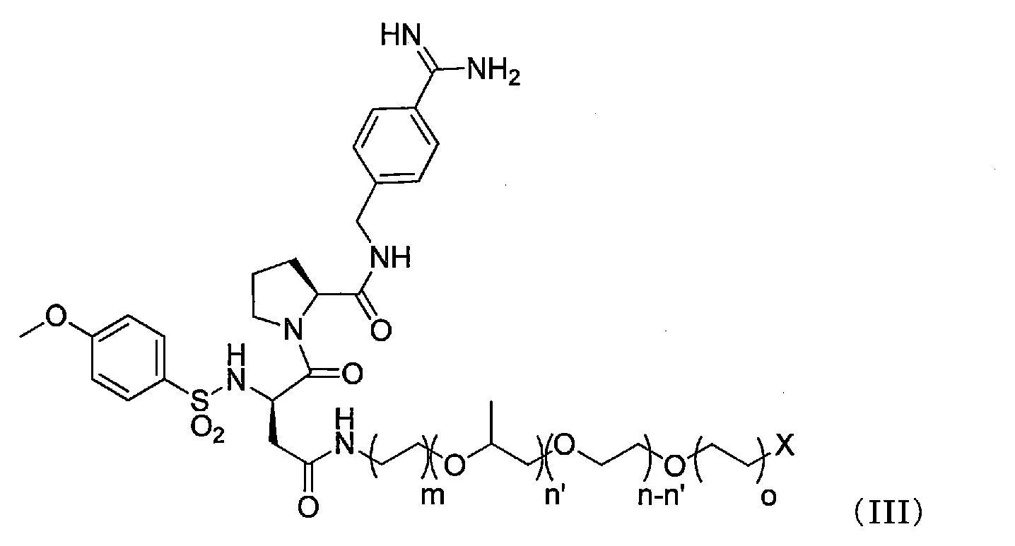

- 前記抗血栓性材料は、アクリル酸、メタクリル酸、α-グルタミン酸、γ-グルタミン酸及びアスパラギン酸からなる群から選択される化合物を構成モノマーとして含むアニオン性ポリマー、又は、シュウ酸、マロン酸、コハク酸、フマル酸、グルタル酸、アジピン酸、ピメリン酸、スベリン酸、アゼライン酸、セバシン酸、リンゴ酸、酒石酸及びクエン酸からなる群から選択されるアニオン性化合物、を含む、請求項1~11のいずれか一項記載の人工血管。
- 前記マルチフィラメント糸は、ポリエステルからなる、請求項1~12のいずれか一項記載の人工血管。
Priority Applications (9)
| Application Number | Priority Date | Filing Date | Title |
|---|---|---|---|
| BR112016018452A BR112016018452A2 (pt) | 2014-02-12 | 2015-02-12 | vaso sanguíneo artificial. |
| CA2935808A CA2935808C (en) | 2014-02-12 | 2015-02-12 | Artificial blood vessel |
| EP15749638.1A EP3106128B1 (en) | 2014-02-12 | 2015-02-12 | Artificial blood vessel |
| AU2015216257A AU2015216257B2 (en) | 2014-02-12 | 2015-02-12 | Artificial blood vessel |
| JP2015508902A JP6520706B2 (ja) | 2014-02-12 | 2015-02-12 | 人工血管 |
| KR1020167017174A KR101835449B1 (ko) | 2014-02-12 | 2015-02-12 | 인공혈관 |
| US15/117,941 US10251742B2 (en) | 2014-02-12 | 2015-02-12 | Artificial blood vessel |
| RU2016136187A RU2646506C1 (ru) | 2014-02-12 | 2015-02-12 | Искусственный кровеносный сосуд |
| CN201580008464.XA CN105979910B (zh) | 2014-02-12 | 2015-02-12 | 人工血管 |
Applications Claiming Priority (2)
| Application Number | Priority Date | Filing Date | Title |
|---|---|---|---|
| JP2014024625 | 2014-02-12 | ||
| JP2014-024625 | 2014-02-12 |
Publications (1)
| Publication Number | Publication Date |
|---|---|
| WO2015122429A1 true WO2015122429A1 (ja) | 2015-08-20 |
Family
ID=53800171
Family Applications (1)
| Application Number | Title | Priority Date | Filing Date |
|---|---|---|---|
| PCT/JP2015/053749 Ceased WO2015122429A1 (ja) | 2014-02-12 | 2015-02-12 | 人工血管 |
Country Status (10)
| Country | Link |
|---|---|
| US (1) | US10251742B2 (ja) |
| EP (1) | EP3106128B1 (ja) |
| JP (1) | JP6520706B2 (ja) |
| KR (1) | KR101835449B1 (ja) |
| CN (1) | CN105979910B (ja) |
| AU (1) | AU2015216257B2 (ja) |
| BR (1) | BR112016018452A2 (ja) |
| CA (1) | CA2935808C (ja) |
| RU (1) | RU2646506C1 (ja) |
| WO (1) | WO2015122429A1 (ja) |
Cited By (1)
| Publication number | Priority date | Publication date | Assignee | Title |
|---|---|---|---|---|
| JPWO2022209633A1 (ja) * | 2021-03-30 | 2022-10-06 |
Families Citing this family (4)
| Publication number | Priority date | Publication date | Assignee | Title |
|---|---|---|---|---|
| CA3058121A1 (en) * | 2017-03-31 | 2018-10-04 | Toray Industries, Inc. | Cylindrical structure |
| CA3107918A1 (en) | 2018-08-03 | 2020-02-06 | VeriGraft AB | Methods of preparing personalized blood vessels |
| CN112089887A (zh) * | 2018-08-30 | 2020-12-18 | 湖南博隽生物医药有限公司 | 一种生物相容性优异的抗血栓人造血管 |
| CN113557042A (zh) * | 2019-02-19 | 2021-10-26 | Tc1有限责任公司 | 人工血管和用于密封人工血管的方法 |
Citations (8)
| Publication number | Priority date | Publication date | Assignee | Title |
|---|---|---|---|---|
| US4695280A (en) * | 1983-06-07 | 1987-09-22 | Toray Industries, Inc. | Artificial vascular graft |
| JP2008511339A (ja) * | 2003-08-13 | 2008-04-17 | メドトロニック・インコーポレーテッド | 相溶性ポリマーブレンドの単層を含む活性薬剤送達システム、医療用デバイス、及び方法 |
| WO2009119761A1 (ja) * | 2008-03-26 | 2009-10-01 | 株式会社カネカ | 抗血栓性表面 |
| JP4461217B2 (ja) * | 2001-01-23 | 2010-05-12 | イェナフィン ゲーエムベーハー | オリゴまたはポリアルキレングリコール結合トロンビン阻害剤 |
| WO2011136243A1 (ja) * | 2010-04-28 | 2011-11-03 | 東レ株式会社 | ステントグラフト用基布およびステントグラフト |
| US20120116492A1 (en) * | 2009-05-30 | 2012-05-10 | Deutsche Institute für Textil-und Faserforschung Denkendorf Stiflung des öffentlichen Rechts | Medical device |
| JP2013505070A (ja) * | 2009-09-17 | 2013-02-14 | ゴア エンタープライズ ホールディングス,インコーポレイティド | 新規なヘパリン体とその利用法 |
| WO2013137263A1 (ja) * | 2012-03-13 | 2013-09-19 | 旭化成せんい株式会社 | 極細ポリエステル繊維及び筒状シームレス織物 |
Family Cites Families (35)
| Publication number | Priority date | Publication date | Assignee | Title |
|---|---|---|---|---|
| US3350488A (en) | 1958-05-27 | 1967-10-31 | Du Pont | Process for the production of sharp-edge fibers |
| US3531368A (en) | 1966-01-07 | 1970-09-29 | Toray Industries | Synthetic filaments and the like |
| JPS6041947B2 (ja) | 1977-12-09 | 1985-09-19 | ユニチカ株式会社 | ポリエステル成形体表面に抗血栓性を付与する方法 |
| JPS6047287B2 (ja) | 1977-12-15 | 1985-10-21 | ユニチカ株式会社 | 抗血栓性ポリアミドの製造方法 |
| GB8300052D0 (en) | 1983-01-04 | 1983-02-09 | Maroof A K H | Apparatus for eye testing |
| JPS6047287A (ja) | 1983-08-24 | 1985-03-14 | Matsushita Electric Ind Co Ltd | 磁気記録再生装置 |
| JPS6077764A (ja) | 1983-10-05 | 1985-05-02 | 東レ株式会社 | 人工血管 |
| JPS614546A (ja) | 1984-06-18 | 1986-01-10 | 三興空気装置株式会社 | 板状成形物の破砕装置 |
| JPS6158190A (ja) | 1984-08-30 | 1986-03-25 | 住友重機械工業株式会社 | 電磁誘導加熱装置 |
| JPS6192666A (ja) | 1984-10-15 | 1986-05-10 | 東レ株式会社 | 人工血管 |
| JPS6352898A (ja) | 1986-08-23 | 1988-03-07 | 日立造船株式会社 | ロ−ルミルのロ−ルセツテイング装置 |
| JPS63115555A (ja) | 1986-11-04 | 1988-05-20 | 東レ株式会社 | 生体適合性とプリクロツテイング性に優れた人工血管 |
| JPS63115554A (ja) | 1986-11-04 | 1988-05-20 | 東レ株式会社 | 擬内膜形成性に優れた人工血管及びその製法 |
| JPH0824686B2 (ja) | 1987-09-01 | 1996-03-13 | 東レ株式会社 | 高機能人工血管 |
| JPS63315048A (ja) | 1988-03-16 | 1988-12-22 | Toray Ind Inc | 人工血管の製造方法 |
| DE69013457T2 (de) * | 1989-04-25 | 1995-02-23 | Toray Industries | Antithrombotisches blutbehandlungssystem. |
| JPH04152075A (ja) | 1990-10-17 | 1992-05-26 | Hitachi Ltd | 歩行機械 |
| JPH04273965A (ja) | 1991-02-27 | 1992-09-30 | Sanyo Electric Co Ltd | 氷片供給装置 |
| DE69125828T2 (de) * | 1991-05-21 | 1997-07-31 | Hewlett Packard Gmbh | Verfahren zur Vorbehandlung der Oberfläche eines medizinischen Artikels |
| JPH0528143A (ja) | 1991-07-22 | 1993-02-05 | Canon Inc | 文書処理装置 |
| JPH0548132A (ja) | 1991-08-08 | 1993-02-26 | Yoshida Kogyo Kk <Ykk> | 非晶質半導体太陽電池の製造方法 |
| JPH0588611A (ja) | 1991-09-30 | 1993-04-09 | Mitsubishi Electric Corp | 交通情報提示装置 |
| JPH07265338A (ja) | 1994-03-31 | 1995-10-17 | Nippon Zeon Co Ltd | 人工血管 |
| JPH0824686A (ja) | 1994-07-14 | 1996-01-30 | Meiwa Kikai Kk | 縦型破砕機の破砕粒度調整装置 |
| JP3799626B2 (ja) | 1995-04-25 | 2006-07-19 | 有限会社ナイセム | 心臓血管修復材及びその製造方法 |
| JP3497612B2 (ja) | 1995-06-12 | 2004-02-16 | テルモ株式会社 | 医療用材料及びその製造方法 |
| JPH10151192A (ja) | 1996-11-26 | 1998-06-09 | Toyobo Co Ltd | 抗血栓性組成物および医用材料 |
| NO984143L (no) | 1998-09-09 | 2000-03-10 | Norsk Hydro As | Ny prosess for å fremstille overflatemodifiserende stoffer |
| US20040213818A1 (en) | 2001-06-05 | 2004-10-28 | Susumu Kashiwabara | Antithrombotic compositions and medical instruments containing the same |
| JP2009545333A (ja) | 2006-05-12 | 2009-12-24 | ゴア エンタープライズ ホールディングス,インコーポレイティド | 物理的操作または殺菌の後に大きな生物活性を有する固定化された生物活性物質 |
| JPWO2008032758A1 (ja) | 2006-09-15 | 2010-01-28 | 東レ株式会社 | 基材およびその製造方法 |
| TW201309354A (zh) | 2011-06-23 | 2013-03-01 | Toray Industries | 醫療材料 |
| EP3075400A4 (en) * | 2013-11-28 | 2017-07-19 | Toray Industries, Inc. | Anti-thrombotic material |
| CA2933676A1 (en) * | 2013-12-18 | 2015-06-25 | Toray Industries, Inc. | Vascular prothesis |
| JP2016220717A (ja) * | 2015-05-27 | 2016-12-28 | 東レ株式会社 | 抗血栓性材料 |
-
2015
- 2015-02-12 CA CA2935808A patent/CA2935808C/en active Active
- 2015-02-12 CN CN201580008464.XA patent/CN105979910B/zh not_active Expired - Fee Related
- 2015-02-12 BR BR112016018452A patent/BR112016018452A2/pt active Search and Examination
- 2015-02-12 EP EP15749638.1A patent/EP3106128B1/en not_active Not-in-force
- 2015-02-12 US US15/117,941 patent/US10251742B2/en active Active
- 2015-02-12 AU AU2015216257A patent/AU2015216257B2/en not_active Ceased
- 2015-02-12 JP JP2015508902A patent/JP6520706B2/ja not_active Expired - Fee Related
- 2015-02-12 KR KR1020167017174A patent/KR101835449B1/ko not_active Expired - Fee Related
- 2015-02-12 RU RU2016136187A patent/RU2646506C1/ru active
- 2015-02-12 WO PCT/JP2015/053749 patent/WO2015122429A1/ja not_active Ceased
Patent Citations (8)
| Publication number | Priority date | Publication date | Assignee | Title |
|---|---|---|---|---|
| US4695280A (en) * | 1983-06-07 | 1987-09-22 | Toray Industries, Inc. | Artificial vascular graft |
| JP4461217B2 (ja) * | 2001-01-23 | 2010-05-12 | イェナフィン ゲーエムベーハー | オリゴまたはポリアルキレングリコール結合トロンビン阻害剤 |
| JP2008511339A (ja) * | 2003-08-13 | 2008-04-17 | メドトロニック・インコーポレーテッド | 相溶性ポリマーブレンドの単層を含む活性薬剤送達システム、医療用デバイス、及び方法 |
| WO2009119761A1 (ja) * | 2008-03-26 | 2009-10-01 | 株式会社カネカ | 抗血栓性表面 |
| US20120116492A1 (en) * | 2009-05-30 | 2012-05-10 | Deutsche Institute für Textil-und Faserforschung Denkendorf Stiflung des öffentlichen Rechts | Medical device |
| JP2013505070A (ja) * | 2009-09-17 | 2013-02-14 | ゴア エンタープライズ ホールディングス,インコーポレイティド | 新規なヘパリン体とその利用法 |
| WO2011136243A1 (ja) * | 2010-04-28 | 2011-11-03 | 東レ株式会社 | ステントグラフト用基布およびステントグラフト |
| WO2013137263A1 (ja) * | 2012-03-13 | 2013-09-19 | 旭化成せんい株式会社 | 極細ポリエステル繊維及び筒状シームレス織物 |
Non-Patent Citations (1)
| Title |
|---|
| See also references of EP3106128A4 * |
Cited By (2)
| Publication number | Priority date | Publication date | Assignee | Title |
|---|---|---|---|---|
| JPWO2022209633A1 (ja) * | 2021-03-30 | 2022-10-06 | ||
| WO2022209633A1 (ja) * | 2021-03-30 | 2022-10-06 | テルモ株式会社 | 人工血管 |
Also Published As
| Publication number | Publication date |
|---|---|
| CN105979910B (zh) | 2018-05-18 |
| KR101835449B1 (ko) | 2018-03-08 |
| AU2015216257A1 (en) | 2016-07-07 |
| EP3106128B1 (en) | 2020-12-02 |
| CA2935808C (en) | 2018-08-21 |
| AU2015216257B2 (en) | 2017-06-08 |
| EP3106128A4 (en) | 2017-10-18 |
| CN105979910A (zh) | 2016-09-28 |
| JPWO2015122429A1 (ja) | 2017-03-30 |
| RU2646506C1 (ru) | 2018-03-05 |
| US10251742B2 (en) | 2019-04-09 |
| BR112016018452A2 (pt) | 2018-06-26 |
| EP3106128A1 (en) | 2016-12-21 |
| US20160354194A1 (en) | 2016-12-08 |
| KR20160091403A (ko) | 2016-08-02 |
| CA2935808A1 (en) | 2015-08-20 |
| JP6520706B2 (ja) | 2019-05-29 |
Similar Documents
| Publication | Publication Date | Title |
|---|---|---|
| WO2016190202A1 (ja) | 筒状織物構造体 | |
| JP6435863B2 (ja) | 抗血栓性材料 | |
| JP6520706B2 (ja) | 人工血管 | |
| JP6863295B2 (ja) | 筒状織物 | |
| JPWO2015080176A1 (ja) | 抗血栓性材料 | |
| EP2990010B1 (en) | Artificial blood vessel | |
| AU2014355478A1 (en) | Vascular prosthesis | |
| JP2016220717A (ja) | 抗血栓性材料 |
Legal Events
| Date | Code | Title | Description |
|---|---|---|---|
| ENP | Entry into the national phase |
Ref document number: 2015508902 Country of ref document: JP Kind code of ref document: A |
|
| 121 | Ep: the epo has been informed by wipo that ep was designated in this application |
Ref document number: 15749638 Country of ref document: EP Kind code of ref document: A1 |
|
| ENP | Entry into the national phase |
Ref document number: 20167017174 Country of ref document: KR Kind code of ref document: A |
|
| ENP | Entry into the national phase |
Ref document number: 2935808 Country of ref document: CA |
|
| ENP | Entry into the national phase |
Ref document number: 2015216257 Country of ref document: AU Date of ref document: 20150212 Kind code of ref document: A |
|
| REEP | Request for entry into the european phase |
Ref document number: 2015749638 Country of ref document: EP |
|
| WWE | Wipo information: entry into national phase |
Ref document number: 2015749638 Country of ref document: EP |
|
| WWE | Wipo information: entry into national phase |
Ref document number: 15117941 Country of ref document: US |
|
| NENP | Non-entry into the national phase |
Ref country code: DE |
|
| ENP | Entry into the national phase |
Ref document number: 2016136187 Country of ref document: RU Kind code of ref document: A |
|
| REG | Reference to national code |
Ref country code: BR Ref legal event code: B01A Ref document number: 112016018452 Country of ref document: BR |
|
| ENP | Entry into the national phase |
Ref document number: 112016018452 Country of ref document: BR Kind code of ref document: A2 Effective date: 20160811 |