WO2015141917A1 - 약물을 함유한 나노입자가 결합된 이중-목적 pat/초음파 조영제 및 이의 제조방법 - Google Patents
약물을 함유한 나노입자가 결합된 이중-목적 pat/초음파 조영제 및 이의 제조방법 Download PDFInfo
- Publication number
- WO2015141917A1 WO2015141917A1 PCT/KR2014/009549 KR2014009549W WO2015141917A1 WO 2015141917 A1 WO2015141917 A1 WO 2015141917A1 KR 2014009549 W KR2014009549 W KR 2014009549W WO 2015141917 A1 WO2015141917 A1 WO 2015141917A1
- Authority
- WO
- WIPO (PCT)
- Prior art keywords
- microbubble
- nanoparticles
- contrast agent
- porphyrin
- lipid
- Prior art date
- Legal status (The legal status is an assumption and is not a legal conclusion. Google has not performed a legal analysis and makes no representation as to the accuracy of the status listed.)
- Ceased
Links
Images

Classifications
-
- A—HUMAN NECESSITIES
- A61—MEDICAL OR VETERINARY SCIENCE; HYGIENE
- A61K—PREPARATIONS FOR MEDICAL, DENTAL OR TOILETRY PURPOSES
- A61K49/00—Preparations for testing in vivo
- A61K49/22—Echographic preparations; Ultrasonic imaging preparations
- A61K49/222—Echographic preparations; Ultrasonic imaging preparations characterised by a special physical form, e.g. emulsions, liposomes
- A61K49/223—Microbubbles, hollow microspheres, free gas bubbles, gas microspheres
-
- A—HUMAN NECESSITIES
- A61—MEDICAL OR VETERINARY SCIENCE; HYGIENE
- A61K—PREPARATIONS FOR MEDICAL, DENTAL OR TOILETRY PURPOSES
- A61K47/00—Medicinal preparations characterised by the non-active ingredients used, e.g. carriers or inert additives; Targeting or modifying agents chemically bound to the active ingredient
- A61K47/50—Medicinal preparations characterised by the non-active ingredients used, e.g. carriers or inert additives; Targeting or modifying agents chemically bound to the active ingredient the non-active ingredient being chemically bound to the active ingredient, e.g. polymer-drug conjugates
- A61K47/69—Medicinal preparations characterised by the non-active ingredients used, e.g. carriers or inert additives; Targeting or modifying agents chemically bound to the active ingredient the non-active ingredient being chemically bound to the active ingredient, e.g. polymer-drug conjugates the conjugate being characterised by physical or galenical forms, e.g. emulsion, particle, inclusion complex, stent or kit
- A61K47/6921—Medicinal preparations characterised by the non-active ingredients used, e.g. carriers or inert additives; Targeting or modifying agents chemically bound to the active ingredient the non-active ingredient being chemically bound to the active ingredient, e.g. polymer-drug conjugates the conjugate being characterised by physical or galenical forms, e.g. emulsion, particle, inclusion complex, stent or kit the form being a particulate, a powder, an adsorbate, a bead or a sphere
- A61K47/6925—Medicinal preparations characterised by the non-active ingredients used, e.g. carriers or inert additives; Targeting or modifying agents chemically bound to the active ingredient the non-active ingredient being chemically bound to the active ingredient, e.g. polymer-drug conjugates the conjugate being characterised by physical or galenical forms, e.g. emulsion, particle, inclusion complex, stent or kit the form being a particulate, a powder, an adsorbate, a bead or a sphere the form being a microcapsule, nanocapsule, microbubble or nanobubble
-
- A—HUMAN NECESSITIES
- A61—MEDICAL OR VETERINARY SCIENCE; HYGIENE
- A61K—PREPARATIONS FOR MEDICAL, DENTAL OR TOILETRY PURPOSES
- A61K9/00—Medicinal preparations characterised by special physical form
- A61K9/0002—Galenical forms characterised by the drug release technique; Application systems commanded by energy
- A61K9/0009—Galenical forms characterised by the drug release technique; Application systems commanded by energy involving or responsive to electricity, magnetism or acoustic waves; Galenical aspects of sonophoresis, iontophoresis, electroporation or electroosmosis
-
- A—HUMAN NECESSITIES
- A61—MEDICAL OR VETERINARY SCIENCE; HYGIENE
- A61K—PREPARATIONS FOR MEDICAL, DENTAL OR TOILETRY PURPOSES
- A61K9/00—Medicinal preparations characterised by special physical form
- A61K9/0012—Galenical forms characterised by the site of application
- A61K9/0019—Injectable compositions; Intramuscular, intravenous, arterial, subcutaneous administration; Compositions to be administered through the skin in an invasive manner
-
- A—HUMAN NECESSITIES
- A61—MEDICAL OR VETERINARY SCIENCE; HYGIENE
- A61K—PREPARATIONS FOR MEDICAL, DENTAL OR TOILETRY PURPOSES
- A61K9/00—Medicinal preparations characterised by special physical form
- A61K9/48—Preparations in capsules, e.g. of gelatin, of chocolate
- A61K9/50—Microcapsules having a gas, liquid or semi-solid filling; Solid microparticles or pellets surrounded by a distinct coating layer, e.g. coated microspheres, coated drug crystals
- A61K9/5005—Wall or coating material
- A61K9/5021—Organic macromolecular compounds
- A61K9/5052—Proteins, e.g. albumin
-
- A—HUMAN NECESSITIES
- A61—MEDICAL OR VETERINARY SCIENCE; HYGIENE
- A61K—PREPARATIONS FOR MEDICAL, DENTAL OR TOILETRY PURPOSES
- A61K9/00—Medicinal preparations characterised by special physical form
- A61K9/48—Preparations in capsules, e.g. of gelatin, of chocolate
- A61K9/50—Microcapsules having a gas, liquid or semi-solid filling; Solid microparticles or pellets surrounded by a distinct coating layer, e.g. coated microspheres, coated drug crystals
- A61K9/51—Nanocapsules; Nanoparticles
- A61K9/5107—Excipients; Inactive ingredients
- A61K9/513—Organic macromolecular compounds; Dendrimers
- A61K9/5169—Proteins, e.g. albumin, gelatin
-
- A—HUMAN NECESSITIES
- A61—MEDICAL OR VETERINARY SCIENCE; HYGIENE
- A61P—SPECIFIC THERAPEUTIC ACTIVITY OF CHEMICAL COMPOUNDS OR MEDICINAL PREPARATIONS
- A61P35/00—Antineoplastic agents
Definitions
- the present invention relates to a dual-purpose photoacoustic tomography (PAT) / ultrasound contrast agent, to which nanoparticles containing a drug are bound, and a method for preparing the same.
- PAT photoacoustic tomography
- Ultrasonic imaging device has the best advantage of quick diagnosis because it can be diagnosed in real time. Unlike MRI and CT, the structure is simple and inexpensive. When the ultrasound contrast agent is administered and then ultrasound is applied, the ultrasound is reflected by the microbubble in the contrast medium, showing the image of the internal organs more clearly.
- These ultrasound contrast agents were developed in 1968 when Gramiak and Shah discovered that the ultrasound signal was enhanced after injecting microbubble into blood vessels.
- Known contrast agents include small bubbles surrounded by gelatin sheaths of U.S. Patent No. 4,276,885, small gases with polysaccharide solid periphery walls of U.S. Patent No. 4,265,251, and fine particles of solid crystalline crystalline compounds of European Patent Publication No. 52575 (e.g. Galatose). Small bubbles used, small bubbles using fatty acids of European Patent Publication No. 0122624, and small bubbles prepared using fatty acids and surfactants of Korean Patent No. 1989-2989.
- Photoacoustic tomography is an imaging technique that is suitable for imaging biological tissues by combining the high resolution of ultrasound imaging with the high contrast of optical imaging.
- the technique is so scalable that it can also be used to image tumors a few centimeters inside the chest.
- the short electromagnetic pulse of the laser is absorbed into the living tissue, so that the acoustic pressure is generated at the part of the tissue that acts as a source of initial ultrasonic waves in the tissue, and the ultrasonic waves thus formed reach the surface of the living tissue. Imaging this is a PAT image.
- Optoacoustic imaging technology is a principle that stimulates cells with laser light to emit ultrasonic waves and detects these ultrasonic waves to create 3D images.
- Optoacoustic imaging technology combining optical imaging and ultrasonic imaging has attracted much attention as the next generation high resolution medical imaging technology because it has non-invasiveness, inexpensiveness, portability, excellent contrast contrast ratio, and excellent spatial resolution.
- the technology is basically a method of illuminating light, so there is a possibility of further development in the future because there is a limitation in the depth of imaging of the body.
- the photoacoustic imaging technique described above has an advantage of more accurately deriving a diagnosis result by combining with PET, CT, MR, or ultrasound.
- the ultrasound diagnosis it is expected that more accurate diagnosis is possible when the above-described photoacoustic imaging technique and the ultrasound diagnosis are used together in terms of realizing an image using ultrasound as in the photoacoustic imaging technique.
- the development of a contrast agent that can be used simultaneously for the above-described photoacoustic imaging technique and ultrasound diagnosis is essential, where the diagnostic image quality should not be degraded by the use of the same contrast agent.
- the development of theragnosis (theragnosis) technology for simultaneously proceeding the current diagnosis and treatment has been actively studied.
- the development of a contrast agent capable of simultaneously diagnosis and treatment by delivering a drug to the carrier PAT, CT, MR or ultrasound contrast agent has a problem that it is difficult to load enough drugs because the space for containing the drug is very small, thus increasing the effectiveness of the treatment dramatically.
- the present inventors have made diligent research efforts to develop a contrast agent for diagnosis and treatment that can be used simultaneously for ultrasound and photoacoustic imaging, and can be simultaneously diagnosed and treated in combination with nanomedicine.
- a contrast agent for diagnosis and treatment that can be used simultaneously for ultrasound and photoacoustic imaging, and can be simultaneously diagnosed and treated in combination with nanomedicine.
- porphyrin is included in a liposome-type microbubble commonly used for ultrasound diagnosis, and nanoparticles containing a drug in the microbubble can be used simultaneously for ultrasound diagnosis and photoacoustic imaging
- the present invention has been completed by discovering that the accuracy of an optoacoustic image can be significantly increased.
- an object of the present invention is to provide a contrast agent that can be used simultaneously for ultrasound diagnosis and photoacoustic imaging.
- Another object of the present invention is to provide a method for preparing the contrast agent.
- the present invention provides a contrast agent for ultrasound diagnosis and photoacoustic imaging.
- the present inventors have made diligent research efforts to develop a contrast agent for diagnosis and treatment that can be used simultaneously for ultrasound and photoacoustic imaging, and can be simultaneously diagnosed and treated in combination with nanomedicine.
- porphyrin is included in a liposome-type microbubble commonly used for ultrasound diagnosis, and nanoparticles containing a drug in the microbubble can be used simultaneously for ultrasound diagnosis and photoacoustic imaging, We confirmed that the accuracy of the optoacoustic image can be significantly increased.
- the present invention provides a dual-purpose photoacoustic tomography (PAT) / ultrasound contrast medium comprising: (a) a gas and porphyrin inside; Microbubble comprising; And (b) a nanoparticle containing a drug bound to the surface of the microbubble.
- PAT photoacoustic tomography
- ultrasound contrast medium comprising: (a) a gas and porphyrin inside; Microbubble comprising; And (b) a nanoparticle containing a drug bound to the surface of the microbubble.
- the term 'dual-purpose' as used herein means that the contrast agent, which is a substance of the present invention, can be used simultaneously for diagnosis and treatment, as well as for ultrasound diagnosis and photoacoustic imaging. Can be.
- Chlorophyll chlorophyll and a substance that is the parent of the related material may refer to a compound of Formula 1 or a material that performs the same / similar functions.
- Solid cancer refers to any cancer consisting of lumps except blood cancer.
- Types of solid cancer include hepatoma, brain tumor, low-grade astrocytoma, high-grade astrocytoma, pituitary adenoma, meningioma (meningioma) and cerebral lymphoma ( CNS lymphoma, Oligodendroglioma, Craniopharyngioma, Ependymoma, Brain stem tumor, Head & Neck Tumor, Larygeal cancer, Oropgaryngeal cancer ), Nasal cavity / PNS tumor, Nasopharyngeal tumor, Salivary gland tumor, Hypopharyngeal cancer, Thyroid cancer, Oral cavity tumor, Thoracic tumor (Chest Tumor), small cell lung cancer, non-small cell lung cancer (NSCLC), thym
- the drug of the present invention may be an organic poorly soluble drug.
- 'organic' can be understood to mean a substance containing carbon in the molecule, and the term 'poorly soluble' means that the pharmacologically active agent is in an aqueous solution (eg, water, saline, injectable dextrose solution, etc.). It may mean that it does not dissolve.
- aqueous solution eg, water, saline, injectable dextrose solution, etc.
- solubility The meaning of the 'solubility' is described in more detail in the solubility criteria as follows. USP / NF generally expresses solubility as the volume of solvent needed to dissolve 1 gram of drug at a specific temperature (eg, 1 g of aspirin at 300 ° C. in H 2 O, 5 ml of ethanol at 25 ° C.). In other references, solubility can be described using more subjective terms, such as those given in Table 1, set forth in Remington's Pharmaceutical Sciences, Mack Publishing Co., Easton, PA, latest edition.
- the term “poorly soluble” of the present invention when water is used as a solvent, means the four solubility categories of the lower part of Table 1, namely "sufficient solubility”, “low solubility”, “very low solubility” and " Virtually insoluble or insoluble ".
- the poorly soluble substance may include a pharmaceutically active agent, a diagnostic agent, a nutritional agent, and the like, and the poorly soluble substance as the pharmacologically active agent is "Therapeutic Category and Biological Activity Index" of The Merck Index (12th Ed'n, 1996). The compounds listed in) may be included.
- the drug of the present invention may be an anticancer agent among organic poorly soluble drugs, for example, acibaicin, aclarubicin, acodazole, acronycin, adozelesin, alanosine, Aldesleukin, Allopurinol Sodium, Altretamine, Aminoglutetimide, Amonapiide, Ampligen, Amsacrine, Androgens, Anguidine, Apidicholine glycinate, Asari, Asparaginase, 5-Azacytidine, Azathioprine, Bacillus Calmette-Guerin (BCG), Bakers Antipol, Beta-2-Dioxythioguanosine, Bisantrene HCl, Bleomycin Sulfate, Bullpanol, Butionine Sulfoximine, BWA 773U82, BW 502U83 / HCl, BW 7U85 mesylate, cerasemide,
- organic poorly soluble drugs for example,
- the nanoparticles of the present invention may include albumin to form self-aggregates.
- a cohesive protein may be used.
- the aggregated structure is maintained while circulating in the blood for a long time to stably deliver the drug, and a known albumin having cancer target may be used.
- self-aggregates may refer to a material that is not collected by an external force but forms an aggregate by intrinsic intermolecular attraction or repulsive force included in the material.
- the size of the nanoparticles is important in the present invention. Because of the poor vascular tissue formed by the cancerous tissue, nanoparticles of tens or hundreds of nanometers in size can accumulate around the cancerous tissue, and the nanocarriers infiltrated by the lymphatic vessels that do not function around the cancerous tissue are not released. It is possible to stay in cancer tissue for a long period of time, which is called an enhanced permeability and retention (EPR) effect, because particles having a diameter of less than 200 nm generally show the best efficiency.
- EPR enhanced permeability and retention
- the size of the nanoparticles of the present invention may preferably be 10-500 nm, more preferably 50-400 nm, most preferably 100-300 nm.
- the nanoparticles of the present invention may be bound to the microbubbles by an activated reactor of a linker or microbubble surface.
- the reactor may be a thiol group, an amine group, or a linker may be a compound including the reactor.
- the microbubbles and the nanoparticles may be amide bond (amide bond) between them.
- the bond may be formed by an amide bond occurring between the carboxyl group in the microbubble and the amine group contained in the albumin.
- the microbubbles of the invention may preferably be gas filled microspheres, gas filled liposomes or gas-forming emulsions, most preferably gas It may be a filled liposome.
- the liposomes are formed by amphiphilic compounds, including phospholipids. Such amphiphilic compounds are typically arranged at the interface between an aqueous medium and an inherently water insoluble organic solvent to stabilize emulsified solvent microdrops.
- the amphiphilic compound has a molecule having a hydrophilic polar head portion (eg a polar or ionic group) capable of reacting with an aqueous medium and a hydrophobic organic tail portion (eg a hydrocarbon chain) capable of reacting with an organic solvent, for example.
- a hydrophilic polar head portion eg a polar or ionic group
- a hydrophobic organic tail portion eg a hydrocarbon chain
- Amphiphilic compounds are mixtures of two liquids (eg water and oil) that are immiscible, mixtures of liquids and gases (eg gas microbubbles in water) or mixtures of liquids and insoluble particles (eg metal nanoparticles in water) Are compounds capable of stabilizing mixtures of materials that are otherwise incompatible with other methods.
- the inert gas and the water are injected into the phospholipid thin film and then ultrasonicated to form liposomes filled with the inert gas therein.
- Amphiphilic phospholipid compounds contain at least one phosphate group and contain at least one, preferably two lipophilic long-chain hydrocarbon groups.
- the amphiphilic phospholipid may be a known compound, for example, diphytanoylphosphatidylcholine (DP), DPPC (1,2-dipalmitoyl-sn-glycero-3-phosphocholine), DHPC (1,2-diheptanoyl) -sn-glycero-3-phosphocholine), DHPE (1,2-dihexanoyl-sn-glycero-3-phosphoethanolamine), DMPC (1,2-dimyristoyl-sn-glycero-3-phosphocholine), DIODPC (1,2- DiODodecyl-sn-Glycero-3-Phosphocholine, DMPS (dimyristoyl phophatidylserine), DLPC (dimyristoyl phosphatidylglycerol, dilauryl phosphatidycholine), DMPE (1,2-dimyristoyl-sn-glycero-3-phosphoethanol
- amphipathic phospholipid compound may use a modified phospholipid compound.
- modified phospholipids include modified phospholipids with addition of polyethylene glycol (PEG), modified phosphatidylethanolamine (DMPE-PEG) or phosphoethanolamine (DSPE-PEG) together with polyethylene glycol (PEG). .
- the amphiphilic phospholipid compound used in the present invention may comprise NHS (N-hydroxy succinimide) for forming amide bonds.
- amphiphilic compound in addition to the amphiphilic compound, it may further include an additional amphiphilic material, for example, lysolipid, stearic acid, polyethylene glycol, polyoxyethylene fatty acid ester, polyoxyethylene fatty acid stearate, polyoxyethylene Fatty alcohols and the like.
- additional amphiphilic material for example, lysolipid, stearic acid, polyethylene glycol, polyoxyethylene fatty acid ester, polyoxyethylene fatty acid stearate, polyoxyethylene Fatty alcohols and the like.
- the gas filled in the liposome of the present invention can be used a known gas without limitation, for example, carbon dioxide, helium, nitrogen, argon, sulfur hexafluoride, perfluorinated gas can be used Can be.
- the gas is preferably a fluoride containing fluorine gas, for example, perfluoropropane (C3F8), sulfur hexafluoride (SF6), perfluoropentane, decafluorobutane and perfluoro Hexane (perfluorohexane).
- the microbubbles of the present invention may preferably have a diameter of 0.1-20 ⁇ m, most preferably 1-10 ⁇ m.
- Contrast agent of the present invention can be administered parenterally, in the case of parenteral administration can be administered by intravenous injection, subcutaneous injection, intramuscular injection, intraperitoneal injection, transdermal administration and the like.
- Suitable dosages of the contrast agent of the present invention may be prescribed in various ways depending on factors such as the formulation method, mode of administration, age, weight, sex, pathological condition, food, time of administration, route of administration, rate of excretion and response to response of the patient. have.
- the dosage of the contrast agent of the present invention may preferably be 0.001-100 mg / kg (body weight).
- the present invention provides a method for preparing a dual-purpose photoacoustic tomography (PAT) / ultrasound contrast medium comprising the following steps: (a) comprising porphyrin Preparing nanoparticles each containing a microbubble and a drug; And (b) reacting the nanoparticles with the microbubbles in water at a predetermined ratio.
- PAT photoacoustic tomography
- the method for producing the microbubble may include forming a lipid thin film by mixing a lipid having an emulsifier, a capporphyrin-lipid, a lipid, and an NHS with an organic solvent;
- the lipid thin film may be hydrated by putting it in water, and injecting the gas into ultrasonic water and maintaining the gas at a high pressure.
- the forming of the lipid thin film may include mixing a molar ratio of a lipid having an emulsifier: porphyrin-lipid: lipid: NHS in a range of 5-10: 15-30: 60-75: 15-30.
- the method of hydrating the lipid thin film and filling the gas may be a known method.
- a mixture of water, glycol, and glycerin is maintained in a container containing a lipid thin film to maintain a temperature of 55 to 60 ° C. It can be dissolved while dissolving it and putting the gas at 200kPa for ultrasonic treatment, or it can be used in combination with ultrasonic and mechanical agitation method.
- Preparing the nanoparticles containing the drug is to dissolve the albumin in water and inject the drug therein to prepare a mixture, the pH of the mixture is adjusted to 7-10, preferably pH 8.0-8.5 and then alcohol And dropping the stream, wherein the albumin can form self-aggregates.
- the present invention provides a dual-purpose photoacoustic tomography (PAT) / ultrasound contrast medium comprising: (a) a microbubble comprising gas and porphyrin inside; And (b) a nanoparticle containing a drug bound to the surface of the microbubble.
- PAT photoacoustic tomography
- ultrasound contrast medium comprising: (a) a microbubble comprising gas and porphyrin inside; And (b) a nanoparticle containing a drug bound to the surface of the microbubble.
- the contrast agent of the present invention can be used simultaneously for ultrasound diagnosis and photoacoustic image diagnosis, and has the advantage of significantly increasing the accuracy of the photoacoustic image.
- PTX-NPs human serum albumin nanoparticles
- paclitaxel paclitaxel
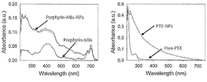
- Figure 2 shows the results of analyzing the presence or absence of porphyrin-MBs-NPs binding by UV-vis spectroscopy.
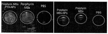
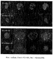
- Figure 3 shows the results of verifying the ultrasound and photoacoustic image of porphyrin-MBs-NPs and porphyrin MBs developed in the present invention.
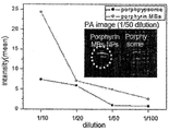
- Figure 4 shows the data that the signal in the photoacoustic signal amplified in porphyrin MBs-NPs compared to porphysome.
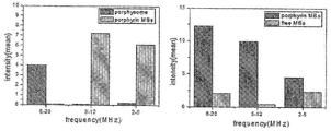
- FIG. 5 shows an experimental result of analyzing the intensity of the signal is detected according to the frequency band of the transducer used when detecting the acoustic signal.
- Figure 6 shows the data analyzing the penetration effect of the prepared agent in MDA-MB-231 cells.
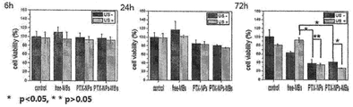
- Figure 7 shows the data analyzed over time the anticancer effect in the cell.
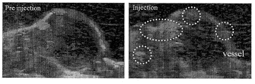
- FIG. 8 shows data obtained by photoacoustic imaging of cancer tissue in a disease animal model.
- % used to indicate the concentration of a particular substance is solid / solid (weight / weight)%, solid / liquid (weight / volume)%, and Liquid / liquid is (volume / volume)%.
- the residue is purified by thin layer chromatography (20 x 20cm pre-coated silica gel plate with fluorescent indicator, 1.5mm thickness), where the retardation factor (Rf) of thin layer chromatography is 0.4. Purification by extraction as a band. The purification method was chromatographed using diol modified silica, and impurities were removed with dichloromethane containing 2% and 5% methanol, and purified with dichloromethane containing 8% methanol. Purified pyropheophrobide-lipid was collected at a concentration of 1 ⁇ mol, dried by flowing nitrogen gas, and stored at -20 ° C in argon environment.
- the extracted porpyrin-lipid purity was determined by high performance liquid chromatography and mass spectrometry (conditions: Phenomenex Jupiter C4 column, 0.4 mL / min flow from 25% to 95% acetonitrile followed by hold 0.1% trifluoroacetic acid, compound eluted at 32min, observed mass: 1013.1).
- Lipids include 1,2-disteraoyl-sn-glycero-3-phosphocholine (DSPC), DSPE-PEG2000-NHS (1,2-dioleoyl-sn-glycero-3-phosphoethanolamine-n- [poly (ethyleneglycol)] 2000- N-hydroxysuccinimide), porphyrin-containing lipid (porphyrin-lipid), polyoxyethylene 40 stearate (POE40s) as an emulsifier were mixed in a chloroform with a molar ratio of 50: 15: 15: 1, and then dissolved in chloroform using a rotary evaporator. Was evaporated completely to form a lipid thin film.
- DSPC 1,2-disteraoyl-sn-glycero-3-phosphocholine
- DSPE-PEG2000-NHS 1,2-dioleoyl-sn-glycero-3-phosphoethanolamine-n- [poly (ethyleneglycol)] 2000- N
- distilled water, propylene glycol and glycerin were mixed at 8: 1: 1 and then added to the lipid thin film.
- Lipids were dissolved while maintaining the temperature at 55-60 ° C.
- SF6 or C3F8 gas was added to a container containing a mixed solution and filled with 200 kPa, and microbubbles (porphyrin-MBs) were prepared through sonication and mechanical agitation.
- HSA Human Serum Albumin
- HSA-NPs HSA nanoparticles
- microbubble containing HSA nanoparticles and porphyrin was mixed at room temperature for 2 hours at a molar ratio of 1: 0.5-2 binding reactor to bind the nanoparticles to the microbubbles with amide bonds. Unbound HSA nanoparticles were removed by centrifugation to complete the contrast agent of Example 1 (porphyrin-MBs-NPs).
- EDC and NHS may be sufficiently added in advance to react to wash the remaining EDC and NHS in a centrifuge, thereby further increasing the coupling efficiency.
- Microbubbles containing porphyrin prepared by the method of Preparation Example was used as Comparative Example 1 (nanoparticles are not bonded to the microbubbles).
- Ultrasonic images were diluted 1/50 times of 0.5 mg / mL porphyrin MBs and porphyrin MBs-NPs into phantoms made of agarose gel and imaged with a commercially available ultrasound diagnostic device using a 2-5 MHz transducer. .
- the diagnostic mode used is harmonic mode, which detects the components from the ultrasound contrast agent, and the intensity of the emitted ultrasound is 0.1 mechanical index.
- Photoacoustic images were obtained by inserting porphyrin MBs and porphyrin MBs-NPs at the same concentration as the ultrasound imaging experiments, and obtaining data by emitting a laser with a wavelength of 700 nm. The images were then reconstructed using the matlab program.
- PTX-NPs nanoparticles containing PTX of 400 nM
- Apoptosis was quantitatively analyzed using MTT assay.
- Balda / C nude mice xenografted MDA-MB-231 cells were subjected to the same conditions as the photoacoustic imaging experiment. First, tumors were injected before injection, and 200 ⁇ L injection of 0.5 mg / mL porphyrin MBs was performed after tail vein injection of porphyrin MBs.
- PTX-NPs human serum albumin nanoparticles
- an anticancer agent paclitaxel: PTX
- porphyrin-MBs porphyrin microbubbles
- PTX-NPs having a size of 190.1 ⁇ 52.6 nm are present in porphyrin-MBs having a size distribution of 1.1 ⁇ 1.2 ⁇ m as data indicating the size change before and after PTX-NPs are bound to porphyrin-MBs-NPs.
- the combined results show that the peak shifts with a size distribution of 1.6 ⁇ 2.5 ⁇ m.
- FIG. 2 is a result of analyzing the presence or absence of porphyrin-MBs-NPs by UV-vis spectroscopy, the data on the left is measured porphyrin-MBs and porphyrin-MBs-NPs.
- porphyrin-MBs only peaks of porphyrin with a wavelength of 700 nm were detected, but when PTX-NPs were combined, PTX-NPs with a wavelength of 227 nm were detected together and nanoparticles containing anticancer agents for treatment (PTX-NPs) It can be seen that is combined.
- the right data of FIG. 2 is a data verifying the loading of nanoparticles of anti-cancer drug (paclitaxel). The uv absorption peak (227 nm) of pure paclitaxel was detected in PTX-NPs, indicating that the nanoparticles were effectively loaded. Can be.
- FIG. 3 is a result of verifying the ultrasound and photoacoustic image of porphyrin-MBs-NPs and porphyrin MBs developed in the present invention, it was found that the effect of the image is not reduced by the binding of PTX-NPs.
- the left ultrasound image of FIG. 3 commercializes an image using a 2-5 MHz transducer of each agent diluted 1/50 (concentration of porphysrin MBs-NPs and porphysome: 10 ⁇ g / mL) in a phantom made of Agarose.
- the ultrasound image was detected using the ultrasonic diagnostic apparatus.
- the diagnostic mode used at this time is the harmonic mode, which is the result of detecting the harmonic component from the porphyrin MBs-NPs.
- the right photoacoustic image of FIG. 3 was injected with each agent diluted 1/50 (concentration of porphysrin MBs-NPs and porphysome: 10 ⁇ g / mL) into the tube, and the image was detected using a 700 nm laser.
- PBS phosphate buffer saline
- FIG. 5 is an experiment in which the signal is detected according to the frequency band of the transducer used when detecting the acoustic signal.
- the graph on the left is data comparing porphyrin MBs and porphysome. At low frequencies, the intensity of porphyrin MBs was about 100-fold higher in the 5-12 MHz transducer and about 60-fold in the 2-5 MHz transducer compared to the porphysome signal.
- This experiment detects signals using transducers having respective frequency bands using porphysomes, porphyrin MBs and freeMBs (microbubble that do not contain porphyrin) diluted 1/50 in the same manner as the photoacoustic imaging experiment of Experiment Method 2. It was. The acquired signals were verified using the matlab program.
- FIG. 6 is a data analyzing the infiltration effect of the prepared agent in MDA-MB-231 cells using the bovine serum albumin combined with FITC to PTX-NPs to detect green color fluorescence, microbubble Fluorescence was detected by loading DiI-c18 (red), a lipophilic fluorescent substance.
- DiI-c18 red
- the penetration effect into the cells was increased by the cavitation effect according to the resonance of ultrasound and microbubble as the ultrasound was applied to the group compared with the ultrasound group.
- Cell imaging was imaged using confocal microscopy.
- FIG. 8 shows data obtained by photoacoustic imaging of cancer tissue in a disease animal model.
- porphyrin MBs-NPs circulated through the blood vessels before and after injection, the photoacoustic intensity increased in the blood vessels.
Landscapes
- Health & Medical Sciences (AREA)
- Chemical & Material Sciences (AREA)
- Life Sciences & Earth Sciences (AREA)
- Veterinary Medicine (AREA)
- Public Health (AREA)
- General Health & Medical Sciences (AREA)
- Animal Behavior & Ethology (AREA)
- Epidemiology (AREA)
- Pharmacology & Pharmacy (AREA)
- Medicinal Chemistry (AREA)
- Bioinformatics & Cheminformatics (AREA)
- Engineering & Computer Science (AREA)
- Physics & Mathematics (AREA)
- Nuclear Medicine, Radiotherapy & Molecular Imaging (AREA)
- Nanotechnology (AREA)
- Biomedical Technology (AREA)
- Optics & Photonics (AREA)
- Acoustics & Sound (AREA)
- Radiology & Medical Imaging (AREA)
- General Chemical & Material Sciences (AREA)
- Chemical Kinetics & Catalysis (AREA)
- Organic Chemistry (AREA)
- Dermatology (AREA)
- Medicines Containing Antibodies Or Antigens For Use As Internal Diagnostic Agents (AREA)
- Medicinal Preparation (AREA)
- Medicines That Contain Protein Lipid Enzymes And Other Medicines (AREA)
- Pharmaceuticals Containing Other Organic And Inorganic Compounds (AREA)
Abstract
Description

| 기술 용어 | 1부의 용질 당 필요한 용매의 부 |
| 매우 높은 가용성 | <1 |
| 높은 가용성 | 1 내지 10 |
| 가용성 | 10 내지 30 |
| 불충분한 가용성 | 30 내지 100 |
| 낮은 가용성 | 100 내지 1000 |
| 매우 낮은 가용성 | 1000 내지 10,000 |
| 사실상 불용성 또는 불용성 | >10,000 |
Claims (15)
- 다음을 포함하는 이중-목적(dual-purpose) PAT(Photoacoustic tomography)/초음파(ultrasound) 조영제:(a) 내부에 가스 및 포르피린(porphyrin)을 포함하는 마이크로버블 ; 및(b) 상기 마이크로 버블의 표면에 결합된 약물을 함유하는 나노입자.
- 제 1 항에 있어서,상기 나노입자는 알부민을 포함하여, 자기 집합체(self-aggregates)를 형성하는 것을 특징으로 하는 조영제.
- 제 1 항에 있어서,상기 나노입자는 직경이 100-300 nm인 것을 특징으로 하는 조영제.
- 제 2 항에 있어서,상기 알부민은 인간혈청 알부민 또는 이의 단편인 것을 특징으로 하는 조영제.
- 제 1 항에 있어서,상기 약물은 도세탁셀(Docetaxel), 시스플라틴(cis-platin), 캠토세신(camptothecin), 파클리탁셀(paclitaxel), 타목시펜(Tamoxifen), 아나스테로졸(Anasterozole), 글리벡(Gleevec), 5-플루오로우라실(5-FU), 플록슈리딘(Floxuridine), 류프로리드(Leuprolide), 플로타미드(Flutamide), 졸레드로네이트(Zoledronate), 독소루비신(Doxorubicin), 빈크리스틴(Vincristine), 젬시타빈(Gemcitabine), 스트렙토조토신(Streptozocin), 카보플라틴(Carboplatin), 토포테칸(Topotecan), 벨로테칸(Belotecan), 이리노테칸(Irinotecan), 비노렐빈(Vinorelbine), 히도록시우레아(hydroxyurea), 발루비신(Valrubicin), 레티노익산(retinoic acid) 계열, 메소트렉세이트(Methotrexate), 메클로레타민(Meclorethamine), 클로람부실(Chlorambucil), 부술판(Busulfan), 독시플루리딘(Doxifluridine), 빈블라스틴(Vinblastin), 마이토마이신(Mitomycin), 프레드니손(Prednisone), 테스토스테론(Testosterone), 미토산트론(Mitoxantron), 아스피린(aspirin), 살리실레이트(salicylates), 이부프로펜(ibuprofen), 나프로센(naproxen), 페노프로펜(fenoprofen), 인도메타신(indomethacin), 페닐부타존(phenyltazone), 시클로포스파미드(cyclophosphamide), 메클로에타민(mechlorethamine), 덱사메타손(dexamethasone), 프레드니솔론(prednisolone), 셀레콕시브(celecoxib), 발데콕시브(valdecoxib), 니메슐리드(nimesulide), 코르티손(cortisone) 및 코르티코스테로이드(corticosteroid)를 포함하는 군으로부터 선택되는 것을 특징으로 하는 조영제.
- 제 1 항에 있어서,상기 마이크로버블은 포르피린을 포함하고, 가스 충전된 마이크로스피어(microspheres), 가스 충전된 리포좀 또는 가스 포밍 에멀젼(gas-forming emulsions)인 것을 특징으로 하는 조영제.
- 제 1 항에 있어서,상기 나노입자는 싸이올기(thiol), 아민기(amine) 바이오틴(biotin)-아비딘(avidin)에 의해 마이크로버블에 결합되는 것을 특징으로 하는 조영제.
- 제 1 항에 있어서,상기 마이크로버블과 상기 나노입자의 결합은 나노입자와 조영제 표면에서의 아미드 결합(amide bond)인 것을 특징으로 하는 조영제.
- 제 1 항에 있어서,상기 마이크로버블의 직경이 0.1-10 ㎛인 것을 특징으로 하는 조영제.
- 다음의 단계를 포함하는 이중-목적(dual-purpose) PAT(Photoacoustic tomography)/초음파(ultrasound) 조영제의 제조방법:(a) 포르피린을 포함하는 마이크로버블과 약물을 함유하는 나노입자를 각각 제조하는 단계; 및(b) 상기 나노입자와 상기 마이크로버블을 소정 비율로 물에 혼합하여 반응시키는 단계.
- 제 10 항에 있어서,상기 마이크로버블을 제조하는 단계는 유화제, 포르피린-지질, 지질, NHS를 구비하는 지질을 유기용매와 혼합하여 지질박막을 형성하는 단계 ;상기 지질박막을 물에 넣어 수화시키고, 여기에 가스를 주입 및 고압으로 유지하면서 초음파 처리하는 단계 ;를 포함하는 것을 특징으로 하는 조영제의 제조방법.
- 제 11 항에 있어서,상기 지질박막을 형성하는 단계는 유화제 : 포르피린-지질 : 지질 : NHS를 구비하는 지질의 몰비를 5-10 : 15-30 : 60-75 : 15-30 범위로 첨가하는 것을 특징으로 하는 조영제의 제조방법.
- 제 10 항에 있어서,상기 약물을 함유하는 나노입자를 제조하는 단계는알부민을 물에 녹인 후 여기에 약물을 주입하여 혼합물을 제조하는 단계 ;상기 혼합물의 pH를 7-9로 조절한 후 알코올류를 적하시키는 단계로서, 상기 단계에 의해 상기 알부민이 자기 집합체(self-aggregates)를 형성하는 것을 특징으로 하는 조영제의 제조방법.
- 제 10 항에 있어서,상기 반응 단계는 상기 마이크로버블 표면에 존재하는 NHS가 물에 가수분해되고, 마이크로버블 표면에 잔존하는 카르복실기와 상기 나노입자에 존재하는 아민기가 아미드 결합하는 단계인 것을 특징으로 하는 조영제의 제조방법.
- 제 10 항에 있어서,상기 나노입자와 상기 마이크로버블의 혼합 비율은 1:0.5-2의 결합 반응기의 몰비인 것을 특징으로 하는 조영제의 제조방법.
Priority Applications (4)
| Application Number | Priority Date | Filing Date | Title |
|---|---|---|---|
| CN201480077306.5A CN106456806B (zh) | 2014-03-19 | 2014-10-13 | 结合有含有药物的纳米粒子的两用的pat/超声造影剂及其制备方法 |
| US15/126,472 US10912848B2 (en) | 2014-03-19 | 2014-10-13 | Dual-purpose PAT/ultrasound contrast agent bound with nanoparticles containing drug and method for preparing same |
| EP14886311.1A EP3120872B1 (en) | 2014-03-19 | 2014-10-13 | Dual-purpose pat/ultrasound contrast agent bound with nanoparticles containing drug and method for preparing same |
| JP2016557079A JP6347850B2 (ja) | 2014-03-19 | 2014-10-13 | 薬物を含有したナノ粒子が結合された二重目的pat/超音波造影剤およびその製造方法 |
Applications Claiming Priority (2)
| Application Number | Priority Date | Filing Date | Title |
|---|---|---|---|
| KR10-2014-0032057 | 2014-03-19 | ||
| KR1020140032057A KR101595795B1 (ko) | 2014-03-19 | 2014-03-19 | 약물을 함유한 나노입자가 결합된 이중-목적 pat/초음파 조영제 및 이의 제조방법 |
Publications (1)
| Publication Number | Publication Date |
|---|---|
| WO2015141917A1 true WO2015141917A1 (ko) | 2015-09-24 |
Family
ID=54144850
Family Applications (1)
| Application Number | Title | Priority Date | Filing Date |
|---|---|---|---|
| PCT/KR2014/009549 Ceased WO2015141917A1 (ko) | 2014-03-19 | 2014-10-13 | 약물을 함유한 나노입자가 결합된 이중-목적 pat/초음파 조영제 및 이의 제조방법 |
Country Status (6)
| Country | Link |
|---|---|
| US (1) | US10912848B2 (ko) |
| EP (1) | EP3120872B1 (ko) |
| JP (1) | JP6347850B2 (ko) |
| KR (1) | KR101595795B1 (ko) |
| CN (1) | CN106456806B (ko) |
| WO (1) | WO2015141917A1 (ko) |
Cited By (4)
| Publication number | Priority date | Publication date | Assignee | Title |
|---|---|---|---|---|
| WO2017089800A1 (en) * | 2015-11-23 | 2017-06-01 | University Of Ulster | Microbubble-chemotherapeutic agent complex for sonodynamic therapy |
| CN109789226A (zh) * | 2016-09-27 | 2019-05-21 | 西江大学校产学协力团 | 利用携带抗癌剂的人血清白蛋白纳米粒子的肝动脉化疗栓塞术用组合物及其制造方法 |
| CN111671922A (zh) * | 2020-07-23 | 2020-09-18 | 四川大学华西医院 | 两亲性梳形嵌段接枝共聚物基超声造影剂及其制备方法 |
| US11406950B2 (en) * | 2017-03-10 | 2022-08-09 | Sogang University Research Foundation | Bubble volume control method and bubble volume controlling apparatus |
Families Citing this family (8)
| Publication number | Priority date | Publication date | Assignee | Title |
|---|---|---|---|---|
| GB201708663D0 (en) * | 2017-05-31 | 2017-07-12 | Univ Ulster | Therapy |
| KR102076518B1 (ko) * | 2018-02-08 | 2020-02-13 | 서강대학교산학협력단 | 수용성 항암제를 담지하는 마이크로버블을 포함하는 간동맥색전술용 조성물 및 그 제조방법 |
| EP3764878B1 (en) * | 2018-03-13 | 2026-02-25 | Trust Bio-sonics Inc. | Composition and methods for sensitive molecular analysis |
| US11890370B2 (en) | 2018-09-21 | 2024-02-06 | Sahmyook University Industry-Academic Cooperation Foundation | Ultrasound-induced drug delivery system using drug carrier comprising nanobubbles and drug |
| KR102198900B1 (ko) * | 2019-05-10 | 2021-01-07 | 서강대학교 산학협력단 | 질병 치료용 나노입자 복합체 및 이의 제조방법 |
| KR102379296B1 (ko) * | 2019-09-02 | 2022-03-29 | (주)아이엠지티 | 면역 마이크로버블 복합체 및 이의 이용 |
| CN113577040A (zh) * | 2021-07-19 | 2021-11-02 | 河北工程大学附属医院 | 负载中药单体的纳米载体及在制备抗肿瘤药物中的应用 |
| US12133891B2 (en) * | 2021-11-02 | 2024-11-05 | Industry Foundation Of Chonnam National University | Microbubble-extracellular vesicle complexes |
Citations (2)
| Publication number | Priority date | Publication date | Assignee | Title |
|---|---|---|---|---|
| WO2000072757A1 (en) * | 1999-06-01 | 2000-12-07 | Drexel University | Surface stabilized microbubbles for use in ultrasound contrast and drug delivery agents |
| KR20050096959A (ko) * | 2003-02-04 | 2005-10-06 | 브라코 인터내셔날 비.브이. | 초음파 조영제 및 그것의 제조방법 |
Family Cites Families (17)
| Publication number | Priority date | Publication date | Assignee | Title |
|---|---|---|---|---|
| US4276885A (en) | 1979-05-04 | 1981-07-07 | Rasor Associates, Inc | Ultrasonic image enhancement |
| US4265251A (en) | 1979-06-28 | 1981-05-05 | Rasor Associates, Inc. | Method of determining pressure within liquid containing vessel |
| CA1170569A (en) | 1980-11-17 | 1984-07-10 | Julia S. Rasor | Microbubble precursors and methods for their production and use |
| DE3313946A1 (de) | 1983-04-15 | 1984-10-18 | Schering AG, 1000 Berlin und 4709 Bergkamen | Mikropartikel und gasblaeschen enthaltende ultraschall-kontrastmittel |
| DE10121982B4 (de) * | 2001-05-05 | 2008-01-24 | Lts Lohmann Therapie-Systeme Ag | Nanopartikel aus Protein mit gekoppeltem Apolipoprotein E zur Überwindung der Blut-Hirn-Schranke und Verfahren zu ihrer Herstellung |
| CA2547024C (en) * | 2003-12-22 | 2013-12-17 | Bracco Research Sa | Gas-filled microvesicle assembly for contrast imaging |
| WO2005063306A1 (en) * | 2003-12-22 | 2005-07-14 | Bracco Research Sa | Assembly of gas-filled microvesicle with active component for contrast imaging |
| US20100112077A1 (en) * | 2006-11-06 | 2010-05-06 | Abraxis Bioscience, Llc | Nanoparticles of paclitaxel and albumin in combination with bevacizumab against cancer |
| EP2117603A2 (en) * | 2006-12-19 | 2009-11-18 | Bracco International B.V. | Targeting and therapeutic compounds and gas-filled microvesicles comprising said compounds |
| CN102573914B (zh) * | 2009-10-16 | 2016-06-29 | 大学健康网络 | 卟啉纳米囊泡 |
| CN101711736B (zh) * | 2009-12-17 | 2011-09-14 | 重庆医科大学 | 一种载药微泡的制备方法 |
| CN101850124B (zh) * | 2010-04-13 | 2012-05-30 | 姬尚义 | 白蛋白纳米-超声微泡载组织型纤溶酶原激活物基因靶向体系及其制备方法 |
| JP2012149034A (ja) * | 2010-12-27 | 2012-08-09 | Canon Inc | 複合粒子、光音響イメージング用造影剤、及び光音響イメージング方法 |
| CA2849538C (en) * | 2011-10-13 | 2020-08-25 | University Health Network | Porphyrin microbubbles |
| CN102525935A (zh) * | 2012-01-18 | 2012-07-04 | 上海交通大学医学院 | 一种纳米药物载体脑内递送方法 |
| KR101487088B1 (ko) * | 2012-10-25 | 2015-01-27 | 서강대학교산학협력단 | 약물을 함유한 나노입자가 결합된 초음파 조영제 및 이의 제조방법 |
| US20150343079A1 (en) * | 2012-10-25 | 2015-12-03 | Sogang University Research Foundation | Ultrasound contrast agent with nanoparticles including drug and method for preparing the same |
-
2014
- 2014-03-19 KR KR1020140032057A patent/KR101595795B1/ko not_active Expired - Fee Related
- 2014-10-13 JP JP2016557079A patent/JP6347850B2/ja not_active Expired - Fee Related
- 2014-10-13 EP EP14886311.1A patent/EP3120872B1/en active Active
- 2014-10-13 CN CN201480077306.5A patent/CN106456806B/zh active Active
- 2014-10-13 US US15/126,472 patent/US10912848B2/en active Active
- 2014-10-13 WO PCT/KR2014/009549 patent/WO2015141917A1/ko not_active Ceased
Patent Citations (2)
| Publication number | Priority date | Publication date | Assignee | Title |
|---|---|---|---|---|
| WO2000072757A1 (en) * | 1999-06-01 | 2000-12-07 | Drexel University | Surface stabilized microbubbles for use in ultrasound contrast and drug delivery agents |
| KR20050096959A (ko) * | 2003-02-04 | 2005-10-06 | 브라코 인터내셔날 비.브이. | 초음파 조영제 및 그것의 제조방법 |
Non-Patent Citations (4)
| Title |
|---|
| HUYNH, E. ET AL.: "Porphyrin shell microbubbles with intrinsic ultrasound and photoacoustic properties", JOURNAL OF THE AMERICAN CHEMICAL SOCIETY, vol. 134, 24 July 2012 (2012-07-24), pages 16464 - 16467, XP055186875 * |
| JI, J. ET AL.: "Ultrasound-targeted transfection of tissue-type plasminogen activator gene carried by albumin nanoparticles to dog myocardium to prevent thrombosis after heart mechanical valve replacement", INTERNATIONAL JOURNAL OF NANOMEDICINE, vol. 7, 2012, pages 2911 - 2919, XP055226658 * |
| MOON, H. ET AL.: "Development of nanoparticle conjugated ultrasound contrast agent for the tumor theragnosis", ABSTRACT OF KSBB FALL MEETING & INTERNATIONAL SYMPOSIUM, October 2013 (2013-10-01), pages 298, XP055341532 * |
| See also references of EP3120872A4 * |
Cited By (10)
| Publication number | Priority date | Publication date | Assignee | Title |
|---|---|---|---|---|
| WO2017089800A1 (en) * | 2015-11-23 | 2017-06-01 | University Of Ulster | Microbubble-chemotherapeutic agent complex for sonodynamic therapy |
| JP2019500410A (ja) * | 2015-11-23 | 2019-01-10 | ユニバーシティ オブ アルスター | 音響力学的療法のための微小気泡−化学療法剤複合体 |
| US11382987B2 (en) | 2015-11-23 | 2022-07-12 | University Of Ulster | Microbubble-chemotherapeutic agent complex for sonodynamic therapy |
| CN109789226A (zh) * | 2016-09-27 | 2019-05-21 | 西江大学校产学协力团 | 利用携带抗癌剂的人血清白蛋白纳米粒子的肝动脉化疗栓塞术用组合物及其制造方法 |
| EP3520824A4 (en) * | 2016-09-27 | 2020-04-29 | Sogang University Research Foundation | COMPOSITION FOR THE HEPATIC ARTERIAL CHEMOEMBOLISATION USING HUMAN SERUM ALUMINUM NANOPARTICLES WITH AN ANTI-DRIVING AGENT AND METHOD FOR THE PRODUCTION THEREOF |
| CN109789226B (zh) * | 2016-09-27 | 2021-11-26 | 艾姆戈特株式会社 | 利用携带抗癌剂的人血清白蛋白纳米粒子的肝动脉化疗栓塞术用组合物及其制造方法 |
| US11617723B2 (en) | 2016-09-27 | 2023-04-04 | Sogang University Research Foundation | Composition for hepatic arterial chemoembolization using human serum albumin nanoparticles carrying anticancer agent, and method for producing same |
| US11406950B2 (en) * | 2017-03-10 | 2022-08-09 | Sogang University Research Foundation | Bubble volume control method and bubble volume controlling apparatus |
| CN111671922A (zh) * | 2020-07-23 | 2020-09-18 | 四川大学华西医院 | 两亲性梳形嵌段接枝共聚物基超声造影剂及其制备方法 |
| CN111671922B (zh) * | 2020-07-23 | 2023-03-21 | 四川大学华西医院 | 两亲性梳形嵌段接枝共聚物基超声造影剂及其制备方法 |
Also Published As
| Publication number | Publication date |
|---|---|
| JP2017511804A (ja) | 2017-04-27 |
| EP3120872A1 (en) | 2017-01-25 |
| EP3120872B1 (en) | 2021-06-09 |
| JP6347850B2 (ja) | 2018-06-27 |
| CN106456806A (zh) | 2017-02-22 |
| US10912848B2 (en) | 2021-02-09 |
| EP3120872A4 (en) | 2017-05-10 |
| KR20150109064A (ko) | 2015-10-01 |
| KR101595795B1 (ko) | 2016-02-22 |
| US20170080114A1 (en) | 2017-03-23 |
| CN106456806B (zh) | 2023-02-17 |
Similar Documents
| Publication | Publication Date | Title |
|---|---|---|
| WO2015141917A1 (ko) | 약물을 함유한 나노입자가 결합된 이중-목적 pat/초음파 조영제 및 이의 제조방법 | |
| Liu et al. | Low-intensity focused ultrasound (LIFU)-activated nanodroplets as a theranostic agent for noninvasive cancer molecular imaging and drug delivery | |
| WO2014065513A1 (ko) | 약물을 함유한 나노입자가 결합된 초음파 조영제 및 이의 제조방법 | |
| US10363313B2 (en) | PSMA ligands and uses thereof | |
| KR102004558B1 (ko) | 선택적 전달 분자 및 사용 방법 | |
| Zhong et al. | Polymeric perfluorocarbon nanoemulsions are ultrasound-activated wireless drug infusion catheters | |
| KR101487088B1 (ko) | 약물을 함유한 나노입자가 결합된 초음파 조영제 및 이의 제조방법 | |
| JP2009508924A (ja) | シリコンを含む造影剤 | |
| WO2019151827A1 (ko) | 빌리루빈 유도체 기반의 진단 및 치료용 초음파 조영제 | |
| US20180116972A1 (en) | Lipid based nanocarrier compositions loaded with metal nanoparticles and therapeutic agent | |
| JP2023511073A (ja) | 好中球エラスターゼ結合ペプチドおよびその組成物 | |
| CN113307824B (zh) | 一种双亲性材料及其在制备脂质体中的应用 | |
| US20190125307A1 (en) | Compositions and methods for targeted contrast agents for molecular imaging | |
| US20220313820A1 (en) | Ultrasound-assisted drug delivery carrier using ultrasound contrast agent containing ligand conjugated with drug through ester bond | |
| Hatayama et al. | Application of a novel near infrared-fluorescence giant vesicle-and polymerasome-based tissue marker for endoscopic and laparoscopic navigation | |
| Medina et al. | Using Alendronic Acid Coupled Fluorescently Labelled SM Liposomes as a Vehicle for Bone Targeting | |
| US20240108685A1 (en) | Oral liposomal compositions | |
| WO2016068647A2 (ko) | 광음향 조영효과 및 항암효과를 나타내는 알부민 나노입자 | |
| JP5303269B2 (ja) | 骨髄指向性薬物送達物質およびその用途 | |
| CN121466314A (zh) | 一种磁性连续体软机器人精准递送的前药纳米水凝胶及其制备方法与应用 | |
| CN104892918B (zh) | 磷脂酰乙醇胺‑羟基聚乙二醇衍生物及制备方法、脂质体造影剂和治疗肿瘤脂质体诊疗药物 | |
| KR20160150080A (ko) | 광음향 조영효과 및 항암효과를 나타내는 알부민 나노입자 |
Legal Events
| Date | Code | Title | Description |
|---|---|---|---|
| 121 | Ep: the epo has been informed by wipo that ep was designated in this application |
Ref document number: 14886311 Country of ref document: EP Kind code of ref document: A1 |
|
| REEP | Request for entry into the european phase |
Ref document number: 2014886311 Country of ref document: EP |
|
| WWE | Wipo information: entry into national phase |
Ref document number: 2014886311 Country of ref document: EP |
|
| ENP | Entry into the national phase |
Ref document number: 2016557079 Country of ref document: JP Kind code of ref document: A |
|
| WWE | Wipo information: entry into national phase |
Ref document number: 15126472 Country of ref document: US |
|
| NENP | Non-entry into the national phase |
Ref country code: DE |